US5740808A - Systems and methods for guilding diagnostic or therapeutic devices in interior tissue regions - Google Patents
Systems and methods for guilding diagnostic or therapeutic devices in interior tissue regionsDownload PDFInfo
- Publication number
- US5740808A US5740808AUS08/739,508US73950896AUS5740808AUS 5740808 AUS5740808 AUS 5740808AUS 73950896 AUS73950896 AUS 73950896AUS 5740808 AUS5740808 AUS 5740808A
- Authority
- US
- United States
- Prior art keywords
- support structure
- tissue
- imaging
- imaging element
- iae
- Prior art date
- Legal status (The legal status is an assumption and is not a legal conclusion. Google has not performed a legal analysis and makes no representation as to the accuracy of the status listed.)
- Expired - Lifetime
Links
Images


Classifications
- A—HUMAN NECESSITIES
- A61—MEDICAL OR VETERINARY SCIENCE; HYGIENE
- A61B—DIAGNOSIS; SURGERY; IDENTIFICATION
- A61B5/00—Measuring for diagnostic purposes; Identification of persons
- A61B5/68—Arrangements of detecting, measuring or recording means, e.g. sensors, in relation to patient
- A61B5/6846—Arrangements of detecting, measuring or recording means, e.g. sensors, in relation to patient specially adapted to be brought in contact with an internal body part, i.e. invasive
- A61B5/6847—Arrangements of detecting, measuring or recording means, e.g. sensors, in relation to patient specially adapted to be brought in contact with an internal body part, i.e. invasive mounted on an invasive device
- A61B5/6852—Catheters
- A61B5/6853—Catheters with a balloon
- A—HUMAN NECESSITIES
- A61—MEDICAL OR VETERINARY SCIENCE; HYGIENE
- A61B—DIAGNOSIS; SURGERY; IDENTIFICATION
- A61B18/00—Surgical instruments, devices or methods for transferring non-mechanical forms of energy to or from the body
- A61B18/04—Surgical instruments, devices or methods for transferring non-mechanical forms of energy to or from the body by heating
- A61B18/12—Surgical instruments, devices or methods for transferring non-mechanical forms of energy to or from the body by heating by passing a current through the tissue to be heated, e.g. high-frequency current
- A61B18/14—Probes or electrodes therefor
- A61B18/1492—Probes or electrodes therefor having a flexible, catheter-like structure, e.g. for heart ablation
- A—HUMAN NECESSITIES
- A61—MEDICAL OR VETERINARY SCIENCE; HYGIENE
- A61B—DIAGNOSIS; SURGERY; IDENTIFICATION
- A61B5/00—Measuring for diagnostic purposes; Identification of persons
- A61B5/0059—Measuring for diagnostic purposes; Identification of persons using light, e.g. diagnosis by transillumination, diascopy, fluorescence
- A61B5/0062—Arrangements for scanning
- A61B5/0066—Optical coherence imaging
- A—HUMAN NECESSITIES
- A61—MEDICAL OR VETERINARY SCIENCE; HYGIENE
- A61B—DIAGNOSIS; SURGERY; IDENTIFICATION
- A61B5/00—Measuring for diagnostic purposes; Identification of persons
- A61B5/24—Detecting, measuring or recording bioelectric or biomagnetic signals of the body or parts thereof
- A61B5/25—Bioelectric electrodes therefor
- A61B5/279—Bioelectric electrodes therefor specially adapted for particular uses
- A61B5/28—Bioelectric electrodes therefor specially adapted for particular uses for electrocardiography [ECG]
- A61B5/283—Invasive
- A61B5/287—Holders for multiple electrodes, e.g. electrode catheters for electrophysiological study [EPS]
- A—HUMAN NECESSITIES
- A61—MEDICAL OR VETERINARY SCIENCE; HYGIENE
- A61B—DIAGNOSIS; SURGERY; IDENTIFICATION
- A61B5/00—Measuring for diagnostic purposes; Identification of persons
- A61B5/68—Arrangements of detecting, measuring or recording means, e.g. sensors, in relation to patient
- A61B5/6846—Arrangements of detecting, measuring or recording means, e.g. sensors, in relation to patient specially adapted to be brought in contact with an internal body part, i.e. invasive
- A61B5/6847—Arrangements of detecting, measuring or recording means, e.g. sensors, in relation to patient specially adapted to be brought in contact with an internal body part, i.e. invasive mounted on an invasive device
- A61B5/6852—Catheters
- A61B5/6858—Catheters with a distal basket, e.g. expandable basket
- A—HUMAN NECESSITIES
- A61—MEDICAL OR VETERINARY SCIENCE; HYGIENE
- A61B—DIAGNOSIS; SURGERY; IDENTIFICATION
- A61B8/00—Diagnosis using ultrasonic, sonic or infrasonic waves
- A61B8/12—Diagnosis using ultrasonic, sonic or infrasonic waves in body cavities or body tracts, e.g. by using catheters
- A—HUMAN NECESSITIES
- A61—MEDICAL OR VETERINARY SCIENCE; HYGIENE
- A61B—DIAGNOSIS; SURGERY; IDENTIFICATION
- A61B8/00—Diagnosis using ultrasonic, sonic or infrasonic waves
- A61B8/42—Details of probe positioning or probe attachment to the patient
- A61B8/4209—Details of probe positioning or probe attachment to the patient by using holders, e.g. positioning frames
- A—HUMAN NECESSITIES
- A61—MEDICAL OR VETERINARY SCIENCE; HYGIENE
- A61B—DIAGNOSIS; SURGERY; IDENTIFICATION
- A61B8/00—Diagnosis using ultrasonic, sonic or infrasonic waves
- A61B8/44—Constructional features of the ultrasonic, sonic or infrasonic diagnostic device
- A61B8/4444—Constructional features of the ultrasonic, sonic or infrasonic diagnostic device related to the probe
- A61B8/4461—Features of the scanning mechanism, e.g. for moving the transducer within the housing of the probe
- A—HUMAN NECESSITIES
- A61—MEDICAL OR VETERINARY SCIENCE; HYGIENE
- A61B—DIAGNOSIS; SURGERY; IDENTIFICATION
- A61B18/00—Surgical instruments, devices or methods for transferring non-mechanical forms of energy to or from the body
- A61B2018/00053—Mechanical features of the instrument of device
- A61B2018/00107—Coatings on the energy applicator
- A61B2018/00148—Coatings on the energy applicator with metal
- A—HUMAN NECESSITIES
- A61—MEDICAL OR VETERINARY SCIENCE; HYGIENE
- A61B—DIAGNOSIS; SURGERY; IDENTIFICATION
- A61B18/00—Surgical instruments, devices or methods for transferring non-mechanical forms of energy to or from the body
- A61B2018/00053—Mechanical features of the instrument of device
- A61B2018/00214—Expandable means emitting energy, e.g. by elements carried thereon
- A—HUMAN NECESSITIES
- A61—MEDICAL OR VETERINARY SCIENCE; HYGIENE
- A61B—DIAGNOSIS; SURGERY; IDENTIFICATION
- A61B2562/00—Details of sensors; Constructional details of sensor housings or probes; Accessories for sensors
- A61B2562/04—Arrangements of multiple sensors of the same type
- A61B2562/043—Arrangements of multiple sensors of the same type in a linear array
- A—HUMAN NECESSITIES
- A61—MEDICAL OR VETERINARY SCIENCE; HYGIENE
- A61B—DIAGNOSIS; SURGERY; IDENTIFICATION
- A61B2562/00—Details of sensors; Constructional details of sensor housings or probes; Accessories for sensors
- A61B2562/04—Arrangements of multiple sensors of the same type
- A61B2562/046—Arrangements of multiple sensors of the same type in a matrix array
- A—HUMAN NECESSITIES
- A61—MEDICAL OR VETERINARY SCIENCE; HYGIENE
- A61B—DIAGNOSIS; SURGERY; IDENTIFICATION
- A61B2562/00—Details of sensors; Constructional details of sensor housings or probes; Accessories for sensors
- A61B2562/08—Sensors provided with means for identification, e.g. barcodes or memory chips
- A—HUMAN NECESSITIES
- A61—MEDICAL OR VETERINARY SCIENCE; HYGIENE
- A61B—DIAGNOSIS; SURGERY; IDENTIFICATION
- A61B5/00—Measuring for diagnostic purposes; Identification of persons
- A61B5/72—Signal processing specially adapted for physiological signals or for diagnostic purposes
- A61B5/7271—Specific aspects of physiological measurement analysis
- A61B5/7285—Specific aspects of physiological measurement analysis for synchronizing or triggering a physiological measurement or image acquisition with a physiological event or waveform, e.g. an ECG signal
- A—HUMAN NECESSITIES
- A61—MEDICAL OR VETERINARY SCIENCE; HYGIENE
- A61B—DIAGNOSIS; SURGERY; IDENTIFICATION
- A61B6/00—Apparatus or devices for radiation diagnosis; Apparatus or devices for radiation diagnosis combined with radiation therapy equipment
- A61B6/54—Control of apparatus or devices for radiation diagnosis
- A61B6/541—Control of apparatus or devices for radiation diagnosis involving acquisition triggered by a physiological signal
- A—HUMAN NECESSITIES
- A61—MEDICAL OR VETERINARY SCIENCE; HYGIENE
- A61B—DIAGNOSIS; SURGERY; IDENTIFICATION
- A61B8/00—Diagnosis using ultrasonic, sonic or infrasonic waves
- A61B8/44—Constructional features of the ultrasonic, sonic or infrasonic diagnostic device
- A61B8/4444—Constructional features of the ultrasonic, sonic or infrasonic diagnostic device related to the probe
- A61B8/445—Details of catheter construction
- A—HUMAN NECESSITIES
- A61—MEDICAL OR VETERINARY SCIENCE; HYGIENE
- A61B—DIAGNOSIS; SURGERY; IDENTIFICATION
- A61B8/00—Diagnosis using ultrasonic, sonic or infrasonic waves
- A61B8/44—Constructional features of the ultrasonic, sonic or infrasonic diagnostic device
- A61B8/4483—Constructional features of the ultrasonic, sonic or infrasonic diagnostic device characterised by features of the ultrasound transducer
- A61B8/4488—Constructional features of the ultrasonic, sonic or infrasonic diagnostic device characterised by features of the ultrasound transducer the transducer being a phased array
- A—HUMAN NECESSITIES
- A61—MEDICAL OR VETERINARY SCIENCE; HYGIENE
- A61B—DIAGNOSIS; SURGERY; IDENTIFICATION
- A61B8/00—Diagnosis using ultrasonic, sonic or infrasonic waves
- A61B8/54—Control of the diagnostic device
- A61B8/543—Control of the diagnostic device involving acquisition triggered by a physiological signal
Definitions
- the inventionis directed to systems and methods for visualizing interior regions of the human body.
- the inventionis directed to systems and methods for mapping or ablating heart tissue for treating cardiac conditions.
- fluoroscopic imagingis widely used to identify anatomic landmarks within the heart. Fluoroscopic imaging is also widely used to locate the position of the ablation electrode or electrodes relative to the targeted ablation site. It is often difficult to identify these anatomic sites using fluoroscopy. It is also difficult, if not impossible, to use fluoroscopy to ascertain that the desired lesion pattern has been created after ablation. Often, the achievement of desired lesion characteristics must be inferred based upon measurements of applied ablation power, system impedance, tissue temperature, and ablation time. Furthermore, fluoroscopy cannot readily locate the border zones between infarcted tissue and normal tissue, where efficacious ablation zones are believed to reside.
- Systems and methodsguide the movement of an operative element within the interior of a body.
- the systems and methodsgenerate a localized positioning matrix about the operative element to locate and guide it.
- a catheter tubecarries an operative element and a support structure that surrounds the operative element.
- the support structurehas a predetermined spatial geometry.
- a steering elementmoves the operative element relative to the support structure.
- the support structurehas associated with it an identification element, which retains an identification code.
- the identification codeuniquely identifies the predetermined spatial geometry of the support structure.
- a guidance elementincludes a generating component, a sensing component, and a processing component.
- the generating componentgenerates an electric field within the support structure.
- the electric fieldis characterized, at least in part, by the predetermined spatial geometry of the support structure.
- the sensing componentsenses spatial variations in the electric field during movement of the operative element.
- the processing componentgenerates an output that locates the operative element relative to the support element based upon an analysis of the sensed spatial variations.
- the identification elementis adapted to provide an identification output representative of the identification code to the guidance element.
- the guidance elementtakes into account the specific spatial geometry information contained identification output when generating its location output.
- Another aspect of the inventionprovides systems and methods for guiding an imaging element within the interior of a body.
- the systems and methodsprovide a catheter tube, which carries an imaging element to visualize tissue.
- the distal regionalso carries a support structure, which surrounds the imaging element.
- a steering elementmoves the imaging element relative to the support structure.
- the generating component of the associated guidance elementgenerates an electric field within the support structure, and the associated sensing component senses spatial variations in the electric field during movement of the imaging element.
- the processing componentgenerates an output that locates the imaging element relative to the support structure based upon an analysis of the sensed spatial variations.
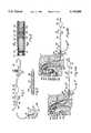
- FIG. 1is a plan view of a system for visualizing tissue that includes a support structure carrying an imaging probe;
- FIG. 2is a side section view of the imaging probe and support structure of FIG. 1 in a collapsed condition within an external slidable sheath;
- FIG. 3is a side section view of a portion of a spline that forms a part of the support structure shown in FIG. 1;
- FIGS. 4A and 4Bare side sectional, somewhat diagrammatic views of the deployment of the support structure and imaging probe shown in FIG. 1 within a heart chamber;
- FIG. 5Ais a side section view of the support structure and imaging probe shown in FIG. 1, showing various paths in which the imaging probe can be moved when located within a body region;
- FIG. 5Bis a side view of an alternative embodiment of an imaging probe and a support structure comprising a single spline element
- FIG. 6is an enlarged view of one embodiment of the support structure and imaging probe, in which the imaging probe includes a rotating ultrasonic transducer crystal;
- FIG. 7is an enlarged view of another embodiment of the support structure and imaging probe, in which the imaging probe includes a fiber optic assembly;
- FIG. 8is a partial side section, perspective, and largely schematic, view of a support structure and imaging probe as shown in FIG. 1, in which the imaging probe is associated with a system to conduct contrast echocardiography to identify potential ablation sites by imaging tissue perfusion;
- FIG. 9is a partial side section, largely schematic view of the support structure and imaging probe shown in FIG. 1, including an electromechanical axial translator connected to the imaging probe;
- FIG. 10is a side section view, somewhat diagrammatic is nature, showing a support structure and imaging probe, in which both the structure and the probe carry electrodes;
- FIG. 11is a side section view of a portion of an electrode-carrying spline that forms a part of the support structure shown in FIG. 10;
- FIG. 12is a side section view of a heart and a perspective view of the support structure and imaging probe shown in FIG. 10, being used in association with a separate roving mapping, pacing, or ablating electrode;
- FIG. 13Ais a side view, with portions removed, of a support assembly comprising a expanded porous body capable of ionic transfer of ablation energy, which carries an interior imaging probe;
- FIG. 13Bis a side elevation view of the porous body shown in FIG. 13A, with the porous body shown in a collapsed condition for introduction into an interior body region;
- FIG. 14is a side view of a support assembly carrying within it the porous body and imaging probe assembly shown in FIGS. 13A and 13B;
- FIG. 15is a side view, somewhat diagrammatic in form, showing a support structure that carries within it a movable imaging probe, the support structure also carrying multiple electrodes sized to create long lesion patterns;
- FIG. 16is an illustration representative of a typical small tissue lesion pattern
- FIG. 17is an illustration representative of a typical larger tissue lesion pattern
- FIG. 18is an illustration representative of a typical long tissue lesion pattern
- FIG. 19is an illustration representative of a typical complex long tissue lesion pattern
- FIG. 20is an illustration-representative of a typical segmented tissue lesion pattern
- FIG. 21is a side section view, somewhat diagrammatic in form, showing a support structure that carries within it an image acquisition element gated according to intracardiac activation sensed by an electrode also carried by the support structure;
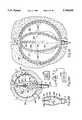
- FIG. 22is a side section view, somewhat diagrammatic in form, of a support structure that carries within it an image acquisition element, also shown with an enlarged perspective view, comprising a phased transducer array that includes multiple transducers panels scored on different planar sections of a piezoelectric material;
- FIG. 23is a side section view of a support structure that carries within it an image acquisition element comprising a phased multiple transducer array carried on flexible spline elements;
- FIG. 24is a side section view of a support structure that carries within it an image acquisition element comprising a phased multiple transducer array carried on an expandable-collapsible body;
- FIG. 25is a side section view, somewhat diagrammatic in form, of a support structure that carries within it an image acquisition element comprising an optical coherence domain reflectometer;
- FIG. 26is a diagrammatic view of a system for identifying the physical characteristics of a support structure using a machine-readable code, to enable the creation of a positioning matrix (shown in FIG. 10) to guide the imaging probe within the structure;
- FIG. 27is a diagrammatic view of one implementation of the machine-readable code used to identify the individual physical characteristics of the support structure shown in FIG. 26;
- FIG. 28is a diagrammatic view of another implementation of the machine-readable code used to identify the individual physical characteristics of the support structure shown in FIG. 26.
- FIG. 1shows a system 10, which embodies features of the invention, for visualizing interior regions of a living body.
- the inventionis well adapted for use inside body lumens, chambers or cavities for either diagnostic or therapeutic purposes. It particularly lends itself to catheter-based procedures, where access to the interior body region is obtained, for example, through the vascular system or alimentary canal, without complex, invasive surgical procedures.
- the inventionmay be used in diverse body regions for diagnosing or treating diseases.
- various aspects of the inventionhave application for the diagnosis and treatment of arrhythmia conditions within the heart, such as ventricular tachycardia or atrial fibrillation.
- the inventionalso has application in the diagnosis or treatment of intravascular ailments, in association, for example, with angioplasty or atherectomy techniques.
- Various aspects of the inventionalso have application for diagnosis or treatment of ailments in the gastrointestinal tract, the prostrate, brain, gall bladder, uterus, and other regions of the body.
- the inventioncan also be used in association with systems and methods that are not necessarily catheter-based. The diverse applicability of the invention in these and other fields of use will become apparent.
- the inventionmakes it possible for a physician to access and visualize or image inter-body regions, to thereby locate and identify abnormalities that may be present.
- the inventionprovides a stable platform through which accurate displays of these images can be created for viewing and analysis by the physician. Accurate images enable the physician to prescribe appropriate treatment or therapy.
- the inventionprovides a system 10 comprising a support structure 20 that carries within it an imaging or visualizing probe 34.
- the system 10includes a flexible catheter tube 12 with a proximal end 14 and a distal end 16.
- the proximal end 14carries an attached handle 18.
- the distal end 16carries the support structure 20.
- the support structure 20can be constructed in various ways. In one preferred embodiment (illustrated in FIG. 1), the structure 20 comprises two or more flexible spline elements 22. In FIG. 1, the support structure 20 includes eight spline elements 22. Of course, fewer or more spline elements 22 can be present.
- FIG. 5Ashows the support structure 20 comprising just two, generally oppositely spaced spline elements 22.
- FIG. 5Bshows the support structure 20 comprising a single spline element 22.
- FIG. 1shows the support structure 20 comprising just two, generally oppositely spaced spline elements 22.
- the distal end 23 of the spline element 22is attached to a stylet 25, carried by the catheter tube 12, which moves the distal end 23 (as shown by arrows 27) along the axis of the catheter tube 12 to adjust the curvature of the spline element 22.
- each spline element 22preferably comprises a flexible core body 84 enclosed within a flexible, electrically nonconductive sleeve 32.
- the sleeve 32is made of, for example, a polymeric, electrically nonconductive material, like polyethylene or polyurethane.
- the sleeve 32is preferable heat shrunk about the core body 84.
- the core body 84is made from resilient, inert wire or plastic. Elastic memory material such as nickel titanium (commercially available as NITINOLTM material) can be used. Resilient injection molded plastic or stainless steel can also be used.
- the core body 84is a thin, rectilinear strip. The rectilinear cross-section imparts resistance to twisting about the longitudinal axis of the core body 84, thereby providing structural stability and good bio-mechanical properties. Other cross-sectional configurations, such as cylindrical, can be used, if desired.
- each core body 84 of the spline elements 22extend longitudinally between a distal hub 24 and a base 26.
- the base 26is carried by the distal end 16 of the catheter tube 12.
- each core body 84is preformed with a convex bias, creating a normally open three-dimensional basket structure expanded about a main center axis 89.
- the system 10includes an outer sheath 44 carried about the catheter tube 12.
- the sheath 44has an inner diameter that is greater than the outer diameter of the catheter tube 12. As a result, the sheath 44 slides along the outside of the catheter tube 12.
- Rearward movementretracts the slidable sheath 44 away from the support structure 20. This removes the compression force.
- the freed support structure 20opens (as FIG. 1 shows) and assumes its three-dimensional shape.
- FIGS. 4A and 4Bshow a representative deployment technique usable when vascular access to a heart chamber is required.
- the physicianuses an introducer 85, made from inert plastic materials (e.g., polyester), having a skin-piercing cannula 86.
- the cannula 86establishes percutaneous access into, for example, the femoral artery 88.
- the exterior end of the introducer 85includes a conventional hemostatic valve 90 to block the outflow of blood and other fluids from the access.
- the valvemay take the form of a conventional slotted membrane or conventional shutter valve arrangement (not shown).
- a valve 90 suitable for usemay be commercial procured from B. Braun Medical Company (Bethlehem, Pa.).
- the introducer 85includes a flushing port 87 to introduce sterile saline to periodically clean the region of the valve 90.
- FIG. 4Ashows, the physician advances a guide sheath 92 through the introducer 85 into the accessed artery 88.
- a guide catheter or guide wire(not shown) may be used in association with the guide sheath 92 to aid in directing the guide sheath 92 through the artery 88 toward the heart 94.
- the views of the heart 94 and other interior regions of the body in this Specificationare not intended to be anatomically accurate in every detail.
- the Figuresshow anatomic details in diagrammatic form as necessary to show the features of the invention.
- the physicianobserves the advancement of the guide sheath 92 through the artery 88 using fluoroscopic or ultrasound imaging, or the like.
- the guide sheath 92can include a radio-opaque compound, such as barium, for this purpose.
- a radio-opaque markercan be placed at the distal end of the guide sheath 92.
- the physicianmaneuvers the guide sheath 92 through the artery 88 retrograde past the aortic valve and into the left ventricle 98.
- the guide sheath 92establishes a passageway through the artery 88 into the ventricle 98, without an invasive open heart surgical procedure.
- a conventional transeptal sheath assembly(not shown) can be used to gain passage through the septum between the left and right atria. Access to the right atrium or ventricle is accomplished in the same manner, but without advancing the transeptal sheath across the atrial septum.
- FIG. 4Ashows, once the guide sheath 92 is placed in the targeted region, the physician advances the catheter tube 12, with the support structure 20 confined within the slidable sheath 44, through the guide sheath 92 and into the targeted region.
- FIG. 4Bshows, pulling back upon the slidable sheath 44 (see arrow 45 in FIG. 4B) allows the structure 20 to spring open within the targeted region for use.
- the shape of the support structure 20(which, in FIG. 4B, is three-dimensional) holds the spline elements 22 in intimate contact against the surrounding tissue mass.
- the support structure 20has an open interior 21, which surrounds the imaging probe 34, keeping the tissue mass from contacting it.
- the geometry of flexible spline elements 22is radially symmetric about the main axis 89. That is, the spline elements 22 uniformly radiate from the main axis 89 at generally equal arcuate, or circumferential, intervals.
- the elements 22also present a geometry that is axially symmetric along the main axis 89. That is, when viewed from the side (as FIGS. 1 and 4B show) the proximal and distal regions of the assembled splines 22 have essentially the same curvilinear geometry along the main axis 89.
- the spline elements 22can form various other geometries that are either radially asymmetric, or axially asymmetric, or both.
- the axial geometry for the structure 20, whether symmetric or asymmetricis selected to best conform to the expected interior contour of the body chamber that the structure 20 will, in use, occupy.
- the interior contour of a heart ventriclediffers from the interior contour of a heart atrium.
- the ability to provide support structures 20 with differing asymmetric shapesmakes it possible to provide one discrete configuration tailored for atrial use and another discrete configuration tailored for ventricular use. Examples of asymmetric arrays of spline structures 20 for use in the heart are shown in copending U.S. application Ser. No. 08/742,569, filed Oct. 28, 1996, entitled "Asymmetric Structures for Supporting Diagnostic or Therapeutic Elements in Internal Body Regions,” which is incorporated herein by reference.
- the imaging probe 34 located within the support structure 20includes a flexible body 36, which extends through a central bore 38 in the catheter tube 12.
- the body 36has a distal region 40 that projects beyond the distal end 16 of the catheter tube 12 into the interior of the support structure 20.
- the body 36also includes a proximal region 42 that carries an auxiliary handle 46.
- Another conventional hemostatic valve 48is located at the distal end 16 of the catheter tube 12 to block the backflow of fluid through the catheter tube 12 while allowing the passage of the body 36.
- the distal body region 40carries an image acquisition element 50, which will be called in abbreviated form the IAE.
- the IAE 50generates visualizing signals representing an image of the area, and objects and tissues that occupy the area, surrounding the structure 20.
- the IAE 50can be of various constructions.
- the IAE 50comprises an ultrasonic transducer 52.
- the transducer 52forms a part of a conventional ultrasound imaging system 54 generally of the type shown in U.S. Pat. No. 5,313,949. This patent is incorporated herein by reference.
- the transducer 52comprises one or more piezoelectric crystals formed of, for example, barium titinate or cinnabar, which is capable of operating at a frequency range of 5 to 20 megahertz.
- Other types of ultrasonic crystal oscillatorscan be used.
- organic electretssuch as polyvinylidene difluoride and vinylidene fluoride-trifluoro-ethylene copolymers can also be used.
- the imaging system 54includes a transmitter 56 coupled to the transducer crystal 52 (see FIG. 6).
- the transmitter 56generates voltage pulses (typically in the range of 10 to 150 volts) for excitation of the transducer crystal 52.
- the voltage pulsescause the transducer crystal 52 to produce sonic waves.
- a motor 58rotates the transducer crystal 52 (being linked by the flexible drive shaft 53, which passes through a bore in the tube 36).
- the transmission of voltage pulses (and, thus, the sonic waves) and the rotation of the transducer crystal 52are synchronized by a timing and control element 60.
- the motor 58rotates the transducer crystal 52 in the range of 500 to 2000 rpm, depending upon the frame rate of the image desired.
- the rotating transducer crystal 52thereby projects the sonic waves in a 360° pattern into the interior of the chamber or cavity that surrounds it.
- Tissueincluding tissue forming anatomic structures, such as heart valves (which is generally designated T in the Figures), and internal tissue structures and deposits or lesions on the tissue, scanned by the rotating transducer crystal 52 will scatter the sonic waves.
- the support structure 20also scatters the sonic waves.
- the scattered wavesreturn to the rotating transducer crystal 52.
- the transducer crystal 52converts the scattered waves into electrical signals.
- the imaging system 54includes a receiver 57, which amplifies these electrical signals.
- the imaging system 54digitally processes the signals, synchronized by the timing and control element 60 to the rotation of the transducer crystal 52, using known display algorithms; for example, conventional radar (PPI) algorithms. These algorithms are based upon the direct relationship that elapsed time ( ⁇ t) between pulse emission and return echo has to the distance (d) of the tissue from the transducer, expressed as follows: ##EQU1##
- ⁇is the speed of sound in the surrounding media.
- the digitally processed signalsare supplied to a display unit 59.
- the display unit 59comprises a screen, which can be, for example, a CRT monitor.
- the display screen 59shows an ultrasound image or profile in the desired format, which depicts the tissue and anatomic structures scanned by the transducer crystal 52.
- the display screen 59can provide a single or multi-dimensional echocardiograph or a non-imaging A-mode display.
- a control console(not shown) may be provided to allow selection by the physician of the desired display format.
- the ultrasonic transducer crystal 52can be operated in conventional fashion without rotation, as shown in U.S. Pat. Nos. 4,697,595, or 4,706,681, or 5,358,148. Each of these patents is incorporated herein by reference.
- the IAE 50comprises a fiber optic assembly 62, which permits direct visualization of tissue.
- a fiber optic assembly 62which permits direct visualization of tissue.
- Various types of fiber optic assemblies 62can be used.
- the illustrated embodimentemploys a fiber optic assembly 62 of the type shown in U.S. Pat. No. 4,976,710, which is incorporated herein by reference.
- the assembly 62includes a transparent balloon 64 carried at the end of the body 36.
- the balloon 64is inflated with a transparent gas or liquid, thereby providing a viewing window that shields the fiber optic channels 66 and 68 from blood contact.
- the channelsincludes an incoming optical fiber channel 66, which passes through the body 36.
- the channel 66is coupled to an exterior source 70 of light.
- the channel 66conveys lights from the source 70 to illuminate the tissue region around the balloon 64.
- the channelsalso include an outgoing optical fiber channel 68, which also passes through the body 36.
- the channel 68is coupled to an eye piece 72, which can be carried, for example, on the handle 46. Using the eye piece 72, the physician can directly view the illuminated region.
- the IAE 50can incorporate other image acquisition techniques.
- the IAE 50can comprise an apparatus for obtaining an image through optical coherence tomography (OCT).
- OCToptical coherence tomography
- a type of OCT imaging device, called an optical coherence domain reflectometer (OCDR)is disclosed in Swanson U.S. Pat. No. 5,321,501, which is incorporated herein by reference.
- the OCDRis capable of electronically performing two- and three-dimensional image scans over an extended longitudinal or depth range with sharp focus and high resolution and sensitivity over the range.
- the IAE 50comprises the distal end 220 of an optic fiber path 222.
- the distal end 220is embedded within an inner sheath 224, which is carried within an outer sheath 226.
- the outer sheath 226extends in the distal body region 40, within the support structure 20.
- the inner sheath 224includes a lens 228, to which the distal fiber path end 220 is optically coupled.
- the inner sheath 224terminates in an angled mirror surface 230, which extends beyond the end of the outer sheath 226.
- the surface 230reflects optical energy along a path that is generally perpendicular to the axis of the distal end 220.
- a motor 232rotates the inner sheath 224 within the outer sheath 226 (arrow 237).
- the lens 228 and the mirror surface 230rotate with the inner sheath 224, scanning about the axis of rotation.
- a second motor 234laterally moves the outer sheath 226 (arrows 236) to scan along the axis of rotation).
- a source 238 of optical energyis coupled to the optic fiber path 222 through an optical coupler 240.
- the source 238generates optical energy of short coherence length, preferably less than 10 micrometers.
- the source 238may, for example, be a light emitting diode, super luminescent diode, or other white light source of suitable wavelength, or a short-pulse laser.
- a reference optical reflector 242is also coupled by an optic fiber path 244 to the optical coupler 240.
- the optical coupler 240splits optical energy from the source 238 through the optic fiber path 222 to the distal optic path end 220 and through the optic fiber path 244 to the optical reflector 242.
- the optical energy supplied to the distal optic path end 220is transmitted by the lens 228 for reflection by the surface 230 toward tissue T.
- the scanned tissue T(including anatomic structures, other internal tissue topographic features, and deposits or lesions on the tissue) reflects the optic energy, as will the surrounding support structure 20.
- the reflected optic energyreturns via the optic path 222 to the optical coupler 240.
- the optical energy supplied to the reference optical reflector 242is reflected back to the optical coupler 240 by a corner-cube retro-reflector 246 and an end mirror 250 (as phantom lines 239 depict).
- the corner-cube retro-reflector 246is mounted on a mechanism 248, which reciprocates the corner-cube retro-reflector 246 toward and away from the optical path 244 and an end mirror 250 (as arrows 241 depict).
- the mechanism 248preferable moves the corner-cube retro-reflector 246 at a uniform, relatively high velocity (for example, greater than 1 cm/sec), causing Doppler shift modulation used to perform heterodyne detection.
- the length or extent of movement of the corner-cube retro-reflector 246 caused by the mechanism 248is at least slightly greater than half the scanning depth desired.
- the total length of the optical path 222 between the optical coupler 240 up to the desired scanning depth pointis also substantially equal to the total length of the optical path 244 between the optical coupler 240 and the end mirror 250. Movement of the corner-cube retro-reflector 246 will cause periodic differences in the reflected path lengths 222 and 244.
- Reflections received from the optical path 222 (from the lens 228) and the optical path 244 (from the end mirror 250)are received by the optical coupler 240.
- the optical coupler 240combines the reflected optical signals. Due to movement of the corner-cube retro-reflector 246, the combined signals have interference fringes for reflections in which the difference in the reflected path lengths is less than the source coherence length. Due to movement of the corner-cube retro-reflector 246, the combined signals also have an instantaneous modulating frequency.
- the combined outputis coupled via fiber optic path 252 to a signal processor 254.
- the signal processor 254converts the optical output of the coupler 240 to voltage-varying electrical signals, which are demodulated and analyzed by a microprocessor to provide an image output to a display device 256.
- the support structure 20 positioned about the distal region of the probe 34remains substantially in contact against surrounding tissue mass T as the IAE 50 operates to acquire the desired image or profile (see FIGS. 5 to 8).
- the support structure 20serves to stabilize the IAE 50 and keep tissue T from contacting and possible occluding the IAE 50.
- Stabilizing the IAE 50is particularly helpful when the geometry of surrounding body chamber or passage 100 is dynamically changing, such as the interior of a heart chamber during systole and diastole.
- the IAE 50is thereby allowed to visualize tissue and anatomic structures T, without the attendant need for constant positioning and repositioning.
- the structure 20thus makes possible the generation of accurate images of the targeted body region by the IAE 50.
- the physiciancan move the IAE 50 within the structure 20 forward and rearward (respectively, arrows 101 and 103 in FIG. 5A) by pushing or pulling upon the auxiliary handle 46.
- the physicianmay also manually rotate the IAE 50 within the structure 20.
- the illustrated and preferred embodimentfurther includes a mechanism 74 for deflecting, or steering, the distal region 40 of the body 36, and with it the IAE 50, transverse of the axis 89 (as depicted in phantom lines 40 in FIG. 5A).
- the construction of the steering mechanism 74can vary.
- the steering mechanism 74is of the type shown in U.S. Pat. No. 5,336,182, which is incorporated by reference.
- the steering mechanism 74 of this constructionincludes an actuator 76 in the auxiliary handle 46.
- the actuator 76takes the form of a cam wheel rotated by means of an external steering lever 78.
- the cam wheel 76holds the proximal ends of right and left steering wires 80.
- the steering wires 80extend from the cam wheel 76 and through the body 36.
- the steering wires 80connect to the left and right sides of a resilient bendable wire 82 or spring present within the distal region 40. Rotation of the cam wheel 76 places tension on steering wires 80 to deflect the distal region 40 of the body 36, and, with it, the IAE 50 (as shown by arrows 107 in FIG. 5A).
- the physiciancan manually move the IAE 50 with respect to the structure 20 in three principal directions.
- the IAE 50can be moved along the axis 86 of the structure 20 by pushing and pulling on the auxiliary handle 46 (arrows 101 and 103).
- the IAE 50can be moved rotationally about the axis 86 of the structure 20 by torquing the auxiliary handle 46 (arrows 105).
- the IAE 50can be moved in a direction normal to the axis 86 of the structure 20 by operating the steering mechanism 74 (arrows 107).
- the physiciancan manually move the IAE 50 in virtually any direction and along any path within the structure 20.
- the IAE 50can thereby image tissue locations either in contact with the exterior surface of the structure 20 or laying outside the reach of the structure 20 itself.
- FIG. 9shows an electro-mechanical system 102 for manipulating the IAE 50 within the structure 20.
- the system 102synchronizes the imaging rate of the IAE 50 with movement of the IAE 50 within the structure 20.
- the systemallows the physician to use the structure 20 to accurately acquire a set of image slices, which can be processed in an automated fashion for display.
- the system 102includes a longitudinal position translator 104 mechanically coupled to the probe handle 46.
- the translator 104includes a stepper motor 106 that incrementally moves an axial screw 111 attached to the handle 46.
- the motor 106rotates the screw 111 to move the IAE 50 at a specified axial translation rate within the structure 20, either forward (arrows 101) or rearward (arrows 103).
- FIG. 9shows, during axial translation, the distal body region 40 carrying the IAE 50 is preferably maintained in a generally straight configuration, without transverse deflection.
- the system 102provides as output axially spaced, data sample slices of the region surrounding the IAE 50.
- an axial translator 104 of the general type shown in FIG. 4 in combination with a rotating transducer crystal 52 of the type shown in FIG. 6is described in U.S. Pat. No. 5,485,846, which is incorporated herein by reference.
- the system 102By rotating the transducer crystal 52 in synchrony with the axial translation rate of the translator 104, the system 102 provides axially spaced, 360° data sample slices of the region perpendicular to the transducer crystal 52.
- Conventional signal processing techniquesare used to reconstruct the data slices taken at specified intervals along the axis into three-dimensional images for display. This technique is well suited for acquiring images inside blood vessels or other body regions having a known, relatively stable geometry.
- the stepper motor 106is preferable gated by a gating circuit 190 (see FIG. 9) to the QRS of an electrocardiogram taken simultaneously with image gathering, for example, by using a surface electrode 188 shown in FIG. 9.
- the gating circuit 190is also synchronized with the imaging system 54 (as described in greater detail in conjunction with FIG. 6), so that the data image slices are recorded in axial increments at either end-diastolic or end-systolic points of the heart beat.
- the data slice recordingsare preferably gated to the p-wave.
- the imagingis preferably gated to the r-wave.
- the circuit 190is gated to the timing of local intracardiac electrogram activation.
- the flexible body 36which carries the transducer 54 within the structure 20, also carries an electrode 184 to sense electrograms in the region of the structure 20.
- the sensed electrogramsare conveyed to the circuit 190 to gate the stepper motor 106, as before described.
- the data slice recordingsare gated to the atrial intracardiac electrogram activation.
- the data slice recordingsare gated to the ventricular intracardiac electrogram activation.
- the body 36 carrying the transducer 54 and the electrode 184is preferably confined for movement within a straight, generally rigid sheath 186.
- the sheath 186guides the body 36 along a known, stable reference axis 183.
- the sheath 186is also preferably constructed of an ultrasonically transparent material, like polyethylene.
- the transducer 54 and electrode 184move in tandem within the confines of the sheath 186 (as shown by arrows 187 and 189 in FIG. 21) in response to the gated action of the stepper motor 106. Because the sheath 186 is ultrasonically transparent, the transducer 54 can remain within the confines of the sheath 186 while acquiring images. Nonlinearities in image reconstruction caused by deflection of the transducer outside of the axis 183, as would occur should the transducer 54 move beyond the sheath 186, are avoided.
- the acquired data image slices, position-gated by the electrograms while maintained along a known, stable reference axis 183,are generated for accurate reconstruction into the desired three-dimensional image.
- a catheter tracking system as described in Smith et al. U.S. Pat. 5,515,853may be used to track the location and orientation of the IAE 50 during movement.
- Another system that can be used for this purposeis disclosed in copending U.S. patent application Ser. No. 08/717,153, filed Sep. 20, 1996 and entitled "Enhanced Accuracy of 3-Dimensional Intraluminal Ultrasound (ILUS) Image Reconstruction,” naming Harm TenHoff as an inventor.
- the structure 20itself can establish a localized position-coordinate matrix about the IAE 50.
- the matrixmakes it possible to ascertain and thereby guide the relative position of the IAE 50 within the structure 20 (and thus within the targeted body cavity), to image specific regions within the targeted body cavity.
- the IAE 50carries an electrode 31 for transmitting electrical energy.
- each spline 22carries an array of multiple electrodes 30 for transmitting electrical energy.
- the electrodes 30are supported about the core body 84 on the flexible, electrically nonconductive sleeve 32, already described.
- the electrodes 30are electrically coupled by wires (not shown), which extend beneath the sleeve 32 through the catheter tube 12 to external connectors 32, which the handle 18 carries (see FIG. 1).
- each electrode 30comprises a solid ring of conductive material, like platinum, which is pressure fitted about the sleeve 32.
- the electrodes 30comprise a conductive material, like platinum-iridium or gold, coated upon the sleeve 32 using conventional coating techniques or an ion beam assisted deposition (IBAD) process.
- the electrodes 30comprise spaced apart lengths of closely wound, spiral coils wrapped about the sleeve 32.
- the coilsare made of electrically conducting material, like copper alloy, platinum, or stainless steel.
- the electrically conducting material of the coilscan be further coated with platinum-iridium or gold to improve its conduction properties and biocompatibility. Further details of the use of coiled electrodes are found in U.S. Pat. No. 5,545,193 entitled "Helically Wound Radio-Frequency Emitting Electrodes for Creating Lesions in Body Tissue," which is incorporated herein by reference.
- the electrodes 30can be formed as part of a ribbon cable circuit assembly, as shown in pending U.S. application Ser. No. 08/206,414, filed Mar. 4, 1994, which is incorporated herein by reference.
- a microprocessor controlled guidance element 108is electrically coupled to the electrodes 30 on the structure 20 and the electrode 31 carried by the IAE 50.
- the element 108conditions the electrodes 30 on the structure 20 and the IAE electrode 31 to generate an electric field (shown in phantom lines 113 in FIG. 10) within the structure 20, while also sensing electrode electric potentials in the electric field. More particularly, the element 108 commands a transmitting electrode, which can be either the IAE electrode 31 or at least one of the electrodes 30 in the structure 20, to transmit electrical energy.
- the element 108commands a sensing electrode, which also can be either the IAE electrode 31 or at least one of the electrodes 30 on the structure 20, to sense electrical energy emitted by the emitting electrode.
- the element 108generates an output by analyzing spatial variations in the electrical potentials within the field 113, which change based upon the relative position of the IAE electrode 31 relative to electrode 30 on the structure 20.
- the variationscan comprise variations in phase, variations in amplitude, or both.
- the element 108generates an output by analyzing spatial variations in impedances between the transmitting and sensing electrodes.
- the outputlocates the IAE 50 within the space defined by the structure 20, in terms of its position relative to the position of the multiple electrodes 30 on the structure 20.
- the element 108includes an output display device 110 (e.g., a CRT, LED display, or a printer), which presents the position-identifying output in a real-time format most useful to the physician for remotely guiding the IAE 50 within the structure 20.
- an output display device 110e.g., a CRT, LED display, or a printer
- structure 20carries an identification component 270.
- the identification component 270carries an assigned identification code XYZ.
- the code XYZidentifies the shape and size of the structure 20 and the distribution of electrodes 30 carried by the structure 20, in terms of the number of electrodes and their spatial arrangement on the structure 20.
- the structure-specific information contained in the code XYZaids the element 108 in creating a positioning matrix using the electrodes 30, to help guide the IAE 50 within the structure 20.
- the coded component 270is located within the handle 46 attached to the proximal end 14 of the catheter tube 12 that carries the structure 20.
- the component 270could be located elsewhere in relation the structure 20.
- the coded component 270is electrically coupled to an external interpreter 278 when the structure 20 is coupled to the element 108 for use.
- the interpreter 278inputs the code XYZ that the coded component 270 contains.
- the interpreter 278electronically compares the input code XYZ to, for example, a preestablished master table 280 of codes contained in memory.
- the master table 280lists, for each code XYZ, the structure-specific information required to create the positioning matrix to guide the IAE 50 within the structure 20.
- the element 108preferably includes functional algorithms 288 which set guidance parameters based upon the code XYZ. These guidance parameters are used by the signal processing component 274 of the element in analyzing the spatial variations of the electric field created within the structure 20 to guide the IAE 150. The guidance parameters are also used to create the position-identifying output displayed on the device 110.
- the algorithms 288preferably disable the guidance signal processing component 274 in the absence of a recognizable code XYX. Thus, only structures 20 possessing a coded component 270 carrying the appropriate identification code XYZ can be used in association with the element 108 to guide the IAE 50.
- the coded component 270can be variously constructed. It can, for example, take the form of an integrated circuit 284 (see FIG. 27), which expresses in digital form the code XYZ for input in ROM chips, EPROM chips, RAM chips, resistors, capacitors, programmed logic devices (PLD's), or diodes. Examples of catheter identification techniques of this type are shown in Jackson et al. U.S. Pat. No. 5,383,874, which is incorporated herein by reference.
- the coded component 270can comprise separate electrical elements 286 (see FIG. 28), each one of which expressing a individual characteristic.
- the electrical elements 286can comprise resistors (R1 to R4), comprising different resistance values, coupled in parallel.
- the interpreter 278measures the resistance value of each resistor R1 to R4.
- the resistance value of the first resistor R1expresses in preestablished code, for example, the number of electrodes on the structure.
- the resistance value of the second resistor R2expresses in preestablished code, for example, the distribution of electrodes on the structure.
- the resistance value of the third resistor R3expresses in preestablished code, for example, the size of the structure.
- the resistance value of the fourth resistor R4expresses in preestablished code, for example, the shape of the structure.
- the electrodes 30/31can define passive markers that, in use, do not transmit or sense electrical energy.
- the markersare detected by the physician using, for example, external fluoroscopy, magnetic imaging, or x-ray to establish the location of the structure 20 and the IAE 50.
- the stability and support that the structure 20 provides the IAE 50is well suited for use in association with an IAE 50 having one or more phased array transducer assemblies.
- the stability and support provided by the structure 20make it possible to accommodate diverse numbers and locations of phased array transducers in close proximity to tissue, to further enhance the resolution and accuracy of images created by the IAE 50.
- the structure 20carries an IAE 50 comprising a phased array 192 of ultrasonic transducers of the type shown, for example, in Shaulov U.S. Pat. No. 4,671,293, which is incorporated herein by reference.
- the array 192includes two groups 194 and 196 of electrodes.
- the electrode groups 194 and 196are differently partitioned by channels 206 on opposite faces or planar sectors 194' and 196' of a piezoelectric material 198.
- the channels 206cut through the electrode surfaces partially into and through the piezoelectric material 198 to prevent mechanical and electrical coupling of the elements.
- the channels 206 on the planar section 194'create spaced transducer elements 202a, 202b, 202c, etc.
- the channels 206 on the planar section 196'create spaced transducer elements 204a, 204b, 204c, etc.
- the electrode groups 194 and 196are alternatively pulsed by a conventional phase array circuit 200.
- the electrode element group 194is grounded, while the transducer elements 204a, 204b, 204c, etc. on the other planar section 196' are simultaneously pulsed, with the phase relationship of the stimulation among the transducer elements 204a, 204b, 204c, etc. set to create a desired beam angle, acquiring an image along the one planar sector 196'.
- the other electrode element group 196is grounded, while the transducer elements 202a, 202b, 202c, etc. on the other planar section 194' are likewise simultaneously pulsed, acquiring another image along the planar sector 194'. Further details, not essential to the invention, are provided in Haykin, Adaptive Filter Theory, Prentice-Hall, Inc. (1991), pp. 60 to 65.
- the signals received by the transducer groups 202a, 202b, 202c, etc. and 204a, 204b, 204c, etc., when pulsed,are processed into amplitude, phase, frequency, and time response components.
- the processed signalsare compared to known configurations with varying transducers activated to produce and measure the desired waveform.
- signals from combinations of transducersare processed, a composite image is produced.
- the phased array 192 shown in FIG. 22permits the real time imaging of two different planar sectors, which can be at any angle with respect to each other.
- FIGS. 23 and 24show other embodiments of an IAE 50 comprising a phased array of transducers carried within the structure 20.
- the IAE 50comprises an array of flexible spline elements 208 having a known geometry.
- the spline elements 208are carried within the support structure 20, which itself comprises a larger diameter array of flexible spline elements 22, as previously discussed in conjunction with FIG. 1.
- Each flexible spline element 208carries a grouping of multiple ultrasonic transducers 210.
- Collapsing the outer structure 20 of spline elements 22 by advancing the sheath 44also collapses the inner IAE structure of spline elements 208.
- the mutually collapsed geometrypresents a low profile allowing joint introduction of the structures 22 and 208 into the desired body region.
- the IAE 50comprises an expandable-collapsible body 212 carried within the support structure 20.
- the structure 20is shown as comprising the array of flexible spline elements 22.
- the exterior surface of the body 212carries an array of multiple ultrasonic transducers 210.
- An interior lumen 214 within the body 216 carrying the IAE 50conducts a fluid under pressure into the interior of the body 212 (as shown by arrows 213 in FIG. 24) to inflate it into a known expanded geometry for use.
- the body 212assumes a collapsed geometry (not shown).
- the advanced sheath 44envelopes the collapsed body 212, along with the outer structure 20, for introduction into the desired body region.
- the ultrasonic transducers 210are placed upon the spline elements 208 or expandable body 212 (which will be collectively called the "substrate") by depositing desired transducer materials or composites thereof onto the substrate. Ion beam assisted deposition, vapor deposition, sputtering, or other methods can be used for this purpose.
- a masking materialis placed on the substrate to keep regions free of the deposited material. Removal of the masking material after deposition of the transducer materials provides the spaced apart array on the substrate. Alternatively, an etching process may be used to selectively remove sectors of the transducer material from the substrate to form the desired spaced apart array.
- the size of each deposited transducer 210 and the density of the overall array of transducers 210should be balanced against the flexibility desired for the substrate, as conventional transducer material tends to be inherently stiffer than the underlying substrate.
- transducers 210can be attached in a preformed state by adhesives or the like to the spline elements 208 or flexible body 212. Again, the size of each attached transducer 210 and the density of the overall array of transducers 210 should be balanced against the flexibility desired for the substrate.
- Signal wiresmay be coupled to the transducers 210 in various ways after or during deposition or attachment; for example by soldering, or by adhesive, or by being deposited over.
- Various other ways to couple signal wires to solid or deposited surfaces on an expandable-collapsible bodyare discussed in copending patent application Ser. No. 08/629,363, entitled “Enhanced Electrical Connections for Electrode Structures,” filed Apr. 8, 1996, which is incorporated herein by reference.
- the signal wiresmay be bundled together for passage through the associated catheter tube 12, or housed in ribbon cables for the same purpose in the manner disclosed in Kordis U.S. Pat. No. 5,499,981, which is incorporated herein by reference.
- the multiple ultrasonic transducers 210could be supported on other types of bodies within the structure 20.
- non-collapsible hemispherical or cylindrical bodieshaving fixed predetermined geometries, could occupy the interior of the structure 20 for the purpose of supporting phased arrays of ultrasonic transducers 210.
- the signal wires and transducersmay be braided into a desired three-dimensional structure.
- the braided structuremay further be laminated to produce an inflatable balloon-like structure.
- the dimensions of these alternative transducer support bodiescan vary, subject to the requirement of accommodating introduction and deployment in an interior body region.
- phased arrays of multiple transducersare found, for example, in Griffith et al. U.S. Pat. No. 4,841,977 and Proudian et al. U.S. Pat. No. 4,917,097.
- Phased arrays of multiple transducersmay be used in association with gating techniques, described above in conjunction with FIG. 9, to lessen the image acquisition time.
- gatingmay be used to synchronize the phased acquisition of multiple plane images with the QRS or intracardiac electrogram activation, particularly if it is desired to analyze the images over more than one heart beat.
- the structure 20can carry an array of electrodes 30 for the purpose of guiding the IAE 50. These same electrodes 30 can also serve to sense electrical impulses in tissue, like myocardial tissue. This sensing function in heart tissue is commonly called "mapping.”
- the support structure 20holds the electrodes 30 in contact against the endocardium.
- the electrodessense the electrical impulses within the myocardium that control heart function.
- the element 108includes or constitutes an external signal processor made, for example, by Prucka Engineering, Inc. (Houston, Tex.).
- Prucka Engineering, Inc.Houston, Tex.
- the processed signalsare analyzed to locate aberrant conductive pathways and identify foci. The foci point to potential ablation sites.
- the electrodes 30 on the support structure 20can be used to derive an electrical characteristic, such as impedance, in heart tissue for the purpose of characterizing tissue and locating aberrant conductive pathways.
- an electrical characteristicsuch as impedance
- Systems and methods for deriving an electrical characteristic of tissue for this purposeare disclosed, for example, in Panescu et al U.S. Pat. No. 5,494,042, which is incorporated herein by reference.
- An electrical characteristicis derived by transmitting electrical energy from one or more electrodes into tissue and sensing the resulting flow of electrical energy through the tissue.
- the IAE 50 carried within the multiple electrode structure 20greatly assists the, physician in mapping or characterizing tissue, whether in the heart or elsewhere in the body, by locating the electrodes 30 in the desired orientation with respect to selected anatomic sites.
- the physiciancan manipulate the IAE 50 in the manners previously described to visual identify the coronary sinus, heart valves, superior and inferior vena cava, the fossa ovalis, the pulmonary veins, and other key anatomic sites in the heart. Relying upon the visual information obtained by the IAE 50, the physician can then orient the multiple electrode structure 20 with respect to one or more of these anatomic sites. Once properly oriented, the physician can further visualize with the IAE 50, to assure that all or a desired number of the electrodes 30 carried by the structure 20 are in intimate contact with tissue required for good signal transmission or good signal acquisition.
- the IAE 50can also be used to help visually steer a separate mapping electrode 112, carried on its own catheter tube 121, outside or within the support structure 20 into the desired location in contact with heart tissue. If the roving electrode 112 is present within the confines of the support structure 20, the structure 20 also serves to stabilize the electrode 112.
- the guidance processing element 108as previously described (see FIG. 10) can be used in association with the structure 20 to electronically home the external mapping electrode 112 to a desired location within the structure 20.
- FIG. 8shows a system 170 that includes the structure 20 carrying an IAE 50 to identify perfusion patterns in myocardial tissue and, thereby, diagnose potential ablation sites within the heart.
- the IAE 50 carried within the structure 20comprises a rotating ultrasonic transducer 52 of the type previously described in conjunction with FIG. 6.
- the system 170 shown in FIG. 8also preferably includes an electro-mechanical system 102 for incrementally moving the transducer 52 within the structure 20 to obtain axially spaced, data sample slices of the region surrounding the transducer 52. The details of this the system 102 have been previously described in conjunction with FIG. 9.
- the electro-mechanical system 102may also be gated to the QRS of an electrocardiogram or to intracardiac electrogram activation to acquire images at either end-diastolic or end-systolic points of the heart cycle, in the manner also previously described in conjunction with FIGS. 9 or 21.
- the system 170 shown in FIG. 8includes a separate catheter 172.
- the catheter 172includes an interior lumen 174, which is coupled to a source of an echoluscient contrast media 176.
- the catheter 172injects the media 176 into the blood stream.
- the echoluscient contrast media 176 usedmay vary.
- the media 176comprises sonicated albumin microbubbles, or their equivalent, having a diameter smaller than red blood cells (which are typically about 8 ⁇ m).
- the microbubbles in the media 176are perfused into tissue, just as the blood components that accompany them.
- the physicianis thereby able to accurately observe the patterns of perfusion of the media 176 into tissue. The more volume of media 176 perfused into tissue, the brighter the ultrasonic image, and vice versa.
- Myocardial tissue that has been infarctedhas significantly lower perfusion characteristics than healthy myocardial tissue. See, for example, Nath et al., "Effects of Radiofrequency Catheter Ablation on Regional Myocardial Blood Flow,” Circulation, 1994; 89: 2667-2672; and Villaneuva et al., "Assessment of Risk Area During Coronary Occlusion and Infarct Size After Reperfusion with Myocardial Contrast Echocardiography Using Left and Right Atrial Injections of Contrast," Circulation, 1993; 88: 596-604).
- the catheter 172is preferably maneuvered percutaneously into a selected coronary vessel.
- the contrast media 176is injected through the catheter lumen 174 into the vessel, and thus into the vascular system near the heart.
- the media 176is distributed throughout the regions of the heart perfused by the coronary artery, increasing the resolution and contrast in a selected localized region. More global distribution of contrast media 176 can be obtained by selecting an injection site in one of the heart chambers or in the pulmonary artery.
- the catheter 172is preferably maneuvered to inject the media 176 into the circumflex coronary artery branch of the left main artery. If myocardial tissue in the anterior aspect of the right or left ventricles is slated for diagnosis, the catheter 172 is preferably maneuvered to inject the media 176 into the left anterior descending (LAD) coronary artery branch of the left main artery. If myocardial tissue in the free wall of the right ventricle or the posterior ventricular septum is slated for diagnosis, the catheter 172 is preferably maneuvered to inject the media 176 into the right coronary artery.
- LADleft anterior descending
- the media 176can be injected directly into the left atrium or left ventricle.
- the body 36 carrying the transducer 52can also include an interior lumen 178 to convey the media 176.
- This approachmay be easier and potentially less traumatic than injection directly into the coronary artery.
- a portion of the media 176will still be dispersed past the coronary arteries and through the systemic arterial system, thereby resulting in a poorer resolution per given volume of media 176 injected. Therefore, a larger volume of media 176 should be injected directly into the left atrium or ventricle to obtain contrast in myocardial tissue comparable to a smaller volume of media 176 injected directly into a coronary artery, as described above.
- contrast media 176may be injected systemically into the femoral vein. Again, with this approach, significant portions of the media 176 will be disbursed within the circulatory system, and, in particular, into the lungs. As just discussed, a larger volume of media 176 should be injected systemically into the femoral vein to obtain contrast in myocardial tissue comparable to a smaller volume of media 176 injected directly into a coronary artery.
- the system 170includes a receiver and processor 180 and display device 182, as earlier described in conjunction with FIG. 6.
- the receiver and processor 180preferably creates a three-dimensional image for display on the device 182.
- an echocardiographic imagemay be created for display without using the axial translation system 102.
- the contrast media 176highlights the differences in perfusion in myocardial tissue surrounding the structure 20. Regions of infarcted tissue are visually characterized, as they are not well perfused with blood and appear in negative contrast to the healthy tissue regions that are well perfused. The same visually characterized, negative contrast regions of infarcted tissue may also form part of the pathways of slow conduction of electrical impulses. These slow conduction pathways may be a substrate for ventricular tachycardia and therefore candidates for cardiac ablation. These candidate regions of slow conduction pathways will, in the presence of the contrast media 186, appear on the ultrasonic device 182 as zones of negative contrast, being significantly less ultrasonically "bright" than well perfused tissue regions.
- the candidate regions of slow conductionwill typically have infarcted tissue interspersed with well perfused tissue.
- the candidate regionswill therefore appear ultrasonically "mottled", with patchy regions of darker contrast interspersed with lighter contrast.
- the mottled zoneswill appear contiguous to negative contrast areas.
- the image resolution of the device 182should preferably be fine enough to discern among mottled zones, light contrast zones, and dark contrast zones.
- the support structure 20maintains the transducer 54 in a stable, substantially unobstructed viewing position near the targeted tissue region.
- the transducer 54thereby generates ultrasonic images of the differences in perfusion of the media 176 throughout the imaged heart tissue.
- the system 170therefore make possible the accurate characterization of tissue for identifying potential ablation sites using contrast echocardiography.
- the stable, unobstructed perfusion images that the system 170 providesalso make it possible to discern the lesion characteristic required to treat the arrhythmia.
- the perfusion patternmay indicate a localized, contained mottled contrast area, suited for treatment by creating an equally localized, small surface area lesion.
- the perfusion patternmay indicate a larger or deeper mottled contrast area, or a mottled contrast area that is elongated or a random complex of different, intersecting geometries.
- the stable, unobstructed perfusion images that the system 170 providesalso make it possible to characterize tissue substrates associated with polymorphic ventricular tachycardia.
- the system 170makes it possible to characterized these regions using echocardiography during normal sinus rhythm.
- Conventional mapping of electrical eventsrequires induction of sometimes hemodynamically unstable rhythms to locate and ablate substrates associated with polymorphic ventricular tachycardia.
- the stable, unobstructed perfusion images that the system 170 providesalso make it possible to discern intermediate contrast zones between "bright” (well perfused tissue) images and negative contrast (not well perfused, infarcted tissue) images. These intermediate contrast zones also delineate the infarcted tissue border. Once identified, tissue ablation can be conducted with the objective of ablating tissue within the border zone, to eliminate the potential for ventricular tachycardia substrates.
- the system 170may characterize tissue morphology based upon echocardiography to locate potential ablation sites in other ways.
- the system 170may image based upon ultrasonic frequency domain analyses.
- the intensity of the second harmonicscan be used to identify tissue morphologies such as scar tissue, ischemic tissue, infarcted tissue, and healthy tissue as a function of tissue elasticity.
- Frequency domain analyses like second harmonicsmay be used without the injection of contrast media 170 to characterize tissue for ablation purposes.
- the system 170 for carrying out contrast echocardiographymay also incorporate an IAE 50 comprising multiple transducers and using phased array techniques to enhance the perfusion images, as previously described in conjunction with FIGS. 22 to 24.
- FIG. 8shows the system 170 being used in association with intracardiac echocardiography. It should also be appreciated that the echocardiography can be used to characterize tissue morphology, and thereby identify potential ablation sites, using external ultrasound transducers located outside the body.
- system 170can be used as an adjunct to other echography procedures; for example, transesophageal or transthoracic echography.
- the analysis of tissue perfusion patterns to characterize myocardial tissue to locate potential ablation sitescan also be accomplished using external imaging techniques other than echography.
- magnetic resonance imagingMRI
- an isotopesuch as gadolinium-chelate
- CTcomputerized tomography
- iodine radiopaque compoundssuch as renografin
- nuclear imagingusing thallium as the contrast material can be used.
- the image resolution of the alternative techniqueshould preferably be fine enough to discern among mottled zones, light contrast zones, and dark contrast zones.
- the alternative imaging techniqueslike echography, can also be used to discern intermediate contrast zones, which delineate infarcted tissue borders.
- the foregoing description of the structure 20 and associated IAE 50exemplify use in the performance of general diagnostic functions, to accurately locate and identify abnormalities that may be present in body cavities or in electrical activities within tissue.
- the structure 20 and associated IAE 50can also aid in providing therapeutic functions, alone or in combination with these and other diagnostic functions.
- the physiciandeploys an ablation element to the site. While various types of ablation energy can be used, in the preferred implementation, the ablation electrode transmits radio frequency energy conveyed from an external generator (not shown).
- the ablation elementcan takes various forms, depending upon the type of lesion required, which, in turn, depends upon the therapeutic effect desired.
- lesions that are characterized as "small and shallow”have a depth of about 0.5 cm, a width of about 10 mm, and a lesion volume of up to 0.2 cm 3 .
- FIG. 16exemplifies the geometry for a typical "small" lesion 118.
- These lesionsare desired in the sinus node for sinus node modifications, or along the A-V groove for various accessory pathway ablations, or along the slow zone of the tricuspid isthmus for atrial flutter (AFL) or AV node slow pathways ablations.
- a physicianwill typically deploy an electrode having approximately an 8F diameter and a 4 mm length to transmit radio frequency energy to create small and shallow lesions in myocardial tissue.
- This type of ablation electrodecan be used in association with the support structure 20, even when the catheter tube bore is occupied by the imaging probe 34.
- the physicianseparately deploys the ablation electrode as a "roving" electrode 112 outside the support structure 20.
- the physicianthen steers the external electrode 112 into the confines of the support structure 20 for ablation (such an electrode 112 can also perform an auxiliary mapping function, as already described).
- the electrode 112is preferably operated in a uni-polar mode during ablation, in which the radio frequency ablation energy transmitted by the electrode 112 is returned through an indifferent patch electrode 114 externally attached to the skin of the patient.
- the support structure 20serves to stabilize the external "roving" ablation electrode 112 within a confined region of the heart.
- the IAE 50can be used in this arrangement to help visually navigate the roving ablation electrode 112 into the desired location in contact with heart tissue.
- the guidance processing element 108 as previously describedcan also be used in association with the structure 20 to electronically home the roving ablation electrode 112 to the desired ablation site contacting the support structure 20.
- the electrode 31 that the IAE 50 carriescan comprise an ablation electrode, in the manner shown in U.S. Pat. No. 5,385,148, which is incorporated herein by reference.
- the exterior diameter of the IAE 50 (with electrode 31)is preferably larger than the interior diameter of the catheter tube bore 38 (see FIG. 5A).
- the slidable sheath 44 that encloses the structure 20 during deploymentalso encloses the IAE 50 and ablation element 31 within the collapsed structure 20.
- Further details of a structure integrating a movable element within a multiple electrode support structurecan be found in U.S. Pat. No. 5,476,495, which is incorporated herein by reference.
- the guidance processing element 108can also create a position-identifying output in a real-time format most useful to the physician for guiding the ablation electrode 31 carried by the IAE 50 within the structure 20 toward a potential site identified for ablation.
- the exterior diameter of the IAE 50(with electrode 31) is smaller than the interior diameter of the catheter tube bore 38.
- the IAE 50 and the entire imaging probe 34can thereby be withdrawn through the catheter tube bore 38 from the catheter tube 12.
- the catheter tube 12 carrying the multiple electrode support structure 20 and the imaging probe 34comprise separately deployed components.
- the imaging probe 34is deployed through the catheter tube 12 only when the visualization function is required.
- the catheter tube bore 38is open to provide passage for other components; for example, the separate mapping or ablation electrode 112 shown in FIG. 12.
- the imaging probe 34can be switched in situ with the mapping or ablation electrode 112, without altering the position of the structure 20.
- FIG. 17exemplifies the geometry of a typical larger surface area lesion 120, compared to the geometry of the smaller lesion 118 shown in FIG. 16.
- FIGS. 13A and 13Bshow an alternative embodiment of the invention, which provides a composite structure 122 carrying an imaging probe 124 and an ablation element 126, which is capable of providing larger lesions.
- the composite structure 122(like structure 20 shown in FIG. 1) is carried at the distal end of a flexible catheter tube 12.
- the proximal end of the catheter tubecarries an attached handle 18 for manipulating the composite structure in the manners previously described.
- the composite structure 122comprises an expandable-collapsible hollow body 128 made from a porous transparent thermoplastic or elastomeric material.
- the size of the pores 129 in the body 128are exaggerated for the purpose of illustration in FIG. 13A.
- the entire body 128may be porous, or the body 128 may include a discrete porous region.
- the body 128carries within it an interior electrode 130, which is formed of an electrically conductive material that has both a relatively high electrical conductivity and a relatively high thermal conductivity. Materials possessing these characteristics include gold, platinum, platinum/iridium, among others. Noble metals are preferred.
- An insulated signal wire 132is coupled to the electrode 130, which electrically couples the electrode 130 to an external radio frequency generator 134.
- An interior lumen 136 within the catheter tube 12conducts an electrically conductive liquid 140 under pressure from an external source 138 into the hollow interior of the expandable-collapsible body 128.
- the electrically conductive liquid 140inflates the body 128 to an enlarged, or expanded, geometry. As will be explained later, it is this expanded geometry that makes possible the formation of the larger lesions desired.
- FIG. 13Bshows, in the absence of the fluid 140, the expandable-collapsible body 128 assumes a collapsed, low profile. It is this low profile that permits straightforward introduction of the structure 122 into the body.
- the electrically conductive liquid 140 within the body 128establishes an electrically conductive path.
- the pores of the porous body 128establish ionic transport of ablation energy from the electrode 130, through the electrically conductive liquid 140, to tissue outside the body.
- the paths of ionic transportare designated by arrows 142 in FIG. 13A.
- the liquid 140possesses a low resistivity to decrease ohmic loses, and thus ohmic heating effects, within the body 128.
- the composition of the electrically conductive liquid 140can vary.
- the liquid 140comprises a hypertonic saline solution, having a sodium chloride concentration at or near saturation, which is about 9% weight by volume.
- Hypertonic saline solutionhas a low resistivity of only about 5 ohm ⁇ cm, compared to blood resistivity of about 150 ohm ⁇ cm and myocardial tissue resistivity of about 500 ohm ⁇ cm.
- the composition of the electrically conductive liquid 140can comprise a hypertonic potassium chloride solution.
- This mediumwhile promoting the desired ionic transfer, requires closer monitoring of the rate at which ionic transport 142 occurs through the pores, to prevent potassium overload.
- hypertonic potassium chloride solutionit is preferred to keep the ionic transport rate below about 10 mEq/min.
- the imaging probe 124is also located within the body 128. As before described, the probe 124 includes a flexible body 36, which extends through a central bore 38 and a hemostatic valve (not shown) at the distal end of the catheter tube 12.
- the body 36has a distal region 40 that projects beyond the distal end 16 of the catheter tube 12 into the interior of the support structure 20.
- the distal body region 40carries an IAE 150, which is sealed from the surrounding liquid 140, for example, within a housing. Like IAE 50 before described, the IAE 150 generates visualizing signals representing an image of objects surrounding the body 128.
- the IAE 150is preferably carried for forward and rearward movement by pushing or pulling upon the body 36.
- the IAE 150is also preferably movable transverse of the body axis by the provision of a steering mechanism 76 in the distal region 40, as already described.
- the IAE 150can be variously constructed, depending upon the transparency of the body 128 to imaging energy.
- the IAE 150can comprise a fiber optic channel, as already generally described (see FIG. 7 or FIG. 25).
- Regenerated cellulose membrane materialstypically used for blood oxygenation, dialysis, or ultrafiltration, can be made to be optically transparent.
- Regenerated celluloseis electrically non-conductive; however, the pores of this material (typically having a diameter smaller than about 0.1 ⁇ m) allow effective ionic transport 142 in response to the applied RF field.
- the relatively small poresprevent transfer of macromolecules through the body 128, so that pressure driven liquid perfusion through the pores 129 is less likely to accompany the ionic transport 142, unless relatively high pressure conditions develop within the body 128.
- Regenerated celluloseis also transparent to ultrasonic energy.
- the IAE 50can thus alternatively comprise an ultrasonic transducer crystal, as also already described (see FIG. 6).
- porous materialswhich are either optically transparent or otherwise transparent to the selected imaging energy, can be used for the body 128.
- Candidate materials having pore sizes larger than regenerated cellulous materialsuch as nylon, polycarbonate, polyvinylidene fluoride (PTFE), polyethersulfone, modified acrylic copolymers, and cellulose acetate, are typically used for blood microfiltration and oxygenation.
- Porous or microporous materialsmay also be fabricated by weaving a material (such as nylon, polyester, polyethylene, polypropylene, fluorocarbon, fine diameter stainless steel, or other fiber) into a mesh having the desired pore size and porosity. These materials permit effective passage of ions in response to the applied RF field.
- Low or essentially no liquid perfusion through the porous body 128is preferred. Limited or essentially no liquid perfusion through the porous body 128 is beneficial for several reasons. First, it limits salt or water overloading, caused by transport of the hypertonic solution into the blood pool. This is especially true, should the hypertonic solution include potassium chloride, as observed above. Furthermore, limited or essentially no liquid perfusion through the porous body 128 allows ionic transport 142 to occur without disruption. When undisturbed by attendant liquid perfusion, ionic transport 142 creates a continuous virtual electrode at the body 128-tissue interface. The virtual electrode efficiently transfers RF energy without need for an electrically conductive metal surface.
- the porous body 128serves a dual purpose. Like the structure 20, the porous body 128 keeps open the interior chamber or passages within the patient's body targeted for imaging, while at the same time keeping tissue T away from potential occluding contact with the IAE 150. The body 128 also helps to stabilize the position of the IAE 50. In these ways, the body 128, like the support structure 20, provides a substantially stationary platform for visualizing tissue and anatomic structures for diagnostic purposes, making possible the creation of an accurate image of the targeted body cavity.
- the porous body 128also serves the therapeutic function as a tissue ablation element.
- the use of a porous body 128, expanded after introduction to an enlarged diametermakes possible the creation of larger lesions in a controlled fashion to ablate epicardial, endocardial, or intramural VT substrates.
- the physiciancan significantly influence the depth of the lesion.
- the use of a low-resistivity body 128results in deeper lesions, and vice versa.
- the porous body 128 and IAE 150can themselves occupy the interior of a multiple spline support structure 146, as shown in FIG. 14.
- the exterior multiple spline structure 146provides added stabilization and protection for the porous body and IAE 150.
- the multiple spline support structure 146may also carry an array of electrodes 148. These electrodes 148 can be used for mapping or characterizing tissue or for guidance of the interior porous ablation body and IAE 150, in the manners previously described.
- Atrial geometry, atrial anisotropy, and histopathologic changes in the left or right atriacan, alone or together, form anatomic obstacles.
- the obstaclescan disrupt the normally uniform propagation of electrical impulses in the atria, resulting in abnormal, irregular heart rhythm, called atrial fibrillation.
- U.S. patent application Ser. No. 08/566,291, filed Dec. 1, 1995, and entitled "Systems and Methods for Creating Complex Lesion Patterns in Body Tissue”discloses catheter-based systems and methods that create complex long lesion patterns in myocardial tissue. In purpose and effect, the systems and methods emulate the open heart maze procedure, but do not require costly and expensive open heart surgery. These systems and methods can be used to perform other curative procedures in the heart as well.
- the multiple spline support structure 152 shown in FIG. 15is well suited for therapeutic use in the atrial regions of the heart.
- a transeptal deploymentis shown, from the right atrium (RA), through the septum (S), into the left atrium (LA), where the support structure 152 is located for use.
- the longitudinal splines 154carry an array of electrodes 156.
- the electrodes 156serve as transmitters of ablation energy.
- An IAE 50as previously described, is movably carried within the interior of the structure 152.
- the electrodes 156are preferably operated in a uni-polar mode, in which the radio frequency ablation energy transmitted by the electrodes 156 is returned through an indifferent patch electrode 158 externally attached to the skin of the patient.
- the electrodes 156can be operated in a bi-polar mode, in which ablation energy emitted by one or more electrodes 156 is returned an adjacent electrode 156 on the spline 154.
- FIG. 18shows a representative long, continuous lesion pattern 160, which is suited to treat atrial fibrillation.
- Continuous, long lesion patterns 160are formed due to additive heating effects when RF ablation energy is applied in a uni-polar mode simultaneously to the adjacent electrodes 156, provided the size and spacing requirements are observed.
- the additive heating effectscause the lesion pattern 160 to span adjacent, spaced apart electrodes 156, creating the desired elongated geometry, shown in FIG. 18.
- the additive heating effectswill also occur when the electrodes 156 are operated simultaneously in a bipolar mode between electrodes 156, again provided the size and spacing requirements are observed.
- the additive heating effects between spaced apart electrodes 156intensify the desired therapeutic heating of tissue contacted by the electrodes 156.
- the additive effectsheat the tissue at and between the adjacent electrodes 156 to higher temperatures than the electrodes 156 would otherwise heat the tissue, if conditioned to individually transit energy to the tissue, or if spaced apart enough to prevent additive heating effects.
- the simultaneous emission of energy by the electrodes 156creates the elongated continuous lesion pattern 160 shown in FIG. 18 due to the additive heating effects.
- the spacing between the electrodes 156is greater than about 5 times the smallest of the diameters of the electrodes 156, the simultaneous emission of energy by the electrodes 156, either bipolar between segments or unipolar to the indifferent patch electrode, does not generate additive heating effects. Instead, the simultaneous emission of energy by the electrodes 156 creates an elongated segmented, or interrupted, lesion pattern 162 in the contacted tissue area, as shown in FIG. 20.
- the simultaneous application of energy by the electrodes 156when the spacing between the electrodes 156 along the contacted tissue area is equal to or less than about 2 times the longest of the lengths of the electrodes 156, the simultaneous application of energy by the electrodes 156, either bipolar between electrodes 156 or unipolar to the indifferent patch electrode, also creates an elongated continuous lesion pattern 160 (FIG. 18) due to additive heating effects. Conversely, when the spacing between the electrodes 156 along the contacted tissue area is greater than about 3 times the longest of the lengths of the electrodes 156, the simultaneous application of energy, either bipolar between electrodes 156 or unipolar to the indifferent patch electrode, creates an elongated segmented, or interrupted, lesion pattern 162 (FIG. 20).
- the assemblyincludes periodic bridge splines 164.
- the bridge splines 164are soldered or otherwise fastened to the adjacent longitudinal splines 154.
- the bridge splines 164carry electrodes 166, or are otherwise made to transmit ablation energy by exposure of electrically conductive material.
- the bridge splines 166create long transverse lesion patterns 168 (see FIG. 19) that span across the long longitudinal lesion patterns 160 created by the adjacent splines 154.
- the transverse lesions 168link the longitudinal lesions 160 to create complex lesion patterns that emulate the patterns formed by incisions during the surgical maze procedure.
- the IAE 50/150 associated with the structures shownpermits the physician to visually inspect the lesion pattern during or after ablation to confirm that the desired pattern and depth have been created. By manipulating the IAE 50/150 in the manner described above during or after ablation, the physician can view the lesions from different directions, to assure that the lesion geometry and depth conforms to expectations.
- the IAE 50/150can also inspect a long lesion pattern (like patterns 160 or 168 in FIG. 19) during or after ablation for gaps or interruptions, which could, if present, provide unwanted pathways for aberrant electrical pulses. Contrast echocardiography, employing contrast media (as earlier described in conjunction with FIG. 8), may also be used to identify gaps in long lesions during or after their formation.
- the IAE 50/150can also help the physician measure the width, length, and depth of the lesion pattern. Using the IAE 50/150, the physician can directly measure these physical lesion characteristics, instead of or as an adjunct to predicting such characteristics from measurements of applied power, impedance, tissue temperature, and ablation time.
- the IAE 50/150can further help the physician characterize tissue morphology. Using the IAE 50/150, the physician can visualize border regions between healthy and infarcted tissue, alone or in combination with electrical impulse sensing with the electrodes 156.
Landscapes
- Health & Medical Sciences (AREA)
- Life Sciences & Earth Sciences (AREA)
- Engineering & Computer Science (AREA)
- Surgery (AREA)
- Public Health (AREA)
- Animal Behavior & Ethology (AREA)
- Veterinary Medicine (AREA)
- Physics & Mathematics (AREA)
- Biomedical Technology (AREA)
- Heart & Thoracic Surgery (AREA)
- Medical Informatics (AREA)
- Molecular Biology (AREA)
- General Health & Medical Sciences (AREA)
- Pathology (AREA)
- Biophysics (AREA)
- Nuclear Medicine, Radiotherapy & Molecular Imaging (AREA)
- Radiology & Medical Imaging (AREA)
- Cardiology (AREA)
- Plasma & Fusion (AREA)
- Otolaryngology (AREA)
- Physiology (AREA)
- Ultra Sonic Daignosis Equipment (AREA)
Abstract
Description
In a general sense, the invention is directed to systems and methods for visualizing interior regions of the human body. In a more particular sense, the invention is directed to systems and methods for mapping or ablating heart tissue for treating cardiac conditions.
Systems and methods for visualizing interior regions of a living body are known. For example, ultrasound systems and methods are shown and described in Yock U.S. Pat. No. 5,313,949 and Webler et al. U.S. Pat. No. 5,485,846.
Due to dynamic forces within the body, it can be difficult to stabilize internal imagining devices to consistently generate accurate images having the quality required to prescribe appropriate treatment or therapy. There is often an attendant need to constantly position and reposition the image acquisition element. In addition, tissue and anatomic structures inside the body can contact and occlude the image acquisition element.
External imaging modalities are available. Still, these alternative modalities have their own shortcomings.
For example, in carrying out endocardial ablation procedures, fluoroscopic imaging is widely used to identify anatomic landmarks within the heart. Fluoroscopic imaging is also widely used to locate the position of the ablation electrode or electrodes relative to the targeted ablation site. It is often difficult to identify these anatomic sites using fluoroscopy. It is also difficult, if not impossible, to use fluoroscopy to ascertain that the desired lesion pattern has been created after ablation. Often, the achievement of desired lesion characteristics must be inferred based upon measurements of applied ablation power, system impedance, tissue temperature, and ablation time. Furthermore, fluoroscopy cannot readily locate the border zones between infarcted tissue and normal tissue, where efficacious ablation zones are believed to reside.
Systems and methods guide the movement of an operative element within the interior of a body. The systems and methods generate a localized positioning matrix about the operative element to locate and guide it.
In one embodiment, a catheter tube carries an operative element and a support structure that surrounds the operative element. The support structure has a predetermined spatial geometry. A steering element moves the operative element relative to the support structure. The support structure has associated with it an identification element, which retains an identification code. The identification code uniquely identifies the predetermined spatial geometry of the support structure. A guidance element includes a generating component, a sensing component, and a processing component. The generating component generates an electric field within the support structure. The electric field is characterized, at least in part, by the predetermined spatial geometry of the support structure. The sensing component senses spatial variations in the electric field during movement of the operative element. The processing component generates an output that locates the operative element relative to the support element based upon an analysis of the sensed spatial variations. The identification element is adapted to provide an identification output representative of the identification code to the guidance element. The guidance element takes into account the specific spatial geometry information contained identification output when generating its location output.
Another aspect of the invention provides systems and methods for guiding an imaging element within the interior of a body. The systems and methods provide a catheter tube, which carries an imaging element to visualize tissue. The distal region also carries a support structure, which surrounds the imaging element. A steering element moves the imaging element relative to the support structure. The generating component of the associated guidance element generates an electric field within the support structure, and the associated sensing component senses spatial variations in the electric field during movement of the imaging element. The processing component generates an output that locates the imaging element relative to the support structure based upon an analysis of the sensed spatial variations.
Other features and advantages of the inventions are set forth in the following Description and Drawings, as well as in the appended Claims.
FIG. 1 is a plan view of a system for visualizing tissue that includes a support structure carrying an imaging probe;
FIG. 2 is a side section view of the imaging probe and support structure of FIG. 1 in a collapsed condition within an external slidable sheath;
FIG. 3 is a side section view of a portion of a spline that forms a part of the support structure shown in FIG. 1;
FIGS. 4A and 4B are side sectional, somewhat diagrammatic views of the deployment of the support structure and imaging probe shown in FIG. 1 within a heart chamber;
FIG. 5A is a side section view of the support structure and imaging probe shown in FIG. 1, showing various paths in which the imaging probe can be moved when located within a body region;
FIG. 5B is a side view of an alternative embodiment of an imaging probe and a support structure comprising a single spline element;
FIG. 6 is an enlarged view of one embodiment of the support structure and imaging probe, in which the imaging probe includes a rotating ultrasonic transducer crystal;
FIG. 7 is an enlarged view of another embodiment of the support structure and imaging probe, in which the imaging probe includes a fiber optic assembly;
FIG. 8 is a partial side section, perspective, and largely schematic, view of a support structure and imaging probe as shown in FIG. 1, in which the imaging probe is associated with a system to conduct contrast echocardiography to identify potential ablation sites by imaging tissue perfusion;
FIG. 9 is a partial side section, largely schematic view of the support structure and imaging probe shown in FIG. 1, including an electromechanical axial translator connected to the imaging probe;
FIG. 10 is a side section view, somewhat diagrammatic is nature, showing a support structure and imaging probe, in which both the structure and the probe carry electrodes;
FIG. 11 is a side section view of a portion of an electrode-carrying spline that forms a part of the support structure shown in FIG. 10;
FIG. 12 is a side section view of a heart and a perspective view of the support structure and imaging probe shown in FIG. 10, being used in association with a separate roving mapping, pacing, or ablating electrode;
FIG. 13A is a side view, with portions removed, of a support assembly comprising a expanded porous body capable of ionic transfer of ablation energy, which carries an interior imaging probe;
FIG. 13B is a side elevation view of the porous body shown in FIG. 13A, with the porous body shown in a collapsed condition for introduction into an interior body region;
FIG. 14 is a side view of a support assembly carrying within it the porous body and imaging probe assembly shown in FIGS. 13A and 13B;
FIG. 15 is a side view, somewhat diagrammatic in form, showing a support structure that carries within it a movable imaging probe, the support structure also carrying multiple electrodes sized to create long lesion patterns;
FIG. 16 is an illustration representative of a typical small tissue lesion pattern;
FIG. 17 is an illustration representative of a typical larger tissue lesion pattern;
FIG. 18 is an illustration representative of a typical long tissue lesion pattern;
FIG. 19 is an illustration representative of a typical complex long tissue lesion pattern;
FIG. 20 is an illustration-representative of a typical segmented tissue lesion pattern;
FIG. 21 is a side section view, somewhat diagrammatic in form, showing a support structure that carries within it an image acquisition element gated according to intracardiac activation sensed by an electrode also carried by the support structure;
FIG. 22 is a side section view, somewhat diagrammatic in form, of a support structure that carries within it an image acquisition element, also shown with an enlarged perspective view, comprising a phased transducer array that includes multiple transducers panels scored on different planar sections of a piezoelectric material;
FIG. 23 is a side section view of a support structure that carries within it an image acquisition element comprising a phased multiple transducer array carried on flexible spline elements;
FIG. 24 is a side section view of a support structure that carries within it an image acquisition element comprising a phased multiple transducer array carried on an expandable-collapsible body;
FIG. 25 is a side section view, somewhat diagrammatic in form, of a support structure that carries within it an image acquisition element comprising an optical coherence domain reflectometer;
FIG. 26 is a diagrammatic view of a system for identifying the physical characteristics of a support structure using a machine-readable code, to enable the creation of a positioning matrix (shown in FIG. 10) to guide the imaging probe within the structure;
FIG. 27 is a diagrammatic view of one implementation of the machine-readable code used to identify the individual physical characteristics of the support structure shown in FIG. 26; and
FIG. 28 is a diagrammatic view of another implementation of the machine-readable code used to identify the individual physical characteristics of the support structure shown in FIG. 26.
The invention may be embodied in several forms without departing from its spirit or essential characteristics. The scope of the invention is defined in the appended claims, rather than in the specific description preceding them. All embodiments that fall within the meaning and range of equivalency of the claims are therefore intended to be embraced by the claims.
FIG. 1 shows asystem 10, which embodies features of the invention, for visualizing interior regions of a living body. The invention is well adapted for use inside body lumens, chambers or cavities for either diagnostic or therapeutic purposes. It particularly lends itself to catheter-based procedures, where access to the interior body region is obtained, for example, through the vascular system or alimentary canal, without complex, invasive surgical procedures.
The invention may be used in diverse body regions for diagnosing or treating diseases. For example, various aspects of the invention have application for the diagnosis and treatment of arrhythmia conditions within the heart, such as ventricular tachycardia or atrial fibrillation. The invention also has application in the diagnosis or treatment of intravascular ailments, in association, for example, with angioplasty or atherectomy techniques. Various aspects of the invention also have application for diagnosis or treatment of ailments in the gastrointestinal tract, the prostrate, brain, gall bladder, uterus, and other regions of the body. The invention can also be used in association with systems and methods that are not necessarily catheter-based. The diverse applicability of the invention in these and other fields of use will become apparent.
I. Visualization for Diagnostic Purposes
The invention makes it possible for a physician to access and visualize or image inter-body regions, to thereby locate and identify abnormalities that may be present. The invention provides a stable platform through which accurate displays of these images can be created for viewing and analysis by the physician. Accurate images enable the physician to prescribe appropriate treatment or therapy.
As implemented in the embodiment shown in FIG. 1, the invention provides asystem 10 comprising asupport structure 20 that carries within it an imaging or visualizingprobe 34. As FIG. 1 shows, thesystem 10 includes aflexible catheter tube 12 with aproximal end 14 and adistal end 16. Theproximal end 14 carries an attachedhandle 18. Thedistal end 16 carries thesupport structure 20.
A. The Support Structure
Thesupport structure 20 can be constructed in various ways. In one preferred embodiment (illustrated in FIG. 1), thestructure 20 comprises two or moreflexible spline elements 22. In FIG. 1, thesupport structure 20 includes eightspline elements 22. Of course, fewer ormore spline elements 22 can be present. For example, FIG. 5A shows thesupport structure 20 comprising just two, generally oppositely spacedspline elements 22. As another example, FIG. 5B shows thesupport structure 20 comprising asingle spline element 22. In FIG. 5B, thedistal end 23 of thespline element 22 is attached to astylet 25, carried by thecatheter tube 12, which moves the distal end 23 (as shown by arrows 27) along the axis of thecatheter tube 12 to adjust the curvature of thespline element 22.
As FIG. 3 shows, eachspline element 22 preferably comprises aflexible core body 84 enclosed within a flexible, electricallynonconductive sleeve 32. Thesleeve 32 is made of, for example, a polymeric, electrically nonconductive material, like polyethylene or polyurethane. Thesleeve 32 is preferable heat shrunk about thecore body 84.
Thecore body 84 is made from resilient, inert wire or plastic. Elastic memory material such as nickel titanium (commercially available as NITINOL™ material) can be used. Resilient injection molded plastic or stainless steel can also be used. Preferably, thecore body 84 is a thin, rectilinear strip. The rectilinear cross-section imparts resistance to twisting about the longitudinal axis of thecore body 84, thereby providing structural stability and good bio-mechanical properties. Other cross-sectional configurations, such as cylindrical, can be used, if desired.
Thecore bodies 84 of thespline elements 22 extend longitudinally between adistal hub 24 and abase 26. Thebase 26 is carried by thedistal end 16 of thecatheter tube 12. As FIG. 1 shows, eachcore body 84 is preformed with a convex bias, creating a normally open three-dimensional basket structure expanded about amain center axis 89.
As FIG. 2 shows, in the illustrated and preferred embodiment, thesystem 10 includes anouter sheath 44 carried about thecatheter tube 12. Thesheath 44 has an inner diameter that is greater than the outer diameter of thecatheter tube 12. As a result, thesheath 44 slides along the outside of thecatheter tube 12.
Forward movement (arrow 43) advances theslidable sheath 44 over thesupport structure 20. In this position, theslidable sheath 44 compresses and collapses thesupport structure 20 into a low profile (shown in FIG. 2) for introduction through a vascular or other body passage to the intended interior site.
Rearward movement (arrow 45) retracts theslidable sheath 44 away from thesupport structure 20. This removes the compression force. The freedsupport structure 20 opens (as FIG. 1 shows) and assumes its three-dimensional shape.
(i) Deployment of the Support Assembly
The methodology for deploying thesupport structure 20 of course varies according to the particular inter-body region targeted for access. FIGS. 4A and 4B show a representative deployment technique usable when vascular access to a heart chamber is required.
The physician uses anintroducer 85, made from inert plastic materials (e.g., polyester), having a skin-piercingcannula 86. Thecannula 86 establishes percutaneous access into, for example, thefemoral artery 88. The exterior end of theintroducer 85 includes a conventionalhemostatic valve 90 to block the outflow of blood and other fluids from the access. The valve may take the form of a conventional slotted membrane or conventional shutter valve arrangement (not shown). Avalve 90 suitable for use may be commercial procured from B. Braun Medical Company (Bethlehem, Pa.). Theintroducer 85 includes a flushingport 87 to introduce sterile saline to periodically clean the region of thevalve 90.
As FIG. 4A shows, the physician advances aguide sheath 92 through theintroducer 85 into the accessedartery 88. A guide catheter or guide wire (not shown) may be used in association with theguide sheath 92 to aid in directing theguide sheath 92 through theartery 88 toward theheart 94. It should be noted that the views of theheart 94 and other interior regions of the body in this Specification are not intended to be anatomically accurate in every detail. The Figures show anatomic details in diagrammatic form as necessary to show the features of the invention.
The physician observes the advancement of theguide sheath 92 through theartery 88 using fluoroscopic or ultrasound imaging, or the like. Theguide sheath 92 can include a radio-opaque compound, such as barium, for this purpose. Alternatively, a radio-opaque marker can be placed at the distal end of theguide sheath 92.
In this way, the physician maneuvers theguide sheath 92 through theartery 88 retrograde past the aortic valve and into theleft ventricle 98. Theguide sheath 92 establishes a passageway through theartery 88 into theventricle 98, without an invasive open heart surgical procedure. If an alternative access to the left atrium or ventricle is desired (as FIG. 15 shows), a conventional transeptal sheath assembly (not shown) can be used to gain passage through the septum between the left and right atria. Access to the right atrium or ventricle is accomplished in the same manner, but without advancing the transeptal sheath across the atrial septum.
As FIG. 4A shows, once theguide sheath 92 is placed in the targeted region, the physician advances thecatheter tube 12, with thesupport structure 20 confined within theslidable sheath 44, through theguide sheath 92 and into the targeted region.
As FIG. 4B shows, pulling back upon the slidable sheath 44 (seearrow 45 in FIG. 4B) allows thestructure 20 to spring open within the targeted region for use. When deployed for use (as FIG. 4B shows), the shape of the support structure 20 (which, in FIG. 4B, is three-dimensional) holds thespline elements 22 in intimate contact against the surrounding tissue mass. As will be explained in greater detail later (and as FIG. 4B shows), thesupport structure 20 has anopen interior 21, which surrounds theimaging probe 34, keeping the tissue mass from contacting it.
As FIGS. 1 and 4B show, the geometry offlexible spline elements 22 is radially symmetric about themain axis 89. That is, thespline elements 22 uniformly radiate from themain axis 89 at generally equal arcuate, or circumferential, intervals.
Theelements 22 also present a geometry that is axially symmetric along themain axis 89. That is, when viewed from the side (as FIGS. 1 and 4B show) the proximal and distal regions of the assembled splines 22 have essentially the same curvilinear geometry along themain axis 89.
Of course, if desired, thespline elements 22 can form various other geometries that are either radially asymmetric, or axially asymmetric, or both. In this respect, the axial geometry for thestructure 20, whether symmetric or asymmetric, is selected to best conform to the expected interior contour of the body chamber that thestructure 20 will, in use, occupy. For example, the interior contour of a heart ventricle differs from the interior contour of a heart atrium. The ability to providesupport structures 20 with differing asymmetric shapes makes it possible to provide one discrete configuration tailored for atrial use and another discrete configuration tailored for ventricular use. Examples of asymmetric arrays ofspline structures 20 for use in the heart are shown in copending U.S. application Ser. No. 08/742,569, filed Oct. 28, 1996, entitled "Asymmetric Structures for Supporting Diagnostic or Therapeutic Elements in Internal Body Regions," which is incorporated herein by reference.
B. The Imaging Probe
As FIG. 5A shows, theimaging probe 34 located within thesupport structure 20 includes aflexible body 36, which extends through acentral bore 38 in thecatheter tube 12. Thebody 36 has adistal region 40 that projects beyond thedistal end 16 of thecatheter tube 12 into the interior of thesupport structure 20. Thebody 36 also includes aproximal region 42 that carries anauxiliary handle 46. Another conventionalhemostatic valve 48 is located at thedistal end 16 of thecatheter tube 12 to block the backflow of fluid through thecatheter tube 12 while allowing the passage of thebody 36.
Thedistal body region 40 carries animage acquisition element 50, which will be called in abbreviated form the IAE. TheIAE 50 generates visualizing signals representing an image of the area, and objects and tissues that occupy the area, surrounding thestructure 20. TheIAE 50 can be of various constructions.
(i) Ultrasonic Imaging
In one embodiment (see FIG. 6), theIAE 50 comprises anultrasonic transducer 52. Thetransducer 52 forms a part of a conventionalultrasound imaging system 54 generally of the type shown in U.S. Pat. No. 5,313,949. This patent is incorporated herein by reference.
Thetransducer 52 comprises one or more piezoelectric crystals formed of, for example, barium titinate or cinnabar, which is capable of operating at a frequency range of 5 to 20 megahertz. Other types of ultrasonic crystal oscillators can be used. For example, organic electrets such as polyvinylidene difluoride and vinylidene fluoride-trifluoro-ethylene copolymers can also be used.
Theimaging system 54 includes atransmitter 56 coupled to the transducer crystal 52 (see FIG. 6). Thetransmitter 56 generates voltage pulses (typically in the range of 10 to 150 volts) for excitation of thetransducer crystal 52. The voltage pulses cause thetransducer crystal 52 to produce sonic waves.
As thetransmitter 56 supplies voltage pulses to thetransducer crystal 52, amotor 58 rotates the transducer crystal 52 (being linked by theflexible drive shaft 53, which passes through a bore in the tube 36). The transmission of voltage pulses (and, thus, the sonic waves) and the rotation of thetransducer crystal 52 are synchronized by a timing and control element 60. Typically, themotor 58 rotates thetransducer crystal 52 in the range of 500 to 2000 rpm, depending upon the frame rate of the image desired. The rotatingtransducer crystal 52 thereby projects the sonic waves in a 360° pattern into the interior of the chamber or cavity that surrounds it.
Tissue, including tissue forming anatomic structures, such as heart valves (which is generally designated T in the Figures), and internal tissue structures and deposits or lesions on the tissue, scanned by the rotatingtransducer crystal 52 will scatter the sonic waves. Thesupport structure 20 also scatters the sonic waves. The scattered waves return to therotating transducer crystal 52. Thetransducer crystal 52 converts the scattered waves into electrical signals. Theimaging system 54 includes areceiver 57, which amplifies these electrical signals. Theimaging system 54 digitally processes the signals, synchronized by the timing and control element 60 to the rotation of thetransducer crystal 52, using known display algorithms; for example, conventional radar (PPI) algorithms. These algorithms are based upon the direct relationship that elapsed time (Δt) between pulse emission and return echo has to the distance (d) of the tissue from the transducer, expressed as follows: ##EQU1##
where ν is the speed of sound in the surrounding media.
The digitally processed signals are supplied to adisplay unit 59. Thedisplay unit 59 comprises a screen, which can be, for example, a CRT monitor. Thedisplay screen 59 shows an ultrasound image or profile in the desired format, which depicts the tissue and anatomic structures scanned by thetransducer crystal 52. Thedisplay screen 59 can provide a single or multi-dimensional echocardiograph or a non-imaging A-mode display. A control console (not shown) may be provided to allow selection by the physician of the desired display format.
Alternatively, theultrasonic transducer crystal 52 can be operated in conventional fashion without rotation, as shown in U.S. Pat. Nos. 4,697,595, or 4,706,681, or 5,358,148. Each of these patents is incorporated herein by reference.
(ii) Fiber Optic Imaging
In another embodiment (see FIG. 7), theIAE 50 comprises afiber optic assembly 62, which permits direct visualization of tissue. Various types offiber optic assemblies 62 can be used.
The illustrated embodiment employs afiber optic assembly 62 of the type shown in U.S. Pat. No. 4,976,710, which is incorporated herein by reference. Theassembly 62 includes atransparent balloon 64 carried at the end of thebody 36. In use, theballoon 64 is inflated with a transparent gas or liquid, thereby providing a viewing window that shields thefiber optic channels
The channels includes an incomingoptical fiber channel 66, which passes through thebody 36. Thechannel 66 is coupled to anexterior source 70 of light. Thechannel 66 conveys lights from thesource 70 to illuminate the tissue region around theballoon 64.
The channels also include an outgoingoptical fiber channel 68, which also passes through thebody 36. Thechannel 68 is coupled to aneye piece 72, which can be carried, for example, on thehandle 46. Using theeye piece 72, the physician can directly view the illuminated region.
(iii) Other Imaging
TheIAE 50 can incorporate other image acquisition techniques. For example, theIAE 50 can comprise an apparatus for obtaining an image through optical coherence tomography (OCT). Image acquisition using OCT is described in Huang et al., "Optical Coherence Tomography," Science, 254, Nov. 22, 1991, pp 1178-1181. A type of OCT imaging device, called an optical coherence domain reflectometer (OCDR) is disclosed in Swanson U.S. Pat. No. 5,321,501, which is incorporated herein by reference. The OCDR is capable of electronically performing two- and three-dimensional image scans over an extended longitudinal or depth range with sharp focus and high resolution and sensitivity over the range.
As shown in FIG. 25, theIAE 50 comprises thedistal end 220 of anoptic fiber path 222. Thedistal end 220 is embedded within aninner sheath 224, which is carried within anouter sheath 226. Theouter sheath 226 extends in thedistal body region 40, within thesupport structure 20.
Theinner sheath 224 includes alens 228, to which the distal fiber path end 220 is optically coupled. Theinner sheath 224 terminates in anangled mirror surface 230, which extends beyond the end of theouter sheath 226. Thesurface 230 reflects optical energy along a path that is generally perpendicular to the axis of thedistal end 220.
Amotor 232 rotates theinner sheath 224 within the outer sheath 226 (arrow 237). Thelens 228 and themirror surface 230 rotate with theinner sheath 224, scanning about the axis of rotation. Asecond motor 234 laterally moves the outer sheath 226 (arrows 236) to scan along the axis of rotation).
Asource 238 of optical energy is coupled to theoptic fiber path 222 through anoptical coupler 240. Thesource 238 generates optical energy of short coherence length, preferably less than 10 micrometers. Thesource 238 may, for example, be a light emitting diode, super luminescent diode, or other white light source of suitable wavelength, or a short-pulse laser.
A referenceoptical reflector 242 is also coupled by an optic fiber path 244 to theoptical coupler 240. Theoptical coupler 240 splits optical energy from thesource 238 through theoptic fiber path 222 to the distal optic path end 220 and through the optic fiber path 244 to theoptical reflector 242.
The optical energy supplied to the distal optic path end 220 is transmitted by thelens 228 for reflection by thesurface 230 toward tissue T. The scanned tissue T (including anatomic structures, other internal tissue topographic features, and deposits or lesions on the tissue) reflects the optic energy, as will the surroundingsupport structure 20. The reflected optic energy returns via theoptic path 222 to theoptical coupler 240.
The optical energy supplied to the referenceoptical reflector 242 is reflected back to theoptical coupler 240 by a corner-cube retro-reflector 246 and an end mirror 250 (as phantom lines 239 depict). The corner-cube retro-reflector 246 is mounted on amechanism 248, which reciprocates the corner-cube retro-reflector 246 toward and away from the optical path 244 and an end mirror 250 (asarrows 241 depict). Themechanism 248 preferable moves the corner-cube retro-reflector 246 at a uniform, relatively high velocity (for example, greater than 1 cm/sec), causing Doppler shift modulation used to perform heterodyne detection.
The length or extent of movement of the corner-cube retro-reflector 246 caused by themechanism 248 is at least slightly greater than half the scanning depth desired. The total length of theoptical path 222 between theoptical coupler 240 up to the desired scanning depth point is also substantially equal to the total length of the optical path 244 between theoptical coupler 240 and theend mirror 250. Movement of the corner-cube retro-reflector 246 will cause periodic differences in the reflectedpath lengths 222 and 244.
Reflections received from the optical path 222 (from the lens 228) and the optical path 244 (from the end mirror 250) are received by theoptical coupler 240. Theoptical coupler 240 combines the reflected optical signals. Due to movement of the corner-cube retro-reflector 246, the combined signals have interference fringes for reflections in which the difference in the reflected path lengths is less than the source coherence length. Due to movement of the corner-cube retro-reflector 246, the combined signals also have an instantaneous modulating frequency.
The combined output is coupled via fiber optic path 252 to asignal processor 254. Thesignal processor 254 converts the optical output of thecoupler 240 to voltage-varying electrical signals, which are demodulated and analyzed by a microprocessor to provide an image output to a display device 256.
Further details of image acquisition and processing using OCDR are not essential to an understanding of the invention, but can be found in the above-cited Swanson U.S. Pat. No. 5,321,501.
C. Manipulating the Imaging Probe
Regardless of the particular construction of theIAE 50, thesupport structure 20 positioned about the distal region of theprobe 34 remains substantially in contact against surrounding tissue mass T as theIAE 50 operates to acquire the desired image or profile (see FIGS. 5 to 8). Thesupport structure 20 serves to stabilize theIAE 50 and keep tissue T from contacting and possible occluding theIAE 50.
Stabilizing theIAE 50 is particularly helpful when the geometry of surrounding body chamber orpassage 100 is dynamically changing, such as the interior of a heart chamber during systole and diastole. TheIAE 50 is thereby allowed to visualize tissue and anatomic structures T, without the attendant need for constant positioning and repositioning. Thestructure 20 thus makes possible the generation of accurate images of the targeted body region by theIAE 50.
(i) Manual
In a preferred embodiment (see FIG. 5A), the physician can move theIAE 50 within thestructure 20 forward and rearward (respectively,arrows auxiliary handle 46. By torquing the handle 46 (arrows 105 in FIG. 5A), the physician may also manually rotate theIAE 50 within thestructure 20.
The illustrated and preferred embodiment further includes amechanism 74 for deflecting, or steering, thedistal region 40 of thebody 36, and with it theIAE 50, transverse of the axis 89 (as depicted inphantom lines 40 in FIG. 5A).
The construction of thesteering mechanism 74 can vary. In the illustrated embodiment, thesteering mechanism 74 is of the type shown in U.S. Pat. No. 5,336,182, which is incorporated by reference. Thesteering mechanism 74 of this construction includes anactuator 76 in theauxiliary handle 46. In the illustrated embodiment, theactuator 76 takes the form of a cam wheel rotated by means of anexternal steering lever 78. Thecam wheel 76 holds the proximal ends of right and leftsteering wires 80. Thesteering wires 80 extend from thecam wheel 76 and through thebody 36. Thesteering wires 80 connect to the left and right sides of a resilientbendable wire 82 or spring present within thedistal region 40. Rotation of thecam wheel 76 places tension on steeringwires 80 to deflect thedistal region 40 of thebody 36, and, with it, the IAE 50 (as shown byarrows 107 in FIG. 5A).
Thus, the physician can manually move theIAE 50 with respect to thestructure 20 in three principal directions. First, theIAE 50 can be moved along theaxis 86 of thestructure 20 by pushing and pulling on the auxiliary handle 46 (arrows 101 and 103). Second, theIAE 50 can be moved rotationally about theaxis 86 of thestructure 20 by torquing the auxiliary handle 46 (arrows 105). Third, theIAE 50 can be moved in a direction normal to theaxis 86 of thestructure 20 by operating the steering mechanism 74 (arrows 107).
By coordinating push-pull and torquing movement of thehandle 46 with operation of the steeringlever 78, the physician can manually move theIAE 50 in virtually any direction and along any path within thestructure 20. TheIAE 50 can thereby image tissue locations either in contact with the exterior surface of thestructure 20 or laying outside the reach of thestructure 20 itself.
(ii) Automated (Acquiring Image Slices)
FIG. 9 shows an electro-mechanical system 102 for manipulating theIAE 50 within thestructure 20. Thesystem 102 synchronizes the imaging rate of theIAE 50 with movement of theIAE 50 within thestructure 20. The system allows the physician to use thestructure 20 to accurately acquire a set of image slices, which can be processed in an automated fashion for display.
The details of thesystem 102 can vary. As shown in FIG. 9, thesystem 102 includes alongitudinal position translator 104 mechanically coupled to the probe handle 46. Thetranslator 104 includes astepper motor 106 that incrementally moves anaxial screw 111 attached to thehandle 46. Themotor 106 rotates thescrew 111 to move theIAE 50 at a specified axial translation rate within thestructure 20, either forward (arrows 101) or rearward (arrows 103). As FIG. 9 shows, during axial translation, thedistal body region 40 carrying theIAE 50 is preferably maintained in a generally straight configuration, without transverse deflection. By synchronizing the axial translation of theIAE 50 within thestructure 20 with the imaging rate of theIAE 50, thesystem 102 provides as output axially spaced, data sample slices of the region surrounding theIAE 50.
For example, the use of anaxial translator 104 of the general type shown in FIG. 4 in combination with arotating transducer crystal 52 of the type shown in FIG. 6 is described in U.S. Pat. No. 5,485,846, which is incorporated herein by reference. By rotating thetransducer crystal 52 in synchrony with the axial translation rate of thetranslator 104, thesystem 102 provides axially spaced, 360° data sample slices of the region perpendicular to thetransducer crystal 52. Conventional signal processing techniques are used to reconstruct the data slices taken at specified intervals along the axis into three-dimensional images for display. This technique is well suited for acquiring images inside blood vessels or other body regions having a known, relatively stable geometry.
When used to acquire images inside a beating heart chamber, thestepper motor 106 is preferable gated by a gating circuit 190 (see FIG. 9) to the QRS of an electrocardiogram taken simultaneously with image gathering, for example, by using asurface electrode 188 shown in FIG. 9. Thegating circuit 190 is also synchronized with the imaging system 54 (as described in greater detail in conjunction with FIG. 6), so that the data image slices are recorded in axial increments at either end-diastolic or end-systolic points of the heart beat. When imaging an atrium, the data slice recordings are preferably gated to the p-wave. When imaging a ventricle, the imaging is preferably gated to the r-wave.
Alternatively, thecircuit 190 is gated to the timing of local intracardiac electrogram activation. In this arrangement (see FIG. 21), theflexible body 36, which carries thetransducer 54 within thestructure 20, also carries anelectrode 184 to sense electrograms in the region of thestructure 20. The sensed electrograms are conveyed to thecircuit 190 to gate thestepper motor 106, as before described. When imaging an atrium, the data slice recordings are gated to the atrial intracardiac electrogram activation. Likewise, when imaging a ventricle, the data slice recordings are gated to the ventricular intracardiac electrogram activation.
As FIG. 21 shows, thebody 36 carrying thetransducer 54 and theelectrode 184 is preferably confined for movement within a straight, generallyrigid sheath 186. Thesheath 186 guides thebody 36 along a known,stable reference axis 183.
Thesheath 186 is also preferably constructed of an ultrasonically transparent material, like polyethylene. Thetransducer 54 andelectrode 184 move in tandem within the confines of the sheath 186 (as shown byarrows stepper motor 106. Because thesheath 186 is ultrasonically transparent, thetransducer 54 can remain within the confines of thesheath 186 while acquiring images. Nonlinearities in image reconstruction caused by deflection of the transducer outside of theaxis 183, as would occur should thetransducer 54 move beyond thesheath 186, are avoided. The acquired data image slices, position-gated by the electrograms while maintained along a known,stable reference axis 183, are generated for accurate reconstruction into the desired three-dimensional image.
Alternatively, a catheter tracking system as described in Smith et al. U.S. Pat. 5,515,853 may be used to track the location and orientation of theIAE 50 during movement. Another system that can be used for this purpose is disclosed in copending U.S. patent application Ser. No. 08/717,153, filed Sep. 20, 1996 and entitled "Enhanced Accuracy of 3-Dimensional Intraluminal Ultrasound (ILUS) Image Reconstruction," naming Harm TenHoff as an inventor.
(iii) Localized Guidance
Thestructure 20 itself can establish a localized position-coordinate matrix about theIAE 50. The matrix makes it possible to ascertain and thereby guide the relative position of theIAE 50 within the structure 20 (and thus within the targeted body cavity), to image specific regions within the targeted body cavity.
In this embodiment (see FIG. 10), theIAE 50 carries anelectrode 31 for transmitting electrical energy. Likewise, eachspline 22 carries an array ofmultiple electrodes 30 for transmitting electrical energy.
In the illustrated embodiment (see FIG. 11), theelectrodes 30 are supported about thecore body 84 on the flexible, electricallynonconductive sleeve 32, already described. Theelectrodes 30 are electrically coupled by wires (not shown), which extend beneath thesleeve 32 through thecatheter tube 12 toexternal connectors 32, which thehandle 18 carries (see FIG. 1).
In the illustrated embodiment, eachelectrode 30 comprises a solid ring of conductive material, like platinum, which is pressure fitted about thesleeve 32. Alternatively, theelectrodes 30 comprise a conductive material, like platinum-iridium or gold, coated upon thesleeve 32 using conventional coating techniques or an ion beam assisted deposition (IBAD) process. Still alternatively, theelectrodes 30 comprise spaced apart lengths of closely wound, spiral coils wrapped about thesleeve 32. The coils are made of electrically conducting material, like copper alloy, platinum, or stainless steel. The electrically conducting material of the coils can be further coated with platinum-iridium or gold to improve its conduction properties and biocompatibility. Further details of the use of coiled electrodes are found in U.S. Pat. No. 5,545,193 entitled "Helically Wound Radio-Frequency Emitting Electrodes for Creating Lesions in Body Tissue," which is incorporated herein by reference.
In yet another alternative embodiment, theelectrodes 30 can be formed as part of a ribbon cable circuit assembly, as shown in pending U.S. application Ser. No. 08/206,414, filed Mar. 4, 1994, which is incorporated herein by reference.
In this arrangement (see FIG. 10), a microprocessor controlledguidance element 108 is electrically coupled to theelectrodes 30 on thestructure 20 and theelectrode 31 carried by theIAE 50. Theelement 108 conditions theelectrodes 30 on thestructure 20 and theIAE electrode 31 to generate an electric field (shown inphantom lines 113 in FIG. 10) within thestructure 20, while also sensing electrode electric potentials in the electric field. More particularly, theelement 108 commands a transmitting electrode, which can be either theIAE electrode 31 or at least one of theelectrodes 30 in thestructure 20, to transmit electrical energy. Theelement 108 commands a sensing electrode, which also can be either theIAE electrode 31 or at least one of theelectrodes 30 on thestructure 20, to sense electrical energy emitted by the emitting electrode.
Theelement 108 generates an output by analyzing spatial variations in the electrical potentials within thefield 113, which change based upon the relative position of theIAE electrode 31 relative to electrode 30 on thestructure 20. The variations can comprise variations in phase, variations in amplitude, or both. Alternatively, theelement 108 generates an output by analyzing spatial variations in impedances between the transmitting and sensing electrodes. The output locates theIAE 50 within the space defined by thestructure 20, in terms of its position relative to the position of themultiple electrodes 30 on thestructure 20.
Theelement 108 includes an output display device 110 (e.g., a CRT, LED display, or a printer), which presents the position-identifying output in a real-time format most useful to the physician for remotely guiding theIAE 50 within thestructure 20.
Further details of establishing a localized coordinate matrix within a multiple electrode structure for the purpose of locating and guiding the movable electrode within the structure are found in copending patent application Ser. No. 08/320,301, filed Oct. 11, 1994 and entitled "Systems and Methods for Guiding Movable Electrode Elements Within Multiple Electrode Structures." This application is incorporated herein by reference.
In a preferred embodiment (see FIG. 26),structure 20 carries anidentification component 270. Theidentification component 270 carries an assigned identification code XYZ. The code XYZ identifies the shape and size of thestructure 20 and the distribution ofelectrodes 30 carried by thestructure 20, in terms of the number of electrodes and their spatial arrangement on thestructure 20. The structure-specific information contained in the code XYZ aids theelement 108 in creating a positioning matrix using theelectrodes 30, to help guide theIAE 50 within thestructure 20.
In the illustrated embodiment (see FIG. 26), the codedcomponent 270 is located within thehandle 46 attached to theproximal end 14 of thecatheter tube 12 that carries thestructure 20. However, thecomponent 270 could be located elsewhere in relation thestructure 20.
The codedcomponent 270 is electrically coupled to anexternal interpreter 278 when thestructure 20 is coupled to theelement 108 for use. Theinterpreter 278 inputs the code XYZ that the codedcomponent 270 contains. Theinterpreter 278 electronically compares the input code XYZ to, for example, a preestablished master table 280 of codes contained in memory. The master table 280 lists, for each code XYZ, the structure-specific information required to create the positioning matrix to guide theIAE 50 within thestructure 20.
Theelement 108 preferably includesfunctional algorithms 288 which set guidance parameters based upon the code XYZ. These guidance parameters are used by thesignal processing component 274 of the element in analyzing the spatial variations of the electric field created within thestructure 20 to guide theIAE 150. The guidance parameters are also used to create the position-identifying output displayed on thedevice 110.
Because knowledge of the physical characteristic of thestructure 20 and the spatial relationship of theelectrodes 30 is important in setting accurate guidance parameters, thealgorithms 288 preferably disable the guidancesignal processing component 274 in the absence of a recognizable code XYX. Thus, onlystructures 20 possessing acoded component 270 carrying the appropriate identification code XYZ can be used in association with theelement 108 to guide theIAE 50.
The codedcomponent 270 can be variously constructed. It can, for example, take the form of an integrated circuit 284 (see FIG. 27), which expresses in digital form the code XYZ for input in ROM chips, EPROM chips, RAM chips, resistors, capacitors, programmed logic devices (PLD's), or diodes. Examples of catheter identification techniques of this type are shown in Jackson et al. U.S. Pat. No. 5,383,874, which is incorporated herein by reference.
Alternatively, the codedcomponent 270 can comprise separate electrical elements 286 (see FIG. 28), each one of which expressing a individual characteristic. For example, theelectrical elements 286 can comprise resistors (R1 to R4), comprising different resistance values, coupled in parallel. Theinterpreter 278 measures the resistance value of each resistor R1 to R4. The resistance value of the first resistor R1 expresses in preestablished code, for example, the number of electrodes on the structure. The resistance value of the second resistor R2 expresses in preestablished code, for example, the distribution of electrodes on the structure. The resistance value of the third resistor R3 expresses in preestablished code, for example, the size of the structure. The resistance value of the fourth resistor R4 expresses in preestablished code, for example, the shape of the structure.
Alternatively, theelectrodes 30/31 can define passive markers that, in use, do not transmit or sense electrical energy. The markers are detected by the physician using, for example, external fluoroscopy, magnetic imaging, or x-ray to establish the location of thestructure 20 and theIAE 50.
D. Multiple Phased Transducer Arrays
The stability and support that thestructure 20 provides theIAE 50 is well suited for use in association with anIAE 50 having one or more phased array transducer assemblies. The stability and support provided by thestructure 20 make it possible to accommodate diverse numbers and locations of phased array transducers in close proximity to tissue, to further enhance the resolution and accuracy of images created by theIAE 50.
In one embodiment, as FIG. 22 shows, thestructure 20 carries anIAE 50 comprising a phasedarray 192 of ultrasonic transducers of the type shown, for example, in Shaulov U.S. Pat. No. 4,671,293, which is incorporated herein by reference. As FIG. 22 shows, thearray 192 includes twogroups electrode groups channels 206 on opposite faces or planar sectors 194' and 196' of apiezoelectric material 198. Thechannels 206 cut through the electrode surfaces partially into and through thepiezoelectric material 198 to prevent mechanical and electrical coupling of the elements.
Thechannels 206 on the planar section 194' create spacedtransducer elements 202a, 202b, 202c, etc. Likewise, thechannels 206 on the planar section 196' create spacedtransducer elements
Theelectrode groups phase array circuit 200. During one pulse cycle, theelectrode element group 194 is grounded, while thetransducer elements transducer elements electrode element group 196 is grounded, while thetransducer elements 202a, 202b, 202c, etc. on the other planar section 194' are likewise simultaneously pulsed, acquiring another image along the planar sector 194'. Further details, not essential to the invention, are provided in Haykin, Adaptive Filter Theory, Prentice-Hall, Inc. (1991), pp. 60 to 65.
The signals received by thetransducer groups 202a, 202b, 202c, etc. and 204a, 204b, 204c, etc., when pulsed, are processed into amplitude, phase, frequency, and time response components. The processed signals are compared to known configurations with varying transducers activated to produce and measure the desired waveform. When signals from combinations of transducers are processed, a composite image is produced.
The phasedarray 192 shown in FIG. 22 permits the real time imaging of two different planar sectors, which can be at any angle with respect to each other.
FIGS. 23 and 24 show other embodiments of anIAE 50 comprising a phased array of transducers carried within thestructure 20.
In the embodiment shown in FIG. 23, theIAE 50 comprises an array offlexible spline elements 208 having a known geometry. Thespline elements 208 are carried within thesupport structure 20, which itself comprises a larger diameter array offlexible spline elements 22, as previously discussed in conjunction with FIG. 1. Eachflexible spline element 208 carries a grouping of multipleultrasonic transducers 210.
Collapsing theouter structure 20 ofspline elements 22 by advancing the sheath 44 (previously described and shown in FIGS. 1 and 2) also collapses the inner IAE structure ofspline elements 208. The mutually collapsed geometry presents a low profile allowing joint introduction of thestructures
In the embodiment shown in FIG. 24, theIAE 50 comprises an expandable-collapsible body 212 carried within thesupport structure 20. Again, thestructure 20 is shown as comprising the array offlexible spline elements 22. Like theflexible spline elements 208 shown in FIG. 23, the exterior surface of thebody 212 carries an array of multipleultrasonic transducers 210.
Aninterior lumen 214 within thebody 216 carrying theIAE 50 conducts a fluid under pressure into the interior of the body 212 (as shown byarrows 213 in FIG. 24) to inflate it into a known expanded geometry for use. In the absence of the fluid, thebody 212 assumes a collapsed geometry (not shown). Theadvanced sheath 44 envelopes thecollapsed body 212, along with theouter structure 20, for introduction into the desired body region.
In the illustrated embodiment, theultrasonic transducers 210 are placed upon thespline elements 208 or expandable body 212 (which will be collectively called the "substrate") by depositing desired transducer materials or composites thereof onto the substrate. Ion beam assisted deposition, vapor deposition, sputtering, or other methods can be used for this purpose.
To create a spaced apart array oftransducers 210, a masking material is placed on the substrate to keep regions free of the deposited material. Removal of the masking material after deposition of the transducer materials provides the spaced apart array on the substrate. Alternatively, an etching process may be used to selectively remove sectors of the transducer material from the substrate to form the desired spaced apart array. The size of each depositedtransducer 210 and the density of the overall array oftransducers 210 should be balanced against the flexibility desired for the substrate, as conventional transducer material tends to be inherently stiffer than the underlying substrate.
Alternatively,transducers 210 can be attached in a preformed state by adhesives or the like to thespline elements 208 orflexible body 212. Again, the size of each attachedtransducer 210 and the density of the overall array oftransducers 210 should be balanced against the flexibility desired for the substrate.
Signal wires may be coupled to thetransducers 210 in various ways after or during deposition or attachment; for example by soldering, or by adhesive, or by being deposited over. Various other ways to couple signal wires to solid or deposited surfaces on an expandable-collapsible body are discussed in copending patent application Ser. No. 08/629,363, entitled "Enhanced Electrical Connections for Electrode Structures," filed Apr. 8, 1996, which is incorporated herein by reference.
The signal wires may be bundled together for passage through the associatedcatheter tube 12, or housed in ribbon cables for the same purpose in the manner disclosed in Kordis U.S. Pat. No. 5,499,981, which is incorporated herein by reference.
It should be appreciated that the multipleultrasonic transducers 210 could be supported on other types of bodies within thestructure 20. For example, non-collapsible hemispherical or cylindrical bodies, having fixed predetermined geometries, could occupy the interior of thestructure 20 for the purpose of supporting phased arrays ofultrasonic transducers 210. Alternatively, the signal wires and transducers may be braided into a desired three-dimensional structure. The braided structure may further be laminated to produce an inflatable balloon-like structure. The dimensions of these alternative transducer support bodies can vary, subject to the requirement of accommodating introduction and deployment in an interior body region.
Other examples of phased arrays of multiple transducers are found, for example, in Griffith et al. U.S. Pat. No. 4,841,977 and Proudian et al. U.S. Pat. No. 4,917,097.
Phased arrays of multiple transducers may be used in association with gating techniques, described above in conjunction with FIG. 9, to lessen the image acquisition time. In the dynamic environment of the heart, gating may be used to synchronize the phased acquisition of multiple plane images with the QRS or intracardiac electrogram activation, particularly if it is desired to analyze the images over more than one heart beat.
E. Visualization During Cardiac Mapping Procedures
(i) Electrical Activity Sensing
As just shown (see FIG. 10) and described, thestructure 20 can carry an array ofelectrodes 30 for the purpose of guiding theIAE 50. Thesesame electrodes 30 can also serve to sense electrical impulses in tissue, like myocardial tissue. This sensing function in heart tissue is commonly called "mapping."
As FIG. 10 shows, when deployed for use inside a heart chamber, thesupport structure 20 holds theelectrodes 30 in contact against the endocardium. The electrodes sense the electrical impulses within the myocardium that control heart function. In this arrangement theelement 108 includes or constitutes an external signal processor made, for example, by Prucka Engineering, Inc. (Houston, Tex.). The processed signals are analyzed to locate aberrant conductive pathways and identify foci. The foci point to potential ablation sites.
Alternatively, or in combination with mapping, theelectrodes 30 on thesupport structure 20 can be used to derive an electrical characteristic, such as impedance, in heart tissue for the purpose of characterizing tissue and locating aberrant conductive pathways. Systems and methods for deriving an electrical characteristic of tissue for this purpose are disclosed, for example, in Panescu et al U.S. Pat. No. 5,494,042, which is incorporated herein by reference. An electrical characteristic is derived by transmitting electrical energy from one or more electrodes into tissue and sensing the resulting flow of electrical energy through the tissue.
TheIAE 50 carried within themultiple electrode structure 20 greatly assists the, physician in mapping or characterizing tissue, whether in the heart or elsewhere in the body, by locating theelectrodes 30 in the desired orientation with respect to selected anatomic sites. For example, when used within the heart, the physician can manipulate theIAE 50 in the manners previously described to visual identify the coronary sinus, heart valves, superior and inferior vena cava, the fossa ovalis, the pulmonary veins, and other key anatomic sites in the heart. Relying upon the visual information obtained by theIAE 50, the physician can then orient themultiple electrode structure 20 with respect to one or more of these anatomic sites. Once properly oriented, the physician can further visualize with theIAE 50, to assure that all or a desired number of theelectrodes 30 carried by thestructure 20 are in intimate contact with tissue required for good signal transmission or good signal acquisition.
As FIG. 12 shows, theIAE 50 can also be used to help visually steer aseparate mapping electrode 112, carried on itsown catheter tube 121, outside or within thesupport structure 20 into the desired location in contact with heart tissue. If theroving electrode 112 is present within the confines of thesupport structure 20, thestructure 20 also serves to stabilize theelectrode 112. Theguidance processing element 108 as previously described (see FIG. 10) can be used in association with thestructure 20 to electronically home theexternal mapping electrode 112 to a desired location within thestructure 20.
(ii) Contrast Echocardiography
FIG. 8 shows a system 170 that includes thestructure 20 carrying anIAE 50 to identify perfusion patterns in myocardial tissue and, thereby, diagnose potential ablation sites within the heart. In this embodiment, theIAE 50 carried within thestructure 20 comprises a rotatingultrasonic transducer 52 of the type previously described in conjunction with FIG. 6. The system 170 shown in FIG. 8 also preferably includes an electro-mechanical system 102 for incrementally moving thetransducer 52 within thestructure 20 to obtain axially spaced, data sample slices of the region surrounding thetransducer 52. The details of this thesystem 102 have been previously described in conjunction with FIG. 9. The electro-mechanical system 102 may also be gated to the QRS of an electrocardiogram or to intracardiac electrogram activation to acquire images at either end-diastolic or end-systolic points of the heart cycle, in the manner also previously described in conjunction with FIGS. 9 or 21.
The system 170 shown in FIG. 8 includes aseparate catheter 172. Thecatheter 172 includes aninterior lumen 174, which is coupled to a source of anecholuscient contrast media 176. Thecatheter 172 injects themedia 176 into the blood stream.
Theecholuscient contrast media 176 used may vary. In a preferred embodiment, themedia 176 comprises sonicated albumin microbubbles, or their equivalent, having a diameter smaller than red blood cells (which are typically about 8 μm).
When carried within the blood stream, the microbubbles in themedia 176 are perfused into tissue, just as the blood components that accompany them. The microbubbles in themedia 176, perfused into tissue, strongly scatter ultrasonic waves. They appear ultrasonically "bright" in contrast to the less ultrasonically "bright" cellular components of blood also perfused into tissue. The physician is thereby able to accurately observe the patterns of perfusion of themedia 176 into tissue. The more volume ofmedia 176 perfused into tissue, the brighter the ultrasonic image, and vice versa.
Myocardial tissue that has been infarcted has significantly lower perfusion characteristics than healthy myocardial tissue. See, for example, Nath et al., "Effects of Radiofrequency Catheter Ablation on Regional Myocardial Blood Flow," Circulation, 1994; 89: 2667-2672; and Villaneuva et al., "Assessment of Risk Area During Coronary Occlusion and Infarct Size After Reperfusion with Myocardial Contrast Echocardiography Using Left and Right Atrial Injections of Contrast," Circulation, 1993; 88: 596-604).
As FIG. 8 shows, thecatheter 172 is preferably maneuvered percutaneously into a selected coronary vessel. Thecontrast media 176 is injected through thecatheter lumen 174 into the vessel, and thus into the vascular system near the heart.
If the selected vessel is the coronary artery, themedia 176 is distributed throughout the regions of the heart perfused by the coronary artery, increasing the resolution and contrast in a selected localized region. More global distribution ofcontrast media 176 can be obtained by selecting an injection site in one of the heart chambers or in the pulmonary artery.
For example, if myocardial tissue in the basil or posterio-lateral aspect of the left ventricle is slated for diagnosis, thecatheter 172 is preferably maneuvered to inject themedia 176 into the circumflex coronary artery branch of the left main artery. If myocardial tissue in the anterior aspect of the right or left ventricles is slated for diagnosis, thecatheter 172 is preferably maneuvered to inject themedia 176 into the left anterior descending (LAD) coronary artery branch of the left main artery. If myocardial tissue in the free wall of the right ventricle or the posterior ventricular septum is slated for diagnosis, thecatheter 172 is preferably maneuvered to inject themedia 176 into the right coronary artery.
Alternatively, themedia 176 can be injected directly into the left atrium or left ventricle. In this arrangement, thebody 36 carrying thetransducer 52 can also include an interior lumen 178 to convey themedia 176. This approach may be easier and potentially less traumatic than injection directly into the coronary artery. However, a portion of themedia 176 will still be dispersed past the coronary arteries and through the systemic arterial system, thereby resulting in a poorer resolution per given volume ofmedia 176 injected. Therefore, a larger volume ofmedia 176 should be injected directly into the left atrium or ventricle to obtain contrast in myocardial tissue comparable to a smaller volume ofmedia 176 injected directly into a coronary artery, as described above.
Furthermore,contrast media 176 may be injected systemically into the femoral vein. Again, with this approach, significant portions of themedia 176 will be disbursed within the circulatory system, and, in particular, into the lungs. As just discussed, a larger volume ofmedia 176 should be injected systemically into the femoral vein to obtain contrast in myocardial tissue comparable to a smaller volume ofmedia 176 injected directly into a coronary artery.
The system 170 includes a receiver andprocessor 180 and display device 182, as earlier described in conjunction with FIG. 6. In synchrony with theaxial translation system 102, the receiver andprocessor 180 preferably creates a three-dimensional image for display on the device 182. Alternatively, an echocardiographic image may be created for display without using theaxial translation system 102.
Thecontrast media 176 highlights the differences in perfusion in myocardial tissue surrounding thestructure 20. Regions of infarcted tissue are visually characterized, as they are not well perfused with blood and appear in negative contrast to the healthy tissue regions that are well perfused. The same visually characterized, negative contrast regions of infarcted tissue may also form part of the pathways of slow conduction of electrical impulses. These slow conduction pathways may be a substrate for ventricular tachycardia and therefore candidates for cardiac ablation. These candidate regions of slow conduction pathways will, in the presence of thecontrast media 186, appear on the ultrasonic device 182 as zones of negative contrast, being significantly less ultrasonically "bright" than well perfused tissue regions. The candidate regions of slow conduction will typically have infarcted tissue interspersed with well perfused tissue. The candidate regions will therefore appear ultrasonically "mottled", with patchy regions of darker contrast interspersed with lighter contrast. The mottled zones will appear contiguous to negative contrast areas. The image resolution of the device 182 should preferably be fine enough to discern among mottled zones, light contrast zones, and dark contrast zones.
Thesupport structure 20 maintains thetransducer 54 in a stable, substantially unobstructed viewing position near the targeted tissue region. Thetransducer 54 thereby generates ultrasonic images of the differences in perfusion of themedia 176 throughout the imaged heart tissue. The system 170 therefore make possible the accurate characterization of tissue for identifying potential ablation sites using contrast echocardiography.
In addition to identifying candidate ablation sites, the stable, unobstructed perfusion images that the system 170 provides, also make it possible to discern the lesion characteristic required to treat the arrhythmia. The perfusion pattern may indicate a localized, contained mottled contrast area, suited for treatment by creating an equally localized, small surface area lesion. Alternatively, the perfusion pattern may indicate a larger or deeper mottled contrast area, or a mottled contrast area that is elongated or a random complex of different, intersecting geometries. These instances give rise to the need for corresponding larger or deeper lesion patterns, or long or intersecting legion patterns, or lesion patterns otherwise having geometries tailored to the geometry of the mottled contrast area.
The stable, unobstructed perfusion images that the system 170 provides also make it possible to characterize tissue substrates associated with polymorphic ventricular tachycardia. The system 170 makes it possible to characterized these regions using echocardiography during normal sinus rhythm. Conventional mapping of electrical events requires induction of sometimes hemodynamically unstable rhythms to locate and ablate substrates associated with polymorphic ventricular tachycardia.
The stable, unobstructed perfusion images that the system 170 provides also make it possible to discern intermediate contrast zones between "bright" (well perfused tissue) images and negative contrast (not well perfused, infarcted tissue) images. These intermediate contrast zones also delineate the infarcted tissue border. Once identified, tissue ablation can be conducted with the objective of ablating tissue within the border zone, to eliminate the potential for ventricular tachycardia substrates.
The system 170 may characterize tissue morphology based upon echocardiography to locate potential ablation sites in other ways. For example, the system 170 may image based upon ultrasonic frequency domain analyses. For example, the intensity of the second harmonics can be used to identify tissue morphologies such as scar tissue, ischemic tissue, infarcted tissue, and healthy tissue as a function of tissue elasticity. Frequency domain analyses like second harmonics may be used without the injection of contrast media 170 to characterize tissue for ablation purposes.
The system 170 for carrying out contrast echocardiography may also incorporate anIAE 50 comprising multiple transducers and using phased array techniques to enhance the perfusion images, as previously described in conjunction with FIGS. 22 to 24.
FIG. 8 shows the system 170 being used in association with intracardiac echocardiography. It should also be appreciated that the echocardiography can be used to characterize tissue morphology, and thereby identify potential ablation sites, using external ultrasound transducers located outside the body.
It should also be appreciated that the system 170 can be used as an adjunct to other echography procedures; for example, transesophageal or transthoracic echography.
The analysis of tissue perfusion patterns to characterize myocardial tissue to locate potential ablation sites can also be accomplished using external imaging techniques other than echography. For example, magnetic resonance imaging (MRI) can be used. Using MRI, an isotope, such as gadolinium-chelate, is injected to serve as the contrast material. As another example, computerized tomography (CT) scanning can be used. Using CT, iodine radiopaque compounds, such as renografin, can be injected to serve as the contrast material. As another example, nuclear imaging using thallium as the contrast material can be used. Using any of these alternative imaging techniques, slow conduction pathways in myocardial tissue will, in the presence of the appropriate contrast media, appear as zones of negative or mottled contrast. As before discussed, the image resolution of the alternative technique should preferably be fine enough to discern among mottled zones, light contrast zones, and dark contrast zones. The alternative imaging techniques, like echography, can also be used to discern intermediate contrast zones, which delineate infarcted tissue borders.
II. Visualization for Therapeutic Purposes
The foregoing description of thestructure 20 and associatedIAE 50 exemplify use in the performance of general diagnostic functions, to accurately locate and identify abnormalities that may be present in body cavities or in electrical activities within tissue. Thestructure 20 and associatedIAE 50 can also aid in providing therapeutic functions, alone or in combination with these and other diagnostic functions.
The following exemplifies this use in the context of treating cardiac arrhythmias. However, it will be appreciated that there are diverse applications where the invention can serve therapeutic functions or both diagnostic and therapeutic functions.
A. Lesion Formation
Once a potential ablation site has been identified by mapping (typically, in the ventricle), or by reference to an anatomic landmark within the heart (typically, in the atrium), or by deriving an electrical characteristic, the physician deploys an ablation element to the site. While various types of ablation energy can be used, in the preferred implementation, the ablation electrode transmits radio frequency energy conveyed from an external generator (not shown). The ablation element can takes various forms, depending upon the type of lesion required, which, in turn, depends upon the therapeutic effect desired.
(i) Smaller Lesions
Typically, lesions that are characterized as "small and shallow" have a depth of about 0.5 cm, a width of about 10 mm, and a lesion volume of up to 0.2 cm3. FIG. 16 exemplifies the geometry for a typical "small"lesion 118. These lesions are desired in the sinus node for sinus node modifications, or along the A-V groove for various accessory pathway ablations, or along the slow zone of the tricuspid isthmus for atrial flutter (AFL) or AV node slow pathways ablations. For this purpose, a physician will typically deploy an electrode having approximately an 8F diameter and a 4 mm length to transmit radio frequency energy to create small and shallow lesions in myocardial tissue.
This type of ablation electrode can be used in association with thesupport structure 20, even when the catheter tube bore is occupied by theimaging probe 34. In this arrangement (see FIG. 12), the physician separately deploys the ablation electrode as a "roving"electrode 112 outside thesupport structure 20. The physician then steers theexternal electrode 112 into the confines of thesupport structure 20 for ablation (such anelectrode 112 can also perform an auxiliary mapping function, as already described). Usually, theelectrode 112 is preferably operated in a uni-polar mode during ablation, in which the radio frequency ablation energy transmitted by theelectrode 112 is returned through an indifferent patch electrode 114 externally attached to the skin of the patient.
Thesupport structure 20 serves to stabilize the external "roving"ablation electrode 112 within a confined region of the heart. TheIAE 50 can be used in this arrangement to help visually navigate theroving ablation electrode 112 into the desired location in contact with heart tissue. Theguidance processing element 108 as previously described (see FIG. 10) can also be used in association with thestructure 20 to electronically home theroving ablation electrode 112 to the desired ablation site contacting thesupport structure 20.
Alternatively (as FIGS. 5 and 10 show), theelectrode 31 that theIAE 50 carries can comprise an ablation electrode, in the manner shown in U.S. Pat. No. 5,385,148, which is incorporated herein by reference. The exterior diameter of the IAE 50 (with electrode 31) is preferably larger than the interior diameter of the catheter tube bore 38 (see FIG. 5A). Thus, while the IAE 50 (and electrode 31) can be freely moved within thestructure 20 in the manner already described, it cannot be withdrawn into the catheter tube bore.
In this arrangement, theslidable sheath 44 that encloses thestructure 20 during deployment (see FIG. 2), also encloses theIAE 50 andablation element 31 within the collapsedstructure 20. Further details of a structure integrating a movable element within a multiple electrode support structure can be found in U.S. Pat. No. 5,476,495, which is incorporated herein by reference.
As before explained, the guidance processing element 108 (FIG. 10) can also create a position-identifying output in a real-time format most useful to the physician for guiding theablation electrode 31 carried by theIAE 50 within thestructure 20 toward a potential site identified for ablation.
In an alternative embodiment, the exterior diameter of the IAE 50 (with electrode 31) is smaller than the interior diameter of the catheter tube bore 38. TheIAE 50 and theentire imaging probe 34 can thereby be withdrawn through the catheter tube bore 38 from thecatheter tube 12. In this arrangement, thecatheter tube 12 carrying the multipleelectrode support structure 20 and theimaging probe 34 comprise separately deployed components. Theimaging probe 34 is deployed through thecatheter tube 12 only when the visualization function is required. When theimaging probe 34 is withdrawn, the catheter tube bore 38 is open to provide passage for other components; for example, the separate mapping orablation electrode 112 shown in FIG. 12. In this arrangement, theimaging probe 34 can be switched in situ with the mapping orablation electrode 112, without altering the position of thestructure 20.
(ii) Larger Lesions
The elimination of ventricular tachycardia (VT) substrates is thought to require significantly larger and deeper lesions, with a penetration depth greater than 1.5 cm, a width of more than 2.0 cm, with a lesion volume of at least 1 cm3. There also remains the need to create lesions having relatively large surface areas with shallow depths. FIG. 17 exemplifies the geometry of a typical largersurface area lesion 120, compared to the geometry of thesmaller lesion 118 shown in FIG. 16.
FIGS. 13A and 13B show an alternative embodiment of the invention, which provides acomposite structure 122 carrying animaging probe 124 and anablation element 126, which is capable of providing larger lesions. The composite structure 122 (likestructure 20 shown in FIG. 1) is carried at the distal end of aflexible catheter tube 12. The proximal end of the catheter tube carries an attachedhandle 18 for manipulating the composite structure in the manners previously described.
Thecomposite structure 122 comprises an expandable-collapsiblehollow body 128 made from a porous transparent thermoplastic or elastomeric material. The size of thepores 129 in thebody 128 are exaggerated for the purpose of illustration in FIG. 13A. Theentire body 128 may be porous, or thebody 128 may include a discrete porous region.
Thebody 128 carries within it aninterior electrode 130, which is formed of an electrically conductive material that has both a relatively high electrical conductivity and a relatively high thermal conductivity. Materials possessing these characteristics include gold, platinum, platinum/iridium, among others. Noble metals are preferred. Aninsulated signal wire 132 is coupled to theelectrode 130, which electrically couples theelectrode 130 to an externalradio frequency generator 134.
An interior lumen 136 within thecatheter tube 12 conducts an electricallyconductive liquid 140 under pressure from anexternal source 138 into the hollow interior of the expandable-collapsible body 128. As FIG. 13A shows, the electricallyconductive liquid 140 inflates thebody 128 to an enlarged, or expanded, geometry. As will be explained later, it is this expanded geometry that makes possible the formation of the larger lesions desired. As FIG. 13B shows, in the absence of the fluid 140, the expandable-collapsible body 128 assumes a collapsed, low profile. It is this low profile that permits straightforward introduction of thestructure 122 into the body.
When radio frequency energy is transmitted by theinterior electrode 130, the electricallyconductive liquid 140 within thebody 128 establishes an electrically conductive path. The pores of theporous body 128 establish ionic transport of ablation energy from theelectrode 130, through the electricallyconductive liquid 140, to tissue outside the body. The paths of ionic transport are designated byarrows 142 in FIG. 13A.
Preferably, the liquid 140 possesses a low resistivity to decrease ohmic loses, and thus ohmic heating effects, within thebody 128. The composition of the electricallyconductive liquid 140 can vary. In the illustrated and preferred embodiment, the liquid 140 comprises a hypertonic saline solution, having a sodium chloride concentration at or near saturation, which is about 9% weight by volume. Hypertonic saline solution has a low resistivity of only about 5 ohm·cm, compared to blood resistivity of about 150 ohm·cm and myocardial tissue resistivity of about 500 ohm·cm.
Alternatively, the composition of the electricallyconductive liquid 140 can comprise a hypertonic potassium chloride solution. This medium, while promoting the desired ionic transfer, requires closer monitoring of the rate at whichionic transport 142 occurs through the pores, to prevent potassium overload. When hypertonic potassium chloride solution is used, it is preferred to keep the ionic transport rate below about 10 mEq/min. Theimaging probe 124 is also located within thebody 128. As before described, theprobe 124 includes aflexible body 36, which extends through acentral bore 38 and a hemostatic valve (not shown) at the distal end of thecatheter tube 12. Thebody 36 has adistal region 40 that projects beyond thedistal end 16 of thecatheter tube 12 into the interior of thesupport structure 20. Thedistal body region 40 carries anIAE 150, which is sealed from the surroundingliquid 140, for example, within a housing. LikeIAE 50 before described, theIAE 150 generates visualizing signals representing an image of objects surrounding thebody 128.
As before explained in conjunction with FIG. 5A, theIAE 150 is preferably carried for forward and rearward movement by pushing or pulling upon thebody 36. TheIAE 150 is also preferably movable transverse of the body axis by the provision of asteering mechanism 76 in thedistal region 40, as already described.
TheIAE 150 can be variously constructed, depending upon the transparency of thebody 128 to imaging energy.
For example, if thebody 128 is transparent to optical energy, theIAE 150 can comprise a fiber optic channel, as already generally described (see FIG. 7 or FIG. 25). Regenerated cellulose membrane materials, typically used for blood oxygenation, dialysis, or ultrafiltration, can be made to be optically transparent. Regenerated cellulose is electrically non-conductive; however, the pores of this material (typically having a diameter smaller than about 0.1 μm) allow effectiveionic transport 142 in response to the applied RF field. At the same time, the relatively small pores prevent transfer of macromolecules through thebody 128, so that pressure driven liquid perfusion through thepores 129 is less likely to accompany theionic transport 142, unless relatively high pressure conditions develop within thebody 128.
Regenerated cellulose is also transparent to ultrasonic energy. TheIAE 50 can thus alternatively comprise an ultrasonic transducer crystal, as also already described (see FIG. 6).
Other porous materials, which are either optically transparent or otherwise transparent to the selected imaging energy, can be used for thebody 128. Candidate materials having pore sizes larger than regenerated cellulous material, such as nylon, polycarbonate, polyvinylidene fluoride (PTFE), polyethersulfone, modified acrylic copolymers, and cellulose acetate, are typically used for blood microfiltration and oxygenation. Porous or microporous materials may also be fabricated by weaving a material (such as nylon, polyester, polyethylene, polypropylene, fluorocarbon, fine diameter stainless steel, or other fiber) into a mesh having the desired pore size and porosity. These materials permit effective passage of ions in response to the applied RF field. However, as many of these materials possess larger pore diameters, pressure driven liquid perfusion, and the attendant transport of macromolecules through the pores, are also more likely to occur at normal inflation pressures for thebody 128. Considerations of overall porosity, perfusion rates, and lodgment of blood cells within the pores of thebody 128 must be taken more into account as pore size increase.
Low or essentially no liquid perfusion through theporous body 128 is preferred. Limited or essentially no liquid perfusion through theporous body 128 is beneficial for several reasons. First, it limits salt or water overloading, caused by transport of the hypertonic solution into the blood pool. This is especially true, should the hypertonic solution include potassium chloride, as observed above. Furthermore, limited or essentially no liquid perfusion through theporous body 128 allowsionic transport 142 to occur without disruption. When undisturbed by attendant liquid perfusion,ionic transport 142 creates a continuous virtual electrode at the body 128-tissue interface. The virtual electrode efficiently transfers RF energy without need for an electrically conductive metal surface.
As shown in FIG. 13A, theporous body 128 serves a dual purpose. Like thestructure 20, theporous body 128 keeps open the interior chamber or passages within the patient's body targeted for imaging, while at the same time keeping tissue T away from potential occluding contact with theIAE 150. Thebody 128 also helps to stabilize the position of theIAE 50. In these ways, thebody 128, like thesupport structure 20, provides a substantially stationary platform for visualizing tissue and anatomic structures for diagnostic purposes, making possible the creation of an accurate image of the targeted body cavity.
Furthermore, through theionic transfer 142 of the RF field generated within thebody 128, theporous body 128 also serves the therapeutic function as a tissue ablation element. The use of aporous body 128, expanded after introduction to an enlarged diameter (see FIG. 13A), makes possible the creation of larger lesions in a controlled fashion to ablate epicardial, endocardial, or intramural VT substrates. By also controlling the porosity, and thus the electrical resistivity of thebody 128, the physician can significantly influence the depth of the lesion. The use of a low-resistivity body 128 results in deeper lesions, and vice versa.
Further details of the use of porous bodies to deliver ablation energy through ionic transport are found in copending patent application Ser. No. 08/631,356, filed Apr. 12, 1996 and entitled "Tissue Heating and Ablation Systems and Methods Using Electrode Structures With Distally Oriented Porous Regions," which is incorporated herein by reference.
In an alternative embodiment, theporous body 128 andIAE 150 can themselves occupy the interior of a multiplespline support structure 146, as shown in FIG. 14. In this arrangement, the exteriormultiple spline structure 146 provides added stabilization and protection for the porous body andIAE 150. As shown in FIG. 14, the multiplespline support structure 146 may also carry an array ofelectrodes 148. Theseelectrodes 148 can be used for mapping or characterizing tissue or for guidance of the interior porous ablation body andIAE 150, in the manners previously described.
(iii) Long Lesions
Atrial geometry, atrial anisotropy, and histopathologic changes in the left or right atria can, alone or together, form anatomic obstacles. The obstacles can disrupt the normally uniform propagation of electrical impulses in the atria, resulting in abnormal, irregular heart rhythm, called atrial fibrillation.
U.S. patent application Ser. No. 08/566,291, filed Dec. 1, 1995, and entitled "Systems and Methods for Creating Complex Lesion Patterns in Body Tissue" discloses catheter-based systems and methods that create complex long lesion patterns in myocardial tissue. In purpose and effect, the systems and methods emulate the open heart maze procedure, but do not require costly and expensive open heart surgery. These systems and methods can be used to perform other curative procedures in the heart as well.
The multiplespline support structure 152 shown in FIG. 15 is well suited for therapeutic use in the atrial regions of the heart. In FIG. 15, a transeptal deployment is shown, from the right atrium (RA), through the septum (S), into the left atrium (LA), where thesupport structure 152 is located for use.
Thelongitudinal splines 154 carry an array ofelectrodes 156. Theelectrodes 156 serve as transmitters of ablation energy. AnIAE 50, as previously described, is movably carried within the interior of thestructure 152.
Theelectrodes 156 are preferably operated in a uni-polar mode, in which the radio frequency ablation energy transmitted by theelectrodes 156 is returned through anindifferent patch electrode 158 externally attached to the skin of the patient. Alternatively, theelectrodes 156 can be operated in a bi-polar mode, in which ablation energy emitted by one ormore electrodes 156 is returned anadjacent electrode 156 on thespline 154.
The size and spacing of theelectrodes 156 shown in FIG. 15 are purposely set for creating continuous, long lesion patterns in tissue. FIG. 18 shows a representative long,continuous lesion pattern 160, which is suited to treat atrial fibrillation. Continuous,long lesion patterns 160 are formed due to additive heating effects when RF ablation energy is applied in a uni-polar mode simultaneously to theadjacent electrodes 156, provided the size and spacing requirements are observed. The additive heating effects cause thelesion pattern 160 to span adjacent, spaced apartelectrodes 156, creating the desired elongated geometry, shown in FIG. 18. The additive heating effects will also occur when theelectrodes 156 are operated simultaneously in a bipolar mode betweenelectrodes 156, again provided the size and spacing requirements are observed.
The additive heating effects between spaced apartelectrodes 156 intensify the desired therapeutic heating of tissue contacted by theelectrodes 156. The additive effects heat the tissue at and between theadjacent electrodes 156 to higher temperatures than theelectrodes 156 would otherwise heat the tissue, if conditioned to individually transit energy to the tissue, or if spaced apart enough to prevent additive heating effects.
When the spacing between theelectrodes 156 is equal to or less than about 3 times the smallest of the diameters of theelectrodes 156, the simultaneous emission of energy by theelectrodes 156, either bipolar between the segments or unipolar to the indifferent patch electrode, creates the elongatedcontinuous lesion pattern 160 shown in FIG. 18 due to the additive heating effects. Conversely, when the spacing between theelectrodes 156 is greater than about 5 times the smallest of the diameters of theelectrodes 156, the simultaneous emission of energy by theelectrodes 156, either bipolar between segments or unipolar to the indifferent patch electrode, does not generate additive heating effects. Instead, the simultaneous emission of energy by theelectrodes 156 creates an elongated segmented, or interrupted,lesion pattern 162 in the contacted tissue area, as shown in FIG. 20.
Alternatively, when the spacing between theelectrodes 156 along the contacted tissue area is equal to or less than about 2 times the longest of the lengths of theelectrodes 156, the simultaneous application of energy by theelectrodes 156, either bipolar betweenelectrodes 156 or unipolar to the indifferent patch electrode, also creates an elongated continuous lesion pattern 160 (FIG. 18) due to additive heating effects. Conversely, when the spacing between theelectrodes 156 along the contacted tissue area is greater than about 3 times the longest of the lengths of theelectrodes 156, the simultaneous application of energy, either bipolar betweenelectrodes 156 or unipolar to the indifferent patch electrode, creates an elongated segmented, or interrupted, lesion pattern 162 (FIG. 20).
In an alternative embodiment (see FIG. 15), the assembly includes periodic bridge splines 164. The bridge splines 164 are soldered or otherwise fastened to the adjacentlongitudinal splines 154. The bridge splines 164 carryelectrodes 166, or are otherwise made to transmit ablation energy by exposure of electrically conductive material. Upon transmission of ablation energy, the bridge splines 166 create long transverse lesion patterns 168 (see FIG. 19) that span across the longlongitudinal lesion patterns 160 created by theadjacent splines 154. Thetransverse lesions 168 link thelongitudinal lesions 160 to create complex lesion patterns that emulate the patterns formed by incisions during the surgical maze procedure.
Further details of the creation of complex long lesion patterns in the treatment of atrial fibrillation are found in copending U.S. application Ser. No. 08/566,291, filed Dec. 1, 1995, and entitled "Systems and Methods for Creating Complex Lesion Patterns in Body Tissue," which is incorporated herein by reference.
B. Lesion Visualization
TheIAE 50/150 associated with the structures shown permits the physician to visually inspect the lesion pattern during or after ablation to confirm that the desired pattern and depth have been created. By manipulating theIAE 50/150 in the manner described above during or after ablation, the physician can view the lesions from different directions, to assure that the lesion geometry and depth conforms to expectations. TheIAE 50/150 can also inspect a long lesion pattern (likepatterns IAE 50/150, completes the long lesion pattern to assure that the intended therapeutic result is achieved.
TheIAE 50/150 can also help the physician measure the width, length, and depth of the lesion pattern. Using theIAE 50/150, the physician can directly measure these physical lesion characteristics, instead of or as an adjunct to predicting such characteristics from measurements of applied power, impedance, tissue temperature, and ablation time.
TheIAE 50/150 can further help the physician characterize tissue morphology. Using theIAE 50/150, the physician can visualize border regions between healthy and infarcted tissue, alone or in combination with electrical impulse sensing with theelectrodes 156.
Various features of the invention are set forth in the following claims.
Claims (28)
1. A system comprising
a catheter tube having a distal region,
an imaging element on the distal region to visualize tissue,
a support structure on the distal region surrounding the imaging element,
a steering element coupled to the imaging element to move the imaging element relative to the support structure, and
a guidance element comprising a generating component to generate an electric field within the support structure, a sensing component to sense spatial variations in the electric field during movement of the imaging element, and a processing component to generate an output that locates the imaging element relative to the support structure based upon an analysis of the sensed spatial variations.
2. A system according to claim 1
wherein the imaging element comprises an ultrasound transducer.
3. A system according to claim 1
wherein the imaging element comprises a fiber optic assembly.
4. A system according to claim 1
wherein the imaging element includes a phased array ultrasound transducer assembly.
5. A system according to claim 1
wherein the support structure has an axis, and
wherein the steering element moves the imaging element along the axis.
6. A system according to claim 1
wherein the support structure has an axis, and
wherein the steering element moves the imaging element transverse to the axis.
7. A probe according to claim 1
wherein the support structure has an axis, and
wherein the steering element rotates the imaging element about the axis.
8. A system according to claim 1
wherein the support element forms a three-dimensional structure.
9. A system according to claim 8
wherein the three-dimensional structure keeps tissue from contacting the imaging element.
10. A system according to claim 1
wherein the generating component includes at least one electrode carried by the imaging element and conditioned by the generating element to transmit electrical energy.
11. A system comprising
a catheter tube having a distal region,
an operative element on the distal region,
a support structure on the distal region surrounding the operative element, the support structure having a predetermined spatial geometry,
a steering element coupled to the operative element to move the operative element relative to the support structure,
an identification code that uniquely identifies the predetermined spatial geometry of the support structure,
an identification element attached in association with the support structure to retain the identification code and adapted to provide an identification output representative of the identification code, and
a guidance element including a generating component to generate an electric field within the support structure, the electric field being characterized, at least in part, by the predetermined spatial geometry of the support structure, a sensing component to sense spatial variations in the electric field during movement of the operative element, an input to receive the identification output, and a processing component to generate an output that locates the operative element relative to the support structure based upon an analysis of the sensed spatial variations, and also taking into account the identification output.
12. A system according to claim 11
wherein the operative element comprises an imaging element for visualizing tissue.
13. A system according to claim 12
wherein the imaging element comprises an ultrasound transducer.
14. A system according to claim 12
wherein the imaging element comprises a fiber optic assembly.
15. A system according to claim 12
wherein the imaging element includes a phased array ultrasound transducer assembly.
16. A system according to claim 11
wherein the support structure has an axis, and
wherein the steering element moves the operative element along the axis.
17. A system according to claim 11
wherein the support structure has an axis, and
wherein the steering element moves the operative element transverse to the axis.
18. A system according to claim 11
wherein the support structure has an axis, and
wherein the steering element rotates the operative element about the axis.
19. A system according to claim 11
wherein the support element forms a three-dimensional structure.
20. A system according to claim 19
wherein the three-dimensional structure keeps tissue from contacting the imaging element.
21. A system according to claim 11
wherein the generating component includes at least one electrode carried by the operative element and conditioned by the generating element to transmit electrical energy.
22. A system according to claim 10 or 21
wherein the sensing element conditions the at least one electrode to sense the spatial variations.
23. A system according to claim 1 or 11
wherein the generating component includes an electrode array carried by the support structure and conditioned by the generating element to transmit electrical energy.
24. A system according to claim 23
wherein the sensing element conditions the electrode array to sense the spatial variations.
25. A system according to claim 1 or 11
wherein the guidance element includes a comparator to compare the identification code to prescribed criteria and generate an output based upon the comparison.
26. A system according to claim 25
wherein the comparator disables the guidance element when the comparison fails to meet a prescribed condition.
27. A system according to claim 1 or 11
wherein the input disables the guidance element in the absence of an identification output.
28. A system according to claim 1 or 11
wherein the input disables the guidance element when the identification output fails to meet preestablished criteria.
Priority Applications (4)
| Application Number | Priority Date | Filing Date | Title |
|---|---|---|---|
| US08/739,508US5740808A (en) | 1996-10-28 | 1996-10-28 | Systems and methods for guilding diagnostic or therapeutic devices in interior tissue regions |
| US08/745,795US5941251A (en) | 1994-10-11 | 1996-11-08 | Systems for locating and guiding operative elements within interior body regions |
| US09/314,248US6095150A (en) | 1994-10-11 | 1999-05-18 | Systems and methods for locating and guiding operative elements within interior body regions |
| US09/585,598US6516807B1 (en) | 1994-10-11 | 2000-06-02 | System and methods for locating and guiding operative elements within interior body regions |
Applications Claiming Priority (1)
| Application Number | Priority Date | Filing Date | Title |
|---|---|---|---|
| US08/739,508US5740808A (en) | 1996-10-28 | 1996-10-28 | Systems and methods for guilding diagnostic or therapeutic devices in interior tissue regions |
Related Child Applications (3)
| Application Number | Title | Priority Date | Filing Date |
|---|---|---|---|
| US08/745,795Continuation-In-PartUS5941251A (en) | 1994-10-11 | 1996-11-08 | Systems for locating and guiding operative elements within interior body regions |
| US09/314,248Continuation-In-PartUS6095150A (en) | 1994-10-11 | 1999-05-18 | Systems and methods for locating and guiding operative elements within interior body regions |
| US09/585,598Continuation-In-PartUS6516807B1 (en) | 1994-10-11 | 2000-06-02 | System and methods for locating and guiding operative elements within interior body regions |
Publications (1)
| Publication Number | Publication Date |
|---|---|
| US5740808Atrue US5740808A (en) | 1998-04-21 |
Family
ID=24972626
Family Applications (1)
| Application Number | Title | Priority Date | Filing Date |
|---|---|---|---|
| US08/739,508Expired - LifetimeUS5740808A (en) | 1994-10-11 | 1996-10-28 | Systems and methods for guilding diagnostic or therapeutic devices in interior tissue regions |
Country Status (1)
| Country | Link |
|---|---|
| US (1) | US5740808A (en) |
Cited By (421)
| Publication number | Priority date | Publication date | Assignee | Title |
|---|---|---|---|---|
| US5876340A (en)* | 1997-04-17 | 1999-03-02 | Irvine Biomedical, Inc. | Ablation apparatus with ultrasonic imaging capabilities |
| US5928151A (en)* | 1997-08-22 | 1999-07-27 | Acuson Corporation | Ultrasonic system and method for harmonic imaging in three dimensions |
| US6122222A (en)* | 1995-03-02 | 2000-09-19 | Acuson Corporation | Ultrasonic transmit and receive system |
| US6129667A (en)* | 1998-02-02 | 2000-10-10 | General Electric Company | Luminal diagnostics employing spectral analysis |
| US6129669A (en)* | 1998-05-22 | 2000-10-10 | Scimed Life Systems, Inc. | Systems and methods for assessing stability of an ablation electrode in contact with heart tissue |
| US6157855A (en)* | 1999-04-02 | 2000-12-05 | Siemens-Elema Ab | Medical apparatus |
| US6174286B1 (en) | 1998-11-25 | 2001-01-16 | Acuson Corporation | Medical diagnostic ultrasound method and system for element switching |
| US6200333B1 (en) | 1997-04-07 | 2001-03-13 | Broncus Technologies, Inc. | Bronchial stenter |
| US6224556B1 (en) | 1998-11-25 | 2001-05-01 | Acuson Corporation | Diagnostic medical ultrasound system and method for using a sparse array |
| US6238347B1 (en)* | 1994-03-11 | 2001-05-29 | Intravascular Research Limited | Ultrasonic transducer array and method of manufacturing the same |
| US6273907B1 (en) | 1997-04-07 | 2001-08-14 | Broncus Technologies, Inc. | Bronchial stenter |
| US6283988B1 (en) | 1997-04-07 | 2001-09-04 | Broncus Technologies, Inc. | Bronchial stenter having expandable electrodes |
| US6283989B1 (en) | 1997-04-07 | 2001-09-04 | Broncus Technolgies, Inc. | Method of treating a bronchial tube with a bronchial stenter having diametrically adjustable electrodes |
| WO2001064121A1 (en)* | 2000-02-29 | 2001-09-07 | Boston Scientific Limited | Rf ablation and ultrasound catheter for crossing chronic total occlusions |
| US20020068866A1 (en)* | 2000-08-14 | 2002-06-06 | Zikorus Arthur W. | Method and apparatus for positioning a catheter relative to an anatomical junction |
| US6411852B1 (en) | 1997-04-07 | 2002-06-25 | Broncus Technologies, Inc. | Modification of airways by application of energy |
| US20020107513A1 (en)* | 2000-04-27 | 2002-08-08 | Hooven Michael D. | Transmural ablation device with thin electrodes |
| US20020143264A1 (en)* | 2001-03-30 | 2002-10-03 | Cardiac Pacemakers, Inc. | Identifying heart failure patients suitable for resynchronization therapy using QRS complex width from an intracardiac electrogram |
| US6488673B1 (en) | 1997-04-07 | 2002-12-03 | Broncus Technologies, Inc. | Method of increasing gas exchange of a lung |
| US20020198457A1 (en)* | 2001-04-30 | 2002-12-26 | Tearney Guillermo J. | Method and apparatus for improving image clarity and sensitivity in optical coherence tomography using dynamic feedback to control focal properties and coherence gating |
| US20030004460A1 (en)* | 2000-01-06 | 2003-01-02 | Bedell Raymond L | Steerable fiberoptic epidural balloon catheter and scope |
| US20030004439A1 (en)* | 1999-02-02 | 2003-01-02 | Transurgical, Inc. | Intrabody HIFU applicator |
| US6517536B2 (en) | 2000-04-27 | 2003-02-11 | Atricure, Inc. | Transmural ablation device and method |
| US20030045798A1 (en)* | 2001-09-04 | 2003-03-06 | Richard Hular | Multisensor probe for tissue identification |
| WO2002098273A3 (en)* | 2001-06-04 | 2003-04-24 | Surgical Navigation Tech | Method and apparatus for electromagnetic navigation of a surgical probe near a metal object |
| US20030158477A1 (en)* | 2001-11-09 | 2003-08-21 | Dorin Panescu | Systems and methods for guiding catheters using registered images |
| US6634363B1 (en) | 1997-04-07 | 2003-10-21 | Broncus Technologies, Inc. | Methods of treating lungs having reversible obstructive pulmonary disease |
| US6645147B1 (en) | 1998-11-25 | 2003-11-11 | Acuson Corporation | Diagnostic medical ultrasound image and system for contrast agent imaging |
| US6645221B1 (en)* | 2000-05-30 | 2003-11-11 | Zuli, Holdings Ltd. | Active arterial embolization filter |
| US20030220574A1 (en)* | 2002-03-18 | 2003-11-27 | Sarcos Investments Lc. | Miniaturized imaging device including utility aperture and SSID |
| US20030222325A1 (en)* | 2002-03-18 | 2003-12-04 | Sarcos Investments Lc. | Miniaturized imaging device with integrated circuit connector system |
| US20040006268A1 (en)* | 1998-09-24 | 2004-01-08 | Super Dimension Ltd Was Filed In Parent Case | System and method of recording and displaying in context of an image a location of at least one point-of-interest in a body during an intra-body medical procedure |
| US6705999B2 (en) | 2001-03-30 | 2004-03-16 | Guidant Corporation | Method and apparatus for determining the coronary sinus vein branch accessed by a coronary sinus lead |
| US6711429B1 (en)* | 1998-09-24 | 2004-03-23 | Super Dimension Ltd. | System and method for determining the location of a catheter during an intra-body medical procedure |
| US6716170B2 (en) | 2002-05-30 | 2004-04-06 | Joseph Szeman Wong | Body channel motion picture production system |
| US20040133109A1 (en)* | 2003-01-07 | 2004-07-08 | Scimed Life Systems, Inc. | Systems and methods for acoustic thermal imaging |
| US20040162516A1 (en)* | 2001-06-20 | 2004-08-19 | Evgenia Mandrusov | Agents that stimulate therapeutic angiogenesis and techniques and devices that enable their delivery |
| US20040199072A1 (en)* | 2003-04-01 | 2004-10-07 | Stacy Sprouse | Integrated electromagnetic navigation and patient positioning device |
| US20050004453A1 (en)* | 2003-01-24 | 2005-01-06 | Tearney Guillermo J. | System and method for identifying tissue using low-coherence interferometry |
| US20050038319A1 (en)* | 2003-08-13 | 2005-02-17 | Benad Goldwasser | Gastrointestinal tool over guidewire |
| US20050038337A1 (en)* | 2003-08-11 | 2005-02-17 | Edwards Jerome R. | Methods, apparatuses, and systems useful in conducting image guided interventions |
| US20050038335A1 (en)* | 2003-08-13 | 2005-02-17 | Yosef Gross | Pressure-propelled system for body lumen |
| US20050038318A1 (en)* | 2003-08-13 | 2005-02-17 | Benad Goldwasser | Gastrointestinal tool over guidewire |
| US20050035295A1 (en)* | 2003-06-06 | 2005-02-17 | Brett Bouma | Process and apparatus for a wavelength tuning source |
| US20050036059A1 (en)* | 2003-08-13 | 2005-02-17 | Benad Goldwasser | Ingestible imaging system |
| EP1510182A2 (en) | 2003-08-28 | 2005-03-02 | Surgical Navigation Technologies, Inc. | Apparatus for performing stereotactic surgery |
| EP1523950A1 (en) | 2003-10-16 | 2005-04-20 | Surgical Navigation Technologies, Inc. | Apparatus for surgical navigation of a multiple piece construct for implantation |
| US20050107688A1 (en)* | 1999-05-18 | 2005-05-19 | Mediguide Ltd. | System and method for delivering a stent to a selected position within a lumen |
| US20050119567A1 (en)* | 2002-04-25 | 2005-06-02 | Cardiac Pacemakers, Inc. | Methods using a dual balloon telescoping guiding catheter |
| US6905498B2 (en) | 2000-04-27 | 2005-06-14 | Atricure Inc. | Transmural ablation device with EKG sensor and pacing electrode |
| US20050128488A1 (en)* | 2003-11-28 | 2005-06-16 | Dvir Yelin | Method and apparatus for three-dimensional spectrally encoded imaging |
| US20050154355A1 (en)* | 2004-01-09 | 2005-07-14 | G.I. View Ltd. | Pressure-propelled system for body lumen |
| US20050159646A1 (en)* | 1997-01-13 | 2005-07-21 | Medispectra, Inc. | Optical probe accessory device for use in in vivo diagnostic procedures |
| US6932811B2 (en) | 2000-04-27 | 2005-08-23 | Atricure, Inc. | Transmural ablation device with integral EKG sensor |
| US20050187545A1 (en)* | 2004-02-20 | 2005-08-25 | Hooven Michael D. | Magnetic catheter ablation device and method |
| US20050187579A1 (en)* | 1997-04-07 | 2005-08-25 | Asthmatx, Inc. | Method for treating an asthma attack |
| US20050197531A1 (en)* | 2004-01-09 | 2005-09-08 | G.I. View Ltd. | Pressure-propelled system for body lumen |
| US6944490B1 (en) | 2002-09-25 | 2005-09-13 | Advanced Cardiovascular Systems, Inc. | Apparatus and method for positioning and delivering a therapeutic tool to the inside of a heart |
| US20050234444A1 (en)* | 2004-04-14 | 2005-10-20 | Hooven Michael D | Electrode and bipolar ablation method using same |
| US20050283216A1 (en)* | 2004-06-21 | 2005-12-22 | Pyles Stephen T | Apparatus and method for displacing tissue obstructions |
| US20060036189A1 (en)* | 1999-10-28 | 2006-02-16 | Surgical Navigational Technologies, Inc. | Surgical sensor |
| US20060039004A1 (en)* | 2004-08-06 | 2006-02-23 | The General Hospital Corporation | Process, system and software arrangement for determining at least one location in a sample using an optical coherence tomography |
| US20060047333A1 (en)* | 2004-08-31 | 2006-03-02 | Tockman Bruce A | Sensor guided epicardial lead |
| US20060058647A1 (en)* | 1999-05-18 | 2006-03-16 | Mediguide Ltd. | Method and system for delivering a medical device to a selected position within a lumen |
| US20060064006A1 (en)* | 1999-05-18 | 2006-03-23 | Mediguide Ltd. | Method and system for determining a three dimensional representation of a tubular organ |
| US20060067620A1 (en)* | 2004-09-29 | 2006-03-30 | The General Hospital Corporation | System and method for optical coherence imaging |
| US20060109478A1 (en)* | 2004-11-24 | 2006-05-25 | The General Hospital Corporation | Devices and arrangements for performing coherence range imaging using a common path interferometer |
| US20060146172A1 (en)* | 2002-03-18 | 2006-07-06 | Jacobsen Stephen C | Miniaturized utility device having integrated optical capabilities |
| US20060149121A1 (en)* | 2005-01-03 | 2006-07-06 | Hughett James D Sr | Instrument guide and method for use |
| US20060149393A1 (en)* | 2004-12-30 | 2006-07-06 | Reynaldo Calderon | Computerized system for monitored retrograde perfusion of tumor sites |
| US20060178707A1 (en)* | 2005-02-10 | 2006-08-10 | Cardiac Pacemakers, Inc. | Method and apparatus for identifying patients with wide QRS complexes |
| US20060206182A1 (en)* | 2004-06-21 | 2006-09-14 | Pyles Stephen T | Apparatus and method for displacing tissue obstructions |
| US20060201351A1 (en)* | 2001-07-02 | 2006-09-14 | Gi View Ltd. | Self-propelled imaging system |
| US20060206183A1 (en)* | 2004-08-31 | 2006-09-14 | Pyles Stephen T | Spinal cord stimulator lead for neurostimulation having a fluid delivery lumen and/0r a distensible balloon |
| US20060233850A1 (en)* | 2005-04-19 | 2006-10-19 | Michal Eugene T | Hydrogel bioscaffoldings and biomedical device coatings |
| US20060247618A1 (en)* | 2004-11-05 | 2006-11-02 | Asthmatx, Inc. | Medical device with procedure improvement features |
| US20060244973A1 (en)* | 2003-10-27 | 2006-11-02 | Seok-Hyun Yun | Method and apparatus for performing optical imaging using frequency-domain interferometry |
| US20060247617A1 (en)* | 2004-11-12 | 2006-11-02 | Asthmatx, Inc. | Energy delivery devices and methods |
| US20060270929A1 (en)* | 2005-05-31 | 2006-11-30 | The General Hospital Corporation | System, method and arrangement which can use spectral encoding heterodyne interferometry techniques for imaging |
| EP1434522A4 (en)* | 2000-10-30 | 2007-01-03 | Gen Hospital Corp | OPTICAL METHODS AND SYSTEMS FOR TISSUE ANALYSIS |
| US20070055144A1 (en)* | 2004-08-12 | 2007-03-08 | Navotek Medical Ltd. | Medical Treatment System and Method |
| US20070060799A1 (en)* | 2005-09-13 | 2007-03-15 | Lyon Torsten M | Apparatus and method for automatic image guided accuracy verification |
| US20070081236A1 (en)* | 2005-09-29 | 2007-04-12 | The General Hospital Corporation | Method and apparatus for optical imaging via spectral encoding |
| US20070106292A1 (en)* | 2004-11-05 | 2007-05-10 | Asthmatx, Inc. | Energy delivery devices and methods |
| US20070102011A1 (en)* | 1998-06-10 | 2007-05-10 | Asthmatx, Inc. | Methods of evaluating individuals having reversible obstructive pulmonary disease |
| US20070118184A1 (en)* | 1998-06-10 | 2007-05-24 | Asthmatx, Inc. | Devices for modification of airways by transfer of energy |
| US7226417B1 (en) | 1995-12-26 | 2007-06-05 | Volcano Corporation | High resolution intravascular ultrasound transducer assembly having a flexible substrate |
| US20070171430A1 (en)* | 2006-01-20 | 2007-07-26 | The General Hospital Corporation | Systems and methods for providing mirror tunnel micropscopy |
| US20070179378A1 (en)* | 2006-01-13 | 2007-08-02 | Siemens Aktiengesellschaft | Mapping catheter as well as mapping catheter apparatus and associated method |
| US20070208400A1 (en)* | 2006-03-01 | 2007-09-06 | The General Hospital Corporation | System and method for providing cell specific laser therapy of atherosclerotic plaques by targeting light absorbers in macrophages |
| US20070205373A1 (en)* | 2004-08-12 | 2007-09-06 | Navotek Medical Ltd. | Localization of a Radioactive Source Within a Body of a Subject |
| US20070218118A1 (en)* | 2005-04-19 | 2007-09-20 | Eugene Michal | Methods and compositions for treating post- myocardial infarction damage |
| US20070244371A1 (en)* | 2006-04-04 | 2007-10-18 | Nguyen Hoa D | Phlebectomy illumination device and methods |
| US7288092B2 (en) | 2003-04-23 | 2007-10-30 | Atricure, Inc. | Method and apparatus for ablating cardiac tissue with guide facility |
| US7291161B2 (en) | 2002-10-02 | 2007-11-06 | Atricure, Inc. | Articulated clamping member |
| US7294334B1 (en) | 2003-04-15 | 2007-11-13 | Advanced Cardiovascular Systems, Inc. | Methods and compositions to treat myocardial conditions |
| US20070263208A1 (en)* | 2006-01-10 | 2007-11-15 | The General Hospital Corporation | Systems and methods for generating data based on one or more spectrally-encoded endoscopy techniques |
| US20070263227A1 (en)* | 2006-05-12 | 2007-11-15 | The General Hospital Corporation | Processes, arrangements and systems for providing a fiber layer thickness map based on optical coherence tomography images |
| US20070274650A1 (en)* | 2006-02-01 | 2007-11-29 | The General Hospital Corporation | Apparatus for controlling at least one of at least two sections of at least one fiber |
| US20080007734A1 (en)* | 2004-10-29 | 2008-01-10 | The General Hospital Corporation | System and method for providing Jones matrix-based analysis to determine non-depolarizing polarization parameters using polarization-sensitive optical coherence tomography |
| US20080025943A1 (en)* | 2006-07-31 | 2008-01-31 | Eugene Michal | Modified two-component gelation systems, methods of use and methods of manufacture |
| US20080064947A1 (en)* | 2006-09-08 | 2008-03-13 | Medtronic, Inc. | Method And Apparatus To Optimize Electrode Placement For Neurological Stimulation |
| US7355716B2 (en) | 2002-01-24 | 2008-04-08 | The General Hospital Corporation | Apparatus and method for ranging and noise reduction of low coherence interferometry LCI and optical coherence tomography OCT signals by parallel detection of spectral bands |
| US7361368B2 (en) | 2002-06-28 | 2008-04-22 | Advanced Cardiovascular Systems, Inc. | Device and method for combining a treatment agent and a gel |
| US20080094637A1 (en)* | 2003-01-24 | 2008-04-24 | The General Hospital Corporation | Apparatus and method for ranging and noise reduction of low coherence interferometry lci and optical coherence tomography oct signals by parallel detection of spectral bands |
| US20080097232A1 (en)* | 2006-10-23 | 2008-04-24 | Rothenberg Peter M | Method of locating the tip of a central venous catheter |
| US20080097424A1 (en)* | 2006-10-20 | 2008-04-24 | Asthmatx, Inc. | Electrode markers and methods of use |
| US20080097542A1 (en)* | 2001-03-30 | 2008-04-24 | Cardiac Pacemakers, Inc. | Method and apparatus for predicting acute response to cardiac resynchronization therapy |
| US7366562B2 (en) | 2003-10-17 | 2008-04-29 | Medtronic Navigation, Inc. | Method and apparatus for surgical navigation |
| US20080119385A1 (en)* | 2006-11-17 | 2008-05-22 | Gene Michal | Modified two-component gelation systems, methods of use and methods of manufacture |
| US20080125745A1 (en)* | 2005-04-19 | 2008-05-29 | Shubhayu Basu | Methods and compositions for treating post-cardial infarction damage |
| US20080123820A1 (en)* | 2006-11-28 | 2008-05-29 | Medtronic Navigation, Inc. | System and method for detecting status of imaging device |
| US20080123922A1 (en)* | 2006-09-08 | 2008-05-29 | Medtronic, Inc. | Method for planning a surgical procedure |
| US20080123923A1 (en)* | 2006-09-08 | 2008-05-29 | Medtronic, Inc. | Method for identification of anatomical landmarks |
| US20080123921A1 (en)* | 2006-09-08 | 2008-05-29 | Medtronic, Inc. | System for identification of anatomical landmarks |
| US7382949B2 (en) | 2004-11-02 | 2008-06-03 | The General Hospital Corporation | Fiber-optic rotational device, optical system and method for imaging a sample |
| US20080131509A1 (en)* | 2006-12-04 | 2008-06-05 | Syed Hossainy | Methods and Compositions for Treating Tissue Using Silk Proteins |
| WO2008069944A1 (en) | 2006-12-01 | 2008-06-12 | Medtronic Navigation, Inc. | Portable electromagnetic navigation system |
| US20080195109A1 (en)* | 2007-02-13 | 2008-08-14 | Hunter Mark W | Navigated cut guide for total knee reconstruction |
| US20080200794A1 (en)* | 2007-02-19 | 2008-08-21 | Robert Teichman | Multi-configuration tracknig array and related method |
| US20080200927A1 (en)* | 2007-02-19 | 2008-08-21 | Steve Hartmann | Automatic identification of tracked surgical devices using an electromagnetic localization system |
| US20080200926A1 (en)* | 2007-02-19 | 2008-08-21 | Laurent Verard | Automatic identification of instruments used with a surgical navigation system |
| US20080206804A1 (en)* | 2007-01-19 | 2008-08-28 | The General Hospital Corporation | Arrangements and methods for multidimensional multiplexed luminescence imaging and diagnosis |
| US7425212B1 (en) | 1998-06-10 | 2008-09-16 | Asthmatx, Inc. | Devices for modification of airways by transfer of energy |
| US20080234586A1 (en)* | 2007-03-19 | 2008-09-25 | The General Hospital Corporation | System and method for providing noninvasive diagnosis of compartment syndrome using exemplary laser speckle imaging procedure |
| US20080262338A1 (en)* | 2007-04-23 | 2008-10-23 | Medtronic Navigation, Inc | Method and apparatus for electromagnetic navigation of a magnetic stimulation probe |
| US20080262473A1 (en)* | 2004-10-19 | 2008-10-23 | Navotek Medical Ltd. | Locating a Catheter Tip Using a Tracked Guide |
| US20080269599A1 (en)* | 2007-04-24 | 2008-10-30 | Medtronic, Inc. | Method for Performing Multiple Registrations in a Navigated Procedure |
| WO2008130361A1 (en) | 2007-04-24 | 2008-10-30 | Medtronic, Inc. | Flexible array for use in navigated surgery |
| US20080269588A1 (en)* | 2007-04-24 | 2008-10-30 | Medtronic, Inc. | Intraoperative Image Registration |
| US20080269602A1 (en)* | 2007-04-24 | 2008-10-30 | Medtronic, Inc. | Method And Apparatus For Performing A Navigated Procedure |
| US20080269600A1 (en)* | 2007-04-24 | 2008-10-30 | Medtronic, Inc. | Flexible Array For Use In Navigated Surgery |
| US20080269777A1 (en)* | 2007-04-25 | 2008-10-30 | Medtronic, Inc. | Method And Apparatus For Controlled Insertion and Withdrawal of Electrodes |
| US7447408B2 (en) | 2004-07-02 | 2008-11-04 | The General Hospital Corproation | Imaging system and related techniques |
| US20080304143A1 (en)* | 2007-06-05 | 2008-12-11 | Jacobsen Stephen C | Mini-scope for multi-directional imaging |
| US7470272B2 (en) | 1997-07-18 | 2008-12-30 | Medtronic, Inc. | Device and method for ablating tissue |
| US20090012509A1 (en)* | 2007-04-24 | 2009-01-08 | Medtronic, Inc. | Navigated Soft Tissue Penetrating Laser System |
| US20090022817A1 (en)* | 2006-11-17 | 2009-01-22 | Michal Eugene T | Methods of modifying myocardial infarction expansion |
| US20090036782A1 (en)* | 2007-07-31 | 2009-02-05 | The General Hospital Corporation | Systems and methods for providing beam scan patterns for high speed doppler optical frequency domain imaging |
| US20090043301A1 (en)* | 2007-08-09 | 2009-02-12 | Asthmatx, Inc. | Monopolar energy delivery devices and methods for controlling current density in tissue |
| US7507235B2 (en) | 2001-01-13 | 2009-03-24 | Medtronic, Inc. | Method and system for organ positioning and stabilization |
| US20090112248A1 (en)* | 2007-10-31 | 2009-04-30 | Maloney James D | Nasogastric tube for use during an ablation procedure |
| US20090122302A1 (en)* | 2007-10-30 | 2009-05-14 | The General Hospital Corporation | System and method for cladding mode detection |
| US20090131734A1 (en)* | 2006-02-16 | 2009-05-21 | Navotek Medical Ltd. | Implantable medical marker and methods of preparation thereof |
| US20090127459A1 (en)* | 2004-08-12 | 2009-05-21 | Navotek Medical Ltd. | Localization of a Radioactive Source |
| US7538859B2 (en) | 2006-02-01 | 2009-05-26 | The General Hospital Corporation | Methods and systems for monitoring and obtaining information of at least one portion of a sample using conformal laser therapy procedures, and providing electromagnetic radiation thereto |
| US20090157042A1 (en)* | 2007-12-12 | 2009-06-18 | Boston Scientific Scimed, Inc. | Methods, devices and compositions for controlled drug delivery to injured myocardium |
| US20090180197A1 (en)* | 2008-01-11 | 2009-07-16 | Sterling Lc | Grin lens microscope system |
| US20090182224A1 (en)* | 1999-05-18 | 2009-07-16 | Mediguide Ltd. | Method and apparatus for invasive device tracking using organ timing signal generated from MPS sensors |
| US7566334B2 (en) | 2004-06-02 | 2009-07-28 | Medtronic, Inc. | Ablation device with jaws |
| US7567349B2 (en) | 2003-03-31 | 2009-07-28 | The General Hospital Corporation | Speckle reduction in optical coherence tomography by path length encoded angular compounding |
| US20090192358A1 (en)* | 2008-01-28 | 2009-07-30 | The General Hospital Corporation | Systems, processes and computer-accessible medium for providing hybrid flourescence and optical coherence tomography imaging |
| US20090225324A1 (en)* | 2008-01-17 | 2009-09-10 | The General Hospital Corporation | Apparatus for providing endoscopic high-speed optical coherence tomography |
| US20090264781A1 (en)* | 2006-08-03 | 2009-10-22 | Christoph Scharf | Method and device for determining and presenting surface charge and dipole densities on cardiac walls |
| US20090281566A1 (en)* | 2003-08-11 | 2009-11-12 | Edwards Jerome R | Bodily sealants and methods and apparatus for image-guided delivery of same |
| US20090287048A1 (en)* | 2008-05-16 | 2009-11-19 | Sterling Lc | Method and apparatus for imaging within a living body |
| US7628780B2 (en) | 2001-01-13 | 2009-12-08 | Medtronic, Inc. | Devices and methods for interstitial injection of biologic agents into tissue |
| US20090323056A1 (en)* | 2007-05-04 | 2009-12-31 | The General Hospital Corporation | Methods, arrangements and systems for obtaining information associated with a sample using optical microscopy |
| US20100030061A1 (en)* | 2008-07-31 | 2010-02-04 | Canfield Monte R | Navigation system for cardiac therapies using gating |
| US20100030312A1 (en)* | 2008-07-31 | 2010-02-04 | Xiaonan Shen | Method and apparatus for lead length determination |
| US20100042041A1 (en)* | 2008-08-18 | 2010-02-18 | Navotek Medical Ltd. | Implantation device for soft tissue markers and other implants |
| US20100081914A1 (en)* | 2008-09-26 | 2010-04-01 | Waynik Jeffrey M | Method and apparatus for positioning a guide relative to a base |
| US7697973B2 (en) | 1999-05-18 | 2010-04-13 | MediGuide, Ltd. | Medical imaging and navigation system |
| US20100144635A1 (en)* | 2003-04-15 | 2010-06-10 | Abbott Cardiovascular Systems Inc. | Methods and compositions to treat myocardial conditions |
| US7740623B2 (en) | 2001-01-13 | 2010-06-22 | Medtronic, Inc. | Devices and methods for interstitial injection of biologic agents into tissue |
| US7742173B2 (en) | 2006-04-05 | 2010-06-22 | The General Hospital Corporation | Methods, arrangements and systems for polarization-sensitive optical frequency domain imaging of a sample |
| US20100160771A1 (en)* | 2007-04-24 | 2010-06-24 | Medtronic, Inc. | Method and Apparatus for Performing a Navigated Procedure |
| US7744562B2 (en) | 2003-01-14 | 2010-06-29 | Medtronics, Inc. | Devices and methods for interstitial injection of biologic agents into tissue |
| US20100188492A1 (en)* | 2008-07-30 | 2010-07-29 | Jacobsen Stephen C | Method And Device For Incremental Wavelength Variation To Analyze Tissue |
| US7797032B2 (en) | 1999-10-28 | 2010-09-14 | Medtronic Navigation, Inc. | Method and system for navigating a catheter probe in the presence of field-influencing objects |
| WO2010104850A1 (en) | 2009-03-10 | 2010-09-16 | Medtronic Xomed, Inc. | Navigating a surgical instrument |
| US20100280328A1 (en)* | 2009-05-01 | 2010-11-04 | Tyco Healthcare Group, Lp | Methods and systems for illumination during phlebectomy procedures |
| US7837679B2 (en) | 2000-10-17 | 2010-11-23 | Asthmatx, Inc. | Control system and process for application of energy to airway walls and other mediums |
| US20100298690A1 (en)* | 2008-01-17 | 2010-11-25 | Christoph Scharf | Device and method for the geometric determination of electrical dipole densities on the cardiac wall |
| US20100318172A1 (en)* | 2009-06-10 | 2010-12-16 | Ulrich Schaefer | Guide wire and method for its use |
| US7854944B2 (en) | 2004-12-17 | 2010-12-21 | Advanced Cardiovascular Systems, Inc. | Tissue regeneration |
| US7865231B2 (en) | 2001-05-01 | 2011-01-04 | The General Hospital Corporation | Method and apparatus for determination of atherosclerotic plaque type by measurement of tissue optical properties |
| US20110034936A1 (en)* | 2007-10-31 | 2011-02-10 | Maloney James D | Nasogastric tube for use during an ablation procedure |
| US7889348B2 (en) | 2005-10-14 | 2011-02-15 | The General Hospital Corporation | Arrangements and methods for facilitating photoluminescence imaging |
| US20110054308A1 (en)* | 1999-05-18 | 2011-03-03 | Amit Cohen | Method and system for superimposing virtual anatomical landmarks on an image |
| US7911621B2 (en) | 2007-01-19 | 2011-03-22 | The General Hospital Corporation | Apparatus and method for controlling ranging depth in optical frequency domain imaging |
| US7920271B2 (en) | 2006-08-25 | 2011-04-05 | The General Hospital Corporation | Apparatus and methods for enhancing optical coherence tomography imaging using volumetric filtering techniques |
| US7921855B2 (en) | 1998-01-07 | 2011-04-12 | Asthmatx, Inc. | Method for treating an asthma attack |
| US20110105897A1 (en)* | 2008-04-15 | 2011-05-05 | Giora Kornblau | Hybrid medical device localization system |
| US7949019B2 (en) | 2007-01-19 | 2011-05-24 | The General Hospital | Wavelength tuning source based on a rotatable reflector |
| US20110137117A1 (en)* | 2002-03-18 | 2011-06-09 | Jacobsen Stephen C | Miniaturized Imaging Device Including GRIN Lens Optically Coupled to SSID |
| US7967816B2 (en) | 2002-01-25 | 2011-06-28 | Medtronic, Inc. | Fluid-assisted electrosurgical instrument with shapeable electrode |
| US20110160536A1 (en)* | 2008-07-30 | 2011-06-30 | Yoram Blum | System and method for enhanced maneuverability |
| US7982879B2 (en) | 2006-02-24 | 2011-07-19 | The General Hospital Corporation | Methods and systems for performing angle-resolved fourier-domain optical coherence tomography |
| US20110201924A1 (en)* | 2002-04-30 | 2011-08-18 | The General Hospital Corporation | Method and Apparatus for Improving Image Clarity and Sensitivity in Optical Tomography Using Dynamic Feedback to Control Focal Properties and Coherence Gating |
| US20110218401A1 (en)* | 2004-01-09 | 2011-09-08 | Oz Cabiri | Pressure-propelled system for body lumen |
| US8018598B2 (en) | 2004-05-29 | 2011-09-13 | The General Hospital Corporation | Process, system and software arrangement for a chromatic dispersion compensation using reflective layers in optical coherence tomography (OCT) imaging |
| US20110237892A1 (en)* | 2008-07-14 | 2011-09-29 | The General Hospital Corporation | Apparatus and methods for color endoscopy |
| US8029528B2 (en)* | 2005-01-03 | 2011-10-04 | Atricure, Inc. | Instrument guide and method for use |
| US8040608B2 (en) | 2007-08-31 | 2011-10-18 | The General Hospital Corporation | System and method for self-interference fluorescence microscopy, and computer-accessible medium associated therewith |
| US8038991B1 (en) | 2003-04-15 | 2011-10-18 | Abbott Cardiovascular Systems Inc. | High-viscosity hyaluronic acid compositions to treat myocardial conditions |
| US8046052B2 (en) | 2002-11-19 | 2011-10-25 | Medtronic Navigation, Inc. | Navigation system for cardiac therapies |
| US8045177B2 (en) | 2007-04-17 | 2011-10-25 | The General Hospital Corporation | Apparatus and methods for measuring vibrations using spectrally-encoded endoscopy |
| WO2011136998A1 (en) | 2010-04-29 | 2011-11-03 | Medtronic Navigation, Inc. | Method and apparatus for surgical navigation |
| US8060185B2 (en) | 2002-11-19 | 2011-11-15 | Medtronic Navigation, Inc. | Navigation system for cardiac therapies |
| US20110295146A1 (en)* | 2008-11-03 | 2011-12-01 | Gefen Ra Anan | Remote pressure sensing system and method thereof |
| US8097864B2 (en) | 2009-01-26 | 2012-01-17 | The General Hospital Corporation | System, method and computer-accessible medium for providing wide-field superresolution microscopy |
| US8112292B2 (en) | 2006-04-21 | 2012-02-07 | Medtronic Navigation, Inc. | Method and apparatus for optimizing a therapy |
| US8145018B2 (en) | 2006-01-19 | 2012-03-27 | The General Hospital Corporation | Apparatus for obtaining information for a structure using spectrally-encoded endoscopy techniques and methods for producing one or more optical arrangements |
| US8150497B2 (en) | 2006-09-08 | 2012-04-03 | Medtronic, Inc. | System for navigating a planned procedure within a body |
| US8150494B2 (en) | 2007-03-29 | 2012-04-03 | Medtronic Navigation, Inc. | Apparatus for registering a physical space to image space |
| US20120109044A1 (en)* | 2001-04-27 | 2012-05-03 | Cormend Technologies, Llc | Prevention of myocardial infraction induced ventricular expansion and remodeling |
| US8175685B2 (en) | 2006-05-10 | 2012-05-08 | The General Hospital Corporation | Process, arrangements and systems for providing frequency domain imaging of a sample |
| US8175681B2 (en) | 2008-12-16 | 2012-05-08 | Medtronic Navigation Inc. | Combination of electromagnetic and electropotential localization |
| US8181656B2 (en) | 1998-06-10 | 2012-05-22 | Asthmatx, Inc. | Methods for treating airways |
| US8208995B2 (en) | 2004-08-24 | 2012-06-26 | The General Hospital Corporation | Method and apparatus for imaging of vessel segments |
| US20120172761A1 (en)* | 2010-12-29 | 2012-07-05 | Nimrod Meller | Medical device guidewire with a position sensor |
| US8235983B2 (en) | 2007-07-12 | 2012-08-07 | Asthmatx, Inc. | Systems and methods for delivering energy to passageways in a patient |
| US8239001B2 (en) | 2003-10-17 | 2012-08-07 | Medtronic Navigation, Inc. | Method and apparatus for surgical navigation |
| US8251070B2 (en) | 2000-03-27 | 2012-08-28 | Asthmatx, Inc. | Methods for treating airways |
| US8277381B2 (en) | 2007-12-21 | 2012-10-02 | Boston Scientific Scimed, Inc. | Low profile intravascular ultrasound catheter |
| US8351665B2 (en) | 2005-04-28 | 2013-01-08 | The General Hospital Corporation | Systems, processes and software arrangements for evaluating information associated with an anatomical structure by an optical coherence ranging technique |
| US8369930B2 (en) | 2009-06-16 | 2013-02-05 | MRI Interventions, Inc. | MRI-guided devices and MRI-guided interventional systems that can track and generate dynamic visualizations of the devices in near real time |
| US8388546B2 (en) | 2006-10-23 | 2013-03-05 | Bard Access Systems, Inc. | Method of locating the tip of a central venous catheter |
| US8388541B2 (en) | 2007-11-26 | 2013-03-05 | C. R. Bard, Inc. | Integrated system for intravascular placement of a catheter |
| US8437833B2 (en) | 2008-10-07 | 2013-05-07 | Bard Access Systems, Inc. | Percutaneous magnetic gastrostomy |
| US8452068B2 (en) | 2008-06-06 | 2013-05-28 | Covidien Lp | Hybrid registration method |
| US8467851B2 (en) | 2005-09-21 | 2013-06-18 | Medtronic Navigation, Inc. | Method and apparatus for positioning a reference frame |
| US8473032B2 (en) | 2008-06-03 | 2013-06-25 | Superdimension, Ltd. | Feature-based registration method |
| US8478382B2 (en) | 2008-02-11 | 2013-07-02 | C. R. Bard, Inc. | Systems and methods for positioning a catheter |
| US8483831B1 (en) | 2008-02-15 | 2013-07-09 | Holaira, Inc. | System and method for bronchial dilation |
| US8494614B2 (en) | 2009-08-31 | 2013-07-23 | Regents Of The University Of Minnesota | Combination localization system |
| US8494613B2 (en) | 2009-08-31 | 2013-07-23 | Medtronic, Inc. | Combination localization system |
| US8593619B2 (en) | 2008-05-07 | 2013-11-26 | The General Hospital Corporation | System, method and computer-accessible medium for tracking vessel motion during three-dimensional coronary artery microscopy |
| US8608661B1 (en) | 2001-11-30 | 2013-12-17 | Advanced Cardiovascular Systems, Inc. | Method for intravascular delivery of a treatment agent beyond a blood vessel wall |
| USD699359S1 (en) | 2011-08-09 | 2014-02-11 | C. R. Bard, Inc. | Ultrasound probe head |
| US8660635B2 (en) | 2006-09-29 | 2014-02-25 | Medtronic, Inc. | Method and apparatus for optimizing a computer assisted surgical procedure |
| US8690762B2 (en) | 2008-06-18 | 2014-04-08 | Raytheon Company | Transparent endoscope head defining a focal length |
| US8696549B2 (en) | 2010-08-20 | 2014-04-15 | Veran Medical Technologies, Inc. | Apparatus and method for four dimensional soft tissue navigation in endoscopic applications |
| US20140121470A1 (en)* | 2011-03-10 | 2014-05-01 | Acutus Medical, Inc. | Device and method for the geometric determination of electrical dipole densities on the cardiac wall |
| US8717428B2 (en) | 2009-10-01 | 2014-05-06 | Raytheon Company | Light diffusion apparatus |
| US8721077B2 (en) | 2011-04-29 | 2014-05-13 | The General Hospital Corporation | Systems, methods and computer-readable medium for determining depth-resolved physical and/or optical properties of scattering media by analyzing measured data over a range of depths |
| US8740895B2 (en) | 2009-10-27 | 2014-06-03 | Holaira, Inc. | Delivery devices with coolable energy emitting assemblies |
| US8781555B2 (en) | 2007-11-26 | 2014-07-15 | C. R. Bard, Inc. | System for placement of a catheter including a signal-generating stylet |
| US8781186B2 (en) | 2010-05-04 | 2014-07-15 | Pathfinder Therapeutics, Inc. | System and method for abdominal surface matching using pseudo-features |
| US8784336B2 (en) | 2005-08-24 | 2014-07-22 | C. R. Bard, Inc. | Stylet apparatuses and methods of manufacture |
| US8801693B2 (en) | 2010-10-29 | 2014-08-12 | C. R. Bard, Inc. | Bioimpedance-assisted placement of a medical device |
| US8804126B2 (en) | 2010-03-05 | 2014-08-12 | The General Hospital Corporation | Systems, methods and computer-accessible medium which provide microscopic images of at least one anatomical structure at a particular resolution |
| US8808280B2 (en) | 2008-05-09 | 2014-08-19 | Holaira, Inc. | Systems, assemblies, and methods for treating a bronchial tree |
| US8828028B2 (en) | 2009-11-03 | 2014-09-09 | Raytheon Company | Suture device and method for closing a planar opening |
| US8828433B2 (en) | 2005-04-19 | 2014-09-09 | Advanced Cardiovascular Systems, Inc. | Hydrogel bioscaffoldings and biomedical device coatings |
| US8838213B2 (en) | 2006-10-19 | 2014-09-16 | The General Hospital Corporation | Apparatus and method for obtaining and providing imaging information associated with at least one portion of a sample, and effecting such portion(s) |
| WO2014151740A1 (en) | 2013-03-15 | 2014-09-25 | Medtronic Navigation, Inc. | System to diminish effects of distortion of a magnetic field with integrated navigation array |
| US20140288575A1 (en)* | 2004-08-31 | 2014-09-25 | Stephen T. Pyles | Method of implanting a spinal cord stimulator lead having multiple obstruction-clearing features |
| WO2014147039A1 (en)* | 2013-03-19 | 2014-09-25 | Erasmus University Medical Center Rotterdam | Intravascular optical imaging system |
| US8849382B2 (en) | 2007-11-26 | 2014-09-30 | C. R. Bard, Inc. | Apparatus and display methods relating to intravascular placement of a catheter |
| US8861910B2 (en) | 2008-06-20 | 2014-10-14 | The General Hospital Corporation | Fused fiber optic coupler arrangement and method for use thereof |
| US20140330133A1 (en)* | 2013-05-02 | 2014-11-06 | VS Medtech, Inc. | Systems and methods for measuring and characterizing interior surfaces of luminal structures |
| US8911439B2 (en) | 2009-11-11 | 2014-12-16 | Holaira, Inc. | Non-invasive and minimally invasive denervation methods and systems for performing the same |
| US8922781B2 (en) | 2004-11-29 | 2014-12-30 | The General Hospital Corporation | Arrangements, devices, endoscopes, catheters and methods for performing optical imaging by simultaneously illuminating and detecting multiple points on a sample |
| US8937724B2 (en) | 2008-12-10 | 2015-01-20 | The General Hospital Corporation | Systems and methods for extending imaging depth range of optical coherence tomography through optical sub-sampling |
| US8965487B2 (en) | 2004-08-24 | 2015-02-24 | The General Hospital Corporation | Process, system and software arrangement for measuring a mechanical strain and elastic properties of a sample |
| USD724745S1 (en) | 2011-08-09 | 2015-03-17 | C. R. Bard, Inc. | Cap for an ultrasound probe |
| WO2015038168A1 (en)* | 2013-09-16 | 2015-03-19 | Empire Technology Development, Llc | Anastomosis probe and cutting device |
| US9008752B2 (en) | 2012-12-14 | 2015-04-14 | Medtronic, Inc. | Method to determine distribution of a material by an infused magnetic resonance image contrast agent |
| US9020224B2 (en) | 2012-04-27 | 2015-04-28 | Medtronic, Inc. | Volume of efficacy model capture |
| WO2014036439A3 (en)* | 2012-08-31 | 2015-06-04 | Acutus Medical, Inc. | Catheter system and methods of medical uses of same, including diagnostic and treatment uses for the heart |
| US9060704B2 (en) | 2008-11-04 | 2015-06-23 | Sarcos Lc | Method and device for wavelength shifted imaging |
| US9069130B2 (en) | 2010-05-03 | 2015-06-30 | The General Hospital Corporation | Apparatus, method and system for generating optical radiation from biological gain media |
| US9076202B2 (en) | 2010-11-08 | 2015-07-07 | Colibri Technologies Inc. | Systems and methods for improved visualization during minimally invasive procedures |
| US9087368B2 (en) | 2006-01-19 | 2015-07-21 | The General Hospital Corporation | Methods and systems for optical imaging or epithelial luminal organs by beam scanning thereof |
| US9125578B2 (en) | 2009-06-12 | 2015-09-08 | Bard Access Systems, Inc. | Apparatus and method for catheter navigation and tip location |
| US9138165B2 (en) | 2012-02-22 | 2015-09-22 | Veran Medical Technologies, Inc. | Systems, methods and devices for forming respiratory-gated point cloud for four dimensional soft tissue navigation |
| US9144664B2 (en) | 2009-10-01 | 2015-09-29 | Sarcos Lc | Method and apparatus for manipulating movement of a micro-catheter |
| US9149328B2 (en) | 2009-11-11 | 2015-10-06 | Holaira, Inc. | Systems, apparatuses, and methods for treating tissue and controlling stenosis |
| US9178330B2 (en) | 2009-02-04 | 2015-11-03 | The General Hospital Corporation | Apparatus and method for utilization of a high-speed optical wavelength tuning source |
| US9176319B2 (en) | 2007-03-23 | 2015-11-03 | The General Hospital Corporation | Methods, arrangements and apparatus for utilizing a wavelength-swept laser using angular scanning and dispersion procedures |
| US9211107B2 (en) | 2011-11-07 | 2015-12-15 | C. R. Bard, Inc. | Ruggedized ultrasound hydrogel insert |
| US9226688B2 (en) | 2009-03-10 | 2016-01-05 | Medtronic Xomed, Inc. | Flexible circuit assemblies |
| US9226800B2 (en) | 2010-04-30 | 2016-01-05 | Medtronic Xomed, Inc. | Navigated malleable surgical instrument |
| US9226689B2 (en) | 2009-03-10 | 2016-01-05 | Medtronic Xomed, Inc. | Flexible circuit sheet |
| US9242005B1 (en) | 2006-08-21 | 2016-01-26 | Abbott Cardiovascular Systems Inc. | Pro-healing agent formulation compositions, methods and treatments |
| US9259290B2 (en) | 2009-06-08 | 2016-02-16 | MRI Interventions, Inc. | MRI-guided surgical systems with proximity alerts |
| US9272132B2 (en) | 2012-11-02 | 2016-03-01 | Boston Scientific Scimed, Inc. | Medical device for treating airways and related methods of use |
| US9283374B2 (en) | 2012-11-05 | 2016-03-15 | Boston Scientific Scimed, Inc. | Devices and methods for delivering energy to body lumens |
| US9295391B1 (en) | 2000-11-10 | 2016-03-29 | The General Hospital Corporation | Spectrally encoded miniature endoscopic imaging probe |
| US9330092B2 (en) | 2011-07-19 | 2016-05-03 | The General Hospital Corporation | Systems, methods, apparatus and computer-accessible-medium for providing polarization-mode dispersion compensation in optical coherence tomography |
| US9341783B2 (en) | 2011-10-18 | 2016-05-17 | The General Hospital Corporation | Apparatus and methods for producing and/or providing recirculating optical delay(s) |
| US9339206B2 (en) | 2009-06-12 | 2016-05-17 | Bard Access Systems, Inc. | Adaptor for endovascular electrocardiography |
| US9339618B2 (en) | 2003-05-13 | 2016-05-17 | Holaira, Inc. | Method and apparatus for controlling narrowing of at least one airway |
| US9351642B2 (en) | 2009-03-12 | 2016-05-31 | The General Hospital Corporation | Non-contact optical system, computer-accessible medium and method for measurement at least one mechanical property of tissue using coherent speckle technique(s) |
| EP3042624A1 (en) | 2004-10-28 | 2016-07-13 | Surgical Navigation Technologies, Inc. | Method and apparatus for calibrating non-linear instruments |
| US9393361B2 (en) | 2012-12-14 | 2016-07-19 | Medtronic, Inc. | Method to determine a material distribution |
| US9398933B2 (en) | 2012-12-27 | 2016-07-26 | Holaira, Inc. | Methods for improving drug efficacy including a combination of drug administration and nerve modulation |
| US9415550B2 (en) | 2012-08-22 | 2016-08-16 | The General Hospital Corporation | System, method, and computer-accessible medium for fabrication miniature endoscope using soft lithography |
| US9441948B2 (en) | 2005-08-09 | 2016-09-13 | The General Hospital Corporation | Apparatus, methods and storage medium for performing polarization-based quadrature demodulation in optical coherence tomography |
| US9445734B2 (en) | 2009-06-12 | 2016-09-20 | Bard Access Systems, Inc. | Devices and methods for endovascular electrography |
| US9456766B2 (en) | 2007-11-26 | 2016-10-04 | C. R. Bard, Inc. | Apparatus for use with needle insertion guidance system |
| US9492097B2 (en) | 2007-11-26 | 2016-11-15 | C. R. Bard, Inc. | Needle length determination and calibration for insertion guidance system |
| US9510905B2 (en) | 2014-11-19 | 2016-12-06 | Advanced Cardiac Therapeutics, Inc. | Systems and methods for high-resolution mapping of tissue |
| US9510758B2 (en) | 2010-10-27 | 2016-12-06 | The General Hospital Corporation | Apparatus, systems and methods for measuring blood pressure within at least one vessel |
| US9517103B2 (en) | 2014-11-19 | 2016-12-13 | Advanced Cardiac Therapeutics, Inc. | Medical instruments with multiple temperature sensors |
| US9521961B2 (en) | 2007-11-26 | 2016-12-20 | C. R. Bard, Inc. | Systems and methods for guiding a medical instrument |
| US9532725B2 (en) | 2014-03-07 | 2017-01-03 | Boston Scientific Scimed Inc. | Medical devices for mapping cardiac tissue |
| US9532724B2 (en) | 2009-06-12 | 2017-01-03 | Bard Access Systems, Inc. | Apparatus and method for catheter navigation using endovascular energy mapping |
| US9539410B2 (en) | 2005-04-19 | 2017-01-10 | Abbott Cardiovascular Systems Inc. | Methods and compositions for treating post-cardial infarction damage |
| US9557154B2 (en) | 2010-05-25 | 2017-01-31 | The General Hospital Corporation | Systems, devices, methods, apparatus and computer-accessible media for providing optical imaging of structures and compositions |
| US9554716B2 (en) | 2007-11-26 | 2017-01-31 | C. R. Bard, Inc. | Insertion guidance system for needles and medical components |
| US9575140B2 (en) | 2008-04-03 | 2017-02-21 | Covidien Lp | Magnetic interference detection system and method |
| US9592086B2 (en) | 2012-07-24 | 2017-03-14 | Boston Scientific Scimed, Inc. | Electrodes for tissue treatment |
| US9629528B2 (en) | 2012-03-30 | 2017-04-25 | The General Hospital Corporation | Imaging system, method and distal attachment for multidirectional field of view endoscopy |
| US9636164B2 (en) | 2015-03-25 | 2017-05-02 | Advanced Cardiac Therapeutics, Inc. | Contact sensing systems and methods |
| US9636031B2 (en) | 2007-11-26 | 2017-05-02 | C.R. Bard, Inc. | Stylets for use with apparatus for intravascular placement of a catheter |
| US9649048B2 (en) | 2007-11-26 | 2017-05-16 | C. R. Bard, Inc. | Systems and methods for breaching a sterile field for intravascular placement of a catheter |
| US9661996B2 (en) | 2009-10-01 | 2017-05-30 | Sarcos Lc | Needle delivered imaging device |
| US9668652B2 (en) | 2013-07-26 | 2017-06-06 | The General Hospital Corporation | System, apparatus and method for utilizing optical dispersion for fourier-domain optical coherence tomography |
| US9687167B2 (en) | 2014-03-11 | 2017-06-27 | Boston Scientific Scimed, Inc. | Medical devices for mapping cardiac tissue |
| US9717442B2 (en) | 2013-03-15 | 2017-08-01 | Medtronic Navigation, Inc. | Method and system for navigating an instrument |
| US9733460B2 (en) | 2014-01-08 | 2017-08-15 | The General Hospital Corporation | Method and apparatus for microscopic imaging |
| US9730600B2 (en) | 2013-10-31 | 2017-08-15 | Boston Scientific Scimed, Inc. | Medical device for high resolution mapping using localized matching |
| US9770293B2 (en) | 2012-06-04 | 2017-09-26 | Boston Scientific Scimed, Inc. | Systems and methods for treating tissue of a passageway within a body |
| US9777053B2 (en) | 2006-02-08 | 2017-10-03 | The General Hospital Corporation | Methods, arrangements and systems for obtaining information associated with an anatomical sample using optical microscopy |
| US9784681B2 (en) | 2013-05-13 | 2017-10-10 | The General Hospital Corporation | System and method for efficient detection of the phase and amplitude of a periodic modulation associated with self-interfering fluorescence |
| US9795301B2 (en) | 2010-05-25 | 2017-10-24 | The General Hospital Corporation | Apparatus, systems, methods and computer-accessible medium for spectral analysis of optical coherence tomography images |
| US9814618B2 (en) | 2013-06-06 | 2017-11-14 | Boston Scientific Scimed, Inc. | Devices for delivering energy and related methods of use |
| US9839372B2 (en) | 2014-02-06 | 2017-12-12 | C. R. Bard, Inc. | Systems and methods for guidance and placement of an intravascular device |
| US9901714B2 (en) | 2008-08-22 | 2018-02-27 | C. R. Bard, Inc. | Catheter assembly including ECG sensor and magnetic assemblies |
| EP3298968A1 (en) | 2006-09-08 | 2018-03-28 | Medtronic, Inc. | Method for identification of anatomical landmarks |
| US9993178B2 (en) | 2016-03-15 | 2018-06-12 | Epix Therapeutics, Inc. | Methods of determining catheter orientation |
| US10039613B2 (en) | 2007-03-01 | 2018-08-07 | Surgical Navigation Technologies, Inc. | Method for localizing an imaging device with a surgical navigation system |
| US10046139B2 (en) | 2010-08-20 | 2018-08-14 | C. R. Bard, Inc. | Reconfirmation of ECG-assisted catheter tip placement |
| US10076258B2 (en) | 2013-11-01 | 2018-09-18 | Boston Scientific Scimed, Inc. | Cardiac mapping using latency interpolation |
| US10080481B2 (en) | 2005-02-10 | 2018-09-25 | G.I. View Ltd. | Advancement techniques for gastrointestinal tool with guiding element |
| US20180303488A1 (en)* | 2017-04-20 | 2018-10-25 | Medtronic, Inc. | Stabilization of a transseptal delivery device |
| US10117576B2 (en) | 2013-07-19 | 2018-11-06 | The General Hospital Corporation | System, method and computer accessible medium for determining eye motion by imaging retina and providing feedback for acquisition of signals from the retina |
| US10166062B2 (en) | 2014-11-19 | 2019-01-01 | Epix Therapeutics, Inc. | High-resolution mapping of tissue with pacing |
| US10201311B2 (en) | 2013-02-08 | 2019-02-12 | Acutus Medical, Inc. | Expandable catheter assembly with flexible printed circuit board (PCB) electrical pathways |
| US10228556B2 (en) | 2014-04-04 | 2019-03-12 | The General Hospital Corporation | Apparatus and method for controlling propagation and/or transmission of electromagnetic radiation in flexible waveguide(s) |
| US10241028B2 (en) | 2011-08-25 | 2019-03-26 | The General Hospital Corporation | Methods, systems, arrangements and computer-accessible medium for providing micro-optical coherence tomography procedures |
| US10278729B2 (en) | 2013-04-26 | 2019-05-07 | Medtronic Xomed, Inc. | Medical device and its construction |
| US10285568B2 (en) | 2010-06-03 | 2019-05-14 | The General Hospital Corporation | Apparatus and method for devices for imaging structures in or at one or more luminal organs |
| US10342608B2 (en) | 2012-10-18 | 2019-07-09 | The Board Of Trustees Of The Leland Stanford Junior University | Ablation catheter system and method for deploying same |
| US10349890B2 (en) | 2015-06-26 | 2019-07-16 | C. R. Bard, Inc. | Connector interface for ECG-based catheter positioning system |
| US10376308B2 (en) | 2015-02-05 | 2019-08-13 | Axon Therapies, Inc. | Devices and methods for treatment of heart failure by splanchnic nerve ablation |
| US10418705B2 (en) | 2016-10-28 | 2019-09-17 | Covidien Lp | Electromagnetic navigation antenna assembly and electromagnetic navigation system including the same |
| US10413377B2 (en) | 2015-03-19 | 2019-09-17 | Medtronic Navigation, Inc. | Flexible skin based patient tracker for optical navigation |
| US10426548B2 (en) | 2006-02-01 | 2019-10-01 | The General Hosppital Corporation | Methods and systems for providing electromagnetic radiation to at least one portion of a sample using conformal laser therapy procedures |
| US10446931B2 (en) | 2016-10-28 | 2019-10-15 | Covidien Lp | Electromagnetic navigation antenna assembly and electromagnetic navigation system including the same |
| US10449330B2 (en) | 2007-11-26 | 2019-10-22 | C. R. Bard, Inc. | Magnetic element-equipped needle assemblies |
| US10478247B2 (en) | 2013-08-09 | 2019-11-19 | Boston Scientific Scimed, Inc. | Expandable catheter and related methods of manufacture and use |
| US10478072B2 (en) | 2013-03-15 | 2019-11-19 | The General Hospital Corporation | Methods and system for characterizing an object |
| US10517505B2 (en) | 2016-10-28 | 2019-12-31 | Covidien Lp | Systems, methods, and computer-readable media for optimizing an electromagnetic navigation system |
| US10524691B2 (en) | 2007-11-26 | 2020-01-07 | C. R. Bard, Inc. | Needle assembly including an aligned magnetic element |
| US10534129B2 (en) | 2007-03-30 | 2020-01-14 | The General Hospital Corporation | System and method providing intracoronary laser speckle imaging for the detection of vulnerable plaque |
| US10531814B2 (en) | 2013-07-25 | 2020-01-14 | Medtronic Navigation, Inc. | Method and apparatus for moving a reference device |
| US10561461B2 (en) | 2017-12-17 | 2020-02-18 | Axon Therapies, Inc. | Methods and devices for endovascular ablation of a splanchnic nerve |
| US10593234B2 (en) | 2015-05-12 | 2020-03-17 | Acutus Medical, Inc. | Cardiac virtualization test tank and testing system and method |
| US10615500B2 (en) | 2016-10-28 | 2020-04-07 | Covidien Lp | System and method for designing electromagnetic navigation antenna assemblies |
| US10617374B2 (en) | 2011-01-28 | 2020-04-14 | Medtronic Navigation, Inc. | Method and apparatus for image-based navigation |
| US10617324B2 (en) | 2014-04-23 | 2020-04-14 | Veran Medical Technologies, Inc | Apparatuses and methods for endobronchial navigation to and confirmation of the location of a target tissue and percutaneous interception of the target tissue |
| US10624701B2 (en) | 2014-04-23 | 2020-04-21 | Veran Medical Technologies, Inc. | Apparatuses and methods for registering a real-time image feed from an imaging device to a steerable catheter |
| US10638952B2 (en) | 2016-10-28 | 2020-05-05 | Covidien Lp | Methods, systems, and computer-readable media for calibrating an electromagnetic navigation system |
| US10639008B2 (en) | 2009-10-08 | 2020-05-05 | C. R. Bard, Inc. | Support and cover structures for an ultrasound probe head |
| US10653318B2 (en) | 2015-05-13 | 2020-05-19 | Acutus Medical, Inc. | Localization system and method useful in the acquisition and analysis of cardiac information |
| US20200155227A1 (en)* | 2018-10-23 | 2020-05-21 | Boston Scientific Scimed, Inc. | Ablation probe system with center working channel |
| US10722311B2 (en) | 2016-10-28 | 2020-07-28 | Covidien Lp | System and method for identifying a location and/or an orientation of an electromagnetic sensor based on a map |
| US10736494B2 (en) | 2014-01-31 | 2020-08-11 | The General Hospital Corporation | System and method for facilitating manual and/or automatic volumetric imaging with real-time tension or force feedback using a tethered imaging device |
| US10751509B2 (en) | 2007-11-26 | 2020-08-25 | C. R. Bard, Inc. | Iconic representations for guidance of an indwelling medical device |
| US10751126B2 (en) | 2016-10-28 | 2020-08-25 | Covidien Lp | System and method for generating a map for electromagnetic navigation |
| US10792106B2 (en) | 2016-10-28 | 2020-10-06 | Covidien Lp | System for calibrating an electromagnetic navigation system |
| US10820885B2 (en) | 2012-06-15 | 2020-11-03 | C. R. Bard, Inc. | Apparatus and methods for detection of a removable cap on an ultrasound probe |
| US10828011B2 (en) | 2013-09-13 | 2020-11-10 | Acutus Medical, Inc. | Devices and methods for determination of electrical dipole densities on a cardiac surface |
| US10842560B2 (en) | 2017-09-08 | 2020-11-24 | Zidan Medical Inc. | Devices and methods for treating lung tumors |
| US10888373B2 (en) | 2017-04-27 | 2021-01-12 | Epix Therapeutics, Inc. | Contact assessment between an ablation catheter and tissue |
| US10893806B2 (en) | 2013-01-29 | 2021-01-19 | The General Hospital Corporation | Apparatus, systems and methods for providing information regarding the aortic valve |
| US10912462B2 (en) | 2014-07-25 | 2021-02-09 | The General Hospital Corporation | Apparatus, devices and methods for in vivo imaging and diagnosis |
| US20210100627A1 (en)* | 2018-04-25 | 2021-04-08 | Intuitive Surgical Operations, Inc. | Systems and methods related to elongate devices |
| US10973584B2 (en) | 2015-01-19 | 2021-04-13 | Bard Access Systems, Inc. | Device and method for vascular access |
| US10992079B2 (en) | 2018-10-16 | 2021-04-27 | Bard Access Systems, Inc. | Safety-equipped connection systems and methods thereof for establishing electrical connections |
| US11000207B2 (en) | 2016-01-29 | 2021-05-11 | C. R. Bard, Inc. | Multiple coil system for tracking a medical device |
| US11006914B2 (en) | 2015-10-28 | 2021-05-18 | Medtronic Navigation, Inc. | Apparatus and method for maintaining image quality while minimizing x-ray dosage of a patient |
| US11103213B2 (en) | 2009-10-08 | 2021-08-31 | C. R. Bard, Inc. | Spacers for use with an ultrasound probe |
| US11123047B2 (en) | 2008-01-28 | 2021-09-21 | The General Hospital Corporation | Hybrid systems and methods for multi-modal acquisition of intravascular imaging data and counteracting the effects of signal absorption in blood |
| US11154354B2 (en) | 2016-07-29 | 2021-10-26 | Axon Therapies, Inc. | Devices, systems, and methods for treatment of heart failure by splanchnic nerve ablation |
| US11179028B2 (en) | 2013-02-01 | 2021-11-23 | The General Hospital Corporation | Objective lens arrangement for confocal endomicroscopy |
| US20220008124A1 (en)* | 2019-04-01 | 2022-01-13 | Zhejiang University | Two-in-one catheter for real-time ultrasound monitoring and radiofrequency ablation |
| US11278231B2 (en) | 2014-03-25 | 2022-03-22 | Acutus Medical, Inc. | Cardiac analysis user interface system and method |
| US11304630B2 (en) | 2005-09-13 | 2022-04-19 | Veran Medical Technologies, Inc. | Apparatus and method for image guided accuracy verification |
| US11331150B2 (en) | 1999-10-28 | 2022-05-17 | Medtronic Navigation, Inc. | Method and apparatus for surgical navigation |
| US11344366B2 (en) | 2015-05-12 | 2022-05-31 | Acutus Medical, Inc. | Ultrasound sequencing system and method |
| US11399759B2 (en) | 2016-05-03 | 2022-08-02 | Acutus Medical, Inc. | Cardiac mapping system with efficiency algorithm |
| US11413090B2 (en) | 2020-01-17 | 2022-08-16 | Axon Therapies, Inc. | Methods and devices for endovascular ablation of a splanchnic nerve |
| US11426141B2 (en) | 2012-12-31 | 2022-08-30 | Intuitive Surgical Operations, Inc. | Systems and methods for interventional procedure planning |
| US11452433B2 (en) | 2013-07-19 | 2022-09-27 | The General Hospital Corporation | Imaging apparatus and method which utilizes multidirectional field of view endoscopy |
| US11474310B2 (en)* | 2020-02-28 | 2022-10-18 | Bard Access Systems, Inc. | Optical connection systems and methods thereof |
| US11490826B2 (en) | 2009-07-14 | 2022-11-08 | The General Hospital Corporation | Apparatus, systems and methods for measuring flow and pressure within a vessel |
| US11490797B2 (en) | 2012-05-21 | 2022-11-08 | The General Hospital Corporation | Apparatus, device and method for capsule microscopy |
| US11525670B2 (en) | 2019-11-25 | 2022-12-13 | Bard Access Systems, Inc. | Shape-sensing systems with filters and methods thereof |
| US11624677B2 (en) | 2020-07-10 | 2023-04-11 | Bard Access Systems, Inc. | Continuous fiber optic functionality monitoring and self-diagnostic reporting system |
| US11622816B2 (en) | 2020-06-26 | 2023-04-11 | Bard Access Systems, Inc. | Malposition detection system |
| US11630009B2 (en) | 2020-08-03 | 2023-04-18 | Bard Access Systems, Inc. | Bragg grated fiber optic fluctuation sensing and monitoring system |
| US11751939B2 (en) | 2018-01-26 | 2023-09-12 | Axon Therapies, Inc. | Methods and devices for endovascular ablation of a splanchnic nerve |
| US11806073B2 (en) | 2019-06-20 | 2023-11-07 | Axon Therapies, Inc. | Methods and devices for endovascular ablation of a splanchnic nerve |
| US11850338B2 (en) | 2019-11-25 | 2023-12-26 | Bard Access Systems, Inc. | Optical tip-tracking systems and methods thereof |
| US11883609B2 (en) | 2020-06-29 | 2024-01-30 | Bard Access Systems, Inc. | Automatic dimensional frame reference for fiber optic |
| US11899249B2 (en) | 2020-10-13 | 2024-02-13 | Bard Access Systems, Inc. | Disinfecting covers for functional connectors of medical devices and methods thereof |
| US11931179B2 (en) | 2020-03-30 | 2024-03-19 | Bard Access Systems, Inc. | Optical and electrical diagnostic systems and methods thereof |
| US11931112B2 (en) | 2019-08-12 | 2024-03-19 | Bard Access Systems, Inc. | Shape-sensing system and methods for medical devices |
| US12064569B2 (en) | 2020-09-25 | 2024-08-20 | Bard Access Systems, Inc. | Fiber optics oximetry system for detection and confirmation |
| US12089902B2 (en) | 2019-07-30 | 2024-09-17 | Coviden Lp | Cone beam and 3D fluoroscope lung navigation |
| US12089815B2 (en) | 2022-03-17 | 2024-09-17 | Bard Access Systems, Inc. | Fiber optic medical systems and devices with atraumatic tip |
| US12140487B2 (en) | 2017-04-07 | 2024-11-12 | Bard Access Systems, Inc. | Optical fiber-based medical device tracking and monitoring system |
| US12178582B2 (en) | 2018-11-09 | 2024-12-31 | Acutus Medical, Inc. | Systems and methods for calculating patient information |
| WO2025027501A1 (en) | 2023-07-28 | 2025-02-06 | Medtronic Navigation, Inc. | Moving resection tools with integrated coil(s) for electromagnetic tracking of themselves |
| US12220219B2 (en) | 2020-11-24 | 2025-02-11 | Bard Access Systems, Inc. | Steerable fiber optic shape sensing enabled elongated medical instrument |
| US12232821B2 (en) | 2021-01-06 | 2025-02-25 | Bard Access Systems, Inc. | Needle guidance using fiber optic shape sensing |
| US12232818B2 (en) | 2020-03-03 | 2025-02-25 | Bard Access Systems, Inc. | System and method for optic shape sensing and electrical signal conduction |
| US12246139B2 (en) | 2020-02-28 | 2025-03-11 | Bard Access Systems, Inc. | Catheter with optic shape sensing capabilities |
| US12285572B2 (en) | 2020-11-18 | 2025-04-29 | Bard Access Systems, Inc. | Optical-fiber stylet holders and methods thereof |
| US12295669B2 (en) | 2019-06-04 | 2025-05-13 | Acutus Medical, Inc. | Systems and methods for performing localization within a body |
| US12318149B2 (en) | 2022-03-08 | 2025-06-03 | Bard Access Systems, Inc. | Medical shape sensing devices and systems |
| US12343117B2 (en) | 2022-06-28 | 2025-07-01 | Bard Access Systems, Inc. | Fiber optic medical systems and methods for identifying blood vessels |
| US12349984B2 (en) | 2022-06-29 | 2025-07-08 | Bard Access Systems, Inc. | System, method, and apparatus for improved confirm of an anatomical position of a medical instrument |
| US12419694B2 (en) | 2021-10-25 | 2025-09-23 | Bard Access Systems, Inc. | Reference plane for medical device placement |
| US12426956B2 (en) | 2022-03-16 | 2025-09-30 | Bard Access Systems, Inc. | Medical system and method for monitoring medical device insertion and illumination patterns |
| US12426954B2 (en) | 2021-01-26 | 2025-09-30 | Bard Access Systems, Inc. | Fiber optic shape sensing system associated with port placement |
Citations (4)
| Publication number | Priority date | Publication date | Assignee | Title |
|---|---|---|---|---|
| US5167233A (en)* | 1991-01-07 | 1992-12-01 | Endosonics Corporation | Dilating and imaging apparatus |
| US5190046A (en)* | 1992-05-01 | 1993-03-02 | Shturman Cardiology Systems, Inc. | Ultrasound imaging balloon catheter |
| US5501227A (en)* | 1986-04-15 | 1996-03-26 | Yock; Paul G. | Angioplasty apparatus facilitating rapid exchange and method |
| US5524630A (en)* | 1988-03-21 | 1996-06-11 | Crowley; Robert J. | Acoustic imaging catheter and the like |
- 1996
- 1996-10-28USUS08/739,508patent/US5740808A/ennot_activeExpired - Lifetime
Patent Citations (4)
| Publication number | Priority date | Publication date | Assignee | Title |
|---|---|---|---|---|
| US5501227A (en)* | 1986-04-15 | 1996-03-26 | Yock; Paul G. | Angioplasty apparatus facilitating rapid exchange and method |
| US5524630A (en)* | 1988-03-21 | 1996-06-11 | Crowley; Robert J. | Acoustic imaging catheter and the like |
| US5167233A (en)* | 1991-01-07 | 1992-12-01 | Endosonics Corporation | Dilating and imaging apparatus |
| US5190046A (en)* | 1992-05-01 | 1993-03-02 | Shturman Cardiology Systems, Inc. | Ultrasound imaging balloon catheter |
Cited By (972)
| Publication number | Priority date | Publication date | Assignee | Title |
|---|---|---|---|---|
| US6238347B1 (en)* | 1994-03-11 | 2001-05-29 | Intravascular Research Limited | Ultrasonic transducer array and method of manufacturing the same |
| US6122222A (en)* | 1995-03-02 | 2000-09-19 | Acuson Corporation | Ultrasonic transmit and receive system |
| US7226417B1 (en) | 1995-12-26 | 2007-06-05 | Volcano Corporation | High resolution intravascular ultrasound transducer assembly having a flexible substrate |
| US20110034809A1 (en)* | 1995-12-26 | 2011-02-10 | Volcano Corporation | High Resolution Intravascular Ultrasound Transducer Assembly Having A Flexible Substrate |
| US7846101B2 (en) | 1995-12-26 | 2010-12-07 | Volcano Corporation | High resolution intravascular ultrasound transducer assembly having a flexible substrate |
| US20050159646A1 (en)* | 1997-01-13 | 2005-07-21 | Medispectra, Inc. | Optical probe accessory device for use in in vivo diagnostic procedures |
| US11033317B2 (en) | 1997-04-07 | 2021-06-15 | Boston Scientific Scimed, Inc. | Methods for treating a lung |
| US9027564B2 (en) | 1997-04-07 | 2015-05-12 | Asthmatx, Inc. | Method for treating a lung |
| US8640711B2 (en) | 1997-04-07 | 2014-02-04 | Asthmatx, Inc. | Method for treating an asthma attack |
| US10058370B2 (en) | 1997-04-07 | 2018-08-28 | Boston Scientific Scimed, Inc. | Method for treating a lung |
| US6273907B1 (en) | 1997-04-07 | 2001-08-14 | Broncus Technologies, Inc. | Bronchial stenter |
| US6283988B1 (en) | 1997-04-07 | 2001-09-04 | Broncus Technologies, Inc. | Bronchial stenter having expandable electrodes |
| US6283989B1 (en) | 1997-04-07 | 2001-09-04 | Broncus Technolgies, Inc. | Method of treating a bronchial tube with a bronchial stenter having diametrically adjustable electrodes |
| US20050187579A1 (en)* | 1997-04-07 | 2005-08-25 | Asthmatx, Inc. | Method for treating an asthma attack |
| US6299633B1 (en) | 1997-04-07 | 2001-10-09 | Broncus Technologies, Inc. | Bronchial stenter |
| US9956023B2 (en) | 1997-04-07 | 2018-05-01 | Boston Scientific Scimed, Inc. | System for treating a lung |
| US7770584B2 (en) | 1997-04-07 | 2010-08-10 | Asthmatx, Inc. | Modification of airways by application of microwave energy |
| US6411852B1 (en) | 1997-04-07 | 2002-06-25 | Broncus Technologies, Inc. | Modification of airways by application of energy |
| US7556624B2 (en) | 1997-04-07 | 2009-07-07 | Asthmatx, Inc. | Method of increasing gas exchange of a lung |
| US6200333B1 (en) | 1997-04-07 | 2001-03-13 | Broncus Technologies, Inc. | Bronchial stenter |
| US6488673B1 (en) | 1997-04-07 | 2002-12-03 | Broncus Technologies, Inc. | Method of increasing gas exchange of a lung |
| US6634363B1 (en) | 1997-04-07 | 2003-10-21 | Broncus Technologies, Inc. | Methods of treating lungs having reversible obstructive pulmonary disease |
| US20030159700A1 (en)* | 1997-04-07 | 2003-08-28 | Laufer Michael D. | Method of increasing gas exchange of a lung |
| US8944071B2 (en) | 1997-04-07 | 2015-02-03 | Asthmatx, Inc. | Method for treating an asthma attack |
| US7740017B2 (en) | 1997-04-07 | 2010-06-22 | Asthmatx, Inc. | Method for treating an asthma attack |
| US7938123B2 (en) | 1997-04-07 | 2011-05-10 | Asthmatx, Inc. | Modification of airways by application of cryo energy |
| US8161978B2 (en) | 1997-04-07 | 2012-04-24 | Asthmatx, Inc. | Methods for treating asthma by damaging nerve tissue |
| US8267094B2 (en) | 1997-04-07 | 2012-09-18 | Asthmatx, Inc. | Modification of airways by application of ultrasound energy |
| US5876340A (en)* | 1997-04-17 | 1999-03-02 | Irvine Biomedical, Inc. | Ablation apparatus with ultrasonic imaging capabilities |
| US7470272B2 (en) | 1997-07-18 | 2008-12-30 | Medtronic, Inc. | Device and method for ablating tissue |
| US7678111B2 (en) | 1997-07-18 | 2010-03-16 | Medtronic, Inc. | Device and method for ablating tissue |
| US5928151A (en)* | 1997-08-22 | 1999-07-27 | Acuson Corporation | Ultrasonic system and method for harmonic imaging in three dimensions |
| US9789331B2 (en) | 1998-01-07 | 2017-10-17 | Boston Scientific Scimed, Inc. | Methods of treating a lung |
| US7921855B2 (en) | 1998-01-07 | 2011-04-12 | Asthmatx, Inc. | Method for treating an asthma attack |
| US8584681B2 (en) | 1998-01-07 | 2013-11-19 | Asthmatx, Inc. | Method for treating an asthma attack |
| US7027869B2 (en) | 1998-01-07 | 2006-04-11 | Asthmatx, Inc. | Method for treating an asthma attack |
| US6129667A (en)* | 1998-02-02 | 2000-10-10 | General Electric Company | Luminal diagnostics employing spectral analysis |
| US6129669A (en)* | 1998-05-22 | 2000-10-10 | Scimed Life Systems, Inc. | Systems and methods for assessing stability of an ablation electrode in contact with heart tissue |
| US20070118190A1 (en)* | 1998-06-10 | 2007-05-24 | Asthmatx, Inc. | Methods of treating asthma |
| US20070102011A1 (en)* | 1998-06-10 | 2007-05-10 | Asthmatx, Inc. | Methods of evaluating individuals having reversible obstructive pulmonary disease |
| US7273055B2 (en) | 1998-06-10 | 2007-09-25 | Asthmatx, Inc. | Methods of treating asthma |
| US20040031494A1 (en)* | 1998-06-10 | 2004-02-19 | Broncus Technologies, Inc. | Methods of treating asthma |
| US7264002B2 (en) | 1998-06-10 | 2007-09-04 | Asthmatx, Inc. | Methods of treating reversible obstructive pulmonary disease |
| US8181656B2 (en) | 1998-06-10 | 2012-05-22 | Asthmatx, Inc. | Methods for treating airways |
| US8534291B2 (en) | 1998-06-10 | 2013-09-17 | Asthmatx, Inc. | Methods of treating inflammation in airways |
| US20070062545A1 (en)* | 1998-06-10 | 2007-03-22 | Asthmatx, Inc. | Methods of regenerating tissue in airways |
| US20040182399A1 (en)* | 1998-06-10 | 2004-09-23 | Asthmatx, Inc. | Methods of treating reversible obstructive pulmonary disease |
| US8464723B2 (en) | 1998-06-10 | 2013-06-18 | Asthmatx, Inc. | Methods of evaluating individuals having reversible obstructive pulmonary disease |
| US7425212B1 (en) | 1998-06-10 | 2008-09-16 | Asthmatx, Inc. | Devices for modification of airways by transfer of energy |
| US20060278244A1 (en)* | 1998-06-10 | 2006-12-14 | Asthmatx, Inc. | Methods of reducing mucus in airways |
| US20070118184A1 (en)* | 1998-06-10 | 2007-05-24 | Asthmatx, Inc. | Devices for modification of airways by transfer of energy |
| US7992572B2 (en) | 1998-06-10 | 2011-08-09 | Asthmatx, Inc. | Methods of evaluating individuals having reversible obstructive pulmonary disease |
| US8443810B2 (en) | 1998-06-10 | 2013-05-21 | Asthmatx, Inc. | Methods of reducing mucus in airways |
| US7542802B2 (en) | 1998-06-10 | 2009-06-02 | Asthmatx, Inc. | Methods of regenerating tissue in airways |
| US8733367B2 (en) | 1998-06-10 | 2014-05-27 | Asthmatx, Inc. | Methods of treating inflammation in airways |
| US20040006268A1 (en)* | 1998-09-24 | 2004-01-08 | Super Dimension Ltd Was Filed In Parent Case | System and method of recording and displaying in context of an image a location of at least one point-of-interest in a body during an intra-body medical procedure |
| US6711429B1 (en)* | 1998-09-24 | 2004-03-23 | Super Dimension Ltd. | System and method for determining the location of a catheter during an intra-body medical procedure |
| US6224556B1 (en) | 1998-11-25 | 2001-05-01 | Acuson Corporation | Diagnostic medical ultrasound system and method for using a sparse array |
| US6174286B1 (en) | 1998-11-25 | 2001-01-16 | Acuson Corporation | Medical diagnostic ultrasound method and system for element switching |
| US6645147B1 (en) | 1998-11-25 | 2003-11-11 | Acuson Corporation | Diagnostic medical ultrasound image and system for contrast agent imaging |
| US20030004439A1 (en)* | 1999-02-02 | 2003-01-02 | Transurgical, Inc. | Intrabody HIFU applicator |
| US6157855A (en)* | 1999-04-02 | 2000-12-05 | Siemens-Elema Ab | Medical apparatus |
| US8332013B2 (en) | 1999-05-18 | 2012-12-11 | MediGuide, Ltd. | System and method for delivering a stent to a selected position within a lumen |
| US10251712B2 (en) | 1999-05-18 | 2019-04-09 | Mediguide Ltd. | Method and apparatus for invasive device tracking using organ timing signal generated from MPS sensors |
| US20050107688A1 (en)* | 1999-05-18 | 2005-05-19 | Mediguide Ltd. | System and method for delivering a stent to a selected position within a lumen |
| US20110054308A1 (en)* | 1999-05-18 | 2011-03-03 | Amit Cohen | Method and system for superimposing virtual anatomical landmarks on an image |
| US9572519B2 (en) | 1999-05-18 | 2017-02-21 | Mediguide Ltd. | Method and apparatus for invasive device tracking using organ timing signal generated from MPS sensors |
| US10856769B2 (en) | 1999-05-18 | 2020-12-08 | St. Jude Medical International Holding S.àr.l. | Method and system for superimposing virtual anatomical landmarks on an image |
| US9956049B2 (en) | 1999-05-18 | 2018-05-01 | Mediguide Ltd. | Method and apparatus for invasive device tracking using organ timing signal generated from MPS sensors |
| US7778688B2 (en) | 1999-05-18 | 2010-08-17 | MediGuide, Ltd. | System and method for delivering a stent to a selected position within a lumen |
| US8442618B2 (en)* | 1999-05-18 | 2013-05-14 | Mediguide Ltd. | Method and system for delivering a medical device to a selected position within a lumen |
| US20090182224A1 (en)* | 1999-05-18 | 2009-07-16 | Mediguide Ltd. | Method and apparatus for invasive device tracking using organ timing signal generated from MPS sensors |
| US20100331950A1 (en)* | 1999-05-18 | 2010-12-30 | Gera Strommer | System and method for delivering a stent to a selected position within a lumen |
| US20060058647A1 (en)* | 1999-05-18 | 2006-03-16 | Mediguide Ltd. | Method and system for delivering a medical device to a selected position within a lumen |
| US20060064006A1 (en)* | 1999-05-18 | 2006-03-23 | Mediguide Ltd. | Method and system for determining a three dimensional representation of a tubular organ |
| US7697973B2 (en) | 1999-05-18 | 2010-04-13 | MediGuide, Ltd. | Medical imaging and navigation system |
| US7840252B2 (en) | 1999-05-18 | 2010-11-23 | MediGuide, Ltd. | Method and system for determining a three dimensional representation of a tubular organ |
| US9833167B2 (en) | 1999-05-18 | 2017-12-05 | Mediguide Ltd. | Method and system for superimposing virtual anatomical landmarks on an image |
| US11331150B2 (en) | 1999-10-28 | 2022-05-17 | Medtronic Navigation, Inc. | Method and apparatus for surgical navigation |
| US8644907B2 (en) | 1999-10-28 | 2014-02-04 | Medtronic Navigaton, Inc. | Method and apparatus for surgical navigation |
| US7797032B2 (en) | 1999-10-28 | 2010-09-14 | Medtronic Navigation, Inc. | Method and system for navigating a catheter probe in the presence of field-influencing objects |
| US8290572B2 (en) | 1999-10-28 | 2012-10-16 | Medtronic Navigation, Inc. | Method and system for navigating a catheter probe in the presence of field-influencing objects |
| US9504530B2 (en) | 1999-10-28 | 2016-11-29 | Medtronic Navigation, Inc. | Method and apparatus for surgical navigation |
| US20060036189A1 (en)* | 1999-10-28 | 2006-02-16 | Surgical Navigational Technologies, Inc. | Surgical sensor |
| US8057407B2 (en) | 1999-10-28 | 2011-11-15 | Medtronic Navigation, Inc. | Surgical sensor |
| US20030004460A1 (en)* | 2000-01-06 | 2003-01-02 | Bedell Raymond L | Steerable fiberoptic epidural balloon catheter and scope |
| US7273468B2 (en)* | 2000-01-06 | 2007-09-25 | Raymond L. Bedell | Steerable fiberoptic epidural balloon catheter and scope |
| JP2003524499A (en)* | 2000-02-29 | 2003-08-19 | ボストン サイエンティフィック リミテッド | RF ablation and ultrasound catheter to prevent chronic total occlusion |
| WO2001064121A1 (en)* | 2000-02-29 | 2001-09-07 | Boston Scientific Limited | Rf ablation and ultrasound catheter for crossing chronic total occlusions |
| US6394956B1 (en) | 2000-02-29 | 2002-05-28 | Scimed Life Systems, Inc. | RF ablation and ultrasound catheter for crossing chronic total occlusions |
| US6638222B2 (en) | 2000-02-29 | 2003-10-28 | Scimed Life Systems, Inc. | RF ablation and ultrasound catheter for crossing chronic total occlusions |
| US8251070B2 (en) | 2000-03-27 | 2012-08-28 | Asthmatx, Inc. | Methods for treating airways |
| US8459268B2 (en) | 2000-03-27 | 2013-06-11 | Asthmatx, Inc. | Methods for treating airways |
| US10561458B2 (en) | 2000-03-27 | 2020-02-18 | Boston Scientific Scimed, Inc. | Methods for treating airways |
| US10278766B2 (en) | 2000-03-27 | 2019-05-07 | Boston Scientific Scimed, Inc. | Methods for treating airways |
| US9358024B2 (en) | 2000-03-27 | 2016-06-07 | Asthmatx, Inc. | Methods for treating airways |
| US7001415B2 (en) | 2000-04-27 | 2006-02-21 | Atricure, Inc. | Transmural ablation device |
| US20050033282A1 (en)* | 2000-04-27 | 2005-02-10 | Hooven Michael D. | Transmural ablation device with parallel electrodes |
| US7393353B2 (en) | 2000-04-27 | 2008-07-01 | Atricure, Inc. | Transmural ablation device with temperature sensor |
| US7113831B2 (en) | 2000-04-27 | 2006-09-26 | Atricure, Inc. | Transmural ablation device |
| US20020107513A1 (en)* | 2000-04-27 | 2002-08-08 | Hooven Michael D. | Transmural ablation device with thin electrodes |
| US6517536B2 (en) | 2000-04-27 | 2003-02-11 | Atricure, Inc. | Transmural ablation device and method |
| US6546935B2 (en) | 2000-04-27 | 2003-04-15 | Atricure, Inc. | Method for transmural ablation |
| US7468061B2 (en) | 2000-04-27 | 2008-12-23 | Atricure, Inc. | Transmural ablation device with integral EKG sensor |
| US6932811B2 (en) | 2000-04-27 | 2005-08-23 | Atricure, Inc. | Transmural ablation device with integral EKG sensor |
| US7487780B2 (en) | 2000-04-27 | 2009-02-10 | Atricure, Inc. | Sub-xyphoid method for ablating cardiac tissue |
| US20050021024A1 (en)* | 2000-04-27 | 2005-01-27 | Hooven Michael D. | Transmural ablation device with temperature sensor |
| US20070135811A1 (en)* | 2000-04-27 | 2007-06-14 | Hooven Michael D | Method for ablating cardiac tissue |
| US7604634B2 (en) | 2000-04-27 | 2009-10-20 | Atricure, Inc. | Transmural ablation device |
| US6889694B2 (en) | 2000-04-27 | 2005-05-10 | Atricure Inc. | Transmural ablation device |
| US6896673B2 (en) | 2000-04-27 | 2005-05-24 | Atricure, Inc. | Method for transmural ablation |
| US6899710B2 (en) | 2000-04-27 | 2005-05-31 | Atricure Inc. | Combination ablation and visualization apparatus for ablating cardiac tissue |
| US6905498B2 (en) | 2000-04-27 | 2005-06-14 | Atricure Inc. | Transmural ablation device with EKG sensor and pacing electrode |
| US7543589B2 (en) | 2000-04-27 | 2009-06-09 | Atricure, Inc. | Method for ablating cardiac tissue |
| US6923806B2 (en) | 2000-04-27 | 2005-08-02 | Atricure Inc. | Transmural ablation device with spring loaded jaws |
| US6984233B2 (en) | 2000-04-27 | 2006-01-10 | Atricure, Inc. | Transmural ablation device with parallel electrodes |
| US6974454B2 (en) | 2000-04-27 | 2005-12-13 | Atricure, Inc. | Transmural ablation device with thermocouple for measuring tissue temperature |
| US20050171530A1 (en)* | 2000-04-27 | 2005-08-04 | Hooven Michael D. | Transmural ablation device |
| US7241292B2 (en) | 2000-04-27 | 2007-07-10 | Atricure, Inc. | Cardiac ablation device with movable hinge |
| US6645221B1 (en)* | 2000-05-30 | 2003-11-11 | Zuli, Holdings Ltd. | Active arterial embolization filter |
| US7789876B2 (en)* | 2000-08-14 | 2010-09-07 | Tyco Healthcare Group, Lp | Method and apparatus for positioning a catheter relative to an anatomical junction |
| US20020068866A1 (en)* | 2000-08-14 | 2002-06-06 | Zikorus Arthur W. | Method and apparatus for positioning a catheter relative to an anatomical junction |
| US7854734B2 (en) | 2000-10-17 | 2010-12-21 | Asthmatx, Inc. | Control system and process for application of energy to airway walls and other mediums |
| US9033976B2 (en) | 2000-10-17 | 2015-05-19 | Asthmatx, Inc. | Modification of airways by application of energy |
| US8257413B2 (en) | 2000-10-17 | 2012-09-04 | Asthmatx, Inc. | Modification of airways by application of energy |
| US7837679B2 (en) | 2000-10-17 | 2010-11-23 | Asthmatx, Inc. | Control system and process for application of energy to airway walls and other mediums |
| US8888769B2 (en) | 2000-10-17 | 2014-11-18 | Asthmatx, Inc. | Control system and process for application of energy to airway walls and other mediums |
| US9931163B2 (en) | 2000-10-17 | 2018-04-03 | Boston Scientific Scimed, Inc. | Energy delivery devices |
| US8465486B2 (en) | 2000-10-17 | 2013-06-18 | Asthmatx, Inc. | Modification of airways by application of energy |
| EP1434522A4 (en)* | 2000-10-30 | 2007-01-03 | Gen Hospital Corp | OPTICAL METHODS AND SYSTEMS FOR TISSUE ANALYSIS |
| US9282931B2 (en) | 2000-10-30 | 2016-03-15 | The General Hospital Corporation | Methods for tissue analysis |
| US8032200B2 (en) | 2000-10-30 | 2011-10-04 | The General Hospital Corporation | Methods and systems for tissue analysis |
| US9295391B1 (en) | 2000-11-10 | 2016-03-29 | The General Hospital Corporation | Spectrally encoded miniature endoscopic imaging probe |
| US7628780B2 (en) | 2001-01-13 | 2009-12-08 | Medtronic, Inc. | Devices and methods for interstitial injection of biologic agents into tissue |
| US7507235B2 (en) | 2001-01-13 | 2009-03-24 | Medtronic, Inc. | Method and system for organ positioning and stabilization |
| US7740623B2 (en) | 2001-01-13 | 2010-06-22 | Medtronic, Inc. | Devices and methods for interstitial injection of biologic agents into tissue |
| US20040116975A1 (en)* | 2001-03-30 | 2004-06-17 | Cardiac Pacemakers, Inc. | Method and apparatus for determining the coronary sinus vein branch accessed by a coronary sinus lead |
| US8849399B2 (en) | 2001-03-30 | 2014-09-30 | Cardiac Pacemakers, Inc. | Identifying heart failure patients suitable for resynchronization therapy using QRS complex width from an intracardiac electrogram |
| US20110092836A1 (en)* | 2001-03-30 | 2011-04-21 | Yinghong Yu | Method and apparatus for determining the coronary sinus vein branch accessed by a coronary sinus lead |
| US20050216065A1 (en)* | 2001-03-30 | 2005-09-29 | Cardiac Pacemakers, Inc. | Identifying heart failure patients suitable for resynchronization therapy using QRS complex width from an intracardiac electogram |
| US20070270915A1 (en)* | 2001-03-30 | 2007-11-22 | Cardiac Pacemakers, Inc. | Method and apparatus for determining the coronary sinus vein branch accessed by a coronary sinus lead |
| US8280512B2 (en) | 2001-03-30 | 2012-10-02 | Cardiac Pacemakers, Inc. | Identifying heart failure patients suitable for resynchronization therapy using QRS complex width from an intracardiac electrogram |
| US7424324B2 (en) | 2001-03-30 | 2008-09-09 | Cardiac Pacemakers, Inc. | Identifying heart failure patients suitable for resynchronization therapy using QRS complex width from an intracardiac electrogram |
| US8214026B2 (en) | 2001-03-30 | 2012-07-03 | Cardiac Pacemakers, Inc. | Method and apparatus for determining the coronary sinus vein branch accessed by a coronary sinus lead |
| US20020143264A1 (en)* | 2001-03-30 | 2002-10-03 | Cardiac Pacemakers, Inc. | Identifying heart failure patients suitable for resynchronization therapy using QRS complex width from an intracardiac electrogram |
| US7881794B2 (en) | 2001-03-30 | 2011-02-01 | Cardiac Pacemakers, Inc. | Method and apparatus for determining the coronary sinus vein branch accessed by a coronary sinus lead |
| US6993389B2 (en) | 2001-03-30 | 2006-01-31 | Cardiac Pacemakers, Inc. | Identifying heart failure patients suitable for resynchronization therapy using QRS complex width from an intracardiac electrogram |
| US8170652B2 (en) | 2001-03-30 | 2012-05-01 | Cardiac Pacemakers, Inc. | Method and apparatus for predicting acute response to cardiac resynchronization therapy |
| US6705999B2 (en) | 2001-03-30 | 2004-03-16 | Guidant Corporation | Method and apparatus for determining the coronary sinus vein branch accessed by a coronary sinus lead |
| US8554321B2 (en) | 2001-03-30 | 2013-10-08 | Cardiac Pacemakers, Inc. | Identifying heart failure patients suitable for resynchronization therapy using QRS complex width from an intracardiac electrogram |
| US8478390B2 (en) | 2001-03-30 | 2013-07-02 | Cardiac Pacemakers, Inc. | Method and apparatus for determing the coronary sinus vein branch accessed by a coronary sinus lead |
| US7260427B2 (en) | 2001-03-30 | 2007-08-21 | Cardiac Pacemakers, Inc. | Method and apparatus for determining the coronary sinus vein branch accessed by a coronary sinus lead |
| US20080097542A1 (en)* | 2001-03-30 | 2008-04-24 | Cardiac Pacemakers, Inc. | Method and apparatus for predicting acute response to cardiac resynchronization therapy |
| US8936027B2 (en)* | 2001-04-27 | 2015-01-20 | Cormend Technologies, Llc | Prevention of myocardial infraction induced ventricular expansion and remodeling |
| US20120109044A1 (en)* | 2001-04-27 | 2012-05-03 | Cormend Technologies, Llc | Prevention of myocardial infraction induced ventricular expansion and remodeling |
| US9931375B2 (en) | 2001-04-27 | 2018-04-03 | Cormend Technologies, Llc | Prevention of myocardial infarction induced ventricular expansion and remodeling |
| US9381157B2 (en)* | 2001-04-27 | 2016-07-05 | Cormend Technologies, Llc | Prevention of myocardial infarction induced ventricular expansion and remodeling |
| US20150139911A1 (en)* | 2001-04-27 | 2015-05-21 | Cormend Technologies, Llc | Prevention of myocardial infarction induced ventricular expansion and remodeling |
| US9897538B2 (en) | 2001-04-30 | 2018-02-20 | The General Hospital Corporation | Method and apparatus for improving image clarity and sensitivity in optical coherence tomography using dynamic feedback to control focal properties and coherence gating |
| US20020198457A1 (en)* | 2001-04-30 | 2002-12-26 | Tearney Guillermo J. | Method and apparatus for improving image clarity and sensitivity in optical coherence tomography using dynamic feedback to control focal properties and coherence gating |
| US8150496B2 (en) | 2001-05-01 | 2012-04-03 | The General Hospital Corporation | Method and apparatus for determination of atherosclerotic plaque type by measurement of tissue optical properties |
| US8050747B2 (en) | 2001-05-01 | 2011-11-01 | The General Hospital Corporation | Method and apparatus for determination of atherosclerotic plaque type by measurement of tissue optical properties |
| US7865231B2 (en) | 2001-05-01 | 2011-01-04 | The General Hospital Corporation | Method and apparatus for determination of atherosclerotic plaque type by measurement of tissue optical properties |
| US20040116803A1 (en)* | 2001-06-04 | 2004-06-17 | Bradley Jascob | Method and apparatus for electromagnetic navigation of a surgical probe near a metal object |
| US9675424B2 (en) | 2001-06-04 | 2017-06-13 | Surgical Navigation Technologies, Inc. | Method for calibrating a navigation system |
| WO2002098273A3 (en)* | 2001-06-04 | 2003-04-24 | Surgical Navigation Tech | Method and apparatus for electromagnetic navigation of a surgical probe near a metal object |
| US8521259B2 (en) | 2001-06-20 | 2013-08-27 | Advanced Cardiovascular Systems, Inc. | Agents that stimulate therapeutic angiogenesis and techniques and devices that enable their delivery |
| US20040162516A1 (en)* | 2001-06-20 | 2004-08-19 | Evgenia Mandrusov | Agents that stimulate therapeutic angiogenesis and techniques and devices that enable their delivery |
| US20060201351A1 (en)* | 2001-07-02 | 2006-09-14 | Gi View Ltd. | Self-propelled imaging system |
| US20030045798A1 (en)* | 2001-09-04 | 2003-03-06 | Richard Hular | Multisensor probe for tissue identification |
| US20050261568A1 (en)* | 2001-09-04 | 2005-11-24 | Bioluminate, Inc. | Multisensor probe for tissue identification |
| US10363017B2 (en) | 2001-09-07 | 2019-07-30 | St. Jude Medical International Holding S.À R.L. | System and method for delivering a stent to a selected position within a lumen |
| US10016592B2 (en) | 2001-10-17 | 2018-07-10 | Boston Scientific Scimed, Inc. | Control system and process for application of energy to airway walls and other mediums |
| US8175680B2 (en) | 2001-11-09 | 2012-05-08 | Boston Scientific Scimed, Inc. | Systems and methods for guiding catheters using registered images |
| US20030158477A1 (en)* | 2001-11-09 | 2003-08-21 | Dorin Panescu | Systems and methods for guiding catheters using registered images |
| US8608661B1 (en) | 2001-11-30 | 2013-12-17 | Advanced Cardiovascular Systems, Inc. | Method for intravascular delivery of a treatment agent beyond a blood vessel wall |
| US20080097709A1 (en)* | 2002-01-24 | 2008-04-24 | The General Hospital Corporation | Apparatus and method for rangings and noise reduction of low coherence interferometry lci and optical coherence tomography oct signals by parallel detection of spectral bands |
| US7643152B2 (en) | 2002-01-24 | 2010-01-05 | The General Hospital Corporation | Apparatus and method for ranging and noise reduction of low coherence interferometry LCI and optical coherence tomography OCT signals by parallel detection of spectral bands |
| US7797119B2 (en) | 2002-01-24 | 2010-09-14 | The General Hospital Corporation | Apparatus and method for rangings and noise reduction of low coherence interferometry LCI and optical coherence tomography OCT signals by parallel detection of spectral bands |
| US7903257B2 (en) | 2002-01-24 | 2011-03-08 | The General Hospital Corporation | Apparatus and method for ranging and noise reduction of low coherence interferometry (LCI) and optical coherence tomography (OCT) signals by parallel detection of spectral bands |
| US7872757B2 (en) | 2002-01-24 | 2011-01-18 | The General Hospital Corporation | Apparatus and method for ranging and noise reduction of low coherence interferometry LCI and optical coherence tomography OCT signals by parallel detection of spectral bands |
| US7630083B2 (en) | 2002-01-24 | 2009-12-08 | The General Hospital Corporation | Apparatus and method for ranging and noise reduction of low coherence interferometry LCI and optical coherence tomography OCT signals by parallel detection of spectral bands |
| US20100094576A1 (en)* | 2002-01-24 | 2010-04-15 | The General Hospital Corporation | Apparatus and method for ranging and noise reduction of low coherence interferometry lci and optical coherence tomography oct signals by parallel detection of spectral bands |
| US7355716B2 (en) | 2002-01-24 | 2008-04-08 | The General Hospital Corporation | Apparatus and method for ranging and noise reduction of low coherence interferometry LCI and optical coherence tomography OCT signals by parallel detection of spectral bands |
| US20080170225A1 (en)* | 2002-01-24 | 2008-07-17 | The General Hospital Corporation | Apparatus and method for ranging and noise reduction of low coherence interferometry lci and optical coherence tomography oct signals by parallel detection of spectral bands |
| US7967816B2 (en) | 2002-01-25 | 2011-06-28 | Medtronic, Inc. | Fluid-assisted electrosurgical instrument with shapeable electrode |
| US7166537B2 (en) | 2002-03-18 | 2007-01-23 | Sarcos Investments Lc | Miniaturized imaging device with integrated circuit connector system |
| US20030222325A1 (en)* | 2002-03-18 | 2003-12-04 | Sarcos Investments Lc. | Miniaturized imaging device with integrated circuit connector system |
| US7787939B2 (en) | 2002-03-18 | 2010-08-31 | Sterling Lc | Miniaturized imaging device including utility aperture and SSID |
| US20030220574A1 (en)* | 2002-03-18 | 2003-11-27 | Sarcos Investments Lc. | Miniaturized imaging device including utility aperture and SSID |
| US7629659B2 (en) | 2002-03-18 | 2009-12-08 | Sterling Lc | Miniaturized imaging device with integrated circuit connector system |
| US20060146172A1 (en)* | 2002-03-18 | 2006-07-06 | Jacobsen Stephen C | Miniaturized utility device having integrated optical capabilities |
| US7591780B2 (en) | 2002-03-18 | 2009-09-22 | Sterling Lc | Miniaturized imaging device with integrated circuit connector system |
| US20080185672A1 (en)* | 2002-03-18 | 2008-08-07 | Jacobsen Stephen C | Miniaturized imaging device with integrated circuit connector system |
| US20050158899A1 (en)* | 2002-03-18 | 2005-07-21 | Sarcos Investments Lc | Miniaturized imaging device with integrated circuit connector system |
| US8614768B2 (en) | 2002-03-18 | 2013-12-24 | Raytheon Company | Miniaturized imaging device including GRIN lens optically coupled to SSID |
| US20110137117A1 (en)* | 2002-03-18 | 2011-06-09 | Jacobsen Stephen C | Miniaturized Imaging Device Including GRIN Lens Optically Coupled to SSID |
| US20050119567A1 (en)* | 2002-04-25 | 2005-06-02 | Cardiac Pacemakers, Inc. | Methods using a dual balloon telescoping guiding catheter |
| US20090182271A1 (en)* | 2002-04-25 | 2009-07-16 | Choi Steven B | Dual Balloon Telescoping Guiding Catheter |
| US20110201924A1 (en)* | 2002-04-30 | 2011-08-18 | The General Hospital Corporation | Method and Apparatus for Improving Image Clarity and Sensitivity in Optical Tomography Using Dynamic Feedback to Control Focal Properties and Coherence Gating |
| US6716170B2 (en) | 2002-05-30 | 2004-04-06 | Joseph Szeman Wong | Body channel motion picture production system |
| US8715265B2 (en) | 2002-06-28 | 2014-05-06 | Abbott Cardiovascular Systems Inc. | Device and method for combining a treatment agent and a gel |
| US8500680B2 (en) | 2002-06-28 | 2013-08-06 | Abbott Cardiovascular Systems Inc. | Device and method for combining a treatment agent and a gel |
| US7361368B2 (en) | 2002-06-28 | 2008-04-22 | Advanced Cardiovascular Systems, Inc. | Device and method for combining a treatment agent and a gel |
| US20080114293A1 (en)* | 2002-06-28 | 2008-05-15 | Claude Charles D | Device and method for combining a treatment agent and a gel |
| US8637069B2 (en) | 2002-06-28 | 2014-01-28 | Abbott Cardiovascular Systems Inc. | Device and method for combining a treatment agent and a gel |
| US6944490B1 (en) | 2002-09-25 | 2005-09-13 | Advanced Cardiovascular Systems, Inc. | Apparatus and method for positioning and delivering a therapeutic tool to the inside of a heart |
| US7291161B2 (en) | 2002-10-02 | 2007-11-06 | Atricure, Inc. | Articulated clamping member |
| US8060185B2 (en) | 2002-11-19 | 2011-11-15 | Medtronic Navigation, Inc. | Navigation system for cardiac therapies |
| US8401616B2 (en) | 2002-11-19 | 2013-03-19 | Medtronic Navigation, Inc. | Navigation system for cardiac therapies |
| US8467853B2 (en) | 2002-11-19 | 2013-06-18 | Medtronic Navigation, Inc. | Navigation system for cardiac therapies |
| US8046052B2 (en) | 2002-11-19 | 2011-10-25 | Medtronic Navigation, Inc. | Navigation system for cardiac therapies |
| US20040133109A1 (en)* | 2003-01-07 | 2004-07-08 | Scimed Life Systems, Inc. | Systems and methods for acoustic thermal imaging |
| US7744562B2 (en) | 2003-01-14 | 2010-06-29 | Medtronics, Inc. | Devices and methods for interstitial injection of biologic agents into tissue |
| US8273072B2 (en) | 2003-01-14 | 2012-09-25 | Medtronic, Inc. | Devices and methods for interstitial injection of biologic agents into tissue |
| US8174702B2 (en) | 2003-01-24 | 2012-05-08 | The General Hospital Corporation | Speckle reduction in optical coherence tomography by path length encoded angular compounding |
| US20080094637A1 (en)* | 2003-01-24 | 2008-04-24 | The General Hospital Corporation | Apparatus and method for ranging and noise reduction of low coherence interferometry lci and optical coherence tomography oct signals by parallel detection of spectral bands |
| US7761139B2 (en) | 2003-01-24 | 2010-07-20 | The General Hospital Corporation | System and method for identifying tissue using low-coherence interferometry |
| US20050004453A1 (en)* | 2003-01-24 | 2005-01-06 | Tearney Guillermo J. | System and method for identifying tissue using low-coherence interferometry |
| US20100157309A1 (en)* | 2003-01-24 | 2010-06-24 | The General Hospital Corporation | Speckle reduction in optical coherence tomography by path length encoded angular compounding |
| US7643153B2 (en) | 2003-01-24 | 2010-01-05 | The General Hospital Corporation | Apparatus and method for ranging and noise reduction of low coherence interferometry LCI and optical coherence tomography OCT signals by parallel detection of spectral bands |
| US9226665B2 (en) | 2003-01-24 | 2016-01-05 | The General Hospital Corporation | Speckle reduction in optical coherence tomography by path length encoded angular compounding |
| US8559012B2 (en) | 2003-01-24 | 2013-10-15 | The General Hospital Corporation | Speckle reduction in optical coherence tomography by path length encoded angular compounding |
| US8054468B2 (en) | 2003-01-24 | 2011-11-08 | The General Hospital Corporation | Apparatus and method for ranging and noise reduction of low coherence interferometry LCI and optical coherence tomography OCT signals by parallel detection of spectral bands |
| US7567349B2 (en) | 2003-03-31 | 2009-07-28 | The General Hospital Corporation | Speckle reduction in optical coherence tomography by path length encoded angular compounding |
| US20040199072A1 (en)* | 2003-04-01 | 2004-10-07 | Stacy Sprouse | Integrated electromagnetic navigation and patient positioning device |
| US20100144635A1 (en)* | 2003-04-15 | 2010-06-10 | Abbott Cardiovascular Systems Inc. | Methods and compositions to treat myocardial conditions |
| US7294334B1 (en) | 2003-04-15 | 2007-11-13 | Advanced Cardiovascular Systems, Inc. | Methods and compositions to treat myocardial conditions |
| US8747385B2 (en) | 2003-04-15 | 2014-06-10 | Abbott Cardiovascular Systems Inc. | Methods and compositions to treat myocardial conditions |
| US8795652B1 (en) | 2003-04-15 | 2014-08-05 | Abbott Cardiovascular Systems Inc. | Methods and compositions to treat myocardial conditions |
| US8038991B1 (en) | 2003-04-15 | 2011-10-18 | Abbott Cardiovascular Systems Inc. | High-viscosity hyaluronic acid compositions to treat myocardial conditions |
| US20080208167A1 (en)* | 2003-04-15 | 2008-08-28 | John Stankus | Methods and compositions to treat myocardial conditions |
| US8821473B2 (en) | 2003-04-15 | 2014-09-02 | Abbott Cardiovascular Systems Inc. | Methods and compositions to treat myocardial conditions |
| US8383158B2 (en) | 2003-04-15 | 2013-02-26 | Abbott Cardiovascular Systems Inc. | Methods and compositions to treat myocardial conditions |
| US7641643B2 (en) | 2003-04-15 | 2010-01-05 | Abbott Cardiovascular Systems Inc. | Methods and compositions to treat myocardial conditions |
| US7288092B2 (en) | 2003-04-23 | 2007-10-30 | Atricure, Inc. | Method and apparatus for ablating cardiac tissue with guide facility |
| US8114075B2 (en) | 2003-04-23 | 2012-02-14 | Atricure, Inc. | Method and apparatus for ablating cardiac tissue with guide facility |
| US20080065066A1 (en)* | 2003-04-23 | 2008-03-13 | Hooven Michael D | Method and apparatus for ablating cardiac tissue with guide facility |
| US9339618B2 (en) | 2003-05-13 | 2016-05-17 | Holaira, Inc. | Method and apparatus for controlling narrowing of at least one airway |
| US10953170B2 (en) | 2003-05-13 | 2021-03-23 | Nuvaira, Inc. | Apparatus for treating asthma using neurotoxin |
| USRE47675E1 (en) | 2003-06-06 | 2019-10-29 | The General Hospital Corporation | Process and apparatus for a wavelength tuning source |
| US7995627B2 (en) | 2003-06-06 | 2011-08-09 | The General Hospital Corporation | Process and apparatus for a wavelength tuning source |
| US7519096B2 (en) | 2003-06-06 | 2009-04-14 | The General Hospital Corporation | Process and apparatus for a wavelength tuning source |
| US20050035295A1 (en)* | 2003-06-06 | 2005-02-17 | Brett Bouma | Process and apparatus for a wavelength tuning source |
| US7724786B2 (en) | 2003-06-06 | 2010-05-25 | The General Hospital Corporation | Process and apparatus for a wavelength tuning source |
| US20080181263A1 (en)* | 2003-06-06 | 2008-07-31 | The General Hospital Corporation | Process and apparatus for a wavelength tuning source |
| US8416818B2 (en) | 2003-06-06 | 2013-04-09 | The General Hospital Corporation | Process and apparatus for a wavelength tuning source |
| US7853307B2 (en) | 2003-08-11 | 2010-12-14 | Veran Medical Technologies, Inc. | Methods, apparatuses, and systems useful in conducting image guided interventions |
| US11426134B2 (en) | 2003-08-11 | 2022-08-30 | Veran Medical Technologies, Inc. | Methods, apparatuses and systems useful in conducting image guided interventions |
| US11154283B2 (en) | 2003-08-11 | 2021-10-26 | Veran Medical Technologies, Inc. | Bodily sealants and methods and apparatus for image-guided delivery of same |
| US20110054309A1 (en)* | 2003-08-11 | 2011-03-03 | Edwards Jerome R | Methods, apparatuses, and systems useful in conductng image guided interventions |
| US20050038337A1 (en)* | 2003-08-11 | 2005-02-17 | Edwards Jerome R. | Methods, apparatuses, and systems useful in conducting image guided interventions |
| US20090281566A1 (en)* | 2003-08-11 | 2009-11-12 | Edwards Jerome R | Bodily sealants and methods and apparatus for image-guided delivery of same |
| US8150495B2 (en) | 2003-08-11 | 2012-04-03 | Veran Medical Technologies, Inc. | Bodily sealants and methods and apparatus for image-guided delivery of same |
| US7398116B2 (en) | 2003-08-11 | 2008-07-08 | Veran Medical Technologies, Inc. | Methods, apparatuses, and systems useful in conducting image guided interventions |
| US8483801B2 (en) | 2003-08-11 | 2013-07-09 | Veran Medical Technologies, Inc. | Methods, apparatuses, and systems useful in conducting image guided interventions |
| US10470725B2 (en) | 2003-08-11 | 2019-11-12 | Veran Medical Technologies, Inc. | Method, apparatuses, and systems useful in conducting image guided interventions |
| US20050038335A1 (en)* | 2003-08-13 | 2005-02-17 | Yosef Gross | Pressure-propelled system for body lumen |
| US20050036059A1 (en)* | 2003-08-13 | 2005-02-17 | Benad Goldwasser | Ingestible imaging system |
| US20050038318A1 (en)* | 2003-08-13 | 2005-02-17 | Benad Goldwasser | Gastrointestinal tool over guidewire |
| US7833176B2 (en) | 2003-08-13 | 2010-11-16 | G. I. View Ltd. | Pressure-propelled system for body lumen |
| US20050038319A1 (en)* | 2003-08-13 | 2005-02-17 | Benad Goldwasser | Gastrointestinal tool over guidewire |
| EP2160994A1 (en) | 2003-08-28 | 2010-03-10 | Surgical Navigation Technologies, Inc. | Apparatus for performing stereotactic surgery |
| EP1510182A2 (en) | 2003-08-28 | 2005-03-02 | Surgical Navigation Technologies, Inc. | Apparatus for performing stereotactic surgery |
| US7925328B2 (en) | 2003-08-28 | 2011-04-12 | Medtronic Navigation, Inc. | Method and apparatus for performing stereotactic surgery |
| US7313430B2 (en) | 2003-08-28 | 2007-12-25 | Medtronic Navigation, Inc. | Method and apparatus for performing stereotactic surgery |
| US7835778B2 (en) | 2003-10-16 | 2010-11-16 | Medtronic Navigation, Inc. | Method and apparatus for surgical navigation of a multiple piece construct for implantation |
| EP1523950A1 (en) | 2003-10-16 | 2005-04-20 | Surgical Navigation Technologies, Inc. | Apparatus for surgical navigation of a multiple piece construct for implantation |
| US8706185B2 (en) | 2003-10-16 | 2014-04-22 | Medtronic Navigation, Inc. | Method and apparatus for surgical navigation of a multiple piece construct for implantation |
| US7751865B2 (en) | 2003-10-17 | 2010-07-06 | Medtronic Navigation, Inc. | Method and apparatus for surgical navigation |
| US8239001B2 (en) | 2003-10-17 | 2012-08-07 | Medtronic Navigation, Inc. | Method and apparatus for surgical navigation |
| US7366562B2 (en) | 2003-10-17 | 2008-04-29 | Medtronic Navigation, Inc. | Method and apparatus for surgical navigation |
| US7818044B2 (en) | 2003-10-17 | 2010-10-19 | Medtronic Navigation, Inc. | Method and apparatus for surgical navigation |
| US8549732B2 (en) | 2003-10-17 | 2013-10-08 | Medtronic Navigation, Inc. | Method of forming an electromagnetic sensing coil in a medical instrument |
| US7971341B2 (en) | 2003-10-17 | 2011-07-05 | Medtronic Navigation, Inc. | Method of forming an electromagnetic sensing coil in a medical instrument for a surgical navigation system |
| US8359730B2 (en) | 2003-10-17 | 2013-01-29 | Medtronic Navigation, Inc. | Method of forming an electromagnetic sensing coil in a medical instrument |
| US8271069B2 (en) | 2003-10-17 | 2012-09-18 | Medtronic Navigation, Inc. | Method and apparatus for surgical navigation |
| US7733497B2 (en) | 2003-10-27 | 2010-06-08 | The General Hospital Corporation | Method and apparatus for performing optical imaging using frequency-domain interferometry |
| US8355138B2 (en) | 2003-10-27 | 2013-01-15 | The General Hospital Corporation | Method and apparatus for performing optical imaging using frequency-domain interferometry |
| US8384909B2 (en) | 2003-10-27 | 2013-02-26 | The General Hospital Corporation | Method and apparatus for performing optical imaging using frequency-domain interferometry |
| US9377290B2 (en) | 2003-10-27 | 2016-06-28 | The General Hospital Corporation | Method and apparatus for performing optical imaging using frequency-domain interferometry |
| US7969578B2 (en) | 2003-10-27 | 2011-06-28 | The General Hospital Corporation | Method and apparatus for performing optical imaging using frequency-domain interferometry |
| US9812846B2 (en) | 2003-10-27 | 2017-11-07 | The General Hospital Corporation | Method and apparatus for performing optical imaging using frequency-domain interferometry |
| US8705046B2 (en) | 2003-10-27 | 2014-04-22 | The General Hospital Corporation | Method and apparatus for performing optical imaging using frequency-domain interferometry |
| US20060244973A1 (en)* | 2003-10-27 | 2006-11-02 | Seok-Hyun Yun | Method and apparatus for performing optical imaging using frequency-domain interferometry |
| US20100309477A1 (en)* | 2003-10-27 | 2010-12-09 | The General Hospital Corporation | Method and apparatus for performing optical imaging using frequency-domain interferometry |
| US7551293B2 (en) | 2003-11-28 | 2009-06-23 | The General Hospital Corporation | Method and apparatus for three-dimensional spectrally encoded imaging |
| US20050128488A1 (en)* | 2003-11-28 | 2005-06-16 | Dvir Yelin | Method and apparatus for three-dimensional spectrally encoded imaging |
| US20110218401A1 (en)* | 2004-01-09 | 2011-09-08 | Oz Cabiri | Pressure-propelled system for body lumen |
| US7635345B2 (en) | 2004-01-09 | 2009-12-22 | G. I. View Ltd. | Pressure-propelled system for body lumen |
| US20050154355A1 (en)* | 2004-01-09 | 2005-07-14 | G.I. View Ltd. | Pressure-propelled system for body lumen |
| US20050197531A1 (en)* | 2004-01-09 | 2005-09-08 | G.I. View Ltd. | Pressure-propelled system for body lumen |
| US7635346B2 (en) | 2004-01-09 | 2009-12-22 | G. I. View Ltd. | Pressure-propelled system for body lumen |
| US8419678B2 (en) | 2004-01-09 | 2013-04-16 | G.I. View Ltd. | Pressure-propelled system for body lumen |
| US20050187545A1 (en)* | 2004-02-20 | 2005-08-25 | Hooven Michael D. | Magnetic catheter ablation device and method |
| US7530980B2 (en) | 2004-04-14 | 2009-05-12 | Atricure, Inc | Bipolar transmural ablation method and apparatus |
| US20050234444A1 (en)* | 2004-04-14 | 2005-10-20 | Hooven Michael D | Electrode and bipolar ablation method using same |
| US8018598B2 (en) | 2004-05-29 | 2011-09-13 | The General Hospital Corporation | Process, system and software arrangement for a chromatic dispersion compensation using reflective layers in optical coherence tomography (OCT) imaging |
| US7875028B2 (en) | 2004-06-02 | 2011-01-25 | Medtronic, Inc. | Ablation device with jaws |
| US7566334B2 (en) | 2004-06-02 | 2009-07-28 | Medtronic, Inc. | Ablation device with jaws |
| US8162941B2 (en) | 2004-06-02 | 2012-04-24 | Medtronic, Inc. | Ablation device with jaws |
| US20060206182A1 (en)* | 2004-06-21 | 2006-09-14 | Pyles Stephen T | Apparatus and method for displacing tissue obstructions |
| US20050283216A1 (en)* | 2004-06-21 | 2005-12-22 | Pyles Stephen T | Apparatus and method for displacing tissue obstructions |
| US8369669B2 (en) | 2004-07-02 | 2013-02-05 | The General Hospital Corporation | Imaging system and related techniques |
| US9664615B2 (en) | 2004-07-02 | 2017-05-30 | The General Hospital Corporation | Imaging system and related techniques |
| US8676013B2 (en) | 2004-07-02 | 2014-03-18 | The General Hospital Corporation | Imaging system using and related techniques |
| US7925133B2 (en) | 2004-07-02 | 2011-04-12 | The General Hospital Corporation | Imaging system and related techniques |
| US7809226B2 (en) | 2004-07-02 | 2010-10-05 | The General Hospital Corporation | Imaging system and related techniques |
| US7809225B2 (en) | 2004-07-02 | 2010-10-05 | The General Hospital Corporation | Imaging system and related techniques |
| US7447408B2 (en) | 2004-07-02 | 2008-11-04 | The General Hospital Corproation | Imaging system and related techniques |
| US8081316B2 (en) | 2004-08-06 | 2011-12-20 | The General Hospital Corporation | Process, system and software arrangement for determining at least one location in a sample using an optical coherence tomography |
| US20060039004A1 (en)* | 2004-08-06 | 2006-02-23 | The General Hospital Corporation | Process, system and software arrangement for determining at least one location in a sample using an optical coherence tomography |
| US9226660B2 (en) | 2004-08-06 | 2016-01-05 | The General Hospital Corporation | Process, system and software arrangement for determining at least one location in a sample using an optical coherence tomography |
| US20070055144A1 (en)* | 2004-08-12 | 2007-03-08 | Navotek Medical Ltd. | Medical Treatment System and Method |
| US20110198510A1 (en)* | 2004-08-12 | 2011-08-18 | Navotek Medical Ltd. | Localization of a radioactive source |
| US8198588B2 (en) | 2004-08-12 | 2012-06-12 | Navotek Medical Ltd. | Localization of a radioactive source within a body of a subject |
| US20090127459A1 (en)* | 2004-08-12 | 2009-05-21 | Navotek Medical Ltd. | Localization of a Radioactive Source |
| US20070205373A1 (en)* | 2004-08-12 | 2007-09-06 | Navotek Medical Ltd. | Localization of a Radioactive Source Within a Body of a Subject |
| US8164064B2 (en) | 2004-08-12 | 2012-04-24 | Navotek Medical Ltd. | Localization of a radioactive source within a body of a subject |
| US7847274B2 (en) | 2004-08-12 | 2010-12-07 | Navotek Medical Ltd. | Localization of a radioactive source within a body of a subject |
| US8198600B2 (en) | 2004-08-12 | 2012-06-12 | Navotek Medical Ltd. | Localization of a radioactive source |
| US7952079B2 (en) | 2004-08-12 | 2011-05-31 | Navotek Medical Ltd. | Localization of a radioactive source |
| US8239002B2 (en) | 2004-08-12 | 2012-08-07 | Novatek Medical Ltd. | Guiding a tool for medical treatment by detecting a source of radioactivity |
| US20070055090A1 (en)* | 2004-08-12 | 2007-03-08 | Navotek Medical Ltd. | Medical Treatment System and Method |
| US8965487B2 (en) | 2004-08-24 | 2015-02-24 | The General Hospital Corporation | Process, system and software arrangement for measuring a mechanical strain and elastic properties of a sample |
| US8208995B2 (en) | 2004-08-24 | 2012-06-26 | The General Hospital Corporation | Method and apparatus for imaging of vessel segments |
| US9254102B2 (en) | 2004-08-24 | 2016-02-09 | The General Hospital Corporation | Method and apparatus for imaging of vessel segments |
| US9763623B2 (en) | 2004-08-24 | 2017-09-19 | The General Hospital Corporation | Method and apparatus for imaging of vessel segments |
| US8880191B2 (en)* | 2004-08-31 | 2014-11-04 | Stephen Pyles | Method of implanting a spinal cord stimulator lead having multiple obstruction-clearing features |
| US9078690B2 (en) | 2004-08-31 | 2015-07-14 | Stephen Pyles | Spinal cord stimulator lead having multiple obstruction-clearing features |
| US7515969B2 (en)* | 2004-08-31 | 2009-04-07 | Cardiac Pacemakers, Inc. | Sensor guided epicardial lead |
| US20090192582A1 (en)* | 2004-08-31 | 2009-07-30 | Tockman Bruce A | Sensor Guided Epicardial Lead |
| US20060047333A1 (en)* | 2004-08-31 | 2006-03-02 | Tockman Bruce A | Sensor guided epicardial lead |
| US8150535B2 (en) | 2004-08-31 | 2012-04-03 | Cardiac Pacemakers, Inc. | Sensor guided epicardial lead |
| US20060206183A1 (en)* | 2004-08-31 | 2006-09-14 | Pyles Stephen T | Spinal cord stimulator lead for neurostimulation having a fluid delivery lumen and/0r a distensible balloon |
| US20140288575A1 (en)* | 2004-08-31 | 2014-09-25 | Stephen T. Pyles | Method of implanting a spinal cord stimulator lead having multiple obstruction-clearing features |
| US20060067620A1 (en)* | 2004-09-29 | 2006-03-30 | The General Hospital Corporation | System and method for optical coherence imaging |
| USRE45512E1 (en) | 2004-09-29 | 2015-05-12 | The General Hospital Corporation | System and method for optical coherence imaging |
| US7366376B2 (en) | 2004-09-29 | 2008-04-29 | The General Hospital Corporation | System and method for optical coherence imaging |
| USRE43875E1 (en) | 2004-09-29 | 2012-12-25 | The General Hospital Corporation | System and method for optical coherence imaging |
| US20080262473A1 (en)* | 2004-10-19 | 2008-10-23 | Navotek Medical Ltd. | Locating a Catheter Tip Using a Tracked Guide |
| EP3042624A1 (en) | 2004-10-28 | 2016-07-13 | Surgical Navigation Technologies, Inc. | Method and apparatus for calibrating non-linear instruments |
| US20080007734A1 (en)* | 2004-10-29 | 2008-01-10 | The General Hospital Corporation | System and method for providing Jones matrix-based analysis to determine non-depolarizing polarization parameters using polarization-sensitive optical coherence tomography |
| US7382949B2 (en) | 2004-11-02 | 2008-06-03 | The General Hospital Corporation | Fiber-optic rotational device, optical system and method for imaging a sample |
| US7949407B2 (en) | 2004-11-05 | 2011-05-24 | Asthmatx, Inc. | Energy delivery devices and methods |
| US10398502B2 (en) | 2004-11-05 | 2019-09-03 | Boston Scientific Scimed, Inc. | Energy delivery devices and methods |
| US7853331B2 (en) | 2004-11-05 | 2010-12-14 | Asthmatx, Inc. | Medical device with procedure improvement features |
| US20060247618A1 (en)* | 2004-11-05 | 2006-11-02 | Asthmatx, Inc. | Medical device with procedure improvement features |
| US10076380B2 (en) | 2004-11-05 | 2018-09-18 | Boston Scientific Scimed, Inc. | Energy delivery devices and methods |
| US8480667B2 (en) | 2004-11-05 | 2013-07-09 | Asthmatx, Inc. | Medical device with procedure improvement features |
| US20070106292A1 (en)* | 2004-11-05 | 2007-05-10 | Asthmatx, Inc. | Energy delivery devices and methods |
| US8920413B2 (en) | 2004-11-12 | 2014-12-30 | Asthmatx, Inc. | Energy delivery devices and methods |
| US20060247617A1 (en)* | 2004-11-12 | 2006-11-02 | Asthmatx, Inc. | Energy delivery devices and methods |
| US20060109478A1 (en)* | 2004-11-24 | 2006-05-25 | The General Hospital Corporation | Devices and arrangements for performing coherence range imaging using a common path interferometer |
| US7995210B2 (en) | 2004-11-24 | 2011-08-09 | The General Hospital Corporation | Devices and arrangements for performing coherence range imaging using a common path interferometer |
| US8922781B2 (en) | 2004-11-29 | 2014-12-30 | The General Hospital Corporation | Arrangements, devices, endoscopes, catheters and methods for performing optical imaging by simultaneously illuminating and detecting multiple points on a sample |
| US7854944B2 (en) | 2004-12-17 | 2010-12-21 | Advanced Cardiovascular Systems, Inc. | Tissue regeneration |
| US20060149393A1 (en)* | 2004-12-30 | 2006-07-06 | Reynaldo Calderon | Computerized system for monitored retrograde perfusion of tumor sites |
| US7914514B2 (en)* | 2004-12-30 | 2011-03-29 | Reynaldo Calderon | Computerized system for monitored retrograde perfusion of tumor sites |
| US8029528B2 (en)* | 2005-01-03 | 2011-10-04 | Atricure, Inc. | Instrument guide and method for use |
| US20070144537A1 (en)* | 2005-01-03 | 2007-06-28 | Salvatore Privitera | Instrument guide and method for use |
| US20060149121A1 (en)* | 2005-01-03 | 2006-07-06 | Hughett James D Sr | Instrument guide and method for use |
| US10080481B2 (en) | 2005-02-10 | 2018-09-25 | G.I. View Ltd. | Advancement techniques for gastrointestinal tool with guiding element |
| US20060178707A1 (en)* | 2005-02-10 | 2006-08-10 | Cardiac Pacemakers, Inc. | Method and apparatus for identifying patients with wide QRS complexes |
| US7283864B2 (en) | 2005-02-10 | 2007-10-16 | Cardiac Pacemakers, Inc. | Method and apparatus for identifying patients with wide QRS complexes |
| US20080071183A1 (en)* | 2005-02-10 | 2008-03-20 | Cardiac Pacemakers, Inc. | Method and apparatus for identifying patients with wide QRS complexes |
| US8303972B2 (en) | 2005-04-19 | 2012-11-06 | Advanced Cardiovascular Systems, Inc. | Hydrogel bioscaffoldings and biomedical device coatings |
| US20070218118A1 (en)* | 2005-04-19 | 2007-09-20 | Eugene Michal | Methods and compositions for treating post- myocardial infarction damage |
| US8187621B2 (en) | 2005-04-19 | 2012-05-29 | Advanced Cardiovascular Systems, Inc. | Methods and compositions for treating post-myocardial infarction damage |
| US8828433B2 (en) | 2005-04-19 | 2014-09-09 | Advanced Cardiovascular Systems, Inc. | Hydrogel bioscaffoldings and biomedical device coatings |
| US20080125745A1 (en)* | 2005-04-19 | 2008-05-29 | Shubhayu Basu | Methods and compositions for treating post-cardial infarction damage |
| US20110077618A1 (en)* | 2005-04-19 | 2011-03-31 | Shubhayu Basu | Methods and Compositions for Treating Post-Cardial Infarction Damage |
| US20060233850A1 (en)* | 2005-04-19 | 2006-10-19 | Michal Eugene T | Hydrogel bioscaffoldings and biomedical device coatings |
| US9539410B2 (en) | 2005-04-19 | 2017-01-10 | Abbott Cardiovascular Systems Inc. | Methods and compositions for treating post-cardial infarction damage |
| US9687630B2 (en) | 2005-04-19 | 2017-06-27 | Abbott Cardiovascular Systems Inc. | Methods and compositions for treating post-cardial infarction damage |
| US8609126B2 (en) | 2005-04-19 | 2013-12-17 | Advanced Cardiovascular Systems, Inc. | Methods and compositions for treating post-myocardial infarction damage |
| US9326682B2 (en) | 2005-04-28 | 2016-05-03 | The General Hospital Corporation | Systems, processes and software arrangements for evaluating information associated with an anatomical structure by an optical coherence ranging technique |
| US8351665B2 (en) | 2005-04-28 | 2013-01-08 | The General Hospital Corporation | Systems, processes and software arrangements for evaluating information associated with an anatomical structure by an optical coherence ranging technique |
| US20060270929A1 (en)* | 2005-05-31 | 2006-11-30 | The General Hospital Corporation | System, method and arrangement which can use spectral encoding heterodyne interferometry techniques for imaging |
| US7859679B2 (en) | 2005-05-31 | 2010-12-28 | The General Hospital Corporation | System, method and arrangement which can use spectral encoding heterodyne interferometry techniques for imaging |
| US9441948B2 (en) | 2005-08-09 | 2016-09-13 | The General Hospital Corporation | Apparatus, methods and storage medium for performing polarization-based quadrature demodulation in optical coherence tomography |
| US8784336B2 (en) | 2005-08-24 | 2014-07-22 | C. R. Bard, Inc. | Stylet apparatuses and methods of manufacture |
| US10004875B2 (en) | 2005-08-24 | 2018-06-26 | C. R. Bard, Inc. | Stylet apparatuses and methods of manufacture |
| US11207496B2 (en) | 2005-08-24 | 2021-12-28 | C. R. Bard, Inc. | Stylet apparatuses and methods of manufacture |
| US10617332B2 (en) | 2005-09-13 | 2020-04-14 | Veran Medical Technologies, Inc. | Apparatus and method for image guided accuracy verification |
| US11304629B2 (en) | 2005-09-13 | 2022-04-19 | Veran Medical Technologies, Inc. | Apparatus and method for image guided accuracy verification |
| US11304630B2 (en) | 2005-09-13 | 2022-04-19 | Veran Medical Technologies, Inc. | Apparatus and method for image guided accuracy verification |
| US20070066881A1 (en)* | 2005-09-13 | 2007-03-22 | Edwards Jerome R | Apparatus and method for image guided accuracy verification |
| US9218664B2 (en) | 2005-09-13 | 2015-12-22 | Veran Medical Technologies, Inc. | Apparatus and method for image guided accuracy verification |
| US9218663B2 (en) | 2005-09-13 | 2015-12-22 | Veran Medical Technologies, Inc. | Apparatus and method for automatic image guided accuracy verification |
| US20110208044A1 (en)* | 2005-09-13 | 2011-08-25 | Edwards Jerome R | Apparatus and method for image guided accuracy verification |
| US20110184276A1 (en)* | 2005-09-13 | 2011-07-28 | Lyon Torsten M | Apparatus and method for automatic image guided accuracy verification |
| US20070060799A1 (en)* | 2005-09-13 | 2007-03-15 | Lyon Torsten M | Apparatus and method for automatic image guided accuracy verification |
| US7920909B2 (en) | 2005-09-13 | 2011-04-05 | Veran Medical Technologies, Inc. | Apparatus and method for automatic image guided accuracy verification |
| US8467851B2 (en) | 2005-09-21 | 2013-06-18 | Medtronic Navigation, Inc. | Method and apparatus for positioning a reference frame |
| US8928889B2 (en) | 2005-09-29 | 2015-01-06 | The General Hospital Corporation | Arrangements and methods for providing multimodality microscopic imaging of one or more biological structures |
| US8384907B2 (en) | 2005-09-29 | 2013-02-26 | The General Hospital Corporation | Method and apparatus for optical imaging via spectral encoding |
| US8760663B2 (en) | 2005-09-29 | 2014-06-24 | The General Hospital Corporation | Method and apparatus for optical imaging via spectral encoding |
| US20070081236A1 (en)* | 2005-09-29 | 2007-04-12 | The General Hospital Corporation | Method and apparatus for optical imaging via spectral encoding |
| US20070229801A1 (en)* | 2005-09-29 | 2007-10-04 | The General Hospital Corporation | Arrangements and methods for providing multimodality microscopic imaging of one or more biological structures |
| US8289522B2 (en) | 2005-09-29 | 2012-10-16 | The General Hospital Corporation | Arrangements and methods for providing multimodality microscopic imaging of one or more biological structures |
| US20110144504A1 (en)* | 2005-09-29 | 2011-06-16 | The General Hospital Corporation | Method and apparatus for optical imaging via spectral encoding |
| US8149418B2 (en) | 2005-09-29 | 2012-04-03 | The General Hospital Corporation | Method and apparatus for optical imaging via spectral encoding |
| US7872759B2 (en) | 2005-09-29 | 2011-01-18 | The General Hospital Corporation | Arrangements and methods for providing multimodality microscopic imaging of one or more biological structures |
| US9513276B2 (en) | 2005-09-29 | 2016-12-06 | The General Hospital Corporation | Method and apparatus for optical imaging via spectral encoding |
| US7847949B2 (en) | 2005-09-29 | 2010-12-07 | The General Hospital Corporation | Method and apparatus for optical imaging via spectral encoding |
| US7843572B2 (en) | 2005-09-29 | 2010-11-30 | The General Hospital Corporation | Method and apparatus for optical imaging via spectral encoding |
| US9304121B2 (en) | 2005-09-29 | 2016-04-05 | The General Hospital Corporation | Method and apparatus for optical imaging via spectral encoding |
| US7889348B2 (en) | 2005-10-14 | 2011-02-15 | The General Hospital Corporation | Arrangements and methods for facilitating photoluminescence imaging |
| US20070112349A1 (en)* | 2005-10-21 | 2007-05-17 | Asthmatx, Inc. | Energy delivery devices and methods |
| US20070263208A1 (en)* | 2006-01-10 | 2007-11-15 | The General Hospital Corporation | Systems and methods for generating data based on one or more spectrally-encoded endoscopy techniques |
| US7796270B2 (en) | 2006-01-10 | 2010-09-14 | The General Hospital Corporation | Systems and methods for generating data based on one or more spectrally-encoded endoscopy techniques |
| US8986217B2 (en)* | 2006-01-13 | 2015-03-24 | Siemens Aktiengesellschaft | Mapping catheter as well as mapping catheter apparatus and associated method |
| US20070179378A1 (en)* | 2006-01-13 | 2007-08-02 | Siemens Aktiengesellschaft | Mapping catheter as well as mapping catheter apparatus and associated method |
| US20100049011A1 (en)* | 2006-01-13 | 2010-02-25 | Jan Boese | Mapping Catheter as Well as Mapping Catheter Apparatus and Associated Method |
| US8145018B2 (en) | 2006-01-19 | 2012-03-27 | The General Hospital Corporation | Apparatus for obtaining information for a structure using spectrally-encoded endoscopy techniques and methods for producing one or more optical arrangements |
| US10987000B2 (en) | 2006-01-19 | 2021-04-27 | The General Hospital Corporation | Methods and systems for optical imaging or epithelial luminal organs by beam scanning thereof |
| US9791317B2 (en) | 2006-01-19 | 2017-10-17 | The General Hospital Corporation | Spectrally-encoded endoscopy techniques and methods |
| US8818149B2 (en) | 2006-01-19 | 2014-08-26 | The General Hospital Corporation | Spectrally-encoded endoscopy techniques, apparatus and methods |
| US9516997B2 (en) | 2006-01-19 | 2016-12-13 | The General Hospital Corporation | Spectrally-encoded endoscopy techniques, apparatus and methods |
| US9646377B2 (en) | 2006-01-19 | 2017-05-09 | The General Hospital Corporation | Methods and systems for optical imaging or epithelial luminal organs by beam scanning thereof |
| US9087368B2 (en) | 2006-01-19 | 2015-07-21 | The General Hospital Corporation | Methods and systems for optical imaging or epithelial luminal organs by beam scanning thereof |
| US20070171430A1 (en)* | 2006-01-20 | 2007-07-26 | The General Hospital Corporation | Systems and methods for providing mirror tunnel micropscopy |
| US20070274650A1 (en)* | 2006-02-01 | 2007-11-29 | The General Hospital Corporation | Apparatus for controlling at least one of at least two sections of at least one fiber |
| US7538859B2 (en) | 2006-02-01 | 2009-05-26 | The General Hospital Corporation | Methods and systems for monitoring and obtaining information of at least one portion of a sample using conformal laser therapy procedures, and providing electromagnetic radiation thereto |
| US7418169B2 (en) | 2006-02-01 | 2008-08-26 | The General Hospital Corporation | Apparatus for controlling at least one of at least two sections of at least one fiber |
| US10426548B2 (en) | 2006-02-01 | 2019-10-01 | The General Hosppital Corporation | Methods and systems for providing electromagnetic radiation to at least one portion of a sample using conformal laser therapy procedures |
| US9186067B2 (en) | 2006-02-01 | 2015-11-17 | The General Hospital Corporation | Apparatus for applying a plurality of electro-magnetic radiations to a sample |
| US9186066B2 (en) | 2006-02-01 | 2015-11-17 | The General Hospital Corporation | Apparatus for applying a plurality of electro-magnetic radiations to a sample |
| US9777053B2 (en) | 2006-02-08 | 2017-10-03 | The General Hospital Corporation | Methods, arrangements and systems for obtaining information associated with an anatomical sample using optical microscopy |
| US20090131734A1 (en)* | 2006-02-16 | 2009-05-21 | Navotek Medical Ltd. | Implantable medical marker and methods of preparation thereof |
| USRE46412E1 (en) | 2006-02-24 | 2017-05-23 | The General Hospital Corporation | Methods and systems for performing angle-resolved Fourier-domain optical coherence tomography |
| US7982879B2 (en) | 2006-02-24 | 2011-07-19 | The General Hospital Corporation | Methods and systems for performing angle-resolved fourier-domain optical coherence tomography |
| US20070208400A1 (en)* | 2006-03-01 | 2007-09-06 | The General Hospital Corporation | System and method for providing cell specific laser therapy of atherosclerotic plaques by targeting light absorbers in macrophages |
| US20070244371A1 (en)* | 2006-04-04 | 2007-10-18 | Nguyen Hoa D | Phlebectomy illumination device and methods |
| US7742173B2 (en) | 2006-04-05 | 2010-06-22 | The General Hospital Corporation | Methods, arrangements and systems for polarization-sensitive optical frequency domain imaging of a sample |
| US8112292B2 (en) | 2006-04-21 | 2012-02-07 | Medtronic Navigation, Inc. | Method and apparatus for optimizing a therapy |
| US10413175B2 (en) | 2006-05-10 | 2019-09-17 | The General Hospital Corporation | Process, arrangements and systems for providing frequency domain imaging of a sample |
| US9364143B2 (en) | 2006-05-10 | 2016-06-14 | The General Hospital Corporation | Process, arrangements and systems for providing frequency domain imaging of a sample |
| US8175685B2 (en) | 2006-05-10 | 2012-05-08 | The General Hospital Corporation | Process, arrangements and systems for providing frequency domain imaging of a sample |
| US7782464B2 (en) | 2006-05-12 | 2010-08-24 | The General Hospital Corporation | Processes, arrangements and systems for providing a fiber layer thickness map based on optical coherence tomography images |
| US20070263227A1 (en)* | 2006-05-12 | 2007-11-15 | The General Hospital Corporation | Processes, arrangements and systems for providing a fiber layer thickness map based on optical coherence tomography images |
| US20100196313A1 (en)* | 2006-07-31 | 2010-08-05 | Abbott Cardiovascular Systems Inc. | Modified Two-Component Gelation Systems, Methods of Use and Methods of Manufacture |
| US7732190B2 (en) | 2006-07-31 | 2010-06-08 | Advanced Cardiovascular Systems, Inc. | Modified two-component gelation systems, methods of use and methods of manufacture |
| US20100196314A1 (en)* | 2006-07-31 | 2010-08-05 | Abbott Cardiovascular Systems Inc. | Modified Two-Component Gelation Systems, Methods of Use and Methods of Manufacture |
| US8486387B2 (en) | 2006-07-31 | 2013-07-16 | Abbott Cardiovascular Systems Inc. | Modified two-component gelation systems, methods of use and methods of manufacture |
| US8486386B2 (en) | 2006-07-31 | 2013-07-16 | Abbott Cardiovascular Systems Inc. | Modified two-component gelation systems, methods of use and methods of manufacture |
| US20080025943A1 (en)* | 2006-07-31 | 2008-01-31 | Eugene Michal | Modified two-component gelation systems, methods of use and methods of manufacture |
| US11013444B2 (en) | 2006-08-03 | 2021-05-25 | Christoph Scharf | Method and device for determining and presenting surface charge and dipole densities on cardiac walls |
| US9610024B2 (en) | 2006-08-03 | 2017-04-04 | Christoph Scharf | Method and device for determining and presenting surface charge and dipole densities on cardiac walls |
| US20090264781A1 (en)* | 2006-08-03 | 2009-10-22 | Christoph Scharf | Method and device for determining and presenting surface charge and dipole densities on cardiac walls |
| US10376171B2 (en) | 2006-08-03 | 2019-08-13 | Christoph Scharf | Method and device for determining and presenting surface charge and dipole densities on cardiac walls |
| US10413206B2 (en) | 2006-08-03 | 2019-09-17 | Christoph Scharf | Method and device for determining and presenting surface charge and dipole densities on cardiac walls |
| US8918158B2 (en) | 2006-08-03 | 2014-12-23 | Christoph Scharf | Method and device for determining and presenting surface charge and dipole densities on cardiac walls |
| US9167982B2 (en) | 2006-08-03 | 2015-10-27 | Christoph Scharf | Method and device for determining and presenting surface charge and dipole densities on cardiac walls |
| US9242005B1 (en) | 2006-08-21 | 2016-01-26 | Abbott Cardiovascular Systems Inc. | Pro-healing agent formulation compositions, methods and treatments |
| US7920271B2 (en) | 2006-08-25 | 2011-04-05 | The General Hospital Corporation | Apparatus and methods for enhancing optical coherence tomography imaging using volumetric filtering techniques |
| US8532741B2 (en) | 2006-09-08 | 2013-09-10 | Medtronic, Inc. | Method and apparatus to optimize electrode placement for neurological stimulation |
| US20080123922A1 (en)* | 2006-09-08 | 2008-05-29 | Medtronic, Inc. | Method for planning a surgical procedure |
| US8725235B2 (en) | 2006-09-08 | 2014-05-13 | Medtronic, Inc. | Method for planning a surgical procedure |
| US20080064947A1 (en)* | 2006-09-08 | 2008-03-13 | Medtronic, Inc. | Method And Apparatus To Optimize Electrode Placement For Neurological Stimulation |
| US8150497B2 (en) | 2006-09-08 | 2012-04-03 | Medtronic, Inc. | System for navigating a planned procedure within a body |
| US8150498B2 (en) | 2006-09-08 | 2012-04-03 | Medtronic, Inc. | System for identification of anatomical landmarks |
| US8160677B2 (en) | 2006-09-08 | 2012-04-17 | Medtronic, Inc. | Method for identification of anatomical landmarks |
| US8160676B2 (en) | 2006-09-08 | 2012-04-17 | Medtronic, Inc. | Method for planning a surgical procedure |
| EP3298968A1 (en) | 2006-09-08 | 2018-03-28 | Medtronic, Inc. | Method for identification of anatomical landmarks |
| US20080123923A1 (en)* | 2006-09-08 | 2008-05-29 | Medtronic, Inc. | Method for identification of anatomical landmarks |
| US20080123921A1 (en)* | 2006-09-08 | 2008-05-29 | Medtronic, Inc. | System for identification of anatomical landmarks |
| US8660635B2 (en) | 2006-09-29 | 2014-02-25 | Medtronic, Inc. | Method and apparatus for optimizing a computer assisted surgical procedure |
| US9597154B2 (en) | 2006-09-29 | 2017-03-21 | Medtronic, Inc. | Method and apparatus for optimizing a computer assisted surgical procedure |
| US9968245B2 (en) | 2006-10-19 | 2018-05-15 | The General Hospital Corporation | Apparatus and method for obtaining and providing imaging information associated with at least one portion of a sample, and effecting such portion(s) |
| US8838213B2 (en) | 2006-10-19 | 2014-09-16 | The General Hospital Corporation | Apparatus and method for obtaining and providing imaging information associated with at least one portion of a sample, and effecting such portion(s) |
| US20080097424A1 (en)* | 2006-10-20 | 2008-04-24 | Asthmatx, Inc. | Electrode markers and methods of use |
| US7931647B2 (en) | 2006-10-20 | 2011-04-26 | Asthmatx, Inc. | Method of delivering energy to a lung airway using markers |
| US9833169B2 (en) | 2006-10-23 | 2017-12-05 | Bard Access Systems, Inc. | Method of locating the tip of a central venous catheter |
| US20080097232A1 (en)* | 2006-10-23 | 2008-04-24 | Rothenberg Peter M | Method of locating the tip of a central venous catheter |
| US9265443B2 (en) | 2006-10-23 | 2016-02-23 | Bard Access Systems, Inc. | Method of locating the tip of a central venous catheter |
| US8512256B2 (en) | 2006-10-23 | 2013-08-20 | Bard Access Systems, Inc. | Method of locating the tip of a central venous catheter |
| US8774907B2 (en) | 2006-10-23 | 2014-07-08 | Bard Access Systems, Inc. | Method of locating the tip of a central venous catheter |
| US9345422B2 (en) | 2006-10-23 | 2016-05-24 | Bard Acess Systems, Inc. | Method of locating the tip of a central venous catheter |
| US8858455B2 (en) | 2006-10-23 | 2014-10-14 | Bard Access Systems, Inc. | Method of locating the tip of a central venous catheter |
| US8388546B2 (en) | 2006-10-23 | 2013-03-05 | Bard Access Systems, Inc. | Method of locating the tip of a central venous catheter |
| US7794407B2 (en)* | 2006-10-23 | 2010-09-14 | Bard Access Systems, Inc. | Method of locating the tip of a central venous catheter |
| US8741326B2 (en) | 2006-11-17 | 2014-06-03 | Abbott Cardiovascular Systems Inc. | Modified two-component gelation systems, methods of use and methods of manufacture |
| US9005672B2 (en) | 2006-11-17 | 2015-04-14 | Abbott Cardiovascular Systems Inc. | Methods of modifying myocardial infarction expansion |
| US20080119385A1 (en)* | 2006-11-17 | 2008-05-22 | Gene Michal | Modified two-component gelation systems, methods of use and methods of manufacture |
| US9775930B2 (en) | 2006-11-17 | 2017-10-03 | Abbott Cardiovascular Systems Inc. | Composition for modifying myocardial infarction expansion |
| US20090022817A1 (en)* | 2006-11-17 | 2009-01-22 | Michal Eugene T | Methods of modifying myocardial infarction expansion |
| US7835557B2 (en) | 2006-11-28 | 2010-11-16 | Medtronic Navigation, Inc. | System and method for detecting status of imaging device |
| US20080123820A1 (en)* | 2006-11-28 | 2008-05-29 | Medtronic Navigation, Inc. | System and method for detecting status of imaging device |
| US20110058026A1 (en)* | 2006-11-28 | 2011-03-10 | Medtronic Navigation, Inc. | System and Method for Detecting Status of Imaging Device |
| US8170313B2 (en) | 2006-11-28 | 2012-05-01 | Medtronic Navigation, Inc. | System and method for detecting status of imaging device |
| WO2008069944A1 (en) | 2006-12-01 | 2008-06-12 | Medtronic Navigation, Inc. | Portable electromagnetic navigation system |
| US8192760B2 (en) | 2006-12-04 | 2012-06-05 | Abbott Cardiovascular Systems Inc. | Methods and compositions for treating tissue using silk proteins |
| US8465772B2 (en) | 2006-12-04 | 2013-06-18 | Abbott Cardiovascular Systems Inc. | Methods and compositions for treating tissue using silk proteins |
| US20080131509A1 (en)* | 2006-12-04 | 2008-06-05 | Syed Hossainy | Methods and Compositions for Treating Tissue Using Silk Proteins |
| US8465773B2 (en) | 2006-12-04 | 2013-06-18 | Abbott Cardiovascular Systems Inc. | Methods and compositions for treating tissue using silk proteins |
| US8828436B2 (en) | 2006-12-04 | 2014-09-09 | Abbott Cardiovascular Systems Inc. | Methods and compositions for treating tissue using silk proteins |
| US20080206804A1 (en)* | 2007-01-19 | 2008-08-28 | The General Hospital Corporation | Arrangements and methods for multidimensional multiplexed luminescence imaging and diagnosis |
| US7911621B2 (en) | 2007-01-19 | 2011-03-22 | The General Hospital Corporation | Apparatus and method for controlling ranging depth in optical frequency domain imaging |
| US7949019B2 (en) | 2007-01-19 | 2011-05-24 | The General Hospital | Wavelength tuning source based on a rotatable reflector |
| US20080195109A1 (en)* | 2007-02-13 | 2008-08-14 | Hunter Mark W | Navigated cut guide for total knee reconstruction |
| US8814874B2 (en) | 2007-02-13 | 2014-08-26 | Medtronic Navigation, Inc. | Navigated cut guide for total knee reconstruction |
| US20080200794A1 (en)* | 2007-02-19 | 2008-08-21 | Robert Teichman | Multi-configuration tracknig array and related method |
| EP3181086A1 (en) | 2007-02-19 | 2017-06-21 | Medtronic Navigation, Inc. | Automatic identification of instruments used with a surgical navigation system |
| US9867674B2 (en) | 2007-02-19 | 2018-01-16 | Medtronic Navigation, Inc. | Automatic identification of tracked surgical devices using an electromagnetic localization system |
| US9801566B2 (en) | 2007-02-19 | 2017-10-31 | Medtronic Navigation, Inc. | Automatic identification of instruments used with a surgical navigation system |
| US8233963B2 (en) | 2007-02-19 | 2012-07-31 | Medtronic Navigation, Inc. | Automatic identification of tracked surgical devices using an electromagnetic localization system |
| US20080200927A1 (en)* | 2007-02-19 | 2008-08-21 | Steve Hartmann | Automatic identification of tracked surgical devices using an electromagnetic localization system |
| US8600478B2 (en) | 2007-02-19 | 2013-12-03 | Medtronic Navigation, Inc. | Automatic identification of instruments used with a surgical navigation system |
| US20080200926A1 (en)* | 2007-02-19 | 2008-08-21 | Laurent Verard | Automatic identification of instruments used with a surgical navigation system |
| US9179984B2 (en) | 2007-02-19 | 2015-11-10 | Medtronic Navigation, Inc. | Multi-configuration tracking array and related method |
| US10039613B2 (en) | 2007-03-01 | 2018-08-07 | Surgical Navigation Technologies, Inc. | Method for localizing an imaging device with a surgical navigation system |
| US20080234586A1 (en)* | 2007-03-19 | 2008-09-25 | The General Hospital Corporation | System and method for providing noninvasive diagnosis of compartment syndrome using exemplary laser speckle imaging procedure |
| US9176319B2 (en) | 2007-03-23 | 2015-11-03 | The General Hospital Corporation | Methods, arrangements and apparatus for utilizing a wavelength-swept laser using angular scanning and dispersion procedures |
| US8150494B2 (en) | 2007-03-29 | 2012-04-03 | Medtronic Navigation, Inc. | Apparatus for registering a physical space to image space |
| EP3097885A1 (en) | 2007-03-29 | 2016-11-30 | Medtronic Navigation, Inc. | Method and apparatus for registering a physical space to image space |
| US10534129B2 (en) | 2007-03-30 | 2020-01-14 | The General Hospital Corporation | System and method providing intracoronary laser speckle imaging for the detection of vulnerable plaque |
| US8045177B2 (en) | 2007-04-17 | 2011-10-25 | The General Hospital Corporation | Apparatus and methods for measuring vibrations using spectrally-encoded endoscopy |
| US10070802B2 (en) | 2007-04-23 | 2018-09-11 | Medtronic Navigation, Inc. | Methods and apparatus for electromagnetic navigation of a magnetic stimulation probe |
| US8543189B2 (en) | 2007-04-23 | 2013-09-24 | Medtronic Navigation, Inc. | Method and apparatus for electromagnetic navigation of a magnetic stimulation probe |
| US20080262338A1 (en)* | 2007-04-23 | 2008-10-23 | Medtronic Navigation, Inc | Method and apparatus for electromagnetic navigation of a magnetic stimulation probe |
| US8010177B2 (en) | 2007-04-24 | 2011-08-30 | Medtronic, Inc. | Intraoperative image registration |
| US8108025B2 (en) | 2007-04-24 | 2012-01-31 | Medtronic, Inc. | Flexible array for use in navigated surgery |
| US9289270B2 (en) | 2007-04-24 | 2016-03-22 | Medtronic, Inc. | Method and apparatus for performing a navigated procedure |
| US20090012509A1 (en)* | 2007-04-24 | 2009-01-08 | Medtronic, Inc. | Navigated Soft Tissue Penetrating Laser System |
| US20080269599A1 (en)* | 2007-04-24 | 2008-10-30 | Medtronic, Inc. | Method for Performing Multiple Registrations in a Navigated Procedure |
| US20080269600A1 (en)* | 2007-04-24 | 2008-10-30 | Medtronic, Inc. | Flexible Array For Use In Navigated Surgery |
| US8301226B2 (en) | 2007-04-24 | 2012-10-30 | Medtronic, Inc. | Method and apparatus for performing a navigated procedure |
| US20080269588A1 (en)* | 2007-04-24 | 2008-10-30 | Medtronic, Inc. | Intraoperative Image Registration |
| US20080269602A1 (en)* | 2007-04-24 | 2008-10-30 | Medtronic, Inc. | Method And Apparatus For Performing A Navigated Procedure |
| WO2008130361A1 (en) | 2007-04-24 | 2008-10-30 | Medtronic, Inc. | Flexible array for use in navigated surgery |
| US8467852B2 (en) | 2007-04-24 | 2013-06-18 | Medtronic, Inc. | Method and apparatus for performing a navigated procedure |
| US8311611B2 (en) | 2007-04-24 | 2012-11-13 | Medtronic, Inc. | Method for performing multiple registrations in a navigated procedure |
| US20100160771A1 (en)* | 2007-04-24 | 2010-06-24 | Medtronic, Inc. | Method and Apparatus for Performing a Navigated Procedure |
| US20080269777A1 (en)* | 2007-04-25 | 2008-10-30 | Medtronic, Inc. | Method And Apparatus For Controlled Insertion and Withdrawal of Electrodes |
| US8734466B2 (en) | 2007-04-25 | 2014-05-27 | Medtronic, Inc. | Method and apparatus for controlled insertion and withdrawal of electrodes |
| US8115919B2 (en) | 2007-05-04 | 2012-02-14 | The General Hospital Corporation | Methods, arrangements and systems for obtaining information associated with a sample using optical microscopy |
| US20090323056A1 (en)* | 2007-05-04 | 2009-12-31 | The General Hospital Corporation | Methods, arrangements and systems for obtaining information associated with a sample using optical microscopy |
| US8358462B2 (en) | 2007-06-05 | 2013-01-22 | Jacobsen Stephen C | Mini-scope for multi-directional imaging |
| US7835074B2 (en) | 2007-06-05 | 2010-11-16 | Sterling Lc | Mini-scope for multi-directional imaging |
| US20080304143A1 (en)* | 2007-06-05 | 2008-12-11 | Jacobsen Stephen C | Mini-scope for multi-directional imaging |
| US8235983B2 (en) | 2007-07-12 | 2012-08-07 | Asthmatx, Inc. | Systems and methods for delivering energy to passageways in a patient |
| US12029476B2 (en) | 2007-07-12 | 2024-07-09 | Boston Scientific Scimed, Inc. | Systems and methods for delivering energy to passageways in a patient |
| US10368941B2 (en) | 2007-07-12 | 2019-08-06 | Boston Scientific Scimed, Inc. | Systems and methods for delivering energy to passageways in a patient |
| US11478299B2 (en) | 2007-07-12 | 2022-10-25 | Boston Scientific Scimed, Inc. | Systems and methods for delivering energy to passageways in a patient |
| US20090036782A1 (en)* | 2007-07-31 | 2009-02-05 | The General Hospital Corporation | Systems and methods for providing beam scan patterns for high speed doppler optical frequency domain imaging |
| US9375158B2 (en) | 2007-07-31 | 2016-06-28 | The General Hospital Corporation | Systems and methods for providing beam scan patterns for high speed doppler optical frequency domain imaging |
| US20090043301A1 (en)* | 2007-08-09 | 2009-02-12 | Asthmatx, Inc. | Monopolar energy delivery devices and methods for controlling current density in tissue |
| US8040608B2 (en) | 2007-08-31 | 2011-10-18 | The General Hospital Corporation | System and method for self-interference fluorescence microscopy, and computer-accessible medium associated therewith |
| US20090122302A1 (en)* | 2007-10-30 | 2009-05-14 | The General Hospital Corporation | System and method for cladding mode detection |
| US7933021B2 (en) | 2007-10-30 | 2011-04-26 | The General Hospital Corporation | System and method for cladding mode detection |
| US8529443B2 (en) | 2007-10-31 | 2013-09-10 | James D. Maloney | Nasogastric tube for use during an ablation procedure |
| US20090112248A1 (en)* | 2007-10-31 | 2009-04-30 | Maloney James D | Nasogastric tube for use during an ablation procedure |
| US20110034936A1 (en)* | 2007-10-31 | 2011-02-10 | Maloney James D | Nasogastric tube for use during an ablation procedure |
| US8506589B2 (en) | 2007-10-31 | 2013-08-13 | James D. Maloney | Nasogastric tube for use during an ablation procedure |
| US9681823B2 (en) | 2007-11-26 | 2017-06-20 | C. R. Bard, Inc. | Integrated system for intravascular placement of a catheter |
| US9526440B2 (en) | 2007-11-26 | 2016-12-27 | C.R. Bard, Inc. | System for placement of a catheter including a signal-generating stylet |
| US9999371B2 (en) | 2007-11-26 | 2018-06-19 | C. R. Bard, Inc. | Integrated system for intravascular placement of a catheter |
| US10105121B2 (en) | 2007-11-26 | 2018-10-23 | C. R. Bard, Inc. | System for placement of a catheter including a signal-generating stylet |
| US9456766B2 (en) | 2007-11-26 | 2016-10-04 | C. R. Bard, Inc. | Apparatus for use with needle insertion guidance system |
| US10165962B2 (en) | 2007-11-26 | 2019-01-01 | C. R. Bard, Inc. | Integrated systems for intravascular placement of a catheter |
| US10231753B2 (en) | 2007-11-26 | 2019-03-19 | C. R. Bard, Inc. | Insertion guidance system for needles and medical components |
| US10238418B2 (en) | 2007-11-26 | 2019-03-26 | C. R. Bard, Inc. | Apparatus for use with needle insertion guidance system |
| US9492097B2 (en) | 2007-11-26 | 2016-11-15 | C. R. Bard, Inc. | Needle length determination and calibration for insertion guidance system |
| US10849695B2 (en) | 2007-11-26 | 2020-12-01 | C. R. Bard, Inc. | Systems and methods for breaching a sterile field for intravascular placement of a catheter |
| US8388541B2 (en) | 2007-11-26 | 2013-03-05 | C. R. Bard, Inc. | Integrated system for intravascular placement of a catheter |
| US11529070B2 (en) | 2007-11-26 | 2022-12-20 | C. R. Bard, Inc. | System and methods for guiding a medical instrument |
| US10751509B2 (en) | 2007-11-26 | 2020-08-25 | C. R. Bard, Inc. | Iconic representations for guidance of an indwelling medical device |
| US10342575B2 (en) | 2007-11-26 | 2019-07-09 | C. R. Bard, Inc. | Apparatus for use with needle insertion guidance system |
| US10966630B2 (en) | 2007-11-26 | 2021-04-06 | C. R. Bard, Inc. | Integrated system for intravascular placement of a catheter |
| US9521961B2 (en) | 2007-11-26 | 2016-12-20 | C. R. Bard, Inc. | Systems and methods for guiding a medical instrument |
| US8781555B2 (en) | 2007-11-26 | 2014-07-15 | C. R. Bard, Inc. | System for placement of a catheter including a signal-generating stylet |
| US9636031B2 (en) | 2007-11-26 | 2017-05-02 | C.R. Bard, Inc. | Stylets for use with apparatus for intravascular placement of a catheter |
| US11134915B2 (en) | 2007-11-26 | 2021-10-05 | C. R. Bard, Inc. | System for placement of a catheter including a signal-generating stylet |
| US10602958B2 (en) | 2007-11-26 | 2020-03-31 | C. R. Bard, Inc. | Systems and methods for guiding a medical instrument |
| US9649048B2 (en) | 2007-11-26 | 2017-05-16 | C. R. Bard, Inc. | Systems and methods for breaching a sterile field for intravascular placement of a catheter |
| US9549685B2 (en) | 2007-11-26 | 2017-01-24 | C. R. Bard, Inc. | Apparatus and display methods relating to intravascular placement of a catheter |
| US10449330B2 (en) | 2007-11-26 | 2019-10-22 | C. R. Bard, Inc. | Magnetic element-equipped needle assemblies |
| US11707205B2 (en) | 2007-11-26 | 2023-07-25 | C. R. Bard, Inc. | Integrated system for intravascular placement of a catheter |
| US8849382B2 (en) | 2007-11-26 | 2014-09-30 | C. R. Bard, Inc. | Apparatus and display methods relating to intravascular placement of a catheter |
| US11779240B2 (en) | 2007-11-26 | 2023-10-10 | C. R. Bard, Inc. | Systems and methods for breaching a sterile field for intravascular placement of a catheter |
| US11123099B2 (en) | 2007-11-26 | 2021-09-21 | C. R. Bard, Inc. | Apparatus for use with needle insertion guidance system |
| US10524691B2 (en) | 2007-11-26 | 2020-01-07 | C. R. Bard, Inc. | Needle assembly including an aligned magnetic element |
| US9554716B2 (en) | 2007-11-26 | 2017-01-31 | C. R. Bard, Inc. | Insertion guidance system for needles and medical components |
| US20090157042A1 (en)* | 2007-12-12 | 2009-06-18 | Boston Scientific Scimed, Inc. | Methods, devices and compositions for controlled drug delivery to injured myocardium |
| US8551072B2 (en)* | 2007-12-12 | 2013-10-08 | Boston Scientific Scimed, Inc. | Methods, devices and compositions for controlled drug delivery to injured myocardium |
| US8814799B2 (en) | 2007-12-21 | 2014-08-26 | Boston Scientific Scimed, Inc. | Low profile intravascular ultrasound catheter |
| US8277381B2 (en) | 2007-12-21 | 2012-10-02 | Boston Scientific Scimed, Inc. | Low profile intravascular ultrasound catheter |
| US20090180197A1 (en)* | 2008-01-11 | 2009-07-16 | Sterling Lc | Grin lens microscope system |
| US7969659B2 (en) | 2008-01-11 | 2011-06-28 | Sterling Lc | Grin lens microscope system |
| US20090225324A1 (en)* | 2008-01-17 | 2009-09-10 | The General Hospital Corporation | Apparatus for providing endoscopic high-speed optical coherence tomography |
| US20100298690A1 (en)* | 2008-01-17 | 2010-11-25 | Christoph Scharf | Device and method for the geometric determination of electrical dipole densities on the cardiac wall |
| US9913589B2 (en) | 2008-01-17 | 2018-03-13 | Christoph Scharf | Device and method for the geometric determination of electrical dipole densities on the cardiac wall |
| US9504395B2 (en) | 2008-01-17 | 2016-11-29 | Christoph Scharf | Device and method for the geometric determination of electrical dipole densities on the cardiac wall |
| US9192318B2 (en) | 2008-01-17 | 2015-11-24 | Christoph Scharf | Device and method for the geometric determination of electrical dipole densities on the cardiac wall |
| US10463267B2 (en) | 2008-01-17 | 2019-11-05 | Christoph Scharf | Device and method for the geometric determination of electrical dipole densities on the cardiac wall |
| US11116438B2 (en) | 2008-01-17 | 2021-09-14 | Christoph Scharf | Device and method for the geometric determination of electrical dipole densities on the cardiac wall |
| US20090192358A1 (en)* | 2008-01-28 | 2009-07-30 | The General Hospital Corporation | Systems, processes and computer-accessible medium for providing hybrid flourescence and optical coherence tomography imaging |
| US11123047B2 (en) | 2008-01-28 | 2021-09-21 | The General Hospital Corporation | Hybrid systems and methods for multi-modal acquisition of intravascular imaging data and counteracting the effects of signal absorption in blood |
| US9332942B2 (en) | 2008-01-28 | 2016-05-10 | The General Hospital Corporation | Systems, processes and computer-accessible medium for providing hybrid flourescence and optical coherence tomography imaging |
| US8478382B2 (en) | 2008-02-11 | 2013-07-02 | C. R. Bard, Inc. | Systems and methods for positioning a catheter |
| US8971994B2 (en) | 2008-02-11 | 2015-03-03 | C. R. Bard, Inc. | Systems and methods for positioning a catheter |
| US9125643B2 (en) | 2008-02-15 | 2015-09-08 | Holaira, Inc. | System and method for bronchial dilation |
| US8483831B1 (en) | 2008-02-15 | 2013-07-09 | Holaira, Inc. | System and method for bronchial dilation |
| US8489192B1 (en) | 2008-02-15 | 2013-07-16 | Holaira, Inc. | System and method for bronchial dilation |
| US12357827B2 (en) | 2008-02-15 | 2025-07-15 | Nuvaira, Inc. | System and method for bronchial dilation |
| US11058879B2 (en) | 2008-02-15 | 2021-07-13 | Nuvaira, Inc. | System and method for bronchial dilation |
| US8731672B2 (en) | 2008-02-15 | 2014-05-20 | Holaira, Inc. | System and method for bronchial dilation |
| US9575140B2 (en) | 2008-04-03 | 2017-02-21 | Covidien Lp | Magnetic interference detection system and method |
| US20110105897A1 (en)* | 2008-04-15 | 2011-05-05 | Giora Kornblau | Hybrid medical device localization system |
| US9173572B2 (en) | 2008-05-07 | 2015-11-03 | The General Hospital Corporation | System, method and computer-accessible medium for tracking vessel motion during three-dimensional coronary artery microscopy |
| US8593619B2 (en) | 2008-05-07 | 2013-11-26 | The General Hospital Corporation | System, method and computer-accessible medium for tracking vessel motion during three-dimensional coronary artery microscopy |
| US10149714B2 (en) | 2008-05-09 | 2018-12-11 | Nuvaira, Inc. | Systems, assemblies, and methods for treating a bronchial tree |
| US8808280B2 (en) | 2008-05-09 | 2014-08-19 | Holaira, Inc. | Systems, assemblies, and methods for treating a bronchial tree |
| US9668809B2 (en) | 2008-05-09 | 2017-06-06 | Holaira, Inc. | Systems, assemblies, and methods for treating a bronchial tree |
| US8961508B2 (en) | 2008-05-09 | 2015-02-24 | Holaira, Inc. | Systems, assemblies, and methods for treating a bronchial tree |
| US8961507B2 (en) | 2008-05-09 | 2015-02-24 | Holaira, Inc. | Systems, assemblies, and methods for treating a bronchial tree |
| US8821489B2 (en) | 2008-05-09 | 2014-09-02 | Holaira, Inc. | Systems, assemblies, and methods for treating a bronchial tree |
| US11937868B2 (en) | 2008-05-09 | 2024-03-26 | Nuvaira, Inc. | Systems, assemblies, and methods for treating a bronchial tree |
| US20090287048A1 (en)* | 2008-05-16 | 2009-11-19 | Sterling Lc | Method and apparatus for imaging within a living body |
| US11074702B2 (en) | 2008-06-03 | 2021-07-27 | Covidien Lp | Feature-based registration method |
| US11783498B2 (en) | 2008-06-03 | 2023-10-10 | Covidien Lp | Feature-based registration method |
| US9117258B2 (en) | 2008-06-03 | 2015-08-25 | Covidien Lp | Feature-based registration method |
| US9659374B2 (en) | 2008-06-03 | 2017-05-23 | Covidien Lp | Feature-based registration method |
| US8473032B2 (en) | 2008-06-03 | 2013-06-25 | Superdimension, Ltd. | Feature-based registration method |
| US10096126B2 (en) | 2008-06-03 | 2018-10-09 | Covidien Lp | Feature-based registration method |
| US8452068B2 (en) | 2008-06-06 | 2013-05-28 | Covidien Lp | Hybrid registration method |
| US8467589B2 (en) | 2008-06-06 | 2013-06-18 | Covidien Lp | Hybrid registration method |
| US10674936B2 (en) | 2008-06-06 | 2020-06-09 | Covidien Lp | Hybrid registration method |
| US9271803B2 (en) | 2008-06-06 | 2016-03-01 | Covidien Lp | Hybrid registration method |
| US11931141B2 (en) | 2008-06-06 | 2024-03-19 | Covidien Lp | Hybrid registration method |
| US10478092B2 (en) | 2008-06-06 | 2019-11-19 | Covidien Lp | Hybrid registration method |
| US10285623B2 (en) | 2008-06-06 | 2019-05-14 | Covidien Lp | Hybrid registration method |
| US8690762B2 (en) | 2008-06-18 | 2014-04-08 | Raytheon Company | Transparent endoscope head defining a focal length |
| US9521946B2 (en) | 2008-06-18 | 2016-12-20 | Sarcos Lc | Transparent endoscope head defining a focal length |
| US8861910B2 (en) | 2008-06-20 | 2014-10-14 | The General Hospital Corporation | Fused fiber optic coupler arrangement and method for use thereof |
| US20110237892A1 (en)* | 2008-07-14 | 2011-09-29 | The General Hospital Corporation | Apparatus and methods for color endoscopy |
| US9254089B2 (en) | 2008-07-14 | 2016-02-09 | The General Hospital Corporation | Apparatus and methods for facilitating at least partial overlap of dispersed ration on at least one sample |
| US10835110B2 (en) | 2008-07-14 | 2020-11-17 | The General Hospital Corporation | Apparatus and method for facilitating at least partial overlap of dispersed ration on at least one sample |
| US20100188492A1 (en)* | 2008-07-30 | 2010-07-29 | Jacobsen Stephen C | Method And Device For Incremental Wavelength Variation To Analyze Tissue |
| US10226600B2 (en) | 2008-07-30 | 2019-03-12 | G.I. View Ltd. | System and method for enhanced maneuverability |
| US8486735B2 (en) | 2008-07-30 | 2013-07-16 | Raytheon Company | Method and device for incremental wavelength variation to analyze tissue |
| US9259142B2 (en) | 2008-07-30 | 2016-02-16 | Sarcos Lc | Method and device for incremental wavelength variation to analyze tissue |
| US20110160536A1 (en)* | 2008-07-30 | 2011-06-30 | Yoram Blum | System and method for enhanced maneuverability |
| US10010373B2 (en) | 2008-07-31 | 2018-07-03 | Medtronic, Inc. | Navigation system for cardiac therapies using gating |
| US20100030312A1 (en)* | 2008-07-31 | 2010-02-04 | Xiaonan Shen | Method and apparatus for lead length determination |
| US20100030061A1 (en)* | 2008-07-31 | 2010-02-04 | Canfield Monte R | Navigation system for cardiac therapies using gating |
| US20100042041A1 (en)* | 2008-08-18 | 2010-02-18 | Navotek Medical Ltd. | Implantation device for soft tissue markers and other implants |
| US7942843B2 (en) | 2008-08-18 | 2011-05-17 | Navotek Medical Ltd. | Implantation device for soft tissue markers and other implants |
| US11027101B2 (en) | 2008-08-22 | 2021-06-08 | C. R. Bard, Inc. | Catheter assembly including ECG sensor and magnetic assemblies |
| US9901714B2 (en) | 2008-08-22 | 2018-02-27 | C. R. Bard, Inc. | Catheter assembly including ECG sensor and magnetic assemblies |
| US8165658B2 (en) | 2008-09-26 | 2012-04-24 | Medtronic, Inc. | Method and apparatus for positioning a guide relative to a base |
| US20100081914A1 (en)* | 2008-09-26 | 2010-04-01 | Waynik Jeffrey M | Method and apparatus for positioning a guide relative to a base |
| US8437833B2 (en) | 2008-10-07 | 2013-05-07 | Bard Access Systems, Inc. | Percutaneous magnetic gastrostomy |
| US9907513B2 (en) | 2008-10-07 | 2018-03-06 | Bard Access Systems, Inc. | Percutaneous magnetic gastrostomy |
| US8702620B2 (en)* | 2008-11-03 | 2014-04-22 | G.I. View Ltd. | Remote pressure sensing system and method thereof |
| US20110295146A1 (en)* | 2008-11-03 | 2011-12-01 | Gefen Ra Anan | Remote pressure sensing system and method thereof |
| US9060704B2 (en) | 2008-11-04 | 2015-06-23 | Sarcos Lc | Method and device for wavelength shifted imaging |
| US9717418B2 (en) | 2008-11-04 | 2017-08-01 | Sarcos Lc | Method and device for wavelength shifted imaging |
| US8937724B2 (en) | 2008-12-10 | 2015-01-20 | The General Hospital Corporation | Systems and methods for extending imaging depth range of optical coherence tomography through optical sub-sampling |
| US8175681B2 (en) | 2008-12-16 | 2012-05-08 | Medtronic Navigation Inc. | Combination of electromagnetic and electropotential localization |
| US8731641B2 (en) | 2008-12-16 | 2014-05-20 | Medtronic Navigation, Inc. | Combination of electromagnetic and electropotential localization |
| US8097864B2 (en) | 2009-01-26 | 2012-01-17 | The General Hospital Corporation | System, method and computer-accessible medium for providing wide-field superresolution microscopy |
| US9178330B2 (en) | 2009-02-04 | 2015-11-03 | The General Hospital Corporation | Apparatus and method for utilization of a high-speed optical wavelength tuning source |
| US9232985B2 (en) | 2009-03-10 | 2016-01-12 | Medtronic Xomed, Inc. | Navigating a surgical instrument |
| US10568699B2 (en) | 2009-03-10 | 2020-02-25 | Medtronic Xomed, Inc. | Navigating a surgical instrument |
| US8504139B2 (en) | 2009-03-10 | 2013-08-06 | Medtronic Xomed, Inc. | Navigating a surgical instrument |
| US9226689B2 (en) | 2009-03-10 | 2016-01-05 | Medtronic Xomed, Inc. | Flexible circuit sheet |
| WO2010104850A1 (en) | 2009-03-10 | 2010-09-16 | Medtronic Xomed, Inc. | Navigating a surgical instrument |
| EP3556313A1 (en) | 2009-03-10 | 2019-10-23 | Medtronic Xomed, Inc. | Navigating a surgical instrument |
| US9226688B2 (en) | 2009-03-10 | 2016-01-05 | Medtronic Xomed, Inc. | Flexible circuit assemblies |
| US20100234724A1 (en)* | 2009-03-10 | 2010-09-16 | Medtronic Xomed, Inc. | Navigating A Surgical Instrument |
| US9351642B2 (en) | 2009-03-12 | 2016-05-31 | The General Hospital Corporation | Non-contact optical system, computer-accessible medium and method for measurement at least one mechanical property of tissue using coherent speckle technique(s) |
| US20100280328A1 (en)* | 2009-05-01 | 2010-11-04 | Tyco Healthcare Group, Lp | Methods and systems for illumination during phlebectomy procedures |
| US9439735B2 (en) | 2009-06-08 | 2016-09-13 | MRI Interventions, Inc. | MRI-guided interventional systems that can track and generate dynamic visualizations of flexible intrabody devices in near real time |
| US9259290B2 (en) | 2009-06-08 | 2016-02-16 | MRI Interventions, Inc. | MRI-guided surgical systems with proximity alerts |
| US20100318172A1 (en)* | 2009-06-10 | 2010-12-16 | Ulrich Schaefer | Guide wire and method for its use |
| US9125578B2 (en) | 2009-06-12 | 2015-09-08 | Bard Access Systems, Inc. | Apparatus and method for catheter navigation and tip location |
| US10231643B2 (en) | 2009-06-12 | 2019-03-19 | Bard Access Systems, Inc. | Apparatus and method for catheter navigation and tip location |
| US11419517B2 (en) | 2009-06-12 | 2022-08-23 | Bard Access Systems, Inc. | Apparatus and method for catheter navigation using endovascular energy mapping |
| US10912488B2 (en) | 2009-06-12 | 2021-02-09 | Bard Access Systems, Inc. | Apparatus and method for catheter navigation and tip location |
| US9445734B2 (en) | 2009-06-12 | 2016-09-20 | Bard Access Systems, Inc. | Devices and methods for endovascular electrography |
| US9339206B2 (en) | 2009-06-12 | 2016-05-17 | Bard Access Systems, Inc. | Adaptor for endovascular electrocardiography |
| US10271762B2 (en) | 2009-06-12 | 2019-04-30 | Bard Access Systems, Inc. | Apparatus and method for catheter navigation using endovascular energy mapping |
| US9532724B2 (en) | 2009-06-12 | 2017-01-03 | Bard Access Systems, Inc. | Apparatus and method for catheter navigation using endovascular energy mapping |
| US8396532B2 (en) | 2009-06-16 | 2013-03-12 | MRI Interventions, Inc. | MRI-guided devices and MRI-guided interventional systems that can track and generate dynamic visualizations of the devices in near real time |
| US8825133B2 (en) | 2009-06-16 | 2014-09-02 | MRI Interventions, Inc. | MRI-guided catheters |
| US8369930B2 (en) | 2009-06-16 | 2013-02-05 | MRI Interventions, Inc. | MRI-guided devices and MRI-guided interventional systems that can track and generate dynamic visualizations of the devices in near real time |
| US8768433B2 (en) | 2009-06-16 | 2014-07-01 | MRI Interventions, Inc. | MRI-guided devices and MRI-guided interventional systems that can track and generate dynamic visualizations of the devices in near real time |
| US8886288B2 (en) | 2009-06-16 | 2014-11-11 | MRI Interventions, Inc. | MRI-guided devices and MRI-guided interventional systems that can track and generate dynamic visualizations of the devices in near real time |
| US11490826B2 (en) | 2009-07-14 | 2022-11-08 | The General Hospital Corporation | Apparatus, systems and methods for measuring flow and pressure within a vessel |
| US8494613B2 (en) | 2009-08-31 | 2013-07-23 | Medtronic, Inc. | Combination localization system |
| US8494614B2 (en) | 2009-08-31 | 2013-07-23 | Regents Of The University Of Minnesota | Combination localization system |
| US9144664B2 (en) | 2009-10-01 | 2015-09-29 | Sarcos Lc | Method and apparatus for manipulating movement of a micro-catheter |
| US8717428B2 (en) | 2009-10-01 | 2014-05-06 | Raytheon Company | Light diffusion apparatus |
| US9661996B2 (en) | 2009-10-01 | 2017-05-30 | Sarcos Lc | Needle delivered imaging device |
| US11998386B2 (en) | 2009-10-08 | 2024-06-04 | C. R. Bard, Inc. | Support and cover structures for an ultrasound probe head |
| US11103213B2 (en) | 2009-10-08 | 2021-08-31 | C. R. Bard, Inc. | Spacers for use with an ultrasound probe |
| US10639008B2 (en) | 2009-10-08 | 2020-05-05 | C. R. Bard, Inc. | Support and cover structures for an ultrasound probe head |
| US9931162B2 (en) | 2009-10-27 | 2018-04-03 | Nuvaira, Inc. | Delivery devices with coolable energy emitting assemblies |
| US8777943B2 (en) | 2009-10-27 | 2014-07-15 | Holaira, Inc. | Delivery devices with coolable energy emitting assemblies |
| US8932289B2 (en) | 2009-10-27 | 2015-01-13 | Holaira, Inc. | Delivery devices with coolable energy emitting assemblies |
| US9005195B2 (en) | 2009-10-27 | 2015-04-14 | Holaira, Inc. | Delivery devices with coolable energy emitting assemblies |
| US9649153B2 (en) | 2009-10-27 | 2017-05-16 | Holaira, Inc. | Delivery devices with coolable energy emitting assemblies |
| US9675412B2 (en) | 2009-10-27 | 2017-06-13 | Holaira, Inc. | Delivery devices with coolable energy emitting assemblies |
| US9017324B2 (en) | 2009-10-27 | 2015-04-28 | Holaira, Inc. | Delivery devices with coolable energy emitting assemblies |
| US8740895B2 (en) | 2009-10-27 | 2014-06-03 | Holaira, Inc. | Delivery devices with coolable energy emitting assemblies |
| US8828028B2 (en) | 2009-11-03 | 2014-09-09 | Raytheon Company | Suture device and method for closing a planar opening |
| US12290309B2 (en) | 2009-11-11 | 2025-05-06 | Nuvaira, Inc. | Systems, apparatuses, and methods for treating tissue and controlling stenosis |
| US11712283B2 (en) | 2009-11-11 | 2023-08-01 | Nuvaira, Inc. | Non-invasive and minimally invasive denervation methods and systems for performing the same |
| US12343060B2 (en) | 2009-11-11 | 2025-07-01 | Nuvaira, Inc. | Non-invasive and minimally invasive denervation methods and systems for performing the same |
| US9649154B2 (en) | 2009-11-11 | 2017-05-16 | Holaira, Inc. | Non-invasive and minimally invasive denervation methods and systems for performing the same |
| US9149328B2 (en) | 2009-11-11 | 2015-10-06 | Holaira, Inc. | Systems, apparatuses, and methods for treating tissue and controlling stenosis |
| US8911439B2 (en) | 2009-11-11 | 2014-12-16 | Holaira, Inc. | Non-invasive and minimally invasive denervation methods and systems for performing the same |
| US11389233B2 (en) | 2009-11-11 | 2022-07-19 | Nuvaira, Inc. | Systems, apparatuses, and methods for treating tissue and controlling stenosis |
| US10610283B2 (en) | 2009-11-11 | 2020-04-07 | Nuvaira, Inc. | Non-invasive and minimally invasive denervation methods and systems for performing the same |
| US9408539B2 (en) | 2010-03-05 | 2016-08-09 | The General Hospital Corporation | Systems, methods and computer-accessible medium which provide microscopic images of at least one anatomical structure at a particular resolution |
| US8896838B2 (en) | 2010-03-05 | 2014-11-25 | The General Hospital Corporation | Systems, methods and computer-accessible medium which provide microscopic images of at least one anatomical structure at a particular resolution |
| US9642531B2 (en) | 2010-03-05 | 2017-05-09 | The General Hospital Corporation | Systems, methods and computer-accessible medium which provide microscopic images of at least one anatomical structure at a particular resolution |
| US9081148B2 (en) | 2010-03-05 | 2015-07-14 | The General Hospital Corporation | Systems, methods and computer-accessible medium which provide microscopic images of at least one anatomical structure at a particular resolution |
| US10463254B2 (en) | 2010-03-05 | 2019-11-05 | The General Hospital Corporation | Light tunnel and lens which provide extended focal depth of at least one anatomical structure at a particular resolution |
| US8804126B2 (en) | 2010-03-05 | 2014-08-12 | The General Hospital Corporation | Systems, methods and computer-accessible medium which provide microscopic images of at least one anatomical structure at a particular resolution |
| WO2011136998A1 (en) | 2010-04-29 | 2011-11-03 | Medtronic Navigation, Inc. | Method and apparatus for surgical navigation |
| EP3607908A1 (en) | 2010-04-29 | 2020-02-12 | Medtronic Navigation, Inc. | Method and apparatus for surgical navigation |
| US9226800B2 (en) | 2010-04-30 | 2016-01-05 | Medtronic Xomed, Inc. | Navigated malleable surgical instrument |
| US9951269B2 (en) | 2010-05-03 | 2018-04-24 | The General Hospital Corporation | Apparatus, method and system for generating optical radiation from biological gain media |
| US9069130B2 (en) | 2010-05-03 | 2015-06-30 | The General Hospital Corporation | Apparatus, method and system for generating optical radiation from biological gain media |
| US8781186B2 (en) | 2010-05-04 | 2014-07-15 | Pathfinder Therapeutics, Inc. | System and method for abdominal surface matching using pseudo-features |
| US9557154B2 (en) | 2010-05-25 | 2017-01-31 | The General Hospital Corporation | Systems, devices, methods, apparatus and computer-accessible media for providing optical imaging of structures and compositions |
| US10939825B2 (en) | 2010-05-25 | 2021-03-09 | The General Hospital Corporation | Systems, devices, methods, apparatus and computer-accessible media for providing optical imaging of structures and compositions |
| US9795301B2 (en) | 2010-05-25 | 2017-10-24 | The General Hospital Corporation | Apparatus, systems, methods and computer-accessible medium for spectral analysis of optical coherence tomography images |
| US10285568B2 (en) | 2010-06-03 | 2019-05-14 | The General Hospital Corporation | Apparatus and method for devices for imaging structures in or at one or more luminal organs |
| US10264947B2 (en) | 2010-08-20 | 2019-04-23 | Veran Medical Technologies, Inc. | Apparatus and method for airway registration and navigation |
| US8696549B2 (en) | 2010-08-20 | 2014-04-15 | Veran Medical Technologies, Inc. | Apparatus and method for four dimensional soft tissue navigation in endoscopic applications |
| US10898057B2 (en) | 2010-08-20 | 2021-01-26 | Veran Medical Technologies, Inc. | Apparatus and method for airway registration and navigation |
| US11690527B2 (en) | 2010-08-20 | 2023-07-04 | Veran Medical Technologies, Inc. | Apparatus and method for four dimensional soft tissue navigation in endoscopic applications |
| US11109740B2 (en) | 2010-08-20 | 2021-09-07 | Veran Medical Technologies, Inc. | Apparatus and method for four dimensional soft tissue navigation in endoscopic applications |
| US10165928B2 (en) | 2010-08-20 | 2019-01-01 | Mark Hunter | Systems, instruments, and methods for four dimensional soft tissue navigation |
| US10046139B2 (en) | 2010-08-20 | 2018-08-14 | C. R. Bard, Inc. | Reconfirmation of ECG-assisted catheter tip placement |
| US9510758B2 (en) | 2010-10-27 | 2016-12-06 | The General Hospital Corporation | Apparatus, systems and methods for measuring blood pressure within at least one vessel |
| US9415188B2 (en) | 2010-10-29 | 2016-08-16 | C. R. Bard, Inc. | Bioimpedance-assisted placement of a medical device |
| US8801693B2 (en) | 2010-10-29 | 2014-08-12 | C. R. Bard, Inc. | Bioimpedance-assisted placement of a medical device |
| EP3988011A1 (en) | 2010-11-08 | 2022-04-27 | Conavi Medical Inc. | Systems for improved visualization during minimally invasive procedures |
| US9907536B2 (en) | 2010-11-08 | 2018-03-06 | Conavi Medical Inc. | Systems and methods for improved visualization during minimally invasive procedures |
| USRE50513E1 (en) | 2010-11-08 | 2025-08-05 | Conavi Medical Inc. | Systems and methods for improved visualization during minimally invasive procedures |
| USRE49218E1 (en) | 2010-11-08 | 2022-09-27 | Conavi Medical Inc. | Systems and methods for improved visualization during minimally invasive procedures |
| US9076202B2 (en) | 2010-11-08 | 2015-07-07 | Colibri Technologies Inc. | Systems and methods for improved visualization during minimally invasive procedures |
| US8764683B2 (en)* | 2010-12-29 | 2014-07-01 | Mediguide Ltd. | Medical device guidewire with a position sensor |
| US20120172761A1 (en)* | 2010-12-29 | 2012-07-05 | Nimrod Meller | Medical device guidewire with a position sensor |
| US10028705B2 (en) | 2010-12-29 | 2018-07-24 | St. Jude Medical International Holding S.À R.L. | Medical device guidewire with a position sensor |
| US10617374B2 (en) | 2011-01-28 | 2020-04-14 | Medtronic Navigation, Inc. | Method and apparatus for image-based navigation |
| US12102417B2 (en)* | 2011-03-10 | 2024-10-01 | Acutus Medical, Inc. | Device and method for the geometric determination of electrical dipole densities on the cardiac wall |
| US10314497B2 (en)* | 2011-03-10 | 2019-06-11 | Acutus Medical Inc. | Device and method for the geometric determination of electrical dipole densities on the cardiac wall |
| US9757044B2 (en)* | 2011-03-10 | 2017-09-12 | Acutus Medical, Inc. | Device and method for the geometric determination of electrical dipole densities on the cardiac wall |
| US11278209B2 (en) | 2011-03-10 | 2022-03-22 | Acutus Medical, Inc. | Device and method for the geometric determination of electrical dipole densities on the cardiac wall |
| US20140121470A1 (en)* | 2011-03-10 | 2014-05-01 | Acutus Medical, Inc. | Device and method for the geometric determination of electrical dipole densities on the cardiac wall |
| US9968268B2 (en)* | 2011-03-10 | 2018-05-15 | Acutus Medical, Inc. | Device and method for the geometric determination of electrical dipole densities on the cardiac wall |
| US20180055374A1 (en)* | 2011-03-10 | 2018-03-01 | Acutus Medical, Inc. | Device and method for the geometric determination of electrical dipole densities on the cardiac wall |
| US8721077B2 (en) | 2011-04-29 | 2014-05-13 | The General Hospital Corporation | Systems, methods and computer-readable medium for determining depth-resolved physical and/or optical properties of scattering media by analyzing measured data over a range of depths |
| US9330092B2 (en) | 2011-07-19 | 2016-05-03 | The General Hospital Corporation | Systems, methods, apparatus and computer-accessible-medium for providing polarization-mode dispersion compensation in optical coherence tomography |
| USD754357S1 (en) | 2011-08-09 | 2016-04-19 | C. R. Bard, Inc. | Ultrasound probe head |
| USD724745S1 (en) | 2011-08-09 | 2015-03-17 | C. R. Bard, Inc. | Cap for an ultrasound probe |
| USD699359S1 (en) | 2011-08-09 | 2014-02-11 | C. R. Bard, Inc. | Ultrasound probe head |
| US10241028B2 (en) | 2011-08-25 | 2019-03-26 | The General Hospital Corporation | Methods, systems, arrangements and computer-accessible medium for providing micro-optical coherence tomography procedures |
| US9341783B2 (en) | 2011-10-18 | 2016-05-17 | The General Hospital Corporation | Apparatus and methods for producing and/or providing recirculating optical delay(s) |
| US9211107B2 (en) | 2011-11-07 | 2015-12-15 | C. R. Bard, Inc. | Ruggedized ultrasound hydrogel insert |
| US9138165B2 (en) | 2012-02-22 | 2015-09-22 | Veran Medical Technologies, Inc. | Systems, methods and devices for forming respiratory-gated point cloud for four dimensional soft tissue navigation |
| US11403753B2 (en) | 2012-02-22 | 2022-08-02 | Veran Medical Technologies, Inc. | Surgical catheter having side exiting medical instrument and related systems and methods for four dimensional soft tissue navigation |
| US11551359B2 (en) | 2012-02-22 | 2023-01-10 | Veran Medical Technologies, Inc | Systems, methods and devices for forming respiratory-gated point cloud for four dimensional soft tissue navigation |
| US10460437B2 (en) | 2012-02-22 | 2019-10-29 | Veran Medical Technologies, Inc. | Method for placing a localization element in an organ of a patient for four dimensional soft tissue navigation |
| US9972082B2 (en) | 2012-02-22 | 2018-05-15 | Veran Medical Technologies, Inc. | Steerable surgical catheter having biopsy devices and related systems and methods for four dimensional soft tissue navigation |
| US10977789B2 (en) | 2012-02-22 | 2021-04-13 | Veran Medical Technologies, Inc. | Systems, methods and devices for forming respiratory-gated point cloud for four dimensional soft tissue navigation |
| US11830198B2 (en) | 2012-02-22 | 2023-11-28 | Veran Medical Technologies, Inc. | Systems, methods and devices for forming respiratory-gated point cloud for four dimensional soft tissue navigation |
| US10249036B2 (en) | 2012-02-22 | 2019-04-02 | Veran Medical Technologies, Inc. | Surgical catheter having side exiting medical instrument and related systems and methods for four dimensional soft tissue navigation |
| US10140704B2 (en) | 2012-02-22 | 2018-11-27 | Veran Medical Technologies, Inc. | Systems, methods and devices for forming respiratory-gated point cloud for four dimensional soft tissue navigation |
| US20240046480A1 (en)* | 2012-02-22 | 2024-02-08 | Veran Medical Technologies, Inc. | Systems, Methods and Devices for Forming Respiratory-Gated Point Cloud for Four Dimensional Soft Tissue Navigation |
| US9629528B2 (en) | 2012-03-30 | 2017-04-25 | The General Hospital Corporation | Imaging system, method and distal attachment for multidirectional field of view endoscopy |
| US9020224B2 (en) | 2012-04-27 | 2015-04-28 | Medtronic, Inc. | Volume of efficacy model capture |
| US11490797B2 (en) | 2012-05-21 | 2022-11-08 | The General Hospital Corporation | Apparatus, device and method for capsule microscopy |
| US9770293B2 (en) | 2012-06-04 | 2017-09-26 | Boston Scientific Scimed, Inc. | Systems and methods for treating tissue of a passageway within a body |
| US10820885B2 (en) | 2012-06-15 | 2020-11-03 | C. R. Bard, Inc. | Apparatus and methods for detection of a removable cap on an ultrasound probe |
| US9592086B2 (en) | 2012-07-24 | 2017-03-14 | Boston Scientific Scimed, Inc. | Electrodes for tissue treatment |
| US9415550B2 (en) | 2012-08-22 | 2016-08-16 | The General Hospital Corporation | System, method, and computer-accessible medium for fabrication miniature endoscope using soft lithography |
| WO2014036439A3 (en)* | 2012-08-31 | 2015-06-04 | Acutus Medical, Inc. | Catheter system and methods of medical uses of same, including diagnostic and treatment uses for the heart |
| US10667753B2 (en) | 2012-08-31 | 2020-06-02 | Acutus Medical, Inc. | Catheter system and methods of medical uses of same, including diagnostic and treatment uses for the heart |
| CN104812297A (en)* | 2012-08-31 | 2015-07-29 | 阿库图森医疗有限公司 | Catheter system and methods of medical uses of same, including diagnostic and treatment uses for heart |
| USD851774S1 (en) | 2012-08-31 | 2019-06-18 | Acutus Medical, Inc. | Set of transducer-electrode pairs for a catheter |
| USD954970S1 (en) | 2012-08-31 | 2022-06-14 | Acutus Medical, Inc. | Set of transducer-electrode pairs for a catheter |
| US10004459B2 (en) | 2012-08-31 | 2018-06-26 | Acutus Medical, Inc. | Catheter system and methods of medical uses of same, including diagnostic and treatment uses for the heart |
| US12318225B2 (en) | 2012-08-31 | 2025-06-03 | Acutus Medical, Inc. | Catheter system and methods of medical uses of same, including diagnostic and treatment uses for the heart |
| USD782686S1 (en) | 2012-08-31 | 2017-03-28 | Acutus Medical, Inc. | Transducer-electrode pair for a catheter |
| AU2013308531B2 (en)* | 2012-08-31 | 2018-05-10 | Acutus Medical, Inc. | Catheter system and methods of medical uses of same, including diagnostic and treatment uses for the heart |
| US10342608B2 (en) | 2012-10-18 | 2019-07-09 | The Board Of Trustees Of The Leland Stanford Junior University | Ablation catheter system and method for deploying same |
| US9272132B2 (en) | 2012-11-02 | 2016-03-01 | Boston Scientific Scimed, Inc. | Medical device for treating airways and related methods of use |
| US9572619B2 (en) | 2012-11-02 | 2017-02-21 | Boston Scientific Scimed, Inc. | Medical device for treating airways and related methods of use |
| US9283374B2 (en) | 2012-11-05 | 2016-03-15 | Boston Scientific Scimed, Inc. | Devices and methods for delivering energy to body lumens |
| US10492859B2 (en) | 2012-11-05 | 2019-12-03 | Boston Scientific Scimed, Inc. | Devices and methods for delivering energy to body lumens |
| US9974609B2 (en) | 2012-11-05 | 2018-05-22 | Boston Scientific Scimed, Inc. | Devices and methods for delivering energy to body lumens |
| US9008752B2 (en) | 2012-12-14 | 2015-04-14 | Medtronic, Inc. | Method to determine distribution of a material by an infused magnetic resonance image contrast agent |
| US9393361B2 (en) | 2012-12-14 | 2016-07-19 | Medtronic, Inc. | Method to determine a material distribution |
| US9398933B2 (en) | 2012-12-27 | 2016-07-26 | Holaira, Inc. | Methods for improving drug efficacy including a combination of drug administration and nerve modulation |
| US11426141B2 (en) | 2012-12-31 | 2022-08-30 | Intuitive Surgical Operations, Inc. | Systems and methods for interventional procedure planning |
| US12408891B2 (en) | 2012-12-31 | 2025-09-09 | Intuitive Surgical Operations, Inc. | Systems and methods for interventional procedure planning |
| US11871898B2 (en) | 2012-12-31 | 2024-01-16 | Intuitive Surgical Operations, Inc. | Systems and methods for interventional procedure planning |
| US10893806B2 (en) | 2013-01-29 | 2021-01-19 | The General Hospital Corporation | Apparatus, systems and methods for providing information regarding the aortic valve |
| US11179028B2 (en) | 2013-02-01 | 2021-11-23 | The General Hospital Corporation | Objective lens arrangement for confocal endomicroscopy |
| US10201311B2 (en) | 2013-02-08 | 2019-02-12 | Acutus Medical, Inc. | Expandable catheter assembly with flexible printed circuit board (PCB) electrical pathways |
| US11331001B2 (en) | 2013-03-15 | 2022-05-17 | Medtronic Navigation, Inc. | Navigation field distortion detection |
| US9717442B2 (en) | 2013-03-15 | 2017-08-01 | Medtronic Navigation, Inc. | Method and system for navigating an instrument |
| EP3893017A1 (en) | 2013-03-15 | 2021-10-13 | Medtronic Navigation, Inc. | Navigation field distortion detection |
| WO2014151740A1 (en) | 2013-03-15 | 2014-09-25 | Medtronic Navigation, Inc. | System to diminish effects of distortion of a magnetic field with integrated navigation array |
| US10478072B2 (en) | 2013-03-15 | 2019-11-19 | The General Hospital Corporation | Methods and system for characterizing an object |
| GB2512077B (en)* | 2013-03-19 | 2019-10-23 | Univ Erasmus Med Ct Rotterdam | Intravascular optical imaging system |
| JP2016512772A (en)* | 2013-03-19 | 2016-05-09 | エラスムス ユニヴァーシティ メディカル センター ロッテルダム | Intravascular optical imaging system |
| WO2014147039A1 (en)* | 2013-03-19 | 2014-09-25 | Erasmus University Medical Center Rotterdam | Intravascular optical imaging system |
| US9962127B2 (en) | 2013-03-19 | 2018-05-08 | Erasmus University Medical Center Rotterdam | Intravascular optical imaging system |
| US10278729B2 (en) | 2013-04-26 | 2019-05-07 | Medtronic Xomed, Inc. | Medical device and its construction |
| US11883063B2 (en) | 2013-04-26 | 2024-01-30 | Medtronic Xomed, Inc. | Medical device and its construction |
| US11701033B2 (en)* | 2013-05-02 | 2023-07-18 | VS Medtech, Inc. | Systems and methods for measuring and characterizing interior surfaces of luminal structures |
| US20140330133A1 (en)* | 2013-05-02 | 2014-11-06 | VS Medtech, Inc. | Systems and methods for measuring and characterizing interior surfaces of luminal structures |
| US10219724B2 (en)* | 2013-05-02 | 2019-03-05 | VS Medtech, Inc. | Systems and methods for measuring and characterizing interior surfaces of luminal structures |
| US11363965B2 (en) | 2013-05-02 | 2022-06-21 | VS Medtech, Inc. | Systems and methods for measuring and characterizing interior surfaces of luminal structures |
| US20220338758A1 (en)* | 2013-05-02 | 2022-10-27 | VS Medtech, Inc. | Systems and methods for measuring and characterizing interior surfaces of luminal structures |
| US9784681B2 (en) | 2013-05-13 | 2017-10-10 | The General Hospital Corporation | System and method for efficient detection of the phase and amplitude of a periodic modulation associated with self-interfering fluorescence |
| US9814618B2 (en) | 2013-06-06 | 2017-11-14 | Boston Scientific Scimed, Inc. | Devices for delivering energy and related methods of use |
| US10117576B2 (en) | 2013-07-19 | 2018-11-06 | The General Hospital Corporation | System, method and computer accessible medium for determining eye motion by imaging retina and providing feedback for acquisition of signals from the retina |
| US11452433B2 (en) | 2013-07-19 | 2022-09-27 | The General Hospital Corporation | Imaging apparatus and method which utilizes multidirectional field of view endoscopy |
| US11957445B2 (en) | 2013-07-25 | 2024-04-16 | Medtronic Navigation, Inc. | Method and apparatus for moving a reference device |
| US10531814B2 (en) | 2013-07-25 | 2020-01-14 | Medtronic Navigation, Inc. | Method and apparatus for moving a reference device |
| EP3760153A2 (en) | 2013-07-25 | 2021-01-06 | Medtronic Navigation, Inc. | Navigation system with means for maintaining a registration in case of a moving reference device |
| US10058250B2 (en) | 2013-07-26 | 2018-08-28 | The General Hospital Corporation | System, apparatus and method for utilizing optical dispersion for fourier-domain optical coherence tomography |
| US9668652B2 (en) | 2013-07-26 | 2017-06-06 | The General Hospital Corporation | System, apparatus and method for utilizing optical dispersion for fourier-domain optical coherence tomography |
| US11801090B2 (en) | 2013-08-09 | 2023-10-31 | Boston Scientific Scimed, Inc. | Expandable catheter and related methods of manufacture and use |
| US10478247B2 (en) | 2013-08-09 | 2019-11-19 | Boston Scientific Scimed, Inc. | Expandable catheter and related methods of manufacture and use |
| US12178604B2 (en) | 2013-09-13 | 2024-12-31 | Acutus Medical, Inc. | Devices and methods for determination of electrical dipole densities on a cardiac surface |
| US10828011B2 (en) | 2013-09-13 | 2020-11-10 | Acutus Medical, Inc. | Devices and methods for determination of electrical dipole densities on a cardiac surface |
| WO2015038168A1 (en)* | 2013-09-16 | 2015-03-19 | Empire Technology Development, Llc | Anastomosis probe and cutting device |
| US9713480B2 (en) | 2013-09-16 | 2017-07-25 | Empire Technology Development Llc | Anastomosis probe and cutting device |
| US9730600B2 (en) | 2013-10-31 | 2017-08-15 | Boston Scientific Scimed, Inc. | Medical device for high resolution mapping using localized matching |
| US10076258B2 (en) | 2013-11-01 | 2018-09-18 | Boston Scientific Scimed, Inc. | Cardiac mapping using latency interpolation |
| US9733460B2 (en) | 2014-01-08 | 2017-08-15 | The General Hospital Corporation | Method and apparatus for microscopic imaging |
| US10736494B2 (en) | 2014-01-31 | 2020-08-11 | The General Hospital Corporation | System and method for facilitating manual and/or automatic volumetric imaging with real-time tension or force feedback using a tethered imaging device |
| US10863920B2 (en) | 2014-02-06 | 2020-12-15 | C. R. Bard, Inc. | Systems and methods for guidance and placement of an intravascular device |
| US9839372B2 (en) | 2014-02-06 | 2017-12-12 | C. R. Bard, Inc. | Systems and methods for guidance and placement of an intravascular device |
| US9532725B2 (en) | 2014-03-07 | 2017-01-03 | Boston Scientific Scimed Inc. | Medical devices for mapping cardiac tissue |
| US9687167B2 (en) | 2014-03-11 | 2017-06-27 | Boston Scientific Scimed, Inc. | Medical devices for mapping cardiac tissue |
| US12402825B2 (en) | 2014-03-25 | 2025-09-02 | Enchannel Medical, Ltd. | Cardiac analysis user interface system and method |
| US11931157B2 (en) | 2014-03-25 | 2024-03-19 | Acutus Medical, Inc. | Cardiac analysis user interface system and method |
| US11278231B2 (en) | 2014-03-25 | 2022-03-22 | Acutus Medical, Inc. | Cardiac analysis user interface system and method |
| US10228556B2 (en) | 2014-04-04 | 2019-03-12 | The General Hospital Corporation | Apparatus and method for controlling propagation and/or transmission of electromagnetic radiation in flexible waveguide(s) |
| US11553968B2 (en) | 2014-04-23 | 2023-01-17 | Veran Medical Technologies, Inc. | Apparatuses and methods for registering a real-time image feed from an imaging device to a steerable catheter |
| US10624701B2 (en) | 2014-04-23 | 2020-04-21 | Veran Medical Technologies, Inc. | Apparatuses and methods for registering a real-time image feed from an imaging device to a steerable catheter |
| US10617324B2 (en) | 2014-04-23 | 2020-04-14 | Veran Medical Technologies, Inc | Apparatuses and methods for endobronchial navigation to and confirmation of the location of a target tissue and percutaneous interception of the target tissue |
| US10912462B2 (en) | 2014-07-25 | 2021-02-09 | The General Hospital Corporation | Apparatus, devices and methods for in vivo imaging and diagnosis |
| US10166062B2 (en) | 2014-11-19 | 2019-01-01 | Epix Therapeutics, Inc. | High-resolution mapping of tissue with pacing |
| US9522037B2 (en) | 2014-11-19 | 2016-12-20 | Advanced Cardiac Therapeutics, Inc. | Treatment adjustment based on temperatures from multiple temperature sensors |
| US11534227B2 (en) | 2014-11-19 | 2022-12-27 | Epix Therapeutics, Inc. | High-resolution mapping of tissue with pacing |
| US10383686B2 (en) | 2014-11-19 | 2019-08-20 | Epix Therapeutics, Inc. | Ablation systems with multiple temperature sensors |
| US9510905B2 (en) | 2014-11-19 | 2016-12-06 | Advanced Cardiac Therapeutics, Inc. | Systems and methods for high-resolution mapping of tissue |
| US10231779B2 (en) | 2014-11-19 | 2019-03-19 | Epix Therapeutics, Inc. | Ablation catheter with high-resolution electrode assembly |
| US10413212B2 (en) | 2014-11-19 | 2019-09-17 | Epix Therapeutics, Inc. | Methods and systems for enhanced mapping of tissue |
| US9522036B2 (en) | 2014-11-19 | 2016-12-20 | Advanced Cardiac Therapeutics, Inc. | Ablation devices, systems and methods of using a high-resolution electrode assembly |
| US9517103B2 (en) | 2014-11-19 | 2016-12-13 | Advanced Cardiac Therapeutics, Inc. | Medical instruments with multiple temperature sensors |
| US10660701B2 (en) | 2014-11-19 | 2020-05-26 | Epix Therapeutics, Inc. | Methods of removing heat from an electrode using thermal shunting |
| US10499983B2 (en) | 2014-11-19 | 2019-12-10 | Epix Therapeutics, Inc. | Ablation systems and methods using heat shunt networks |
| US9592092B2 (en) | 2014-11-19 | 2017-03-14 | Advanced Cardiac Therapeutics, Inc. | Orientation determination based on temperature measurements |
| US11701171B2 (en) | 2014-11-19 | 2023-07-18 | Epix Therapeutics, Inc. | Methods of removing heat from an electrode using thermal shunting |
| US11642167B2 (en) | 2014-11-19 | 2023-05-09 | Epix Therapeutics, Inc. | Electrode assembly with thermal shunt member |
| US11135009B2 (en) | 2014-11-19 | 2021-10-05 | Epix Therapeutics, Inc. | Electrode assembly with thermal shunt member |
| US10973584B2 (en) | 2015-01-19 | 2021-04-13 | Bard Access Systems, Inc. | Device and method for vascular access |
| US10912610B2 (en) | 2015-02-05 | 2021-02-09 | Axon Therapies, Inc. | Devices and methods for treatment of heart failure by splanchnic nerve ablation |
| US11864826B2 (en) | 2015-02-05 | 2024-01-09 | Axon Therapies, Inc. | Devices and methods for treatment of heart failure by splanchnic nerve ablation |
| US11376066B2 (en) | 2015-02-05 | 2022-07-05 | Axon Therapies, Inc. | Devices and methods for treatment of heart failure by splanchnic nerve ablation |
| US10376308B2 (en) | 2015-02-05 | 2019-08-13 | Axon Therapies, Inc. | Devices and methods for treatment of heart failure by splanchnic nerve ablation |
| US11432896B2 (en) | 2015-03-19 | 2022-09-06 | Medtronic Navigation, Inc. | Flexible skin based patient tracker for optical navigation |
| US10413377B2 (en) | 2015-03-19 | 2019-09-17 | Medtronic Navigation, Inc. | Flexible skin based patient tracker for optical navigation |
| US9636164B2 (en) | 2015-03-25 | 2017-05-02 | Advanced Cardiac Therapeutics, Inc. | Contact sensing systems and methods |
| US11576714B2 (en) | 2015-03-25 | 2023-02-14 | Epix Therapeutics, Inc. | Contact sensing systems and methods |
| US10675081B2 (en) | 2015-03-25 | 2020-06-09 | Epix Therapeutics, Inc. | Contact sensing systems and methods |
| US10593234B2 (en) | 2015-05-12 | 2020-03-17 | Acutus Medical, Inc. | Cardiac virtualization test tank and testing system and method |
| US12161398B2 (en) | 2015-05-12 | 2024-12-10 | Acutus Medical, Inc. | Ultrasound sequencing method |
| US11344366B2 (en) | 2015-05-12 | 2022-05-31 | Acutus Medical, Inc. | Ultrasound sequencing system and method |
| US12053258B2 (en) | 2015-05-13 | 2024-08-06 | Acutus Medical, Inc. | Localization system and method useful in the acquisition and analysis of cardiac information |
| US10653318B2 (en) | 2015-05-13 | 2020-05-19 | Acutus Medical, Inc. | Localization system and method useful in the acquisition and analysis of cardiac information |
| US11026630B2 (en) | 2015-06-26 | 2021-06-08 | C. R. Bard, Inc. | Connector interface for ECG-based catheter positioning system |
| US10349890B2 (en) | 2015-06-26 | 2019-07-16 | C. R. Bard, Inc. | Connector interface for ECG-based catheter positioning system |
| US11006914B2 (en) | 2015-10-28 | 2021-05-18 | Medtronic Navigation, Inc. | Apparatus and method for maintaining image quality while minimizing x-ray dosage of a patient |
| US11801024B2 (en) | 2015-10-28 | 2023-10-31 | Medtronic Navigation, Inc. | Apparatus and method for maintaining image quality while minimizing x-ray dosage of a patient |
| US11000207B2 (en) | 2016-01-29 | 2021-05-11 | C. R. Bard, Inc. | Multiple coil system for tracking a medical device |
| US9993178B2 (en) | 2016-03-15 | 2018-06-12 | Epix Therapeutics, Inc. | Methods of determining catheter orientation |
| US12121291B2 (en) | 2016-03-15 | 2024-10-22 | Epix Therapeutics, Inc. | Methods of determining catheter orientation |
| US11389230B2 (en) | 2016-03-15 | 2022-07-19 | Epix Therapeutics, Inc. | Systems for determining catheter orientation |
| US11179197B2 (en) | 2016-03-15 | 2021-11-23 | Epix Therapeutics, Inc. | Methods of determining catheter orientation |
| US11399759B2 (en) | 2016-05-03 | 2022-08-02 | Acutus Medical, Inc. | Cardiac mapping system with efficiency algorithm |
| US11154354B2 (en) | 2016-07-29 | 2021-10-26 | Axon Therapies, Inc. | Devices, systems, and methods for treatment of heart failure by splanchnic nerve ablation |
| US11801092B2 (en) | 2016-07-29 | 2023-10-31 | Axon Therapies, Inc. | Devices, systems, and methods for treatment of heart failure by splanchnic nerve ablation |
| US10751126B2 (en) | 2016-10-28 | 2020-08-25 | Covidien Lp | System and method for generating a map for electromagnetic navigation |
| US10446931B2 (en) | 2016-10-28 | 2019-10-15 | Covidien Lp | Electromagnetic navigation antenna assembly and electromagnetic navigation system including the same |
| US10418705B2 (en) | 2016-10-28 | 2019-09-17 | Covidien Lp | Electromagnetic navigation antenna assembly and electromagnetic navigation system including the same |
| US10792106B2 (en) | 2016-10-28 | 2020-10-06 | Covidien Lp | System for calibrating an electromagnetic navigation system |
| US11759264B2 (en) | 2016-10-28 | 2023-09-19 | Covidien Lp | System and method for identifying a location and/or an orientation of an electromagnetic sensor based on a map |
| US10638952B2 (en) | 2016-10-28 | 2020-05-05 | Covidien Lp | Methods, systems, and computer-readable media for calibrating an electromagnetic navigation system |
| US10615500B2 (en) | 2016-10-28 | 2020-04-07 | Covidien Lp | System and method for designing electromagnetic navigation antenna assemblies |
| US11786314B2 (en) | 2016-10-28 | 2023-10-17 | Covidien Lp | System for calibrating an electromagnetic navigation system |
| US10722311B2 (en) | 2016-10-28 | 2020-07-28 | Covidien Lp | System and method for identifying a location and/or an orientation of an electromagnetic sensor based on a map |
| US11672604B2 (en) | 2016-10-28 | 2023-06-13 | Covidien Lp | System and method for generating a map for electromagnetic navigation |
| US10517505B2 (en) | 2016-10-28 | 2019-12-31 | Covidien Lp | Systems, methods, and computer-readable media for optimizing an electromagnetic navigation system |
| US12140487B2 (en) | 2017-04-07 | 2024-11-12 | Bard Access Systems, Inc. | Optical fiber-based medical device tracking and monitoring system |
| US20180303488A1 (en)* | 2017-04-20 | 2018-10-25 | Medtronic, Inc. | Stabilization of a transseptal delivery device |
| US11832829B2 (en)* | 2017-04-20 | 2023-12-05 | Medtronic, Inc. | Stabilization of a transseptal delivery device |
| US20210077117A1 (en)* | 2017-04-20 | 2021-03-18 | Medtronic, Inc. | Stabilization of a transseptal delivery device |
| US10888373B2 (en) | 2017-04-27 | 2021-01-12 | Epix Therapeutics, Inc. | Contact assessment between an ablation catheter and tissue |
| US10893903B2 (en) | 2017-04-27 | 2021-01-19 | Epix Therapeutics, Inc. | Medical instruments having contact assessment features |
| US11617618B2 (en) | 2017-04-27 | 2023-04-04 | Epix Therapeutics, Inc. | Contact assessment between an ablation catheter and tissue |
| US11925409B2 (en) | 2017-09-08 | 2024-03-12 | Zidan Medical Inc. | Devices and methods for treating lung tumors |
| US12408978B2 (en) | 2017-09-08 | 2025-09-09 | Zidan Medical, Inc. | Devices and methods for treating lung tumors |
| US10842560B2 (en) | 2017-09-08 | 2020-11-24 | Zidan Medical Inc. | Devices and methods for treating lung tumors |
| US10561461B2 (en) | 2017-12-17 | 2020-02-18 | Axon Therapies, Inc. | Methods and devices for endovascular ablation of a splanchnic nerve |
| US11712296B2 (en) | 2017-12-17 | 2023-08-01 | Axon Therapies, Inc. | Methods and devices for endovascular ablation of a splanchnic nerve |
| US11844569B1 (en) | 2018-01-26 | 2023-12-19 | Axon Therapies, Inc. | Methods and devices for endovascular ablation of a splanchnic nerve |
| US11751939B2 (en) | 2018-01-26 | 2023-09-12 | Axon Therapies, Inc. | Methods and devices for endovascular ablation of a splanchnic nerve |
| US20210100627A1 (en)* | 2018-04-25 | 2021-04-08 | Intuitive Surgical Operations, Inc. | Systems and methods related to elongate devices |
| US11621518B2 (en) | 2018-10-16 | 2023-04-04 | Bard Access Systems, Inc. | Safety-equipped connection systems and methods thereof for establishing electrical connections |
| US10992079B2 (en) | 2018-10-16 | 2021-04-27 | Bard Access Systems, Inc. | Safety-equipped connection systems and methods thereof for establishing electrical connections |
| US11944372B2 (en)* | 2018-10-23 | 2024-04-02 | Boston Scientific Scimed, Inc. | Ablation probe system with center working channel |
| CN112888391A (en)* | 2018-10-23 | 2021-06-01 | 波士顿科学国际有限公司 | Ablation probe system with central working channel |
| US20200155227A1 (en)* | 2018-10-23 | 2020-05-21 | Boston Scientific Scimed, Inc. | Ablation probe system with center working channel |
| US12178582B2 (en) | 2018-11-09 | 2024-12-31 | Acutus Medical, Inc. | Systems and methods for calculating patient information |
| US20220008124A1 (en)* | 2019-04-01 | 2022-01-13 | Zhejiang University | Two-in-one catheter for real-time ultrasound monitoring and radiofrequency ablation |
| US12295669B2 (en) | 2019-06-04 | 2025-05-13 | Acutus Medical, Inc. | Systems and methods for performing localization within a body |
| US12349966B2 (en) | 2019-06-20 | 2025-07-08 | Axon Therapies, Inc. | Methods and devices for endovascular ablation of a splanchnic nerve |
| US11806073B2 (en) | 2019-06-20 | 2023-11-07 | Axon Therapies, Inc. | Methods and devices for endovascular ablation of a splanchnic nerve |
| US12089902B2 (en) | 2019-07-30 | 2024-09-17 | Coviden Lp | Cone beam and 3D fluoroscope lung navigation |
| US11931112B2 (en) | 2019-08-12 | 2024-03-19 | Bard Access Systems, Inc. | Shape-sensing system and methods for medical devices |
| US11525670B2 (en) | 2019-11-25 | 2022-12-13 | Bard Access Systems, Inc. | Shape-sensing systems with filters and methods thereof |
| US12403288B2 (en) | 2019-11-25 | 2025-09-02 | Bard Access Systems, Inc. | Optical tip-tracking systems and methods thereof |
| US12130127B2 (en) | 2019-11-25 | 2024-10-29 | Bard Access Systems, Inc. | Shape-sensing systems with filters and methods thereof |
| US11850338B2 (en) | 2019-11-25 | 2023-12-26 | Bard Access Systems, Inc. | Optical tip-tracking systems and methods thereof |
| US11413090B2 (en) | 2020-01-17 | 2022-08-16 | Axon Therapies, Inc. | Methods and devices for endovascular ablation of a splanchnic nerve |
| US11504185B2 (en) | 2020-01-17 | 2022-11-22 | Axon Therapies, Inc. | Methods and devices for endovascular ablation of a splanchnic nerve |
| US20230266543A1 (en)* | 2020-02-28 | 2023-08-24 | Bard Access Systems, Inc. | Optical Connection Systems and Methods Thereof |
| US12287520B2 (en)* | 2020-02-28 | 2025-04-29 | Bard Access Systems, Inc. | Optical connection systems and methods thereof |
| US11474310B2 (en)* | 2020-02-28 | 2022-10-18 | Bard Access Systems, Inc. | Optical connection systems and methods thereof |
| US11638536B1 (en)* | 2020-02-28 | 2023-05-02 | Bard Access Systems, Inc. | Optical connection systems and methods thereof |
| US12246139B2 (en) | 2020-02-28 | 2025-03-11 | Bard Access Systems, Inc. | Catheter with optic shape sensing capabilities |
| US12232818B2 (en) | 2020-03-03 | 2025-02-25 | Bard Access Systems, Inc. | System and method for optic shape sensing and electrical signal conduction |
| US12376794B2 (en) | 2020-03-30 | 2025-08-05 | Bard Access Systems, Inc. | Optical and electrical diagnostic systems and methods thereof |
| US11931179B2 (en) | 2020-03-30 | 2024-03-19 | Bard Access Systems, Inc. | Optical and electrical diagnostic systems and methods thereof |
| US11622816B2 (en) | 2020-06-26 | 2023-04-11 | Bard Access Systems, Inc. | Malposition detection system |
| US12390283B2 (en) | 2020-06-26 | 2025-08-19 | Bard Access Systems, Inc. | Malposition detection system |
| US12397131B2 (en) | 2020-06-29 | 2025-08-26 | Bard Access Systems, Inc. | Automatic dimensional frame reference for fiber optic |
| US11883609B2 (en) | 2020-06-29 | 2024-01-30 | Bard Access Systems, Inc. | Automatic dimensional frame reference for fiber optic |
| US12264996B2 (en) | 2020-07-10 | 2025-04-01 | Bard Access Systems, Inc. | Continuous fiber optic functionality monitoring and self-diagnostic reporting system |
| US11624677B2 (en) | 2020-07-10 | 2023-04-11 | Bard Access Systems, Inc. | Continuous fiber optic functionality monitoring and self-diagnostic reporting system |
| US12038338B2 (en) | 2020-08-03 | 2024-07-16 | Bard Access Systems, Inc. | Bragg grated fiber optic fluctuation sensing and monitoring system |
| US11630009B2 (en) | 2020-08-03 | 2023-04-18 | Bard Access Systems, Inc. | Bragg grated fiber optic fluctuation sensing and monitoring system |
| US12064569B2 (en) | 2020-09-25 | 2024-08-20 | Bard Access Systems, Inc. | Fiber optics oximetry system for detection and confirmation |
| US11899249B2 (en) | 2020-10-13 | 2024-02-13 | Bard Access Systems, Inc. | Disinfecting covers for functional connectors of medical devices and methods thereof |
| US12181720B2 (en) | 2020-10-13 | 2024-12-31 | Bard Access Systems, Inc. | Disinfecting covers for functional connectors of medical devices and methods thereof |
| US12285572B2 (en) | 2020-11-18 | 2025-04-29 | Bard Access Systems, Inc. | Optical-fiber stylet holders and methods thereof |
| US12220219B2 (en) | 2020-11-24 | 2025-02-11 | Bard Access Systems, Inc. | Steerable fiber optic shape sensing enabled elongated medical instrument |
| US12232821B2 (en) | 2021-01-06 | 2025-02-25 | Bard Access Systems, Inc. | Needle guidance using fiber optic shape sensing |
| US12426954B2 (en) | 2021-01-26 | 2025-09-30 | Bard Access Systems, Inc. | Fiber optic shape sensing system associated with port placement |
| US12419694B2 (en) | 2021-10-25 | 2025-09-23 | Bard Access Systems, Inc. | Reference plane for medical device placement |
| US12318149B2 (en) | 2022-03-08 | 2025-06-03 | Bard Access Systems, Inc. | Medical shape sensing devices and systems |
| US12426956B2 (en) | 2022-03-16 | 2025-09-30 | Bard Access Systems, Inc. | Medical system and method for monitoring medical device insertion and illumination patterns |
| US12089815B2 (en) | 2022-03-17 | 2024-09-17 | Bard Access Systems, Inc. | Fiber optic medical systems and devices with atraumatic tip |
| US12343117B2 (en) | 2022-06-28 | 2025-07-01 | Bard Access Systems, Inc. | Fiber optic medical systems and methods for identifying blood vessels |
| US12349984B2 (en) | 2022-06-29 | 2025-07-08 | Bard Access Systems, Inc. | System, method, and apparatus for improved confirm of an anatomical position of a medical instrument |
| WO2025027501A1 (en) | 2023-07-28 | 2025-02-06 | Medtronic Navigation, Inc. | Moving resection tools with integrated coil(s) for electromagnetic tracking of themselves |
| WO2025027507A1 (en) | 2023-07-28 | 2025-02-06 | Medtronic Navigation, Inc. | Motor driven resection tools with magnetized distal end for electromagnetic tracking thereof |
Similar Documents
| Publication | Publication Date | Title |
|---|---|---|
| US5740808A (en) | Systems and methods for guilding diagnostic or therapeutic devices in interior tissue regions | |
| US5752518A (en) | Systems and methods for visualizing interior regions of the body | |
| US5722403A (en) | Systems and methods using a porous electrode for ablating and visualizing interior tissue regions | |
| US5908445A (en) | Systems for visualizing interior tissue regions including an actuator to move imaging element | |
| US5848969A (en) | Systems and methods for visualizing interior tissue regions using expandable imaging structures | |
| US6658279B2 (en) | Ablation and imaging catheter | |
| JP3898754B2 (en) | Imaging, potential detection and ablation catheters | |
| US10052121B2 (en) | Method for ablating body tissue | |
| US6456864B1 (en) | Systems and methods for guiding movable electrode elements within multiple-electrode structures | |
| US5941251A (en) | Systems for locating and guiding operative elements within interior body regions | |
| US5722402A (en) | Systems and methods for guiding movable electrode elements within multiple-electrode structures | |
| RU2539010C2 (en) | Device, method and computer program for cardiac value measurement | |
| US20050014995A1 (en) | Direct, real-time imaging guidance of cardiac catheterization | |
| US20030073891A1 (en) | System and method for mapping and ablating body tissue of the interior region of the heart | |
| CA2509727A1 (en) | Systems for guiding catheters using registered images | |
| JP2004503290A (en) | Surgical ablation probe forming a peripheral region | |
| CN120659583A (en) | Integrated cardiac mapping and piezoelectric micromachined ultrasound transducer (pMUT) ultrasound imaging catheter system and method | |
| AU2012232978C1 (en) | Device for ablating body tissue | |
| AU2014210611A1 (en) | Device for ablating body tissue |
Legal Events
| Date | Code | Title | Description |
|---|---|---|---|
| AS | Assignment | Owner name:EP TECHNOLOGIES,INC., CALIFORNIA Free format text:ASSIGNMENT OF ASSIGNORS INTEREST;ASSIGNORS:PANESCU, DORIN;MCGEE, DAVID;WHAYNE, JAMES G.;AND OTHERS;REEL/FRAME:008292/0481 Effective date:19961024 | |
| STCF | Information on status: patent grant | Free format text:PATENTED CASE | |
| CC | Certificate of correction | ||
| FPAY | Fee payment | Year of fee payment:4 | |
| FPAY | Fee payment | Year of fee payment:8 | |
| FPAY | Fee payment | Year of fee payment:12 |