US5725493A - Intravitreal medicine delivery - Google Patents
Intravitreal medicine deliveryDownload PDFInfo
- Publication number
- US5725493A US5725493AUS08/353,804US35380494AUS5725493AUS 5725493 AUS5725493 AUS 5725493AUS 35380494 AUS35380494 AUS 35380494AUS 5725493 AUS5725493 AUS 5725493A
- Authority
- US
- United States
- Prior art keywords
- medicine
- reservoir
- eyeball
- housing
- cavity
- Prior art date
- Legal status (The legal status is an assumption and is not a legal conclusion. Google has not performed a legal analysis and makes no representation as to the accuracy of the status listed.)
- Expired - Lifetime
Links
- 239000003814drugSubstances0.000titleclaimsabstractdescription164
- 238000012384transportation and deliveryMethods0.000titleclaimsabstractdescription56
- 229940079593drugDrugs0.000claimsabstractdescription54
- 238000002347injectionMethods0.000claimsdescription52
- 239000007924injectionSubstances0.000claimsdescription52
- 210000005252bulbus oculiAnatomy0.000claimsdescription49
- 210000001508eyeAnatomy0.000claimsdescription29
- 239000012530fluidSubstances0.000claimsdescription28
- 210000000795conjunctivaAnatomy0.000claimsdescription26
- 239000000463materialSubstances0.000claimsdescription17
- 210000001760tenon capsuleAnatomy0.000claimsdescription10
- 238000004891communicationMethods0.000claimsdescription9
- 210000000744eyelidAnatomy0.000claimsdescription9
- 230000033001locomotionEffects0.000claimsdescription6
- 239000013618particulate matterSubstances0.000claimsdescription6
- 239000004033plasticSubstances0.000claimsdescription6
- 229920003023plasticPolymers0.000claimsdescription6
- 238000005086pumpingMethods0.000claimsdescription6
- 238000009792diffusion processMethods0.000claimsdescription5
- 230000002093peripheral effectEffects0.000claimsdescription5
- 230000000295complement effectEffects0.000claimsdescription3
- 238000003825pressingMethods0.000claimsdescription3
- 210000000887faceAnatomy0.000claimsdescription2
- 210000004279orbitAnatomy0.000claims12
- 230000006835compressionEffects0.000claims2
- 238000007906compressionMethods0.000claims2
- 238000003780insertionMethods0.000claims1
- 230000037431insertionEffects0.000claims1
- 239000007943implantSubstances0.000abstractdescription33
- 238000000034methodMethods0.000abstractdescription20
- 238000001356surgical procedureMethods0.000abstractdescription15
- 239000002831pharmacologic agentSubstances0.000abstractdescription12
- 238000002513implantationMethods0.000abstractdescription9
- 230000006378damageEffects0.000abstractdescription4
- 208000002847Surgical WoundDiseases0.000abstractdescription2
- 230000009286beneficial effectEffects0.000abstractdescription2
- 239000003795chemical substances by applicationSubstances0.000description11
- 210000001519tissueAnatomy0.000description7
- 239000004621biodegradable polymerSubstances0.000description6
- 229920002988biodegradable polymerPolymers0.000description6
- 210000001525retinaAnatomy0.000description6
- 238000003860storageMethods0.000description6
- 201000010099diseaseDiseases0.000description5
- 208000037265diseases, disorders, signs and symptomsDiseases0.000description5
- 239000003102growth factorSubstances0.000description5
- 230000008859changeEffects0.000description4
- 239000013536elastomeric materialSubstances0.000description4
- 208000015181infectious diseaseDiseases0.000description4
- 239000012528membraneSubstances0.000description4
- 210000003205muscleAnatomy0.000description4
- 229920000426MicroplasticPolymers0.000description3
- 239000011324beadSubstances0.000description3
- 230000008901benefitEffects0.000description3
- 230000000903blocking effectEffects0.000description3
- 239000008188pelletSubstances0.000description3
- 230000002207retinal effectEffects0.000description3
- 229920002379silicone rubberPolymers0.000description3
- 238000013022ventingMethods0.000description3
- 206010000060Abdominal distensionDiseases0.000description2
- 108020000948Antisense OligonucleotidesProteins0.000description2
- 206010012689Diabetic retinopathyDiseases0.000description2
- 206010038848Retinal detachmentDiseases0.000description2
- 230000009471actionEffects0.000description2
- 239000000074antisense oligonucleotideSubstances0.000description2
- 238000012230antisense oligonucleotidesMethods0.000description2
- 239000003443antiviral agentSubstances0.000description2
- 239000002775capsuleSubstances0.000description2
- 210000004087corneaAnatomy0.000description2
- 230000000694effectsEffects0.000description2
- 238000001415gene therapyMethods0.000description2
- 239000002502liposomeSubstances0.000description2
- 208000002780macular degenerationDiseases0.000description2
- 230000004048modificationEffects0.000description2
- 238000012986modificationMethods0.000description2
- 229920003229poly(methyl methacrylate)Polymers0.000description2
- 229920000642polymerPolymers0.000description2
- 239000004926polymethyl methacrylateSubstances0.000description2
- 230000000284resting effectEffects0.000description2
- 230000004264retinal detachmentEffects0.000description2
- 210000003786scleraAnatomy0.000description2
- 239000004945silicone rubberSubstances0.000description2
- 241000894007speciesSpecies0.000description2
- 208000002177CataractDiseases0.000description1
- 208000032843HemorrhageDiseases0.000description1
- 229920000271Kevlar®Polymers0.000description1
- 239000004677NylonSubstances0.000description1
- 208000002158Proliferative VitreoretinopathyDiseases0.000description1
- 201000007737Retinal degenerationDiseases0.000description1
- 206010038934Retinopathy proliferativeDiseases0.000description1
- 239000003242anti bacterial agentSubstances0.000description1
- 229940124599anti-inflammatory drugDrugs0.000description1
- 230000003110anti-inflammatory effectEffects0.000description1
- 230000000840anti-viral effectEffects0.000description1
- 229940088710antibiotic agentDrugs0.000description1
- 229940121375antifungal agentDrugs0.000description1
- 239000003429antifungal agentSubstances0.000description1
- 239000002246antineoplastic agentSubstances0.000description1
- 229940041181antineoplastic drugDrugs0.000description1
- 229940121357antiviralsDrugs0.000description1
- 230000003115biocidal effectEffects0.000description1
- 230000015572biosynthetic processEffects0.000description1
- 208000034158bleedingDiseases0.000description1
- 230000000740bleeding effectEffects0.000description1
- 230000004397blinkingEffects0.000description1
- 230000036772blood pressureEffects0.000description1
- 210000004204blood vesselAnatomy0.000description1
- 229920002678cellulosePolymers0.000description1
- 239000001913celluloseSubstances0.000description1
- 210000003169central nervous systemAnatomy0.000description1
- 238000010276constructionMethods0.000description1
- 238000002716delivery methodMethods0.000description1
- 230000000994depressogenic effectEffects0.000description1
- 238000003745diagnosisMethods0.000description1
- 238000010586diagramMethods0.000description1
- 238000001647drug administrationMethods0.000description1
- 238000012377drug deliveryMethods0.000description1
- 239000000839emulsionSubstances0.000description1
- 238000005516engineering processMethods0.000description1
- 230000003628erosive effectEffects0.000description1
- 239000003889eye dropSubstances0.000description1
- 229940012356eye dropsDrugs0.000description1
- 230000004424eye movementEffects0.000description1
- 210000000720eyelashAnatomy0.000description1
- 230000004907fluxEffects0.000description1
- 239000003966growth inhibitorSubstances0.000description1
- 238000011065in-situ storageMethods0.000description1
- 230000009545invasionEffects0.000description1
- 238000011835investigationMethods0.000description1
- 238000005342ion exchangeMethods0.000description1
- 239000004761kevlarSubstances0.000description1
- 238000004519manufacturing processMethods0.000description1
- 239000003550markerSubstances0.000description1
- 238000002483medicationMethods0.000description1
- 229910052751metalInorganic materials0.000description1
- 239000002184metalSubstances0.000description1
- 208000021971neovascular inflammatory vitreoretinopathyDiseases0.000description1
- 229920001778nylonPolymers0.000description1
- 210000000056organAnatomy0.000description1
- 230000003204osmotic effectEffects0.000description1
- 239000011236particulate materialSubstances0.000description1
- 230000007310pathophysiologyEffects0.000description1
- 229920001296polysiloxanePolymers0.000description1
- 238000004393prognosisMethods0.000description1
- 230000006785proliferative vitreoretinopathyEffects0.000description1
- 230000002035prolonged effectEffects0.000description1
- 230000001737promoting effectEffects0.000description1
- 239000012858resilient materialSubstances0.000description1
- 230000004258retinal degenerationEffects0.000description1
- 230000002441reversible effectEffects0.000description1
- 238000007789sealingMethods0.000description1
- 239000007787solidSubstances0.000description1
- 239000011343solid materialSubstances0.000description1
- 239000000243solutionSubstances0.000description1
- 150000003431steroidsChemical class0.000description1
- 238000006467substitution reactionMethods0.000description1
- 239000000725suspensionSubstances0.000description1
- 230000009885systemic effectEffects0.000description1
- 229910052715tantalumInorganic materials0.000description1
- GUVRBAGPIYLISA-UHFFFAOYSA-Ntantalum atomChemical compound[Ta]GUVRBAGPIYLISA-UHFFFAOYSA-N0.000description1
- 230000001225therapeutic effectEffects0.000description1
- 238000002560therapeutic procedureMethods0.000description1
- 231100000331toxicToxicity0.000description1
- 230000002588toxic effectEffects0.000description1
- 239000003981vehicleSubstances0.000description1
Images


Classifications
- A—HUMAN NECESSITIES
- A61—MEDICAL OR VETERINARY SCIENCE; HYGIENE
- A61F—FILTERS IMPLANTABLE INTO BLOOD VESSELS; PROSTHESES; DEVICES PROVIDING PATENCY TO, OR PREVENTING COLLAPSING OF, TUBULAR STRUCTURES OF THE BODY, e.g. STENTS; ORTHOPAEDIC, NURSING OR CONTRACEPTIVE DEVICES; FOMENTATION; TREATMENT OR PROTECTION OF EYES OR EARS; BANDAGES, DRESSINGS OR ABSORBENT PADS; FIRST-AID KITS
- A61F9/00—Methods or devices for treatment of the eyes; Devices for putting in contact-lenses; Devices to correct squinting; Apparatus to guide the blind; Protective devices for the eyes, carried on the body or in the hand
- A61F9/0008—Introducing ophthalmic products into the ocular cavity or retaining products therein
- A61F9/0017—Introducing ophthalmic products into the ocular cavity or retaining products therein implantable in, or in contact with, the eye, e.g. ocular inserts
- A—HUMAN NECESSITIES
- A61—MEDICAL OR VETERINARY SCIENCE; HYGIENE
- A61M—DEVICES FOR INTRODUCING MEDIA INTO, OR ONTO, THE BODY; DEVICES FOR TRANSDUCING BODY MEDIA OR FOR TAKING MEDIA FROM THE BODY; DEVICES FOR PRODUCING OR ENDING SLEEP OR STUPOR
- A61M2210/00—Anatomical parts of the body
- A61M2210/06—Head
- A61M2210/0612—Eyes
Definitions
- This inventionpertains to intravitreal medicine delivery and more particularly to an implant device for delivering medicine to the vitreal cavity of the eye, and to a method for introducing medicines into the vitreal cavity using the device.
- drugscan be given orally or intravenously, but the blood vessels within the retina (and other parts of the central nervous system) are relatively impermeable to many agents. Furthermore, these drugs may have significant systemic side effects on other organs of the body. Drugs can be directly injected into the vitreous cavity, via a needle passed through the pars plana, and this technique is currently employed to combat certain severe, sight-threatening infections. However, this procedure itself entails certain risks, such as infection, bleeding, cataract formation, and retinal detachment. Furthermore, the majority of the injected drug is often cleared from the vitreous cavity within several days, necessitating multiple injections for prolonged treatment.
- devicesfor improving the introduction of drugs to the vitreal cavity.
- One such deviceis a biodegradable polymer designed to be injected into the vitreous cavity where it slowly releases drug as it dissolves.
- a similar drug-containing polymerhas been developed which is made in the shape of a tack or plug to be surgically inserted into the eyewall at the pars plana so that it projects into the vitreous cavity.
- Liposomes containing pharmacological agentshave been developed to slowly release the agent after injection into the vitreous cavity.
- Another deviceis a plastic pellet which contains a retinal drug (ganoclovir) and is sutured inside of the vitreous cavity where the drug slowly dissolves into the cavity.
- ganoclovirretinal drug
- the device with the longest release ratethe plastic pellet of ganiclovir, requires a very large eyewall incision (5 mm) to implant due to its large size.
- the other devicescan be implanted or injected through a smaller incision (1 mm or less), those that are injected freely into the vitreous cavity instead of anchored to the eyewall can migrate within the eye and come in direct contact with the retina where they can block vision or release high local concentrations of drug which could potentially prove toxic to delicate retinal tissue.
- the plug or pelletis in essence the drug itself, treatment is limited to this drug, so that another surgery or procedure is required to change it if administration of a different drug is desired.
- the present inventionis directed to intravitreal medicine delivery involving an implant device and method wherein a wide variety of beneficial drugs or other pharmacological agents or medicines can be introduced into the vitreous cavity over an extended period of time, as much as the life of the patient, with only a single initial surgery to implant the device; wherein the surgical incision needed for implantation is minimized; wherein additional amounts of drugs over an initial supply can be introduced without further invasive surgery; wherein the type of medicine can be varied or changed depending on such factors as the disease being treated or drug availability; wherein the dosage of agent being administered is controllable; and wherein damage or interference by the implant to various parts of the eye is avoided.
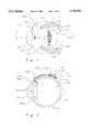
- FIG. 1is a plan view of one embodiment of an intravitreal medicine delivery device according to the present invention shown implanted on a human eyeball, which is schematically shown, it being understood that both the device and the eyeball are enlarged from actual size.
- FIG. 2is a cross-section taken along line 2--2 of FIG. 1.
- FIG. 3is an isometric view of the subject intravitreal medicine delivery device, even further enlarged from the size in FIG. 1 and showing the three principle parts of the device, namely, a housing, a tube, and an elbow.
- FIG. 4is a plan view of the intravitreal medicine delivery device shown in FIG. 3.
- FIG. 5is a side elevation of the device shown in FIG. 4.
- FIG. 6is a cross-section along line 6--6 of FIG. 4.
- FIG. 7is a cross-section along line 7--7 of FIG. 4.
- FIG. 8is a greatly enlarged schematic view of an eye showing the subject intravitreal medicine delivery device implanted on the eyeball and representing the eyelid being lifted to expose the region of the eye where the implant is located.
- FIG. 9is similar to FIG. 8, still enlarged but on a smaller scale, and demonstrating how the injection port of the subject device can be readily identified through overlying tissue.
- FIGS. 10 and 11are both cutaway, sectional views of a portion of the intravitreal medicine delivery device of the present invention illustrating a valve in the elbow, with FIG. 11 showing the valve in a different position from FIG. 10.
- FIGS. 12a, 12b, 12c, 12d and 12eare schematic views showing the sequence of positions of the valve of FIGS. 10 and 11.
- FIG. 13is a cutaway sectional view of a portion of the subject delivery device and showing an optional feature in the tube of the device.
- FIG. 14is a plan view of a portion of the subject medicine delivery device, but depicting another embodiment of the housing of the device.
- FIG. 15is a side elevation of the embodiment of FIG. 14.
- FIG. 16is a cross-section along line 16--16 of FIG. 14.
- FIG. 17is a plan view of a portion of the subject delivery device but depicting still another embodiment of the housing.
- FIG. 18is an isometric view of another embodiment of an elbow of the subject delivery device which differs from the elbow shown in FIG. 3.
- FIG. 19is also an isometric view of still another embodiment of the elbow.
- FIGS. 1-6The first embodiment of an intravitreal medicine delivery device of the present invention is identified in FIGS. 1-6 by the numeral 30 and is shown in FIGS. 1 and 2 as being implanted on a human eyeball or globe 34.
- the eyeballis shown schematically and in just enough detail to enable an understanding of the present invention. Certain parts of the eye are thus briefly identified with reference numerals.
- Schematically represented in either or both of FIGS. 1, 2, 8 and 9are the cornea 36, the sclera 38, the conjunctiva and Tenon's capsule 44, the iris 46, the retina 50, the lens 56, the eyelid 60, eyelashes 62, the vitreous cavity 66, the pars plana 68, the superior rectus muscle 72, and the lateral rectus muscle 74.
- the first embodiment of the intravitreal delivery device 30is shown in detail in FIGS. 3-7 and is now described.
- the deviceincludes a housing 80 most of which is preferably pre-formed of semi-rigid material to have a concavo-convex shape and a low profile so as during surgery to fit easily and closely against an eyeball, as 34.
- the housingpreferably has a generally rectangular outline (FIG. 4) or a trapezoidal outline (FIG. 17) but this outline may be varied to facilitate implantation as will be understood by those skilled in the art.
- the housing 80(FIGS. 3-6) has a base or bottom wall 84 which has an outwardly, spherically concave shape or curvature generally complementary to the curvature of the eyeball 34.
- This bottom wallis made of a tough material impenetrable by an injection needle or syringe and is preferably of a plastic, such as nylon, Kevlar, or polymethyl methacrylate (PMMA), but may be made of metal, such as tantalum.
- the housingalso has a top or upper wall 86 which has an outwardly spherically convex shape and is joined in a fluid-tight manner to the bottom wall along a peripheral edge 90.
- the top and bottom wallsmay be one-piece and integral along the edge or of two different pieces sealed along the edge.
- This edgehas an anterior segment 92, a posterior segment 94, and lateral segments 96 and 97 which meet at four corners of the housing.
- Outer corner fixation tabs 98project from the anterior comers of the housing and have suture-receiving eyelets 100.
- Spaced, inner anterior fixation tabs 102project from the anterior segment 92 and also have suture-receiving eyelets 104.
- the spaced top and bottom walls 86 and 84provide a fluid-tight storage reservoir or chamber 110 in the housing 80.
- This reservoirhas a maximum height dimension indicated by the numeral 112 and measured radially of the curved top and bottom walls and where the spacing between the walls is the greatest. As an example but not limiting to the invention, this maximum height dimension is preferably about 3-4 mm. Of course, the height of the reservoir tapers downward toward all four segments 92, 94, 96 and 97 (FIGS. 3, 5 and 6) of the edge 90. It is also noted that the lateral dimensions of the reservoir, that is the length and width dimensions, measured generally spherically of the walls, are relatively much greater than the maximum height. For the embodiment shown in FIG. 4, but again not limiting to the invention, the width or distance between lateral edges 96 and 97 is approximately 1.5 cm and the length or distance between the anterior and posterior edges 92 and 94 is approximately 1 cm.
- the top wall 86(FIGS. 3-6) of the housing 80 has a slightly uplifted, arcuate, anterior medicine-injection port or area 122 closely adjacent to the anterior edge 92 and having opposite ends 123 closely adjacent to the anterior corners of the housing.
- the top wallalso has a main or remainder portion 124 of greater area than the injection port.
- This main portionis of the same material as the bottom wall 84 and is the part of the top wall which is joined to the bottom wall. Thus, this main portion is needle-impenetrable and fluid-impervious.
- the injection port 122(FIGS. 3-6), however, is of a material, such as silicone rubber, which is penetrable by a needle or syringe but which reseats itself when the needle is withdrawn so that the port is normally fluid-impervious.
- An important feature of the portis that the material thereof is colored, for example, blue, as indicated in the drawings. This coloration is important to provide a marker or identification mark which is visible exteriorly of the eye, especially through covering tissue or patches, to facilitate location of the port by attending medical personnel.
- the reservoir 110(FIGS. 5 and 6) has an anterior fluid outlet 130 located in the top wall 86 of the housing 80 between the anterior segment 92 and the injection port 122 and is thus between the inner tabs 102.
- a filter 134is optionally mounted in the outlet to prevent particulate materials, such as biodegradable polymer, from passing out of the reservoir and into the vitreous cavity, while permitting medicines released from the dissolving polymers to pass out of the reservoir 110 and into the cavity.
- This filtermay take various forms such as a semi-permeable, osmotic membrane or a porous cellulose filter such as a Millipore filter.
- itmay be a thin capillary tube, or coil of capillary tube, designed to restrict the flow of drug-containing fluid from the reservoir 110 into the vitreous cavity 66.
- the subject intravitreal medicine delivery device 30also includes a tapered, elongated delivery tube 140 (FIGS. 3-7) of fluid-impervious compressible elastomeric material, such as a silicone elastomer, thus providing a fluid passageway or lumen 144.
- the tubeis of oval cross-section as illustrated, but is not limited to this cross-section.
- the tubehas a low or flat profile, like the housing, and includes a base and upper walls 146 and 148 which are joined at lateral edges.
- the passagewayhas a relatively small minor axis or dimension, as best seen in FIG. 7, which is preferably less than the height maximum height dimension 112 of the reservoir 110.
- this minor axisis preferably about the size of a 19-20 gauge lumen.
- the base wallmay be outwardly convex, as shown, or concave to conform to the curvature of the eyeball.
- the top wallis resiliently compressible toward the base wall by applying slight digital pressure against the wall to constrict the passageway. When the pressure is relieved, the top wall returns to its normally relaxed position with the passageway fully open.
- the tube 140(FIGS. 3-7) has a relatively wide proximate end 150 connected to the housing 80 around the outlet 130 so that the passageway 144 is in fluid communication with the reservoir 110.
- the passagewayis an extension of the reservoir so that the reservoir and the passageway together constitute one large storage reservoir or chamber.
- the tubeconverges anteriorly from the housing to a relatively narrow distal end 152, it being noted that the major axis of the passageway thus varies from a maximum at the proximate end to a minimum at the distal end.
- the tubeis preferably preformed with, and has sufficient rigidity to maintain, a longitudinal curvature complementary to the curvature of an eyeball, but is sufficiently flexible to allow limited flexing at the juncture of the tube and the housing.
- the subject delivery device 30also includes a semi-rigid tubular elbow 160 having an in-line connector sleeve 162 attached to the distal end 152 of the tube 140 and an intravitreal extension 164 which projects from the sleeve at an angle to the in-line sleeve, preferably of about eighty to ninety degrees, and terminates in a tip 166.
- the sleeveis of about the same diameter as the minor axis or height of the tube, as best seen in FIG. 7, and although given by way of example only, is about 19-20 gauge.
- the elbowthus provides a lumen or fluid passageway 168 in fluid communication with the passageway in the tube and opening at the tip.
- Another pair of fixation tabs 172project laterally from the sleeve 162 of the elbow 160 and have suture eyelets 174.
- the elbowmay be made of the same material as the housing.
- the length of the combined tube 140 and elbow 160, from the anterior segment 92 to the bend of the elbow,is about 7 mm, and the maximum transverse dimension of the tube at its proximate end is about 3 mm.
- the method of the present invention and use of the subject delivery device 30are best described by reference to FIGS. 1, 2, 8 and 9.
- the delivery deviceis implanted within the socket of the eye on the episcleral surface of the globe (eye wall) 66 by placing the device under the conjunctiva 44 and Tenon's capsule.
- the deviceis located between the superior and lateral rectus muscles 72 and 74 with the base wall 84 fitted against the eyeball 34 and the anterior edge 92 located posteriorly of the muscle roots and slightly posteriorly of the equator of the eyeball, as best seen in FIG. 1.
- the injection port 122faces anteriorly, and the tube 140 projects anteriorly with the tip 166 at the pars plana 68.
- a hole 188(FIG. 2) of about 1.0 mm is incised through the eyeball 34 at the pars plana 68 and into the vitreous cavity 66.
- the intravitreal extension 164 of the elbow 160is inserted through the hole so that it projects into the cavity a distance of preferably approximately 4 mm.
- the rigidity of the elbowallows the position of the intravitreal extension 164 to be controlled as it is inserted into the cavity so as to avoid contact of the tube with the lens 56, cornea 36 or iris 46.
- the device 30is then attached to the episclera 38 with sutures 184 (FIG. 1) in selected eyelets 100 (and/or 104) and eyelets 174 (FIG. 4).
- the tabs 98 and 102are provided as alternates in case one location or the other is preferred for the sutures.
- a sceral patch or graft 190(FIG. 8) is sutured over the tube 140 for the purpose of preventing of erosion of the implant tube through the conjunctiva, which could lead to infection and require removal of the implant.
- the rigidity of the elbow and its attachment to the sclera(FIGS. 1 and 2) maintain the intravitreal extension of the implant tube in the position in which it is initially placed so that it does not move around in the cavity as the implant is worn and used by the patient.
- a supply of medicine 194(FIG. 2) is placed in the reservoir 110 before or after implantation.
- drugsthat may be used are antivirals, antibiotics, steroids, growth factors or inhibitors, anti-sense oligonucleotides, mediators of gene therapy, and others. These may be in the form of fluids, solids, suspensions, emulsions, slow-release or time-release beads, capsules, liposomes, ion-exchange beads, biodegradable polymers, pellets, or other micro-particulate vehicles.
- the medicine 194is shown as time-release beads in FIG. 2.
- the eyelid 60remains lifted (FIG. 8) to expose the region where the device 30 is implanted. Because of its color, the injection port 122 is visible through the conjunctiva tissue 44 covering the device.
- the tube 140may be depressed to block the passageway 144 (FIG. 6), and an injection needle 202 of a syringe 200 (FIG. 9) filled with the medicine 194 penetrates the port, and the medicine is injected.
- the needle 202is withdrawn and the tube 140 is released to expand to normal size (FIG. 2).
- the port 122reseats itself.
- the medicine 194is thus exposed to the vitreous cavity 66 over an extended period of time. Whether the medicine is in the form of solid material from which the medicine diffuses or whether the medicine is fluid-borne, this exposure through the passageways 144 and 168, allows the medicine to diffuse, flow or otherwise move or pass from the reservoir 110 into the vitreal cavity.
- the impetus for diffusion of the medicine 194 from the reservoir 110 into the vitreous cavity 66occurs for several reasons.
- the concentration gradient between the medicine in the reservoir and vitreous fluidwill set up its own diffusion gradient causing a movement of medicine into the cavity.
- the patientcan cause flank fluid flow by digitally pressing against the eyelid 60 and thereby against the top wall 148 of the tube 140 to force fluid-borne medicine into the vitreous cavity.
- the compressible top wall 148 in combination with the storage reservoir and passageways 144 and 168may serve as a pump which is generally identified by numeral 210.
- injection of medicine via reservoirincreases pressure in the reservoir driving a net flux of fluid through the tube into the vitreous cavity.
- normal physiologic forcessuch as blinking, eye movements, and normal blood pressure pulses, will cause small pressure gradients to develop in the implant tube promoting passage of medicine into the vitreous cavity.
- This movement of medicine 194 (FIG. 2) from the storage reservoir 110 into the vitreous cavity 66will normally continue, or be forced to flow by the patient's pumping of the tube 140, for an extended period of time whereupon it may be necessary to replenish the original supply of the medicine. Also, because of the discovery of a new and better drug or the need for a different drug or other medicine, it may be desirable to change the medication. In any case, the patient returns to the physician's office for the following simple procedure. The eyelid 60 is lifted (FIGS. 8 and 9) and the patient looks down to expose the region of the implanted delivery device 30.
- the scleral patch 190, the conjunctiva and Tenon's capsule 44 and a fibrous capsule(not specifically shown in the drawings but believed to be well understood), which grows over the device under the conjunctiva obscures the device 30, except for the injection port 122, as illustrated in FIG. 9.
- the portis visible through the tissue because it is identified with a color which can be seen through the tissue.
- the injection port 122is penetrated with the syringe needle 202 and a new supply of medicine 194 is injected into the reservoir 110.
- venting of the implant reservoir 110may be required for injection of larger volumes of medicine.
- Pressure on the tubeis released and the needle is withdrawn, the port sealing after the withdrawal.
- the intravitreal delivery device 30is then capable of repeating its delivery of medicine to the vitreous cavity 66 over an extended period of time, as previously described. Of prime significance, a repeat surgery to re-introduce a new implant, or another invasive procedure to inject a supply of medicine, is avoided.
- suturing tabs 98, 102, and 172A significant relationship between the location of the suturing tabs 98, 102, and 172 and the injection port 122 is to be noted. With reference to FIG. 1, it will be seen that all the sutures 184 are located anteriorly of the port. Thus, when the injection needle 202 (FIG. 9) presses against the port to thrust the needle through the port, the sutured tabs resist posterior movement of the implant device, as 30, and maintain it in its implanted position.
- the second embodiment 30a(FIGS. 10-12) of the present invention is the same as the first embodiment except that a valve 250 is incorporated in the elbow 160 to enhance the pumping action previously described.
- the valve shown and describedis a ball-check valve, but other valves having a similar function can be employed.
- the valve shownincludes a valve body 252 fitted in the connector sleeve 162.
- the valve bodyprovides an outer chamber 254 having an annular inlet 256 opening into the passageway 144 of the tube 140, an annular outlet 258 opening toward the intravitreal extension 164, an annular valve seat 260 between the inlet and outlet, and a dosage chamber 264 between the valve seat and the outlet.
- the valvealso includes a resiliently expandable, conical mouthpiece 268 of a suitable elastomeric material whose apex is normally closed in its relaxed condition but which opens when fluid pressure inside the valve body, in the direction of the apex, forces it open. Still further, the valve includes a resilient ball 270 in the outer chamber and a spring 272 between the ball and the valve seat.
- the storage reservoir 110is filled with fluid-borne medicine, as represented by the numeral 280, either during initial manufacture or by subsequent injection through the injection port 122.
- fluid-borne medicinerepresented by the numeral 280
- FIG. 12athe condition of the valve in its resting state is shown in FIG. 12a wherein the ball 270 floats in the outer chamber 254 being held away from the seat 260 by the spring 272 (FIG. 10).
- fluid-borne medicine from the reservoirfills not only the passageway 144 but also the outer chamber 254 (FIG. 11) and the dosage chamber 264. Under these conditions, there is insufficient pressure to open the mouthpiece 268.
- the patient or attendantpresses against the patient's eyelid 60 thereby pressing against the tube wall 148 (FIG. 11) compressing the tube 140 and constricting the passageway 144 and exerting a fluid pressure toward the vitreous cavity 66 (FIG. 12b).
- This pressure increaseis sufficient to force the ball 270 against the spring 272 until the ball seals (FIG. 12c) against the seat 260 thereby temporarily blocking fluid flow through the valve.
- Such pumping actionthus causes a limited quantity of fluid, essentially the amount in the dosage chamber 264, that is, the predetermined dose or aliquot, to flow into the vitreous cavity 66.
- the valve 250 and the tube 140working in cooperative relationship with the reservoir 110 and the elbow 160, constitute a pump 210'.
- the third embodiment 30b of the present inventionis the same as the first embodiment 30 except as follows.
- FIG. 13there is illustrated a tube 140' connected to the housing 80 and the elbow 160.
- a series of upper and lower baffles 300 and 302are respectively attached to the top and base walls 148 and 146 of the tube and project into the passageway 144 in interdigitated, spaced relationship.
- the bafflesare semi-oval or semi-elliptical, but if a tube has a different cross-section, the shape of the baffles would be changed to conform. For example, if the tube is circular, the baffles would be semi-circular.
- the purpose of these bafflesis to facilitate closure of the passageway when the walls of the tube are compressed during filling the storage reservoir 110, while allowing flow through the passageway at all other times when the tube is in normally fully expanded or open condition.
- the fourth embodiment of the delivery deviceis identified by the numeral 30c and relates to a different housing construction but is otherwise the same as the first embodiment 30.
- a housing 80'enclosing a reservoir 110' which is in communication with a tube, such as 140 or 141', which is only partially shown in FIGS. 14-16 since this embodiment does not involve a modification of the tube.
- the modified housing 80'has a base wall 84' like the base wall 84 (FIG. 6) and injection port 122' like the injection port 122 (FIGS. 3-6), but a top wall 86' different from the top wall 86 (FIG. 4).
- the top wall 86'(FIG. 14) has a main portion 124' including a peripheral section 326 and a medial section or roof 328 located posteriorly of the injection port 122'.
- the peripheral section 326(excluding the injection port 122') is made of relatively rigid, impenetrable material, such as described for the base wall 84 and the main portion 124 of the first embodiment 30.
- the injection port 122'is constructed of the same penetrable, resealable material as the port 122.
- the medial section 328is made of a softer, flexible and resilient material, such as silicone rubber, to allow the medial section or roof to be compressed and thereafter expand to its relaxed state.
- this medial sectionmay be semi-permeable to preclude a vacuum from developing in the reservoir when the tube 140 or 140' is compressed while minimizing any appreciable loss of fluid-borne medicine.
- the medial section 328may be made of an elastomeric material which is distended by the injection of a large volume of medicine into the reservoir 110' via the injection port 122'.
- the distention of the medial sectionincreases the pressure in the reservoir and forces drug-containing fluid into the tube 140 or 140' where egress into the vitreous is controlled by passage through a filter or capillary tube 134'.
- the entire housing 80'may be made of elastomeric material to provide such pressure by distention of the housing.
- the fifth embodimentinvolves a differently shaped housing 80", to which reference was previously made.
- the housing of this embodimenthas a generally trapezoidal outline in plan view.
- Such a trapezoidal shapemay be preferred to facilitate implantation.
- the trapezoidal housinghas an injection port 122", anterior tabs 98" with suture eyelets 100", and lateral tabs 340 with suture eyelets 342.
- the preferred dimensions of the trapezoidal housingare similar to the rectangular housing except that the posterior edge is about 1.5 cm and the anterior edge is about 0.75 cm.
- FIGS. 18 and 19An additional feature which may be incorporated in the previously described embodiments of the present invention has two species as shown in FIGS. 18 and 19.
- FIG. 18there is illustrated a tubular elbow 160a having an intravitreal extension 164' in which is provided a plurality of holes or slits 350.
- a semi-permeable membrane or filter 360 in the shape of a relatively rigidly-formed cupis attached to the open tip 166' of a tubular elbow 160b and projects therefrom.
- medicinepasses from the reservoir 110 through the passageways 144 and 168 and through the wick 352, or the membrane 360, from which it diffuses into the vitreous cavity 66.
- the wick or membraneis optionally used to obviate the possibility of occlusion of the drug passage into the vitreous cavity by particulate matter from the reservoir.
- the intravitreal medicine delivery device 30, 30a, 30c and method of the present inventionit will be understood how the device and method result in numerous advantages and provide solutions to various problems experienced by prior art devices and methods.
- the subject inventionenables a wide variety of drugs and other pharmacological agents and medicines to be introduced into the vitreous cavity over the lifetime of the patient with only a single initial surgery and with minimum intraocular invasion to implant the device.
- the subject device 30is not so limited and can utilize the broad range of medicinal agents now available or becoming available. Moreover, the device 30 and method allow the dosage delivered to the vitreous cavity 66 to be controlled and furthermore, allows the patient to control the timing of the deliveries. Additional features incorporated into the device filter the medicine before it enters the vitreous cavity. Also, the device is designed to facilitate its implantation so that it fits closely and relatively comfortably against the eyeball and so that damage or interference with other parts of the eye are avoided.
Landscapes
- Health & Medical Sciences (AREA)
- Ophthalmology & Optometry (AREA)
- Engineering & Computer Science (AREA)
- Biomedical Technology (AREA)
- Heart & Thoracic Surgery (AREA)
- Vascular Medicine (AREA)
- Life Sciences & Earth Sciences (AREA)
- Animal Behavior & Ethology (AREA)
- General Health & Medical Sciences (AREA)
- Public Health (AREA)
- Veterinary Medicine (AREA)
- Infusion, Injection, And Reservoir Apparatuses (AREA)
Abstract
Description
This invention pertains to intravitreal medicine delivery and more particularly to an implant device for delivering medicine to the vitreal cavity of the eye, and to a method for introducing medicines into the vitreal cavity using the device.
Within the past several decades, great advances have been made in the diagnosis and treatment of vitreoretinal diseases. Advances in laser technology, and vitreoretinal surgical techniques, have significantly improved the prognosis of numerous retinal conditions including diabetic retinopathy, macular degeneration, and retinal detachment. As the pathophysiology of these and many other vitreoretinal diseases is also becoming more clearly understood, a host of potential pharmacological agents is currently under investigation.
In addition to the numerous antibiotic, antiviral, and antifungal agents currently used to treat infections of the retina and vitreous, many antiinflammatory and anticancer drugs have been shown to be useful in treating diseases such as proliferative vitreoretinopathy. As the role of growth factors involved in diabetic retinopathy, macular degeneration, and other retinal degenerations is elucidated, new classes of agents have been found to be of possible benefit, including growth factors themselves, blocking antibodies to growth factors, antisense oligonucleotides, and even gene therapy with growth factor inserts.
Unfortunately, the delivery of drugs to the retina is often problematic. Most agents given topically to the eye (in the form of eye drops) do not penetrate through the anterior segment of the eye well enough to reach the vitreous or retina in therapeutic concentrations. Medications can be given orally or intravenously, but the blood vessels within the retina (and other parts of the central nervous system) are relatively impermeable to many agents. Furthermore, these drugs may have significant systemic side effects on other organs of the body. Drugs can be directly injected into the vitreous cavity, via a needle passed through the pars plana, and this technique is currently employed to combat certain severe, sight-threatening infections. However, this procedure itself entails certain risks, such as infection, bleeding, cataract formation, and retinal detachment. Furthermore, the majority of the injected drug is often cleared from the vitreous cavity within several days, necessitating multiple injections for prolonged treatment.
Accordingly, devices have been developed for improving the introduction of drugs to the vitreal cavity. One such device is a biodegradable polymer designed to be injected into the vitreous cavity where it slowly releases drug as it dissolves. A similar drug-containing polymer has been developed which is made in the shape of a tack or plug to be surgically inserted into the eyewall at the pars plana so that it projects into the vitreous cavity. Liposomes containing pharmacological agents have been developed to slowly release the agent after injection into the vitreous cavity. Another device is a plastic pellet which contains a retinal drug (ganoclovir) and is sutured inside of the vitreous cavity where the drug slowly dissolves into the cavity.
Although such devices as those briefly referred to above are apparently effective in delivering drugs into the vitreous cavity, they have significant disadvantages. First, all of these devices contain a certain amount of drug which when expended cannot be replenished without repeating the surgical implantation or intravitreal injection. Although these different devices can release drugs for weeks to months (or in the case of the plastic pellet, almost one year), certain indications for intravitreal drug administration require extended or lifetime therapy. Therefore, multiple procedures are often required and are highly undesirable.
Secondly, the device with the longest release rate, the plastic pellet of ganiclovir, requires a very large eyewall incision (5 mm) to implant due to its large size. Although the other devices can be implanted or injected through a smaller incision (1 mm or less), those that are injected freely into the vitreous cavity instead of anchored to the eyewall can migrate within the eye and come in direct contact with the retina where they can block vision or release high local concentrations of drug which could potentially prove toxic to delicate retinal tissue.
Third, many pharmacological agents cannot easily and effectively be incorporated into biodegradable polymers. Furthermore, many potential pharmacological agents would not remain stable for extended periods of time in the vitreous cavity. Therefore, there is a significant limitation to what pharmacological agents can be administered via the slow release devices currently available.
Fourth, since the plug or pellet is in essence the drug itself, treatment is limited to this drug, so that another surgery or procedure is required to change it if administration of a different drug is desired.
The present invention is directed to intravitreal medicine delivery involving an implant device and method wherein a wide variety of beneficial drugs or other pharmacological agents or medicines can be introduced into the vitreous cavity over an extended period of time, as much as the life of the patient, with only a single initial surgery to implant the device; wherein the surgical incision needed for implantation is minimized; wherein additional amounts of drugs over an initial supply can be introduced without further invasive surgery; wherein the type of medicine can be varied or changed depending on such factors as the disease being treated or drug availability; wherein the dosage of agent being administered is controllable; and wherein damage or interference by the implant to various parts of the eye is avoided.
It is an object of the present invention to provide for improved delivery of drugs and other pharmacological agents to the vitreous cavity of the eye, especially for treating vitreoretinal diseases.
It is another object to enable medicinal agents to be delivered to the vitreous cavity with a single initial surgery and without the need for repeated invasive surgeries or procedures.
It is yet another object of this invention to allow replenishment of the drug or other medicinal agent within an implant attached to the eyewall by injection into the implant through the conjunctivae and not through the eye wall.
It is further object to be able to change the pharmacological agent being dispensed from an eye implant device into the vitreal cavity, without surgery or other invasive procedure.
It is also an object to control the dosage of drugs or other pharmacological agents delivered to the vitreous cavity from an implant device.
It is still another object of the present invention to enable the patient to control delivery of dosages of medicines to the vitreous cavity with a delivery device implanted on the patient's eyeball.
It is a still farther object to implant an intravitreal medicine delivery device which dispenses medicine into the vitreous cavity without interference with or damage to various parts of the eye.
It is yet another object of the present invention to prevent inadvertent puncture of the eyeball by an injection needle used to replenish the supply of drugs or other agents in an eye implant device.
It is also an object of the subject invention to prevent an uncontrolled movement of medicines into the vitreous cavity from an intravitreal delivery implant when the supply of medicines in the implant is being replenished in situ.
It is another object of this invention to filter medicines being dispensed from an implant device before being admitted to the vitreous cavity thereby to prevent unwanted particulate matter, such as undissolved biodegradable polymer, from moving into the cavity, while allowing medicines to pass into the cavity.
It is a feature of the present invention to have a relatively large drug or other medicine-containing reservoir which is located outside the eyewall in the sub-conjunctival space and which is connected to the vitreous cavity by a smaller tube through the pars plana so that a substantial amount of medicine can be brought into the vitreous cavity with a minimum of eyewall incision.
It is a feature of this invention to provide an intravitreal medicine delivery device with a pump for moving predetermined doses of drugs or other agents into the vitreous cavity and which can be operated by the patient.
It is another feature to provide an intravitreal drug delivery device which has a low profile for closely-fitting implantation under the conjunctiva and Tenon's capsule of the eyeball.
It is another feature of this invention to provide an eye implant device with an injection port which can be readily identified through overlying tissue for injecting an additional supply of medicine into the implant device.
It is still another feature of this invention to suture a medication delivery implant device to the eyewall so that it facilitates subsequent injections of medication into it while maintaining its implanted position.
These and other objects, features, and advantages of the subject invention will become apparent upon reference to the accompanying drawings, the following description, and the appended claims.
FIG. 1 is a plan view of one embodiment of an intravitreal medicine delivery device according to the present invention shown implanted on a human eyeball, which is schematically shown, it being understood that both the device and the eyeball are enlarged from actual size.
FIG. 2 is a cross-section taken alongline 2--2 of FIG. 1.
FIG. 3 is an isometric view of the subject intravitreal medicine delivery device, even further enlarged from the size in FIG. 1 and showing the three principle parts of the device, namely, a housing, a tube, and an elbow.
FIG. 4 is a plan view of the intravitreal medicine delivery device shown in FIG. 3.
FIG. 5 is a side elevation of the device shown in FIG. 4.
FIG. 6 is a cross-section alongline 6--6 of FIG. 4.
FIG. 7 is a cross-section along line 7--7 of FIG. 4.
FIG. 8 is a greatly enlarged schematic view of an eye showing the subject intravitreal medicine delivery device implanted on the eyeball and representing the eyelid being lifted to expose the region of the eye where the implant is located.
FIG. 9 is similar to FIG. 8, still enlarged but on a smaller scale, and demonstrating how the injection port of the subject device can be readily identified through overlying tissue.
FIGS. 10 and 11 are both cutaway, sectional views of a portion of the intravitreal medicine delivery device of the present invention illustrating a valve in the elbow, with FIG. 11 showing the valve in a different position from FIG. 10.
FIGS. 12a, 12b, 12c, 12d and 12e are schematic views showing the sequence of positions of the valve of FIGS. 10 and 11.
FIG. 13 is a cutaway sectional view of a portion of the subject delivery device and showing an optional feature in the tube of the device.
FIG. 14 is a plan view of a portion of the subject medicine delivery device, but depicting another embodiment of the housing of the device.
FIG. 15 is a side elevation of the embodiment of FIG. 14.
FIG. 16 is a cross-section alongline 16--16 of FIG. 14.
FIG. 17 is a plan view of a portion of the subject delivery device but depicting still another embodiment of the housing.
FIG. 18 is an isometric view of another embodiment of an elbow of the subject delivery device which differs from the elbow shown in FIG. 3.
FIG. 19 is also an isometric view of still another embodiment of the elbow.
Intravitreal Medicine Delivery Device-First Embodiment
The first embodiment of an intravitreal medicine delivery device of the present invention is identified in FIGS. 1-6 by the numeral 30 and is shown in FIGS. 1 and 2 as being implanted on a human eyeball orglobe 34. The eyeball is shown schematically and in just enough detail to enable an understanding of the present invention. Certain parts of the eye are thus briefly identified with reference numerals. Schematically represented in either or both of FIGS. 1, 2, 8 and 9 are thecornea 36, thesclera 38, the conjunctiva and Tenon'scapsule 44, theiris 46, theretina 50, thelens 56, theeyelid 60,eyelashes 62, thevitreous cavity 66, the pars plana 68, thesuperior rectus muscle 72, and thelateral rectus muscle 74.
The first embodiment of theintravitreal delivery device 30 is shown in detail in FIGS. 3-7 and is now described. The device includes ahousing 80 most of which is preferably pre-formed of semi-rigid material to have a concavo-convex shape and a low profile so as during surgery to fit easily and closely against an eyeball, as 34. In plan view the housing preferably has a generally rectangular outline (FIG. 4) or a trapezoidal outline (FIG. 17) but this outline may be varied to facilitate implantation as will be understood by those skilled in the art.
The housing 80 (FIGS. 3-6) has a base orbottom wall 84 which has an outwardly, spherically concave shape or curvature generally complementary to the curvature of theeyeball 34. This bottom wall is made of a tough material impenetrable by an injection needle or syringe and is preferably of a plastic, such as nylon, Kevlar, or polymethyl methacrylate (PMMA), but may be made of metal, such as tantalum. The housing also has a top orupper wall 86 which has an outwardly spherically convex shape and is joined in a fluid-tight manner to the bottom wall along aperipheral edge 90. The top and bottom walls may be one-piece and integral along the edge or of two different pieces sealed along the edge. This edge has ananterior segment 92, aposterior segment 94, andlateral segments corner fixation tabs 98 project from the anterior comers of the housing and have suture-receivingeyelets 100. Spaced, inneranterior fixation tabs 102 project from theanterior segment 92 and also have suture-receivingeyelets 104.
The spaced top andbottom walls 86 and 84 (FIGS. 5 and 6) provide a fluid-tight storage reservoir orchamber 110 in thehousing 80. This reservoir has a maximum height dimension indicated by the numeral 112 and measured radially of the curved top and bottom walls and where the spacing between the walls is the greatest. As an example but not limiting to the invention, this maximum height dimension is preferably about 3-4 mm. Of course, the height of the reservoir tapers downward toward all foursegments edge 90. It is also noted that the lateral dimensions of the reservoir, that is the length and width dimensions, measured generally spherically of the walls, are relatively much greater than the maximum height. For the embodiment shown in FIG. 4, but again not limiting to the invention, the width or distance betweenlateral edges posterior edges
Of particular significance, however, is that the top wall 86 (FIGS. 3-6) of thehousing 80 has a slightly uplifted, arcuate, anterior medicine-injection port orarea 122 closely adjacent to theanterior edge 92 and having opposite ends 123 closely adjacent to the anterior corners of the housing. The top wall also has a main orremainder portion 124 of greater area than the injection port. This main portion is of the same material as thebottom wall 84 and is the part of the top wall which is joined to the bottom wall. Thus, this main portion is needle-impenetrable and fluid-impervious.
The injection port 122 (FIGS. 3-6), however, is of a material, such as silicone rubber, which is penetrable by a needle or syringe but which reseats itself when the needle is withdrawn so that the port is normally fluid-impervious. An important feature of the port is that the material thereof is colored, for example, blue, as indicated in the drawings. This coloration is important to provide a marker or identification mark which is visible exteriorly of the eye, especially through covering tissue or patches, to facilitate location of the port by attending medical personnel.
The reservoir 110 (FIGS. 5 and 6) has an anteriorfluid outlet 130 located in thetop wall 86 of thehousing 80 between theanterior segment 92 and theinjection port 122 and is thus between theinner tabs 102. Afilter 134 is optionally mounted in the outlet to prevent particulate materials, such as biodegradable polymer, from passing out of the reservoir and into the vitreous cavity, while permitting medicines released from the dissolving polymers to pass out of thereservoir 110 and into the cavity. This filter may take various forms such as a semi-permeable, osmotic membrane or a porous cellulose filter such as a Millipore filter. Furthermore, it may be a thin capillary tube, or coil of capillary tube, designed to restrict the flow of drug-containing fluid from thereservoir 110 into thevitreous cavity 66.
The subject intravitrealmedicine delivery device 30 also includes a tapered, elongated delivery tube 140 (FIGS. 3-7) of fluid-impervious compressible elastomeric material, such as a silicone elastomer, thus providing a fluid passageway orlumen 144. Preferably, the tube is of oval cross-section as illustrated, but is not limited to this cross-section. The tube has a low or flat profile, like the housing, and includes a base andupper walls maximum height dimension 112 of thereservoir 110. Again, although not limiting to the invention, this minor axis is preferably about the size of a 19-20 gauge lumen. The base wall may be outwardly convex, as shown, or concave to conform to the curvature of the eyeball. Significantly, the top wall is resiliently compressible toward the base wall by applying slight digital pressure against the wall to constrict the passageway. When the pressure is relieved, the top wall returns to its normally relaxed position with the passageway fully open.
The tube 140 (FIGS. 3-7) has a relatively wideproximate end 150 connected to thehousing 80 around theoutlet 130 so that thepassageway 144 is in fluid communication with thereservoir 110. In effect, the passageway is an extension of the reservoir so that the reservoir and the passageway together constitute one large storage reservoir or chamber. The tube converges anteriorly from the housing to a relatively narrowdistal end 152, it being noted that the major axis of the passageway thus varies from a maximum at the proximate end to a minimum at the distal end. As shown in FIGS. 5 and 6, the tube is preferably preformed with, and has sufficient rigidity to maintain, a longitudinal curvature complementary to the curvature of an eyeball, but is sufficiently flexible to allow limited flexing at the juncture of the tube and the housing.
The subject delivery device 30 (FIGS. 3-6) also includes a semi-rigidtubular elbow 160 having an in-line connector sleeve 162 attached to thedistal end 152 of thetube 140 and anintravitreal extension 164 which projects from the sleeve at an angle to the in-line sleeve, preferably of about eighty to ninety degrees, and terminates in atip 166. The sleeve is of about the same diameter as the minor axis or height of the tube, as best seen in FIG. 7, and although given by way of example only, is about 19-20 gauge. The elbow thus provides a lumen orfluid passageway 168 in fluid communication with the passageway in the tube and opening at the tip. Another pair offixation tabs 172 project laterally from thesleeve 162 of theelbow 160 and havesuture eyelets 174. The elbow may be made of the same material as the housing.
Once again, to provide an example of a preferred embodiment of the intravitreal delivery device 30 (FIG. 4) but not limiting to the invention, the length of the combinedtube 140 andelbow 160, from theanterior segment 92 to the bend of the elbow, is about 7 mm, and the maximum transverse dimension of the tube at its proximate end is about 3 mm.
Intravitreal Medicine Delivery Method
The method of the present invention and use of thesubject delivery device 30 are best described by reference to FIGS. 1, 2, 8 and 9. The delivery device is implanted within the socket of the eye on the episcleral surface of the globe (eye wall) 66 by placing the device under theconjunctiva 44 and Tenon's capsule. The device is located between the superior andlateral rectus muscles base wall 84 fitted against theeyeball 34 and theanterior edge 92 located posteriorly of the muscle roots and slightly posteriorly of the equator of the eyeball, as best seen in FIG. 1. Thusly located, theinjection port 122 faces anteriorly, and thetube 140 projects anteriorly with thetip 166 at thepars plana 68.
A hole 188 (FIG. 2) of about 1.0 mm is incised through theeyeball 34 at the pars plana 68 and into thevitreous cavity 66. Theintravitreal extension 164 of theelbow 160 is inserted through the hole so that it projects into the cavity a distance of preferably approximately 4 mm. In this regard, it is to be noted that the rigidity of the elbow allows the position of theintravitreal extension 164 to be controlled as it is inserted into the cavity so as to avoid contact of the tube with thelens 56,cornea 36 oriris 46. Thedevice 30 is then attached to theepisclera 38 with sutures 184 (FIG. 1) in selected eyelets 100 (and/or 104) and eyelets 174 (FIG. 4). Thetabs tube 140 for the purpose of preventing of erosion of the implant tube through the conjunctiva, which could lead to infection and require removal of the implant. The rigidity of the elbow and its attachment to the sclera (FIGS. 1 and 2) maintain the intravitreal extension of the implant tube in the position in which it is initially placed so that it does not move around in the cavity as the implant is worn and used by the patient.
A supply of medicine 194 (FIG. 2) is placed in thereservoir 110 before or after implantation. Examples of drugs that may be used are antivirals, antibiotics, steroids, growth factors or inhibitors, anti-sense oligonucleotides, mediators of gene therapy, and others. These may be in the form of fluids, solids, suspensions, emulsions, slow-release or time-release beads, capsules, liposomes, ion-exchange beads, biodegradable polymers, pellets, or other micro-particulate vehicles. As an example, themedicine 194 is shown as time-release beads in FIG. 2.
If drugs or other agents are injected after the implant surgery, theeyelid 60 remains lifted (FIG. 8) to expose the region where thedevice 30 is implanted. Because of its color, theinjection port 122 is visible through theconjunctiva tissue 44 covering the device. Thetube 140 may be depressed to block the passageway 144 (FIG. 6), and aninjection needle 202 of a syringe 200 (FIG. 9) filled with themedicine 194 penetrates the port, and the medicine is injected.
If a large volume ofmedicine 194 is to be instilled into the reservoir 110 (FIG. 9), either initially or to refill theimplant 30 at a later date, venting of the implant reservoir by a second needle, not shown but placed through theinjection port 122 simultaneously with injection, may be required. Injection of small volumes of medicine into the reservoir, however, may not require venting.
After the injection is completed (FIG. 9), theneedle 202 is withdrawn and thetube 140 is released to expand to normal size (FIG. 2). By blocking the passageway during filling of the reservoir, fluid flow or fluid pressure through the tube andelbow 160 and into thevitreous cavity 66 is precluded. When the needle is withdrawn, theport 122 reseats itself.
Following the implant surgery and as the patient wears the implanted device 30 (FIG. 2), themedicine 194 is thus exposed to thevitreous cavity 66 over an extended period of time. Whether the medicine is in the form of solid material from which the medicine diffuses or whether the medicine is fluid-borne, this exposure through thepassageways reservoir 110 into the vitreal cavity.
The impetus for diffusion of themedicine 194 from thereservoir 110 into thevitreous cavity 66 occurs for several reasons. First, the concentration gradient between the medicine in the reservoir and vitreous fluid will set up its own diffusion gradient causing a movement of medicine into the cavity. Secondly, the patient can cause flank fluid flow by digitally pressing against theeyelid 60 and thereby against thetop wall 148 of thetube 140 to force fluid-borne medicine into the vitreous cavity. Thus, the compressibletop wall 148 in combination with the storage reservoir andpassageways numeral 210. Thirdly, injection of medicine via reservoir increases pressure in the reservoir driving a net flux of fluid through the tube into the vitreous cavity. Finally, normal physiologic forces, such as blinking, eye movements, and normal blood pressure pulses, will cause small pressure gradients to develop in the implant tube promoting passage of medicine into the vitreous cavity.
This movement of medicine 194 (FIG. 2) from thestorage reservoir 110 into thevitreous cavity 66 will normally continue, or be forced to flow by the patient's pumping of thetube 140, for an extended period of time whereupon it may be necessary to replenish the original supply of the medicine. Also, because of the discovery of a new and better drug or the need for a different drug or other medicine, it may be desirable to change the medication. In any case, the patient returns to the physician's office for the following simple procedure. Theeyelid 60 is lifted (FIGS. 8 and 9) and the patient looks down to expose the region of the implanteddelivery device 30. Thescleral patch 190, the conjunctiva and Tenon'scapsule 44 and a fibrous capsule (not specifically shown in the drawings but believed to be well understood), which grows over the device under the conjunctiva obscures thedevice 30, except for theinjection port 122, as illustrated in FIG. 9. The port is visible through the tissue because it is identified with a color which can be seen through the tissue.
While digitally compressing the tube 140 (FIGS. 8 and 9) to block thepassageway 144, as previously described, theinjection port 122 is penetrated with thesyringe needle 202 and a new supply ofmedicine 194 is injected into thereservoir 110. As previously indicated, venting of theimplant reservoir 110 may be required for injection of larger volumes of medicine. Pressure on the tube is released and the needle is withdrawn, the port sealing after the withdrawal. Theintravitreal delivery device 30 is then capable of repeating its delivery of medicine to thevitreous cavity 66 over an extended period of time, as previously described. Of prime significance, a repeat surgery to re-introduce a new implant, or another invasive procedure to inject a supply of medicine, is avoided.
A significant relationship between the location of thesuturing tabs injection port 122 is to be noted. With reference to FIG. 1, it will be seen that all thesutures 184 are located anteriorly of the port. Thus, when the injection needle 202 (FIG. 9) presses against the port to thrust the needle through the port, the sutured tabs resist posterior movement of the implant device, as 30, and maintain it in its implanted position.
Intravitreal Delivery Device-Second Embodiment
Thesecond embodiment 30a (FIGS. 10-12) of the present invention is the same as the first embodiment except that avalve 250 is incorporated in theelbow 160 to enhance the pumping action previously described. The valve shown and described is a ball-check valve, but other valves having a similar function can be employed. The valve shown, however, includes avalve body 252 fitted in theconnector sleeve 162. The valve body provides anouter chamber 254 having anannular inlet 256 opening into thepassageway 144 of thetube 140, anannular outlet 258 opening toward theintravitreal extension 164, anannular valve seat 260 between the inlet and outlet, and adosage chamber 264 between the valve seat and the outlet. The valve also includes a resiliently expandable,conical mouthpiece 268 of a suitable elastomeric material whose apex is normally closed in its relaxed condition but which opens when fluid pressure inside the valve body, in the direction of the apex, forces it open. Still further, the valve includes aresilient ball 270 in the outer chamber and aspring 272 between the ball and the valve seat.
In operation of theintravitreal delivery device 30a when it incorporates the valve 250 (FIGS. 10-12), thestorage reservoir 110 is filled with fluid-borne medicine, as represented by the numeral 280, either during initial manufacture or by subsequent injection through theinjection port 122. With reference to the flow diagrams of FIGS. 12a-12e, the condition of the valve in its resting state is shown in FIG. 12a wherein theball 270 floats in theouter chamber 254 being held away from theseat 260 by the spring 272 (FIG. 10). Because of the pressure gradients previously described in regard to the first embodiment, fluid-borne medicine from the reservoir fills not only thepassageway 144 but also the outer chamber 254 (FIG. 11) and thedosage chamber 264. Under these conditions, there is insufficient pressure to open themouthpiece 268.
When a dose ofmedicine 280 is to be delivered to thevitreous cavity 66, the patient or attendant presses against the patient'seyelid 60 thereby pressing against the tube wall 148 (FIG. 11) compressing thetube 140 and constricting thepassageway 144 and exerting a fluid pressure toward the vitreous cavity 66 (FIG. 12b). This pressure increase is sufficient to force theball 270 against thespring 272 until the ball seals (FIG. 12c) against theseat 260 thereby temporarily blocking fluid flow through the valve. Such pumping action thus causes a limited quantity of fluid, essentially the amount in thedosage chamber 264, that is, the predetermined dose or aliquot, to flow into thevitreous cavity 66. Thus, thevalve 250 and thetube 140, working in cooperative relationship with thereservoir 110 and theelbow 160, constitute a pump 210'.
When pressure on theeyelid 60 and thus thetube wall 148 is relieved (FIGS. 12d and 12e), a reverse pressure develops because of the lowering of pressure in thepassageway 144, whereby thevalve ball 270 unseats, and themouthpiece 268 collapses to its closed position. Fluid pressure in the device then returns to an equilibrium state and the valve returns to its resting state, as shown in FIG. 12e.
Intravitreal Delivery Device-Third Embodiment
The third embodiment 30b of the present invention is the same as thefirst embodiment 30 except as follows. In FIG. 13, there is illustrated a tube 140' connected to thehousing 80 and theelbow 160. A series of upper andlower baffles base walls passageway 144 in interdigitated, spaced relationship. Since the tube is oval, the baffles are semi-oval or semi-elliptical, but if a tube has a different cross-section, the shape of the baffles would be changed to conform. For example, if the tube is circular, the baffles would be semi-circular. The purpose of these baffles is to facilitate closure of the passageway when the walls of the tube are compressed during filling thestorage reservoir 110, while allowing flow through the passageway at all other times when the tube is in normally fully expanded or open condition.
Intravitreal Delivery Device-Fourth Embodiment
The fourth embodiment of the delivery device is identified by the numeral 30c and relates to a different housing construction but is otherwise the same as thefirst embodiment 30. In FIG. 14, there is shown a housing 80' enclosing a reservoir 110' which is in communication with a tube, such as 140 or 141', which is only partially shown in FIGS. 14-16 since this embodiment does not involve a modification of the tube. The modified housing 80', however, has a base wall 84' like the base wall 84 (FIG. 6) and injection port 122' like the injection port 122 (FIGS. 3-6), but a top wall 86' different from the top wall 86 (FIG. 4). The top wall 86' (FIG. 14) has a main portion 124' including aperipheral section 326 and a medial section orroof 328 located posteriorly of the injection port 122'.
As best shown in FIGS. 14 and 15, the peripheral section 326 (excluding the injection port 122') is made of relatively rigid, impenetrable material, such as described for thebase wall 84 and themain portion 124 of thefirst embodiment 30. The injection port 122' is constructed of the same penetrable, resealable material as theport 122. Themedial section 328, however, is made of a softer, flexible and resilient material, such as silicone rubber, to allow the medial section or roof to be compressed and thereafter expand to its relaxed state. In addition, this medial section may be semi-permeable to preclude a vacuum from developing in the reservoir when thetube 140 or 140' is compressed while minimizing any appreciable loss of fluid-borne medicine.
Alternatively, themedial section 328 may be made of an elastomeric material which is distended by the injection of a large volume of medicine into the reservoir 110' via the injection port 122'. The distention of the medial section increases the pressure in the reservoir and forces drug-containing fluid into thetube 140 or 140' where egress into the vitreous is controlled by passage through a filter or capillary tube 134'. Still further, the entire housing 80' may be made of elastomeric material to provide such pressure by distention of the housing.
Intravitreal Delivery Device-Fifth Embodiment
The fifth embodiment (FIG. 17) involves a differently shapedhousing 80", to which reference was previously made. Instead of thehousings 80 or 80', each of which has a generally rectangular periphery, the housing of this embodiment has a generally trapezoidal outline in plan view. Such a trapezoidal shape may be preferred to facilitate implantation. The trapezoidal housing has aninjection port 122",anterior tabs 98" withsuture eyelets 100", andlateral tabs 340 withsuture eyelets 342. Again, by way of example only, the preferred dimensions of the trapezoidal housing are similar to the rectangular housing except that the posterior edge is about 1.5 cm and the anterior edge is about 0.75 cm.
Intravitreal Delivery Device-Additional Feature
An additional feature which may be incorporated in the previously described embodiments of the present invention has two species as shown in FIGS. 18 and 19. In FIG. 18, there is illustrated a tubular elbow 160a having an intravitreal extension 164' in which is provided a plurality of holes or slits 350. A semi-permeable wick, insert, or filter 352 of very flaccid material, such as a silicone sponge, is positioned in the passageway of the extension.
In FIG. 19, a semi-permeable membrane orfilter 360 in the shape of a relatively rigidly-formed cup is attached to the open tip 166' of atubular elbow 160b and projects therefrom.
When either species shown in FIGS. 18 and 19 is incorporated in theintravitreal delivery device reservoir 110 through thepassageways wick 352, or themembrane 360, from which it diffuses into thevitreous cavity 66. The wick or membrane is optionally used to obviate the possibility of occlusion of the drug passage into the vitreous cavity by particulate matter from the reservoir.
From the foregoing description of the intravitrealmedicine delivery device
Thereafter, to resupply thedevice 30 with more medicine or to change the medicine, only a simple injection intoreservoir 110 through the speciallyidentifiable injection port
As contrasted with biodegradable polymer pellets directly implanted into the eyeball and which do not lend themselves to many potential pharmacological agents, thesubject device 30 is not so limited and can utilize the broad range of medicinal agents now available or becoming available. Moreover, thedevice 30 and method allow the dosage delivered to thevitreous cavity 66 to be controlled and furthermore, allows the patient to control the timing of the deliveries. Additional features incorporated into the device filter the medicine before it enters the vitreous cavity. Also, the device is designed to facilitate its implantation so that it fits closely and relatively comfortably against the eyeball and so that damage or interference with other parts of the eye are avoided.
Although preferred embodiments have been shown and described, various modifications and substitutions may be made thereto without departing from the spirit and scope of the invention. Accordingly, it is to be understood that the present invention has been described by way of illustration and not limitation.
Claims (28)
1. An intravitreal medicine delivery device for delivering medicine to the vitreal cavity of an eye having an eyeball covered by conjunctiva, comprising reservoir means for holding a supply of medicine, means for enabling passage of medicine out of the reservoir means, and means for attaching both the reservoir means and the enabling means under the conjunctiva of an eye so that the entire device is under the conjunctiva, said enabling means being adapted to enable passage of medicine into the vitreal cavity of the eye.
2. The device of claim 1 wherein the reservoir means includes means for filling the reservoir means while the reservoir means and the enabling means both remain under the conjunctiva.
3. The device of claim 2 wherein the reservoir filling means is capable of being visible through the conjunctiva when implanted.
4. The device of claim 1 wherein the reservoir means includes means for pumping medicine into the vitreal cavity.
5. The device of claim 4 wherein the pumping means includes valve means which allows fluid-borne medicine to flow from the reservoir means into the vitreal cavity but restricts fluid flow from the vitreal cavity into the reservoir means, and wherein the pumping means also includes a compressible and expandable portion of the enabling means which upon expansion draws fluid-borne medicine from the reservoir means and upon compression, forces fluid-borne medicine into the vitreal cavity.
6. The device of claim 1 wherein the enabling means includes a fluid passageway having a proximate end in communication with the reservoir means and a distal end adapted for communication with the vitreal cavity, said passageway being resiliently compressible to constrict and expand the passageway thereby to draw fluid-borne medicine from the reservoir means into the passageway through said proximate end and to pump fluid-borne medicine from the passageway out of the distal end into the vitreal cavity, said enabling means further including means for allowing fluid flow out of the reservoir means but restricting flow into the reservoir means.
7. The device of claim 1 wherein the reservoir means includes an anteriorly facing injection port penetrable by an injection needle when the needle is forced into the port but fluid-impervious when the needle is withdrawn, and wherein the attaching means includes means for attaching the reservoir means adjacent to the vitreal cavity at a location anteriorly of the injection port so that said attaching means resists movement of the reservoir means when an injection needle applies force against the port.
8. A device for delivering medicine into the vitreous cavity of an eye including an eyelid and an eyeball, said eye also having conjunctiva and Tenon's capsule covering the eyeball, comprising a housing having a reservoir for holding a supply of medicine, a tube having one end connected to the housing in communication with the reservoir and an opposite end, and means for attaching the housing to an eyeball so that the housing and both ends of the tube are under the conjunctiva and Tenon's capsule whereby the housing and the tube are covered by the conjunctiva and Tenon's capsule, said opposite end of the tube being adapted to project through the eyeball into the vitreous cavity, the housing having wall portions surrounding the reservoir, one of said wall portions including a reservoir refilling means which faces anteriorly of the eye when the housing is attached to the eyeball and which is then accessible to a refilling instrument, said refilling means allowing a refilling instrument to introduce medicine through the conjunctiva and Tenon's capsule into the reservoir but preventing medicine from escaping from the reservoir.
9. The delivery device of claim 8 wherein said refilling means has a predetermined marking which after the housing is attached is capable of being visible through the conjunctiva and Tenon's capsule exteriorly of the eye when the eyelid is lifted, said marking distinguishing said refilling means to facilitate its location.
10. The delivery device of claim 9 wherein said marking is a color.
11. The delivery device of claim 8 wherein the reservoir refilling means is a port area of a material which is normally fluid-impervious but which can be penetrated by a sharp refilling instrument, and wherein wall portions of the housing are impenetrable to the injection instrument except for said port area.
12. The delivery device of claim 8 wherein said reservoir has an inside and an outside, wherein at least part of the wall portions of the housing is semi-permeable to minimize the pressure differential between the inside and the outside of said reservoir.
13. The device of claim 8 wherein the wall portions include a base wall and a top wall joined to the base wall, said base wall having a curvature generally complementary to the curvature of an eyeball, said top wall including a needle-penetrable and resealable port constituting said refilling means, and a remainder portion, the base wall and the remainder portion of the top wall being impenetrable by an injecting needle.
14. The device of claim 8 wherein the wall portions include a relatively hard base wall impenetrable by a sharp instrument and shaped to fit against an eyeball and a top wall in closely spaced relationship to the base wall to define a minimal height dimension of said reservoir, said reservoir having lateral dimensions relatively much greater than said height dimension, said lateral dimensions extending spherically of the eyeball when the base wall is positioned on the eyeball, and wherein the top wall includes an injection area constituting the refilling means and a medial roof area, said injection area being penetrable by a sharp injection instrument but being reseatable when the instrument is withdrawn, said medial roof area being soft and flexible enough to allow its compressibility toward and away from the base wall, and said top wall being otherwise relatively hard and impenetrable by a sharp injection instrument.
15. An intravitreal medicine delivery device for delivering medicine to the vitreal cavity of an eyeball covered by conjunctiva and within an eye socket, comprising reservoir means for holding a supply of medicine, means for attaching the reservoir means within an eye socket exteriorly of and closely adjacent to the eyeball, means for enabling passage of medicine from the reservoir means into said vitreal cavity, said reservoir means including a relatively low-profile housing having an eyeball-shaped, spherical curvature adapted to fit closely against the eyeball under the conjunctiva and Tenon's capsule, said housing including a reservoir having a minimal height dimension measured radially of said curvature and lateral dimensions measured spherically of the housing and being substantially greater than the height dimension, said enabling means including a tube projecting from the housing and having an angular extension for projecting into the vitreal cavity.
16. The device of claim 15 wherein the housing has a medicine injecting port facing outwardly from the housing in the same direction that the tube projects from the housing.
17. An intravitreal medicine delivery device for delivering medicine to the vitreal cavity within an eyeball in the socket of an eye in a living body, comprising a housing having a reservoir capable of containing medicine; means for attaching the housing within the socket of an eye adjacent to the vitreal cavity of the eyeball; means for conducting medicine in a fluid medium from the reservoir into the vitreal cavity; and valve means in the conducting means, actuatable exteriorly of the body in which the eye is located, and having a dosage chamber in communication with said reservoir adapted to communicate with the vitreal cavity through the conducting means, said valve means being actuatable to enable a predetermined dosage of medicine to be collected in the dosage chamber and to be forced therefrom through said conducting means into the vitreal cavity.
18. The device of claim 17 wherein said valve means includes a valve body having an inlet port communicating with the reservoir, an annular outlet port defining a valve seat and spaced from the inlet port, a conical seal having a base connected to the outlet port and a resiliently distendible apical mouth projecting away from the outlet port, said mouth being normally closed when relaxed but opening under internal fluid pressure, said dosage chamber being defined by the space between the closed mouth and the outlet port, a ball between the inlet and outlet ports and adapted to float in fluid in the valve, and a spring between the valve seat and the ball which normally prevents the ball from contacting the valve seat but yields under fluid pressure to cause the ball to contact the valve seat after a predetermined dose of fluid passes through the outlet port into the dosage chamber.
19. An intravitreal medicine delivery device for delivering medicine to the vitreal cavity of an eyeball covered by conjunctiva and within an eye socket, comprising reservoir means for holding a supply of medicine, means for attaching the reservoir means within an eye socket exteriorly of and closely adjacent to the eyeball, means for enabling passage of medicine from the reservoir means into said vitreal cavity, the medicine in the reservoir being incorporated in particulate matter from which it diffuses, said enabling means including means for controlling the diffusion of medicine from the reservoir means into the vitreal cavity while restricting passage of said particulate matter into said cavity.
20. The device of claim 19 wherein the enabling means has a proximate end connected to the reservoir means, and wherein the diffusion controlling means is a semi-permeable member positioned at the proximate end of the enabling means.
21. The device of claim 19 wherein the enabling means has a distal end adapted to project into the vitreal cavity, and wherein the diffusion controlling means is a semi-permeable member positioned at the distal end of the enabling means.
22. The device of claim 21 wherein said distal end is a perforate tubular extension, and wherein said semi-permeable member is positioned in the extension.
23. An intravitreal medicine delivery device for delivering medicine to the vitreal cavity of an eyeball covered by conjunctiva and within an eye socket, comprising reservoir means for holding a supply of medicine, means for attaching the reservoir means within an eye socket exteriorly of and closely adjacent to the eyeball, means for enabling passage of medicine from the reservoir means into said vitreal cavity, and means for restricting passage of materials from the vitreal cavity into the reservoir means and for preventing non-medicinal materials from moving into the vitreous cavity.
24. A device for delivering medication to the vitreous cavity of an eyeball comprising a housing having a plastic wall surrounding a reservoir for holding medication, said wall having a base of a first plastic material impenetrable to an injection instrument and adapted to fit complementarily against an eyeball, a top joined to the base having posterior and anterior portions and including an injection area in the anterior portion, said top being also of said first plastic material except for the injection area, said injection area being of a second plastic material which is penetrable by an injection instrument but which reseats after the instrument is withdrawn, and means connected to the housing in communication with the reservoir and projecting anteriorly away from the housing for insertion into the vitreous cavity of the eyeball for allowing movement of medicine from the reservoir into the vitreous cavity.
25. An intravitreal medicine delivery device for delivering medicine to the vitreal cavity of an eyeball covered by conjunctiva and within an eye socket, comprising reservoir means for holding a supply of medicine, means for attaching the reservoir means within an eye socket exteriorly of and closely adjacent to the eyeball, means for enabling passage of medicine from the reservoir means into said vitreal cavity, the medicine in the reservoir being incorporated in particulate matter, and means for preventing passage of particulate matter from the reservoir means into the vitreal cavity.
26. An intravitreal medicine delivery device for delivering medicine to the vitreal cavity in the eyeball of an eye, comprising a relatively rigid, pre-formed plastic housing substantially all of which is of fluid-impervious material for holding a supply of medicine, said housing having a peripheral edge including anterior and posterior portions joined by opposed lateral portions, an outwardly convex top wall, and an outwardly concave bottom wall adapted to fit against an eyeball, said opposed portions of the peripheral edge being spaced by a lateral distance substantially greater than a height dimension between said top and bottom walls whereby said housing has a flat profile so as to hug an eyeball against which the bottom wall is placed, an elongated tube of fluid-impervious material and of oval cross- section and being connected to the housing in fluid communication therewith, said tube having a major axis and a minor axis, and a fluid-conducting elbow connected to the tube and having a terminal end projecting angularly from the tube for extension into the vitreal cavity of said eyeball, said tube projecting anteriorly of the housing and with its major axis converging anteriorly between the housing and the elbow.
27. An intravitreal medicine delivery device for delivering medicine to the vitreal cavity of an eyeball covered by conjunctiva and within an eye socket, comprising reservoir means for holding a supply of medicine, means for enabling passage of medicine from the reservoir means into said vitreal cavity, means for attaching the reservoir means and the enabling means under the conjunctiva within an eye socket exteriorly of and closely adjacent to the eyeball so that the entire device is under the conjunctiva, the reservoir means having wall means for holding a supply of medicine, the enabling means having wall means providing a passageway through which medicine can pass from the reservoir means to the vitreal cavity, and at least one of said wall means including a resiliently movable portion for applying pressure to move medicine from the reservoir means into the cavity.
28. An intravitreal medicine delivery device for delivering medicine to the vitreal cavity of an eyeball covered by conjunctiva and within an eye socket, comprising reservoir means for holding a supply of medicine, means for attaching the reservoir means within an eye socket exteriorly of and closely adjacent to the eyeball, means for enabling passage of medicine from the reservoir means into said vitreal cavity, the enabling means being a resiliently compressible tube having upper and lower walls defining a passageway which contracts and expands in response to compression and release of the tube and including spaced upper and lower baffles mounted on the upper and lower walls respectively and projecting into the passageway toward the opposite wall to close the passageway when the passageway is contracted but to allow fluid flow when the passageway is expanded.
Priority Applications (3)
| Application Number | Priority Date | Filing Date | Title |
|---|---|---|---|
| US08/353,804US5725493A (en) | 1994-12-12 | 1994-12-12 | Intravitreal medicine delivery |
| US08/904,821US5830173A (en) | 1994-12-12 | 1997-08-01 | Intravitreal medicine delivery |
| US09/184,925US6251090B1 (en) | 1994-12-12 | 1998-11-02 | Intravitreal medicine delivery |
Applications Claiming Priority (1)
| Application Number | Priority Date | Filing Date | Title |
|---|---|---|---|
| US08/353,804US5725493A (en) | 1994-12-12 | 1994-12-12 | Intravitreal medicine delivery |
Related Child Applications (1)
| Application Number | Title | Priority Date | Filing Date |
|---|---|---|---|
| US08/904,821DivisionUS5830173A (en) | 1994-12-12 | 1997-08-01 | Intravitreal medicine delivery |
Publications (1)
| Publication Number | Publication Date |
|---|---|
| US5725493Atrue US5725493A (en) | 1998-03-10 |
Family
ID=23390649
Family Applications (3)
| Application Number | Title | Priority Date | Filing Date |
|---|---|---|---|
| US08/353,804Expired - LifetimeUS5725493A (en) | 1994-12-12 | 1994-12-12 | Intravitreal medicine delivery |
| US08/904,821Expired - LifetimeUS5830173A (en) | 1994-12-12 | 1997-08-01 | Intravitreal medicine delivery |
| US09/184,925Expired - Fee RelatedUS6251090B1 (en) | 1994-12-12 | 1998-11-02 | Intravitreal medicine delivery |
Family Applications After (2)
| Application Number | Title | Priority Date | Filing Date |
|---|---|---|---|
| US08/904,821Expired - LifetimeUS5830173A (en) | 1994-12-12 | 1997-08-01 | Intravitreal medicine delivery |
| US09/184,925Expired - Fee RelatedUS6251090B1 (en) | 1994-12-12 | 1998-11-02 | Intravitreal medicine delivery |
Country Status (1)
| Country | Link |
|---|---|
| US (3) | US5725493A (en) |
Cited By (148)
| Publication number | Priority date | Publication date | Assignee | Title |
|---|---|---|---|---|
| US5913852A (en)* | 1995-07-21 | 1999-06-22 | Nemours Foundation | Drain cannula |
| US6024719A (en)* | 1998-07-06 | 2000-02-15 | Morris; Robert E | Method and apparatus for performing surgery inside the human retina using fluidic internal limiting membrane (ILM) seperation (FILMS) |
| US6050970A (en)* | 1997-05-08 | 2000-04-18 | Pharmacia & Upjohn Company | Method and apparatus for inserting a glaucoma implant in an anterior and posterior segment of the eye |
| WO2000062760A1 (en)* | 1999-04-19 | 2000-10-26 | Eyelab Group, Llc | Ophthalmic insert and method for sustained release of medication to the eye |
| WO2001028474A1 (en)* | 1999-10-21 | 2001-04-26 | Alcon Inc. | Ophthalmic drug delivery device |
| US6251090B1 (en)* | 1994-12-12 | 2001-06-26 | Robert Logan Avery | Intravitreal medicine delivery |
| US6413245B1 (en) | 1999-10-21 | 2002-07-02 | Alcon Universal Ltd. | Sub-tenon drug delivery |
| US6413540B1 (en) | 1999-10-21 | 2002-07-02 | Alcon Universal Ltd. | Drug delivery device |
| US20030064088A1 (en)* | 2001-08-29 | 2003-04-03 | Carvalho Ricardo Azevedo Pontes De | Implantable and sealable system for unidirectional delivery of therapeutic agents to tissues |
| US20030069560A1 (en)* | 2001-05-03 | 2003-04-10 | Massachusetts Eye And Ear Infirmary | Implantable drug delivery device and use thereof |
| US6595945B2 (en)* | 2001-01-09 | 2003-07-22 | J. David Brown | Glaucoma treatment device and method |
| US6713081B2 (en) | 2001-03-15 | 2004-03-30 | The United States Of America As Represented By The Department Of Health And Human Services | Ocular therapeutic agent delivery devices and methods for making and using such devices |
| US20040073156A1 (en)* | 2001-01-09 | 2004-04-15 | Brown J. David | Glaucoma treatment device and method |
| US20040092911A1 (en)* | 2001-07-23 | 2004-05-13 | Yoseph Yaacobi | Ophthalmic drug delivery device |
| US20040111050A1 (en)* | 2000-04-14 | 2004-06-10 | Gregory Smedley | Implantable ocular pump to reduce intraocular pressure |
| US20040133155A1 (en)* | 2000-08-30 | 2004-07-08 | Varner Sign Erickson | Devices for intraocular drug delivery |
| US20040217153A1 (en)* | 1998-07-30 | 2004-11-04 | Jean-Paul Farroni | Method of integrated circuit assembly |
| US20040230183A1 (en)* | 2003-02-18 | 2004-11-18 | Wisam Breegi | Drug delivery device and syringe for filling the same |
| WO2004066871A3 (en)* | 2003-01-24 | 2004-11-25 | Doheny Retina Inst | Reservoirs with subretinal cannula for subretinal drug delivery |
| US20040254519A1 (en)* | 2000-04-14 | 2004-12-16 | Hosheng Tu | Glaucoma treatment device |
| US20050019371A1 (en)* | 2003-05-02 | 2005-01-27 | Anderson Aron B. | Controlled release bioactive agent delivery device |
| US6852688B2 (en) | 2000-03-10 | 2005-02-08 | University Of Florida | Compositions for treating diabetic retinopathy and methods of using same |
| US20050064010A1 (en)* | 2003-09-18 | 2005-03-24 | Cooper Eugene R. | Transscleral delivery |
| US20050113806A1 (en)* | 2001-08-29 | 2005-05-26 | De Carvalho Ricardo A.P. | Implantable and sealable medical device for unidirectional delivery of therapeutic agents to tissues |
| US20050130906A1 (en)* | 2003-11-20 | 2005-06-16 | Matier William L. | Amelioration of macular degeneration and other ophthalmic diseases |
| US20050175708A1 (en)* | 2002-05-02 | 2005-08-11 | Carrasquillo Karen G. | Drug delivery systems and use thereof |
| US20050196424A1 (en)* | 2003-05-02 | 2005-09-08 | Chappa Ralph A. | Medical devices and methods for producing the same |
| US20050208103A1 (en)* | 1999-01-05 | 2005-09-22 | Adamis Anthony P | Targeted transscleral controlled release drug delivery to the retina and choroid |
| US20060020248A1 (en)* | 2004-07-26 | 2006-01-26 | Prescott Anthony D | Lacrimal insert having reservoir with controlled release of medication and method of manufacturing the same |
| US20060024350A1 (en)* | 2004-06-24 | 2006-02-02 | Varner Signe E | Biodegradable ocular devices, methods and systems |
| US20060039952A1 (en)* | 2003-07-10 | 2006-02-23 | Yoseph Yaacobi | Ophthalmic drug delivery device |
| US20060067980A1 (en)* | 2004-09-30 | 2006-03-30 | Bausch & Lomb Incorporated | Capsule for encasing tablets for surgical insertion into the human body |
| US20060116741A1 (en)* | 2001-06-29 | 2006-06-01 | Palanker Daniel V | Optically controlled microfluidic chip |
| US20060182771A1 (en)* | 2005-02-09 | 2006-08-17 | Dor Philippe J | Formulations for ocular treatment |
| US7094226B2 (en) | 2001-07-23 | 2006-08-22 | Alcon, Inc. | Ophthalmic drug delivery device |
| US20060241749A1 (en)* | 2001-08-28 | 2006-10-26 | Hosheng Tu | Glaucoma stent system |
| US20060258994A1 (en)* | 2005-05-12 | 2006-11-16 | Avery Robert L | Implantable delivery device for administering pharmacological agents to an internal portion of a body |
| US20060292190A1 (en)* | 2003-05-19 | 2006-12-28 | Othera Pharmaceuticals, Inc. | Amelioration of vitrectomy-induced cataracts |
| US20070027537A1 (en)* | 2002-08-02 | 2007-02-01 | David Castillejos | Method and intra-sclera implant for treatment of glaucoma and presbyopia |
| US20070092570A1 (en)* | 1999-10-21 | 2007-04-26 | Missel Paul J | Drug delivery device |
| US20070156079A1 (en)* | 2005-09-16 | 2007-07-05 | Bg Implant, Inc. | Glaucoma Treatment Devices and Methods |
| US20070203190A1 (en)* | 2006-02-22 | 2007-08-30 | Ghanshyam Patil | Hydroxylamines and derivatives for the inhibition of complement activation |
| US20070212397A1 (en)* | 2005-09-15 | 2007-09-13 | Roth Daniel B | Pharmaceutical delivery device and method for providing ocular treatment |
| US20070265294A1 (en)* | 2006-03-23 | 2007-11-15 | Kleinman David M | Formulations and methods for vascular permeability-related diseases or conditions |
| US20080039792A1 (en)* | 2006-03-14 | 2008-02-14 | Ellis Meng | Mems device and method for delivery of therapeutic agents |
| US20080103376A1 (en)* | 2006-10-27 | 2008-05-01 | Felder Robin A | Microelectronic biosensor plug |
| US20080277332A1 (en)* | 2007-05-11 | 2008-11-13 | Becton, Dickinson And Company | Micromachined membrane filter device for a glaucoma implant and method for making the same |
| US20080312283A1 (en)* | 2005-05-26 | 2008-12-18 | Othera Pharmaceuticals, Inc. | Use of Hydroxylamine Derivates for Inhibiting Vitrectomy-Induced Cataracts |
| US20090074786A1 (en)* | 2005-02-09 | 2009-03-19 | Macusight, Inc. | Formulations for treating ocular diseases and conditions |
| US20090192493A1 (en)* | 2008-01-03 | 2009-07-30 | University Of Southern California | Implantable drug-delivery devices, and apparatus and methods for refilling the devices |
| US20090240215A1 (en)* | 2007-12-20 | 2009-09-24 | Mark Humayun | Apparatus and methods for delivering therapeutic agents |
| US20090306594A1 (en)* | 2008-05-08 | 2009-12-10 | Changlin Pang | Drug-delivery pumps and methods of manufacture |
| US20090306595A1 (en)* | 2008-05-08 | 2009-12-10 | Jason Shih | Implantable drug-delivery devices, and apparatus and methods for filling the devices |
| US20090306585A1 (en)* | 2008-05-08 | 2009-12-10 | Changlin Pang | Implantable pumps and cannulas therefor |
| US20100152646A1 (en)* | 2008-02-29 | 2010-06-17 | Reshma Girijavallabhan | Intravitreal injection device and method |
| US20100174272A1 (en)* | 2009-01-02 | 2010-07-08 | Weiner Alan L | In-situ refillable ophthalmic implant |
| US20100189765A1 (en)* | 2008-11-26 | 2010-07-29 | Erickson Signe R | Implantable ocular drug delivery device and methods |
| EP2218442A1 (en) | 2005-11-09 | 2010-08-18 | CombinatoRx, Inc. | Methods, compositions, and kits for the treatment of ophthalmic disorders |
| US20100249691A1 (en)* | 2009-03-26 | 2010-09-30 | Abbott Medical Optics Inc. | Glaucoma shunts with flow management and improved surgical performance |
| US20100255061A1 (en)* | 2009-01-29 | 2010-10-07 | Forsight Labs, Llc | Posterior Segment Drug Delivery |
| US20100286654A1 (en)* | 2009-05-06 | 2010-11-11 | Cesario Pereira Dos Santos | Multiple Thermal Sensors in a Multiple Processor Environment for Temperature Control in a Drug Delivery Device |
| JP2010264273A (en)* | 2001-06-12 | 2010-11-25 | Johns Hopkins Univ School Of Medicine | Reservoir device for intraocular drug delivery |
| USD629503S1 (en) | 2009-05-08 | 2010-12-21 | Sean Caffey | Implantable drug-delivery pump |
| WO2010147653A1 (en) | 2009-06-16 | 2010-12-23 | Bikam Pharmaceuticals, Inc. | Opsin-binding ligands, compositions and methods of use |
| WO2011039648A1 (en) | 2009-09-30 | 2011-04-07 | Glaxo Wellcome Manufacturing Pte Ltd. | Methods of administration and treatment |
| US20110125090A1 (en)* | 2008-11-13 | 2011-05-26 | Peyman Gholam A | Ophthalmic drug delivery system and method |
| EP2329821A1 (en) | 2005-11-29 | 2011-06-08 | GlaxoSmithKline LLC | Treatment of ocular neovascular disorders such as macular degeneration, angiod streaks, uveitis and macular edema |
| US20110152767A1 (en)* | 2009-12-22 | 2011-06-23 | Pinedjian Raffi S | Method and Apparatus for Drug Delivery |
| US20110184358A1 (en)* | 2010-01-27 | 2011-07-28 | Weiner Alan L | Pulsatile peri-corneal drug delivery device |
| US20110190323A1 (en)* | 2008-08-28 | 2011-08-04 | President And Fellows Of Harvard College | Cortistatin analogues and syntheses thereof |
| US20110202032A1 (en)* | 2008-05-08 | 2011-08-18 | Jason Shih | Drug-delivery pumps with dynamic, adaptive control |
| EP2374451A2 (en) | 2005-07-27 | 2011-10-12 | University of Florida Research Foundation, Inc. | Histone deacetylase inhibitors (HDAC) that correct protein misfolding and uses thereof |
| EP2383286A1 (en) | 2010-04-30 | 2011-11-02 | INSERM (Institut National de la Santé et de la Recherche Médicale) | Methods and compositions for treatment of retinal degenerative diseases |
| KR101127502B1 (en)* | 2002-03-11 | 2012-03-23 | 알콘, 인코퍼레이티드 | Implantable drug delivery system |
| WO2012025415A3 (en)* | 2010-08-25 | 2012-05-10 | Implandata Ophthalmic Products Gmbh | Sensor for intraocular pressure |
| WO2012149452A1 (en) | 2011-04-29 | 2012-11-01 | Regents Of The University Of Michigan | Compounds, formulations, and methods of protein kinase c inhibition |
| US20120302972A1 (en)* | 2007-10-04 | 2012-11-29 | Higuchi John W | Intrascleral Drug Delivery Device and Associated Methods |
| WO2012174064A1 (en) | 2011-06-14 | 2012-12-20 | Bikam Pharmaceuticals, Inc. | Opsin-binding ligands, compositions and methods of use |
| US8353856B2 (en) | 2008-11-05 | 2013-01-15 | Abbott Medical Optics Inc. | Glaucoma drainage shunts and methods of use |
| WO2013058809A1 (en) | 2011-10-19 | 2013-04-25 | Bikam Pharmaceuticals, Inc. | Opsin-binding ligands, compositions and methods of use |
| WO2013081642A1 (en) | 2011-12-01 | 2013-06-06 | Bikam Pharmaceuticals, Inc. | Opsin-binding ligands, compositions and methods of use |
| US8492400B2 (en) | 2006-02-09 | 2013-07-23 | Santen Pharmaceutical Co., Ltd. | Stable formulations, and methods of their preparation and use |
| US8506515B2 (en) | 2006-11-10 | 2013-08-13 | Glaukos Corporation | Uveoscleral shunt and methods for implanting same |
| WO2013124477A1 (en) | 2012-02-24 | 2013-08-29 | INSERM (Institut National de la Santé et de la Recherche Médicale) | Methods and compositions for treatment of retinal degenerative diseases |
| US8623395B2 (en) | 2010-01-29 | 2014-01-07 | Forsight Vision4, Inc. | Implantable therapeutic device |
| WO2014056895A1 (en) | 2012-10-08 | 2014-04-17 | Universität Leipzig | A device for a medical treatment of a sclera |
| US20140114410A1 (en)* | 2012-09-23 | 2014-04-24 | Mark Philip Breazzano | Eyeball stabilizing apparatus and method of use |
| US20140276329A1 (en)* | 2013-03-15 | 2014-09-18 | Abbott Medical Optics Inc. | Trans-sclera portal for delivery of therapeutic agents |
| WO2014164703A1 (en) | 2013-03-11 | 2014-10-09 | University Of Florida Research Foundation, Inc. | Delivery of card protein as therapy for occular inflammation |
| US8905963B2 (en) | 2010-08-05 | 2014-12-09 | Forsight Vision4, Inc. | Injector apparatus and method for drug delivery |
| EP2691126A4 (en)* | 2011-03-31 | 2015-09-09 | Ocuject Llc | Device and method for intraocular drug delivery |
| EP2755600A4 (en)* | 2011-09-16 | 2015-09-16 | Forsight Vision4 Inc | APPARATUS AND METHODS FOR EXCHANGING FLUIDS |
| US9180050B2 (en) | 2004-08-17 | 2015-11-10 | California Institute Of Technology | Implantable intraocular pressure drain |
| EP2944628A1 (en) | 2011-11-30 | 2015-11-18 | Bikam Pharmaceuticals, Inc. | Opsin-binding ligands, compositions and methods of use |
| US9308355B2 (en) | 2012-06-01 | 2016-04-12 | Surmodies, Inc. | Apparatus and methods for coating medical devices |
| US9320647B2 (en) | 2010-03-31 | 2016-04-26 | Ocuject, Llc | Device and method for intraocular drug delivery |
| US9333297B2 (en) | 2008-05-08 | 2016-05-10 | Minipumps, Llc | Drug-delivery pump with intelligent control |
| US9421129B2 (en) | 2012-04-02 | 2016-08-23 | Ocuject, Llc | Intraocular delivery devices and methods therefor |
| US9474756B2 (en) | 2014-08-08 | 2016-10-25 | Forsight Vision4, Inc. | Stable and soluble formulations of receptor tyrosine kinase inhibitors, and methods of preparation thereof |
| US9492315B2 (en) | 2010-08-05 | 2016-11-15 | Forsight Vision4, Inc. | Implantable therapeutic device |
| US9504603B2 (en) | 2012-04-02 | 2016-11-29 | Ocuject, Llc | Intraocular delivery devices and methods therefor |
| US9526654B2 (en) | 2013-03-28 | 2016-12-27 | Forsight Vision4, Inc. | Ophthalmic implant for delivering therapeutic substances |
| US9603997B2 (en) | 2011-03-14 | 2017-03-28 | Minipumps, Llc | Implantable drug pumps and refill devices therefor |
| EP2600920A4 (en)* | 2010-08-05 | 2017-10-04 | Forsight Vision4, Inc. | Subconjunctival implant for posterior segment drug delivery |
| US9827401B2 (en) | 2012-06-01 | 2017-11-28 | Surmodics, Inc. | Apparatus and methods for coating medical devices |
| WO2018044974A1 (en)* | 2016-08-31 | 2018-03-08 | Syed Khizer Rahim Khaderi | Methods and systems for treating intracranial hypertension and related indications using an optic nerve stent or shunt |
| US9919099B2 (en) | 2011-03-14 | 2018-03-20 | Minipumps, Llc | Implantable drug pumps and refill devices therefor |
| US9968603B2 (en) | 2013-03-14 | 2018-05-15 | Forsight Vision4, Inc. | Systems for sustained intraocular delivery of low solubility compounds from a port delivery system implant |
| EP3320884A1 (en)* | 2016-11-15 | 2018-05-16 | Yu-Kang Huang | Eye nebulizer |
| US9994582B2 (en) | 2013-12-24 | 2018-06-12 | President And Fellows Of Harvard College | Cortistatin analogues and syntheses and uses thereof |
| US10098836B2 (en) | 2013-05-02 | 2018-10-16 | Retina Foundation Of The Southwest | Method for forming a molded two-layer ocular implant |
| US10166142B2 (en) | 2010-01-29 | 2019-01-01 | Forsight Vision4, Inc. | Small molecule delivery with implantable therapeutic device |
| US10206813B2 (en) | 2009-05-18 | 2019-02-19 | Dose Medical Corporation | Implants with controlled drug delivery features and methods of using same |
| CN109431678A (en)* | 2018-12-17 | 2019-03-08 | 中国医学科学院北京协和医院 | Through sclerocorneal drug delivery system |
| US10245178B1 (en) | 2011-06-07 | 2019-04-02 | Glaukos Corporation | Anterior chamber drug-eluting ocular implant |
| US10273264B2 (en) | 2015-07-01 | 2019-04-30 | President And Fellows Of Harvard College | Cortistatin analogues and syntheses and uses thereof |
| US10273240B2 (en) | 2015-05-08 | 2019-04-30 | President And Fellows Of Harvard College | Cortistatin analogues, syntheses, and uses thereof |
| US10272035B2 (en) | 2008-11-13 | 2019-04-30 | Gholam A. Peyman | Ophthalmic drug delivery method |
| US10286146B2 (en) | 2011-03-14 | 2019-05-14 | Minipumps, Llc | Implantable drug pumps and refill devices therefor |
| WO2019144186A1 (en) | 2018-01-23 | 2019-08-01 | Southern Eye Equipment | Expression vector and method |
| US10383764B2 (en)* | 2016-10-31 | 2019-08-20 | Ebrahim Elahi | Eyelid implant device |
| WO2019161378A1 (en) | 2018-02-19 | 2019-08-22 | University Of Miami | Cyclophosphamide for treating ophthalmic inflammation |
| US10398592B2 (en) | 2011-06-28 | 2019-09-03 | Forsight Vision4, Inc. | Diagnostic methods and apparatus |
| US10406029B2 (en) | 2001-04-07 | 2019-09-10 | Glaukos Corporation | Ocular system with anchoring implant and therapeutic agent |
| US10500091B2 (en) | 2014-11-10 | 2019-12-10 | Forsight Vision4, Inc. | Expandable drug delivery devices and methods of use |
| US10617557B2 (en) | 2010-08-05 | 2020-04-14 | Forsight Vision4, Inc. | Combined drug delivery methods and apparatus |
| US10736778B2 (en) | 2014-12-31 | 2020-08-11 | Microoptx Inc. | Glaucoma treatment devices and methods |
| US10842669B2 (en) | 2008-11-13 | 2020-11-24 | Gholam A. Peyman | Ophthalmic drug delivery method |
| US10874548B2 (en) | 2010-11-19 | 2020-12-29 | Forsight Vision4, Inc. | Therapeutic agent formulations for implanted devices |
| US10959941B2 (en) | 2014-05-29 | 2021-03-30 | Glaukos Corporation | Implants with controlled drug delivery features and methods of using same |
| US10980667B2 (en) | 2015-09-30 | 2021-04-20 | Microoptx Inc. | Eye treatment devices and methods |
| US11318043B2 (en) | 2016-04-20 | 2022-05-03 | Dose Medical Corporation | Bioresorbable ocular drug delivery device |
| US11369741B2 (en)* | 2016-11-03 | 2022-06-28 | Snu R&Db Foundation | Automatic recharging micro-jet drug injection device preventing jet speed down problem of repeated injection |
| US11419759B2 (en) | 2017-11-21 | 2022-08-23 | Forsight Vision4, Inc. | Fluid exchange apparatus for expandable port delivery system and methods of use |
| US11432959B2 (en) | 2015-11-20 | 2022-09-06 | Forsight Vision4, Inc. | Porous structures for extended release drug delivery devices |
| US11564833B2 (en) | 2015-09-25 | 2023-01-31 | Glaukos Corporation | Punctal implants with controlled drug delivery features and methods of using same |
| US11583528B2 (en) | 2016-02-17 | 2023-02-21 | Children's Medical Center Corporation | FFA1 (GPR40) as a therapeutic target for neural angiogenesis diseases or disorders |
| US11617680B2 (en) | 2016-04-05 | 2023-04-04 | Forsight Vision4, Inc. | Implantable ocular drug delivery devices |
| US11628466B2 (en) | 2018-11-29 | 2023-04-18 | Surmodics, Inc. | Apparatus and methods for coating medical devices |
| US11672701B2 (en) | 2018-10-25 | 2023-06-13 | Amo Groningen B.V. | Bleb control glaucoma shunts |
| US20230181358A1 (en)* | 2016-08-31 | 2023-06-15 | Syed Khizer Rahim Khaderi | Methods and Systems for Creating a Fluid and Pressure Equilibrium Between the Sub-Arachnoid Space and the Intraocular Compartment |
| US11819590B2 (en) | 2019-05-13 | 2023-11-21 | Surmodics, Inc. | Apparatus and methods for coating medical devices |
| US11925578B2 (en) | 2015-09-02 | 2024-03-12 | Glaukos Corporation | Drug delivery implants with bi-directional delivery capacity |
| USD1033637S1 (en) | 2022-01-24 | 2024-07-02 | Forsight Vision4, Inc. | Fluid exchange device |
| US12042432B1 (en) | 2024-01-11 | 2024-07-23 | Michael Reynard | Method and device for the treatment of glaucoma |
| US12201555B2 (en) | 2009-05-18 | 2025-01-21 | Dose Medical Corporation | Drug eluting ocular implant |
| US12274640B1 (en) | 2024-11-08 | 2025-04-15 | Michael Reynard | Implant for electrolysis of aqueous humor |
| US12419783B2 (en) | 2010-11-24 | 2025-09-23 | Glaukos Corporation | Drug eluting ocular implant |
Families Citing this family (178)
| Publication number | Priority date | Publication date | Assignee | Title |
|---|---|---|---|---|
| AU9596698A (en) | 1997-10-15 | 1999-05-03 | Thomas Jefferson University | Nitric oxide donor compositions, methods, apparatus, and kits for preventing or alleviating vasoconstriction or vasospasm in a mammal |
| KR20020035476A (en) | 1999-04-26 | 2002-05-11 | 지엠피 비젼 솔루션즈 인코포레이티드 | Shunt device and method for treating glaucoma |
| US20050119737A1 (en)* | 2000-01-12 | 2005-06-02 | Bene Eric A. | Ocular implant and methods for making and using same |
| US6589203B1 (en)* | 2000-01-26 | 2003-07-08 | Peter Mitrev | Glaucoma drainage device implant |
| US6471666B1 (en) | 2000-02-24 | 2002-10-29 | Steven A. Odrich | Injectable glaucoma device |
| US7867186B2 (en) | 2002-04-08 | 2011-01-11 | Glaukos Corporation | Devices and methods for treatment of ocular disorders |
| US7708711B2 (en) | 2000-04-14 | 2010-05-04 | Glaukos Corporation | Ocular implant with therapeutic agents and methods thereof |
| US20020143284A1 (en)* | 2001-04-03 | 2002-10-03 | Hosheng Tu | Drug-releasing trabecular implant for glaucoma treatment |
| US20050277864A1 (en)* | 2000-04-14 | 2005-12-15 | David Haffner | Injectable gel implant for glaucoma treatment |
| US20050049578A1 (en)* | 2000-04-14 | 2005-03-03 | Hosheng Tu | Implantable ocular pump to reduce intraocular pressure |
| US20030133972A1 (en)* | 2000-10-11 | 2003-07-17 | Targesome, Inc. | Targeted multivalent macromolecules |
| US6875165B2 (en) | 2001-02-22 | 2005-04-05 | Retinalabs, Inc. | Method of radiation delivery to the eye |
| US20020197210A1 (en)* | 2001-03-08 | 2002-12-26 | Bednarski Mark David | Stabilized therapeutic and imaging agents |
| AU2002258754B2 (en)* | 2001-04-07 | 2006-08-17 | Glaukos Corporation | Glaucoma stent and methods thereof for glaucoma treatment |
| US6981958B1 (en)* | 2001-05-02 | 2006-01-03 | Glaukos Corporation | Implant with pressure sensor for glaucoma treatment |
| US7488303B1 (en)* | 2002-09-21 | 2009-02-10 | Glaukos Corporation | Ocular implant with anchor and multiple openings |
| US7678065B2 (en) | 2001-05-02 | 2010-03-16 | Glaukos Corporation | Implant with intraocular pressure sensor for glaucoma treatment |
| WO2002089699A2 (en) | 2001-05-03 | 2002-11-14 | Glaukos Corporation | Medical device and methods of use for glaucoma treatment |
| US7186232B1 (en) | 2002-03-07 | 2007-03-06 | Glaukoa Corporation | Fluid infusion methods for glaucoma treatment |
| US7951155B2 (en) | 2002-03-15 | 2011-05-31 | Glaukos Corporation | Combined treatment for cataract and glaucoma treatment |
| US9301875B2 (en) | 2002-04-08 | 2016-04-05 | Glaukos Corporation | Ocular disorder treatment implants with multiple opening |
| US7108685B2 (en)* | 2002-04-15 | 2006-09-19 | Boston Scientific Scimed, Inc. | Patch stabilization of rods for treatment of cardiac muscle |
| US20040024345A1 (en)* | 2002-04-19 | 2004-02-05 | Morteza Gharib | Glaucoma implant with valveless flow bias |
| AU2003224316A1 (en)* | 2002-05-01 | 2003-11-17 | Institute Of Ophthalmology | Device for delivery of biologically active agents |
| CN100469776C (en) | 2002-05-17 | 2009-03-18 | 奥特拉控股公司 | Improvement in the development of cataracts and other eye diseases |
| US6969525B2 (en)* | 2002-06-08 | 2005-11-29 | James C. Y. Chow | Surgically implanted time release medication for post-surgical treatment of a patient |
| AU2003248777B2 (en)* | 2002-07-15 | 2008-02-14 | Alcon, Inc. | Non-polymeric lipophilic pharmaceutical implant compositions for intraocular use |
| US20070184089A1 (en)* | 2002-07-15 | 2007-08-09 | Alcon, Inc. | Non-Polymeric Lipophilic Pharmaceutical Implant Compositions for Intraocular Use |
| RU2294722C2 (en)* | 2002-09-18 | 2007-03-10 | Аллерган, Инк. | Method and device for introducing eye implants |
| US7488471B2 (en)* | 2002-10-28 | 2009-02-10 | Mmp, Inc. | Transparent oil-in-water emulsion |
| US7483750B2 (en)* | 2003-03-21 | 2009-01-27 | Second Sight Medical Products, Inc. | Transretinal implant and method of implantation |
| US20040225250A1 (en)* | 2003-05-05 | 2004-11-11 | Michael Yablonski | Internal shunt and method for treating glaucoma |
| US7825134B2 (en)* | 2003-05-19 | 2010-11-02 | Othera Holding, Inc. | Amelioration of cataracts, macular degeneration and other ophthalmic diseases |
| US7561916B2 (en) | 2005-06-24 | 2009-07-14 | Ethicon Endo-Surgery, Inc. | Implantable medical device with indicator |
| US7862546B2 (en)* | 2003-06-16 | 2011-01-04 | Ethicon Endo-Surgery, Inc. | Subcutaneous self attaching injection port with integral moveable retention members |
| US7850660B2 (en)* | 2003-12-19 | 2010-12-14 | Ethicon Endo-Surgery, Inc. | Implantable medical device with simultaneous attachment mechanism and method |
| US8715243B2 (en)* | 2003-06-16 | 2014-05-06 | Ethicon Endo-Surgery, Inc. | Injection port applier with downward force actuation |
| US20060069340A1 (en)* | 2003-06-16 | 2006-03-30 | Solx, Inc. | Shunt for the treatment of glaucoma |
| AU2005322060B2 (en) | 2003-06-27 | 2011-11-17 | Ethicon Incorporated | Postpartum cells derived from umbilical cord tissue, and methods of making and using the same |
| US9592258B2 (en) | 2003-06-27 | 2017-03-14 | DePuy Synthes Products, Inc. | Treatment of neurological injury by administration of human umbilical cord tissue-derived cells |
| US11191789B2 (en) | 2003-06-27 | 2021-12-07 | DePuy Synthes Products, Inc. | Cartilage and bone repair and regeneration using postpartum-derived cells |
| US7875272B2 (en)* | 2003-06-27 | 2011-01-25 | Ethicon, Incorporated | Treatment of stroke and other acute neuraldegenerative disorders using postpartum derived cells |
| US8491883B2 (en)* | 2003-06-27 | 2013-07-23 | Advanced Technologies And Regenerative Medicine, Llc | Treatment of amyotrophic lateral sclerosis using umbilical derived cells |
| US9572840B2 (en) | 2003-06-27 | 2017-02-21 | DePuy Synthes Products, Inc. | Regeneration and repair of neural tissue using postpartum-derived cells |
| US8790637B2 (en) | 2003-06-27 | 2014-07-29 | DePuy Synthes Products, LLC | Repair and regeneration of ocular tissue using postpartum-derived cells |
| US8518390B2 (en) | 2003-06-27 | 2013-08-27 | Advanced Technologies And Regenerative Medicine, Llc | Treatment of stroke and other acute neural degenerative disorders via intranasal administration of umbilical cord-derived cells |
| US20050181018A1 (en)* | 2003-09-19 | 2005-08-18 | Peyman Gholam A. | Ocular drug delivery |
| US8777889B2 (en)* | 2004-06-15 | 2014-07-15 | Ceramatec, Inc. | Apparatus and method for administering a therapeutic agent into tissue |
| US8066659B2 (en)* | 2004-06-15 | 2011-11-29 | Ceramatec, Inc. | Apparatus and method for treating and dispensing a material into tissue |
| US7615030B2 (en)* | 2003-10-06 | 2009-11-10 | Active O, Llc | Apparatus and method for administering a therapeutic agent into tissue |
| US7291125B2 (en) | 2003-11-14 | 2007-11-06 | Transcend Medical, Inc. | Ocular pressure regulation |
| US8162897B2 (en)* | 2003-12-19 | 2012-04-24 | Ethicon Endo-Surgery, Inc. | Audible and tactile feedback |
| US7976520B2 (en)* | 2004-01-12 | 2011-07-12 | Nulens Ltd. | Eye wall anchored fixtures |
| US20050250788A1 (en)* | 2004-01-30 | 2005-11-10 | Hosheng Tu | Aqueous outflow enhancement with vasodilated aqueous cavity |
| US7563222B2 (en) | 2004-02-12 | 2009-07-21 | Neovista, Inc. | Methods and apparatus for intraocular brachytherapy |
| EP1720608B1 (en) | 2004-02-12 | 2010-11-17 | NeoVista, Inc. | Apparatus for intraocular brachytherapy |
| US9549895B2 (en)* | 2004-04-23 | 2017-01-24 | Massachusetts Eye And Ear Infirmary | Methods and compositions for preserving the viability of photoreceptor cells |
| EP1750693A4 (en)* | 2004-05-25 | 2010-06-02 | Othera Holding Inc | OCULOSELECTIVE MEDICINES AND PROMEDICAMENTS |
| US20060110428A1 (en) | 2004-07-02 | 2006-05-25 | Eugene Dejuan | Methods and devices for the treatment of ocular conditions |
| US20060034891A1 (en)* | 2004-08-12 | 2006-02-16 | Laurie Lawin | Biodegradable controlled release bioactive agent delivery device |
| CA2586430A1 (en)* | 2004-11-03 | 2006-05-18 | Solx, Inc. | Shunt for the treatment of glaucoma |
| US20060166361A1 (en)* | 2004-12-21 | 2006-07-27 | Agnieszka Seyda | Postpartum cells derived from placental tissue, and methods of making, culturing, and using the same |
| US20060171930A1 (en)* | 2004-12-21 | 2006-08-03 | Agnieszka Seyda | Postpartum cells derived from umbilical cord tissue, and methods of making, culturing, and using the same |
| US20060153815A1 (en)* | 2004-12-21 | 2006-07-13 | Agnieszka Seyda | Tissue engineering devices for the repair and regeneration of tissue |
| WO2006071778A2 (en) | 2004-12-23 | 2006-07-06 | Ethicon Incorporated | Treatment of parkinson's disease and related disorders using postpartum derived cells |
| US7641627B2 (en)* | 2005-02-23 | 2010-01-05 | Camras Carl B | Method and apparatus for reducing intraocular pressure |
| US9186274B2 (en)* | 2005-02-23 | 2015-11-17 | Camras Vision Inc. | Method and apparatus for reducing intraocular pressure |
| US20070077270A1 (en)* | 2005-03-28 | 2007-04-05 | Clemson University | Delivery devices and methods for long-term, targeted delivery of therapeutic agents to the eye and ear |
| GB2424886A (en)* | 2005-04-04 | 2006-10-11 | Dxs Ltd | Polynucleotide primers against epidermal growth factor receptor and method of detecting gene mutations |
| US20070005016A1 (en)* | 2005-06-20 | 2007-01-04 | Williams David F | Ocular injection device and method |
| US7651483B2 (en)* | 2005-06-24 | 2010-01-26 | Ethicon Endo-Surgery, Inc. | Injection port |
| US7918844B2 (en) | 2005-06-24 | 2011-04-05 | Ethicon Endo-Surgery, Inc. | Applier for implantable medical device |
| US8353906B2 (en)* | 2005-08-01 | 2013-01-15 | Ceramatec, Inc. | Electrochemical probe and method for in situ treatment of a tissue |
| BRPI0618677A2 (en) | 2005-11-15 | 2011-09-06 | Neovista Inc | methods and apparatus for intraocular brachytherapy |
| EP1971681B1 (en)* | 2005-12-16 | 2017-08-23 | DePuy Synthes Products, Inc. | Compositions and methods for inhibiting adverse immune response in histocompatibility-mismatched transplantation |
| US9125906B2 (en) | 2005-12-28 | 2015-09-08 | DePuy Synthes Products, Inc. | Treatment of peripheral vascular disease using umbilical cord tissue-derived cells |
| SG170789A1 (en)* | 2005-12-28 | 2011-05-30 | Ethicon Inc | Treatment of peripheral vascular disease using postpartum-derived cells |
| WO2007084582A2 (en) | 2006-01-17 | 2007-07-26 | Forsight Labs, Llc | Drug delivery treatment device |
| WO2007087061A2 (en) | 2006-01-17 | 2007-08-02 | Transcend Medical, Inc. | Glaucoma treatment device |
| CN101062407A (en) | 2006-04-29 | 2007-10-31 | 中国科学院上海生命科学研究院 | Function of erythropoietin in the preventing and treating of retinal injury |
| US20070293807A1 (en)* | 2006-05-01 | 2007-12-20 | Lynch Mary G | Dual drainage pathway shunt device and method for treating glaucoma |
| US20100034808A1 (en) | 2006-11-21 | 2010-02-11 | Toru Nakazawa | Compositions and methods for preserving cells of the eye |
| JP4957258B2 (en)* | 2007-01-15 | 2012-06-20 | 富士通株式会社 | Step counting device and step counting method |
| EP2620429A1 (en) | 2007-02-22 | 2013-07-31 | Colby Pharmaceutical Company | Hydroxylamine compounds and methods of their use |
| WO2009012406A1 (en) | 2007-07-17 | 2009-01-22 | Transcend Medical, Inc. | Ocular implant with hydrogel expansion capabilities reference to priority document |
| US8968396B2 (en) | 2007-07-23 | 2015-03-03 | Powervision, Inc. | Intraocular lens delivery systems and methods of use |
| US8480638B2 (en)* | 2007-10-04 | 2013-07-09 | Aciont, Inc. | Intraocular iontophoretic device and associated methods |
| WO2009046335A1 (en)* | 2007-10-05 | 2009-04-09 | Ethicon, Incorporated | Repair and regeneration of renal tissue using human umbilical cord tissue-derived cells |
| WO2009096851A1 (en)* | 2008-01-28 | 2009-08-06 | Milux Holding Sa | A drainage device comprising a filter cleaning device |
| JP5591226B2 (en)* | 2008-05-12 | 2014-09-17 | ユニバーシティ・オブ・ユタ・リサーチ・ファウンデイション | Intraocular drug delivery device and related methods |
| US10064819B2 (en) | 2008-05-12 | 2018-09-04 | University Of Utah Research Foundation | Intraocular drug delivery device and associated methods |
| US9095404B2 (en)* | 2008-05-12 | 2015-08-04 | University Of Utah Research Foundation | Intraocular drug delivery device and associated methods |
| BRPI0913380A2 (en) | 2008-06-04 | 2015-11-24 | Neovista Inc | portable radiation release system for advancing a radiation source wire |
| EP2334268B1 (en)* | 2008-06-25 | 2017-08-02 | Novartis Ag | Ocular implant with shape change capabilities |
| BRPI0923068B1 (en) | 2008-12-19 | 2021-05-25 | Advanced Technologies And Regenerative Medicine, Llc | USE OF CELLS DERIVED FROM UMBILICAL CORD TISSUE TO TREAT A PATIENT HAVING A PULMONARY DISEASE, DISORDER OR INJURY |
| US10179900B2 (en)* | 2008-12-19 | 2019-01-15 | DePuy Synthes Products, Inc. | Conditioned media and methods of making a conditioned media |
| CA2747758C (en)* | 2008-12-19 | 2020-09-22 | Ethicon, Incorporated | Regeneration and repair of neural tissue following injury |
| CN102481321B (en)* | 2008-12-19 | 2017-12-19 | 德普伊新特斯产品公司 | Umbilical cord tissue-derived cells for the treatment of neuropathic pain and spasticity |
| ES2920877T3 (en) | 2009-01-28 | 2022-08-11 | Alcon Inc | Ocular implant placement system |
| ES2629003T3 (en)* | 2009-03-26 | 2017-08-07 | DePuy Synthes Products, Inc. | Human umbilical cord tissue cells as a treatment for Alzheimer's disease |
| WO2011022772A1 (en) | 2009-08-27 | 2011-03-03 | Bionomics Limited | Combination therapy for treating proliferative diseases |
| JP2013502440A (en) | 2009-08-27 | 2013-01-24 | バイオノミックス リミテッド | Treatment of macular degeneration |
| US8419673B2 (en) | 2009-09-21 | 2013-04-16 | Alcon Research, Ltd. | Glaucoma drainage device with pump |
| US8721580B2 (en)* | 2009-09-21 | 2014-05-13 | Alcon Research, Ltd. | Power saving glaucoma drainage device |
| US8545431B2 (en)* | 2009-09-21 | 2013-10-01 | Alcon Research, Ltd. | Lumen clearing valve for glaucoma drainage device |
| US8257295B2 (en) | 2009-09-21 | 2012-09-04 | Alcon Research, Ltd. | Intraocular pressure sensor with external pressure compensation |
| US8529492B2 (en) | 2009-12-23 | 2013-09-10 | Trascend Medical, Inc. | Drug delivery devices and methods |
| US8038650B2 (en)* | 2010-02-22 | 2011-10-18 | Microsert Ltd. | Slow release liquid drug delivery device |
| US8900298B2 (en) | 2010-02-23 | 2014-12-02 | Powervision, Inc. | Fluid for accommodating intraocular lenses |
| AU2011242465B2 (en) | 2010-04-23 | 2017-01-19 | Massachusetts Eye And Ear Infirmary | Methods and compositions for preserving photoreceptor and retinal pigment epithelial cells |
| US20110288525A1 (en)* | 2010-05-20 | 2011-11-24 | Paul Hallen | Ocular surgical procedure |
| US8993626B2 (en) | 2010-06-25 | 2015-03-31 | The United States Of America, As Represented By The Secretary, Department Of Health And Human Services | Methods of treatment using sterculic acid |
| US20140024598A1 (en) | 2010-11-01 | 2014-01-23 | Demetrios Vavvas | Methods and compositions for preserving retinal ganglion cells |
| RU2590864C2 (en)* | 2011-01-18 | 2016-07-10 | МИНИПАМПЗ, ЭлЭлСи | Surgical instrument for implantation |
| US10842849B2 (en) | 2011-02-28 | 2020-11-24 | The Schepens Eye Research Institute, Inc. | Methods for promoting neuronal outgrowth by insulin-like growth factor binding protein-like 1 (IGFBPL-1) in glaucoma or leber's optic neuropathy |
| US20150005254A1 (en) | 2011-06-27 | 2015-01-01 | Massachusetts Eye And Ear Infirmary | Methods for treating ocular inflammatory disorders |
| EP2540261A1 (en)* | 2011-06-30 | 2013-01-02 | Sanofi-Aventis Deutschland GmbH | Intraocular medicament delivery device |
| WO2013025840A1 (en) | 2011-08-15 | 2013-02-21 | Massachusetts Eye And Ear Infirmary | Methods for preserving photoreceptor cell viability following retinal detachment |
| EP2755549A1 (en) | 2011-09-13 | 2014-07-23 | Dose Medical Corporation | Intraocular physiological sensor |
| WO2013052795A2 (en)* | 2011-10-05 | 2013-04-11 | Journey Michelle L | Instruments and methods for surgical sizing and marking |
| US8585631B2 (en) | 2011-10-18 | 2013-11-19 | Alcon Research, Ltd. | Active bimodal valve system for real-time IOP control |
| JP6254090B2 (en) | 2011-10-21 | 2017-12-27 | マサチューセッツ・アイ・アンド・イア・インファーマリー | Methods and compositions for promoting axonal regeneration and neural function |
| US8840578B2 (en) | 2011-12-09 | 2014-09-23 | Alcon Research, Ltd. | Multilayer membrane actuators |
| US8579848B2 (en) | 2011-12-09 | 2013-11-12 | Alcon Research, Ltd. | Active drainage systems with pressure-driven valves and electronically-driven pump |
| US9622910B2 (en) | 2011-12-12 | 2017-04-18 | Alcon Research, Ltd. | Active drainage systems with dual-input pressure-driven values |
| US9125721B2 (en) | 2011-12-13 | 2015-09-08 | Alcon Research, Ltd. | Active drainage systems with dual-input pressure-driven valves |
| US9339187B2 (en) | 2011-12-15 | 2016-05-17 | Alcon Research, Ltd. | External pressure measurement system and method for an intraocular implant |
| RU2636220C2 (en) | 2011-12-23 | 2017-11-21 | Депуи Синтез Продактс, Инк. | Detection of cells obtained from umbilical cord tissue |
| WO2013116061A1 (en) | 2012-02-03 | 2013-08-08 | Forsight Vision4, Inc. | Insertion and removal methods and apparatus for therapeutic devices |
| US10085633B2 (en) | 2012-04-19 | 2018-10-02 | Novartis Ag | Direct visualization system for glaucoma treatment |
| US9241832B2 (en) | 2012-04-24 | 2016-01-26 | Transcend Medical, Inc. | Delivery system for ocular implant |
| US9211374B2 (en) | 2012-05-25 | 2015-12-15 | Robert F. Wallace | Therapeutic implantable device |
| EP2895123B1 (en) | 2012-09-17 | 2017-06-07 | Novartis Ag | Expanding ocular implant devices |
| US11090468B2 (en) | 2012-10-25 | 2021-08-17 | Surmodics, Inc. | Apparatus and methods for coating medical devices |
| US9763829B2 (en) | 2012-11-14 | 2017-09-19 | Novartis Ag | Flow promoting ocular implant |
| US9295389B2 (en) | 2012-12-17 | 2016-03-29 | Novartis Ag | Systems and methods for priming an intraocular pressure sensor in an intraocular implant |
| US9528633B2 (en) | 2012-12-17 | 2016-12-27 | Novartis Ag | MEMS check valve |
| US9572712B2 (en) | 2012-12-17 | 2017-02-21 | Novartis Ag | Osmotically actuated fluidic valve |
| US10561605B2 (en) | 2013-01-22 | 2020-02-18 | Robert F. Wallace | Electrospun therapeutic carrier and implant |
| US9730638B2 (en) | 2013-03-13 | 2017-08-15 | Glaukos Corporation | Intraocular physiological sensor |
| EP3785668A1 (en) | 2013-03-15 | 2021-03-03 | Alcon Inc. | Intraocular lens storage and loading devices and methods of use |
| US9592151B2 (en) | 2013-03-15 | 2017-03-14 | Glaukos Corporation | Systems and methods for delivering an ocular implant to the suprachoroidal space within an eye |
| US9987163B2 (en) | 2013-04-16 | 2018-06-05 | Novartis Ag | Device for dispensing intraocular substances |
| US9226851B2 (en) | 2013-08-24 | 2016-01-05 | Novartis Ag | MEMS check valve chip and methods |
| US9283115B2 (en) | 2013-08-26 | 2016-03-15 | Novartis Ag | Passive to active staged drainage device |
| US9289324B2 (en) | 2013-08-26 | 2016-03-22 | Novartis Ag | Externally adjustable passive drainage device |
| US10092735B2 (en) | 2013-10-09 | 2018-10-09 | Matthew Q. Shaw | Therapeutic delivery device |
| JP2016536021A (en) | 2013-11-07 | 2016-11-24 | エディタス・メディシン,インコーポレイテッド | CRISPR-related methods and compositions with governing gRNA |
| WO2015103480A1 (en) | 2014-01-02 | 2015-07-09 | Massachusetts Eye & Ear Infirmary | Treating ocular neovascularization |
| EP3114227B1 (en) | 2014-03-05 | 2021-07-21 | Editas Medicine, Inc. | Crispr/cas-related methods and compositions for treating usher syndrome and retinitis pigmentosa |
| US11339437B2 (en) | 2014-03-10 | 2022-05-24 | Editas Medicine, Inc. | Compositions and methods for treating CEP290-associated disease |
| US11141493B2 (en) | 2014-03-10 | 2021-10-12 | Editas Medicine, Inc. | Compositions and methods for treating CEP290-associated disease |
| ES2745769T3 (en) | 2014-03-10 | 2020-03-03 | Editas Medicine Inc | CRISPR / CAS related procedures and compositions for treating Leber 10 congenital amaurosis (LCA10) |
| US9603742B2 (en) | 2014-03-13 | 2017-03-28 | Novartis Ag | Remote magnetic driven flow system |
| US9681983B2 (en) | 2014-03-13 | 2017-06-20 | Novartis Ag | Debris clearance system for an ocular implant |
| ES2803102T3 (en) | 2014-07-15 | 2021-01-22 | Forsight Vision4 Inc | Eye implant delivery device |
| US10201451B2 (en) | 2014-08-29 | 2019-02-12 | Camras Vision Inc. | Device and method for reducing intraocular pressure |
| US10342702B2 (en) | 2014-08-29 | 2019-07-09 | Camras Vision Inc. | Apparatus and method for reducing intraocular pressure |
| US9655777B2 (en) | 2015-04-07 | 2017-05-23 | Novartis Ag | System and method for diagphragm pumping using heating element |
| US10524958B2 (en) | 2015-09-30 | 2020-01-07 | Alievio, Inc. | Method and apparatus for reducing intraocular pressure |
| AU2016343991B2 (en) | 2015-10-30 | 2022-12-01 | Editas Medicine, Inc. | CRISPR/CAS-related methods and compositions for treating herpes simplex virus |
| MX2019000050A (en) | 2016-07-05 | 2019-05-02 | Janssen Biotech Inc | Treatment of retinal vascular disease using progenitor cells. |
| WO2018026976A1 (en) | 2016-08-02 | 2018-02-08 | Editas Medicine, Inc. | Compositions and methods for treating cep290 associated disease |
| EP3484463B1 (en) | 2016-08-19 | 2020-02-12 | The United States of America, as Represented by The Secretary, Department of Health and Human Services Office of Technology Transfer | Selective estrogen-receptor modulators (serms) confer protection against photoreceptor degeneration |
| EP3532616A1 (en) | 2016-10-28 | 2019-09-04 | Editas Medicine, Inc. | Crispr/cas-related methods and compositions for treating herpes simplex virus |
| JP7171060B2 (en) | 2017-05-05 | 2022-11-15 | ユニバーシティ オブ ピッツバーグ - オブ ザ コモンウェルス システム オブ ハイヤー エデュケイション | Ocular application of matrix-bound vesicles (MBV) |
| US10744313B2 (en)* | 2017-07-27 | 2020-08-18 | University Of Utah Research Foundation | Therapeutic delivery device |
| WO2019169291A1 (en) | 2018-03-02 | 2019-09-06 | The United States Of America, As Represented By The Secretary, Department Of Health And Human Services | Use of il-34 to treat retinal inflammation and neurodegeneration |
| CN112689488B (en) | 2018-09-06 | 2024-04-12 | 洛桑联邦理工学院 | Device for treating excess intraocular fluid with elastic membrane |
| EP3931326A1 (en) | 2019-02-25 | 2022-01-05 | Editas Medicine, Inc. | Crispr/rna-guided nuclease-related methods and compositions for treating rho-associated autosomal-dominant retinitis pigmentosa (adrp) |
| JP7703543B2 (en) | 2020-01-17 | 2025-07-07 | ザ ユナイテッド ステイツ オブ アメリカ アズ リプリゼンテッド バイ ザ セクレタリー、デパートメント オブ ヘルス アンド ヒューマン サービシーズ | Gene therapy for the treatment of CRX autosomal dominant retinopathy - Patents.com |
| US20230301984A1 (en) | 2020-07-02 | 2023-09-28 | The Usa, As Represented By The Secretary, Department Of Health And Human Services | Cyclic compounds for use in treating retinal degeneration |
| EP4323522A1 (en) | 2021-04-16 | 2024-02-21 | Editas Medicine, Inc. | Crispr/rna-guided nuclease-related methods and compositions for treating rho-associated autosomal-dominant retinitis pigmentosa (adrp) |
| JP2025056809A (en)* | 2022-03-08 | 2025-04-09 | テルモ株式会社 | Administration device and administration method |
| WO2024196814A1 (en) | 2023-03-17 | 2024-09-26 | The United States Of America, As Represented By The Secretary, Department Of Health And Human Services | Methods for treatment of age-related macular degeneration |
| CN119265241A (en) | 2023-06-30 | 2025-01-07 | 深圳菁童生命科学有限公司 | A method for rejuvenating cells by initiation phase reprogramming mediated by transcription factors without phase separation |
| WO2025006182A1 (en) | 2023-06-30 | 2025-01-02 | Alcon Inc. | System and methods for compensating for intraocular lens tilt |
| WO2025083169A1 (en) | 2023-10-17 | 2025-04-24 | Tenpoint Therapeutics Limited | Combination of a vegf inhibitor and a complement pathway inhibitor for treating ocular disorders |
Citations (7)
| Publication number | Priority date | Publication date | Assignee | Title |
|---|---|---|---|---|
| US3916899A (en)* | 1973-04-25 | 1975-11-04 | Alza Corp | Osmotic dispensing device with maximum and minimum sizes for the passageway |
| US4543088A (en)* | 1983-11-07 | 1985-09-24 | American Hospital Supply Corporation | Self-sealing subcutaneous injection site |
| US4553973A (en)* | 1982-07-12 | 1985-11-19 | Alza Corporation | Process for preparing osmotic device |
| US4751926A (en)* | 1986-09-12 | 1988-06-21 | Dow Corning Wright Corporation | Instrument for subcutaneous insertion of an injection reservoir |
| US4781675A (en)* | 1985-11-27 | 1988-11-01 | White Thomas C | Infusion cannula |
| US5171213A (en)* | 1991-08-14 | 1992-12-15 | Price Jr Francis W | Technique for fistulization of the eye and an eye filtration prosthesis useful therefor |
| US5178604A (en)* | 1990-05-31 | 1993-01-12 | Iovision, Inc. | Glaucoma implant |
Family Cites Families (7)
| Publication number | Priority date | Publication date | Assignee | Title |
|---|---|---|---|---|
| US3977404A (en)* | 1975-09-08 | 1976-08-31 | Alza Corporation | Osmotic device having microporous reservoir |
| US5989579A (en)* | 1986-10-02 | 1999-11-23 | Escalon Medical Corp. | Ocular insert with anchoring protrusions |
| US4760837A (en)* | 1987-02-19 | 1988-08-02 | Inamed Development Company | Apparatus for verifying the position of needle tip within the injection reservoir of an implantable medical device |
| US5462739A (en)* | 1991-11-21 | 1995-10-31 | Yeda Research And Development Co., Ltd. | Microdelivery device and method for enhanced drug administration to the eye |
| US5407441A (en)* | 1992-06-04 | 1995-04-18 | Greenbaum; Scott | Ophthalmologic cannula |
| US5472436A (en)* | 1994-07-26 | 1995-12-05 | Fremstad; Daria A. | Ocular appliance for delivering medication |
| US5725493A (en)* | 1994-12-12 | 1998-03-10 | Avery; Robert Logan | Intravitreal medicine delivery |
- 1994
- 1994-12-12USUS08/353,804patent/US5725493A/ennot_activeExpired - Lifetime
- 1997
- 1997-08-01USUS08/904,821patent/US5830173A/ennot_activeExpired - Lifetime
- 1998
- 1998-11-02USUS09/184,925patent/US6251090B1/ennot_activeExpired - Fee Related
Patent Citations (7)
| Publication number | Priority date | Publication date | Assignee | Title |
|---|---|---|---|---|
| US3916899A (en)* | 1973-04-25 | 1975-11-04 | Alza Corp | Osmotic dispensing device with maximum and minimum sizes for the passageway |
| US4553973A (en)* | 1982-07-12 | 1985-11-19 | Alza Corporation | Process for preparing osmotic device |
| US4543088A (en)* | 1983-11-07 | 1985-09-24 | American Hospital Supply Corporation | Self-sealing subcutaneous injection site |
| US4781675A (en)* | 1985-11-27 | 1988-11-01 | White Thomas C | Infusion cannula |
| US4751926A (en)* | 1986-09-12 | 1988-06-21 | Dow Corning Wright Corporation | Instrument for subcutaneous insertion of an injection reservoir |
| US5178604A (en)* | 1990-05-31 | 1993-01-12 | Iovision, Inc. | Glaucoma implant |
| US5171213A (en)* | 1991-08-14 | 1992-12-15 | Price Jr Francis W | Technique for fistulization of the eye and an eye filtration prosthesis useful therefor |
Non-Patent Citations (21)
| Title |
|---|
| Eliason et al; An Ocular Perfusion System; Invest. Opthalmol. Vis. Sci., Vol. 19, No. 1, Jan. 1980; pp. 102 105.* |
| Eliason et al; An Ocular Perfusion System; Invest. Opthalmol. Vis. Sci., Vol. 19, No. 1, Jan. 1980; pp. 102-105. |
| Hashizoe et al; Scleral Plug of Biodegradable Polymers for Controlled Release in the Vitreous: Arch Ophthalmol/ vol. 112, Oct. 1994; pp. 1380 1384.* |
| Hashizoe et al; Scleral Plug of Biodegradable Polymers for Controlled Release in the Vitreous: Arch Ophthalmol/ vol. 112, Oct. 1994; pp. 1380-1384. |
| Jabs; Treatment of Cytomegalovirus Retinitis 1992; Arch Ophthlmol vol. 110, Feb. 1992; pp. 185 187.* |
| Jabs; Treatment of Cytomegalovirus Retinitis-1992; Arch Ophthlmol-vol. 110, Feb. 1992; pp. 185-187. |
| Kimura et al; A New Vitreal Drug Delivery System Using An Implantable Biodegradable Polymeric Device: Investigative Opthalmology & Visual Science, May, 1994, vol. 35, No. 6; pp. 2815 2819.* |
| Kimura et al; A New Vitreal Drug Delivery System Using An Implantable Biodegradable Polymeric Device: Investigative Opthalmology & Visual Science, May, 1994, vol. 35, No. 6; pp. 2815-2819. |
| Krupin; Krupin Eye Valve; Hood Laboratories Catalogue F 079 Rev Nov. 1992 Four Pages Unnumbered.* |
| Michelson, et al; Experimental Endophthalmitis Treated With an Implantable Osmotic Minipump; Arch Opthalmol vol. 97, Jul. 1979; pp. 1345 1346.* |
| Michelson, et al; Experimental Endophthalmitis Treated With an Implantable Osmotic Minipump; Arch Opthalmol-vol. 97, Jul. 1979; pp. 1345-1346. |
| Miki et al; A Method For Chronic Drug Infusion Into the Eye; Japanese Jornal of Ophthalmology, vol. 28: 140 146, 1984.* |
| Miki et al; A Method For Chronic Drug Infusion Into the Eye; Japanese Jornal of Ophthalmology, vol. 28: 140-146, 1984. |
| Optimed ; The Optimed Advantage Glaucoma Pressure Regulator; Optimed Advertising Brochure, Seven Pages Unnumbered; Jour. Glaucoma, vol. 2 3, 1993.* |
| Optimed; The Optimed Advantage Glaucoma Pressure Regulator; Optimed Advertising Brochure, Seven Pages Unnumbered; Jour. Glaucoma, vol. 2 #3, 1993. |
| Rubsamen et al; Prevention of Experimental Proliferative Vitreoretin Opathy With a Biodegradable Intravitreal Implant for the Sustained Release of Fluorouracil; Arch Ophthalmol/ vol. 112, Mar. 1994; pp. 407 413.* |
| Rubsamen et al; Prevention of Experimental Proliferative Vitreoretin-Opathy With a Biodegradable Intravitreal Implant for the Sustained Release of Fluorouracil; Arch Ophthalmol/ vol. 112, Mar. 1994; pp. 407-413. |
| Sanborn et al: Sustained Release Ganciclovir Therapy for Treatment of Cytomegalovirus Retinitis; Arch Ophthmol Vol. 110, Feb. 1992; pp. 188 195.* |
| Sanborn et al: Sustained-Release Ganciclovir Therapy for Treatment of Cytomegalovirus Retinitis; Arch Ophthmol-Vol. 110, Feb. 1992; pp. 188-195. |
| Smith et al; Intravitreal Sustained Release Ganiclovir; Arch Ophthlmol vol. 110,Feb. 1992; pp. 255 258.* |
| Smith et al; Intravitreal Sustained-Release Ganiclovir; Arch Ophthlmol-vol. 110,Feb. 1992; pp. 255-258. |
Cited By (334)
| Publication number | Priority date | Publication date | Assignee | Title |
|---|---|---|---|---|
| US6251090B1 (en)* | 1994-12-12 | 2001-06-26 | Robert Logan Avery | Intravitreal medicine delivery |
| US5913852A (en)* | 1995-07-21 | 1999-06-22 | Nemours Foundation | Drain cannula |
| US6050970A (en)* | 1997-05-08 | 2000-04-18 | Pharmacia & Upjohn Company | Method and apparatus for inserting a glaucoma implant in an anterior and posterior segment of the eye |
| US6196993B1 (en)* | 1998-04-20 | 2001-03-06 | Eyelab Group, Llc | Ophthalmic insert and method for sustained release of medication to the eye |
| US6024719A (en)* | 1998-07-06 | 2000-02-15 | Morris; Robert E | Method and apparatus for performing surgery inside the human retina using fluidic internal limiting membrane (ILM) seperation (FILMS) |
| US6210357B1 (en) | 1998-07-06 | 2001-04-03 | Robert E Morris | Apparatus for performing surgery inside the human retina using fluidic internal limiting membrane (ILM) separation (films) |
| US20040217153A1 (en)* | 1998-07-30 | 2004-11-04 | Jean-Paul Farroni | Method of integrated circuit assembly |
| US20050208103A1 (en)* | 1999-01-05 | 2005-09-22 | Adamis Anthony P | Targeted transscleral controlled release drug delivery to the retina and choroid |
| WO2000062760A1 (en)* | 1999-04-19 | 2000-10-26 | Eyelab Group, Llc | Ophthalmic insert and method for sustained release of medication to the eye |
| EP1891942A1 (en)* | 1999-04-19 | 2008-02-27 | Eyelab Group, LLC | Opthalmic insert and method for sustained release of medication to the eye |
| WO2001028474A1 (en)* | 1999-10-21 | 2001-04-26 | Alcon Inc. | Ophthalmic drug delivery device |
| KR100732262B1 (en)* | 1999-10-21 | 2007-06-25 | 알콘, 인코퍼레이티드 | Drug delivery devices |
| KR100752821B1 (en)* | 1999-10-21 | 2007-08-29 | 알콘, 인코퍼레이티드 | Ophthalmic Drug Delivery Device |
| EP1522289A3 (en)* | 1999-10-21 | 2008-01-23 | Alcon, Inc | Sub-tenon drug delivery |
| US7943162B2 (en) | 1999-10-21 | 2011-05-17 | Alcon, Inc. | Drug delivery device |
| AU764226B2 (en)* | 1999-10-21 | 2003-08-14 | Alcon Inc. | Ophthalmic drug delivery device |
| US6669950B2 (en) | 1999-10-21 | 2003-12-30 | Alcon, Inc. | Ophthalmic drug delivery device |
| CN100341470C (en)* | 1999-10-21 | 2007-10-10 | 爱尔康公司 | Ophthalmic drug delivery device |
| US6413245B1 (en) | 1999-10-21 | 2002-07-02 | Alcon Universal Ltd. | Sub-tenon drug delivery |
| JP2003511205A (en)* | 1999-10-21 | 2003-03-25 | アルコン,インコーポレイティド | Eye drop delivery device |
| AU2003262099B2 (en)* | 1999-10-21 | 2005-05-26 | Alcon, Inc. | Ophthalmic drug delivery device |
| US20050112175A1 (en)* | 1999-10-21 | 2005-05-26 | Yoseph Yaacobi | Drug delivery device |
| US20040131655A1 (en)* | 1999-10-21 | 2004-07-08 | Yoseph Yaacobi | Ophthalmic drug delivery device |
| US20040131654A1 (en)* | 1999-10-21 | 2004-07-08 | Yoseph Yaacobi | Ophthalmic drug delivery device |
| US6808719B2 (en) | 1999-10-21 | 2004-10-26 | Alcon, Inc. | Drug delivery device |
| US6416777B1 (en)* | 1999-10-21 | 2002-07-09 | Alcon Universal Ltd. | Ophthalmic drug delivery device |
| US20070092570A1 (en)* | 1999-10-21 | 2007-04-26 | Missel Paul J | Drug delivery device |
| US6413540B1 (en) | 1999-10-21 | 2002-07-02 | Alcon Universal Ltd. | Drug delivery device |
| US6852688B2 (en) | 2000-03-10 | 2005-02-08 | University Of Florida | Compositions for treating diabetic retinopathy and methods of using same |
| US20040254519A1 (en)* | 2000-04-14 | 2004-12-16 | Hosheng Tu | Glaucoma treatment device |
| US20100056979A1 (en)* | 2000-04-14 | 2010-03-04 | Glaukos Corporation | Implantable ocular pump to reduce intraocular pressure |
| US20040111050A1 (en)* | 2000-04-14 | 2004-06-10 | Gregory Smedley | Implantable ocular pump to reduce intraocular pressure |
| US20040133155A1 (en)* | 2000-08-30 | 2004-07-08 | Varner Sign Erickson | Devices for intraocular drug delivery |
| US8096972B2 (en) | 2000-08-30 | 2012-01-17 | Johns Hopkins University | Devices for intraocular drug delivery |
| US20060276739A1 (en)* | 2001-01-09 | 2006-12-07 | Brown J D | Glaucoma treatment device and method |
| US6881198B2 (en) | 2001-01-09 | 2005-04-19 | J. David Brown | Glaucoma treatment device and method |
| US20100168644A1 (en)* | 2001-01-09 | 2010-07-01 | Brown J David | Glaucoma Treatment Device and Method |
| US20060079828A1 (en)* | 2001-01-09 | 2006-04-13 | Brown J D | Glaucoma treatment device and method |
| US20040073156A1 (en)* | 2001-01-09 | 2004-04-15 | Brown J. David | Glaucoma treatment device and method |
| US6595945B2 (en)* | 2001-01-09 | 2003-07-22 | J. David Brown | Glaucoma treatment device and method |
| US6713081B2 (en) | 2001-03-15 | 2004-03-30 | The United States Of America As Represented By The Department Of Health And Human Services | Ocular therapeutic agent delivery devices and methods for making and using such devices |
| US20100272777A1 (en)* | 2001-03-15 | 2010-10-28 | The Government Of The United States Of America, As Represented By The Secretary, | Ocular therapeutic agent delivery devices and methdos for making and using such devices |
| US20070190111A1 (en)* | 2001-03-15 | 2007-08-16 | Govemment Of The U.S.A, Represented By The Secretary, Department. Of Health And Human | Ocular therapeutic agent delivery devices and methods for making and using such devices |
| US7658364B2 (en) | 2001-03-15 | 2010-02-09 | The United States Of America As Represented By The Department Of Health And Human Services | Ocular therapeutic agent delivery devices and methods for making and using such devices |
| EP2772225A1 (en) | 2001-03-15 | 2014-09-03 | THE UNITED STATES OF AMERICA, represented by THE SECRETARY, DEPARTMENT OF HEALTH AND HUMAN SERVICES | Ocular therapeutic agent delivery devices and methods for making and using such devices |
| US10406029B2 (en) | 2001-04-07 | 2019-09-10 | Glaukos Corporation | Ocular system with anchoring implant and therapeutic agent |
| US20030069560A1 (en)* | 2001-05-03 | 2003-04-10 | Massachusetts Eye And Ear Infirmary | Implantable drug delivery device and use thereof |
| US7563255B2 (en)* | 2001-05-03 | 2009-07-21 | Massachusetts Eye And Ear Infirmary | Implantable drug delivery device and use thereof |
| JP2014039834A (en)* | 2001-06-12 | 2014-03-06 | Johns Hopkins Univ School Of Medicine | Reservoir device for intraocular drug delivery |
| US20110098686A1 (en)* | 2001-06-12 | 2011-04-28 | Johns Hopkins University | Reservoir Device for Intraocular Drug Delivery |
| US9180046B2 (en) | 2001-06-12 | 2015-11-10 | The Johns Hopkins University School Of Medicine | Reservoir device for intraocular drug delivery |
| US8486052B2 (en) | 2001-06-12 | 2013-07-16 | The Johns Hopkins University School Of Medicine | Reservoir device for intraocular drug delivery |
| US10470924B2 (en) | 2001-06-12 | 2019-11-12 | The Johns Hopkins University | Reservoir device for intraocular drug delivery |
| JP2010264273A (en)* | 2001-06-12 | 2010-11-25 | Johns Hopkins Univ School Of Medicine | Reservoir device for intraocular drug delivery |
| EP2316394A1 (en)* | 2001-06-12 | 2011-05-04 | The Johns Hopkins University School Of Medicine | Reservoir device for intraocular drug delivery |
| JP2012096056A (en)* | 2001-06-12 | 2012-05-24 | Johns Hopkins Univ School Of Medicine | Reservoir device for intraocular drug delivery |
| US9522082B2 (en) | 2001-06-12 | 2016-12-20 | The Johns Hopkins University | Reservoir device for intraocular drug delivery |
| US20060116741A1 (en)* | 2001-06-29 | 2006-06-01 | Palanker Daniel V | Optically controlled microfluidic chip |
| US7556621B2 (en)* | 2001-06-29 | 2009-07-07 | The Board Of Trustees Of The Leland Stanford Junior University | Optically controlled microfluidic chip |
| EP1385452A4 (en)* | 2001-07-23 | 2006-01-18 | Alcon Inc | Ophthalmic drug delivery device |
| US7094226B2 (en) | 2001-07-23 | 2006-08-22 | Alcon, Inc. | Ophthalmic drug delivery device |
| US20040092911A1 (en)* | 2001-07-23 | 2004-05-13 | Yoseph Yaacobi | Ophthalmic drug delivery device |
| US6986900B2 (en) | 2001-07-23 | 2006-01-17 | Alcon, Inc. | Ophthalmic drug delivery device |
| CN100349562C (en)* | 2001-07-23 | 2007-11-21 | 爱尔康公司 | Ophthalmic drug delivery device |
| US20060241749A1 (en)* | 2001-08-28 | 2006-10-26 | Hosheng Tu | Glaucoma stent system |
| US10285856B2 (en) | 2001-08-28 | 2019-05-14 | Glaukos Corporation | Implant delivery system and methods thereof for treating ocular disorders |
| US9561131B2 (en)* | 2001-08-28 | 2017-02-07 | Glaukos Corporation | Implant delivery system and methods thereof for treating ocular disorders |
| US7195774B2 (en) | 2001-08-29 | 2007-03-27 | Carvalho Ricardo Azevedo Ponte | Implantable and sealable system for unidirectional delivery of therapeutic agents to tissues |
| US20050113806A1 (en)* | 2001-08-29 | 2005-05-26 | De Carvalho Ricardo A.P. | Implantable and sealable medical device for unidirectional delivery of therapeutic agents to tissues |
| US20030064088A1 (en)* | 2001-08-29 | 2003-04-03 | Carvalho Ricardo Azevedo Pontes De | Implantable and sealable system for unidirectional delivery of therapeutic agents to tissues |
| US7749528B2 (en) | 2001-08-29 | 2010-07-06 | Ricardo Azevedo Pontes De Carvalho | Implantable and sealable medical device for unidirectional delivery of therapeutic agents to tissues |
| KR101127502B1 (en)* | 2002-03-11 | 2012-03-23 | 알콘, 인코퍼레이티드 | Implantable drug delivery system |
| US20050175708A1 (en)* | 2002-05-02 | 2005-08-11 | Carrasquillo Karen G. | Drug delivery systems and use thereof |
| US20070027537A1 (en)* | 2002-08-02 | 2007-02-01 | David Castillejos | Method and intra-sclera implant for treatment of glaucoma and presbyopia |
| US7794437B2 (en)* | 2003-01-24 | 2010-09-14 | Doheny Retina Institute | Reservoirs with subretinal cannula for subretinal drug delivery |
| US20060200097A1 (en)* | 2003-01-24 | 2006-09-07 | Doheny Retina Institute | Reservoirs with subretinal cannula for subretinal drug delivery |
| WO2004066871A3 (en)* | 2003-01-24 | 2004-11-25 | Doheny Retina Inst | Reservoirs with subretinal cannula for subretinal drug delivery |
| US20040230183A1 (en)* | 2003-02-18 | 2004-11-18 | Wisam Breegi | Drug delivery device and syringe for filling the same |
| US20060167435A1 (en)* | 2003-02-18 | 2006-07-27 | Adamis Anthony P | Transscleral drug delivery device and related methods |
| US7976862B2 (en) | 2003-05-02 | 2011-07-12 | Surmodics, Inc. | Controlled release bioactive agent delivery device |
| US8034369B2 (en) | 2003-05-02 | 2011-10-11 | Surmodics, Inc. | Controlled release bioactive agent delivery device |
| US8246974B2 (en) | 2003-05-02 | 2012-08-21 | Surmodics, Inc. | Medical devices and methods for producing the same |
| US20050287188A1 (en)* | 2003-05-02 | 2005-12-29 | Anderson Aron B | Controlled release bioactive agent delivery device |
| US20050281863A1 (en)* | 2003-05-02 | 2005-12-22 | Anderson Aron B | Controlled release bioactive agent delivery device |
| US20050276837A1 (en)* | 2003-05-02 | 2005-12-15 | Anderson Aron B | Controlled release bioactive agent delivery device |
| US20050271706A1 (en)* | 2003-05-02 | 2005-12-08 | Anderson Aron B | Controlled release bioactive agent delivery device |
| US7824704B2 (en) | 2003-05-02 | 2010-11-02 | Surmodics, Inc. | Controlled release bioactive agent delivery device |
| US20050019371A1 (en)* | 2003-05-02 | 2005-01-27 | Anderson Aron B. | Controlled release bioactive agent delivery device |
| US20060013835A1 (en)* | 2003-05-02 | 2006-01-19 | Anderson Aron B | Controlled release bioactive agent delivery device |
| US20050271703A1 (en)* | 2003-05-02 | 2005-12-08 | Anderson Aron B | Controlled release bioactive agent delivery device |
| US20050196424A1 (en)* | 2003-05-02 | 2005-09-08 | Chappa Ralph A. | Medical devices and methods for producing the same |
| US8021680B2 (en) | 2003-05-02 | 2011-09-20 | Surmodics, Inc. | Controlled release bioactive agent delivery device |
| US7589107B2 (en) | 2003-05-19 | 2009-09-15 | Othera Holding, Inc. | Amelioration of vitrectomy-induced cataracts |
| US20060292190A1 (en)* | 2003-05-19 | 2006-12-28 | Othera Pharmaceuticals, Inc. | Amelioration of vitrectomy-induced cataracts |
| US20060039952A1 (en)* | 2003-07-10 | 2006-02-23 | Yoseph Yaacobi | Ophthalmic drug delivery device |
| EP1684669A4 (en)* | 2003-07-10 | 2008-01-02 | Alcon Inc | Ophthalmic drug delivery device |
| US20050064010A1 (en)* | 2003-09-18 | 2005-03-24 | Cooper Eugene R. | Transscleral delivery |
| US7585517B2 (en) | 2003-09-18 | 2009-09-08 | Macusight, Inc. | Transscleral delivery |
| EP2052720A2 (en) | 2003-11-20 | 2009-04-29 | Othera Holding, Inc. | Use of at least one hydroxylamine compound for the treatment of eye disease |
| US20050130906A1 (en)* | 2003-11-20 | 2005-06-16 | Matier William L. | Amelioration of macular degeneration and other ophthalmic diseases |
| US20060024350A1 (en)* | 2004-06-24 | 2006-02-02 | Varner Signe E | Biodegradable ocular devices, methods and systems |
| US20060020248A1 (en)* | 2004-07-26 | 2006-01-26 | Prescott Anthony D | Lacrimal insert having reservoir with controlled release of medication and method of manufacturing the same |
| US7117870B2 (en)* | 2004-07-26 | 2006-10-10 | Clarity Corporation | Lacrimal insert having reservoir with controlled release of medication and method of manufacturing the same |
| US9180050B2 (en) | 2004-08-17 | 2015-11-10 | California Institute Of Technology | Implantable intraocular pressure drain |
| US20060067980A1 (en)* | 2004-09-30 | 2006-03-30 | Bausch & Lomb Incorporated | Capsule for encasing tablets for surgical insertion into the human body |
| US8663639B2 (en) | 2005-02-09 | 2014-03-04 | Santen Pharmaceutical Co., Ltd. | Formulations for treating ocular diseases and conditions |
| US8637070B2 (en) | 2005-02-09 | 2014-01-28 | Santen Pharmaceutical Co., Ltd. | Rapamycin formulations and methods of their use |
| US8927005B2 (en) | 2005-02-09 | 2015-01-06 | Santen Pharmaceutical Co., Ltd. | Liquid formulations for treatment of diseases or conditions |
| US20060264453A1 (en)* | 2005-02-09 | 2006-11-23 | Macusight, Inc. | Rapamycin formulations and methods of their use |
| US8367097B2 (en) | 2005-02-09 | 2013-02-05 | Santen Pharmaceutical Co., Ltd. | Liquid formulations for treatment of diseases or conditions |
| US20100227879A1 (en)* | 2005-02-09 | 2010-09-09 | Macusight, Inc. | Liquid formulations for treatment of diseases or conditions |
| US20060182771A1 (en)* | 2005-02-09 | 2006-08-17 | Dor Philippe J | Formulations for ocular treatment |
| US20090074786A1 (en)* | 2005-02-09 | 2009-03-19 | Macusight, Inc. | Formulations for treating ocular diseases and conditions |
| US9381153B2 (en) | 2005-02-09 | 2016-07-05 | Santen Pharmaceutical Co., Ltd. | Liquid formulations for treatment of diseases or conditions |
| US9387165B2 (en) | 2005-02-09 | 2016-07-12 | Santen Pharmaceutical Co., Ltd. | Rapamycin formulations and methods of their use |
| US20060258994A1 (en)* | 2005-05-12 | 2006-11-16 | Avery Robert L | Implantable delivery device for administering pharmacological agents to an internal portion of a body |
| US20080312283A1 (en)* | 2005-05-26 | 2008-12-18 | Othera Pharmaceuticals, Inc. | Use of Hydroxylamine Derivates for Inhibiting Vitrectomy-Induced Cataracts |
| EP2374451A2 (en) | 2005-07-27 | 2011-10-12 | University of Florida Research Foundation, Inc. | Histone deacetylase inhibitors (HDAC) that correct protein misfolding and uses thereof |
| US20070212397A1 (en)* | 2005-09-15 | 2007-09-13 | Roth Daniel B | Pharmaceutical delivery device and method for providing ocular treatment |
| US20070156079A1 (en)* | 2005-09-16 | 2007-07-05 | Bg Implant, Inc. | Glaucoma Treatment Devices and Methods |
| EP2218442A1 (en) | 2005-11-09 | 2010-08-18 | CombinatoRx, Inc. | Methods, compositions, and kits for the treatment of ophthalmic disorders |
| EP2329821A1 (en) | 2005-11-29 | 2011-06-08 | GlaxoSmithKline LLC | Treatment of ocular neovascular disorders such as macular degeneration, angiod streaks, uveitis and macular edema |
| US8492400B2 (en) | 2006-02-09 | 2013-07-23 | Santen Pharmaceutical Co., Ltd. | Stable formulations, and methods of their preparation and use |
| US8658667B2 (en) | 2006-02-09 | 2014-02-25 | Santen Pharmaceutical Co., Ltd. | Stable formulations, and methods of their preparation and use |
| US20070203190A1 (en)* | 2006-02-22 | 2007-08-30 | Ghanshyam Patil | Hydroxylamines and derivatives for the inhibition of complement activation |
| US9693894B2 (en) | 2006-03-14 | 2017-07-04 | The University Of Southern California | MEMS device and method for delivery of therapeutic agents |
| US20110144617A1 (en)* | 2006-03-14 | 2011-06-16 | The University Of Southern California | Mems device and method for delivery of therapeutic agents |
| US20110144619A1 (en)* | 2006-03-14 | 2011-06-16 | The University Of Southern California | Mems device and method for delivery of therapeutic agents |
| US20080039792A1 (en)* | 2006-03-14 | 2008-02-14 | Ellis Meng | Mems device and method for delivery of therapeutic agents |
| US20100305550A1 (en)* | 2006-03-14 | 2010-12-02 | Ellis Meng | Mems device and method for delivery of therapeutic agents |
| US7887508B2 (en) | 2006-03-14 | 2011-02-15 | The University Of Southern California | MEMS device and method for delivery of therapeutic agents |
| US8308686B2 (en) | 2006-03-14 | 2012-11-13 | The University Of Southern California | MEMS device and method for delivery of therapeutic agents |
| US8764708B2 (en) | 2006-03-14 | 2014-07-01 | The University Of Southern California | MEMS device and method for delivery of therapeutic agents |
| US9452156B2 (en) | 2006-03-23 | 2016-09-27 | Santen Pharmaceutical Co., Ltd. | Formulations and methods for vascular permeability-related diseases or conditions |
| US8222271B2 (en) | 2006-03-23 | 2012-07-17 | Santen Pharmaceutical Co., Ltd. | Formulations and methods for vascular permeability-related diseases or conditions |
| US8486960B2 (en) | 2006-03-23 | 2013-07-16 | Santen Pharmaceutical Co., Ltd. | Formulations and methods for vascular permeability-related diseases or conditions |
| US20070265294A1 (en)* | 2006-03-23 | 2007-11-15 | Kleinman David M | Formulations and methods for vascular permeability-related diseases or conditions |
| US8090426B2 (en) | 2006-10-27 | 2012-01-03 | Felder Robin A | Microelectronic biosensor plug |
| US8364232B2 (en) | 2006-10-27 | 2013-01-29 | Felder Robin A | Microelectronic biosensor plug |
| US20080103376A1 (en)* | 2006-10-27 | 2008-05-01 | Felder Robin A | Microelectronic biosensor plug |
| US8506515B2 (en) | 2006-11-10 | 2013-08-13 | Glaukos Corporation | Uveoscleral shunt and methods for implanting same |
| US12186237B2 (en) | 2006-11-10 | 2025-01-07 | Glaukos Corporation | Uveoscleral shunt and methods for implanting same |
| US9962290B2 (en) | 2006-11-10 | 2018-05-08 | Glaukos Corporation | Uveoscleral shunt and methods for implanting same |
| US10828195B2 (en) | 2006-11-10 | 2020-11-10 | Glaukos Corporation | Uveoscleral shunt and methods for implanting same |
| US20080277332A1 (en)* | 2007-05-11 | 2008-11-13 | Becton, Dickinson And Company | Micromachined membrane filter device for a glaucoma implant and method for making the same |
| US20200085618A1 (en)* | 2007-10-04 | 2020-03-19 | Aciont, Inc. | Intrascleral drug delivery device and associated methods |
| US20120302972A1 (en)* | 2007-10-04 | 2012-11-29 | Higuchi John W | Intrascleral Drug Delivery Device and Associated Methods |
| US10398594B2 (en)* | 2007-10-04 | 2019-09-03 | Aciont, Inc. | Intrascleral drug delivery device and associated methods |
| US20090240215A1 (en)* | 2007-12-20 | 2009-09-24 | Mark Humayun | Apparatus and methods for delivering therapeutic agents |
| US9271866B2 (en) | 2007-12-20 | 2016-03-01 | University Of Southern California | Apparatus and methods for delivering therapeutic agents |
| US10117774B2 (en) | 2007-12-20 | 2018-11-06 | University Of Southern California | Apparatus and methods for delivering therapeutic agents |
| US9308124B2 (en) | 2007-12-20 | 2016-04-12 | University Of Southern California | Apparatus and methods for delivering therapeutic agents |
| US20090192493A1 (en)* | 2008-01-03 | 2009-07-30 | University Of Southern California | Implantable drug-delivery devices, and apparatus and methods for refilling the devices |
| US9901687B2 (en) | 2008-01-03 | 2018-02-27 | University Of Southern California | Implantable drug-delivery devices, and apparatus and methods for refilling the devices |
| US20100152646A1 (en)* | 2008-02-29 | 2010-06-17 | Reshma Girijavallabhan | Intravitreal injection device and method |
| US8529538B2 (en) | 2008-05-08 | 2013-09-10 | Minipumps, Llc | Drug-delivery pumps and methods of manufacture |
| US9333297B2 (en) | 2008-05-08 | 2016-05-10 | Minipumps, Llc | Drug-delivery pump with intelligent control |
| US20090306594A1 (en)* | 2008-05-08 | 2009-12-10 | Changlin Pang | Drug-delivery pumps and methods of manufacture |
| US20090306595A1 (en)* | 2008-05-08 | 2009-12-10 | Jason Shih | Implantable drug-delivery devices, and apparatus and methods for filling the devices |
| US20090306585A1 (en)* | 2008-05-08 | 2009-12-10 | Changlin Pang | Implantable pumps and cannulas therefor |
| US9849238B2 (en) | 2008-05-08 | 2017-12-26 | Minipumps, Llc | Drug-delivery pump with intelligent control |
| US20090311133A1 (en)* | 2008-05-08 | 2009-12-17 | Changlin Pang | Drug-delivery pumps and methods of manufacture |
| US8486278B2 (en) | 2008-05-08 | 2013-07-16 | Minipumps, Llc | Drug-delivery pumps and methods of manufacture |
| US8348897B2 (en) | 2008-05-08 | 2013-01-08 | Minipumps, Llc | Implantable drug-delivery devices, and apparatus and methods for filling the devices |
| US9861525B2 (en) | 2008-05-08 | 2018-01-09 | Minipumps, Llc | Drug-delivery pumps and methods of manufacture |
| US20100004639A1 (en)* | 2008-05-08 | 2010-01-07 | Changlin Pang | Drug-delivery pumps and methods of manufacture |
| US9623174B2 (en) | 2008-05-08 | 2017-04-18 | Minipumps, Llc | Implantable pumps and cannulas therefor |
| US9987417B2 (en) | 2008-05-08 | 2018-06-05 | Minipumps, Llc | Implantable drug-delivery devices, and apparatus and methods for filling the devices |
| US9283322B2 (en) | 2008-05-08 | 2016-03-15 | Minipumps, Llc | Drug-delivery pump with dynamic, adaptive control |
| US9199035B2 (en) | 2008-05-08 | 2015-12-01 | Minipumps, Llc. | Drug-delivery pumps with dynamic, adaptive control |
| US9162024B2 (en) | 2008-05-08 | 2015-10-20 | Minipumps, Llc | Drug-delivery pumps and methods of manufacture |
| US8231608B2 (en) | 2008-05-08 | 2012-07-31 | Minipumps, Llc | Drug-delivery pumps and methods of manufacture |
| US8231609B2 (en) | 2008-05-08 | 2012-07-31 | Minipumps, Llc | Drug-delivery pumps and methods of manufacture |
| US9107995B2 (en) | 2008-05-08 | 2015-08-18 | Minipumps, Llc | Drug-delivery pumps and methods of manufacture |
| US9050407B2 (en) | 2008-05-08 | 2015-06-09 | Minipumps, Llc | Implantable drug-delivery devices, and apparatus and methods for filling the devices |
| US20110202032A1 (en)* | 2008-05-08 | 2011-08-18 | Jason Shih | Drug-delivery pumps with dynamic, adaptive control |
| US20110190323A1 (en)* | 2008-08-28 | 2011-08-04 | President And Fellows Of Harvard College | Cortistatin analogues and syntheses thereof |
| US10202400B2 (en) | 2008-08-28 | 2019-02-12 | President And Fellows Of Harvard College | Cortistatin analogues and syntheses thereof |
| US9714255B2 (en) | 2008-08-28 | 2017-07-25 | President And Fellows Of Harvard College | Cortistatin analogues and syntheses thereof |
| US8920357B2 (en) | 2008-11-05 | 2014-12-30 | Abbott Medical Optics Inc. | Glaucoma drainage shunts and methods of use |
| US9468558B2 (en) | 2008-11-05 | 2016-10-18 | Abbott Medical Optics Inc. | Glaucoma drainage shunts and methods of use |
| US8353856B2 (en) | 2008-11-05 | 2013-01-15 | Abbott Medical Optics Inc. | Glaucoma drainage shunts and methods of use |
| US10492948B2 (en) | 2008-11-05 | 2019-12-03 | Johnson & Johnson Surgical Vision, Inc. | Glaucoma drainage shunts and methods of use |
| US9486357B2 (en) | 2008-11-13 | 2016-11-08 | Gholam A. Peyman | Ophthalmic drug delivery system and method |
| US10842669B2 (en) | 2008-11-13 | 2020-11-24 | Gholam A. Peyman | Ophthalmic drug delivery method |
| US10272035B2 (en) | 2008-11-13 | 2019-04-30 | Gholam A. Peyman | Ophthalmic drug delivery method |
| US20110125090A1 (en)* | 2008-11-13 | 2011-05-26 | Peyman Gholam A | Ophthalmic drug delivery system and method |
| US20100189765A1 (en)* | 2008-11-26 | 2010-07-29 | Erickson Signe R | Implantable ocular drug delivery device and methods |
| US20100174272A1 (en)* | 2009-01-02 | 2010-07-08 | Weiner Alan L | In-situ refillable ophthalmic implant |
| US9417238B2 (en) | 2009-01-29 | 2016-08-16 | Forsight Vision4, Inc. | Posterior segment drug delivery |
| US10813788B2 (en) | 2009-01-29 | 2020-10-27 | Forsight Vision4, Inc. | Implantable therapeutic device |
| US9066779B2 (en) | 2009-01-29 | 2015-06-30 | Forsight Vision4, Inc. | Implantable therapeutic device |
| US11642310B2 (en) | 2009-01-29 | 2023-05-09 | Forsight Vision4, Inc. | Posterior segment drug delivery |
| US10656152B2 (en) | 2009-01-29 | 2020-05-19 | Forsight Vision4, Inc. | Posterior segment drug delivery |
| US9851351B2 (en) | 2009-01-29 | 2017-12-26 | Forsight Vision4, Inc. | Posterior segment drug delivery |
| US8277830B2 (en) | 2009-01-29 | 2012-10-02 | Forsight Vision4, Inc. | Posterior segment drug delivery |
| US8795712B2 (en) | 2009-01-29 | 2014-08-05 | Forsight Vision4, Inc. | Posterior segment drug delivery |
| US8808727B2 (en) | 2009-01-29 | 2014-08-19 | Forsight Vision4, Inc. | Posterior segment drug delivery |
| US8399006B2 (en) | 2009-01-29 | 2013-03-19 | Forsight Vision4, Inc. | Posterior segment drug delivery |
| US8298578B2 (en) | 2009-01-29 | 2012-10-30 | Forsight Vision4, Inc. | Posterior segment drug delivery |
| US20100255061A1 (en)* | 2009-01-29 | 2010-10-07 | Forsight Labs, Llc | Posterior Segment Drug Delivery |
| US8702639B2 (en) | 2009-03-26 | 2014-04-22 | Abbott Medical Optics Inc. | Glaucoma shunts with flow management and improved surgical performance |
| US20100249691A1 (en)* | 2009-03-26 | 2010-09-30 | Abbott Medical Optics Inc. | Glaucoma shunts with flow management and improved surgical performance |
| US8372036B2 (en) | 2009-05-06 | 2013-02-12 | Alcon Research, Ltd. | Multi-layer heat assembly for a drug delivery device |
| US8632511B2 (en) | 2009-05-06 | 2014-01-21 | Alcon Research, Ltd. | Multiple thermal sensors in a multiple processor environment for temperature control in a drug delivery device |
| US20100286632A1 (en)* | 2009-05-06 | 2010-11-11 | Cesario Pereira Dos Santos | Multi-Layer Heat Assembly For A Drug Delivery Device |
| US20100286654A1 (en)* | 2009-05-06 | 2010-11-11 | Cesario Pereira Dos Santos | Multiple Thermal Sensors in a Multiple Processor Environment for Temperature Control in a Drug Delivery Device |
| USD629503S1 (en) | 2009-05-08 | 2010-12-21 | Sean Caffey | Implantable drug-delivery pump |
| US10206813B2 (en) | 2009-05-18 | 2019-02-19 | Dose Medical Corporation | Implants with controlled drug delivery features and methods of using same |
| US11426306B2 (en) | 2009-05-18 | 2022-08-30 | Dose Medical Corporation | Implants with controlled drug delivery features and methods of using same |
| US12376989B2 (en) | 2009-05-18 | 2025-08-05 | Glaukos Corporation | Drug eluting ocular implants and methods of treating an ocular disorder |
| US12201557B2 (en) | 2009-05-18 | 2025-01-21 | Dose Medical Corporation | Drug eluting ocular implant and method of treating an ocular disorder |
| US12201555B2 (en) | 2009-05-18 | 2025-01-21 | Dose Medical Corporation | Drug eluting ocular implant |
| WO2010147653A1 (en) | 2009-06-16 | 2010-12-23 | Bikam Pharmaceuticals, Inc. | Opsin-binding ligands, compositions and methods of use |
| EP3100723A1 (en) | 2009-06-16 | 2016-12-07 | Bikam Pharmaceuticals, Inc. | Opsin-binding ligands, compositions and methods of use |
| WO2011039648A1 (en) | 2009-09-30 | 2011-04-07 | Glaxo Wellcome Manufacturing Pte Ltd. | Methods of administration and treatment |
| US8177747B2 (en) | 2009-12-22 | 2012-05-15 | Alcon Research, Ltd. | Method and apparatus for drug delivery |
| US20110152767A1 (en)* | 2009-12-22 | 2011-06-23 | Pinedjian Raffi S | Method and Apparatus for Drug Delivery |
| WO2011094170A1 (en) | 2010-01-27 | 2011-08-04 | Alcon Research, Ltd. | Pulsatile peri-corneal drug delivery device |
| US8469934B2 (en) | 2010-01-27 | 2013-06-25 | Alcon Research, Ltd. | Pulsatile peri-corneal drug delivery device |
| US20110184358A1 (en)* | 2010-01-27 | 2011-07-28 | Weiner Alan L | Pulsatile peri-corneal drug delivery device |
| US10166142B2 (en) | 2010-01-29 | 2019-01-01 | Forsight Vision4, Inc. | Small molecule delivery with implantable therapeutic device |
| US8623395B2 (en) | 2010-01-29 | 2014-01-07 | Forsight Vision4, Inc. | Implantable therapeutic device |
| US9408746B2 (en) | 2010-03-31 | 2016-08-09 | Ocuject, Llc | Device and method for intraocular drug delivery |
| US10905587B2 (en) | 2010-03-31 | 2021-02-02 | Ocuject, Llc | Device and method for intraocular drug delivery |
| US10251779B2 (en) | 2010-03-31 | 2019-04-09 | Ocuject, Llc | Device and method for intraocular drug delivery |
| US9320647B2 (en) | 2010-03-31 | 2016-04-26 | Ocuject, Llc | Device and method for intraocular drug delivery |
| EP2383286A1 (en) | 2010-04-30 | 2011-11-02 | INSERM (Institut National de la Santé et de la Recherche Médicale) | Methods and compositions for treatment of retinal degenerative diseases |
| EP2600920A4 (en)* | 2010-08-05 | 2017-10-04 | Forsight Vision4, Inc. | Subconjunctival implant for posterior segment drug delivery |
| US10617557B2 (en) | 2010-08-05 | 2020-04-14 | Forsight Vision4, Inc. | Combined drug delivery methods and apparatus |
| US9492315B2 (en) | 2010-08-05 | 2016-11-15 | Forsight Vision4, Inc. | Implantable therapeutic device |
| US10265215B2 (en) | 2010-08-05 | 2019-04-23 | Forsight Vision4, Inc. | Injector apparatus and method for drug delivery |
| US8905963B2 (en) | 2010-08-05 | 2014-12-09 | Forsight Vision4, Inc. | Injector apparatus and method for drug delivery |
| US9861521B2 (en) | 2010-08-05 | 2018-01-09 | Forsight Vision4, Inc. | Injector apparatus and method for drug delivery |
| US11786396B2 (en) | 2010-08-05 | 2023-10-17 | Forsight Vision4, Inc. | Injector apparatus and method for drug delivery |
| US11679027B2 (en) | 2010-08-05 | 2023-06-20 | Forsight Vision4, Inc. | Combined drug delivery methods and apparatus |
| US9033911B2 (en) | 2010-08-05 | 2015-05-19 | Forsight Vision4, Inc. | Injector apparatus and method for drug delivery |
| JP2013538089A (en)* | 2010-08-25 | 2013-10-10 | インプランダータ オフタルミック プロドゥクツ ゲーエムベーハー | Sclera sensor |
| WO2012025415A3 (en)* | 2010-08-25 | 2012-05-10 | Implandata Ophthalmic Products Gmbh | Sensor for intraocular pressure |
| AU2011295177B2 (en)* | 2010-08-25 | 2014-05-29 | Implandata Ophthalmic Products Gmbh | Sensor for intraocular pressure |
| US10874548B2 (en) | 2010-11-19 | 2020-12-29 | Forsight Vision4, Inc. | Therapeutic agent formulations for implanted devices |
| US11065151B2 (en) | 2010-11-19 | 2021-07-20 | Forsight Vision4, Inc. | Therapeutic agent formulations for implanted devices |
| US12419783B2 (en) | 2010-11-24 | 2025-09-23 | Glaukos Corporation | Drug eluting ocular implant |
| US10286146B2 (en) | 2011-03-14 | 2019-05-14 | Minipumps, Llc | Implantable drug pumps and refill devices therefor |
| US9919099B2 (en) | 2011-03-14 | 2018-03-20 | Minipumps, Llc | Implantable drug pumps and refill devices therefor |
| US9603997B2 (en) | 2011-03-14 | 2017-03-28 | Minipumps, Llc | Implantable drug pumps and refill devices therefor |
| EP2691126A4 (en)* | 2011-03-31 | 2015-09-09 | Ocuject Llc | Device and method for intraocular drug delivery |
| WO2012149452A1 (en) | 2011-04-29 | 2012-11-01 | Regents Of The University Of Michigan | Compounds, formulations, and methods of protein kinase c inhibition |
| US10245178B1 (en) | 2011-06-07 | 2019-04-02 | Glaukos Corporation | Anterior chamber drug-eluting ocular implant |
| WO2012174064A1 (en) | 2011-06-14 | 2012-12-20 | Bikam Pharmaceuticals, Inc. | Opsin-binding ligands, compositions and methods of use |
| US10398592B2 (en) | 2011-06-28 | 2019-09-03 | Forsight Vision4, Inc. | Diagnostic methods and apparatus |
| US11813196B2 (en) | 2011-06-28 | 2023-11-14 | Forsight Vision4, Inc. | Diagnostic methods and apparatus |
| EP2755600A4 (en)* | 2011-09-16 | 2015-09-16 | Forsight Vision4 Inc | APPARATUS AND METHODS FOR EXCHANGING FLUIDS |
| US10653554B2 (en) | 2011-09-16 | 2020-05-19 | Forsight Vision4, Inc. | Fluid exchange apparatus and methods |
| US9883968B2 (en) | 2011-09-16 | 2018-02-06 | Forsight Vision4, Inc. | Fluid exchange apparatus and methods |
| WO2013058809A1 (en) | 2011-10-19 | 2013-04-25 | Bikam Pharmaceuticals, Inc. | Opsin-binding ligands, compositions and methods of use |
| EP2944628A1 (en) | 2011-11-30 | 2015-11-18 | Bikam Pharmaceuticals, Inc. | Opsin-binding ligands, compositions and methods of use |
| WO2013081642A1 (en) | 2011-12-01 | 2013-06-06 | Bikam Pharmaceuticals, Inc. | Opsin-binding ligands, compositions and methods of use |
| WO2013124477A1 (en) | 2012-02-24 | 2013-08-29 | INSERM (Institut National de la Santé et de la Recherche Médicale) | Methods and compositions for treatment of retinal degenerative diseases |
| US9603739B2 (en) | 2012-04-02 | 2017-03-28 | Ocuject, Llc | Intraocular delivery devices and methods therefor |
| US11865039B2 (en) | 2012-04-02 | 2024-01-09 | Ocuject, Llc | Intraocular delivery devices and methods therefor |
| US9895259B2 (en) | 2012-04-02 | 2018-02-20 | Ocuject, Llc | Intraocular delivery devices and methods therefor |
| US10478335B2 (en) | 2012-04-02 | 2019-11-19 | Ocuject, Llc | Intraocular delivery devices and methods therefor |
| US10524957B2 (en) | 2012-04-02 | 2020-01-07 | Ocuject, Llc | Intraocular delivery devices and methods therefor |
| US9421129B2 (en) | 2012-04-02 | 2016-08-23 | Ocuject, Llc | Intraocular delivery devices and methods therefor |
| US11554041B2 (en) | 2012-04-02 | 2023-01-17 | Ocuject, Llc | Intraocular delivery devices and methods therefor |
| US9504603B2 (en) | 2012-04-02 | 2016-11-29 | Ocuject, Llc | Intraocular delivery devices and methods therefor |
| US9913750B2 (en) | 2012-04-02 | 2018-03-13 | Ocuject, Llc | Injection devices with a twist-lock safety mechanism |
| US9308355B2 (en) | 2012-06-01 | 2016-04-12 | Surmodies, Inc. | Apparatus and methods for coating medical devices |
| US10507309B2 (en) | 2012-06-01 | 2019-12-17 | Surmodics, Inc. | Apparatus and methods for coating medical devices |
| US10099041B2 (en) | 2012-06-01 | 2018-10-16 | Surmodics, Inc. | Apparatus and methods for coating medical devices |
| US9827401B2 (en) | 2012-06-01 | 2017-11-28 | Surmodics, Inc. | Apparatus and methods for coating medical devices |
| US9623215B2 (en) | 2012-06-01 | 2017-04-18 | Surmodics, Inc. | Apparatus and methods for coating medical devices |
| US20140114410A1 (en)* | 2012-09-23 | 2014-04-24 | Mark Philip Breazzano | Eyeball stabilizing apparatus and method of use |
| US9107728B2 (en)* | 2012-09-23 | 2015-08-18 | Mark Philip Breazzano | Eyeball stabilizing apparatus and method of use |
| CN104936563A (en)* | 2012-10-08 | 2015-09-23 | 莱比锡大学 | Device for medical treatment of sclera |
| US9889041B2 (en) | 2012-10-08 | 2018-02-13 | Universitat Leipzig | Device for a medical treatment of a sclera |
| WO2014056895A1 (en) | 2012-10-08 | 2014-04-17 | Universität Leipzig | A device for a medical treatment of a sclera |
| WO2014164703A1 (en) | 2013-03-11 | 2014-10-09 | University Of Florida Research Foundation, Inc. | Delivery of card protein as therapy for occular inflammation |
| US9968603B2 (en) | 2013-03-14 | 2018-05-15 | Forsight Vision4, Inc. | Systems for sustained intraocular delivery of low solubility compounds from a port delivery system implant |
| US12427057B2 (en) | 2013-03-15 | 2025-09-30 | Glaukos Corporation | Controlled drug delivery ocular implants and methods of using same |
| US9597227B2 (en)* | 2013-03-15 | 2017-03-21 | Abbott Medical Optics Inc. | Trans-sclera portal for delivery of therapeutic agents |
| US20140276329A1 (en)* | 2013-03-15 | 2014-09-18 | Abbott Medical Optics Inc. | Trans-sclera portal for delivery of therapeutic agents |
| US12208034B2 (en) | 2013-03-15 | 2025-01-28 | Dose Medical Corporation | Implants with controlled drug delivery features and methods of using same |
| US11253394B2 (en) | 2013-03-15 | 2022-02-22 | Dose Medical Corporation | Controlled drug delivery ocular implants and methods of using same |
| US9526654B2 (en) | 2013-03-28 | 2016-12-27 | Forsight Vision4, Inc. | Ophthalmic implant for delivering therapeutic substances |
| US11510810B2 (en) | 2013-03-28 | 2022-11-29 | Forsight Vision4, Inc. | Ophthalmic implant for delivering therapeutic substances |
| US12115102B2 (en) | 2013-03-28 | 2024-10-15 | Forsight Vision4, Inc. | Ophthalmic implant for delivering therapeutic substances |
| US10398593B2 (en) | 2013-03-28 | 2019-09-03 | Forsight Vision4, Inc. | Ophthalmic implant for delivering therapeutic substances |
| US10881609B2 (en) | 2013-05-02 | 2021-01-05 | Retina Foundation Of The Southwest | Methods for treating eye disorders using ocular implants |
| US10098836B2 (en) | 2013-05-02 | 2018-10-16 | Retina Foundation Of The Southwest | Method for forming a molded two-layer ocular implant |
| US10449145B2 (en) | 2013-05-02 | 2019-10-22 | Retina Foundation Of The Southwest | Two-layer ocular implant |
| EP3865119A1 (en) | 2013-05-02 | 2021-08-18 | Retina Foundation of the Southwest | Two-layer ocular implant |
| US10508121B2 (en) | 2013-12-24 | 2019-12-17 | President And Fellows Of Harvard College | Cortistatin analogues and syntheses and uses thereof |
| US9994582B2 (en) | 2013-12-24 | 2018-06-12 | President And Fellows Of Harvard College | Cortistatin analogues and syntheses and uses thereof |
| US10273241B2 (en) | 2013-12-24 | 2019-04-30 | President And Fellows Of Harvard College | Cortistatin analogues and syntheses and uses thereof |
| US10959941B2 (en) | 2014-05-29 | 2021-03-30 | Glaukos Corporation | Implants with controlled drug delivery features and methods of using same |
| US11992551B2 (en) | 2014-05-29 | 2024-05-28 | Glaukos Corporation | Implants with controlled drug delivery features and methods of using same |
| US9895369B2 (en) | 2014-08-08 | 2018-02-20 | Forsight Vision4, Inc | Stable and soluble formulations of receptor tyrosine kinase inhibitors, and methods of preparation thereof |
| US10363255B2 (en) | 2014-08-08 | 2019-07-30 | Forsight Vision4, Inc. | Stable and soluble formulations of receptor tyrosine kinase inhibitors, and methods of preparation thereof |
| US10765677B2 (en) | 2014-08-08 | 2020-09-08 | Forsight Vision4, Inc. | Stable and soluble formulations of receptor tyrosine kinase inhibitors, and methods of preparation thereof |
| US9474756B2 (en) | 2014-08-08 | 2016-10-25 | Forsight Vision4, Inc. | Stable and soluble formulations of receptor tyrosine kinase inhibitors, and methods of preparation thereof |
| US12251336B2 (en) | 2014-11-10 | 2025-03-18 | Forsight Vision4, Inc. | Expandable drug delivery devices and methods of use |
| US11110001B2 (en) | 2014-11-10 | 2021-09-07 | Forsight Vision4, Inc. | Expandable drug delivery devices and methods of use |
| US10500091B2 (en) | 2014-11-10 | 2019-12-10 | Forsight Vision4, Inc. | Expandable drug delivery devices and methods of use |
| US10736778B2 (en) | 2014-12-31 | 2020-08-11 | Microoptx Inc. | Glaucoma treatment devices and methods |
| US10273240B2 (en) | 2015-05-08 | 2019-04-30 | President And Fellows Of Harvard College | Cortistatin analogues, syntheses, and uses thereof |
| US10273264B2 (en) | 2015-07-01 | 2019-04-30 | President And Fellows Of Harvard College | Cortistatin analogues and syntheses and uses thereof |
| US11925578B2 (en) | 2015-09-02 | 2024-03-12 | Glaukos Corporation | Drug delivery implants with bi-directional delivery capacity |
| US11564833B2 (en) | 2015-09-25 | 2023-01-31 | Glaukos Corporation | Punctal implants with controlled drug delivery features and methods of using same |
| US12350193B2 (en) | 2015-09-30 | 2025-07-08 | Microoptx Inc. | Dry eye treatment devices and methods |
| US10980667B2 (en) | 2015-09-30 | 2021-04-20 | Microoptx Inc. | Eye treatment devices and methods |
| US11432959B2 (en) | 2015-11-20 | 2022-09-06 | Forsight Vision4, Inc. | Porous structures for extended release drug delivery devices |
| US12201556B2 (en) | 2015-11-20 | 2025-01-21 | Forsight Vision4, Inc. | Porous structures for extended release drug delivery devices |
| US11583528B2 (en) | 2016-02-17 | 2023-02-21 | Children's Medical Center Corporation | FFA1 (GPR40) as a therapeutic target for neural angiogenesis diseases or disorders |
| US11617680B2 (en) | 2016-04-05 | 2023-04-04 | Forsight Vision4, Inc. | Implantable ocular drug delivery devices |
| US12102560B2 (en) | 2016-04-05 | 2024-10-01 | Forsight Vision4, Inc. | Implantable ocular drug delivery devices |
| US11318043B2 (en) | 2016-04-20 | 2022-05-03 | Dose Medical Corporation | Bioresorbable ocular drug delivery device |
| US20230181358A1 (en)* | 2016-08-31 | 2023-06-15 | Syed Khizer Rahim Khaderi | Methods and Systems for Creating a Fluid and Pressure Equilibrium Between the Sub-Arachnoid Space and the Intraocular Compartment |
| US10912673B2 (en) | 2016-08-31 | 2021-02-09 | Syed Khizer Rahim Khaderi | Methods and systems for treating intracranial hypertension and related indications using an optic nerve stent or shunt |
| WO2018044974A1 (en)* | 2016-08-31 | 2018-03-08 | Syed Khizer Rahim Khaderi | Methods and systems for treating intracranial hypertension and related indications using an optic nerve stent or shunt |
| US10383764B2 (en)* | 2016-10-31 | 2019-08-20 | Ebrahim Elahi | Eyelid implant device |
| US11369741B2 (en)* | 2016-11-03 | 2022-06-28 | Snu R&Db Foundation | Automatic recharging micro-jet drug injection device preventing jet speed down problem of repeated injection |
| EP3320884A1 (en)* | 2016-11-15 | 2018-05-16 | Yu-Kang Huang | Eye nebulizer |
| US11419759B2 (en) | 2017-11-21 | 2022-08-23 | Forsight Vision4, Inc. | Fluid exchange apparatus for expandable port delivery system and methods of use |
| WO2019144186A1 (en) | 2018-01-23 | 2019-08-01 | Southern Eye Equipment | Expression vector and method |
| WO2019161378A1 (en) | 2018-02-19 | 2019-08-22 | University Of Miami | Cyclophosphamide for treating ophthalmic inflammation |
| US11672701B2 (en) | 2018-10-25 | 2023-06-13 | Amo Groningen B.V. | Bleb control glaucoma shunts |
| US11628466B2 (en) | 2018-11-29 | 2023-04-18 | Surmodics, Inc. | Apparatus and methods for coating medical devices |
| CN109431678A (en)* | 2018-12-17 | 2019-03-08 | 中国医学科学院北京协和医院 | Through sclerocorneal drug delivery system |
| US11819590B2 (en) | 2019-05-13 | 2023-11-21 | Surmodics, Inc. | Apparatus and methods for coating medical devices |
| USD1033637S1 (en) | 2022-01-24 | 2024-07-02 | Forsight Vision4, Inc. | Fluid exchange device |
| US12042432B1 (en) | 2024-01-11 | 2024-07-23 | Michael Reynard | Method and device for the treatment of glaucoma |
| US12274640B1 (en) | 2024-11-08 | 2025-04-15 | Michael Reynard | Implant for electrolysis of aqueous humor |
Also Published As
| Publication number | Publication date |
|---|---|
| US5830173A (en) | 1998-11-03 |
| US6251090B1 (en) | 2001-06-26 |
Similar Documents
| Publication | Publication Date | Title |
|---|---|---|
| US5725493A (en) | Intravitreal medicine delivery | |
| US4781675A (en) | Infusion cannula | |
| AU2002319606B2 (en) | Ophthalmic drug delivery device | |
| ES2231257T3 (en) | DEVICE FOR THE ADMINISTRATION OF PHARMACOS. | |
| EP0228185B1 (en) | Tissue-implantable fluid-dissipating device | |
| JP4261343B2 (en) | Ophthalmic drug administration device | |
| JP5896966B2 (en) | Reservoir device for intraocular drug delivery | |
| JP4685311B2 (en) | Ophthalmic drug delivery device | |
| US20130218081A1 (en) | Pharmaceutical Delivery Device and Method for Providing Ocular Treatment | |
| US20070219632A1 (en) | Method and intra-sclera implant for treatment of glaucoma and presbyopia | |
| CN112689486A (en) | Ophthalmic devices for drug delivery | |
| AU2002319606A1 (en) | Ophthalmic drug delivery device | |
| AU2002319596A1 (en) | Ophthalmic drug delivery device | |
| WO1993021874A2 (en) | Subconjunctival implants for ocular drug delivery | |
| CA1284076C (en) | Tissue-implantable fluid conducting device | |
| Weiner | Refillable devices for therapy of ophthalmic diseases | |
| HK40044866A (en) | Ophthalmic device for drug delivery | |
| HK1072177B (en) | Drug delivery device |
Legal Events
| Date | Code | Title | Description |
|---|---|---|---|
| STCF | Information on status: patent grant | Free format text:PATENTED CASE | |
| REMI | Maintenance fee reminder mailed | ||
| FPAY | Fee payment | Year of fee payment:4 | |
| SULP | Surcharge for late payment | ||
| FPAY | Fee payment | Year of fee payment:8 | |
| AS | Assignment | Owner name:ROBERT L. AVERY, CALIFORNIA Free format text:ASSIGNMENT OF ASSIGNORS INTEREST;ASSIGNOR:LUTTRULL, JEFFERY KEVIN;REEL/FRAME:019805/0428 Effective date:20070821 | |
| REMI | Maintenance fee reminder mailed | ||
| FPAY | Fee payment | Year of fee payment:12 | |
| SULP | Surcharge for late payment | Year of fee payment:11 |