US5719399A - Imaging and characterization of tissue based upon the preservation of polarized light transmitted therethrough - Google Patents
Imaging and characterization of tissue based upon the preservation of polarized light transmitted therethroughDownload PDFInfo
- Publication number
- US5719399A US5719399AUS08/573,939US57393995AUS5719399AUS 5719399 AUS5719399 AUS 5719399AUS 57393995 AUS57393995 AUS 57393995AUS 5719399 AUS5719399 AUS 5719399A
- Authority
- US
- United States
- Prior art keywords
- light
- polarization
- pulse
- tissue
- emergent
- Prior art date
- Legal status (The legal status is an assumption and is not a legal conclusion. Google has not performed a legal analysis and makes no representation as to the accuracy of the status listed.)
- Expired - Fee Related
Links
Images


Classifications
- A—HUMAN NECESSITIES
- A61—MEDICAL OR VETERINARY SCIENCE; HYGIENE
- A61B—DIAGNOSIS; SURGERY; IDENTIFICATION
- A61B5/00—Measuring for diagnostic purposes; Identification of persons
- A61B5/0059—Measuring for diagnostic purposes; Identification of persons using light, e.g. diagnosis by transillumination, diascopy, fluorescence
- A61B5/0082—Measuring for diagnostic purposes; Identification of persons using light, e.g. diagnosis by transillumination, diascopy, fluorescence adapted for particular medical purposes
- A61B5/0091—Measuring for diagnostic purposes; Identification of persons using light, e.g. diagnosis by transillumination, diascopy, fluorescence adapted for particular medical purposes for mammography
- A—HUMAN NECESSITIES
- A61—MEDICAL OR VETERINARY SCIENCE; HYGIENE
- A61B—DIAGNOSIS; SURGERY; IDENTIFICATION
- A61B1/00—Instruments for performing medical examinations of the interior of cavities or tubes of the body by visual or photographical inspection, e.g. endoscopes; Illuminating arrangements therefor
- A61B1/04—Instruments for performing medical examinations of the interior of cavities or tubes of the body by visual or photographical inspection, e.g. endoscopes; Illuminating arrangements therefor combined with photographic or television appliances
- A61B1/042—Instruments for performing medical examinations of the interior of cavities or tubes of the body by visual or photographical inspection, e.g. endoscopes; Illuminating arrangements therefor combined with photographic or television appliances characterised by a proximal camera, e.g. a CCD camera
- A—HUMAN NECESSITIES
- A61—MEDICAL OR VETERINARY SCIENCE; HYGIENE
- A61B—DIAGNOSIS; SURGERY; IDENTIFICATION
- A61B5/00—Measuring for diagnostic purposes; Identification of persons
- A61B5/0059—Measuring for diagnostic purposes; Identification of persons using light, e.g. diagnosis by transillumination, diascopy, fluorescence
- A61B5/0082—Measuring for diagnostic purposes; Identification of persons using light, e.g. diagnosis by transillumination, diascopy, fluorescence adapted for particular medical purposes
- A61B5/0084—Measuring for diagnostic purposes; Identification of persons using light, e.g. diagnosis by transillumination, diascopy, fluorescence adapted for particular medical purposes for introduction into the body, e.g. by catheters
- A61B5/0086—Measuring for diagnostic purposes; Identification of persons using light, e.g. diagnosis by transillumination, diascopy, fluorescence adapted for particular medical purposes for introduction into the body, e.g. by catheters using infrared radiation
- A—HUMAN NECESSITIES
- A61—MEDICAL OR VETERINARY SCIENCE; HYGIENE
- A61B—DIAGNOSIS; SURGERY; IDENTIFICATION
- A61B5/00—Measuring for diagnostic purposes; Identification of persons
- A61B5/43—Detecting, measuring or recording for evaluating the reproductive systems
- A61B5/4306—Detecting, measuring or recording for evaluating the reproductive systems for evaluating the female reproductive systems, e.g. gynaecological evaluations
- A61B5/4312—Breast evaluation or disorder diagnosis
- G—PHYSICS
- G01—MEASURING; TESTING
- G01N—INVESTIGATING OR ANALYSING MATERIALS BY DETERMINING THEIR CHEMICAL OR PHYSICAL PROPERTIES
- G01N21/00—Investigating or analysing materials by the use of optical means, i.e. using sub-millimetre waves, infrared, visible or ultraviolet light
- G01N21/17—Systems in which incident light is modified in accordance with the properties of the material investigated
- G01N21/47—Scattering, i.e. diffuse reflection
- G01N21/4795—Scattering, i.e. diffuse reflection spatially resolved investigating of object in scattering medium
- G—PHYSICS
- G01—MEASURING; TESTING
- G01N—INVESTIGATING OR ANALYSING MATERIALS BY DETERMINING THEIR CHEMICAL OR PHYSICAL PROPERTIES
- G01N21/00—Investigating or analysing materials by the use of optical means, i.e. using sub-millimetre waves, infrared, visible or ultraviolet light
- G01N21/17—Systems in which incident light is modified in accordance with the properties of the material investigated
- G01N21/47—Scattering, i.e. diffuse reflection
- G01N21/49—Scattering, i.e. diffuse reflection within a body or fluid
Definitions
- the present inventionrelates generally to methods for imaging objects located in or behind turbid media and more particularly to a novel method for imaging objects in or behind turbid media.
- a turbid, i.e., highly scattering, mediumis highly desirable.
- the detection of a tumor embedded within a tissueis one such example.
- X-ray techniquesdo provide some measure of success in detecting objects in turbid media, they are not typically well-suited for detecting very small objects, e.g., tumors less than 1 mm in size embedded in tissues, or for detecting objects in thick media.
- X-ray radiationcan present safety hazards to a person exposed thereto.
- Ultrasound and magnetic resonance imaging (MRI)offer alternatives to the use of X-rays but have their own drawbacks.
- transilluminationAnother technique used to detect objects in turbid media, such as tumors in tissues, is transillumination.
- visible lightis incident on one side of a medium and the light emergent from the opposite side of the medium is used to form an image.
- Objects embedded in the mediumtypically absorb the incident light and appear in the image as shadows.
- the usefulness of transillumination as a detection techniqueis severely limited in those instances in which the medium is thick or the object is very small. This is because light scattering within the medium contributes to noise and reduces the intensity of the unscattered light used to form the image shadow.
- Photons migrating through a turbid mediumhave traditionally been categorized into three major signal components: (1) the ballistic (coherent) photons which arrive first by traveling over the shortest, most direct path; (2) the snake (quasi-coherent) photons which arrive within the first ⁇ t after the ballistic photons and which deviate, only to a very slight extent, off a straight-line propagation path; and (3) the diffusive (incoherent) photons which experience comparatively more scattering than do ballistic and snake photons and, therefore, deviate more considerably from the straight-line propagation path followed by ballistic and snake photons.
- the second harmonic of the pulse trainwhich is generated by transmission through a potassium dihydrate phosphate (KDP) crystal, is used as the gating source.
- the illumination sourceis sent through a variable time-delay and is then used to transilluminate, from one side, the turbid medium containing the opaque object.
- the signal from the turbid medium located at the front focal plane of a lensis collected and transformed to a Kerr cell located at its back focal plane (i.e., the Fourier-transform spectral plane of a 4F system). That portion of the Kerr cell located at the focal point of the 4F system is gated at the appropriate time using the gating source so that only the ballistic and snake components are permitted to pass therethrough.
- the spatial-filtered and temporal-segmented signalis then imaged by a second lens onto a CCD camera.
- the object located in the turbid mediumis a tumor embedded within a tissue
- one drawback common to all of the techniques described aboveis that none of these techniques affords one with the capability of characterizing the detected tumor as a malignant tumor or as a benign tumor, as the case may be.
- the present inventionrelates to a method for imaging an object located in or behind a turbid medium, said method comprising the steps of: (a) illuminating the object through the turbid medium with a pulse of light, the pulse of light being polarized and having an initial state of polarization, whereby light consisting of a ballistic component, a snake-like component and a diffuse component emerges from the illuminated turbid medium; (b) passing the emergent light from the illuminated turbid medium through polarizing means oriented parallel to the initial state of polarization of said pulse of light to preferentially select said ballistic component and said snake-like component; and (c) forming an image of the light passed through said polarizing means.
- the present inventionrelates to a method for imaging an object located in or behind a turbid medium, said method comprising the steps of: (a) illuminating the object through the turbid medium with a pulse of light, the pulse of light being polarized and having an initial state of polarization, whereby light consisting of a ballistic component, a snake-like component and a diffuse component emerges from the illuminated turbid medium; (b) passing the emergent light from the illuminated turbid medium through an analyzer oriented perpendicular to the initial state of polarization of said pulse of light; and (c) forming an image of the light passed through said analyzer.
- the present inventionrelates to a method for imaging an object located in or behind a turbid medium, said method comprising the steps of: (a) illuminating the object through the turbid medium with a first pulse of light, the first pulse of light being polarized and having an initial state of polarization, whereby light from the first pulse consisting of a ballistic component, a snake-like component and a diffuse component emerges from the illuminated turbid medium; (b) passing the light from the first pulse emergent from the illuminated turbid medium through one of a polarizer oriented parallel to the initial state of polarization of said first pulse of light and an analyzer oriented perpendicular to the inital state of polarization of said first pulse of light; (c) detecting the light from the first pulse passed through one of said polarizer and said analyzer; (d) illuminating the object through the turbid medium with a second pulse of light, the second pulse of light being polarized in an initial state parallel to that of said
- the polarizer and analyzer of the aforementioned methodmay represent two distinct physical objects or may represent a single polarizer or a single analyzer that is arranged to serve either as a polarizer or as an analyzer, depending on its orientation.
- the present inventionrelates to a method for imaging an object located in or behind a turbid medium, said method comprising the steps of: (a) illuminating the object through the turbid medium with a pulse of light, the pulse of light being polarized and having an initial state of polarization, whereby light from the pulse consisting of a ballistic component, a snake-like component and a diffuse component emerges from the illuminated turbid medium; (b) using a polarizing beam-splitter to split the light from the pulse emergent from the illuminated turbid medium into a first component having a state of polarization parallel to the initial state of polarization of the first pulse and a second component having a state of polarization perpendicular to the initial state of polarization of the first pulse; (c) detecting the first component; (d) detecting the second component; and (e) forming an image of the object in or behind the turbid medium using the light detected in steps (c) and (d).
- the present inventionrelates to a method for identifying a tissue, said method comprising the steps of: (a) illuminating the tissue with a pulse of light, the pulse of light being polarized and having an initial state of polarization, whereby light consisting of a ballistic component, a snake-like component and a diffuse component emerges from the illuminated tissue; (b) passing the emergent light from the illuminated tissue through one of a polarizer oriented parallel to the initial state of polarization of said pulse of light and an analyzer oriented perpendicular to the initial state of polarization of said pulse of light; (c) detecting the light passed through one of said polarizer and said analyzer; and (d) comparing the detected light to appropriate standards so as to identify the type of tissue tested.
- the methods described abovemay be repeated for one or more additional light pulses having wavelengths different from the first or initial pulse of light.
- the present inventionrelates to an apparatus for imaging an object in a turbid medium, the apparatus comprising: (a) means for illuminating an object in a turbid medium from a first side thereof with a pulse of light, said pulse of light being polarized and having an initial state of polarization, whereby light consisting of a ballistic component, a snake-like component and a diffuse component emerges from the turbid medium; (b) polarization means for selectively passing the light emergent from the turbid medium based upon its polarization; (c) means for temporally resolving light passed through said polarization means; and (d) means for detecting the temporally resolved light.
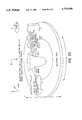
- FIG. 1is a schematic view of a first embodiment of an apparatus for imaging and/or characterizing a tissue, the apparatus being constructed according to the teachings of the present invention
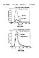
- FIGS. 2(a) and 2(b)are graphic representations of light intensity measured as a function of time using the apparatus of FIG. 1 for a human breast fat tissue sample and a human breast tumor tissue sample, respectively, the apparatus of FIG. 1 being arranged so as to illuminate the samples with 6.5 ps pulses of polarized light having a wavelength of 1064 nm, the thin line representing light intensity measured with the pair of polarizers of the apparatus of FIG. 1 oriented parallel to one another, the thick line representing light intensity measured with the pair of polarizers oriented perpendicular to one another;
- FIGS. 3(a) and 3(b)are graphic representations of light intensity measured as a function of time using the apparatus of FIG. 1 for a human breast fat tissue sample and a human breast tumor tissue sample, respectively, the apparatus of FIG. 1 being arranged so as to illuminate the samples with 5 ps pulses of polarized light having a wavelength of 532 nm, the thin line representing light intensity measured with the pair of polarizers of the apparatus of FIG. 1 oriented parallel to one another, the thick line representing light intensity measured with the pair of polarizers oriented perpendicular to one another;
- FIG. 4is a graphic representation of the temporal light intensity profile obtained for a human breast malignant tumor tissue by subtracting the light intensity measured as a function of time using the apparatus of FIG. 1 with the pair of polarizers oriented perpendicular to one another from the light intensity measured as a function of time using the apparatus of FIG. 1 with the pair of polarizers oriented parallel to one another;
- FIG. 5is a graphic representation of the degree of polarization D( ⁇ ,t) intensity measurements of FIGS. 2(a) and 2(b) for a human breast fat tissue sample and a human breast minor tissue sample, respectively, fitted to the equation: ##EQU1##
- FIG. 6is a schematic view of a second embodiment of an apparatus for imaging and/or characterizing a tissue, the apparatus being constructed according to the teachings of the present invention
- FIG. 7is a schematic view of a third embodiment of an apparatus for imaging and/or characterizing a tissue, the apparatus being constructed according to the teachings of the present invention.
- FIG. 8is a schematic view of a fourth embodiment of an apparatus for imaging and/or characterizing a tissue, the apparatus being constructed according to the teachings of the present invention.
- FIG. 9is a schematic view of a fifth embodiment of an apparatus for imaging and/or characterizing a tissue, the apparatus being constructed according to the teachings of the present invention.
- FIG. 10is a schematic view of a sixth embodiment of an apparatus for imaging and/or characterizing a tissue, the apparatus being constructed according to the teachings of the present invention.
- FIG. 11is a schematic view of one type of combination analyzer and imaging system adapted for use in the systems of FIGS. 1 and 6 through 10;
- FIG. 12is a schematic view of a first alternative combination analyzer and imaging system to that shown in FIG. 11;
- FIG. 13is a schematic view of a second alternative combination analyzer and imaging system to that shown in FIG. 11;
- FIG. 14is a schematic view of a third alternative combination analyzer and imaging system to that shown in FIG. 11;
- FIG. 15is a schematic view of a seventh embodiment of an apparatus for imaging and/or characterizing a tissue, the apparatus being constructed according to the teachings of the present invention.
- FIG. 16is a schematic view of an eighth embodiment of an apparatus for imaging and/or characterizing a tissue, the apparatus being constructed according to the teachings of the present invention.
- FIG. 17is a schematic view of a ninth embodiment of an apparatus for imaging and/or characterizing a tissue, the apparatus being constructed according to the teachings of the present invention.
- FIG. 18is a schematic view of a tenth embodiment of an apparatus for imaging and/or characterizing a tissue, the apparatus being constructed according to the teachings of the present invention
- FIG. 19is a schematic view of an eleventh embodiment of an apparatus for imaging and/or characterizing a tissue, the apparatus being constructed according to the teachings of the present invention.
- FIG. 20is a schematic view of a twelfth embodiment of an apparatus for imaging and/or characterizing a tissue, the apparatus being constructed according to the teachings of the present invention
- FIG. 21is a schematic view of a thirteenth embodiment of an apparatus for imaging and/or characterizing a tissue, the apparatus being constructed according to the teachings of the present invention
- FIGS. 22(a) and 22(b)are graphic representations of the temporal profiles of the degree of polarization of the emergent light under 1064 nm laser pulsed illumination of human breast tissue for (a) 3 mm fat and cancer tissue samples obtained from a patient; and (b) 3.3 mm fat and glandular tissue samples obtained from the same patient, respectively; and
- FIG. 23is a graphic representation of the degree of polarization in the transverse position of 532 nm laser light transmitted through 3 mm thick breast tissue sample containing a cancerous tumor section.
- the present inventionis based, in part, on the discovery that, when initially polarized light is transmitted through a turbid medium, such as human tissue, the ballistic and snake-like components of the light emergent from the turbid medium maintain the polarization of the initially polarized light while the diffuse component of the light emergent from the turbid medium becomes completely depolarized.
- the present inventionis also based, in part, on the discovery that initially polarized light transmitted through a turbid medium, such as human tissue, maintains its polarization, to a lesser or greater extent, depending upon the wavelength of the initially polarized light and depending upon the type of turbid medium traversed.
- the foregoing discoveriescan be used to, among other things, improve the optical imaging quality of optical tomography and mammography applications, by doing the following: a) improving the time-gating of the ballistic and snake light components of light emergent from a tissue when polarized laser pulses are used to image the tissue; b) selectively collecting forwardly propagating light in the case of continuous or pulsed light input; and c) enabling the characterization of the type of human tissue tested based on the degree that the tissue preserves polarization at different wavelengths.
- Human breast tissuecomprises fat tissue, glandular tissue, cysts and tumor tissue (both benign and malignant).
- tumor tissueboth benign and malignant.
- FIG. 1there is schematically shown an experimental set-up of an apparatus constructed according to the teachings of the present invention for imaging and/or characterizing a tissue, the apparatus being represented generally by reference numeral 11.
- Apparatus 11comprises a laser source 13.
- laser source 13comprises a mode-locked Nd:YAG laser for producing a series of laser pulses 15.
- Laser pulses 15are 6.5 ps pulses at 1064 nm with a repetition rate of 82 MHz and/or, at a second harmonic thereof, 5 ps pulses at 532 nm.
- Apparatus 11also includes a beam splitter 17 for splitting each pulse 15 into a pair of pulses 19 and 21. Pulse 19 is used to trigger a streak camera 23.
- a photodiode 25is disposed along the path of pulse 19 before streak camera 23 so that pulse 19 triggers streak camera 23 at an appropriate time.
- Apparatus 11also includes a first polarizer 31 disposed along the path of pulse 21, polarizer 31 being oriented parallel to the initial state of polarization of pulse 21 to ensure the polarization thereof.
- a beam splitter 33is disposed after first polarizer 31 for splitting pulse 21 into a pair of pulses 35 and 37. Pulse 35, which is used as a reference pulse, is deflected off a mirror 39 and is transmitted to streak camera 23 by a collection optical fiber 41 having a diameter of 110 ⁇ m.
- Pulse 37which has an average power of about 400 mW and a diameter of 3 mm, is used for propagation through a tissue sample, which may be, for example, breast tissue samples having a thickness of about 3 mm or 12 mm in a holder having a thickness of about 5 cm.
- Apparatusfurther includes a polarizer 43 disposed on the opposite side of the sample upon which pulse 37 is incident.
- polarizer 43may be oriented either parallel to the initial state of polarization of polarizer 31 or perpendicular to the initial state of polarization of polarizer 31 for use as an analyzer of the polarization state of the emergent light.
- Apparatusalso includes a fiber bundle 45, which is used for the collection of light passed through polarizer 43 and for the transmission of said light to streak camera 23.
- Bundle 45is approximately 1.2 mm in diameter and comprises a plurality of fibers of 110 mm in diameter.
- the end of bundle 45 coupled to the input of streak camera 23has its fiber elements aligned in a line 7.5 mm in length and 110 mm thickness.
- Fiber 41is coupled to bundle 45 2 mm on the side of the 7.5 mm line of bundle 45.
- Streak camera 23/SIT 47has a temporal resolution capability of about 10 ps.
- Apparatus 11further includes a temporal analyzer 49 which receives the output from streak camera 23/SIT 47.
- FIGS. 2(a) and 2(b)show the intensity of the output pulses emergent from a fat tissue sample 3 mm in thickness and from a malignant tumor tissue sample 3 mm in thickness, respectively, with polarizers 31 and 43 parallel to one another (thin line) and perpendicular to one another (thick line) when laser pulses 6.5 ps in duration with a wavelength of 1064 nm were illuminated therewith.
- zero timeis the time that the incident pulse enters the sample while the first pulse at negative time (-80 ps) is the reference pulse.
- FIGS. 3(a) and 3(b)show the same types of intensity profiles obtained from the same samples as in FIGS. 2(a) and 2(b), the only difference being that the incident laser pulses in FIGS. 3(a) and 3(b) are 5 ps in duration with a wavelength of 532 nm.
- FIG. 4shows the temporal profile resulting from the subtraction of the components of the output pulse parallel and perpendicular to the incident polarization when 1064 nm laser pulses were used to propagate through 12 mm thick cancer breast tissue.
- the intensity difference profileextends only 100 ps after the arrival of the ballistic component while the output pulse extends for over 500 ps. This means that by subtracting the parallel and perpendicular components of the output pulse we can selectively measure the signal from the early part of the output profile.
- This techniquecan be used as a time gate to select the early portion of the output pulse to improve imaging of objects in scattering media.
- the spectral and temporal degree of polarization D( ⁇ ,t) of the output pulseis given by the following equation: ##EQU2## where I( ⁇ ,t) parallel and I( ⁇ ,t) perpendicular are the components of the output pulse with polarization parallel and perpendicular to the incident polarization respectively.
- the peak values of the degree of polarizationwill be denoted by D.
- FIG. 5displays the temporal profiles of the degree of polarization D(t) using 1064 nm laser pulses to propagate the 3 mm thick fat (thin line) and tumor (thick line) tissues obtained using Eq. 1 and the polarized intensity profiles of the output pulse shown in FIGS. 2(a) and 2(b).
- tissuesdepolarize polarized pulses to different degrees due to changes in their structure and makeup. This effect is demonstrated by the fact that D fat is different than D tumor . This suggests that, by measuring the degree of polarization spatially D( ⁇ ,t,R), where R represents the spatial coordinates of the sample, information can be obtained on the type of tissue under investigation and can be used to image an object located inside the tissue. Combining this observation with the fact that the signal measured (I parallel -I perpendicular ) contains only the ballistic and snake components of the output pulses, which are the components used for imaging, images can be obtained in tissues containing information about the type and location of the tissues under investigation.
- the spatial change of the degree of polarization ⁇ D(R)can be used to image regions of tissues which have different components such as cysts, tumors, fat, etc.
- the ratio of the degree of polarization during propagation of 1064 and 532 nm laser light in fat and cancer breast tissue samplesare different. This is shown in FIGS. 2(a), 2(b), 3(a) and 3(b). More specifically, the peak value D of the degree of polarization for several different components has been determined to be as follows: ##EQU3##
- a polarization spatial mapcan be formed in accordance with the teachings of the present invention by measuring D(R) at different positions R either point by point or using CCD image mapping.
- Polarization gating according to the present inventioncan also be combined with time and space gating to enhance image resolution and contrast.
- FIG. 6there is shown a schematic view of a second embodiment constructed according to the teachings of the present invention of an apparatus for imaging and/or characterizing a tissue, the apparatus being represented generally by reference numeral 71.
- Apparatus 71includes a polarizer 73, which ensures polarized incident light, and a polarizer/analyzer 75, which, depending upon its orientation, is used to discriminate the two polarization components of the emergent light from the sample S.
- Apparatus 71also includes an imaging system 77, which is used to obtain the images from the two polarization components. Finally, one- or two- or three-dimensional mapping of the sample is obtained using appropriate electronics and computer software 79 to estimate and image the degree of polarization (D or d or p) of the emergent light.
- FIG. 7there is shown a schematic view of a third embodiment constructed according to the teachings of the present invention of an apparatus for imaging and/or characterizing a tissue, the apparatus being represented generally by reference numeral 81.
- Apparatus 81includes a polarizer 83, which is used to ensure that polarized light is transmitted to a sample S, a time and/or spatial gate 85 (e.g., Kerr gate or parametric gate or electronic gate and/or 4F Fourier spatial gate or another equivalent gating device), which is used to select only early-arriving and forwardly-propagating photons of the emergent light from sample S, a polarizer/analyzer 87, which, depending upon its orientation, is used to discriminate the two polarization components of the emergent, gated light, and an imaging system 89, which is used to obtain the images from the two polarization components.
- Apparatus 81also includes electronics and computer software 90, which is used to estimate and image the degree of polarization (D or d or p) of the emergent light.
- FIG. 8there is a schematic view of a fourth embodiment constructed according to the teachings of the present invention of an apparatus for imaging and/or characterizing a tissue, the apparatus being represented generally by reference numeral 91.
- Apparatus 91includes a polarizer 93, which is used to ensure that polarized light is transmitted to a sample S, a polarizer/analyzer 95, which, depending upon its orientation, is used to discriminate the two polarization components of the emergent light from sample S, a time and/or spatial gate 97 (e.g,, Kerr gate or parametric gate or electronic gate and/or 4F Fourier spatial gate or another equivalent gating device), which is used to select only early-arriving and forwardly-propagating photons of the emergent light from analyzer 95, and an imaging system 99, which is used to obtain the images from the two polarization components.
- Apparatus 91also includes electronics and computer software 100, which is used to estimate and image the degree of polarization (D or d or p) of the emergent light.
- FIG. 9there is a schematic view of a fifth embodiment constructed according to the teachings of the present invention of an apparatus for imaging and/or characterizing a tissue, the apparatus being represented generally by reference numeral 101.
- Apparatus 101includes a polarizer 103, which is used to ensure that polarized light is transmitted to a sample S, a polarizer/analyzer 105, which, depending upon its orientation, is used to discriminate the two polarization components of the emergent light from sample S, an image intensifier 107, which is time gated and is appropriately triggered to allow only the early part of the emergent light from analyzer 105 to be amplified, and a CCD imaging system 109, which is used to obtain the images from the two polarization components.
- Apparatus 101also includes electronics and computer software 110, which is used to estimate and image the degree of polarization (D or d or p) of the emergent light.
- FIG. 10there is a schematic view of a sixth embodiment constructed according to the teachings of the present invention of an apparatus for imaging and/or characterizing a tissue, the apparatus being represented generally by reference numeral 111.
- Apparatus 111includes a polarizer 113, which is used to ensure that polarized light is transmitted to a sample S, a polarizer/analyzer 115, which, depending upon its orientation, is used to discriminate the two polarization components of the emergent light from sample S, a 4F Fourier space gate 116, which is used to reduce the diffusive component of the emergent light, an image intensifier 117, which is time gated and is appropriately triggered to allow only the early part of the emergent light from gate 116 to be amplified, and a CCD imaging system 119, which is used to obtain the images from the two polarization components.
- Apparatus 111also includes electronics and computer software 120, which is used to estimate and image the degree of polarization (D or d or p) of the emergent light.
- the incident polarizationis linear, analyzer 123 comprising a linear polarizer, whose orientation may be altered by mechanical or electrooptical means so that the two polarization image components of the emergent light (i.e., parallel and perpendicular to the incident light polarization) are recorded at different times by the imaging unit 125.
- FIG. 12there is shown a schematic view of an alternative combination 131 to that shown in FIG. 11.
- the incident polarizationis circular (elliptical), analyzer 133 comprising a retardation plate 135 ( ⁇ /4 for circular polarization) and a linear polarizer 137 with orientation that may be altered via mechanical or electrooptical means at ⁇ 45° with respect to the o-axis of retardation plate 133 so that the two polarization image components of the emergent light (left-hand and right-hand circular) are recorded at different times by imaging unit 139.
- FIG. 13there is shown a schematic view of another alternative combination 141 to that shown in FIG. 11.
- the incident polarizationis linear
- the analyzercomprising a polarizing beam splitter 143 so that the two polarization image components of the emergent light (parallel and perpendicular to the incident light polarization) are recorded simultaneously by two imaging units 145 and 147.
- FIG. 14there is shown a schematic view of yet another alternative combination 151 to that shown in FIG. 11.
- the incident polarizationis circular (elliptical)
- the analyzercomprising a retardation plate 153 ( ⁇ /4 for circular polarization) and a polarizing beam splitter 155 with axis at ⁇ 45° with respect to the o-axis of retardation plate 153 so that the two polarization image components of the emerged light (parallel and perpendicular to the incident light polarization) are recorded simultaneously by imaging units 157 and 159.
- FIG. 15there is shown a schematic view of a seventh embodiment constructed according to the teachings of the present invention of an apparatus for imaging and/or characterizing a tissue, the apparatus being represented generally by reference numeral 161.
- Apparatus 161includes a light source for producing a light beam.
- the light beamis collimated and then polarized using a polarizer.
- the beampropagates through the sample.
- the emerged lightis collected by appropriate optical elements (lenses and/or mirrors) and directed into a point detector (e.g., photodiode, photomultiplier) after passing through an analyzer and a time and/or spatial gate.
- the apparatusis mounted on a table accommodating x-y-z and/or ⁇ -r-z translational/rotational stages that allow one to scan the sample to obtain a point by point mapping.
- Apparatus 161also includes electronics and computer software (not shown), which is used to estimate and image the degree of polarization (D or d or p) of the emergent light.
- FIG. 16there is shown a schematic view of an eighth embodiment constructed according to the teachings of the present invention of an apparatus for imaging and/or characterizing a tissue, the apparatus being represented generally by reference numeral 171.
- Apparatus 171includes a light source for producing a light beam.
- the light beamis collimated and polarized using a polarizer.
- the beampropagates the sample.
- the emergent lightis collected by appropriate optical elements (lenses and/or mirrors) and directed into a fast point detector (photodiode, photomultiplier) after passing through the analyzer.
- the apparatusis mounted on a table accommodating x-y-z and/or ⁇ -r-z translational/rotational stages that allow one to scan the sample to obtain a point by point mapping.
- the signal of the fast photodetectoris amplified and directed into the input of a sampling oscilloscope where the signal is time-gated to selectively obtain the intensity of the early part of emergent light.
- a lock-in amplifier or a boxcar integratorthe sensitivity of the system can be improved (if needed) and the time-gated signal is sent to a computer for image processing.
- FIG. 17there is shown a schematic view of a ninth embodiment constructed according to the teachings of the present invention of an apparatus for imaging and/or characterizing a tissue, the apparatus being represented generally by reference numeral 181.
- Apparatus 181includes a light source for producing a light beam.
- the light beamis collimated and polarized using a polarizer.
- a beam deflectoris used to scan the sample in the y-z plane. The beam propagates the sample.
- the emergent lightis collected by a beam collector scanning in the y-z plane in phase with the beam deflector and directed into a point detector (photodiode, photomultiplier) after passing through the analyzer and the time and/or spatial gate.
- the deflector with the in-phase beam collectorallow scanning of the sample to obtain a point by point mapping.
- Appartus 181also includes electronics and computer software (not shown), which is used to estimate and image the degree of polarization (D or d or p) of the emergent light.
- FIG. 18there is shown a schematic view of a tenth embodiment constructed according to the teachings of the present invention of an apparatus for imaging and/or characterizing a tissue, the apparatus being represented generally by reference numeral 191.
- Apparatus 191includes a light source for producing a light beam.
- the light beamis collimated and polarized using a polarizer.
- a beam deflectoris used to scan the sample in the y-z plane. The beam propagates the sample.
- the emergent lightis collected by a beam collector scanning in the y-z plane in phase with the beam deflector and directed into a fast point photodetector (photodiode, photomultiplier) after passing through the analyzer.
- the signal of the fast photodetectoris amplified and directed into the input of a sampling oscilloscope where the signal is time-gated to selectively obtain the intensity of the early part of emergent light.
- the sensitivity of the systemcan be improved (if needed) and the time-gated signal is sent to a computer for image processing.
- the deflector with the in-phase beam collectorallow scanning of the sample to obtain a point by point mapping.
- FIG. 19there is shown a schematic view of an eleventh embodiment constructed according to the teachings of the present invention of an apparatus for imaging and/or characterizing a tissue, the apparatus being represented generally by reference numeral 201.
- Apparatus 201includes a light source for producing a light beam.
- the light beamis collimated and polarized using a polarizer.
- the beampropagates the sample.
- the emergent lightis collected by appropriate optical elements (lenses and/or mirrors) and directed into an array detector after passing through the analyzer and the time and/or spatial gate.
- the apparatusis mounted on a table accommodating x-y-z and/or ⁇ -r-z translational/rotational stages that allow one to scan the sample to obtain a point by point mapping.
- Appartus 201also includes electronics and computer software (not shown), which is used to estimate and image the degree of polarization (D or d or p) of the emergent light.
- FIG. 20there is shown a schematic view of a twelfth embodiment constructed according to the teachings of the present invention of an apparatus for imaging and/or characterizing a tissue, the apparatus being represented generally by reference numeral 211.
- Apparatus 211includes a light source for producing a light beam.
- the light beamis collimated and polarized using a polarizer.
- the beampropagates the sample.
- the emergent lightis collected by appropriate optical elements (lenses and/or mirrors) and after passing through the analyzer and the time and/or spatial gate is directed into a two-dimensional detector to record a shadowgram.
- the apparatusis mounted on a table accommodating x-y-z and/or ⁇ -r-z translational/rotational stages that allow one to scan the sample.
- Appartus 211also includes electronics and computer software (not shown), which is used to estimate and image the degree of polarization (D or d or p) of the emergent light.
- FIG. 21there is shown a schematic view of a thirteenth embodiment constructed according to the teachings of the present invention of an apparatus for imaging and/or characterizing a tissue, the apparatus being represented generally by reference numeral 221.
- Apparatus 221includes a light source for producing a light beam.
- the light beamis collimated, expanded and polarized using a polarizer.
- the beampropagates the sample.
- the emergent lightis collected by appropriate optical elements (lenses and/or mirrors) and after passing through the analyzer and the time and/or spatial gate is directed into a two-dimensional detector to record a shadowgram.
- the apparatusis mounted on a table accommodating x-y-z and/or ⁇ -r-z translational/rotational stages that allow one to scan the sample.
- Appartus 221also includes electronics and computer software (not shown), which is used to estimate and image the degree of polarization (D or d or p) of the emergent light.
- FIGS. 22(a) and 22(b)there can be seen the temporal profiles of the degree of polarization of the emergent light under 1064 nm laser pulsed illumination of human breast tissue in the case of (a) 3 mm fat and cancer tumor tissue samples obtained from a patient; and (b) 3.3 mm fat and glandular tissue samples obtained from the same patient, respectively.
- FIGS. 22(a) and 22(b)demonstrate that cancer tumor tissue depolarizes light faster than does normal tissue.
- FIG. 23there can be seen a graphic representation of the degree of polarization in the transverse position of 532 nm laser light transmitted through 3 mm thick breast tissue sample containing a cancerous tumor section.
- the 532 nm laser beamwas expanded to illuminate the sample.
- the part of the sample containing the cancer tumordepolarizes almost completely the 532 nm light while light propagating through the fat tissue is partially polarized.
- the profileclearly distinguishes the cancerous part of the sample from the normal part of the sample.
- the incident lightshould be polarized.
- the light sourcecan be any laser available or any other pulsed or continuous light source which provides polarized light with or without the use of a polarizing element.
- the operating wavelength rangeis preferably 400-1600 nm.
- the two polarization components of the output lightare recorded. From these components the degree of polarization preservation of the output light is calculated. Accordingly, the two polarization components can be measured simultaneously (using for example a polarizing beam-spliter and two detectors) or at different times (using one photodetector and an analyzer that can change its polarization orientation or by changing the input polarization and leaving the analyzer at the same position).
- An imageis obtained by recording the degree of polarization of the output light propagating through the scattering medium point by point or through mapping. Accordingly, a suitable detector currently available is a cooled CCD. The two images of the parallel and perpendicular polarization components of the propagating light are recorded and via interimage operation involving these components an image of the degree of polarization is obtained.
- the image of the degree of polarizationchanges with respect to the relative intensity of the observed types of tissues (tumor, fat, etc.).
- a pseudocolored imagecan be generated containing additional information on the types of tissues observed (e.g. separating benign from malignant tumors).
- the polarized light source utilizedcan be monochromatic or containing a band of frequencies.
- the degree of polarization image at various wavelengthscan be obtained by a) varying the wavelength (for monochromatic light), b) varying the central wavelength or c) varying the bandwidth (for a band of frequencies).
- the polarization preservation imaging techniquecan be combined with other techniques that provide selection of the photons propagated in the forward direction.
- Fourier space gate and/or Kerr gate or gates based on picosecond optical or electronic switchescan be combined with the polarization imaging system for enhanced image resolution.
- the polarization preservation in tissues and random mediacan be used for time gating the early part of the output pulse
- the time gate obtained through the polarization preservationopens with the arrival of the ballistic light up to about 100 ps after the arrival of the ballistic light;
- Polarization preservationcan be used for selective collection of the light propagating in the forward direction in human tissues
- the degree of polarization preservation of light propagating in human tissueis different depending on the type of tissue (fat, glandular, tumors or cysts);
- the degree of polarization of light that propagate through tissuedepends on the wavelength of the propagating light and tissue type
- the change of the degree of polarization as a function of the wavelength of the propagating lightcan be used for obtaining information on the type of the tissue under investigation;
- the degree of polarization of the propagating lightcan be measured by any function containing the parallel (I parallel ) and perpendicular (I perpendicular ) components of the output light such as eq. 1, I para -I perp !, I para /I perp ! and I perp /(I para +I perp )!;
- the degree of polarization of the propagating lightcan be measured using the integral in time of D(t) (instead of the peak value) or using the integral in time of I(t) parallel and I(t) perpendicular in combination with paragraph 10 above;
- Polarization preservationcan be used for imaging in human tissue point by point or over area
- a map of D(R)can be used for imaging
- the polarization spatial time gate D(R,t)can be used in combination with Fourier space gate and time gate (optical or electronic gate);
- a ⁇ D(R, ⁇ ) mapcan be used for imaging minors and allow for the differentiation of malignant from benign tumors
- a combination of D( ⁇ ,t,R) and/or ⁇ D(R, ⁇ ) map with any time gating technique selecting the forwardly propagating photons (ballistic and snake part of the output pulse) such as Kerr gate or gates based on picosecond optical or electronic swishes and electronic time gatescan be used for enhanced image resolution;
- Optical tomography and mammographycan be achieved using the following principals: a) the polarization preservation of initially polarized light propagating in tissues, b) the dependence of the degree of polarization on the wavelength of the propagating light, and c) the mapping dependence of the degree of polarization of the output light to the type of tissue (fat, glandular, tumors, cysts);
- the polarized light sources required for use with the polarization preservation techniquecan be pulsed or continuous, laser source or any other light source operating in the spectral region between 400 and 1600 nm such as Ti:Sapphire laser (800-1000 nm), Cr4+:Forsterite laser (1100-1300 nm) or Cr4+:YAG (1300-1600 nm).
- Ti:Sapphire laser800-1000 nm
- Cr4+:Forsterite laser(1100-1300 nm)
- Cr4+:YAG(1300-1600 nm
- the preservation of the polarization of light propagating in human tissueis a property with great importance in the field of optical tomography and mammography.
- the initially polarized light propagating through the tissueretains some of the information concerning its initial polarization.
- the degree of polarization of the output lightdepends on the wavelength of the light and the type of the tissue (glandular, fat, cyst or minor).
- the polarization preservation techniquecan be used as a 100 ps time gate and as a tool for optical imaging and characterization of the type of tissue for diagnostic reasons.
- the polarization preservation imaging techniquecan be combined with other time-gated imaging techniques for enhanced image clarity and volume of information.
Landscapes
- Health & Medical Sciences (AREA)
- Life Sciences & Earth Sciences (AREA)
- Physics & Mathematics (AREA)
- General Health & Medical Sciences (AREA)
- Pathology (AREA)
- Surgery (AREA)
- Biomedical Technology (AREA)
- Heart & Thoracic Surgery (AREA)
- Medical Informatics (AREA)
- Molecular Biology (AREA)
- Engineering & Computer Science (AREA)
- Animal Behavior & Ethology (AREA)
- Biophysics (AREA)
- Public Health (AREA)
- Veterinary Medicine (AREA)
- Chemical & Material Sciences (AREA)
- Analytical Chemistry (AREA)
- Biochemistry (AREA)
- General Physics & Mathematics (AREA)
- Immunology (AREA)
- Optics & Photonics (AREA)
- Gynecology & Obstetrics (AREA)
- Reproductive Health (AREA)
- Nuclear Medicine, Radiotherapy & Molecular Imaging (AREA)
- Radiology & Medical Imaging (AREA)
- Investigating Or Analysing Materials By Optical Means (AREA)
Abstract
Description
Claims (62)
Priority Applications (5)
| Application Number | Priority Date | Filing Date | Title |
|---|---|---|---|
| US08/573,939US5719399A (en) | 1995-12-18 | 1995-12-18 | Imaging and characterization of tissue based upon the preservation of polarized light transmitted therethrough |
| US08/704,841US5847394A (en) | 1995-12-18 | 1996-08-28 | Imaging of objects based upon the polarization or depolarization of light |
| US08/734,340US5929443A (en) | 1995-12-18 | 1996-10-21 | Imaging of objects based upon the polarization or depolarization of light |
| AU14304/97AAU1430497A (en) | 1995-12-18 | 1996-12-18 | Imaging and characterization of tissue based upon the preservation of polarized light transmitted therethrough |
| PCT/US1996/020566WO1997022871A1 (en) | 1995-12-18 | 1996-12-18 | Imaging and characterization of tissue based upon the preservation of polarized light transmitted therethrough |
Applications Claiming Priority (1)
| Application Number | Priority Date | Filing Date | Title |
|---|---|---|---|
| US08/573,939US5719399A (en) | 1995-12-18 | 1995-12-18 | Imaging and characterization of tissue based upon the preservation of polarized light transmitted therethrough |
Related Child Applications (1)
| Application Number | Title | Priority Date | Filing Date |
|---|---|---|---|
| US08/704,841Continuation-In-PartUS5847394A (en) | 1995-12-18 | 1996-08-28 | Imaging of objects based upon the polarization or depolarization of light |
Publications (1)
| Publication Number | Publication Date |
|---|---|
| US5719399Atrue US5719399A (en) | 1998-02-17 |
Family
ID=24294009
Family Applications (2)
| Application Number | Title | Priority Date | Filing Date |
|---|---|---|---|
| US08/573,939Expired - Fee RelatedUS5719399A (en) | 1995-12-18 | 1995-12-18 | Imaging and characterization of tissue based upon the preservation of polarized light transmitted therethrough |
| US08/704,841Expired - Fee RelatedUS5847394A (en) | 1995-12-18 | 1996-08-28 | Imaging of objects based upon the polarization or depolarization of light |
Family Applications After (1)
| Application Number | Title | Priority Date | Filing Date |
|---|---|---|---|
| US08/704,841Expired - Fee RelatedUS5847394A (en) | 1995-12-18 | 1996-08-28 | Imaging of objects based upon the polarization or depolarization of light |
Country Status (3)
| Country | Link |
|---|---|
| US (2) | US5719399A (en) |
| AU (1) | AU1430497A (en) |
| WO (1) | WO1997022871A1 (en) |
Cited By (196)
| Publication number | Priority date | Publication date | Assignee | Title |
|---|---|---|---|---|
| US5929443A (en)* | 1995-12-18 | 1999-07-27 | The Research Foundation City College Of New York | Imaging of objects based upon the polarization or depolarization of light |
| WO2000043750A3 (en)* | 1999-01-25 | 2000-11-23 | Newton Lab Inc | Imaging of tissue using polarized light |
| WO2000076399A1 (en) | 1999-06-11 | 2000-12-21 | Cardiomend, L.L.C. | Device and method for testing tissue |
| US6264609B1 (en)* | 1999-09-15 | 2001-07-24 | Wake Forest University | Ultrasound apparatus and method for tissue characterization |
| US6364829B1 (en) | 1999-01-26 | 2002-04-02 | Newton Laboratories, Inc. | Autofluorescence imaging system for endoscopy |
| US20020114765A1 (en)* | 1997-01-17 | 2002-08-22 | Grable Richard J. | Laser imaging apparatus using biomedical markers that bind to cancer cells |
| US20020183624A1 (en)* | 2001-06-05 | 2002-12-05 | Rio Grande Medical Technologies, Inc. | Apparatus and method of biometric determination using specialized optical spectroscopy systems |
| US20030028100A1 (en)* | 2001-05-01 | 2003-02-06 | Tearney Guillermo J. | Method and apparatus for determination of atherosclerotic plaque type by measurement of tissue optical properties |
| US6529759B1 (en) | 2001-03-08 | 2003-03-04 | Electrical Geodesics, Inc. | Method for mapping internal body tissue |
| US20030048499A1 (en)* | 2001-04-04 | 2003-03-13 | Alfano Robert R. | Methods of improving line of sight wireless optical communication through adverse environmental conditions |
| US6537211B1 (en) | 1998-01-26 | 2003-03-25 | Massachusetts Institute Of Technology | Flourescence imaging endoscope |
| US20030093005A1 (en)* | 2001-11-13 | 2003-05-15 | Tucker Don M. | Method for neural current imaging |
| US6587711B1 (en) | 1999-07-22 | 2003-07-01 | The Research Foundation Of Cuny | Spectral polarizing tomographic dermatoscope |
| US6594521B2 (en) | 1999-12-17 | 2003-07-15 | Electrical Geodesics, Inc. | Method for localizing electrical activity in the body |
| US6665557B1 (en) | 1999-12-01 | 2003-12-16 | The Research Foundation Of City College Of New York | Sprectroscopic and time-resolved optical methods and apparatus for imaging objects in turbed media |
| US20040047493A1 (en)* | 1999-10-08 | 2004-03-11 | Lumidigm, Inc. | Apparatus and method for identification of individuals by near-infrared spectrum |
| US20040111031A1 (en)* | 1999-07-22 | 2004-06-10 | Alfano Robert R. | Spectral polarizing tomographic dermatoscope |
| US20040240712A1 (en)* | 2003-04-04 | 2004-12-02 | Lumidigm, Inc. | Multispectral biometric sensor |
| US20050004453A1 (en)* | 2003-01-24 | 2005-01-06 | Tearney Guillermo J. | System and method for identifying tissue using low-coherence interferometry |
| US20050007582A1 (en)* | 2003-07-07 | 2005-01-13 | Lumidigm, Inc. | Methods and apparatus for collection of optical reference measurements for monolithic sensors |
| US20050018201A1 (en)* | 2002-01-24 | 2005-01-27 | De Boer Johannes F | Apparatus and method for ranging and noise reduction of low coherence interferometry lci and optical coherence tomography oct signals by parallel detection of spectral bands |
| US20050018200A1 (en)* | 2002-01-11 | 2005-01-27 | Guillermo Tearney J. | Apparatus for low coherence ranging |
| US20050035295A1 (en)* | 2003-06-06 | 2005-02-17 | Brett Bouma | Process and apparatus for a wavelength tuning source |
| US20050128488A1 (en)* | 2003-11-28 | 2005-06-16 | Dvir Yelin | Method and apparatus for three-dimensional spectrally encoded imaging |
| US20050185847A1 (en)* | 2003-12-11 | 2005-08-25 | Lumidigm, Inc. | Methods and systems for estimation of personal characteristics from biometric measurements |
| US20050205667A1 (en)* | 2003-04-04 | 2005-09-22 | Lumidigm, Inc. | Combined total-internal-reflectance and tissue imaging systems and methods |
| US20050265585A1 (en)* | 2004-06-01 | 2005-12-01 | Lumidigm, Inc. | Multispectral liveness determination |
| US20050271258A1 (en)* | 2004-06-01 | 2005-12-08 | Lumidigm, Inc. | Multispectral imaging biometrics |
| US20050280828A1 (en)* | 2001-10-16 | 2005-12-22 | The General Hospital Corporation | Systems and methods for imaging a sample |
| US20060013544A1 (en)* | 2004-07-02 | 2006-01-19 | Bouma Brett E | Imaging system and related techniques |
| US7006676B1 (en) | 2000-01-21 | 2006-02-28 | Medical Optical Imaging, Inc. | Method and apparatus for detecting an abnormality within a host medium utilizing frequency-swept modulation diffusion tomography |
| US20060058592A1 (en)* | 2004-08-24 | 2006-03-16 | The General Hospital Corporation | Process, system and software arrangement for measuring a mechanical strain and elastic properties of a sample |
| US20060058622A1 (en)* | 2004-08-24 | 2006-03-16 | The General Hospital Corporation | Method and apparatus for imaging of vessel segments |
| US20060055936A1 (en)* | 2004-09-10 | 2006-03-16 | The General Hospital Corporation | System and method for optical coherence imaging |
| US20060062438A1 (en)* | 2003-04-04 | 2006-03-23 | Lumidigm, Inc. | Comparative texture analysis of tissue for biometric spoof detection |
| US20060067620A1 (en)* | 2004-09-29 | 2006-03-30 | The General Hospital Corporation | System and method for optical coherence imaging |
| US20060093276A1 (en)* | 2004-11-02 | 2006-05-04 | The General Hospital Corporation | Fiber-optic rotational device, optical system and method for imaging a sample |
| US20060109478A1 (en)* | 2004-11-24 | 2006-05-25 | The General Hospital Corporation | Devices and arrangements for performing coherence range imaging using a common path interferometer |
| US20060110015A1 (en)* | 2003-04-04 | 2006-05-25 | Lumidigm, Inc. | Systems and methods for improved biometric feature definition |
| US20060114473A1 (en)* | 2004-11-29 | 2006-06-01 | The General Hospital Corporation | Arrangements, devices, endoscopes, catheters and methods for performing optical imaging by simultaneously illuminating and detecting multiple points on a sample |
| US20060227333A1 (en)* | 2003-03-31 | 2006-10-12 | Tearney Guillermo J | Speckle reduction in optical coherence tomography by path length encoded angular compounding |
| US20060244973A1 (en)* | 2003-10-27 | 2006-11-02 | Seok-Hyun Yun | Method and apparatus for performing optical imaging using frequency-domain interferometry |
| US20060270929A1 (en)* | 2005-05-31 | 2006-11-30 | The General Hospital Corporation | System, method and arrangement which can use spectral encoding heterodyne interferometry techniques for imaging |
| US20060279742A1 (en)* | 2005-06-01 | 2006-12-14 | The General Hospital Corporation | Apparatus, method and system for performing phase-resolved optical frequency domain imaging |
| US20070009935A1 (en)* | 2005-05-13 | 2007-01-11 | The General Hospital Corporation | Arrangements, systems and methods capable of providing spectral-domain optical coherence reflectometry for a sensitive detection of chemical and biological sample |
| US20070012886A1 (en)* | 2005-04-28 | 2007-01-18 | The General Hospital Corporation | Systems. processes and software arrangements for evaluating information associated with an anatomical structure by an optical coherence ranging technique |
| US20070030475A1 (en)* | 2003-04-04 | 2007-02-08 | Lumidigm, Inc. | White-light spectral biometric sensors |
| US20070038040A1 (en)* | 2005-04-22 | 2007-02-15 | The General Hospital Corporation | Arrangements, systems and methods capable of providing spectral-domain polarization-sensitive optical coherence tomography |
| US20070035743A1 (en)* | 2005-08-09 | 2007-02-15 | The General Hospital Corporation | Apparatus, methods and storage medium for performing polarization-based quadrature demodulation in optical coherence tomography |
| US20070049833A1 (en)* | 2005-08-16 | 2007-03-01 | The General Hospital Corporation | Arrangements and methods for imaging in vessels |
| US20070058252A1 (en)* | 2004-09-27 | 2007-03-15 | Honeywell International Inc. | Circular polarization illumination based analyzer system |
| US20070073162A1 (en)* | 2000-10-30 | 2007-03-29 | The General Hospital Corporation | Methods and systems for tissue analysis |
| US20070087445A1 (en)* | 2005-10-14 | 2007-04-19 | The General Hospital Corporation | Arrangements and methods for facilitating photoluminescence imaging |
| US20070116331A1 (en)* | 2004-06-01 | 2007-05-24 | Lumidigm, Inc. | System and method for robust fingerprint acquisition |
| US20070121196A1 (en)* | 2005-09-29 | 2007-05-31 | The General Hospital Corporation | Method and apparatus for method for viewing and analyzing of one or more biological samples with progressively increasing resolutions |
| CN1325022C (en)* | 1999-04-01 | 2007-07-11 | 成象诊断系统公司 | Laser imaging setup using biochemical markers that bind tumor cells |
| US20070167835A1 (en)* | 2005-07-25 | 2007-07-19 | Massachusetts Institute Of Technology | Tri modal spectroscopic imaging |
| US20070171433A1 (en)* | 2006-01-20 | 2007-07-26 | The General Hospital Corporation | Systems and processes for providing endogenous molecular imaging with mid-infrared light |
| US20070171430A1 (en)* | 2006-01-20 | 2007-07-26 | The General Hospital Corporation | Systems and methods for providing mirror tunnel micropscopy |
| US20070179487A1 (en)* | 2006-02-01 | 2007-08-02 | The General Hospital Corporation | Apparatus for applying a plurality of electro-magnetic radiations to a sample |
| US20070188855A1 (en)* | 2006-01-19 | 2007-08-16 | The General Hospital Corporation | Apparatus for obtaining information for a structure using spectrally-encoded endoscopy teachniques and methods for producing one or more optical arrangements |
| US20070201033A1 (en)* | 2006-02-24 | 2007-08-30 | The General Hospital Corporation | Methods and systems for performing angle-resolved fourier-domain optical coherence tomography |
| US20070208400A1 (en)* | 2006-03-01 | 2007-09-06 | The General Hospital Corporation | System and method for providing cell specific laser therapy of atherosclerotic plaques by targeting light absorbers in macrophages |
| US20070223006A1 (en)* | 2006-01-19 | 2007-09-27 | The General Hospital Corporation | Systems and methods for performing rapid fluorescence lifetime, excitation and emission spectral measurements |
| US20070239033A1 (en)* | 2006-03-17 | 2007-10-11 | The General Hospital Corporation | Arrangement, method and computer-accessible medium for identifying characteristics of at least a portion of a blood vessel contained within a tissue using spectral domain low coherence interferometry |
| US20070236700A1 (en)* | 2006-04-05 | 2007-10-11 | The General Hospital Corporation | Methods, arrangements and systems for polarization-sensitive optical frequency domain imaging of a sample |
| US20070263227A1 (en)* | 2006-05-12 | 2007-11-15 | The General Hospital Corporation | Processes, arrangements and systems for providing a fiber layer thickness map based on optical coherence tomography images |
| US20070263208A1 (en)* | 2006-01-10 | 2007-11-15 | The General Hospital Corporation | Systems and methods for generating data based on one or more spectrally-encoded endoscopy techniques |
| US20070274650A1 (en)* | 2006-02-01 | 2007-11-29 | The General Hospital Corporation | Apparatus for controlling at least one of at least two sections of at least one fiber |
| US20070276269A1 (en)* | 2006-05-10 | 2007-11-29 | The General Hospital Corporation | Process, arrangements and systems for providing frequency domain imaging of a sample |
| US20070282403A1 (en)* | 2006-02-01 | 2007-12-06 | The General Hospital Corporation | Methods and systems for providing electromagnetic radiation to at least one portion of a sample using conformal laser therapy procedures |
| US20080002211A1 (en)* | 2006-01-20 | 2008-01-03 | The General Hospital Corporation | System, arrangement and process for providing speckle reductions using a wave front modulation for optical coherence tomography |
| US20080007734A1 (en)* | 2004-10-29 | 2008-01-10 | The General Hospital Corporation | System and method for providing Jones matrix-based analysis to determine non-depolarizing polarization parameters using polarization-sensitive optical coherence tomography |
| US20080021275A1 (en)* | 2006-01-19 | 2008-01-24 | The General Hospital Corporation | Methods and systems for optical imaging or epithelial luminal organs by beam scanning thereof |
| US20080027317A1 (en)* | 2006-06-29 | 2008-01-31 | Fred Wood | Scanned laser vein contrast enhancer |
| US20080049232A1 (en)* | 2006-08-25 | 2008-02-28 | The General Hospital Coporation | Apparatus and methods for enhancing optical coherence tomography imaging using volumetric filtering techniques |
| US20080097225A1 (en)* | 2006-10-19 | 2008-04-24 | The General Hospital Corporation | Apparatus and method for obtaining and providing imaging information associated with at least one portion of a sample, and effecting such portion(s) |
| US20080094637A1 (en)* | 2003-01-24 | 2008-04-24 | The General Hospital Corporation | Apparatus and method for ranging and noise reduction of low coherence interferometry lci and optical coherence tomography oct signals by parallel detection of spectral bands |
| US7394919B2 (en) | 2004-06-01 | 2008-07-01 | Lumidigm, Inc. | Multispectral biometric imaging |
| US20080175280A1 (en)* | 2007-01-19 | 2008-07-24 | The General Hospital Corporation | Wavelength tuning source based on a rotatable reflector |
| US20080192988A1 (en)* | 2006-07-19 | 2008-08-14 | Lumidigm, Inc. | Multibiometric multispectral imager |
| US20080206804A1 (en)* | 2007-01-19 | 2008-08-28 | The General Hospital Corporation | Arrangements and methods for multidimensional multiplexed luminescence imaging and diagnosis |
| US20080234586A1 (en)* | 2007-03-19 | 2008-09-25 | The General Hospital Corporation | System and method for providing noninvasive diagnosis of compartment syndrome using exemplary laser speckle imaging procedure |
| US20080232653A1 (en)* | 2007-03-21 | 2008-09-25 | Lumidigm, Inc. | Biometrics based on locally consistent features |
| US20080234567A1 (en)* | 2007-03-19 | 2008-09-25 | The General Hospital Corporation | Apparatus and method for providing a noninvasive diagnosis of internal bleeding |
| US20080232410A1 (en)* | 2007-03-23 | 2008-09-25 | The General Hospital Corporation | Methods, arrangements and apparatus for utilizing a wavelength-swept laser using angular scanning and dispersion procedures |
| US20080262359A1 (en)* | 2007-03-30 | 2008-10-23 | The General Hospital Corporation | System and method providing intracoronary laser speckle imaging for the detection of vulnerable plaque |
| US20080262314A1 (en)* | 2007-04-17 | 2008-10-23 | The General Hospital Corporation | Apparatus and methods for measuring vibrations using spectrally-encoded endoscopy |
| US20080287808A1 (en)* | 2006-09-12 | 2008-11-20 | The General Hospital Corporation | Apparatus, probe and method for providing depth assessment in an anatomical structure |
| US20080298649A1 (en)* | 2004-06-01 | 2008-12-04 | Lumidigm, Inc. | Hygienic biometric sensors |
| WO2009005748A1 (en)* | 2007-06-29 | 2009-01-08 | The Trustees Of Columbia University In The City Ofnew York | Optical imaging or spectroscopy systems and methods |
| US20090036782A1 (en)* | 2007-07-31 | 2009-02-05 | The General Hospital Corporation | Systems and methods for providing beam scan patterns for high speed doppler optical frequency domain imaging |
| US20090046903A1 (en)* | 2007-04-10 | 2009-02-19 | Lumidigm, Inc. | Biometric Detection Using Spatial, Temporal, And/Or Spectral Techniques |
| US20090059360A1 (en)* | 2007-08-31 | 2009-03-05 | The General Hospital Corporation | System and method for self-interference fluorescence microscopy, and computer-accessible medium associated therewith |
| US20090073439A1 (en)* | 2007-09-15 | 2009-03-19 | The General Hospital Corporation | Apparatus, computer-accessible medium and method for measuring chemical and/or molecular compositions of coronary atherosclerotic plaques in anatomical structures |
| US20090076396A1 (en)* | 2007-09-17 | 2009-03-19 | The General Hospital Corporation | Optical wavelength range for high contrast imaging of cancer |
| US20090080709A1 (en)* | 2006-07-19 | 2009-03-26 | Lumidigm, Inc. | Whole-Hand Multispectral Biometric Imaging |
| US20090122302A1 (en)* | 2007-10-30 | 2009-05-14 | The General Hospital Corporation | System and method for cladding mode detection |
| US20090131801A1 (en)* | 2007-10-12 | 2009-05-21 | The General Hospital Corporation | Systems and processes for optical imaging of luminal anatomic structures |
| US7545963B2 (en) | 2003-04-04 | 2009-06-09 | Lumidigm, Inc. | Texture-biometrics sensor |
| US20090192358A1 (en)* | 2008-01-28 | 2009-07-30 | The General Hospital Corporation | Systems, processes and computer-accessible medium for providing hybrid flourescence and optical coherence tomography imaging |
| US20090225324A1 (en)* | 2008-01-17 | 2009-09-10 | The General Hospital Corporation | Apparatus for providing endoscopic high-speed optical coherence tomography |
| US20090245591A1 (en)* | 2006-07-19 | 2009-10-01 | Lumidigm, Inc. | Contactless Multispectral Biometric Capture |
| US7620212B1 (en) | 2002-08-13 | 2009-11-17 | Lumidigm, Inc. | Electro-optical sensor |
| US20090323056A1 (en)* | 2007-05-04 | 2009-12-31 | The General Hospital Corporation | Methods, arrangements and systems for obtaining information associated with a sample using optical microscopy |
| US20100081926A1 (en)* | 2008-09-29 | 2010-04-01 | Searete Llc, A Limited Liability Corporation Of The State Of Delaware | Histological facilitation systems and methods |
| US20100081928A1 (en)* | 2008-09-29 | 2010-04-01 | Searete Llc, A Limited Liability Corporation Of The State Of Delaware | Histological Facilitation systems and methods |
| US20100081915A1 (en)* | 2008-09-29 | 2010-04-01 | Searete Llc, Alimited Liability Corporation Of The State Of Delaware | Histological facilitation systems and methods |
| US20100081927A1 (en)* | 2008-09-29 | 2010-04-01 | Searete Llc, A Limited Liability Corporation Of The State Of Delaware | Histological facilitation systems and methods |
| US20100081916A1 (en)* | 2008-09-29 | 2010-04-01 | Searete Llc, A Limited Liability Corporation Of The State Of Delaware. | Histological facilitation systems and methods |
| US20100081924A1 (en)* | 2008-09-29 | 2010-04-01 | Searete Llc, A Limited Liability Corporation Of The State Of Delaware | Histological facilitation systems and methods |
| US20100110414A1 (en)* | 2008-05-07 | 2010-05-06 | The General Hospital Corporation | System, method and computer-accessible medium for tracking vessel motion during three-dimensional coronary artery microscopy |
| US20100157298A1 (en)* | 2007-08-01 | 2010-06-24 | Qinetiq Limited | Polarimetric imaging apparatus |
| US20100165335A1 (en)* | 2006-08-01 | 2010-07-01 | The General Hospital Corporation | Systems and methods for receiving and/or analyzing information associated with electro-magnetic radiation |
| US20100207037A1 (en)* | 2009-01-26 | 2010-08-19 | The General Hospital Corporation | System, method and computer-accessible medium for providing wide-field superresolution microscopy |
| US7801339B2 (en) | 2006-07-31 | 2010-09-21 | Lumidigm, Inc. | Biometrics with spatiospectral spoof detection |
| US7801338B2 (en) | 2005-04-27 | 2010-09-21 | Lumidigm, Inc. | Multispectral biometric sensors |
| US7804984B2 (en) | 2006-07-31 | 2010-09-28 | Lumidigm, Inc. | Spatial-spectral fingerprint spoof detection |
| US20100246902A1 (en)* | 2009-02-26 | 2010-09-30 | Lumidigm, Inc. | Method and apparatus to combine biometric sensing and other functionality |
| US20100249607A1 (en)* | 2008-09-26 | 2010-09-30 | Massachusetts Institute Of Technology | Quantitative spectroscopic imaging |
| US20100255270A1 (en)* | 2007-09-10 | 2010-10-07 | Werner Stuebiger | Fabric and Fabric Laminate |
| US7846091B2 (en) | 1999-01-26 | 2010-12-07 | Newton Laboratories, Inc. | Autofluorescence imaging system for endoscopy |
| US20110021925A1 (en)* | 2006-06-29 | 2011-01-27 | Fred Wood | Mounted vein contrast enchancer |
| US7911621B2 (en) | 2007-01-19 | 2011-03-22 | The General Hospital Corporation | Apparatus and method for controlling ranging depth in optical frequency domain imaging |
| US20110085708A1 (en)* | 2009-08-26 | 2011-04-14 | Lumidigm. Inc. | Multiplexed biometric imaging |
| US20110137178A1 (en)* | 2009-10-06 | 2011-06-09 | The General Hospital Corporation | Devices and methods for imaging particular cells including eosinophils |
| US20110201942A1 (en)* | 2008-10-27 | 2011-08-18 | Koninklijke Philips Electronics N.V. | Device and method for optically examining the interior of a body part |
| US20110218403A1 (en)* | 2010-03-05 | 2011-09-08 | The General Hospital Corporation | Systems, methods and computer-accessible medium which provide microscopic images of at least one anatomical structure at a particular resolution |
| US8018598B2 (en) | 2004-05-29 | 2011-09-13 | The General Hospital Corporation | Process, system and software arrangement for a chromatic dispersion compensation using reflective layers in optical coherence tomography (OCT) imaging |
| US20110224541A1 (en)* | 2009-12-08 | 2011-09-15 | The General Hospital Corporation | Methods and arrangements for analysis, diagnosis, and treatment monitoring of vocal folds by optical coherence tomography |
| US20110226940A1 (en)* | 2008-06-20 | 2011-09-22 | The General Hospital Corporation | Fused fiber optic coupler arrangement and method for use thereof |
| US20110237892A1 (en)* | 2008-07-14 | 2011-09-29 | The General Hospital Corporation | Apparatus and methods for color endoscopy |
| US8081316B2 (en) | 2004-08-06 | 2011-12-20 | The General Hospital Corporation | Process, system and software arrangement for determining at least one location in a sample using an optical coherence tomography |
| US20130094025A1 (en)* | 2011-10-17 | 2013-04-18 | National University Corporation Hokkaido University | Component concentration measurement device and component concentration measurement method |
| US20130188133A1 (en)* | 2012-01-20 | 2013-07-25 | Canon Kabushiki Kaisha | Image processing apparatus and image processing method |
| US20130188134A1 (en)* | 2012-01-20 | 2013-07-25 | Canon Kabushiki Kaisha | Image processing apparatus and image processing method |
| US8570149B2 (en) | 2010-03-16 | 2013-10-29 | Lumidigm, Inc. | Biometric imaging using an optical adaptive interface |
| US8619237B2 (en) | 2009-12-04 | 2013-12-31 | The Trustees Of Columbia University In The City Of New York | Laser-scanning intersecting plane tomography such as for high speed volumetric optical imaging |
| US20140043609A1 (en)* | 2012-08-08 | 2014-02-13 | Ut-Battelle, Llc | Method for using polarization gating to measure a scattering sample |
| US20140066781A1 (en)* | 2012-08-28 | 2014-03-06 | Electronics And Telecommunications Research Institute | Medical diagnosis device and method for controlling the device |
| US8721077B2 (en) | 2011-04-29 | 2014-05-13 | The General Hospital Corporation | Systems, methods and computer-readable medium for determining depth-resolved physical and/or optical properties of scattering media by analyzing measured data over a range of depths |
| US8787630B2 (en) | 2004-08-11 | 2014-07-22 | Lumidigm, Inc. | Multispectral barcode imaging |
| US8937724B2 (en) | 2008-12-10 | 2015-01-20 | The General Hospital Corporation | Systems and methods for extending imaging depth range of optical coherence tomography through optical sub-sampling |
| US8979267B2 (en) | 2012-01-20 | 2015-03-17 | Canon Kabushiki Kaisha | Imaging apparatus and method for controlling the same |
| US9033499B2 (en) | 2012-01-20 | 2015-05-19 | Canon Kabushiki Kaisha | Image processing apparatus and image processing method |
| US9042966B2 (en) | 2006-01-10 | 2015-05-26 | Accuvein, Inc. | Three dimensional imaging of veins |
| US9061109B2 (en) | 2009-07-22 | 2015-06-23 | Accuvein, Inc. | Vein scanner with user interface |
| US9069130B2 (en) | 2010-05-03 | 2015-06-30 | The General Hospital Corporation | Apparatus, method and system for generating optical radiation from biological gain media |
| US9072426B2 (en) | 2012-08-02 | 2015-07-07 | AccuVein, Inc | Device for detecting and illuminating vasculature using an FPGA |
| US9115972B2 (en) | 2010-07-09 | 2015-08-25 | Canon Kabushiki Kaisha | Optical tomographic imaging apparatus and imaging method therefor to acquire images indicating polarization information |
| US9178330B2 (en) | 2009-02-04 | 2015-11-03 | The General Hospital Corporation | Apparatus and method for utilization of a high-speed optical wavelength tuning source |
| US9247872B2 (en) | 2012-01-20 | 2016-02-02 | Canon Kabushiki Kaisha | Image processing apparatus and image processing method |
| US9247873B2 (en) | 2012-01-20 | 2016-02-02 | Canon Kabushiki Kaisha | Imaging apparatus |
| US9295391B1 (en) | 2000-11-10 | 2016-03-29 | The General Hospital Corporation | Spectrally encoded miniature endoscopic imaging probe |
| US9330092B2 (en) | 2011-07-19 | 2016-05-03 | The General Hospital Corporation | Systems, methods, apparatus and computer-accessible-medium for providing polarization-mode dispersion compensation in optical coherence tomography |
| US9341783B2 (en) | 2011-10-18 | 2016-05-17 | The General Hospital Corporation | Apparatus and methods for producing and/or providing recirculating optical delay(s) |
| US9345427B2 (en) | 2006-06-29 | 2016-05-24 | Accuvein, Inc. | Method of using a combination vein contrast enhancer and bar code scanning device |
| US9351642B2 (en) | 2009-03-12 | 2016-05-31 | The General Hospital Corporation | Non-contact optical system, computer-accessible medium and method for measurement at least one mechanical property of tissue using coherent speckle technique(s) |
| US9415550B2 (en) | 2012-08-22 | 2016-08-16 | The General Hospital Corporation | System, method, and computer-accessible medium for fabrication miniature endoscope using soft lithography |
| US9430819B2 (en) | 2007-06-28 | 2016-08-30 | Accuvein, Inc. | Automatic alignment of a contrast enhancement system |
| US9492117B2 (en) | 2006-01-10 | 2016-11-15 | Accuvein, Inc. | Practitioner-mounted micro vein enhancer |
| US9510758B2 (en) | 2010-10-27 | 2016-12-06 | The General Hospital Corporation | Apparatus, systems and methods for measuring blood pressure within at least one vessel |
| US9557154B2 (en) | 2010-05-25 | 2017-01-31 | The General Hospital Corporation | Systems, devices, methods, apparatus and computer-accessible media for providing optical imaging of structures and compositions |
| US9629528B2 (en) | 2012-03-30 | 2017-04-25 | The General Hospital Corporation | Imaging system, method and distal attachment for multidirectional field of view endoscopy |
| US9668652B2 (en) | 2013-07-26 | 2017-06-06 | The General Hospital Corporation | System, apparatus and method for utilizing optical dispersion for fourier-domain optical coherence tomography |
| US9733460B2 (en) | 2014-01-08 | 2017-08-15 | The General Hospital Corporation | Method and apparatus for microscopic imaging |
| US9777053B2 (en) | 2006-02-08 | 2017-10-03 | The General Hospital Corporation | Methods, arrangements and systems for obtaining information associated with an anatomical sample using optical microscopy |
| US9784681B2 (en) | 2013-05-13 | 2017-10-10 | The General Hospital Corporation | System and method for efficient detection of the phase and amplitude of a periodic modulation associated with self-interfering fluorescence |
| US9795301B2 (en) | 2010-05-25 | 2017-10-24 | The General Hospital Corporation | Apparatus, systems, methods and computer-accessible medium for spectral analysis of optical coherence tomography images |
| US9854977B2 (en) | 2006-01-10 | 2018-01-02 | Accuvein, Inc. | Scanned laser vein contrast enhancer using a single laser, and modulation circuitry |
| US20180217051A1 (en)* | 2015-06-02 | 2018-08-02 | Centre National De La Recherche Scientifique - Cnrs | Acoustic-optical imaging methods and systems |
| US10117576B2 (en) | 2013-07-19 | 2018-11-06 | The General Hospital Corporation | System, method and computer accessible medium for determining eye motion by imaging retina and providing feedback for acquisition of signals from the retina |
| US10228556B2 (en) | 2014-04-04 | 2019-03-12 | The General Hospital Corporation | Apparatus and method for controlling propagation and/or transmission of electromagnetic radiation in flexible waveguide(s) |
| US10238294B2 (en) | 2006-06-29 | 2019-03-26 | Accuvein, Inc. | Scanned laser vein contrast enhancer using one laser |
| US10241028B2 (en) | 2011-08-25 | 2019-03-26 | The General Hospital Corporation | Methods, systems, arrangements and computer-accessible medium for providing micro-optical coherence tomography procedures |
| US10285568B2 (en) | 2010-06-03 | 2019-05-14 | The General Hospital Corporation | Apparatus and method for devices for imaging structures in or at one or more luminal organs |
| US10371614B2 (en) | 2016-11-03 | 2019-08-06 | Canon U.S.A., Inc. | Diagnostic spectrally encoded endoscopy apparatuses and systems and methods for use with same |
| US10376148B2 (en) | 2012-12-05 | 2019-08-13 | Accuvein, Inc. | System and method for laser imaging and ablation of cancer cells using fluorescence |
| US10478072B2 (en) | 2013-03-15 | 2019-11-19 | The General Hospital Corporation | Methods and system for characterizing an object |
| US10736494B2 (en) | 2014-01-31 | 2020-08-11 | The General Hospital Corporation | System and method for facilitating manual and/or automatic volumetric imaging with real-time tension or force feedback using a tethered imaging device |
| US10813588B2 (en) | 2006-01-10 | 2020-10-27 | Accuvein, Inc. | Micro vein enhancer |
| US10893806B2 (en) | 2013-01-29 | 2021-01-19 | The General Hospital Corporation | Apparatus, systems and methods for providing information regarding the aortic valve |
| US10912462B2 (en) | 2014-07-25 | 2021-02-09 | The General Hospital Corporation | Apparatus, devices and methods for in vivo imaging and diagnosis |
| US11051697B2 (en) | 2006-06-29 | 2021-07-06 | Accuvein, Inc. | Multispectral detection and presentation of an object's characteristics |
| US11123047B2 (en) | 2008-01-28 | 2021-09-21 | The General Hospital Corporation | Hybrid systems and methods for multi-modal acquisition of intravascular imaging data and counteracting the effects of signal absorption in blood |
| US11179028B2 (en) | 2013-02-01 | 2021-11-23 | The General Hospital Corporation | Objective lens arrangement for confocal endomicroscopy |
| US11253198B2 (en) | 2006-01-10 | 2022-02-22 | Accuvein, Inc. | Stand-mounted scanned laser vein contrast enhancer |
| US11278240B2 (en) | 2006-01-10 | 2022-03-22 | Accuvein, Inc. | Trigger-actuated laser vein contrast enhancer |
| US11452433B2 (en) | 2013-07-19 | 2022-09-27 | The General Hospital Corporation | Imaging apparatus and method which utilizes multidirectional field of view endoscopy |
| US11490826B2 (en) | 2009-07-14 | 2022-11-08 | The General Hospital Corporation | Apparatus, systems and methods for measuring flow and pressure within a vessel |
| US11490797B2 (en) | 2012-05-21 | 2022-11-08 | The General Hospital Corporation | Apparatus, device and method for capsule microscopy |
| US20230011994A1 (en)* | 2019-11-27 | 2023-01-12 | Temple University-Of The Commonwealth System Of Higher Education | Method and system for enhanced photon microscopy |
| USD999379S1 (en) | 2010-07-22 | 2023-09-19 | Accuvein, Inc. | Vein imager and cradle in combination |
| US12048560B2 (en) | 2006-01-10 | 2024-07-30 | Accuvein, Inc. | Vein scanner configured for single-handed lifting and use |
| US12295744B2 (en) | 2006-01-10 | 2025-05-13 | Accuvein, Inc. | Micro vein enhancer with two lasers and two optical detectors configured for removing surface topology |
| US12408865B2 (en) | 2006-01-10 | 2025-09-09 | Accuvein Inc. | Vein imaging device with differential image resolution at the center and the extremities of the vein image |
Families Citing this family (55)
| Publication number | Priority date | Publication date | Assignee | Title |
|---|---|---|---|---|
| US6205353B1 (en) | 1998-12-22 | 2001-03-20 | Research Foundation Of Cuny | Time-resolved optical backscattering tomographic image reconstruction in scattering turbid media |
| US6515737B2 (en)* | 2000-01-04 | 2003-02-04 | The Regents Of The University Of California | High-resolution imaging and target designation through clouds or smoke |
| US6495833B1 (en)* | 2000-01-20 | 2002-12-17 | Research Foundation Of Cuny | Sub-surface imaging under paints and coatings using early light spectroscopy |
| IL135571A0 (en) | 2000-04-10 | 2001-05-20 | Doron Adler | Minimal invasive surgery imaging system |
| US6692430B2 (en) | 2000-04-10 | 2004-02-17 | C2Cure Inc. | Intra vascular imaging apparatus |
| FR2810737B1 (en)* | 2000-06-23 | 2003-04-18 | Oreal | APPARATUS AND METHOD FOR EXAMINING A SURFACE |
| EP1311813B9 (en)* | 2000-07-13 | 2012-05-02 | President and Fellows of Harvard College | System and method for epi-detected coherent anti-stokes raman scattering microscopy |
| ATE539330T1 (en)* | 2001-07-03 | 2012-01-15 | Harvard College | MICROSCOPIC APPARATUS AND METHOD USING POLARIZED COHERENT ANTI-STOKES RAM SCATTERING |
| EP1463441A4 (en)* | 2001-12-11 | 2009-01-21 | C2Cure Inc | Apparatus, method and system for intravascular photographic imaging |
| EP1472517A1 (en)* | 2002-01-18 | 2004-11-03 | Newton Laboratories, Inc. | Spectroscopic diagnostic methods and system |
| US7561255B1 (en)* | 2002-04-19 | 2009-07-14 | Billmers Richard I | System for viewing objects at a fire scene and method of use |
| WO2003098913A2 (en) | 2002-05-16 | 2003-11-27 | Cbyond Inc. | Miniature camera head |
| US7257437B2 (en)* | 2002-07-05 | 2007-08-14 | The Regents Of The University Of California | Autofluorescence detection and imaging of bladder cancer realized through a cystoscope |
| US7016717B2 (en) | 2002-07-05 | 2006-03-21 | The Regents Of The University Of California | Near-infrared spectroscopic tissue imaging for medical applications |
| US8285015B2 (en) | 2002-07-05 | 2012-10-09 | Lawrence Livermore Natioonal Security, LLC | Simultaneous acquisition of differing image types |
| AU2004220870B2 (en)* | 2003-03-18 | 2011-03-17 | The General Hospital Corporation | Polarized light devices and methods |
| US20060173355A1 (en)* | 2003-04-17 | 2006-08-03 | Alfano Robert R | Detecting human cancer through spectral optical imaging using key water absorption wavelengths |
| US7706862B2 (en)* | 2003-04-17 | 2010-04-27 | Research Foundation Of The City University Of New York | Detecting human cancer through spectral optical imaging using key water absorption wavelengths |
| EP1639331B1 (en)* | 2003-06-04 | 2016-08-24 | Samsung Electronics Co., Ltd. | Measurements of optical inhomogeneity and other properties in substances using propagation modes of light |
| US7587236B2 (en)* | 2004-01-08 | 2009-09-08 | Lawrence Livermore National Security, Llc | Optical spectroscopy for the detection of ischemic tissue injury |
| US20060164510A1 (en)* | 2005-01-24 | 2006-07-27 | Doron Adler | Sensor with narrow mounting profile |
| WO2006086579A2 (en)* | 2005-02-09 | 2006-08-17 | Inlight Solutions, Inc. | Methods and apparatus for noninvasive determinations of analytes |
| US9597024B2 (en)* | 2005-02-09 | 2017-03-21 | Medici Instruments Llc | Methods and apparatuses for noninvasive determinations of analytes |
| WO2006116701A2 (en)* | 2005-04-28 | 2006-11-02 | Research Foundation Of The City University Of New York | Imaging systems and methods to improve backscattering imaging using circular polarization memory |
| US20090012405A1 (en)* | 2006-01-20 | 2009-01-08 | Takemi Hasegawa | Imaging system |
| US7701561B2 (en)* | 2006-02-13 | 2010-04-20 | Boston Applied Technologies Incorporated | Polarization imaging apparatus |
| US8514380B2 (en)* | 2006-02-13 | 2013-08-20 | Boston Applied Technologies, Inc. | Polarization imaging apparatus with auto-calibration |
| US8214023B2 (en)* | 2006-09-21 | 2012-07-03 | Institute Of Critical Care Medicine | Microcirculation imaging |
| US20100185064A1 (en)* | 2007-01-05 | 2010-07-22 | Jadran Bandic | Skin analysis methods |
| RU2469639C2 (en)* | 2007-02-20 | 2012-12-20 | Конинклейке Филипс Электроникс Н.В. | Optical device for estimating optical depth in sample |
| US20120059254A1 (en)* | 2007-03-14 | 2012-03-08 | Wang Lifan | Apparatus and method for phase-space reduction for imaging of fluorescing, scattering and/or absorbing structures |
| US8888680B2 (en)* | 2008-07-07 | 2014-11-18 | Olympus Medical Systems Corp. | Method and apparatus for foreign matter detection for blood content sensors |
| JP5242304B2 (en)* | 2008-09-04 | 2013-07-24 | オリンパスメディカルシステムズ株式会社 | Observation system |
| US8872125B2 (en) | 2009-04-03 | 2014-10-28 | Lawrence Livermore National Security, Llc | Solution-grown crystals for neutron radiation detectors, and methods of solution growth |
| US8461546B2 (en) | 2009-04-03 | 2013-06-11 | Lawrence Livermore National Security, Llc | Compounds for neutron radiation detectors and systems thereof |
| JP5223794B2 (en)* | 2009-06-26 | 2013-06-26 | セイコーエプソン株式会社 | Magnetic sensor |
| US8269191B2 (en)* | 2010-11-05 | 2012-09-18 | The United States Of America As Represented By The Secretary Of The Army | System and method for measuring depolarization |
| KR101699812B1 (en)* | 2011-01-31 | 2017-01-26 | 스케이드 메디컬 엘티디. | Method and system for probing morphology of a tissue surface |
| US9309456B2 (en) | 2011-04-15 | 2016-04-12 | Lawrence Livermore National Security, Llc | Plastic scintillator with effective pulse shape discrimination for neutron and gamma detection |
| US9001326B2 (en) | 2011-12-13 | 2015-04-07 | Welch Allyn, Inc. | Method and apparatus for observing subsurfaces of a target material |
| US9121947B2 (en) | 2012-01-23 | 2015-09-01 | Lawrence Livermore National Security, Llc | Stress reduction for pillar filled structures |
| RU2623299C2 (en)* | 2012-02-28 | 2017-06-23 | Конинклейке Филипс Н.В. | Device for skin treatment based on energy |
| US8580054B2 (en) | 2012-04-04 | 2013-11-12 | Lawrence Livermore National Security, Llc | Melt-castable energetic compounds comprising oxadiazoles and methods of production thereof |
| US9650564B2 (en) | 2012-05-14 | 2017-05-16 | Lawrence Livermore National Security, Llc | System and plastic scintillator for discrimination of thermal neutron, fast neutron, and gamma radiation |
| US9518936B2 (en) | 2012-11-30 | 2016-12-13 | Asml Netherlands B.V. | Method and apparatus for determining lithographic quality of a structure |
| AP2016009037A0 (en)* | 2013-04-21 | 2016-02-29 | Mobileoct Ltd | Polarized light imaging apparatus and methods thereof for separating light from a surface of a sample its deeper diffuse layers |
| WO2014204675A1 (en) | 2013-06-18 | 2014-12-24 | Lawrence Livermore National Security, Llc | Imaging system and method for enhanced visualization of near surface vascular structures |
| US9257763B2 (en) | 2013-07-02 | 2016-02-09 | Gyrus Acmi, Inc. | Hybrid interconnect |
| US9510739B2 (en) | 2013-07-12 | 2016-12-06 | Gyrus Acmi, Inc. | Endoscope small imaging system |
| US9274237B2 (en) | 2013-07-26 | 2016-03-01 | Lawrence Livermore National Security, Llc | Lithium-containing scintillators for thermal neutron, fast neutron, and gamma detection |
| US9594061B2 (en)* | 2014-02-11 | 2017-03-14 | Orbital Atk, Inc. | Methods and apparatus for detecting defects in an object of interest |
| JP6464558B2 (en)* | 2014-03-14 | 2019-02-06 | 株式会社リコー | Optical sensor, optical inspection apparatus, and internal characteristic estimation method |
| JP6567850B2 (en)* | 2015-03-20 | 2019-08-28 | 富士通株式会社 | Imaging device |
| US10058393B2 (en) | 2015-10-21 | 2018-08-28 | P Tech, Llc | Systems and methods for navigation and visualization |
| US12343163B2 (en) | 2018-08-09 | 2025-07-01 | Michael Feeney | Imaging system and method for enhanced visualization of near surface vascular structures |
Citations (2)
| Publication number | Priority date | Publication date | Assignee | Title |
|---|---|---|---|---|
| US4945239A (en)* | 1989-03-29 | 1990-07-31 | Center For Innovative Technology | Early detection of breast cancer using transillumination |
| US5371368A (en)* | 1992-07-23 | 1994-12-06 | Alfano; Robert R. | Ultrafast optical imaging of objects in a scattering medium |
Family Cites Families (6)
| Publication number | Priority date | Publication date | Assignee | Title |
|---|---|---|---|---|
| US5296853A (en)* | 1989-06-26 | 1994-03-22 | The Boeing Company | Laser ice detector |
| US5180122A (en)* | 1991-05-10 | 1993-01-19 | Fmc Corporation | Apparatus for deicing |
| US5475370A (en)* | 1992-10-20 | 1995-12-12 | Robotic Vision Systems, Inc. | System for detecting ice or snow on surface which specularly reflects light |
| US5484121A (en)* | 1993-11-12 | 1996-01-16 | Padawer; Jacques | Icing detector for aircraft surfaces |
| US5400144A (en)* | 1994-01-27 | 1995-03-21 | Gagnon; Robert E. | Method and apparatus for remote detection and thickness measurement of ice or liquid layer |
| US5500530A (en)* | 1994-10-31 | 1996-03-19 | Spar Aerospace Limited | Electro-optic ice detection |
- 1995
- 1995-12-18USUS08/573,939patent/US5719399A/ennot_activeExpired - Fee Related
- 1996
- 1996-08-28USUS08/704,841patent/US5847394A/ennot_activeExpired - Fee Related
- 1996-12-18WOPCT/US1996/020566patent/WO1997022871A1/enactiveApplication Filing
- 1996-12-18AUAU14304/97Apatent/AU1430497A/ennot_activeAbandoned
Patent Citations (2)
| Publication number | Priority date | Publication date | Assignee | Title |
|---|---|---|---|---|
| US4945239A (en)* | 1989-03-29 | 1990-07-31 | Center For Innovative Technology | Early detection of breast cancer using transillumination |
| US5371368A (en)* | 1992-07-23 | 1994-12-06 | Alfano; Robert R. | Ultrafast optical imaging of objects in a scattering medium |
Non-Patent Citations (4)
| Title |
|---|
| Patterson et al., "Absorption spectroscopy in tissue-simulating materials: a theoretical and experimental study of photon paths," Applied Optics, 34(1):22-30 (Jan. 1, 1995). |
| Patterson et al., Absorption spectroscopy in tissue simulating materials: a theoretical and experimental study of photon paths, Applied Optics, 34(1):22 30 (Jan. 1, 1995).* |
| Tsuchiya et al., "Photon Migration for Turbid Biological Medium Having Various Shapes," Jpn. J. Appl. Phys., 34:L79-81 (Jan. 1, 1995). |
| Tsuchiya et al., Photon Migration for Turbid Biological Medium Having Various Shapes, Jpn. J. Appl. Phys., 34:L79 81 (Jan. 1, 1995).* |
Cited By (440)
| Publication number | Priority date | Publication date | Assignee | Title |
|---|---|---|---|---|
| US5929443A (en)* | 1995-12-18 | 1999-07-27 | The Research Foundation City College Of New York | Imaging of objects based upon the polarization or depolarization of light |
| EP1181511A4 (en)* | 1997-01-17 | 2002-09-25 | Imaging Diagnostic Systems Inc | Laser imaging apparatus using biomedical markers that bind to cancer cells |
| US6693287B2 (en) | 1997-01-17 | 2004-02-17 | Imaging Diagnostic Systems, Inc. | Laser imaging apparatus using biomedical markers that bind to cancer cells |
| EP1477785A1 (en)* | 1997-01-17 | 2004-11-17 | Imaging Diagnostic Systems, Inc. | A method for collecting data for use in image reconstruction of a tissue |
| US20020114765A1 (en)* | 1997-01-17 | 2002-08-22 | Grable Richard J. | Laser imaging apparatus using biomedical markers that bind to cancer cells |
| US9487398B2 (en) | 1997-06-09 | 2016-11-08 | Hid Global Corporation | Apparatus and method of biometric determination using specialized optical spectroscopy systems |
| US20080304712A1 (en)* | 1997-06-09 | 2008-12-11 | Lumidigm, Inc. | Apparatus and method of biometric determination using specialized optical spectroscopy systems |
| US6537211B1 (en) | 1998-01-26 | 2003-03-25 | Massachusetts Institute Of Technology | Flourescence imaging endoscope |
| US7235045B2 (en) | 1998-01-26 | 2007-06-26 | Massachusetts Institute Of Technology | Fluorescence imaging endoscope |
| US20030191368A1 (en)* | 1998-01-26 | 2003-10-09 | Massachusetts Institute Of Technology | Fluorescence imaging endoscope |
| WO2000043750A3 (en)* | 1999-01-25 | 2000-11-23 | Newton Lab Inc | Imaging of tissue using polarized light |
| US7846091B2 (en) | 1999-01-26 | 2010-12-07 | Newton Laboratories, Inc. | Autofluorescence imaging system for endoscopy |
| US8764643B2 (en) | 1999-01-26 | 2014-07-01 | Hoya Corporation | Autofluorescence imaging system for endoscopy |
| US6364829B1 (en) | 1999-01-26 | 2002-04-02 | Newton Laboratories, Inc. | Autofluorescence imaging system for endoscopy |
| US20110213252A1 (en)* | 1999-01-26 | 2011-09-01 | Fulghum Stephen F | Autofluorescence imaging system for endoscopy |
| CN1325022C (en)* | 1999-04-01 | 2007-07-11 | 成象诊断系统公司 | Laser imaging setup using biochemical markers that bind tumor cells |
| WO2000076399A1 (en) | 1999-06-11 | 2000-12-21 | Cardiomend, L.L.C. | Device and method for testing tissue |
| US20040111031A1 (en)* | 1999-07-22 | 2004-06-10 | Alfano Robert R. | Spectral polarizing tomographic dermatoscope |
| US6587711B1 (en) | 1999-07-22 | 2003-07-01 | The Research Foundation Of Cuny | Spectral polarizing tomographic dermatoscope |
| US6264609B1 (en)* | 1999-09-15 | 2001-07-24 | Wake Forest University | Ultrasound apparatus and method for tissue characterization |
| US7203345B2 (en) | 1999-10-08 | 2007-04-10 | Lumidigm, Inc. | Apparatus and method for identification of individuals by near-infrared spectrum |
| US20040047493A1 (en)* | 1999-10-08 | 2004-03-11 | Lumidigm, Inc. | Apparatus and method for identification of individuals by near-infrared spectrum |
| US6665557B1 (en) | 1999-12-01 | 2003-12-16 | The Research Foundation Of City College Of New York | Sprectroscopic and time-resolved optical methods and apparatus for imaging objects in turbed media |
| US6594521B2 (en) | 1999-12-17 | 2003-07-15 | Electrical Geodesics, Inc. | Method for localizing electrical activity in the body |
| US7006676B1 (en) | 2000-01-21 | 2006-02-28 | Medical Optical Imaging, Inc. | Method and apparatus for detecting an abnormality within a host medium utilizing frequency-swept modulation diffusion tomography |
| US9282931B2 (en) | 2000-10-30 | 2016-03-15 | The General Hospital Corporation | Methods for tissue analysis |
| US8032200B2 (en) | 2000-10-30 | 2011-10-04 | The General Hospital Corporation | Methods and systems for tissue analysis |
| US20070073162A1 (en)* | 2000-10-30 | 2007-03-29 | The General Hospital Corporation | Methods and systems for tissue analysis |
| US7231243B2 (en) | 2000-10-30 | 2007-06-12 | The General Hospital Corporation | Optical methods for tissue analysis |
| US9295391B1 (en) | 2000-11-10 | 2016-03-29 | The General Hospital Corporation | Spectrally encoded miniature endoscopic imaging probe |
| US6529759B1 (en) | 2001-03-08 | 2003-03-04 | Electrical Geodesics, Inc. | Method for mapping internal body tissue |
| US7142904B1 (en) | 2001-03-08 | 2006-11-28 | Electrical Geodesics, Inc. | Method and composition for probing the body through the skin |
| US20030048499A1 (en)* | 2001-04-04 | 2003-03-13 | Alfano Robert R. | Methods of improving line of sight wireless optical communication through adverse environmental conditions |
| US7106972B2 (en)* | 2001-04-04 | 2006-09-12 | The Research Foundation Of The City University Of New York | Methods of improving line of sight wireless optical communication through adverse environmental conditions |
| US7865231B2 (en) | 2001-05-01 | 2011-01-04 | The General Hospital Corporation | Method and apparatus for determination of atherosclerotic plaque type by measurement of tissue optical properties |
| US8150496B2 (en) | 2001-05-01 | 2012-04-03 | The General Hospital Corporation | Method and apparatus for determination of atherosclerotic plaque type by measurement of tissue optical properties |
| US20030028100A1 (en)* | 2001-05-01 | 2003-02-06 | Tearney Guillermo J. | Method and apparatus for determination of atherosclerotic plaque type by measurement of tissue optical properties |
| US8050747B2 (en) | 2001-05-01 | 2011-11-01 | The General Hospital Corporation | Method and apparatus for determination of atherosclerotic plaque type by measurement of tissue optical properties |
| US20090036770A1 (en)* | 2001-05-01 | 2009-02-05 | The General Hospital Corporation | Method and apparatus for determination of atherosclerotic plaque type by measurement of tissue optical properties |
| US20020183624A1 (en)* | 2001-06-05 | 2002-12-05 | Rio Grande Medical Technologies, Inc. | Apparatus and method of biometric determination using specialized optical spectroscopy systems |
| US7890158B2 (en) | 2001-06-05 | 2011-02-15 | Lumidigm, Inc. | Apparatus and method of biometric determination using specialized optical spectroscopy systems |
| US7613504B2 (en) | 2001-06-05 | 2009-11-03 | Lumidigm, Inc. | Spectroscopic cross-channel method and apparatus for improved optical measurements of tissue |
| US6980299B1 (en) | 2001-10-16 | 2005-12-27 | General Hospital Corporation | Systems and methods for imaging a sample |
| US7148970B2 (en) | 2001-10-16 | 2006-12-12 | The General Hospital Corporation | Systems and methods for imaging a sample |
| US20050280828A1 (en)* | 2001-10-16 | 2005-12-22 | The General Hospital Corporation | Systems and methods for imaging a sample |
| US20030093005A1 (en)* | 2001-11-13 | 2003-05-15 | Tucker Don M. | Method for neural current imaging |
| US7840250B2 (en) | 2001-11-13 | 2010-11-23 | Electrical Geodesics, Inc. | Method for neural current imaging |
| US20050018200A1 (en)* | 2002-01-11 | 2005-01-27 | Guillermo Tearney J. | Apparatus for low coherence ranging |
| US7310150B2 (en) | 2002-01-11 | 2007-12-18 | The General Hospital Corporation | Apparatus and method for low coherence ranging |
| US7797119B2 (en) | 2002-01-24 | 2010-09-14 | The General Hospital Corporation | Apparatus and method for rangings and noise reduction of low coherence interferometry LCI and optical coherence tomography OCT signals by parallel detection of spectral bands |
| US7355716B2 (en) | 2002-01-24 | 2008-04-08 | The General Hospital Corporation | Apparatus and method for ranging and noise reduction of low coherence interferometry LCI and optical coherence tomography OCT signals by parallel detection of spectral bands |
| US7903257B2 (en) | 2002-01-24 | 2011-03-08 | The General Hospital Corporation | Apparatus and method for ranging and noise reduction of low coherence interferometry (LCI) and optical coherence tomography (OCT) signals by parallel detection of spectral bands |
| US7643152B2 (en) | 2002-01-24 | 2010-01-05 | The General Hospital Corporation | Apparatus and method for ranging and noise reduction of low coherence interferometry LCI and optical coherence tomography OCT signals by parallel detection of spectral bands |
| US7872757B2 (en) | 2002-01-24 | 2011-01-18 | The General Hospital Corporation | Apparatus and method for ranging and noise reduction of low coherence interferometry LCI and optical coherence tomography OCT signals by parallel detection of spectral bands |
| US20080170225A1 (en)* | 2002-01-24 | 2008-07-17 | The General Hospital Corporation | Apparatus and method for ranging and noise reduction of low coherence interferometry lci and optical coherence tomography oct signals by parallel detection of spectral bands |
| US7630083B2 (en) | 2002-01-24 | 2009-12-08 | The General Hospital Corporation | Apparatus and method for ranging and noise reduction of low coherence interferometry LCI and optical coherence tomography OCT signals by parallel detection of spectral bands |
| US20050018201A1 (en)* | 2002-01-24 | 2005-01-27 | De Boer Johannes F | Apparatus and method for ranging and noise reduction of low coherence interferometry lci and optical coherence tomography oct signals by parallel detection of spectral bands |
| US20100094576A1 (en)* | 2002-01-24 | 2010-04-15 | The General Hospital Corporation | Apparatus and method for ranging and noise reduction of low coherence interferometry lci and optical coherence tomography oct signals by parallel detection of spectral bands |
| US20080097709A1 (en)* | 2002-01-24 | 2008-04-24 | The General Hospital Corporation | Apparatus and method for rangings and noise reduction of low coherence interferometry lci and optical coherence tomography oct signals by parallel detection of spectral bands |
| US7620212B1 (en) | 2002-08-13 | 2009-11-17 | Lumidigm, Inc. | Electro-optical sensor |
| US8054468B2 (en) | 2003-01-24 | 2011-11-08 | The General Hospital Corporation | Apparatus and method for ranging and noise reduction of low coherence interferometry LCI and optical coherence tomography OCT signals by parallel detection of spectral bands |
| US20100157309A1 (en)* | 2003-01-24 | 2010-06-24 | The General Hospital Corporation | Speckle reduction in optical coherence tomography by path length encoded angular compounding |
| US7761139B2 (en) | 2003-01-24 | 2010-07-20 | The General Hospital Corporation | System and method for identifying tissue using low-coherence interferometry |
| US20110092823A1 (en)* | 2003-01-24 | 2011-04-21 | The General Hospital Corporation | System and Method for Identifying Tissue Using Low-Coherence Interferometry |
| US20080094637A1 (en)* | 2003-01-24 | 2008-04-24 | The General Hospital Corporation | Apparatus and method for ranging and noise reduction of low coherence interferometry lci and optical coherence tomography oct signals by parallel detection of spectral bands |
| US20050004453A1 (en)* | 2003-01-24 | 2005-01-06 | Tearney Guillermo J. | System and method for identifying tissue using low-coherence interferometry |
| US8174702B2 (en) | 2003-01-24 | 2012-05-08 | The General Hospital Corporation | Speckle reduction in optical coherence tomography by path length encoded angular compounding |
| US8559012B2 (en) | 2003-01-24 | 2013-10-15 | The General Hospital Corporation | Speckle reduction in optical coherence tomography by path length encoded angular compounding |
| US9226665B2 (en) | 2003-01-24 | 2016-01-05 | The General Hospital Corporation | Speckle reduction in optical coherence tomography by path length encoded angular compounding |
| US7643153B2 (en) | 2003-01-24 | 2010-01-05 | The General Hospital Corporation | Apparatus and method for ranging and noise reduction of low coherence interferometry LCI and optical coherence tomography OCT signals by parallel detection of spectral bands |
| US20060227333A1 (en)* | 2003-03-31 | 2006-10-12 | Tearney Guillermo J | Speckle reduction in optical coherence tomography by path length encoded angular compounding |
| US7567349B2 (en) | 2003-03-31 | 2009-07-28 | The General Hospital Corporation | Speckle reduction in optical coherence tomography by path length encoded angular compounding |
| US7386152B2 (en) | 2003-04-04 | 2008-06-10 | Lumidigm, Inc. | Noninvasive alcohol sensor |
| US7751594B2 (en) | 2003-04-04 | 2010-07-06 | Lumidigm, Inc. | White-light spectral biometric sensors |
| US20040240712A1 (en)* | 2003-04-04 | 2004-12-02 | Lumidigm, Inc. | Multispectral biometric sensor |
| US7545963B2 (en) | 2003-04-04 | 2009-06-09 | Lumidigm, Inc. | Texture-biometrics sensor |
| US20050205667A1 (en)* | 2003-04-04 | 2005-09-22 | Lumidigm, Inc. | Combined total-internal-reflectance and tissue imaging systems and methods |
| US20060110015A1 (en)* | 2003-04-04 | 2006-05-25 | Lumidigm, Inc. | Systems and methods for improved biometric feature definition |
| US7440597B2 (en) | 2003-04-04 | 2008-10-21 | Rowe Robert K | Liveness sensor |
| US20060062438A1 (en)* | 2003-04-04 | 2006-03-23 | Lumidigm, Inc. | Comparative texture analysis of tissue for biometric spoof detection |
| US20060202028A1 (en)* | 2003-04-04 | 2006-09-14 | Lumidigm, Inc. | Multispectral biometric sensor |
| US7627151B2 (en) | 2003-04-04 | 2009-12-01 | Lumidigm, Inc. | Systems and methods for improved biometric feature definition |
| US20060210120A1 (en)* | 2003-04-04 | 2006-09-21 | Lumidigm, Inc. | Biometric sensor |
| US8184873B2 (en) | 2003-04-04 | 2012-05-22 | Lumidigm, Inc. | White-light spectral biometric sensors |
| US7668350B2 (en) | 2003-04-04 | 2010-02-23 | Lumidigm, Inc. | Comparative texture analysis of tissue for biometric spoof detection |
| US7819311B2 (en) | 2003-04-04 | 2010-10-26 | Lumidigm, Inc. | Multispectral biometric sensor |
| US7147153B2 (en) | 2003-04-04 | 2006-12-12 | Lumidigm, Inc. | Multispectral biometric sensor |
| US20070030475A1 (en)* | 2003-04-04 | 2007-02-08 | Lumidigm, Inc. | White-light spectral biometric sensors |
| US20060002598A1 (en)* | 2003-04-04 | 2006-01-05 | Lumidigm, Inc. | Noninvasive alcohol sensor |
| US20060002597A1 (en)* | 2003-04-04 | 2006-01-05 | Lumidigm, Inc. | Liveness sensor |
| US7347365B2 (en) | 2003-04-04 | 2008-03-25 | Lumidigm, Inc. | Combined total-internal-reflectance and tissue imaging systems and methods |
| US7735729B2 (en) | 2003-04-04 | 2010-06-15 | Lumidigm, Inc. | Biometric sensor |
| US20050035295A1 (en)* | 2003-06-06 | 2005-02-17 | Brett Bouma | Process and apparatus for a wavelength tuning source |
| USRE47675E1 (en) | 2003-06-06 | 2019-10-29 | The General Hospital Corporation | Process and apparatus for a wavelength tuning source |
| US7519096B2 (en) | 2003-06-06 | 2009-04-14 | The General Hospital Corporation | Process and apparatus for a wavelength tuning source |
| US7995627B2 (en) | 2003-06-06 | 2011-08-09 | The General Hospital Corporation | Process and apparatus for a wavelength tuning source |
| US20080181263A1 (en)* | 2003-06-06 | 2008-07-31 | The General Hospital Corporation | Process and apparatus for a wavelength tuning source |
| US8416818B2 (en) | 2003-06-06 | 2013-04-09 | The General Hospital Corporation | Process and apparatus for a wavelength tuning source |
| US7724786B2 (en) | 2003-06-06 | 2010-05-25 | The General Hospital Corporation | Process and apparatus for a wavelength tuning source |
| US20050007582A1 (en)* | 2003-07-07 | 2005-01-13 | Lumidigm, Inc. | Methods and apparatus for collection of optical reference measurements for monolithic sensors |
| US20060244973A1 (en)* | 2003-10-27 | 2006-11-02 | Seok-Hyun Yun | Method and apparatus for performing optical imaging using frequency-domain interferometry |
| US9377290B2 (en) | 2003-10-27 | 2016-06-28 | The General Hospital Corporation | Method and apparatus for performing optical imaging using frequency-domain interferometry |
| US7733497B2 (en) | 2003-10-27 | 2010-06-08 | The General Hospital Corporation | Method and apparatus for performing optical imaging using frequency-domain interferometry |
| US8355138B2 (en) | 2003-10-27 | 2013-01-15 | The General Hospital Corporation | Method and apparatus for performing optical imaging using frequency-domain interferometry |
| US20100309477A1 (en)* | 2003-10-27 | 2010-12-09 | The General Hospital Corporation | Method and apparatus for performing optical imaging using frequency-domain interferometry |
| US8384909B2 (en) | 2003-10-27 | 2013-02-26 | The General Hospital Corporation | Method and apparatus for performing optical imaging using frequency-domain interferometry |
| US9812846B2 (en) | 2003-10-27 | 2017-11-07 | The General Hospital Corporation | Method and apparatus for performing optical imaging using frequency-domain interferometry |
| US7969578B2 (en) | 2003-10-27 | 2011-06-28 | The General Hospital Corporation | Method and apparatus for performing optical imaging using frequency-domain interferometry |
| US8705046B2 (en) | 2003-10-27 | 2014-04-22 | The General Hospital Corporation | Method and apparatus for performing optical imaging using frequency-domain interferometry |
| US7551293B2 (en) | 2003-11-28 | 2009-06-23 | The General Hospital Corporation | Method and apparatus for three-dimensional spectrally encoded imaging |
| US20050128488A1 (en)* | 2003-11-28 | 2005-06-16 | Dvir Yelin | Method and apparatus for three-dimensional spectrally encoded imaging |
| US7263213B2 (en) | 2003-12-11 | 2007-08-28 | Lumidigm, Inc. | Methods and systems for estimation of personal characteristics from biometric measurements |
| US20050185847A1 (en)* | 2003-12-11 | 2005-08-25 | Lumidigm, Inc. | Methods and systems for estimation of personal characteristics from biometric measurements |
| US8018598B2 (en) | 2004-05-29 | 2011-09-13 | The General Hospital Corporation | Process, system and software arrangement for a chromatic dispersion compensation using reflective layers in optical coherence tomography (OCT) imaging |
| US7831072B2 (en) | 2004-06-01 | 2010-11-09 | Lumidigm, Inc. | Multispectral imaging biometrics |
| US7539330B2 (en) | 2004-06-01 | 2009-05-26 | Lumidigm, Inc. | Multispectral liveness determination |
| US8913800B2 (en) | 2004-06-01 | 2014-12-16 | Lumidigm, Inc. | Optical biometrics imaging with films |
| US7835554B2 (en) | 2004-06-01 | 2010-11-16 | Lumidigm, Inc. | Multispectral imaging biometrics |
| US7394919B2 (en) | 2004-06-01 | 2008-07-01 | Lumidigm, Inc. | Multispectral biometric imaging |
| US20100067748A1 (en)* | 2004-06-01 | 2010-03-18 | Lumidigm, Inc. | Two camera biometric imaging |
| US20050271258A1 (en)* | 2004-06-01 | 2005-12-08 | Lumidigm, Inc. | Multispectral imaging biometrics |
| US20070116331A1 (en)* | 2004-06-01 | 2007-05-24 | Lumidigm, Inc. | System and method for robust fingerprint acquisition |
| US7460696B2 (en) | 2004-06-01 | 2008-12-02 | Lumidigm, Inc. | Multispectral imaging biometrics |
| US20080298649A1 (en)* | 2004-06-01 | 2008-12-04 | Lumidigm, Inc. | Hygienic biometric sensors |
| US20050265585A1 (en)* | 2004-06-01 | 2005-12-01 | Lumidigm, Inc. | Multispectral liveness determination |
| US8229185B2 (en) | 2004-06-01 | 2012-07-24 | Lumidigm, Inc. | Hygienic biometric sensors |
| US20090092290A1 (en)* | 2004-06-01 | 2009-04-09 | Lumidigm, Inc. | Multispectral Imaging Biometrics |
| US8165357B2 (en) | 2004-06-01 | 2012-04-24 | Lumidigm, Inc. | Two camera biometric imaging |
| US7508965B2 (en) | 2004-06-01 | 2009-03-24 | Lumidigm, Inc. | System and method for robust fingerprint acquisition |
| US20090003765A1 (en)* | 2004-07-02 | 2009-01-01 | The General Hospital Corporation | Imaging system and related techniques |
| US7447408B2 (en) | 2004-07-02 | 2008-11-04 | The General Hospital Corproation | Imaging system and related techniques |
| US20060013544A1 (en)* | 2004-07-02 | 2006-01-19 | Bouma Brett E | Imaging system and related techniques |
| US7809226B2 (en) | 2004-07-02 | 2010-10-05 | The General Hospital Corporation | Imaging system and related techniques |
| US7925133B2 (en) | 2004-07-02 | 2011-04-12 | The General Hospital Corporation | Imaging system and related techniques |
| US7809225B2 (en) | 2004-07-02 | 2010-10-05 | The General Hospital Corporation | Imaging system and related techniques |
| US8369669B2 (en) | 2004-07-02 | 2013-02-05 | The General Hospital Corporation | Imaging system and related techniques |
| US8676013B2 (en) | 2004-07-02 | 2014-03-18 | The General Hospital Corporation | Imaging system using and related techniques |
| US9664615B2 (en) | 2004-07-02 | 2017-05-30 | The General Hospital Corporation | Imaging system and related techniques |
| US8081316B2 (en) | 2004-08-06 | 2011-12-20 | The General Hospital Corporation | Process, system and software arrangement for determining at least one location in a sample using an optical coherence tomography |
| US9226660B2 (en) | 2004-08-06 | 2016-01-05 | The General Hospital Corporation | Process, system and software arrangement for determining at least one location in a sample using an optical coherence tomography |
| US8787630B2 (en) | 2004-08-11 | 2014-07-22 | Lumidigm, Inc. | Multispectral barcode imaging |
| US20060058592A1 (en)* | 2004-08-24 | 2006-03-16 | The General Hospital Corporation | Process, system and software arrangement for measuring a mechanical strain and elastic properties of a sample |
| US8965487B2 (en) | 2004-08-24 | 2015-02-24 | The General Hospital Corporation | Process, system and software arrangement for measuring a mechanical strain and elastic properties of a sample |
| US9254102B2 (en) | 2004-08-24 | 2016-02-09 | The General Hospital Corporation | Method and apparatus for imaging of vessel segments |
| US8208995B2 (en) | 2004-08-24 | 2012-06-26 | The General Hospital Corporation | Method and apparatus for imaging of vessel segments |
| US20060058622A1 (en)* | 2004-08-24 | 2006-03-16 | The General Hospital Corporation | Method and apparatus for imaging of vessel segments |
| US9763623B2 (en) | 2004-08-24 | 2017-09-19 | The General Hospital Corporation | Method and apparatus for imaging of vessel segments |
| USRE44042E1 (en) | 2004-09-10 | 2013-03-05 | The General Hospital Corporation | System and method for optical coherence imaging |
| US20060055936A1 (en)* | 2004-09-10 | 2006-03-16 | The General Hospital Corporation | System and method for optical coherence imaging |
| US7365859B2 (en) | 2004-09-10 | 2008-04-29 | The General Hospital Corporation | System and method for optical coherence imaging |
| US7630075B2 (en)* | 2004-09-27 | 2009-12-08 | Honeywell International Inc. | Circular polarization illumination based analyzer system |
| US20070058252A1 (en)* | 2004-09-27 | 2007-03-15 | Honeywell International Inc. | Circular polarization illumination based analyzer system |
| USRE45512E1 (en) | 2004-09-29 | 2015-05-12 | The General Hospital Corporation | System and method for optical coherence imaging |
| US7366376B2 (en) | 2004-09-29 | 2008-04-29 | The General Hospital Corporation | System and method for optical coherence imaging |
| US20060067620A1 (en)* | 2004-09-29 | 2006-03-30 | The General Hospital Corporation | System and method for optical coherence imaging |
| USRE43875E1 (en) | 2004-09-29 | 2012-12-25 | The General Hospital Corporation | System and method for optical coherence imaging |
| US20080007734A1 (en)* | 2004-10-29 | 2008-01-10 | The General Hospital Corporation | System and method for providing Jones matrix-based analysis to determine non-depolarizing polarization parameters using polarization-sensitive optical coherence tomography |
| US20060093276A1 (en)* | 2004-11-02 | 2006-05-04 | The General Hospital Corporation | Fiber-optic rotational device, optical system and method for imaging a sample |
| US7382949B2 (en) | 2004-11-02 | 2008-06-03 | The General Hospital Corporation | Fiber-optic rotational device, optical system and method for imaging a sample |
| US7995210B2 (en) | 2004-11-24 | 2011-08-09 | The General Hospital Corporation | Devices and arrangements for performing coherence range imaging using a common path interferometer |
| US20060109478A1 (en)* | 2004-11-24 | 2006-05-25 | The General Hospital Corporation | Devices and arrangements for performing coherence range imaging using a common path interferometer |
| US20060114473A1 (en)* | 2004-11-29 | 2006-06-01 | The General Hospital Corporation | Arrangements, devices, endoscopes, catheters and methods for performing optical imaging by simultaneously illuminating and detecting multiple points on a sample |
| US8922781B2 (en) | 2004-11-29 | 2014-12-30 | The General Hospital Corporation | Arrangements, devices, endoscopes, catheters and methods for performing optical imaging by simultaneously illuminating and detecting multiple points on a sample |
| US20070038040A1 (en)* | 2005-04-22 | 2007-02-15 | The General Hospital Corporation | Arrangements, systems and methods capable of providing spectral-domain polarization-sensitive optical coherence tomography |
| US7801338B2 (en) | 2005-04-27 | 2010-09-21 | Lumidigm, Inc. | Multispectral biometric sensors |
| US9326682B2 (en) | 2005-04-28 | 2016-05-03 | The General Hospital Corporation | Systems, processes and software arrangements for evaluating information associated with an anatomical structure by an optical coherence ranging technique |
| US8351665B2 (en) | 2005-04-28 | 2013-01-08 | The General Hospital Corporation | Systems, processes and software arrangements for evaluating information associated with an anatomical structure by an optical coherence ranging technique |
| US20070012886A1 (en)* | 2005-04-28 | 2007-01-18 | The General Hospital Corporation | Systems. processes and software arrangements for evaluating information associated with an anatomical structure by an optical coherence ranging technique |
| US20070009935A1 (en)* | 2005-05-13 | 2007-01-11 | The General Hospital Corporation | Arrangements, systems and methods capable of providing spectral-domain optical coherence reflectometry for a sensitive detection of chemical and biological sample |
| US7859679B2 (en) | 2005-05-31 | 2010-12-28 | The General Hospital Corporation | System, method and arrangement which can use spectral encoding heterodyne interferometry techniques for imaging |
| US20060270929A1 (en)* | 2005-05-31 | 2006-11-30 | The General Hospital Corporation | System, method and arrangement which can use spectral encoding heterodyne interferometry techniques for imaging |
| US9060689B2 (en) | 2005-06-01 | 2015-06-23 | The General Hospital Corporation | Apparatus, method and system for performing phase-resolved optical frequency domain imaging |
| US20060279742A1 (en)* | 2005-06-01 | 2006-12-14 | The General Hospital Corporation | Apparatus, method and system for performing phase-resolved optical frequency domain imaging |
| US20070167835A1 (en)* | 2005-07-25 | 2007-07-19 | Massachusetts Institute Of Technology | Tri modal spectroscopic imaging |
| US9433351B2 (en)* | 2005-07-25 | 2016-09-06 | Massachusetts Institute Of Technology | Tri modal spectroscopic imaging |
| US20120259228A1 (en)* | 2005-07-25 | 2012-10-11 | Chung-Chieh Yu | Tri modal spectroscopic imaging |
| US9441948B2 (en) | 2005-08-09 | 2016-09-13 | The General Hospital Corporation | Apparatus, methods and storage medium for performing polarization-based quadrature demodulation in optical coherence tomography |
| US20070035743A1 (en)* | 2005-08-09 | 2007-02-15 | The General Hospital Corporation | Apparatus, methods and storage medium for performing polarization-based quadrature demodulation in optical coherence tomography |
| US20070049833A1 (en)* | 2005-08-16 | 2007-03-01 | The General Hospital Corporation | Arrangements and methods for imaging in vessels |
| US7847949B2 (en) | 2005-09-29 | 2010-12-07 | The General Hospital Corporation | Method and apparatus for optical imaging via spectral encoding |
| US9513276B2 (en) | 2005-09-29 | 2016-12-06 | The General Hospital Corporation | Method and apparatus for optical imaging via spectral encoding |
| US8384907B2 (en) | 2005-09-29 | 2013-02-26 | The General Hospital Corporation | Method and apparatus for optical imaging via spectral encoding |
| US9304121B2 (en) | 2005-09-29 | 2016-04-05 | The General Hospital Corporation | Method and apparatus for optical imaging via spectral encoding |
| US8289522B2 (en) | 2005-09-29 | 2012-10-16 | The General Hospital Corporation | Arrangements and methods for providing multimodality microscopic imaging of one or more biological structures |
| US8149418B2 (en) | 2005-09-29 | 2012-04-03 | The General Hospital Corporation | Method and apparatus for optical imaging via spectral encoding |
| US8760663B2 (en) | 2005-09-29 | 2014-06-24 | The General Hospital Corporation | Method and apparatus for optical imaging via spectral encoding |
| US8928889B2 (en) | 2005-09-29 | 2015-01-06 | The General Hospital Corporation | Arrangements and methods for providing multimodality microscopic imaging of one or more biological structures |
| US20110144504A1 (en)* | 2005-09-29 | 2011-06-16 | The General Hospital Corporation | Method and apparatus for optical imaging via spectral encoding |
| US7872759B2 (en) | 2005-09-29 | 2011-01-18 | The General Hospital Corporation | Arrangements and methods for providing multimodality microscopic imaging of one or more biological structures |
| US20070229801A1 (en)* | 2005-09-29 | 2007-10-04 | The General Hospital Corporation | Arrangements and methods for providing multimodality microscopic imaging of one or more biological structures |
| US20070233396A1 (en)* | 2005-09-29 | 2007-10-04 | The General Hospital Corporation | Method and apparatus for optical imaging via spectral encoding |
| US7843572B2 (en) | 2005-09-29 | 2010-11-30 | The General Hospital Corporation | Method and apparatus for optical imaging via spectral encoding |
| US20070121196A1 (en)* | 2005-09-29 | 2007-05-31 | The General Hospital Corporation | Method and apparatus for method for viewing and analyzing of one or more biological samples with progressively increasing resolutions |
| US20110149296A1 (en)* | 2005-09-29 | 2011-06-23 | The General Hospital Corporation | Method and apparatus for optical imaging via spectral encoding |
| US20070087445A1 (en)* | 2005-10-14 | 2007-04-19 | The General Hospital Corporation | Arrangements and methods for facilitating photoluminescence imaging |
| US7889348B2 (en) | 2005-10-14 | 2011-02-15 | The General Hospital Corporation | Arrangements and methods for facilitating photoluminescence imaging |
| US11357449B2 (en) | 2006-01-10 | 2022-06-14 | Accuvein, Inc. | Micro vein enhancer for hands-free imaging for a venipuncture procedure |
| US11399768B2 (en) | 2006-01-10 | 2022-08-02 | Accuvein, Inc. | Scanned laser vein contrast enhancer utilizing surface topology |
| US9854977B2 (en) | 2006-01-10 | 2018-01-02 | Accuvein, Inc. | Scanned laser vein contrast enhancer using a single laser, and modulation circuitry |
| US11109806B2 (en) | 2006-01-10 | 2021-09-07 | Accuvein, Inc. | Three dimensional imaging of veins |
| US11172880B2 (en) | 2006-01-10 | 2021-11-16 | Accuvein, Inc. | Vein imager with a dual buffer mode of operation |
| US11191482B2 (en) | 2006-01-10 | 2021-12-07 | Accuvein, Inc. | Scanned laser vein contrast enhancer imaging in an alternating frame mode |
| US7796270B2 (en) | 2006-01-10 | 2010-09-14 | The General Hospital Corporation | Systems and methods for generating data based on one or more spectrally-encoded endoscopy techniques |
| US9788788B2 (en) | 2006-01-10 | 2017-10-17 | AccuVein, Inc | Three dimensional imaging of veins |
| US12089951B2 (en) | 2006-01-10 | 2024-09-17 | AccuVeiw, Inc. | Scanned laser vein contrast enhancer with scanning correlated to target distance |
| US11253198B2 (en) | 2006-01-10 | 2022-02-22 | Accuvein, Inc. | Stand-mounted scanned laser vein contrast enhancer |
| US9492117B2 (en) | 2006-01-10 | 2016-11-15 | Accuvein, Inc. | Practitioner-mounted micro vein enhancer |
| US11278240B2 (en) | 2006-01-10 | 2022-03-22 | Accuvein, Inc. | Trigger-actuated laser vein contrast enhancer |
| US9949688B2 (en) | 2006-01-10 | 2018-04-24 | Accuvein, Inc. | Micro vein enhancer with a dual buffer mode of operation |
| US9125629B2 (en) | 2006-01-10 | 2015-09-08 | Accuvein, Inc. | Vial-mounted micro vein enhancer |
| US9788787B2 (en) | 2006-01-10 | 2017-10-17 | Accuvein, Inc. | Patient-mounted micro vein enhancer |
| US12295744B2 (en) | 2006-01-10 | 2025-05-13 | Accuvein, Inc. | Micro vein enhancer with two lasers and two optical detectors configured for removing surface topology |
| US10500350B2 (en) | 2006-01-10 | 2019-12-10 | Accuvein, Inc. | Combination vein contrast enhancer and bar code scanning device |
| US10470706B2 (en) | 2006-01-10 | 2019-11-12 | Accuvein, Inc. | Micro vein enhancer for hands-free imaging for a venipuncture procedure |
| US12048560B2 (en) | 2006-01-10 | 2024-07-30 | Accuvein, Inc. | Vein scanner configured for single-handed lifting and use |
| US11484260B2 (en) | 2006-01-10 | 2022-11-01 | Accuvein, Inc. | Patient-mounted micro vein enhancer |
| US9044207B2 (en) | 2006-01-10 | 2015-06-02 | Accuvein, Inc. | Micro vein enhancer for use with a vial holder |
| US11642080B2 (en) | 2006-01-10 | 2023-05-09 | Accuvein, Inc. | Portable hand-held vein-image-enhancing device |
| US11638558B2 (en) | 2006-01-10 | 2023-05-02 | Accuvein, Inc. | Micro vein enhancer |
| US9042966B2 (en) | 2006-01-10 | 2015-05-26 | Accuvein, Inc. | Three dimensional imaging of veins |
| US10617352B2 (en) | 2006-01-10 | 2020-04-14 | Accuvein, Inc. | Patient-mounted micro vein enhancer |
| US20070263208A1 (en)* | 2006-01-10 | 2007-11-15 | The General Hospital Corporation | Systems and methods for generating data based on one or more spectrally-encoded endoscopy techniques |
| US10813588B2 (en) | 2006-01-10 | 2020-10-27 | Accuvein, Inc. | Micro vein enhancer |
| US10258748B2 (en) | 2006-01-10 | 2019-04-16 | Accuvein, Inc. | Vein scanner with user interface for controlling imaging parameters |
| US12408865B2 (en) | 2006-01-10 | 2025-09-09 | Accuvein Inc. | Vein imaging device with differential image resolution at the center and the extremities of the vein image |
| US20080021275A1 (en)* | 2006-01-19 | 2008-01-24 | The General Hospital Corporation | Methods and systems for optical imaging or epithelial luminal organs by beam scanning thereof |
| US9516997B2 (en) | 2006-01-19 | 2016-12-13 | The General Hospital Corporation | Spectrally-encoded endoscopy techniques, apparatus and methods |
| US8818149B2 (en) | 2006-01-19 | 2014-08-26 | The General Hospital Corporation | Spectrally-encoded endoscopy techniques, apparatus and methods |
| US20070223006A1 (en)* | 2006-01-19 | 2007-09-27 | The General Hospital Corporation | Systems and methods for performing rapid fluorescence lifetime, excitation and emission spectral measurements |
| US20070188855A1 (en)* | 2006-01-19 | 2007-08-16 | The General Hospital Corporation | Apparatus for obtaining information for a structure using spectrally-encoded endoscopy teachniques and methods for producing one or more optical arrangements |
| US10987000B2 (en) | 2006-01-19 | 2021-04-27 | The General Hospital Corporation | Methods and systems for optical imaging or epithelial luminal organs by beam scanning thereof |
| US8145018B2 (en) | 2006-01-19 | 2012-03-27 | The General Hospital Corporation | Apparatus for obtaining information for a structure using spectrally-encoded endoscopy techniques and methods for producing one or more optical arrangements |
| US9087368B2 (en) | 2006-01-19 | 2015-07-21 | The General Hospital Corporation | Methods and systems for optical imaging or epithelial luminal organs by beam scanning thereof |
| US9646377B2 (en) | 2006-01-19 | 2017-05-09 | The General Hospital Corporation | Methods and systems for optical imaging or epithelial luminal organs by beam scanning thereof |
| US9791317B2 (en) | 2006-01-19 | 2017-10-17 | The General Hospital Corporation | Spectrally-encoded endoscopy techniques and methods |
| US20070171433A1 (en)* | 2006-01-20 | 2007-07-26 | The General Hospital Corporation | Systems and processes for providing endogenous molecular imaging with mid-infrared light |
| US20080002211A1 (en)* | 2006-01-20 | 2008-01-03 | The General Hospital Corporation | System, arrangement and process for providing speckle reductions using a wave front modulation for optical coherence tomography |
| US20070171430A1 (en)* | 2006-01-20 | 2007-07-26 | The General Hospital Corporation | Systems and methods for providing mirror tunnel micropscopy |
| US9186067B2 (en) | 2006-02-01 | 2015-11-17 | The General Hospital Corporation | Apparatus for applying a plurality of electro-magnetic radiations to a sample |
| US9186066B2 (en) | 2006-02-01 | 2015-11-17 | The General Hospital Corporation | Apparatus for applying a plurality of electro-magnetic radiations to a sample |
| US20070179487A1 (en)* | 2006-02-01 | 2007-08-02 | The General Hospital Corporation | Apparatus for applying a plurality of electro-magnetic radiations to a sample |
| US20070177152A1 (en)* | 2006-02-01 | 2007-08-02 | The General Hospital Corporation | Methods and systems for monitoring and obtaining information of at least one portion of a sample using conformal laser therapy procedures, and providing electromagnetic radiation thereto |
| US20070274650A1 (en)* | 2006-02-01 | 2007-11-29 | The General Hospital Corporation | Apparatus for controlling at least one of at least two sections of at least one fiber |
| US20070282403A1 (en)* | 2006-02-01 | 2007-12-06 | The General Hospital Corporation | Methods and systems for providing electromagnetic radiation to at least one portion of a sample using conformal laser therapy procedures |
| US7418169B2 (en) | 2006-02-01 | 2008-08-26 | The General Hospital Corporation | Apparatus for controlling at least one of at least two sections of at least one fiber |
| US7538859B2 (en) | 2006-02-01 | 2009-05-26 | The General Hospital Corporation | Methods and systems for monitoring and obtaining information of at least one portion of a sample using conformal laser therapy procedures, and providing electromagnetic radiation thereto |
| US10426548B2 (en) | 2006-02-01 | 2019-10-01 | The General Hosppital Corporation | Methods and systems for providing electromagnetic radiation to at least one portion of a sample using conformal laser therapy procedures |
| US9777053B2 (en) | 2006-02-08 | 2017-10-03 | The General Hospital Corporation | Methods, arrangements and systems for obtaining information associated with an anatomical sample using optical microscopy |
| US20070201033A1 (en)* | 2006-02-24 | 2007-08-30 | The General Hospital Corporation | Methods and systems for performing angle-resolved fourier-domain optical coherence tomography |
| USRE46412E1 (en) | 2006-02-24 | 2017-05-23 | The General Hospital Corporation | Methods and systems for performing angle-resolved Fourier-domain optical coherence tomography |
| US7982879B2 (en) | 2006-02-24 | 2011-07-19 | The General Hospital Corporation | Methods and systems for performing angle-resolved fourier-domain optical coherence tomography |
| US20070208400A1 (en)* | 2006-03-01 | 2007-09-06 | The General Hospital Corporation | System and method for providing cell specific laser therapy of atherosclerotic plaques by targeting light absorbers in macrophages |
| US20070239033A1 (en)* | 2006-03-17 | 2007-10-11 | The General Hospital Corporation | Arrangement, method and computer-accessible medium for identifying characteristics of at least a portion of a blood vessel contained within a tissue using spectral domain low coherence interferometry |
| US20070236700A1 (en)* | 2006-04-05 | 2007-10-11 | The General Hospital Corporation | Methods, arrangements and systems for polarization-sensitive optical frequency domain imaging of a sample |
| US7742173B2 (en) | 2006-04-05 | 2010-06-22 | The General Hospital Corporation | Methods, arrangements and systems for polarization-sensitive optical frequency domain imaging of a sample |
| US9364143B2 (en) | 2006-05-10 | 2016-06-14 | The General Hospital Corporation | Process, arrangements and systems for providing frequency domain imaging of a sample |
| US20070276269A1 (en)* | 2006-05-10 | 2007-11-29 | The General Hospital Corporation | Process, arrangements and systems for providing frequency domain imaging of a sample |
| US10413175B2 (en) | 2006-05-10 | 2019-09-17 | The General Hospital Corporation | Process, arrangements and systems for providing frequency domain imaging of a sample |
| US8175685B2 (en) | 2006-05-10 | 2012-05-08 | The General Hospital Corporation | Process, arrangements and systems for providing frequency domain imaging of a sample |
| US7782464B2 (en) | 2006-05-12 | 2010-08-24 | The General Hospital Corporation | Processes, arrangements and systems for providing a fiber layer thickness map based on optical coherence tomography images |
| US20070263227A1 (en)* | 2006-05-12 | 2007-11-15 | The General Hospital Corporation | Processes, arrangements and systems for providing a fiber layer thickness map based on optical coherence tomography images |
| US10357200B2 (en) | 2006-06-29 | 2019-07-23 | Accuvein, Inc. | Scanning laser vein contrast enhancer having releasable handle and scan head |
| US9186063B2 (en) | 2006-06-29 | 2015-11-17 | Accu Vein, Inc. | Scanned laser vein contrast enhancer using one laser for a detection mode and a display mode |
| US10238294B2 (en) | 2006-06-29 | 2019-03-26 | Accuvein, Inc. | Scanned laser vein contrast enhancer using one laser |
| US12193838B2 (en) | 2006-06-29 | 2025-01-14 | Accuvein, Inc. | Scanned laser vein contrast enhancer with reduced laser intensity during scan line reversals |
| US9345427B2 (en) | 2006-06-29 | 2016-05-24 | Accuvein, Inc. | Method of using a combination vein contrast enhancer and bar code scanning device |
| US20080027317A1 (en)* | 2006-06-29 | 2008-01-31 | Fred Wood | Scanned laser vein contrast enhancer |
| US8838210B2 (en) | 2006-06-29 | 2014-09-16 | AccuView, Inc. | Scanned laser vein contrast enhancer using a single laser |
| US11051755B2 (en) | 2006-06-29 | 2021-07-06 | Accuvein, Inc. | Scanned laser vein contrast enhancer using a retro collective mirror |
| US11523739B2 (en) | 2006-06-29 | 2022-12-13 | Accuvein, Inc. | Multispectral detection and presentation of an object's characteristics |
| US20110021925A1 (en)* | 2006-06-29 | 2011-01-27 | Fred Wood | Mounted vein contrast enchancer |
| US9226664B2 (en) | 2006-06-29 | 2016-01-05 | Accuvein, Inc. | Scanned laser vein contrast enhancer using a single laser |
| US11051697B2 (en) | 2006-06-29 | 2021-07-06 | Accuvein, Inc. | Multispectral detection and presentation of an object's characteristics |
| US20090080709A1 (en)* | 2006-07-19 | 2009-03-26 | Lumidigm, Inc. | Whole-Hand Multispectral Biometric Imaging |
| US20110235872A1 (en)* | 2006-07-19 | 2011-09-29 | Lumidigm, Inc. | Contactless multispectral biometric capture |
| US8781181B2 (en) | 2006-07-19 | 2014-07-15 | Lumidigm, Inc. | Contactless multispectral biometric capture |
| US7899217B2 (en) | 2006-07-19 | 2011-03-01 | Lumidign, Inc. | Multibiometric multispectral imager |
| US20090245591A1 (en)* | 2006-07-19 | 2009-10-01 | Lumidigm, Inc. | Contactless Multispectral Biometric Capture |
| US20080192988A1 (en)* | 2006-07-19 | 2008-08-14 | Lumidigm, Inc. | Multibiometric multispectral imager |
| US8831297B2 (en) | 2006-07-19 | 2014-09-09 | Lumidigm, Inc. | Contactless multispectral biometric capture |
| US7995808B2 (en) | 2006-07-19 | 2011-08-09 | Lumidigm, Inc. | Contactless multispectral biometric capture |
| US8175346B2 (en) | 2006-07-19 | 2012-05-08 | Lumidigm, Inc. | Whole-hand multispectral biometric imaging |
| US7804984B2 (en) | 2006-07-31 | 2010-09-28 | Lumidigm, Inc. | Spatial-spectral fingerprint spoof detection |
| US7801339B2 (en) | 2006-07-31 | 2010-09-21 | Lumidigm, Inc. | Biometrics with spatiospectral spoof detection |
| US20100165335A1 (en)* | 2006-08-01 | 2010-07-01 | The General Hospital Corporation | Systems and methods for receiving and/or analyzing information associated with electro-magnetic radiation |
| US7920271B2 (en) | 2006-08-25 | 2011-04-05 | The General Hospital Corporation | Apparatus and methods for enhancing optical coherence tomography imaging using volumetric filtering techniques |
| US20080049232A1 (en)* | 2006-08-25 | 2008-02-28 | The General Hospital Coporation | Apparatus and methods for enhancing optical coherence tomography imaging using volumetric filtering techniques |
| US20080287808A1 (en)* | 2006-09-12 | 2008-11-20 | The General Hospital Corporation | Apparatus, probe and method for providing depth assessment in an anatomical structure |
| US9968245B2 (en) | 2006-10-19 | 2018-05-15 | The General Hospital Corporation | Apparatus and method for obtaining and providing imaging information associated with at least one portion of a sample, and effecting such portion(s) |
| US20080097225A1 (en)* | 2006-10-19 | 2008-04-24 | The General Hospital Corporation | Apparatus and method for obtaining and providing imaging information associated with at least one portion of a sample, and effecting such portion(s) |
| US8838213B2 (en) | 2006-10-19 | 2014-09-16 | The General Hospital Corporation | Apparatus and method for obtaining and providing imaging information associated with at least one portion of a sample, and effecting such portion(s) |
| US20110222563A1 (en)* | 2007-01-19 | 2011-09-15 | The General Hospital Corporation | Wavelength tuning source based on a rotatable reflector |
| US7911621B2 (en) | 2007-01-19 | 2011-03-22 | The General Hospital Corporation | Apparatus and method for controlling ranging depth in optical frequency domain imaging |
| US20080175280A1 (en)* | 2007-01-19 | 2008-07-24 | The General Hospital Corporation | Wavelength tuning source based on a rotatable reflector |
| US7949019B2 (en) | 2007-01-19 | 2011-05-24 | The General Hospital | Wavelength tuning source based on a rotatable reflector |
| US20080206804A1 (en)* | 2007-01-19 | 2008-08-28 | The General Hospital Corporation | Arrangements and methods for multidimensional multiplexed luminescence imaging and diagnosis |
| US20080234567A1 (en)* | 2007-03-19 | 2008-09-25 | The General Hospital Corporation | Apparatus and method for providing a noninvasive diagnosis of internal bleeding |
| US20080234586A1 (en)* | 2007-03-19 | 2008-09-25 | The General Hospital Corporation | System and method for providing noninvasive diagnosis of compartment syndrome using exemplary laser speckle imaging procedure |
| US8285010B2 (en) | 2007-03-21 | 2012-10-09 | Lumidigm, Inc. | Biometrics based on locally consistent features |
| US20080232653A1 (en)* | 2007-03-21 | 2008-09-25 | Lumidigm, Inc. | Biometrics based on locally consistent features |
| US20080232410A1 (en)* | 2007-03-23 | 2008-09-25 | The General Hospital Corporation | Methods, arrangements and apparatus for utilizing a wavelength-swept laser using angular scanning and dispersion procedures |
| US9176319B2 (en) | 2007-03-23 | 2015-11-03 | The General Hospital Corporation | Methods, arrangements and apparatus for utilizing a wavelength-swept laser using angular scanning and dispersion procedures |
| US20080262359A1 (en)* | 2007-03-30 | 2008-10-23 | The General Hospital Corporation | System and method providing intracoronary laser speckle imaging for the detection of vulnerable plaque |
| US10534129B2 (en) | 2007-03-30 | 2020-01-14 | The General Hospital Corporation | System and method providing intracoronary laser speckle imaging for the detection of vulnerable plaque |
| US20090046903A1 (en)* | 2007-04-10 | 2009-02-19 | Lumidigm, Inc. | Biometric Detection Using Spatial, Temporal, And/Or Spectral Techniques |
| US8355545B2 (en) | 2007-04-10 | 2013-01-15 | Lumidigm, Inc. | Biometric detection using spatial, temporal, and/or spectral techniques |
| US8045177B2 (en) | 2007-04-17 | 2011-10-25 | The General Hospital Corporation | Apparatus and methods for measuring vibrations using spectrally-encoded endoscopy |
| US20080262314A1 (en)* | 2007-04-17 | 2008-10-23 | The General Hospital Corporation | Apparatus and methods for measuring vibrations using spectrally-encoded endoscopy |
| US20090323056A1 (en)* | 2007-05-04 | 2009-12-31 | The General Hospital Corporation | Methods, arrangements and systems for obtaining information associated with a sample using optical microscopy |
| US8115919B2 (en) | 2007-05-04 | 2012-02-14 | The General Hospital Corporation | Methods, arrangements and systems for obtaining information associated with a sample using optical microscopy |
| US9430819B2 (en) | 2007-06-28 | 2016-08-30 | Accuvein, Inc. | Automatic alignment of a contrast enhancement system |
| US10580119B2 (en) | 2007-06-28 | 2020-03-03 | Accuvein, Inc. | Automatic alignment of a contrast enhancement system |
| US10096096B2 (en) | 2007-06-28 | 2018-10-09 | Accuvein, Inc. | Automatic alignment of a contrast enhancement system |
| US10713766B2 (en) | 2007-06-28 | 2020-07-14 | Accuvein, Inc. | Automatic alignment of a contrast enhancement system |
| US9760982B2 (en) | 2007-06-28 | 2017-09-12 | Accuvein, Inc. | Automatic alignment of a contrast enhancement system |
| US11847768B2 (en) | 2007-06-28 | 2023-12-19 | Accuvein Inc. | Automatic alignment of a contrast enhancement system |
| US11132774B2 (en) | 2007-06-28 | 2021-09-28 | Accuvein, Inc. | Automatic alignment of a contrast enhancement system |
| US12232846B2 (en) | 2007-06-29 | 2025-02-25 | The Trustees Of Columbia University In The City Of New York | Optical imaging or spectroscopy systems and methods |
| US11109758B2 (en) | 2007-06-29 | 2021-09-07 | The Trustees Of Columbia University In The City Of New York | Optical imaging or spectroscopy systems and methods |
| US20100168586A1 (en)* | 2007-06-29 | 2010-07-01 | The Trustees Of Columbia University In The City Of New York | Optical imaging or spectroscopy systems and methods |
| US9655523B2 (en) | 2007-06-29 | 2017-05-23 | The Trustees Of Columbia University In The City Of New York | Optical imaging or spectroscopy systems and methods |
| WO2009005748A1 (en)* | 2007-06-29 | 2009-01-08 | The Trustees Of Columbia University In The City Ofnew York | Optical imaging or spectroscopy systems and methods |
| US11653834B2 (en) | 2007-06-29 | 2023-05-23 | The Trustees Of Columbia University In The City Of New York | Optical imaging or spectroscopy systems and methods |
| US9375158B2 (en) | 2007-07-31 | 2016-06-28 | The General Hospital Corporation | Systems and methods for providing beam scan patterns for high speed doppler optical frequency domain imaging |
| US20090036782A1 (en)* | 2007-07-31 | 2009-02-05 | The General Hospital Corporation | Systems and methods for providing beam scan patterns for high speed doppler optical frequency domain imaging |
| US20100157298A1 (en)* | 2007-08-01 | 2010-06-24 | Qinetiq Limited | Polarimetric imaging apparatus |
| US8040608B2 (en) | 2007-08-31 | 2011-10-18 | The General Hospital Corporation | System and method for self-interference fluorescence microscopy, and computer-accessible medium associated therewith |
| US20090059360A1 (en)* | 2007-08-31 | 2009-03-05 | The General Hospital Corporation | System and method for self-interference fluorescence microscopy, and computer-accessible medium associated therewith |
| US20100255270A1 (en)* | 2007-09-10 | 2010-10-07 | Werner Stuebiger | Fabric and Fabric Laminate |
| US20090073439A1 (en)* | 2007-09-15 | 2009-03-19 | The General Hospital Corporation | Apparatus, computer-accessible medium and method for measuring chemical and/or molecular compositions of coronary atherosclerotic plaques in anatomical structures |
| US20090076396A1 (en)* | 2007-09-17 | 2009-03-19 | The General Hospital Corporation | Optical wavelength range for high contrast imaging of cancer |
| US20090131801A1 (en)* | 2007-10-12 | 2009-05-21 | The General Hospital Corporation | Systems and processes for optical imaging of luminal anatomic structures |
| US20090122302A1 (en)* | 2007-10-30 | 2009-05-14 | The General Hospital Corporation | System and method for cladding mode detection |
| US7933021B2 (en) | 2007-10-30 | 2011-04-26 | The General Hospital Corporation | System and method for cladding mode detection |
| US20090225324A1 (en)* | 2008-01-17 | 2009-09-10 | The General Hospital Corporation | Apparatus for providing endoscopic high-speed optical coherence tomography |
| US9332942B2 (en) | 2008-01-28 | 2016-05-10 | The General Hospital Corporation | Systems, processes and computer-accessible medium for providing hybrid flourescence and optical coherence tomography imaging |
| US20090192358A1 (en)* | 2008-01-28 | 2009-07-30 | The General Hospital Corporation | Systems, processes and computer-accessible medium for providing hybrid flourescence and optical coherence tomography imaging |
| US11123047B2 (en) | 2008-01-28 | 2021-09-21 | The General Hospital Corporation | Hybrid systems and methods for multi-modal acquisition of intravascular imaging data and counteracting the effects of signal absorption in blood |
| US20100110414A1 (en)* | 2008-05-07 | 2010-05-06 | The General Hospital Corporation | System, method and computer-accessible medium for tracking vessel motion during three-dimensional coronary artery microscopy |
| US9173572B2 (en) | 2008-05-07 | 2015-11-03 | The General Hospital Corporation | System, method and computer-accessible medium for tracking vessel motion during three-dimensional coronary artery microscopy |
| US8593619B2 (en) | 2008-05-07 | 2013-11-26 | The General Hospital Corporation | System, method and computer-accessible medium for tracking vessel motion during three-dimensional coronary artery microscopy |
| US8861910B2 (en) | 2008-06-20 | 2014-10-14 | The General Hospital Corporation | Fused fiber optic coupler arrangement and method for use thereof |
| US20110226940A1 (en)* | 2008-06-20 | 2011-09-22 | The General Hospital Corporation | Fused fiber optic coupler arrangement and method for use thereof |
| US20110237892A1 (en)* | 2008-07-14 | 2011-09-29 | The General Hospital Corporation | Apparatus and methods for color endoscopy |
| US10835110B2 (en) | 2008-07-14 | 2020-11-17 | The General Hospital Corporation | Apparatus and method for facilitating at least partial overlap of dispersed ration on at least one sample |
| US9254089B2 (en) | 2008-07-14 | 2016-02-09 | The General Hospital Corporation | Apparatus and methods for facilitating at least partial overlap of dispersed ration on at least one sample |
| US20100249607A1 (en)* | 2008-09-26 | 2010-09-30 | Massachusetts Institute Of Technology | Quantitative spectroscopic imaging |
| US20100081915A1 (en)* | 2008-09-29 | 2010-04-01 | Searete Llc, Alimited Liability Corporation Of The State Of Delaware | Histological facilitation systems and methods |
| US20100081927A1 (en)* | 2008-09-29 | 2010-04-01 | Searete Llc, A Limited Liability Corporation Of The State Of Delaware | Histological facilitation systems and methods |
| US20100081928A1 (en)* | 2008-09-29 | 2010-04-01 | Searete Llc, A Limited Liability Corporation Of The State Of Delaware | Histological Facilitation systems and methods |
| US20100081926A1 (en)* | 2008-09-29 | 2010-04-01 | Searete Llc, A Limited Liability Corporation Of The State Of Delaware | Histological facilitation systems and methods |
| US20100081924A1 (en)* | 2008-09-29 | 2010-04-01 | Searete Llc, A Limited Liability Corporation Of The State Of Delaware | Histological facilitation systems and methods |
| US20100081916A1 (en)* | 2008-09-29 | 2010-04-01 | Searete Llc, A Limited Liability Corporation Of The State Of Delaware. | Histological facilitation systems and methods |
| US20110201942A1 (en)* | 2008-10-27 | 2011-08-18 | Koninklijke Philips Electronics N.V. | Device and method for optically examining the interior of a body part |
| CN102196765A (en)* | 2008-10-27 | 2011-09-21 | 皇家飞利浦电子股份有限公司 | Device and method for optically examining the interior of a body part |
| US8937724B2 (en) | 2008-12-10 | 2015-01-20 | The General Hospital Corporation | Systems and methods for extending imaging depth range of optical coherence tomography through optical sub-sampling |
| US20100207037A1 (en)* | 2009-01-26 | 2010-08-19 | The General Hospital Corporation | System, method and computer-accessible medium for providing wide-field superresolution microscopy |
| US8097864B2 (en) | 2009-01-26 | 2012-01-17 | The General Hospital Corporation | System, method and computer-accessible medium for providing wide-field superresolution microscopy |
| US9178330B2 (en) | 2009-02-04 | 2015-11-03 | The General Hospital Corporation | Apparatus and method for utilization of a high-speed optical wavelength tuning source |
| US20100246902A1 (en)* | 2009-02-26 | 2010-09-30 | Lumidigm, Inc. | Method and apparatus to combine biometric sensing and other functionality |
| US9351642B2 (en) | 2009-03-12 | 2016-05-31 | The General Hospital Corporation | Non-contact optical system, computer-accessible medium and method for measurement at least one mechanical property of tissue using coherent speckle technique(s) |
| US11490826B2 (en) | 2009-07-14 | 2022-11-08 | The General Hospital Corporation | Apparatus, systems and methods for measuring flow and pressure within a vessel |
| US9789267B2 (en) | 2009-07-22 | 2017-10-17 | Accuvein, Inc. | Vein scanner with user interface |
| US10518046B2 (en) | 2009-07-22 | 2019-12-31 | Accuvein, Inc. | Vein scanner with user interface |
| USD999380S1 (en) | 2009-07-22 | 2023-09-19 | Accuvein, Inc. | Vein imager and cradle in combination |
| US9061109B2 (en) | 2009-07-22 | 2015-06-23 | Accuvein, Inc. | Vein scanner with user interface |
| US12426835B2 (en) | 2009-07-22 | 2025-09-30 | Accuvein, Inc. | Vein scanner with housing configured for single-handed lifting and use |
| US12318219B2 (en) | 2009-07-22 | 2025-06-03 | Accuvein Inc. | Vein scanner with limited photodiode FOV moving synchronously with scanned laser light |
| US12433535B2 (en) | 2009-07-22 | 2025-10-07 | Accuvein, Inc. | Method and system for optimizing a projected vein image and identifying vein locations using vein scanner |
| US11826166B2 (en) | 2009-07-22 | 2023-11-28 | Accuvein, Inc. | Vein scanner with housing configured for single-handed lifting and use |
| US20110085708A1 (en)* | 2009-08-26 | 2011-04-14 | Lumidigm. Inc. | Multiplexed biometric imaging |
| US8731250B2 (en) | 2009-08-26 | 2014-05-20 | Lumidigm, Inc. | Multiplexed biometric imaging |
| US8872908B2 (en) | 2009-08-26 | 2014-10-28 | Lumidigm, Inc | Dual-imager biometric sensor |
| US20110211055A1 (en)* | 2009-08-26 | 2011-09-01 | Lumidigm, Inc. | Dual-imager biometric sensor |
| US20110137178A1 (en)* | 2009-10-06 | 2011-06-09 | The General Hospital Corporation | Devices and methods for imaging particular cells including eosinophils |
| US8619237B2 (en) | 2009-12-04 | 2013-12-31 | The Trustees Of Columbia University In The City Of New York | Laser-scanning intersecting plane tomography such as for high speed volumetric optical imaging |
| US20110224541A1 (en)* | 2009-12-08 | 2011-09-15 | The General Hospital Corporation | Methods and arrangements for analysis, diagnosis, and treatment monitoring of vocal folds by optical coherence tomography |
| US10463254B2 (en) | 2010-03-05 | 2019-11-05 | The General Hospital Corporation | Light tunnel and lens which provide extended focal depth of at least one anatomical structure at a particular resolution |
| US9081148B2 (en) | 2010-03-05 | 2015-07-14 | The General Hospital Corporation | Systems, methods and computer-accessible medium which provide microscopic images of at least one anatomical structure at a particular resolution |
| US8896838B2 (en) | 2010-03-05 | 2014-11-25 | The General Hospital Corporation | Systems, methods and computer-accessible medium which provide microscopic images of at least one anatomical structure at a particular resolution |
| US8804126B2 (en) | 2010-03-05 | 2014-08-12 | The General Hospital Corporation | Systems, methods and computer-accessible medium which provide microscopic images of at least one anatomical structure at a particular resolution |
| US9642531B2 (en) | 2010-03-05 | 2017-05-09 | The General Hospital Corporation | Systems, methods and computer-accessible medium which provide microscopic images of at least one anatomical structure at a particular resolution |
| US20110218403A1 (en)* | 2010-03-05 | 2011-09-08 | The General Hospital Corporation | Systems, methods and computer-accessible medium which provide microscopic images of at least one anatomical structure at a particular resolution |
| US9408539B2 (en) | 2010-03-05 | 2016-08-09 | The General Hospital Corporation | Systems, methods and computer-accessible medium which provide microscopic images of at least one anatomical structure at a particular resolution |
| US8570149B2 (en) | 2010-03-16 | 2013-10-29 | Lumidigm, Inc. | Biometric imaging using an optical adaptive interface |
| US9069130B2 (en) | 2010-05-03 | 2015-06-30 | The General Hospital Corporation | Apparatus, method and system for generating optical radiation from biological gain media |
| US9951269B2 (en) | 2010-05-03 | 2018-04-24 | The General Hospital Corporation | Apparatus, method and system for generating optical radiation from biological gain media |
| US9795301B2 (en) | 2010-05-25 | 2017-10-24 | The General Hospital Corporation | Apparatus, systems, methods and computer-accessible medium for spectral analysis of optical coherence tomography images |
| US9557154B2 (en) | 2010-05-25 | 2017-01-31 | The General Hospital Corporation | Systems, devices, methods, apparatus and computer-accessible media for providing optical imaging of structures and compositions |
| US10939825B2 (en) | 2010-05-25 | 2021-03-09 | The General Hospital Corporation | Systems, devices, methods, apparatus and computer-accessible media for providing optical imaging of structures and compositions |
| US10285568B2 (en) | 2010-06-03 | 2019-05-14 | The General Hospital Corporation | Apparatus and method for devices for imaging structures in or at one or more luminal organs |
| US9115972B2 (en) | 2010-07-09 | 2015-08-25 | Canon Kabushiki Kaisha | Optical tomographic imaging apparatus and imaging method therefor to acquire images indicating polarization information |
| USD998152S1 (en) | 2010-07-22 | 2023-09-05 | Accuvein, Inc. | Vein imager cradle |
| USD999379S1 (en) | 2010-07-22 | 2023-09-19 | Accuvein, Inc. | Vein imager and cradle in combination |
| US9510758B2 (en) | 2010-10-27 | 2016-12-06 | The General Hospital Corporation | Apparatus, systems and methods for measuring blood pressure within at least one vessel |
| US8721077B2 (en) | 2011-04-29 | 2014-05-13 | The General Hospital Corporation | Systems, methods and computer-readable medium for determining depth-resolved physical and/or optical properties of scattering media by analyzing measured data over a range of depths |
| US9330092B2 (en) | 2011-07-19 | 2016-05-03 | The General Hospital Corporation | Systems, methods, apparatus and computer-accessible-medium for providing polarization-mode dispersion compensation in optical coherence tomography |
| US10241028B2 (en) | 2011-08-25 | 2019-03-26 | The General Hospital Corporation | Methods, systems, arrangements and computer-accessible medium for providing micro-optical coherence tomography procedures |
| US20130094025A1 (en)* | 2011-10-17 | 2013-04-18 | National University Corporation Hokkaido University | Component concentration measurement device and component concentration measurement method |
| US9341783B2 (en) | 2011-10-18 | 2016-05-17 | The General Hospital Corporation | Apparatus and methods for producing and/or providing recirculating optical delay(s) |
| US20130188134A1 (en)* | 2012-01-20 | 2013-07-25 | Canon Kabushiki Kaisha | Image processing apparatus and image processing method |
| US9717409B2 (en)* | 2012-01-20 | 2017-08-01 | Canon Kabushiki Kaisha | Image processing apparatus and image processing method |
| US20160100756A1 (en)* | 2012-01-20 | 2016-04-14 | Canon Kabushiki Kaisha | Image processing apparatus and image processing method |
| US9192293B2 (en)* | 2012-01-20 | 2015-11-24 | Canon Kabushiki Kaisha | Image processing apparatus and image processing method |
| US9241625B2 (en)* | 2012-01-20 | 2016-01-26 | Canon Kabushiki Kaisha | Image processing apparatus and image processing method |
| US9247872B2 (en) | 2012-01-20 | 2016-02-02 | Canon Kabushiki Kaisha | Image processing apparatus and image processing method |
| US9033499B2 (en) | 2012-01-20 | 2015-05-19 | Canon Kabushiki Kaisha | Image processing apparatus and image processing method |
| US20130188133A1 (en)* | 2012-01-20 | 2013-07-25 | Canon Kabushiki Kaisha | Image processing apparatus and image processing method |
| US8979267B2 (en) | 2012-01-20 | 2015-03-17 | Canon Kabushiki Kaisha | Imaging apparatus and method for controlling the same |
| US9247873B2 (en) | 2012-01-20 | 2016-02-02 | Canon Kabushiki Kaisha | Imaging apparatus |
| US9629528B2 (en) | 2012-03-30 | 2017-04-25 | The General Hospital Corporation | Imaging system, method and distal attachment for multidirectional field of view endoscopy |
| US11490797B2 (en) | 2012-05-21 | 2022-11-08 | The General Hospital Corporation | Apparatus, device and method for capsule microscopy |
| US9782079B2 (en) | 2012-08-02 | 2017-10-10 | Accuvein, Inc. | Device for detecting and illuminating the vasculature using an FPGA |
| US11510617B2 (en) | 2012-08-02 | 2022-11-29 | Accuvein, Inc. | Device for detecting and illuminating the vasculature using an FPGA |
| US10568518B2 (en) | 2012-08-02 | 2020-02-25 | Accuvein, Inc. | Device for detecting and illuminating the vasculature using an FPGA |
| US9072426B2 (en) | 2012-08-02 | 2015-07-07 | AccuVein, Inc | Device for detecting and illuminating vasculature using an FPGA |
| US9097647B2 (en)* | 2012-08-08 | 2015-08-04 | Ut-Battelle, Llc | Method for using polarization gating to measure a scattering sample |
| US20140043609A1 (en)* | 2012-08-08 | 2014-02-13 | Ut-Battelle, Llc | Method for using polarization gating to measure a scattering sample |
| US9415550B2 (en) | 2012-08-22 | 2016-08-16 | The General Hospital Corporation | System, method, and computer-accessible medium for fabrication miniature endoscope using soft lithography |
| US20140066781A1 (en)* | 2012-08-28 | 2014-03-06 | Electronics And Telecommunications Research Institute | Medical diagnosis device and method for controlling the device |
| US10517483B2 (en) | 2012-12-05 | 2019-12-31 | Accuvein, Inc. | System for detecting fluorescence and projecting a representative image |
| US11439307B2 (en) | 2012-12-05 | 2022-09-13 | Accuvein, Inc. | Method for detecting fluorescence and ablating cancer cells of a target surgical area |
| US10376148B2 (en) | 2012-12-05 | 2019-08-13 | Accuvein, Inc. | System and method for laser imaging and ablation of cancer cells using fluorescence |
| US10376147B2 (en) | 2012-12-05 | 2019-08-13 | AccuVeiw, Inc. | System and method for multi-color laser imaging and ablation of cancer cells using fluorescence |
| US10893806B2 (en) | 2013-01-29 | 2021-01-19 | The General Hospital Corporation | Apparatus, systems and methods for providing information regarding the aortic valve |
| US11179028B2 (en) | 2013-02-01 | 2021-11-23 | The General Hospital Corporation | Objective lens arrangement for confocal endomicroscopy |
| US10478072B2 (en) | 2013-03-15 | 2019-11-19 | The General Hospital Corporation | Methods and system for characterizing an object |
| US9784681B2 (en) | 2013-05-13 | 2017-10-10 | The General Hospital Corporation | System and method for efficient detection of the phase and amplitude of a periodic modulation associated with self-interfering fluorescence |
| US11452433B2 (en) | 2013-07-19 | 2022-09-27 | The General Hospital Corporation | Imaging apparatus and method which utilizes multidirectional field of view endoscopy |
| US10117576B2 (en) | 2013-07-19 | 2018-11-06 | The General Hospital Corporation | System, method and computer accessible medium for determining eye motion by imaging retina and providing feedback for acquisition of signals from the retina |
| US9668652B2 (en) | 2013-07-26 | 2017-06-06 | The General Hospital Corporation | System, apparatus and method for utilizing optical dispersion for fourier-domain optical coherence tomography |
| US10058250B2 (en) | 2013-07-26 | 2018-08-28 | The General Hospital Corporation | System, apparatus and method for utilizing optical dispersion for fourier-domain optical coherence tomography |
| US9733460B2 (en) | 2014-01-08 | 2017-08-15 | The General Hospital Corporation | Method and apparatus for microscopic imaging |
| US10736494B2 (en) | 2014-01-31 | 2020-08-11 | The General Hospital Corporation | System and method for facilitating manual and/or automatic volumetric imaging with real-time tension or force feedback using a tethered imaging device |
| US10228556B2 (en) | 2014-04-04 | 2019-03-12 | The General Hospital Corporation | Apparatus and method for controlling propagation and/or transmission of electromagnetic radiation in flexible waveguide(s) |
| US10912462B2 (en) | 2014-07-25 | 2021-02-09 | The General Hospital Corporation | Apparatus, devices and methods for in vivo imaging and diagnosis |
| US20180217051A1 (en)* | 2015-06-02 | 2018-08-02 | Centre National De La Recherche Scientifique - Cnrs | Acoustic-optical imaging methods and systems |
| US10371614B2 (en) | 2016-11-03 | 2019-08-06 | Canon U.S.A., Inc. | Diagnostic spectrally encoded endoscopy apparatuses and systems and methods for use with same |
| US12282147B2 (en)* | 2019-11-27 | 2025-04-22 | Temple University-Of The Commonwealth System Of Higher Education | Method and system for enhanced photon microscopy |
| US20230011994A1 (en)* | 2019-11-27 | 2023-01-12 | Temple University-Of The Commonwealth System Of Higher Education | Method and system for enhanced photon microscopy |
Also Published As
| Publication number | Publication date |
|---|---|
| US5847394A (en) | 1998-12-08 |
| AU1430497A (en) | 1997-07-14 |
| WO1997022871A1 (en) | 1997-06-26 |
Similar Documents
| Publication | Publication Date | Title |
|---|---|---|
| US5719399A (en) | Imaging and characterization of tissue based upon the preservation of polarized light transmitted therethrough | |
| US5799656A (en) | Optical imaging of breast tissues to enable the detection therein of calcification regions suggestive of cancer | |
| US5371368A (en) | Ultrafast optical imaging of objects in a scattering medium | |
| Hebden et al. | Optical imaging in medicine: I. Experimental techniques | |
| US5813988A (en) | Time-resolved diffusion tomographic imaging in highly scattering turbid media | |
| US5451785A (en) | Upconverting and time-gated two-dimensional infrared transillumination imaging | |
| US5261410A (en) | Method for determining if a tissue is a malignant tumor tissue, a benign tumor tissue, or a normal or benign tissue using Raman spectroscopy | |
| US5570182A (en) | Method for detection of dental caries and periodontal disease using optical imaging | |
| US7859679B2 (en) | System, method and arrangement which can use spectral encoding heterodyne interferometry techniques for imaging | |
| JP3107914B2 (en) | Apparatus and method for measuring absorption information inside scattering absorber | |
| US7515265B2 (en) | Imaging systems and methods to improve backscattering imaging using circular polarization memory | |
| USRE42641E1 (en) | Depth-resolved spectroscopic optical coherence tomography | |
| US20090219544A1 (en) | Systems, methods and computer-accessible medium for providing spectral-domain optical coherence phase microscopy for cell and deep tissue imaging | |
| US20070201033A1 (en) | Methods and systems for performing angle-resolved fourier-domain optical coherence tomography | |
| Hebden et al. | The spatial resolution performance of a time‐resolved optical imaging system using temporal extrapolation | |
| Di Sieno et al. | Characterization of a time-resolved non-contact scanning diffuse optical imaging system exploiting fast-gated single-photon avalanche diode detection | |
| Ramachandran | Imaging through turbid media | |
| Demos et al. | Polarization filter for biomedical tissue optical imaging | |
| US5644429A (en) | 2-dimensional imaging of translucent objects in turbid media | |
| Wang | Ultrasonic modulation of scattered light in turbid media and a potential novel tomography in biomedicine | |
| Di Sieno et al. | Systematic study of the effect of ultrasound gel on the performances of time-domain diffuse optics and diffuse correlation spectroscopy | |
| EP1146811B1 (en) | Depth discrimination of hetorogeneities in turbid media | |
| Xu et al. | Investigation of a diffuse optical measurements-assisted quantitative photoacoustic tomographic method in reflection geometry | |
| Yoo | Ultrashort laser pulse propagation and imaging in biological tissue and model random media: steps towards optical mammography | |
| JPH10246697A (en) | Optical inspection method and optical inspection device |
Legal Events
| Date | Code | Title | Description |
|---|---|---|---|
| AS | Assignment | Owner name:RESEARCH FOUNDATION OF CITY COLLEGE OF NEW YORK, T Free format text:ASSIGNMENT OF ASSIGNORS INTEREST;ASSIGNORS:ALFANO, ROBERT R.;DEMOS, STAVROS G.;REEL/FRAME:008047/0912 Effective date:19960416 | |
| REMI | Maintenance fee reminder mailed | ||
| FEPP | Fee payment procedure | Free format text:PETITION RELATED TO MAINTENANCE FEES DENIED/DISMISSED (ORIGINAL EVENT CODE: PMFD); ENTITY STATUS OF PATENT OWNER: SMALL ENTITY Free format text:PETITION RELATED TO MAINTENANCE FEES FILED (ORIGINAL EVENT CODE: PMFP); ENTITY STATUS OF PATENT OWNER: SMALL ENTITY | |
| FEPP | Fee payment procedure | Free format text:PETITION RELATED TO MAINTENANCE FEES FILED (ORIGINAL EVENT CODE: PMFP); ENTITY STATUS OF PATENT OWNER: SMALL ENTITY | |
| FEPP | Fee payment procedure | Free format text:PETITION RELATED TO MAINTENANCE FEES DENIED/DISMISSED (ORIGINAL EVENT CODE: PMFD); ENTITY STATUS OF PATENT OWNER: SMALL ENTITY | |
| FEPP | Fee payment procedure | Free format text:PETITION RELATED TO MAINTENANCE FEES GRANTED (ORIGINAL EVENT CODE: PMFG); ENTITY STATUS OF PATENT OWNER: SMALL ENTITY Free format text:PETITION RELATED TO MAINTENANCE FEES FILED (ORIGINAL EVENT CODE: PMFP); ENTITY STATUS OF PATENT OWNER: SMALL ENTITY | |
| FP | Lapsed due to failure to pay maintenance fee | Effective date:20020217 | |
| FPAY | Fee payment | Year of fee payment:4 | |
| SULP | Surcharge for late payment | ||
| PRDP | Patent reinstated due to the acceptance of a late maintenance fee | Effective date:20020923 | |
| FPAY | Fee payment | Year of fee payment:8 | |
| REMI | Maintenance fee reminder mailed | ||
| LAPS | Lapse for failure to pay maintenance fees | ||
| STCH | Information on status: patent discontinuation | Free format text:PATENT EXPIRED DUE TO NONPAYMENT OF MAINTENANCE FEES UNDER 37 CFR 1.362 | |
| FP | Lapsed due to failure to pay maintenance fee | Effective date:20100217 |