US5716981A - Anti-angiogenic compositions and methods of use - Google Patents
Anti-angiogenic compositions and methods of useDownload PDFInfo
- Publication number
- US5716981A US5716981AUS08/478,203US47820395AUS5716981AUS 5716981 AUS5716981 AUS 5716981AUS 47820395 AUS47820395 AUS 47820395AUS 5716981 AUS5716981 AUS 5716981A
- Authority
- US
- United States
- Prior art keywords
- paclitaxel
- stent
- microspheres
- angiogenic
- tumor
- Prior art date
- Legal status (The legal status is an assumption and is not a legal conclusion. Google has not performed a legal analysis and makes no representation as to the accuracy of the status listed.)
- Expired - Lifetime, expires
Links
- 239000000203mixtureSubstances0.000titleclaimsabstractdescription220
- 238000000034methodMethods0.000titleclaimsabstractdescription146
- 230000001772anti-angiogenic effectEffects0.000titleabstractdescription183
- 229960001592paclitaxelDrugs0.000claimsabstractdescription327
- RCINICONZNJXQF-MZXODVADSA-NtaxolChemical compoundO([C@@H]1[C@@]2(C[C@@H](C(C)=C(C2(C)C)[C@H](C([C@]2(C)[C@@H](O)C[C@H]3OC[C@]3([C@H]21)OC(C)=O)=O)OC(=O)C)OC(=O)[C@H](O)[C@@H](NC(=O)C=1C=CC=CC=1)C=1C=CC=CC=1)O)C(=O)C1=CC=CC=C1RCINICONZNJXQF-MZXODVADSA-N0.000claimsabstractdescription327
- 229930012538PaclitaxelNatural products0.000claimsabstractdescription326
- 210000004204blood vesselAnatomy0.000claimsabstractdescription91
- 206010006440Bronchial obstructionDiseases0.000claimsabstractdescription7
- 206010044291Tracheal obstructionDiseases0.000claimsabstractdescription7
- 229920001610polycaprolactonePolymers0.000claimsdescription74
- 239000004632polycaprolactoneSubstances0.000claimsdescription68
- 239000005038ethylene vinyl acetateSubstances0.000claimsdescription64
- 229920001200poly(ethylene-vinyl acetate)Polymers0.000claimsdescription63
- 230000002792vascularEffects0.000claimsdescription41
- 229920001577copolymerPolymers0.000claimsdescription10
- 229920000747poly(lactic acid)Polymers0.000claimsdescription8
- 210000003437tracheaAnatomy0.000claimsdescription8
- 210000003238esophagusAnatomy0.000claimsdescription7
- 210000003708urethraAnatomy0.000claimsdescription7
- 206010030178Oesophageal obstructionDiseases0.000claimsdescription6
- 210000000621bronchiAnatomy0.000claimsdescription6
- 206010046459urethral obstructionDiseases0.000claimsdescription6
- 208000031481Pathologic ConstrictionDiseases0.000claimsdescription3
- 230000036262stenosisEffects0.000claimsdescription3
- 208000037804stenosisDiseases0.000claimsdescription3
- 230000000306recurrent effectEffects0.000claimsdescription2
- 239000004626polylactic acidSubstances0.000claims1
- 239000002870angiogenesis inducing agentSubstances0.000abstractdescription80
- 230000001740anti-invasionEffects0.000abstractdescription5
- 150000004508retinoic acid derivativesChemical class0.000abstractdescription3
- 239000004005microsphereSubstances0.000description228
- 206010028980NeoplasmDiseases0.000description227
- 229920000642polymerPolymers0.000description118
- YMWUJEATGCHHMB-UHFFFAOYSA-NDichloromethaneChemical compoundClCClYMWUJEATGCHHMB-UHFFFAOYSA-N0.000description114
- 210000004027cellAnatomy0.000description84
- 230000000694effectsEffects0.000description84
- 210000001519tissueAnatomy0.000description81
- 239000000243solutionSubstances0.000description77
- XLYOFNOQVPJJNP-UHFFFAOYSA-NwaterSubstancesOXLYOFNOQVPJJNP-UHFFFAOYSA-N0.000description76
- 239000003814drugSubstances0.000description74
- 229940079593drugDrugs0.000description69
- 208000037265diseases, disorders, signs and symptomsDiseases0.000description58
- 239000011521glassSubstances0.000description58
- 201000010099diseaseDiseases0.000description56
- DQXBYHZEEUGOBF-UHFFFAOYSA-Nbut-3-enoic acid;etheneChemical compoundC=C.OC(=O)CC=CDQXBYHZEEUGOBF-UHFFFAOYSA-N0.000description55
- 239000010408filmSubstances0.000description50
- 210000000440neutrophilAnatomy0.000description50
- 239000004372Polyvinyl alcoholSubstances0.000description49
- 229920002451polyvinyl alcoholPolymers0.000description49
- 230000033115angiogenesisEffects0.000description43
- 201000011510cancerDiseases0.000description43
- 229920001244Poly(D,L-lactide)Polymers0.000description41
- 239000000725suspensionSubstances0.000description35
- 150000001875compoundsChemical class0.000description33
- 230000005764inhibitory processEffects0.000description33
- 210000004185liverAnatomy0.000description32
- 239000013078crystalSubstances0.000description31
- 238000002474experimental methodMethods0.000description30
- 238000004519manufacturing processMethods0.000description29
- 239000006072pasteSubstances0.000description29
- 238000002360preparation methodMethods0.000description29
- 230000004044responseEffects0.000description29
- ZRMMVODKVLXCBB-UHFFFAOYSA-N1-n-cyclohexyl-4-n-phenylbenzene-1,4-diamineChemical compoundC1CCCCC1NC(C=C1)=CC=C1NC1=CC=CC=C1ZRMMVODKVLXCBB-UHFFFAOYSA-N0.000description28
- 239000012071phaseSubstances0.000description27
- 238000011282treatmentMethods0.000description27
- FAPWRFPIFSIZLT-UHFFFAOYSA-MSodium chlorideChemical compound[Na+].[Cl-]FAPWRFPIFSIZLT-UHFFFAOYSA-M0.000description26
- -1oxo transition metalChemical class0.000description26
- FIAFUQMPZJWCLV-UHFFFAOYSA-NsuraminChemical compoundOS(=O)(=O)C1=CC(S(O)(=O)=O)=C2C(NC(=O)C3=CC=C(C(=C3)NC(=O)C=3C=C(NC(=O)NC=4C=C(C=CC=4)C(=O)NC=4C(=CC=C(C=4)C(=O)NC=4C5=C(C=C(C=C5C(=CC=4)S(O)(=O)=O)S(O)(=O)=O)S(O)(=O)=O)C)C=CC=3)C)=CC=C(S(O)(=O)=O)C2=C1FIAFUQMPZJWCLV-UHFFFAOYSA-N0.000description26
- 210000002437synoviocyteAnatomy0.000description26
- 241001465754MetazoaSpecies0.000description25
- HEMHJVSKTPXQMS-UHFFFAOYSA-MSodium hydroxideChemical compound[OH-].[Na+]HEMHJVSKTPXQMS-UHFFFAOYSA-M0.000description24
- 230000015572biosynthetic processEffects0.000description24
- 230000001965increasing effectEffects0.000description24
- 241001111421PannusSpecies0.000description23
- 229920000392ZymosanPolymers0.000description23
- 210000001367arteryAnatomy0.000description23
- 238000003756stirringMethods0.000description23
- 239000012153distilled waterSubstances0.000description22
- 230000008569processEffects0.000description22
- WEVYAHXRMPXWCK-UHFFFAOYSA-NAcetonitrileChemical compoundCC#NWEVYAHXRMPXWCK-UHFFFAOYSA-N0.000description21
- 241000700159RattusSpecies0.000description21
- 210000000845cartilageAnatomy0.000description21
- LOKCTEFSRHRXRJ-UHFFFAOYSA-Idipotassium trisodium dihydrogen phosphate hydrogen phosphate dichlorideChemical compoundP(=O)(O)(O)[O-].[K+].P(=O)(O)([O-])[O-].[Na+].[Na+].[Cl-].[K+].[Cl-].[Na+]LOKCTEFSRHRXRJ-UHFFFAOYSA-I0.000description21
- 210000002889endothelial cellAnatomy0.000description21
- 230000012010growthEffects0.000description21
- 239000002953phosphate buffered salineSubstances0.000description21
- 210000001503jointAnatomy0.000description20
- 210000004087corneaAnatomy0.000description19
- 238000002347injectionMethods0.000description19
- 239000007924injectionSubstances0.000description19
- 239000011780sodium chlorideSubstances0.000description19
- 230000010102embolizationEffects0.000description18
- 238000003780insertionMethods0.000description18
- 230000037431insertionEffects0.000description18
- JVTAAEKCZFNVCJ-UHFFFAOYSA-Nlactic acidChemical compoundCC(O)C(O)=OJVTAAEKCZFNVCJ-UHFFFAOYSA-N0.000description18
- 239000007921spraySubstances0.000description18
- 238000001356surgical procedureMethods0.000description18
- 206010003246arthritisDiseases0.000description17
- 238000003556assayMethods0.000description17
- 238000011161developmentMethods0.000description17
- 230000002440hepatic effectEffects0.000description17
- 230000003902lesionEffects0.000description17
- 210000005166vasculatureAnatomy0.000description17
- 208000027418Wounds and injuryDiseases0.000description16
- 230000007423decreaseEffects0.000description16
- 230000018109developmental processEffects0.000description16
- 230000002209hydrophobic effectEffects0.000description16
- 108090000623proteins and genesProteins0.000description16
- 238000002560therapeutic procedureMethods0.000description16
- 102000002274Matrix MetalloproteinasesHuman genes0.000description15
- 108010000684Matrix MetalloproteinasesProteins0.000description15
- 230000006378damageEffects0.000description15
- 238000005538encapsulationMethods0.000description15
- 238000011068loading methodMethods0.000description15
- 239000000463materialSubstances0.000description15
- 239000011159matrix materialSubstances0.000description15
- 235000018102proteinsNutrition0.000description15
- 102000004169proteins and genesHuman genes0.000description15
- 206010039073rheumatoid arthritisDiseases0.000description15
- 239000003795chemical substances by applicationSubstances0.000description14
- 230000006870functionEffects0.000description14
- 230000002757inflammatory effectEffects0.000description14
- 238000002271resectionMethods0.000description14
- 231100000241scarToxicity0.000description14
- 239000006228supernatantSubstances0.000description14
- 239000004094surface-active agentSubstances0.000description14
- 206010027476MetastasesDiseases0.000description13
- 102000003896MyeloperoxidasesHuman genes0.000description13
- 108090000235MyeloperoxidasesProteins0.000description13
- 206010052428WoundDiseases0.000description13
- 230000001419dependent effectEffects0.000description13
- 239000000499gelSubstances0.000description13
- 238000011534incubationMethods0.000description13
- 230000004054inflammatory processEffects0.000description13
- 229920000609methyl cellulosePolymers0.000description13
- 239000001923methylcelluloseSubstances0.000description13
- 235000010981methylcelluloseNutrition0.000description13
- 239000008188pelletSubstances0.000description13
- 230000035755proliferationEffects0.000description13
- 239000007787solidSubstances0.000description13
- 238000005507sprayingMethods0.000description13
- 210000004881tumor cellAnatomy0.000description13
- IAZDPXIOMUYVGZ-UHFFFAOYSA-NDimethylsulphoxideChemical compoundCS(C)=OIAZDPXIOMUYVGZ-UHFFFAOYSA-N0.000description12
- LFQSCWFLJHTTHZ-UHFFFAOYSA-NEthanolChemical compoundCCOLFQSCWFLJHTTHZ-UHFFFAOYSA-N0.000description12
- 206010061218InflammationDiseases0.000description12
- OUUQCZGPVNCOIJ-UHFFFAOYSA-MSuperoxideChemical compound[O-][O]OUUQCZGPVNCOIJ-UHFFFAOYSA-M0.000description12
- 238000004458analytical methodMethods0.000description12
- 239000011248coating agentSubstances0.000description12
- 238000000576coating methodMethods0.000description12
- 239000003112inhibitorSubstances0.000description12
- 229960005314suraminDrugs0.000description12
- 238000012360testing methodMethods0.000description12
- 230000004614tumor growthEffects0.000description12
- IJGRMHOSHXDMSA-UHFFFAOYSA-NAtomic nitrogenChemical compoundN#NIJGRMHOSHXDMSA-UHFFFAOYSA-N0.000description11
- 206010029113NeovascularisationDiseases0.000description11
- 210000004369bloodAnatomy0.000description11
- 239000008280bloodSubstances0.000description11
- 201000000159corneal neovascularizationDiseases0.000description11
- 230000003247decreasing effectEffects0.000description11
- 235000013601eggsNutrition0.000description11
- 210000002257embryonic structureAnatomy0.000description11
- 210000004072lungAnatomy0.000description11
- 229920001992poloxamer 407Polymers0.000description11
- 229920002959polymer blendPolymers0.000description11
- 108060005980CollagenaseProteins0.000description10
- 102000029816CollagenaseHuman genes0.000description10
- 206010055665Corneal neovascularisationDiseases0.000description10
- 206010061876ObstructionDiseases0.000description10
- 239000004037angiogenesis inhibitorSubstances0.000description10
- 210000000013bile ductAnatomy0.000description10
- 230000017531blood circulationEffects0.000description10
- 230000015556catabolic processEffects0.000description10
- 239000003153chemical reaction reagentSubstances0.000description10
- 229960002424collagenaseDrugs0.000description10
- 238000009826distributionMethods0.000description10
- 238000000338in vitroMethods0.000description10
- 239000010410layerSubstances0.000description10
- 210000000265leukocyteAnatomy0.000description10
- 229920003023plasticPolymers0.000description10
- 239000004033plasticSubstances0.000description10
- 239000000523sampleSubstances0.000description10
- 210000001258synovial membraneAnatomy0.000description10
- 229910052723transition metalInorganic materials0.000description10
- 102000004190EnzymesHuman genes0.000description9
- 108090000790EnzymesProteins0.000description9
- 208000002260KeloidDiseases0.000description9
- OKKJLVBELUTLKV-UHFFFAOYSA-NMethanolChemical compoundOCOKKJLVBELUTLKV-UHFFFAOYSA-N0.000description9
- 230000003187abdominal effectEffects0.000description9
- 239000000969carrierSubstances0.000description9
- 238000005266castingMethods0.000description9
- 210000002808connective tissueAnatomy0.000description9
- 238000006731degradation reactionMethods0.000description9
- 210000003981ectodermAnatomy0.000description9
- 229940088598enzymeDrugs0.000description9
- 238000004128high performance liquid chromatographyMethods0.000description9
- 230000001969hypertrophic effectEffects0.000description9
- 230000002401inhibitory effectEffects0.000description9
- 210000001117keloidAnatomy0.000description9
- 239000004310lactic acidSubstances0.000description9
- 235000014655lactic acidNutrition0.000description9
- 229960000448lactic acidDrugs0.000description9
- 230000036210malignancyEffects0.000description9
- 239000002245particleSubstances0.000description9
- 210000001525retinaAnatomy0.000description9
- 150000003624transition metalsChemical class0.000description9
- VEEGZPWAAPPXRB-BJMVGYQFSA-N(3e)-3-(1h-imidazol-5-ylmethylidene)-1h-indol-2-oneChemical compoundO=C1NC2=CC=CC=C2\C1=C/C1=CN=CN1VEEGZPWAAPPXRB-BJMVGYQFSA-N0.000description8
- 206010012689Diabetic retinopathyDiseases0.000description8
- 208000010412GlaucomaDiseases0.000description8
- AEMRFAOFKBGASW-UHFFFAOYSA-NGlycolic acidChemical compoundOCC(O)=OAEMRFAOFKBGASW-UHFFFAOYSA-N0.000description8
- 102100039364Metalloproteinase inhibitor 1Human genes0.000description8
- 102000029749MicrotubuleHuman genes0.000description8
- 108091022875MicrotubuleProteins0.000description8
- 229920002125Sokalan®Polymers0.000description8
- 238000002835absorbanceMethods0.000description8
- 229940121369angiogenesis inhibitorDrugs0.000description8
- 210000003445biliary tractAnatomy0.000description8
- 230000036770blood supplyEffects0.000description8
- 230000003628erosive effectEffects0.000description8
- 238000009472formulationMethods0.000description8
- 238000002844meltingMethods0.000description8
- 230000008018meltingEffects0.000description8
- 210000004688microtubuleAnatomy0.000description8
- 210000000214mouthAnatomy0.000description8
- 210000000056organAnatomy0.000description8
- 239000000047productSubstances0.000description8
- 241000894007speciesSpecies0.000description8
- 239000000126substanceSubstances0.000description8
- 201000004595synovitisDiseases0.000description8
- 108010035532CollagenProteins0.000description7
- 102000008186CollagenHuman genes0.000description7
- FBOZXECLQNJBKD-ZDUSSCGKSA-NL-methotrexateChemical compoundC=1N=C2N=C(N)N=C(N)C2=NC=1CN(C)C1=CC=C(C(=O)N[C@@H](CCC(O)=O)C(O)=O)C=C1FBOZXECLQNJBKD-ZDUSSCGKSA-N0.000description7
- 102000016943MuramidaseHuman genes0.000description7
- 108010014251MuramidaseProteins0.000description7
- 241000699670Mus sp.Species0.000description7
- 108010062010N-Acetylmuramoyl-L-alanine AmidaseProteins0.000description7
- 241000283973Oryctolagus cuniculusSpecies0.000description7
- 230000002776aggregationEffects0.000description7
- 238000004220aggregationMethods0.000description7
- 230000002491angiogenic effectEffects0.000description7
- 210000000601blood cellAnatomy0.000description7
- 210000004556brainAnatomy0.000description7
- 210000003837chick embryoAnatomy0.000description7
- 238000013270controlled releaseMethods0.000description7
- 238000004090dissolutionMethods0.000description7
- 239000012530fluidSubstances0.000description7
- 239000003193general anesthetic agentSubstances0.000description7
- 210000002767hepatic arteryAnatomy0.000description7
- 238000001727in vivoMethods0.000description7
- 239000004325lysozymeSubstances0.000description7
- 235000010335lysozymeNutrition0.000description7
- 229960000274lysozymeDrugs0.000description7
- 230000009401metastasisEffects0.000description7
- 230000011278mitosisEffects0.000description7
- 230000014399negative regulation of angiogenesisEffects0.000description7
- 201000003142neovascular glaucomaDiseases0.000description7
- 230000002829reductive effectEffects0.000description7
- 150000003431steroidsChemical class0.000description7
- 239000011550stock solutionSubstances0.000description7
- 230000008961swellingEffects0.000description7
- 230000001225therapeutic effectEffects0.000description7
- 210000003462veinAnatomy0.000description7
- IAKHMKGGTNLKSZ-INIZCTEOSA-N(S)-colchicineChemical compoundC1([C@@H](NC(C)=O)CC2)=CC(=O)C(OC)=CC=C1C1=C2C=C(OC)C(OC)=C1OCIAKHMKGGTNLKSZ-INIZCTEOSA-N0.000description6
- CURLTUGMZLYLDI-UHFFFAOYSA-NCarbon dioxideChemical compoundO=C=OCURLTUGMZLYLDI-UHFFFAOYSA-N0.000description6
- 208000032843HemorrhageDiseases0.000description6
- 241000282414Homo sapiensSpecies0.000description6
- 102000000589Interleukin-1Human genes0.000description6
- 108010002352Interleukin-1Proteins0.000description6
- 208000035346Margins of ExcisionDiseases0.000description6
- 102000000422Matrix Metalloproteinase 3Human genes0.000description6
- 102000005741MetalloproteasesHuman genes0.000description6
- 108010006035MetalloproteasesProteins0.000description6
- 102100026262Metalloproteinase inhibitor 2Human genes0.000description6
- 108010038512Platelet-Derived Growth FactorProteins0.000description6
- 102000010780Platelet-Derived Growth FactorHuman genes0.000description6
- 230000004913activationEffects0.000description6
- 208000009956adenocarcinomaDiseases0.000description6
- 210000002159anterior chamberAnatomy0.000description6
- 239000002775capsuleSubstances0.000description6
- 230000001413cellular effectEffects0.000description6
- 210000001612chondrocyteAnatomy0.000description6
- 229920001436collagenPolymers0.000description6
- 239000000470constituentSubstances0.000description6
- 238000009792diffusion processMethods0.000description6
- 238000001839endoscopyMethods0.000description6
- 235000019441ethanolNutrition0.000description6
- 210000002950fibroblastAnatomy0.000description6
- 239000003517fumeSubstances0.000description6
- 210000004969inflammatory cellAnatomy0.000description6
- 210000002540macrophageAnatomy0.000description6
- 238000005259measurementMethods0.000description6
- 239000012528membraneSubstances0.000description6
- 231100001221nontumorigenicToxicity0.000description6
- 230000000750progressive effectEffects0.000description6
- 230000009467reductionEffects0.000description6
- 238000003860storageMethods0.000description6
- 108091007196stromelysinProteins0.000description6
- 230000004083survival effectEffects0.000description6
- LSGOVYNHVSXFFJ-UHFFFAOYSA-Nvanadate(3-)Chemical compound[O-][V]([O-])([O-])=OLSGOVYNHVSXFFJ-UHFFFAOYSA-N0.000description6
- 239000003981vehicleSubstances0.000description6
- 230000035899viabilityEffects0.000description6
- 230000000007visual effectEffects0.000description6
- ZKRFOXLVOKTUTA-KQYNXXCUSA-N9-(5-phosphoribofuranosyl)-6-mercaptopurineChemical compoundO[C@@H]1[C@H](O)[C@@H](COP(O)(O)=O)O[C@H]1N1C(NC=NC2=S)=C2N=C1ZKRFOXLVOKTUTA-KQYNXXCUSA-N0.000description5
- 229930190007BaccatinNatural products0.000description5
- 208000032544CicatrixDiseases0.000description5
- ITRJWOMZKQRYTA-RFZYENFJSA-NCortisone acetateChemical compoundC1CC2=CC(=O)CC[C@]2(C)[C@@H]2[C@@H]1[C@@H]1CC[C@@](C(=O)COC(=O)C)(O)[C@@]1(C)CC2=OITRJWOMZKQRYTA-RFZYENFJSA-N0.000description5
- 229920003345Elvax®Polymers0.000description5
- 101000669513Homo sapiens Metalloproteinase inhibitor 1Proteins0.000description5
- 102000055008Matrilin ProteinsHuman genes0.000description5
- 108010072582Matrilin ProteinsProteins0.000description5
- 241001529936MurinaeSpecies0.000description5
- 239000004677NylonSubstances0.000description5
- 108010022233Plasminogen Activator Inhibitor 1Proteins0.000description5
- 102000004179Plasminogen Activator Inhibitor 2Human genes0.000description5
- 108090000614Plasminogen Activator Inhibitor 2Proteins0.000description5
- 102100039418Plasminogen activator inhibitor 1Human genes0.000description5
- 239000004743PolypropyleneSubstances0.000description5
- 102000016611ProteoglycansHuman genes0.000description5
- 108010067787ProteoglycansProteins0.000description5
- 210000001744T-lymphocyteAnatomy0.000description5
- 108010031372Tissue Inhibitor of Metalloproteinase-2Proteins0.000description5
- 238000013459approachMethods0.000description5
- QVGXLLKOCUKJST-UHFFFAOYSA-Natomic oxygenChemical compound[O]QVGXLLKOCUKJST-UHFFFAOYSA-N0.000description5
- 208000034158bleedingDiseases0.000description5
- 230000000740bleeding effectEffects0.000description5
- 230000008859changeEffects0.000description5
- 238000007906compressionMethods0.000description5
- 230000006835compressionEffects0.000description5
- 229960003290cortisone acetateDrugs0.000description5
- 238000012258culturingMethods0.000description5
- 230000034994deathEffects0.000description5
- 231100000517deathToxicity0.000description5
- 239000006196dropSubstances0.000description5
- 208000002409gliosarcomaDiseases0.000description5
- 238000010348incorporationMethods0.000description5
- 230000028709inflammatory responseEffects0.000description5
- 208000014674injuryDiseases0.000description5
- 230000009545invasionEffects0.000description5
- 239000007788liquidSubstances0.000description5
- 208000014018liver neoplasmDiseases0.000description5
- 230000003211malignant effectEffects0.000description5
- 210000004379membraneAnatomy0.000description5
- 210000003716mesodermAnatomy0.000description5
- 229960000485methotrexateDrugs0.000description5
- 229920001778nylonPolymers0.000description5
- 201000008482osteoarthritisDiseases0.000description5
- 239000001301oxygenSubstances0.000description5
- 229910052760oxygenInorganic materials0.000description5
- 230000007170pathologyEffects0.000description5
- 229920001155polypropylenePolymers0.000description5
- 239000011148porous materialSubstances0.000description5
- 108090000765processed proteins & peptidesProteins0.000description5
- 201000007914proliferative diabetic retinopathyDiseases0.000description5
- 230000037387scarsEffects0.000description5
- 239000013049sedimentSubstances0.000description5
- 229910001220stainless steelInorganic materials0.000description5
- 239000010935stainless steelSubstances0.000description5
- WFKWXMTUELFFGS-UHFFFAOYSA-NtungstenChemical compound[W]WFKWXMTUELFFGS-UHFFFAOYSA-N0.000description5
- 210000000685uterine arteryAnatomy0.000description5
- 238000005303weighingMethods0.000description5
- JRBJSXQPQWSCCF-UHFFFAOYSA-N3,3'-DimethoxybenzidineChemical compoundC1=C(N)C(OC)=CC(C=2C=C(OC)C(N)=CC=2)=C1JRBJSXQPQWSCCF-UHFFFAOYSA-N0.000description4
- OZJPLYNZGCXSJM-UHFFFAOYSA-N5-valerolactoneChemical compoundO=C1CCCCO1OZJPLYNZGCXSJM-UHFFFAOYSA-N0.000description4
- 208000022211Arteriovenous MalformationsDiseases0.000description4
- 201000004569BlindnessDiseases0.000description4
- 102000004127CytokinesHuman genes0.000description4
- 108090000695CytokinesProteins0.000description4
- 229920002307DextranPolymers0.000description4
- LYCAIKOWRPUZTN-UHFFFAOYSA-NEthylene glycolChemical compoundOCCOLYCAIKOWRPUZTN-UHFFFAOYSA-N0.000description4
- 102000018233Fibroblast Growth FactorHuman genes0.000description4
- 108050007372Fibroblast Growth FactorProteins0.000description4
- WSFSSNUMVMOOMR-UHFFFAOYSA-NFormaldehydeChemical compoundO=CWSFSSNUMVMOOMR-UHFFFAOYSA-N0.000description4
- 108010010803GelatinProteins0.000description4
- HTTJABKRGRZYRN-UHFFFAOYSA-NHeparinChemical compoundOC1C(NC(=O)C)C(O)OC(COS(O)(=O)=O)C1OC1C(OS(O)(=O)=O)C(O)C(OC2C(C(OS(O)(=O)=O)C(OC3C(C(O)C(O)C(O3)C(O)=O)OS(O)(=O)=O)C(CO)O2)NS(O)(=O)=O)C(C(O)=O)O1HTTJABKRGRZYRN-UHFFFAOYSA-N0.000description4
- MHAJPDPJQMAIIY-UHFFFAOYSA-NHydrogen peroxideChemical compoundOOMHAJPDPJQMAIIY-UHFFFAOYSA-N0.000description4
- 206010023330Keloid scarDiseases0.000description4
- 102000003855L-lactate dehydrogenaseHuman genes0.000description4
- 108700023483L-lactate dehydrogenasesProteins0.000description4
- 108010074338LymphokinesProteins0.000description4
- 102000008072LymphokinesHuman genes0.000description4
- 206010027457Metastases to liverDiseases0.000description4
- 102000035195PeptidasesHuman genes0.000description4
- 108091005804PeptidasesProteins0.000description4
- 102000019197Superoxide DismutaseHuman genes0.000description4
- 108010012715Superoxide dismutaseProteins0.000description4
- 241000202349Taxus brevifoliaSpecies0.000description4
- 108010031374Tissue Inhibitor of Metalloproteinase-1Proteins0.000description4
- JXLYSJRDGCGARV-WWYNWVTFSA-NVinblastineNatural productsO=C(O[C@H]1[C@](O)(C(=O)OC)[C@@H]2N(C)c3c(cc(c(OC)c3)[C@]3(C(=O)OC)c4[nH]c5c(c4CCN4C[C@](O)(CC)C[C@H](C3)C4)cccc5)[C@@]32[C@H]2[C@@]1(CC)C=CCN2CC3)CJXLYSJRDGCGARV-WWYNWVTFSA-N0.000description4
- 230000002159abnormal effectEffects0.000description4
- 239000000443aerosolSubstances0.000description4
- SHGAZHPCJJPHSC-YCNIQYBTSA-Nall-trans-retinoic acidChemical compoundOC(=O)\C=C(/C)\C=C\C=C(/C)\C=C\C1=C(C)CCCC1(C)CSHGAZHPCJJPHSC-YCNIQYBTSA-N0.000description4
- 238000002583angiographyMethods0.000description4
- 239000002246antineoplastic agentSubstances0.000description4
- 230000010108arterial embolizationEffects0.000description4
- 230000005744arteriovenous malformationEffects0.000description4
- 230000033228biological regulationEffects0.000description4
- 230000000903blocking effectEffects0.000description4
- 230000036760body temperatureEffects0.000description4
- 210000002302brachial arteryAnatomy0.000description4
- 210000000481breastAnatomy0.000description4
- 229910002092carbon dioxideInorganic materials0.000description4
- 230000032823cell divisionEffects0.000description4
- 210000001072colonAnatomy0.000description4
- 238000010790dilutionMethods0.000description4
- 239000012895dilutionSubstances0.000description4
- QXYJCZRRLLQGCR-UHFFFAOYSA-NdioxomolybdenumChemical compoundO=[Mo]=OQXYJCZRRLLQGCR-UHFFFAOYSA-N0.000description4
- 230000003511endothelial effectEffects0.000description4
- 210000003038endotheliumAnatomy0.000description4
- 238000001704evaporationMethods0.000description4
- 230000008020evaporationEffects0.000description4
- 210000001105femoral arteryAnatomy0.000description4
- 239000007789gasSubstances0.000description4
- 239000008273gelatinSubstances0.000description4
- 229920000159gelatinPolymers0.000description4
- 235000019322gelatineNutrition0.000description4
- 235000011852gelatine dessertsNutrition0.000description4
- 150000004676glycansChemical class0.000description4
- 230000005484gravityEffects0.000description4
- 239000003102growth factorSubstances0.000description4
- 229960002897heparinDrugs0.000description4
- 229920000669heparinPolymers0.000description4
- 210000005161hepatic lobeAnatomy0.000description4
- 206010020718hyperplasiaDiseases0.000description4
- 230000028993immune responseEffects0.000description4
- 230000006698inductionEffects0.000description4
- 230000000670limiting effectEffects0.000description4
- 210000001161mammalian embryoAnatomy0.000description4
- 230000035800maturationEffects0.000description4
- 230000007246mechanismEffects0.000description4
- JKQOBWVOAYFWKG-UHFFFAOYSA-Nmolybdenum trioxideChemical compoundO=[Mo](=O)=OJKQOBWVOAYFWKG-UHFFFAOYSA-N0.000description4
- 229910052757nitrogenInorganic materials0.000description4
- 210000000496pancreasAnatomy0.000description4
- 230000001575pathological effectEffects0.000description4
- 229920001223polyethylene glycolPolymers0.000description4
- 229920001282polysaccharidePolymers0.000description4
- 239000005017polysaccharideSubstances0.000description4
- 230000003389potentiating effectEffects0.000description4
- 230000002035prolonged effectEffects0.000description4
- 150000003254radicalsChemical class0.000description4
- 229930002330retinoic acidNatural products0.000description4
- 238000004626scanning electron microscopyMethods0.000description4
- 239000002904solventChemical group0.000description4
- 210000002784stomachAnatomy0.000description4
- 230000035882stressEffects0.000description4
- 239000012730sustained-release formSubstances0.000description4
- 229910052721tungstenInorganic materials0.000description4
- 239000010937tungstenSubstances0.000description4
- 210000003932urinary bladderAnatomy0.000description4
- VBEQCZHXXJYVRD-GACYYNSASA-NuroantheloneChemical compoundC([C@@H](C(=O)N[C@H](C(=O)N[C@@H](CS)C(=O)N[C@@H](CC(N)=O)C(=O)N[C@@H](CS)C(=O)N[C@H](C(=O)N[C@@H]([C@@H](C)CC)C(=O)NCC(=O)N[C@@H](CC=1C=CC(O)=CC=1)C(=O)N[C@@H](CO)C(=O)NCC(=O)N[C@@H](CC(O)=O)C(=O)N[C@@H](CCCNC(N)=N)C(=O)N[C@@H](CS)C(=O)N[C@@H](CCC(N)=O)C(=O)N[C@@H]([C@@H](C)O)C(=O)N[C@@H](CCCNC(N)=N)C(=O)N[C@@H](CC(O)=O)C(=O)N[C@@H](CC(C)C)C(=O)N[C@@H](CCCNC(N)=N)C(=O)N[C@@H](CC=1C2=CC=CC=C2NC=1)C(=O)N[C@@H](CC=1C2=CC=CC=C2NC=1)C(=O)N[C@@H](CCC(O)=O)C(=O)N[C@@H](CC(C)C)C(=O)N[C@@H](CCCNC(N)=N)C(O)=O)C(C)C)[C@@H](C)O)NC(=O)[C@H](CO)NC(=O)[C@H](CC(O)=O)NC(=O)[C@H](CC(C)C)NC(=O)[C@H](CO)NC(=O)[C@H](CCC(O)=O)NC(=O)[C@@H](NC(=O)[C@H](CC=1NC=NC=1)NC(=O)[C@H](CCSC)NC(=O)[C@H](CS)NC(=O)[C@@H](NC(=O)CNC(=O)CNC(=O)[C@H](CC(N)=O)NC(=O)[C@H](CC(C)C)NC(=O)[C@H](CS)NC(=O)[C@H](CC=1C=CC(O)=CC=1)NC(=O)CNC(=O)[C@H](CC(O)=O)NC(=O)[C@H](CC=1C=CC(O)=CC=1)NC(=O)[C@H](CO)NC(=O)[C@H](CO)NC(=O)[C@H]1N(CCC1)C(=O)[C@H](CS)NC(=O)CNC(=O)[C@H]1N(CCC1)C(=O)[C@H](CC=1C=CC(O)=CC=1)NC(=O)[C@H](CO)NC(=O)[C@@H](N)CC(N)=O)C(C)C)[C@@H](C)CC)C1=CC=C(O)C=C1VBEQCZHXXJYVRD-GACYYNSASA-N0.000description4
- 229910000352vanadyl sulfateInorganic materials0.000description4
- 229940041260vanadyl sulfateDrugs0.000description4
- 230000006444vascular growthEffects0.000description4
- 229960003048vinblastineDrugs0.000description4
- JXLYSJRDGCGARV-XQKSVPLYSA-NvincaleukoblastineChemical compoundC([C@@H](C[C@]1(C(=O)OC)C=2C(=CC3=C([C@]45[C@H]([C@@]([C@H](OC(C)=O)[C@]6(CC)C=CCN([C@H]56)CC4)(O)C(=O)OC)N3C)C=2)OC)C[C@@](C2)(O)CC)N2CCC2=C1NC1=CC=CC=C21JXLYSJRDGCGARV-XQKSVPLYSA-N0.000description4
- 238000012800visualizationMethods0.000description4
- 238000005406washingMethods0.000description4
- 230000029663wound healingEffects0.000description4
- 2380000106003H thymidine incorporation assayMethods0.000description3
- 208000008822AnkylosisDiseases0.000description3
- UHOVQNZJYSORNB-UHFFFAOYSA-NBenzeneChemical compoundC1=CC=CC=C1UHOVQNZJYSORNB-UHFFFAOYSA-N0.000description3
- 208000026310Breast neoplasmDiseases0.000description3
- 108010075031Cytochromes cProteins0.000description3
- 102400001368Epidermal growth factorHuman genes0.000description3
- 101800003838Epidermal growth factorProteins0.000description3
- 102000010834Extracellular Matrix ProteinsHuman genes0.000description3
- 108010037362Extracellular Matrix ProteinsProteins0.000description3
- 208000014061Extranodal ExtensionDiseases0.000description3
- 206010018338GliomaDiseases0.000description3
- PEDCQBHIVMGVHV-UHFFFAOYSA-NGlycerineChemical compoundOCC(O)COPEDCQBHIVMGVHV-UHFFFAOYSA-N0.000description3
- 206010019695Hepatic neoplasmDiseases0.000description3
- VEXZGXHMUGYJMC-UHFFFAOYSA-NHydrochloric acidChemical compoundClVEXZGXHMUGYJMC-UHFFFAOYSA-N0.000description3
- 102000015696InterleukinsHuman genes0.000description3
- 108010063738InterleukinsProteins0.000description3
- 206010023198Joint ankylosisDiseases0.000description3
- 208000012868OvergrowthDiseases0.000description3
- 208000034530PLAA-associated neurodevelopmental diseaseDiseases0.000description3
- QGMRQYFBGABWDR-UHFFFAOYSA-MPentobarbital sodiumChemical group[Na+].CCCC(C)C1(CC)C(=O)NC(=O)[N-]C1=OQGMRQYFBGABWDR-UHFFFAOYSA-M0.000description3
- 208000007641PinealomaDiseases0.000description3
- 206010038848Retinal detachmentDiseases0.000description3
- NWGKJDSIEKMTRX-AAZCQSIUSA-NSorbitan monooleateChemical compoundCCCCCCCC\C=C/CCCCCCCC(=O)OC[C@@H](O)[C@H]1OC[C@H](O)[C@H]1ONWGKJDSIEKMTRX-AAZCQSIUSA-N0.000description3
- 229920002472StarchPolymers0.000description3
- YXFVVABEGXRONW-UHFFFAOYSA-NTolueneChemical compoundCC1=CC=CC=C1YXFVVABEGXRONW-UHFFFAOYSA-N0.000description3
- 102000004887Transforming Growth Factor betaHuman genes0.000description3
- 108090001012Transforming Growth Factor betaProteins0.000description3
- 208000009443Vascular MalformationsDiseases0.000description3
- 210000003815abdominal wallAnatomy0.000description3
- 230000001070adhesive effectEffects0.000description3
- 239000002671adjuvantSubstances0.000description3
- 230000004075alterationEffects0.000description3
- XSDQTOBWRPYKKA-UHFFFAOYSA-NamilorideChemical compoundNC(=N)NC(=O)C1=NC(Cl)=C(N)N=C1NXSDQTOBWRPYKKA-UHFFFAOYSA-N0.000description3
- 229960002576amilorideDrugs0.000description3
- 239000003242anti bacterial agentSubstances0.000description3
- 229940088710antibiotic agentDrugs0.000description3
- 210000001742aqueous humorAnatomy0.000description3
- 210000001188articular cartilageAnatomy0.000description3
- 230000008901benefitEffects0.000description3
- 210000001772blood plateletAnatomy0.000description3
- 239000000872bufferSubstances0.000description3
- 150000001720carbohydratesChemical class0.000description3
- 235000014633carbohydratesNutrition0.000description3
- 230000008355cartilage degradationEffects0.000description3
- 210000003169central nervous systemAnatomy0.000description3
- 238000002512chemotherapyMethods0.000description3
- 208000006990cholangiocarcinomaDiseases0.000description3
- 210000003711chorioallantoic membraneAnatomy0.000description3
- 230000001684chronic effectEffects0.000description3
- 230000004087circulationEffects0.000description3
- 229960001338colchicineDrugs0.000description3
- 208000029742colonic neoplasmDiseases0.000description3
- 210000003683corneal stromaAnatomy0.000description3
- 230000007797corrosionEffects0.000description3
- 238000005260corrosionMethods0.000description3
- 231100000433cytotoxicToxicity0.000description3
- 230000001472cytotoxic effectEffects0.000description3
- 230000000593degrading effectEffects0.000description3
- 239000003085diluting agentSubstances0.000description3
- 229910001873dinitrogenInorganic materials0.000description3
- 238000012377drug deliveryMethods0.000description3
- 238000001035dryingMethods0.000description3
- 210000001198duodenumAnatomy0.000description3
- 230000003073embolic effectEffects0.000description3
- 239000003995emulsifying agentSubstances0.000description3
- 210000001900endodermAnatomy0.000description3
- 238000007459endoscopic retrograde cholangiopancreatographyMethods0.000description3
- 229940116977epidermal growth factorDrugs0.000description3
- 210000003743erythrocyteAnatomy0.000description3
- FRPJXPJMRWBBIH-RBRWEJTLSA-NestramustineChemical compoundClCCN(CCCl)C(=O)OC1=CC=C2[C@H]3CC[C@](C)([C@H](CC4)O)[C@@H]4[C@@H]3CCC2=C1FRPJXPJMRWBBIH-RBRWEJTLSA-N0.000description3
- 229960001842estramustineDrugs0.000description3
- 210000002744extracellular matrixAnatomy0.000description3
- 238000000605extractionMethods0.000description3
- 229940126864fibroblast growth factorDrugs0.000description3
- 230000001497fibrovascularEffects0.000description3
- 239000000706filtrateSubstances0.000description3
- 210000001654germ layerAnatomy0.000description3
- PCHJSUWPFVWCPO-UHFFFAOYSA-NgoldChemical compound[Au]PCHJSUWPFVWCPO-UHFFFAOYSA-N0.000description3
- 229910052737goldInorganic materials0.000description3
- 239000010931goldSubstances0.000description3
- 230000023597hemostasisEffects0.000description3
- 150000004677hydratesChemical class0.000description3
- 239000000017hydrogelSubstances0.000description3
- 238000002513implantationMethods0.000description3
- 230000006872improvementEffects0.000description3
- 230000001939inductive effectEffects0.000description3
- 229940047122interleukinsDrugs0.000description3
- 230000002427irreversible effectEffects0.000description3
- 208000018937joint inflammationDiseases0.000description3
- 230000002197limbic effectEffects0.000description3
- 230000007774longtermEffects0.000description3
- 210000004698lymphocyteAnatomy0.000description3
- 230000002132lysosomal effectEffects0.000description3
- 208000007106menorrhagiaDiseases0.000description3
- 230000004060metabolic processEffects0.000description3
- 206010061289metastatic neoplasmDiseases0.000description3
- 230000000394mitotic effectEffects0.000description3
- MEFBJEMVZONFCJ-UHFFFAOYSA-NmolybdateChemical compound[O-][Mo]([O-])(=O)=OMEFBJEMVZONFCJ-UHFFFAOYSA-N0.000description3
- 231100000252nontoxicToxicity0.000description3
- 230000003000nontoxic effectEffects0.000description3
- 235000015097nutrientsNutrition0.000description3
- 229940100654ophthalmic suspensionDrugs0.000description3
- 230000014207opsonizationEffects0.000description3
- 230000008520organizationEffects0.000description3
- PQQKPALAQIIWST-UHFFFAOYSA-NoxomolybdenumChemical class[Mo]=OPQQKPALAQIIWST-UHFFFAOYSA-N0.000description3
- VVRQVWSVLMGPRN-UHFFFAOYSA-NoxotungstenChemical class[W]=OVVRQVWSVLMGPRN-UHFFFAOYSA-N0.000description3
- 229940094443oxytocics prostaglandinsDrugs0.000description3
- 230000002093peripheral effectEffects0.000description3
- 239000008194pharmaceutical compositionSubstances0.000description3
- 239000000546pharmaceutical excipientSubstances0.000description3
- 229920003229poly(methyl methacrylate)Polymers0.000description3
- 239000004926polymethyl methacrylateSubstances0.000description3
- 229920001184polypeptidePolymers0.000description3
- 102000004196processed proteins & peptidesHuman genes0.000description3
- 230000002062proliferating effectEffects0.000description3
- 150000003180prostaglandinsChemical class0.000description3
- 210000002307prostateAnatomy0.000description3
- 230000005855radiationEffects0.000description3
- 108020003175receptorsProteins0.000description3
- 102000005962receptorsHuman genes0.000description3
- 238000010992refluxMethods0.000description3
- 230000004264retinal detachmentEffects0.000description3
- 230000002207retinal effectEffects0.000description3
- 238000001878scanning electron micrographMethods0.000description3
- 230000036573scar formationEffects0.000description3
- 210000003625skullAnatomy0.000description3
- 239000011734sodiumSubstances0.000description3
- 238000000935solvent evaporationMethods0.000description3
- 210000000952spleenAnatomy0.000description3
- 239000008107starchSubstances0.000description3
- 235000019698starchNutrition0.000description3
- 230000000638stimulationEffects0.000description3
- 238000013268sustained releaseMethods0.000description3
- 210000005222synovial tissueAnatomy0.000description3
- 238000003786synthesis reactionMethods0.000description3
- 238000011521systemic chemotherapyMethods0.000description3
- 239000003826tabletSubstances0.000description3
- GUVRBAGPIYLISA-UHFFFAOYSA-Ntantalum atomChemical compound[Ta]GUVRBAGPIYLISA-UHFFFAOYSA-N0.000description3
- ZRKFYGHZFMAOKI-QMGMOQQFSA-NtgfbetaChemical compoundC([C@H](NC(=O)[C@H](C(C)C)NC(=O)CNC(=O)[C@H](CCC(O)=O)NC(=O)[C@H](CCCNC(N)=N)NC(=O)[C@H](CC(N)=O)NC(=O)[C@H](CC(C)C)NC(=O)[C@H]([C@@H](C)O)NC(=O)[C@H](CCC(O)=O)NC(=O)[C@H]([C@@H](C)O)NC(=O)[C@H](CC(C)C)NC(=O)CNC(=O)[C@H](C)NC(=O)[C@H](CO)NC(=O)[C@H](CCC(N)=O)NC(=O)[C@@H](NC(=O)[C@H](C)NC(=O)[C@H](C)NC(=O)[C@@H](NC(=O)[C@H](CC(C)C)NC(=O)[C@@H](N)CCSC)C(C)C)[C@@H](C)CC)C(=O)N[C@@H]([C@@H](C)O)C(=O)N[C@@H](C(C)C)C(=O)N[C@@H](CC=1C=CC=CC=1)C(=O)N[C@@H](C)C(=O)N1[C@@H](CCC1)C(=O)N[C@@H]([C@@H](C)O)C(=O)N[C@@H](CC(N)=O)C(=O)N[C@@H](CCC(O)=O)C(=O)N[C@@H](C)C(=O)N[C@@H](CC=1C=CC=CC=1)C(=O)N[C@@H](CCCNC(N)=N)C(=O)N[C@@H](C)C(=O)N[C@@H](CC(C)C)C(=O)N1[C@@H](CCC1)C(=O)N1[C@@H](CCC1)C(=O)N[C@@H](CCCNC(N)=N)C(=O)N[C@@H](CCC(O)=O)C(=O)N[C@@H](CCCNC(N)=N)C(=O)N[C@@H](CO)C(=O)N[C@@H](CCCNC(N)=N)C(=O)N[C@@H](CC(C)C)C(=O)N[C@@H](CC(C)C)C(O)=O)C1=CC=C(O)C=C1ZRKFYGHZFMAOKI-QMGMOQQFSA-N0.000description3
- 230000000699topical effectEffects0.000description3
- 231100000331toxicToxicity0.000description3
- 230000002588toxic effectEffects0.000description3
- 238000012546transferMethods0.000description3
- 229960001727tretinoinDrugs0.000description3
- PBYZMCDFOULPGH-UHFFFAOYSA-NtungstateChemical compound[O-][W]([O-])(=O)=OPBYZMCDFOULPGH-UHFFFAOYSA-N0.000description3
- DZKDPOPGYFUOGI-UHFFFAOYSA-Ntungsten(iv) oxideChemical compoundO=[W]=ODZKDPOPGYFUOGI-UHFFFAOYSA-N0.000description3
- 210000003934vacuoleAnatomy0.000description3
- 229910052720vanadiumInorganic materials0.000description3
- LEONUFNNVUYDNQ-UHFFFAOYSA-Nvanadium atomChemical compound[V]LEONUFNNVUYDNQ-UHFFFAOYSA-N0.000description3
- UUUGYDOQQLOJQA-UHFFFAOYSA-Lvanadyl sulfateChemical compound[V+2]=O.[O-]S([O-])(=O)=OUUUGYDOQQLOJQA-UHFFFAOYSA-L0.000description3
- KIUKXJAPPMFGSW-DNGZLQJQSA-N(2S,3S,4S,5R,6R)-6-[(2S,3R,4R,5S,6R)-3-Acetamido-2-[(2S,3S,4R,5R,6R)-6-[(2R,3R,4R,5S,6R)-3-acetamido-2,5-dihydroxy-6-(hydroxymethyl)oxan-4-yl]oxy-2-carboxy-4,5-dihydroxyoxan-3-yl]oxy-5-hydroxy-6-(hydroxymethyl)oxan-4-yl]oxy-3,4,5-trihydroxyoxane-2-carboxylic acidChemical compoundCC(=O)N[C@H]1[C@H](O)O[C@H](CO)[C@@H](O)[C@@H]1O[C@H]1[C@H](O)[C@@H](O)[C@H](O[C@H]2[C@@H]([C@@H](O[C@H]3[C@@H]([C@@H](O)[C@H](O)[C@H](O3)C(O)=O)O)[C@H](O)[C@@H](CO)O2)NC(C)=O)[C@@H](C(O)=O)O1KIUKXJAPPMFGSW-DNGZLQJQSA-N0.000description2
- IADUEWIQBXOCDZ-VKHMYHEASA-N(S)-azetidine-2-carboxylic acidChemical compoundOC(=O)[C@@H]1CCN1IADUEWIQBXOCDZ-VKHMYHEASA-N0.000description2
- UEJJHQNACJXSKW-UHFFFAOYSA-N2-(2,6-dioxopiperidin-3-yl)-1H-isoindole-1,3(2H)-dioneChemical compoundO=C1C2=CC=CC=C2C(=O)N1C1CCC(=O)NC1=OUEJJHQNACJXSKW-UHFFFAOYSA-N0.000description2
- BOCATKBAKISRLA-UHFFFAOYSA-N4-propyl-5-pyridin-4-yl-3h-1,3-oxazol-2-oneChemical compoundN1C(=O)OC(C=2C=CN=CC=2)=C1CCCBOCATKBAKISRLA-UHFFFAOYSA-N0.000description2
- RZVAJINKPMORJF-UHFFFAOYSA-NAcetaminophenChemical compoundCC(=O)NC1=CC=C(O)C=C1RZVAJINKPMORJF-UHFFFAOYSA-N0.000description2
- 108010088751AlbuminsProteins0.000description2
- 102000009027AlbuminsHuman genes0.000description2
- 201000002862Angle-Closure GlaucomaDiseases0.000description2
- CIWBSHSKHKDKBQ-JLAZNSOCSA-NAscorbic acidChemical compoundOC[C@H](O)[C@H]1OC(=O)C(O)=C1OCIWBSHSKHKDKBQ-JLAZNSOCSA-N0.000description2
- 206010003571AstrocytomaDiseases0.000description2
- 206010004593Bile duct cancerDiseases0.000description2
- 208000003174Brain NeoplasmsDiseases0.000description2
- 206010006187Breast cancerDiseases0.000description2
- OYPRJOBELJOOCE-UHFFFAOYSA-NCalciumChemical compound[Ca]OYPRJOBELJOOCE-UHFFFAOYSA-N0.000description2
- 206010007710Cartilage injuryDiseases0.000description2
- 206010009944Colon cancerDiseases0.000description2
- 102000007644Colony-Stimulating FactorsHuman genes0.000description2
- 108010071942Colony-Stimulating FactorsProteins0.000description2
- 206010011026Corneal lesionDiseases0.000description2
- 241000557626Corvus coraxSpecies0.000description2
- LUEYTMPPCOCKBX-KWYHTCOPSA-NCuracin AChemical compoundC=CC[C@H](OC)CC\C(C)=C\C=C\CC\C=C/[C@@H]1CSC([C@H]2[C@H](C2)C)=N1LUEYTMPPCOCKBX-KWYHTCOPSA-N0.000description2
- LUEYTMPPCOCKBX-UHFFFAOYSA-NCuracin ANatural productsC=CCC(OC)CCC(C)=CC=CCCC=CC1CSC(C2C(C2)C)=N1LUEYTMPPCOCKBX-UHFFFAOYSA-N0.000description2
- 102100030497Cytochrome cHuman genes0.000description2
- AOJJSUZBOXZQNB-TZSSRYMLSA-NDoxorubicinChemical compoundO([C@H]1C[C@@](O)(CC=2C(O)=C3C(=O)C=4C=CC=C(C=4C(=O)C3=C(O)C=21)OC)C(=O)CO)[C@H]1C[C@H](N)[C@H](O)[C@H](C)O1AOJJSUZBOXZQNB-TZSSRYMLSA-N0.000description2
- 208000001976Endocrine Gland NeoplasmsDiseases0.000description2
- 206010014967EpendymomaDiseases0.000description2
- 206010015548EuthanasiaDiseases0.000description2
- GHASVSINZRGABV-UHFFFAOYSA-NFluorouracilChemical compoundFC1=CNC(=O)NC1=OGHASVSINZRGABV-UHFFFAOYSA-N0.000description2
- 208000032612Glial tumorDiseases0.000description2
- SXRSQZLOMIGNAQ-UHFFFAOYSA-NGlutaraldehydeChemical compoundO=CCCCC=OSXRSQZLOMIGNAQ-UHFFFAOYSA-N0.000description2
- 108010017080Granulocyte Colony-Stimulating FactorProteins0.000description2
- 102000004269Granulocyte Colony-Stimulating FactorHuman genes0.000description2
- 108010017213Granulocyte-Macrophage Colony-Stimulating FactorProteins0.000description2
- 102100039620Granulocyte-macrophage colony-stimulating factorHuman genes0.000description2
- WZUVPPKBWHMQCE-UHFFFAOYSA-NHaematoxylinChemical compoundC12=CC(O)=C(O)C=C2CC2(O)C1C1=CC=C(O)C(O)=C1OC2WZUVPPKBWHMQCE-UHFFFAOYSA-N0.000description2
- 206010073069Hepatic cancerDiseases0.000description2
- NTYJJOPFIAHURM-UHFFFAOYSA-NHistamineChemical compoundNCCC1=CN=CN1NTYJJOPFIAHURM-UHFFFAOYSA-N0.000description2
- 208000017604Hodgkin diseaseDiseases0.000description2
- 208000021519Hodgkin lymphomaDiseases0.000description2
- 208000010747Hodgkins lymphomaDiseases0.000description2
- 241000282412HomoSpecies0.000description2
- 108090000723Insulin-Like Growth Factor IProteins0.000description2
- 206010022773Intracranial pressure increasedDiseases0.000description2
- QPJBONAWFAURGB-UHFFFAOYSA-LLobenzarit disodiumChemical compound[Na+].[Na+].[O-]C(=O)C1=CC=CC=C1NC1=CC(Cl)=CC=C1C([O-])=OQPJBONAWFAURGB-UHFFFAOYSA-L0.000description2
- 239000007987MES bufferSubstances0.000description2
- 241000191938Micrococcus luteusSpecies0.000description2
- 229910015667MoO4Inorganic materials0.000description2
- ZOKXTWBITQBERF-UHFFFAOYSA-NMolybdenumChemical compound[Mo]ZOKXTWBITQBERF-UHFFFAOYSA-N0.000description2
- 102000013967MonokinesHuman genes0.000description2
- 108010050619MonokinesProteins0.000description2
- 102100023302Myelin-oligodendrocyte glycoproteinHuman genes0.000description2
- ZDZOTLJHXYCWBA-VCVYQWHSSA-NN-debenzoyl-N-(tert-butoxycarbonyl)-10-deacetyltaxolChemical compoundO([C@H]1[C@H]2[C@@](C([C@H](O)C3=C(C)[C@@H](OC(=O)[C@H](O)[C@@H](NC(=O)OC(C)(C)C)C=4C=CC=CC=4)C[C@]1(O)C3(C)C)=O)(C)[C@@H](O)C[C@H]1OC[C@]12OC(=O)C)C(=O)C1=CC=CC=C1ZDZOTLJHXYCWBA-VCVYQWHSSA-N0.000description2
- 229920002274NalgenePolymers0.000description2
- 208000015914Non-Hodgkin lymphomasDiseases0.000description2
- KDLHZDBZIXYQEI-UHFFFAOYSA-NPalladiumChemical compound[Pd]KDLHZDBZIXYQEI-UHFFFAOYSA-N0.000description2
- 206010061902Pancreatic neoplasmDiseases0.000description2
- 229920002732PolyanhydridePolymers0.000description2
- 239000004698PolyethyleneSubstances0.000description2
- 239000002202Polyethylene glycolSubstances0.000description2
- 229920000954PolyglycolidePolymers0.000description2
- RJKFOVLPORLFTN-LEKSSAKUSA-NProgesteroneChemical compoundC1CC2=CC(=O)CC[C@]2(C)[C@@H]2[C@@H]1[C@@H]1CC[C@H](C(=O)C)[C@@]1(C)CC2RJKFOVLPORLFTN-LEKSSAKUSA-N0.000description2
- LOUPRKONTZGTKE-WZBLMQSHSA-NQuinineChemical compoundC([C@H]([C@H](C1)C=C)C2)C[N@@]1[C@@H]2[C@H](O)C1=CC=NC2=CC=C(OC)C=C21LOUPRKONTZGTKE-WZBLMQSHSA-N0.000description2
- 206010038933Retinopathy of prematurityDiseases0.000description2
- 229910000831SteelInorganic materials0.000description2
- 208000005718Stomach NeoplasmsDiseases0.000description2
- 238000000692Student's t-testMethods0.000description2
- 229930006000SucroseNatural products0.000description2
- CZMRCDWAGMRECN-UGDNZRGBSA-NSucroseChemical compoundO[C@H]1[C@H](O)[C@@H](CO)O[C@@]1(CO)O[C@@H]1[C@H](O)[C@@H](O)[C@H](O)[C@@H](CO)O1CZMRCDWAGMRECN-UGDNZRGBSA-N0.000description2
- 239000004809TeflonSubstances0.000description2
- 229920006362Teflon®Polymers0.000description2
- 208000007536ThrombosisDiseases0.000description2
- IQFYYKKMVGJFEH-XLPZGREQSA-NThymidineChemical compoundO=C1NC(=O)C(C)=CN1[C@@H]1O[C@H](CO)[C@@H](O)C1IQFYYKKMVGJFEH-XLPZGREQSA-N0.000description2
- 102000005353Tissue Inhibitor of Metalloproteinase-1Human genes0.000description2
- RTAQQCXQSZGOHL-UHFFFAOYSA-NTitaniumChemical compound[Ti]RTAQQCXQSZGOHL-UHFFFAOYSA-N0.000description2
- 108010009583Transforming Growth FactorsProteins0.000description2
- 102000009618Transforming Growth FactorsHuman genes0.000description2
- 108060008682Tumor Necrosis FactorProteins0.000description2
- 102000000852Tumor Necrosis Factor-alphaHuman genes0.000description2
- 108090000435Urokinase-type plasminogen activatorProteins0.000description2
- 102000003990Urokinase-type plasminogen activatorHuman genes0.000description2
- XTXRWKRVRITETP-UHFFFAOYSA-NVinyl acetateChemical compoundCC(=O)OC=CXTXRWKRVRITETP-UHFFFAOYSA-N0.000description2
- 210000001015abdomenAnatomy0.000description2
- 239000002253acidSubstances0.000description2
- 230000009471actionEffects0.000description2
- 239000000853adhesiveSubstances0.000description2
- 239000002998adhesive polymerSubstances0.000description2
- 210000003484anatomyAnatomy0.000description2
- 230000006427angiogenic responseEffects0.000description2
- 238000002399angioplastyMethods0.000description2
- 230000000964angiostatic effectEffects0.000description2
- 239000005557antagonistSubstances0.000description2
- 229940121363anti-inflammatory agentDrugs0.000description2
- 239000002260anti-inflammatory agentSubstances0.000description2
- 230000003110anti-inflammatory effectEffects0.000description2
- 229940121375antifungal agentDrugs0.000description2
- 239000003429antifungal agentSubstances0.000description2
- 239000000427antigenSubstances0.000description2
- 108091007433antigensProteins0.000description2
- 102000036639antigensHuman genes0.000description2
- 239000003904antiprotozoal agentSubstances0.000description2
- 239000008346aqueous phaseSubstances0.000description2
- 239000007864aqueous solutionSubstances0.000description2
- 238000000889atomisationMethods0.000description2
- 210000003719b-lymphocyteAnatomy0.000description2
- 229910052788bariumInorganic materials0.000description2
- DSAJWYNOEDNPEQ-UHFFFAOYSA-Nbarium atomChemical compound[Ba]DSAJWYNOEDNPEQ-UHFFFAOYSA-N0.000description2
- 239000011324beadSubstances0.000description2
- 230000027455bindingEffects0.000description2
- 239000012620biological materialSubstances0.000description2
- 238000001815biotherapyMethods0.000description2
- 230000002051biphasic effectEffects0.000description2
- 239000007978cacodylate bufferSubstances0.000description2
- 239000011575calciumSubstances0.000description2
- 229910052791calciumInorganic materials0.000description2
- 239000001569carbon dioxideSubstances0.000description2
- 230000030833cell deathEffects0.000description2
- 230000004663cell proliferationEffects0.000description2
- 230000036755cellular responseEffects0.000description2
- 229920002678cellulosePolymers0.000description2
- 239000001913celluloseSubstances0.000description2
- 235000010980celluloseNutrition0.000description2
- 238000006243chemical reactionMethods0.000description2
- 230000010109chemoembolizationEffects0.000description2
- 230000000973chemotherapeutic effectEffects0.000description2
- 210000000991chicken eggAnatomy0.000description2
- DQLATGHUWYMOKM-UHFFFAOYSA-LcisplatinChemical compoundN[Pt](N)(Cl)ClDQLATGHUWYMOKM-UHFFFAOYSA-L0.000description2
- 229960004316cisplatinDrugs0.000description2
- 210000002858crystal cellAnatomy0.000description2
- 208000002445cystadenocarcinomaDiseases0.000description2
- 229940127089cytotoxic agentDrugs0.000description2
- 230000008021depositionEffects0.000description2
- 230000001066destructive effectEffects0.000description2
- 238000002059diagnostic imagingMethods0.000description2
- 238000000502dialysisMethods0.000description2
- 238000007598dipping methodMethods0.000description2
- 208000035475disorderDiseases0.000description2
- 239000006185dispersionSubstances0.000description2
- 201000011523endocrine gland cancerDiseases0.000description2
- 230000002255enzymatic effectEffects0.000description2
- 229930013356epothiloneNatural products0.000description2
- 150000003883epothilone derivativesChemical class0.000description2
- 229940011871estrogenDrugs0.000description2
- 239000000262estrogenSubstances0.000description2
- 230000005284excitationEffects0.000description2
- 208000021045exocrine pancreatic carcinomaDiseases0.000description2
- 239000000284extractSubstances0.000description2
- 230000020764fibrinolysisEffects0.000description2
- 230000003328fibroblastic effectEffects0.000description2
- 239000000834fixativeSubstances0.000description2
- 229960002949fluorouracilDrugs0.000description2
- 235000013305foodNutrition0.000description2
- 239000012634fragmentSubstances0.000description2
- RWSXRVCMGQZWBV-WDSKDSINSA-NglutathioneChemical compoundOC(=O)[C@@H](N)CCC(=O)N[C@@H](CS)C(=O)NCC(O)=ORWSXRVCMGQZWBV-WDSKDSINSA-N0.000description2
- 239000008187granular materialSubstances0.000description2
- 238000010438heat treatmentMethods0.000description2
- 238000010231histologic analysisMethods0.000description2
- 230000002962histologic effectEffects0.000description2
- 229940088597hormoneDrugs0.000description2
- 239000005556hormoneSubstances0.000description2
- 229920002674hyaluronanPolymers0.000description2
- 229960003160hyaluronic acidDrugs0.000description2
- JYGXADMDTFJGBT-VWUMJDOOSA-NhydrocortisoneChemical compoundO=C1CC[C@]2(C)[C@H]3[C@@H](O)C[C@](C)([C@@](CC4)(O)C(=O)CO)[C@@H]4[C@@H]3CCC2=C1JYGXADMDTFJGBT-VWUMJDOOSA-N0.000description2
- 230000007062hydrolysisEffects0.000description2
- 238000006460hydrolysis reactionMethods0.000description2
- WGCNASOHLSPBMP-UHFFFAOYSA-NhydroxyacetaldehydeNatural productsOCC=OWGCNASOHLSPBMP-UHFFFAOYSA-N0.000description2
- 230000000642iatrogenic effectEffects0.000description2
- 210000003090iliac arteryAnatomy0.000description2
- 210000000987immune systemAnatomy0.000description2
- 238000011065in-situ storageMethods0.000description2
- CGIGDMFJXJATDK-UHFFFAOYSA-NindomethacinChemical compoundCC1=C(CC(O)=O)C2=CC(OC)=CC=C2N1C(=O)C1=CC=C(Cl)C=C1CGIGDMFJXJATDK-UHFFFAOYSA-N0.000description2
- 239000004615ingredientSubstances0.000description2
- NOESYZHRGYRDHS-UHFFFAOYSA-NinsulinChemical compoundN1C(=O)C(NC(=O)C(CCC(N)=O)NC(=O)C(CCC(O)=O)NC(=O)C(C(C)C)NC(=O)C(NC(=O)CN)C(C)CC)CSSCC(C(NC(CO)C(=O)NC(CC(C)C)C(=O)NC(CC=2C=CC(O)=CC=2)C(=O)NC(CCC(N)=O)C(=O)NC(CC(C)C)C(=O)NC(CCC(O)=O)C(=O)NC(CC(N)=O)C(=O)NC(CC=2C=CC(O)=CC=2)C(=O)NC(CSSCC(NC(=O)C(C(C)C)NC(=O)C(CC(C)C)NC(=O)C(CC=2C=CC(O)=CC=2)NC(=O)C(CC(C)C)NC(=O)C(C)NC(=O)C(CCC(O)=O)NC(=O)C(C(C)C)NC(=O)C(CC(C)C)NC(=O)C(CC=2NC=NC=2)NC(=O)C(CO)NC(=O)CNC2=O)C(=O)NCC(=O)NC(CCC(O)=O)C(=O)NC(CCCNC(N)=N)C(=O)NCC(=O)NC(CC=3C=CC=CC=3)C(=O)NC(CC=3C=CC=CC=3)C(=O)NC(CC=3C=CC(O)=CC=3)C(=O)NC(C(C)O)C(=O)N3C(CCC3)C(=O)NC(CCCCN)C(=O)NC(C)C(O)=O)C(=O)NC(CC(N)=O)C(O)=O)=O)NC(=O)C(C(C)CC)NC(=O)C(CO)NC(=O)C(C(C)O)NC(=O)C1CSSCC2NC(=O)C(CC(C)C)NC(=O)C(NC(=O)C(CCC(N)=O)NC(=O)C(CC(N)=O)NC(=O)C(NC(=O)C(N)CC=1C=CC=CC=1)C(C)C)CC1=CN=CN1NOESYZHRGYRDHS-UHFFFAOYSA-N0.000description2
- 210000004692intercellular junctionAnatomy0.000description2
- 239000005367kimaxSubstances0.000description2
- 210000000629knee jointAnatomy0.000description2
- 201000007270liver cancerDiseases0.000description2
- 230000033001locomotionEffects0.000description2
- HWYHZTIRURJOHG-UHFFFAOYSA-NluminolChemical compoundO=C1NNC(=O)C2=C1C(N)=CC=C2HWYHZTIRURJOHG-UHFFFAOYSA-N0.000description2
- 208000020816lung neoplasmDiseases0.000description2
- 229920002521macromoleculePolymers0.000description2
- 201000001441melanomaDiseases0.000description2
- 206010027191meningiomaDiseases0.000description2
- 229910052751metalInorganic materials0.000description2
- 239000002184metalSubstances0.000description2
- 230000001394metastastic effectEffects0.000description2
- 238000000386microscopyMethods0.000description2
- 238000002156mixingMethods0.000description2
- 150000002751molybdenumChemical class0.000description2
- 229910052750molybdenumInorganic materials0.000description2
- 239000011733molybdenumSubstances0.000description2
- 229910000476molybdenum oxideInorganic materials0.000description2
- 230000000877morphologic effectEffects0.000description2
- 230000003232mucoadhesive effectEffects0.000description2
- 210000003205muscleAnatomy0.000description2
- 230000017074necrotic cell deathEffects0.000description2
- 210000005036nerveAnatomy0.000description2
- HLXZNVUGXRDIFK-UHFFFAOYSA-Nnickel titaniumChemical compound[Ti].[Ti].[Ti].[Ti].[Ti].[Ti].[Ti].[Ti].[Ti].[Ti].[Ti].[Ni].[Ni].[Ni].[Ni].[Ni].[Ni].[Ni].[Ni].[Ni].[Ni].[Ni].[Ni].[Ni].[Ni]HLXZNVUGXRDIFK-UHFFFAOYSA-N0.000description2
- 229910001000nickel titaniumInorganic materials0.000description2
- 229910052758niobiumInorganic materials0.000description2
- 239000010955niobiumSubstances0.000description2
- GUCVJGMIXFAOAE-UHFFFAOYSA-Nniobium atomChemical compound[Nb]GUCVJGMIXFAOAE-UHFFFAOYSA-N0.000description2
- 229940021182non-steroidal anti-inflammatory drugDrugs0.000description2
- 230000003287optical effectEffects0.000description2
- 239000012074organic phaseSubstances0.000description2
- 230000003647oxidationEffects0.000description2
- 238000007254oxidation reactionMethods0.000description2
- 230000036961partial effectEffects0.000description2
- 238000003921particle size analysisMethods0.000description2
- 230000037361pathwayEffects0.000description2
- 210000003200peritoneal cavityAnatomy0.000description2
- 230000000649photocoagulationEffects0.000description2
- 230000000704physical effectEffects0.000description2
- 210000004180plasmocyteAnatomy0.000description2
- 229920001432poly(L-lactide)Polymers0.000description2
- 229920000573polyethylenePolymers0.000description2
- 230000002265preventionEffects0.000description2
- 230000035752proliferative phaseEffects0.000description2
- 150000003147proline derivativesChemical class0.000description2
- 229940024999proteolytic enzymes for treatment of wounds and ulcersDrugs0.000description2
- 239000005297pyrexSubstances0.000description2
- 239000002510pyrogenSubstances0.000description2
- 230000002285radioactive effectEffects0.000description2
- 238000001959radiotherapyMethods0.000description2
- 238000011084recoveryMethods0.000description2
- 229920005989resinPolymers0.000description2
- 239000011347resinSubstances0.000description2
- 230000000717retained effectEffects0.000description2
- 208000032253retinal ischemiaDiseases0.000description2
- 230000002441reversible effectEffects0.000description2
- 208000011581secondary neoplasmDiseases0.000description2
- 230000003248secreting effectEffects0.000description2
- QZAYGJVTTNCVMB-UHFFFAOYSA-NserotoninChemical compoundC1=C(O)C=C2C(CCN)=CNC2=C1QZAYGJVTTNCVMB-UHFFFAOYSA-N0.000description2
- 210000000998shell membraneAnatomy0.000description2
- CMZUMMUJMWNLFH-UHFFFAOYSA-Nsodium metavanadateChemical compound[Na+].[O-][V](=O)=OCMZUMMUJMWNLFH-UHFFFAOYSA-N0.000description2
- 238000000638solvent extractionMethods0.000description2
- 206010041823squamous cell carcinomaDiseases0.000description2
- 239000007858starting materialSubstances0.000description2
- 239000010959steelSubstances0.000description2
- 230000004936stimulating effectEffects0.000description2
- 201000000498stomach carcinomaDiseases0.000description2
- 239000005720sucroseSubstances0.000description2
- 230000008093supporting effectEffects0.000description2
- 208000024891symptomDiseases0.000description2
- 229910052715tantalumInorganic materials0.000description2
- 229940063683taxotereDrugs0.000description2
- 229960003433thalidomideDrugs0.000description2
- 230000002885thrombogenetic effectEffects0.000description2
- 229940104230thymidineDrugs0.000description2
- 229910052719titaniumInorganic materials0.000description2
- 239000010936titaniumSubstances0.000description2
- 238000011200topical administrationMethods0.000description2
- 230000001988toxicityEffects0.000description2
- 231100000419toxicityToxicity0.000description2
- 239000003053toxinSubstances0.000description2
- 231100000765toxinToxicity0.000description2
- 108700012359toxinsProteins0.000description2
- 210000001585trabecular meshworkAnatomy0.000description2
- 230000007704transitionEffects0.000description2
- 230000032258transportEffects0.000description2
- 230000005748tumor developmentEffects0.000description2
- ZNOKGRXACCSDPY-UHFFFAOYSA-Ntungsten(VI) oxideInorganic materialsO=[W](=O)=OZNOKGRXACCSDPY-UHFFFAOYSA-N0.000description2
- 238000000108ultra-filtrationMethods0.000description2
- 238000000825ultraviolet detectionMethods0.000description2
- 210000004291uterusAnatomy0.000description2
- 125000005287vanadyl groupChemical group0.000description2
- 230000007998vessel formationEffects0.000description2
- 229960004528vincristineDrugs0.000description2
- OGWKCGZFUXNPDA-XQKSVPLYSA-NvincristineChemical compoundC([N@]1C[C@@H](C[C@]2(C(=O)OC)C=3C(=CC4=C([C@]56[C@H]([C@@]([C@H](OC(C)=O)[C@]7(CC)C=CCN([C@H]67)CC5)(O)C(=O)OC)N4C=O)C=3)OC)C[C@@](C1)(O)CC)CC1=C2NC2=CC=CC=C12OGWKCGZFUXNPDA-XQKSVPLYSA-N0.000description2
- OGWKCGZFUXNPDA-UHFFFAOYSA-NvincristineNatural productsC1C(CC)(O)CC(CC2(C(=O)OC)C=3C(=CC4=C(C56C(C(C(OC(C)=O)C7(CC)C=CCN(C67)CC5)(O)C(=O)OC)N4C=O)C=3)OC)CN1CCC1=C2NC2=CC=CC=C12OGWKCGZFUXNPDA-UHFFFAOYSA-N0.000description2
- 230000004304visual acuityEffects0.000description2
- 230000004393visual impairmentEffects0.000description2
- 238000003260vortexingMethods0.000description2
- 230000004580weight lossEffects0.000description2
- AHOUBRCZNHFOSL-YOEHRIQHSA-N(+)-CasbolChemical compoundC1=CC(F)=CC=C1[C@H]1[C@H](COC=2C=C3OCOC3=CC=2)CNCC1AHOUBRCZNHFOSL-YOEHRIQHSA-N0.000description1
- XEEQGYMUWCZPDN-DOMZBBRYSA-N(-)-(11S,2'R)-erythro-mefloquineChemical compoundC([C@@H]1[C@@H](O)C=2C3=CC=CC(=C3N=C(C=2)C(F)(F)F)C(F)(F)F)CCCN1XEEQGYMUWCZPDN-DOMZBBRYSA-N0.000description1
- NGGMYCMLYOUNGM-UHFFFAOYSA-N(-)-fumagillinNatural productsO1C(CC=C(C)C)C1(C)C1C(OC)C(OC(=O)C=CC=CC=CC=CC(O)=O)CCC21CO2NGGMYCMLYOUNGM-UHFFFAOYSA-N0.000description1
- XMAYWYJOQHXEEK-OZXSUGGESA-N(2R,4S)-ketoconazoleChemical compoundC1CN(C(=O)C)CCN1C(C=C1)=CC=C1OC[C@@H]1O[C@@](CN2C=NC=C2)(C=2C(=CC(Cl)=CC=2)Cl)OC1XMAYWYJOQHXEEK-OZXSUGGESA-N0.000description1
- IADUEWIQBXOCDZ-UHFFFAOYSA-N(2S)-azetidine-2-carboxylic acidNatural productsOC(=O)C1CCN1IADUEWIQBXOCDZ-UHFFFAOYSA-N0.000description1
- OMGHIGVFLOPEHJ-BYPYZUCNSA-N(2s)-2,5-dihydro-1h-pyrrole-2-carboxylic acidChemical compoundOC(=O)[C@H]1NCC=C1OMGHIGVFLOPEHJ-BYPYZUCNSA-N0.000description1
- RDJGLLICXDHJDY-NSHDSACASA-N(2s)-2-(3-phenoxyphenyl)propanoic acidChemical compoundOC(=O)[C@@H](C)C1=CC=CC(OC=2C=CC=CC=2)=C1RDJGLLICXDHJDY-NSHDSACASA-N0.000description1
- MRXDGVXSWIXTQL-HYHFHBMOSA-N(2s)-2-[[(1s)-1-(2-amino-1,4,5,6-tetrahydropyrimidin-6-yl)-2-[[(2s)-4-methyl-1-oxo-1-[[(2s)-1-oxo-3-phenylpropan-2-yl]amino]pentan-2-yl]amino]-2-oxoethyl]carbamoylamino]-3-phenylpropanoic acidChemical compoundC([C@H](NC(=O)N[C@H](C(=O)N[C@@H](CC(C)C)C(=O)N[C@@H](CC=1C=CC=CC=1)C=O)C1NC(N)=NCC1)C(O)=O)C1=CC=CC=C1MRXDGVXSWIXTQL-HYHFHBMOSA-N0.000description1
- XJRPQJYLNDRHQD-JDIVNGTKSA-N(3s)-4-[(2s)-2-[[2-[[(2s)-1-[[(2s,3s)-1-[[2-[[(2s)-1-[[(2s)-1-amino-5-(diaminomethylideneamino)-1-oxopentan-2-yl]amino]-3-hydroxy-1-oxopropan-2-yl]amino]-2-oxoethyl]amino]-3-methyl-1-oxopentan-2-yl]amino]-3-(4-hydroxyphenyl)-1-oxopropan-2-yl]amino]-2-oxoeChemical compoundC([C@@H](C(=O)N[C@@H]([C@@H](C)CC)C(=O)NCC(=O)N[C@@H](CO)C(=O)N[C@@H](CCCN=C(N)N)C(N)=O)NC(=O)CNC(=O)[C@H]1N(CCC1)C(=O)[C@H](CC(O)=O)NC(=O)[C@@H](N)CS)C1=CC=C(O)C=C1XJRPQJYLNDRHQD-JDIVNGTKSA-N0.000description1
- WKJGTOYAEQDNIA-IOOZKYRYSA-N(6r,7r)-7-[[(2r)-2-amino-2-phenylacetyl]amino]-3-chloro-8-oxo-5-thia-1-azabicyclo[4.2.0]oct-2-ene-2-carboxylic acid;hydrateChemical compoundO.C1([C@H](C(=O)N[C@@H]2C(N3C(=C(Cl)CS[C@@H]32)C(O)=O)=O)N)=CC=CC=C1WKJGTOYAEQDNIA-IOOZKYRYSA-N0.000description1
- RTHCYVBBDHJXIQ-MRXNPFEDSA-N(R)-fluoxetineChemical compoundO([C@H](CCNC)C=1C=CC=CC=1)C1=CC=C(C(F)(F)F)C=C1RTHCYVBBDHJXIQ-MRXNPFEDSA-N0.000description1
- WHTVZRBIWZFKQO-AWEZNQCLSA-N(S)-chloroquineChemical compoundClC1=CC=C2C(N[C@@H](C)CCCN(CC)CC)=CC=NC2=C1WHTVZRBIWZFKQO-AWEZNQCLSA-N0.000description1
- 108091032973(ribonucleotides)n+mProteins0.000description1
- FSJSYDFBTIVUFD-SUKNRPLKSA-N(z)-4-hydroxypent-3-en-2-one;oxovanadiumChemical compound[V]=O.C\C(O)=C\C(C)=O.C\C(O)=C\C(C)=OFSJSYDFBTIVUFD-SUKNRPLKSA-N0.000description1
- RKDVKSZUMVYZHH-UHFFFAOYSA-N1,4-dioxane-2,5-dioneChemical compoundO=C1COC(=O)CO1RKDVKSZUMVYZHH-UHFFFAOYSA-N0.000description1
- LZYHJNCQMJNPKL-UHFFFAOYSA-N1-(4-tert-butylphenyl)-3-(2-chloroethyl)ureaChemical compoundCC(C)(C)C1=CC=C(NC(=O)NCCCl)C=C1LZYHJNCQMJNPKL-UHFFFAOYSA-N0.000description1
- FUFLCEKSBBHCMO-UHFFFAOYSA-N11-dehydrocorticosteroneNatural productsO=C1CCC2(C)C3C(=O)CC(C)(C(CC4)C(=O)CO)C4C3CCC2=C1FUFLCEKSBBHCMO-UHFFFAOYSA-N0.000description1
- SGTNSNPWRIOYBX-UHFFFAOYSA-N2-(3,4-dimethoxyphenyl)-5-{[2-(3,4-dimethoxyphenyl)ethyl](methyl)amino}-2-(propan-2-yl)pentanenitrileChemical compoundC1=C(OC)C(OC)=CC=C1CCN(C)CCCC(C#N)(C(C)C)C1=CC=C(OC)C(OC)=C1SGTNSNPWRIOYBX-UHFFFAOYSA-N0.000description1
- KISWVXRQTGLFGD-UHFFFAOYSA-N2-[[2-[[6-amino-2-[[2-[[2-[[5-amino-2-[[2-[[1-[2-[[6-amino-2-[(2,5-diamino-5-oxopentanoyl)amino]hexanoyl]amino]-5-(diaminomethylideneamino)pentanoyl]pyrrolidine-2-carbonyl]amino]-3-hydroxypropanoyl]amino]-5-oxopentanoyl]amino]-5-(diaminomethylideneamino)pChemical compoundC1CCN(C(=O)C(CCCN=C(N)N)NC(=O)C(CCCCN)NC(=O)C(N)CCC(N)=O)C1C(=O)NC(CO)C(=O)NC(CCC(N)=O)C(=O)NC(CCCN=C(N)N)C(=O)NC(CO)C(=O)NC(CCCCN)C(=O)NC(C(=O)NC(CC(C)C)C(O)=O)CC1=CC=C(O)C=C1KISWVXRQTGLFGD-UHFFFAOYSA-N0.000description1
- VKUYLANQOAKALN-UHFFFAOYSA-N2-[benzyl-(4-methoxyphenyl)sulfonylamino]-n-hydroxy-4-methylpentanamideChemical compoundC1=CC(OC)=CC=C1S(=O)(=O)N(C(CC(C)C)C(=O)NO)CC1=CC=CC=C1VKUYLANQOAKALN-UHFFFAOYSA-N0.000description1
- NYPGBHKJFKQTIY-TYYBGVCCSA-N2-cyanoethylazanium;(e)-4-hydroxy-4-oxobut-2-enoateChemical compoundNCCC#N.OC(=O)\C=C\C(O)=ONYPGBHKJFKQTIY-TYYBGVCCSA-N0.000description1
- QCVGEOXPDFCNHA-UHFFFAOYSA-N5,5-dimethyl-2,4-dioxo-1,3-oxazolidine-3-carboxamideChemical compoundCC1(C)OC(=O)N(C(N)=O)C1=OQCVGEOXPDFCNHA-UHFFFAOYSA-N0.000description1
- QWMFKVNJIYNWII-UHFFFAOYSA-N5-bromo-2-(2,5-dimethylpyrrol-1-yl)pyridineChemical compoundCC1=CC=C(C)N1C1=CC=C(Br)C=N1QWMFKVNJIYNWII-UHFFFAOYSA-N0.000description1
- 108010066676AbrinProteins0.000description1
- 208000003200AdenomaDiseases0.000description1
- 206010001233Adenoma benignDiseases0.000description1
- 239000000275Adrenocorticotropic HormoneSubstances0.000description1
- 229920000936AgarosePolymers0.000description1
- 206010002091AnaesthesiaDiseases0.000description1
- 201000003076AngiosarcomaDiseases0.000description1
- 108090000084AntiportersProteins0.000description1
- 102000003669AntiportersHuman genes0.000description1
- 108020000948Antisense OligonucleotidesProteins0.000description1
- 206010003226Arteriovenous fistulaDiseases0.000description1
- BSYNRYMUTXBXSQ-UHFFFAOYSA-NAspirinChemical compoundCC(=O)OC1=CC=CC=C1C(O)=OBSYNRYMUTXBXSQ-UHFFFAOYSA-N0.000description1
- 208000037260Atherosclerotic PlaqueDiseases0.000description1
- 208000023275Autoimmune diseaseDiseases0.000description1
- 239000004342Benzoyl peroxideSubstances0.000description1
- OMPJBNCRMGITSC-UHFFFAOYSA-NBenzoylperoxideChemical compoundC=1C=CC=CC=1C(=O)OOC(=O)C1=CC=CC=C1OMPJBNCRMGITSC-UHFFFAOYSA-N0.000description1
- DWRXFEITVBNRMK-UHFFFAOYSA-NBeta-D-1-ArabinofuranosylthymineNatural productsO=C1NC(=O)C(C)=CN1C1C(O)C(O)C(CO)O1DWRXFEITVBNRMK-UHFFFAOYSA-N0.000description1
- 208000003609Bile Duct AdenomaDiseases0.000description1
- ROFVEXUMMXZLPA-UHFFFAOYSA-NBipyridylChemical groupN1=CC=CC=C1C1=CC=CC=N1ROFVEXUMMXZLPA-UHFFFAOYSA-N0.000description1
- 206010004966BiteDiseases0.000description1
- 206010005003Bladder cancerDiseases0.000description1
- 102000004506Blood ProteinsHuman genes0.000description1
- 108010017384Blood ProteinsProteins0.000description1
- 241000283690Bos taurusSpecies0.000description1
- 108091003079Bovine Serum AlbuminProteins0.000description1
- 229910004829CaWO4Inorganic materials0.000description1
- 102000019025Calcium-Calmodulin-Dependent Protein KinasesHuman genes0.000description1
- 108010026870Calcium-Calmodulin-Dependent Protein KinasesProteins0.000description1
- 102000000584CalmodulinHuman genes0.000description1
- 108010041952CalmodulinProteins0.000description1
- KLWPJMFMVPTNCC-UHFFFAOYSA-NCamptothecinNatural productsCCC1(O)C(=O)OCC2=C1C=C3C4Nc5ccccc5C=C4CN3C2=OKLWPJMFMVPTNCC-UHFFFAOYSA-N0.000description1
- 208000017897Carcinoma of esophagusDiseases0.000description1
- 241001200905Carpilius corallinusSpecies0.000description1
- 102000052052Casein Kinase IIHuman genes0.000description1
- 108010010919Casein Kinase IIProteins0.000description1
- 208000002177CataractDiseases0.000description1
- 208000003163Cavernous HemangiomaDiseases0.000description1
- 108010067225Cell Adhesion MoleculesProteins0.000description1
- 102000016289Cell Adhesion MoleculesHuman genes0.000description1
- 229930186147CephalosporinNatural products0.000description1
- 108091006146ChannelsProteins0.000description1
- 229920002101ChitinPolymers0.000description1
- 108010049048Cholera ToxinProteins0.000description1
- 102000009016Cholera ToxinHuman genes0.000description1
- 208000004378Choroid plexus papillomaDiseases0.000description1
- OLVPQBGMUGIKIW-UHFFFAOYSA-NChymostatinNatural productsC=1C=CC=CC=1CC(C=O)NC(=O)C(C(C)CC)NC(=O)C(C1NC(N)=NCC1)NC(=O)NC(C(O)=O)CC1=CC=CC=C1OLVPQBGMUGIKIW-UHFFFAOYSA-N0.000description1
- 235000001258Cinchona calisayaNutrition0.000description1
- 108010040512ClupeineProteins0.000description1
- 102000000503Collagen Type IIHuman genes0.000description1
- 108010041390Collagen Type IIProteins0.000description1
- 206010069729Collateral circulationDiseases0.000description1
- 208000005812Colloid CystsDiseases0.000description1
- 206010010356Congenital anomalyDiseases0.000description1
- RYGMFSIKBFXOCR-UHFFFAOYSA-NCopperChemical compound[Cu]RYGMFSIKBFXOCR-UHFFFAOYSA-N0.000description1
- 206010061788Corneal infectionDiseases0.000description1
- 206010011033Corneal oedemaDiseases0.000description1
- 102400000739CorticotropinHuman genes0.000description1
- 101800000414CorticotropinProteins0.000description1
- MFYSYFVPBJMHGN-ZPOLXVRWSA-NCortisoneChemical compoundO=C1CC[C@]2(C)[C@H]3C(=O)C[C@](C)([C@@](CC4)(O)C(=O)CO)[C@@H]4[C@@H]3CCC2=C1MFYSYFVPBJMHGN-ZPOLXVRWSA-N0.000description1
- MFYSYFVPBJMHGN-UHFFFAOYSA-NCortisoneNatural productsO=C1CCC2(C)C3C(=O)CC(C)(C(CC4)(O)C(=O)CO)C4C3CCC2=C1MFYSYFVPBJMHGN-UHFFFAOYSA-N0.000description1
- 229920000742CottonPolymers0.000description1
- 208000009798CraniopharyngiomaDiseases0.000description1
- PMATZTZNYRCHOR-CGLBZJNRSA-NCyclosporin AChemical compoundCC[C@@H]1NC(=O)[C@H]([C@H](O)[C@H](C)C\C=C\C)N(C)C(=O)[C@H](C(C)C)N(C)C(=O)[C@H](CC(C)C)N(C)C(=O)[C@H](CC(C)C)N(C)C(=O)[C@@H](C)NC(=O)[C@H](C)NC(=O)[C@H](CC(C)C)N(C)C(=O)[C@H](C(C)C)NC(=O)[C@H](CC(C)C)N(C)C(=O)CN(C)C1=OPMATZTZNYRCHOR-CGLBZJNRSA-N0.000description1
- 108010036949CyclosporineProteins0.000description1
- 241000701022CytomegalovirusSpecies0.000description1
- JVTAAEKCZFNVCJ-UWTATZPHSA-ND-lactic acidChemical compoundC[C@@H](O)C(O)=OJVTAAEKCZFNVCJ-UWTATZPHSA-N0.000description1
- VVNCNSJFMMFHPL-VKHMYHEASA-ND-penicillamineChemical compoundCC(C)(S)[C@@H](N)C(O)=OVVNCNSJFMMFHPL-VKHMYHEASA-N0.000description1
- 108020004414DNAProteins0.000description1
- 239000012624DNA alkylating agentSubstances0.000description1
- 230000006820DNA synthesisEffects0.000description1
- 229920004934Dacron®Polymers0.000description1
- 208000001154Dermoid CystDiseases0.000description1
- 229920004937Dexon®Polymers0.000description1
- 229920001353DextrinPolymers0.000description1
- 239000004375DextrinSubstances0.000description1
- FEWJPZIEWOKRBE-JCYAYHJZSA-NDextrotartaric acidChemical compoundOC(=O)[C@H](O)[C@@H](O)C(O)=OFEWJPZIEWOKRBE-JCYAYHJZSA-N0.000description1
- IIUZTXTZRGLYTI-UHFFFAOYSA-NDihydrogriseofulvinNatural productsCOC1CC(=O)CC(C)C11C(=O)C(C(OC)=CC(OC)=C2Cl)=C2O1IIUZTXTZRGLYTI-UHFFFAOYSA-N0.000description1
- 108010053187Diphtheria ToxinProteins0.000description1
- 102000016607Diphtheria ToxinHuman genes0.000description1
- 206010061818Disease progressionDiseases0.000description1
- QXNVGIXVLWOKEQ-UHFFFAOYSA-NDisodiumChemical compound[Na][Na]QXNVGIXVLWOKEQ-UHFFFAOYSA-N0.000description1
- KCXVZYZYPLLWCC-UHFFFAOYSA-NEDTAChemical compoundOC(=O)CN(CC(O)=O)CCN(CC(O)=O)CC(O)=OKCXVZYZYPLLWCC-UHFFFAOYSA-N0.000description1
- 108010000912Egg ProteinsProteins0.000description1
- 102000002322Egg ProteinsHuman genes0.000description1
- 206010014714Endocrine neoplasmsDiseases0.000description1
- 201000009273EndometriosisDiseases0.000description1
- 108010041308Endothelial Growth FactorsProteins0.000description1
- 102000002045EndothelinHuman genes0.000description1
- 108050009340EndothelinProteins0.000description1
- 102000010911Enzyme PrecursorsHuman genes0.000description1
- 108010062466Enzyme PrecursorsProteins0.000description1
- 208000010305Epidermal CystDiseases0.000description1
- ZPLVYYNMRMBNGE-UHFFFAOYSA-NEponemycinNatural productsCC(C)CCCCC(=O)NC(CO)C(=O)NC(CC(C)=C)C(=O)C1(CO)CO1ZPLVYYNMRMBNGE-UHFFFAOYSA-N0.000description1
- 208000000461Esophageal NeoplasmsDiseases0.000description1
- 208000010201ExanthemaDiseases0.000description1
- 208000005163Extra-Adrenal ParagangliomaDiseases0.000description1
- 206010015866ExtravasationDiseases0.000description1
- 102000009123FibrinHuman genes0.000description1
- 108010073385FibrinProteins0.000description1
- BWGVNKXGVNDBDI-UHFFFAOYSA-NFibrin monomerChemical groupCNC(=O)CNC(=O)CNBWGVNKXGVNDBDI-UHFFFAOYSA-N0.000description1
- 102000008946FibrinogenHuman genes0.000description1
- 108010049003FibrinogenProteins0.000description1
- LFMYNZPAVPMEGP-PIDGMYBPSA-NFluvoxamine maleateChemical compoundOC(=O)\C=C/C(O)=O.COCCCC\C(=N/OCCN)C1=CC=C(C(F)(F)F)C=C1LFMYNZPAVPMEGP-PIDGMYBPSA-N0.000description1
- 208000004057Focal Nodular HyperplasiaDiseases0.000description1
- 229940123414Folate antagonistDrugs0.000description1
- 241000233866FungiSpecies0.000description1
- 206010017708GanglioneuroblastomaDiseases0.000description1
- 201000003741Gastrointestinal carcinomaDiseases0.000description1
- 108700004714Gelonium multiflorum GELProteins0.000description1
- 229930182566GentamicinNatural products0.000description1
- CEAZRRDELHUEMR-URQXQFDESA-NGentamicinChemical compoundO1[C@H](C(C)NC)CC[C@@H](N)[C@H]1O[C@H]1[C@H](O)[C@@H](O[C@@H]2[C@@H]([C@@H](NC)[C@@](C)(O)CO2)O)[C@H](N)C[C@@H]1NCEAZRRDELHUEMR-URQXQFDESA-N0.000description1
- 241000235503GlomusSpecies0.000description1
- 206010018404GlucagonomaDiseases0.000description1
- WQZGKKKJIJFFOK-GASJEMHNSA-NGlucoseNatural productsOC[C@H]1OC(O)[C@H](O)[C@@H](O)[C@@H]1OWQZGKKKJIJFFOK-GASJEMHNSA-N0.000description1
- 108010024636GlutathioneProteins0.000description1
- 102000003886GlycoproteinsHuman genes0.000description1
- 108090000288GlycoproteinsProteins0.000description1
- 206010018691GranulomaDiseases0.000description1
- UXWOXTQWVMFRSE-UHFFFAOYSA-NGriseoviridinNatural productsO=C1OC(C)CC=C(C(NCC=CC=CC(O)CC(O)C2)=O)SCC1NC(=O)C1=COC2=N1UXWOXTQWVMFRSE-UHFFFAOYSA-N0.000description1
- 108010051696Growth HormoneProteins0.000description1
- 229910003536H2 WO4Inorganic materials0.000description1
- 208000001258HemangiosarcomaDiseases0.000description1
- 206010019629Hepatic adenomaDiseases0.000description1
- 206010061998Hepatic lesionDiseases0.000description1
- 208000005100Herpetic KeratitisDiseases0.000description1
- 241000598436Human T-cell lymphotropic virusSpecies0.000description1
- 206010020751HypersensitivityDiseases0.000description1
- 206010020880HypertrophyDiseases0.000description1
- 206010021143HypoxiaDiseases0.000description1
- HEFNNWSXXWATRW-UHFFFAOYSA-NIbuprofenChemical compoundCC(C)CC1=CC=C(C(C)C(O)=O)C=C1HEFNNWSXXWATRW-UHFFFAOYSA-N0.000description1
- DGAQECJNVWCQMB-PUAWFVPOSA-MIlexoside XXIXChemical compoundC[C@@H]1CC[C@@]2(CC[C@@]3(C(=CC[C@H]4[C@]3(CC[C@@H]5[C@@]4(CC[C@@H](C5(C)C)OS(=O)(=O)[O-])C)C)[C@@H]2[C@]1(C)O)C)C(=O)O[C@H]6[C@@H]([C@H]([C@@H]([C@H](O6)CO)O)O)O.[Na+]DGAQECJNVWCQMB-PUAWFVPOSA-M0.000description1
- 108060003951ImmunoglobulinProteins0.000description1
- 206010061216InfarctionDiseases0.000description1
- 102100023915InsulinHuman genes0.000description1
- 108090001061InsulinProteins0.000description1
- 102000004218Insulin-Like Growth Factor IHuman genes0.000description1
- 102000008070Interferon-gammaHuman genes0.000description1
- 108010074328Interferon-gammaProteins0.000description1
- 102000014150InterferonsHuman genes0.000description1
- 108010050904InterferonsProteins0.000description1
- 108010002350Interleukin-2Proteins0.000description1
- 108090001005Interleukin-6Proteins0.000description1
- 102000004889Interleukin-6Human genes0.000description1
- UETNIIAIRMUTSM-UHFFFAOYSA-NJacareubinNatural productsCC1(C)OC2=CC3Oc4c(O)c(O)ccc4C(=O)C3C(=C2C=C1)OUETNIIAIRMUTSM-UHFFFAOYSA-N0.000description1
- 206010023129Jaundice cholestaticDiseases0.000description1
- 206010023232Joint swellingDiseases0.000description1
- 229910004863K2 MoO4Inorganic materials0.000description1
- 208000007766Kaposi sarcomaDiseases0.000description1
- 208000007666Klatskin TumorDiseases0.000description1
- 208000004554LeishmaniasisDiseases0.000description1
- 206010024612LipomaDiseases0.000description1
- 208000002404Liver Cell AdenomaDiseases0.000description1
- 206010025323LymphomasDiseases0.000description1
- 108010026959LyoduraProteins0.000description1
- 108010046938Macrophage Colony-Stimulating FactorProteins0.000description1
- 102000007651Macrophage Colony-Stimulating FactorHuman genes0.000description1
- 208000002720MalnutritionDiseases0.000description1
- 102000000380Matrix Metalloproteinase 1Human genes0.000description1
- 108010016113Matrix Metalloproteinase 1Proteins0.000description1
- 208000000172MedulloblastomaDiseases0.000description1
- 108010052285Membrane ProteinsProteins0.000description1
- 241000699696MerionesSpecies0.000description1
- 206010027406MesotheliomaDiseases0.000description1
- 108050006599Metalloproteinase inhibitor 1Proteins0.000description1
- 108050006602Metalloproteinase inhibitor 2Proteins0.000description1
- BYBLEWFAAKGYCD-UHFFFAOYSA-NMiconazoleChemical compoundClC1=CC(Cl)=CC=C1COC(C=1C(=CC(Cl)=CC=1)Cl)CN1C=NC=C1BYBLEWFAAKGYCD-UHFFFAOYSA-N0.000description1
- 241000192041MicrococcusSpecies0.000description1
- MSFSPUZXLOGKHJ-UHFFFAOYSA-NMuraminsaeureNatural productsOC(=O)C(C)OC1C(N)C(O)OC(CO)C1OMSFSPUZXLOGKHJ-UHFFFAOYSA-N0.000description1
- 241000699666Mus <mouse, genus>Species0.000description1
- 101710135898Myc proto-oncogene proteinProteins0.000description1
- 102100038895Myc proto-oncogene proteinHuman genes0.000description1
- 241000204031MycoplasmaSpecies0.000description1
- 102000047918Myelin BasicHuman genes0.000description1
- 102000006386Myelin ProteinsHuman genes0.000description1
- 108010083674Myelin ProteinsProteins0.000description1
- 101710107068Myelin basic proteinProteins0.000description1
- 108010000123Myelin-Oligodendrocyte GlycoproteinProteins0.000description1
- 102100035044Myosin light chain kinase, smooth muscleHuman genes0.000description1
- 108010074596Myosin-Light-Chain KinaseProteins0.000description1
- 229910017968NH4 VO3Inorganic materials0.000description1
- 101710172108Na(+)/H(+) antiporter NhaCProteins0.000description1
- 229910004729Na2 MoO4Inorganic materials0.000description1
- 229910004803Na2 WO4.2H2 OInorganic materials0.000description1
- 229910019501NaVO3Inorganic materials0.000description1
- CMWTZPSULFXXJA-UHFFFAOYSA-NNaproxenNatural productsC1=C(C(C)C(O)=O)C=CC2=CC(OC)=CC=C21CMWTZPSULFXXJA-UHFFFAOYSA-N0.000description1
- 208000001894Nasopharyngeal NeoplasmsDiseases0.000description1
- 206010028851NecrosisDiseases0.000description1
- DDUHZTYCFQRHIY-UHFFFAOYSA-NNegwer: 6874Natural productsCOC1=CC(=O)CC(C)C11C(=O)C(C(OC)=CC(OC)=C2Cl)=C2O1DDUHZTYCFQRHIY-UHFFFAOYSA-N0.000description1
- 206010029260NeuroblastomaDiseases0.000description1
- 201000004404NeurofibromaDiseases0.000description1
- 208000005890NeuromaDiseases0.000description1
- 206010051081Nodular regenerative hyperplasiaDiseases0.000description1
- MSHZHSPISPJWHW-PVDLLORBSA-NO-(chloroacetylcarbamoyl)fumagillolChemical compoundC([C@H]([C@H]([C@@H]1[C@]2(C)[C@H](O2)CC=C(C)C)OC)OC(=O)NC(=O)CCl)C[C@@]21CO2MSHZHSPISPJWHW-PVDLLORBSA-N0.000description1
- 201000005267Obstructive JaundiceDiseases0.000description1
- 201000010133OligodendrogliomaDiseases0.000description1
- 241000243985Onchocerca volvulusSpecies0.000description1
- 102000004140Oncostatin MHuman genes0.000description1
- 108090000630Oncostatin MProteins0.000description1
- 206010073938Ophthalmic herpes simplexDiseases0.000description1
- 101000793655Oryza sativa subsp. japonica CalreticulinProteins0.000description1
- 206010033128Ovarian cancerDiseases0.000description1
- 206010061535Ovarian neoplasmDiseases0.000description1
- 229920002201Oxidized cellulosePolymers0.000description1
- 208000037064Papilloma of choroid plexusDiseases0.000description1
- 206010033799ParalysisDiseases0.000description1
- 208000000821Parathyroid NeoplasmsDiseases0.000description1
- 208000034038Pathologic NeovascularizationDiseases0.000description1
- 229930182555PenicillinNatural products0.000description1
- 108091093037Peptide nucleic acidProteins0.000description1
- 108010013639PeptidoglycanProteins0.000description1
- 206010057249PhagocytosisDiseases0.000description1
- 201000007286Pilocytic astrocytomaDiseases0.000description1
- 206010050487PinealoblastomaDiseases0.000description1
- 102000001938Plasminogen ActivatorsHuman genes0.000description1
- 108010001014Plasminogen ActivatorsProteins0.000description1
- 102000004211Platelet factor 4Human genes0.000description1
- 108090000778Platelet factor 4Proteins0.000description1
- RVGRUAULSDPKGF-UHFFFAOYSA-NPoloxamerChemical compoundC1CO1.CC1CO1RVGRUAULSDPKGF-UHFFFAOYSA-N0.000description1
- 229920000331PolyhydroxybutyratePolymers0.000description1
- 229920001710PolyorthoesterPolymers0.000description1
- 108010013381PorinsProteins0.000description1
- 102000017033PorinsHuman genes0.000description1
- ZLMJMSJWJFRBEC-UHFFFAOYSA-NPotassiumChemical compound[K]ZLMJMSJWJFRBEC-UHFFFAOYSA-N0.000description1
- GOOHAUXETOMSMM-UHFFFAOYSA-NPropylene oxideChemical compoundCC1CO1GOOHAUXETOMSMM-UHFFFAOYSA-N0.000description1
- 108010007568ProtaminesProteins0.000description1
- 102000007327ProtaminesHuman genes0.000description1
- 239000004365ProteaseSubstances0.000description1
- 102000003923Protein Kinase CHuman genes0.000description1
- 108090000315Protein Kinase CProteins0.000description1
- 102000004022Protein-Tyrosine KinasesHuman genes0.000description1
- 108090000412Protein-Tyrosine KinasesProteins0.000description1
- 108010010974ProteolipidsProteins0.000description1
- 102000016202ProteolipidsHuman genes0.000description1
- 102000007568Proto-Oncogene Proteins c-fosHuman genes0.000description1
- 108010071563Proto-Oncogene Proteins c-fosProteins0.000description1
- 108700033844Pseudomonas aeruginosa toxAProteins0.000description1
- 201000004681PsoriasisDiseases0.000description1
- 108010007125Pulmonary Surfactant-Associated Protein CProteins0.000description1
- 102000007620Pulmonary Surfactant-Associated Protein CHuman genes0.000description1
- 206010037649Pyogenic granulomaDiseases0.000description1
- 206010037660PyrexiaDiseases0.000description1
- 241000700157Rattus norvegicusSpecies0.000description1
- 208000006265Renal cell carcinomaDiseases0.000description1
- 108010039491RicinProteins0.000description1
- 241000710799Rubella virusSpecies0.000description1
- 206010039710SclerodermaDiseases0.000description1
- 108010071390Serum AlbuminProteins0.000description1
- 102000007562Serum AlbuminHuman genes0.000description1
- 108010079723Shiga ToxinProteins0.000description1
- XUIMIQQOPSSXEZ-UHFFFAOYSA-NSiliconChemical compound[Si]XUIMIQQOPSSXEZ-UHFFFAOYSA-N0.000description1
- 208000000453Skin NeoplasmsDiseases0.000description1
- 102000013275SomatomedinsHuman genes0.000description1
- 102100038803SomatotropinHuman genes0.000description1
- 241000251778Squalus acanthiasSpecies0.000description1
- 206010042033Stevens-Johnson syndromeDiseases0.000description1
- 231100000168Stevens-Johnson syndromeToxicity0.000description1
- 102100030416Stromelysin-1Human genes0.000description1
- 101710108790Stromelysin-1Proteins0.000description1
- 208000001662Subependymal GliomaDiseases0.000description1
- QAOWNCQODCNURD-UHFFFAOYSA-LSulfateChemical compound[O-]S([O-])(=O)=OQAOWNCQODCNURD-UHFFFAOYSA-L0.000description1
- 230000024932T cell mediated immunityEffects0.000description1
- FEWJPZIEWOKRBE-UHFFFAOYSA-NTartaric acidNatural products[H+].[H+].[O-]C(=O)C(O)C(O)C([O-])=OFEWJPZIEWOKRBE-UHFFFAOYSA-N0.000description1
- 241001678487Taxomyces andreanaeSpecies0.000description1
- 206010043276TeratomaDiseases0.000description1
- 239000004098TetracyclineSubstances0.000description1
- AUYYCJSJGJYCDS-LBPRGKRZSA-NThyrolarChemical classIC1=CC(C[C@H](N)C(O)=O)=CC(I)=C1OC1=CC=C(O)C(I)=C1AUYYCJSJGJYCDS-LBPRGKRZSA-N0.000description1
- 108090000373Tissue Plasminogen ActivatorProteins0.000description1
- 102000003978Tissue Plasminogen ActivatorHuman genes0.000description1
- 108050006955Tissue-type plasminogen activatorProteins0.000description1
- 230000010632Transcription Factor ActivityEffects0.000description1
- 101710150448Transcriptional regulator MycProteins0.000description1
- 206010052779Transplant rejectionsDiseases0.000description1
- GLNADSQYFUSGOU-GPTZEZBUSA-JTrypan blueChemical compound[Na+].[Na+].[Na+].[Na+].C1=C(S([O-])(=O)=O)C=C2C=C(S([O-])(=O)=O)C(/N=N/C3=CC=C(C=C3C)C=3C=C(C(=CC=3)\N=N\C=3C(=CC4=CC(=CC(N)=C4C=3O)S([O-])(=O)=O)S([O-])(=O)=O)C)=C(O)C2=C1NGLNADSQYFUSGOU-GPTZEZBUSA-J0.000description1
- 102000004243TubulinHuman genes0.000description1
- 108090000704TubulinProteins0.000description1
- GBOGMAARMMDZGR-UHFFFAOYSA-NUNPD149280Natural productsN1C(=O)C23OC(=O)C=CC(O)CCCC(C)CC=CC3C(O)C(=C)C(C)C2C1CC1=CC=CC=C1GBOGMAARMMDZGR-UHFFFAOYSA-N0.000description1
- 208000007097Urinary Bladder NeoplasmsDiseases0.000description1
- 206010057469Vascular stenosisDiseases0.000description1
- 206010058990Venous occlusionDiseases0.000description1
- 102100035071VimentinHuman genes0.000description1
- 108010065472VimentinProteins0.000description1
- 229940122803Vinca alkaloidDrugs0.000description1
- 241000700605VirusesSpecies0.000description1
- 208000034698Vitreous haemorrhageDiseases0.000description1
- 208000013058Weber syndromeDiseases0.000description1
- 230000001594aberrant effectEffects0.000description1
- 238000005299abrasionMethods0.000description1
- 238000010521absorption reactionMethods0.000description1
- 230000035508accumulationEffects0.000description1
- 238000009825accumulationMethods0.000description1
- 229960001138acetylsalicylic acidDrugs0.000description1
- 229960004150aciclovirDrugs0.000description1
- MKUXAQIIEYXACX-UHFFFAOYSA-NaciclovirChemical compoundN1C(N)=NC(=O)C2=C1N(COCCO)C=N2MKUXAQIIEYXACX-UHFFFAOYSA-N0.000description1
- 230000002378acidificating effectEffects0.000description1
- 208000006336acinar cell carcinomaDiseases0.000description1
- 239000012190activatorSubstances0.000description1
- 230000001154acute effectEffects0.000description1
- 239000000654additiveSubstances0.000description1
- 201000008395adenosquamous carcinomaDiseases0.000description1
- 230000001464adherent effectEffects0.000description1
- 229940009456adriamycinDrugs0.000description1
- 210000002945adventitial reticular cellAnatomy0.000description1
- 229920003232aliphatic polyesterPolymers0.000description1
- 239000003513alkaliSubstances0.000description1
- 125000005910alkyl carbonate groupChemical group0.000description1
- 229940045714alkyl sulfonate alkylating agentDrugs0.000description1
- 150000008052alkyl sulfonatesChemical class0.000description1
- 208000026935allergic diseaseDiseases0.000description1
- 239000002160alpha blockerSubstances0.000description1
- 102000003801alpha-2-AntiplasminHuman genes0.000description1
- 108090000183alpha-2-AntiplasminProteins0.000description1
- 235000001014amino acidNutrition0.000description1
- 150000001413amino acidsChemical class0.000description1
- 229940126575aminoglycosideDrugs0.000description1
- APUPEJJSWDHEBO-UHFFFAOYSA-Pammonium molybdateChemical compound[NH4+].[NH4+].[O-][Mo]([O-])(=O)=OAPUPEJJSWDHEBO-UHFFFAOYSA-P0.000description1
- 239000011609ammonium molybdateSubstances0.000description1
- 235000018660ammonium molybdateNutrition0.000description1
- 229940010552ammonium molybdateDrugs0.000description1
- 230000037005anaesthesiaEffects0.000description1
- 230000003444anaesthetic effectEffects0.000description1
- 238000000540analysis of varianceMethods0.000description1
- 206010002224anaplastic astrocytomaDiseases0.000description1
- 230000003872anastomosisEffects0.000description1
- 239000002333angiotensin II receptor antagonistSubstances0.000description1
- 238000010171animal modelMethods0.000description1
- 229940045799anthracyclines and related substanceDrugs0.000description1
- 230000001430anti-depressive effectEffects0.000description1
- 230000000118anti-neoplastic effectEffects0.000description1
- 238000009175antibody therapyMethods0.000description1
- 239000000935antidepressant agentSubstances0.000description1
- 229940005513antidepressantsDrugs0.000description1
- 210000000612antigen-presenting cellAnatomy0.000description1
- 229940030225antihemorrhagicsDrugs0.000description1
- 239000004599antimicrobialSubstances0.000description1
- 239000003080antimitotic agentSubstances0.000description1
- 229940034982antineoplastic agentDrugs0.000description1
- 229940045719antineoplastic alkylating agent nitrosoureasDrugs0.000description1
- 239000003963antioxidant agentSubstances0.000description1
- 235000006708antioxidantsNutrition0.000description1
- 239000000074antisense oligonucleotideSubstances0.000description1
- 238000012230antisense oligonucleotidesMethods0.000description1
- 239000003443antiviral agentSubstances0.000description1
- 210000000709aortaAnatomy0.000description1
- 239000007900aqueous suspensionSubstances0.000description1
- 230000002917arthritic effectEffects0.000description1
- 239000012237artificial materialSubstances0.000description1
- 210000003567ascitic fluidAnatomy0.000description1
- 235000010323ascorbic acidNutrition0.000description1
- 229960005070ascorbic acidDrugs0.000description1
- 239000011668ascorbic acidSubstances0.000description1
- 239000012298atmosphereSubstances0.000description1
- 230000005784autoimmunityEffects0.000description1
- 230000004323axial lengthEffects0.000description1
- UNTBPXHCXVWYOI-UHFFFAOYSA-Oazanium;oxido(dioxo)vanadiumChemical compound[NH4+].[O-][V](=O)=OUNTBPXHCXVWYOI-UHFFFAOYSA-O0.000description1
- 201000007917background diabetic retinopathyDiseases0.000description1
- 230000001580bacterial effectEffects0.000description1
- 108010078540bacterio-opsinProteins0.000description1
- 239000011805ballSubstances0.000description1
- 210000002469basement membraneAnatomy0.000description1
- 235000019400benzoyl peroxideNutrition0.000description1
- OVHDZBAFUMEXCX-UHFFFAOYSA-Nbenzyl 4-methylbenzenesulfonateChemical compoundC1=CC(C)=CC=C1S(=O)(=O)OCC1=CC=CC=C1OVHDZBAFUMEXCX-UHFFFAOYSA-N0.000description1
- WQZGKKKJIJFFOK-VFUOTHLCSA-Nbeta-D-glucoseChemical compoundOC[C@H]1O[C@@H](O)[C@H](O)[C@@H](O)[C@@H]1OWQZGKKKJIJFFOK-VFUOTHLCSA-N0.000description1
- IQFYYKKMVGJFEH-UHFFFAOYSA-Nbeta-L-thymidineNatural productsO=C1NC(=O)C(C)=CN1C1OC(CO)C(O)C1IQFYYKKMVGJFEH-UHFFFAOYSA-N0.000description1
- 210000000941bileAnatomy0.000description1
- 201000007180bile duct carcinomaDiseases0.000description1
- 201000002024bile duct cystadenomaDiseases0.000description1
- 229920002988biodegradable polymerPolymers0.000description1
- 239000004621biodegradable polymerSubstances0.000description1
- 229950008548bisantreneDrugs0.000description1
- 210000001124body fluidAnatomy0.000description1
- 201000007327bone benign neoplasmDiseases0.000description1
- 239000012888bovine serumSubstances0.000description1
- 201000007293brain stem infarctionDiseases0.000description1
- 238000013276bronchoscopyMethods0.000description1
- 230000001680brushing effectEffects0.000description1
- 239000007975buffered salineSubstances0.000description1
- 239000006227byproductSubstances0.000description1
- 229940127093camptothecinDrugs0.000description1
- VSJKWCGYPAHWDS-FQEVSTJZSA-NcamptothecinChemical compoundC1=CC=C2C=C(CN3C4=CC5=C(C3=O)COC(=O)[C@]5(O)CC)C4=NC2=C1VSJKWCGYPAHWDS-FQEVSTJZSA-N0.000description1
- 210000001043capillary endothelial cellAnatomy0.000description1
- 125000001951carbamoylamino groupChemical groupC(N)(=O)N*0.000description1
- 208000002458carcinoid tumorDiseases0.000description1
- 210000001715carotid arteryAnatomy0.000description1
- 208000015100cartilage diseaseDiseases0.000description1
- 230000008422cartilage matrix degradationEffects0.000description1
- 239000003054catalystSubstances0.000description1
- 229960005361cefaclorDrugs0.000description1
- 229960004841cefadroxilDrugs0.000description1
- NBFNMSULHIODTC-CYJZLJNKSA-Ncefadroxil monohydrateChemical compoundO.C1([C@@H](N)C(=O)N[C@H]2[C@@H]3N(C2=O)C(=C(CS3)C)C(O)=O)=CC=C(O)C=C1NBFNMSULHIODTC-CYJZLJNKSA-N0.000description1
- 229960001139cefazolinDrugs0.000description1
- MLYYVTUWGNIJIB-BXKDBHETSA-NcefazolinChemical compoundS1C(C)=NN=C1SCC1=C(C(O)=O)N2C(=O)[C@@H](NC(=O)CN3N=NN=C3)[C@H]2SC1MLYYVTUWGNIJIB-BXKDBHETSA-N0.000description1
- 238000004113cell cultureMethods0.000description1
- 230000015861cell surface bindingEffects0.000description1
- 239000006285cell suspensionSubstances0.000description1
- 210000002421cell wallAnatomy0.000description1
- 208000015114central nervous system diseaseDiseases0.000description1
- 201000005667central retinal vein occlusionDiseases0.000description1
- 238000005119centrifugationMethods0.000description1
- 229940124587cephalosporinDrugs0.000description1
- 150000001780cephalosporinsChemical class0.000description1
- 210000001175cerebrospinal fluidAnatomy0.000description1
- 238000012512characterization methodMethods0.000description1
- 239000002738chelating agentSubstances0.000description1
- 239000002975chemoattractantSubstances0.000description1
- 229940044683chemotherapy drugDrugs0.000description1
- 229960003677chloroquineDrugs0.000description1
- WHTVZRBIWZFKQO-UHFFFAOYSA-NchloroquineNatural productsClC1=CC=C2C(NC(C)CCCN(CC)CC)=CC=NC2=C1WHTVZRBIWZFKQO-UHFFFAOYSA-N0.000description1
- 201000005043chondromalaciaDiseases0.000description1
- 230000003011chondroprotective effectEffects0.000description1
- 108010086192chymostatinProteins0.000description1
- 229960001265ciclosporinDrugs0.000description1
- 210000003287ciliary arteryAnatomy0.000description1
- LOUPRKONTZGTKE-UHFFFAOYSA-NcinchonineNatural productsC1C(C(C2)C=C)CCN2C1C(O)C1=CC=NC2=CC=C(OC)C=C21LOUPRKONTZGTKE-UHFFFAOYSA-N0.000description1
- 239000007979citrate bufferSubstances0.000description1
- 229960004022clotrimazoleDrugs0.000description1
- VNFPBHJOKIVQEB-UHFFFAOYSA-NclotrimazoleChemical compoundClC1=CC=CC=C1C(N1C=NC=C1)(C=1C=CC=CC=1)C1=CC=CC=C1VNFPBHJOKIVQEB-UHFFFAOYSA-N0.000description1
- 238000004581coalescenceMethods0.000description1
- 239000002442collagenase inhibitorSubstances0.000description1
- 239000000084colloidal systemSubstances0.000description1
- 238000002052colonoscopyMethods0.000description1
- 229940047120colony stimulating factorsDrugs0.000description1
- 238000011284combination treatmentMethods0.000description1
- 210000001953common bile ductAnatomy0.000description1
- 210000003459common hepatic ductAnatomy0.000description1
- 238000004891communicationMethods0.000description1
- 238000003271compound fluorescence assayMethods0.000description1
- 238000011109contaminationMethods0.000description1
- 230000008602contractionEffects0.000description1
- 239000002872contrast mediaSubstances0.000description1
- 239000000599controlled substanceSubstances0.000description1
- 238000011443conventional therapyMethods0.000description1
- 238000001816coolingMethods0.000description1
- 229910052802copperInorganic materials0.000description1
- 239000010949copperSubstances0.000description1
- 201000004778corneal edemaDiseases0.000description1
- 210000004351coronary vesselAnatomy0.000description1
- 239000003246corticosteroidSubstances0.000description1
- 229960001334corticosteroidsDrugs0.000description1
- IDLFZVILOHSSID-OVLDLUHVSA-NcorticotropinChemical compoundC([C@@H](C(=O)N[C@@H](CO)C(=O)N[C@@H](CCSC)C(=O)N[C@@H](CCC(O)=O)C(=O)N[C@@H](CC=1NC=NC=1)C(=O)N[C@@H](CC=1C=CC=CC=1)C(=O)N[C@@H](CCCNC(N)=N)C(=O)N[C@@H](CC=1C2=CC=CC=C2NC=1)C(=O)NCC(=O)N[C@@H](CCCCN)C(=O)N1[C@@H](CCC1)C(=O)N[C@@H](C(C)C)C(=O)NCC(=O)N[C@@H](CCCCN)C(=O)N[C@@H](CCCCN)C(=O)N[C@@H](CCCNC(N)=N)C(=O)N[C@@H](CCCNC(N)=N)C(=O)N1[C@@H](CCC1)C(=O)N[C@@H](C(C)C)C(=O)N[C@@H](CCCCN)C(=O)N[C@@H](C(C)C)C(=O)N[C@@H](CC=1C=CC(O)=CC=1)C(=O)N1[C@@H](CCC1)C(=O)N[C@@H](CC(N)=O)C(=O)NCC(=O)N[C@@H](C)C(=O)N[C@@H](CCC(O)=O)C(=O)N[C@@H](CC(O)=O)C(=O)N[C@@H](CCC(O)=O)C(=O)N[C@@H](CO)C(=O)N[C@@H](C)C(=O)N[C@@H](CCC(O)=O)C(=O)N[C@@H](C)C(=O)N[C@@H](CC=1C=CC=CC=1)C(=O)N1[C@@H](CCC1)C(=O)N[C@@H](CC(C)C)C(=O)N[C@@H](CCC(O)=O)C(=O)N[C@@H](CC=1C=CC=CC=1)C(O)=O)NC(=O)[C@@H](N)CO)C1=CC=C(O)C=C1IDLFZVILOHSSID-OVLDLUHVSA-N0.000description1
- 229960000258corticotropinDrugs0.000description1
- 229960004544cortisoneDrugs0.000description1
- 239000006059cover glassSubstances0.000description1
- AMHIJMKZPBMCKI-PKLGAXGESA-NctdsChemical compoundO[C@@H]1[C@@H](OS(O)(=O)=O)[C@@H]2O[C@H](COS(O)(=O)=O)[C@H]1O[C@H]([C@@H]([C@H]1OS(O)(=O)=O)OS(O)(=O)=O)O[C@H](CO)[C@H]1O[C@@H](O[C@@H]1CO)[C@H](OS(O)(=O)=O)[C@@H](OS(O)(=O)=O)[C@@H]1O[C@@H](O[C@@H]1CO)[C@H](OS(O)(=O)=O)[C@@H](OS(O)(=O)=O)[C@@H]1O[C@@H](O[C@@H]1CO)[C@H](OS(O)(=O)=O)[C@@H](OS(O)(=O)=O)[C@@H]1O[C@@H](O[C@@H]1CO)[C@H](OS(O)(=O)=O)[C@@H](OS(O)(=O)=O)[C@@H]1O[C@@H](O[C@@H]1CO)[C@H](OS(O)(=O)=O)[C@@H](OS(O)(=O)=O)[C@@H]1O2AMHIJMKZPBMCKI-PKLGAXGESA-N0.000description1
- 238000005520cutting processMethods0.000description1
- 229930182912cyclosporinNatural products0.000description1
- GBOGMAARMMDZGR-JREHFAHYSA-Ncytochalasin BNatural productsC[C@H]1CCC[C@@H](O)C=CC(=O)O[C@@]23[C@H](C=CC1)[C@H](O)C(=C)[C@@H](C)[C@@H]2[C@H](Cc4ccccc4)NC3=OGBOGMAARMMDZGR-JREHFAHYSA-N0.000description1
- GBOGMAARMMDZGR-TYHYBEHESA-Ncytochalasin BChemical compoundC([C@H]1[C@@H]2[C@@H](C([C@@H](O)[C@@H]3/C=C/C[C@H](C)CCC[C@@H](O)/C=C/C(=O)O[C@@]23C(=O)N1)=C)C)C1=CC=CC=C1GBOGMAARMMDZGR-TYHYBEHESA-N0.000description1
- 230000003436cytoskeletal effectEffects0.000description1
- 230000001086cytosolic effectEffects0.000description1
- 231100000135cytotoxicityToxicity0.000description1
- 230000003013cytotoxicityEffects0.000description1
- 230000007547defectEffects0.000description1
- 238000011257definitive treatmentMethods0.000description1
- 230000003111delayed effectEffects0.000description1
- 230000002939deleterious effectEffects0.000description1
- 210000004443dendritic cellAnatomy0.000description1
- 238000013461designMethods0.000description1
- 235000019425dextrinNutrition0.000description1
- 229910003460diamondInorganic materials0.000description1
- 239000010432diamondSubstances0.000description1
- 239000002027dichloromethane extractSubstances0.000description1
- 238000000113differential scanning calorimetryMethods0.000description1
- 230000004069differentiationEffects0.000description1
- 102000038379digestive enzymesHuman genes0.000description1
- 108091007734digestive enzymesProteins0.000description1
- 229960004166diltiazemDrugs0.000description1
- HSUGRBWQSSZJOP-RTWAWAEBSA-NdiltiazemChemical compoundC1=CC(OC)=CC=C1[C@H]1[C@@H](OC(C)=O)C(=O)N(CCN(C)C)C2=CC=CC=C2S1HSUGRBWQSSZJOP-RTWAWAEBSA-N0.000description1
- 238000007865dilutingMethods0.000description1
- SLPJGDQJLTYWCI-UHFFFAOYSA-Ndimethyl-(4,5,6,7-tetrabromo-1h-benzoimidazol-2-yl)-amineChemical compoundBrC1=C(Br)C(Br)=C2NC(N(C)C)=NC2=C1BrSLPJGDQJLTYWCI-UHFFFAOYSA-N0.000description1
- FAQSSRBQWPBYQC-UZUXQKAQSA-Ndioxomolybdenum;(z)-4-hydroxypent-3-en-2-one;(e)-4-hydroxypent-3-en-2-oneChemical compoundO=[Mo]=O.C\C(O)=C\C(C)=O.C\C(O)=C/C(C)=OFAQSSRBQWPBYQC-UZUXQKAQSA-N0.000description1
- 231100000676disease causative agentToxicity0.000description1
- 230000005750disease progressionEffects0.000description1
- 239000002270dispersing agentSubstances0.000description1
- 239000002612dispersion mediumSubstances0.000description1
- 230000009429distressEffects0.000description1
- 150000004141diterpene derivativesChemical class0.000description1
- VSJKWCGYPAHWDS-UHFFFAOYSA-Ndl-camptothecinNatural productsC1=CC=C2C=C(CN3C4=CC5=C(C3=O)COC(=O)C5(O)CC)C4=NC2=C1VSJKWCGYPAHWDS-UHFFFAOYSA-N0.000description1
- 239000003534dna topoisomerase inhibitorSubstances0.000description1
- 231100000673dose–response relationshipToxicity0.000description1
- 230000002222downregulating effectEffects0.000description1
- 230000003828downregulationEffects0.000description1
- 238000009506drug dissolution testingMethods0.000description1
- 210000001951dura materAnatomy0.000description1
- 235000014103egg whiteNutrition0.000description1
- 150000002066eicosanoidsChemical class0.000description1
- 238000000635electron micrographMethods0.000description1
- 230000002124endocrineEffects0.000description1
- 230000010595endothelial cell migrationEffects0.000description1
- ZUBDGKVDJUIMQQ-UBFCDGJISA-Nendothelin-1Chemical compoundC([C@@H](C(=O)N[C@@H](CC(C)C)C(=O)N[C@@H](CC(O)=O)C(=O)N[C@@H]([C@@H](C)CC)C(=O)N[C@@H]([C@@H](C)CC)C(=O)N[C@@H](CC=1C2=CC=CC=C2NC=1)C(O)=O)NC(=O)[C@H]1NC(=O)[C@H](CC=2C=CC=CC=2)NC(=O)[C@@H](CC=2C=CC(O)=CC=2)NC(=O)[C@H](C(C)C)NC(=O)[C@H]2CSSC[C@@H](C(N[C@H](CO)C(=O)N[C@@H](CO)C(=O)N[C@H](CC(C)C)C(=O)N[C@@H](CCSC)C(=O)N[C@H](CC(O)=O)C(=O)N[C@@H](CCCCN)C(=O)N[C@@H](CCC(O)=O)C(=O)N2)=O)NC(=O)[C@@H](CO)NC(=O)[C@H](N)CSSC1)C1=CNC=N1ZUBDGKVDJUIMQQ-UBFCDGJISA-N0.000description1
- 230000002708enhancing effectEffects0.000description1
- HKSZLNNOFSGOKW-UHFFFAOYSA-Nent-staurosporineNatural productsC12=C3N4C5=CC=CC=C5C3=C3CNC(=O)C3=C2C2=CC=CC=C2N1C1CC(NC)C(OC)C4(C)O1HKSZLNNOFSGOKW-UHFFFAOYSA-N0.000description1
- YQGOJNYOYNNSMM-UHFFFAOYSA-NeosinChemical compound[Na+].OC(=O)C1=CC=CC=C1C1=C2C=C(Br)C(=O)C(Br)=C2OC2=C(Br)C(O)=C(Br)C=C21YQGOJNYOYNNSMM-UHFFFAOYSA-N0.000description1
- 208000017338epidermoid cystsDiseases0.000description1
- 230000008472epithelial growthEffects0.000description1
- 210000000981epitheliumAnatomy0.000description1
- ZPLVYYNMRMBNGE-TWOQFEAHSA-NeponemycinChemical compoundCC(C)CCCCC(=O)N[C@@H](CO)C(=O)N[C@@H](CC(C)=C)C(=O)[C@@]1(CO)CO1ZPLVYYNMRMBNGE-TWOQFEAHSA-N0.000description1
- 230000034964establishment of cell polarityEffects0.000description1
- 238000005530etchingMethods0.000description1
- 210000002388eustachian tubeAnatomy0.000description1
- 238000011156evaluationMethods0.000description1
- 201000005884exanthemDiseases0.000description1
- 230000007717exclusionEffects0.000description1
- 230000036251extravasationEffects0.000description1
- 239000003889eye dropSubstances0.000description1
- 229940012356eye dropsDrugs0.000description1
- 210000003191femoral veinAnatomy0.000description1
- 229960001419fenoprofenDrugs0.000description1
- 239000012894fetal calf serumSubstances0.000description1
- 230000001605fetal effectEffects0.000description1
- 229950003499fibrinDrugs0.000description1
- 229940012952fibrinogenDrugs0.000description1
- 230000003480fibrinolytic effectEffects0.000description1
- 206010016629fibromaDiseases0.000description1
- 239000012997ficoll-paqueSubstances0.000description1
- 239000010419fine particleSubstances0.000description1
- XRECTZIEBJDKEO-UHFFFAOYSA-NflucytosineChemical compoundNC1=NC(=O)NC=C1FXRECTZIEBJDKEO-UHFFFAOYSA-N0.000description1
- 229960004413flucytosineDrugs0.000description1
- GNBHRKFJIUUOQI-UHFFFAOYSA-NfluoresceinChemical compoundO1C(=O)C2=CC=CC=C2C21C1=CC=C(O)C=C1OC1=CC(O)=CC=C21GNBHRKFJIUUOQI-UHFFFAOYSA-N0.000description1
- 238000002594fluoroscopyMethods0.000description1
- 229960002464fluoxetineDrugs0.000description1
- 239000006260foamSubstances0.000description1
- 238000013467fragmentationMethods0.000description1
- 238000006062fragmentation reactionMethods0.000description1
- 230000008014freezingEffects0.000description1
- 238000007710freezingMethods0.000description1
- NGGMYCMLYOUNGM-CSDLUJIJSA-NfumagillinChemical compoundC([C@H]([C@H]([C@@H]1[C@]2(C)[C@H](O2)CC=C(C)C)OC)OC(=O)\C=C\C=C\C=C\C=C\C(O)=O)C[C@@]21CO2NGGMYCMLYOUNGM-CSDLUJIJSA-N0.000description1
- 229960000936fumagillinDrugs0.000description1
- 210000000232gallbladderAnatomy0.000description1
- 229940044627gamma-interferonDrugs0.000description1
- 229960002963ganciclovirDrugs0.000description1
- IRSCQMHQWWYFCW-UHFFFAOYSA-NganciclovirChemical compoundO=C1NC(N)=NC2=C1N=CN2COC(CO)COIRSCQMHQWWYFCW-UHFFFAOYSA-N0.000description1
- 201000008361ganglioneuromaDiseases0.000description1
- 210000001035gastrointestinal tractAnatomy0.000description1
- 238000002575gastroscopyMethods0.000description1
- 238000002695general anesthesiaMethods0.000description1
- 208000005017glioblastomaDiseases0.000description1
- 239000003862glucocorticoidSubstances0.000description1
- 239000008103glucoseSubstances0.000description1
- 229960003180glutathioneDrugs0.000description1
- 235000011187glycerolNutrition0.000description1
- 229940015045gold sodium thiomalateDrugs0.000description1
- DDUHZTYCFQRHIY-RBHXEPJQSA-NgriseofulvinChemical compoundCOC1=CC(=O)C[C@@H](C)[C@@]11C(=O)C(C(OC)=CC(OC)=C2Cl)=C2O1DDUHZTYCFQRHIY-RBHXEPJQSA-N0.000description1
- 229960002867griseofulvinDrugs0.000description1
- 208000035474group of diseaseDiseases0.000description1
- 239000000122growth hormoneSubstances0.000description1
- 210000003128headAnatomy0.000description1
- 208000014829head and neck neoplasmDiseases0.000description1
- 230000035876healingEffects0.000description1
- 230000005802health problemEffects0.000description1
- 201000011066hemangiomaDiseases0.000description1
- 239000002874hemostatic agentSubstances0.000description1
- 210000002989hepatic veinAnatomy0.000description1
- 208000006359hepatoblastomaDiseases0.000description1
- 201000002735hepatocellular adenomaDiseases0.000description1
- 206010073071hepatocellular carcinomaDiseases0.000description1
- 231100000844hepatocellular carcinomaToxicity0.000description1
- 210000003494hepatocyteAnatomy0.000description1
- 208000018060hilar cholangiocarcinomaDiseases0.000description1
- 229960001340histamineDrugs0.000description1
- 238000010562histological examinationMethods0.000description1
- 230000003054hormonal effectEffects0.000description1
- 230000005745host immune responseEffects0.000description1
- 229960000890hydrocortisoneDrugs0.000description1
- 230000009610hypersensitivityEffects0.000description1
- 201000004108hypersplenismDiseases0.000description1
- 230000007954hypoxiaEffects0.000description1
- 229960001680ibuprofenDrugs0.000description1
- 238000010191image analysisMethods0.000description1
- 238000003384imaging methodMethods0.000description1
- 230000037189immune system physiologyEffects0.000description1
- 230000003053immunizationEffects0.000description1
- 102000018358immunoglobulinHuman genes0.000description1
- 229940072221immunoglobulinsDrugs0.000description1
- 239000003018immunosuppressive agentSubstances0.000description1
- 229940125721immunosuppressive agentDrugs0.000description1
- 239000007943implantSubstances0.000description1
- 230000001976improved effectEffects0.000description1
- 238000005462in vivo assayMethods0.000description1
- 208000019016inability to swallowDiseases0.000description1
- 201000001371inclusion conjunctivitisDiseases0.000description1
- 238000007373indentationMethods0.000description1
- 229960000905indomethacinDrugs0.000description1
- 230000007574infarctionEffects0.000description1
- 239000012678infectious agentSubstances0.000description1
- 230000008595infiltrationEffects0.000description1
- 238000001764infiltrationMethods0.000description1
- 230000003960inflammatory cascadeEffects0.000description1
- 208000027866inflammatory diseaseDiseases0.000description1
- 238000011221initial treatmentMethods0.000description1
- 229940125396insulinDrugs0.000description1
- 206010022498insulinomaDiseases0.000description1
- 229940047124interferonsDrugs0.000description1
- 201000002313intestinal cancerDiseases0.000description1
- 238000001361intraarterial administrationMethods0.000description1
- 230000003834intracellular effectEffects0.000description1
- 230000004410intraocular pressureEffects0.000description1
- 238000007912intraperitoneal administrationMethods0.000description1
- 230000001788irregularEffects0.000description1
- 208000028867ischemiaDiseases0.000description1
- 230000000302ischemic effectEffects0.000description1
- 229960000201isosorbide dinitrateDrugs0.000description1
- MOYKHGMNXAOIAT-JGWLITMVSA-Nisosorbide dinitrateChemical compound[O-][N+](=O)O[C@H]1CO[C@@H]2[C@H](O[N+](=O)[O-])CO[C@@H]21MOYKHGMNXAOIAT-JGWLITMVSA-N0.000description1
- 210000005067joint tissueAnatomy0.000description1
- 229960004125ketoconazoleDrugs0.000description1
- 210000003734kidneyAnatomy0.000description1
- 210000003127kneeAnatomy0.000description1
- 201000010260leiomyomaDiseases0.000description1
- 150000002617leukotrienesChemical class0.000description1
- 210000003041ligamentAnatomy0.000description1
- 210000005228liver tissueAnatomy0.000description1
- 208000018769loss of visionDiseases0.000description1
- 231100000864loss of visionToxicity0.000description1
- 229940009622luvoxDrugs0.000description1
- 210000001165lymph nodeAnatomy0.000description1
- 210000004324lymphatic systemAnatomy0.000description1
- 210000005004lymphoid follicleAnatomy0.000description1
- 208000019420lymphoid neoplasmDiseases0.000description1
- 208000002780macular degenerationDiseases0.000description1
- 238000007726management methodMethods0.000description1
- 239000003550markerSubstances0.000description1
- 239000002609mediumSubstances0.000description1
- 229960001962mefloquineDrugs0.000description1
- 239000000155meltSubstances0.000description1
- 208000010943meningeal sarcomaDiseases0.000description1
- 201000007468meninges hemangiopericytomaDiseases0.000description1
- 201000003776meninges sarcomaDiseases0.000description1
- 230000005499meniscusEffects0.000description1
- 230000005906menstruationEffects0.000description1
- 108020004999messenger RNAProteins0.000description1
- 230000007102metabolic functionEffects0.000description1
- 239000003475metalloproteinase inhibitorSubstances0.000description1
- ALTWGIIQPLQAAM-UHFFFAOYSA-NmetavanadateChemical compound[O-][V](=O)=OALTWGIIQPLQAAM-UHFFFAOYSA-N0.000description1
- 229960000282metronidazoleDrugs0.000description1
- VAOCPAMSLUNLGC-UHFFFAOYSA-NmetronidazoleChemical compoundCC1=NC=C([N+]([O-])=O)N1CCOVAOCPAMSLUNLGC-UHFFFAOYSA-N0.000description1
- 229960002509miconazoleDrugs0.000description1
- 239000003094microcapsuleSubstances0.000description1
- 239000013081microcrystalSubstances0.000description1
- 108700005457microfibrillarProteins0.000description1
- 238000001000micrographMethods0.000description1
- 239000011859microparticleSubstances0.000description1
- 210000004925microvascular endothelial cellAnatomy0.000description1
- 210000004088microvesselAnatomy0.000description1
- 230000027939micturitionEffects0.000description1
- 229960001156mitoxantroneDrugs0.000description1
- KKZJGLLVHKMTCM-UHFFFAOYSA-NmitoxantroneChemical compoundO=C1C2=C(O)C=CC(O)=C2C(=O)C2=C1C(NCCNCCO)=CC=C2NCCNCCOKKZJGLLVHKMTCM-UHFFFAOYSA-N0.000description1
- VLAPMBHFAWRUQP-UHFFFAOYSA-Lmolybdic acidChemical compoundO[Mo](O)(=O)=OVLAPMBHFAWRUQP-UHFFFAOYSA-L0.000description1
- 210000001616monocyteAnatomy0.000description1
- 238000010172mouse modelMethods0.000description1
- 230000021332multicellular organism growthEffects0.000description1
- 210000000663muscle cellAnatomy0.000description1
- 210000005012myelinAnatomy0.000description1
- 208000009091myxomaDiseases0.000description1
- 201000004057myxopapillary ependymomaDiseases0.000description1
- NJSMWLQOCQIOPE-OCHFTUDZSA-Nn-[(e)-[10-[(e)-(4,5-dihydro-1h-imidazol-2-ylhydrazinylidene)methyl]anthracen-9-yl]methylideneamino]-4,5-dihydro-1h-imidazol-2-amineChemical compoundN1CCN=C1N\N=C\C(C1=CC=CC=C11)=C(C=CC=C2)C2=C1\C=N\NC1=NCCN1NJSMWLQOCQIOPE-OCHFTUDZSA-N0.000description1
- FVSUYFWWFUVGRG-UHFFFAOYSA-Nnaphthalen-1-ylureaPolymersC1=CC=C2C(NC(=O)N)=CC=CC2=C1FVSUYFWWFUVGRG-UHFFFAOYSA-N0.000description1
- 229960002009naproxenDrugs0.000description1
- CMWTZPSULFXXJA-VIFPVBQESA-NnaproxenChemical compoundC1=C([C@H](C)C(O)=O)C=CC2=CC(OC)=CC=C21CMWTZPSULFXXJA-VIFPVBQESA-N0.000description1
- 210000005170neoplastic cellAnatomy0.000description1
- 230000009826neoplastic cell growthEffects0.000description1
- 208000007538neurilemmomaDiseases0.000description1
- 230000000926neurological effectEffects0.000description1
- 210000002569neuronAnatomy0.000description1
- 230000007935neutral effectEffects0.000description1
- HYIMSNHJOBLJNT-UHFFFAOYSA-NnifedipineChemical compoundCOC(=O)C1=C(C)NC(C)=C(C(=O)OC)C1C1=CC=CC=C1[N+]([O-])=OHYIMSNHJOBLJNT-UHFFFAOYSA-N0.000description1
- 229960001597nifedipineDrugs0.000description1
- 238000012148non-surgical treatmentMethods0.000description1
- 230000000683nonmetastatic effectEffects0.000description1
- 235000021590normal dietNutrition0.000description1
- 238000010899nucleationMethods0.000description1
- 102000039446nucleic acidsHuman genes0.000description1
- 108020004707nucleic acidsProteins0.000description1
- 150000007523nucleic acidsChemical class0.000description1
- 235000018343nutrient deficiencyNutrition0.000description1
- 229960000988nystatinDrugs0.000description1
- VQOXZBDYSJBXMA-NQTDYLQESA-Nnystatin A1Chemical compoundO[C@H]1[C@@H](N)[C@H](O)[C@@H](C)O[C@H]1O[C@H]1/C=C/C=C/C=C/C=C/CC/C=C/C=C/[C@H](C)[C@@H](O)[C@@H](C)[C@H](C)OC(=O)C[C@H](O)C[C@H](O)C[C@H](O)CC[C@@H](O)[C@H](O)C[C@](O)(C[C@H](O)[C@H]2C(O)=O)O[C@H]2C1VQOXZBDYSJBXMA-NQTDYLQESA-N0.000description1
- 230000000414obstructive effectEffects0.000description1
- 208000002042onchocerciasisDiseases0.000description1
- 102000027450oncoproteinsHuman genes0.000description1
- 108091008819oncoproteinsProteins0.000description1
- 238000011017operating methodMethods0.000description1
- 210000001328optic nerveAnatomy0.000description1
- 239000003960organic solventSubstances0.000description1
- 210000003300oropharynxAnatomy0.000description1
- 239000012285osmium tetroxideSubstances0.000description1
- 229910000489osmium tetroxideInorganic materials0.000description1
- 210000002997osteoclastAnatomy0.000description1
- 229940127075other antimetaboliteDrugs0.000description1
- 210000001672ovaryAnatomy0.000description1
- 210000003101oviductAnatomy0.000description1
- 229940107304oxidized celluloseDrugs0.000description1
- 238000002640oxygen therapyMethods0.000description1
- 239000005022packaging materialSubstances0.000description1
- 230000036407painEffects0.000description1
- 229910052763palladiumInorganic materials0.000description1
- 201000002528pancreatic cancerDiseases0.000description1
- 208000008443pancreatic carcinomaDiseases0.000description1
- 210000000277pancreatic ductAnatomy0.000description1
- 239000012188paraffin waxSubstances0.000description1
- 229960002296paroxetineDrugs0.000description1
- 230000008506pathogenesisEffects0.000description1
- 229960001639penicillamineDrugs0.000description1
- 150000002960penicillinsChemical class0.000description1
- 210000003899penisAnatomy0.000description1
- YBVNFKZSMZGRAD-UHFFFAOYSA-Npentamidine isethionateChemical compoundOCCS(O)(=O)=O.OCCS(O)(=O)=O.C1=CC(C(=N)N)=CC=C1OCCCCCOC1=CC=C(C(N)=N)C=C1YBVNFKZSMZGRAD-UHFFFAOYSA-N0.000description1
- 229960001624pentamidine isethionateDrugs0.000description1
- 239000000137peptide hydrolase inhibitorSubstances0.000description1
- 230000010412perfusionEffects0.000description1
- 210000003668pericyteAnatomy0.000description1
- 210000004976peripheral blood cellAnatomy0.000description1
- 230000005043peripheral visionEffects0.000description1
- 239000003330peritoneal dialysis fluidSubstances0.000description1
- 230000035699permeabilityEffects0.000description1
- 230000008782phagocytosisEffects0.000description1
- 239000008177pharmaceutical agentSubstances0.000description1
- 239000000825pharmaceutical preparationSubstances0.000description1
- 229940127557pharmaceutical productDrugs0.000description1
- 229960002895phenylbutazoneDrugs0.000description1
- VYMDGNCVAMGZFE-UHFFFAOYSA-NphenylbutazonumChemical compoundO=C1C(CCCC)C(=O)N(C=2C=CC=CC=2)N1C1=CC=CC=C1VYMDGNCVAMGZFE-UHFFFAOYSA-N0.000description1
- 230000004962physiological conditionEffects0.000description1
- 201000003113pineoblastomaDiseases0.000description1
- 206010035059pineocytomaDiseases0.000description1
- 229940127126plasminogen activatorDrugs0.000description1
- 108700028325pokeweed antiviralProteins0.000description1
- 229920001983poloxamerPolymers0.000description1
- 229920001298poly(D,L lactide)-block-poly(ethylene glycol) methyl ether-block-poly(D,L lactide)Polymers0.000description1
- 239000005015poly(hydroxybutyrate)Substances0.000description1
- 239000005020polyethylene terephthalateSubstances0.000description1
- 229920006254polymer filmPolymers0.000description1
- 238000006116polymerization reactionMethods0.000description1
- 229920000193polymethacrylatePolymers0.000description1
- 229920001296polysiloxanePolymers0.000description1
- 210000003137popliteal arteryAnatomy0.000description1
- 230000001124posttranscriptional effectEffects0.000description1
- 229910052700potassiumInorganic materials0.000description1
- 239000011591potassiumSubstances0.000description1
- 239000008057potassium phosphate bufferSubstances0.000description1
- 229960005205prednisoloneDrugs0.000description1
- OIGNJSKKLXVSLS-VWUMJDOOSA-NprednisoloneChemical compoundO=C1C=C[C@]2(C)[C@H]3[C@@H](O)C[C@](C)([C@@](CC4)(O)C(=O)CO)[C@@H]4[C@@H]3CCC2=C1OIGNJSKKLXVSLS-VWUMJDOOSA-N0.000description1
- 229960004618prednisoneDrugs0.000description1
- XOFYZVNMUHMLCC-ZPOLXVRWSA-NprednisoneChemical compoundO=C1C=C[C@]2(C)[C@H]3C(=O)C[C@](C)([C@@](CC4)(O)C(=O)CO)[C@@H]4[C@@H]3CCC2=C1XOFYZVNMUHMLCC-ZPOLXVRWSA-N0.000description1
- 230000035935pregnancyEffects0.000description1
- 230000002028prematureEffects0.000description1
- 239000003755preservative agentSubstances0.000description1
- 230000002947procoagulating effectEffects0.000description1
- 229960003387progesteroneDrugs0.000description1
- 239000000186progesteroneSubstances0.000description1
- 238000004393prognosisMethods0.000description1
- 238000011321prophylaxisMethods0.000description1
- 229940048914protamineDrugs0.000description1
- 239000012268protein inhibitorSubstances0.000description1
- 229940121649protein inhibitorDrugs0.000description1
- 238000005086pumpingMethods0.000description1
- 230000001179pupillary effectEffects0.000description1
- 239000000649purine antagonistSubstances0.000description1
- 239000003790pyrimidine antagonistSubstances0.000description1
- 238000011002quantificationMethods0.000description1
- 229960000948quinineDrugs0.000description1
- 230000003439radiotherapeutic effectEffects0.000description1
- 206010037844rashDiseases0.000description1
- 239000002464receptor antagonistSubstances0.000description1
- 229940044551receptor antagonistDrugs0.000description1
- 230000026267regulation of growthEffects0.000description1
- 230000001105regulatory effectEffects0.000description1
- 210000002254renal arteryAnatomy0.000description1
- 238000011160researchMethods0.000description1
- 230000004233retinal vasculatureEffects0.000description1
- 208000004644retinal vein occlusionDiseases0.000description1
- 230000000250revascularizationEffects0.000description1
- 210000003296salivaAnatomy0.000description1
- 239000012266salt solutionSubstances0.000description1
- 230000037390scarringEffects0.000description1
- 206010039667schwannomaDiseases0.000description1
- 238000007789sealingMethods0.000description1
- 229940076279serotoninDrugs0.000description1
- 238000010008shearingMethods0.000description1
- 210000002107sheath cellAnatomy0.000description1
- 230000035939shockEffects0.000description1
- 208000007056sickle cell anemiaDiseases0.000description1
- 229910052710siliconInorganic materials0.000description1
- 239000010703siliconSubstances0.000description1
- 229920002379silicone rubberPolymers0.000description1
- 239000004945silicone rubberSubstances0.000description1
- 239000002356single layerSubstances0.000description1
- 201000000849skin cancerDiseases0.000description1
- 201000008261skin carcinomaDiseases0.000description1
- 201000006000skin carcinoma in situDiseases0.000description1
- 231100000046skin rashToxicity0.000description1
- 229910052708sodiumInorganic materials0.000description1
- IHQKEDIOMGYHEB-UHFFFAOYSA-Msodium dimethylarsinateChemical compound[Na+].C[As](C)([O-])=OIHQKEDIOMGYHEB-UHFFFAOYSA-M0.000description1
- 239000011684sodium molybdateSubstances0.000description1
- 235000015393sodium molybdateNutrition0.000description1
- TVXXNOYZHKPKGW-UHFFFAOYSA-Nsodium molybdate (anhydrous)Chemical compound[Na+].[Na+].[O-][Mo]([O-])(=O)=OTVXXNOYZHKPKGW-UHFFFAOYSA-N0.000description1
- AGHLUVOCTHWMJV-UHFFFAOYSA-Jsodium;gold(3+);2-sulfanylbutanedioateChemical compound[Na+].[Au+3].[O-]C(=O)CC(S)C([O-])=O.[O-]C(=O)CC(S)C([O-])=OAGHLUVOCTHWMJV-UHFFFAOYSA-J0.000description1
- 238000000807solvent castingMethods0.000description1
- 238000000527sonicationMethods0.000description1
- 210000005070sphincterAnatomy0.000description1
- 230000002269spontaneous effectEffects0.000description1
- 230000007480spreadingEffects0.000description1
- 238000003892spreadingMethods0.000description1
- 239000003381stabilizerSubstances0.000description1
- 238000010186stainingMethods0.000description1
- 235000000891standard dietNutrition0.000description1
- HKSZLNNOFSGOKW-FYTWVXJKSA-NstaurosporineChemical compoundC12=C3N4C5=CC=CC=C5C3=C3CNC(=O)C3=C2C2=CC=CC=C2N1[C@H]1C[C@@H](NC)[C@@H](OC)[C@]4(C)O1HKSZLNNOFSGOKW-FYTWVXJKSA-N0.000description1
- CGPUWJWCVCFERF-UHFFFAOYSA-NstaurosporineNatural productsC12=C3N4C5=CC=CC=C5C3=C3CNC(=O)C3=C2C2=CC=CC=C2N1C1CC(NC)C(OC)C4(OC)O1CGPUWJWCVCFERF-UHFFFAOYSA-N0.000description1
- 239000000021stimulantSubstances0.000description1
- 210000005065subchondral bone plateAnatomy0.000description1
- 238000007920subcutaneous administrationMethods0.000description1
- 208000030819subependymomaDiseases0.000description1
- 239000000758substrateSubstances0.000description1
- 235000000346sugarNutrition0.000description1
- 150000008163sugarsChemical class0.000description1
- 229960005404sulfamethoxazoleDrugs0.000description1
- 229960001940sulfasalazineDrugs0.000description1
- NCEXYHBECQHGNR-QZQOTICOSA-NsulfasalazineChemical compoundC1=C(O)C(C(=O)O)=CC(\N=N\C=2C=CC(=CC=2)S(=O)(=O)NC=2N=CC=CC=2)=C1NCEXYHBECQHGNR-QZQOTICOSA-N0.000description1
- NCEXYHBECQHGNR-UHFFFAOYSA-NsulfasalazineNatural productsC1=C(O)C(C(=O)O)=CC(N=NC=2C=CC(=CC=2)S(=O)(=O)NC=2N=CC=CC=2)=C1NCEXYHBECQHGNR-UHFFFAOYSA-N0.000description1
- 229940124530sulfonamideDrugs0.000description1
- 150000003456sulfonamidesChemical class0.000description1
- JLKIGFTWXXRPMT-UHFFFAOYSA-NsulphamethoxazoleChemical compoundO1C(C)=CC(NS(=O)(=O)C=2C=CC(N)=CC=2)=N1JLKIGFTWXXRPMT-UHFFFAOYSA-N0.000description1
- 229910021653sulphate ionInorganic materials0.000description1
- 238000011477surgical interventionMethods0.000description1
- 230000002459sustained effectEffects0.000description1
- 210000004243sweatAnatomy0.000description1
- 208000011580syndromic diseaseDiseases0.000description1
- 210000001179synovial fluidAnatomy0.000description1
- 230000002194synthesizing effectEffects0.000description1
- 238000007910systemic administrationMethods0.000description1
- 231100000057systemic toxicityToxicity0.000description1
- 238000012353t testMethods0.000description1
- 229960003454tamoxifen citrateDrugs0.000description1
- FQZYTYWMLGAPFJ-OQKDUQJOSA-Ntamoxifen citrateChemical compound[H+].[H+].[H+].[O-]C(=O)CC(O)(CC([O-])=O)C([O-])=O.C=1C=CC=CC=1C(/CC)=C(C=1C=CC(OCCN(C)C)=CC=1)/C1=CC=CC=C1FQZYTYWMLGAPFJ-OQKDUQJOSA-N0.000description1
- 239000011975tartaric acidSubstances0.000description1
- 235000002906tartaric acidNutrition0.000description1
- RCINICONZNJXQF-XAZOAEDWSA-Ntaxol®Chemical compoundO([C@@H]1[C@@]2(CC(C(C)=C(C2(C)C)[C@H](C([C@]2(C)[C@@H](O)C[C@H]3OC[C@]3(C21)OC(C)=O)=O)OC(=O)C)OC(=O)[C@H](O)[C@@H](NC(=O)C=1C=CC=CC=1)C=1C=CC=CC=1)O)C(=O)C1=CC=CC=C1RCINICONZNJXQF-XAZOAEDWSA-N0.000description1
- 230000002123temporal effectEffects0.000description1
- 229960002180tetracyclineDrugs0.000description1
- 229930101283tetracyclineNatural products0.000description1
- 235000019364tetracyclineNutrition0.000description1
- 150000003522tetracyclinesChemical class0.000description1
- 229940124597therapeutic agentDrugs0.000description1
- 238000013162therapeutic embolizationMethods0.000description1
- 238000002076thermal analysis methodMethods0.000description1
- 230000008719thickeningEffects0.000description1
- 239000010409thin filmSubstances0.000description1
- DZLNHFMRPBPULJ-UHFFFAOYSA-NthioprolineChemical compoundOC(=O)C1CSCN1DZLNHFMRPBPULJ-UHFFFAOYSA-N0.000description1
- 229940036555thyroid hormoneDrugs0.000description1
- 239000005495thyroid hormoneSubstances0.000description1
- 229950001139timonacicDrugs0.000description1
- 239000003104tissue culture mediaSubstances0.000description1
- 230000008354tissue degradationEffects0.000description1
- 230000025366tissue developmentEffects0.000description1
- 229960000187tissue plasminogen activatorDrugs0.000description1
- 229960000707tobramycinDrugs0.000description1
- NLVFBUXFDBBNBW-PBSUHMDJSA-NtobramycinChemical compoundN[C@@H]1C[C@H](O)[C@@H](CN)O[C@@H]1O[C@H]1[C@H](O)[C@@H](O[C@@H]2[C@@H]([C@@H](N)[C@H](O)[C@@H](CO)O2)O)[C@H](N)C[C@@H]1NNLVFBUXFDBBNBW-PBSUHMDJSA-N0.000description1
- FUSNMLFNXJSCDI-UHFFFAOYSA-NtolnaftateChemical compoundC=1C=C2C=CC=CC2=CC=1OC(=S)N(C)C1=CC=CC(C)=C1FUSNMLFNXJSCDI-UHFFFAOYSA-N0.000description1
- 229940125379topical corticosteroidDrugs0.000description1
- 229940044693topoisomerase inhibitorDrugs0.000description1
- 206010044325trachomaDiseases0.000description1
- 238000013518transcriptionMethods0.000description1
- 230000035897transcriptionEffects0.000description1
- 238000002054transplantationMethods0.000description1
- 230000008733traumaEffects0.000description1
- 150000004684trihydratesChemical class0.000description1
- IHIXIJGXTJIKRB-UHFFFAOYSA-Ntrisodium vanadateChemical compound[Na+].[Na+].[Na+].[O-][V]([O-])([O-])=OIHIXIJGXTJIKRB-UHFFFAOYSA-N0.000description1
- GPRLSGONYQIRFK-MNYXATJNSA-NtritonChemical compound[3H+]GPRLSGONYQIRFK-MNYXATJNSA-N0.000description1
- 239000002690trypanocidal agentSubstances0.000description1
- 230000005747tumor angiogenesisEffects0.000description1
- 230000005740tumor formationEffects0.000description1
- 229910001930tungsten oxideInorganic materials0.000description1
- CMPGARWFYBADJI-UHFFFAOYSA-Ltungstic acidChemical compoundO[W](O)(=O)=OCMPGARWFYBADJI-UHFFFAOYSA-L0.000description1
- 241001529453unidentified herpesvirusSpecies0.000description1
- 238000009827uniform distributionMethods0.000description1
- 230000002620ureteric effectEffects0.000description1
- 201000005112urinary bladder cancerDiseases0.000description1
- 230000002485urinary effectEffects0.000description1
- 230000001515vagal effectEffects0.000description1
- 150000003681vanadiumChemical class0.000description1
- IBYSTTGVDIFUAY-UHFFFAOYSA-Nvanadium monoxideChemical class[V]=OIBYSTTGVDIFUAY-UHFFFAOYSA-N0.000description1
- VLOPEOIIELCUML-UHFFFAOYSA-Lvanadium(2+);sulfateChemical compound[V+2].[O-]S([O-])(=O)=OVLOPEOIIELCUML-UHFFFAOYSA-L0.000description1
- 230000006496vascular abnormalityEffects0.000description1
- 230000009790vascular invasionEffects0.000description1
- 210000002620vena cava superiorAnatomy0.000description1
- 210000000264venuleAnatomy0.000description1
- 229960001722verapamilDrugs0.000description1
- 210000005048vimentinAnatomy0.000description1
- 210000000605viral structureAnatomy0.000description1
- 230000003313weakening effectEffects0.000description1
- 210000002268woolAnatomy0.000description1
- 229960002555zidovudineDrugs0.000description1
- HBOMLICNUCNMMY-XLPZGREQSA-NzidovudineChemical compoundO=C1NC(=O)C(C)=CN1[C@@H]1O[C@H](CO)[C@@H](N=[N+]=[N-])C1HBOMLICNUCNMMY-XLPZGREQSA-N0.000description1
Images


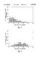

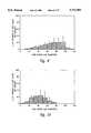


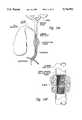


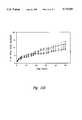


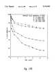


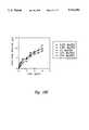
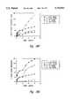


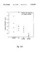


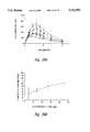


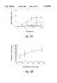


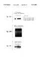
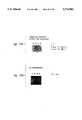


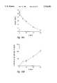


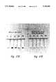

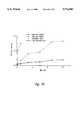


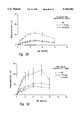


Classifications
- A—HUMAN NECESSITIES
- A61—MEDICAL OR VETERINARY SCIENCE; HYGIENE
- A61K—PREPARATIONS FOR MEDICAL, DENTAL OR TOILETRY PURPOSES
- A61K9/00—Medicinal preparations characterised by special physical form
- A61K9/14—Particulate form, e.g. powders, Processes for size reducing of pure drugs or the resulting products, Pure drug nanoparticles
- A61K9/16—Agglomerates; Granulates; Microbeadlets ; Microspheres; Pellets; Solid products obtained by spray drying, spray freeze drying, spray congealing,(multiple) emulsion solvent evaporation or extraction
- A61K9/1605—Excipients; Inactive ingredients
- A61K9/1629—Organic macromolecular compounds
- A61K9/1635—Organic macromolecular compounds obtained by reactions only involving carbon-to-carbon unsaturated bonds, e.g. polyvinyl pyrrolidone, poly(meth)acrylates
- A—HUMAN NECESSITIES
- A61—MEDICAL OR VETERINARY SCIENCE; HYGIENE
- A61K—PREPARATIONS FOR MEDICAL, DENTAL OR TOILETRY PURPOSES
- A61K31/00—Medicinal preparations containing organic active ingredients
- A61K31/12—Ketones
- A61K31/122—Ketones having the oxygen directly attached to a ring, e.g. quinones, vitamin K1, anthralin
- A—HUMAN NECESSITIES
- A61—MEDICAL OR VETERINARY SCIENCE; HYGIENE
- A61K—PREPARATIONS FOR MEDICAL, DENTAL OR TOILETRY PURPOSES
- A61K31/00—Medicinal preparations containing organic active ingredients
- A61K31/13—Amines
- A61K31/135—Amines having aromatic rings, e.g. ketamine, nortriptyline
- A61K31/136—Amines having aromatic rings, e.g. ketamine, nortriptyline having the amino group directly attached to the aromatic ring, e.g. benzeneamine
- A—HUMAN NECESSITIES
- A61—MEDICAL OR VETERINARY SCIENCE; HYGIENE
- A61K—PREPARATIONS FOR MEDICAL, DENTAL OR TOILETRY PURPOSES
- A61K31/00—Medicinal preparations containing organic active ingredients
- A61K31/185—Acids; Anhydrides, halides or salts thereof, e.g. sulfur acids, imidic, hydrazonic or hydroximic acids
- A61K31/19—Carboxylic acids, e.g. valproic acid
- A61K31/20—Carboxylic acids, e.g. valproic acid having a carboxyl group bound to a chain of seven or more carbon atoms, e.g. stearic, palmitic, arachidic acids
- A61K31/203—Retinoic acids ; Salts thereof
- A—HUMAN NECESSITIES
- A61—MEDICAL OR VETERINARY SCIENCE; HYGIENE
- A61K—PREPARATIONS FOR MEDICAL, DENTAL OR TOILETRY PURPOSES
- A61K31/00—Medicinal preparations containing organic active ingredients
- A61K31/33—Heterocyclic compounds
- A61K31/335—Heterocyclic compounds having oxygen as the only ring hetero atom, e.g. fungichromin
- A61K31/337—Heterocyclic compounds having oxygen as the only ring hetero atom, e.g. fungichromin having four-membered rings, e.g. taxol
- A—HUMAN NECESSITIES
- A61—MEDICAL OR VETERINARY SCIENCE; HYGIENE
- A61K—PREPARATIONS FOR MEDICAL, DENTAL OR TOILETRY PURPOSES
- A61K31/00—Medicinal preparations containing organic active ingredients
- A61K31/33—Heterocyclic compounds
- A61K31/395—Heterocyclic compounds having nitrogen as a ring hetero atom, e.g. guanethidine or rifamycins
- A61K31/495—Heterocyclic compounds having nitrogen as a ring hetero atom, e.g. guanethidine or rifamycins having six-membered rings with two or more nitrogen atoms as the only ring heteroatoms, e.g. piperazine or tetrazines
- A61K31/505—Pyrimidines; Hydrogenated pyrimidines, e.g. trimethoprim
- A61K31/519—Pyrimidines; Hydrogenated pyrimidines, e.g. trimethoprim ortho- or peri-condensed with heterocyclic rings
- A—HUMAN NECESSITIES
- A61—MEDICAL OR VETERINARY SCIENCE; HYGIENE
- A61K—PREPARATIONS FOR MEDICAL, DENTAL OR TOILETRY PURPOSES
- A61K31/00—Medicinal preparations containing organic active ingredients
- A61K31/56—Compounds containing cyclopenta[a]hydrophenanthrene ring systems; Derivatives thereof, e.g. steroids
- A61K31/57—Compounds containing cyclopenta[a]hydrophenanthrene ring systems; Derivatives thereof, e.g. steroids substituted in position 17 beta by a chain of two carbon atoms, e.g. pregnane or progesterone
- A—HUMAN NECESSITIES
- A61—MEDICAL OR VETERINARY SCIENCE; HYGIENE
- A61K—PREPARATIONS FOR MEDICAL, DENTAL OR TOILETRY PURPOSES
- A61K33/00—Medicinal preparations containing inorganic active ingredients
- A61K33/24—Heavy metals; Compounds thereof
- A—HUMAN NECESSITIES
- A61—MEDICAL OR VETERINARY SCIENCE; HYGIENE
- A61K—PREPARATIONS FOR MEDICAL, DENTAL OR TOILETRY PURPOSES
- A61K38/00—Medicinal preparations containing peptides
- A61K38/16—Peptides having more than 20 amino acids; Gastrins; Somatostatins; Melanotropins; Derivatives thereof
- A61K38/55—Protease inhibitors
- A61K38/57—Protease inhibitors from animals; from humans
- A—HUMAN NECESSITIES
- A61—MEDICAL OR VETERINARY SCIENCE; HYGIENE
- A61K—PREPARATIONS FOR MEDICAL, DENTAL OR TOILETRY PURPOSES
- A61K45/00—Medicinal preparations containing active ingredients not provided for in groups A61K31/00 - A61K41/00
- A61K45/06—Mixtures of active ingredients without chemical characterisation, e.g. antiphlogistics and cardiaca
- A—HUMAN NECESSITIES
- A61—MEDICAL OR VETERINARY SCIENCE; HYGIENE
- A61K—PREPARATIONS FOR MEDICAL, DENTAL OR TOILETRY PURPOSES
- A61K47/00—Medicinal preparations characterised by the non-active ingredients used, e.g. carriers or inert additives; Targeting or modifying agents chemically bound to the active ingredient
- A61K47/50—Medicinal preparations characterised by the non-active ingredients used, e.g. carriers or inert additives; Targeting or modifying agents chemically bound to the active ingredient the non-active ingredient being chemically bound to the active ingredient, e.g. polymer-drug conjugates
- A61K47/69—Medicinal preparations characterised by the non-active ingredients used, e.g. carriers or inert additives; Targeting or modifying agents chemically bound to the active ingredient the non-active ingredient being chemically bound to the active ingredient, e.g. polymer-drug conjugates the conjugate being characterised by physical or galenical forms, e.g. emulsion, particle, inclusion complex, stent or kit
- A61K47/6921—Medicinal preparations characterised by the non-active ingredients used, e.g. carriers or inert additives; Targeting or modifying agents chemically bound to the active ingredient the non-active ingredient being chemically bound to the active ingredient, e.g. polymer-drug conjugates the conjugate being characterised by physical or galenical forms, e.g. emulsion, particle, inclusion complex, stent or kit the form being a particulate, a powder, an adsorbate, a bead or a sphere
- A61K47/6927—Medicinal preparations characterised by the non-active ingredients used, e.g. carriers or inert additives; Targeting or modifying agents chemically bound to the active ingredient the non-active ingredient being chemically bound to the active ingredient, e.g. polymer-drug conjugates the conjugate being characterised by physical or galenical forms, e.g. emulsion, particle, inclusion complex, stent or kit the form being a particulate, a powder, an adsorbate, a bead or a sphere the form being a solid microparticle having no hollow or gas-filled cores
- A—HUMAN NECESSITIES
- A61—MEDICAL OR VETERINARY SCIENCE; HYGIENE
- A61K—PREPARATIONS FOR MEDICAL, DENTAL OR TOILETRY PURPOSES
- A61K47/00—Medicinal preparations characterised by the non-active ingredients used, e.g. carriers or inert additives; Targeting or modifying agents chemically bound to the active ingredient
- A61K47/50—Medicinal preparations characterised by the non-active ingredients used, e.g. carriers or inert additives; Targeting or modifying agents chemically bound to the active ingredient the non-active ingredient being chemically bound to the active ingredient, e.g. polymer-drug conjugates
- A61K47/69—Medicinal preparations characterised by the non-active ingredients used, e.g. carriers or inert additives; Targeting or modifying agents chemically bound to the active ingredient the non-active ingredient being chemically bound to the active ingredient, e.g. polymer-drug conjugates the conjugate being characterised by physical or galenical forms, e.g. emulsion, particle, inclusion complex, stent or kit
- A61K47/6957—Medicinal preparations characterised by the non-active ingredients used, e.g. carriers or inert additives; Targeting or modifying agents chemically bound to the active ingredient the non-active ingredient being chemically bound to the active ingredient, e.g. polymer-drug conjugates the conjugate being characterised by physical or galenical forms, e.g. emulsion, particle, inclusion complex, stent or kit the form being a device or a kit, e.g. stents or microdevices
- A—HUMAN NECESSITIES
- A61—MEDICAL OR VETERINARY SCIENCE; HYGIENE
- A61K—PREPARATIONS FOR MEDICAL, DENTAL OR TOILETRY PURPOSES
- A61K9/00—Medicinal preparations characterised by special physical form
- A61K9/14—Particulate form, e.g. powders, Processes for size reducing of pure drugs or the resulting products, Pure drug nanoparticles
- A61K9/16—Agglomerates; Granulates; Microbeadlets ; Microspheres; Pellets; Solid products obtained by spray drying, spray freeze drying, spray congealing,(multiple) emulsion solvent evaporation or extraction
- A61K9/1605—Excipients; Inactive ingredients
- A61K9/1629—Organic macromolecular compounds
- A61K9/1641—Organic macromolecular compounds obtained otherwise than by reactions only involving carbon-to-carbon unsaturated bonds, e.g. polyethylene glycol, poloxamers
- A61K9/1647—Polyesters, e.g. poly(lactide-co-glycolide)
- A—HUMAN NECESSITIES
- A61—MEDICAL OR VETERINARY SCIENCE; HYGIENE
- A61K—PREPARATIONS FOR MEDICAL, DENTAL OR TOILETRY PURPOSES
- A61K9/00—Medicinal preparations characterised by special physical form
- A61K9/14—Particulate form, e.g. powders, Processes for size reducing of pure drugs or the resulting products, Pure drug nanoparticles
- A61K9/16—Agglomerates; Granulates; Microbeadlets ; Microspheres; Pellets; Solid products obtained by spray drying, spray freeze drying, spray congealing,(multiple) emulsion solvent evaporation or extraction
- A61K9/1605—Excipients; Inactive ingredients
- A61K9/1629—Organic macromolecular compounds
- A61K9/1658—Proteins, e.g. albumin, gelatin
- A—HUMAN NECESSITIES
- A61—MEDICAL OR VETERINARY SCIENCE; HYGIENE
- A61K—PREPARATIONS FOR MEDICAL, DENTAL OR TOILETRY PURPOSES
- A61K9/00—Medicinal preparations characterised by special physical form
- A61K9/14—Particulate form, e.g. powders, Processes for size reducing of pure drugs or the resulting products, Pure drug nanoparticles
- A61K9/16—Agglomerates; Granulates; Microbeadlets ; Microspheres; Pellets; Solid products obtained by spray drying, spray freeze drying, spray congealing,(multiple) emulsion solvent evaporation or extraction
- A61K9/167—Agglomerates; Granulates; Microbeadlets ; Microspheres; Pellets; Solid products obtained by spray drying, spray freeze drying, spray congealing,(multiple) emulsion solvent evaporation or extraction with an outer layer or coating comprising drug; with chemically bound drugs or non-active substances on their surface
- A—HUMAN NECESSITIES
- A61—MEDICAL OR VETERINARY SCIENCE; HYGIENE
- A61K—PREPARATIONS FOR MEDICAL, DENTAL OR TOILETRY PURPOSES
- A61K9/00—Medicinal preparations characterised by special physical form
- A61K9/48—Preparations in capsules, e.g. of gelatin, of chocolate
- A61K9/50—Microcapsules having a gas, liquid or semi-solid filling; Solid microparticles or pellets surrounded by a distinct coating layer, e.g. coated microspheres, coated drug crystals
- A61K9/51—Nanocapsules; Nanoparticles
- A61K9/5107—Excipients; Inactive ingredients
- A61K9/513—Organic macromolecular compounds; Dendrimers
- A61K9/5146—Organic macromolecular compounds; Dendrimers obtained otherwise than by reactions only involving carbon-to-carbon unsaturated bonds, e.g. polyethylene glycol, polyamines, polyanhydrides
- A61K9/5153—Polyesters, e.g. poly(lactide-co-glycolide)
- A—HUMAN NECESSITIES
- A61—MEDICAL OR VETERINARY SCIENCE; HYGIENE
- A61K—PREPARATIONS FOR MEDICAL, DENTAL OR TOILETRY PURPOSES
- A61K9/00—Medicinal preparations characterised by special physical form
- A61K9/48—Preparations in capsules, e.g. of gelatin, of chocolate
- A61K9/50—Microcapsules having a gas, liquid or semi-solid filling; Solid microparticles or pellets surrounded by a distinct coating layer, e.g. coated microspheres, coated drug crystals
- A61K9/51—Nanocapsules; Nanoparticles
- A61K9/5107—Excipients; Inactive ingredients
- A61K9/513—Organic macromolecular compounds; Dendrimers
- A61K9/5161—Polysaccharides, e.g. alginate, chitosan, cellulose derivatives; Cyclodextrin
- A—HUMAN NECESSITIES
- A61—MEDICAL OR VETERINARY SCIENCE; HYGIENE
- A61K—PREPARATIONS FOR MEDICAL, DENTAL OR TOILETRY PURPOSES
- A61K9/00—Medicinal preparations characterised by special physical form
- A61K9/48—Preparations in capsules, e.g. of gelatin, of chocolate
- A61K9/50—Microcapsules having a gas, liquid or semi-solid filling; Solid microparticles or pellets surrounded by a distinct coating layer, e.g. coated microspheres, coated drug crystals
- A61K9/51—Nanocapsules; Nanoparticles
- A61K9/5107—Excipients; Inactive ingredients
- A61K9/513—Organic macromolecular compounds; Dendrimers
- A61K9/5169—Proteins, e.g. albumin, gelatin
- A—HUMAN NECESSITIES
- A61—MEDICAL OR VETERINARY SCIENCE; HYGIENE
- A61K—PREPARATIONS FOR MEDICAL, DENTAL OR TOILETRY PURPOSES
- A61K9/00—Medicinal preparations characterised by special physical form
- A61K9/70—Web, sheet or filament bases ; Films; Fibres of the matrix type containing drug
- A61K9/7007—Drug-containing films, membranes or sheets
- A—HUMAN NECESSITIES
- A61—MEDICAL OR VETERINARY SCIENCE; HYGIENE
- A61L—METHODS OR APPARATUS FOR STERILISING MATERIALS OR OBJECTS IN GENERAL; DISINFECTION, STERILISATION OR DEODORISATION OF AIR; CHEMICAL ASPECTS OF BANDAGES, DRESSINGS, ABSORBENT PADS OR SURGICAL ARTICLES; MATERIALS FOR BANDAGES, DRESSINGS, ABSORBENT PADS OR SURGICAL ARTICLES
- A61L31/00—Materials for other surgical articles, e.g. stents, stent-grafts, shunts, surgical drapes, guide wires, materials for adhesion prevention, occluding devices, surgical gloves, tissue fixation devices
- A61L31/08—Materials for coatings
- A61L31/10—Macromolecular materials
- A—HUMAN NECESSITIES
- A61—MEDICAL OR VETERINARY SCIENCE; HYGIENE
- A61L—METHODS OR APPARATUS FOR STERILISING MATERIALS OR OBJECTS IN GENERAL; DISINFECTION, STERILISATION OR DEODORISATION OF AIR; CHEMICAL ASPECTS OF BANDAGES, DRESSINGS, ABSORBENT PADS OR SURGICAL ARTICLES; MATERIALS FOR BANDAGES, DRESSINGS, ABSORBENT PADS OR SURGICAL ARTICLES
- A61L31/00—Materials for other surgical articles, e.g. stents, stent-grafts, shunts, surgical drapes, guide wires, materials for adhesion prevention, occluding devices, surgical gloves, tissue fixation devices
- A61L31/14—Materials characterised by their function or physical properties, e.g. injectable or lubricating compositions, shape-memory materials, surface modified materials
- A61L31/145—Hydrogels or hydrocolloids
- A—HUMAN NECESSITIES
- A61—MEDICAL OR VETERINARY SCIENCE; HYGIENE
- A61L—METHODS OR APPARATUS FOR STERILISING MATERIALS OR OBJECTS IN GENERAL; DISINFECTION, STERILISATION OR DEODORISATION OF AIR; CHEMICAL ASPECTS OF BANDAGES, DRESSINGS, ABSORBENT PADS OR SURGICAL ARTICLES; MATERIALS FOR BANDAGES, DRESSINGS, ABSORBENT PADS OR SURGICAL ARTICLES
- A61L31/00—Materials for other surgical articles, e.g. stents, stent-grafts, shunts, surgical drapes, guide wires, materials for adhesion prevention, occluding devices, surgical gloves, tissue fixation devices
- A61L31/14—Materials characterised by their function or physical properties, e.g. injectable or lubricating compositions, shape-memory materials, surface modified materials
- A61L31/16—Biologically active materials, e.g. therapeutic substances
- C—CHEMISTRY; METALLURGY
- C07—ORGANIC CHEMISTRY
- C07K—PEPTIDES
- C07K14/00—Peptides having more than 20 amino acids; Gastrins; Somatostatins; Melanotropins; Derivatives thereof
- C07K14/81—Protease inhibitors
- C07K14/8107—Endopeptidase (E.C. 3.4.21-99) inhibitors
- C07K14/811—Serine protease (E.C. 3.4.21) inhibitors
- A—HUMAN NECESSITIES
- A61—MEDICAL OR VETERINARY SCIENCE; HYGIENE
- A61L—METHODS OR APPARATUS FOR STERILISING MATERIALS OR OBJECTS IN GENERAL; DISINFECTION, STERILISATION OR DEODORISATION OF AIR; CHEMICAL ASPECTS OF BANDAGES, DRESSINGS, ABSORBENT PADS OR SURGICAL ARTICLES; MATERIALS FOR BANDAGES, DRESSINGS, ABSORBENT PADS OR SURGICAL ARTICLES
- A61L2300/00—Biologically active materials used in bandages, wound dressings, absorbent pads or medical devices
- A61L2300/40—Biologically active materials used in bandages, wound dressings, absorbent pads or medical devices characterised by a specific therapeutic activity or mode of action
- A61L2300/416—Anti-neoplastic or anti-proliferative or anti-restenosis or anti-angiogenic agents, e.g. paclitaxel, sirolimus
- A—HUMAN NECESSITIES
- A61—MEDICAL OR VETERINARY SCIENCE; HYGIENE
- A61L—METHODS OR APPARATUS FOR STERILISING MATERIALS OR OBJECTS IN GENERAL; DISINFECTION, STERILISATION OR DEODORISATION OF AIR; CHEMICAL ASPECTS OF BANDAGES, DRESSINGS, ABSORBENT PADS OR SURGICAL ARTICLES; MATERIALS FOR BANDAGES, DRESSINGS, ABSORBENT PADS OR SURGICAL ARTICLES
- A61L2300/00—Biologically active materials used in bandages, wound dressings, absorbent pads or medical devices
- A61L2300/60—Biologically active materials used in bandages, wound dressings, absorbent pads or medical devices characterised by a special physical form
- A61L2300/606—Coatings
- A—HUMAN NECESSITIES
- A61—MEDICAL OR VETERINARY SCIENCE; HYGIENE
- A61L—METHODS OR APPARATUS FOR STERILISING MATERIALS OR OBJECTS IN GENERAL; DISINFECTION, STERILISATION OR DEODORISATION OF AIR; CHEMICAL ASPECTS OF BANDAGES, DRESSINGS, ABSORBENT PADS OR SURGICAL ARTICLES; MATERIALS FOR BANDAGES, DRESSINGS, ABSORBENT PADS OR SURGICAL ARTICLES
- A61L2300/00—Biologically active materials used in bandages, wound dressings, absorbent pads or medical devices
- A61L2300/60—Biologically active materials used in bandages, wound dressings, absorbent pads or medical devices characterised by a special physical form
- A61L2300/62—Encapsulated active agents, e.g. emulsified droplets
- A61L2300/622—Microcapsules
- C—CHEMISTRY; METALLURGY
- C08—ORGANIC MACROMOLECULAR COMPOUNDS; THEIR PREPARATION OR CHEMICAL WORKING-UP; COMPOSITIONS BASED THEREON
- C08L—COMPOSITIONS OF MACROMOLECULAR COMPOUNDS
- C08L2203/00—Applications
- C08L2203/02—Applications for biomedical use
- C—CHEMISTRY; METALLURGY
- C08—ORGANIC MACROMOLECULAR COMPOUNDS; THEIR PREPARATION OR CHEMICAL WORKING-UP; COMPOSITIONS BASED THEREON
- C08L—COMPOSITIONS OF MACROMOLECULAR COMPOUNDS
- C08L23/00—Compositions of homopolymers or copolymers of unsaturated aliphatic hydrocarbons having only one carbon-to-carbon double bond; Compositions of derivatives of such polymers
- C08L23/02—Compositions of homopolymers or copolymers of unsaturated aliphatic hydrocarbons having only one carbon-to-carbon double bond; Compositions of derivatives of such polymers not modified by chemical after-treatment
- C08L23/04—Homopolymers or copolymers of ethene
- C08L23/08—Copolymers of ethene
- C08L23/0846—Copolymers of ethene with unsaturated hydrocarbons containing atoms other than carbon or hydrogen
- C08L23/0853—Ethene vinyl acetate copolymers
- C—CHEMISTRY; METALLURGY
- C08—ORGANIC MACROMOLECULAR COMPOUNDS; THEIR PREPARATION OR CHEMICAL WORKING-UP; COMPOSITIONS BASED THEREON
- C08L—COMPOSITIONS OF MACROMOLECULAR COMPOUNDS
- C08L2312/00—Crosslinking
Definitions
- the present inventionrelates generally to compositions and methods for treating cancer and other angiogenic-dependent diseases, and more specifically, to compositions comprising anti-angiogenic factors and polymeric carriers, stents which have been coated with such compositions, as well as method for utilizing these stents and compositions.
- Angiogenesis-dependent diseasesrepresent a significant portion of all diseases for which medical treatment is sought.
- canceris the second leading cause of death in the United States, and accounts for over one-fifth of the total mortality. Briefly, cancer is characterized by the uncontrolled division of a population of cells which, most typically, leads to the formation of one or more tumors. Such tumors are also characterized by the ingrowth of vasculature which provide various factors that permit continued tumor growth. Although cancer is generally more readily diagnosed than in the past, many forms, even if detected early, are still incurable.
- a variety of methodsare presently utilized to treat cancer, including for example, various surgical procedures. If treated with surgery alone however, many patients (particularly those with certain types of cancer, such as breast, brain, colon and hepatic cancer) will experience recurrence of the cancer. Therefore, in addition to surgery, many cancers are also treated with a combination of therapies involving cytotoxic chemotherapeutic drugs (e.g., vincristine, vinblastine, cisplatin, methotrexate, 5-FU, etc.) and/or radiation therapy.
- cytotoxic chemotherapeutic drugse.g., vincristine, vinblastine, cisplatin, methotrexate, 5-FU, etc.
- radiation therapye.g., a combination of therapies involving cytotoxic chemotherapeutic drugs (e.g., vincristine, vinblastine, cisplatin, methotrexate, 5-FU, etc.) and/or radiation therapy.
- cytotoxic chemotherapeutic drugse.
- chemo- and radiation therapiesIn addition to surgical, chemo- and radiation therapies, others have attempted to utilize an individual's own immune system in order to eliminate cancerous cells. For example, some have suggested the use of bacterial or viral components as adjuvants in order to stimulate the immune system to destroy tumor cells. (See generally "Principles of Cancer Biotherapy,” Oldham (ed.), Raven Press, New York, 1987.) Such agents have generally been useful as adjuvants and as nonspecific stimulants in animal tumor models, but have not as of yet proved to be generally effective in humans.
- Lymphokineshave also been utilized in the treatment of cancer. Briefly, lymphokines are secreted by a variety of cells, and generally have an effect on specific cells in the generation of an immune response. Examples of lymphokines include Interleukins (IL)-1, -2, -3, and -4, as well as colony stimulating factors such as G-CSF, GM-CSF, and M-CSF. Recently, one group has utilized IL-2 to stimulate peripheral blood cells in order to expand and produce large quantities of cells which are cytotoxic to tumor cells (Rosenberg et al., N. Engl. J. Med. 313:1485-1492, 1985).
- IL-2Interleukins
- antibodiesmay be developed which recognize certain cell surface antigens that are either unique, or more prevalent on cancer cells compared to normal cells. These antibodies, or “magic bullets,” may be utilized either alone or conjugated with a toxin in order to specifically target and kill tumor cells (Dillman, "Antibody Therapy,” Principles of Cancer Biotherapy, Oldham (ed.), Raven Press, Ltd., New York, 1987).
- Magic bulletsmay be utilized either alone or conjugated with a toxin in order to specifically target and kill tumor cells.
- one difficultyis that most monoclonal antibodies are of murine origin, and thus hypersensitivity against the murine antibody may limit its efficacy, particularly after repeated therapies. Common side effects include fever, sweats and chills, skin rashes, arthritis, and nerve palsies.
- the number who will relapse due to local recurrence of the diseaseamounts to 133,000 patients annually (or 21% of all those with localized disease).
- the number who will relapse due to distant metastases of the diseaseis 68,000 patients annually (11% of all those with localized disease).
- Another 102,139 patients annuallywill die as a direct result of an inability to control the local growth of the disease.
- Non-surgical treatment for hepatic metastasesinclude systemic chemotherapy, radiation, chemoembolization, hepatic arterial chemotherapy, and intraarterial radiation.
- systemic chemotherapy and hepatic arterial chemotherapyinitially reduces lesions in 15-20%, and 80% of patients, respectively
- the lesionsinvariably reoccur.
- Surgical resection of liver metastasesrepresents the only possibility for a cure, but such a procedure is possible in only 5% of patients with metastases, and in only 15-20% of patients with primary hepatic cancer.
- liver metastasesmay be temporarily decreased utilizing such methods, but tumors typically respond by causing the growth of new blood vessels into the tumor.
- a related problem to tumor formationis the development of cancerous blockages which inhibit the flow of material through body passageways, such as the bile ducts, trachea, esophagus, vasculature and urethra.
- One device, the stenthas been developed in order to hold open passageways which have been blocked by tumors or other substances.
- Representative examples of common stentsinclude the Wallstent, Strecker stent, Gianturco stent, and the Palmaz stent.
- the major problem with stentsis that they do not prevent the ingrowth of tumor or inflammatory material through the interstices of the stent.
- this materialreaches the inside of a stent and compromises the stent lumen, it may result in blockage of the body passageway into which it has been inserted.
- presence of a stent in the bodymay induce reactive or inflammatory tissue (e.g., blood vessels, fibroblasts, white blood cells) to enter the stent tureen, resulting in partial or complete closure of the stent.
- reactive or inflammatory tissuee.g., blood vessels, fibroblasts, white blood cells
- the present inventionprovides compositions and methods suitable for treating cancers, as well as other non-tumerigenic angiogenesis-dependent diseases, and further provides other related advantages.
- compositionscomprising (a) an anti-angiogenic factor and (b) a polymeric carrier.
- a wide variety of moleculesmay be utilized within the scope of the present invention as anti-angiogenic factors, including for example Anti-Invasive Factor, retinoic acids and their derivatives, paclitaxel including analogues and derivatives thereof, Suramin, Tissue Inhibitor of Metalloproteinase-1, Tissue Inhibitor of Metalloproteinase-2, Plasminogen Activator Inhibitor-1 and Plasminogen Activator Inhibitor-2, and lighter "d group" transition metals.
- Anti-Invasive Factorretinoic acids and their derivatives
- paclitaxelincluding analogues and derivatives thereof
- SuraminSuramin
- Tissue Inhibitor of Metalloproteinase-1Tissue Inhibitor of Metalloproteinase-2
- Plasminogen Activator Inhibitor-1 and Plasminogen Activator Inhibitor-2and lighter "d group” transition metals.
- polymeric carriersmay be utilized, representative examples of which include poly (ethylene-vinyl acetate) (40% cross-linked), poly (D,L-lactic acid) oligomers and polymers, poly (L-lactic acid) oligomers and polymers, poly (glycolic acid), copolymers of lactic acid and glycolic acid, poly (caprolactone), poly (valerolactone), poly (anhydrides), copolymers of poly (caprolactone) or poly (lactic acid) with polyethylene glycol, and blends thereof.
- poly (ethylene-vinyl acetate) (40% cross-linked)poly (D,L-lactic acid) oligomers and polymers
- poly (L-lactic acid) oligomers and polymerspoly (glycolic acid), copolymers of lactic acid and glycolic acid
- poly (caprolactone)poly (valerolactone), poly (anhydrides), copolymers of poly (caprolactone) or poly (lactic acid) with polyethylene glycol, and blends thereof
- the compositionscomprise a compound which disrupts microtubule function, such as, for example, paclitaxel, estramustine, colchicine, methotrexate, curacin-A, epothilone, vinblastine or tBCEV.
- the compositionscomprise a polymeric carrier and a lighter d gropu transition metal (e.g., a vanadium species, molybdenum species, tungsten species, titanium species, niobium species or tantalum species) which inhibits the formation of new blood vessels.
- the compositionhas an average size of 15 to 200 ⁇ m, within other embodiments, the polymeric carrier of the composition has a molecular weight ranging from less than 1,000 daltons to greater than 200,000 to 300,000 daltons.
- the compositions provided hereinmay be formed into films with a thickness of between 100 ⁇ m and 2 mm, or thermologically active compositions which are liquid at one temperature (e.g., above 45° C.) and solid or semi-solid at another (e.g., 37° C.).
- methods for embolizing a blood vesselcomprising the step of delivering into the vessel a therapeutically effective amount of an anti-angiogenic composition (as described above), such that the blood vessel is effectively occluded.
- the anti-angiogenic compositionis delivered to a blood vessel which nourishes a tumor.
- stentscomprising a generally tubular structure, the surface being coated with one or more anti-angiogenic compositions.
- methodsare provided for expanding the tureen of a body passageway, comprising inserting a stent into the passageway, the stent having a generally tubular structure, the surface of the structure being coated with an anti-angiogenic composition as described above, such that the passageway is expanded.
- biliary obstructionscomprising inserting a biliary stent into a biliary passageway; for eliminating urethral obstructions, comprising inserting a urethral stent into a urethra; for eliminating esophageal obstructions, comprising inserting an esophageal stent into an esophagus; and for eliminating tracheal/bronchial obstructions, comprising inserting a tracheal/bronchial stent into the trachea or bronchi.
- the stenthas a generally tubular structure, the surface of which is coated with an anti-angiogenic composition as described above.
- methods for treating tumor excision sitescomprising administering an anti-angiogenic composition as described above to the resection margins of a tumor subsequent to excision, such that the local recurrence of cancer and the formation of new blood vessels at the site is inhibited.
- methods for treating corneal neovascularizationcomprising the step of administering to a patient a therapeutically effective amount of an anti-angiogenic composition as described above to the cornea, such that the formation of blood vessels is inhibited.
- the anti-angiogenic compositionfurther comprises a topical corticosteroid.
- methodsare provided for inhibiting angiogenesis in patients with non-tumorigenic, angiogenesis-dependent diseases, comprising administering to a patient a therapeutically effective amount of paclitaxel to a patient with a non-tumorigenic angiogenesis-dependent disease, such that the formation of new blood vessels is inhibited.
- methodsare provided for embolizing blood vessels in non-tumorigenic, angiogenesis-dependent diseases, comprising delivering to the vessel a therapeutically effective amount of a composition comprising paclitaxel, such that the blood vessel is effectively occluded.
- methodsfor expanding the lumen of a body passageway, comprising inserting a stent into the passageway, the stent having a generally tubular structure, the surface of the structure being coated with a composition comprising paclitaxel, such that the passageway is expanded.
- biliary obstructionscomprising inserting a biliary stent into a biliary passageway; for eliminating urethral obstructions, comprising inserting a urethral stent into a urethra; for eliminating esophageal obstructions, comprising inserting an esophageal stent into an esophagus; and for eliminating tracheal/bronchial obstructions, comprising inserting a tracheal/bronchial stent into the trachea or bronchi.
- the stenthas a generally tubular structure, the surface of the structure being coated with a composition comprising paclitaxel.
- methodsfor treating a tumor excision site, comprising administering a composition comprising paclitaxel to the resection margin of a tumor subsequent to excision, such that the local recurrence of cancer and the formation of new blood vessels at the site is inhibited.
- methodsare provided for treating neovascular diseases of the eye, comprising administering to a patient a therapeutically effective amount of an anti-angiogenic factor (such as a compound which disrupts microtubule function) to the eye, such that the formation of new vessels is inhibited.
- an anti-angiogenic factorsuch as a compound which disrupts microtubule function
- an anti-angiogenic factorsuch as a compound which disrupts microtubule function
- a compositioncomprising an anti-angiogenic factor and a polymeric carrier to a joint.
- the anti-angiogenic factormay be a compound which disrupts microtubule function such as paclitaxel, or an element from the lighter ⁇ d group ⁇ transition metals, such as a vanadium species.
- pharmaceutical productscomprising (a) a compound which disrupts microtubule function, in a container, and (b) a notice associated with the container in form prescribed by a governmental agency regulating the manufacture, use, or sale of pharmaceuticals, which notice is reflective of approval by the agency of a compound which disrupts microtubule function, for human or veterinary administration to treat non-tumorigenic angiogenesis-dependent diseases such as, for example, inflammatory arthritis or neovascular diseases of the eye.
- Federal Lawrequires that the use of a pharmaceutical agent in the therapy of humans be approved by an agency of the Federal government.
- FIG. 1Ais a photograph which shows a shell-less egg culture on day 6.
- FIG. 1Bis a digitized computer-displayed image taken with a stereomicroscope of living, unstained capillaries (1040 ⁇ ).
- FIG. 1Cis a photograph of a corrosion casting which shows CAM microvasculature that are fed by larger, underlying vessels. (arrows; 1300 ⁇ ).
- FIG. 1Dis a photograph which depicts a 0.5 mm thick plastic section cut transversely through the CAM, and recorded at the light microscope level.
- FIG. 1Eis a photograph at the electron microscope level (3500 ⁇ ) wherein typical capillary structure is presented showing thin-walled endothelial cells (arrowheads) and an associated pericyte.
- FIGS. 2A, 2B, 2C and 2Dare a series of digitized images of four different, unstained CAMs taken after a 48 hour exposure to digitized images of four different living, unstained CAMs were taken after a 48 h exposure to 10 ⁇ paclitaxel per 10 ml of methylcellulose.
- the transparent methylcellulose disk (*) containing paclitaxelis present on each CAM and is positioned over a singular avascular zone (A) with surrounding blood islands (Is). These avascular areas extend beyond the disk and typically have a diameter of 6 min.
- FIG. 2Dillustrates the typical "elbowing" effect (arrowheads) of both small and large vessels being redirected away from the periphery of the avascular zone.
- FIGS. 3A, 3B and 3Care a series of photographs of 0.5 mm thick plastic sections transversely cut through a paclitaxel-treated CAM at three different locations within the avascular zone.
- FIGS. 4A, 4B and 4Care series of electron micrographs which were taken from locations similar to that of FIGS. 3A, 3B and 3C (respectively) above.
- FIG. 4Ashows a small capillary lying subjacent to the ectodermal layer (Ec) possessing three endothelial cells arrested in mitosis (*).
- Ecectodermal layer
- FIG. 4Bshows the early avascular phase contains extravasated blood cells subjacent to the ectoderm; these blood cells are intermixed with presumptive endothelial cells (*) and their processes. Degrative cellular vacuoles (arrowhead).
- FIG. 4Cshows that in response to paclitaxel, the ecto-mesodermal interface has become populated with cells in various stages of degradation containing dense vacuoles and granules (arrowheads).
- FIG. 5is a bar graph which depicts the size distribution of microspheres by number (5% poly (ethylene-vinyl acetate) with 10 mg sodium suramin into 5% PVA).
- FIG. 6is a bar graph which depicts the size distribution of microspheres by weight (5% poly (ethylene-vinyl acetate) with 10 mg sodium suramin into 5% PVA).
- FIG. 7is a graph which depicts the weight of encapsulation of Sodium Suramin in 50 mg poly (ethylene-vinyl acetate).
- FIG. 8is a graph which depicts the percent of encapsulation of Sodium Suramin in 50 mg poly (ethylene-vinyl acetate).
- FIG. 9is a bar graph which depicts the size distribution by weight of 5% ELVAX microspheres containing 10 mg sodium suramin made in 5% PVA containing 10% NaCl.
- FIG. 10is a bar graph which depicts the size distribution by weight of 5% microspheres containing 10 mg sodium suramin made in 5% PVA containing 10% NaCl.
- FIG. 11is a bar graph which depicts the size distribution by number of 5% microspheres containing 10 mg sodium suramin made in 5% PVA containing 10% NaCl.
- FIG. 12is a line graph which depicts the time course of sodium suramin release.
- FIG. 13is an illustration of a representative embodiment of hepatic tumor embolization.
- FIG. 14is an illustration of the insertion of a representative stent coated with an anti-angiogenic composition.
- FIG. 15Ais a graph which shows the effect of the EVA:PLA polymer blend ratio upon aggregation of microspheres.
- FIG. 15Bis a scanning electron micrograph which shows the size of "small” microspheres.
- FIG. 15C(which includes a magnified inset--labelled “15C-inset") is a scanning electron micrograph which shows the size of "large” microspheres.
- FIG. 15Dis a graph which depicts the time course of in vitro paclitaxel release from 0.6% w/v paclitaxel-loaded 50:50 EVA:PLA polymer blend microspheres into phosphate buffered saline (pH 7.4) at 37° C.
- FIG. 15Fis a photograph of a CAM which shows the results of paclitaxel release by microspheres ("MS").
- FIG. 15Fis a photograph similar to that of 15E at increased magnification.
- FIG. 16Ais a graph which shows release rate profiles from polycaprolactone microspheres containing 1%, 2%, 5% or 10% paclitaxel into phosphate buffered saline at 37° C.
- FIG. 16Bis a photograph which shows a CAM treated with control microspheres.
- FIG. 16Cis a photograph which shows a CAM treated with 5% paclitaxel loaded microspheres.
- FIGS. 17A and 17Bare two graphs which show the release of paclitaxel from EVA films, and the percent paclitaxel remaining in those same films over time.
- FIG. 17Cis a graph which shows the swelling of EVA/F127 films with no paclitaxel over time.
- FIG. 17Dis a graph which shows the swelling of EVA/Span 80 films with no paclitaxel over time.
- FIG. 17Eis a graph which depicts a stress vs. strain curve for various EVA/F127 blends.
- FIGS. 18A and 18Bare two graphs which show the melting point of PCL/MePEG polymer blends as a function of % MePEG in the formulation (18A), and the percent increase in time needed for PCL paste at 60° C. to being to solidify as a function of the amount of MePEG in the formulation (18B).
- FIG. 18Cis a graph which depicts the softness of varying PCL/MePEG polymer blends.
- FIG. 18Dis a graph which shows the percent weight change over time for polymer blends of various MePEG concentrations.
- FIG. 18Eis a graph which depicts the rate of paclitaxel release over time from various polymer blends loaded with 1% paclitaxel.
- FIGS. 18F and 18Gare graphs which depict the effect of varying quantities of paclitaxel on the total amount of paclitaxel released from a 20% MePEG/PCL blend.
- FIG. 18His a graph which depicts the effect of MePEG on the tensile strength of a MePEG/PCL polymer.
- FIG. 19Ais a photograph which shows control (unloaded) thermopaste on a CAM. Note that both large vessels and small vessels (capillaries) are found immediately adjacent to the paste. Blood flow in the area around and under the paste is unaffected.
- FIG. 19Bis a photograph of 20% paclitaxel-loaded thermopaste on a CAM. Note the disruption of the vasculature when compared to the surrounding tissues. The drug loaded paste has blocked the growth of the capillaries, caused regression of the larger vessels, and created a region of avascularity on the CAM assay.
- FIG. 19Cis a photograph of 0.5% paclitaxel-loaded thermopaste on a CAM (Mag.--40 ⁇ ).
- FIG. 19Dis a photograph of control (unloaded) Thermopaste on a CAM.
- the control pasteshows normal blood vessel organization. Functional vessels are located immediately adjacent to the unloaded paste.
- FIGS. 20A and 20Bare two photographs of a CAM having a tumor treated with control (unloaded) thermopaste. Briefly, in FIG. 20 A the central white mass is the tumor tissue. Note the abundance of blood vessels entering the tumor from the CAM in all directions. The tumor induces the ingrowth of the host vasculature through the production of "angiogenic factors.” The tumor tissue expands distally along the blood vessels which supply it.
- FIG. 20Bis an underside view of the CAM shown in 20A. Briefly, this view demonstrates the radial appearance of the blood vessels which enter the tumor like the spokes of a wheel. Note that the blood vessel density is greater in the vicinity of the tumor than it is in the surrounding normal CAM tissue. FIGS.
- FIG. 20C and 20Dare two photographs of a CAM having a tumor treated with 20% paclitaxel-loaded thermopaste.
- the central white massis the tumor tissue.
- the sustained release of the angiogenesis inhibitoris capable of overcoming the angiogenic stimulus produced by the tumor.
- the tumor itselfis poorly vascularized and is progressively decreasing in size.
- FIG. 20Dis taken from the underside of the CAM shown in 20C, and demonstrates the disruption of blood flow into the tumor when compared to control tumor tissue. Note that the blood vessel density is reduced in the vicinity of the tumor and is sparser than that of the normal surrounding CAM tissue.
- FIG. 21Ais a graph which shows the effect of paclitaxel/PCL on tumor growth.
- FIGS. 21B and 21Care two photographs which show the effect of control, 10%, and 20% paclitaxel-loaded thermopaste on tumor growth.
- FIG. 22Ais a photograph of synovium from a PBS injected joint.
- FIG. 22Bis a photograph of synovium from a microsphere injected joint.
- FIG. 22Cis a photograph of cartilage from joints injected with PBS, and
- FIG. 22Dis a photograph of cartilage from joints injected with microspheres.
- the plastic ringwas used to localize the drug treatment to the CAM. Note the lack of blood vessels located within and immediately adjacent to the ring. The functional blood vessels bordering the avascular zone are defined by their "elbowing" morphology away form the drug source.
- FIG. 25is a photograph of a control stent. Briefly, this image shows the longitudinal orientation of a nylon stent incorporated within gliosarcoma tissue of the rat liver. Ingrowth within the nylon stent is evident.
- FIG. 26is a photograph of a control stent. Briefly, this image also illustrates tumor ingrowth within the tureen of the nylon stent.
- FIG. 27is a photograph of a lung. Briefly, in addition to large liver tumors, metastasis to the lung is common. Such metastases are evident by the presence of small white lobules seen throughout the lung.
- FIG. 29Bis a graph which shows the time course concentration dependence of paclitaxel inhibition of plasma opsonized CPPD crystal induced neutrophil chemiluminescence.
- FIG. 30Bis a graphic which shows the time course concentration dependence of paclitaxel inhibition of plasma opsonized CPPD crystal induced neutrophil superoxide anion production; n 3.
- FIG. 31Bis a graph which shows plasma opsonized zymosan induced neutrophil superoxide anion production. Effect of paclitaxel at (o) no paclitaxel, (•) 28 ⁇ M, ( ⁇ ) Control (cells alone); n 3.
- FIG. 32Bis a graph which shows the concentration dependence of paclitaxel inhibition of myeloperoxidase release from neutrophils in response to plasma opsonized CPPD crystals; n 3.
- FIG. 33is a graph which shows lysozyme release from neutrophils (5 ⁇ 10 6 /ml) in response to plasma opsonized CPPD crystals (50 mg/ml). Effect of paclitaxel at (o) no paclitaxel, (•) 28 ⁇ M, ( ⁇ ) Control (cells alone), ( ⁇ ) Control (cells and paclitaxel at 28 ⁇ M); n 3.
- FIG. 34is a graph which depicts proliferation of synoviocytes at various concentrations of paclitaxel.
- FIG. 35is a bar graph which depicts the cytotoxicity of paclitaxel at various concentrations to proliferating synoviocytes.
- FIGS. 36A, 36B, and 36Care photographs of a series of gels which show the effect of various concentrations of paclitaxel on c-FOS expression.
- FIGS. 37A and 37Bare photographs of a series of gels which show the effect of various concentrations of paclitaxel on collagenase expression.
- FIG. 38is a bar graph which depicts the effects of paclitaxel on viability of normal chondrocytes in vitro.
- FIG. 39is a graph which shows the percentage of paclitaxel release based upon gelatinized-paclitaxel of either a large (7200 ⁇ m) or small (2100 ⁇ m) size.
- FIG. 40is a graph which shows the effect of gelatin and/or sodium chloride on the release of paclitaxel from PCL.
- FIG. 41is a graph which shows the release of paclitaxel from PDLLA-PEG-PDLLA cylinders containing 20% paclitaxel.
- FIG. 42Ais a graph which depicts the time course of paclitaxel release from 2.5 mg pellets of PCL.
- FIG. 42Bis a graph which shows the percent paclitaxel remaining in the pellet, over time.
- FIG. 43Ais a graph which shows the effect of MePEG on paclitaxel release from PCL paste leaded with 20% paclitaxel.
- FIG. 43Bis a graph which shows the percent paclitaxel remaining in the pellet, over time.
- FIGS. 44A and 44Bare graphs which show the effect of various concentrations of MePEG in PCL in terms of melting point (44A) and time to solidify (44B).
- FIG. 45is a graph which shows the effect of MePEG incorporation into PCL on the tensile strength and time to fail of the polymer.
- FIG. 46is a graph which shows the effect of irradiation on paclitaxel release.
- FIGS. 47A, B, C, D and Eshow the effect of MTX release from PCL over time.
- FIG. 48is a graph of particle diameter ( ⁇ m) determined by a Coulter® LS130 Particle Size Analysis.
- FIG. 49is a graph of particle diameter ( ⁇ m) determined by a Coulter® LS130 Particle Size Analysis.
- FIG. 50is a graph which shows paclitaxel release from various polymeric formulations.
- FIG. 51is a graph which shows the effect of plasma opsonization of polymeric microspheres on the chemiluminescence response of neutrophils (20 mg/ml microspheres in 0.5 ml of cells (conc. 5 ⁇ 10 6 cells/ml) to PCL microspheres.
- FIG. 52is a graph which shows the effect of precoating plasma +/-2% pluronic F127 on the chemiluminescence response of neutrophils (5 ⁇ 10 6 cells/ml) to PCL microspheres.
- FIG. 53is a graph which shows the effect of precoating plasma +/-2% pluronic F127 on the chemiluminescence response of neutrophils (5 ⁇ 10 6 cells/ml) to PMMA microspheres.
- FIG. 54is a graph which shows the effect of precoating plasma +/-2% pluronic F127 on the chemiluminescence response of neutrophils (5 ⁇ 10 6 cells/ml) to PLA microspheres.
- FIG. 55is a graph which shows the effect of precoating plasma +/-2% pluronic F127 on the chemiluminescence response of neutrophils (5 ⁇ 10 6 cells/ml) to EVA:PLA microspheres.
- FIG. 56is a graph which shows the effect of precoating IgG (2 mg/ml), or 2% pluronic F127 then IgG (2 mg/ml) on the chemiluminescence response of neutrophils to PCL microspheres.
- FIG. 57is a graph which shows the effect of precoating IgG (2 mg/ml), or 2% pluronic F127 then IgG (2 mg/ml) on the chemiluminescence response of neutrophils to PMMA microspheres.
- FIG. 58is a graph which shows the effect of precoating IgG (2 mg/ml), or 2% pluronic F127 then IgG (2 mg/ml) on the chemiluminescence response of neutrophils to PVA microspheres.
- FIG. 59is a graph which shows the effect of precoating IgG (2 mg/ml), or 2% pluronic F127 then IgG (2 mg/ml) on the chemiluminescence response of neutrophils to EVA:PLA microspheres.
- FIG. 60is a photograph of 10% methotrexate ("MTX") loaded microspheres made from a 50:50 ratio of PLA:GA (IV 0.78).
- MTXmethotrexate
- FIG. 61is a graph which depicts the release of 10% loaded vanadyl sulfate from PCL.
- FIG. 62is a photograph of hyaluronic acid microspheres containing vanadium sulfate.
- FIG. 63Ais a graph which depicts the release of organic vanadate from PCL.
- FIG. 63Bdepicts the percentage of organic vanadate remaining over a time course.
- FIG. 64is a photograph showing poly D,L, lactic acid microspheres containing organic vanadate.
- FIGS. 65A and 65Bare photographs of control (uncoated) stents which show typical epithelial ingrowth seen at both 8 weeks (A) and at 16 weeks (B). Indentations of the stent tines (t) and narrowing of the lumen (lu) are shown. There is progressive epithelial overgrowth of the stent surface over this time by fibrous and inflammatory tissue.
- FIGS. 66A, 66B, 66C, and 66Dare a series of photographs which show control and paclitaxel-coated biliary stents.
- FIG. 66Aillustrates the obliteration of the stent lumen by the process of benign epithelial overgrowth.
- the paclitaxel-treated biliary ductremains patent (66C).
- normal biliary tract epitheliumis present with only minimal alteration of the mucosal lining by the coated stent tines (t).
- anti-angiogenic factorsshould be understood to include any protein, peptide, chemical, or other molecule, which acts to inhibit vascular growth.
- a variety of methodsmay be readily utilized to determine the anti-angiogenic activity of a given factor, including for example, chick chorioallantoic membrane ("CAM") assays.
- CAMchick chorioallantoic membrane
- inhibition of vascular growth by the sample to be testedmay be readily determined by visualization of the chick chorioallantoic membrane in the region surrounding the methyl cellulose disk. Inhibition of vascular growth may also be determined quantitatively, for example, by determining the number and size of blood vessels surrounding the methyl cellulose disk, as compared to a control methyl cellulose disk.
- anti-angiogenic factors as described hereinare considered to inhibit the formation of new blood vessels if they do so in merely a statistically significant manner, as compared to a control, within preferred aspects such anti-angiogenic factors will completely inhibit the formation of new blood vessels, as well as reduce the size and number of previously existing vessels.
- CAM assayIn addition to the CAM assay described above, a variety of other assays may also be utilized to determine the efficacy of anti-angiogenic factors in vivo, including for example, mouse models which have been developed for this purpose (see Roberston et al., Cancer. Res. 51:1339-1344, 1991). In addition, a variety of representative in vivo assays relating to various aspects of the inventions described herein have also been described in more detail below in Examples 5 to 7, and 17 to 19.
- compositionscomprising an anti-angiogenic factor, and a polymeric carrier.
- anti-angiogenic factorsmay be readily utilized within the context of the present invention. Representative examples include Anti-Invasive Factor, retinoic acid and derivatives thereof, paclitaxel, Suramin, Tissue Inhibitor of Metalloproteinase-1, Tissue Inhibitor of Metalloproteinase-2, Plasminogen Activator Inhibitor-1, Plasminogen Activator Inhibitor-2, and various forms of the lighter “d group” transition metals. These and other anti-angiogenic factors will be discussed in more detail below.
- Anti-Invasive Factorwhich is prepared from extracts of cartilage, contains constituents which are responsible for inhibiting the growth of new blood vessels. These constituents comprise a family of 7 low molecular weight proteins ( ⁇ 50,000 daltons) (Kuettner and Pauli, "Inhibition of neovascularization by a cartilage factor” in Development of the Vascular System, Pitman Books (CIBA Foundation Symposium 100), pp. 163-173, 1983), including a variety of proteins which have inhibitory effects against a variety of proteases (Eisentein et al, Am. J. Pathol.
- AIF suitable for use within the present inventionmay be readily prepared utilizing techniques known in the art (e.g., Eisentein et al, supra; Kuettner and Pauli, supra; and Langer et al., supra). Purified constituents of AIF such as Cartilage-Derived Inhibitor ("CDI") (see Moses et at., Science 248:1408-1410, 1990) may also be readily prepared and utilized within the context of the present invention.
- CDICartilage-Derived Inhibitor
- Retinoic acidsalter the metabolism of extracellular matrix components, resulting in the inhibition of angiogenesis. Addition of proline analogs, angiostatic steroids, or heparin may be utilized in order to synergistically increase the anti-angiogenic effect of transretinoic acid. Retinoic acid, as well as derivatives thereof which may also be utilized in the context of the present invention, may be readily obtained from commercial sources, including for example, Sigma Chemical Co. (#R2625).
- Paclitaxelis a highly derivatized diterpenoid (Wani et al., J. Am. Chem. Soc. 93:2325, 1971) which has been obtained from the harvested and dried bark of Taxus brevifolia (Pacific Yew.) and Taxomyces Andreanae and Endophytic Fungus of the Pacific Yew (Stierle et al., Science 60:214-216, 1993).
- Taxus brevifoliaPacific Yew.
- Taxomyces Andreanae and Endophytic Fungus of the Pacific YewStierle et al., Science 60:214-216, 1993.
- paclitaxelacts to stabilize microtubular structures by binding tubulin to form abnormal mitotic spindles.
- “Paclitaxel”(which should be understood herein to include analogues and derivatives such as, for example, TAXOL®, TAXOTERE®, 10-desacetyl analogues of paclitaxel and 3'N-desbenzoyl-3'N-t-butoxy carbonyl analogues of paclitaxel) may be readily prepared utilizing techniques known to those skilled in the art (see also WO 94/07882, WO 94/07881, WO 94/07880, WO 94/07876, WO 93/23555, WO 93/10076, U.S. Pat. Nos.
- Tissue Inhibitor of Metalloproteinases-1(“TIMP”) is secreted by endothelial cells which also secrete MTPases. TIMP is glycosylated and has a molecular weight of 28.5 kDa. TIMP-1 regulates angiogenesis by binding to activated metalloproteinases, thereby suppressing the invasion of blood vessels into the extracellular matrix. Tissue Inhibitor of Metalloproteinases-2 (“TIMP-2”) may also be utilized to inhibit angiogenesis. Briefly, TIMP-2 is a 21 kDa nonglycosylated protein which binds to metalloproteinases in both the active and latent, proenzyme forms. Both TIMP-1 and TIMP-2 may be obtained from commercial sources such as Synergen, Boulder, Colo.
- Plasminogen Activator Inhibitor-1is a 50 kDa glycoprotein which is present in blood platelets, and can also be synthesized by endothelial cells and muscle cells. PAI-1 inhibits t-PA and urokinase plasminogen activator at the basolateral site of the endothelium, and additionally regulates the fibrinolysis process. Plasminogen Activator Inhibitor-2 (PAI-2) is generally found only in the blood under certain circumstances such as in pregnancy, and in the presence of tumors. Briefly, PAI-2 is a 56 kDa protein which is secreted by monocytes and macrophages. It is believed to regulate fibrinolytic activity, and in particular inhibits urokinase plasminogen activator and tissue plasminogen activator, thereby preventing fibrinolysis.
- PAI-1is a 50 kDa glycoprotein which is present in blood platelets, and can also be synthesized by endothelial cells and muscle cells.
- Lighter "d group” transition metalsinclude, for example, vanadium, molybdenum, tungsten, titanium, niobium, and tantalum species. Such transition metal species may form transition metal complexes. Suitable complexes of the above-mentioned transition metal species include oxo transition metal complexes.
- vanadium complexesinclude oxo vanadium complexes such as vanadate and vanadyl complexes.
- Suitable vanadate complexesinclude metavanadate (i.e., VO 3 - ) and orthovanadate (i.e., VO 4 3- ) complexes such as, for example, ammonium metavanadate (i.e., NH 4 VO 3 ), sodium metavanadate (i.e., NaVO 3 ), and sodium orthovanadate (i.e., Na 3 VO 4 ).
- Suitable vanadyl (i.e., VO 2+ ) complexesinclude, for example, vanadyl acetylacetonate and vanadyl sulfate including vanadyl sulfate hydrates such as vanadyl sulfate mono- and trihydrates.
- tungsten and molybdenum complexesalso include oxo complexes.
- Suitable oxo tungsten complexesinclude tungstate and tungsten oxide complexes.
- Suitable tungstate (i.e., WO 4 2- ) complexesinclude ammonium tungstate (i.e., (NH 4 ) 2 WO 4 ), calcium tungstate (i.e., CaWO 4 ), sodium tungstate dihydrate (i.e., Na 2 WO 4 .2H 2 O), and tungstic acid (i.e., H 2 WO 4 ).
- Suitable tungsten oxidesinclude tungsten (IV) oxide (i.e., WO 2 ) and tungsten (VI) oxide (i.e., WO 3 ).
- Suitable oxo molybdenum complexesinclude molybdate, molybdenum oxide, and molybdenyl complexes.
- Suitable molybdate (i.e., MoO 4 2- ) complexesinclude ammonium molybdate (i.e., (NH 4 ) 2 MoO 4 ) and its hydrates, sodium molybdate (i.e., Na 2 MoO 4 ) and its hydrates, and potassium molybdate (i.e., K 2 MoO 4 ) and its hydrates.
- Suitable molybdenum oxidesinclude molybdenum (VI) oxide (i.e., MoO 2 ), molybdenum (VI) oxide (i.e., MoO 3 ), and molybdic acid.
- Suitable molybdenyl (i.e., MoO 2 2+ ) complexesinclude, for example, molybdenyl acetylacetonate.
- Other suitable tungsten and molybdenum complexesinclude hydroxo derivatives derived from, for example, glycerol, tartaric acid, and sugars.
- a wide variety of other anti-angiogenic factorsmay also be utilized within the context of the present invention.
- Representative examplesinclude Platelet Factor 4 (Sigma Chemical Co., #F1385); Protamine Sulphate (Clupeine) (Sigma Chemical Co., #P4505); Sulphated Chitin Derivatives (prepared from queen crab shells), (Sigma Chemical Co., #C3641; Murata et al., Cancer Res.
- SP-PGSulphated Polysaccharide Peptidoglycan Complex
- Sulphated Polysaccharide Peptidoglycan ComplexSulphated Polysaccharide Peptidoglycan Complex (SP-PG) (the function of this compound may be enhanced by the presence of steroids such as estrogen, and tamoxifen citrate); Staurosporine (Sigma Chemical Co., #S4400); Modulators of Matrix Metabolism, including for example, proline analogs ⁇ (L-azetidine-2-carboxylic acid (LACA) (Sigma Chemical Co., #A0760)), cishydroxyproIine, d,L-3,4-dehydroproline (Sigma Chemical Co., #D0265), Thiaproline (Sigma Chemical Co., #T0631)!, ⁇ , ⁇ -dipyridyl (Sigma Chemical Co., #D7505), ⁇ -aminopropionitrile fumarate (Sigma Chemical Co., #A3134)! ⁇ ; M
- paclitaxelshould be understood to refer to not only the common chemically available form of paclitaxel, but analogues (e.g., taxotere, as noted above) and paclitaxel conjugates (e.g., paclitaxel-PEG, paclitaxel-dextran, or paclitaxel-xylos).
- Anti-angiogenic compositions of the present inventionmay additionally comprise a wide variety of compounds in addition to the anti-angiogenic factor and polymeric carrier.
- anti-angiogenic compositions of the present inventionmay also, within certain embodiments of the invention, also comprise one or more antibiotics, anti-inflammatories, anti-vital agents, anti-fungal agents and/or anti-protozoal agents.
- antibiotics included within the compositions described hereininclude: penicillins; cephalosporins such as cefadroxil, cefazolin, cefaclor; aminoglycosides such as gentamycin and tobramycin; sulfonamides such as sulfamethoxazole; and metronidazole.
- anti-inflammatoriesinclude: steroids such as prednisone, prednisolone, hydrocortisone, adrenocorticotropic hormone, and sulfasalazine; and non-steroidal anti-inflammatory drugs ("NSAIDS") such as aspirin, ibuprofen, naproxen, fenoprofen, indomethacin, and phenylbutazone.
- NSAIDSnon-steroidal anti-inflammatory drugs
- antiviral agentsinclude acyclovir, ganciclovir, zidovudine.
- antifungal agentsinclude: nystatin, ketoconazole, griseofulvin, flucytosine, miconazole, clotrimazole.
- antiprotozoal agentsinclude: pentamidine isethionate, quinine, chloroquine, and mefloquine.
- Anti-angiogenic compositions of the present inventionmay also contain one or more hormones such as thyroid hormone, estrogen, progesterone, cortisone and/or growth hormone, other biologically active molecules such as insulin, as well as T H 1 (e.g., Interleukins-2, -12, and -15, gamma interferon) or T H 2 (e.g., Interleukins-4 and -10) cytokines.
- hormonessuch as thyroid hormone, estrogen, progesterone, cortisone and/or growth hormone, other biologically active molecules such as insulin, as well as T H 1 (e.g., Interleukins-2, -12, and -15, gamma interferon) or T H 2 (e.g., Interleukins-4 and -10) cytokines.
- anti-angiogenic compositionswhich contain one or more compounds which disrupt microtubule function.
- compoundsinclude paclitaxel (discussed above), estramustine (available from Sigma; Wang and Stearns Cancer Res. 48:6262-6271, 1988), epothilone, curacin-A, colchicine, methotrexate, vinblastine and 4-tert-butyl- 3-(2-chloroethyl)ureido!benzene ("tBCEU”).
- Anti-angiogenic compositions of the present inventionmay also contain a wide variety of other compounds, including for example: ⁇ -adrenergic blocking agents, angiotensin II receptor antagonists and receptor antagonists for histamine, serotonin, endothelin; inhibitors of the sodium/hydrogen antiporter (e.g., amiloride and its derivatives); agents that modulate intracellular Ca 2+ transport such as L-type (e.g., diltiazem, nifedipine, verapamil) or T-type Ca 2+ channel blockers (e.g., amiloride), calmodulin antagonists (e.g., H 7 ) and inhibitors of the sodium/calcium antiporter (e.g., amiloride); ap-1 inhibitors (for tyrosine kinases, protein kinase C, myosin light chain kinase, Ca 2+ /calmodulin kinase II, casein kinase II); anti
- cytokine and/or growth factorsas well as their respective receptors, (e.g., the interleukins, ⁇ , ⁇ or ⁇ -IFN, GM-CSF, G-CSF, epidermal growth factor, transforming growth factors alpha and beta, TNF, and antagonists of vascular epithelial growth factor, endothelial growth factor, acidic or basic fibroblast growth factors, and platelet dervived growth factor); inhibitors of the IP 3 receptor (e.g., heparin); protease and collagenase inhibitors (e.g., TIMPs, discussed above); nitrovasodilators (e.g., isosorbide dinitrate); anti-mitotic agents (e.g., colchicine, anthracyclines and other antibiotics, folate antagonists and other anti-metabolites, vinca alkaloids, nitrosoureas, DNA alkylating agents, top
- Anti-angiogenic compositions of the present inventionmay also comprise additional ingredients such as surfactants (either hydrophilic or hydrophobic; see Example 13), anti-neoplastic or chemotherapeutic agents (e.g., 5-fluorouracil, vincristine, vinblastine, cisplatin, doxyrubicin, adriamycin, or tamocifen), radioactive agents (e.g., Cu-64, Ca-67, Ga-68, Zr-89, Ku-97, Tc-99m, Rh-105, Pd-109, In-111, I-123, I-125, I-131, Re-186, Re-188, Au-198, Au-199, Pb-203, At-211, Pb-212 and Bi-212) or toxins (e.g., ricin, abrin, diphtheria toxin, cholera toxin, gelonin, pokeweed antiviral protein, tritin, Shigella toxin, and Pseu
- anti-angiogenic compositions of the present inventioncomprise an anti-angiogenic factor and a polymeric carrier.
- anti-angiogenic compositions of the present inventionare provided in a wide variety of polymeric carders, including for example both biodegradable and non-biodegradable compositions.
- biodegradable compositionsinclude albumin, gelatin, starch, cellulose, dextrans, polysaccharides, fibrinogen, poly (D,L lactide), poly (D,L-lactide-co-glycolide), poly (glycolide), poly (hydroxybutyrate), poly (alkylcarbonate) and poly (orthoesters) (see generally, Illium, L., Davids, S. S. (eds.) "Polymers in controlled Drug Delivery” Wright, Bristol, 1987; Arshady, J. Controlled Release 17:1-22, 1991; Pitt, Int. J. Phar. 59:173-196, 1990; Holland et al., J. Controlled Release 4:155-0180, 1986).
- nondegradable polymersinclude EVA copolymers, silicone rubber and poly (methylmethacrylate).
- Particularly preferred polymeric carriersinclude poly (ethylene-vinyl acetate)(40% cross-linked), poly (D,L-lactic acid) oligomers and polymers, poly (L-lactic acid) oligomers and polymers, poly (glycolic acid), copolymers of lactic acid and glycolic acid, poly (caprolactone), poly (valerolactone), polyanhydrides, copolymers of poly (caprolactone) or poly (lactic acid) with polyethylene glycol and blends thereof.
- Polymeric carriersmay be fashioned in a variety of forms, including for example, rod-shaped devices, pellets, slabs, or capsules (see. e.g., Goodell et al., Am. J. Hosp. Pharm. 43:1454-1461, 1986; Langer et al., "Controlled release of macromolecules from polymers", in Biomedical polymers, Polymeric materials and pharmaceuticals for biomedical use, Goldberg, E. P., Nakagim, A. (eds.) Academic Press, pp. 113-137, 1980; Rhine et al., J. Pharm. Sci. 69:265-270, 1980; Brown et al., J. Pharm. Sci.
- Anti-angiogenic factorsmay be linked by occlusion in the matrices of the polymer, bound by covalent linkages, or encapsulated in microcapsules.
- anti-angiogenic compositionsare provided in non-capsular formulations such as microspheres (ranging from nanometers to micrometers in size), pastes, threads of various size, films and sprays.
- anti-angiogenic compositions of the present invention(which comprise one or more anti-angiogenic factors, and a polymeric carrier) are fashioned in a manner appropriate to the intended use.
- the anti-angiogenic compositionshould be biocompatible, and release one or more anti-angiogenic factors over a period of several days to months.
- "quick release” or "burst" anti-angiogenic compositionsare provided that release greater than 10%, 20%, or 25% (w/v) of an anti-angiogenic factor (e.g., paclitaxel) over a period of 7 to 10 days.
- Such “quick release” compositionsshould, within certain embodiments, be capable of releasing chemotherapeutic levels (where applicable) of a desired anti-angiogenic factor.
- "low release” anti-angiogenic compositionsare provided that release less than 1% (w/v) of an anti-angiogenic factor over a period of 7 to 10 days.
- anti-angiogenic compositions of the present inventionshould preferably be stable for several months and capable of being produced and maintained under sterile conditions.
- anti-angiogenic compositionsmay be fashioned in any size ranging from 50 nm to 500 ⁇ m, depending upon the particular use.
- the anti-angiogenic compositionwhen used for the purpose of tumor embolization (as discussed below), it is generally preferable to fashion the anti-angiogenic composition in microspheres of between 15 and 500 ⁇ m, preferably between 15 and 200 ⁇ m, and most preferably, between 25 and 150 ⁇ m.
- such compositionsmay also be readily applied as a "spray", which solidifies into a film or coating.
- Such spraysmay be prepared from microspheres of a wide array of sizes, including for example, from 0.1 ⁇ m to 3 ⁇ m, from 10 ⁇ m to 30 ⁇ m, and from 30 ⁇ m to 100 ⁇ m (see Example 8).
- Anti-angiogenic compositionsmay also be prepared, given the disclosure provided herein, for a variety of other applications.
- the anti-angiogenic factors of the present inventionmay be incorporated into muco-adhesive polymers (e.g., polyacrylic acids such as (CARBOPOL®, dextron, polymethacrylate, or starch (see LeYung and Robinson, J. of Controlled Rel. 5:223, 1988)), or nanometer-sized microspheres (see generally, Kreuter J. Controlled Release 16:169-176, 1991; Couvreur and Vauthier, J. Controlled Release 17:187-198, 1991).
- muco-adhesive polymerse.g., polyacrylic acids such as (CARBOPOL®, dextron, polymethacrylate, or starch (see LeYung and Robinson, J. of Controlled Rel. 5:223, 1988)
- nanometer-sized microspheressee generally, Kreuter J. Controlled Release 16:169-176, 1991; Couvre
- Anti-angiogenic compositions of the present inventionmay also be prepared in a variety of "paste" or gel forms.
- anti-angiogenic compositionsare provided which are liquid at one temperature (e.g., temperature greater than 37° C., such as 40° C., 45° C., 50° C., 55° C. or 60° C.), and solid or semi-solid at another temperature (e.g., ambient body temperature, or any temperature lower than 37° C.).
- temperaturee.g., temperature greater than 37° C., such as 40° C., 45° C., 50° C., 55° C. or 60° C.
- solid or semi-solid at another temperaturee.g., ambient body temperature, or any temperature lower than 37° C.
- the anti-angiogenic compositions of the present inventionmay be formed as a film.
- such filmsare generally less than 5, 4, 3, 2, or 1, mm thick, more preferably less than 0.75 mm or 0.5 mm thick, and most preferably less than 500 ⁇ m to 100 ⁇ m thick.
- Such filmsare preferably flexible with a good tensile strength (e.g., greater than 50, preferably greater than 100, and more preferably greater than 150 or 200N/cm 2 ), good adhesive properties (i.e., readily adheres to moist or wet surfaces), and has controlled permeability. Representative examples of such films are set forth below in the Examples (see e.g., Example 13).
- polymeric carrierswhich are adapted to contain and release a hydrophobic compound, the carrier containing the hydrophobic compound in combination with a carbohydrate, protein or polypeptide.
- the polymeric carriercontains or comprises regions, pockets, or granules of one or more hydrophobic compounds.
- hydrophobic compoundsmay be incorporated within a matrix which contains the hydrophobic compound, followed by incorporation of the matrix within the polymeric carrier.
- hydrophobic compoundsmay be contained within a hydrophobic core, and this core contained within a hydrophilic shell.
- paclitaxelmay be incorporated into a hydrophobic core (e.g., of the poly D,L lactic acid-PEG or MePEG aggregate) which has a hydrophilic shell.
- hydrophobic compoundsmay be released from the polymeric carriers described above, including for example: certain hydrophobic compounds which disrupt microtubule function such as paclitaxel and estramustine; hydrophobic proteins such as myelin basic protein, proteolipid proteins of CNS myelin, hydrophobic cell wall protein, porins, membrane proteins (EMBO J. 12(9):3409-3415, 1993), myelin oligodendrocyte glycoprotein ("MOG”) (Biochem. and Mol. Biol. Int. 30(5):945-958, 1993, P27 Cancer Res. 53(17):4096-4101, 1913, bacterioopsin, human surfactant protein ("HSB”; J. Biol. Chem. 268(15):11160-11166, 1993), and SP-B or SP-C (Biochimica et Biophysica Acta 1105(1):161-169, 1992).
- hydrophobic proteinssuch as myelin basic protein, proteolipid proteins of CNS myelin,
- the present inventionalso provides a variety of methods which utilize the above-described anti-angiogenic compositions.
- methodsare provided for embolizing a blood vessel, comprising the step of delivering into the vessel a therapeutically effective amount of an anti-angiogenic composition (as described above), such that the blood vessel is effectively occluded.
- Therapeutically effective amounts suitable for occluding blood vesselsmay be readily determined given the disclosure provided below, and as described in Example 6.
- the anti-angiogenic compositionis delivered to a blood vessel which nourishes a tumor (see FIG. 13).
- anti-angiogenic compositions of the present inventionare preferably non-toxic, thrombogenic, easy to inject down vascular catheters, radio-opaque, rapid and permanent in effect, sterile, and readily available in different shapes or sizes at the time of the procedure.
- the compositionspreferably result in the slow (ideally, over a period of several weeks to months) release of an anti-angiogenic factor.
- Particularly preferred anti-angiogenic compositionsshould have a predictable size of 15-200 ⁇ m after being injected into the vascular system. Preferably, they should not clump into larger particles either in solution or once injected.
- preferable compositionsshould not change shape or physical properties during storage prior to use.
- Embolization therapymay be utilized in at least three principal ways to assist in the management of neoplasms: (1) definitive treatment of tumors (usually benign); (2) for preoperative embolization; and (3) for palliative embolization.
- benign tumorsmay sometimes be successfully treated by embolization therapy alone.
- examples of such tumorsinclude simple tumors of vascular origin (e.g., haemangiomas), endocrine tumors such as parathyroid adenomas, and benign bone tumors.
- preoperative embolizationmay be employed hours or days before surgical resection in order to reduce operative blood loss, shorten the duration of the operation, and reduce the risk of dissemination of viable malignant cells by surgical manipulation of the tumor.
- Many tumorsmay be successfully embolized preoperatively, including for example nasopharyngeal tumors, glomus jugular tumors, meningiomas, chemodectomas, and vagal neuromas.
- Embolizationmay also be utilized as a primary mode of treatment for inoperable malignancies, in order to extend the survival time of patients with advanced disease. Embolization may produce a marked improvement in the quality of life of patients with malignant tumors by alleviating unpleasant symptoms such as bleeding, venous obstruction and tracheal compression. The greatest benefit from palliative tumor embolization, however, may be seen in patients suffering from the humoral effects of malignant endocrine tumors, wherein metastases from carcinoid tumors and other endocrine neoplasms such as insulinomas and glucagonomas may be slow growing, and yet cause great distress by virtue of the endocrine syndromes which they produce.
- embolization therapy utilizing anti-angiogenic compositions of the present inventionis typically performed in a similar manner, regardless of the site.
- angiographya road map of the blood vessels
- the cathetermay be inserted either percutaneously or by surgery.
- the blood vesselis then embolized by refluxing anti-angiogenic compositions of the present invention through the catheter, until flow is observed to cease. Occlusion may be confirmed by repeating the angiogram.
- Embolization therapygenerally results in the distribution of compositions containing anti-angiogenic factors throughout the interstices of the tumor or vascular mass to be treated.
- the physical bulk of the embolic particles clogging the arterial lumenresults in the occlusion of the blood supply.
- an anti-angiogenic factor(s)prevents the formation of new blood vessels to supply the tumor or vascular mass, enhancing the devitalizing effect of cutting off the blood supply.
- tumorsare typically divided into two classes: benign and malignant.
- benign tumorthe cells retain their differentiated features and do not divide in a completely uncontrolled manner.
- the tumoris localized and nonmetastatic.
- malignant tumorthe cells become undifferentiated, do not respond to the body's growth and hormonal signals, and multiply in an uncontrolled manner; the tumor is invasive and capable of spreading to distant sites (metastasizing).
- metastasesof the liver may be treated utilizing embolization therapy.
- a catheteris inserted via the femoral or brachial artery and advanced into the hepatic artery by steering it through the arterial system under fluoroscopic guidance.
- the catheteris advanced into the hepatic arterial tree as far as necessary to allow complete blockage of the blood vessels supplying the tumor(s), while sparing as many of the arterial branches supplying normal structures as possible.
- thiswill be a segmental branch of the hepatic artery, but it could be that the entire hepatic artery distal to the origin of the gastroduodenal artery, or even multiple separate arteries, will need to be blocked depending on the extent of tumor and its individual blood supply.
- the arteryis embolized by injecting anti-angiogenic compositions (as described above) through the arterial catheter until flow in the artery to be blocked ceases, preferably even after observation for 5 minutes. Occlusion of the artery may be confirmed by injecting radiopaque contrast through the catheter and demonstrating by fluoroscopy or X-ray film that the vessel which previously filled with contrast no longer does so. The same procedure may be repeated with each feeding artery to be occluded.
- both benign and malignant tumorsmay be embolized utilizing compositions of the present invention.
- Representative examples of benign hepatic tumorsinclude Hepatocellular Adenoma, Cavernous Haemangioma, and Focal Nodular Hyperplasia.
- Other benign tumorswhich are more rare and often do not have clinical manifestations, may also be treated. These include Bile Duct Adenomas, Bile Duct Cystadenomas, Fibromas, Lipomas, Leiomyomas, Mesotheliomas, Teratomas, Myxomas, and Nodular Regenerative Hyperplasia.
- Malignant Hepatic Tumorsare generally subdivided into two categories: primary and secondary.
- Primary tumorsarise directly from the tissue in which they are found.
- a primary liver tumoris derived originally from the cells which make up the liver tissue (such as hepatocytes and biliary cells).
- Representative examples of primary hepatic malignancies which may be treated by arterial embolizationinclude Hepatocellularcarcinoma, Cholangiocarcinoma, Angiosarcoma, Cystadenocarcinoma, Squamous Cell Carcinoma, and Hepatoblastoma.
- a secondary tumor, or metastasisis a tumor which originated elsewhere in the body but has now spread to a distant organ.
- the common routes for metastasisare direct growth into adjacent structures, spread through the vascular or lymphatic systems, and tracking along tissue planes and body spaces (peritoneal fluid, cerebrospinal fluid, etc.).
- Secondary hepatic tumorsare one of the most common causes of death in the cancer patient and are by far and away the most common form of liver tumor.
- tumors which are most likely to spread to the liverinclude: cancer of the stomach, colon, and pancreas; melanoma; tumors of the lung, oropharynx, and bladder; Hodgkin's and non-Hodgkin's lymphoma; tumors of the breast, ovary, and prostate.
- Each one of the above-named primary tumorshas numerous different tumor types which may be treated by arterial embolization (for example, there are over 32 different types of ovarian cancer).
- arteriovenous malformationmay be treated by administration of one of the above-described compositions.
- arteriovenous malformationsrefers to a group of diseases wherein at least one (and most typically, many) abnormal communications between arteries and veins occur, resulting in a local tumor-like mass composed predominantly of blood vessels. Such disease may be either congenital or acquired.
- an arteriovenous malformationmay be treated by inserting a catheter via the femoral or brachial artery, and advancing it into the feeding artery under fluoroscopic guidance.
- the catheteris preferably advanced as far as necessary to allow complete blockage of the blood vessels supplying the vascular malformation, while sparing as many of the arterial branches supplying normal structures as possible (ideally this will be a single artery, but most often multiple separate arteries may need to be occluded, depending on the extent of the vascular malformation and its individual blood supply).
- each arterymay be embolized utilizing anti-angiogenic compositions of the present invention.
- embolizationmay be accomplished in order to treat conditions of excessive bleeding.
- menorrhagiaexcessive bleeding with menstruation
- the uterine arteriesare branches of the internal iliac arteries bilaterally.
- a cathetermay be inserted via the femoral or brachial artery, and advanced into each uterine artery by steering it through the arterial system under fluoroscopic guidance. The catheter should be advanced as far as necessary to allow complete blockage of the blood vessels to the uterus, while sparing as many arterial branches that arise from the uterine artery and supply normal structures as possible.
- each artery on each sidemay be embolized, but occasionally multiple separate arteries may need to be blocked depending on the individual blood supply.
- each arterymay be embolized by administration of the anti-angiogenic compositions as described above.
- arterial embolizationmay be accomplished in a variety of other conditions, including for example, for acute bleeding, vascular abnormalities, central nervous system disorders, and hypersplenism.
- stentscomprising a generally tubular structure (which includes for example, spiral shapes), the surface of which is coated with a composition as described above.
- a stentis a scaffolding, usually cylindrical in shape, that may be inserted into a body passageway (e.g., bile ducts) or a portion of a body passageway, which has been narrowed, irregularly contoured, obstructed, or occluded by a disease process (e.g., ingrowth by a tumor) in order to prevent closure or reclosure of the passageway.
- Stentsact by physically holding open the walls of the body passage into which they are inserted.
- stentsmay be utilized within the context of the present invention, including for example, esophageal stents, vascular stents, biliary stents, pancreatic stents, ureteric and urethral stents, lacrimal stents, Eustachian tube stents, fallopian tube stents and tracheal/bronchial stents.
- Stentsmay be readily obtained from commercial sources, or constructed in accordance with well-known techniques.
- Representative examples of stentsinclude those described in U.S. Pat. No. 4,768,523, entitled “Hydrogel Adhesive;” U.S. Pat. No. 4,776,337, entitled “Expandable Intraluminal Graft, and Method and Apparatus for Implanting and Expandable Intraluminal Graft;” U.S. Pat. No. 5,041,126 entitled “Endovascular Stent and Delivery System;” U.S. Pat. No. 5,052,998 entitled “Indwelling Stent and Method of Use;” U.S. Pat. No.
- Stentsmay be coated with anti-angiogenic compositions or anti-angiogenic factors of the present invention in a variety of manners, including for example: (a) by directly affixing to the stent an anti-angiogenic composition (e.g., by either spraying the stent with a polymer/drug film, or by dipping the stent into a polymer/drug solution), (b) by coating the stent with a substance such as a hydrogel which will in turn absorb the anti-angiogenic composition (or anti-angiogenic factor above), (c) by interweaving anti-angiogenic composition coated thread (or the polymer itself formed into a thread) into the stent structure, (d) by inserting the stent into a sleeve or mesh which is comprised of or coated with an anti-angiogenic composition, or (e) constructing the stent itself with an anti-angiogenic composition.
- an anti-angiogenic compositione.g., by either spraying the s
- the compositionshould firmly adhere to the stent during storage and at the time of insertion, and should not be dislodged from the stent when the diameter is expanded from its collapsed size to its full expansion size.
- the anti-angiogenic compositionshould also preferably not degrade during storage, prior to insertion, or when warmed to body temperature after expansion inside the body. In addition, it should preferably coat the stent smoothly and evenly, with a uniform distribution of angiogenesis inhibitor, while not changing the stent contour.
- the anti-angiogenic compositionshould provide a uniform, predictable, prolonged release of the anti-angiogenic factor into the tissue surrounding the stent once it has been deployed.
- the compositionshould not render the stem thrombogenic (causing blood clots to form), or cause significant turbulence in blood flow (more than the stent itself would be expected to cause if it was uncoated).
- methodsfor expanding the lumen of a body passageway, comprising inserting a stent into the passageway, the stent having a generally tubular structure, the surface of the structure being coated with an anti-angiogenic composition (or, an antioangiogenic factor alone), such that the passageway is expanded.
- an anti-angiogenic compositionor, an antioangiogenic factor alone
- stentsare inserted in a similar fashion regardless of the site or the disease being treated.
- a preinsertion examinationusually a diagnostic imaging procedure, endoscopy, or direct visualization at the time of surgery, is generally first performed in order to determine the appropriate positioning for stent insertion.
- a guidewireis then advanced through the lesion or proposed site of insertion, and over this is passed a delivery catheter which allows a stent in its collapsed form to be inserted.
- stentsare capable of being compressed, so that they can be inserted through tiny cavities via small catheters, and then expanded to a larger diameter once they are at the desired location. Once expanded, the stent physically forces the wails of the passageway apart and holds them open.
- the stentmay be self-expanding (e.g., the Wallstent and Gianturco stents), balloon expandable (e.g., the Palmaz stent and Strecker stent), or implanted by a change in temperature (e.g., the Nitinol stent).
- Stentsare typically maneuvered into place under radiologic or direct visual control, taking particular care to place the stent precisely across the narrowing in the organ being treated.
- the delivery catheteris then removed, leaving the stent standing on its own as a scaffold.
- a post insertion examinationusually an x-ray, is often utilized to confirm appropriate positioning.
- methodsfor eliminating biliary obstructions, comprising inserting a biliary stent into a biliary passageway, the stent having a generally tubular structure, the surface of the structure being coated with a composition as described above, such that the biliary obstruction is eliminated.
- tumor overgrowth of the common bile ductresults in progressive cholestatic jaundice which is incompatible with life.
- the biliary system which drains bile from the liver into the duodenumis most often obstructed by (1) a tumor composed of bile duct cells (cholangiocarcinoma), (2) a tumor which invades the bile duct (e.g., pancreatic carcinoma), or (3) a tumor which exerts extrinsic pressure and compresses the bile duct (e.g., enlarged lymph nodes).
- a tumor composed of bile duct cellscholangiocarcinoma
- a tumor which invades the bile ducte.g., pancreatic carcinoma
- a tumor which exerts extrinsic pressure and compresses the bile ducte.g., enlarged lymph nodes.
- Both primary biliary tumors, as well as other tumors which cause compression of the biliary treemay be treated utilizing the stents described herein.
- One example of primary biliary tumorsare adenocarcinomas (which are also called Klatskin tumors when found at the bifurcation of the common hepatic duct). These tumors are also referred to as biliary carcinomas, choledocholangiocarcinomas, or adenocarcinomas of the biliary system.
- Benign tumors which affect the bile ducte.g., adenoma of the biliary system
- squamous cell carcinomas of the bile duct and adenocarcinomas of the gallbladdermay also cause compression of the biliary tree and therefore, result in biliary obstruction.
- Compression of the biliary treeis most commonly due to tumors of the liver and pancreas which compress and therefore obstruct the ducts. Most of the tumors from the pancreas arise from cells of the pancreatic ducts. This is a highly fatal form of cancer (5% of all cancer deaths; 26,000 new cases per year in the U.S.) with an average of 6 months survival and a 1 year survival rate of only 10%. When these tumors are located in the head of the pancreas they frequently cause biliary obstruction, and this detracts significantly from the quality of life of the patient.
- pancreatic tumorsWhile all types of pancreatic tumors are generally referred to as "carcinoma of the pancreas" there are histologic subtypes including: adenocarcinoma, adenosquamous carcinoma, cystadeno-carcinoma, and acinar cell carcinoma. Hepatic tumors, as discussed above, may also cause compression of the biliary tree, and therefore cause obstruction of the biliary ducts.
- a biliary stentis first inserted into a biliary passageway in one of several ways: from the top end by inserting a needle through the abdominal wall and through the liver (a percutaneous transhepatic cholangiogram or "PTC”); from the bottom end by cannulating the bile duct through an endoscope inserted through the mouth, stomach, and duodenum (an endoscopic retrograde cholangiogram or "ERCP”); or by direct incision during a surgical procedure.
- PTCpercutaneous transhepatic cholangiogram
- ERCPendoscopic retrograde cholangiogram
- a preinsertion examination, PTC, ERCP, or direct visualization at the time of surgeryshould generally be performed to determine the appropriate position for stent insertion.
- a guidewireis then advanced through the lesion, and over this a delivery catheter is passed to allow the stent to be inserted in its collapsed form.
- the diagnostic examwas a PTC
- the guidewire and delivery catheteris inserted via the abdominal wall, while if the original exam was an ERCP the stent may be placed via the mouth.
- the stentis then positioned under radiologic, endoscopic, or direct visual control taking particular care to place it precisely across the narrowing in the bile duct.
- the delivery catheteris then removed leaving the stent standing as a scaffolding which holds the bile duct open. A further cholangiogram may be performed to document that the stent is appropriately positioned.
- esophageal obstructionscomprising inserting an esophageal stent into an esophagus, the stent having a generally tubular structure, the surface of the structure being coated with an anti-angiogenic composition as described above, such that the esophageal obstruction is eliminated.
- the esophagusis the hollow tube which transports food and liquids from the mouth to the stomach. Cancer of the esophagus or invasion by cancer arising in adjacent organs (e.g., cancer of the stomach or lung) results in the inability to swallow food or saliva.
- a preinsertion examinationusually a barium swallow or endoscopy should generally be performed in order to determine the appropriate position for stent insertion.
- a catheter or endoscopemay then be positioned through the mouth, and a guidewire is advanced through the blockage.
- a stent delivery catheteris passed over the guidewire under radiologic or endoscopic control, and a stent is placed precisely across the narrowing in the esophagus.
- a post insertion examinationusually a barium swallow x-ray, may be utilized to confirm appropriate positioning.
- tracheal/bronchial obstructionscomprising inserting a tracheal/bronchial stent into the trachea or bronchi, the stent having a generally tubular structure, the surface of which is coated with an anti-angiogenic composition as described above, such that the tracheal/bronchial obstruction is eliminated.
- the trachea and bronchiare tubes which carry air from the mouth and nose to the lungs.
- preinsertion examinationshould generally be performed in order to determine the appropriate position for stent insertion.
- a catheter or endoscopeis then positioned through the mouth, and a guidewire advanced through the blockage.
- a delivery catheteris then passed over the guidewire in order to allow a collapsed stent to be inserted.
- the stentis placed under radiologic or endoscopic control in order to place it precisely across the narrowing.
- the delivery cathetermay then be removed leaving the stent standing as a scaffold on its own.
- a post insertion examinationusually a bronchoscopy may be utilized to confirm appropriate positioning.
- urethral obstructionscomprising inserting a urethral stent into a urethra, the stent having a generally tubular structure, the surface of the structure being coated with an anti-angiogenic composition as described above, such that the urethral obstruction is eliminated.
- the urethrais the tube which drains the bladder through the penis. Extrinsic narrowing of the urethra as it passes through the prostate, due to hypertrophy of the prostate, occurs in virtually every man over the age of 60 and causes progressive difficulty with urination.
- a preinsertion examinationusually an endoscopy or urethrogram should generally first be performed in order to determine the appropriate position for stent insertion, which is above the external urinary sphincter at the lower end, and close to flush with the bladder neck at the upper end.
- An endoscope or catheteris then positioned through the penile opening and a guidewire advanced into the bladder.
- a delivery catheteris then passed over the guidewire in order to allow stent insertion.
- the delivery catheteris then removed, and the stent expanded into place.
- a post insertion examinationusually endoscopy or retrograde urethrogram, may be utilized to confirm appropriate position.
- vascular obstructionscomprising inserting a vascular stent into a blood vessel, the stent having a generally tubular structure, the surface of the structure being coated with an anti-angiogenic composition as described above, such that the vascular obstruction is eliminated.
- stentsmay be placed in a wide array of blood vessels, both arteries and veins, to prevent recurrent stenosis at the site of failed angioplasties, to treat narrowings that would likely fail if treated with angioplasty, and to treat post surgical narrowings (e.g., dialysis graft stenosis).
- Suitable sitesinclude the iliac, renal, and coronary arteries, the superior vena cava, and in dialysis grafts.
- angiographyis first performed in order to localize the site for placement of the stent. This is typically accomplished by injecting radiopaque contrast through a catheter inserted into an artery or vein as an x-ray is taken. A catheter may then be inserted either percutaneously or by surgery into the femoral artery, brachial artery, femoral vein, or brachial vein, and advanced into the appropriate blood vessel by steering it through the vascular system under fluoroscopic guidance. A stent may then be positioned across the vascular stenosis. A post insertion angiogram may also be utilized in order to confirm appropriate positioning.
- anti-angiogenic compositionsmay be utilized in a wide variety of surgical procedures.
- an anti-angiogenic compositionsin the form of, for example, a spray or film
- an anti-angiogenic compositionsmay be utilized to coat or spray an area prior to removal of a tumor, in order to isolate normal surrounding tissues from malignant tissue, and/or to prevent the spread of disease to surrounding tissues.
- anti-angiogenic compositionse.g., in the form of a spray
- surgical meshes which have been coated with anti-angiogenic compositions of the present inventionmay be utilized in any procedure wherein a surgical mesh might be utilized.
- a surgical mesh ladened with an anti-angiogenic compositionmay be utilized during abdominal cancer resection surgery (e.g., subsequent to colon resection) in order to provide support to the structure, and to release an amount of the anti-angiogenic factor.
- methodsfor treating tumor excision sites, comprising administering an anti-angiogenic composition as described above to the resection margins of a tumor subsequent to excision, such that the local recurrence of cancer and the formation of new blood vessels at the site is inhibited.
- the anti-angiogenic composition(s)or anti-angiogenic factor(s) alone
- are administered directly to the tumor excision sitee.g., applied by swabbing, brushing or otherwise coating the resection margins of the tumor with the anti-angiogenic composition(s) or factor(s)
- the anti-angiogenic composition(s) or factor(s)may be incorporated into known surgical pastes prior to administration.
- the anti-angiogenic compositionsare applied after hepatic resections for malignancy, and after neurosurgical operations.
- anti-angiogenic compositionsmay be administered to the resection margin of a wide variety of tumors, including for example, breast, colon, brain and hepatic tumors.
- anti-angiogenic compositionsmay be administered to the site of a neurological tumor subsequent to excision, such that the formation of new blood vessels at the site are inhibited.
- the brainis highly functionally localized; i.e., each specific anatomical region is specialized to carry out a specific function. Therefore it is the location of brain pathology that is often more important than the type. A relatively small lesion in a key area can be far more devastating than a much larger lesion in a less important area.
- a lesion on the surface of the brainmay be easy to resect surgically, while the same tumor located deep in the brain may not (one would have to cut through too many vital structures to reach it).
- benign tumorscan be dangerous for several reasons: they may grow in a key area and cause significant damage; even though they would be cured by surgical resection this may not be possible; and finally, if left unchecked they can cause increased intracranial pressure.
- the skullis an enclosed space incapable of expansion. Therefore, if something is growing in one location, something else must be being compressed in another location--the result is increased pressure in the skull or increased intracranial pressure. If such a condition is left untreated, vital structures can be compressed, resulting in death.
- CNScentral nervous system
- Glial Tumorssuch as Anaplastic Astrocytoma, Glioblastoma Multiform, Pilocytic Astrocytoma, Oligodendroglioma, Ependymoma, Myxopapillary Ependymoma, Subependymoma, Choroid Plexus Papilloma
- Neuron Tumorse.g., Neuroblastoma, Ganglioneuroblastoma, Ganglioneuroma, and Medulloblastoma
- Pineal Gland Tumorse.g., Pineoblastoma and Pineocytoma
- Menigeal Tumorse.g., Meningioma, Meningeal Hemangiopericytoma, Meningeal Sarcoma
- Tumors of Nerve Sheath Cellse.g., Schwannoma (Neurolemmoma) and Neurofibroma
- SchwannomaNeurofibroma
- rheumatoid arthritisis a multisystem chronic, relapsing, inflammatory disease of unknown cause.
- RAis basically a severe form of chronic synovitis that sometimes leads to destruction and ankylosis of affected joints (taken from Robbins Pathological Basis of Disease, by R. S. Cotran, V. Kumar, and S. L. Robbins, W.B. Saunders Co., 1989).
- the diseaseis characterized by a marked thickening of the synovial membrane which forms villous projections that extend into the joint space, multilayering of the synoviocyte lining (synoviocyte proliferation), infiltration of the synovial membrane with white blood cells (macrophages, lymphocytes, plasma cells, and lymphoid follicles; called an "inflammatory synovitis"), and deposition of fibrin with cellular necrosis within the synovium.
- the tissue formed as a result of this processis called pannus and eventually the pannus grows to fill the joint space. The pannus develops an extensive network of new blood vessels through the process of angiogenesis which is essential to the evolution of the synovitis.
- RAis an autoimmune disease, and that many different arthriogenic stimuli activate the immune response in the immunogenetically susceptible host.
- exogenous infectious agentsEbstein-Barr Virus, Rubella virus, Cytomegalovirus, Herpes Virus, Human T-cell Lymphotropic Virus, Mycoplasma, and others
- endogenous proteinscollagen, proteoglycans, altered immunoglobulins
- autoimmunityplays a role in the progression of the disease.
- the relevant antigenis ingested by antigen-presenting cells (macrophages or dendritic cells in the synovial membrane), processed, and presented to T lymphocytes.
- the T cellsinitiate a cellular immune response and stimulate the proliferation and differentiation of B lymphocytes into plasma cells.
- the end resultis the production of an excessive inappropriate immune response directed against the host tissues e.g., antibodies directed against Type II collagen, antibodies directed against the Fc portion of autologous IgG (called "Rheumatoid Factor”)!.
- Thisfurther amplifies the immune response and hastens the destruction of the cartilage tissue. Once this cascade is initiated numerous mediators of cartilage destruction are responsible for the progression of rheumatoid arthritis.
- methods for treating or preventing inflammatory arthritiscomprising the step of administering to a patient a therapeutically effective amount of an anti-angiogenic factor or anti-angiogenic composition to a joint.
- anti-angiogenic factorsincluding anti-angiogenic compositions, as described above
- an oral agente.g., containing the anti-angiogenic factor thalidomide.
- efficatious administration of the anti-angiogenic factors and compositions described hereinmay be assessed in several ways, including: (1) by preventing or lessening the pathological and/or clinical symptoms associated with rheumatoid arthritis; (2) by downregulating the white blood cell response which initiates the inflammatory cascade and results in synovitis, swelling, pain, and tissue destruction; (3) by inhibiting the "tumor-like" proliferation of synoviocytes that leads to the development of a locally invasive and destructive pannus tissue: (4) by decreasing the production/activity of matrix metalloproteinases produced by white blood cells, synoviocytes, chondrocytes, and endothelial cells, which degrade the cartilage matrix and result in irreversible destruction of the articular cartilage; and (5) by inhibiting blood vessel formation which provides the framework and nutrients necessary for the growth and development of the pannus tissue.
- the anti-angiogenic factors or compositionsshould not be toxic to normal chondrocyte
- Neutrophilsare found in abundance in the synovial fluid, but only in small numbers in the synovial membrane itself. It is estimated that more than 1 billion neutrophils enter a moderately inflamed rheumatoid knee joint each day (Hollingsworth et al., 1967) and remain there because no pathway exists by which they can leave the joint. These cells release reactive free radicals and lysosomal enzymes which degrade the cartilage tissue. Other PMN products such as prostaglandins and leukotrienes augment the inflammatory response and recruit more inflammatory cells into the joint tissue.
- Lymphocytesare present in abundance in the diseased synovial tissue. Activated T cells produce a variety of lymphokines and cooperate with B cells to produce autoantibodies. T cells products result in the activation macrophages, a cell which is thought to have an important role in the pathology of the disease.
- the macrophagesproduce a variety destructive lysosomal enzymes, prostaglandins, and monokines and are also capable of stimulating angiogenesis.
- One of the more important monokines secreted by macrophagesis IL-1.
- IL-1is known to: stimulate synthesis and release of collagenase by synoviocytes and synovial fibroblasts, inhibit proteoglycan synthesis by chondrocytes, activate osteoclasts, induce changes in the endothelium of the synovial vasculature stimulation of endothelial production of plasminogen activator and colony stimulating factor, expression of leukocyte adhesion molecules, promotion of procoagulant activity (Wider et al., 1991)!, and act as a chemoattractant for lymphocytes and neutrophils.
- downregulation of the white blood cell response, or inhibition of the inflammatory responsemay be assessed by determination of the effect of the anti-angiogenic factor or anti-angiogenic composition on the response of neutrophils stimulated with opsonized CPPD crystals or opsonized zyrosan. Such methods are illustrated in more detail below in Example 22.
- synovial lining cellsbecome activated by products of inflammation or through phagocytosis of immune complexes.
- Several subtypes of synovial lining cellshave been identified and all of them become intensely activated and undergo excessive hyperplasia and growth when stimulated.
- the synovial tissueorganizes to form a pannus, the number of synoviocytes, blood vessels, connective tissue elements, and inflammatory cells increases to form a mass 100 times its original size.
- the synovitis in rheumatoid arthritisbehaves much like a localized neoplasia (Harris, 1990).
- synovial cellsdevelop the phenotypic characteristics of anchorage-independent growth usually associated with neoplastic cells if they given sufficient plateletderived growth factor (Lafyatis et al., 1989).
- the synoviocytesalso produce large amounts of collagenase, stromelysin, prostaglandins, and Interleukin-1.
- the tumor-like proliferation of the cells of the synovial connective tissue stromaproduces a pannus with many features of a localized malignancy.
- the pannusexpresses high levels of oncoproteins such as c-myc and c-fos, produces metalloproteinases to facilitate surrounding tissue invasion, express cytoskeletal markers characteristic of poorly differentiated mesenchymal tissue (e.g., vimentin); synoviocytes in vitro grow rapidly, do not contact inhibit, form foci, and can be grown under anchorage-independent conditions in soft agarose; and pannus tissue is capable of inducing the growth of a supporting vasculature (i.e. angiogenesis). All these findings are suggestive of a tissue in which normal growth regulation as been lost.
- inhibition of synoviocyte proliferationmay be determined by, for example, analysis of 3H-thymidine incorporation into synoviocytes, or in vitro synoviocyte proliferation. Such methods are illustrated in more detail below in Example 23.
- MMPMatrix Metalloproteinases
- collagenase and stromelysinbecome the major gene products of the pannus and may comprise as much as 2% of the messenger RNAs produced by the synovial fibroblasts (Brinkerhoff and Auble, 1990).
- Increased levels of collagenase and stromelysinare present in the cartilage of patients with RA and the level of enzyme activity in the joint correlates well with the severity of the lesion (Martel-Pelletier et al., 1993; Walakovitis et al., 1992). Because these enzymes are fundamental to the pathology of RA and result in irreversible cartilage damage, many therapeutic strategies have been devised to inhibit their effects.
- TIMPSTissue Inhibitors of Metalloproteinases. Many of these protein inhibitors bind with the active MMPs to form 1:1 noncovalent complexes which inactivate the MMP enzymes.
- the TIMPsare produced locally by chondrocytes and synovial fibroblasts and are likely responsible for the normal regulation of connective tissue degradation. It is thought that much of the damage to the cartilage matrix is due to a local imbalance between MMP and TIMP activity. This is probably due to increased production of metalloproteinases while the production of TIMPs remains at a normal or constant level (Vincetti et al., 1994).
- TIMPse.g., the chemically modified tetracycline molecules, collagen substrate analogues
- TIMP productionretinoids, transforming growth factor ⁇ , IL-6, IL-1 1, oncostatin M
- An alternative approachis to inhibit or downregulate the production of the MMPs to restore a normal balance of activity.
- Naturally occurring compoundsTNF ⁇ , all-trans retinoic acid
- synthetic compoundsretinoids, glucocorticoid hormones
- a post-transcriptional method of blocking MMP releasecould also be expected to result in a decrease in the amount of MMP produced and an improved balance between MMP and TIMP activity in the joint.
- a decrease in the production or activity of MMP'smay be determined by, for example, analysis of IL-1 induced collagenase expression.
- analysis of IL-1 induced collagenase expressionOne such method is illustrated in more detail below in Example 24.
- PDGFplateletderived growth factor
- TGF- ⁇fibroblast growth factor
- FGFfibroblast growth factor
- the migrating angiogenic vesselsthemselves produce and secrete increased levels of metalloproteinases such as collagenase and stromelysin capable of degrading the cartilage matrix (Case et al., 1989).
- the newly formed vesselsare also quite "leaky” with gaps present between the microvascular endothelial cells. This facilitates the exudation of plasma proteins into the synovium (which increases swelling), enhances WBCs movement from the circulation into the pannus tissue (which increases inflammation), and leads to the perivascular accumulation of mononuclear inflammatory cells (Wilder et al., 1991).
- the endothelial tissueplays an important role in the development of this disease by expressing the necessary surface receptors to allow inflammatory cells to leave the circulation and enter the developing pannus, secreting proteolytic enzymes capable of degrading the cartilage matrix, and proliferating to form the new vessels (angiogenesis) required for the pannus tissue to increase in size and invade adjacent tissues.
- inhibition of new blood vessel formationmay be readily determined in a variety of asays, including the CAM assay described above and within Example 2.
- the present inventionalso provides methods for treating neovascular diseases of the eye, including for example, corneal neovascularization, neovascular glaucoma, proliferative diabetic retinopathy, retrolental fibroblasia and macular degeneration.
- neovascular diseases of the eyeincluding for example, corneal neovascularization, neovascular glaucoma, proliferative diabetic retinopathy, retrolental fibroblasia and macular degeneration.
- corneal neovascularizationas a result of injury to the anterior segment is a significant cause of decreased visual acuity and blindness, and a major risk factor for rejection of corneal allografts.
- corneal angiogenesisinvolves three phases: a pre-vascular latent period, active neovascularization, and vascular maturation and regression.
- the identity and mechanism of various angiogenic factors, including elements of the inflammatory response, such as leukocytes, platelets, cytokines, and eicosanoids, or unidentified plasma constituentshave yet to be revealed.
- Topical corticosteroidsappear to have some clinical utility, presumably by limiting stromal inflammation.
- neovascular diseases of the eyesuch as corneal neovascularization (including corneal graft neovascularization)
- corneal neovascularizationincluding corneal graft neovascularization
- an anti-angiogenic compositionas described above
- the corneais a tissue which normally lacks blood vessels.
- capillariesmay extend into the cornea from the pericorneal vascular plexus of the limbus.
- the corneabecomes vascularized, it also becomes clouded, resulting in a decline in the patient's visual acuity. Visual loss may become complete if the cornea completely opacitates.
- Blood vesselscan enter the cornea in a variety of patterns and depths, depending upon the process which incites the neovascularization. These patterns have been traditionally defined by ophthalmologists in the following types: pannus trachomatosus, pannus leprosus, pannus phylctenulosus, pannus degenerativus, and glaucomatous pannus.
- the corneal stromamay also be invaded by branches of the anterior ciliary artery (called interstitial vascularization) which causes several distinct clinical lesions: terminal loops, a "brush-like" pattern, an umbel form, a lattice form, interstitial arcades (from episcleral vessels), and aberrant irregular vessels.
- corneal neovascularizatione.g., corneal infections (e.g., trachoma, herpes simplex keratitis, leishmaniasis and onchocerciasis), immunological processes (e.g., graft rejection and Stevens-Johnson's syndrome), alkali burns, trauma, inflammation (of any cause), toxic and nutritional deficiency states, and as a complication of wearing contact lenses.
- corneal infectionse.g., trachoma, herpes simplex keratitis, leishmaniasis and onchocerciasis
- immunological processese.g., graft rejection and Stevens-Johnson's syndrome
- alkali burnstrauma, inflammation (of any cause)
- toxic and nutritional deficiency statese.g., as a complication of wearing contact lenses.
- the cause of corneal neovascularizationmay vary, the response of the cornea to the insult and the subsequent vascular ingrowth is similar regardless of the cause. Briefly, the location of the injury appears to be of importance as only those lesions situated within a critical distance of the limbus will incite an angiogenic response. This is likely due to the fact that the angiogenic factors responsible for eliciting the vascular invasion are created at the site of the lesion, and must diffuse to the site of the nearest blood vessels (the limbus) in order to exert their effect. Past a certain distance from the limbus, this would no longer be possible and the limbic endothelium would not be induced to grow into the cornea.
- neovascularization of the corneaappears to only occur in association with an inflammatory cell infiltrate, and the degree of angiogenesis is proportional to the extent of the inflammatory reaction.
- Corneal edemafurther facilitates blood vessel ingrowth by loosening the corneal stromal framework and providing a pathway of "least resistance" through which the capillaries can grow.
- capillary growth into the corneaproceeds in the same manner as it occurs in other tissues.
- the normally quiescent endothelial cells of the limbic capillaries and venulesare stimulated to divide and migrate.
- the endothelial cellsproject away from their vessels of origin, digest the surrounding basement membrane and the tissue through which they will travel, and migrate towards the source of the angiogenic stimulus.
- the blind ended sproutsacquire a lumen and then anastomose together to form capillary loops.
- the end resultis the establishment of a vascular plexus within the corneal stroma.
- Anti-angiogenic factors and compositions of the present inventionare useful by blocking the stimulatory effects of angiogenesis promoters, reducing endothelial cell division, decreasing endothelial cell migration, and impairing the activity of the proteolytic enzymes secreted by the endothelium.
- an anti-angiogenic factormay be prepared for topical administration in saline (combined with any of the preservatives and antimicrobial agents commonly used in ocular preparations), and administered in eyedrop form.
- the anti-angiogenic factor solution or suspensionmay be prepared in its pure form and administered several times daily.
- anti-angiogenic compositions, prepared as described abovemay also be administered directly to the cornea,
- the anti-angiogenic compositionis prepared with a muco-adhesive polymer which binds to cornea.
- the anti-angiogenic factors or anti-angiogenic compositionsmay be utilized as an adjunct to conventional steroid therapy.
- Topical therapymay also be useful prophylactically in corneal lesions which are known to have a high probability of inducing an angiogenic response (such as chemical bums). In these instances the treatment, likely in combination with steroids, may be instituted immediately to help prevent subsequent complications.
- the anti-angiogenic compositions described abovemay be injected directly into the corneal stroma by an ophthalmologist under microscopic guidance.
- the preferred site of injectionmay vary with the morphology of the individual lesion, but the goal of the administration would be to place the composition at the advancing front of the vasculature (i.e., interspersed between the blood vessels and the normal cornea). In most cases this would involve perilimbic corneal injection to "protect" the cornea from the advancing blood vessels. This method may also be utilized shortly after a corneal insult in order to prophylactically prevent corneal neovascularization.
- the materialcould be injected in the perilimbic cornea interspersed between the corneal lesion and its undesired potential limbic blood supply.
- Such methodsmay also be utilized in a similar fashion to prevent capillary invasion of transplanted corneas.
- injectionsmight only be required 2-3 times per year.
- Asteroidcould also be added to the injection solution to reduce inflammation resulting from the injection itself.
- methodsfor treating neovascular glaucoma, comprising the step of administering to a patient a therapeutically effective amount of an anti-angiogenic composition to the eye, such that the formation of blood vessels is inhibited.
- neovascular glaucomais a pathological condition wherein new capillaries develop in the iris of the eye.
- the angiogenesisusually originates from vessels located at the pupillary margin, and progresses across the root of the iris and into the trabecular meshwork.
- Fibroblasts and other connective tissue elementsare associated with the capillary growth and a fibrovascular membrane develops which spreads across the anterior surface of the iris.
- this tissuereaches the anterior chamber angle where it forms synechiae.
- synechiaecoalesce, scar, and contract to ultimately close off the anterior chamber angle.
- the scar formationprevents adequate drainage of aqueous humor through the angle and into the trabecular meshwork, resulting in an increase in intraocular pressure that may result in blindness.
- Neovascular glaucomagenerally occurs as a complication of diseases in which retinal ischemia is predominant. In particular, about one third of the patients with this disorder have diabetic retinopathy and 28% have central retinal vein occlusion. Other causes include chronic retinal detachment, end-stage glaucoma, carotid artery obstructive disease, retrolental fibroplasia, sickle-cell anemia, intraocular tumors, and carotid cavernous fistulas. In its early stages, neovascular glaucoma may be diagnosed by high magnification slitlamp biomicroscopy, where it reveals small, dilated, disorganized capillaries (which leak fluorescein) on the surface of the iris. Later gonioscopy demonstrates progressive obliteration of the anterior chamber angle by fibrovascular bands. While the anterior chamber angle is still open, conservative therapies may be of assistance. However, once the angle closes surgical intervention is required in order to alleviate the pressure.
- anti-angiogenic factorsmay be administered topically to the eye in order to treat early forms of neovascular glaucoma.
- anti-angiogenic compositionsmay be implanted by injection of the composition into the region of the anterior chamber angle. This provides a sustained localized increase of anti-angiogenic factor, and prevents blood vessel growth into the area. Implanted or injected anti-angiogenic compositions which are placed between the advancing capillaries of the iris and the anterior chamber angle can "defend" the open angle from neovascularization. As capillaries will not grow within a significant radius of the anti-angiogenic composition, patency of the angle could be maintained.
- the anti-angiogenic compositionmay also be placed in any location such that the anti-angiogenic factor is continuously released into the aqueous humor.
- methodsfor treating proliferative diabetic retinopathy, comprising the step of administering to a patient a therapeutically effective amount of an anti-angiogenic composition to the eyes, such that the formation of blood vessels is inhibited.
- neovascular tissuesprouts from the optic nerve (usually within 10 mm of the edge), and from the surface of the retina in regions where tissue perfusion is poor. Initially the capillaries grow between the inner limiting membrane of the retina and the posterior surface of the vitreous. Eventually, the vessels grow into the vitreous and through the inner limiting membrane. As the vitreous contracts, traction is applied to the vessels, often resulting in shearing of the vessels and blinding of the vitreous due to hemorrhage. Fibrous traction from scarring in the retina may also produce retinal detachment.
- the conventional therapy of choiceis panretinal photocoagulation to decrease retinal tissue, and thereby decrease retinal oxygen demands.
- Complications of this therapyinclude a decrease in peripheral vision of up to 50% of patients, mechanical abrasions of the cornea, laser-induced cataract formation, acute glaucoma, and stimulation of subretinal neovascular growth (which can result in loss of vision).
- this procedureis performed only when several risk factors are present, and the risk-benefit ratio is clearly in favor of intervention.
- proliferative diabetic retinopathymay be treated by injection of an anti-angiogenic factor(s) (or anti-angiogenic composition) into the aqueous humor or the vitreous, in order to increase the local concentration of anti-angiogenic factor in the retina.
- this treatmentshould be initiated prior to the acquisition of severe disease requiring photocoagulation.
- arteries which feed the neovascular lesionsmay be embolized (utilizing anti-angiogenic compositions, as described above)
- methodsfor treating retrolental fibroblasia, comprising the step of administering to a patient a therapeutically effective amount of an anti-angiogenic factor (or anti-angiogenic composition) to the eye, such that the formation of blood vessels is inhibited.
- an anti-angiogenic factoror anti-angiogenic composition
- retrolental fibroblasiais a condition occurring in premature infants who receive oxygen therapy.
- the peripheral retinal vasculatureparticularly on the temporal side, does not become fully formed until the end of fetal life.
- Excessive oxygen(even levels which would be physiologic at term) and the formation of oxygen free radicals are thought to be important by causing damage to the blood vessels of the immature retina. These vessels constrict, and then become structurally obliterated on exposure to oxygen.
- the peripheral retinafails to vascularize and retinal ischemia ensues.
- neovascularizationis induced at the junction of the normal and the ischemic retina.
- Neovascular angle-closure glaucomais also a complication of this condition.
- topical administration of anti-angiogenic factorsmay be accomplished in infants which are at high risk for developing this condition in an attempt to cut down on the incidence of progression of retrolental fibroplasia.
- intravitreous injections and/or intraocular implants of an anti-angiogenic compositionmay be utilized. Such methods are particularly preferred in cases of established disease, in order to reduce the need for surgery.
- Anti-angiogenic factors and compositions of the present inventionmay be utilized in a variety of additional methods in order to therapeutically treat a cancer or tumor.
- anti-angiogenic factors or compositions described hereinmay be formulated for topical delivery, in order to treat cancers such as skin cancer, head and neck tumors, breast tumors, and Kaposi's sarcoma.
- the anti-angiogenic factors or compositions provided hereinmay be utilized to treat superficial forms of bladder cancer by, for example, intravesical administration.
- non-tumorigenic angiogenesis-dependent diseaseswhich are characterized by the abnormal growth of blood vessels may also be treated with the anti-angiogenic factors or compositions of the present invention.
- non-tumorigenic angiogenesis-dependent diseasesinclude hypertrophic scars and keloids, proliferative diabetic retinopathy (discussed above), rheumatoid arthritis (discussed above), arteriovenous malformations (discussed above), atherosclerotic plaques, delayed wound healing, hemophilic joints, nonunion fractures, Osier-Weber syndrome, psoriasis, pyogenic granuloma, scleroderma, tracoma, menorrhagia (discussed above) and vascular adhesions.
- methods for treating hypertrophic scars and keloidscomprising the step of administering one of the above-described anti-angiogenic compositions to a hypertrophic scar or keloid.
- inflammationoccurs in response to an injury which is severe enough to break the skin.
- blood and tissue fluidform an adhesive coagulum and fibrinous network which serves to bind the wound surfaces together.
- proliferative phasein which there is ingrowth of capillaries and connective tissue from the wound edges, and closure of the skin defect.
- capillary and fibroblastic proliferationbegins wherein the scar contracts and becomes less cellular, less vascular, and appears flat and white. This final phase may take between 6 and 12 months.
- the scarmay become red and raised. If the scar remains within the boundaries of the original wound it is referred to as a hypertrophic scar, but if it extends beyond the original scar and into the surrounding tissue, the lesion is referred to as a keloid. Hypertrophic scars and keloids are produced during the second and third phases of scar formation. Several wounds are particularly prone to excessive endothelial and fibroblastic proliferation, including bums, open wounds, and infected wounds. With hypertrophic scars, some degree of maturation occurs and gradual improvement occurs. In the case of keloids however, an actual tumor is produced which can become quite large. Spontaneous improvement in such cases rarely occurs.
- anti-angiogenic factors alone, or anti-angiogenic compositions as described aboveare directly injected into a hypertrophic scar or keloid, in order to prevent the progression of these lesions.
- the frequency of injectionswill depend upon the release kinetics of the polymer used (if present), and the clinical response.
- This therapyis of particular value in the prophylactic treatment of conditions which are known to result in the development of hypertrophic scars and keloids (e.g., burns), and is preferably initiated after the proliferative phase has had time to progress (approximately 14 days after the initial injury), but before hypertrophic scar or keloid development.
- vascular graftscomprising a synthetic tube, the surface of which is coated with an anti-angiogenic composition as described above.
- vascular graftsare synthetic tubes, usually made of Dacron or Gotrex, inserted surgically to bypass arterial blockages, most frequently from the aorta to the femoral, or the femoral to the popliteal artery.
- a major problem which particularly complicates femoral-popliteal bypass graftsis the formation of a subendothelial scar-like reaction in the blood vessel wall called neointimal hyperplasia, which narrows the lumen within and adjacent to either end of the graft, and which can be progressive.
- a graft coated with or containing anti-angiogenic factorsmay be utilized to limit the formation of neointimal hyperplasia at either end of the graft.
- the graftmay then be surgically placed by conventional bypass techniques.
- Anti-angiogenic compositions of the present inventionmay also be utilized in a variety of other manners. For example, they may be incorporated into surgical sutures in order to prevent stitch granulomas, implanted in the uterus (in the same manner as an IUD) for the treatment of menorrhagia or as a form of female birth control, administered as either a peritoneal lavage fluid or for peritoneal implantation in the treatment of endometriosis, attached to a monoclonal antibody directed against activated endothelial cells as a form of systemic chemotherapy, or utilized in diagnostic imaging when attached to a radioactively labeled monoclonal antibody which recognizes activated endothelial cells.
- anti-angiogenic compositions of the present inventionmay be formulated in a variety of forms (e.g., microspheres, pastes, films or sprays). Further, the compositions of the present invention may be formulated to contain more than one anti-angiogenic factor, to contain a variety of additional compounds, to have certain physical properties (e.g., elasticity, a particular melting point, or a specified release rate). Within certain embodiments of the invention, compositions may be combined in order to achieve a desired effect (e.g., several preparations of microspheres may be combined in order to achieve both a quick and a slow or prolonged release of one or more anti-angiogenic factor).
- Anti-angiogenic factors and compositions of the present inventionmay be administered either alone, or in combination with pharmaceutically or physiologically acceptable carrier, excipients or diluents.
- such carriersshould be nontoxic to recipients at the dosages and concentrations employed.
- the preparation of such compositionsentails combining the therapeutic agent with buffers, antioxidants such as ascorbic acid, low molecular weight (less than about 10 residues) polypeptides, proteins, amino acids, carbohydrates including glucose, sucrose or dextrins, chelating agents such as EDTA, glutathione and other stabilizers and excipients.
- Neutral buffered saline or saline mixed with nonspecific serum albuminare exemplary appropriate diluents.
- anti-angiogenic factors, anti-angiogenic compositions, or pharmaceutical compositions provided hereinmay be prepared for administration by a variety of different routes, including for example intrarticularly, intraocularly, intranasally, intradermally, sublingually, orally, topically, intravesically, intrathecally, topically, intravenously, intraperitoneally, intracranially, intramuscularly, subcutaneously, or even directly into a tumor or disease site.
- Other representative routes of administrationinclude gastroscopy, ECRP and colonoscopy, which do not require full operating procedures and hospitalization, but may require the presence of medical personnel.
- the anti-angiogenic factors, anti-angiogenic compositions and pharmaceutical compositions provided hereinmay be placed within containers, along with packaging material which provides instructions regarding the use of such materials.
- such instructionswill include a tangible expression describing the reagent concentration, as well as within certain embodiments, relative amounts of excipient ingredients or diluents (e.g., water, saline or PBS) which may be necessary to reconstitute the anti-angiogenic factor, anti-angiogenic composition, or pharmaceutical composition.
- the shoulder girdle and skull from a dogfishis excised, then scraped with a scalpel in order to remove all muscle and associated connective tissue from the cartilage.
- the cartilageis then homogenized with a tissue grinder, and extracted by continuous stirring at room temperature for 2 to 5 days in a solution containing 2.0M guanidium hydrochloride and 0.02M MES at pH 6.0.
- the cartilage extractis passed through gauze netting in order to remove the larger constituents.
- the filtrateis then passed through an Amicon ultrafiltration unit which utilizes spiral-wound cartridges, with a molecular weight cutoff of 100,000.
- the filtrate(containing proteins with a molecular weight of less than 100,000 daltons) is then dialyzed against 0.02M MES buffer (pH 6) with an Amicon ultrafiltration unit which retains proteins with a molecular weight of greater than 3,000 daltons. Utilizing this method, low molecular weight proteins and constituents are removed, as well as excessive amounts of guanidium HCl.
- the dialysateis concentrated to a final concentration 9 mg/ml.
- Paclitaxel(Sigma, St. Louis, Mich.) was mixed at concentrations of 1, 5, 10, 30 ⁇ g per 10 ml aliquot of 0.5% aqueous methylcellulose. Since paclitaxel is insoluble in water, glass beads were used to produce fine particles. Ten microliter aliquots of this solution were dried on parafilm for 1 hour forming disks 2 mm in diameter. The dried disks containing paclitaxel were then carefully placed at the growing edge of each CAM at day 6 of incubation. Controls were obtained by placing paclitaxel-free methylcellulose disks on the CAMs over the same time course. After a 2 day exposure (day 8 of incubation) the vasculature was examined with the aid of a stereomicroscope.
- Liposyn IIa white opaque solution, was injected into the CAM to increase the visibility of the vascular details.
- the vasculature of unstained, living embryoswere imaged using a Zeiss stereomicroscope which was interfaced with a video camera (Dage-MTI Inc., Michigan City, Ind.). These video signals were then displayed at 160 times magnification and captured using an image analysis system (Vidas, Kontron; Etching, Germany). Image negatives were then made on a graphics recorder (Model 3000; Matrix Instruments, Orangeburg, N.Y.).
- the membranes of the 8 day-old shell-less embryowere flooded with 2% glutaraldehyde in 0.1M Na cacodylate buffer; additional fixative was injected under the CAM. After 10 minutes in situ, the CAM was removed and placed into fresh fixative for 2 hours at room temperature. The tissue was then washed overnight in cacodylate buffer containing 6% sucrose. The areas of interest were postfixed in 1% osmium tetroxide for 1.5 hours at 4° C. The tissues were then dehydrated in a graded series of ethanols, solvent exchanged with propylene oxide, and embedded in Spurr resin. Thin sections were cut with a diamond knife, placed on copper grids, stained, and examined in a Joel 1200EX electron microscope. Similarly, 0.5 mm sections were cut and stained with toluene blue for light microscopy.
- chick embryoswere used for the corrosion casting technique.
- Mercox resinTed Pella, Inc., Redding, Calif.
- the casting materialconsisted of 2.5 grams of Metcox CL-2B polymer and 0.05 grams of catalyst (55% benzoyl peroxide) having a 5 minute polymerization time.
- the plasticwas allowed to sit in situ for an hour at room temperature and then overnight in an oven at 65° C.
- the CAMwas then placed in 50% aqueous solution of sodium hydroxide to digest all organic components.
- the plastic castswere washed extensively in distilled water, air-dried, coated with gold/palladium, and viewed with the Philips 501B scanning electron microscope.
- FIGS. 1-4Results of the above experiments are shown in FIGS. 1-4. Briefly, the general features of the normal chick shell-less egg culture are shown in FIG. 1A. At day 6 of incubation, the embryo is centrally positioned to a radially expanding network of blood vessels; the CAM develops adjacent to the embryo. These growing vessels lie close to the surface and are readily visible making this system an idealized model for the study of angiogenesis. Living, unstained capillary networks of the CAM can be imaged noninvasively with a stereomicroscope. FIG. 1B illustrates such a vascular area in which the cellular blood elements within capillaries were recorded with the use of a video/computer interface.
- FIG. 1CThe 3-dimensional architecture of such CAM capillary networks is shown by the corrosion casting method and viewed in the scanning electron microscope (FIG. 1C). These castings revealed underlying vessels which project toward the CAM surface where they form a single layer of anastomotic capillaries.
- Transverse sections through the CAMshow an outer ectoderm consisting of a double cell layer, a broader mesodermal layer containing capillaries which lie subjacent to the ectoderm, adventitial cells, and an inner, single endodermal cell layer (FIG. 1D).
- FIG. 1Dthe typical structural details of the CAM capillaries are demonstrated.
- these vesselslie in close association with the inner cell layer of ectoderm (FIG. 1E)
- each CAMwas examined under living conditions with a stereomicroscope equipped with a video/computer interface in order to evaluate the effects on angiogenesis.
- This imaging setupwas used at a magnification of 160 times which permitted the direct visualization of blood cells within the capillaries; thereby blood flow in areas of interest could be easily assessed and recorded.
- the inhibition of angiogenesiswas defined as an area of the CAM lacking a capillary network and vascular blood flow.
- avascular zoneswere assessed on a 4 point avascular gradient (Table I). This scale represents the degree of overall inhibition with maximal inhibition represented as a 3 on the avascular gradient scale.
- Paclitaxelwas very consistent and induced a maximal avascular zone (6 mm in diameter or a 3 on the avasculare gradient scale) within 48 hours depending on its concentration.
- Typical paclitaxel-treated CAMsare also shown with the transparent methylcellulose disk centrally positioned over the avascular zone measuring 6 mm in diameter. At a slightly higher magnification, the periphery of such avascular zones is clearly evident (FIG. 2C); the surrounding functional vessels were often redirected away from the source of paclitaxel (FIGS. 2C and 2D). Such angular redirecting of blood flow was never observed under normal conditions. Another feature of the effects of paclitaxel was the formation of blood islands within the avascular zone representing the aggregation of blood cells.
- the associated morphological alterations of the paclitaxel-treated CAMare readily apparent at both the light and electron microscopic levels.
- three distinct phases of general transition from the normal to the avascular stateare shown. Near the periphery of the avascular zone the CAM is hallmarked by an abundance of mitotic cells within all three germ layers (FIGS. 3A and 4A). This enhanced mitotic division was also a consistent observation for capillary endothelial cells. However, the endothelial cells remained junctionally intact with no extravasation of blood cells. With further degradation, the CAM is characterized by the breakdown and dissolution of capillaries (FIGS. 3B and 4B).
- the presumptive endothelial cellstypically arrested in mitosis, still maintain a close spatial relationship with blood cells and lie subjacent to the ectoderm; however, these cells are not junctionally linked.
- the most central portion of the avascular zonewas characterized by a thickened ectodermal and endodermal layer (FIGS. 3C and 4C). Although these layers were thickened, the cellular junctions remained intact and the layers maintained their structural characteristics. Within the mesoderm, scattered mitotically arrested cells were abundant; these cells did not exhibit the endothelial cell polarization observed in the former phase. Also, throughout this avascular region, degenerating cells were common as noted by the electron dense vacuoles and cellular debris (FIG. 4C).
- Paclitaxel-treated avascular zonesalso revealed an abundance of cells arrested in mitosis in all three germ layers of the CAM; this was unique to paclitaxel since no previous study has illustrated such an event.
- endothelial cellscould not undergo their normal metabolic functions involved in angiogenesis.
- the avascular zone formed by suramin and cortisone acetatedo not produce mitotically arrested cells in the CAM; they only prevented further blood vessel growth into the treated area. Therefore, even though these agents are anti-angiogenic, there are many points in which the angiogenesis process may be targeted.
- 5% ELVAXpoly(ethylene-vinyl acetate) cross-linked with 5% vinyl acetate
- DCMdichloromethane
- PVAPolyvinyl Alcohol
- Tubes containing different weights of the drugare then suspended in a multi-sample water bath at 40° for 90 minutes with automated stirring. The mixtures are removed, and microsphere samples taken for size analysis. Tubes are centrifuged at 1000 g for 5 min. The PVA supernatant is removed and saved for analysis (nonencapsulated drug).
- microspheresare then washed (vortexed) in 5 ml of water and recentrifuged. The 5 ml wash is saved for analysis (surface bound drug).
- Microspheresare then wetted in 50 ul of methanol, and vortexed in 1 ml of DCM to dissolve the ELVAX. The microspheres are then warmed to 40° C., and 5 ml of 50° C. water is slowly added with stirring. This procedure results in the immediate evaporation of DCM, thereby causing the release of sodium suramin into the 5 ml of water.
- FIGS. 5-11Results are shown in FIGS. 5-10. Briefly, the size distribution of microspheres by number (FIG. 5) or by weight (FIG. 6) appears to be unaffected by inclusion of the drug in the DCM. Good yields of microspheres in the 20 to 60 ⁇ m range may be obtained.
- the encapsulation of suraminis very low ( ⁇ 1%) (see FIG. 8). However as the weight of drug is increased in the DCM the total amount of drug encapsulated increased although the % encapsulation decreased. As is shown in FIG. 7, 50 ug of drug may be encapsulated in 50 mg of ELVAX. Encapsulation of sodium suramin in 2.5% PVA containing 10% NaCl is shown in FIG. 9 (size distribution by weight). Encapsulation of sodium suramin in 5% PVA containing 10% NaCl is shown in FIGS. 10 and 11 (size distribution by weight, and number, respectively).
- each agentwas mixed with 0.5% methylcellulose and applied the dried disks containing the agent onto the developing blood vessels of the 6-day old CAM.
- a combination treatment of suramin (70 ⁇ g) with cortisone acetate (20 ⁇ g)was successful in inhibiting angiogenesis when tested on the CAM for 48 hours.
- the resulting avascular regionmeasured 6 mm in diameter and revealed an absence of blood flow and the appearance of sparse blood islands (FIGS. 28A and 28B).
- paclitaxel or baccatina paclitaxel analog, available from Inflazyme Pharmaceuticals Inc., Vancouver, British Columbia, Canada
- baccatina paclitaxel analog, available from Inflazyme Pharmaceuticals Inc., Vancouver, British Columbia, Canada
- paclitaxel or baccatina paclitaxel analog, available from Inflazyme Pharmaceuticals Inc., Vancouver, British Columbia, Canada
- a dissolution machineFort-spindle dissolution tester, VanderKanp, Van Kell Industries Inc., U.S.A.
- paclitaxel encapsulationis undertaken in a uv/vis assay (uv/vis lambda max. at 237 mm, fluorescence assay at excitation 237, emission at 325 nm; Fluorescence results are presented in square brackets !).
- a uv/vis assayuv/vis lambda max. at 237 mm, fluorescence assay at excitation 237, emission at 325 nm; Fluorescence results are presented in square brackets !.
- baccatin100+/-15 ⁇ g 83+/-231 g! of baccatin can be encapsulated from a total of 500 ⁇ g starting material. This represents a 20% (+/-3%) 17% (+/-5%) of the original weight of baccatin, and 2% (+/-0.3%) 1.7% (+/-0.5%)! by weight of the polymer. After 18 hours of tumbling in an oven at 37° C., 55% (+/-13%) 60% (+/-23%)! of the baccatin is released from the microspheres.
- Fisher rats weighing approximately 300 gramsare anesthetized, and a 1 cm transverse upper abdominal incision is made.
- Two-tenths of a milliliter of saline containing 1 ⁇ 10 6 live 9 L gliosarcoma cellsare injected into 2 of the 5 hepatic lobes by piercing a 27 gauge needle 1 cm through the liver capsule.
- the abdominal woundis closed with 6.0 resorptible suture and skin clips and the GA terminated.
- the tumor depositswill measure approximately 1 cm.
- both hepatic tumorsare resected and the bare margin of the liver is packed with a hemostatic agent.
- the ratsare divided into two groups: half is administered polymeric carrier alone, and the other half receives an anti-angiogenic composition.
- Ratsare sacrificed 2, 7, 14, 21 and 84 days post hepatic resection.
- the ratsare euthanized by injecting Euthanyl into the dorsal vein of the tail.
- the liver, spleen, and both lungsare removed, and histologic analysis is performed in order to study the tumors for evidence of anti-angiogenic activity.
- Fisher rats weighing approximately 300 gramsare anesthetized. Utilizing aseptic procedures, a 1 cm transverse upper abdominal incision is made, and the liver identified. Two-tenths of a milliliter of saline containing 1 million live 9 L gliosarcoma cells (eluted immediately prior from tissue culture) is injected into each of the 5 hepatic lobes by piercing a 27 gauge needle 1 cm through the liver capsule. One-tenth of a milliliter of normal saline is injected into the needle as it is withdrawn to ensure that there is no spillage of cells into the peritoneal cavity. A pledger of gelfoam is placed on each of the puncture sites to ensure hemostasis.
- the abdominal woundis closed with 6.0 resorptible suture with skin clips, and the anesthetic terminated.
- the ratis returned to the animal care facility to have a standard diet for 14 days, at which time each tumor deposit will measure 1 cm in diameter.
- the same procedureis repeated using Westar rats and a Colon Cancer cell line (Radiologic Oncology Lab, M. D. Anderson, Houston, Tex.). In this instance, 3 weeks are required post-injection for the tumor deposits to measure 1 cm in diameter each.
- the same general anesthetic procedureis followed and a midline abdominal incision is performed.
- the duodenumis flipped and the gastroduodenal artery is identified and mobilized.
- Tiesare placed above and below a cutdown site on the midportion of the gastroduodenal artery (GDA), and 0.038 inch polyethylene tubing is introduced in a retrograde fashion into the artery using an operating microscope. The tie below the insertion point will ligate the artery, while the one above will fix the catheter in place.
- Angiographyis performed by injecting 0.5 ml of 60% radiopaque contrast material through the catheter as an x-ray is taken.
- the hepatic arteryis then embolized by refluxing particles measuring 15-200 ⁇ m through the gastroduodenal artery catheter until flow, observed via the operating microscope, is seen to cease for at least 30 seconds. Occlusion of the hepatic artery is confirmed by repeating an angiogram through the GDA catheter. Utilizing this procedure, one-half of the rats receive 15-200 ⁇ m particles of polymer alone, and the other half receive 15-200 ⁇ m particles of the polymer-anti-angiogenic factor composition. The upper GDA ligature is tightened to occlude the GDA as the catheter is withdrawn to ensure hemostasis, and the hepatic artery (although embolized) is left intact. The abdomen is closed with 6.0 absorbable suture and surgical clips.
- the ratsare subsequently sacrificed at 2, 7, 14, 21 and 84 days post-embolization in order to determine efficacy of the anti-angiogenic factor.
- general anestheticis given, and utilizing aseptic precautions, a midline incision performed.
- the GDAis mobilized again, and after placing a ligature near the junction of the GDA and the hepatic artery (i.e., well above the site of the previous cutdown), a 0.038-inch polyethylene tubing is inserted via cutdown of the vessel and angiography is performed.
- the ratis then euthanized by injecting Euthanyl into the dorsal vein of the tail. Once euthanasia is confirmed, the liver is removed en bloc along with the stomach, spleen and both lungs.
- Histologic analysisis performed on a prepared slide stained with hematoxylin and eosin ("H and E") stain. Briefly, the lungs are sectioned at 1 cm intervals to assess passage of embolic material through the hepatic veins and into the right side of circulation. The stomach and spleen are also sectioned in order to assess inadvertent immobilization from reflux of particles into the celiac access of the collateral circulation.
- H and Ehematoxylin and eosin
- General anestheticis administered to 300 gram Fisher rats. A 1 cm transverse incision is then made in the upper abdomen, and the liver identified. In the most superficial lobe, 0.2 ml of saline containing 1 million cells of 9 L gliosarcoma cells (eluted from tissue culture immediately prior to use) is injected via a 27 gauge needle to a depth of 1 cm into the liver capsule. Hemostasis is achieved after removal of the needle by placing a pledger of gelfoam at the puncture sites. Saline is injected as the needle is removed to ensure no spillage of cells into the peritoneal cavity or along the needle track. The general anesthetic is terminated, and the animal returned to the animal care center and placed on a normal diet.
- the hepatic lobe containing the tumoris identified through a midline incision.
- a 16 gauge angiographic needleis then inserted through the hepatic capsule into the tumor, a 0.038-inch guidewire passed through the needle, and the needle withdrawn over the guidewire.
- a number 5 French dilatoris passed over the guide into the tumor and withdrawn.
- a number 5 French delivery catheteris then passed over the wire containing a self-expanding stainless steel Wallstent (5 mm in diameter and 1 cm long). The stent is deployed into the tumor and the guidewire delivery catheter is removed.
- One-third of the ratshave a conventional stainless steel stent inserted into the tumor, one-third a stainless steel stent coated with polymer, and one third a stent coated with the polymer-anti-angiogenic factor compound.
- the general anestheticis terminated and the rat returned to the animal care facility.
- a plain abdominal X-rayis performed at 2 days in order to assess the degree of stent opening. Rats are sacrificed at 2, 7, 14, 28 and 56 days post-stent insertion by injecting Euthanyl, and their livers removed en bloc once euthanasia is confirmed. After fixation in formaldehyde for 48 hours, the liver is sectioned at 0.5 mm intervals; including severing the stent transversely using a fresh blade for each slice. Histologic sections stained with H and E are then analyzed to assess the degree of tumor ingrowth into the stent lumen.
- Equipment which is preferred for the manufacture of microspheres described belowinclude: 200 ml water jacketed beaker (Kimax or Pyrex), Haake circulating water bath, overhead stirrer and controller with 2 inch diameter (4 blade, propeller type stainless steel stirrer--Fisher brand), 500 ml glass beaker, hot plate/stirrer (Corning brand), 4 ⁇ 50 ml polypropylene centrifuge tubes (Nalgene), glass scintillation vials with plastic insert caps, table top centrifuge (GPR Beckman), high speed centrifuge--floor model (JS 21 Beckman), Mettler analytical balance (AJ 100, 0.1 mg), Mettler digital top loading balance (AE 163, 0.01 mg), automatic pipetter (Gilson).
- Reagentsinclude Polycaprolactone ("PCL”--mol wt 10,000 to 20,000; Polysciences, Warringon, Pa., USA), “washed” (see later method of “washing") Ethylene Vinyl Acetate (“EVA”), Poly(DL)lactic acid (“PLA”--mol wt 15,000 to 25,000; Polysciences), Polyvinyl Alcohol (“PVA”--mol wt 124,000 to 186,000; 99% hydrolyzed; Aldrich Chemical Co., Milwaukee, Wis., USA), Dichloromethane (“DCM” or “methylene chloride”; HPLC grade Fisher scientific), and distilled water.
- PCLPolycaprolactone
- EVAEthylene Vinyl Acetate
- PVAPoly(DL)lactic acid
- PVAPolyvinyl Alcohol
- DCMDichloromethane
- HPLC grade Fisher scientificdistilled water.
- the stirreris then started, and 10 ml of polymer solution (polymer solution used based on type of microspheres being produced) is then dripped into the stirring PVA over a period of 2 minutes using a 5 ml automatic pipetier. After 3 minutes the stir speed is adjusted (see Table IV), and the solution stirred for an additional 2.5 hours.
- the stirring bladeis then removed from the microsphere preparation, and rinsed with 10 ml of distilled water so that the rinse solution drains into the microsphere preparation.
- the microsphere preparationis then poured into a 500 ml beaker, and the jacketed water bath washed with 70 ml of distilled water, which is also allowed to drain into the microsphere preparation.
- the 180 ml microsphere preparationis then stirred with a glass rod, and equal amounts are poured into four polypropylene 50 ml centrifuge tubes. The tubes are then capped, and centrifuged for 10 minutes (force given in Table IV). A 5 ml automatic pipetier or vacuum suction is then utilized to draw 45 ml of the PVA solution off of each microsphere pellet.
- microsphere preparationis transferred into a preweighed glass scintillation vial. The vial is capped, and left overnight at room temperature (25° C.) in order to allow the microspheres to sediment out under gravity. Microspheres which fall in the size range of 0.1 um to 3 um do not sediment out under gravity, so they are lea in the 10 ml suspension.
- microspheresAfter the microspheres have sat at room temperature overnight, a 5 ml automatic pipetter or vacuum suction is used to draw the supernatant off of the sedimented microspheres.
- the microspheresare allowed to dry in the uncapped vial in a drawer for a period of one week or until they are fully dry (vial at constant weight). Faster drying may be accomplished by leaving the uncapped vial under a slow stream of nitrogen gas (flow approx. 10 ml/min.) in the fume hood. When fully dry (vial at constant weight), the vial is weighed and capped. The labeled, capped vial is stored at room temperature in a drawer. Microspheres are normally stored no longer than 3 months.
- microsphereswill not sediment out, so they are left in suspension at 4° C. for a maximum of four weeks.
- concentration of microspheres in the 10 ml suspensiona 200 ⁇ l sample of the suspension is pipetted into a 1.5 ml preweighed microfuge tube. The tube is then centrifuged at 10,000 g (Eppendorf table top microfuge), the supernatant removed, and the tube allowed to dry at 50° C. overnight. The tube is then reweighed in order to determine the weight of dried microspheres within the tube.
- paclitaxel containing microspheresIn order to prepare paclitaxel containing microspheres, an appropriate amount of weighed paclitaxel (based upon the percentage of paclitaxel to be encapsulated) is placed directly into a 20 ml glass scintillation vial. Ten milliliters of an appropriate polymer solution is then added to the vial containing the paclitaxel, which is then vortexed until the paclitaxel has dissolved.
- Microspheres containing paclitaxelmay then be produced essentially as described above in steps (C) through (E).
- Reagents and equipment which are utilized within the following experimentsinclude (medical grade stents obtained commercially from a variety of manufacturers; e.g., the "Strecker” stent) and holding apparatus, 20 ml glass scintillation vial with cap (plastic insert type), TLC atomizer, Nitrogen gas tank, glass test tubes (various sizes from 1 ml and up), glass beakers (various sizes), Pasteur pipette, tweezers, Polycaprolactone ("PCL"--mol wt 10,000 to 20,000; Polysciences), Paclitaxel (Sigma Chemical Co., St.
- EVAEthylene vinyl acetate
- PDAPoly(DL)lactic acid
- DCMdichloromethane
- a sufficient quantity of polymeris weighed directly into a 20 ml glass scintillation vial, and sufficient DCM added in order to achieve a 2% w/v solution.
- the vialis then capped and mixed by hand in order to dissolve the polymer.
- the stentis then assembled in a vertical orientation, tying the stent to a retort stand with nylon. Position this stent holding apparatus 6 to 12 inches above the fume hood floor on a suitable support (e.g., inverted 2000 ml glass beaker) to enable horizontal spraying.
- a suitable supporte.g., inverted 2000 ml glass beaker
- a suitable volume (minimum 5 ml) of the 2% polymer solutionis transferred to a separate 20 ml glass scintillation vial.
- An appropriate amount of paclitaxelis then added to the solution and dissolved by hand shaking.
- the 20 ml glass vialacts as a reservoir.
- the nitrogen tankto the gas inlet of the atomizer.
- To spray the stentuse 5 second oscillating sprays with a 15 second dry time between sprays. After 5 sprays, rotate the stent 90° and spray that portion of the stent. Repeat until all sides of the stent have been sprayed. During the dry time, finger crimp the gas line to avoid wastage of the spray.
- Sprayingis continued until a suitable amount of polymer is deposited on the stents.
- the amountmay be based on the specific stent application in vivo. To determine the amount, weigh the stent after spraying has been completed and the stent has dried. Subtract the original weight of the stent from the finished weight and this produces the amount of polymer (plus paclitaxel) applied to the stent. Store the coated stent in a sealed container.
- compositionsmay be produced: (1) as a "thermopaste” that is applied to a desired site as a fluid, and hardens to a solid of the desired shape at a specified temperature (e.g., body temperature); (2) as a spray (i.e., "nanospray") which may delivered to a desired site either directly or through a specialized apparatus (e.g., endoscopy), and which subsequently hardens to a solid which adheres to the tissue to which it is applied; (3) as an adherent, pliable, resilient, angiogeneis inhibitor-polymer film applied to a desired site either directly or through a specialized apparatus, and which preferably adheres to the site to which it is applied; and (4) as a fluid composed of a suspension of microspheres in an appropriate carrier medium, which is applied to a desired site either directly or via a specialized apparatus, and which
- Reagents and equipment which are utilized within the following experimentsinclude a sterile glass syringe (1 ml), Corning hot plate/stirrer, 20 ml glass scintillation vial, moulds (e.g., 50 ⁇ l DSC pan or 50 ml centrifuge tube cap inner portion), scalpel and tweezers, Polycaprolactone ("PCL"--mol wt 10,000 to 20,000; Polysciences, Warrington, Pa. USA), and Paclitaxel (Sigma grade 95% purity minimum).
- PCLPolycaprolactone
- the syringemay be reheated to 60° C. and administered as a liquid which solidifies when cooled to body temperature.
- Nanosprayis a suspension of small microspheres in saline. If the microspheres are very small (i.e., under 1 ⁇ m in diameter) they form a colloid so that the suspension will not sediment under gravity. As is described in more detail below, a suspension of 0.1 ⁇ m to 1 ⁇ m microparticles may be created suitable for deposition onto tissue through a finger pumped aerosol.
- Equipment and materials which may be utilized to produce nanosprayinclude 200 ml water jacketed beaker (Kimax or Pyrex), Haake circulating water bath, overhead stirrer and controller with 2 inch diameter (4 blade, propeller type stainless steel stirrer; Fisher brand), 500 ml glass beaker, hot plate/stirrer (Corning brand), 4 ⁇ 50 ml polypropylene centrifuge tubes (Nalgene), glass scintillation vials with plastic insert caps, table top centrifuge (Beckman), high speed centrifuge--floor model (JS 21 Beckman), Mettler analytical balance (AJ 100, 0.1 mg), Mettler digital top loading balance (AE 163, 0.01 mg), automatic pipetter (Gilson), sterile pipette tips, pump action aerosol (Pfeiffer pharmaceuticals) 20 ml, laminar flow hood, Polycaprolactone ("PCL"--mol wt 10,000 to 20,000; Polysciences, Warrington, Pa.
- PCLPolycaprolact
- EVAEthylene Vinyl Acetate
- PVAPoly(DL)lactic acid
- PVAPolyvinyl Alcohol
- DCMDichloromethane
- DCMDichloromethane
- the solutioncan be prepared by following the procedure given below, or by diluting the 5% (w/v) PVA stock solution prepared for production of microspheres (see Example 8). Briefly, 17.5 g of PVA is weighed directly into a 600 ml glass beaker, and 500 ml of distilled water is added. Place a 3 inch Teflon coated stir bar in the beaker. Cover the beaker with a cover glass to reduce evaporation losses. Place the beaker in a 2000 ml glass beaker containing 300 ml of water. This will act as a water bath. Stir the PVA at 300 rpm at 85° C. (Corning hot plate/stirrer) for 2 hours or until fully dissolved.
- Dissolving of the PVAcan be determined by a visual check; the solution should be clear. Use a pipette to transfer the solution to a glass screw top storage container and store at 4° C. for a maximum of two months. This solution should be warmed to room temperature before use or dilution.
- nanospray containing paclitaxeluse Paclitaxel (Sigma grade 95% purity).
- To prepare the polymer drug stock solutionweigh the appropriate amount of paclitaxel directly into a 20 ml glass scintillation vial. The appropriate amount is determined based on the percentage of paclitaxel to be in the nanospray. For example, if nanospray containing 5% paclitaxel was required, then the amount of paclitaxel weighed would be 25 mg since the amount of polymer added is 10 ml of a 5% polymer in DCM solution (see next step).
- polymer/druge.g., paclitaxel
- the term filmrefers to a polymer formed into one of many geometric shapes.
- the filmmay be a thin, elastic sheet of polymer or a 2 mm thick disc of polymer. This film is designed to be placed on exposed tissue so that any encapsulated drug is released from the polymer over a long period of time at the tissue site. Films may be made by several processes, including for example, by casting, and by spraying.
- polymerIn the casting technique, polymer is either melted and poured into a shape or dissolved in dichloromethane and poured into a shape. The polymer then either solidifies as it cools or solidifies as the solvent evaporates, respectively.
- the spraying techniquethe polymer is dissolved in solvent and sprayed onto glass, as the solvent evaporates the polymer solidifies on the glass. Repeated spraying enables a build up of polymer into a film that can be peeled from the glass.
- Reagents and equipment which were utilized within these experimentsinclude a small beaker, Corning hot plate stirrer, casting moulds (e.g., 50 ml centrifuge tube caps) and mould holding apparatus, 20 ml glass scintillation vial with cap (Plastic insert type), TLC atomizer, Nitrogen gas tank, Polycaprolactone ("PCL”--mol wt 10,000 to 20,000; Polysciences), Paclitaxel (Sigma 95% purity), Ethanol, "washed” (see previous) Ethylene vinyl acetate (“EVA”), Poly(DL)lactic acid (“PLA”--mol wt 15,000 to 25,000; Polysciences), Dichloromethane (HPLC grade Fisher Scientific).
- Nanopasteis a suspension of microspheres suspended in a hydrophilic gel.
- the gel or pastecan be smeared over tissue as a method of locating drug loaded microspheres close to the target tissue. Being water based, the paste will soon become diluted with bodily fluids causing a decrease in the stickiness of the paste and a tendency of the microspheres to be deposited on nearby tissue. A pool of microsphere encapsulated drug is thereby located close to the target tissue.
- Reagents and equipment which were utilized within these experimentsinclude glass beakers, Carbopol 925 (pharmaceutical grade, Goodyear Chemical Co.), distilled water, sodium hydroxide (1M) in water solution, sodium hydroxide solution (5M) in water solution, microspheres in the 0.1 lm to 3 lm size range suspended in water at 20% w/v (See previous).
- This exampledescribes the preparation of paclitaxel-loaded microspheres composed of a blend of biodegradable poly (d,l-lactic acid) (PLA) polymer and nondegradable ethylene-vinyl acetate (EVA) copolymer.
- PLAbiodegradable poly
- EVAethylene-vinyl acetate
- Reagents which were utilized in these experimentsinclude paclitaxel, which is purchased from Sigma Chemical Co. (St. Louis, Mo.); PLA (molecular weight 15,000-25,000) and EVA (60% vinyl acetate) (purchased from Polysciences (Warrington, Pa.); polyvinyl alcohol (PVA) (molecular weight 124,000-186,000, 99% hydrolysed, purchased from Aldrich Chemical Co. (Milwaukee, Wis.)) and Dichloromethane (DCM) (HPLC grade, obtained from Fisher Scientific Co). Distilled water is used throughout.
- paclitaxelwhich is purchased from Sigma Chemical Co. (St. Louis, Mo.)
- PLAmolecular weight 15,000-25,000
- EVA50% vinyl acetate
- PVApolyvinyl alcohol
- DCMDichloromethane
- Microspheresare prepared essentially as described in Example 8 utilizing the solvent evaporation method. Briefly, 5% w/v polymer solutions in 20 mL DCM are prepared using blends of EVA:PLA between 35:65 to 90:10. To 5 mL of 2.5% w/v PVA in water in a 20 mL glass vial is added I mL of the polymer solution dropwise with stirring. Six similar vials are assembled in a six position overhead stirrer, dissolution testing apparatus (Vanderkamp) and stirred at 200 rpm. The temperature of the vials is increased from room temperature to 40° C. over 15 min and held at 40° C. for 2 hours.
- Vanderkampdissolution testing apparatus
- Vialsare centrifuged at 500 ⁇ g and the microspheres washed three times in water.
- the microsphere samplesaggregated during the washing stage due to the removal of the dispersing or emulsifying agent, PVA.
- This aggregation effectcould be analyzed semi-quantitatively since aggregated microspheres fused and the fused polymer mass floated on the surface of the wash water. This surface polymer layer is discarded during the wash treatments and the remaining, pelleted microspheres are weighed. The % aggregation is determined from
- Paclitaxel loaded microspheres(0.6% w/w paclitaxel) are prepared by dissolving the paclitaxel in the 5% w/v polymer solution in DCM.
- the polymer blend usedis 50:50 EVA:PLA.
- a "large" size fraction and "small” size fraction of microspheresare produced by adding the paclitaxel/polymer solution dropwise into 2.5% w/v PVA and 5% w/v PVA respectively.
- the dispersionsare stirred at 40° C. at 200 rpm for 2 hours, centrifuged and washed 3 times in water as described previously.
- Microspheresare air dried and samples are sized using an optical microscope with a stage micrometer. Over 300 microspheres are counted per sample.
- Control microspheres(paclitaxel absent) are prepared and sized as described previously.
- Known weights of paclitaxel-loaded microspheresare dissolved in 1 mL DCM, 20 mL of 40% acetonitrile in water at 50° C. are added and vortexed until the DCM had been evaporated.
- concentration of paclitaxel in the 40% acetonitrileis determined by HPLC using a mobile phase of water:methanol:acetonitrile (37:5:58) at a flow rate of 1 mL/min (Beckman isocratic pump), a C8 reverse phase column (Beckman) and UV detection at 232 nm.
- Microspheresare placed on sample holders, sputter coated with gold and micrographs obtained using a Philips 501B SEM operating at 15 kV.
- Fertilized, domestic chick embryosare incubated for 4 days prior to shell-less culturing.
- the egg contentsare incubated at 90% relative humidity and 3% CO 2 for 2 days.
- 1 mg aliquots of 0.6% paclitaxel loaded or control (paclitaxel free) microspheresare placed directly on the CAM surface.
- the vasculatureis examined using a stereomicroscope interfaced with a video camera; the video signals are then displayed on a computer and video printed.
- Microspheres prepared from 100% EVAare freely suspended in solutions of PVA but aggregated and coalesced or fused extensively on subsequent washing in water to remove the PVA. Blending EVA with an increasing proportion of PLA produced microspheres showing a decreased tendency to aggregate and coalesce when washed in water, as described in FIG. 15A.
- a 50:50 blend of EVA:PLAformed microspheres with good physical stability, that is the microspheres remained discrete and well suspended with negligible aggregation and coalescence.
- the size range for the "small" size fraction microspheresis determined to be >95% of the microsphere sample (by weight) between 10-30 ⁇ m and for the "large” size fraction, >95% of the sample (by weight) between 30-100 ⁇ m.
- Representative scanning electron micrographs of paclitaxel loaded 50:50 EVA:PLA microspheres in the "small” and “large” size rangesare shown in FIGS. 15B and 15C, respectively.
- the microspheresare spherical with a smooth surface and with no evidence of solid drug on the surface of the microspheres.
- the efficiency of loading 50:50 EVA:PLA microspheres with paclitaxelis between 95-100% at initial paclitaxel concentrations of between 100-1000 mg paclitaxel per 50 mg polymer. There is no significant difference (Student t-test, p ⁇ 0.05) between the encapsulation efficiencies for either "small” or "large” microspheres.
- the time course of paclitaxel release from 0.6% w/v loaded 50:50 EVA:PLA microspheresis shown in FIG. 15D for "small" size (open circles) and "large” size (closed circles) microspheres.
- the release rate studiesare carried out in triplicate tubes in 3 separate experiments.
- the release profilesare biphasic with an initial rapid release of paclitaxel or "burst" phase occurring over the first 4 days from both size range microspheres. This is followed by a phase of much slower release. There is no significant difference between the release rates from "small” or "large” microspheres. Between 10-13% of the total paclitaxel content of the microspheres is released in 50 days.
- the paclitaxel loaded microspheres(0.6% w/v loading) are tested using the CAM assay and the results are shown in FIG. 15E.
- the paclitaxel microspheresreleased sufficient drug to produce a zone of avascularity in the surrounding tissue (FIG. 15F).
- CAMs treated with control microspherespaclitaxel absent
- there is a normal capillary network architecture(figure not shown.)
- EVAis a tissue compatible nondegradable polymer which has been used extensively for the controlled delivery of macromolecules over long time periods (>100 days).
- EVAis initially selected as a polymeric biomaterial for preparing microspheres with paclitaxel dispersed in the polymer matrix.
- Polymers and copolymers based on lactic acid and glycolic acidare physiologically inert and biocompatible and degrade by hydrolysis to toxicologically acceptable products.
- Copolymers of lactic acid and glycolic acidshave faster degradation rates than PLA and drug loaded microspheres prepared using these copolymers are unsuitable for prolonged, controlled release over several months.
- Dollinger and Sawan blended PLA with EVAand showed that the degradation lifetime of PLA is increased as the proportion of EVA in the blend is increased. They suggested that blends of EVA and PLA should provide a polymer matrix with better mechanical stability and control of drug release rates than PLA.
- FIG. 15Ashows that increasing the proportion of PLA in a EVA:PLA blend decreased the extent of aggregation of the microsphere suspensions.
- Blends of 50% or less EVA in the EVA:PLA matrixproduced physically stable microsphere suspensions in water or PBS.
- a blend of 50:50 EVA:PLAis selected for all subsequent studies.
- Different size range fractions of microspherescould be prepared by changing the concentration of the emulsifier, PVA in the aqueous phase.
- "Small" microspheresare produced at the higher PVA concentration of 5% w/v whereas "large” microspheres are produced at 2.5% w/v PVA. All other production variables are the same for both microsphere size fractions.
- the higher concentration of emulsifiergave a more viscous aqueous dispersion medium and produced smaller droplets of polymer/paclitaxel/DCM emulsified in the aqueous phase and thus smaller microspheres.
- the paclitaxel loaded microspherescontained between 95-100% of the initial paclitaxel added to the organic phase encapsulated within the solid microspheres. The low water solubility of paclitaxel favoured partitioning into the organic phase containing the polymer.
- Release rates of paclitaxel from the 50:50 EVA:PLA microspheresare very slow with less than 15% of the loaded paclitaxel being released in 50 days.
- the initial burst phase of drug releasemay be due to diffusion of drug from the superficial region of the microspheres (close to the microsphere surface).
- microspheresare analyzed from the amount of drug remaining.
- the values for the percent of paclitaxel remaining in the 50 day incubation microsphere samplesare 94%+/-9% and 89%+/-12% for "large” and "small” size fraction microspheres, respectively.
- Microspheres loaded with 6 mg per mg of polymerprovided extensive inhibition of angiogenesis when placed on the CAM of the embryonic chick (FIGS. 15E and 15F).
- This exampleevaluates the in vitro release rate profile of paclitaxel from biodegradable microspheres of poly(e-caprolactone) and demonstrates the anti-angiogenic activity of paclitaxel released from these microspheres when placed on the CAM.
- PCLpoly(e-caprolactone)
- DCMdichloromethane
- PVPpolyvinyl alcohol
- Microspheresare prepared essentially as described in Example 8 utilizing the solvent evaporation method. Briefly, 5%w/w paclitaxel loaded microspheres are prepared by dissolving 10 mg of paclitaxel and 190 mg of PCL in 2 ml of DCM, adding to 100 ml of 1% PVP aqueous solution and stirring at 1000 rpm at 25° C. for 2 hours. The suspension of microspheres is centrifuged at 1000 ⁇ g for 10 minutes (Beckman GPR), the supernatant removed and the microspheres washed three times with water. The washed microspheres are air-dried overnight and stored at room temperature. Control microspheres (paclitaxel absent) are prepared as described above. Microspheres containing I% and 2% paclitaxel are also prepared. Microspheres are sized using an optical microscope with a stage micrometer.
- a known weight of drug-loaded microspheres(about 5 mg) is dissolved in 8 ml of acetonitrile and 2 ml distilled water is added to precipitate the polymer.
- the mixtureis centrifuged at 1000 g for 10 minutes and the amount of paclitaxel encapsulated is calculated from the absorbance of the supernatant measured in a UV spectrophotometer (Hewlett-Packard 8452A Diode Array Spectrophotometer) at 232 nm.
- paclitaxel-loaded microspheresare suspended in 20 ml of 10 mM phosphate buffered saline, pH 7.4 (PBS) in screw-capped tubes.
- the tubesare tumbled end-over-end at 37° C. and at given time intervals 19.5 ml of supernatant is removed (after allowing the microspheres to settle at the bottom), filtered through a 0.45 um membrane filter and retained for paclitaxel analysis.
- An equal volume of PBSis replaced in each tube to maintain sink conditions throughout the study.
- the filtratesare extracted with 3 ⁇ 1 ml DCM, the DCM extracts evaporated to dryness under a stream of nitrogen, redissolved in 1 ml acetonitrile and analyzed by HPLC using a mobile phase of water:methanol:acetonitrile (37:5:58) at a flow rate of 1 ml min -1 (Beckman Isocratic Pump), a C8 reverse phase column (Beckman), and UV detection (Shimadzu SPD A) at 232 nm.
- Fertilized, domestic chick embryosare incubated for 4 days prior to shell-less culturing.
- 1 mg aliquots of 5% paclitaxel-loaded or control (paclitaxel-free) microspheresare placed directly on the CAM surface.
- the vasculatureis examined using a stereomicroscope interfaced with a video camera; the video signals are then displayed on a computer and video printed.
- Microspheresare placed on sample holders, sputter-coated with gold and then placed in a Philips 501B Scanning Electron Microscope operating at 15 kV.
- the size range for the microsphere samplesis between 30-100 um, although there is evidence in all paclitaxel-loaded or control microsphere batches of some microspheres falling outside this range.
- the efficiency of loading PCL microspheres with paclitaxelis always greater than 95% for all drug loadings studied. Scanning electron microscopy demonstrated that the microspheres are all spherical and many showed a rough or pitted surface morphology. There appeared to be no evidence of solid drug on the surface of the microspheres.
- the time courses of paclitaxel release from 1%, 2% and 5% loaded PCL microspheresare shown in FIG. 16A.
- the release rate profilesare bi-phasic. There is an initial rapid release of paclitaxel or "burst phase" at all drug loadings. The burst phase occurred over 1-2 days at 1% and 2% paclitaxel loading and over 3-4 days for 5% loaded microspheres. The initial phase of rapid release is followed by a phase of significantly slower drug release. For microspheres containing 1% or 2% paclitaxel there is no further drug release after 21 days. At 5% paclitaxel loading, the microspheres had released about 20% of the total drug content after 21 days.
- FIG. 16Bshows CAMs treated with control PCL microspheres
- FIG. 16Cshows treatment with 5% paclitaxel loaded microspheres.
- the CAM with the control microspheresshows a normal capillary network architecture.
- the CAM treated with paclitaxel-PCL microspheresshows marked vascular regression and zones which are devoid of a capillary network.
- the solvent evaporation method of manufacturing paclitaxel-loaded microspheresproduced very high paclitaxel encapsulation efficiencies of between 95-100%. This is due to the poor water solubility of paclitaxel and its hydrophobic nature favouring partitioning in the organic solvent phase containing the polymer.
- the biphasic release profile for paclitaxelis typical of the release pattern for many drugs from biodegradable polymer matrices.
- Poly(e-caprolactone)is an aliphatic polyester which can be degraded by hydrolysis under physiological conditions and it is non-toxic and tissue compatible.
- the degradation of PCLis significantly slower than that of the extensively investigated polymers and copolymers of lactic and glycolic acids and is therefore suitable for the design of long-term drug delivery systems.
- the initial rapid or burst phase of paclitaxel releaseis thought to be due to diffusional release of the drug from the superficial region of the microspheres (close to the microsphere surface).
- paclitaxel in the second (slower) phase of the release profilesis not likely due to degradation or erosion of PCL because studies have shown that under in vitro conditions in water there is no significant weight loss or surface erosion of PCL over a 7.5-week period.
- the slower phase of paclitaxel releaseis probably due to dissolution of the drug within fluid-filled pores in the polymer matrix and diffusion through the pores.
- the greater release rate at higher paclitaxel loadingis probably a result of a more extensive pore network within the polymer matrix.
- Paclitaxel microspheres with 5% loadinghave been shown to release sufficient drug to produce extensive inhibition of angiogenesis when placed on the CAM.
- the inhibition of blood vessel growthresulted in an avascular zone as shown in FIG. 16C.
- Paclitaxel-Loaded Polymeric FilmsComposed of Ethylene Vinyl Acetate and a Surfactant
- the surfactants being examinedare two hydrophobic surfactants (Span 80 and Pluronic L101) and one hydrophilic surfactant (Pluronic F127).
- the pluronic surfactantsare themselves polymers, which is an attractive properly since they can be blended with EVA to optimize various drug delivery properties.
- Span 80is a smaller molecule which is in some manner dispersed in the polymer matrix, and does not form a blend.
- Surfactantsis useful in modulating the release rates of paclitaxel from films and optimizing certain physical parameters of the films.
- One aspect of the surfactant blend films which indicates that drug release rates can be controlledis the ability to vary the rate and extent to which the compound will swell in water. Diffusion of water into a polymer-drug matrix is critical to the release of drug from the carrier.
- FIGS. 17C and 17Dshow the degree of swelling of the films as the level of surfactant in the blend is altered. Pure EVA films do not swell to any significant extent in over 2 months. However, by increasing the level of surfactant added to the EVA it is possible to increase the degree of swelling of the compound, and by increasing hydrophilicity swelling can also be increased.
- FIGS. 17A-EResults of experiments with these films are shown below in FIGS. 17A-E.
- FIG. 17Ashows paclitaxel release (in mg) over time from pure EVA films.
- FIG. 17Bshows the percentage of drug remaining for the same films.
- paclitaxel loadingincreases (i.e., percentage of paclitaxel by weight is increased)
- drug release ratesincrease, showing the expected concentration dependence.
- percent paclitaxel remaining in the filmalso increases, indicating that higher loading may be more attractive for long-term release formulations.
- FIG. 17Eshows stress/strain curves for pure EVA and EVA-Surfactant blend films. This crude measurement of stress demonstrates that the elasticity of films is increased with the addition of Pluronic F127, and that the tensile strength (stress on breaking) is increased in a concentration dependent manner with the addition of Pluronic F127. Elasticity and strength are important considerations in designing a film which can be manipulated for particular clinical applications without causing permanent deformation of the compound.
- Methoxypolyethylene Glycol 350(MePEG) Into Poly(E-Caprolactone) to Develop a Formuation for the Controlled Delivery of Paclitaxel from a Paste
- MePEGmethoxypolyethylene glycol 350
- MePEGis liquid at room temperature, and has a freezing point of 10° to -5° C.
- MePEG/PCL pasteis prepared by first dissolving a quantity of paclitaxel into MePEG, and then incorporating this into melted PCL.
- One advantage with this methodis that no DCM is required.
- the melting point of PCL/MePEG polymer blendsmay be determined by differential scanning calorimetry from 30° C. to 70° C. at a heating rate of 2.5° C. per minute. Results of this experiment are shown in FIGS. 18A and 18B. Briefly, as shown in FIG. 18A the melting point of the polymer blend (as determined by thermal analysis) is decreased by MePEG in a concentration dependent manner. The melting point of the polymer blends as a function of MePEG concentration is shown in FIG. 18A. This lower melting point also translates into an increased time for the polymer blends to solidify from melt as shown in FIG. 18B. a 30:70 blend of MePEG:PCL takes more than twice as long to solidify from the fluid melt than does PCL alone.
- a sample of paraffin waxis also tested and the needle penetrated into this a distance of 7.25 mm+/-0.3 mm.
- Pellets of polymerare incubated in phosphate buffered saline (PBS, pH 7.4) at 37° C., and % change in polymer weight is measured over time.
- PBSphosphate buffered saline
- % change in polymer weightis measured over time.
- the amount of weight lostincreases with the concentration of MePEG originally present in the blend. It is likely that this weight loss is due to the release of MePEG from the polymer matrix into the incubating fluid. This would indicate that paclitaxel will readily be released from a MePEG/PCL blend since paclitaxel is first dissolved in MePEG before incorporation into PCL.
- Thermopastesare made up containing between 0.8% and 20% MePEG in PCL. These are loaded with 1% paclitaxel. The release of paclitaxel over time from 10 mg pellets in PBS buffer at 37° C. is monitored using HPLC. As is shown in FIG. 18E, the amount of MePEG in the formulation does not affect the amount of paclitaxel that is released.
- Thermopastesare made up containing 20% MePEG in PCL and loaded with between 0.2% and 10% paclitaxel.
- the release of paclitaxel over timeis measured as described above.
- FIG. 18Fthe amount of paclitaxel released over time increases with increased paclitaxel loading.
- FIG. 18Gthe order is reversed (FIG. 18G). This gives information about the residual paclitaxel remaining in the paste and allows for a projection of the period of time over which paclitaxel may be released from the 20% MePEG Thermopaste.
- a CT-40 mechanical strength testeris used to measure the strength of solid polymer "tablets" of diameter 0.88 cm and an average thickness of 0.560 cm.
- the polymer tabletsare blends of MePEG at concentrations of 0%, 5%, 10% or 20% in PCL.
- Results of this testare shown in FIG. 18H, where both the tensile strength and the time to failure are plotted as a function of % MePEG in the blend.
- Single variable ANOVAindicated that the tablet thicknesses within each group are not different.
- the addition of MePEG into PCLdecreased the hardness of the resulting solid.
- Paclitaxelis incorporated into thermopaste at concentrations of 0.05%, 0.1%, 0.25%, 0.5%, 1.0%, 5%, 10%, and 20% (w/v) essentially as described above (see Example 10), and used in the following experiments on the CAM. Dried thermopaste disks weighing 3 mg were made by heating the paste to 60° C., forming drop size aliquots, and then allowing it to cool.
- thermopaste and thermopaste containing 20% paclitaxelwere also heated to 60° C. and placed directly on the growing edge of each CAM at day 6 of incubation; two animals each were treated in this manner. There was no observable difference in the results obtained using the different methods of administration indicating that the temperature of the paste at the time of application was not a factor in the outcome.
- thermopaste0.05%, 0.1%, 0.25%, 0.5%, 1.0%, 5%, 10%, and 20%
- concentration of paclitaxel-loaded thermopaste0.05%, 0.1%, 0.25%, 0.5%, 1.0%, 5%, 10%, and 20%
- the 20% paclitaxel-loaded thermopasteinduced an extensive area of avascularity (see FIG. 19B) in all 6 of the CAMs receiving this treatment.
- the highest degree of inhibitionwas defined as a region of avascularity covering an area of 6 mm in diameter. All of the CAMs treated with 20% paclitaxel-loaded thermopaste displayed this degree of angiogenesis inhibition.
- thermopasteas low as 0.25%, can release a significant amount of drug to induce angiogenesis inhibition on the CAM.
- thermopastedid not inhibit angiogenesis on the CAM (see FIG. 19A); this higher magnification view (note that the edge of the paste is seen at the top of the image) demonstrates that the vessels adjacent to the paste are unaffected by the thermopaste. This suggests that the avascular effect observed is due to the sustained release of paclitaxel and is not due to the polymer itself or due to a secondary pressure effect of the paste on the developing vasculature.
- thermopastereleases sufficient quantities of angiogenesis inhibitor (in this case paclitaxel) to inhibit the normal development of the CAM vasculature.
- angiogenesis inhibitorin this case paclitaxel
- Fertilized domestic chick embryosare incubated for 3 days prior to having their shells removed.
- the egg contentsare emptied by removing the shell located around the airspace, severing the interior shell membrane, perforating the opposite end of the shell and allowing the egg contents to gently slide out from the blunted end.
- the contentsare emptied into round-bottom sterilized glass bowls, covered with petri dish covers and incubated at 90% relative humidity and 3% carbon dioxide (see Example 2).
- MDAY-D2 cells(a murine lymphoid tumor) is injected into mice and allowed to grow into tumors weighing 0.5-1.0 g.
- the miceare sacrificed, the tumor sites wiped with alcohol, excised, placed in sterile tissue culture media, and diced into 1 mm pieces under a laminar flow hood.
- CAM surfacesPrior to placing the dissected tumors onto the 9-day old chick embryos, CAM surfaces are gently scraped with a 30 gauge needle to insure tumor implantation.
- the tumorsare then placed on the CAMs after 8 days of incubation (4 days after deshelling), and allowed to grow on the CAM for four days to establish a vascular supply.
- Four embryosare prepared utilizing this method, each embryo receiving 3 tumors. For these embryos, one tumor receives 20% paclitaxel-loaded thermopaste, the second tumor unloaded thermopaste, and the third tumor no treatment. The treatments are continued for two days before the results were recorded.
- the explanted MDAY-D2 tumorssecrete angiogenic factors which induce the ingrowth of capillaries (derived from the CAM) into the tumor mass and allow it to continue to grow in size. Since all the vessels of the tumor are derived from the CAM, while all the tumor cells are derived from the explant, it is possible to assess the effect of therapeutic interventions on these two processes independently.
- This assayhas been used to determine the effectiveness of paclitaxel-loaded thermopaste on: (a) inhibiting the vascularization of the tumor and (b) inhibiting the growth of the tumor cells themselves.
- the tumorswere well vascularized with an increase in the number and density of vessels when compared to that of the normal surrounding tissue, and dramatically more vessels than were observed in the tumors treated with paclitaxel-loaded paste.
- the newly formed vesselsentered the tumor from all angles appearing like spokes attached to the center of a wheel (see FIGS. 20A and 20B).
- the control tumorscontinued to increase in size and mass during the course of the study. Histologically, numerous dilated thin-walled capillaries were seen in the periphery of the tumor and few endothelial were seen to be in cell division.
- the tumor tissuewas well vascularized and viable throughout.
- the tumor treated with 20% paclitaxel-loaded thermopastethe tumor measured 330 mm ⁇ 597 mm; the immediate periphery of the tumor has 14 blood vessels, while the tumor mass has only 3-4 small capillaries.
- the tumor treated with unloaded thermopastethe tumor size was 623 mm ⁇ 678 mm; the immediate periphery of the tumor has 54 blood vessels, while the tumor mass has 12-14 small blood vessels.
- the surrounding CAMitself contained many more blood vessels as compared to the area surrounding the paclitaxel-treated tumor.
- thermopastereleases sufficient quantities of angiogenesis inhibitor (in this case paclitaxel) to inhibit the pathological angiogenesis which accompanies tumor growth and development.
- angiogenesisis maximally stimulated by the tumor cells which produce angiogenic factors capable of inducing the ingrowth of capillaries from the surrounding tissue into the tumor mass.
- the 20% paclitaxel-loaded thermopasteis capable of blocking this process and limiting the ability of the tumor tissue to maintain an adequate blood supply. This results in a decrease in the tumor mass both through a cytotoxic effect of the drug on the tumor cells themselves and by depriving the tissue of the nutrients required for growth and expansion.
- the murine MDAY-D2 tumor modelmay be used to examine the effect of local slow release of the chemotherapeutic and anti-angiogenic compounds such as paclitaxel on tumor growth, tumor metastasis, and animal survival.
- the MDAY-D2 tumor cell lineis grown in a cell suspension consisting of 5% Fetal Calf Serum in alpha mere media. The cells are incubated at 37° C. in a humidified atmosphere supplemented with 5% carbon dioxide, and are diluted by a factor of 15 every 3 days until a sufficient number of cells are obtained. Following the incubation period the cells are examined by light microscopy for viability and then are centrifuged at 1500 rpm for 5 minutes. PBS is added to the cells to achieve a dilution of 1,000,000 cells per ml.
- miceTen week old DBA/2j female mice are acclimatized for 3-4 days after arrival. Each mouse is then injected subcutaneously in the posteriolateral flank with 100,000 MDAY-D2 cells in 100 ml of PBS. Previous studies have shown that this procedure produces a visible tumor at the injection site in 3-4 days, reach a size of 1.0-1.7 g by 14 days, and produces visible metastases in the liver 19-25 days post-injection. Depending upon the objective of the study a therapeutic intervention can be instituted at any point in the progression of the disease.
- miceare injected with 140,000 MDAY-D2 cells s.c. and the tumors allowed to grow.
- the miceare divided into groups of 5.
- the tumor sitewas surgically opened under anesthesia, the local region treated with the drug-loaded thermopaste or control thermopaste without disturbing the existing tumor tissue, and the wound was closed.
- the groups of 5received either no treatment (wound merely closed), polymer (PCL) alone, 10% paclitaxel-loaded thermopaste, or 20% paclitaxel-loaded thermopaste (only 4 animals injected) implanted adjacent to the tumor site.
- micewere sacrificed, the tumors were dissected and examined (grossly and histologically) for tumor growth, tumor metastasis, local and systemic toxicity resulting from the treatment, effect on wound healing, effect on tumor vascularity, and condition of the paste remaining at the incision site.
- the weights of the tumors for each animalis shown in the table below:
- thermopaste loaded with 20% paclitaxelreduced tumor growth by over 85% (average weight 0.105) as compared to control animals (average weight 0.681).
- Animals treated with thermopaste alone or thermopaste containing 10% paclitaxelhad only modest effects on tumor growth; tumor weights were reduced by only 10% and 35% respectively (FIG. 21A). Therefore, thermopaste containing 20% paclitaxel was more effective in reducing tumor growth than thermopaste containing 10% paclitaxel (see FIG. 21C; see also FIG. 21B).
- Thermopastewas detected in some of the animals at the site of administration. Polymer varying in weight between 0.026 g to 0.078 g was detected in 8 of 15 mice. Every animal in the group containing 20% paclitaxel-loaded thermopaste contained some residual polymer suggesting that it was less susceptible to dissolution. Histologically, the tumors treated with paclitaxel-loaded thermopaste contained lower cellularity and more tissue necrosis than control tumors. The vasculature was reduced and endothelial cells were frequently seen to be arrested in cell division. The paclitaxel-loaded thermopaste did not appear to affect the integrity or cellularity of the skin or tissues surrounding the tumor. Grossly, wound healing was unaffected.
- sterile, pliable, stretchable drug-polymer compoundsmay be utilized in accordance with the methods described herein in order to isolate normal surrounding tissues from malignant tissue during resection of cancer.
- Such materialprevents iatrogenic spread of the disease to adjacent organs through inadvertent contamination by cancer cells.
- Such polymersmay be particularly useful if placed around the liver and/or other abdominal contents during bowel cancer resection surgery in order to prevent intraperitoneal spread of the disease to the liver.
- Surgical filmsare prepared as described in Example 10. Thin films measuring approximately 1 cm ⁇ 1 cm are prepared containing either polymer alone (PCL) or PCL loaded with 5% paclitaxel.
- Rat Hepatic Tumor ModelIn an initial study Wistar rats weighing approximately 300 g underwent general anesthesia and a 3-5 cm abdominal incision is made along the midline. In the largest hepatic lobe, a 1 cm incision is made in the hepatic parenchyma and part of the liver edge is resected. a concentration of 1 million live 9 L Glioma tumor cells (eluted from tissue culture immediately prior to the procedure) suspended in 100 ml of phosphate buffered saline are deposited onto the cut liver edge with a 30 gauge needle. The surgical is then placed over the cut liver edge containing the tumor cells and affixed in place with Gelfoam.
- livers treated with polymer aloneBoth livers treated with polymer plus paclitaxel are completely free of tumour when examined histologically. Also of importance, the liver capsule had regenerated and grown completely over the polymeric film and the cut surface of the liver indicating that there is no significant effect on wound healing. There is no evidence of local hepatic toxicity surrounding any (drug-loaded or drug-free) of the surgical films.
- paclitaxelhas been chosen for the initial studies because it is a potent inhibitor of neovascularization. In this manner, paclitaxel in high local concentrations will prove to be a disease modifying agent in arthritis.
- microsphereshave a deleterious effect on joints. Briefly, plain PCL and paclitaxel-loaded microspheres are prepared as described previously in Example 8. Three rabbits are injected intra-articularly with 0.5-5.0 ⁇ m, 10-30 ⁇ m, or 30-80 ⁇ m microspheres in a total volume of 0.2 mls (containing 0.5 mg of microspheres). The joints are assessed visually (clinically) on a daily basis. After two weeks the animals are sacrificed and the joints examined histologically for evidence of inflammation and depletion of proteoglycans.
- the rabbit inflammatory arthritis and osteoarthritis modelsmay be utilized in order to evaluate the use of microspheres in reducing synovitis and cartilage degradation.
- degenerative arthritisis induced by a partial tear of the cruciate ligament and meniscus of the knee. After 4 to 6 weeks, the rabbits develop erosions in the cartilage similar to that observed in human osteoarthritis.
- Inflammatory arthritisis induced by immunizing rabbits with bovine serum albumen (BSA) in Complete Freund's Adjuvant (CFA). After 3 weeks, rabbits containing a high titer of anti-BSA antibody receive an intra-articular injection of BSA (5 mg). Joint swelling and pronounced synovitis is apparent by seven days, a proteoglycan depletion is observed by 7 to 14 days, and cartilage erosions are observed by 4 to 6 weeks.
- BSAbovine serum albumen
- CFAComplete Freund's Adjuvant
- Inflammatory arthritisis induced as described above. After 4 days, the joints are injected with microspheres containing 5% paclitaxel or vehicle. One group of animals is sacrificed on day 14 and another on day 28. The joints are examined histologically for inflammation and cartilage degradation. The experiment is designed to determine if paclitaxel microspheres can affect joint inflammation and cartilage matrix degradation.
- Angiogenesis-inhibitor microspheresmay be further examined in an osteoarthritis model. Briefly, degenerative arthritis is induced in rabbits as described above, and the joints receive an intra-articular injection of microspheres (5% paclitaxel or vehicle only) on day 4. The animals are sacrificed on day 21 and day 42 and the joints examined histologically for evidence of cartilage degradation.
- FIGS. 22A to 22Dare photographs of synovium from PBS injected joints.
- FIG. 22Bis a photograph of joints injected with microspheres.
- FIG. 22Cis a photograph of cartilage from joints injected with PBS, and
- FIG. 22Dis a photograph of cartilage from joints injected with microspheres.
- Microspherescan be injected intra-articularly without causing any discernible changes to the joint surface. This indicates that this method may be an effective means of delivering a targeted, sustained-release of disease-modifying agents to diseased joints, while minimizing the toxicity which could be associated with the systemic administration of such biologically active compounds.
- microspherescan be formulated into specific sizes with defined drug release kinetics. It has also been demonstrated that paclitaxel is a potent inhibitor of angiogenesis and that it is released from microspheres in quantities sufficient to block neovascularization on the CAM assay. Therefore, angiogenesis-inhibitor-loaded (e.g., paclitaxel-loaded) microspheres may be administered intra-articularly in order to block the neovascularization that occurs in diseases such as rheumatoid arthritis. In this manner the drug-loaded microspheres can act as a "chondroprotective" agent which protects the cartilage from irreversible destruction from invading neovascular pannus tissue.
- an ophthalmic suspension of 0.3% pactitaxel and a 10% paclitaxel microsphere suspensionwas first prepared and tested on the CAM in order to determine whether sufficient quantities of paclitaxel could be released to inhibit angiogenesis.
- the ophthalmic dropswere tested on the CAM.
- a 0.5 mL syringewas sliced into rings measuring 1 mm thick. These rings, which formed wells when placed onto the CAM were used to localize a 15 ⁇ L aliquot of ophthalmic suspension to the CAM's blood vessels.
- the paclitaxel (0.3%) suspensionwas tested on 11 embryos, whereas the 10% paclitaxel-loaded microsphere suspension was tested on 4 other embryos.
- the control (unloaded) ophthalmic suspensionwas tested on the remaining 5 CAMs. After a 48 hour period, Liposyn II, a white opaque solution, was injected into the CAM which increased the visibility of the vascular details when observed with a stereomicroscope.
- the 0.3% paclitaxel suspensioninhibited angiogenesis on 11/11 CAMs tested and the 10% paclitaxel-loaded microsphere suspension inhibited angiogenesis on 4/4 of the embryos tested.
- This avascular zonewas defined as a region containing disrupted blood vessel fragments and discontinuous blood flow.
- the functional vessels adjacent to the avascular zonewere modified in such a way to redirect the blood flow away from the drug source; these vessels possessed an angular architecture which was not evident in the control (unloaded) thermopaste treated CAMs.
- control (unloaded) ophthalmic vehicledid not inhibit angiogenesis on any of the 5 CAMs tested; this was evident by the functional vessels visible within the center of the application ring (FIG. 23B).
- paclitaxelwas sufficiently released from the ophthalmic drop suspension to inhibit angiogenesis on the CAM. Therefore, since paclitaxel can be released from this vehicle system, it may likewise be utilized in the treatment of neovascular disease of the eye, such as corneal neovascularization.
- stentsare currently used for the prevention of luminal closure induced by a disease process, such as biliary tumor ingrowth. Although stents prevent tumor ingrowth temporarily, tumor ingrowth eventually recurs.
- exemplary stentswere coated with an EVA polymer containing paclitaxel at concentrations of 33%, 10%, and 2.5% and were tested for their ability to inhibit angiogenesis on the CAM.
- paclitaxel-coated stent tynes(3 mm in size) were placed onto the growing vessels of the CAM at day 6 of development. Of these CAMs, 3 received 33% paclitaxel-loaded stent coating, 5 CAMs received 2.5% paclitaxel, and 1 CAM received 10% paclitaxel-loaded stent coating. In addition, control stents, coated with unloaded EVA, were tested on a total of 6 CAMs. After 48 hours, Liposyn II was injected within the CAM to increase the vascular details during observations.
- control stentswere prepared as described above and placed into established gliosarcoma tumors of the rat liver. After a 7 day period, these rats were sedated and perfused with a 2.0% glutaraldehyde in sodium cacodylate solution. The livers were excised and the stents were dissected away from the surrounding tissue. Images of the gross anatomy revealed that the nylon stents had become incorporated into the tumor and tumor ingrowth had been established within the lumen of the stent (FIG. 25 and 26). FIG. 27 shows that metastasis had occurred within the lung.
- control coatingdid not inhibit angiogenesis and maintained its normal architecture in all of the 6 CAMs which were tested (FIG. 24B).
- paclitaxel coated stentshave the capability of releasing sufficient drug to inhibit angiogenesis on the CAM, paclitaxel coated stents may likewise be utilized for a variety of applications in order to prevent tumor ingrowth within the binary lumen.
- paclitaxelis a strong inhibitor of particulate inducted neutrophil activation as measured by chemiluminescence, superoxide anion production and degranulation in response to plasma opsonized microcrystals or zymosan.
- CPPD (triclinic) crystalswere prepared.
- the size distribution of the crystalswas approximately 33% less than 10 ⁇ m, 58% between 10 and 20 ⁇ m and 9% greater than 20 ⁇ m.
- Crystals prepared under the above conditionsare pyrogen free and crystals produced under sterile, pyrogen free conditions produced the same magnitude of neutrophil response as crystals prepared under normal, non-sterile laboratory conditions.
- Neutrophilswere prepared from freshly collected human citrated whole blood. Briefly, 400 ml of blood were mixed with 80 ml of 4% dextran T500 (Phamacia LKB, Biotechnology AB Uppsala, Sweden) in HBSS and allowed to settle for 1 h. Plasma was collected continuously and 5 ml applied to 5 ml of Ficoll Paque (Pharmacia) in 15 ml polypropylene tubes (Corning, N.Y.). Following centrifugation at 500 ⁇ g for 30 min, the neutrophil pellets were washed free of erythrocytes by 20 s of hypotonic shock. Neutrophils were resuspended in HBSS, kept on ice and used for experiments within 3 h. Neutrophil viability and purity was always greater than 90%.
- a stock solution of paclitaxel at 12 mM in DMSOwas freshly prepared before each experiment.
- This stock solutionwas diluted in DMSO to give solutions of paclitaxel in the 1 to 10 mM concentration range.
- Equal volumes of these diluted paclitaxel solutionswas added to neutrophils at 5,000,000 cells per ml under mild vortexing to achieve concentrations of 0 to 50 ⁇ M with a final DMSO concentration of 0.5%. Cells were incubated for 20 minutes at 33° C. then for 10 minutes at 37° C. before addition to crystals or zymosan.
- chemiluminescence studieswere performed at a cell concentration of 5,000,000 cells/ml in HBSS with CPPD (50 mg/ml). In all experiments 0.5 ml of cells was added to 25 mg of CPPD or 0.5 mg of zymosan in 1.5 ml capped Eppendorf tubes. 10 ⁇ l of luminol dissolved in 25% DMSO in HBSS was added to a final concentration of 1 ⁇ M and the samples were mixed to initiate neutrophil activation by the crystals or zymosan. Chemiluminescence was monitored using an LKB Luminometer (Model 1250.) at 37° C. with shaking immediately prior to measurements to resuspend the crystals or zymosan. Control tubes contained cells, drug and luminol (crystals absent).
- Superoxide anion concentrationswere measured using the superoxide dismutase inhibitable reduction of cytochrome c assay. Briefly, 25 mg of crystals or 0.5 mg of zymosan was placed in a 1.5 ml capped Eppendorf tube and warmed to 37° C. 0.5 ml of cells at 370 C. were added together with ferricytochrome c (final concentration 1.2 mg/ml) and the cells were activated by shaking the capped tubes. At appropriate times tubes were centrifuged at 10,000 g for 10 seconds and the supernatant collected for assay be measuring the absorbance of 550 nm. Control tubes were set up under the same conditions with the inclusion of superoxide dismutase at 600 units per ml.
- Eppendorf tubes containing either 25 mg of CPPD or 1 mg of zymosanwere preheated to 37° C. 0.5 ml of cells at 37° C. were added followed by vigorous shaking to initiate the reactions. At appropriate times, tubes were centrifuged at 10,000 ⁇ g for 10 seconds and 0.4 ml of supernatant was stored at -20° C. for later assay.
- Lysozymewas measured by the decrease in absorbance at 450 nm of a Micrococcus lysodeikticus suspension. Briefly, Micrococcus lysodeikticus was suspended at 0.1 mg/ml in 65 mM potassium phosphate buffer, pH 6.2 and the absorbance at 450 nm was adjusted to 0.7 units by dilution. The crystal (or zymosan) and cell supernatant (100 ⁇ l) was added to 2.5 ml of the Micrococcus suspension and the decrease in absorbance was monitored. Lysozyme standards (chicken egg white) in the 0 to 2000 units/ml range were prepared and a calibration graph of lyzozyme concentration against the rate of decrease in the absorbance at 450 nm was obtained.
- MPO activitywas measured by the increase in absorbance at 450 nm that accompanies the oxidation of dianisidine. 7.8 mg of dianisidine was dissolved in 100 ml of 0.1M citrate buffer, pH 5.5 at 3.2 mM by sonication. To a 1 ml cuvette, 0.89 mL of the dianisidine solution was added, followed by 50 ⁇ l of 1% Triton ⁇ 100, 10 ⁇ l of a 0.05% hydrogen peroxide in water solution and 50 ul of crystal-cell supernatant. MPO activity was determined from the change in absorbance (450 nm) per minute, Delta a 450, using the following equation:
- Lactate Dehydrogenase(LDH) was measured. Control tubes containing cells with drug (crystals absent) from degranulation experiments were also assayed for LDH.
- Paclitaxel at 28 ⁇ Mproduced strong inhibition of both plasma opsonized CPPD and plasma opsonised zymosan induced neutrophil chemiluminescence as shown in FIGS. 29A, 29B and 33A respectively.
- the inhibition of the peak chemiluminescence responsewas 52% (+/-12%) and 45% (+/-11%) for CPPD and zymosan respectively.
- the inhibition by paclitaxel at 28 ⁇ M of both plasma opsonized CPPD and plasma opsonized zymosan induced chemiluminescencewas significant at all times from 3 to 16 minutes (FIGS. 29A, 29B and 33A).
- 29A and 29Bshow the concentration dependence of paclitaxel inhibition of plasma opsonized CPPD induced neutrophil chemiluminescence. In all experiments control samples never produced chemiluminescence values of greater than 5 mV and the addition of paclitaxel at all concentrations used in this study had no effect on the chemiluminescence values of controls.
- FIG. 2The time course of plasma opsonised CPPD crystal induced superoxide anion production, as measured by the superoxide dismutase (SOD) inhibitable reduction of cytochrome c, is shown in FIG. 2.
- SODsuperoxide dismutase
- FIGS. 32 and 33show the time course of the release of MPO and lysozyme respectively, from neutrophils stimulated by plasma coated CPPD.
- FIG. 32Ashows that paclitaxel inhibits myeloperoxidase release from plasma opsonized CPPD activated neutrophils in the first 9 minutes of the crystal-cell incubation. Paclitaxel significantly inhibited CPPD induced myeloperoxidase release at all times as shown in FIG. 32A.
- FIG. 32Bshows the concentration dependence of paclitaxel inhibition of CPPD induced myeloperoxidase release.
- Paclitaxel at 28 ⁇ Mreduced lysozyme release and this inhibition of degranulation was significant at all times as shown in FIG. 33.
- paclitaxelis a strong inhibitor of crystal induced neutrophil activation.
- opsonized zymozanby showing similar levels of inhibition in neutrophil responses to another form of particulate activator, opsonized zymozan, it is evident that the inhibitory activity of paclitaxel is not limited to neutrophil responses to crystals.
- Synoviocyteswere incubated with different concentrations of paclitaxel (10 -5 M, 10 -6 M, 10 -7 M, and 10 -8 M) continuously for 6 or 24 hours in vitro. At these times, 1 ⁇ 10 -6 cpm of 3 H-thymidine was added to the cell culture and incubated for 2 hours at 37° C. The cells were placed through a cell harvester, washed through a filter, the filters were cut, and the amount of radiation contained in the filter sections determined. Once the amount of thymidine incorporated into the cells was ascertained, it was used to determine the rate of cell proliferation. This experiment was repeated three times and the data collated together.
- paclitaxel10 -5 M, 10 -6 M, 10 -7 M, and 10 -8 M
- Bovine synovial fibroblastswere grown in the presence and absence of differing concentrations (10 -5 M, 10 -6 M, 10 -7 M, and 10 -8 M) of paclitaxel for 24 hours. At the end of this time period the total number of viable synoviocyte cells was determined visually by dye exclusion counting using Trypan blue staining. This experiment was conducted 4 times and the data collated.
- paclitaxelwas cytotoxic to proliferating synovial fibroblasts in a concentration dependent manner. Paclitaxel at concentrations as low as 10 -7 M is capable of inhibiting proliferation of the synoviocytes (see FIG. 35). At higher concentrations of paclitaxel (10 -6 M and 10 -5 M) the drug was toxic to the synovial fibroblasts in vitro.
- paclitaxelis capable of inhibiting the proliferation of synovial fibroblasts at relatively low concentrations in vitro. Therefore, given the role of these cells in the development of pannus tissue and their growth during the pathogenesis of rheumatoid arthritis, blocking synoviocyte proliferation can be expected to favorably affect the outcome of the disease in vivo.
- C-fosis an oncogene transcription factor shown to be involved and required for the induction of genes involved in cell proliferation and collagenase expression.
- IL-1interleukin-1
- TNFtumor necrosis factor
- the fos and collagenase mRNAcan be detected using the respective cDNA probes and analyzed by Northern blot analysis. This allows the determination of agents capable of inhibiting collagenase production and an approximation of the step in the collagenase synthetic pathway that is affected by the treatment.
- Chondrocyteswere treated with different concentrations of paclitaxel (10 -6 M, 10 -7 M, and 10 -8 M) for 2 hours and then treated with TNFa (Sigma Chemical Co., St. Louis, Mo.) at 30 ng/ml for 1 hour.
- Human recombinant TNF ⁇was dissolved in phosphate buffered saline (PBS) with 0.1% bovine serum albumin (BSA).
- PBSphosphate buffered saline
- BSAbovine serum albumin
- Total RNA from bovine articular chondrocyteswas isolated by the acidified guanidine isothiocyanate method and the levels of c-fos mRNA determined by Northern blot analysis.
- RNA samples(12 ⁇ g) were analyzed by gel electrophoresis in a denaturing 1% agarose gel, transferred to a nylon membrane (Bio-Rad), cross-linked with an ultraviolet cross-linker (Stratagene UV stratalinker 1800), and hybridized with 32 P-labeled rat c-fos DNA.
- mRNA for tubulin and total RNAwere used as controls.
- the blots described abovewere subsequently stripped of DNA and re-probed with 32 P-labeled rat P-tubulin cDNA. This experimented was conducted three times and the data collated.
- RNA sampleswere prepared as described above using 32 P-labeled rat collagenase cDNA.
- Paclitaxelis capable of inhibiting collagenase production by chondrocytes in vitro at concentrations of 10 -6 M. This inhibition occurs downstream from the transcription factor activity of c-fos, but still represents a secondary gene response, as collagenase mRNA production is affected. As such, paclitaxel inhibition of collagenase production is not strictly due to interruption of the microtubules involved in the protein secretory pathway (which is dependent upon microtubular function for the movement of secretory granules), but acts at the level of the gene response to stimulation of collagenase production. Regardless of the mechanism of action, paclitaxel is capable of inhibiting collagenase production by at least one cell type known to produce this enzyme in the arthritic disease process.
- a disease modifying agentWhile it is important that a disease modifying agent be capable of strongly inhibiting a variety of inappropriate cellular activities (proliferation, inflammation, proteolytic enzyme production) which occur in excess during the development of RA, it must not be toxic to the normal joint tissue. It is particularly critical that normal chondrocytes not be damaged, as this would hasten the destruction of the articular cartilage and lead to progression of the disease. In this example, the effect of paclitaxel on normal chondrocyte viability in vitro was examined.
- chondrocyteswere incubated in the presence (10 -5 M, 10 -7 M, and 10 -9 M) or absence (control) of paclitaxel for 72 hours. At the end of this time period, the total number of viable chondrocytes was determined visually by dye exclusion counting using Trypan blue staining. This experiment was conducted 4 times and the data collated.
- results of this experimentare shown in FIG. 38.
- paclitaxeldoes not affect the viability of normal chondrocytes in vitro even at high concentrations (10 -5 M) of paclitaxel. More specifically, even at drug concentrations sufficient to block the pathological processes described in the preceding examples, there is no cytotoxicity to normal chondrocytes.
- paclitaxelfor ophthalmic use were prepared.
- the particle size distribution of paclitaxel as received from a supplierwas not within acceptable limits for ophthalmic use. In particular, for ophthalmic drops at least 90% of the particles should preferably be below 10 ⁇ m, with no particle above 20 ⁇ m.
- Two methodswere used to reduce the particle size. The first method involved precipitating paclitaxel from its solution in acetone. Briefly, 150 mg of paclitaxel was dissolved in 5 ml of acetone. This solution was added in a gentle stream, with stirring, to 20 ml of Sterile water USP to precipitate the drug. The suspension was homogenized with the Dounce homogenizer until about 90% of the drug was under 10 ⁇ m.
- the suspensionwas allowed to stand for about I hour. The larger particles settled and were separated from the smaller ones by decantation. The larger particles were again reduced until all particles were under 20 ⁇ m (see FIG. 48).
- This suspensionwas added to the one previously decanted and the acetone was evaporated by heating at 50° C. for 2 hours and then in a vacuum oven at 30° C. and 25 torr overnight to remove the residual acetone.
- Sodium chloride(0.45 g) was dissolved in 5 ml Sterile water USP. This solution and 20 ml of 5% PVA solution were mixed with the paclitaxel suspension, made up to 50 ml with sterile water and bottled.
- Paclitaxel suspension(0.3%) was also prepared by adding 150 mg of paclitaxel to 10 ml of sterile water and comminuted using the Fritsch Pulverizer for 15 minutes. It was not possible to produce particles lower than 60 ⁇ m with this method probably because the solid paclitaxel was not hard and brittle (see FIG. 49).
- the suspensionwas mixed with 5 ml sterile water containing 0.45 g of NaCl and 20 ml of 5% PVA solution and made up to 50 ml with sterile water.
- Paclitaxel microspheres containing 10% paclitaxel in PCLwere also prepared. Briefly, paclitaxel (60 mg) and PCL (540 mg) were dissolved in 3 ml of DCM, 20 ml of 3% PVA solution was added and homogenized with the Polytron homogenizer at point 3 setting for about 1 minute. The emulsion was poured into a 30 ml beaker and stirred until the microspheres were formed (about 3 hours). This suspension was placed in a vacuum oven, at 30° C. and 25 torr, overnight to remove residual DCM. The small microsphere suspension (15 ml) was decanted and evaporated under vacuum to about 5 ml and assayed for paclitaxel. This suspension was mixed with 2 ml solution of NaCl (0.45 g) solution and made up to 10 ml with sterile water.
- Prednisolone acetate suspension containing 1% drugwas prepared by homogenizing the appropriate amount of the drug (as received) in 20 ml of 5% PVA, NaCl solution was added and made up to volume with sterile water.
- Corneal angiogenesisis induced in male New Zealand white rabbits (2.5 to 3.0 kg) essentially as described by Scroggs et al., Invest. Ophthalmol. Vis. Sci. 32:2105-2111, 1991. Briefly, rabbits are anesthetized with a subcutaneous injection of 0.15 cc of a 1:1 mixture of ketamine (80 mg/ml) and xylazine (4 mg/ml), and the eyes cauterized by applying the tip of a new silver-potassium nitrate applicator (75% silver nitrate: 25% potassium nitrate; Graham-Field, Hauppage, N.Y.) 3 to 4 millimeters from the corneo-scleral limbus.
- one drop of the study solution(e.g., the study solutions may be vehicle alone, prednisolone acetate 1%, or 0.3% paclitaxel in suspension) is applied to the cauterized eyes. Gentamicin ophthalmic ointment is then applied to the treated eyes. Over the next two weeks, one drop of the study solution is applied four times daily.
- the study solutione.g., the study solutions may be vehicle alone, prednisolone acetate 1%, or 0.3% paclitaxel in suspension
- Gentamicin ophthalmic ointmentis then applied to the treated eyes.
- one drop of the study solutionis applied four times daily.
- thermopaste0.5 ml aliquots of a 10% paclitaxel-loaded microsphere suspension and a 20% paclitaxel-loaded thermopaste is administered via subconjuctival injection to the experimental animals.
- the photographsare randomly presented to a masked observer who grades the corneal vessels based upon a 0 to 4 scale of vessel density, and who measures the total extent in clock hours of circumferential corneal neovascularization.
- Vessel density gradeis based on two standard photographs obtained from pilot experiments that had been assigned grades 2 (moderate vessel density) and 4 (severe vessel density) respectively. Grades 1 and 3 were established be interpolation; grade 0 is applied to corneas that demonstrate a central cautery scar, but the absence of new vessel growth.
- PCLpolycaprolactone
- PEGpoly(ethylene glycol)
- low molecular weight poly(DL-lactic acid)gives fast degradation, ranging from one day to a few months depending on its initial molecular weight.
- the release of paclitaxel, in this case,is dominated by polymer degradation.
- Another feature of low molecular weight PDLLAis its low melting temperature, (i.e., 40° C.-60° C.), which makes it suitable material for making Thermopaste.
- several different methodscan be utilized in order to control the polymer degradation rate, including, for example, by changing molecular weight of the PDLLA, and/or by mixing it with high mol wt.
- D,L-lactic acidwas purchased from Sigma Chemical Co., St. Louis, Mo.
- PCLmolecular weight 10-20,000 was obtained from Polysciences, Warrington, Pa.
- High molecular weight PDLLAintrinsic viscosity 0.60 dl/g
- PLGA50:50 composition, viscosity 0.58 dl/g were from Birmingham Polymers.
- Low molecular weight PDLLAwas synthesized from DL-lactic acid through polycondensation. Briefly, DL-lactic acid was heated in a glass beaker at 200° C. with nitrogen purge and magnetic stirring for a desired time. The viscosity increased during the polymerization, due to the increase of molecular weight. Three batches were obtained with different polymerization times, i.e., 40 min (molecular weight 800), 120 min, 160 min.
- Paclitaxelwas loaded, at 20%, into the following materials by hand mixing at a temperature about 60° C.
- Low molecular weight PDLLA 40 minwas a soft material with light yellow color. The color is perhaps due to the oxidation during the polycondensation. Low molecular weight PDLLA 120 min (yellow) and 160 min (brown) were brittle solids at room temperature. They all become melts at 60° C. Mixtures of 50:50 high molecular weight PDLLA or PLGA with low molecular weight PDLLA 40 min also melted about 60° C.
- the release of paclitaxel from formulations 2-5were shown in FIG. 50.
- Low molecular weight PDLLA 40 min and 120 mingave the fastest release due to the break up of the paste. The release was perhaps solubility limited.
- Low molecular weight PDLLA 160 min.also gave a fast release yet maintained an intact pellet. For example, 10% of loaded paclitaxel was released with one day.
- the 50:50 mixtures of high molecular weight PDLLA or PLGA with low molecular weight PDLLA 40 minwere slower, i.e., 3.4% and 2.2% release within one day.
- polymeric carriersmay be manufactured, including for example, (1) low molecular weight (500-10,000) poly(D,L-lactic acid), poly(L-lactic acid), poly(glycolic acid), poly(6-hydroxycaproic acid), poly(5-hydroxyvaleric acid), poly(4-hydroxybutyric acid), and their copolymers; (2) blends of above (#1) above; (3) blends of (#1) above with high molecular weight poly(DL-lactic acid), poly(L-lactic acid), poly(glycolic acid), poly(6-hydroxycaproic acid), poly(5-hydroxyvaleric acid), poly(4-hydroxybutyric acid), and their copolymers; and (4) copolymers of poly(ethylene glycol) and pluronics with poly(D,L-lactic acid), poly(L-lactic acid), poly(glycolic acid), poly(6-hydroxycaproic acid), poly(5-hydroxyvaleric acid), poly(4-hydroxybutyric acid), and their copolymers; and (4) copolymers
- Microsphereswere manufactured from Poly (DL) lactic acid (PLA), poly methylmethacrylate (PMMA), polycaprolactone (PCL) and 50:50 Ethylene vinyl acetate (EVA):PLA essentially as described in Example 8. Size ranged from 10 to 100 um with a mean diameter 45 um.
- HBSSHanks Buffered Salt Solution
- Neutrophil activation levelswere determined by the generation of reactive oxygen species as determined by chemiluminescence.
- chemiluminescencewas determined by using an LKB luminometer with 1 uM luminol enhancer.
- Plasma precoating (or opsonization) of microsphereswas performed by suspending 10 mg of microspheres in 0.5 ml of plasma and tumbling at 37° C. for 30 min.
- Microsphere surfaceswere modified using a surfactant called Pluronic F127 (BASF) by suspending 10 mg of microspheres in 0.5 ml of 2% w/w solution of F127 in HBSS for 30 min at 37° C.
- Microsphereswere then washed twice in 1 ml of HBSS before adding to neutrophils or to plasma for further precoating.
- Pluronic F127BASF
- FIG. 51shows that the untreated microspheres give chemiluminescence values less than 50 mV. These values represent low levels of neutrophil activation. By way of comparison, inflammatory microcrystals might give values close to 1000 mV, soluble chemical activators might give values close to 5000 mV. However, when the microspheres are precoated with plasma, all chemiluminescence values are amplified to the 100 to 300 mV range (see FIG. 51). These levels of neutrophil response or activation can be considered mildly inflammatory. PMMA gave the biggest response and could be regarded as the most inflammatory.
- PLA and PCLboth become three to four times more potent in activating neutrophils after plasma pretreatment (or opsonization) but there is little difference between the two polymers in this regard.
- EVA:PLAis not likely to be used in angiogenesis formulations since the microspheres are difficult to dry and resuspend in aqueous buffer. This effect of plasma is termed opsonization and results from the adsorption of antibodies or complement molecules onto the surface. These adsorbed species interact with receptors on white blood cells and cause an amplified cell activation.
- FIGS. 52-55describe the effects of plasma precoating of PCL, PMMA, PLA and EVA:PLA respectively as well as showing the effect of pluronic F127 precoating prior to plasma precoating of microspheres. These figures all show the same effect: (1) plasma precoating amplifies the response; (2) Pluronic F127 precoating has no effect on its own; (3) the amplified neutrophil response caused by plasma precoating can be strongly inhibited by pretreating the microsphere surface with 2% pluronic F127.
- FIGS. 56-59likewise show the effect of precoating PCL, PMMA, PLA or EVA:PLA microspheres (respectively) with either IgG (2 mg/ml) or 2% pluronic F127 then IgG (2 mg/ml).
- IgG2 mg/ml
- pluronic F127the amplified response caused by precoating microspheres with IgG can be inhibited by treatment with pluronic F 127.
- Polycaprolactonewas then heated to 60° C., and granules added to a final ratio of 40:60 (w/w).
- the polymeric compositionwas placed into a 1 ml syringe and extruded.
- FIGS. 39 and 40Results of these experiments are shown in FIGS. 39 and 40. Briefly, FIG. 38 shows a greater percentage of paclitaxel released when large gelatinized particles (>200 ⁇ m) are utilized. FIG. 39 shows that addition of NaCl is not preferred when higher amounts of paclitaxel release is desired.
- D,L, lactide(Aldrich Chemical Co.) was added to polyethylene glycol (molecular weight 8,000; Sigma Chemical Co., St. Louis, Mo.) in a tube and heated with 0.5% stannous octoate (Sigma Chemical Co.) for 4 hours at 150° C. in an oven.
- This processproduces a copolymer of poly(D,L-lactic acid) with polyethylene glycol as a triblock polymer (i.e., PDLLA-PEG-PDLLA). Paclitaxel release from this polymer is shown in FIG. 41.
- a known weight of a polymer(typically a 2.5. mg pellet) is added to a 15 ml test tube containing 14 ml of a buffer containing 10 mm Na 2 HPO 4 --NaH 2 PO 4 , 0.145 m NaCl and 0.4 g/l bovine serum albumin.
- the tubesare capped and tumbled at 37° C. At specific times all the 14 ml of the liquid buffer are removed and replaced with fresh liquid buffer.
- the liquid bufferis added to 1 milliliter of methylene chloride and shaken for 1 minute to extract all the paclitaxel into the methylene chloride.
- the aqueous phaseis then removed and the methylene chloride phase is dried under nitrogen.
- the residueis then dissolved in 60% acetonitrile: 40% water and the solution is injected on to a HPLC system using the following conditions: C8 column (Beckman Instruments USA), mobile phase of 58%:5%:37% acetonitrile:methanol:water at a flow rate of 1 minute per minute.
- the collected bufferis then analyzed at 232 nm,
- the collected bufferis applied directly to the HPLC column with no need for extraction in methylene chloride.
- MTXis analyzed at 302 nm.
- the liquid bufferis analyzed directly using a UV/VIS spectrometer in the 200 to 300 nm range.
- Polycaprolactone containing various concentrations of paclitaxelwas prepared as described in Example 10. The release of paclitaxel over time was measured by HPLC essentially as described above. Results are shown in FIG. 42.
- MePEG at various concentrationswas formulated into PCL paste containing 20% paclitaxel, utilizing the methods described in Example 10.
- the release of paclitaxel over timewas measured by HPLC essentially as described above. Results of this study are shown in FIG. 43.
- MePEG at various concentrationswas analyzed for melting point using DSC analysis at a heating rate of 2.5° C. per minute. Results are shown in FIGS. 44A (melting point vs. % MePEG) and 44B (percent increase in time to solidify vs. % MePEG).
- PCL containing MePEG at various concentrationswas tested for tensile strength and time to fail by a CT-40 Mechanical Strength Tester. Results are shown in FIG. 45.
- PCL:MePEG (80:20) paste loaded with 20% paclitaxelwas ⁇ -irradiated and analyzed for paclitaxel release over time. Results are set forth in FIG. 46.
- MePEGmakes the polymer less brittle and more wax like, reduces the melting point and increases the solidification time of the polymer. All these factors improve the application properties of the paste.
- MePEGhas no effect on the release of paclitaxel from PCL. Gamma-irradiation appears to have little effect on paclitaxel release.
- Methotrexate(“MTX”; Sigma Chemical Co.) is ground in a pestle and mortar to reduce the particle size to below 5 microns. It is then mixed as a dry powder with polycaprolactone (molecular wt 18000 Birmingham Polymers, Alabama USA). The mixture is heated to 65° C. for 5 minutes and the molten polymer/methotrexate mixture is stirred into a smooth paste for 5 minutes. The molten paste is then taken into a 1 mL syringe, and extruded as desired.
- polycaprolactonemolerolactone
- FIGS. 47A-EResults are shown in FIGS. 47A-E. Briefly, FIG. 47A shows MTX release from PCL discs containing 20% MePEG and various concentrations of MTX. FIG. 47B shows a similar experiment for paste which does not contain MePEG. FIGS. 47C, D, and E show the amount of MTX remaining in the disk.
- Methotrexate(Sigma) was ground in a pestle and mortar to reduce the particle size to below 5 microns.
- MTX loaded microspherescan be reproducibly manufactured in the 30 to 160 micron size range.
- HAhyaluronic acid
- Vanadyl Sulfate(Fisher Scientific) is first ground in a pestle and mortar to reduce the particle size, then dispersed into melted PCL as described above for MTX. It is then taken up into a syringe to solidify and is ready for use.
- Drug releasewas determined essentially as described above in Example 33, except that a 65 mg pellet of a 10% w/w VOSO 4 :PCL was suspended in 10 ml of water and the supernatant analyzed for released Vanadyl Sulphate using UV/Vis absorbance spectroscopy of the peak in the 200 to 300 nm range.
- Resultsare shown in FIG. 61. Briefly, from a polymeric composition containing 10% VOSO 4 , 1 mg of VOSO 4 was released in 6 hours, 3 mg after 2 days and 5 mg by day 6.
- Vanadyl sulfatewas incorporated into microspheres of polylactic acid or hyaluronic acid essentially as described in Example 36B. Results are shown in FIG. 62.
- Organic vanadatemay also be loaded into microspheres essentially as described in Example 36A. Such microspheres are shown in FIG. 64 for poly D,L lactic acid (M.W. 500,000; Polysciences).
- PDLLA-MePEG and PDLLA-PEG-PDLLAare block copolymers with hydrophobic (PDLLA) and hydrophilic (PEG or MePEG) regions. At appropriate molecular weights and chemical composition, they may form tiny aggregates of hydrophobic PDLLA core and hydrophilic MePEG shell. Paclitaxel can be loaded into the hydrophobic core, thereby providing paclitaxel with an increased "solubility".
- D,L-lactidewas purchased from Aldrich, Stannous octoate, poly (ethylene glycol) (mol. wt. 8,000), MePEG (mol. wt. 2,000 and 5,000) were from Sigma. MePEG (mol. wt. 750) was from Union Carbide.
- the copolymerswere synthesized by a ring opening polymerization procedure using stannous octoate as a catalyst (Deng et al, J. Polym. Sci., Polym, Lett. 28:411-416, 1990; Cohn et al, J. Biomed, Mater. Res. 22:993-1009, 1988).
- the polymerswere dissolved in acetonitrile and centrifuged at 10,000 g for 5 minutes to discard any non-dissolvable impurities.
- Paclitaxel acetonitrile solutionwas then added to each polymer solution to give a solution with paclitaxel (paclitaxel+polymer)of 10%-wt.
- the solvent acetonitrilewas then removed to obtain a clear paclitaxel/PDLLA-MePEG matrix, under a stream of nitrogen and 60° C. warming. Distilled water, 0.9% NaCl saline, or 5% dextrose was added at four times weight of the matrix.
- the matrixwas finally "dissolved” with the help of vortex mixing and periodic warming at 60° C. Clear solutions were obtained in all the cases.
- the particle sizeswere all below 50 nm as determined by a submicron particle sizer, NICOMP Model 270.
- the formulationsare given in Table V.
- PDLLA-MePEG compositionsform clear solutions in water, 0.9% saline, or 5% dextrose, indicating the formation of tiny aggregates in the range of nanometers.
- Paclitaxelwas loaded into PDLLA-MePEG nanoparticles successfully. For example, at % loading (this represents 10 mg paclitaxel in 1 ml paclitaxel/PDLLA-MePEG/aqueous system), a clear solution was obtained from 2000-50/50 and 2000-40/60. The particle size was about 20 nm.
- various tubes within the bodycan be occluded by disease processes.
- One method for treating such occlusionis to insert an endoluminal stent within the tube in order to relieve the obstruction.
- the stentsthemselves are often overgrown by epithelial cells, thus limiting the duration and effectiveness of the treatment.
- stainless steel stentswere coated with paclitaxel-loaded EVA polymer and placed into the biliary duct of microswine in order to assess prevention of benign epithelial overgrowth.
- Yucatan microswinewere placed under general anesthetic and a 5 cm transverse upper abdominal incision performed.
- the gallbladderwas grasped and sewn to the anterior abdominal wall and a tiny incision was made in the gallbladder fundus.
- a 5F catheterwas inserted into the gallbladder and radiopaque contrast injected to outline the biliary tree.
- a hydrophilic guidewirewas advanced through the cystic duct into the common bile duct, and over this a 7F (purpose--built, reusable) delivery catheter containing a stainless steel (5 mm diameter ⁇ 4.2 cm long) was advanced and deployed in the common bile duct. The delivery catheter was withdrawn into the gallbladder and a repeat cholangiogram performed.
- the gallbladder incisionwas closed, a radiopaque staple was fixed at the incision site, and then the abdominal incision was closed.
- the swinewas randomized into groups of receiving uncoated stents, polymer coated stents, and paclitaxel-loaded (33%) polymer coated stents. Tantalum (chiral) stainless steel stents and stainless steel Wallstents were used for each of these studies. Swines from each group were sacrificed at 14, 28, 56, and 112 days post-stent insertion by injecting Euthanyl. After sacrifice, the gallbladder was cannulated percutaneously under X-ray by puncturing at the staple and radiopaque contrast injected to outline the biliary tree.
- X-rayswere taken and analyzed for narrowing at or adjacent to the stent.
- the liver and biliary treewere removed en bloc.
- the portion of bile duct containing the stentwas sectioned transversely at 1 cm intervals and the histologic sections were used to assess the degree of overgrowth of the stent.
- the liverwas also examined histologically for signs of chronic obstruction or inflammation.
- the bile duct mucosa at the site of stent insertion in the sacrificed pigspartially overgrew the stent tines in a crescentic manner over approximately 25-30% of the radius of the stent (FIGS. 65A, 66A, and 66B).
- one pigcontained thick bile containing inflammatory cells within the lumen.
- pigs which were sacrificedpresented a stent which was completely overgrown distally by fibrous tissue, and no evidence of a lumen (FIG. 65B). Histologically, the tissue was uniformly fibrous. Surprisingly, the liver biopsy of all of the control treated swines were normal and there was no evidence of obstructive changes.
- stents coated with ethylene vinyl acetate and 33% paclitaxelwere inserted into the biliary duct. After an 8 week exposure, one pig was sacrificed and showed a slight indentation of the bile duct mucosa at the site of the stent tines and no indication of overgrowth (FIGS. 66C and 66D). The underlying mucosa was normal apart from some inflammatory cell infiltration. Non-obstructive bile concretions were noted in the lumen of the stent. The liver biopsy was normal, with no evidence of obstructive changes.
Landscapes
- Health & Medical Sciences (AREA)
- Chemical & Material Sciences (AREA)
- Life Sciences & Earth Sciences (AREA)
- General Health & Medical Sciences (AREA)
- Public Health (AREA)
- Epidemiology (AREA)
- Animal Behavior & Ethology (AREA)
- Veterinary Medicine (AREA)
- Medicinal Chemistry (AREA)
- Pharmacology & Pharmacy (AREA)
- Engineering & Computer Science (AREA)
- Bioinformatics & Cheminformatics (AREA)
- Biomedical Technology (AREA)
- Heart & Thoracic Surgery (AREA)
- Optics & Photonics (AREA)
- Nanotechnology (AREA)
- Physics & Mathematics (AREA)
- Vascular Medicine (AREA)
- Surgery (AREA)
- Organic Chemistry (AREA)
- Molecular Biology (AREA)
- Proteomics, Peptides & Aminoacids (AREA)
- Gastroenterology & Hepatology (AREA)
- Genetics & Genomics (AREA)
- Biophysics (AREA)
- Zoology (AREA)
- Immunology (AREA)
- Inorganic Chemistry (AREA)
- Biochemistry (AREA)
- Dispersion Chemistry (AREA)
- Pharmaceuticals Containing Other Organic And Inorganic Compounds (AREA)
- Medicinal Preparation (AREA)
Abstract
Description
TABLE I ______________________________________ AVASCULAR GRADIENT ______________________________________ 0 --normal vascularity 1 -- lacking somemicrovascular movement 2* -- small avascular zone approximately 2 mm indiameter 3* -- avascularity extending beyond the disk (6 mm in diameter) ______________________________________
TABLE II ______________________________________ Angiogenic Inhibition by Paclitaxel Paclitaxel in Methylcellulose Disks Embryos Evaluated (μg) positive/total) % Inhibition ______________________________________ 0.25 2/11 18 0.5 6/11 54 1 6/15 40 5 20/27 76 10 16/21 76 30 31/31 100 0 (control) 0/40 0 ______________________________________
TABLE III ______________________________________ Angiogenic Inhibition of Paclitaxel-Loaded Thermopaste Paclitaxel-loaded Thermopaste (%) Embryos Evaluated (positive/n) ______________________________________ 0.25 4/4 0.5 4/4 1 8/8 5 4/4 10 5/5 20 6/6 0 (control) 0/30 ______________________________________
TABLE IV ______________________________________ PVA concentrations, stir speeds, and centrifugal force requirements for each diameter range of microspheres. PRO- DUCTION MICROSPHERE DIAMETER RANGES STAGE 30 μm to 100μm 10 μm to 30 μm 0.1 μm to 3 μm ______________________________________ PVA 2.5% (w/v) (i.e.,) 5% (w/v) (i.e., 3.5% (w/v) (i.e., concentration dilute 5% stock undiluted stock) dilute 5% stock with distilled with distilled waterwater Starting Stir 500 rpm +/- 500 rpm +/- 3000 rpm +/-Speed 50rpm 50rpm 200 rpm AdjustedStir 500 rpm +/- 500 rpm+/- 2500 rpm +/-Speed 50rpm 50rpm 200 rpm Centrifuge 1000 g +/- 1000 g +/- 10 000 g +/- Force 100 g 100 g 1000 g (Table top model) (Table top model) (High speed model) ______________________________________
TABLE IV ______________________________________ Angiogenic Inhibition of Paclitaxel-Loaded Thermopaste Paclitaxel-loaded Thermopaste (%) Embryos Evaluated (positive/n) ______________________________________ 0.05 0/9 0.1 1/8 0.25 4/4 0.5 4/4 1 8/8 5 4/4 10 5/5 20 6/6 0 (control) 0/30 ______________________________________
TABLE V ______________________________________ Tumor Weights (gm)Control Control 10% Paclitaxel 20% Paclitaxel Animal No. (empty) (PCL) Thermopaste Thermopaste ______________________________________ 1 1.387 1.137 0.487 0.114 2 0.589 0.763 0.589 0.192 3 0.461 0.525 0.447 0.071 4 0.606 0.282 0.274 0.042 5 0.353 0.277 0.362 Mean 0.6808 0.6040 0.4318 0.1048 Std. Deviation 0.4078 0.3761 0.1202 0.0653 P Value 0.7647 0.358 0.036 ______________________________________
Dianisidine oxidation (nmol/min)=50×Delta a 450
TABLE V ______________________________________ Formulations of Paclitaxel/PDLLA-MePEG* Paclitaxel Loading (final PDLLA-MePEG Dissolving Media paclitaxel concentrate) ______________________________________ 2000/50/50water 10% (20 mg/ml) 2000/40/60water 10% (20 mg/ml) 2000/50/50 0.9% saline 5% (10 mg/ml) 2000/50/50 0.9% saline 10% (20 mg/ml) 2000/50/50 5% dextrose 10% (10 mg/ml) 2000/50/50 5% dextrose 10% (20 mg/ml) ______________________________________
TABLE VI ______________________________________ Composition of PDLLA-PEG-PDLLA Copolymer Name Wt. of PEG (g) Wt. of DL-lactide (g) ______________________________________ PDLLA-PEG-PDLLA 1 9 90/10 PDLLA-PEG-PDLLA 2 8 80/20 PDLLA-PEG-PDLLA 3 7 70/30 PDLLA-PEG-PDLLA 4 6 60/40 PDLLA-PEG-PDLLA 14 6 30-/70 ______________________________________ *PEG molecular weight. 8,000.
__________________________________________________________________________SEQUENCE LISTING (1) GENERAL INFORMATION: (iii) NUMBER OF SEQUENCES: 1 (2) INFORMATION FOR SEQ ID NO:1: (i) SEQUENCE CHARACTERISTICS: (A) LENGTH: 9 amino acids (B) TYPE: amino acid (C) STRANDEDNESS: single (D) TOPOLOGY: linear (ii) MOLECULE TYPE: peptide (v) FRAGMENT TYPE: N-terminal (xi) SEQUENCE DESCRIPTION: SEQ ID NO:1: CysAspProGlyTyrIleGlySerArg 15 __________________________________________________________________________
Claims (18)
Priority Applications (2)
| Application Number | Priority Date | Filing Date | Title |
|---|---|---|---|
| US08/478,203US5716981A (en) | 1993-07-19 | 1995-06-07 | Anti-angiogenic compositions and methods of use |
| US08/984,258US20020165265A1 (en) | 1993-07-19 | 1997-12-03 | Anti-angiogenic compositions and methods of use |
Applications Claiming Priority (5)
| Application Number | Priority Date | Filing Date | Title |
|---|---|---|---|
| US9453693A | 1993-07-19 | 1993-07-19 | |
| PCT/CA1994/000373WO1995003036A1 (en) | 1993-07-19 | 1994-07-19 | Anti-angiogenic compositions and methods of use |
| WOPCT/CA94/00373 | 1994-07-19 | ||
| US41716095A | 1995-04-03 | 1995-04-03 | |
| US08/478,203US5716981A (en) | 1993-07-19 | 1995-06-07 | Anti-angiogenic compositions and methods of use |
Related Parent Applications (1)
| Application Number | Title | Priority Date | Filing Date |
|---|---|---|---|
| US41716095ADivision | 1993-07-19 | 1995-04-03 |
Related Child Applications (1)
| Application Number | Title | Priority Date | Filing Date |
|---|---|---|---|
| US08/984,258ContinuationUS20020165265A1 (en) | 1993-07-19 | 1997-12-03 | Anti-angiogenic compositions and methods of use |
Publications (1)
| Publication Number | Publication Date |
|---|---|
| US5716981Atrue US5716981A (en) | 1998-02-10 |
Family
ID=46251529
Family Applications (4)
| Application Number | Title | Priority Date | Filing Date |
|---|---|---|---|
| US08/472,413Expired - LifetimeUS5886026A (en) | 1993-07-19 | 1995-06-07 | Anti-angiogenic compositions and methods of use |
| US08/478,914Expired - LifetimeUS5994341A (en) | 1993-07-19 | 1995-06-07 | Anti-angiogenic Compositions and methods for the treatment of arthritis |
| US08/478,203Expired - LifetimeUS5716981A (en) | 1993-07-19 | 1995-06-07 | Anti-angiogenic compositions and methods of use |
| US08/984,258AbandonedUS20020165265A1 (en) | 1993-07-19 | 1997-12-03 | Anti-angiogenic compositions and methods of use |
Family Applications Before (2)
| Application Number | Title | Priority Date | Filing Date |
|---|---|---|---|
| US08/472,413Expired - LifetimeUS5886026A (en) | 1993-07-19 | 1995-06-07 | Anti-angiogenic compositions and methods of use |
| US08/478,914Expired - LifetimeUS5994341A (en) | 1993-07-19 | 1995-06-07 | Anti-angiogenic Compositions and methods for the treatment of arthritis |
Family Applications After (1)
| Application Number | Title | Priority Date | Filing Date |
|---|---|---|---|
| US08/984,258AbandonedUS20020165265A1 (en) | 1993-07-19 | 1997-12-03 | Anti-angiogenic compositions and methods of use |
Country Status (1)
| Country | Link |
|---|---|
| US (4) | US5886026A (en) |
Cited By (773)
| Publication number | Priority date | Publication date | Assignee | Title |
|---|---|---|---|---|
| US5879697A (en)* | 1997-04-30 | 1999-03-09 | Schneider Usa Inc | Drug-releasing coatings for medical devices |
| WO1999016416A3 (en)* | 1997-09-29 | 1999-08-05 | Schering Ag | Coated medical implants, method for producing them, and use of the same for preventing restenosis |
| US5980972A (en)* | 1996-12-20 | 1999-11-09 | Schneider (Usa) Inc | Method of applying drug-release coatings |
| US6013099A (en)* | 1998-04-29 | 2000-01-11 | Medtronic, Inc. | Medical device for delivering a water-insoluble therapeutic salt or substance |
| US6099562A (en)* | 1996-06-13 | 2000-08-08 | Schneider (Usa) Inc. | Drug coating with topcoat |
| US6106454A (en)* | 1997-06-17 | 2000-08-22 | Medtronic, Inc. | Medical device for delivering localized radiation |
| US6120847A (en)* | 1999-01-08 | 2000-09-19 | Scimed Life Systems, Inc. | Surface treatment method for stent coating |
| WO2000056283A1 (en)* | 1999-03-24 | 2000-09-28 | The B.F.Goodrich Company | Inhibition of matrix metalloproteinases with polymers and pharmaceutical applications thereof |
| US6153252A (en)* | 1998-06-30 | 2000-11-28 | Ethicon, Inc. | Process for coating stents |
| US6156373A (en)* | 1999-05-03 | 2000-12-05 | Scimed Life Systems, Inc. | Medical device coating methods and devices |
| US6168619B1 (en)* | 1998-10-16 | 2001-01-02 | Quanam Medical Corporation | Intravascular stent having a coaxial polymer member and end sleeves |
| WO2000050016A3 (en)* | 1999-02-23 | 2001-01-18 | Angiotech Pharm Inc | Compositions and methods for improving integrity of compromised body passageways and cavities |
| US6203536B1 (en) | 1997-06-17 | 2001-03-20 | Medtronic, Inc. | Medical device for delivering a therapeutic substance and method therefor |
| US6231536B1 (en) | 1998-05-22 | 2001-05-15 | M. Rigdon Lentz | Method and compositions for treatments of cancers |
| US6231600B1 (en) | 1995-02-22 | 2001-05-15 | Scimed Life Systems, Inc. | Stents with hybrid coating for medical devices |
| US6251136B1 (en)* | 1999-12-08 | 2001-06-26 | Advanced Cardiovascular Systems, Inc. | Method of layering a three-coated stent using pharmacological and polymeric agents |
| US20010007083A1 (en)* | 1999-12-29 | 2001-07-05 | Roorda Wouter E. | Device and active component for inhibiting formation of thrombus-inflammatory cell matrix |
| US6258121B1 (en) | 1999-07-02 | 2001-07-10 | Scimed Life Systems, Inc. | Stent coating |
| US6262107B1 (en) | 1996-03-12 | 2001-07-17 | Pg-Txl Company L.P. | Water soluble paclitaxel prodrugs |
| US6273901B1 (en) | 1999-08-10 | 2001-08-14 | Scimed Life Systems, Inc. | Thrombosis filter having a surface treatment |
| WO2000074634A3 (en)* | 1999-06-03 | 2001-08-23 | Jessie L S Au | Methods and compositions for modulating cell proliferation and cell death |
| US6299604B1 (en)* | 1998-08-20 | 2001-10-09 | Cook Incorporated | Coated implantable medical device |
| US20010029328A1 (en)* | 1998-01-20 | 2001-10-11 | Crowley Robert J. | Readable probe array for in-vivo use |
| US6306166B1 (en)* | 1997-08-13 | 2001-10-23 | Scimed Life Systems, Inc. | Loading and release of water-insoluble drugs |
| US6306421B1 (en) | 1992-09-25 | 2001-10-23 | Neorx Corporation | Therapeutic inhibitor of vascular smooth muscle cells |
| US6309669B1 (en)* | 1984-03-16 | 2001-10-30 | The United States Of America As Represented By The Secretary Of The Army | Therapeutic treatment and prevention of infections with a bioactive materials encapsulated within a biodegradable-biocompatible polymeric matrix |
| US6333347B1 (en) | 1999-01-29 | 2001-12-25 | Angiotech Pharmaceuticals & Advanced Research Tech | Intrapericardial delivery of anti-microtubule agents |
| US20020002294A1 (en)* | 1997-09-24 | 2002-01-03 | D' Amato Robert J. | Estrogenic compounds as antiangiogenic agents |
| US20020013275A1 (en)* | 1992-09-25 | 2002-01-31 | Neorx Corporation | Therapeutic inhibitor of vascular smooth muscle cells |
| US6344028B1 (en)* | 1994-06-30 | 2002-02-05 | Boston Scientific Corporation | Replenishable stent and delivery system |
| US20020018795A1 (en)* | 2000-04-13 | 2002-02-14 | Whitbourne Richard J. | Targeted therapeutic agent release devices and methods of making and using the same |
| US6358556B1 (en) | 1995-04-19 | 2002-03-19 | Boston Scientific Corporation | Drug release stent coating |
| US6358989B1 (en) | 1993-05-13 | 2002-03-19 | Neorx Corporation | Therapeutic inhibitor of vascular smooth muscle cells |
| US6364856B1 (en) | 1998-04-14 | 2002-04-02 | Boston Scientific Corporation | Medical device with sponge coating for controlled drug release |
| US6403635B1 (en) | 1993-07-29 | 2002-06-11 | The United States Of America As Represented By The Secretary Of The Department Of Health And Human Services | Method of treating atherosclerosis or restenosis using microtubule stabilizing agent |
| US6410587B1 (en) | 1997-04-11 | 2002-06-25 | Neorx Corporation | Compounds and therapies for the prevention of vascular and non-vascular pathologies |
| US20020082433A1 (en)* | 2000-08-18 | 2002-06-27 | Agoston Gregory E. | Antiangiogenic agents |
| US20020082680A1 (en)* | 2000-10-16 | 2002-06-27 | Shanley John F. | Expandable medical device for delivery of beneficial agent |
| US6419692B1 (en) | 1999-02-03 | 2002-07-16 | Scimed Life Systems, Inc. | Surface protection method for stents and balloon catheters for drug delivery |
| US20020107563A1 (en)* | 2001-02-05 | 2002-08-08 | Shanley John F. | Expandable medical device with locking mechanism |
| US6441025B2 (en) | 1996-03-12 | 2002-08-27 | Pg-Txl Company, L.P. | Water soluble paclitaxel derivatives |
| EP1234585A2 (en) | 1998-09-04 | 2002-08-28 | The Regents Of The University Of Michigan | Methods and compositions for the prevention or treatment of cancer |
| US20020119147A1 (en)* | 1999-11-20 | 2002-08-29 | Cytologic, Llc | Apparatus for enhancing immune responses in mammals |
| US6451373B1 (en) | 2000-08-04 | 2002-09-17 | Advanced Cardiovascular Systems, Inc. | Method of forming a therapeutic coating onto a surface of an implantable prosthesis |
| US20020133183A1 (en)* | 2000-09-29 | 2002-09-19 | Lentz David Christian | Coated medical devices |
| US20020164290A1 (en)* | 1997-02-07 | 2002-11-07 | 3M Innovative Properties Company | Biocompatible compounds for sustained release pharmaceutical drug delivery systems |
| US20020183581A1 (en)* | 2001-05-31 | 2002-12-05 | Yoe Brandon James | Radiation or drug delivery source with activity gradient to minimize edge effects |
| US6491938B2 (en) | 1993-05-13 | 2002-12-10 | Neorx Corporation | Therapeutic inhibitor of vascular smooth muscle cells |
| US6495579B1 (en) | 1996-12-02 | 2002-12-17 | Angiotech Pharmaceuticals, Inc. | Method for treating multiple sclerosis |
| US6503556B2 (en) | 2000-12-28 | 2003-01-07 | Advanced Cardiovascular Systems, Inc. | Methods of forming a coating for a prosthesis |
| US6503954B1 (en) | 2000-03-31 | 2003-01-07 | Advanced Cardiovascular Systems, Inc. | Biocompatible carrier containing actinomycin D and a method of forming the same |
| US20030009214A1 (en)* | 1998-03-30 | 2003-01-09 | Shanley John F. | Medical device with beneficial agent delivery mechanism |
| US6506411B2 (en) | 1993-07-19 | 2003-01-14 | Angiotech Pharmaceuticals, Inc. | Anti-angiogenic compositions and methods of use |
| US20030023644A1 (en)* | 2001-07-13 | 2003-01-30 | Mattias Bryborn | Editing data |
| US6515009B1 (en) | 1991-09-27 | 2003-02-04 | Neorx Corporation | Therapeutic inhibitor of vascular smooth muscle cells |
| US6515016B2 (en) | 1996-12-02 | 2003-02-04 | Angiotech Pharmaceuticals, Inc. | Composition and methods of paclitaxel for treating psoriasis |
| US20030028244A1 (en)* | 1995-06-07 | 2003-02-06 | Cook Incorporated | Coated implantable medical device |
| US20030036794A1 (en)* | 1995-06-07 | 2003-02-20 | Cook Incorporated | Coated implantable medical device |
| US20030039689A1 (en)* | 2001-04-26 | 2003-02-27 | Jianbing Chen | Polymer-based, sustained release drug delivery system |
| US20030040790A1 (en)* | 1998-04-15 | 2003-02-27 | Furst Joseph G. | Stent coating |
| US6527799B2 (en) | 1998-10-29 | 2003-03-04 | Conor Medsystems, Inc. | Expandable medical device with ductile hinges |
| US6530951B1 (en)* | 1996-10-24 | 2003-03-11 | Cook Incorporated | Silver implantable medical device |
| US20030055106A1 (en)* | 1999-08-12 | 2003-03-20 | Robert Faure | Therapeutic uses of peroxometallic compounds |
| US20030054977A1 (en)* | 1999-10-12 | 2003-03-20 | Cell Therapeutics, Inc. | Manufacture of polyglutamate-therapeutic agent conjugates |
| US20030054036A1 (en)* | 2001-03-13 | 2003-03-20 | Richard Liggins | Micellar drug delivery vehicles and precursors thereto and uses thereof |
| US6540776B2 (en) | 2000-12-28 | 2003-04-01 | Advanced Cardiovascular Systems, Inc. | Sheath for a prosthesis and methods of forming the same |
| US20030065345A1 (en)* | 2001-09-28 | 2003-04-03 | Kevin Weadock | Anastomosis devices and methods for treating anastomotic sites |
| US6545097B2 (en) | 2000-12-12 | 2003-04-08 | Scimed Life Systems, Inc. | Drug delivery compositions and medical devices containing block copolymer |
| US20030068355A1 (en)* | 2001-08-20 | 2003-04-10 | Shanley John F. | Therapeutic agent delivery device with protective separating layer |
| US20030083740A1 (en)* | 2001-10-22 | 2003-05-01 | Chandrashekhar Pathak | Liquid and low melting coatings for stents |
| US20030088308A1 (en)* | 2000-09-18 | 2003-05-08 | Inflow Dynamics Inc. | Primarily niobium stent |
| US20030097173A1 (en)* | 2000-04-13 | 2003-05-22 | Debashis Dutta | Biodegradable drug delivery material for stent |
| US20030144728A1 (en)* | 2000-09-18 | 2003-07-31 | Inflow Dynamics Inc. | Metal stent with surface layer of noble metal oxide and method of fabrication |
| US20030148979A1 (en)* | 2001-10-03 | 2003-08-07 | Selective Genetics, Inc. | Traversal of nucleic acid molecules through a fluid space and expression in repair cells |
| US20030153970A1 (en)* | 2002-02-14 | 2003-08-14 | Rao Doreen S. | System for maintaining body canal patency and method thereof |
| US20030157161A1 (en)* | 2001-05-01 | 2003-08-21 | Angiotech Pharmaceuticals, Inc. | Compositions and methods for treating inflammatory conditions utilizing protein or polysaccharide containing anti-microtubule agents |
| US20030157142A1 (en)* | 2000-08-11 | 2003-08-21 | Stefan Nagel | Implants with a phosphazene-containing coating |
| US20030166197A1 (en)* | 2001-05-10 | 2003-09-04 | Ecker Joseph R. | Ethylene insensitive plants |
| US20030185896A1 (en)* | 2002-03-29 | 2003-10-02 | Marcia Buiser | Embolization |
| US20030185895A1 (en)* | 2002-03-29 | 2003-10-02 | Janel Lanphere | Drug delivery particle |
| US20030195461A1 (en)* | 1994-12-12 | 2003-10-16 | Omeros Corporation | Irrigation solution and methods for inhibition of tumor cell adhesion, pain and inflammation |
| US20030199425A1 (en)* | 1997-06-27 | 2003-10-23 | Desai Neil P. | Compositions and methods for treatment of hyperplasia |
| US20030203985A1 (en)* | 2002-04-04 | 2003-10-30 | Scimed Life Systems, Inc., A Minnesota Corporation | Forming a chemically cross-linked particle of a desired shape and diameter |
| US20030212022A1 (en)* | 2001-03-23 | 2003-11-13 | Jean-Marie Vogel | Compositions and methods for gene therapy |
| US20030211230A1 (en)* | 2001-06-28 | 2003-11-13 | Pacetti Stephen D. | Stent mounting assembly and a method of using the same to coat a stent |
| US20030211973A1 (en)* | 2000-03-17 | 2003-11-13 | Cell Therapeutics, Inc. | Polyglutamic acid-camptothecin conjugates and methods of preparation |
| US20030209494A1 (en)* | 1998-05-06 | 2003-11-13 | Alltech Associates, Inc. | Continuously regenerated and integrated suppressor and detector for suppressed ion chromatography and method |
| US20030211165A1 (en)* | 2000-03-24 | 2003-11-13 | Jean-Marie Vogel | Microspheres for active embolization |
| US20030216758A1 (en)* | 2001-12-28 | 2003-11-20 | Angiotech Pharmaceuticals, Inc. | Coated surgical patches |
| US20030219772A1 (en)* | 2001-09-28 | 2003-11-27 | Kuyl Antoinette Cornelia Van Der | Means and methods for treatment evaluation |
| US6663880B1 (en) | 2001-11-30 | 2003-12-16 | Advanced Cardiovascular Systems, Inc. | Permeabilizing reagents to increase drug delivery and a method of local delivery |
| US6663662B2 (en) | 2000-12-28 | 2003-12-16 | Advanced Cardiovascular Systems, Inc. | Diffusion barrier layer for implantable devices |
| US6663881B2 (en) | 1993-01-28 | 2003-12-16 | Neorx Corporation | Therapeutic inhibitor of vascular smooth muscle cells |
| US20030233150A1 (en)* | 2002-03-29 | 2003-12-18 | George Bourne | Tissue treatment |
| US20030234466A1 (en)* | 2000-07-14 | 2003-12-25 | Rasmussen Thomas Buch | Method of moulding a pharmaceutical composition in a packaging material |
| US20030236513A1 (en)* | 2002-06-19 | 2003-12-25 | Scimed Life Systems, Inc. | Implantable or insertable medical devices for controlled delivery of a therapeutic agent |
| US20030235603A1 (en)* | 2002-06-19 | 2003-12-25 | Schwarz Marlene C. | Multiphase polymeric drug release region |
| US20030236439A1 (en)* | 2002-01-30 | 2003-12-25 | Gregory Agoston | Non-steroidal analogs of 2-methoxyestradiol |
| US20030236408A1 (en)* | 1993-08-06 | 2003-12-25 | D'amato Robert John | Estrogenic compounds as anti-mitotic agents |
| US20030235602A1 (en)* | 2002-06-19 | 2003-12-25 | Schwarz Marlene C. | Implantable or insertable medical devices for controlled delivery of a therapeutic agent |
| US20030236514A1 (en)* | 2002-06-19 | 2003-12-25 | Schwarz Marlene C. | Implantable or insertable medical devices for controlled delivery of a therapeutic agent |
| US20040014936A1 (en)* | 2000-04-11 | 2004-01-22 | Michael Grunze | Poly-tri-fluoro-ethoxypolyphosphazene coverings and films |
| US20040022853A1 (en)* | 2001-04-26 | 2004-02-05 | Control Delivery Systems, Inc. | Polymer-based, sustained release drug delivery system |
| US20040029270A1 (en)* | 2000-09-06 | 2004-02-12 | Lucie Germain | Vitro human angiogenesis model |
| US20040029952A1 (en)* | 1999-09-03 | 2004-02-12 | Yung-Ming Chen | Ethylene vinyl alcohol composition and coating |
| US20040037887A1 (en)* | 2002-06-12 | 2004-02-26 | Scimed Life Systems, Inc. | Bulking agent |
| US20040037886A1 (en)* | 2002-08-26 | 2004-02-26 | Li-Chien Hsu | Drug eluting coatings for medical implants |
| US20040043052A1 (en)* | 2002-05-24 | 2004-03-04 | Angiotech Pharmaceuticals, Inc. | Compositions and methods for coating medical implants |
| US20040052859A1 (en)* | 2001-05-09 | 2004-03-18 | Wu Steven Z. | Microparticle coated medical device |
| US20040054104A1 (en)* | 2002-09-05 | 2004-03-18 | Pacetti Stephen D. | Coatings for drug delivery devices comprising modified poly(ethylene-co-vinyl alcohol) |
| US6709706B2 (en) | 1995-02-22 | 2004-03-23 | Scimed Life Systems, Inc. | Hydrophilic coating and substrates coated therewith having enhanced durablity and lubricity |
| US6713119B2 (en) | 1999-09-03 | 2004-03-30 | Advanced Cardiovascular Systems, Inc. | Biocompatible coating for a prosthesis and a method of forming the same |
| US6712845B2 (en) | 2001-04-24 | 2004-03-30 | Advanced Cardiovascular Systems, Inc. | Coating for a stent and a method of forming the same |
| US20040063805A1 (en)* | 2002-09-19 | 2004-04-01 | Pacetti Stephen D. | Coatings for implantable medical devices and methods for fabrication thereof |
| US6716444B1 (en) | 2000-09-28 | 2004-04-06 | Advanced Cardiovascular Systems, Inc. | Barriers for polymer-coated implantable medical devices and methods for making the same |
| US20040073294A1 (en)* | 2002-09-20 | 2004-04-15 | Conor Medsystems, Inc. | Method and apparatus for loading a beneficial agent into an expandable medical device |
| US20040072922A1 (en)* | 2002-10-09 | 2004-04-15 | Hossainy Syed F.A. | Rate limiting barriers for implantable medical devices |
| US6723858B2 (en) | 1993-08-06 | 2004-04-20 | The Children's Medical Center Corporation | Estrogenic compounds as anti-mitotic agents |
| US6726923B2 (en) | 2001-01-16 | 2004-04-27 | Vascular Therapies, Llc | Apparatus and methods for preventing or treating failure of hemodialysis vascular access and other vascular grafts |
| US20040081642A1 (en)* | 1999-10-29 | 2004-04-29 | Joseph Loscalzo | Methods of treating vascular diseases characterized by nitric oxide insufficiency related applications |
| US20040086542A1 (en)* | 1999-12-23 | 2004-05-06 | Hossainy Syed F.A. | Coating for implantable devices and a method of forming the same |
| US20040092883A1 (en)* | 2002-10-23 | 2004-05-13 | Casey Thomas V. | Mixing and delivery of therapeutic compositions |
| US20040091543A1 (en)* | 2002-10-23 | 2004-05-13 | Barbara Bell | Embolic compositions |
| US20040096504A1 (en)* | 2000-12-22 | 2004-05-20 | Gene Michal | Ethylene-carboxyl copolymers as drug delivery matrices |
| US20040098117A1 (en)* | 2001-06-29 | 2004-05-20 | Hossainy Syed F.A. | Composite stent with regioselective material and a method of forming the same |
| US20040097479A1 (en)* | 1999-08-23 | 2004-05-20 | Agoston Gregory E. | Methods of obtaining 2-methoxyestradiol of high purity |
| US20040096662A1 (en)* | 2002-08-09 | 2004-05-20 | Janel Lanphere | Embolization |
| US6740681B1 (en)* | 1998-01-11 | 2004-05-25 | Yeda Research And Development Co. Ltd. | Pharmaceutical compositions comprising thiram |
| US20040102758A1 (en)* | 2000-09-29 | 2004-05-27 | Davila Luis A. | Medical devices, drug coatings and methods for maintaining the drug coatings thereon |
| US20040105778A1 (en)* | 2002-10-04 | 2004-06-03 | Elan Pharma International Limited | Gamma irradiation of solid nanoparticulate active agents |
| US20040106987A1 (en)* | 2002-12-03 | 2004-06-03 | Maria Palasis | Medical devices for delivery of therapeutic agents |
| US6746773B2 (en) | 2000-09-29 | 2004-06-08 | Ethicon, Inc. | Coatings for medical devices |
| US6749626B1 (en) | 2000-03-31 | 2004-06-15 | Advanced Cardiovascular Systems, Inc. | Actinomycin D for the treatment of vascular disease |
| US20040117007A1 (en)* | 2001-03-16 | 2004-06-17 | Sts Biopolymers, Inc. | Medicated stent having multi-layer polymer coating |
| US6753071B1 (en) | 2001-09-27 | 2004-06-22 | Advanced Cardiovascular Systems, Inc. | Rate-reducing membrane for release of an agent |
| US20040127977A1 (en)* | 2002-09-20 | 2004-07-01 | Conor Medsystems, Inc. | Expandable medical device with openings for delivery of multiple beneficial agents |
| US20040127976A1 (en)* | 2002-09-20 | 2004-07-01 | Conor Medsystems, Inc. | Method and apparatus for loading a beneficial agent into an expandable medical device |
| US20040126408A1 (en)* | 2002-07-15 | 2004-07-01 | Alcon, Inc. | Bioerodible film for ophthalmic drug delivery |
| US6759054B2 (en) | 1999-09-03 | 2004-07-06 | Advanced Cardiovascular Systems, Inc. | Ethylene vinyl alcohol composition and coating |
| US6764505B1 (en) | 2001-04-12 | 2004-07-20 | Advanced Cardiovascular Systems, Inc. | Variable surface area stent |
| US6764507B2 (en) | 2000-10-16 | 2004-07-20 | Conor Medsystems, Inc. | Expandable medical device with improved spatial distribution |
| US20040142014A1 (en)* | 2002-11-08 | 2004-07-22 | Conor Medsystems, Inc. | Method and apparatus for reducing tissue damage after ischemic injury |
| US20040143321A1 (en)* | 2002-11-08 | 2004-07-22 | Conor Medsystems, Inc. | Expandable medical device and method for treating chronic total occlusions with local delivery of an angiogenic factor |
| US20040143322A1 (en)* | 2002-11-08 | 2004-07-22 | Conor Medsystems, Inc. | Method and apparatus for treating vulnerable artherosclerotic plaque |
| US20040146546A1 (en)* | 2002-09-26 | 2004-07-29 | Angiotech Pharmaceuticals, Inc. | Perivascular wraps |
| US20040162609A1 (en)* | 1999-12-23 | 2004-08-19 | Hossainy Syed F.A. | Coating for implantable devices and a method of forming the same |
| US6780424B2 (en) | 2001-03-30 | 2004-08-24 | Charles David Claude | Controlled morphologies in polymer drug for release of drugs from polymer films |
| US20040175406A1 (en)* | 2003-03-06 | 2004-09-09 | Schwarz Marlene C. | Implantable or insertable medical devices containing miscible polymer blends for controlled delivery of a therapeutic agent |
| US20040181277A1 (en)* | 1998-04-15 | 2004-09-16 | Icon Interventional Systems, Inc., An Ohio Corporation | Irradiated stent coating |
| US20040186553A1 (en)* | 1997-04-15 | 2004-09-23 | Yan John Y. | Medicated porous metal prosthesis and a method of making the same |
| US20040182312A1 (en)* | 2001-05-31 | 2004-09-23 | Pacetti Stephen D | Apparatus and method for coating implantable devices |
| US20040193255A1 (en)* | 2003-03-28 | 2004-09-30 | Shanley John F. | Therapeutic agent delivery device with controlled therapeutic agent release rates |
| US20040199241A1 (en)* | 2002-12-30 | 2004-10-07 | Angiotech International Ag | Silk stent grafts |
| US20040202692A1 (en)* | 2003-03-28 | 2004-10-14 | Conor Medsystems, Inc. | Implantable medical device and method for in situ selective modulation of agent delivery |
| US20040204756A1 (en)* | 2004-02-11 | 2004-10-14 | Diaz Stephen Hunter | Absorbent article with improved liquid acquisition capacity |
| US20040202691A1 (en)* | 2003-04-08 | 2004-10-14 | Richard Robert E. | Implantable or insertable medical devices containing radiation-crosslinked polymer for controlled delivery of a therapeutic agent |
| US20040214807A1 (en)* | 1993-08-06 | 2004-10-28 | D'amato Robert J. | Estrogenic compounds as anti-mitotic agents |
| US20040215335A1 (en)* | 2003-04-25 | 2004-10-28 | Brin David S. | Methods and apparatus for treatment of aneurysmal tissue |
| US20040220556A1 (en)* | 1999-08-05 | 2004-11-04 | Broncus Technologies, Inc. | Devices and methods for maintaining collateral channels in tissue |
| US20040219223A1 (en)* | 1997-10-10 | 2004-11-04 | Scimed Life Systems, Inc. | Therapeutic inhibitor of vascular smooth muscle cells |
| US20040220665A1 (en)* | 1999-09-03 | 2004-11-04 | Hossainy Syed F.A. | Thermal treatment of a drug eluting implantable medical device |
| US20040224001A1 (en)* | 2003-05-08 | 2004-11-11 | Pacetti Stephen D. | Stent coatings comprising hydrophilic additives |
| US6818016B1 (en)* | 1997-06-27 | 2004-11-16 | The Regents Of The University Of Michigan | Methods for coating stents with DNA and expression of recombinant genes from DNA coated stents in vivo |
| US6818247B1 (en) | 2000-03-31 | 2004-11-16 | Advanced Cardiovascular Systems, Inc. | Ethylene vinyl alcohol-dimethyl acetamide composition and a method of coating a stent |
| US20040230288A1 (en)* | 2002-04-17 | 2004-11-18 | Rosenthal Arthur L. | Medical devices adapted for controlled in vivo structural change after implantation |
| US20040241094A1 (en)* | 2001-09-13 | 2004-12-02 | Hesson Chung | Oily paclitaxel composition and formulation for chemoembolization and preparation method thereof |
| US20040249449A1 (en)* | 2003-06-05 | 2004-12-09 | Conor Medsystems, Inc. | Drug delivery device and method for bi-directional drug delivery |
| US20040249443A1 (en)* | 2001-08-20 | 2004-12-09 | Shanley John F. | Expandable medical device for treating cardiac arrhythmias |
| US20040253203A1 (en)* | 2003-06-11 | 2004-12-16 | Hossainy Syed F.A. | Bioabsorbable, biobeneficial polyester polymers for use in drug eluting stent coatings |
| US20040254629A1 (en)* | 2003-04-25 | 2004-12-16 | Brian Fernandes | Methods and apparatus for treatment of aneurysmal tissue |
| US6833153B1 (en) | 2000-10-31 | 2004-12-21 | Advanced Cardiovascular Systems, Inc. | Hemocompatible coatings on hydrophobic porous polymers |
| US20040265475A1 (en)* | 2000-10-26 | 2004-12-30 | Hossainy Syed F.A. | Selective coating of medical devices |
| US20050002986A1 (en)* | 2000-05-12 | 2005-01-06 | Robert Falotico | Drug/drug delivery systems for the prevention and treatment of vascular disease |
| US20050004002A1 (en)* | 2002-12-09 | 2005-01-06 | American Bioscience, Inc. | Compositions and methods of delivery of pharmacological agents |
| US20050010170A1 (en)* | 2004-02-11 | 2005-01-13 | Shanley John F | Implantable medical device with beneficial agent concentration gradient |
| US6844333B1 (en)* | 1999-03-26 | 2005-01-18 | The Trustees Of The University Of Pennsylvania | Method of treating atherosclerosis |
| US20050014737A1 (en)* | 2003-05-28 | 2005-01-20 | Agoston Gregory E. | Antiangiogenic agents |
| US20050021127A1 (en)* | 2003-07-21 | 2005-01-27 | Kawula Paul John | Porous glass fused onto stent for drug retention |
| US20050021126A1 (en)* | 1998-12-31 | 2005-01-27 | Angiotech Pharmaceuticals, Inc. | Stent grafts with bioactive coatings |
| US20050025802A1 (en)* | 2003-07-31 | 2005-02-03 | Richard Robert E. | Implantable or insertable medical devices containing acrylic copolymer for controlled delivery of therapeutic agent |
| US20050025799A1 (en)* | 2003-07-30 | 2005-02-03 | Hossainy Syed F. A. | Biologically absorbable coatings for implantable devices and methods for fabricating the same |
| US20050025797A1 (en)* | 2003-04-08 | 2005-02-03 | Xingwu Wang | Medical device with low magnetic susceptibility |
| US20050027283A1 (en)* | 2003-07-31 | 2005-02-03 | Richard Robert E. | Implantable or insertable medical devices containing silicone copolymer for controlled delivery of therapeutic agent |
| US20050025801A1 (en)* | 2003-07-31 | 2005-02-03 | Richard Robert E. | Implantable or insertable medical devices containing radiation-treated polymer for improved delivery of therapeutic agent |
| US20050025803A1 (en)* | 2003-07-31 | 2005-02-03 | Richard Robert E. | Implantable or insertable medical devices containing graft copolymer for controlled delivery of therapeutic agents |
| US6855125B2 (en) | 1999-05-20 | 2005-02-15 | Conor Medsystems, Inc. | Expandable medical device delivery system and method |
| US20050043786A1 (en)* | 2003-08-18 | 2005-02-24 | Medtronic Ave, Inc. | Methods and apparatus for treatment of aneurysmal tissue |
| US20050060021A1 (en)* | 2003-09-16 | 2005-03-17 | O'brien Barry | Medical devices |
| US20050069630A1 (en)* | 2003-09-30 | 2005-03-31 | Advanced Cardiovascular Systems, Inc. | Stent mandrel fixture and method for selectively coating surfaces of a stent |
| US20050070936A1 (en)* | 2003-09-30 | 2005-03-31 | Pacetti Stephen D. | Coatings for drug delivery devices comprising hydrolitically stable adducts of poly(ethylene-co-vinyl alcohol) and methods for fabricating the same |
| US20050074544A1 (en)* | 2003-10-07 | 2005-04-07 | Pacetti Stephen D. | System and method for coating a tubular implantable medical device |
| US20050079132A1 (en)* | 2003-04-08 | 2005-04-14 | Xingwu Wang | Medical device with low magnetic susceptibility |
| US6887458B1 (en)* | 1999-05-03 | 2005-05-03 | Kyu-Ho Lee | Vascular embolic materials having multifunctions |
| US20050095428A1 (en)* | 2003-11-04 | 2005-05-05 | Dicarlo Paul | Embolic compositions |
| US20050095197A1 (en)* | 2003-10-31 | 2005-05-05 | Tuszynski Jack A. | Anti-mitotic compound |
| US20050100529A1 (en)* | 2003-11-06 | 2005-05-12 | Zeldis Jerome B. | Methods of using and compositions comprising immunomodulatory compounds for the treatment and management of asbestos-related diseases and disorders |
| US20050100577A1 (en)* | 2003-11-10 | 2005-05-12 | Parker Theodore L. | Expandable medical device with beneficial agent matrix formed by a multi solvent system |
| US20050101522A1 (en)* | 2001-03-26 | 2005-05-12 | Ulrich Speck | Preparation for the prophylaxis of restenosis |
| US20050107531A1 (en)* | 2003-11-14 | 2005-05-19 | Claude Charles D. | Block copolymers of acrylates and methacrylates with fluoroalkenes |
| US20050106204A1 (en)* | 2003-11-19 | 2005-05-19 | Hossainy Syed F. | Biologically beneficial coatings for implantable devices containing fluorinated polymers and methods for fabricating the same |
| US20050106203A1 (en)* | 2001-06-27 | 2005-05-19 | Roorda Wouter E. | Polyacrylates coating for implantable medical devices |
| US6896965B1 (en) | 2002-11-12 | 2005-05-24 | Advanced Cardiovascular Systems, Inc. | Rate limiting barriers for implantable devices |
| US20050112172A1 (en)* | 2003-11-26 | 2005-05-26 | Pacetti Stephen D. | Biobeneficial coating compostions and methods of making and using thereof |
| US20050112171A1 (en)* | 2003-11-21 | 2005-05-26 | Yiwen Tang | Coatings for implantable devices including biologically erodable polyesters and methods for fabricating the same |
| US20050113748A1 (en)* | 1994-07-13 | 2005-05-26 | Marian Devonec | Therapeutic device for the selective cytoreduction treatment of an obstruction in a natural lumen or passage of the human or animal body |
| US20050129775A1 (en)* | 2003-08-29 | 2005-06-16 | Scimed Life Systems, Inc. | Ferromagnetic particles and methods |
| US20050131201A1 (en)* | 2003-12-16 | 2005-06-16 | Pacetti Stephen D. | Biologically absorbable coatings for implantable devices based on poly(ester amides) and methods for fabricating the same |
| US20050137381A1 (en)* | 2003-12-19 | 2005-06-23 | Pacetti Stephen D. | Biobeneficial polyamide/polyethylene glycol polymers for use with drug eluting stents |
| US20050137207A1 (en)* | 1997-06-05 | 2005-06-23 | Neal Rosen | Method of treating cancer |
| US20050143752A1 (en)* | 2001-09-19 | 2005-06-30 | Abbott Laboratories Vascular Entities Limited | Cold-molding process for loading a stent onto a stent delivery system |
| US20050143808A1 (en)* | 2003-02-26 | 2005-06-30 | Hossainy Syed F.A. | Coating for implantable medical devices |
| US20050149158A1 (en)* | 2003-11-10 | 2005-07-07 | Angiotech International Ag | Medical implants and anti-scarring agents |
| US20050149175A1 (en)* | 2003-11-10 | 2005-07-07 | Angiotech International Ag | Intravascular devices and fibrosis-inducing agents |
| US20050147647A1 (en)* | 2003-12-24 | 2005-07-07 | Thierry Glauser | Coatings for implantable medical devices comprising hydrophilic substances and methods for fabricating the same |
| US20050152950A1 (en)* | 1995-11-13 | 2005-07-14 | Saffran Bruce N. | Method and apparatus for macromolecular delivery using a coated membrane |
| US20050165476A1 (en)* | 2004-01-23 | 2005-07-28 | Furst Joseph G. | Vascular grafts with amphiphilic block copolymer coatings |
| US20050169957A1 (en)* | 2002-12-11 | 2005-08-04 | Hossainy Syed F. | Biocompatible polyacrylate compositions for medical applications |
| US20050171594A1 (en)* | 1998-12-31 | 2005-08-04 | Angiotech International Ag | Stent grafts with bioactive coatings |
| US20050175665A1 (en)* | 2003-11-20 | 2005-08-11 | Angiotech International Ag | Polymer compositions and methods for their use |
| US20050187376A1 (en)* | 2002-09-05 | 2005-08-25 | Pacetti Stephen D. | Coating for controlled release of drugs from implantable medical devices |
| US20050186248A1 (en)* | 2003-02-26 | 2005-08-25 | Hossainy Syed F. | Stent coating |
| US20050192657A1 (en)* | 2004-02-26 | 2005-09-01 | Colen Fredericus A. | Medical devices |
| US20050192258A1 (en)* | 2000-08-18 | 2005-09-01 | Agoston Gregory E. | Antiangiogenic agents |
| US20050196422A1 (en)* | 2003-02-26 | 2005-09-08 | Hossainy Syed F. | Methods for fabricating a coating for implantable medical devices |
| US20050196449A1 (en)* | 2004-03-02 | 2005-09-08 | Dicarlo Paul | Embolization |
| US20050202072A1 (en)* | 1999-04-16 | 2005-09-15 | Novo Nordisk A/S | Dry mouldable drug formulation |
| US20050203075A1 (en)* | 2004-03-12 | 2005-09-15 | Agoston Gregory E. | Antiangiogenic agents |
| US20050208093A1 (en)* | 2004-03-22 | 2005-09-22 | Thierry Glauser | Phosphoryl choline coating compositions |
| US20050208091A1 (en)* | 2004-03-16 | 2005-09-22 | Pacetti Stephen D | Biologically absorbable coatings for implantable devices based on copolymers having ester bonds and methods for fabricating the same |
| US20050209680A1 (en)* | 1997-04-15 | 2005-09-22 | Gale David C | Polymer and metal composite implantable medical devices |
| US20050214339A1 (en)* | 2004-03-29 | 2005-09-29 | Yiwen Tang | Biologically degradable compositions for medical applications |
| US6953560B1 (en) | 2000-09-28 | 2005-10-11 | Advanced Cardiovascular Systems, Inc. | Barriers for polymer-coated implantable medical devices and methods for making the same |
| US20050232971A1 (en)* | 2000-04-13 | 2005-10-20 | Hossainy Syed F | Biodegradable polymers for use with implantable medical devices |
| US20050233062A1 (en)* | 1999-09-03 | 2005-10-20 | Hossainy Syed F | Thermal treatment of an implantable medical device |
| US20050238870A1 (en)* | 2004-04-22 | 2005-10-27 | Marcia Buiser | Embolization |
| US20050239508A1 (en)* | 2004-04-23 | 2005-10-27 | Schwarz Marlene C | Medical articles having therapeutic-agent-containing regions formed from coalesced polymer particles |
| US20050238686A1 (en)* | 1999-12-23 | 2005-10-27 | Advanced Cardiovascular Systems, Inc. | Coating for implantable devices and a method of forming the same |
| US20050239088A1 (en)* | 2003-05-16 | 2005-10-27 | Shepard H M | Intron fusion proteins, and methods of identifying and using same |
| US20050244363A1 (en)* | 2004-04-30 | 2005-11-03 | Hossainy Syed F A | Hyaluronic acid based copolymers |
| US20050244371A1 (en)* | 1998-05-22 | 2005-11-03 | Biopheresis Technologies, Llc | Method and system to remove cytokine inhibitor in patients |
| US20050245637A1 (en)* | 2004-04-30 | 2005-11-03 | Hossainy Syed F A | Methods for modulating thermal and mechanical properties of coatings on implantable devices |
| WO2005102216A1 (en)* | 2004-04-19 | 2005-11-03 | Synthes (U.S.A.) | Method of implanting a sterile, active agent-coated material and composition made according to same |
| US20050249770A1 (en)* | 1996-12-02 | 2005-11-10 | Angiotech International Ag | Compositions and methods for treating or preventing inflammatory diseases |
| US20050266038A1 (en)* | 2004-05-27 | 2005-12-01 | Thierry Glauser | Antifouling heparin coatings |
| US20050265960A1 (en)* | 2004-05-26 | 2005-12-01 | Pacetti Stephen D | Polymers containing poly(ester amides) and agents for use with medical articles and methods of fabricating the same |
| US20050263916A1 (en)* | 2004-06-01 | 2005-12-01 | Janel Lanphere | Embolization |
| US20050265996A1 (en)* | 2004-04-30 | 2005-12-01 | Biopheresis Technologies, Inc. | Method and system to remove soluble TNFR1, TNFR2, and IL2 in patients |
| US20050273161A1 (en)* | 2002-11-26 | 2005-12-08 | Advanced Cardiovascular Systems, Inc. | Electrostatic loading of drugs on implantable medical devices |
| US20050278021A1 (en)* | 2000-10-31 | 2005-12-15 | Med Institute, Inc. | Coated medical device |
| US20050276836A1 (en)* | 1997-06-11 | 2005-12-15 | Michelle Wilson | Coated vaginal devices for vaginal delivery of therapeutically effective and/or health-promoting agents |
| US20050281861A1 (en)* | 2004-04-30 | 2005-12-22 | Allergan, Inc. | Macromolecule-containing sustained release intraocular implants and related methods |
| US20050283229A1 (en)* | 1997-04-15 | 2005-12-22 | Steve Dugan | Coatings for controlling erosion of a substrate of an implantable medical device |
| US20050282253A1 (en)* | 1995-10-23 | 2005-12-22 | Folkman M J | Therapeutic antiangiogenic endostatin compositions |
| US20050287184A1 (en)* | 2004-06-29 | 2005-12-29 | Hossainy Syed F A | Drug-delivery stent formulations for restenosis and vulnerable plaque |
| US20050287287A1 (en)* | 2004-06-24 | 2005-12-29 | Parker Theodore L | Methods and systems for loading an implantable medical device with beneficial agent |
| US20060002977A1 (en)* | 2004-06-30 | 2006-01-05 | Stephen Dugan | Anti-proliferative and anti-inflammatory agent combination for treatment of vascular disorders with an implantable medical device |
| US20060002968A1 (en)* | 2004-06-30 | 2006-01-05 | Gordon Stewart | Anti-proliferative and anti-inflammatory agent combination for treatment of vascular disorders |
| US20060002974A1 (en)* | 2002-06-21 | 2006-01-05 | Advanced Cardiovascular Systems, Inc. | Polycationic peptide coatings and methods of coating implantable medical devices |
| US20060009840A1 (en)* | 2002-03-15 | 2006-01-12 | Hossainy Syed F | Carrier for releasing a therapeutic substance in response to the presence of an enzyme |
| US20060014720A1 (en)* | 2004-06-18 | 2006-01-19 | Advanced Cardiovascular Systems, Inc. | Heparin prodrugs and drug delivery stents formed therefrom |
| US20060019023A1 (en)* | 2001-12-28 | 2006-01-26 | Hossainy Syed F | Method of coating implantable medical devices |
| US20060020330A1 (en)* | 2004-07-26 | 2006-01-26 | Bin Huang | Method of fabricating an implantable medical device with biaxially oriented polymers |
| US20060020243A1 (en)* | 2002-09-20 | 2006-01-26 | Ulrich Speck | Medical device for dispensing medicaments |
| US6994867B1 (en) | 2002-06-21 | 2006-02-07 | Advanced Cardiovascular Systems, Inc. | Biocompatible carrier containing L-arginine |
| US6997949B2 (en) | 1993-04-26 | 2006-02-14 | Medtronic, Inc. | Medical device for delivering a therapeutic agent and method of preparation |
| US20060034888A1 (en)* | 2004-07-30 | 2006-02-16 | Advanced Cardiovascular Systems, Inc. | Coatings for implantable devices comprising poly (hydroxy-alkanoates) and diacid linkages |
| US20060035012A1 (en)* | 2001-06-27 | 2006-02-16 | Advanced Cardiovascular Systems, Inc. | Method of using a mandrel to coat a stent |
| US20060041102A1 (en)* | 2004-08-23 | 2006-02-23 | Advanced Cardiovascular Systems, Inc. | Implantable devices comprising biologically absorbable polymers having constant rate of degradation and methods for fabricating the same |
| US7005137B1 (en) | 2002-06-21 | 2006-02-28 | Advanceed Cardiovascular Systems, Inc. | Coating for implantable medical devices |
| US20060047095A1 (en)* | 2004-08-31 | 2006-03-02 | Pacetti Stephen D | Polymers of fluorinated monomers and hydrophilic monomers |
| US20060043650A1 (en)* | 2004-08-26 | 2006-03-02 | Hossainy Syed F | Methods for manufacturing a coated stent-balloon assembly |
| US20060047336A1 (en)* | 2004-08-25 | 2006-03-02 | Gale David C | Stent-catheter assembly with a releasable connection for stent retention |
| USD516723S1 (en) | 2004-07-06 | 2006-03-07 | Conor Medsystems, Inc. | Stent wall structure |
| US20060051390A1 (en)* | 2004-09-03 | 2006-03-09 | Schwarz Marlene C | Medical devices having self-forming rate-controlling barrier for drug release |
| US7011842B1 (en) | 2002-06-21 | 2006-03-14 | Advanced Cardiovascular Systems, Inc. | Polycationic peptide coatings and methods of making the same |
| US20060058868A1 (en)* | 2004-09-10 | 2006-03-16 | Gale David C | Compositions containing fast-leaching plasticizers for improved performance of medical devices |
| US20060054166A1 (en)* | 1999-11-05 | 2006-03-16 | Pari Gmbh Spezialisten Fur Effektive Inhalation | Inhalation nebulizer |
| US20060062821A1 (en)* | 2002-06-21 | 2006-03-23 | Simhambhatla Murthy V | Polycationic peptide coatings and methods of making the same |
| US20060062824A1 (en)* | 2004-09-22 | 2006-03-23 | Advanced Cardiovascular Systems, Inc. | Medicated coatings for implantable medical devices including polyacrylates |
| US20060069427A1 (en)* | 2004-09-24 | 2006-03-30 | Savage Douglas R | Drug-delivery endovascular stent and method for treating restenosis |
| US7022372B1 (en) | 2002-11-12 | 2006-04-04 | Advanced Cardiovascular Systems, Inc. | Compositions for coating implantable medical devices |
| US7022334B1 (en) | 2002-03-20 | 2006-04-04 | Advanced Cardiovascular Systems, Inc. | Therapeutic composition and a method of coating implantable medical devices |
| US20060074191A1 (en)* | 2004-10-06 | 2006-04-06 | Desnoyer Jessica R | Blends of poly(ester amide) polymers |
| US20060078588A1 (en)* | 2002-09-27 | 2006-04-13 | Advanced Cardiovascular Systems, Inc. | Biocompatible coatings for stents |
| US20060089705A1 (en)* | 1995-04-19 | 2006-04-27 | Boston Scientific Scimed, Inc. | Drug release coated stent |
| US20060089485A1 (en)* | 2004-10-27 | 2006-04-27 | Desnoyer Jessica R | End-capped poly(ester amide) copolymers |
| US20060093647A1 (en)* | 2004-10-29 | 2006-05-04 | Villafana Manuel A | Multiple layer coating composition |
| US20060093842A1 (en)* | 2004-10-29 | 2006-05-04 | Desnoyer Jessica R | Poly(ester amide) filler blends for modulation of coating properties |
| US20060095122A1 (en)* | 2004-10-29 | 2006-05-04 | Advanced Cardiovascular Systems, Inc. | Implantable devices comprising biologically absorbable star polymers and methods for fabricating the same |
| US20060111546A1 (en)* | 2004-11-24 | 2006-05-25 | Pacetti Stephen D | Biologically absorbable coatings for implantable devices based on polyesters and methods for fabricating the same |
| US20060116360A1 (en)* | 2004-11-29 | 2006-06-01 | Fogler William E | Method of administering anti-angiogenic agents and a method of treating disease using same |
| US20060115449A1 (en)* | 2004-11-30 | 2006-06-01 | Advanced Cardiovascular Systems, Inc. | Bioabsorbable, biobeneficial, tyrosine-based polymers for use in drug eluting stent coatings |
| US20060115513A1 (en)* | 2004-11-29 | 2006-06-01 | Hossainy Syed F A | Derivatized poly(ester amide) as a biobeneficial coating |
| US7056523B1 (en) | 2002-06-21 | 2006-06-06 | Advanced Cardiovascular Systems, Inc. | Implantable medical devices incorporating chemically conjugated polymers and oligomers of L-arginine |
| US7056591B1 (en) | 2003-07-30 | 2006-06-06 | Advanced Cardiovascular Systems, Inc. | Hydrophobic biologically absorbable coatings for drug delivery devices and methods for fabricating the same |
| US20060118122A1 (en)* | 2003-04-29 | 2006-06-08 | Martens Paul W | Medical device with antimicrobial layer |
| US20060122690A1 (en)* | 2001-05-02 | 2006-06-08 | Inflow Dynnamics | Stent device and method |
| US20060134165A1 (en)* | 2004-12-22 | 2006-06-22 | Pacetti Stephen D | Polymers of fluorinated monomers and hydrocarbon monomers |
| US20060136051A1 (en)* | 1998-07-27 | 2006-06-22 | Icon Interventional Systems, Inc. | Coated medical device |
| US20060135796A1 (en)* | 2000-08-18 | 2006-06-22 | Agoston Gregory E | Antiangiogenic agents |
| US20060134211A1 (en)* | 2004-12-16 | 2006-06-22 | Miv Therapeutics Inc. | Multi-layer drug delivery device and method of manufacturing same |
| US20060134218A1 (en)* | 2004-07-13 | 2006-06-22 | Abul-Khoudoud Omran R | Adhesive composition for carrying therapeutic agents as delivery vehicle on coatings applied to vascular grafts |
| US20060142541A1 (en)* | 2004-12-27 | 2006-06-29 | Hossainy Syed F A | Poly(ester amide) block copolymers |
| US7070798B1 (en) | 2002-06-21 | 2006-07-04 | Advanced Cardiovascular Systems, Inc. | Coatings for implantable medical devices incorporating chemically-bound polymers and oligomers of L-arginine |
| US20060147491A1 (en)* | 2005-01-05 | 2006-07-06 | Dewitt David M | Biodegradable coating compositions including multiple layers |
| US20060147412A1 (en)* | 2004-12-30 | 2006-07-06 | Hossainy Syed F | Polymers containing poly(hydroxyalkanoates) and agents for use with medical articles and methods of fabricating the same |
| US20060147489A1 (en)* | 2003-03-28 | 2006-07-06 | Conor Medsystems, Inc. | Implantable medical device with beneficial agent concentration gradient |
| US20060149354A1 (en)* | 2001-08-20 | 2006-07-06 | Conor Medsystems, Inc. | Expandable medical device with improved spatial distribution |
| US7077860B2 (en) | 1997-04-24 | 2006-07-18 | Advanced Cardiovascular Systems, Inc. | Method of reducing or eliminating thrombus formation |
| US20060160985A1 (en)* | 2005-01-14 | 2006-07-20 | Pacetti Stephen D | Poly(hydroxyalkanoate-co-ester amides) and agents for use with medical articles |
| US20060177482A1 (en)* | 2002-09-26 | 2006-08-10 | Ni Ding | Stent with coatings containing self-assembled monolayers |
| US7094256B1 (en) | 2002-12-16 | 2006-08-22 | Advanced Cardiovascular Systems, Inc. | Coatings for implantable medical device containing polycationic peptides |
| US20060189785A1 (en)* | 1998-12-17 | 2006-08-24 | The University Of British Columbia | Micellar drug delivery systems for hydrophobic drugs |
| US20060193892A1 (en)* | 2001-10-26 | 2006-08-31 | Icon Medical Corp. | Polymer biodegradable medical device |
| US20060199010A1 (en)* | 2005-03-02 | 2006-09-07 | Dicarlo Paul | Particles |
| US20060200226A1 (en)* | 2005-03-03 | 2006-09-07 | Icon Medical Corp. | Metal alloys for medical devices |
| US20060198868A1 (en)* | 2005-01-05 | 2006-09-07 | Dewitt David M | Biodegradable coating compositions comprising blends |
| US20060200168A1 (en)* | 2005-03-03 | 2006-09-07 | Azam Anwar | System and method for providing access in divergent directions in a vascular environment |
| US20060201601A1 (en)* | 2005-03-03 | 2006-09-14 | Icon Interventional Systems, Inc. | Flexible markers |
| US7108701B2 (en) | 2001-09-28 | 2006-09-19 | Ethicon, Inc. | Drug releasing anastomosis devices and methods for treating anastomotic sites |
| US20060207501A1 (en)* | 2002-12-12 | 2006-09-21 | Advanced Cardiovascular Systems, Inc. | Clamp mandrel fixture and a method of using the same to minimize coating defects |
| US20060216431A1 (en)* | 2005-03-28 | 2006-09-28 | Kerrigan Cameron K | Electrostatic abluminal coating of a stent crimped on a balloon catheter |
| US20060222679A1 (en)* | 2005-03-31 | 2006-10-05 | Conor Medsystems, Inc. | System and method for loading a beneficial agent into a medical device |
| US20060224237A1 (en)* | 2005-03-03 | 2006-10-05 | Icon Medical Corp. | Fragile structure protective coating |
| US20060224226A1 (en)* | 2005-03-31 | 2006-10-05 | Bin Huang | In-vivo radial orientation of a polymeric implantable medical device |
| US20060224170A1 (en)* | 2005-03-30 | 2006-10-05 | Michael Duff | Surgical marker clip and method for cholangiography |
| US20060228453A1 (en)* | 1997-09-26 | 2006-10-12 | Cromack Keith R | Delivery of highly lipophilic agents via medical devices |
| US20060229695A1 (en)* | 2005-04-12 | 2006-10-12 | Brown Daniel G | Stents with profiles for gripping a balloon catheter and molds for fabricating stents |
| US20060227580A1 (en)* | 1994-04-26 | 2006-10-12 | Comarco Wireless Technologies Inc. | Programmable power supply |
| US20060228452A1 (en)* | 1998-09-24 | 2006-10-12 | Cromack Keith R | Delivery of highly lipophilic agents via medical devices |
| US20060235503A1 (en)* | 2001-05-07 | 2006-10-19 | Cordis Corporation | Local drug delivery devices and methods for maintaining the drug coatings thereon |
| US20060240144A1 (en)* | 2005-04-21 | 2006-10-26 | Shalaby Shalaby W | Biodegradable chewing gum bases |
| US20060240070A1 (en)* | 1998-09-24 | 2006-10-26 | Cromack Keith R | Delivery of highly lipophilic agents via medical devices |
| US20060246209A1 (en)* | 2001-09-17 | 2006-11-02 | Mcniven Andrew | Fluid treatment of a polymeric coating on an implantable medical device |
| US20060247610A1 (en)* | 2005-04-28 | 2006-11-02 | Janel Lanphere | Tissue-treatment methods |
| US20060251582A1 (en)* | 2005-05-09 | 2006-11-09 | Biosphere Medical Sa | Compositions and methods using microspheres and non-ionic contrast agents |
| US20060259067A1 (en)* | 2005-05-10 | 2006-11-16 | Welch Eric D | Intravascular filter with drug reservoir |
| US20060257493A1 (en)* | 2005-04-28 | 2006-11-16 | Amiji Mansoor M | Nanoparticulate delivery systems for treating multi-drug resistance |
| US20060264914A1 (en)* | 2005-03-03 | 2006-11-23 | Icon Medical Corp. | Metal alloys for medical devices |
| US20060265048A1 (en)* | 2005-05-18 | 2006-11-23 | Advanced Cardiovascular Systems, Inc. | Polymeric stent patterns |
| US20060271168A1 (en)* | 2002-10-30 | 2006-11-30 | Klaus Kleine | Degradable medical device |
| US20060276385A1 (en)* | 2003-01-13 | 2006-12-07 | Hanjoong Jo | Anti-inflammatory agents and methods of their use |
| US20060286102A1 (en)* | 2004-05-14 | 2006-12-21 | Pei Jin | Cell surface receptor isoforms and methods of identifying and using the same |
| US20060287715A1 (en)* | 2005-06-20 | 2006-12-21 | Atladottir Svava M | Method of manufacturing an implantable polymeric medical device |
| US20060292690A1 (en)* | 2005-06-22 | 2006-12-28 | Cesco Bioengineering Co., Ltd. | Method of making cell growth surface |
| US20060292300A1 (en)* | 2005-06-24 | 2006-12-28 | Tan Sharon M L | Methods and systems for coating particles |
| US20070003688A1 (en)* | 2005-06-30 | 2007-01-04 | Advanced Cardiovascular Systems, Inc. | Stent fixture and method for reducing coating defects |
| US20070004973A1 (en)* | 2005-06-15 | 2007-01-04 | Tan Sharon M L | Tissue treatment methods |
| US20070004689A1 (en)* | 2004-03-12 | 2007-01-04 | Agoston Gregory E | Antiangiogenic agents |
| US20070014830A1 (en)* | 2005-06-08 | 2007-01-18 | Tijsma Edze J | Drug-releasing sinus stent |
| US20070016284A1 (en)* | 2001-09-07 | 2007-01-18 | Advanced Cardiovascular Systems, Inc. | Polymeric coating for reducing the rate of release of a therapeutic substance from a stent |
| US20070020381A1 (en)* | 2002-03-27 | 2007-01-25 | Advanced Cardiovascular Systems, Inc. | 40-O-(2-hydroxy)ethyl-rapamycin coated stent |
| US20070020380A1 (en)* | 2005-07-25 | 2007-01-25 | Ni Ding | Methods of providing antioxidants to a drug containing product |
| US7169178B1 (en) | 2002-11-12 | 2007-01-30 | Advanced Cardiovascular Systems, Inc. | Stent with drug coating |
| US20070023974A1 (en)* | 2005-07-29 | 2007-02-01 | Wu Patrick P | Polymeric stent polishing method and apparatus |
| US20070026034A1 (en)* | 1997-09-26 | 2007-02-01 | Burke Sandra E | Compositions, systems, kits, and methods of administering rapamycin analogs with paclitaxel using medical devices |
| US20070031611A1 (en)* | 2005-08-04 | 2007-02-08 | Babaev Eilaz P | Ultrasound medical stent coating method and device |
| US20070032864A1 (en)* | 1998-07-27 | 2007-02-08 | Icon Interventional Systems, Inc. | Thrombosis inhibiting graft |
| US20070032634A1 (en)* | 2005-08-02 | 2007-02-08 | Gale David C | Method for extending shelf-life of constructs of semi-crystallizable polymers |
| US7175874B1 (en) | 2001-11-30 | 2007-02-13 | Advanced Cardiovascular Systems, Inc. | Apparatus and method for coating implantable devices |
| US7175873B1 (en) | 2001-06-27 | 2007-02-13 | Advanced Cardiovascular Systems, Inc. | Rate limiting barriers for implantable devices and methods for fabrication thereof |
| US20070036856A1 (en)* | 2005-08-11 | 2007-02-15 | Cheng Xiu X | Diltiazem controlled release formulation and method of manufacture |
| US20070038290A1 (en)* | 2005-08-15 | 2007-02-15 | Bin Huang | Fiber reinforced composite stents |
| US7179288B2 (en) | 1998-03-30 | 2007-02-20 | Conor Medsystems, Inc. | Expandable medical device for delivery of beneficial agent |
| US20070043426A1 (en)* | 2005-08-16 | 2007-02-22 | Abbate Anthony J | Polymeric stent patterns |
| US20070050010A1 (en)* | 1995-06-07 | 2007-03-01 | Cook Incorporated | Coated implantable medical device |
| US20070045255A1 (en)* | 2005-08-23 | 2007-03-01 | Klaus Kleine | Laser induced plasma machining with an optimized process gas |
| US20070045252A1 (en)* | 2005-08-23 | 2007-03-01 | Klaus Kleine | Laser induced plasma machining with a process gas |
| US20070055364A1 (en)* | 2005-08-23 | 2007-03-08 | Hossainy Syed F A | Controlled disintegrating implantable medical devices |
| US20070051379A1 (en)* | 2005-09-08 | 2007-03-08 | Lash Robert E | Method for monitoring viability of tissue flaps |
| US20070051307A1 (en)* | 2005-08-16 | 2007-03-08 | Babaev Eilaz P | Ultrasound apparatus and methods for mixing liquids and coating stents |
| US20070052550A1 (en)* | 2005-09-06 | 2007-03-08 | Collette Herman D | Hydraulic Oscillator For Use in a Transmitter Valve |
| US20070059434A1 (en)* | 2002-07-18 | 2007-03-15 | Roorda Wouter E | Rate limiting barriers for implantable devices and methods for fabrication thereof |
| US20070065514A1 (en)* | 2005-09-22 | 2007-03-22 | Howell Mark D | Method for enhancing immune responses in mammals |
| US20070073377A1 (en)* | 2005-09-27 | 2007-03-29 | Mcdonald Michael B | Method for placing a stent through a constricted lumen, and medical device |
| US7201935B1 (en) | 2002-09-17 | 2007-04-10 | Advanced Cardiovascular Systems, Inc. | Plasma-generated coatings for medical devices and methods for fabricating thereof |
| US20070083226A1 (en)* | 2005-10-12 | 2007-04-12 | Buiser Marcia S | Coil assemblies, components and methods |
| US20070083219A1 (en)* | 2005-10-12 | 2007-04-12 | Buiser Marcia S | Embolic coil introducer sheath locking mechanisms |
| US20070083252A1 (en)* | 2005-09-27 | 2007-04-12 | Mcdonald Michael B | Method for placing a stent through a constricted lumen, and medical device |
| US20070092563A1 (en)* | 1996-10-01 | 2007-04-26 | Abraxis Bioscience, Inc. | Novel formulations of pharmacological agents, methods for the preparation thereof and methods for the use thereof |
| US20070093547A1 (en)* | 1997-06-27 | 2007-04-26 | Desai Neil P | Novel formulations of pharmacological agents, methods for the preparation thereof and methods for the use thereof |
| US7211150B1 (en) | 2002-12-09 | 2007-05-01 | Advanced Cardiovascular Systems, Inc. | Apparatus and method for coating and drying multiple stents |
| US7217286B2 (en) | 1997-04-18 | 2007-05-15 | Cordis Corporation | Local delivery of rapamycin for treatment of proliferative sequelae associated with PTCA procedures, including delivery using a modified stent |
| US7217426B1 (en) | 2002-06-21 | 2007-05-15 | Advanced Cardiovascular Systems, Inc. | Coatings containing polycationic peptides for cardiovascular therapy |
| US20070110788A1 (en)* | 2005-11-14 | 2007-05-17 | Hissong James B | Injectable formulation capable of forming a drug-releasing device |
| US20070110787A1 (en)* | 2002-11-12 | 2007-05-17 | Hossainy Syed F | Stent coatings incorporating nanoparticles |
| US20070112421A1 (en)* | 2005-11-14 | 2007-05-17 | O'brien Barry | Medical device with a grooved surface |
| US20070117862A1 (en)* | 1993-02-22 | 2007-05-24 | Desai Neil P | Novel formulations of pharmacological agents, methods for the preparation thereof and methods for the use thereof |
| US7223282B1 (en) | 2001-09-27 | 2007-05-29 | Advanced Cardiovascular Systems, Inc. | Remote activation of an implantable device |
| US20070123973A1 (en)* | 2001-10-26 | 2007-05-31 | Roth Noah M | Biodegradable device |
| US20070128246A1 (en)* | 2005-12-06 | 2007-06-07 | Hossainy Syed F A | Solventless method for forming a coating |
| US20070135909A1 (en)* | 2005-12-08 | 2007-06-14 | Desnoyer Jessica R | Adhesion polymers to improve stent retention |
| US20070142560A1 (en)* | 2005-12-21 | 2007-06-21 | Young-Ho Song | Block copolymer particles |
| US20070141340A1 (en)* | 2005-12-21 | 2007-06-21 | Young-Ho Song | Block copolymer particles |
| US20070142893A1 (en)* | 2005-12-19 | 2007-06-21 | Buiser Marcia S | Embolic coils |
| US20070141339A1 (en)* | 2005-12-21 | 2007-06-21 | Young-Ho Song | Block copolymer particles |
| US20070142859A1 (en)* | 2005-12-19 | 2007-06-21 | Boston Scientific Scimed, Inc. | Embolic coils |
| US20070150047A1 (en)* | 1995-06-07 | 2007-06-28 | Med Institute, Inc. | Implantable medical device with bioabsorbable coating |
| US20070151961A1 (en)* | 2006-01-03 | 2007-07-05 | Klaus Kleine | Fabrication of an implantable medical device with a modified laser beam |
| US20070156230A1 (en)* | 2006-01-04 | 2007-07-05 | Dugan Stephen R | Stents with radiopaque markers |
| US20070155943A1 (en)* | 2005-12-30 | 2007-07-05 | Industrial Technology Research Institute | Aliphatic polyester polymer compositions and preparation method thereof |
| US20070178129A1 (en)* | 2006-02-01 | 2007-08-02 | Boston Scientific Scimed, Inc. | Bioabsorbable metal medical device and method of manufacture |
| US20070179219A1 (en)* | 2006-01-31 | 2007-08-02 | Bin Huang | Method of fabricating an implantable medical device using gel extrusion and charge induced orientation |
| US20070176403A1 (en)* | 2006-02-01 | 2007-08-02 | Dennis Calderone | Air adjustable seat |
| US20070180274A1 (en)* | 2002-02-20 | 2007-08-02 | Hitachi, Ltd. | Method of performing active data copying processing, and storage subsystem and storage control apparatus for performing active data copying processing |
| US20070185069A1 (en)* | 2005-11-14 | 2007-08-09 | Plum Stacy M | Anti-angiogenic activity of 2-methoxyestradiol in combination with anti-cancer agents |
| US20070184277A1 (en)* | 2001-08-17 | 2007-08-09 | Celonova Biosciences Germany Gmbh | Device based on nitinol , a process for its production, and its use |
| US7255891B1 (en) | 2003-02-26 | 2007-08-14 | Advanced Cardiovascular Systems, Inc. | Method for coating implantable medical devices |
| US20070196428A1 (en)* | 2006-02-17 | 2007-08-23 | Thierry Glauser | Nitric oxide generating medical devices |
| US20070202323A1 (en)* | 2006-02-28 | 2007-08-30 | Kleiner Lothar W | Coating construct containing poly (vinyl alcohol) |
| US20070203520A1 (en)* | 1995-06-07 | 2007-08-30 | Dennis Griffin | Endovascular filter |
| US20070207181A1 (en)* | 2006-03-03 | 2007-09-06 | Kleiner Lothar W | Coating containing PEGylated hyaluronic acid and a PEGylated non-hyaluronic acid polymer |
| US20070212393A1 (en)* | 2006-03-08 | 2007-09-13 | Sahajanand Medical Technologies Pvt. Ltd. | Compositions and coatings for implantable medical devices |
| US20070231365A1 (en)* | 2006-03-31 | 2007-10-04 | Yunbing Wang | Degradable polymeric implantable medical devices with a continuous phase and discrete phase |
| US20070231410A1 (en)* | 2006-03-20 | 2007-10-04 | Fogler William E | Disease modifying anti-arthritic activity of 2-Methoxyestradiol |
| US20070231363A1 (en)* | 2006-03-29 | 2007-10-04 | Yung-Ming Chen | Coatings formed from stimulus-sensitive material |
| US20070237741A1 (en)* | 2006-04-11 | 2007-10-11 | Figuly Garret D | Medical treatment applications of swellable and deformable microspheres |
| US20070237830A1 (en)* | 2006-04-11 | 2007-10-11 | Figuly Garret D | Microsphere powder of high density, swellable, deformable, durable occlusion-forming microspheres |
| US20070237956A1 (en)* | 2006-04-11 | 2007-10-11 | Figuly Garret D | Process for preparation of swellable and deformable microspheres |
| US7285304B1 (en) | 2003-06-25 | 2007-10-23 | Advanced Cardiovascular Systems, Inc. | Fluid treatment of a polymeric coating on an implantable medical device |
| US7288609B1 (en) | 2003-03-04 | 2007-10-30 | Advanced Cardiovascular Systems, Inc. | Coatings for drug delivery devices based on poly (orthoesters) |
| US20070253999A1 (en)* | 2006-04-28 | 2007-11-01 | Bin Huang | Method of fabricating an implantable medical device to reduce chance of late inflammatory response |
| US20070254012A1 (en)* | 2006-04-28 | 2007-11-01 | Ludwig Florian N | Controlled degradation and drug release in stents |
| US20070259101A1 (en)* | 2006-05-02 | 2007-11-08 | Kleiner Lothar W | Microporous coating on medical devices |
| US20070259102A1 (en)* | 2006-05-04 | 2007-11-08 | Mcniven Andrew | Methods and devices for coating stents |
| US7294329B1 (en) | 2002-07-18 | 2007-11-13 | Advanced Cardiovascular Systems, Inc. | Poly(vinyl acetal) coatings for implantable medical devices |
| US20070264353A1 (en)* | 2006-05-10 | 2007-11-15 | Medtronic, Inc. | Extracellular polysaccharide solvating system for treatment of bacterial ear conditions |
| US20070264310A1 (en)* | 2006-05-10 | 2007-11-15 | Medtronic, Inc. | Solvating system and sealant for medical use in the middle or inner ear |
| US20070264342A1 (en)* | 2006-05-10 | 2007-11-15 | Medtronic, Inc. | Solvating system and sealant for medical use in the sinuses and nasal passages |
| US7300662B2 (en) | 2000-05-12 | 2007-11-27 | Cordis Corporation | Drug/drug delivery systems for the prevention and treatment of vascular disease |
| US20070271763A1 (en)* | 2006-05-25 | 2007-11-29 | Bin Huang | Method of crimping a polymeric stent |
| US20070282433A1 (en)* | 2006-06-01 | 2007-12-06 | Limon Timothy A | Stent with retention protrusions formed during crimping |
| US20070281073A1 (en)* | 2006-06-01 | 2007-12-06 | Gale David C | Enhanced adhesion of drug delivery coatings on stents |
| US20070278720A1 (en)* | 2006-05-30 | 2007-12-06 | Yunbing Wang | Implantable medical devices made from polymer-bioceramic composite |
| US20070280851A1 (en)* | 2006-06-01 | 2007-12-06 | Abigail Freeman | Radiation sterilization of medical devices |
| US20070280988A1 (en)* | 2006-05-31 | 2007-12-06 | Ludwig Florian N | Coating layers for medical devices and methods of making the same |
| US20070282434A1 (en)* | 2006-05-30 | 2007-12-06 | Yunbing Wang | Copolymer-bioceramic composite implantable medical devices |
| US20070282426A1 (en)* | 2006-05-30 | 2007-12-06 | Yunbing Wang | Polymer-and polymer blend-bioceramic composite implantable medical devices |
| US20070282425A1 (en)* | 2006-05-31 | 2007-12-06 | Klaus Kleine | Drug delivery spiral coil construct |
| US20070283552A1 (en)* | 2006-05-30 | 2007-12-13 | Gale David C | Manufacturing process for polymeric stents |
| US20070286941A1 (en)* | 2006-06-13 | 2007-12-13 | Bin Huang | Surface treatment of a polymeric stent |
| US20070286882A1 (en)* | 2006-06-09 | 2007-12-13 | Yiwen Tang | Solvent systems for coating medical devices |
| US20070293938A1 (en)* | 2006-06-15 | 2007-12-20 | Gale David C | Methods of fabricating stents with enhanced fracture toughness |
| US20070292495A1 (en)* | 2006-06-15 | 2007-12-20 | Ludwig Florian N | Nanoshells for drug delivery |
| US20070292518A1 (en)* | 2006-06-14 | 2007-12-20 | Ludwig Florian N | Nanoshell therapy |
| US20070290412A1 (en)* | 2006-06-19 | 2007-12-20 | John Capek | Fabricating a stent with selected properties in the radial and axial directions |
| US7311980B1 (en) | 2004-08-02 | 2007-12-25 | Advanced Cardiovascular Systems, Inc. | Polyactive/polylactic acid coatings for an implantable device |
| US20070299504A1 (en)* | 2006-06-26 | 2007-12-27 | Gale David C | Polymer composite stent with polymer particles |
| US20070299461A1 (en)* | 2006-06-21 | 2007-12-27 | Boston Scientific Scimed, Inc. | Embolic coils and related components, systems, and methods |
| US20070298354A1 (en)* | 2006-06-27 | 2007-12-27 | Ni Ding | Carbon coating on an implantable device |
| US20070299511A1 (en)* | 2006-06-27 | 2007-12-27 | Gale David C | Thin stent coating |
| US20080001333A1 (en)* | 2006-06-30 | 2008-01-03 | Klaus Kleine | Method of fabricating a stent with features by blow molding |
| US20080009938A1 (en)* | 2006-07-07 | 2008-01-10 | Bin Huang | Stent with a radiopaque marker and method for making the same |
| US20080008739A1 (en)* | 2006-07-07 | 2008-01-10 | Hossainy Syed F A | Phase-separated block copolymer coatings for implantable medical devices |
| US20080015686A1 (en)* | 2006-07-17 | 2008-01-17 | Gale David C | Controlled degradation of stents |
| US20080010947A1 (en)* | 2006-07-13 | 2008-01-17 | Bin Huang | Reduced temperature sterilization of stents |
| US20080014244A1 (en)* | 2006-07-13 | 2008-01-17 | Gale David C | Implantable medical devices and coatings therefor comprising physically crosslinked block copolymers |
| US20080021307A1 (en)* | 2006-07-13 | 2008-01-24 | Abigail Freeman | Radio frequency identification monitoring of stents |
| US20080016668A1 (en)* | 2006-07-18 | 2008-01-24 | Bin Huang | Stent crimping apparatus and method |
| US7323209B1 (en) | 2003-05-15 | 2008-01-29 | Advanced Cardiovascular Systems, Inc. | Apparatus and method for coating stents |
| US7329413B1 (en) | 2003-11-06 | 2008-02-12 | Advanced Cardiovascular Systems, Inc. | Coatings for drug delivery devices having gradient of hydration and methods for fabricating thereof |
| US20080038310A1 (en)* | 2006-06-09 | 2008-02-14 | Hossainy Syed F A | Coating comprising an elastin-based copolymer |
| US20080051880A1 (en)* | 2006-08-01 | 2008-02-28 | Gale David C | Drug delivery after biodegradation of the stent scaffolding |
| US20080058916A1 (en)* | 2006-05-31 | 2008-03-06 | Bin Huang | Method of fabricating polymeric self-expandable stent |
| US20080054091A1 (en)* | 2005-08-04 | 2008-03-06 | Bacoustics Llc | Ultrasonic atomization and/or seperation system |
| US20080063685A1 (en)* | 2006-09-13 | 2008-03-13 | Yunbing Wang | Degradable polymeric implantable medical devices with continuous phase and discrete phase |
| US20080071351A1 (en)* | 2006-09-15 | 2008-03-20 | Boston Scientific Scimed, Inc. | Endoprosthesis with adjustable surface features |
| US20080071358A1 (en)* | 2006-09-18 | 2008-03-20 | Boston Scientific Scimed, Inc. | Endoprostheses |
| US20080071352A1 (en)* | 2006-09-15 | 2008-03-20 | Boston Scientific Scimed, Inc. | Bioerodible endoprosthesis with biostable inorganic layers |
| US20080075690A1 (en)* | 2006-09-22 | 2008-03-27 | Mark Douglas Howell | Method for enhancing immune responses in mammals |
| US20080082162A1 (en)* | 2006-09-15 | 2008-04-03 | Boston Scientific Scimed, Inc. | Bioerodible endoprostheses and methods of making the same |
| US20080086205A1 (en)* | 2006-10-10 | 2008-04-10 | Celonova Biosciences, Inc. | Bioprosthetic Heart Valve With Polyphosphazene |
| US20080091262A1 (en)* | 2006-10-17 | 2008-04-17 | Gale David C | Drug delivery after biodegradation of the stent scaffolding |
| US7363074B1 (en) | 2002-08-20 | 2008-04-22 | Advanced Cardiovascular Systems, Inc. | Coatings comprising self-assembled molecular structures and a method of delivering a drug using the same |
| US20080097568A1 (en)* | 2006-10-20 | 2008-04-24 | Savage Douglas R | Drug-delivery endovascular stent and method of use |
| US20080097581A1 (en)* | 1998-03-30 | 2008-04-24 | Shanley John F | Expandable medical device with beneficial agent concentration gradient |
| US20080095816A1 (en)* | 2006-10-10 | 2008-04-24 | Celonova Biosciences, Inc. | Compositions and Devices Comprising Silicone and Specific Polyphosphazenes |
| US20080097577A1 (en)* | 2006-10-20 | 2008-04-24 | Boston Scientific Scimed, Inc. | Medical device hydrogen surface treatment by electrochemical reduction |
| US20080103584A1 (en)* | 2006-10-25 | 2008-05-01 | Biosensors International Group | Temporal Intraluminal Stent, Methods of Making and Using |
| US20080103583A1 (en)* | 2003-01-10 | 2008-05-01 | Debashis Dutta | Biodegradable drug delivery material for stent |
| US20080107704A1 (en)* | 2006-11-06 | 2008-05-08 | Medtronic Vascular, Inc. | Block Biodegradable Copolymers for Medical Devices |
| US20080125857A1 (en)* | 2000-10-31 | 2008-05-29 | Advanced Cardiovascular Systems, Inc. | Hemocompatible polymers on hydrophobic porous polymers |
| US20080124372A1 (en)* | 2006-06-06 | 2008-05-29 | Hossainy Syed F A | Morphology profiles for control of agent release rates from polymer matrices |
| US20080132564A1 (en)* | 2001-03-13 | 2008-06-05 | Angiotech International Ag | Micellar drug delivery vehicles and precursors thereto and uses thereof |
| US20080131479A1 (en)* | 2006-08-02 | 2008-06-05 | Jan Weber | Endoprosthesis with three-dimensional disintegration control |
| US20080138433A1 (en)* | 2002-07-05 | 2008-06-12 | Celonova Biosciences, Inc. | Vasodilator eluting blood storage and administration devices with a specific polyphosphazene coating and methods for their manufacture and use |
| US20080138377A1 (en)* | 2002-07-05 | 2008-06-12 | Celonova Biosciences, Inc. | Vasodilator Eluting Luminal Stent Devices With A Specific Polyphosphazene Coating and Methods for Their Manufacture and Use |
| US20080140002A1 (en)* | 2006-12-06 | 2008-06-12 | Kamal Ramzipoor | System for delivery of biologically active substances with actuating three dimensional surface |
| US20080145393A1 (en)* | 2006-12-13 | 2008-06-19 | Trollsas Mikael O | Coating of fast absorption or dissolution |
| US20080142616A1 (en)* | 2006-12-15 | 2008-06-19 | Bacoustics Llc | Method of Producing a Directed Spray |
| US20080147166A1 (en)* | 1995-06-07 | 2008-06-19 | Bates Brian L | Coated implantable medical device |
| US20080145658A1 (en)* | 2006-12-15 | 2008-06-19 | Boston Scientific Scimed, Inc. | Freeze Thaw Methods For Making Polymer Particles |
| US20080155108A1 (en)* | 2004-02-05 | 2008-06-26 | Robert Paul Morris | Method And System For Transmitting Data Utilizing Multiple Communication Modes Simultaneously |
| US20080161907A1 (en)* | 2001-09-17 | 2008-07-03 | Psivida Inc. | Stent coated with a sustained-release drug delivery and method for use thereof |
| US20080160095A1 (en)* | 1993-02-22 | 2008-07-03 | Abraxis Bioscience, Llc | Novel formulations of pharmacological agents, methods for the preparation thereof and methods for the use thereof |
| US7396539B1 (en) | 2002-06-21 | 2008-07-08 | Advanced Cardiovascular Systems, Inc. | Stent coatings with engineered drug release rate |
| US20080175887A1 (en)* | 2006-11-20 | 2008-07-24 | Lixiao Wang | Treatment of Asthma and Chronic Obstructive Pulmonary Disease With Anti-proliferate and Anti-inflammatory Drugs |
| US7404979B1 (en) | 2002-09-30 | 2008-07-29 | Advanced Cardiovascular Systems Inc. | Spin coating apparatus and a method for coating implantable devices |
| US20080183268A1 (en)* | 1995-06-07 | 2008-07-31 | Cook Incorporated | Coated implantable medical device |
| US20080188925A1 (en)* | 2007-02-01 | 2008-08-07 | Zhao Jonathon Z | Antithrombotic and anti-restenotic drug eluting stent |
| US20080195037A1 (en)* | 2007-02-08 | 2008-08-14 | James Britton Hissong | Film forming polymeric sealant for medical use |
| US20080215138A1 (en)* | 1995-06-07 | 2008-09-04 | Bates Brian L | Coated implantable medical device |
| US20080213278A1 (en)* | 1997-09-26 | 2008-09-04 | Abbott Laboratories | Method Of Treating Disorders Using Compositions Comprising Zotarolimus And Paclitaxel |
| US20080226812A1 (en)* | 2006-05-26 | 2008-09-18 | Yung Ming Chen | Stent coating apparatus and method |
| US20080234243A1 (en)* | 2007-01-31 | 2008-09-25 | Lavallee Theresa M | Method of treating amyloidosis mediated diseases |
| US20080234812A1 (en)* | 2002-09-30 | 2008-09-25 | Abbott Cardiovascular Systems Inc. | Stent spin coating method |
| US20080243068A1 (en)* | 2005-12-29 | 2008-10-02 | Kamal Ramzipoor | Methods and apparatus for treatment of venous insufficiency |
| US20080243228A1 (en)* | 2007-03-28 | 2008-10-02 | Yunbing Wang | Implantable medical devices fabricated from block copolymers |
| US7431959B1 (en) | 2003-07-31 | 2008-10-07 | Advanced Cardiovascular Systems Inc. | Method and system for irradiation of a drug eluting implantable medical device |
| US20080255509A1 (en)* | 2006-11-20 | 2008-10-16 | Lutonix, Inc. | Medical device rapid drug releasing coatings comprising oils, fatty acids, and/or lipids |
| US20080254114A1 (en)* | 2005-03-03 | 2008-10-16 | Elan Corporation Plc | Controlled Release Compositions Comprising Heterocyclic Amide Derivative Nanoparticles |
| US7438722B1 (en) | 2002-09-20 | 2008-10-21 | Advanced Cardiovascular Systems, Inc. | Method for treatment of restenosis |
| US20080262606A1 (en)* | 2004-07-30 | 2008-10-23 | Ni Ding | Polymers containing siloxane monomers |
| US20080262630A1 (en)* | 2005-10-13 | 2008-10-23 | Synthes (U.S.A. | Drug-Impregnated Encasement |
| US7441513B1 (en) | 2003-09-26 | 2008-10-28 | Advanced Cardiovascular Systems, Inc. | Plasma-generated coating apparatus for medical devices and a method of coating deposition |
| US20080279929A1 (en)* | 1998-11-02 | 2008-11-13 | Elan Corproation Plc | Nanoparticulate and Controlled Release Compositions Comprising Cefditoren |
| US20080276866A1 (en)* | 2002-11-25 | 2008-11-13 | Madriaga Domingo S | Support Assembly for Stent Coating |
| US20080276935A1 (en)* | 2006-11-20 | 2008-11-13 | Lixiao Wang | Treatment of asthma and chronic obstructive pulmonary disease with anti-proliferate and anti-inflammatory drugs |
| US20080280973A1 (en)* | 2004-06-28 | 2008-11-13 | Wender Paul A | Laulimalide Analogues as Therapeutic Agents |
| US20080286288A1 (en)* | 2001-04-13 | 2008-11-20 | Human Genome Sciences, Inc. | Vascular endothelial growth factor 2 |
| US20080299002A1 (en)* | 2007-06-01 | 2008-12-04 | Abigail Freeman | Radiation sterilization of medical devices |
| US20080300670A1 (en)* | 2007-05-30 | 2008-12-04 | Gueriguian Vincent J | Fabricating a stent from a blow molded tube |
| US20080306591A1 (en)* | 2007-06-05 | 2008-12-11 | Yunbing Wang | Implantable medical devices with elastomeric block copolymer coatings |
| US20080306582A1 (en)* | 2007-06-05 | 2008-12-11 | Yunbing Wang | Implantable medical devices with elastomeric copolymer coatings |
| US20080306592A1 (en)* | 2007-06-05 | 2008-12-11 | Yunbing Wang | Elastomeric copolymer coatings for implantable medical devices |
| US20080317843A1 (en)* | 2006-07-12 | 2008-12-25 | Elan Corporation Plc | Nanoparticulate formulations of modafinil |
| US20090005860A1 (en)* | 2007-06-27 | 2009-01-01 | Gale David C | Method to fabricate a stent having selected morphology to reduce restenosis |
| US20090004240A1 (en)* | 2000-08-11 | 2009-01-01 | Celonova Biosciences, Inc. | Implants with a phosphazene-containing coating |
| US20090006895A1 (en)* | 2001-09-03 | 2009-01-01 | Frank May | Method for debugging reconfigurable architectures |
| US7481835B1 (en) | 2004-10-29 | 2009-01-27 | Advanced Cardiovascular Systems, Inc. | Encapsulated covered stent |
| US20090041845A1 (en)* | 2007-08-08 | 2009-02-12 | Lothar Walter Kleiner | Implantable medical devices having thin absorbable coatings |
| US7491233B1 (en) | 2002-07-19 | 2009-02-17 | Advanced Cardiovascular Systems Inc. | Purified polymers for coatings of implantable medical devices |
| US20090060973A1 (en)* | 2002-05-24 | 2009-03-05 | Angiotech International Ag | Compositions and methods for coating medical implants |
| US7504125B1 (en) | 2001-04-27 | 2009-03-17 | Advanced Cardiovascular Systems, Inc. | System and method for coating implantable devices |
| US20090088835A1 (en)* | 2007-09-28 | 2009-04-02 | Abbott Cardiovascular Systems Inc. | Implantable medical devices fabricated from block copolymers |
| US20090110730A1 (en)* | 2007-10-30 | 2009-04-30 | Celonova Biosciences, Inc. | Loadable Polymeric Particles for Marking or Masking Individuals and Methods of Preparing and Using the Same |
| US20090117637A1 (en)* | 2001-01-11 | 2009-05-07 | Celonova Biosciences, Inc. | Substrates containing polyphosphazene as matrices and substrates containing polyphosphazene with a micro-structured surface |
| US20090149568A1 (en)* | 2003-05-01 | 2009-06-11 | Abbott Cardiovascular Systems Inc. | Biodegradable Coatings For Implantable Medical Devices |
| US7553377B1 (en) | 2004-04-27 | 2009-06-30 | Advanced Cardiovascular Systems, Inc. | Apparatus and method for electrostatic coating of an abluminal stent surface |
| US20090170769A1 (en)* | 2005-05-13 | 2009-07-02 | Pei Jin | Cell surface receptor isoforms and methods of identifying and using the same |
| US7560492B1 (en) | 2003-11-25 | 2009-07-14 | Advanced Cardiovascular Systems, Inc. | Polysulfone block copolymers as drug-eluting coating material |
| US20090181160A1 (en)* | 2007-12-19 | 2009-07-16 | Abbott Laboratories | Methods for applying an application material to an implantable device |
| US7563324B1 (en) | 2003-12-29 | 2009-07-21 | Advanced Cardiovascular Systems Inc. | System and method for coating an implantable medical device |
| US7563454B1 (en) | 2003-05-01 | 2009-07-21 | Advanced Cardiovascular Systems, Inc. | Coatings for implantable medical devices |
| EP2087907A2 (en) | 1999-11-10 | 2009-08-12 | BioPheresis Technologies, Inc. | Method and system to remove cytokine inhibitor in patients |
| US20090216317A1 (en)* | 2005-03-23 | 2009-08-27 | Cromack Keith R | Delivery of Highly Lipophilic Agents Via Medical Devices |
| US7585516B2 (en) | 2001-11-12 | 2009-09-08 | Advanced Cardiovascular Systems, Inc. | Coatings for drug delivery devices |
| US7588642B1 (en) | 2004-11-29 | 2009-09-15 | Advanced Cardiovascular Systems, Inc. | Abluminal stent coating apparatus and method using a brush assembly |
| US20090232865A1 (en)* | 2004-10-27 | 2009-09-17 | Abbott Cardiovascular Systems Inc. | End-Capped Poly(Ester Amide) Copolymers |
| US7591841B2 (en) | 2005-12-16 | 2009-09-22 | Advanced Cardiovascular Systems, Inc. | Implantable devices for accelerated healing |
| US20090286761A1 (en)* | 2002-12-16 | 2009-11-19 | Jin Cheng | Anti-Proliferative and Anti-Inflammatory Agent Combination for Treatment of Vascular Disorders with an Implantable Medical Device |
| US20090297602A1 (en)* | 1998-11-02 | 2009-12-03 | Devane John G | Modified Release Loxoprofen Compositions |
| US7632307B2 (en) | 2004-12-16 | 2009-12-15 | Advanced Cardiovascular Systems, Inc. | Abluminal, multilayer coating constructs for drug-delivery stents |
| US7638156B1 (en) | 2005-12-19 | 2009-12-29 | Advanced Cardiovascular Systems, Inc. | Apparatus and method for selectively coating a medical article |
| US7637941B1 (en) | 2005-05-11 | 2009-12-29 | Advanced Cardiovascular Systems, Inc. | Endothelial cell binding coatings for rapid encapsulation of bioerodable stents |
| US7645504B1 (en) | 2003-06-26 | 2010-01-12 | Advanced Cardiovascular Systems, Inc. | Coatings for implantable medical devices comprising hydrophobic and hydrophilic polymers |
| US7645474B1 (en) | 2003-07-31 | 2010-01-12 | Advanced Cardiovascular Systems, Inc. | Method and system of purifying polymers for use with implantable medical devices |
| US7651695B2 (en) | 2001-05-18 | 2010-01-26 | Advanced Cardiovascular Systems, Inc. | Medicated stents for the treatment of vascular disease |
| US20100042067A1 (en)* | 2008-08-18 | 2010-02-18 | Cook Incorporated | Embolization particles and method for making same |
| US20100055093A1 (en)* | 2006-06-12 | 2010-03-04 | Receptor Biologix Inc. | Pan-cell surface receptor-specific therapeutics |
| US20100055294A1 (en)* | 2008-08-29 | 2010-03-04 | Lutonix, Inc. | Methods and apparatuses for coating balloon catheters |
| US20100063585A1 (en)* | 2006-07-03 | 2010-03-11 | Hemoteq Ag | Manufacture, method and use of active substance-releasing medical products for permanently keeping blood vessels open |
| US7682669B1 (en) | 2001-07-30 | 2010-03-23 | Advanced Cardiovascular Systems, Inc. | Methods for covalently immobilizing anti-thrombogenic material into a coating on a medical device |
| US7682648B1 (en) | 2000-05-31 | 2010-03-23 | Advanced Cardiovascular Systems, Inc. | Methods for forming polymeric coatings on stents |
| US20100104734A1 (en)* | 2003-02-26 | 2010-04-29 | Orosa Dennis R | Coated stent and method of making the same |
| US20100131044A1 (en)* | 2006-07-13 | 2010-05-27 | Udayan Patel | Stent |
| US7735449B1 (en) | 2005-07-28 | 2010-06-15 | Advanced Cardiovascular Systems, Inc. | Stent fixture having rounded support structures and method for use thereof |
| US20100152841A1 (en)* | 2008-12-16 | 2010-06-17 | Vipul Dave | Adhesion promoting primer for coated surfaces |
| US20100161039A1 (en)* | 2008-12-23 | 2010-06-24 | Vipul Dave | Adhesion promoting temporary mask for coated surfaces |
| US7753285B2 (en) | 2007-07-13 | 2010-07-13 | Bacoustics, Llc | Echoing ultrasound atomization and/or mixing system |
| US20100179475A1 (en)* | 2007-01-21 | 2010-07-15 | Erika Hoffmann | Medical product for treating stenosis of body passages and for preventing threatening restenosis |
| EP2213264A1 (en) | 2009-01-30 | 2010-08-04 | Cordis Corporation | Reservoir eluting stent |
| US7776926B1 (en) | 2002-12-11 | 2010-08-17 | Advanced Cardiovascular Systems, Inc. | Biocompatible coating for implantable medical devices |
| US20100209472A1 (en)* | 2006-11-20 | 2010-08-19 | Lixiao Wang | Drug releasing coatings for medical devices |
| US7780095B2 (en) | 2007-07-13 | 2010-08-24 | Bacoustics, Llc | Ultrasound pumping apparatus |
| US7785512B1 (en) | 2003-07-31 | 2010-08-31 | Advanced Cardiovascular Systems, Inc. | Method and system of controlled temperature mixing and molding of polymers with active agents for implantable medical devices |
| US7794776B1 (en) | 2006-06-29 | 2010-09-14 | Abbott Cardiovascular Systems Inc. | Modification of polymer stents with radiation |
| US7795467B1 (en) | 2005-04-26 | 2010-09-14 | Advanced Cardiovascular Systems, Inc. | Bioabsorbable, biobeneficial polyurethanes for use in medical devices |
| US20100255102A1 (en)* | 2003-05-22 | 2010-10-07 | Elan Pharma International Limited | Sterilization of dispersions of nanoparticulate active agents with gamma radiation |
| US7823263B2 (en) | 2006-07-11 | 2010-11-02 | Abbott Cardiovascular Systems Inc. | Method of removing stent islands from a stent |
| US20100278801A1 (en)* | 2007-10-16 | 2010-11-04 | Shepard H Michael | Compositions comprising optimized her1 and her3 multimers and methods of use thereof |
| US20100278898A1 (en)* | 2004-04-30 | 2010-11-04 | Allergan, Inc. | Intraocular pressure reduction with intracameral bimatoprost implants |
| US20100298952A1 (en)* | 2009-05-20 | 2010-11-25 | Arsenal Medical | Medical implant |
| US7842377B2 (en) | 2003-08-08 | 2010-11-30 | Boston Scientific Scimed, Inc. | Porous polymeric particle comprising polyvinyl alcohol and having interior to surface porosity-gradient |
| US7842737B2 (en) | 2006-09-29 | 2010-11-30 | Abbott Cardiovascular Systems Inc. | Polymer blend-bioceramic composite implantable medical devices |
| EP2263697A2 (en) | 2002-07-15 | 2010-12-22 | Board of Regents, The University of Texas System | Duramycin peptide binding to anionic phospholipids and aminophospholipids conjugates and their use in treating viral infections |
| US7858183B2 (en) | 2005-03-02 | 2010-12-28 | Boston Scientific Scimed, Inc. | Particles |
| WO2011003095A1 (en) | 2009-07-02 | 2011-01-06 | Tryton Medical, Inc. | Ostium support for treating vascular bifurcations |
| US7867547B2 (en) | 2005-12-19 | 2011-01-11 | Advanced Cardiovascular Systems, Inc. | Selectively coating luminal surfaces of stents |
| US20110015664A1 (en)* | 2009-07-17 | 2011-01-20 | Boston Scientific Scimed, Inc. | Nucleation of Drug Delivery Balloons to Provide Improved Crystal Size and Density |
| US7875285B1 (en) | 2003-07-15 | 2011-01-25 | Advanced Cardiovascular Systems, Inc. | Medicated coatings for implantable medical devices having controlled rate of release |
| US7875233B2 (en) | 2004-09-30 | 2011-01-25 | Advanced Cardiovascular Systems, Inc. | Method of fabricating a biaxially oriented implantable medical device |
| US7892592B1 (en) | 2004-11-30 | 2011-02-22 | Advanced Cardiovascular Systems, Inc. | Coating abluminal surfaces of stents and other implantable medical devices |
| US20110052708A1 (en)* | 1993-02-22 | 2011-03-03 | Patrick Soon-Shiong | Methods and formulations for the delivery of pharmacologically active agents |
| US7919075B1 (en) | 2002-03-20 | 2011-04-05 | Advanced Cardiovascular Systems, Inc. | Coatings for implantable medical devices |
| EP2305324A1 (en) | 1999-03-25 | 2011-04-06 | Metabolix, Inc. | Medical devices and applications of polyhydroxyalkanoate polymers |
| US20110082464A1 (en)* | 2009-10-05 | 2011-04-07 | Arsenal Medical, Inc. | Polymeric Implant Delivery System |
| EP2311490A2 (en) | 1998-07-13 | 2011-04-20 | Board of Regents, The University of Texas System | Uses of antibodies to aminophospholipids for cancer treatment |
| US7951185B1 (en) | 2006-01-06 | 2011-05-31 | Advanced Cardiovascular Systems, Inc. | Delivery of a stent at an elevated temperature |
| US7951194B2 (en) | 2006-05-26 | 2011-05-31 | Abbott Cardiovascular Sysetms Inc. | Bioabsorbable stent with radiopaque coating |
| US7955381B1 (en) | 2007-06-29 | 2011-06-07 | Advanced Cardiovascular Systems, Inc. | Polymer-bioceramic composite implantable medical device with different types of bioceramic particles |
| US20110160698A1 (en)* | 2007-07-03 | 2011-06-30 | Hemoteq Ag | Balloon Catheter for Treating Stenosis of Body Passages and for Preventing Threatening Restenosis |
| US20110160645A1 (en)* | 2009-12-31 | 2011-06-30 | Boston Scientific Scimed, Inc. | Cryo Activated Drug Delivery and Cutting Balloons |
| USH2260H1 (en) | 2005-02-17 | 2011-07-05 | Angiotech International Ag | Stents combined with paclitaxel derivatives |
| US7976891B1 (en) | 2005-12-16 | 2011-07-12 | Advanced Cardiovascular Systems, Inc. | Abluminal stent coating apparatus and method of using focused acoustic energy |
| US7985252B2 (en) | 2008-07-30 | 2011-07-26 | Boston Scientific Scimed, Inc. | Bioerodible endoprosthesis |
| US7985441B1 (en) | 2006-05-04 | 2011-07-26 | Yiwen Tang | Purification of polymers for coating applications |
| US20110195095A1 (en)* | 2004-11-16 | 2011-08-11 | Gary Liversidge | Injectable nanoparticulate olanzapine formulations |
| US7998192B2 (en) | 2008-05-09 | 2011-08-16 | Boston Scientific Scimed, Inc. | Endoprostheses |
| US8002821B2 (en) | 2006-09-18 | 2011-08-23 | Boston Scientific Scimed, Inc. | Bioerodible metallic ENDOPROSTHESES |
| US8003156B2 (en) | 2006-05-04 | 2011-08-23 | Advanced Cardiovascular Systems, Inc. | Rotatable support elements for stents |
| US8012454B2 (en) | 2002-08-30 | 2011-09-06 | Boston Scientific Scimed, Inc. | Embolization |
| US20110218606A1 (en)* | 2010-03-02 | 2011-09-08 | Medtronic Vascular, Inc. | Methods for Stabilizing Femoral Vessels |
| US8016881B2 (en) | 2002-07-31 | 2011-09-13 | Icon Interventional Systems, Inc. | Sutures and surgical staples for anastamoses, wound closures, and surgical closures |
| US8017237B2 (en) | 2006-06-23 | 2011-09-13 | Abbott Cardiovascular Systems, Inc. | Nanoshells on polymers |
| US8021676B2 (en) | 2005-07-08 | 2011-09-20 | Advanced Cardiovascular Systems, Inc. | Functionalized chemically inert polymers for coatings |
| EP2371363A1 (en) | 2004-01-22 | 2011-10-05 | University of Miami | Coenzyme Q10 formulations for use in the treatment of solid tumours |
| US8043553B1 (en) | 2004-09-30 | 2011-10-25 | Advanced Cardiovascular Systems, Inc. | Controlled deformation of a polymer tube with a restraining surface in fabricating a medical article |
| US8048150B2 (en) | 2006-04-12 | 2011-11-01 | Boston Scientific Scimed, Inc. | Endoprosthesis having a fiber meshwork disposed thereon |
| US8048441B2 (en) | 2007-06-25 | 2011-11-01 | Abbott Cardiovascular Systems, Inc. | Nanobead releasing medical devices |
| US8052744B2 (en) | 2006-09-15 | 2011-11-08 | Boston Scientific Scimed, Inc. | Medical devices and methods of making the same |
| US8052745B2 (en) | 2007-09-13 | 2011-11-08 | Boston Scientific Scimed, Inc. | Endoprosthesis |
| US8052912B2 (en) | 2003-12-01 | 2011-11-08 | Advanced Cardiovascular Systems, Inc. | Temperature controlled crimping |
| US8062350B2 (en) | 2006-06-14 | 2011-11-22 | Abbott Cardiovascular Systems Inc. | RGD peptide attached to bioabsorbable stents |
| US8062673B2 (en) | 2006-04-11 | 2011-11-22 | E I Du Pont De Nemours And Company | Process for embolization using swellable and deformable microspheres |
| US8080055B2 (en) | 2006-12-28 | 2011-12-20 | Boston Scientific Scimed, Inc. | Bioerodible endoprostheses and methods of making the same |
| US8099849B2 (en) | 2006-12-13 | 2012-01-24 | Abbott Cardiovascular Systems Inc. | Optimizing fracture toughness of polymeric stent |
| US8101197B2 (en) | 2005-12-19 | 2012-01-24 | Stryker Corporation | Forming coils |
| US8109904B1 (en) | 2007-06-25 | 2012-02-07 | Abbott Cardiovascular Systems Inc. | Drug delivery medical devices |
| US8147769B1 (en) | 2007-05-16 | 2012-04-03 | Abbott Cardiovascular Systems Inc. | Stent and delivery system with reduced chemical degradation |
| US8173176B2 (en) | 2004-03-30 | 2012-05-08 | Boston Scientific Scimed, Inc. | Embolization |
| US8173062B1 (en) | 2004-09-30 | 2012-05-08 | Advanced Cardiovascular Systems, Inc. | Controlled deformation of a polymer tube in fabricating a medical article |
| US8197881B2 (en) | 2003-09-22 | 2012-06-12 | Conor Medsystems, Inc. | Method and apparatus for loading a beneficial agent into an expandable medical device |
| US20120179251A1 (en)* | 1997-10-10 | 2012-07-12 | Senorx, Inc. | Tissue marking implant |
| US8236046B2 (en) | 2008-06-10 | 2012-08-07 | Boston Scientific Scimed, Inc. | Bioerodible endoprosthesis |
| US8236048B2 (en) | 2000-05-12 | 2012-08-07 | Cordis Corporation | Drug/drug delivery systems for the prevention and treatment of vascular disease |
| US8241554B1 (en) | 2004-06-29 | 2012-08-14 | Advanced Cardiovascular Systems, Inc. | Method of forming a stent pattern on a tube |
| US8252016B2 (en) | 2005-01-13 | 2012-08-28 | Azam Anwar | System and method for providing embolic protection |
| US8262723B2 (en) | 2007-04-09 | 2012-09-11 | Abbott Cardiovascular Systems Inc. | Implantable medical devices fabricated from polymer blends with star-block copolymers |
| US8267992B2 (en) | 2009-03-02 | 2012-09-18 | Boston Scientific Scimed, Inc. | Self-buffering medical implants |
| US8303643B2 (en) | 2001-06-27 | 2012-11-06 | Remon Medical Technologies Ltd. | Method and device for electrochemical formation of therapeutic species in vivo |
| US8333000B2 (en) | 2006-06-19 | 2012-12-18 | Advanced Cardiovascular Systems, Inc. | Methods for improving stent retention on a balloon catheter |
| US8361538B2 (en) | 2007-12-19 | 2013-01-29 | Abbott Laboratories | Methods for applying an application material to an implantable device |
| US8366662B2 (en) | 2006-11-20 | 2013-02-05 | Lutonix, Inc. | Drug releasing coatings for medical devices |
| US8382824B2 (en) | 2008-10-03 | 2013-02-26 | Boston Scientific Scimed, Inc. | Medical implant having NANO-crystal grains with barrier layers of metal nitrides or fluorides |
| US8414927B2 (en) | 2006-11-03 | 2013-04-09 | Boston Scientific Scimed, Inc. | Cross-linked polymer particles |
| US8425459B2 (en) | 2006-11-20 | 2013-04-23 | Lutonix, Inc. | Medical device rapid drug releasing coatings comprising a therapeutic agent and a contrast agent |
| US8425550B2 (en) | 2004-12-01 | 2013-04-23 | Boston Scientific Scimed, Inc. | Embolic coils |
| US8425591B1 (en) | 2007-06-11 | 2013-04-23 | Abbott Cardiovascular Systems Inc. | Methods of forming polymer-bioceramic composite medical devices with bioceramic particles |
| US8454997B2 (en) | 2001-12-18 | 2013-06-04 | Novo Nordisk A/S | Solid dose micro implant |
| US8486028B2 (en) | 2005-10-07 | 2013-07-16 | Bard Peripheral Vascular, Inc. | Tissue marking apparatus having drug-eluting tissue marker |
| US8486135B2 (en) | 2006-06-01 | 2013-07-16 | Abbott Cardiovascular Systems Inc. | Implantable medical devices fabricated from branched polymers |
| EP2614837A1 (en) | 2007-11-09 | 2013-07-17 | Affitech Research AS | Anti-VEGF antibody compositions and methods |
| US20130190893A1 (en)* | 2012-01-24 | 2013-07-25 | Lifecell Corporation | Elongated tissue matrices |
| US8498693B2 (en) | 1999-02-02 | 2013-07-30 | Senorx, Inc. | Intracorporeal marker and marker delivery device |
| US8506617B1 (en) | 2002-06-21 | 2013-08-13 | Advanced Cardiovascular Systems, Inc. | Micronized peptide coated stent |
| EP2633864A1 (en) | 2008-07-25 | 2013-09-04 | The Regents of the University of Colorado | Clip inhibitors and methods of modulating immune function |
| US8535372B1 (en) | 2006-06-16 | 2013-09-17 | Abbott Cardiovascular Systems Inc. | Bioabsorbable stent with prohealing layer |
| US8540765B2 (en) | 2009-05-20 | 2013-09-24 | 480 Biomedical, Inc. | Medical implant |
| WO2013142380A1 (en) | 2012-03-22 | 2013-09-26 | The Regents Of The University Of California | Oncovector nucleic acid molecules and methods of use |
| US8551512B2 (en) | 2004-03-22 | 2013-10-08 | Advanced Cardiovascular Systems, Inc. | Polyethylene glycol/poly(butylene terephthalate) copolymer coated devices including EVEROLIMUS |
| US8568469B1 (en) | 2004-06-28 | 2013-10-29 | Advanced Cardiovascular Systems, Inc. | Stent locking element and a method of securing a stent on a delivery system |
| US8579931B2 (en) | 1999-06-17 | 2013-11-12 | Bard Peripheral Vascular, Inc. | Apparatus for the percutaneous marking of a lesion |
| US20130317600A1 (en)* | 2009-05-20 | 2013-11-28 | Maria Palasis | Drug eluting medical implant |
| US8626270B2 (en) | 1999-02-02 | 2014-01-07 | Senorx, Inc. | Cavity-filling biopsy site markers |
| US8626269B2 (en) | 2003-05-23 | 2014-01-07 | Senorx, Inc. | Fibrous marker and intracorporeal delivery thereof |
| US8634899B2 (en) | 2003-11-17 | 2014-01-21 | Bard Peripheral Vascular, Inc. | Multi mode imaging marker |
| US8639315B2 (en) | 2003-05-23 | 2014-01-28 | Senorx, Inc. | Marker or filler forming fluid |
| US8637068B2 (en) | 2004-04-30 | 2014-01-28 | Allergan, Inc. | Hypotensive prostamide-containing biodegradable intraocular implants and related methods |
| US8647659B2 (en) | 2010-01-22 | 2014-02-11 | Allergan, Inc. | Intracameral sustained release therapeutic agent implants |
| US8669360B2 (en) | 2011-08-05 | 2014-03-11 | Boston Scientific Scimed, Inc. | Methods of converting amorphous drug substance into crystalline form |
| US8670818B2 (en) | 2008-12-30 | 2014-03-11 | C. R. Bard, Inc. | Marker delivery device for tissue marker placement |
| US8668732B2 (en) | 2010-03-23 | 2014-03-11 | Boston Scientific Scimed, Inc. | Surface treated bioerodible metal endoprostheses |
| US8685430B1 (en) | 2006-07-14 | 2014-04-01 | Abbott Cardiovascular Systems Inc. | Tailored aliphatic polyesters for stent coatings |
| US8703167B2 (en) | 2006-06-05 | 2014-04-22 | Advanced Cardiovascular Systems, Inc. | Coatings for implantable medical devices for controlled release of a hydrophilic drug and a hydrophobic drug |
| US8703169B1 (en) | 2006-08-15 | 2014-04-22 | Abbott Cardiovascular Systems Inc. | Implantable device having a coating comprising carrageenan and a biostable polymer |
| US8718745B2 (en) | 2000-11-20 | 2014-05-06 | Senorx, Inc. | Tissue site markers for in vivo imaging |
| US8741378B1 (en) | 2001-06-27 | 2014-06-03 | Advanced Cardiovascular Systems, Inc. | Methods of coating an implantable device |
| US8747878B2 (en) | 2006-04-28 | 2014-06-10 | Advanced Cardiovascular Systems, Inc. | Method of fabricating an implantable medical device by controlling crystalline structure |
| US8752267B2 (en) | 2006-05-26 | 2014-06-17 | Abbott Cardiovascular Systems Inc. | Method of making stents with radiopaque markers |
| US8778256B1 (en) | 2004-09-30 | 2014-07-15 | Advanced Cardiovascular Systems, Inc. | Deformation of a polymer tube in the fabrication of a medical article |
| US8778375B2 (en) | 2005-04-29 | 2014-07-15 | Advanced Cardiovascular Systems, Inc. | Amorphous poly(D,L-lactide) coating |
| US8778014B1 (en) | 2004-03-31 | 2014-07-15 | Advanced Cardiovascular Systems, Inc. | Coatings for preventing balloon damage to polymer coated stents |
| US8784433B2 (en) | 2002-06-17 | 2014-07-22 | Senorx, Inc. | Plugged tip delivery tube for marker placement |
| US8784790B2 (en) | 2008-06-12 | 2014-07-22 | Medtronic Xomed, Inc. | Method for treating chronic wounds with an extracellular polymeric substance solvating system |
| US8802129B2 (en) | 2004-04-30 | 2014-08-12 | Allergan, Inc. | Methods for treating retinopathy with extended therapeutic effect |
| US8808618B2 (en) | 2005-03-03 | 2014-08-19 | Icon Medical Corp. | Process for forming an improved metal alloy stent |
| US8808726B2 (en) | 2006-09-15 | 2014-08-19 | Boston Scientific Scimed. Inc. | Bioerodible endoprostheses and methods of making the same |
| US8834884B2 (en) | 2006-11-03 | 2014-09-16 | Allergan, Inc. | Sustained release drug delivery systems comprising a water soluble therapeutic agent and a release modifier |
| WO2014144107A1 (en) | 2013-03-15 | 2014-09-18 | Hunter William L | Devices, systems and methods for monitoring hip replacements |
| US8840660B2 (en) | 2006-01-05 | 2014-09-23 | Boston Scientific Scimed, Inc. | Bioerodible endoprostheses and methods of making the same |
| US20140294977A1 (en)* | 2011-11-23 | 2014-10-02 | Durect Corporation | Radiation-Sterilized Biodegradable Drug Delivery Composition |
| US8852214B2 (en) | 2011-02-04 | 2014-10-07 | University Of Utah Research Foundation | System for tissue fixation to bone |
| US8858577B2 (en) | 2010-05-19 | 2014-10-14 | University Of Utah Research Foundation | Tissue stabilization system |
| USD715442S1 (en) | 2013-09-24 | 2014-10-14 | C. R. Bard, Inc. | Tissue marker for intracorporeal site identification |
| USD715942S1 (en) | 2013-09-24 | 2014-10-21 | C. R. Bard, Inc. | Tissue marker for intracorporeal site identification |
| USD716450S1 (en) | 2013-09-24 | 2014-10-28 | C. R. Bard, Inc. | Tissue marker for intracorporeal site identification |
| USD716451S1 (en) | 2013-09-24 | 2014-10-28 | C. R. Bard, Inc. | Tissue marker for intracorporeal site identification |
| US8889211B2 (en) | 2010-09-02 | 2014-11-18 | Boston Scientific Scimed, Inc. | Coating process for drug delivery balloons using heat-induced rewrap memory |
| US8945156B2 (en) | 2010-05-19 | 2015-02-03 | University Of Utah Research Foundation | Tissue fixation |
| WO2015015278A1 (en) | 2013-07-31 | 2015-02-05 | Sahajanand Medical Technologies Pvt. Ltd. | mTOR INHIBITOR ELUTING MEDICAL DEVICE |
| US8992601B2 (en) | 2009-05-20 | 2015-03-31 | 480 Biomedical, Inc. | Medical implants |
| US8998846B2 (en) | 2006-11-20 | 2015-04-07 | Lutonix, Inc. | Drug releasing coatings for balloon catheters |
| US9034245B2 (en) | 2010-03-04 | 2015-05-19 | Icon Medical Corp. | Method for forming a tubular medical device |
| US9042965B2 (en) | 2006-12-18 | 2015-05-26 | C. R. Bard, Inc. | Biopsy marker with in situ-generated imaging properties |
| US9044162B2 (en) | 1999-02-02 | 2015-06-02 | Senorx, Inc. | Marker delivery device with releasable plug |
| US9050393B2 (en) | 2005-02-08 | 2015-06-09 | Bruce N. Saffran | Medical devices and methods for modulation of physiology using device-based surface chemistry |
| US9056152B2 (en) | 2011-08-25 | 2015-06-16 | Boston Scientific Scimed, Inc. | Medical device with crystalline drug coating |
| US9056155B1 (en) | 2007-05-29 | 2015-06-16 | Abbott Cardiovascular Systems Inc. | Coatings having an elastic primer layer |
| US9101583B2 (en) | 2004-04-30 | 2015-08-11 | Allergan, Inc. | Microparticles manufactured in an oil-in-water process comprising a prostamide |
| US9107850B2 (en) | 2004-10-25 | 2015-08-18 | Celonova Biosciences, Inc. | Color-coded and sized loadable polymeric particles for therapeutic and/or diagnostic applications and methods of preparing and using the same |
| US9107899B2 (en) | 2005-03-03 | 2015-08-18 | Icon Medical Corporation | Metal alloys for medical devices |
| US9114162B2 (en) | 2004-10-25 | 2015-08-25 | Celonova Biosciences, Inc. | Loadable polymeric particles for enhanced imaging in clinical applications and methods of preparing and using the same |
| US9149341B2 (en) | 1999-02-02 | 2015-10-06 | Senorx, Inc | Deployment of polysaccharide markers for treating a site within a patient |
| US9173733B1 (en) | 2006-08-21 | 2015-11-03 | Abbott Cardiovascular Systems Inc. | Tracheobronchial implantable medical device and methods of use |
| US9198785B2 (en) | 2010-01-30 | 2015-12-01 | Abbott Cardiovascular Systems Inc. | Crush recoverable polymer scaffolds |
| US9309347B2 (en) | 2009-05-20 | 2016-04-12 | Biomedical, Inc. | Bioresorbable thermoset polyester/urethane elastomers |
| US9326949B2 (en) | 2004-04-30 | 2016-05-03 | Allergan, Inc. | Method of making oil-in-oil emulsified polymeric implants containing a hypotensive lipid |
| US9364588B2 (en) | 2014-02-04 | 2016-06-14 | Abbott Cardiovascular Systems Inc. | Drug delivery scaffold or stent with a novolimus and lactide based coating such that novolimus has a minimum amount of bonding to the coating |
| US20160175491A1 (en)* | 2013-04-10 | 2016-06-23 | Massachusetts Institute Of Technology | Local Drug Delivery Devices and Methods for Treating Cancer |
| US9381683B2 (en) | 2011-12-28 | 2016-07-05 | DePuy Synthes Products, Inc. | Films and methods of manufacture |
| US9427309B2 (en) | 2012-07-30 | 2016-08-30 | Conextions, Inc. | Soft tissue repair devices, systems, and methods |
| US9492316B2 (en) | 2013-10-31 | 2016-11-15 | Allergan, Inc. | Prostamide-containing intraocular implants and methods of use thereof |
| US9498358B2 (en) | 2002-09-20 | 2016-11-22 | Innovational Holdings Llc | Implantable medical device with openings for delivery of beneficial agents with combination release kinetics |
| US9579424B2 (en) | 2006-10-20 | 2017-02-28 | Biosensors International Group, Ltd. | Drug delivery endovascular stent and method of use |
| US9579077B2 (en) | 2006-12-12 | 2017-02-28 | C.R. Bard, Inc. | Multiple imaging mode tissue marker |
| US9629632B2 (en) | 2012-07-30 | 2017-04-25 | Conextions, Inc. | Soft tissue repair devices, systems, and methods |
| US9700704B2 (en) | 2006-11-20 | 2017-07-11 | Lutonix, Inc. | Drug releasing coatings for balloon catheters |
| US9737640B2 (en) | 2006-11-20 | 2017-08-22 | Lutonix, Inc. | Drug releasing coatings for medical devices |
| US9775846B2 (en) | 2004-04-30 | 2017-10-03 | Allergan, Inc. | Hypotensive lipid-containing biodegradable intraocular implants and related implants |
| US9820824B2 (en) | 1999-02-02 | 2017-11-21 | Senorx, Inc. | Deployment of polysaccharide markers for treating a site within a patent |
| US9827119B2 (en) | 2010-01-30 | 2017-11-28 | Abbott Cardiovascular Systems Inc. | Polymer scaffolds having a low crossing profile |
| US9848956B2 (en) | 2002-11-18 | 2017-12-26 | Bard Peripheral Vascular, Inc. | Self-contained, self-piercing, side-expelling marking apparatus |
| US9999527B2 (en) | 2015-02-11 | 2018-06-19 | Abbott Cardiovascular Systems Inc. | Scaffolds having radiopaque markers |
| WO2018145109A1 (en) | 2017-02-06 | 2018-08-09 | Massachusetts Institute Of Technology | Methods and products related to glutaminase inhibitors |
| US10076591B2 (en) | 2010-03-31 | 2018-09-18 | Abbott Cardiovascular Systems Inc. | Absorbable coating for implantable device |
| US10219804B2 (en) | 2012-07-30 | 2019-03-05 | Conextions, Inc. | Devices, systems, and methods for repairing soft tissue and attaching soft tissue to bone |
| EP3459572A1 (en) | 2007-11-14 | 2019-03-27 | Biosensors International Group, Ltd. | Automated coating method |
| US10307274B2 (en) | 2011-07-29 | 2019-06-04 | Abbott Cardiovascular Systems Inc. | Methods for uniform crimping and deployment of a polymer scaffold |
| US10314912B2 (en) | 2012-10-29 | 2019-06-11 | Ariste Medical, Llc | Polymer coating compositions and coated products |
| US10342635B2 (en) | 2005-04-20 | 2019-07-09 | Bard Peripheral Vascular, Inc. | Marking device with retractable cannula |
| US10369256B2 (en) | 2009-07-10 | 2019-08-06 | Boston Scientific Scimed, Inc. | Use of nanocrystals for drug delivery from a balloon |
| US10390935B2 (en) | 2012-07-30 | 2019-08-27 | Conextions, Inc. | Soft tissue to bone repair devices, systems, and methods |
| US10500304B2 (en) | 2013-06-21 | 2019-12-10 | DePuy Synthes Products, Inc. | Films and methods of manufacture |
| US10499855B2 (en) | 2012-12-21 | 2019-12-10 | Canary Medical Inc. | Stent graft monitoring assembly and method of use thereof |
| US10524694B2 (en) | 2014-06-25 | 2020-01-07 | Canaray Medical Inc. | Devices, systems and methods for using and monitoring tubes in body passageways |
| US10568994B2 (en) | 2009-05-20 | 2020-02-25 | 480 Biomedical Inc. | Drug-eluting medical implants |
| US10610387B2 (en) | 2015-06-12 | 2020-04-07 | Abbott Cardiovascular Systems Inc. | Scaffolds having a radiopaque marker and methods for attaching a marker to a scaffold |
| US10729820B2 (en) | 2014-04-22 | 2020-08-04 | Ariste Medical, Llc | Methods and processes for application of drug delivery polymeric coatings |
| US10758623B2 (en) | 2013-12-09 | 2020-09-01 | Durect Corporation | Pharmaceutically active agent complexes, polymer complexes, and compositions and methods involving the same |
| US10786604B2 (en) | 2008-09-23 | 2020-09-29 | Senorx, Inc. | Porous bioabsorbable implant |
| US10835241B2 (en) | 2012-07-30 | 2020-11-17 | Conextions, Inc. | Devices, systems, and methods for repairing soft tissue and attaching soft tissue to bone |
| US10973770B2 (en) | 2004-10-25 | 2021-04-13 | Varian Medical Systems, Inc. | Color-coded and sized loadable polymeric particles for therapeutic and/or diagnostic applications and methods of preparing and using the same |
| US10973509B2 (en) | 2017-12-20 | 2021-04-13 | Conextions, Inc. | Devices, systems, and methods for repairing soft tissue and attaching soft tissue to bone |
| US10993914B2 (en) | 2015-10-16 | 2021-05-04 | Ipsen Biopharm Ltd. | Stabilizing camptothecin pharmaceutical compositions |
| US11052079B2 (en) | 2004-05-03 | 2021-07-06 | Ipsen Biopharm Ltd. | Liposomes useful for drug delivery |
| US11253252B2 (en) | 2012-07-30 | 2022-02-22 | Conextions, Inc. | Devices, systems, and methods for repairing soft tissue and attaching soft tissue to bone |
| EP3988061A1 (en) | 2013-03-15 | 2022-04-27 | Canary Medical Inc. | Stent monitoring assembly and method of use thereof |
| US11547397B2 (en) | 2017-12-20 | 2023-01-10 | Conextions, Inc. | Devices, systems, and methods for repairing soft tissue and attaching soft tissue to bone |
| US11583384B2 (en) | 2014-03-12 | 2023-02-21 | Conextions, Inc. | Devices, systems, and methods for repairing soft tissue and attaching soft tissue to bone |
| US11696822B2 (en) | 2016-09-28 | 2023-07-11 | Conextions, Inc. | Devices, systems, and methods for repairing soft tissue and attaching soft tissue to bone |
| US11766506B2 (en) | 2016-03-04 | 2023-09-26 | Mirus Llc | Stent device for spinal fusion |
| US11779273B2 (en) | 2016-03-23 | 2023-10-10 | Canary Medical Inc. | Implantable reporting processor for an alert implant |
| US11779685B2 (en) | 2014-06-24 | 2023-10-10 | Mirus Llc | Metal alloys for medical devices |
| US11786126B2 (en) | 2014-09-17 | 2023-10-17 | Canary Medical Inc. | Devices, systems and methods for using and monitoring medical devices |
| US11944531B2 (en) | 2012-07-30 | 2024-04-02 | Conextions, Inc. | Devices, systems, and methods for repairing soft tissue and attaching soft tissue to bone |
| US11957334B2 (en) | 2012-07-30 | 2024-04-16 | Conextions, Inc. | Devices, systems, and methods for repairing soft tissue and attaching soft tissue to bone |
| US11998348B2 (en) | 2014-06-25 | 2024-06-04 | Canary Medical Switzerland Ag | Devices, systems and methods for using and monitoring heart valves |
| US12097044B2 (en) | 2013-06-23 | 2024-09-24 | Canary Medical Inc. | Devices, systems and methods for monitoring knee replacements |
| US12102317B2 (en) | 2017-12-20 | 2024-10-01 | Conextions, Inc. | Devices, systems, and methods for repairing soft tissue and attaching soft tissue to bone |
| US12142376B2 (en) | 2019-06-06 | 2024-11-12 | Canary Medical Inc. | Intelligent joint prosthesis |
| US12138181B2 (en) | 2019-06-06 | 2024-11-12 | Canary Medical Inc. | Intelligent joint prosthesis |
| US12440153B2 (en) | 2021-12-06 | 2025-10-14 | Canary Medical Inc. | Implantable reporting processor for an alert implant |
Families Citing this family (467)
| Publication number | Priority date | Publication date | Assignee | Title |
|---|---|---|---|---|
| US20030203976A1 (en) | 1993-07-19 | 2003-10-30 | William L. Hunter | Anti-angiogenic compositions and methods of use |
| US5626862A (en) | 1994-08-02 | 1997-05-06 | Massachusetts Institute Of Technology | Controlled local delivery of chemotherapeutic agents for treating solid tumors |
| US6777217B1 (en) | 1996-03-26 | 2004-08-17 | President And Fellows Of Harvard College | Histone deacetylases, and uses related thereto |
| CN1219872A (en) | 1996-05-24 | 1999-06-16 | 血管技术药物公司 | Compositions and methods for treating or preventing diseases of the body passage |
| US6624138B1 (en) | 2001-09-27 | 2003-09-23 | Gp Medical | Drug-loaded biological material chemically treated with genipin |
| US20050163818A1 (en)* | 1996-11-05 | 2005-07-28 | Hsing-Wen Sung | Drug-eluting device chemically treated with genipin |
| JP4579351B2 (en) | 1996-12-03 | 2010-11-10 | スローン−ケッタリング インスティトュート フォア キャンサー リサーチ | Synthesis of epothilone and its intermediates and analogs and uses thereof |
| US6204388B1 (en)* | 1996-12-03 | 2001-03-20 | Sloan-Kettering Institute For Cancer Research | Synthesis of epothilones, intermediates thereto and analogues thereof |
| DE69827138T2 (en)* | 1997-08-11 | 2006-02-23 | Allergan, Inc., Irvine | Sterile retinoid-containing biodegradable implant with improved biocompatibility and process for its preparation |
| US6306426B1 (en)* | 1997-08-11 | 2001-10-23 | Allergan Sales, Inc. | Implant device with a retinoid for improved biocompatibility |
| US20020164374A1 (en)* | 1997-10-29 | 2002-11-07 | John Jackson | Polymeric systems for drug delivery and uses thereof |
| US6485514B1 (en)* | 1997-12-12 | 2002-11-26 | Supergen, Inc. | Local delivery of therapeutic agents |
| US7713297B2 (en) | 1998-04-11 | 2010-05-11 | Boston Scientific Scimed, Inc. | Drug-releasing stent with ceramic-containing layer |
| EP1174157B1 (en) | 1998-04-27 | 2005-06-29 | Surmodics Inc. | Bioactive agent release coating |
| US6984400B2 (en)* | 1998-07-14 | 2006-01-10 | Yissum Research Development Company Of The Hebrew University Of Jerusalem | Method of treating restenosis using bisphosphonate nanoparticles |
| US7008645B2 (en)* | 1998-07-14 | 2006-03-07 | Yissum Research Development Company Of The Hebrew University Of Jerusalem | Method of inhibiting restenosis using bisphosphonates |
| IL125336A0 (en) | 1998-07-14 | 1999-03-12 | Yissum Res Dev Co | Compositions for inhibition and treatment of restinosis |
| ATE399541T1 (en)* | 1998-08-18 | 2008-07-15 | Univ California | EPIDERMAL GROWTH FACTOR RECEPTOR ANTAGONISTS FOR THE TREATMENT OF HIGHLY INCREASE MUCUS SECRETION IN THE LUNGS |
| EP1117384A1 (en)* | 1998-10-01 | 2001-07-25 | Elan Pharma International Limited | Controlled release nanoparticulate compositions |
| US8236352B2 (en) | 1998-10-01 | 2012-08-07 | Alkermes Pharma Ireland Limited | Glipizide compositions |
| US20040013613A1 (en)* | 2001-05-18 | 2004-01-22 | Jain Rajeev A | Rapidly disintegrating solid oral dosage form |
| US8293277B2 (en)* | 1998-10-01 | 2012-10-23 | Alkermes Pharma Ireland Limited | Controlled-release nanoparticulate compositions |
| US7067144B2 (en) | 1998-10-20 | 2006-06-27 | Omeros Corporation | Compositions and methods for systemic inhibition of cartilage degradation |
| AU775575B2 (en) | 1998-10-27 | 2004-08-05 | Yale University | Conductance of improperly folded proteins through the secretory pathway |
| US7521068B2 (en)* | 1998-11-12 | 2009-04-21 | Elan Pharma International Ltd. | Dry powder aerosols of nanoparticulate drugs |
| US6350464B1 (en) | 1999-01-11 | 2002-02-26 | Guilford Pharmaceuticals, Inc. | Methods for treating ovarian cancer, poly (phosphoester) compositions, and biodegradable articles for same |
| US6656496B1 (en) | 1999-03-01 | 2003-12-02 | The Uab Research Foundation | Porous tissue scaffolding materials and uses thereof |
| JP2002539157A (en) | 1999-03-15 | 2002-11-19 | トラスティーズ オブ ボストン ユニバーシティ | Angiogenesis inhibition |
| US6537585B1 (en) | 1999-03-26 | 2003-03-25 | Guilford Pharmaceuticals, Inc. | Methods and compositions for treating solid tumors |
| US20090104273A1 (en)* | 1999-06-22 | 2009-04-23 | Elan Pharma International Ltd. | Novel nifedipine compositions |
| US6787136B1 (en)* | 1999-09-03 | 2004-09-07 | The Brigham And Women's Hospital, Inc. | Methods and compositions for treatment of inflammatory disease using cadherin-11 modulating agents |
| EP2289549A3 (en) | 1999-10-01 | 2011-06-15 | Immunogen, Inc. | Immunoconjugates for treating cancer |
| US20030236300A1 (en)* | 1999-10-27 | 2003-12-25 | Yale University | Conductance of improperly folded proteins through the secretory pathway and related methods for treating disease |
| US20020132770A1 (en)* | 1999-10-27 | 2002-09-19 | Caplan Michael T. | Conductance of improperly folded proteins through the secretory pathway and related methods for treating disease |
| US20030149113A1 (en)* | 2001-10-12 | 2003-08-07 | Caplan Michael J. | Conductance of improperly folded proteins through the secretory pathway and related methods for treating disease |
| US6503528B1 (en)* | 1999-11-19 | 2003-01-07 | Abbott Laboratories | Polymeric compositions and a method of making the same |
| US20030129724A1 (en) | 2000-03-03 | 2003-07-10 | Grozinger Christina M. | Class II human histone deacetylases, and uses related thereto |
| WO2001066144A2 (en)* | 2000-03-08 | 2001-09-13 | Rhode Island Hospital, A Lifespan Partner | Antineoplastic combination comprising an inhibitor of angiogenesis and an inhibitor of dna topoisomerase i enzyme activity |
| US6489314B1 (en)* | 2001-04-03 | 2002-12-03 | Kosan Biosciences, Inc. | Epothilone derivatives and methods for making and using the same |
| DE60131537T2 (en) | 2000-06-22 | 2008-10-23 | Nitromed, Inc., Lexington | NITROSED AND NITROSYLATED TAXANES, PREPARATIONS AND METHODS OF USE |
| DE60135352D1 (en)* | 2000-08-30 | 2008-09-25 | Univ Johns Hopkins | DEVICE FOR INTRA-OCCULAR ACTIVE AGGREGATION |
| US7198795B2 (en)* | 2000-09-21 | 2007-04-03 | Elan Pharma International Ltd. | In vitro methods for evaluating the in vivo effectiveness of dosage forms of microparticulate of nanoparticulate active agent compositions |
| EP1372742A2 (en)* | 2000-11-16 | 2004-01-02 | Microspherix LLC | Polymeric imagable brachytherapy seed |
| US7776310B2 (en) | 2000-11-16 | 2010-08-17 | Microspherix Llc | Flexible and/or elastic brachytherapy seed or strand |
| US6514193B2 (en)* | 2000-11-16 | 2003-02-04 | Microspherix Llc | Method of administering a therapeutically active substance |
| US20080228284A1 (en)* | 2001-01-11 | 2008-09-18 | Celonova Biosciences, Inc. | Specific Polyphosphazene-Containing Three-Dimensional Bone Support Implants and Methods for Their Use |
| EP1353659A1 (en)* | 2001-01-24 | 2003-10-22 | Mestex AG | Use of neurotoxic substances in producing a medicament for treating joint pains |
| US6893859B2 (en) | 2001-02-13 | 2005-05-17 | Kosan Biosciences, Inc. | Epothilone derivatives and methods for making and using the same |
| US20040185101A1 (en)* | 2001-03-27 | 2004-09-23 | Macromed, Incorporated. | Biodegradable triblock copolymers as solubilizing agents for drugs and method of use thereof |
| US7244853B2 (en) | 2001-05-09 | 2007-07-17 | President And Fellows Of Harvard College | Dioxanes and uses thereof |
| WO2003007914A2 (en)* | 2001-07-19 | 2003-01-30 | Guilford Pharmaceuticals, Inc. | Biocompatible polymer containing composition for treatment of prostate cancers |
| US20030134892A1 (en)* | 2001-07-19 | 2003-07-17 | Wenbin Dang | Compositions for treatment of head and neck cancers, and methods of making and using the same |
| WO2003026744A1 (en)* | 2001-09-25 | 2003-04-03 | Alcon, Inc. | The use of epothilones and analogs in conjunction with ophthalmic surgery |
| TW200306819A (en) | 2002-01-25 | 2003-12-01 | Vertex Pharma | Indazole compounds useful as protein kinase inhibitors |
| ES2343405T3 (en) | 2002-02-04 | 2010-07-30 | Elan Pharma International Ltd. | NANOPARTICULATED COMPOSITIONS THAT HAVE LISOZIMA AS A SURFACE STABILIZER. |
| WO2003078427A1 (en) | 2002-03-15 | 2003-09-25 | Vertex Pharmaceuticals, Inc. | Azolylaminoazines as inhibitors of protein kinases |
| CA2479665C (en)* | 2002-03-20 | 2011-08-30 | Elan Pharma International Ltd. | Nanoparticulate compositions of angiogenesis inhibitors |
| US20050234130A1 (en)* | 2002-04-22 | 2005-10-20 | Ryozo Nagai | Medicament for therapeutic treatment of vascular disease |
| US7008979B2 (en)* | 2002-04-30 | 2006-03-07 | Hydromer, Inc. | Coating composition for multiple hydrophilic applications |
| US7101394B2 (en)* | 2002-05-02 | 2006-09-05 | Boston Scientific Scimed, Inc. | Energetically-controlled delivery of biologically active material from an implanted medical device |
| WO2004058753A1 (en) | 2002-05-06 | 2004-07-15 | Vertex Pharmaceuticals Incorporated | Thiadiazoles or oxadiazoles and their use as inhibitors of jak protein kinase |
| US7285285B2 (en)* | 2002-05-13 | 2007-10-23 | L'oreal | Photoactivatable diazirine-active agent compounds, compositions comprising them and uses thereof |
| WO2003103580A2 (en)* | 2002-06-10 | 2003-12-18 | Combinatorx, Incorporated | Combinations for the treatment of rheumatoid arthritis |
| US7649023B2 (en)* | 2002-06-11 | 2010-01-19 | Novartis Ag | Biodegradable block copolymeric compositions for drug delivery |
| US20030232087A1 (en)* | 2002-06-18 | 2003-12-18 | Lawin Laurie R. | Bioactive agent release coating with aromatic poly(meth)acrylates |
| US7097850B2 (en)* | 2002-06-18 | 2006-08-29 | Surmodics, Inc. | Bioactive agent release coating and controlled humidity method |
| CN100346850C (en)* | 2003-05-28 | 2007-11-07 | 微创医疗器械(上海)有限公司 | Medicine coating rack |
| US20080138792A1 (en)* | 2002-07-05 | 2008-06-12 | Celonova Biosciences, Inc. | Vasodilator eluting medical devices with a specific polyphosphazene coating and methods for their manufacture and use |
| US20080226723A1 (en)* | 2002-07-05 | 2008-09-18 | Celonova Biosciences, Inc. | Loadable Polymeric Particles for Therapeutic Use in Erectile Dysfunction and Methods of Preparing and Using the Same |
| US20050214343A1 (en)* | 2002-07-18 | 2005-09-29 | Patrice Tremble | Medical devices comprising a protein-tyrosine kinase inhibitor to inhibit restonosis |
| US7649006B2 (en) | 2002-08-23 | 2010-01-19 | Sloan-Kettering Institute For Cancer Research | Synthesis of epothilones, intermediates thereto and analogues thereof |
| US6921769B2 (en) | 2002-08-23 | 2005-07-26 | Sloan-Kettering Institute For Cancer Research | Synthesis of epothilones, intermediates thereto and analogues thereof |
| DK1506203T3 (en) | 2002-08-23 | 2007-05-14 | Sloan Kettering Inst Cancer | Synthesis of epothilones, intermediates thereof, analogs thereof and uses thereof |
| US7758636B2 (en) | 2002-09-20 | 2010-07-20 | Innovational Holdings Llc | Expandable medical device with openings for delivery of multiple beneficial agents |
| US20040058899A1 (en)* | 2002-09-25 | 2004-03-25 | Klimko Peter G. | Use of epothilones and analogs in conjunction with ophthalmic surgery |
| WO2004041789A1 (en) | 2002-11-01 | 2004-05-21 | Vertex Pharmaceuticals Incorporated | Compositions useful as inhibitors of jak and other protein kinases |
| US7101707B2 (en)* | 2002-12-23 | 2006-09-05 | Cedars-Sinai Medical Center | Secondary sprouting for isolation and expansion of endothelial sprout cells and endothelial precursor cells from a mixed population and for screening substances |
| US20040121968A1 (en)* | 2002-12-23 | 2004-06-24 | Alexander Ljubimov | Antiangiogenesis by inhibiting protein kinase CK2 activity |
| WO2004058238A1 (en)* | 2002-12-23 | 2004-07-15 | Alcon, Inc. | Use of cathepsin k inhibitors for the treatment of glaucoma |
| AU2004237774B2 (en)* | 2003-05-02 | 2009-09-10 | Surmodics, Inc. | Implantable controlled release bioactive agent delivery device |
| US8246974B2 (en)* | 2003-05-02 | 2012-08-21 | Surmodics, Inc. | Medical devices and methods for producing the same |
| CA2525393A1 (en)* | 2003-05-28 | 2004-12-23 | Conor Medsystems, Inc. | Methods of delivering anti-restenotic agents from a stent |
| EP2243781A1 (en) | 2003-05-30 | 2010-10-27 | Vertex Pharmaceuticals, Inc. | Pyrazolopyridine derivatives as inhibitors of JAK and CDK2 protein kinases |
| WO2004113305A2 (en) | 2003-06-16 | 2004-12-29 | Vertex Pharmaceuticals Incorporated | Diamino substituted quinazoline derivatives as promoters of smn2 |
| US10517883B2 (en)* | 2003-06-27 | 2019-12-31 | Zuli Holdings Ltd. | Method of treating acute myocardial infarction |
| US20060051407A1 (en)* | 2003-06-27 | 2006-03-09 | Yoram Richter | Method of treating ischemia-reperfusion injury |
| EP2332912A1 (en) | 2003-08-08 | 2011-06-15 | Vertex Pharmaceuticals Incorporated | Heteroarylaminosulfonylphenylderivates for use as sodium or calcium channel blockers in the treatment of pain |
| ATE536353T1 (en) | 2003-08-20 | 2011-12-15 | Vertex Pharma | 2-(4-AMINO-1,2,5-OXADIAZOLE-4-YL)-IMIDAZOLE COMPOUNDS AS PROTEIN KINASE INHIBITORS |
| US20050182061A1 (en)* | 2003-10-02 | 2005-08-18 | Jeremy Green | Phthalimide compounds useful as protein kinase inhibitors |
| CN1897950A (en)* | 2003-10-14 | 2007-01-17 | 惠氏公司 | Fused aryl and heteroaryl derivatives and methods of use |
| US20060020215A1 (en)* | 2003-11-06 | 2006-01-26 | Callahan Alfred S Iii | Systems and methods for health screening for vascular disease |
| NZ547220A (en) | 2003-11-14 | 2009-12-24 | Vertex Pharma | Thiazoles and oxazoles useful as modulators of ATP-binding cassette transporters |
| AU2004293075A1 (en)* | 2003-11-20 | 2005-06-09 | Angiotech International Ag | Soft tissue implants and anti-scarring agents |
| ES2631362T3 (en) | 2004-01-30 | 2017-08-30 | Vertex Pharmaceuticals Incorporated | ATP binding cassette conveyor modulators |
| WO2005074913A2 (en)* | 2004-01-30 | 2005-08-18 | Angiotech International Ag | Compositions and methods for treating contracture |
| WO2007056341A1 (en) | 2005-11-08 | 2007-05-18 | Vertex Pharmaceuticals Incorporated | Heterocyclic modulators of atp-binding cassette transporters |
| US20060211752A1 (en) | 2004-03-16 | 2006-09-21 | Kohn Leonard D | Use of phenylmethimazoles, methimazole derivatives, and tautomeric cyclic thiones for the treatment of autoimmune/inflammatory diseases associated with toll-like receptor overexpression |
| WO2005097228A2 (en)* | 2004-04-06 | 2005-10-20 | Surmodics, Inc. | Coating compositions for bioactive agents |
| EP1751133B1 (en) | 2004-04-28 | 2010-04-14 | Vertex Pharmaceuticals Incorporated | Compositions useful as inhibitors of rock and other protein kinases |
| EP3943489A1 (en) | 2004-05-14 | 2022-01-26 | Vertex Pharmaceuticals Incorporated | Pyrrole inhibitors of erk protein kinase, synthesis thereof and intermediates thereto |
| WO2006010008A1 (en) | 2004-06-22 | 2006-01-26 | Vertex Pharmaceuticals Incorporated | Heterocyclic derivatives for modulation of calcium channels |
| US8354427B2 (en) | 2004-06-24 | 2013-01-15 | Vertex Pharmaceutical Incorporated | Modulators of ATP-binding cassette transporters |
| EP3216787B1 (en) | 2004-06-24 | 2020-02-19 | Vertex Pharmaceuticals Incorporated | Intermediates for the preparation of modulators of atp-binding cassette transporters |
| EP2532650A3 (en) | 2004-06-24 | 2013-11-06 | Vertex Pharmaceuticals Incorporated | Modulators of ATP-binding cassette transporters |
| EP1765819B1 (en) | 2004-06-30 | 2014-03-12 | Vertex Pharmaceuticals Inc. | Azaindoles useful as inhibitors of protein kinases |
| US8361490B2 (en)* | 2004-09-16 | 2013-01-29 | Theracoat Ltd. | Biocompatible drug delivery apparatus and methods |
| US20060083770A1 (en)* | 2004-10-15 | 2006-04-20 | Specialty Coating Systems, Inc. | Medical devices and methods of preparation and use |
| CA2590294A1 (en) | 2004-12-13 | 2006-06-22 | Sunesis Pharmaceuticals, Inc. | Pyrido pyrimidinones, dihydro pyrimido pyrimidinones and pteridinones useful as raf kinase inhibitors |
| US8735394B2 (en) | 2005-02-18 | 2014-05-27 | Abraxis Bioscience, Llc | Combinations and modes of administration of therapeutic agents and combination therapy |
| CA2598239C (en)* | 2005-02-18 | 2019-10-29 | Abraxis Bioscience, Inc. | Nanoparticulate formulations of taxanes and carrier proteins for use in combination chemotherapy |
| AU2005100176A4 (en)* | 2005-03-01 | 2005-04-07 | Gym Tv Pty Ltd | Garbage bin clip |
| US20070049579A1 (en)* | 2005-03-04 | 2007-03-01 | Ryozo Nagai | Medicament having neovascularization promoting action |
| US20090232784A1 (en)* | 2005-03-10 | 2009-09-17 | Dale Feldman | Endothelial predecessor cell seeded wound healing scaffold |
| US8999289B2 (en) | 2005-03-22 | 2015-04-07 | President And Fellows Of Harvard College | Treatment of protein degradation disorders |
| JP2008540539A (en) | 2005-05-10 | 2008-11-20 | バーテックス ファーマシューティカルズ インコーポレイテッド | Bicyclic derivatives as regulators of ion channels |
| MX2007014486A (en) | 2005-05-16 | 2008-02-12 | Vertex Pharma | Bicyclic derivatives as modulators of ion channels. |
| WO2006127587A1 (en) | 2005-05-20 | 2006-11-30 | Vertex Pharmaceuticals Incorporated | Pyrrolopyridines useful as inhibitors of protein kinase |
| US8187159B2 (en) | 2005-07-22 | 2012-05-29 | Biocompatibles, UK | Therapeutic member including a rail used in brachytherapy and other radiation therapy |
| US7736293B2 (en) | 2005-07-22 | 2010-06-15 | Biocompatibles Uk Limited | Implants for use in brachytherapy and other radiation therapy that resist migration and rotation |
| KR20080047375A (en) | 2005-07-29 | 2008-05-28 | 콘서트 파마슈티컬즈, 인크. | Pharmaceutical compound |
| MX2008002019A (en) | 2005-08-11 | 2008-04-16 | Vertex Pharma | Modulators of cystic fibrosis transmembrane conductance regulator. |
| KR20080080486A (en)* | 2005-09-19 | 2008-09-04 | 카이 파마슈티컬즈 | Protein Kinase C Peptide Modulators of Angiogenesis |
| AU2006297351A1 (en) | 2005-09-30 | 2007-04-12 | Vertex Pharmaceuticals Incorporated | Deazapurines useful as inhibitors of janus kinases |
| US7994372B2 (en)* | 2005-10-31 | 2011-08-09 | Specialty Coating Systems, Inc. | Parylene variants and methods of synthesis and use |
| US7838023B2 (en)* | 2005-11-16 | 2010-11-23 | Nitromed, Inc. | Furoxan compounds, compositions and methods of use |
| RU2008129821A (en) | 2005-12-21 | 2010-01-27 | Вертекс Фармасьютикалз Инкорпорейтед (Us) | HETEROCYCLIC DERIVATIVES AS ION CHANNEL MODULATORS |
| US20070148390A1 (en)* | 2005-12-27 | 2007-06-28 | Specialty Coating Systems, Inc. | Fluorinated coatings |
| EP3219705B1 (en) | 2005-12-28 | 2020-03-11 | Vertex Pharmaceuticals Incorporated | Pharmaceutical compositions of the amorphous form of n-[2,4-bis(1,1-dimethylethyl)-5-hydroxyphenyl]-1,4-dihydro-4-oxoquinoline-3-carboxamide |
| TW201412738A (en) | 2006-01-17 | 2014-04-01 | Vertex Pharma | Azaindoles useful as inhibitors of janus kinases |
| US8383855B2 (en) | 2006-02-14 | 2013-02-26 | President And Fellows Of Harvard College | Histone deacetylase inhibitors |
| US8222423B2 (en) | 2006-02-14 | 2012-07-17 | Dana-Farber Cancer Institute, Inc. | Bifunctional histone deacetylase inhibitors |
| EP2001469B1 (en)* | 2006-03-16 | 2012-05-23 | Metabolic Solutions Development Company LLC | Thiazolidinedione analogues |
| AU2007227552A1 (en)* | 2006-03-16 | 2007-09-27 | Metabolic Solutions Development Company | Combination therapies of thiazolidinedione analogues and glucocorticoid agonists |
| NZ571871A (en)* | 2006-03-16 | 2011-07-29 | Metabolic Solutions Dev Co | Thiazolidinedione analogues for the treatment of metabolic inflammation mediated disease |
| US20070224235A1 (en) | 2006-03-24 | 2007-09-27 | Barron Tenney | Medical devices having nanoporous coatings for controlled therapeutic agent delivery |
| US8187620B2 (en) | 2006-03-27 | 2012-05-29 | Boston Scientific Scimed, Inc. | Medical devices comprising a porous metal oxide or metal material and a polymer coating for delivering therapeutic agents |
| US7645789B2 (en) | 2006-04-07 | 2010-01-12 | Vertex Pharmaceuticals Incorporated | Indole derivatives as CFTR modulators |
| US10022352B2 (en) | 2006-04-07 | 2018-07-17 | Vertex Pharmaceuticals Incorporated | Modulators of ATP-binding cassette transporters |
| ES2659364T3 (en) | 2006-04-07 | 2018-03-15 | Vertex Pharmaceuticals Incorporated | Conveyor modulators of the ATP binding cassette |
| USRE50453E1 (en) | 2006-04-07 | 2025-06-10 | Vertex Pharmaceuticals Incorporated | Indole derivatives as CFTR modulators |
| WO2007120647A2 (en) | 2006-04-11 | 2007-10-25 | Vertex Pharmaceuticals Incorporated | Compositions useful as inhibitors of voltage-gated sodium channels |
| US8304451B2 (en)* | 2006-05-03 | 2012-11-06 | President And Fellows Of Harvard College | Histone deacetylase and tubulin deacetylase inhibitors |
| WO2007130616A2 (en) | 2006-05-04 | 2007-11-15 | Nanovir, Llc | Polyamides for treating human papilloma virus |
| US20070293428A1 (en)* | 2006-06-19 | 2007-12-20 | Zanella John M | Methods of treating joint pain in a subject by using an anti-angiogenic agent |
| US8815275B2 (en) | 2006-06-28 | 2014-08-26 | Boston Scientific Scimed, Inc. | Coatings for medical devices comprising a therapeutic agent and a metallic material |
| WO2008002778A2 (en) | 2006-06-29 | 2008-01-03 | Boston Scientific Limited | Medical devices with selective coating |
| WO2008006097A2 (en)* | 2006-07-07 | 2008-01-10 | Intezyne Technologies Llc | Covalent modification of metal surfaces |
| US20080051911A1 (en)* | 2006-08-23 | 2008-02-28 | Wilson-Cook Medical Inc. | Stent with antimicrobial drainage lumen surface |
| EP2068757B1 (en) | 2006-09-14 | 2011-05-11 | Boston Scientific Limited | Medical devices with drug-eluting coating |
| EP2073806B1 (en)* | 2006-10-12 | 2012-02-15 | Xenon Pharmaceuticals Inc. | Use of spiro-oxindole compounds as therapeutic agents |
| RU2009120622A (en) | 2006-11-01 | 2010-12-10 | Вертекс Фармасьютикалз Инкорпорейтед (Us) | TRICYCLIC HETEROaryL COMPOUNDS AS JANUS KINASE INHIBITORS |
| NZ576642A (en) | 2006-11-03 | 2011-11-25 | Vertex Pharma | Azaindole derivatives as cftr modulators |
| US8563573B2 (en) | 2007-11-02 | 2013-10-22 | Vertex Pharmaceuticals Incorporated | Azaindole derivatives as CFTR modulators |
| US7981150B2 (en) | 2006-11-09 | 2011-07-19 | Boston Scientific Scimed, Inc. | Endoprosthesis with coatings |
| JP5645408B2 (en) | 2006-11-16 | 2014-12-24 | カイ ファーマシューティカルズ インコーポレーティッド | Polycationic calcium modulator peptides for the treatment of hyperparathyroidism and hypercalcemic disorders |
| US8247421B2 (en) | 2006-12-21 | 2012-08-21 | Vertex Pharmaceuticals Incorporated | 5-cyano-4-(pyrrolo [2,3B] pyridine-3-yl)-pyrimidine derivatives useful as protein kinase inhibitors |
| DE102007003184A1 (en)* | 2007-01-22 | 2008-07-24 | Orlowski, Michael, Dr. | Method for loading structured surfaces |
| US7652178B2 (en) | 2007-02-26 | 2010-01-26 | Specialty Coating Systems, Inc. | Perfluoroparacyclophane and methods of synthesis and use thereof |
| US8431149B2 (en) | 2007-03-01 | 2013-04-30 | Boston Scientific Scimed, Inc. | Coated medical devices for abluminal drug delivery |
| US8070797B2 (en) | 2007-03-01 | 2011-12-06 | Boston Scientific Scimed, Inc. | Medical device with a porous surface for delivery of a therapeutic agent |
| EP2918578A1 (en) | 2007-03-16 | 2015-09-16 | Concert Pharmaceuticals, Inc. | Inhibitors of cholesterol ester transfer protein |
| CA2701638A1 (en)* | 2007-04-02 | 2008-10-09 | Concert Pharmaceuticals Inc. | Pharmaceutical calcimimetics |
| US8067054B2 (en) | 2007-04-05 | 2011-11-29 | Boston Scientific Scimed, Inc. | Stents with ceramic drug reservoir layer and methods of making and using the same |
| WO2008128166A1 (en)* | 2007-04-13 | 2008-10-23 | Concert Pharmaceuticals Inc. | Deuterated derivatives of 4-(6-fluoro-1, 2-benzisoxazol-3-yl) piperidine compounds |
| BRPI0810362A2 (en)* | 2007-04-19 | 2014-10-29 | Concert Pharmaceuticals Inc | DEFERRED MORPHOLINYL COMPOUNDS |
| WO2008133949A1 (en) | 2007-04-25 | 2008-11-06 | Concert Pharmaceuticals, Inc. | Analogues of cilostazol |
| JP4642134B2 (en) | 2007-05-01 | 2011-03-02 | コンサート ファーマシューティカルズ インコーポレイテッド | Naphthyl (ethyl) acetamide |
| EP4183787A1 (en) | 2007-05-01 | 2023-05-24 | Concert Pharmaceuticals Inc. | Morphinan compounds |
| DK2345653T3 (en) | 2007-05-01 | 2013-04-08 | Concert Pharmaceuticals Inc | Morphinan compounds |
| DK3825306T3 (en) | 2007-05-01 | 2023-09-11 | Sun Pharmaceutical Ind Inc | MORPHINE COMPOUNDS |
| US7820666B2 (en)* | 2007-05-08 | 2010-10-26 | Concert Pharmaceuticals, Inc. | Tetrahydrotriazolopyrazine derivatives and uses thereof |
| NZ581259A (en) | 2007-05-09 | 2012-07-27 | Vertex Pharma | Modulators of cystic fibrosis transmembrane conductance regulator |
| US7976915B2 (en) | 2007-05-23 | 2011-07-12 | Boston Scientific Scimed, Inc. | Endoprosthesis with select ceramic morphology |
| US20080293777A1 (en)* | 2007-05-23 | 2008-11-27 | Erlanson Daniel A | Weight Loss Treatment |
| US8802184B2 (en) | 2007-05-30 | 2014-08-12 | Abbott Cardiovascular Systems Inc. | Medical devices containing biobeneficial particles |
| RS51226B (en) | 2007-06-12 | 2010-12-31 | Concert Pharmaceuticals Inc. | Azapeptide derivatives as hiv protease inhibitors |
| AU2008273017C1 (en) | 2007-06-29 | 2014-02-13 | Millennium Pharmaceuticals, Inc. | Heterocyclic compounds useful as Raf kinase inhibitors |
| TWI444379B (en) | 2007-06-29 | 2014-07-11 | Sunesis Pharmaceuticals Inc | Compounds useful as raf kinase inhibitors |
| CA2702317A1 (en)* | 2007-07-09 | 2009-01-15 | Concert Pharmaceuticals, Inc. | Novel pyrimidinecarboxamide derivatives |
| US7942926B2 (en) | 2007-07-11 | 2011-05-17 | Boston Scientific Scimed, Inc. | Endoprosthesis coating |
| US8002823B2 (en) | 2007-07-11 | 2011-08-23 | Boston Scientific Scimed, Inc. | Endoprosthesis coating |
| EP2187988B1 (en) | 2007-07-19 | 2013-08-21 | Boston Scientific Limited | Endoprosthesis having a non-fouling surface |
| US8815273B2 (en) | 2007-07-27 | 2014-08-26 | Boston Scientific Scimed, Inc. | Drug eluting medical devices having porous layers |
| US7931683B2 (en) | 2007-07-27 | 2011-04-26 | Boston Scientific Scimed, Inc. | Articles having ceramic coated surfaces |
| WO2009018340A2 (en) | 2007-07-31 | 2009-02-05 | Boston Scientific Scimed, Inc. | Medical device coating by laser cladding |
| JP2010535541A (en) | 2007-08-03 | 2010-11-25 | ボストン サイエンティフィック リミテッド | Coating for medical devices with large surface area |
| JP5647519B2 (en) | 2007-09-13 | 2014-12-24 | コンサート ファーマシューティカルズ インコーポレイテッド | Synthesis of deuterated catechol and benzo [d] [1,3] dioxole and its derivatives |
| US8304441B2 (en) | 2007-09-14 | 2012-11-06 | Metabolic Solutions Development Company, Llc | Thiazolidinedione analogues for the treatment of metabolic diseases |
| AU2008298592A1 (en)* | 2007-09-14 | 2009-03-19 | Nitto Denko Corporation | Drug carriers |
| RU2486179C2 (en) | 2007-09-14 | 2013-06-27 | МЕТАБОЛИК СОЛЮШНЗ ДЕВЕЛОПМЕНТ КОМПАНИ, ЭлЭлСи | Thiazolidinedione analogues for treating diabetes and dyslipidemia |
| CA2701946A1 (en) | 2007-10-11 | 2009-04-16 | Vertex Pharmaceuticals Incorporated | Heteroaryl amides useful as inhibitors of voltage-gated sodium channels |
| US8410124B2 (en)* | 2007-10-18 | 2013-04-02 | Concert Pharmaceuticals Inc. | Deuterated etravirine |
| US20090110738A1 (en)* | 2007-10-26 | 2009-04-30 | Celonova Biosciences, Inc. | Loadable Polymeric Particles for Cosmetic and Reconstructive Tissue Augmentation Applications and Methods of Preparing and Using the Same |
| US20090110731A1 (en)* | 2007-10-30 | 2009-04-30 | Celonova Biosciences, Inc. | Loadable Polymeric Microparticles for Therapeutic Use in Alopecia and Methods of Preparing and Using the Same |
| US8216632B2 (en) | 2007-11-02 | 2012-07-10 | Boston Scientific Scimed, Inc. | Endoprosthesis coating |
| US8029554B2 (en) | 2007-11-02 | 2011-10-04 | Boston Scientific Scimed, Inc. | Stent with embedded material |
| US7938855B2 (en) | 2007-11-02 | 2011-05-10 | Boston Scientific Scimed, Inc. | Deformable underlayer for stent |
| JP2011503112A (en) | 2007-11-13 | 2011-01-27 | バーテックス ファーマシューティカルズ インコーポレイテッド | 4 (-3-(-2- (phenyl) morpholino) -2-oxopyrrolidin-1-yl) -N- (thiazol-2-yl) benzenesulfonamide derivatives as modulators of ion channels for the treatment of pain And related compounds |
| SI2578571T1 (en) | 2007-11-16 | 2016-01-29 | Vertex Pharmaceuticals Incorporated | Isoquinoline modulators of ATP-binding cassette transporters |
| WO2009073757A1 (en) | 2007-12-07 | 2009-06-11 | Vertex Pharmaceuticals Incorporated | Solid forms of 3-(6-(1-(2,2-difluorobenzo[d][1,3] dioxol-5-yl) cyclopropanecarboxamido)-3-methylpyridin-2-yl) benzoic acid |
| AU2008343173A1 (en) | 2007-12-19 | 2009-07-09 | Aj Park | Pyrazolo [1,5-a] pyrimidines useful as JAK2 inhibitors |
| EP2265334A2 (en) | 2008-02-29 | 2010-12-29 | Concert Pharmaceuticals, Inc. | Substituted xanthine derivatives |
| EP2615085B1 (en) | 2008-03-31 | 2015-08-26 | Vertex Pharmaceuticals Incorporated | Pyridyl derivatives as CFTR modulators |
| US8920491B2 (en) | 2008-04-22 | 2014-12-30 | Boston Scientific Scimed, Inc. | Medical devices having a coating of inorganic material |
| US8932346B2 (en) | 2008-04-24 | 2015-01-13 | Boston Scientific Scimed, Inc. | Medical devices having inorganic particle layers |
| WO2009146310A1 (en)* | 2008-05-28 | 2009-12-03 | Concert Pharmaceuticals Inc. | Deuterated tizanidine |
| EP2303350A2 (en) | 2008-06-18 | 2011-04-06 | Boston Scientific Scimed, Inc. | Endoprosthesis coating |
| CA2731730C (en) | 2008-07-23 | 2017-06-13 | President And Fellows Of Harvard College | Deacetylase inhibitors and uses thereof |
| WO2010019557A1 (en)* | 2008-08-12 | 2010-02-18 | Concert Pharmaceuticals Inc. | N-phenyl-2-pyrimidineamine derivatives |
| US8114429B2 (en) | 2008-09-15 | 2012-02-14 | Cv Ingenuity Corp. | Local delivery of water-soluble or water-insoluble therapeutic agents to the surface of body lumens |
| US8257722B2 (en) | 2008-09-15 | 2012-09-04 | Cv Ingenuity Corp. | Local delivery of water-soluble or water-insoluble therapeutic agents to the surface of body lumens |
| US8128951B2 (en) | 2008-09-15 | 2012-03-06 | Cv Ingenuity Corp. | Local delivery of water-soluble or water-insoluble therapeutic agents to the surface of body lumens |
| US9198968B2 (en) | 2008-09-15 | 2015-12-01 | The Spectranetics Corporation | Local delivery of water-soluble or water-insoluble therapeutic agents to the surface of body lumens |
| MX2011002994A (en) | 2008-09-19 | 2011-05-30 | Concert Pharmaceuticals Inc | Morphinan compounds. |
| MX2011004018A (en) | 2008-10-14 | 2011-06-24 | Ning Xi | Compounds and methods of use. |
| PT2350090E (en) | 2008-10-17 | 2015-11-13 | Xenon Pharmaceuticals Inc | Spiro-oxindole compounds and their use as therapeutic agents |
| WO2010048526A2 (en) | 2008-10-23 | 2010-04-29 | Vertex Pharmaceuticals, Incorporated | Modulators of cystic fibrosis transmembrane conductance regulator |
| DK2397158T3 (en) | 2008-10-30 | 2016-07-25 | Concert Pharmaceuticals Inc | COMBINATION OF MORPHINANFORBINDELSER AND antidepressants in treating pseudobulbar affect |
| WO2010062690A1 (en) | 2008-10-30 | 2010-06-03 | Concert Pharmaceuticals Inc. | Combination of morphinan compounds and antidepressant for the treatment of pseudobulbar affect, neurological diseases, intractable and chronic pain and brain injury |
| CA2742509A1 (en) | 2008-11-04 | 2010-05-14 | Anchor Therapeutics, Inc. | Cxcr4 receptor compounds |
| UA104876C2 (en) | 2008-11-06 | 2014-03-25 | Вертекс Фармасьютікалз Інкорпорейтед | Modulators of atp-binding cassette transporters |
| EP2940016A1 (en) | 2008-11-06 | 2015-11-04 | Vertex Pharmaceuticals Incorporated | Modulators of ATP-binding cassette transporters |
| US8231980B2 (en) | 2008-12-03 | 2012-07-31 | Boston Scientific Scimed, Inc. | Medical implants including iridium oxide |
| WO2010065755A1 (en) | 2008-12-04 | 2010-06-10 | Concert Pharmaceuticals, Inc. | Deuterated pyridinones |
| MX2011006997A (en) | 2008-12-30 | 2011-10-21 | Sunesis Pharmaceuticals Inc | Heteroaryl compounds useful as raf kinase inhibitors. |
| US20110053961A1 (en) | 2009-02-27 | 2011-03-03 | Concert Pharmaceuticals, Inc. | Substituted xanthine derivatives |
| US8071156B2 (en) | 2009-03-04 | 2011-12-06 | Boston Scientific Scimed, Inc. | Endoprostheses |
| EP2408771B1 (en) | 2009-03-17 | 2016-11-16 | Concert Pharmaceuticals, Inc. | Pyrazinoisoquinoline compounds |
| EP2408749B1 (en) | 2009-03-20 | 2018-05-16 | Vertex Pharmaceuticals Incorporated | Modulators of cystic fibrosis transmembrane conductance regulator |
| NO2821400T3 (en) | 2009-03-20 | 2018-02-24 | ||
| US8287937B2 (en) | 2009-04-24 | 2012-10-16 | Boston Scientific Scimed, Inc. | Endoprosthese |
| WO2010138889A1 (en) | 2009-05-28 | 2010-12-02 | Concert Pharmaceuticals, Inc. | Peptides for the treatment of hcv infections |
| US8263776B2 (en) | 2009-05-28 | 2012-09-11 | Vertex Pharmaceuticals Incorporated | Inhibitors of c-Met protein kinase |
| JP5686796B2 (en) | 2009-05-28 | 2015-03-18 | バーテックス ファーマシューティカルズ インコーポレイテッドVertex Pharmaceuticals Incorporated | Aminopyrazole triazolothiadiazole inhibitors of c-Met protein kinase |
| PT2442809T (en) | 2009-06-17 | 2016-12-06 | Vertex Pharma | Inhibitors of influenza viruses replication |
| AU2010260249B2 (en) | 2009-06-18 | 2013-09-05 | Concert Pharmaceuticals, Inc. | Deuterated isoindoline-1,3-dione derivatives as PDE4 and TNF-alpha inhibitors |
| US8501738B2 (en) | 2009-06-23 | 2013-08-06 | Concert Pharmaceuticals, Inc. | Substituted triazolo-pyridazine derivatives |
| AR077252A1 (en) | 2009-06-29 | 2011-08-10 | Xenon Pharmaceuticals Inc | ESPIROOXINDOL COMPOUND ENANTIOMERS AND THEIR USES AS THERAPEUTIC AGENTS |
| WO2011006066A1 (en) | 2009-07-10 | 2011-01-13 | Ironwood Pharmaceuticals, Inc. | Cb receptor agonists |
| CN107674114B (en) | 2009-07-29 | 2022-10-25 | 凯伊药品公司 | Therapeutic agents for reducing parathyroid hormone levels |
| US8716344B2 (en) | 2009-08-11 | 2014-05-06 | President And Fellows Of Harvard College | Class- and isoform-specific HDAC inhibitors and uses thereof |
| AU2010292148A1 (en) | 2009-09-11 | 2012-04-12 | Vertex Pharmaceuticals Incorporated | Compositions of N-benzyl-3-(4-chlorophenyl)-2- [methyl-[2-oxo-2-(3,4,5-trimethoxyphenyl) acetyl]amino]-N- [3-(4-pyridyl)-1-[2-[4-pyridyl)ethy]propyl]propanamide and uses thereof |
| MY179342A (en) | 2009-10-14 | 2020-11-04 | Xenon Pharmaceuticals Inc | Synthetic methods for spiro-oxindole compounds |
| US8278460B2 (en) | 2009-10-15 | 2012-10-02 | Concert Pharmaceuticals, Inc. | Substituted benzimidazoles |
| WO2011050325A1 (en) | 2009-10-22 | 2011-04-28 | Vertex Pharmaceuticals Incorporated | Compositions for treatment of cystic fibrosis and other chronic diseases |
| CA2778492A1 (en) | 2009-10-23 | 2011-04-28 | Vertex Pharmaceuticals Incorporated | Process for preparing modulators of cystic fibrosis transmembrane conductance regulator |
| MX2012004791A (en) | 2009-10-23 | 2013-02-01 | Vertex Pharma | Solid forms of n-(4-(7-azabicyclo[2.2.1]heptan-7-yl)-2-trifluorom ethyl)phenyl)-4-oxo-5-(trifluoromethyl)-1,4-dihydroquinoline-3-c arboxamide. |
| WO2011071996A1 (en) | 2009-12-08 | 2011-06-16 | Ironwood Pharmaceuticals, Inc. | Faah inhibitors |
| EP3202401B1 (en) | 2009-12-15 | 2019-10-23 | Cirius Therapeutics, Inc. | Ppar-sparing thiazolidinediones and combinations for the treatment of neurodegenerative diseases |
| CA2783264A1 (en) | 2009-12-15 | 2011-07-14 | Metabolic Solutions Development Company, Llc | Ppar-sparing thiazolidinediones and combinations for the treatment of diabetes mellitus and other metabolic diseases |
| NO2513070T3 (en) | 2009-12-15 | 2018-04-07 | ||
| CN102917705A (en) | 2009-12-15 | 2013-02-06 | 新陈代谢解决方案开发公司 | PPAR-sparing thiazolidinediones and combinations for the treatment of neurodegenerative diseases |
| AU2010333829A1 (en) | 2009-12-23 | 2012-07-12 | Ironwood Pharmaceuticals, Inc. | CRTH2 modulators |
| WO2011085216A2 (en) | 2010-01-08 | 2011-07-14 | Ironwood Pharmaceuticals, Inc. | Use of faah inhibitors for treating parkinson's disease and restless legs syndrome |
| CA2787756C (en) | 2010-01-22 | 2019-06-18 | Acetylon Pharmaceuticals, Inc. | Reverse amide compounds as protein deacetylase inhibitors and methods of use thereof |
| WO2011100359A1 (en) | 2010-02-09 | 2011-08-18 | Ironwood Pharmaceuticals, Inc. | Cannabinoid agonists |
| US20130196960A1 (en) | 2010-02-09 | 2013-08-01 | Ironwood Pharmaceuticals, Inc. | Cannabinoid Receptor Agonists |
| EP2536696A1 (en) | 2010-02-18 | 2012-12-26 | Concert Pharmaceuticals Inc. | Pyrimidine derivatives |
| CA2788440A1 (en) | 2010-02-26 | 2011-09-01 | Xenon Pharmaceuticals Inc. | Pharmaceutical compositions of spiro-oxindole compound for topical administration and their use as therapeutic agents |
| EP2566494B1 (en) | 2010-02-26 | 2017-11-29 | Acer Therapeutics, Inc. | Cxcr4 receptor compounds |
| US8575361B2 (en) | 2010-03-02 | 2013-11-05 | Concert Pharmaceuticals Inc. | Tetrahydronaphthalene derivatives |
| EP3144296A1 (en) | 2010-03-02 | 2017-03-22 | Concert Pharmaceuticals, Inc. | Deuterated tetrahydronaphthalene derivatives |
| US20130178475A1 (en) | 2010-03-17 | 2013-07-11 | Ironwood Pharmaceuticals, Inc. | sGC STIMULATORS |
| EP3037415A1 (en) | 2010-03-19 | 2016-06-29 | Vertex Pharmaceuticals Inc. | Solid form of n-[2,4-bis(1,1-dimethylethyl)-5-hydroxyphenyl]-1,4-dihydro-4-oxoquinoline-3-carboxamide |
| EP2826776B1 (en) | 2010-03-25 | 2020-11-25 | Vertex Pharmaceuticals Incorporated | Solid dispersion of amorphous form of (r)-1(2,2-difluorobenzo(d)(1,3)dioxol-5-yl)-n-(1-(2,3-dihydroxypropyl)-6-fluoro-2-(1-hydroxy-2-methylpropan-2-yl)-1h-indol-5-yl)-cyclopropanecarboxamide |
| US8802868B2 (en) | 2010-03-25 | 2014-08-12 | Vertex Pharmaceuticals Incorporated | Solid forms of (R)-1(2,2-difluorobenzo[D][1,3]dioxo1-5-yl)-N-(1-(2,3-dihydroxypropyl-6-fluoro-2-(1-hydroxy-2-methylpropan2-yl)-1H-Indol-5-yl)-Cyclopropanecarboxamide |
| CA3087813A1 (en) | 2010-03-29 | 2011-10-06 | Abraxis Bioscience, Llc | Methods of treating cancer |
| BR112012024590A2 (en) | 2010-03-29 | 2016-05-31 | Abraxis Bioscience Inc | methods of enhancing drug release and efficacy of therapeutic agents |
| WO2011123719A2 (en) | 2010-03-31 | 2011-10-06 | Ironwood Pharmaceuticals, Inc. | Use of faah inhibitors for treating abdominal, visceral and pelvic pain |
| ES2579965T3 (en) | 2010-04-07 | 2016-08-17 | Vertex Pharmaceuticals Incorporated | Solid forms of 3- (6- (1- (2,2-difluorobenzo [d] [1,3] dioxol-5-yl) cyclopropane carboxamide) -3-methylpyridin-2-yl) benzoic |
| RU2012149458A (en) | 2010-04-21 | 2014-05-27 | МЕТАБОЛИК СОЛЮШНЗ ДЕВЕЛОПМЕНТ КОМПАНИ, ЭлЭлСи | ANALOGUES OF THIAZOLIDINDIONE |
| CA2796602A1 (en) | 2010-04-22 | 2011-10-27 | Vertex Pharmaceuticals Incorporated | Pharmaceutical compositions and administrations thereof |
| WO2011133956A1 (en) | 2010-04-22 | 2011-10-27 | Vertex Pharmaceuticals Incorporated | Pharmaceutical compositions and administrations thereof |
| WO2011133951A1 (en) | 2010-04-22 | 2011-10-27 | Vertex Pharmaceuticals Incorporated | Pharmaceutical compositions and administrations thereof |
| SG184987A1 (en) | 2010-04-22 | 2012-11-29 | Vertex Pharma | Process of producing cycloalkylcarboxamido-indole compounds |
| US8877188B2 (en) | 2010-05-04 | 2014-11-04 | The Brigham And Women's Hospital, Inc. | Detection and treatment of non-dermal fibrosis |
| CA2812526A1 (en) | 2010-05-06 | 2011-11-10 | Vertex Pharmaceuticals Incorporated | Heterocyclic chromene-spirocyclic piperidine amides as modulators of ion channels |
| WO2011146886A1 (en) | 2010-05-20 | 2011-11-24 | Vertex Pharmaceuticals Incorporated | Processes for producing modulators of cystic fibrosis transmembrane conductance regulator |
| US8263777B2 (en) | 2010-05-27 | 2012-09-11 | Vertex Pharmaceuticals Incorporated | Aminopyrazole triazolothiadiazole inhibitor of c-Met protein kinase |
| WO2011153010A1 (en) | 2010-06-04 | 2011-12-08 | Abraxis Biosciences, Llc | Methods of treatment of pancreatic cancer |
| EP2588465B1 (en) | 2010-06-30 | 2017-01-25 | Ironwood Pharmaceuticals, Inc. | Sgc stimulators |
| WO2012009137A1 (en) | 2010-07-12 | 2012-01-19 | Ironwood Pharmaceuticals, Inc. | Crth2 modulators |
| WO2012009134A1 (en) | 2010-07-12 | 2012-01-19 | Ironwood Pharmaceuticals, Inc. | Crth2 modulators |
| US9107922B2 (en) | 2010-07-16 | 2015-08-18 | Concert Pharmaceuticals, Inc. | Pyrimidinecarboxamide derivatives |
| CN107266433A (en) | 2010-11-09 | 2017-10-20 | 铁木医药有限公司 | sGC stimulators |
| EP2640709B1 (en) | 2010-11-16 | 2016-04-06 | Acetylon Pharmaceuticals, Inc. | Pyrimidine hydroxy amide compounds as protein deacetylase inhibitors and methods of use thereof |
| WO2012079075A1 (en) | 2010-12-10 | 2012-06-14 | Concert Pharmaceuticals, Inc. | Deuterated phthalimide derivatives |
| KR20140014110A (en) | 2010-12-16 | 2014-02-05 | 버텍스 파마슈티칼스 인코포레이티드 | Inhibitors of influenza viruses replication |
| AR084433A1 (en) | 2010-12-22 | 2013-05-15 | Ironwood Pharmaceuticals Inc | FAAH INHIBITORS AND PHARMACEUTICAL COMPOSITIONS CONTAINING THEM |
| WO2012088431A1 (en) | 2010-12-23 | 2012-06-28 | Ironwood Pharmaceuticals, Inc. | Faah inhibitors |
| TWI520964B (en) | 2011-02-02 | 2016-02-11 | 維泰克斯製藥公司 | Pyrrolopyrazine-spirocyclic piperidine amides as modulators of ion channels |
| US8447329B2 (en) | 2011-02-08 | 2013-05-21 | Longsand Limited | Method for spatially-accurate location of a device using audio-visual information |
| JP5940562B2 (en) | 2011-02-18 | 2016-06-29 | バーテックス ファーマシューティカルズ インコーポレイテッドVertex Pharmaceuticals Incorporated | Chroman-spirocyclic piperidine amides as modulators of ion channels |
| SG190735A1 (en) | 2011-02-28 | 2013-07-31 | Calitor Sciences Llc | Substituted quinoline compounds and methods of use |
| US8828996B2 (en) | 2011-03-14 | 2014-09-09 | Vertex Pharmaceuticals Incorporated | Morpholine-spirocyclic piperidine amides as modulators of ion channels |
| WO2012129381A1 (en) | 2011-03-22 | 2012-09-27 | Concert Pharmaceuticals Inc. | Deuterated preladenant |
| EP2694532B1 (en) | 2011-04-01 | 2021-04-21 | Aureogen Biosciences, Inc. | Aureobasidin derivatives and methods of synthesis |
| WO2012149083A1 (en) | 2011-04-28 | 2012-11-01 | Metabolic Solutions Development Company, Llc | Ppar-sparing thiazolidinediones for the treatment of kidney related diseases |
| WO2012151361A1 (en) | 2011-05-03 | 2012-11-08 | Concert Pharmaceuticals Inc. | Carbamoylpyridone derivatives |
| US20140128469A1 (en) | 2011-05-10 | 2014-05-08 | Concert Pharmaceuticals Inc. | Deuterated n-butyl bumetanide |
| FR2975911A1 (en) | 2011-06-06 | 2012-12-07 | Univ Strasbourg | BISACODYL AND THE LIKE AS MEDICAMENTS FOR THE TREATMENT OF CANCER |
| US8685383B2 (en) | 2011-06-10 | 2014-04-01 | Mersana Therapeautics, Inc. | Protein-polymer-drug conjugates |
| WO2012178142A1 (en) | 2011-06-23 | 2012-12-27 | Metabolic Solutions Development Company, Llc | Ppar-sparing compounds and combinations fort the treatment of diabetes and other metabolic diseases |
| WO2012177956A1 (en) | 2011-06-23 | 2012-12-27 | Metabolic Solutions Development Comapny, Llc | Ppar-sparing compounds for use in the treatment of diabetes and other metabolic diseases |
| UA118010C2 (en) | 2011-08-01 | 2018-11-12 | Вертекс Фармасьютікалз Інкорпорейтед | INFLUENCES OF INFLUENZA VIRUS REPLICATION |
| WO2013067248A1 (en) | 2011-11-04 | 2013-05-10 | Vertex Pharmaceuticals Incorporated | Benzoxazines as modulators of ion channels |
| RU2640420C2 (en) | 2011-11-08 | 2018-01-09 | Вертекс Фармасьютикалз Инкорпорейтед | Modulators of atp-binding cartridge transporters |
| HK1202086A1 (en) | 2011-11-10 | 2015-09-18 | Kai Pharmaceuticals, Inc. | Calcimimetics and methods for their use |
| CN104066731B (en) | 2011-12-27 | 2016-06-15 | 铁木医药有限公司 | 2-Benzyl, 3-(pyrimidin-2-yl)-substituted pyrazoles useful as SGC stimulators |
| AU2013208109A1 (en) | 2012-01-09 | 2014-08-14 | Anchor Therapeutics, Inc. | APJ receptor compounds |
| JP6215230B2 (en) | 2012-01-16 | 2017-10-18 | バーテックス ファーマシューティカルズ インコーポレイテッドVertex Pharmaceuticals Incorporated | Pyran-spirocyclic piperidine amides as modulators of ion channels |
| WO2013130849A1 (en) | 2012-02-29 | 2013-09-06 | Concert Pharmaceuticals, Inc. | Substituted dioxopiperidinyl phthalimide derivatives |
| WO2013155422A1 (en) | 2012-04-12 | 2013-10-17 | Ironwood Pharmaceuticals, Inc. | Methods of treating alopecia and acne |
| WO2013155465A1 (en) | 2012-04-13 | 2013-10-17 | Concert Pharmaceuticals, Inc. | Substituted xanthine derivatives |
| EP2838879A1 (en) | 2012-04-20 | 2015-02-25 | Concert Pharmaceuticals Inc. | Deuterated rigosertib |
| CA2869847A1 (en) | 2012-04-20 | 2013-10-24 | Vertex Pharmaceuticals Incorporated | Solid forms of n-[2,4-bis(1,1-dimethylethyl)-5-hydroxyphenyl]-1,4-dihydro-4-oxoquinoline-3-carboxamide |
| JP6549482B2 (en) | 2012-06-01 | 2019-07-24 | サーモディクス,インコーポレイテッド | Device and method for coating a balloon catheter |
| US9827401B2 (en) | 2012-06-01 | 2017-11-28 | Surmodics, Inc. | Apparatus and methods for coating medical devices |
| BR122023027277A2 (en) | 2012-06-15 | 2024-01-23 | Concert Pharmaceuticals, Inc. | DEUTERATE DERIVATIVES OF RUXOLITINIB AND PHARMACEUTICAL COMPOSITION |
| WO2014004676A1 (en) | 2012-06-26 | 2014-01-03 | Ironwood Pharmaceuticals, Inc. | Use of faah inhibitors as neuroprotective agents in the cns |
| US9956385B2 (en) | 2012-06-28 | 2018-05-01 | The Spectranetics Corporation | Post-processing of a medical device to control morphology and mechanical properties |
| CA2908929C (en) | 2012-07-12 | 2021-01-26 | Concert Pharmaceuticals, Inc. | Deuterated idebenone |
| AR092857A1 (en) | 2012-07-16 | 2015-05-06 | Vertex Pharma | PHARMACEUTICAL COMPOSITIONS OF (R) -1- (2,2-DIFLUOROBENZO [D] [1,3] DIOXOL-5-IL) -N- (1- (2,3-DIHYDROXIPROPIL) -6-FLUORO-2- ( 1-HYDROXI-2-METHYLPROPAN-2-IL) -1H-INDOL-5-IL) CYCLOPROPANCARBOXAMIDE AND ADMINISTRATION OF THE SAME |
| RU2650895C2 (en) | 2012-07-28 | 2018-04-18 | Саншайн Лейк Фарма Ко., Лтд. | Substituted pyrazolone compounds and methods of use |
| WO2014022639A1 (en) | 2012-08-01 | 2014-02-06 | Vertex Pharmaceuticals Incorporated | Solid forms of (4-isopropoxy-3-methoxyphenyl)(2'-methyl-6'-(trifluoromethyl)-3',4'-dihydro-2'h-spiro[piperidine-4,1'-pyrrolo[1,2-a]pyrazine]-1-yl)methanone |
| AU2013302519B2 (en) | 2012-08-17 | 2017-11-02 | Concert Pharmaceuticals, Inc. | Deuterated baricitinib |
| WO2014047111A1 (en) | 2012-09-18 | 2014-03-27 | Ironwood Pharmaceuticals, Inc. | Sgc stimulators |
| US9364460B2 (en) | 2012-09-19 | 2016-06-14 | Douglas V. Faller | PKC delta inhibitors for use as therapeutics |
| EP2897953B8 (en) | 2012-09-19 | 2019-06-26 | Cyclerion Therapeutics, Inc. | Sgc stimulators |
| TWI574962B (en) | 2012-11-14 | 2017-03-21 | 加拓科學公司 | Heteroaromatic compounds as pi3 kinase modulators and methods of use |
| CA2892863C (en) | 2012-12-10 | 2022-03-15 | Mersana Therapeutics, Inc. | Polymeric scaffold based on phf for targeted drug delivery |
| WO2014093114A1 (en) | 2012-12-11 | 2014-06-19 | Metabolic Solutions Development Company Llc | Ppar-sparing thiazolidinediones and combinations for the treatment of neurodegenerative diseases |
| EP2931316B1 (en) | 2012-12-12 | 2019-02-20 | Mersana Therapeutics, Inc. | Hydroxyl-polymer-drug-protein conjugates |
| AU2013361320A1 (en) | 2012-12-20 | 2015-07-02 | Concert Pharmaceuticals, Inc. | Deuterated ALK inhibitors |
| WO2014110322A2 (en) | 2013-01-11 | 2014-07-17 | Concert Pharmaceuticals, Inc. | Substituted dioxopiperidinyl phthalimide derivatives |
| WO2014120815A1 (en) | 2013-01-31 | 2014-08-07 | Vertex Pharmaceuticals Incorporated | Quinoline and quinazoline amides as modulators of sodium channels |
| SI2953931T1 (en) | 2013-01-31 | 2017-08-31 | Vertex Pharmaceuticals Incorporated | Pyridone amides as modulators of sodium channels |
| CA2898294C (en) | 2013-02-21 | 2020-06-09 | Calitor Sciences, Llc | Heteroaromatic compounds as pi3 kinase modulators |
| US9993427B2 (en) | 2013-03-14 | 2018-06-12 | Biorest Ltd. | Liposome formulation and manufacture |
| BR112015023349A2 (en) | 2013-03-15 | 2017-07-18 | Ironwood Pharmaceuticals Inc | compound, pharmaceutical composition, and method of treating a disease, health condition or disorder |
| EA032094B1 (en) | 2013-03-15 | 2019-04-30 | Консерт Фармасьютикалс, Инк. | Deuterated palbociclib |
| EP2968268B1 (en) | 2013-03-15 | 2020-07-29 | Concert Pharmaceuticals Inc. | Inhibitors of the enzyme udp-glucose: n-acyl-sphingosine glucosyltransferase |
| HU231191B1 (en) | 2013-04-15 | 2021-08-30 | Szegedi Tudományegyetem | Isotope containing morphine molecules |
| WO2014198645A1 (en) | 2013-06-11 | 2014-12-18 | Bayer Pharma Aktiengesellschaft | Combinations for the treatment of cancer comprising a mps-1 kinase inhibitor and a mitotic inhibitor |
| EP2815749A1 (en) | 2013-06-20 | 2014-12-24 | IP Gesellschaft für Management mbH | Solid form of 4-amino-2-(2,6-dioxopiperidine-3-yl)isoindoline-1,3-dione having specified X-ray diffraction pattern |
| US20160159861A1 (en) | 2013-07-18 | 2016-06-09 | Anchor Therapeutics, Inc. | APJ Receptor Compounds |
| WO2015009889A1 (en) | 2013-07-18 | 2015-01-22 | Concert Pharmaceuticals, Inc. | Deuterated intedanib derivatives and their use for the treatment of proliferative disorders |
| KR102215620B1 (en) | 2013-07-19 | 2021-02-16 | 버텍스 파마슈티칼스 인코포레이티드 | Sulfonamides as modulators of sodium channels |
| CN105764884B (en) | 2013-07-22 | 2019-01-18 | 新陈代谢解决方案开发公司 | PPAR-modulating compounds for the treatment of metabolic diseases |
| US9676790B2 (en) | 2013-08-30 | 2017-06-13 | Concert Pharmaceuticals, Inc. | Substituted thienotriazolodiazapines |
| RU2016117810A (en) | 2013-10-11 | 2017-11-17 | Асана Биосайенсис, Ллк | CONJUGATES PROTEIN-POLYMER-MEDICINE |
| CA2926586C (en) | 2013-10-11 | 2020-04-07 | Mersana Therapeutics, Inc. | Polymeric scaffold based on phf for targeted drug delivery |
| LT3068782T (en) | 2013-11-13 | 2018-09-10 | Vertex Pharmaceuticals Incorporated | Methods of preparing inhibitors of influenza viruses replication |
| UA121376C2 (en) | 2013-11-13 | 2020-05-25 | Вертекс Фармасьютикалз Інкорпорейтед | INFLUENZA REPLICATION INHIBITORS |
| CA2933250A1 (en) | 2013-12-11 | 2015-06-18 | Ironwood Pharmaceuticals, Inc. | Sgc stimulators |
| RU2692766C1 (en) | 2013-12-13 | 2019-06-27 | Вертекс Фармасьютикалз Инкорпорейтед | Prodrugs of pyridonamides used as modulators of sodium channels |
| US20160324856A1 (en) | 2014-01-13 | 2016-11-10 | Ironwood Pharmaceuticals, Inc. | Use of sgc stimulators for the treatment of neuromuscular disorders |
| US10525171B2 (en) | 2014-01-24 | 2020-01-07 | The Spectranetics Corporation | Coatings for medical devices |
| CN106255679B (en) | 2014-02-27 | 2019-09-06 | 默克专利有限公司 | As NAThe heterocyclic compound and application thereof of V channel inhibitor |
| UA124567C2 (en) | 2014-04-15 | 2021-10-13 | Вертекс Фармасьютикалз Інкорпорейтед | PHARMACEUTICAL COMPOSITIONS FOR TREATMENT OF DISEASES MEDIATED BY CYCLE-SIDE REGULATOR OF TRANSMEMBRANE CONDUCTIVITY |
| WO2015179772A1 (en) | 2014-05-23 | 2015-11-26 | Concert Pharmaceuticals, Inc. | Deuterated phenylquinazolinone and phenylisoquinolinone compounds |
| CA2949559C (en) | 2014-06-06 | 2023-08-29 | Research Triangle Institute | Apelin receptor (apj) agonists and uses thereof |
| WO2016044445A2 (en) | 2014-09-17 | 2016-03-24 | Ironwood Pharmaceuticals, Inc. | sGC STIMULATORS |
| WO2016044447A1 (en) | 2014-09-17 | 2016-03-24 | Ironwood Pharmaceuticals, Inc. | Pyrazole derivatives as sgc stimulators |
| US10844064B2 (en) | 2014-09-17 | 2020-11-24 | Cyclerion Therapeutics, Inc. | sGC stimulators |
| WO2016061488A1 (en) | 2014-10-17 | 2016-04-21 | Concert Pharmaceuticals, Inc. | Amine reuptake inhibitors |
| WO2016073545A1 (en) | 2014-11-06 | 2016-05-12 | Concert Pharmaceuticals, Inc. | Phenyloxadiazole benzoic acids |
| WO2016089814A1 (en) | 2014-12-02 | 2016-06-09 | Concert Pharmaceuticals, Inc. | Deuterated analogues of daclatasvir |
| US11097005B2 (en) | 2014-12-15 | 2021-08-24 | The Brigham And Women's Hospital, Inc. | Use of cadherin-11 antagonists to treat metabolic disorders and/or increase insulin sensitivity |
| WO2016105518A1 (en) | 2014-12-23 | 2016-06-30 | Dana-Farber Cancer Institute, Inc. | Methods to induce targeted protein degradation through bifunctional molecules |
| US9682033B2 (en) | 2015-02-05 | 2017-06-20 | Teva Pharmaceuticals International Gmbh | Methods of treating postherpetic neuralgia with a topical formulation of a spiro-oxindole compound |
| WO2016144860A1 (en) | 2015-03-06 | 2016-09-15 | Ironwood Pharmaceuticals, Inc. | Faah inhibitors for the treatment or prevention of nausea |
| US20180044375A1 (en) | 2015-03-06 | 2018-02-15 | Concert Pharmaceuticals, Inc. | Deuterated emricasan |
| HK1249893A1 (en) | 2015-03-31 | 2018-11-16 | Vertex Pharmaceuticals (Europe) Limited | Deuterated vx-661 |
| WO2016176335A1 (en) | 2015-04-27 | 2016-11-03 | Concert Pharmaceuticals, Inc. | Deuterated otx-015 |
| JP6857617B2 (en) | 2015-05-13 | 2021-04-14 | バーテックス ファーマシューティカルズ インコーポレイテッドVertex Pharmaceuticals Incorporated | Influenza virus replication inhibitor |
| JP6704416B2 (en) | 2015-05-13 | 2020-06-03 | バーテックス ファーマシューティカルズ インコーポレイテッドVertex Pharmaceuticals Incorporated | Methods for preparing inhibitors of influenza virus replication |
| EP3302431B1 (en) | 2015-06-04 | 2020-11-11 | Crititech, Inc. | Taxane particles and their use |
| RU2739033C2 (en) | 2015-06-18 | 2020-12-21 | Акьюитибайо Корпорейшн | Implantable drug delivery compositions and methods for using them |
| US20180243289A1 (en) | 2015-07-30 | 2018-08-30 | Concert Pharmaceuticals, Inc. | Deuterated morphinan compounds for treating agitation |
| WO2017020005A1 (en) | 2015-07-30 | 2017-02-02 | Concert Pharmaceuticals, Inc. | Morphinan compounds for use in treating agitation |
| WO2017024317A2 (en) | 2015-08-06 | 2017-02-09 | Dana-Farber Cancer Institute, Inc. | Methods to induce targeted protein degradation through bifunctional molecules |
| CN108778244B (en) | 2015-09-16 | 2022-04-01 | Dfb索里亚有限责任公司 | Delivery of drug nanoparticles and methods of use thereof |
| CA3001655C (en) | 2015-10-19 | 2023-08-01 | Sunshine Lake Pharma Co., Ltd. | A salt of egfr inhibitor, crystalline form and uses thereof |
| WO2017087795A1 (en) | 2015-11-19 | 2017-05-26 | Concert Pharmaceuticals, Inc. | Deuterated epi-743 |
| WO2017100558A1 (en) | 2015-12-09 | 2017-06-15 | Research Triangle Institute | Improved apelin receptor (apj) agonists and uses thereof |
| AU2016371762A1 (en) | 2015-12-14 | 2018-06-21 | Cyclerion Therapeutics, Inc. | Use of sGC stimulators for the treatment of gastrointestinal sphincter dysfunction |
| WO2017147003A1 (en) | 2016-02-26 | 2017-08-31 | Novobiotic Pharmaceuticals, Llc | Novel macrocyclic antibiotics and uses thereof |
| CA3016289A1 (en) | 2016-03-03 | 2017-09-08 | Universite De Moncton | Modulators of lipoxygenase and cyclooxygenase enzyme activity |
| BR112018069628A2 (en) | 2016-04-04 | 2019-02-12 | Crititech, Inc. | solid tumor treatment methods |
| EP3474901B1 (en) | 2016-06-27 | 2025-08-06 | Tagworks Pharmaceuticals B.V. | Cleavable tetrazine used in bio-orthogonal drug activation |
| CN115215801A (en) | 2016-07-04 | 2022-10-21 | 雅芳制药公司 | Synthesis method of deuterated dextromethorphan |
| JP2019527709A (en) | 2016-08-08 | 2019-10-03 | オーレオジェン バイオサイエンシーズ, インコーポレイテッド | Aureobasidin derivatives and methods of synthesis |
| DK3500572T3 (en) | 2016-08-19 | 2022-12-19 | Univ Bristol | CYTISIS DERIVATIVES FOR THE TREATMENT OF ADDICTION |
| CN117105939A (en) | 2016-09-02 | 2023-11-24 | 帝善多制药公司 | Fused bicyclic sGC stimulators |
| CA3042548A1 (en) | 2016-11-08 | 2018-05-17 | Cyclerion Therapeutics, Inc. | Treatment of cns diseases with sgc stimulators |
| MA46755A (en) | 2016-11-08 | 2021-06-02 | Cyclerion Therapeutics Inc | CMS STIMULATORS |
| US11135307B2 (en) | 2016-11-23 | 2021-10-05 | Mersana Therapeutics, Inc. | Peptide-containing linkers for antibody-drug conjugates |
| WO2018111795A2 (en) | 2016-12-13 | 2018-06-21 | Ironwood Pharmaceuticals, Inc. | Use of sgc stimulators for the treatment of esophageal motility disorders |
| WO2018119076A1 (en) | 2016-12-21 | 2018-06-28 | Research Triangle Institute | Diaryl purine derivatives with improved bioavailability |
| WO2018163131A1 (en) | 2017-03-10 | 2018-09-13 | Quiapeg Pharmaceuticals Ab | Releasable conjugates |
| WO2018170196A1 (en) | 2017-03-15 | 2018-09-20 | Dfb Soria, Llc | Topical therapy for the treatment of skin malignancies using nanoparticles of taxanes |
| WO2018213426A1 (en) | 2017-05-16 | 2018-11-22 | Vertex Pharmaceuticals Incorporated | Deuterated pyridone amides and prodrugs thereof as modulators of sodium channels |
| WO2018213609A1 (en) | 2017-05-17 | 2018-11-22 | Ausubel Frederick M | Antibiotic compounds |
| WO2018227037A1 (en) | 2017-06-09 | 2018-12-13 | Crititech, Inc. | Treatment of epithelial cysts by intracystic injection of antineoplastic particles |
| US11020491B2 (en)* | 2017-06-14 | 2021-06-01 | Rutgers, The State University Of New Jersey | In vivo radical-mediated polymerization for targeted delivery of trophic factors |
| CA3063436A1 (en) | 2017-06-14 | 2018-12-20 | Crititech, Inc. | Methods for treating lung disorders |
| US12419966B2 (en) | 2017-06-14 | 2025-09-23 | Rutgers, The State University Of New Jersey | In vivo radical-mediated polymerization for targeted delivery of trophic factors |
| WO2018232252A1 (en) | 2017-06-15 | 2018-12-20 | Mayo Foundation For Medical Education And Research | Methods to treat gliomas using a stat3 inhibitor |
| US11396516B2 (en) | 2017-06-16 | 2022-07-26 | The General Hospital Corporation | Tricyclic compounds as Cyp1 inhibitors |
| CN111315414A (en) | 2017-06-22 | 2020-06-19 | 梅尔莎纳医疗公司 | Methods of producing drug-loaded polymer scaffolds and protein-polymer-drug conjugates |
| UY37806A (en) | 2017-07-11 | 2020-01-31 | Vertex Pharma | CARBOXAMIDES AS MODULATORS OF SODIUM CHANNELS |
| CA3076919A1 (en) | 2017-10-03 | 2019-04-11 | Crititech, Inc. | Local delivery of antineoplastic particles in combination with systemic delivery of immunotherapeutic agents for the treatment of cancer |
| AU2018352382B2 (en) | 2017-10-16 | 2022-06-02 | Dana-Farber Cancer Institute, Inc. | Compounds and methods for treating cancer |
| WO2019084271A1 (en) | 2017-10-25 | 2019-05-02 | Children's Medical Center Corporation | Papd5 inhibitors and methods of use thereof |
| KR20200115547A (en) | 2018-01-31 | 2020-10-07 | 메이오 파운데이션 포 메디칼 에쥬케이션 앤드 리써치 | How to treat fibrotic pathology |
| US20190343817A1 (en) | 2018-02-12 | 2019-11-14 | Vertex Pharmaceuticals Incorporated | Method of treating pain |
| US11497726B2 (en) | 2018-03-16 | 2022-11-15 | Dfb Soria, Ll. | Topical therapy for the treatment of cervical intraepithelial neoplasia (CIN) and cervical cancer using nanoparticles of taxanes |
| CA3099419A1 (en) | 2018-05-04 | 2019-11-07 | Tagworks Pharmaceuticals B.V. | Tetrazines for high click conjugation yield in vivo and high click release yield |
| AU2019262521B2 (en) | 2018-05-04 | 2024-04-18 | Tagworks Pharmaceuticals B.V. | Compounds comprising a linker for increasing transcyclooctene stability |
| KR20210029790A (en) | 2018-07-05 | 2021-03-16 | 메이오 파운데이션 포 메디칼 에쥬케이션 앤드 리써치 | PIKfyve inhibitor |
| CN112955216A (en) | 2018-09-12 | 2021-06-11 | 奎亚培格制药公司 | Releasable GLP-1 conjugates |
| US20200129435A1 (en)* | 2018-10-24 | 2020-04-30 | Uvic Industry Partnerships Inc. | Composition for delivering a therapeutic agent and methods for making and using |
| BR112021008012A2 (en) | 2018-10-29 | 2021-11-03 | Mersana Therapeutics Inc | Engineered cysteine antibody-drug conjugates with peptide-containing linkers |
| US11628466B2 (en) | 2018-11-29 | 2023-04-18 | Surmodics, Inc. | Apparatus and methods for coating medical devices |
| WO2020142748A1 (en) | 2019-01-03 | 2020-07-09 | University Of Pittsburgh - Of The Commonwealth System Of Higher Education | Methods and materials for increasing transcription factor eb polypeptide levels |
| WO2020146682A1 (en) | 2019-01-10 | 2020-07-16 | Vertex Pharmaceuticals Incorporated | Carboxamides as modulators of sodium channels |
| WO2020146612A1 (en) | 2019-01-10 | 2020-07-16 | Vertex Pharmaceuticals Incorporated | Esters and carbamates as modulators of sodium channels |
| US11819590B2 (en) | 2019-05-13 | 2023-11-21 | Surmodics, Inc. | Apparatus and methods for coating medical devices |
| IL289094A (en) | 2019-06-17 | 2022-02-01 | Tagworks Pharmaceuticals B V | Tetrazines for high click release speed and yield |
| EP3983363B1 (en) | 2019-06-17 | 2024-04-10 | Tagworks Pharmaceuticals B.V. | Compounds for fast and efficient click release |
| WO2021021922A1 (en) | 2019-07-30 | 2021-02-04 | Mayo Foundation For Medical Education And Research | Compounds and methods for treating fibrotic pathologies |
| EP4027987A4 (en) | 2019-09-10 | 2023-11-29 | University of Pittsburgh - Of the Commonwealth System of Higher Education | Methods and materials for increasing level of phosphorylated ampk protein |
| JOP20220130A1 (en) | 2019-12-06 | 2023-01-30 | Vertex Pharma | Substituted tetrahydrofurans as modulators of sodium channels |
| WO2021243015A1 (en) | 2020-05-27 | 2021-12-02 | The Penn State Research Foundation | Antibacterial compounds |
| JP2024520648A (en) | 2021-06-04 | 2024-05-24 | バーテックス ファーマシューティカルズ インコーポレイテッド | Substituted tetrahydrofuran-2-carboxamides as sodium channel modulators |
| EP4346818A1 (en) | 2021-06-04 | 2024-04-10 | Vertex Pharmaceuticals Incorporated | Solid dosage forms and dosing regimens comprising (2r,3s,4s,5r)-4-[[3-(3,4-difluoro-2-methoxy-phenyl)-4,5-dimethyl-5-(trifluoromethyl) tetrahydrofuran-2-carbonyl]amino]pyridine-2-carboxamide |
| KR20240031300A (en) | 2021-06-04 | 2024-03-07 | 버텍스 파마슈티칼스 인코포레이티드 | N-(Hydroxyalkyl (hetero)aryl) tetrahydrofuran carboxamide as a sodium channel modulator |
| AU2022286438A1 (en) | 2021-06-04 | 2023-11-30 | Vertex Pharmaceuticals Incorporated | N-(hydroxyalkyl (hetero)aryl) tetrahydrofuran carboxamide analogs as modulators of sodium channels |
| AU2022286511A1 (en) | 2021-06-04 | 2023-11-30 | Vertex Pharmaceuticals Incorporated | Hydroxy and (halo)alkoxy substituted tetrahydrofurans as modulators of sodium channels |
| CN117794918A (en) | 2021-06-04 | 2024-03-29 | 沃泰克斯药物股份有限公司 | Substituted tetrahydrofuran analogs as sodium channel modulators |
| WO2022261627A1 (en) | 2021-06-07 | 2022-12-15 | The General Hospital Corporation | Inhibitors of ttbk1 |
| WO2022265984A1 (en) | 2021-06-14 | 2022-12-22 | Curtails Llc | Use of nep inhibitors for the treatment of gastrointestinal sphincter disorders |
| US20240342188A1 (en) | 2021-08-11 | 2024-10-17 | Curtails Llc | Use of NEP Inhibitors for the Treatment of Laminitis |
| CA3230774A1 (en) | 2021-09-06 | 2023-03-09 | Veraxa Biotech Gmbh | Novel aminoacyl-trna synthetase variants for genetic code expansion in eukaryotes |
| WO2023039276A1 (en) | 2021-09-13 | 2023-03-16 | Curtails Llc | Use of ibat inhibitors and antimicrobials for the treatment of diseases |
| DK4186529T3 (en) | 2021-11-25 | 2025-08-25 | Veraxa Biotech Gmbh | IMPROVED ANTIBODY-PAYLOAD CONJUGATES (APC) MANUFACTURED BY SITE-SPECIFIC CONJUGATION USING GENETIC CODE EXPANSION |
| KR20240105469A (en) | 2021-11-25 | 2024-07-05 | 베락사 바이오테크 게엠베하 | Improved antibody-payload conjugates (APC) prepared by site-specific conjugation using genetic code expansion |
| CN118574641A (en) | 2021-12-08 | 2024-08-30 | 欧洲分子生物学实验室 | Hydrophilic tetrazine functionalized supports for preparing targeted conjugates |
| IL314951B2 (en) | 2022-02-15 | 2025-07-01 | Tagworks Pharmaceuticals B V | Masked interleukin 12 protein |
| EP4511115A1 (en) | 2022-04-22 | 2025-02-26 | Vertex Pharmaceuticals Incorporated | Heteroaryl compounds for the treatment of pain |
| PE20250931A1 (en) | 2022-04-22 | 2025-04-02 | Vertex Pharma | HETEROARYL COMPOUNDS FOR PAIN TREATMENT |
| CN119487003A (en) | 2022-04-22 | 2025-02-18 | 沃泰克斯药物股份有限公司 | Heteroaryl compounds for the treatment of pain |
| CN119677737A (en) | 2022-04-22 | 2025-03-21 | 沃泰克斯药物股份有限公司 | Heteroaryl compounds for the treatment of pain |
| JP2025524727A (en) | 2022-07-15 | 2025-07-30 | フィアン セラピューティクス エルティーディー | Antibody-drug conjugates that bind to CDCP1 and uses thereof |
| AU2023358214A1 (en) | 2022-10-12 | 2025-05-08 | Tagworks Pharmaceuticals B.V. | Strained bicyclononenes |
| AU2023391870A1 (en) | 2022-12-06 | 2025-06-05 | Vertex Pharmaceuticals Incorporated | Process for the synthesis of substituted tetrahydrofuran modulators of sodium channels |
| WO2024153789A1 (en) | 2023-01-20 | 2024-07-25 | Basf Se | Stabilized biopolymer composition, their manufacture and use |
| AU2024237490A1 (en) | 2023-03-10 | 2025-09-25 | Tagworks Pharmaceuticals B.V. | Trans-cyclooctene with improved t-linker |
| WO2025021929A1 (en) | 2023-07-27 | 2025-01-30 | Veraxa Biotech Gmbh | Hydrophilic trans-cyclooctene (hytco) compounds, constructs and conjugates containing the same |
| WO2025056807A1 (en) | 2023-09-15 | 2025-03-20 | Basf Se | Stabilized biopolymer composition, their manufacture and use |
| WO2025090480A1 (en) | 2023-10-23 | 2025-05-01 | Vertex Pharmaceuticals Incorporated | Heteroaryl compounds for the treatment of pain |
| WO2025090516A1 (en) | 2023-10-23 | 2025-05-01 | Vertex Pharmaceuticals Incorporated | Methods of preparing compounds for treating pain and solid forms thereof |
| WO2025090511A1 (en) | 2023-10-23 | 2025-05-01 | Vertex Pharmaceuticals Incorporated | Methods of preparing modulators of sodium channels and solid forms of the same for treating pain |
| TW202523313A (en) | 2023-10-23 | 2025-06-16 | 美商維泰克斯製藥公司 | Heteroaryl compounds for the treatment of pain |
| WO2025149667A1 (en) | 2024-01-12 | 2025-07-17 | Pheon Therapeutics Ltd | Antibody drug conjugates and uses thereof |
| WO2025174248A1 (en) | 2024-02-16 | 2025-08-21 | Tagworks Pharmaceuticals B.V. | Trans-cyclooctenes with "or gate" release |
Citations (64)
| Publication number | Priority date | Publication date | Assignee | Title |
|---|---|---|---|---|
| US4300244A (en)* | 1979-09-19 | 1981-11-17 | Carbomedics, Inc. | Cardiovascular grafts |
| US4531936A (en)* | 1981-01-29 | 1985-07-30 | Gordon Robert T | Device and method for the selective delivery of drugs to the myocardium |
| US4542025A (en)* | 1982-07-29 | 1985-09-17 | The Stolle Research And Development Corporation | Injectable, long-acting microparticle formulation for the delivery of anti-inflammatory agents |
| US4580568A (en)* | 1984-10-01 | 1986-04-08 | Cook, Incorporated | Percutaneous endovascular stent and method for insertion thereof |
| US4733665A (en)* | 1985-11-07 | 1988-03-29 | Expandable Grafts Partnership | Expandable intraluminal graft, and method and apparatus for implanting an expandable intraluminal graft |
| US4800882A (en)* | 1987-03-13 | 1989-01-31 | Cook Incorporated | Endovascular stent and delivery system |
| US4824436A (en)* | 1985-04-09 | 1989-04-25 | Harvey Wolinsky | Method for the prevention of restenosis |
| US4834755A (en)* | 1983-04-04 | 1989-05-30 | Pfizer Hospital Products Group, Inc. | Triaxially-braided fabric prosthesis |
| EP0375520A1 (en)* | 1988-12-20 | 1990-06-27 | Centre International De Recherches Dermatologiques Galderma - Cird Galderma | Cosmetic or pharmaceutical composition containing microspheres of polymers or lipids charged with at least one active substance |
| US4955878A (en)* | 1986-04-04 | 1990-09-11 | Biotechnology, Inc. | Kit for preventing or treating arterial dysfunction resulting from angioplasty procedures |
| US4960790A (en)* | 1989-03-09 | 1990-10-02 | University Of Kansas | Derivatives of taxol, pharmaceutical compositions thereof and methods for the preparation thereof |
| WO1990013332A1 (en)* | 1989-05-11 | 1990-11-15 | Cedars-Sinai Medical Center | Stent with sustained drug delivery |
| US4989601A (en)* | 1988-05-02 | 1991-02-05 | Medical Engineering & Development Institute, Inc. | Method, apparatus, and substance for treating tissue having neoplastic cells |
| US4990155A (en)* | 1989-05-19 | 1991-02-05 | Wilkoff Howard M | Surgical stent method and apparatus |
| US5019090A (en)* | 1988-09-01 | 1991-05-28 | Corvita Corporation | Radially expandable endoprosthesis and the like |
| WO1991010424A1 (en)* | 1990-01-12 | 1991-07-25 | Northwestern University | Method of inhibiting angiogenesis of tumors |
| WO1991011193A1 (en)* | 1990-01-25 | 1991-08-08 | Children's Hospital | Method and compositions for inhibiting angiogenesis |
| WO1991012779A1 (en)* | 1990-02-28 | 1991-09-05 | Medtronic, Inc. | Intralumenal drug eluting prosthesis |
| US5049132A (en)* | 1990-01-08 | 1991-09-17 | Cordis Corporation | Balloon catheter for delivering therapeutic agents |
| US5053048A (en)* | 1988-09-22 | 1991-10-01 | Cordis Corporation | Thromboresistant coating |
| US5059178A (en)* | 1988-08-03 | 1991-10-22 | Ya Wang D | Method of percutaneously removing a thrombus from a blood vessel by using catheters and system for removing a thrombus from a blood vessel by using catheters |
| US5059166A (en)* | 1989-12-11 | 1991-10-22 | Medical Innovative Technologies R & D Limited Partnership | Intra-arterial stent with the capability to inhibit intimal hyperplasia |
| US5059211A (en)* | 1987-06-25 | 1991-10-22 | Duke University | Absorbable vascular stent |
| EP0470569A1 (en)* | 1990-08-08 | 1992-02-12 | Takeda Chemical Industries, Ltd. | Intravascular embolizing agent containing angiogenesis inhibiting substance |
| US5092841A (en)* | 1990-05-17 | 1992-03-03 | Wayne State University | Method for treating an arterial wall injured during angioplasty |
| US5102417A (en)* | 1985-11-07 | 1992-04-07 | Expandable Grafts Partnership | Expandable intraluminal graft, and method and apparatus for implanting an expandable intraluminal graft |
| WO1992012717A2 (en)* | 1991-01-15 | 1992-08-06 | A composition containing a tetracycline and use for inhibiting angiogenesis | |
| WO1992015286A1 (en)* | 1991-02-27 | 1992-09-17 | Nova Pharmaceutical Corporation | Anti-infective and anti-inflammatory releasing systems for medical devices |
| US5171217A (en)* | 1991-02-28 | 1992-12-15 | Indiana University Foundation | Method for delivery of smooth muscle cell inhibitors |
| US5171262A (en)* | 1989-06-15 | 1992-12-15 | Cordis Corporation | Non-woven endoprosthesis |
| US5176617A (en)* | 1989-12-11 | 1993-01-05 | Medical Innovative Technologies R & D Limited Partnership | Use of a stent with the capability to inhibit malignant growth in a vessel such as a biliary duct |
| WO1993006792A1 (en)* | 1991-10-04 | 1993-04-15 | Scimed Life Systems, Inc. | Biodegradable drug delivery vascular stent |
| US5213580A (en)* | 1988-08-24 | 1993-05-25 | Endoluminal Therapeutics, Inc. | Biodegradable polymeric endoluminal sealing process |
| US5234456A (en)* | 1990-02-08 | 1993-08-10 | Pfizer Hospital Products Group, Inc. | Hydrophilic stent |
| EP0566245A1 (en)* | 1992-03-19 | 1993-10-20 | Medtronic, Inc. | Intraluminal stent |
| EP0567816A1 (en)* | 1992-04-30 | 1993-11-03 | BEHRINGWERKE Aktiengesellschaft | Use of inhibitors of plasminogen activators for treating of inflammations and wounds |
| US5286254A (en)* | 1990-06-15 | 1994-02-15 | Cortrak Medical, Inc. | Drug delivery apparatus and method |
| US5304121A (en)* | 1990-12-28 | 1994-04-19 | Boston Scientific Corporation | Drug delivery system making use of a hydrogel polymer coating |
| US5314688A (en)* | 1991-11-21 | 1994-05-24 | Eli Lilly And Company | Local delivery of dipyridamole for the treatment of proliferative diseases |
| US5342348A (en)* | 1992-12-04 | 1994-08-30 | Kaplan Aaron V | Method and device for treating and enlarging body lumens |
| US5342621A (en)* | 1992-09-15 | 1994-08-30 | Advanced Cardiovascular Systems, Inc. | Antithrombogenic surface |
| WO1994021308A1 (en)* | 1993-03-18 | 1994-09-29 | Cedars-Sinai Medical Center | Drug incorporating and releasing polymeric coating for bioprosthesis |
| WO1994024961A1 (en)* | 1993-04-23 | 1994-11-10 | Schneider (Usa) Inc. | Covered stent and stent delivery device |
| EP0627226A1 (en)* | 1993-06-04 | 1994-12-07 | Istituto Nazionale Per Lo Studio E La Cura Dei Tumori | Coated stent |
| US5380299A (en)* | 1993-08-30 | 1995-01-10 | Med Institute, Inc. | Thrombolytic treated intravascular medical device |
| US5383928A (en)* | 1992-06-10 | 1995-01-24 | Emory University | Stent sheath for local drug delivery |
| WO1995003036A1 (en)* | 1993-07-19 | 1995-02-02 | Angiogenesis Technologies, Inc. | Anti-angiogenic compositions and methods of use |
| WO1995003795A1 (en)* | 1993-07-29 | 1995-02-09 | The Government Of The United States Of America, Represented By The Secretary, Department Of Health And Human Services | Method of treating atherosclerosis or restenosis using microtubule stabilizing agent |
| US5403858A (en)* | 1991-07-08 | 1995-04-04 | Rhone-Poulenc Rorer, S.A. | New compositions containing taxane derivatives |
| US5407683A (en)* | 1990-06-01 | 1995-04-18 | Research Corporation Technologies, Inc. | Pharmaceutical solutions and emulsions containing taxol |
| US5415869A (en)* | 1993-11-12 | 1995-05-16 | The Research Foundation Of State University Of New York | Taxol formulation |
| US5419760A (en)* | 1993-01-08 | 1995-05-30 | Pdt Systems, Inc. | Medicament dispensing stent for prevention of restenosis of a blood vessel |
| US5439686A (en)* | 1993-02-22 | 1995-08-08 | Vivorx Pharmaceuticals, Inc. | Methods for in vivo delivery of substantially water insoluble pharmacologically active agents and compositions useful therefor |
| US5439446A (en)* | 1994-06-30 | 1995-08-08 | Boston Scientific Corporation | Stent and therapeutic delivery system |
| US5441745A (en)* | 1982-03-30 | 1995-08-15 | Vestar, Inc. | Method of delivering micellular particles encapsulating chemotherapeutic agents to tumors in a body |
| US5447724A (en)* | 1990-05-17 | 1995-09-05 | Harbor Medical Devices, Inc. | Medical device polymer |
| US5449513A (en)* | 1992-08-14 | 1995-09-12 | Research Development Corporation Of Japan | Physical trapping type polymeric micelle drug preparation |
| US5457113A (en)* | 1993-10-15 | 1995-10-10 | Eli Lilly And Company | Methods for inhibiting vascular smooth muscle cell proliferation and restinosis |
| US5460817A (en)* | 1988-01-19 | 1995-10-24 | Allied Colloids Ltd. | Particulate composition comprising a core of matrix polymer with active ingredient distributed therein |
| US5462866A (en)* | 1991-12-23 | 1995-10-31 | Vanderbilt University | Semipermeable microspheres encapsulating biological material |
| US5464650A (en)* | 1993-04-26 | 1995-11-07 | Medtronic, Inc. | Intravascular stent and method |
| US5464450A (en)* | 1991-10-04 | 1995-11-07 | Scimed Lifesystems Inc. | Biodegradable drug delivery vascular stent |
| US5473055A (en)* | 1992-06-19 | 1995-12-05 | Farmitalia Carlo Erba S.R.L. | Polymer-bound paclitaxel derivatives |
| EP0679373B1 (en)* | 1994-04-28 | 1999-01-13 | Advanced Cardiovascular Systems, Inc. | Intravascular prosthesis with anti-thrombogenic coating |
Family Cites Families (39)
| Publication number | Priority date | Publication date | Assignee | Title |
|---|---|---|---|---|
| US4391797A (en)* | 1977-01-05 | 1983-07-05 | The Children's Hospital Medical Center | Systems for the controlled release of macromolecules |
| US5286763A (en)* | 1983-03-22 | 1994-02-15 | Massachusetts Institute Of Technology | Bioerodible polymers for drug delivery in bone |
| US4591496A (en)* | 1984-01-16 | 1986-05-27 | Massachusetts Institute Of Technology | Process for making systems for the controlled release of macromolecules |
| ATE81254T1 (en)* | 1985-02-26 | 1992-10-15 | Univ Johns Hopkins | NEOVASCULARIZATION INHIBITORS AND THEIR PRODUCTION. |
| GB8720440D0 (en)* | 1987-08-28 | 1987-10-07 | Smith & Nephew Ass | Curable compositions |
| US5001009A (en)* | 1987-09-02 | 1991-03-19 | Sterilization Technical Services, Inc. | Lubricious hydrophilic composite coated on substrates |
| US4983395A (en)* | 1987-11-12 | 1991-01-08 | Theratech Inc. | Device for administering an active agent to the skin or mucosa |
| US5446070A (en)* | 1991-02-27 | 1995-08-29 | Nover Pharmaceuticals, Inc. | Compositions and methods for topical administration of pharmaceutically active agents |
| US5039521A (en)* | 1989-01-11 | 1991-08-13 | Hyal Pharmaceutical Corporation | Immune cell proliferation inhibitors |
| US5061724A (en)* | 1989-07-18 | 1991-10-29 | Sheldon Gertner | Method for treating arthritically inflamed body joints, particularly joints having gouty arthritis |
| US5525348A (en)* | 1989-11-02 | 1996-06-11 | Sts Biopolymers, Inc. | Coating compositions comprising pharmaceutical agents |
| US5069899A (en)* | 1989-11-02 | 1991-12-03 | Sterilization Technical Services, Inc. | Anti-thrombogenic, anti-microbial compositions containing heparin |
| CA2028069C (en)* | 1989-11-02 | 2001-02-20 | Richard James Whitbourne | Anti-thrombogenic, and/or anti-microbial composition |
| DK0626854T3 (en)* | 1990-07-12 | 1999-04-19 | Sts Biopolymers Inc | Anti-thrombogenic and / or anti-microbial preparation |
| US5278324A (en)* | 1990-08-28 | 1994-01-11 | Virginia Tech Intellectual Properties, Inc. | Water soluble derivatives of taxol |
| ZA918168B (en)* | 1990-10-16 | 1993-04-14 | Takeda Chemical Industries Ltd | Prolonged release preparation and polymers thereof. |
| US5511563A (en)* | 1991-06-21 | 1996-04-30 | Diamond; Donald A. | Apparatus and method for treating rheumatoid and psoriatic arthritis |
| US5330768A (en)* | 1991-07-05 | 1994-07-19 | Massachusetts Institute Of Technology | Controlled drug delivery using polymer/pluronic blends |
| US5344644A (en)* | 1991-08-01 | 1994-09-06 | Takeda Chemical Industries, Ltd. | Water-soluble composition for sustained-release |
| US5302397A (en)* | 1991-11-19 | 1994-04-12 | Amsden Brian G | Polymer-based drug delivery system |
| US5260066A (en)* | 1992-01-16 | 1993-11-09 | Srchem Incorporated | Cryogel bandage containing therapeutic agent |
| US5301664A (en)* | 1992-03-06 | 1994-04-12 | Sievers Robert E | Methods and apparatus for drug delivery using supercritical solutions |
| CA2132711C (en)* | 1992-03-23 | 2005-02-08 | Aquilar Rahman | Liposome encapsulated paclitaxel and a method of using the same |
| US5461140A (en)* | 1992-04-30 | 1995-10-24 | Pharmaceutical Delivery Systems | Bioerodible polymers for solid controlled release pharmaceutical compositions |
| JPH05310573A (en)* | 1992-05-01 | 1993-11-22 | Dotsuto:Kk | Therapeutic agent for rheumatism |
| AU4406793A (en)* | 1992-06-04 | 1993-12-30 | Clover Consolidated, Limited | Water-soluble polymeric carriers for drug delivery |
| US5472954A (en)* | 1992-07-14 | 1995-12-05 | Cyclops H.F. | Cyclodextrin complexation |
| CA2086874E (en)* | 1992-08-03 | 2000-01-04 | Renzo Mauro Canetta | Methods for administration of taxol |
| CA2145190A1 (en)* | 1992-09-22 | 1994-03-31 | The Government Of The United States Of America As Represented By The Sec Retary, Department Of Health And Human Services | Use of taxol for treating lymphomas and breast cancer |
| FR2698543B1 (en)* | 1992-12-02 | 1994-12-30 | Rhone Poulenc Rorer Sa | New taxoid-based compositions. |
| CA2092271C (en)* | 1993-03-09 | 2009-10-13 | Eddie Reed | Use of g-csf for treating taxol side-effects |
| AU6361294A (en)* | 1993-03-09 | 1994-09-26 | Enzon, Inc. | Taxol-based compositions with enhanced bioactivity |
| SE9301422D0 (en)* | 1993-04-28 | 1993-04-28 | Kabi Pharmacia Ab | METHOD AND MEANS FOR INHIBITING POSTERIOR CAPSULE OPACIFICATION |
| HU213200B (en)* | 1993-05-12 | 1997-03-28 | Chinoin Gyogyszer Es Vegyeszet | The cyclodextrin or cyclodextrin derivative cluster complexes of taxol, taxotere, or taxus, pharmaceutical preparations containing them and process for their production |
| WO1994026254A1 (en)* | 1993-05-17 | 1994-11-24 | The Liposome Company, Inc. | Incorporation of taxol into liposomes and gels |
| US5455046A (en)* | 1993-09-09 | 1995-10-03 | Edward Mendell Co., Inc. | Sustained release heterodisperse hydrogel systems for insoluble drugs |
| US5443505A (en)* | 1993-11-15 | 1995-08-22 | Oculex Pharmaceuticals, Inc. | Biocompatible ocular implants |
| US5529786A (en)* | 1994-02-28 | 1996-06-25 | Moore; Eugene R. | Process and product for treatment of rheumatoid arthritis |
| US5583153A (en)* | 1994-10-06 | 1996-12-10 | Regents Of The University Of California | Use of taxol in the treatment of rheumatoid arthritis |
- 1995
- 1995-06-07USUS08/472,413patent/US5886026A/ennot_activeExpired - Lifetime
- 1995-06-07USUS08/478,914patent/US5994341A/ennot_activeExpired - Lifetime
- 1995-06-07USUS08/478,203patent/US5716981A/ennot_activeExpired - Lifetime
- 1997
- 1997-12-03USUS08/984,258patent/US20020165265A1/ennot_activeAbandoned
Patent Citations (71)
| Publication number | Priority date | Publication date | Assignee | Title |
|---|---|---|---|---|
| US4300244A (en)* | 1979-09-19 | 1981-11-17 | Carbomedics, Inc. | Cardiovascular grafts |
| US4531936A (en)* | 1981-01-29 | 1985-07-30 | Gordon Robert T | Device and method for the selective delivery of drugs to the myocardium |
| US5441745A (en)* | 1982-03-30 | 1995-08-15 | Vestar, Inc. | Method of delivering micellular particles encapsulating chemotherapeutic agents to tumors in a body |
| US4542025A (en)* | 1982-07-29 | 1985-09-17 | The Stolle Research And Development Corporation | Injectable, long-acting microparticle formulation for the delivery of anti-inflammatory agents |
| US4834755A (en)* | 1983-04-04 | 1989-05-30 | Pfizer Hospital Products Group, Inc. | Triaxially-braided fabric prosthesis |
| US4580568A (en)* | 1984-10-01 | 1986-04-08 | Cook, Incorporated | Percutaneous endovascular stent and method for insertion thereof |
| US4824436A (en)* | 1985-04-09 | 1989-04-25 | Harvey Wolinsky | Method for the prevention of restenosis |
| US4733665C2 (en)* | 1985-11-07 | 2002-01-29 | Expandable Grafts Partnership | Expandable intraluminal graft and method and apparatus for implanting an expandable intraluminal graft |
| US4776337A (en)* | 1985-11-07 | 1988-10-11 | Expandable Grafts Partnership | Expandable intraluminal graft, and method and apparatus for implanting an expandable intraluminal graft |
| US4733665B1 (en)* | 1985-11-07 | 1994-01-11 | Expandable Grafts Partnership | Expandable intraluminal graft,and method and apparatus for implanting an expandable intraluminal graft |
| US4739762A (en)* | 1985-11-07 | 1988-04-26 | Expandable Grafts Partnership | Expandable intraluminal graft, and method and apparatus for implanting an expandable intraluminal graft |
| US5102417A (en)* | 1985-11-07 | 1992-04-07 | Expandable Grafts Partnership | Expandable intraluminal graft, and method and apparatus for implanting an expandable intraluminal graft |
| US4739762B1 (en)* | 1985-11-07 | 1998-10-27 | Expandable Grafts Partnership | Expandable intraluminal graft and method and apparatus for implanting an expandable intraluminal graft |
| US4776337B1 (en)* | 1985-11-07 | 2000-12-05 | Cordis Corp | Expandable intraluminal graft and method and apparatus for implanting an expandable intraluminal graft |
| US4733665A (en)* | 1985-11-07 | 1988-03-29 | Expandable Grafts Partnership | Expandable intraluminal graft, and method and apparatus for implanting an expandable intraluminal graft |
| US4955878A (en)* | 1986-04-04 | 1990-09-11 | Biotechnology, Inc. | Kit for preventing or treating arterial dysfunction resulting from angioplasty procedures |
| US4800882A (en)* | 1987-03-13 | 1989-01-31 | Cook Incorporated | Endovascular stent and delivery system |
| US5059211A (en)* | 1987-06-25 | 1991-10-22 | Duke University | Absorbable vascular stent |
| US5460817A (en)* | 1988-01-19 | 1995-10-24 | Allied Colloids Ltd. | Particulate composition comprising a core of matrix polymer with active ingredient distributed therein |
| US4989601A (en)* | 1988-05-02 | 1991-02-05 | Medical Engineering & Development Institute, Inc. | Method, apparatus, and substance for treating tissue having neoplastic cells |
| US5059178A (en)* | 1988-08-03 | 1991-10-22 | Ya Wang D | Method of percutaneously removing a thrombus from a blood vessel by using catheters and system for removing a thrombus from a blood vessel by using catheters |
| US5213580A (en)* | 1988-08-24 | 1993-05-25 | Endoluminal Therapeutics, Inc. | Biodegradable polymeric endoluminal sealing process |
| US5019090A (en)* | 1988-09-01 | 1991-05-28 | Corvita Corporation | Radially expandable endoprosthesis and the like |
| US5053048A (en)* | 1988-09-22 | 1991-10-01 | Cordis Corporation | Thromboresistant coating |
| US5292512A (en)* | 1988-12-20 | 1994-03-08 | Centre Internationale De Recherches Dermatologiques (C.I.R.D.) | Cosmetic or pharmaceutical composition containing microspheres of polymers or of fatty substances filled with at least one active product |
| EP0375520A1 (en)* | 1988-12-20 | 1990-06-27 | Centre International De Recherches Dermatologiques Galderma - Cird Galderma | Cosmetic or pharmaceutical composition containing microspheres of polymers or lipids charged with at least one active substance |
| US4960790A (en)* | 1989-03-09 | 1990-10-02 | University Of Kansas | Derivatives of taxol, pharmaceutical compositions thereof and methods for the preparation thereof |
| WO1990013332A1 (en)* | 1989-05-11 | 1990-11-15 | Cedars-Sinai Medical Center | Stent with sustained drug delivery |
| US4990155A (en)* | 1989-05-19 | 1991-02-05 | Wilkoff Howard M | Surgical stent method and apparatus |
| US5171262A (en)* | 1989-06-15 | 1992-12-15 | Cordis Corporation | Non-woven endoprosthesis |
| US5059166A (en)* | 1989-12-11 | 1991-10-22 | Medical Innovative Technologies R & D Limited Partnership | Intra-arterial stent with the capability to inhibit intimal hyperplasia |
| US5176617A (en)* | 1989-12-11 | 1993-01-05 | Medical Innovative Technologies R & D Limited Partnership | Use of a stent with the capability to inhibit malignant growth in a vessel such as a biliary duct |
| US5049132A (en)* | 1990-01-08 | 1991-09-17 | Cordis Corporation | Balloon catheter for delivering therapeutic agents |
| WO1991010424A1 (en)* | 1990-01-12 | 1991-07-25 | Northwestern University | Method of inhibiting angiogenesis of tumors |
| WO1991011193A1 (en)* | 1990-01-25 | 1991-08-08 | Children's Hospital | Method and compositions for inhibiting angiogenesis |
| US5234456A (en)* | 1990-02-08 | 1993-08-10 | Pfizer Hospital Products Group, Inc. | Hydrophilic stent |
| WO1991012779A1 (en)* | 1990-02-28 | 1991-09-05 | Medtronic, Inc. | Intralumenal drug eluting prosthesis |
| US5092841A (en)* | 1990-05-17 | 1992-03-03 | Wayne State University | Method for treating an arterial wall injured during angioplasty |
| US5447724A (en)* | 1990-05-17 | 1995-09-05 | Harbor Medical Devices, Inc. | Medical device polymer |
| US5407683A (en)* | 1990-06-01 | 1995-04-18 | Research Corporation Technologies, Inc. | Pharmaceutical solutions and emulsions containing taxol |
| US5286254A (en)* | 1990-06-15 | 1994-02-15 | Cortrak Medical, Inc. | Drug delivery apparatus and method |
| EP0470569A1 (en)* | 1990-08-08 | 1992-02-12 | Takeda Chemical Industries, Ltd. | Intravascular embolizing agent containing angiogenesis inhibiting substance |
| US5304121A (en)* | 1990-12-28 | 1994-04-19 | Boston Scientific Corporation | Drug delivery system making use of a hydrogel polymer coating |
| WO1992012717A2 (en)* | 1991-01-15 | 1992-08-06 | A composition containing a tetracycline and use for inhibiting angiogenesis | |
| WO1992015286A1 (en)* | 1991-02-27 | 1992-09-17 | Nova Pharmaceutical Corporation | Anti-infective and anti-inflammatory releasing systems for medical devices |
| US5171217A (en)* | 1991-02-28 | 1992-12-15 | Indiana University Foundation | Method for delivery of smooth muscle cell inhibitors |
| US5403858A (en)* | 1991-07-08 | 1995-04-04 | Rhone-Poulenc Rorer, S.A. | New compositions containing taxane derivatives |
| US5464450A (en)* | 1991-10-04 | 1995-11-07 | Scimed Lifesystems Inc. | Biodegradable drug delivery vascular stent |
| WO1993006792A1 (en)* | 1991-10-04 | 1993-04-15 | Scimed Life Systems, Inc. | Biodegradable drug delivery vascular stent |
| US5314688A (en)* | 1991-11-21 | 1994-05-24 | Eli Lilly And Company | Local delivery of dipyridamole for the treatment of proliferative diseases |
| US5462866A (en)* | 1991-12-23 | 1995-10-31 | Vanderbilt University | Semipermeable microspheres encapsulating biological material |
| EP0566245A1 (en)* | 1992-03-19 | 1993-10-20 | Medtronic, Inc. | Intraluminal stent |
| EP0567816A1 (en)* | 1992-04-30 | 1993-11-03 | BEHRINGWERKE Aktiengesellschaft | Use of inhibitors of plasminogen activators for treating of inflammations and wounds |
| US5383928A (en)* | 1992-06-10 | 1995-01-24 | Emory University | Stent sheath for local drug delivery |
| US5473055A (en)* | 1992-06-19 | 1995-12-05 | Farmitalia Carlo Erba S.R.L. | Polymer-bound paclitaxel derivatives |
| US5449513A (en)* | 1992-08-14 | 1995-09-12 | Research Development Corporation Of Japan | Physical trapping type polymeric micelle drug preparation |
| US5342621A (en)* | 1992-09-15 | 1994-08-30 | Advanced Cardiovascular Systems, Inc. | Antithrombogenic surface |
| US5342348A (en)* | 1992-12-04 | 1994-08-30 | Kaplan Aaron V | Method and device for treating and enlarging body lumens |
| US5419760A (en)* | 1993-01-08 | 1995-05-30 | Pdt Systems, Inc. | Medicament dispensing stent for prevention of restenosis of a blood vessel |
| US5439686A (en)* | 1993-02-22 | 1995-08-08 | Vivorx Pharmaceuticals, Inc. | Methods for in vivo delivery of substantially water insoluble pharmacologically active agents and compositions useful therefor |
| WO1994021308A1 (en)* | 1993-03-18 | 1994-09-29 | Cedars-Sinai Medical Center | Drug incorporating and releasing polymeric coating for bioprosthesis |
| WO1994024961A1 (en)* | 1993-04-23 | 1994-11-10 | Schneider (Usa) Inc. | Covered stent and stent delivery device |
| US5464650A (en)* | 1993-04-26 | 1995-11-07 | Medtronic, Inc. | Intravascular stent and method |
| EP0627226A1 (en)* | 1993-06-04 | 1994-12-07 | Istituto Nazionale Per Lo Studio E La Cura Dei Tumori | Coated stent |
| WO1995003036A1 (en)* | 1993-07-19 | 1995-02-02 | Angiogenesis Technologies, Inc. | Anti-angiogenic compositions and methods of use |
| WO1995003795A1 (en)* | 1993-07-29 | 1995-02-09 | The Government Of The United States Of America, Represented By The Secretary, Department Of Health And Human Services | Method of treating atherosclerosis or restenosis using microtubule stabilizing agent |
| US5380299A (en)* | 1993-08-30 | 1995-01-10 | Med Institute, Inc. | Thrombolytic treated intravascular medical device |
| US5457113A (en)* | 1993-10-15 | 1995-10-10 | Eli Lilly And Company | Methods for inhibiting vascular smooth muscle cell proliferation and restinosis |
| US5415869A (en)* | 1993-11-12 | 1995-05-16 | The Research Foundation Of State University Of New York | Taxol formulation |
| EP0679373B1 (en)* | 1994-04-28 | 1999-01-13 | Advanced Cardiovascular Systems, Inc. | Intravascular prosthesis with anti-thrombogenic coating |
| US5439446A (en)* | 1994-06-30 | 1995-08-08 | Boston Scientific Corporation | Stent and therapeutic delivery system |
Non-Patent Citations (40)
| Title |
|---|
| Bartoli et al., "In vitro and in vivo antitumoral activity of free, and encapsulated taxol," Journal of Microencapsulation 7(2): 191-197, 1990. |
| Bartoli et al., In vitro and in vivo antitumoral activity of free, and encapsulated taxol, Journal of Microencapsulation 7(2): 191 197, 1990.* |
| Bissett and Kaye, "Taxol and Taxotere--Current Status and Future Prospects," European Journal of Cancer 29A(9): 1228-1231, 1993. |
| Bissett and Kaye, Taxol and Taxotere Current Status and Future Prospects, European Journal of Cancer 29A(9): 1228 1231, 1993.* |
| De Scheerder et al., "Biocompatibility of polymer-coated oversized metallic stents implanted in normal porcine coronary arteries," Atherosclerosis 114: 105-114, 1995. |
| De Scheerder et al., Biocompatibility of polymer coated oversized metallic stents implanted in normal porcine coronary arteries, Atherosclerosis 114: 105 114, 1995.* |
| Floy et al., "Design of Biodegradable Polymer Systems for Controlled Release of Bioactive Agents," in El-Nokaly et al. (eds.), Polymeric Delivery Systems: Properties and Applications, American Chemical Society, Washington, D.C., 1993, pp. 154-167. |
| Floy et al., Design of Biodegradable Polymer Systems for Controlled Release of Bioactive Agents, in El Nokaly et al. (eds.), Polymeric Delivery Systems: Properties and Applications, American Chemical Society, Washington, D.C., 1993, pp. 154 167.* |
| Holland et al., "Polymers for Biodegradable Medical Devices. 1. The Potential of Polyesters as Controlled Macromolecular Release Systems," Journal of Controlled Release 4: 155-180, 1986. |
| Holland et al., Polymers for Biodegradable Medical Devices. 1. The Potential of Polyesters as Controlled Macromolecular Release Systems, Journal of Controlled Release 4: 155 180, 1986.* |
| Jalil, R., "Biodegradable Poly(Lactic Acid) and Poly(Lactide-Co-Glycolide) Polymers in Sustained Drug Delivery," Drug Development and Industrial Pharmacy 16(16): 2353-2367, 1990. |
| Jalil, R., Biodegradable Poly(Lactic Acid) and Poly(Lactide Co Glycolide) Polymers in Sustained Drug Delivery, Drug Development and Industrial Pharmacy 16(16): 2353 2367, 1990.* |
| Kohler and Goldspiel, "Evaluation of New Drugs. Paclitaxel (Taxol)," Pharmacotherapy 14(1): 3-34, 1994. |
| Kohler and Goldspiel, Evaluation of New Drugs. Paclitaxel (Taxol), Pharmacotherapy 14(1): 3 34, 1994.* |
| Kuhn, J., "Pharmacology and Pharmocokinetics of Paclitaxel," Annals of Pharmacotherapy 28: S15-S17, 1994. |
| Kuhn, J., Pharmacology and Pharmocokinetics of Paclitaxel, Annals of Pharmacotherapy 28: S15 S17, 1994.* |
| Langer, R., "1994 Whitaker Lecture: Polymers for Drug Delivery and Tissue Engineering," Annals of Biomedical Engineering 23: 101-111, 1995. |
| Langer, R., 1994 Whitaker Lecture: Polymers for Drug Delivery and Tissue Engineering, Annals of Biomedical Engineering 23: 101 111, 1995.* |
| Passl, W., "Synthetic Polymers in Modern Pharmacy," Prog. Polym. Sci. 14(5): 629-677, 1989. |
| Passl, W., Synthetic Polymers in Modern Pharmacy, Prog. Polym. Sci. 14(5): 629 677, 1989.* |
| Rowinsky et al., "Taxol: A Novel Investigational Antimicrotubule Agent," Journal of the National Cancer Institute 82: 1247-1259, 1990. |
| Rowinsky et al., Taxol: A Novel Investigational Antimicrotubule Agent, Journal of the National Cancer Institute 82: 1247 1259, 1990.* |
| Sauro et al., "The Anti-Tumor Agent, Taxol, Attenuates Contractile Activity in Rat Aortic Smooth Muscle," Life Sciences 56(7): PL 157-161, 1995. |
| Sauro et al., The Anti Tumor Agent, Taxol, Attenuates Contractile Activity in Rat Aortic Smooth Muscle, Life Sciences 56(7): PL 157 161, 1995.* |
| Schindler et al., "Biodegradable Polymers for Sustained Drug Delivery," Contemp. Top. Polym. Sci. 2: 251-89, 1977. |
| Schindler et al., Biodegradable Polymers for Sustained Drug Delivery, Contemp. Top. Polym. Sci. 2: 251 89, 1977.* |
| Sinko and Kohn, "Polymeric Drug Delivery Systems. An Overview," in El-Nokaly et al. (eds), Polymeric Delivery Systems: Properties and Applications, American Chemical Society, Washington D.C., 1993, pp. 18-41. |
| Sinko and Kohn, Polymeric Drug Delivery Systems. An Overview, in El Nokaly et al. (eds), Polymeric Delivery Systems: Properties and Applications, American Chemical Society, Washington D.C., 1993, pp. 18 41.* |
| Sollott et al., "Taxol Inhibits Neointimal Smooth Muscle Cell Accumulation after Angioplasty in the Rat," Journal of Clinical Investigation 95: 1869-1876, 1995. |
| Sollott et al., Taxol Inhibits Neointimal Smooth Muscle Cell Accumulation after Angioplasty in the Rat, Journal of Clinical Investigation 95: 1869 1876, 1995.* |
| Sonnichsen and Relling, "Clinical Pharmacokinetics of Paclitaxel," Clin. Pharmacokinet. 27(4): 256-269, 1994. |
| Sonnichsen and Relling, Clinical Pharmacokinetics of Paclitaxel, Clin. Pharmacokinet. 27(4): 256 269, 1994.* |
| Spencer and Faulds, "Paclitaxel. A Review of its Pharmacodynamic and Pharmacokinetic Properties and Therapeutic Potential in the Treatment of Cancer," Drugs 48(5): 794-847, 1994. |
| Spencer and Faulds, Paclitaxel. A Review of its Pharmacodynamic and Pharmacokinetic Properties and Therapeutic Potential in the Treatment of Cancer, Drugs 48(5): 794 847, 1994.* |
| Vainionp aa et al., Surgical Applications of Biodegradable Polymers in Human Tissues, Prog. Polym. Sci. 14: 679 716, 1989.* |
| Vainionpaa et al., "Surgical Applications of Biodegradable Polymers in Human Tissues," Prog. Polym. Sci. 14: 679-716, 1989. |
| Walter et al., "Interstitial Taxol Delivered from a Biodegradable Polymer Implant against Experimental Malignant Glioma," Cancer Research 54:2207-2212, 1994. |
| Walter et al., Interstitial Taxol Delivered from a Biodegradable Polymer Implant against Experimental Malignant Glioma, Cancer Research 54:2207 2212, 1994.* |
| Yasumori et al., "Placement of Covered Self-Expanding Metallic Stents in the Common Bile Duct A Feasibility Study," Journal of Vascular and Interventional Radiology 4: 773-778, 1993. |
| Yasumori et al., Placement of Covered Self Expanding Metallic Stents in the Common Bile Duct A Feasibility Study, Journal of Vascular and Interventional Radiology 4: 773 778, 1993.* |
Cited By (1737)
| Publication number | Priority date | Publication date | Assignee | Title |
|---|---|---|---|---|
| US6309669B1 (en)* | 1984-03-16 | 2001-10-30 | The United States Of America As Represented By The Secretary Of The Army | Therapeutic treatment and prevention of infections with a bioactive materials encapsulated within a biodegradable-biocompatible polymeric matrix |
| US6515009B1 (en) | 1991-09-27 | 2003-02-04 | Neorx Corporation | Therapeutic inhibitor of vascular smooth muscle cells |
| US6306421B1 (en) | 1992-09-25 | 2001-10-23 | Neorx Corporation | Therapeutic inhibitor of vascular smooth muscle cells |
| US8067022B2 (en) | 1992-09-25 | 2011-11-29 | Boston Scientific Scimed, Inc. | Therapeutic inhibitor of vascular smooth muscle cells |
| US6599928B2 (en) | 1992-09-25 | 2003-07-29 | Neorx Corporation | Therapeutic inhibitor of vascular smooth muscle cells |
| US20020013275A1 (en)* | 1992-09-25 | 2002-01-31 | Neorx Corporation | Therapeutic inhibitor of vascular smooth muscle cells |
| US6569441B2 (en) | 1993-01-28 | 2003-05-27 | Neorx Corporation | Therapeutic inhibitor of vascular smooth muscle cells |
| US6663881B2 (en) | 1993-01-28 | 2003-12-16 | Neorx Corporation | Therapeutic inhibitor of vascular smooth muscle cells |
| US20090196931A1 (en)* | 1993-01-28 | 2009-08-06 | Kunz Lawrence L | Therapeutic inhibitor of vascular smooth muscle cells |
| US20110052708A1 (en)* | 1993-02-22 | 2011-03-03 | Patrick Soon-Shiong | Methods and formulations for the delivery of pharmacologically active agents |
| US20070122468A1 (en)* | 1993-02-22 | 2007-05-31 | Desai Neil P | Novel formulations of pharmacological agents, methods for the preparation thereof and methods for the use thereof |
| US20070128290A1 (en)* | 1993-02-22 | 2007-06-07 | Abraxis Bioscience, Inc. | Novel formulations of pharmacological agents, methods for the preparation thereof and methods for the use thereof |
| US20070117862A1 (en)* | 1993-02-22 | 2007-05-24 | Desai Neil P | Novel formulations of pharmacological agents, methods for the preparation thereof and methods for the use thereof |
| US20080160095A1 (en)* | 1993-02-22 | 2008-07-03 | Abraxis Bioscience, Llc | Novel formulations of pharmacological agents, methods for the preparation thereof and methods for the use thereof |
| US20070122465A1 (en)* | 1993-02-22 | 2007-05-31 | Desai Neil P | Novel formulations of pharmacological agents, methods for the preparation thereof and methods for the use thereof |
| US20070191473A1 (en)* | 1993-02-22 | 2007-08-16 | Desai Neil P | Novel formulations of pharmacological agents, methods for the preparation thereof and methods for the use thereof |
| US7811317B2 (en) | 1993-04-26 | 2010-10-12 | Medtronic, Inc. | Medical devices for delivering a therapeutic agent and method of preparation |
| US7419696B2 (en) | 1993-04-26 | 2008-09-02 | Medtronic, Inc. | Medical devices for delivering a therapeutic agent and method of preparation |
| US20060085064A1 (en)* | 1993-04-26 | 2006-04-20 | Medtronic, Inc. | Medical devices for delivering a therapeutic agent and method of preparation |
| US6997949B2 (en) | 1993-04-26 | 2006-02-14 | Medtronic, Inc. | Medical device for delivering a therapeutic agent and method of preparation |
| US20080275544A1 (en)* | 1993-04-26 | 2008-11-06 | Medtronic, Inc. | Medical devices for delivering a therapeutic agent and method of preparation |
| US6358989B1 (en) | 1993-05-13 | 2002-03-19 | Neorx Corporation | Therapeutic inhibitor of vascular smooth muscle cells |
| US6491938B2 (en) | 1993-05-13 | 2002-12-10 | Neorx Corporation | Therapeutic inhibitor of vascular smooth muscle cells |
| US20070003630A1 (en)* | 1993-07-19 | 2007-01-04 | Angiotech Pharmaceuticals, Inc. | Anti-angiogenic compositions and methods of use |
| US20110066251A1 (en)* | 1993-07-19 | 2011-03-17 | Angiotech Pharmaceuticals, Inc. | Anti-angiogenic compositions and methods of use |
| US7820193B2 (en) | 1993-07-19 | 2010-10-26 | Angiotech Pharmaceuticals, Inc. | Anti-angiogenic compositions and methods of use |
| US6506411B2 (en) | 1993-07-19 | 2003-01-14 | Angiotech Pharmaceuticals, Inc. | Anti-angiogenic compositions and methods of use |
| US20050208137A1 (en)* | 1993-07-19 | 2005-09-22 | Angiotech Pharmaceuticals, Inc. | Anti-angiogenic compositions and methods of use |
| US20070003629A1 (en)* | 1993-07-19 | 2007-01-04 | Angiotech Pharmaceuticals, Inc. | Anti-angiogenic compositions and methods of use |
| US20060240113A1 (en)* | 1993-07-19 | 2006-10-26 | Angiotech Pharmaceuticals, Inc. | Anti-angiogenic compositions and methods of use |
| US20050123605A1 (en)* | 1993-07-19 | 2005-06-09 | Angiotech Pharmaceuticals, Inc. | Anti-angiogenic compositions and methods of use |
| US20060121117A1 (en)* | 1993-07-19 | 2006-06-08 | Angiotech Pharmaceuticals, Inc. | Anti-angiogenic compositions and methods of use |
| US6403635B1 (en) | 1993-07-29 | 2002-06-11 | The United States Of America As Represented By The Secretary Of The Department Of Health And Human Services | Method of treating atherosclerosis or restenosis using microtubule stabilizing agent |
| US20070123583A1 (en)* | 1993-07-29 | 2007-05-31 | The Government Of The U.S.A. As Represented By The Secretary Of The Dept. Of Health & Human Services | Method for treating atherosclerosis or restenosis using microtubule stabilizing agent |
| US20060100268A1 (en)* | 1993-07-29 | 2006-05-11 | The Govt. Of U.S.A. As Represented By The Secretary Of The Dept. Of Health & Human Services | Method of treating atherosclerosis or restenosis using microtube stabilizing agent |
| US20080194671A1 (en)* | 1993-07-29 | 2008-08-14 | The Gov.Of The Usa As Represented By The Secretary Of The Department Of Health And Human Services | Method for treating atherosclerosis or restenosis using microtubule stabilizing agent |
| US20100137418A1 (en)* | 1993-07-29 | 2010-06-03 | The Gov't Of The U.S.A. As Represented By Secretary Of The Dept. Of Health & Human Services | Method for treating atherosclerosis or restenosis using microtubule stabilizing agent |
| US6429232B1 (en) | 1993-07-29 | 2002-08-06 | The United States Of America As Represented By The Secretary Of The Department Of Health And Human Services | Method of treating atherosclerosis or restenosis using microtubule stabilizing agent |
| US6500859B2 (en) | 1993-07-29 | 2002-12-31 | The United States Of America As Represented By The Secretary Of The Department Of Health And Human Services | Method for treating atherosclerosis or restenosis using microtubule stabilizing agent |
| US20110190387A1 (en)* | 1993-07-29 | 2011-08-04 | The Government Of The Usa As Represented By The Secretary, Department Of Health And Human Services | Method for treating atherosclerosis or restenosis using microtubule stabilizing agent |
| US20030236408A1 (en)* | 1993-08-06 | 2003-12-25 | D'amato Robert John | Estrogenic compounds as anti-mitotic agents |
| US20040214807A1 (en)* | 1993-08-06 | 2004-10-28 | D'amato Robert J. | Estrogenic compounds as anti-mitotic agents |
| US7109187B2 (en) | 1993-08-06 | 2006-09-19 | The Children's Medical Center Corporation | Estrogenic compounds as anti-mitotic agents |
| US6723858B2 (en) | 1993-08-06 | 2004-04-20 | The Children's Medical Center Corporation | Estrogenic compounds as anti-mitotic agents |
| US20060227580A1 (en)* | 1994-04-26 | 2006-10-12 | Comarco Wireless Technologies Inc. | Programmable power supply |
| US6344028B1 (en)* | 1994-06-30 | 2002-02-05 | Boston Scientific Corporation | Replenishable stent and delivery system |
| US6949083B2 (en)* | 1994-07-13 | 2005-09-27 | Marian Devonec | Therapeutic device for the selective cytoreduction treatment of an obstruction in a natural lumen or passage of the human or animal body |
| US8211085B2 (en) | 1994-07-13 | 2012-07-03 | Marian Devonec | Therapeutic device for the selective cytoreduction treatment of an obstruction in a natural lumen or passage of the human or animal body |
| US20050113748A1 (en)* | 1994-07-13 | 2005-05-26 | Marian Devonec | Therapeutic device for the selective cytoreduction treatment of an obstruction in a natural lumen or passage of the human or animal body |
| US20030195461A1 (en)* | 1994-12-12 | 2003-10-16 | Omeros Corporation | Irrigation solution and methods for inhibition of tumor cell adhesion, pain and inflammation |
| US8158670B2 (en) | 1995-02-15 | 2012-04-17 | Boston Scientific Scimed, Inc. | Therapeutic inhibitor of vascular smooth muscle cells |
| US8097642B2 (en) | 1995-02-15 | 2012-01-17 | Boston Scientific Scimed, Inc. | Therapeutic inhibitor of vascular smooth muscle cells |
| US20070134314A1 (en)* | 1995-02-15 | 2007-06-14 | Boston Scientific Scimed, Inc. | Therapeutic inhibitor of vascular smooth muscle cells |
| US6231600B1 (en) | 1995-02-22 | 2001-05-15 | Scimed Life Systems, Inc. | Stents with hybrid coating for medical devices |
| US6709706B2 (en) | 1995-02-22 | 2004-03-23 | Scimed Life Systems, Inc. | Hydrophilic coating and substrates coated therewith having enhanced durablity and lubricity |
| US6358556B1 (en) | 1995-04-19 | 2002-03-19 | Boston Scientific Corporation | Drug release stent coating |
| US20050187611A1 (en)* | 1995-04-19 | 2005-08-25 | Boston Scientific Scimed, Inc. | Drug coating with topcoat |
| US20050208200A1 (en)* | 1995-04-19 | 2005-09-22 | Boston Scientific Scimed, Inc. | Drug coating with topcoat |
| US20040049265A1 (en)* | 1995-04-19 | 2004-03-11 | Schneider (Usa) Inc. | Drug coating with topcoat |
| US20060089705A1 (en)* | 1995-04-19 | 2006-04-27 | Boston Scientific Scimed, Inc. | Drug release coated stent |
| US20060088654A1 (en)* | 1995-04-19 | 2006-04-27 | Boston Scientific Scimed, Inc. | Drug release coated stent |
| US20050106210A1 (en)* | 1995-04-19 | 2005-05-19 | Boston Scientific Scimed, Inc. | Medical device with drug |
| US20020071902A1 (en)* | 1995-04-19 | 2002-06-13 | Ni Ding | Drug release stent coating |
| US6620194B2 (en) | 1995-04-19 | 2003-09-16 | Boston Scientific Scimed, Inc. | Drug coating with topcoat |
| US20030036794A1 (en)* | 1995-06-07 | 2003-02-20 | Cook Incorporated | Coated implantable medical device |
| US8758428B2 (en) | 1995-06-07 | 2014-06-24 | Cook Medical Technologies Llc | Coated implantable medical device |
| US20030028244A1 (en)* | 1995-06-07 | 2003-02-06 | Cook Incorporated | Coated implantable medical device |
| US8556962B2 (en) | 1995-06-07 | 2013-10-15 | Cook Medical Technologies Llc | Coated implantable medical device |
| US7611533B2 (en) | 1995-06-07 | 2009-11-03 | Cook Incorporated | Coated implantable medical device |
| US20080183268A1 (en)* | 1995-06-07 | 2008-07-31 | Cook Incorporated | Coated implantable medical device |
| US20080145394A1 (en)* | 1995-06-07 | 2008-06-19 | Bates Brian L | Coated implantable medical device |
| US20080147166A1 (en)* | 1995-06-07 | 2008-06-19 | Bates Brian L | Coated implantable medical device |
| US20070150047A1 (en)* | 1995-06-07 | 2007-06-28 | Med Institute, Inc. | Implantable medical device with bioabsorbable coating |
| US20080145399A1 (en)* | 1995-06-07 | 2008-06-19 | Bates Brian L | Coated implantable medical device |
| US20110196479A1 (en)* | 1995-06-07 | 2011-08-11 | Cook Incorporated | Coated implantable medical device |
| US7410665B2 (en) | 1995-06-07 | 2008-08-12 | Cook Incorporated | Coated implantable medical device |
| US20040243225A1 (en)* | 1995-06-07 | 2004-12-02 | Ragheb Anthony O. | Coated implantable medical device |
| US20090285975A1 (en)* | 1995-06-07 | 2009-11-19 | Bates Brian L | Methods for making implantable medical devices |
| US7811622B2 (en) | 1995-06-07 | 2010-10-12 | Cook Incorporated | Coated implantable medical device |
| US7846202B2 (en) | 1995-06-07 | 2010-12-07 | Cook Incorporated | Coated implantable medical device |
| US8257433B2 (en) | 1995-06-07 | 2012-09-04 | Cook Medical Technologies Llc | Coated implantable medical device |
| US20080145398A1 (en)* | 1995-06-07 | 2008-06-19 | Bates Brian L | Coated implantable medical device |
| US20080215138A1 (en)* | 1995-06-07 | 2008-09-04 | Bates Brian L | Coated implantable medical device |
| US8945206B2 (en) | 1995-06-07 | 2015-02-03 | Cook Medical Technologies Llc | Methods for making implantable medical devices |
| US7799070B2 (en) | 1995-06-07 | 2010-09-21 | Cook Incorporated | Coated implantable medical device |
| US8469943B2 (en) | 1995-06-07 | 2013-06-25 | Cook Medical Technologies Llc | Coated implantable medical device |
| US20070203520A1 (en)* | 1995-06-07 | 2007-08-30 | Dennis Griffin | Endovascular filter |
| US20060195176A1 (en)* | 1995-06-07 | 2006-08-31 | Cook Incorporated | Coated implantable medical device |
| US7550005B2 (en) | 1995-06-07 | 2009-06-23 | Cook Incorporated | Coated implantable medical device |
| US20070168012A1 (en)* | 1995-06-07 | 2007-07-19 | Med Institute, Inc. | Coated implantable medical device |
| US20050222677A1 (en)* | 1995-06-07 | 2005-10-06 | Bates Brian L | Coated implantable medical device |
| US20040047909A1 (en)* | 1995-06-07 | 2004-03-11 | Ragheb Anthony O. | Coated implantable medical device |
| US8313521B2 (en)* | 1995-06-07 | 2012-11-20 | Cook Medical Technologies Llc | Method of delivering an implantable medical device with a bioabsorbable coating |
| US7862605B2 (en) | 1995-06-07 | 2011-01-04 | Med Institute, Inc. | Coated implantable medical device |
| US7611532B2 (en) | 1995-06-07 | 2009-11-03 | Cook Incorporated | Coated implantable medical device |
| US7901453B2 (en) | 1995-06-07 | 2011-03-08 | Cook Incorporated | Coated implantable medical device |
| US7445628B2 (en) | 1995-06-07 | 2008-11-04 | Cook Incorporated | Method of treating a patient with a coated implantable medical device |
| US7867275B2 (en) | 1995-06-07 | 2011-01-11 | Cook Incorporated | Coated implantable medical device method |
| US7896914B2 (en) | 1995-06-07 | 2011-03-01 | Cook Incorporated | Coated implantable medical device |
| US20070050010A1 (en)* | 1995-06-07 | 2007-03-01 | Cook Incorporated | Coated implantable medical device |
| US20080132992A1 (en)* | 1995-06-07 | 2008-06-05 | Cook Incorporated | Coated implantable medical device |
| US20080145396A1 (en)* | 1995-06-07 | 2008-06-19 | Bates Brian L | Coated implantable medical device |
| US7495089B2 (en) | 1995-10-23 | 2009-02-24 | The Children's Medical Center Corporation | Therapeutic antiangiogenic endostatin compositions |
| US7867975B2 (en) | 1995-10-23 | 2011-01-11 | The Children's Medical Center Corporation | Therapeutic antiangiogenic endostatin compositions |
| US20050282253A1 (en)* | 1995-10-23 | 2005-12-22 | Folkman M J | Therapeutic antiangiogenic endostatin compositions |
| US20090137518A1 (en)* | 1995-10-23 | 2009-05-28 | The Children's Medical Center Corporation | Therapeutic Antiangiogenic Endostatin Compositions |
| US20050152950A1 (en)* | 1995-11-13 | 2005-07-14 | Saffran Bruce N. | Method and apparatus for macromolecular delivery using a coated membrane |
| US20030073617A1 (en)* | 1996-03-12 | 2003-04-17 | Pg-Txl Company, L.P. | Water soluble paclitaxel derivatives |
| US20030114518A1 (en)* | 1996-03-12 | 2003-06-19 | Pg-Txl Company, L.P. | Water soluble paclitaxel derivatives |
| US20030176320A1 (en)* | 1996-03-12 | 2003-09-18 | Chun Li | Water soluble paclitaxel derivatives |
| US20060287220A1 (en)* | 1996-03-12 | 2006-12-21 | Cell Therapeutics, Inc. | Water soluble paclitaxel prodrugs |
| US6262107B1 (en) | 1996-03-12 | 2001-07-17 | Pg-Txl Company L.P. | Water soluble paclitaxel prodrugs |
| US20060135404A1 (en)* | 1996-03-12 | 2006-06-22 | Cell Therapeutics, Inc. | Water soluble paclitaxel derivatives |
| US20030130178A1 (en)* | 1996-03-12 | 2003-07-10 | Pg-Txl Company, L.P. | Water soluble paclitaxel dervatives |
| US20030130341A1 (en)* | 1996-03-12 | 2003-07-10 | Pg-Txl Company, L.P. | Water soluble paclitaxel derivatives |
| US7135496B2 (en) | 1996-03-12 | 2006-11-14 | Pg-Txl Company, L.P. | Water soluble paclitaxel derivatives |
| US20030130170A1 (en)* | 1996-03-12 | 2003-07-10 | Pg-Txl Company, L.P. | Water soluble paclitaxel derivatives |
| US20030113335A1 (en)* | 1996-03-12 | 2003-06-19 | Pg-Txl Company, L.P. | Water soluble paclitaxel derivatives |
| US20030147807A1 (en)* | 1996-03-12 | 2003-08-07 | Pg-Txl Company, L.P. | Water soluble paclitaxel derivatives |
| US20030114363A1 (en)* | 1996-03-12 | 2003-06-19 | Pg-Txl Company, L.P. | Water soluble paclitaxel derivatives |
| US7060724B2 (en) | 1996-03-12 | 2006-06-13 | Pg-Txl Company, L.P. | Water soluble paclitaxel derivatives |
| US20040018960A1 (en)* | 1996-03-12 | 2004-01-29 | Pg-Txl Company, L.P. | Water soluble paclitaxel derivatives |
| US6441025B2 (en) | 1996-03-12 | 2002-08-27 | Pg-Txl Company, L.P. | Water soluble paclitaxel derivatives |
| US6884817B2 (en) | 1996-03-12 | 2005-04-26 | Pg-Txl Company, L.P. | Water soluble paclitaxel derivatives |
| US7384977B2 (en) | 1996-03-12 | 2008-06-10 | Pg-Txl Company, L.P. | Water soluble paclitaxel prodrugs |
| US20030073615A1 (en)* | 1996-03-12 | 2003-04-17 | Cell Therapeutics, Inc. | Water soluble paclitaxel derivatives |
| US6284305B1 (en) | 1996-06-13 | 2001-09-04 | Schneider (Usa) Inc. | Drug coating with topcoat |
| US6099562A (en)* | 1996-06-13 | 2000-08-08 | Schneider (Usa) Inc. | Drug coating with topcoat |
| US20070092563A1 (en)* | 1996-10-01 | 2007-04-26 | Abraxis Bioscience, Inc. | Novel formulations of pharmacological agents, methods for the preparation thereof and methods for the use thereof |
| US8137684B2 (en) | 1996-10-01 | 2012-03-20 | Abraxis Bioscience, Llc | Formulations of pharmacological agents, methods for the preparation thereof and methods for the use thereof |
| US6530951B1 (en)* | 1996-10-24 | 2003-03-11 | Cook Incorporated | Silver implantable medical device |
| US20080153900A1 (en)* | 1996-12-02 | 2008-06-26 | Angiotech International Ag | Compositions and methods for treating or preventing imflammatory diseases |
| US6495579B1 (en) | 1996-12-02 | 2002-12-17 | Angiotech Pharmaceuticals, Inc. | Method for treating multiple sclerosis |
| US6689803B2 (en) | 1996-12-02 | 2004-02-10 | Angiotech Pharmaceuticals, Inc. | Compositions and methods for treating surgical adhesions |
| US6515016B2 (en) | 1996-12-02 | 2003-02-04 | Angiotech Pharmaceuticals, Inc. | Composition and methods of paclitaxel for treating psoriasis |
| US20080113035A1 (en)* | 1996-12-02 | 2008-05-15 | Angiotech International Ag | Compositions and methods for treating or preventing inflammatory diseases |
| US20050249770A1 (en)* | 1996-12-02 | 2005-11-10 | Angiotech International Ag | Compositions and methods for treating or preventing inflammatory diseases |
| US5980972A (en)* | 1996-12-20 | 1999-11-09 | Schneider (Usa) Inc | Method of applying drug-release coatings |
| US7687054B2 (en)* | 1997-02-07 | 2010-03-30 | 3M Innovative Properties Company | Biocompatible compounds for sustained release pharmaceutical drug delivery systems |
| US20020164290A1 (en)* | 1997-02-07 | 2002-11-07 | 3M Innovative Properties Company | Biocompatible compounds for sustained release pharmaceutical drug delivery systems |
| US6720350B2 (en) | 1997-03-31 | 2004-04-13 | Scimed Life Systems, Inc. | Therapeutic inhibitor of vascular smooth muscle cells |
| US7084171B2 (en) | 1997-04-11 | 2006-08-01 | Neorx Corporation | Compounds and therapies for the prevention of vascular and non-vascular pathologies |
| US20050020667A1 (en)* | 1997-04-11 | 2005-01-27 | Grainger David J. | Compounds and therapies for the prevention of vascular and non-vascular pathologies |
| US20060084696A1 (en)* | 1997-04-11 | 2006-04-20 | Grainger David J | Compounds and therapies for the prevention of vascular and non-vascular pathologies |
| US6410587B1 (en) | 1997-04-11 | 2002-06-25 | Neorx Corporation | Compounds and therapies for the prevention of vascular and non-vascular pathologies |
| US7511070B2 (en) | 1997-04-11 | 2009-03-31 | Poniard Pharmaceuticals, Inc. | Compounds and therapies for the prevention of vascular and non-vascular pathologies |
| US8007529B2 (en) | 1997-04-15 | 2011-08-30 | Advanced Cardiovascular Systems, Inc. | Medicated porous metal prosthesis |
| US10028851B2 (en) | 1997-04-15 | 2018-07-24 | Advanced Cardiovascular Systems, Inc. | Coatings for controlling erosion of a substrate of an implantable medical device |
| US20050209680A1 (en)* | 1997-04-15 | 2005-09-22 | Gale David C | Polymer and metal composite implantable medical devices |
| US20040186553A1 (en)* | 1997-04-15 | 2004-09-23 | Yan John Y. | Medicated porous metal prosthesis and a method of making the same |
| US8172897B2 (en) | 1997-04-15 | 2012-05-08 | Advanced Cardiovascular Systems, Inc. | Polymer and metal composite implantable medical devices |
| US7699890B2 (en) | 1997-04-15 | 2010-04-20 | Advanced Cardiovascular Systems, Inc. | Medicated porous metal prosthesis and a method of making the same |
| US20080288058A1 (en)* | 1997-04-15 | 2008-11-20 | Advanced Cardovascular Systems | Medicated porous metal prosthesis and a method of making the same |
| US20050283229A1 (en)* | 1997-04-15 | 2005-12-22 | Steve Dugan | Coatings for controlling erosion of a substrate of an implantable medical device |
| US7217286B2 (en) | 1997-04-18 | 2007-05-15 | Cordis Corporation | Local delivery of rapamycin for treatment of proliferative sequelae associated with PTCA procedures, including delivery using a modified stent |
| US7223286B2 (en) | 1997-04-18 | 2007-05-29 | Cordis Corporation | Local delivery of rapamycin for treatment of proliferative sequelae associated with PTCA procedures, including delivery using a modified stent |
| US7229473B2 (en) | 1997-04-18 | 2007-06-12 | Cordis Corporation | Local delivery of rapamycin for treatment of proliferative sequelae associated with PTCA procedures, including delivery using a modified stent |
| US7077860B2 (en) | 1997-04-24 | 2006-07-18 | Advanced Cardiovascular Systems, Inc. | Method of reducing or eliminating thrombus formation |
| US20060178738A1 (en)* | 1997-04-24 | 2006-08-10 | Yan John Y | Coated endovascular stent |
| US5879697A (en)* | 1997-04-30 | 1999-03-09 | Schneider Usa Inc | Drug-releasing coatings for medical devices |
| US6042875A (en)* | 1997-04-30 | 2000-03-28 | Schneider (Usa) Inc. | Drug-releasing coatings for medical devices |
| US20050137207A1 (en)* | 1997-06-05 | 2005-06-23 | Neal Rosen | Method of treating cancer |
| US20050276836A1 (en)* | 1997-06-11 | 2005-12-15 | Michelle Wilson | Coated vaginal devices for vaginal delivery of therapeutically effective and/or health-promoting agents |
| US6203536B1 (en) | 1997-06-17 | 2001-03-20 | Medtronic, Inc. | Medical device for delivering a therapeutic substance and method therefor |
| US20040111150A1 (en)* | 1997-06-17 | 2004-06-10 | Medtronic Vascular, Inc. | Medical device for delivering a therapeutic substance and method therefor |
| US6730120B2 (en) | 1997-06-17 | 2004-05-04 | Medtronic, Inc. | Medical device for delivering a therapeutic substance and method therefor |
| US6106454A (en)* | 1997-06-17 | 2000-08-22 | Medtronic, Inc. | Medical device for delivering localized radiation |
| US20050038499A1 (en)* | 1997-06-27 | 2005-02-17 | Nabel Elizabeth G. | Methods for coating stents with DNA and expression of recombinant genes from DNA coated stents in vivo |
| US20070093547A1 (en)* | 1997-06-27 | 2007-04-26 | Desai Neil P | Novel formulations of pharmacological agents, methods for the preparation thereof and methods for the use thereof |
| US20080112997A1 (en)* | 1997-06-27 | 2008-05-15 | Nabel Elizabeth G | Methods for coating stents with DNA and expression of recombinant genes from DNA coated stents in vivo |
| US8853260B2 (en) | 1997-06-27 | 2014-10-07 | Abraxis Bioscience, Llc | Formulations of pharmacological agents, methods for the preparation thereof and methods for the use thereof |
| US6818016B1 (en)* | 1997-06-27 | 2004-11-16 | The Regents Of The University Of Michigan | Methods for coating stents with DNA and expression of recombinant genes from DNA coated stents in vivo |
| US20030199425A1 (en)* | 1997-06-27 | 2003-10-23 | Desai Neil P. | Compositions and methods for treatment of hyperplasia |
| US20080021385A1 (en)* | 1997-08-13 | 2008-01-24 | Scimed Life Systems, Inc. | Loading and release of water-insoluble drugs |
| US20060002973A1 (en)* | 1997-08-13 | 2006-01-05 | Scimed Life Systems, Inc. | Loading and release of water-insoluble drugs |
| US6306166B1 (en)* | 1997-08-13 | 2001-10-23 | Scimed Life Systems, Inc. | Loading and release of water-insoluble drugs |
| US20020002294A1 (en)* | 1997-09-24 | 2002-01-03 | D' Amato Robert J. | Estrogenic compounds as antiangiogenic agents |
| US8257726B2 (en)* | 1997-09-26 | 2012-09-04 | Abbott Laboratories | Compositions, systems, kits, and methods of administering rapamycin analogs with paclitaxel using medical devices |
| US20080213278A1 (en)* | 1997-09-26 | 2008-09-04 | Abbott Laboratories | Method Of Treating Disorders Using Compositions Comprising Zotarolimus And Paclitaxel |
| US20060228453A1 (en)* | 1997-09-26 | 2006-10-12 | Cromack Keith R | Delivery of highly lipophilic agents via medical devices |
| US8318190B2 (en)* | 1997-09-26 | 2012-11-27 | Abbott Laboratories | Method of treating disorders using compositions comprising zotarolimus and paclitaxel |
| US20070026034A1 (en)* | 1997-09-26 | 2007-02-01 | Burke Sandra E | Compositions, systems, kits, and methods of administering rapamycin analogs with paclitaxel using medical devices |
| US8257725B2 (en)* | 1997-09-26 | 2012-09-04 | Abbott Laboratories | Delivery of highly lipophilic agents via medical devices |
| WO1999016416A3 (en)* | 1997-09-29 | 1999-08-05 | Schering Ag | Coated medical implants, method for producing them, and use of the same for preventing restenosis |
| US9039763B2 (en) | 1997-10-10 | 2015-05-26 | Senorx, Inc. | Tissue marking implant |
| US8668737B2 (en)* | 1997-10-10 | 2014-03-11 | Senorx, Inc. | Tissue marking implant |
| US20040219223A1 (en)* | 1997-10-10 | 2004-11-04 | Scimed Life Systems, Inc. | Therapeutic inhibitor of vascular smooth muscle cells |
| US9480554B2 (en) | 1997-10-10 | 2016-11-01 | Senorx, Inc. | Tissue marking implant |
| US10058416B2 (en) | 1997-10-10 | 2018-08-28 | Senorx, Inc. | Tissue marking implant |
| US20120179251A1 (en)* | 1997-10-10 | 2012-07-12 | Senorx, Inc. | Tissue marking implant |
| US6740681B1 (en)* | 1998-01-11 | 2004-05-25 | Yeda Research And Development Co. Ltd. | Pharmaceutical compositions comprising thiram |
| US7302289B2 (en)* | 1998-01-20 | 2007-11-27 | Scimed Life Systems, Inc. | Readable probe array for in-vivo use |
| US20010029328A1 (en)* | 1998-01-20 | 2001-10-11 | Crowley Robert J. | Readable probe array for in-vivo use |
| US8140148B2 (en) | 1998-01-20 | 2012-03-20 | Boston Scientific Scimed Ltd. | Readable probe array for in vivo use |
| US7896912B2 (en) | 1998-03-30 | 2011-03-01 | Innovational Holdings, Llc | Expandable medical device with S-shaped bridging elements |
| US20080243070A1 (en)* | 1998-03-30 | 2008-10-02 | Shanley John F | Expandable medical device with beneficial agent delivery mechanism |
| US8361537B2 (en) | 1998-03-30 | 2013-01-29 | Innovational Holdings, Llc | Expandable medical device with beneficial agent concentration gradient |
| US6562065B1 (en) | 1998-03-30 | 2003-05-13 | Conor Medsystems, Inc. | Expandable medical device with beneficial agent delivery mechanism |
| US7909865B2 (en) | 1998-03-30 | 2011-03-22 | Conor Medsystems, LLC | Expandable medical device for delivery of beneficial agent |
| US8052735B2 (en) | 1998-03-30 | 2011-11-08 | Innovational Holdings, Llc | Expandable medical device with ductile hinges |
| US20080097581A1 (en)* | 1998-03-30 | 2008-04-24 | Shanley John F | Expandable medical device with beneficial agent concentration gradient |
| US20090163995A1 (en)* | 1998-03-30 | 2009-06-25 | Shanley John F | Expandable medical device for delivery of beneficial agent |
| US20040193249A1 (en)* | 1998-03-30 | 2004-09-30 | Shanley John F. | Expandable medical device with S-shaped bridging elements |
| US8052734B2 (en) | 1998-03-30 | 2011-11-08 | Innovational Holdings, Llc | Expandable medical device with beneficial agent delivery mechanism |
| US20030009214A1 (en)* | 1998-03-30 | 2003-01-09 | Shanley John F. | Medical device with beneficial agent delivery mechanism |
| US8623068B2 (en) | 1998-03-30 | 2014-01-07 | Conor Medsystems, Inc. | Expandable medical device with ductile hinges |
| US7819912B2 (en) | 1998-03-30 | 2010-10-26 | Innovational Holdings Llc | Expandable medical device with beneficial agent delivery mechanism |
| US7179288B2 (en) | 1998-03-30 | 2007-02-20 | Conor Medsystems, Inc. | Expandable medical device for delivery of beneficial agent |
| US20080249609A1 (en)* | 1998-03-30 | 2008-10-09 | Shanley John F | Expandable medical device with ductile hinges |
| US20080294242A1 (en)* | 1998-03-30 | 2008-11-27 | Shanley John F | Expandable medical device with ductile hinges |
| US8439968B2 (en) | 1998-03-30 | 2013-05-14 | Innovational Holdings, Llc | Expandable medical device for delivery of beneficial agent |
| US8206435B2 (en) | 1998-03-30 | 2012-06-26 | Conor Medsystems, Inc. | Expandable medical device for delivery of beneficial agent |
| US20030167085A1 (en)* | 1998-03-30 | 2003-09-04 | Conor Medsystems, Inc. | Expandable medical device with beneficial agent delivery mechanism |
| US20090069883A1 (en)* | 1998-04-14 | 2009-03-12 | Ni Ding | Medical device with sponge coating for controlled drug release |
| US6364856B1 (en) | 1998-04-14 | 2002-04-02 | Boston Scientific Corporation | Medical device with sponge coating for controlled drug release |
| US7462165B2 (en) | 1998-04-14 | 2008-12-09 | Boston Scientific Scimed, Inc. | Medical device with sponge coating for controlled drug release |
| US20030040790A1 (en)* | 1998-04-15 | 2003-02-27 | Furst Joseph G. | Stent coating |
| US20090062904A1 (en)* | 1998-04-15 | 2009-03-05 | Icon Interventional Systems, Inc. | Stent coating |
| US8114152B2 (en) | 1998-04-15 | 2012-02-14 | Icon Interventional Systems, Inc. | Stent coating |
| US20040181277A1 (en)* | 1998-04-15 | 2004-09-16 | Icon Interventional Systems, Inc., An Ohio Corporation | Irradiated stent coating |
| US8603158B2 (en) | 1998-04-15 | 2013-12-10 | Icon Interventional Systems, Inc | Irradiated stent coating |
| US6399144B2 (en) | 1998-04-29 | 2002-06-04 | Medtronic Inc. | Medical device for delivering a therapeutic substance and method therefor |
| US6013099A (en)* | 1998-04-29 | 2000-01-11 | Medtronic, Inc. | Medical device for delivering a water-insoluble therapeutic salt or substance |
| US20030209494A1 (en)* | 1998-05-06 | 2003-11-13 | Alltech Associates, Inc. | Continuously regenerated and integrated suppressor and detector for suppressed ion chromatography and method |
| US20050244371A1 (en)* | 1998-05-22 | 2005-11-03 | Biopheresis Technologies, Llc | Method and system to remove cytokine inhibitor in patients |
| US20080057060A1 (en)* | 1998-05-22 | 2008-03-06 | Biopheresis Technologies, Llc | Method and system to remove cytokine inhibitor in patients |
| US7854717B1 (en) | 1998-05-22 | 2010-12-21 | Biopheresis Technologies, Inc. | Method and compositions for treatment of cancers |
| US8133490B2 (en) | 1998-05-22 | 2012-03-13 | Biopheresis Technologies, Inc. | Method and system to remove cytokine inhibitors in patients |
| US20080145333A1 (en)* | 1998-05-22 | 2008-06-19 | Biopheresis Technologies, Inc. | Method and system to remove soluble tnfr1, tnfr2, and il2 in patients |
| US6231536B1 (en) | 1998-05-22 | 2001-05-15 | M. Rigdon Lentz | Method and compositions for treatments of cancers |
| US8197430B1 (en) | 1998-05-22 | 2012-06-12 | Biopheresis Technologies, Inc. | Method and system to remove cytokine inhibitor in patients |
| US6620382B1 (en) | 1998-05-22 | 2003-09-16 | Biopheresis Technologies, Llc. | Method and compositions for treatment of cancers |
| US20100285044A1 (en)* | 1998-05-22 | 2010-11-11 | Lentz M Rigdon | Method and compositions for treatment of cancers |
| US6153252A (en)* | 1998-06-30 | 2000-11-28 | Ethicon, Inc. | Process for coating stents |
| EP2311490A2 (en) | 1998-07-13 | 2011-04-20 | Board of Regents, The University of Texas System | Uses of antibodies to aminophospholipids for cancer treatment |
| US8070796B2 (en) | 1998-07-27 | 2011-12-06 | Icon Interventional Systems, Inc. | Thrombosis inhibiting graft |
| US20060136051A1 (en)* | 1998-07-27 | 2006-06-22 | Icon Interventional Systems, Inc. | Coated medical device |
| US20070032864A1 (en)* | 1998-07-27 | 2007-02-08 | Icon Interventional Systems, Inc. | Thrombosis inhibiting graft |
| US7967855B2 (en) | 1998-07-27 | 2011-06-28 | Icon Interventional Systems, Inc. | Coated medical device |
| US6299604B1 (en)* | 1998-08-20 | 2001-10-09 | Cook Incorporated | Coated implantable medical device |
| EP1234585A2 (en) | 1998-09-04 | 2002-08-28 | The Regents Of The University Of Michigan | Methods and compositions for the prevention or treatment of cancer |
| US20060228452A1 (en)* | 1998-09-24 | 2006-10-12 | Cromack Keith R | Delivery of highly lipophilic agents via medical devices |
| US20060240070A1 (en)* | 1998-09-24 | 2006-10-26 | Cromack Keith R | Delivery of highly lipophilic agents via medical devices |
| US8257724B2 (en)* | 1998-09-24 | 2012-09-04 | Abbott Laboratories | Delivery of highly lipophilic agents via medical devices |
| US6168619B1 (en)* | 1998-10-16 | 2001-01-02 | Quanam Medical Corporation | Intravascular stent having a coaxial polymer member and end sleeves |
| US6527799B2 (en) | 1998-10-29 | 2003-03-04 | Conor Medsystems, Inc. | Expandable medical device with ductile hinges |
| US20080279929A1 (en)* | 1998-11-02 | 2008-11-13 | Elan Corproation Plc | Nanoparticulate and Controlled Release Compositions Comprising Cefditoren |
| US8119163B2 (en) | 1998-11-02 | 2012-02-21 | Alkermes Pharma Ireland Limited | Nanoparticulate and controlled release compositions comprising cefditoren |
| US20090297602A1 (en)* | 1998-11-02 | 2009-12-03 | Devane John G | Modified Release Loxoprofen Compositions |
| US20060189785A1 (en)* | 1998-12-17 | 2006-08-24 | The University Of British Columbia | Micellar drug delivery systems for hydrophobic drugs |
| US20050021126A1 (en)* | 1998-12-31 | 2005-01-27 | Angiotech Pharmaceuticals, Inc. | Stent grafts with bioactive coatings |
| US20100174356A1 (en)* | 1998-12-31 | 2010-07-08 | Angiotech International Ag | Stent grafts with bioactive coatings |
| US20050171594A1 (en)* | 1998-12-31 | 2005-08-04 | Angiotech International Ag | Stent grafts with bioactive coatings |
| US6120847A (en)* | 1999-01-08 | 2000-09-19 | Scimed Life Systems, Inc. | Surface treatment method for stent coating |
| US6333347B1 (en) | 1999-01-29 | 2001-12-25 | Angiotech Pharmaceuticals & Advanced Research Tech | Intrapericardial delivery of anti-microtubule agents |
| US9149341B2 (en) | 1999-02-02 | 2015-10-06 | Senorx, Inc | Deployment of polysaccharide markers for treating a site within a patient |
| US9044162B2 (en) | 1999-02-02 | 2015-06-02 | Senorx, Inc. | Marker delivery device with releasable plug |
| US9237937B2 (en) | 1999-02-02 | 2016-01-19 | Senorx, Inc. | Cavity-filling biopsy site markers |
| US9820824B2 (en) | 1999-02-02 | 2017-11-21 | Senorx, Inc. | Deployment of polysaccharide markers for treating a site within a patent |
| US8626270B2 (en) | 1999-02-02 | 2014-01-07 | Senorx, Inc. | Cavity-filling biopsy site markers |
| US8498693B2 (en) | 1999-02-02 | 2013-07-30 | Senorx, Inc. | Intracorporeal marker and marker delivery device |
| US9861294B2 (en) | 1999-02-02 | 2018-01-09 | Senorx, Inc. | Marker delivery device with releasable plug |
| US10172674B2 (en) | 1999-02-02 | 2019-01-08 | Senorx, Inc. | Intracorporeal marker and marker delivery device |
| US9649093B2 (en) | 1999-02-02 | 2017-05-16 | Senorx, Inc. | Cavity-filling biopsy site markers |
| US8965486B2 (en) | 1999-02-02 | 2015-02-24 | Senorx, Inc. | Cavity filling biopsy site markers |
| US6419692B1 (en) | 1999-02-03 | 2002-07-16 | Scimed Life Systems, Inc. | Surface protection method for stents and balloon catheters for drug delivery |
| US6656156B2 (en) | 1999-02-03 | 2003-12-02 | Scimed Life Systems, Inc. | Dual surface protection coating for drug delivery balloon catheters and stents |
| US20070104767A1 (en)* | 1999-02-23 | 2007-05-10 | University Of British Columbia | Compositions and methods for improving integrity of compromised body passageways and cavities |
| US20030124197A1 (en)* | 1999-02-23 | 2003-07-03 | University Of British Columbia | Compositions and methods for improving integrity of compromised body passageways and cavities |
| WO2000050016A3 (en)* | 1999-02-23 | 2001-01-18 | Angiotech Pharm Inc | Compositions and methods for improving integrity of compromised body passageways and cavities |
| WO2000056283A1 (en)* | 1999-03-24 | 2000-09-28 | The B.F.Goodrich Company | Inhibition of matrix metalloproteinases with polymers and pharmaceutical applications thereof |
| EP2305324A1 (en) | 1999-03-25 | 2011-04-06 | Metabolix, Inc. | Medical devices and applications of polyhydroxyalkanoate polymers |
| US6844333B1 (en)* | 1999-03-26 | 2005-01-18 | The Trustees Of The University Of Pennsylvania | Method of treating atherosclerosis |
| US20050096302A1 (en)* | 1999-03-26 | 2005-05-05 | The Trustees Of The University Of Pennsylvania | Method of treating atherosclerosis |
| US8518430B2 (en) | 1999-04-16 | 2013-08-27 | Novo Nordisk A/S | Dry mouldable drug formulation |
| US20050202072A1 (en)* | 1999-04-16 | 2005-09-15 | Novo Nordisk A/S | Dry mouldable drug formulation |
| US8084053B2 (en) | 1999-04-16 | 2011-12-27 | Novo Nordisk A/S | Dry mouldable drug formulation |
| US7666844B2 (en) | 1999-04-16 | 2010-02-23 | Novo Nordisk A/S | Dry mouldable drug formulation |
| US20100196477A1 (en)* | 1999-04-16 | 2010-08-05 | Novo Nordisk A/S | Dry Mouldable drug formulation |
| US6887458B1 (en)* | 1999-05-03 | 2005-05-03 | Kyu-Ho Lee | Vascular embolic materials having multifunctions |
| US6322847B1 (en) | 1999-05-03 | 2001-11-27 | Boston Scientific, Inc. | Medical device coating methods and devices |
| US6156373A (en)* | 1999-05-03 | 2000-12-05 | Scimed Life Systems, Inc. | Medical device coating methods and devices |
| US6855125B2 (en) | 1999-05-20 | 2005-02-15 | Conor Medsystems, Inc. | Expandable medical device delivery system and method |
| US6599912B1 (en) | 1999-06-03 | 2003-07-29 | Jessie L. -S. Au | Methods and compositions for modulating cell proliferation and cell death |
| KR100903243B1 (en)* | 1999-06-03 | 2009-06-17 | 제시 엘. 에스. 에이유 | Methods and Compositions for Regulating Cell Proliferation and Cell Death |
| WO2000074634A3 (en)* | 1999-06-03 | 2001-08-23 | Jessie L S Au | Methods and compositions for modulating cell proliferation and cell death |
| US9579159B2 (en) | 1999-06-17 | 2017-02-28 | Bard Peripheral Vascular, Inc. | Apparatus for the percutaneous marking of a lesion |
| US10463446B2 (en) | 1999-06-17 | 2019-11-05 | Bard Peripheral Vascular, Inc. | Apparatus for the percutaneous marking of a lesion |
| US8579931B2 (en) | 1999-06-17 | 2013-11-12 | Bard Peripheral Vascular, Inc. | Apparatus for the percutaneous marking of a lesion |
| US6258121B1 (en) | 1999-07-02 | 2001-07-10 | Scimed Life Systems, Inc. | Stent coating |
| US6569195B2 (en) | 1999-07-02 | 2003-05-27 | Scimed Life Systems, Inc. | Stent coating |
| US20090318904A9 (en)* | 1999-08-05 | 2009-12-24 | Broncus Technologies, Inc. | Devices and methods for maintaining collateral channels in tissue |
| US20040220556A1 (en)* | 1999-08-05 | 2004-11-04 | Broncus Technologies, Inc. | Devices and methods for maintaining collateral channels in tissue |
| US6273901B1 (en) | 1999-08-10 | 2001-08-14 | Scimed Life Systems, Inc. | Thrombosis filter having a surface treatment |
| US6589266B2 (en) | 1999-08-10 | 2003-07-08 | Scimed Life Systems, Inc. | Thrombosis filter having a surface treatment |
| US20030055106A1 (en)* | 1999-08-12 | 2003-03-20 | Robert Faure | Therapeutic uses of peroxometallic compounds |
| US20060025619A1 (en)* | 1999-08-23 | 2006-02-02 | Agoston Gregory E | Methods of using 2-methoxyestradiol of high purity |
| US7235540B2 (en) | 1999-08-23 | 2007-06-26 | Entremed, Inc. | Methods of using 2-methoxyestradiol of high purity |
| US7087592B1 (en) | 1999-08-23 | 2006-08-08 | Entre Med, Inc. | Compositions comprising purified 2-methoxyestradiol and methods of producing same |
| US20040097479A1 (en)* | 1999-08-23 | 2004-05-20 | Agoston Gregory E. | Methods of obtaining 2-methoxyestradiol of high purity |
| US20040029952A1 (en)* | 1999-09-03 | 2004-02-12 | Yung-Ming Chen | Ethylene vinyl alcohol composition and coating |
| US20040220665A1 (en)* | 1999-09-03 | 2004-11-04 | Hossainy Syed F.A. | Thermal treatment of a drug eluting implantable medical device |
| US20050233062A1 (en)* | 1999-09-03 | 2005-10-20 | Hossainy Syed F | Thermal treatment of an implantable medical device |
| US6713119B2 (en) | 1999-09-03 | 2004-03-30 | Advanced Cardiovascular Systems, Inc. | Biocompatible coating for a prosthesis and a method of forming the same |
| US7682647B2 (en) | 1999-09-03 | 2010-03-23 | Advanced Cardiovascular Systems, Inc. | Thermal treatment of a drug eluting implantable medical device |
| US6759054B2 (en) | 1999-09-03 | 2004-07-06 | Advanced Cardiovascular Systems, Inc. | Ethylene vinyl alcohol composition and coating |
| US7807211B2 (en) | 1999-09-03 | 2010-10-05 | Advanced Cardiovascular Systems, Inc. | Thermal treatment of an implantable medical device |
| US20030054977A1 (en)* | 1999-10-12 | 2003-03-20 | Cell Therapeutics, Inc. | Manufacture of polyglutamate-therapeutic agent conjugates |
| US7537785B2 (en)* | 1999-10-29 | 2009-05-26 | Nitromed, Inc. | Composition for treating vascular diseases characterized by nitric oxide insufficiency |
| US20040081642A1 (en)* | 1999-10-29 | 2004-04-29 | Joseph Loscalzo | Methods of treating vascular diseases characterized by nitric oxide insufficiency related applications |
| US20060054166A1 (en)* | 1999-11-05 | 2006-03-16 | Pari Gmbh Spezialisten Fur Effektive Inhalation | Inhalation nebulizer |
| EP2087907A2 (en) | 1999-11-10 | 2009-08-12 | BioPheresis Technologies, Inc. | Method and system to remove cytokine inhibitor in patients |
| US20110201986A1 (en)* | 1999-11-20 | 2011-08-18 | Cytologic, Inc. | Method for enhancing immune responses in mammals |
| US20020119147A1 (en)* | 1999-11-20 | 2002-08-29 | Cytologic, Llc | Apparatus for enhancing immune responses in mammals |
| US6251136B1 (en)* | 1999-12-08 | 2001-06-26 | Advanced Cardiovascular Systems, Inc. | Method of layering a three-coated stent using pharmacological and polymeric agents |
| US6790228B2 (en) | 1999-12-23 | 2004-09-14 | Advanced Cardiovascular Systems, Inc. | Coating for implantable devices and a method of forming the same |
| US20040086542A1 (en)* | 1999-12-23 | 2004-05-06 | Hossainy Syed F.A. | Coating for implantable devices and a method of forming the same |
| US6908624B2 (en) | 1999-12-23 | 2005-06-21 | Advanced Cardiovascular Systems, Inc. | Coating for implantable devices and a method of forming the same |
| US20040162609A1 (en)* | 1999-12-23 | 2004-08-19 | Hossainy Syed F.A. | Coating for implantable devices and a method of forming the same |
| US20050238686A1 (en)* | 1999-12-23 | 2005-10-27 | Advanced Cardiovascular Systems, Inc. | Coating for implantable devices and a method of forming the same |
| US20080167711A1 (en)* | 1999-12-29 | 2008-07-10 | Advanced Cardiovascular Systems Inc. | Device And Active Component For Inhibiting Formation Of Thrombus-Inflammatory Cell Matrix |
| US20010007083A1 (en)* | 1999-12-29 | 2001-07-05 | Roorda Wouter E. | Device and active component for inhibiting formation of thrombus-inflammatory cell matrix |
| US20030216289A1 (en)* | 2000-03-17 | 2003-11-20 | Cell Therapeutics, Inc. | Polyglutamic acid-camptothecin conjugates and methods of preparation |
| US20030211973A1 (en)* | 2000-03-17 | 2003-11-13 | Cell Therapeutics, Inc. | Polyglutamic acid-camptothecin conjugates and methods of preparation |
| US7173041B2 (en) | 2000-03-17 | 2007-02-06 | Cell Therapeutics Inc. | Polyglutamic acid-camptothecin conjugates and methods of preparation |
| US7153864B2 (en) | 2000-03-17 | 2006-12-26 | Cell Therapeutics Inc. | Polyglutamic acid-camptothecin conjugates and methods of preparation |
| US20060063732A1 (en)* | 2000-03-24 | 2006-03-23 | Jean-Marie Vogel | Compositions and methods for gene therapy |
| US8741351B2 (en) | 2000-03-24 | 2014-06-03 | Biosphere Medical, Inc. | Microspheres for active embolization |
| US20030211165A1 (en)* | 2000-03-24 | 2003-11-13 | Jean-Marie Vogel | Microspheres for active embolization |
| US10265271B2 (en) | 2000-03-24 | 2019-04-23 | Biosphere Medical, Inc. | Microspheres for the treatment of a prostate hyperplasia by active embolization |
| US8697137B2 (en)* | 2000-03-24 | 2014-04-15 | Biosphere Medical, Inc. | Methods of using microspheres for active embolization |
| US20080220077A1 (en)* | 2000-03-24 | 2008-09-11 | Biosphere Medical, Inc. | Microspheres for active embolization |
| US6503954B1 (en) | 2000-03-31 | 2003-01-07 | Advanced Cardiovascular Systems, Inc. | Biocompatible carrier containing actinomycin D and a method of forming the same |
| US6818247B1 (en) | 2000-03-31 | 2004-11-16 | Advanced Cardiovascular Systems, Inc. | Ethylene vinyl alcohol-dimethyl acetamide composition and a method of coating a stent |
| US6749626B1 (en) | 2000-03-31 | 2004-06-15 | Advanced Cardiovascular Systems, Inc. | Actinomycin D for the treatment of vascular disease |
| US7265199B2 (en) | 2000-04-11 | 2007-09-04 | Celonova Biosciences Germany Gmbh | Poly-tri-fluoro-ethoxypolyphosphazene coverings and films |
| US20040014936A1 (en)* | 2000-04-11 | 2004-01-22 | Michael Grunze | Poly-tri-fluoro-ethoxypolyphosphazene coverings and films |
| US20050232971A1 (en)* | 2000-04-13 | 2005-10-20 | Hossainy Syed F | Biodegradable polymers for use with implantable medical devices |
| US20090082853A1 (en)* | 2000-04-13 | 2009-03-26 | Advanced Cardiovascular Systems Inc. | Biodegradable drug delivery material for stent |
| US7390333B2 (en) | 2000-04-13 | 2008-06-24 | Advanced Cardiovascular Systems, Inc. | Biodegradable drug delivery material for stent |
| US20030097173A1 (en)* | 2000-04-13 | 2003-05-22 | Debashis Dutta | Biodegradable drug delivery material for stent |
| US8101200B2 (en) | 2000-04-13 | 2012-01-24 | Angiotech Biocoatings, Inc. | Targeted therapeutic agent release devices and methods of making and using the same |
| US7470283B2 (en) | 2000-04-13 | 2008-12-30 | Advanced Cardiovascular Systems, Inc. | Biodegradable drug delivery material for stent |
| US8585754B2 (en) | 2000-04-13 | 2013-11-19 | Abbott Cardiovascular Systems Inc. | Stent formed of a Biodegradable material |
| US20020018795A1 (en)* | 2000-04-13 | 2002-02-14 | Whitbourne Richard J. | Targeted therapeutic agent release devices and methods of making and using the same |
| US20030105518A1 (en)* | 2000-04-13 | 2003-06-05 | Debashis Dutta | Biodegradable drug delivery material for stent |
| US7875283B2 (en) | 2000-04-13 | 2011-01-25 | Advanced Cardiovascular Systems, Inc. | Biodegradable polymers for use with implantable medical devices |
| US8414642B2 (en) | 2000-04-13 | 2013-04-09 | Advanced Cardiovascular Systems, Inc. | Biodegradable stent of a polyorthoester polymer or a polyanhydride polymer |
| US7300662B2 (en) | 2000-05-12 | 2007-11-27 | Cordis Corporation | Drug/drug delivery systems for the prevention and treatment of vascular disease |
| US20090204204A1 (en)* | 2000-05-12 | 2009-08-13 | Cordis Corporation | Drug/Drug Deliver Systems For The Prevention And Treatment Of Vascular Disease |
| US20050002986A1 (en)* | 2000-05-12 | 2005-01-06 | Robert Falotico | Drug/drug delivery systems for the prevention and treatment of vascular disease |
| US8236048B2 (en) | 2000-05-12 | 2012-08-07 | Cordis Corporation | Drug/drug delivery systems for the prevention and treatment of vascular disease |
| US7682648B1 (en) | 2000-05-31 | 2010-03-23 | Advanced Cardiovascular Systems, Inc. | Methods for forming polymeric coatings on stents |
| US20030234466A1 (en)* | 2000-07-14 | 2003-12-25 | Rasmussen Thomas Buch | Method of moulding a pharmaceutical composition in a packaging material |
| US6451373B1 (en) | 2000-08-04 | 2002-09-17 | Advanced Cardiovascular Systems, Inc. | Method of forming a therapeutic coating onto a surface of an implantable prosthesis |
| US6733768B2 (en) | 2000-08-04 | 2004-05-11 | Advanced Cardiovascular Systems, Inc. | Composition for coating an implantable prosthesis |
| US20040047978A1 (en)* | 2000-08-04 | 2004-03-11 | Hossainy Syed F.A. | Composition for coating an implantable prosthesis |
| US6986899B2 (en) | 2000-08-04 | 2006-01-17 | Advanced Cardiovascular Systems, Inc. | Composition for coating an implantable prosthesis |
| US20030157142A1 (en)* | 2000-08-11 | 2003-08-21 | Stefan Nagel | Implants with a phosphazene-containing coating |
| US20090004240A1 (en)* | 2000-08-11 | 2009-01-01 | Celonova Biosciences, Inc. | Implants with a phosphazene-containing coating |
| US20020082433A1 (en)* | 2000-08-18 | 2002-06-27 | Agoston Gregory E. | Antiangiogenic agents |
| US20060135796A1 (en)* | 2000-08-18 | 2006-06-22 | Agoston Gregory E | Antiangiogenic agents |
| US20050192258A1 (en)* | 2000-08-18 | 2005-09-01 | Agoston Gregory E. | Antiangiogenic agents |
| US7135581B2 (en) | 2000-08-18 | 2006-11-14 | Entremed, Inc. | Antiangiogenic agents |
| US20040029270A1 (en)* | 2000-09-06 | 2004-02-12 | Lucie Germain | Vitro human angiogenesis model |
| US7604703B2 (en) | 2000-09-18 | 2009-10-20 | Boston Scientific Scimed, Inc. | Primarily niobium stent |
| US20060196581A1 (en)* | 2000-09-18 | 2006-09-07 | Torsten Scheuermann | Primarily niobium stent |
| US20030088308A1 (en)* | 2000-09-18 | 2003-05-08 | Inflow Dynamics Inc. | Primarily niobium stent |
| US7402173B2 (en) | 2000-09-18 | 2008-07-22 | Boston Scientific Scimed, Inc. | Metal stent with surface layer of noble metal oxide and method of fabrication |
| US7101391B2 (en) | 2000-09-18 | 2006-09-05 | Inflow Dynamics Inc. | Primarily niobium stent |
| US20030144728A1 (en)* | 2000-09-18 | 2003-07-31 | Inflow Dynamics Inc. | Metal stent with surface layer of noble metal oxide and method of fabrication |
| US6953560B1 (en) | 2000-09-28 | 2005-10-11 | Advanced Cardiovascular Systems, Inc. | Barriers for polymer-coated implantable medical devices and methods for making the same |
| US7691401B2 (en) | 2000-09-28 | 2010-04-06 | Advanced Cardiovascular Systems, Inc. | Poly(butylmethacrylate) and rapamycin coated stent |
| US6716444B1 (en) | 2000-09-28 | 2004-04-06 | Advanced Cardiovascular Systems, Inc. | Barriers for polymer-coated implantable medical devices and methods for making the same |
| US20070198081A1 (en)* | 2000-09-28 | 2007-08-23 | Daniel Castro | Poly(butylmethacrylate) and rapamycin coated stent |
| US20040102758A1 (en)* | 2000-09-29 | 2004-05-27 | Davila Luis A. | Medical devices, drug coatings and methods for maintaining the drug coatings thereon |
| US7056550B2 (en) | 2000-09-29 | 2006-06-06 | Ethicon, Inc. - Usa | Medical devices, drug coatings and methods for maintaining the drug coatings thereon |
| US8303609B2 (en) | 2000-09-29 | 2012-11-06 | Cordis Corporation | Coated medical devices |
| US6746773B2 (en) | 2000-09-29 | 2004-06-08 | Ethicon, Inc. | Coatings for medical devices |
| US20020133183A1 (en)* | 2000-09-29 | 2002-09-19 | Lentz David Christian | Coated medical devices |
| US8187321B2 (en) | 2000-10-16 | 2012-05-29 | Innovational Holdings, Llc | Expandable medical device for delivery of beneficial agent |
| US7850728B2 (en) | 2000-10-16 | 2010-12-14 | Innovational Holdings Llc | Expandable medical device for delivery of beneficial agent |
| US20060009838A1 (en)* | 2000-10-16 | 2006-01-12 | Conor Medsystems, Inc. | Expandable medical device for delivery of beneficial agent |
| US7208010B2 (en) | 2000-10-16 | 2007-04-24 | Conor Medsystems, Inc. | Expandable medical device for delivery of beneficial agent |
| US20040122506A1 (en)* | 2000-10-16 | 2004-06-24 | Conor Medsystems, Inc. | Expandable medical device for delivery of beneficial agent |
| US20020082680A1 (en)* | 2000-10-16 | 2002-06-27 | Shanley John F. | Expandable medical device for delivery of beneficial agent |
| US6764507B2 (en) | 2000-10-16 | 2004-07-20 | Conor Medsystems, Inc. | Expandable medical device with improved spatial distribution |
| US7297159B2 (en) | 2000-10-26 | 2007-11-20 | Advanced Cardiovascular Systems, Inc. | Selective coating of medical devices |
| US20040265475A1 (en)* | 2000-10-26 | 2004-12-30 | Hossainy Syed F.A. | Selective coating of medical devices |
| US8974522B2 (en) | 2000-10-31 | 2015-03-10 | Cook Medical Technologies Llc | Coated medical device |
| US9694162B2 (en) | 2000-10-31 | 2017-07-04 | Cook Medical Technologies Llc | Coated medical device |
| US20080132592A1 (en)* | 2000-10-31 | 2008-06-05 | Advanced Cardiovascular Systems Inc. | Hemocompatible polymers on hydrophobic porous polymers |
| US20080125857A1 (en)* | 2000-10-31 | 2008-05-29 | Advanced Cardiovascular Systems, Inc. | Hemocompatible polymers on hydrophobic porous polymers |
| US6833153B1 (en) | 2000-10-31 | 2004-12-21 | Advanced Cardiovascular Systems, Inc. | Hemocompatible coatings on hydrophobic porous polymers |
| US9814865B2 (en) | 2000-10-31 | 2017-11-14 | Cook Medical Technologies Llc | Coated medical device |
| US20170105858A1 (en)* | 2000-10-31 | 2017-04-20 | Cook Medical Technologies Llc | Coated medical device |
| US8172793B2 (en) | 2000-10-31 | 2012-05-08 | Cook Medical Technologies Llc | Coated medical device |
| US20090136560A1 (en)* | 2000-10-31 | 2009-05-28 | Bates Brian L | Coated medical device |
| US8673387B2 (en) | 2000-10-31 | 2014-03-18 | Cook Medical Technologies Llc | Coated medical device |
| US7807210B1 (en) | 2000-10-31 | 2010-10-05 | Advanced Cardiovascular Systems, Inc. | Hemocompatible polymers on hydrophobic porous polymers |
| US20050278021A1 (en)* | 2000-10-31 | 2005-12-15 | Med Institute, Inc. | Coated medical device |
| US8718745B2 (en) | 2000-11-20 | 2014-05-06 | Senorx, Inc. | Tissue site markers for in vivo imaging |
| US20050215722A1 (en)* | 2000-12-12 | 2005-09-29 | Leonard Pinchunk | Drug delivery compositions and medical devices containing block copolymer |
| US7622530B2 (en) | 2000-12-12 | 2009-11-24 | Boston Scientific Scimed, Inc. | Drug delivery compositions and medical devices containing block copolymer |
| US6545097B2 (en) | 2000-12-12 | 2003-04-08 | Scimed Life Systems, Inc. | Drug delivery compositions and medical devices containing block copolymer |
| US20030171496A1 (en)* | 2000-12-12 | 2003-09-11 | Leonard Pinchuk | Drug delivery compositions and medical devices containing block copolymer |
| US6855770B2 (en) | 2000-12-12 | 2005-02-15 | Scimed Life Systems, Inc. | Drug delivery compositions and medical devices containing block copolymer |
| US20100092540A1 (en)* | 2000-12-12 | 2010-04-15 | Boston Scientific Scimed, Inc. | Drug delivery compositions and medical devices containing block copolymer |
| US7923509B2 (en) | 2000-12-12 | 2011-04-12 | Boston Scientific Scimed, Inc. | Drug delivery compositions and medical devices containing block copolymer |
| US6824559B2 (en) | 2000-12-22 | 2004-11-30 | Advanced Cardiovascular Systems, Inc. | Ethylene-carboxyl copolymers as drug delivery matrices |
| US7678143B2 (en) | 2000-12-22 | 2010-03-16 | Advanced Cardiovascular Systems, Inc. | Ethylene-carboxyl copolymers as drug delivery matrices |
| US20040096504A1 (en)* | 2000-12-22 | 2004-05-20 | Gene Michal | Ethylene-carboxyl copolymers as drug delivery matrices |
| US20040047980A1 (en)* | 2000-12-28 | 2004-03-11 | Pacetti Stephen D. | Method of forming a diffusion barrier layer for implantable devices |
| US7390523B2 (en) | 2000-12-28 | 2008-06-24 | Advanced Cardiovascular Systems Inc. | Method of forming a diffusion barrier layer for implantable devices |
| US6503556B2 (en) | 2000-12-28 | 2003-01-07 | Advanced Cardiovascular Systems, Inc. | Methods of forming a coating for a prosthesis |
| US7820190B2 (en) | 2000-12-28 | 2010-10-26 | Advanced Cardiovascular Systems, Inc. | Coating for implantable devices and a method of forming the same |
| US6540776B2 (en) | 2000-12-28 | 2003-04-01 | Advanced Cardiovascular Systems, Inc. | Sheath for a prosthesis and methods of forming the same |
| US20030072868A1 (en)* | 2000-12-28 | 2003-04-17 | Sameer Harish | Methods of forming a coating for a prosthesis |
| US6663662B2 (en) | 2000-12-28 | 2003-12-16 | Advanced Cardiovascular Systems, Inc. | Diffusion barrier layer for implantable devices |
| US20090117637A1 (en)* | 2001-01-11 | 2009-05-07 | Celonova Biosciences, Inc. | Substrates containing polyphosphazene as matrices and substrates containing polyphosphazene with a micro-structured surface |
| US9080146B2 (en) | 2001-01-11 | 2015-07-14 | Celonova Biosciences, Inc. | Substrates containing polyphosphazene as matrices and substrates containing polyphosphazene with a micro-structured surface |
| US8858982B2 (en) | 2001-01-16 | 2014-10-14 | Vascular Therapies, Llc | Apparatus and methods for preventing or treating failure of hemodialysis vascular access and other vascular grafts |
| US7807191B2 (en) | 2001-01-16 | 2010-10-05 | Vascular Therapies, Llc | Apparatus and methods for preventing or treating failure of hemodialysis vascular access and other vascular grafts |
| US10272073B2 (en) | 2001-01-16 | 2019-04-30 | Vascular Therapies, Inc. | Apparatus and methods for preventing or treating failure of hemodialysis vascular access and other vascular grafts |
| US6726923B2 (en) | 2001-01-16 | 2004-04-27 | Vascular Therapies, Llc | Apparatus and methods for preventing or treating failure of hemodialysis vascular access and other vascular grafts |
| US20080091263A1 (en)* | 2001-01-16 | 2008-04-17 | Iyer Sriram S | Apparatus and Methods for Preventing or Treating Failure of Hemodialysis Vascular Access and Other Vascular Grafts |
| US20020107563A1 (en)* | 2001-02-05 | 2002-08-08 | Shanley John F. | Expandable medical device with locking mechanism |
| US6964680B2 (en) | 2001-02-05 | 2005-11-15 | Conor Medsystems, Inc. | Expandable medical device with tapered hinge |
| US20080132564A1 (en)* | 2001-03-13 | 2008-06-05 | Angiotech International Ag | Micellar drug delivery vehicles and precursors thereto and uses thereof |
| US20030054036A1 (en)* | 2001-03-13 | 2003-03-20 | Richard Liggins | Micellar drug delivery vehicles and precursors thereto and uses thereof |
| US20080146652A1 (en)* | 2001-03-13 | 2008-06-19 | Angiotech International Ag | Micellar drug delivery vehicles and precursors thereto and uses thereof |
| EP2014307A2 (en) | 2001-03-13 | 2009-01-14 | Angiotech International Ag | Micellar drug delivery vehicles and uses thereof |
| US7771468B2 (en) | 2001-03-16 | 2010-08-10 | Angiotech Biocoatings Corp. | Medicated stent having multi-layer polymer coating |
| US8287590B2 (en) | 2001-03-16 | 2012-10-16 | Angiotech Biocoatings Corp. | Medicated stent having multi-layer polymer coating |
| US20040117007A1 (en)* | 2001-03-16 | 2004-06-17 | Sts Biopolymers, Inc. | Medicated stent having multi-layer polymer coating |
| US20100331967A1 (en)* | 2001-03-16 | 2010-12-30 | Angiotech Biocoatings Corp. | Medicated stent having multi-layer polymer coating |
| US20030212022A1 (en)* | 2001-03-23 | 2003-11-13 | Jean-Marie Vogel | Compositions and methods for gene therapy |
| US20080102033A1 (en)* | 2001-03-26 | 2008-05-01 | Ulrich Speck | Preparation for the prophylaxis of restenosis |
| US20050250672A9 (en)* | 2001-03-26 | 2005-11-10 | Ulrich Speck | Preparation for the prophylaxis of restenosis |
| US20100278997A1 (en)* | 2001-03-26 | 2010-11-04 | Ulrich Speck | Preparation for restenosis prevention |
| US7750041B2 (en)* | 2001-03-26 | 2010-07-06 | Bayer Schering Pharma Aktiengesellschaft | Preparation for the prophylaxis of restenosis |
| US9066990B2 (en) | 2001-03-26 | 2015-06-30 | Bayer Intellectual Property Gmbh | Preparation for restenosis prevention |
| US20080102034A1 (en)* | 2001-03-26 | 2008-05-01 | Ulrich Speck | Preparation for the prophylaxis of restenosis |
| US8389043B2 (en) | 2001-03-26 | 2013-03-05 | Bayer Pharma Aktiengesellschaft | Preparation for restenosis prevention |
| US20050101522A1 (en)* | 2001-03-26 | 2005-05-12 | Ulrich Speck | Preparation for the prophylaxis of restenosis |
| US6780424B2 (en) | 2001-03-30 | 2004-08-24 | Charles David Claude | Controlled morphologies in polymer drug for release of drugs from polymer films |
| US20050100609A1 (en)* | 2001-03-30 | 2005-05-12 | Claude Charles D. | Phase-separated polymer coatings |
| US7364748B2 (en) | 2001-03-30 | 2008-04-29 | Advanced Cardiovascular Systems, Inc. | Controlled morphologies in polymer drug for release of drugs from polymer films |
| US20040191293A1 (en)* | 2001-03-30 | 2004-09-30 | Claude Charles David | Controlled morphologies in polymer drug for release of drugs from polymer films |
| US7674493B2 (en) | 2001-04-12 | 2010-03-09 | Advanced Cardiovascular Systems, Inc. | Method of making a variable surface area stent |
| US20100116785A1 (en)* | 2001-04-12 | 2010-05-13 | Syed Hossainy | Method of making a variable surface area stent |
| US6764505B1 (en) | 2001-04-12 | 2004-07-20 | Advanced Cardiovascular Systems, Inc. | Variable surface area stent |
| US20040191404A1 (en)* | 2001-04-12 | 2004-09-30 | Syed Hossainy | Method of making a variable surface area stent |
| US8119184B2 (en) | 2001-04-12 | 2012-02-21 | Advanced Cardiovascular Systems, Inc. | Method of making a variable surface area stent |
| US20110229466A1 (en)* | 2001-04-13 | 2011-09-22 | Human Genome Sciences, Inc. | Vascular endothelial growth factor 2 |
| US8784809B2 (en) | 2001-04-13 | 2014-07-22 | Human Genome Sciences, Inc. | Vascular endothelial growth factor 2 |
| US8216569B2 (en) | 2001-04-13 | 2012-07-10 | Human Genome Sciences, Inc. | Vascular endothelial growth factor 2 |
| US9403905B2 (en) | 2001-04-13 | 2016-08-02 | Human Genome Sciences, Inc. | Vascular endothelial growth factor 2 |
| US20080286288A1 (en)* | 2001-04-13 | 2008-11-20 | Human Genome Sciences, Inc. | Vascular endothelial growth factor 2 |
| US8206708B2 (en)* | 2001-04-13 | 2012-06-26 | Human Genome Sciences, Inc. | Vascular endothelial growth factor 2 |
| US7875677B2 (en) | 2001-04-20 | 2011-01-25 | The University Of British Columbia | Micellar drug delivery systems for hydrophobic drugs |
| US8110243B2 (en) | 2001-04-24 | 2012-02-07 | Advanced Cardiovascular Systems, Inc. | Coating for a stent and a method of forming the same |
| US20040073298A1 (en)* | 2001-04-24 | 2004-04-15 | Hossainy Syed Faiyaz Ahmed | Coating for a stent and a method of forming the same |
| US6712845B2 (en) | 2001-04-24 | 2004-03-30 | Advanced Cardiovascular Systems, Inc. | Coating for a stent and a method of forming the same |
| US20080215141A1 (en)* | 2001-04-24 | 2008-09-04 | Syed Faiyaz Ahmed Hossainy | Coating for a Stent and a Method of Forming the Same |
| US20090123546A1 (en)* | 2001-04-26 | 2009-05-14 | Psivida, Inc. | Polymer-based, sustained release drug delivery system |
| US20040022853A1 (en)* | 2001-04-26 | 2004-02-05 | Control Delivery Systems, Inc. | Polymer-based, sustained release drug delivery system |
| US20030039689A1 (en)* | 2001-04-26 | 2003-02-27 | Jianbing Chen | Polymer-based, sustained release drug delivery system |
| US8007858B2 (en) | 2001-04-27 | 2011-08-30 | Advanced Cardiovascular Systems, Inc. | System and method for coating implantable devices |
| US20090136556A1 (en)* | 2001-04-27 | 2009-05-28 | Advanced Cardiovascular Systems, Inc. | System And Method For Coating Implantable Devices |
| US7504125B1 (en) | 2001-04-27 | 2009-03-17 | Advanced Cardiovascular Systems, Inc. | System and method for coating implantable devices |
| US20070213393A1 (en)* | 2001-05-01 | 2007-09-13 | Angiotech International Ag | Compositions and methods for treating inflammatory conditions utilizing protein or polysaccharide containing anti-microtubule agents |
| US20030157161A1 (en)* | 2001-05-01 | 2003-08-21 | Angiotech Pharmaceuticals, Inc. | Compositions and methods for treating inflammatory conditions utilizing protein or polysaccharide containing anti-microtubule agents |
| US7625410B2 (en) | 2001-05-02 | 2009-12-01 | Boston Scientific Scimed, Inc. | Stent device and method |
| US20060122690A1 (en)* | 2001-05-02 | 2006-06-08 | Inflow Dynnamics | Stent device and method |
| US20060235503A1 (en)* | 2001-05-07 | 2006-10-19 | Cordis Corporation | Local drug delivery devices and methods for maintaining the drug coatings thereon |
| US20040052859A1 (en)* | 2001-05-09 | 2004-03-18 | Wu Steven Z. | Microparticle coated medical device |
| US8603536B2 (en) | 2001-05-09 | 2013-12-10 | Advanced Cardiovascular Systems, Inc. | Microparticle coated medical device |
| US20040052858A1 (en)* | 2001-05-09 | 2004-03-18 | Wu Steven Z. | Microparticle coated medical device |
| US8361539B2 (en) | 2001-05-09 | 2013-01-29 | Advanced Cardiovascular Systems, Inc. | Methods of forming microparticle coated medical device |
| US20030166197A1 (en)* | 2001-05-10 | 2003-09-04 | Ecker Joseph R. | Ethylene insensitive plants |
| US7651695B2 (en) | 2001-05-18 | 2010-01-26 | Advanced Cardiovascular Systems, Inc. | Medicated stents for the treatment of vascular disease |
| US20080102193A1 (en)* | 2001-05-31 | 2008-05-01 | Pacetti Stephen D | Method For Coating Implantable Devices |
| US20020183581A1 (en)* | 2001-05-31 | 2002-12-05 | Yoe Brandon James | Radiation or drug delivery source with activity gradient to minimize edge effects |
| US7862495B2 (en) | 2001-05-31 | 2011-01-04 | Advanced Cardiovascular Systems, Inc. | Radiation or drug delivery source with activity gradient to minimize edge effects |
| US20040182312A1 (en)* | 2001-05-31 | 2004-09-23 | Pacetti Stephen D | Apparatus and method for coating implantable devices |
| US7335391B1 (en) | 2001-05-31 | 2008-02-26 | Advanced Cardiovascular Systems, Inc. | Method for coating implantable devices |
| US7985440B2 (en) | 2001-06-27 | 2011-07-26 | Advanced Cardiovascular Systems, Inc. | Method of using a mandrel to coat a stent |
| US20060065193A1 (en)* | 2001-06-27 | 2006-03-30 | Advanced Cardiovascular Systems, Inc. | Device for supporting a stent during coating of the stent |
| US10064982B2 (en) | 2001-06-27 | 2018-09-04 | Abbott Cardiovascular Systems Inc. | PDLLA stent coating |
| US8303643B2 (en) | 2001-06-27 | 2012-11-06 | Remon Medical Technologies Ltd. | Method and device for electrochemical formation of therapeutic species in vivo |
| US7175873B1 (en) | 2001-06-27 | 2007-02-13 | Advanced Cardiovascular Systems, Inc. | Rate limiting barriers for implantable devices and methods for fabrication thereof |
| US7247313B2 (en) | 2001-06-27 | 2007-07-24 | Advanced Cardiovascular Systems, Inc. | Polyacrylates coatings for implantable medical devices |
| US8741378B1 (en) | 2001-06-27 | 2014-06-03 | Advanced Cardiovascular Systems, Inc. | Methods of coating an implantable device |
| US20060035012A1 (en)* | 2001-06-27 | 2006-02-16 | Advanced Cardiovascular Systems, Inc. | Method of using a mandrel to coat a stent |
| US20050106203A1 (en)* | 2001-06-27 | 2005-05-19 | Roorda Wouter E. | Polyacrylates coating for implantable medical devices |
| US7258891B2 (en) | 2001-06-28 | 2007-08-21 | Advanced Cardiovascular Systems, Inc. | Stent mounting assembly and a method of using the same to coat a stent |
| US20030211230A1 (en)* | 2001-06-28 | 2003-11-13 | Pacetti Stephen D. | Stent mounting assembly and a method of using the same to coat a stent |
| US20070116856A1 (en)* | 2001-06-29 | 2007-05-24 | Advanced Cardiovascular Systems, Inc. | Composite stent with regioselective material |
| US20040098117A1 (en)* | 2001-06-29 | 2004-05-20 | Hossainy Syed F.A. | Composite stent with regioselective material and a method of forming the same |
| US20070118212A1 (en)* | 2001-06-29 | 2007-05-24 | Advanced Cardiovascular Systems, Inc. | Composite stent with regioselective material |
| US7169173B2 (en) | 2001-06-29 | 2007-01-30 | Advanced Cardiovascular Systems, Inc. | Composite stent with regioselective material and a method of forming the same |
| US8961584B2 (en) | 2001-06-29 | 2015-02-24 | Abbott Cardiovascular Systems Inc. | Composite stent with regioselective material |
| US8025916B2 (en) | 2001-06-29 | 2011-09-27 | Abbott Cardiovascular Systems Inc. | Methods for forming a composite stent with regioselective material |
| US20030023644A1 (en)* | 2001-07-13 | 2003-01-30 | Mattias Bryborn | Editing data |
| US7682669B1 (en) | 2001-07-30 | 2010-03-23 | Advanced Cardiovascular Systems, Inc. | Methods for covalently immobilizing anti-thrombogenic material into a coating on a medical device |
| US20070184277A1 (en)* | 2001-08-17 | 2007-08-09 | Celonova Biosciences Germany Gmbh | Device based on nitinol , a process for its production, and its use |
| US8101275B2 (en) | 2001-08-17 | 2012-01-24 | Celonova Biosciences, Inc. | Device based on nitinol, a process for its production, and its use |
| US20060064157A1 (en)* | 2001-08-20 | 2006-03-23 | Conor Medsystems, Inc. | Expandable medical device for delivery of beneficial agent |
| EP2108343A1 (en) | 2001-08-20 | 2009-10-14 | Conor Medsystems, Inc. | Expandable medical device for delivery of beneficial agent |
| US7842083B2 (en) | 2001-08-20 | 2010-11-30 | Innovational Holdings, Llc. | Expandable medical device with improved spatial distribution |
| US7208011B2 (en) | 2001-08-20 | 2007-04-24 | Conor Medsystems, Inc. | Implantable medical device with drug filled holes |
| US20060149354A1 (en)* | 2001-08-20 | 2006-07-06 | Conor Medsystems, Inc. | Expandable medical device with improved spatial distribution |
| US20040249443A1 (en)* | 2001-08-20 | 2004-12-09 | Shanley John F. | Expandable medical device for treating cardiac arrhythmias |
| EP2263619A2 (en) | 2001-08-20 | 2010-12-22 | Conor Medsystems, Inc. | Expandable medical advice for delivery of beneficial agent |
| US7517362B2 (en) | 2001-08-20 | 2009-04-14 | Innovational Holdings Llc. | Therapeutic agent delivery device with controlled therapeutic agent release rates |
| US20030068355A1 (en)* | 2001-08-20 | 2003-04-10 | Shanley John F. | Therapeutic agent delivery device with protective separating layer |
| EP1774928A2 (en) | 2001-08-20 | 2007-04-18 | Conor Medsystems, Inc. | Expandable medical device for delivery of beneficial agent |
| US20050058684A1 (en)* | 2001-08-20 | 2005-03-17 | Shanley John F. | Therapeutic agent delivery device with controlled therapeutic agent release rates |
| US7850727B2 (en) | 2001-08-20 | 2010-12-14 | Innovational Holdings, Llc | Expandable medical device for delivery of beneficial agent |
| US20090006895A1 (en)* | 2001-09-03 | 2009-01-01 | Frank May | Method for debugging reconfigurable architectures |
| US8303651B1 (en) | 2001-09-07 | 2012-11-06 | Advanced Cardiovascular Systems, Inc. | Polymeric coating for reducing the rate of release of a therapeutic substance from a stent |
| US20070016284A1 (en)* | 2001-09-07 | 2007-01-18 | Advanced Cardiovascular Systems, Inc. | Polymeric coating for reducing the rate of release of a therapeutic substance from a stent |
| US20100041744A1 (en)* | 2001-09-13 | 2010-02-18 | Daehwa Pharm. Co., Ltd. | Oily paclitaxel composition and formulation for chemoembolization and preparation method thereof |
| US20040241094A1 (en)* | 2001-09-13 | 2004-12-02 | Hesson Chung | Oily paclitaxel composition and formulation for chemoembolization and preparation method thereof |
| US20080161907A1 (en)* | 2001-09-17 | 2008-07-03 | Psivida Inc. | Stent coated with a sustained-release drug delivery and method for use thereof |
| US7989018B2 (en) | 2001-09-17 | 2011-08-02 | Advanced Cardiovascular Systems, Inc. | Fluid treatment of a polymeric coating on an implantable medical device |
| US20060246209A1 (en)* | 2001-09-17 | 2006-11-02 | Mcniven Andrew | Fluid treatment of a polymeric coating on an implantable medical device |
| US20050143752A1 (en)* | 2001-09-19 | 2005-06-30 | Abbott Laboratories Vascular Entities Limited | Cold-molding process for loading a stent onto a stent delivery system |
| US10166131B2 (en) | 2001-09-19 | 2019-01-01 | Abbott Laboratories Vascular Enterprises Limited | Process for loading a stent onto a stent delivery system |
| US9295570B2 (en) | 2001-09-19 | 2016-03-29 | Abbott Laboratories Vascular Enterprises Limited | Cold-molding process for loading a stent onto a stent delivery system |
| US7014913B2 (en) | 2001-09-27 | 2006-03-21 | Advanced Cardiovascular Systems, Inc. | Rate-reducing membrane for release of an agent |
| US20040234737A1 (en)* | 2001-09-27 | 2004-11-25 | Advanced Cardiovascular Systems Inc. | Rate-reducing membrane for release of an agent |
| US20060121179A1 (en)* | 2001-09-27 | 2006-06-08 | Pacetti Stephen D | Rate-reducing membrane for release of an agent |
| US7223282B1 (en) | 2001-09-27 | 2007-05-29 | Advanced Cardiovascular Systems, Inc. | Remote activation of an implantable device |
| US20070191938A1 (en)* | 2001-09-27 | 2007-08-16 | Advanced Cardiovascular Systems, Inc. | Remote activation of an implantable device |
| US20070191937A1 (en)* | 2001-09-27 | 2007-08-16 | Advanced Cardiovascular Systems, Inc. | Remote activation of an implantable device |
| US6753071B1 (en) | 2001-09-27 | 2004-06-22 | Advanced Cardiovascular Systems, Inc. | Rate-reducing membrane for release of an agent |
| US20070111008A1 (en)* | 2001-09-27 | 2007-05-17 | Pacetti Stephen D | Rate-reducing membrane for release of an agent |
| US20030219772A1 (en)* | 2001-09-28 | 2003-11-27 | Kuyl Antoinette Cornelia Van Der | Means and methods for treatment evaluation |
| US7108701B2 (en) | 2001-09-28 | 2006-09-19 | Ethicon, Inc. | Drug releasing anastomosis devices and methods for treating anastomotic sites |
| US20030065345A1 (en)* | 2001-09-28 | 2003-04-03 | Kevin Weadock | Anastomosis devices and methods for treating anastomotic sites |
| US7534774B2 (en) | 2001-10-03 | 2009-05-19 | Tissue Repair Company | Traversal of nucleic acid molecules through a fluid space and expression in repair cells |
| US20100086598A1 (en)* | 2001-10-03 | 2010-04-08 | Tissue Repair Company | Traversal of nucleic acid molecules through a fluid space and expression in repair cells |
| WO2003029429A3 (en)* | 2001-10-03 | 2004-04-01 | Selective Genetics Inc | Traversal of nucleic acid molecules through a fluid space and expression in repair cells |
| US20030148979A1 (en)* | 2001-10-03 | 2003-08-07 | Selective Genetics, Inc. | Traversal of nucleic acid molecules through a fluid space and expression in repair cells |
| US7622135B2 (en) | 2001-10-22 | 2009-11-24 | Ev3 Peripheral, Inc. | Coated stent |
| US20110064868A1 (en)* | 2001-10-22 | 2011-03-17 | Ev3 Peripheral, Inc. | Liquid and low melting coatings for stents |
| US8449905B2 (en) | 2001-10-22 | 2013-05-28 | Covidien Lp | Liquid and low melting coatings for stents |
| US20030083740A1 (en)* | 2001-10-22 | 2003-05-01 | Chandrashekhar Pathak | Liquid and low melting coatings for stents |
| US9333279B2 (en) | 2001-10-22 | 2016-05-10 | Covidien Lp | Coated stent comprising an HMG-CoA reductase inhibitor |
| US7323189B2 (en) | 2001-10-22 | 2008-01-29 | Ev3 Peripheral, Inc. | Liquid and low melting coatings for stents |
| US20060165752A1 (en)* | 2001-10-22 | 2006-07-27 | Ev3 Peripheral, Inc. | Coated stent |
| US8900618B2 (en) | 2001-10-22 | 2014-12-02 | Covidien Lp | Liquid and low melting coatings for stents |
| US20060193892A1 (en)* | 2001-10-26 | 2006-08-31 | Icon Medical Corp. | Polymer biodegradable medical device |
| US8100963B2 (en) | 2001-10-26 | 2012-01-24 | Icon Medical Corp. | Biodegradable device |
| US8740973B2 (en) | 2001-10-26 | 2014-06-03 | Icon Medical Corp. | Polymer biodegradable medical device |
| US20070123973A1 (en)* | 2001-10-26 | 2007-05-31 | Roth Noah M | Biodegradable device |
| US20090291882A1 (en)* | 2001-11-12 | 2009-11-26 | Advanced Cardiovascular Systems, Inc. | Coatings for drug delivery devices |
| US8883186B2 (en) | 2001-11-12 | 2014-11-11 | Advanced Cardiovascular Systems, Inc. | Coatings for drug delivery devices |
| US20090291881A1 (en)* | 2001-11-12 | 2009-11-26 | Advanced Cardiovascular Systems, Inc. | Coatings for drug delivery devices |
| US8124119B2 (en) | 2001-11-12 | 2012-02-28 | Advanced Cardiovascular Systems, Inc. | Coatings for drug delivery devices |
| US9173982B2 (en) | 2001-11-12 | 2015-11-03 | Advanced Cardiovascular Systems, Inc. | Coatings for drug delivery devices |
| US8349350B2 (en) | 2001-11-12 | 2013-01-08 | Advanced Cardiovascular Systems, Inc. | Coatings for drug delivery devices |
| US7585516B2 (en) | 2001-11-12 | 2009-09-08 | Advanced Cardiovascular Systems, Inc. | Coatings for drug delivery devices |
| US8192785B2 (en) | 2001-11-30 | 2012-06-05 | Advanced Cardiovascular Systems, Inc. | Apparatus and method for coating implantable devices |
| US6663880B1 (en) | 2001-11-30 | 2003-12-16 | Advanced Cardiovascular Systems, Inc. | Permeabilizing reagents to increase drug delivery and a method of local delivery |
| US7014861B2 (en) | 2001-11-30 | 2006-03-21 | Advanced Cardiovascular Systems, Inc. | Permeabilizing reagents to increase drug delivery and a method of local delivery |
| US20040086550A1 (en)* | 2001-11-30 | 2004-05-06 | Roorda Wouter E. | Permeabilizing reagents to increase drug delivery and a method of local delivery |
| US7175874B1 (en) | 2001-11-30 | 2007-02-13 | Advanced Cardiovascular Systems, Inc. | Apparatus and method for coating implantable devices |
| US20070110891A1 (en)* | 2001-11-30 | 2007-05-17 | Advanced Cardiovascular Systems, Inc. | Apparatus and method for coating implantable devices |
| US8454997B2 (en) | 2001-12-18 | 2013-06-04 | Novo Nordisk A/S | Solid dose micro implant |
| US20030216758A1 (en)* | 2001-12-28 | 2003-11-20 | Angiotech Pharmaceuticals, Inc. | Coated surgical patches |
| US20060286287A1 (en)* | 2001-12-28 | 2006-12-21 | Advanced Cardiovascular Systems, Inc. | Method of coating implantable medical devices |
| US7115300B1 (en) | 2001-12-28 | 2006-10-03 | Advanced Cardiovascular Systems, Inc. | Method of coating implantable medical devices |
| US20060019023A1 (en)* | 2001-12-28 | 2006-01-26 | Hossainy Syed F | Method of coating implantable medical devices |
| US7601384B2 (en) | 2001-12-28 | 2009-10-13 | Advanced Cardiovascular Systems, Inc. | Method of coating implantable medical devices |
| US20030236439A1 (en)* | 2002-01-30 | 2003-12-25 | Gregory Agoston | Non-steroidal analogs of 2-methoxyestradiol |
| US20030153970A1 (en)* | 2002-02-14 | 2003-08-14 | Rao Doreen S. | System for maintaining body canal patency and method thereof |
| US8506647B2 (en) | 2002-02-14 | 2013-08-13 | Boston Scientific Scimed, Inc. | System for maintaining body canal patency |
| US20070180274A1 (en)* | 2002-02-20 | 2007-08-02 | Hitachi, Ltd. | Method of performing active data copying processing, and storage subsystem and storage control apparatus for performing active data copying processing |
| US20060009840A1 (en)* | 2002-03-15 | 2006-01-12 | Hossainy Syed F | Carrier for releasing a therapeutic substance in response to the presence of an enzyme |
| US8563025B2 (en) | 2002-03-20 | 2013-10-22 | Advanced Cardiovascular Systems, Inc. | Coatings for implantable medical devices |
| US7919075B1 (en) | 2002-03-20 | 2011-04-05 | Advanced Cardiovascular Systems, Inc. | Coatings for implantable medical devices |
| US7022334B1 (en) | 2002-03-20 | 2006-04-04 | Advanced Cardiovascular Systems, Inc. | Therapeutic composition and a method of coating implantable medical devices |
| US20060073183A1 (en)* | 2002-03-20 | 2006-04-06 | Advanced Cardiovascular Systems, Inc. | Therapeutic composition and a method of coating implantable medical devices |
| US8961588B2 (en) | 2002-03-27 | 2015-02-24 | Advanced Cardiovascular Systems, Inc. | Method of coating a stent with a release polymer for 40-O-(2-hydroxy)ethyl-rapamycin |
| US20070020382A1 (en)* | 2002-03-27 | 2007-01-25 | Advanced Cardiovascular Systems, Inc. | 40-O-(2-hydroxy)ethyl-rapamycin coated stent |
| US8173199B2 (en) | 2002-03-27 | 2012-05-08 | Advanced Cardiovascular Systems, Inc. | 40-O-(2-hydroxy)ethyl-rapamycin coated stent |
| US20070032853A1 (en)* | 2002-03-27 | 2007-02-08 | Hossainy Syed F | 40-O-(2-hydroxy)ethyl-rapamycin coated stent |
| US20070020381A1 (en)* | 2002-03-27 | 2007-01-25 | Advanced Cardiovascular Systems, Inc. | 40-O-(2-hydroxy)ethyl-rapamycin coated stent |
| US7611542B2 (en) | 2002-03-29 | 2009-11-03 | Boston Scientific Scimed, Inc. | Tissue treatment |
| US7462366B2 (en) | 2002-03-29 | 2008-12-09 | Boston Scientific Scimed, Inc. | Drug delivery particle |
| US20030233150A1 (en)* | 2002-03-29 | 2003-12-18 | George Bourne | Tissue treatment |
| US20030185895A1 (en)* | 2002-03-29 | 2003-10-02 | Janel Lanphere | Drug delivery particle |
| US7094369B2 (en) | 2002-03-29 | 2006-08-22 | Scimed Life Systems, Inc. | Processes for manufacturing polymeric microspheres |
| US20060210710A1 (en)* | 2002-03-29 | 2006-09-21 | Scimed Life Systems, Inc. A Minnesota Corporation | Processes for manufacturing polymeric microspheres |
| US7131997B2 (en) | 2002-03-29 | 2006-11-07 | Scimed Life Systems, Inc. | Tissue treatment |
| US20030185896A1 (en)* | 2002-03-29 | 2003-10-02 | Marcia Buiser | Embolization |
| US20070059375A1 (en)* | 2002-03-29 | 2007-03-15 | Scimed Life Systems, Inc., A Minnesota Corporation | Tissue treatment |
| US7588780B2 (en) | 2002-03-29 | 2009-09-15 | Boston Scientific Scimed, Inc. | Embolization |
| US7053134B2 (en) | 2002-04-04 | 2006-05-30 | Scimed Life Systems, Inc. | Forming a chemically cross-linked particle of a desired shape and diameter |
| US7288319B2 (en) | 2002-04-04 | 2007-10-30 | Boston Scientific Scimed Inc. | Forming a chemically cross-linked particle of a desired shape and diameter |
| US20030203985A1 (en)* | 2002-04-04 | 2003-10-30 | Scimed Life Systems, Inc., A Minnesota Corporation | Forming a chemically cross-linked particle of a desired shape and diameter |
| US20080174053A1 (en)* | 2002-04-04 | 2008-07-24 | Scimed Life Systems, Inc. | Forming a chemically cross-linked particle of a desired shape and diameter |
| US7507772B2 (en) | 2002-04-04 | 2009-03-24 | Boston Scientific Scimed, Inc. | Forming a chemically cross-linked particle of a desired shape and diameter |
| US20040230288A1 (en)* | 2002-04-17 | 2004-11-18 | Rosenthal Arthur L. | Medical devices adapted for controlled in vivo structural change after implantation |
| US20080241217A1 (en)* | 2002-05-24 | 2008-10-02 | Angiotech International Ag | Compositions and methods for coating medical implants |
| US8372420B2 (en) | 2002-05-24 | 2013-02-12 | Angiotech International Ag | Compositions and methods for coating medical implants |
| US20080089952A1 (en)* | 2002-05-24 | 2008-04-17 | Angiotech International Ag | Compositions and methods for coating medical implants |
| US20040043052A1 (en)* | 2002-05-24 | 2004-03-04 | Angiotech Pharmaceuticals, Inc. | Compositions and methods for coating medical implants |
| US20070270447A1 (en)* | 2002-05-24 | 2007-11-22 | Angiotech International Ag | Compositions and methods for coating medical implants |
| US8425927B2 (en) | 2002-05-24 | 2013-04-23 | Angiotech International Ag | Compositions and methods for coating medical implants |
| US20080113000A1 (en)* | 2002-05-24 | 2008-05-15 | Angiotech International Ag | Compositions and methods for coating medical implants |
| US20060127438A1 (en)* | 2002-05-24 | 2006-06-15 | Angiotech International Ag | Compositions and methods for coating medical implants |
| US20090060973A1 (en)* | 2002-05-24 | 2009-03-05 | Angiotech International Ag | Compositions and methods for coating medical implants |
| US8313760B2 (en) | 2002-05-24 | 2012-11-20 | Angiotech International Ag | Compositions and methods for coating medical implants |
| US20080145514A1 (en)* | 2002-05-24 | 2008-06-19 | Angiotech International Ag | Compositions and methods for coating medical implants |
| US20110064774A1 (en)* | 2002-06-12 | 2011-03-17 | Boston Scientific Scimed, Inc. | Bulking agent |
| US20040037887A1 (en)* | 2002-06-12 | 2004-02-26 | Scimed Life Systems, Inc. | Bulking agent |
| US8394400B2 (en) | 2002-06-12 | 2013-03-12 | Boston Scientific Scimed, Inc. | Bulking agent |
| US10398724B2 (en) | 2002-06-12 | 2019-09-03 | Boston Scientific Scimed, Inc. | Bulking agents |
| US8586071B2 (en) | 2002-06-12 | 2013-11-19 | Boston Scientific Scimed, Inc. | Bulking agents |
| US8784433B2 (en) | 2002-06-17 | 2014-07-22 | Senorx, Inc. | Plugged tip delivery tube for marker placement |
| US20110123594A1 (en)* | 2002-06-19 | 2011-05-26 | Boston Scientific Scimed, Inc. | Implantable or Insertable Medical Devices For Controlled Delivery of a Therapeutic Agent |
| US7105175B2 (en) | 2002-06-19 | 2006-09-12 | Boston Scientific Scimed, Inc. | Implantable or insertable medical devices for controlled delivery of a therapeutic agent |
| US7939094B2 (en) | 2002-06-19 | 2011-05-10 | Boston Scientific Scimed, Inc. | Multiphase polymeric drug release region |
| US20070003599A1 (en)* | 2002-06-19 | 2007-01-04 | Schwarz Marlene C | Implantable or insertable medical devices for controlled delivery of a therapeutic agent |
| US20080311173A1 (en)* | 2002-06-19 | 2008-12-18 | Boston Scientific Scimed, Inc. | Implantable or insertable medical devices for controlled delivery of a therapeutic agent |
| US7901702B2 (en) | 2002-06-19 | 2011-03-08 | Boston Scientific Scimed, Inc. | Implantable or insertable medical devices for controlled delivery of a therapeutic agent |
| US8211455B2 (en) | 2002-06-19 | 2012-07-03 | Boston Scientific Scimed, Inc. | Implantable or insertable medical devices for controlled delivery of a therapeutic agent |
| US20030235602A1 (en)* | 2002-06-19 | 2003-12-25 | Schwarz Marlene C. | Implantable or insertable medical devices for controlled delivery of a therapeutic agent |
| US20030236513A1 (en)* | 2002-06-19 | 2003-12-25 | Scimed Life Systems, Inc. | Implantable or insertable medical devices for controlled delivery of a therapeutic agent |
| US20030236514A1 (en)* | 2002-06-19 | 2003-12-25 | Schwarz Marlene C. | Implantable or insertable medical devices for controlled delivery of a therapeutic agent |
| US20030235603A1 (en)* | 2002-06-19 | 2003-12-25 | Schwarz Marlene C. | Multiphase polymeric drug release region |
| US7005137B1 (en) | 2002-06-21 | 2006-02-28 | Advanceed Cardiovascular Systems, Inc. | Coating for implantable medical devices |
| US7011842B1 (en) | 2002-06-21 | 2006-03-14 | Advanced Cardiovascular Systems, Inc. | Polycationic peptide coatings and methods of making the same |
| US7803394B2 (en) | 2002-06-21 | 2010-09-28 | Advanced Cardiovascular Systems, Inc. | Polycationic peptide hydrogel coatings for cardiovascular therapy |
| US7217426B1 (en) | 2002-06-21 | 2007-05-15 | Advanced Cardiovascular Systems, Inc. | Coatings containing polycationic peptides for cardiovascular therapy |
| US20090005861A1 (en)* | 2002-06-21 | 2009-01-01 | Hossainy Syed F A | Stent coatings with engineered drug release rate |
| US7056523B1 (en) | 2002-06-21 | 2006-06-06 | Advanced Cardiovascular Systems, Inc. | Implantable medical devices incorporating chemically conjugated polymers and oligomers of L-arginine |
| US20060002974A1 (en)* | 2002-06-21 | 2006-01-05 | Advanced Cardiovascular Systems, Inc. | Polycationic peptide coatings and methods of coating implantable medical devices |
| US8067023B2 (en) | 2002-06-21 | 2011-11-29 | Advanced Cardiovascular Systems, Inc. | Implantable medical devices incorporating plasma polymerized film layers and charged amino acids |
| US7803406B2 (en) | 2002-06-21 | 2010-09-28 | Advanced Cardiovascular Systems, Inc. | Polycationic peptide coatings and methods of coating implantable medical devices |
| US7794743B2 (en) | 2002-06-21 | 2010-09-14 | Advanced Cardiovascular Systems, Inc. | Polycationic peptide coatings and methods of making the same |
| US7875286B2 (en) | 2002-06-21 | 2011-01-25 | Advanced Cardiovascular Systems, Inc. | Polycationic peptide coatings and methods of coating implantable medical devices |
| US7070798B1 (en) | 2002-06-21 | 2006-07-04 | Advanced Cardiovascular Systems, Inc. | Coatings for implantable medical devices incorporating chemically-bound polymers and oligomers of L-arginine |
| US7033602B1 (en) | 2002-06-21 | 2006-04-25 | Advanced Cardiovascular Systems, Inc. | Polycationic peptide coatings and methods of coating implantable medical devices |
| US9084671B2 (en) | 2002-06-21 | 2015-07-21 | Advanced Cardiovascular Systems, Inc. | Methods of forming a micronized peptide coated stent |
| US7901703B2 (en) | 2002-06-21 | 2011-03-08 | Advanced Cardiovascular Systems, Inc. | Polycationic peptides for cardiovascular therapy |
| US20060062821A1 (en)* | 2002-06-21 | 2006-03-23 | Simhambhatla Murthy V | Polycationic peptide coatings and methods of making the same |
| US6994867B1 (en) | 2002-06-21 | 2006-02-07 | Advanced Cardiovascular Systems, Inc. | Biocompatible carrier containing L-arginine |
| US8506617B1 (en) | 2002-06-21 | 2013-08-13 | Advanced Cardiovascular Systems, Inc. | Micronized peptide coated stent |
| US7396539B1 (en) | 2002-06-21 | 2008-07-08 | Advanced Cardiovascular Systems, Inc. | Stent coatings with engineered drug release rate |
| US20080138433A1 (en)* | 2002-07-05 | 2008-06-12 | Celonova Biosciences, Inc. | Vasodilator eluting blood storage and administration devices with a specific polyphosphazene coating and methods for their manufacture and use |
| US20080138377A1 (en)* | 2002-07-05 | 2008-06-12 | Celonova Biosciences, Inc. | Vasodilator Eluting Luminal Stent Devices With A Specific Polyphosphazene Coating and Methods for Their Manufacture and Use |
| US7731685B2 (en) | 2002-07-12 | 2010-06-08 | Cook Incorporated | Coated medical device |
| US10532190B2 (en) | 2002-07-12 | 2020-01-14 | Cook Medical Technologies Llc | Coated medical device |
| US20110015725A1 (en)* | 2002-07-12 | 2011-01-20 | Bates Brian L | Coated medical device |
| US7803149B2 (en) | 2002-07-12 | 2010-09-28 | Cook Incorporated | Coated medical device |
| US20060020331A1 (en)* | 2002-07-12 | 2006-01-26 | Cook Incorporated | Coated medical device |
| EP2263697A2 (en) | 2002-07-15 | 2010-12-22 | Board of Regents, The University of Texas System | Duramycin peptide binding to anionic phospholipids and aminophospholipids conjugates and their use in treating viral infections |
| EP2357009A1 (en) | 2002-07-15 | 2011-08-17 | Board of Regents, The University of Texas System | Duramycin peptide binding to anionic phospholipids and aminophospholipids conjugates and their use in treating viral infections |
| EP2266624A2 (en) | 2002-07-15 | 2010-12-29 | Board of Regents, The University of Texas System | Duramycin peptide binding to anionic phospholipids and aminophospholipids conjugates and their use in treating viral infections |
| US20040126408A1 (en)* | 2002-07-15 | 2004-07-01 | Alcon, Inc. | Bioerodible film for ophthalmic drug delivery |
| EP2283869A2 (en) | 2002-07-15 | 2011-02-16 | Board of Regents, The University of Texas System | Selected antibodies and duramycin peptides binding to anionic phospholipids and aminophospholipids and their use in the treatment of viral infections and cancer |
| EP2269656A2 (en) | 2002-07-15 | 2011-01-05 | Board Of Regents, The University Of Texas | Selected antibodies binding to anionic phospholipids and aminophospholipids and their use in treatment |
| US8084054B2 (en)* | 2002-07-15 | 2011-12-27 | Novartis Ag | Bioerodible film for ophthalmic drug delivery |
| EP2283868A2 (en) | 2002-07-15 | 2011-02-16 | Board of Regents, The University of Texas System | Selected antibodies and duramycin peptides binding to anionic phospholipids and aminophospholipids and their use in the treatment of viral infections and cancer |
| EP2281578A2 (en) | 2002-07-15 | 2011-02-09 | Board of Regents, The University of Texas System | Selected antibodies and duramycin peptides binding to anionic phospholipids and aminophospholipids and their use in treating viral infections and cancer |
| US7294329B1 (en) | 2002-07-18 | 2007-11-13 | Advanced Cardiovascular Systems, Inc. | Poly(vinyl acetal) coatings for implantable medical devices |
| US8551446B2 (en) | 2002-07-18 | 2013-10-08 | Advanced Cardiovascular Systems, Inc. | Poly(vinyl acetal) coatings for implantable medical devices |
| US7622146B2 (en) | 2002-07-18 | 2009-11-24 | Advanced Cardiovascular Systems, Inc. | Rate limiting barriers for implantable devices and methods for fabrication thereof |
| US20080044675A1 (en)* | 2002-07-18 | 2008-02-21 | Advanced Cardiovascular Systems, Inc. | Poly(vinyl acetal) coatings for implantable medical devices |
| US20070059434A1 (en)* | 2002-07-18 | 2007-03-15 | Roorda Wouter E | Rate limiting barriers for implantable devices and methods for fabrication thereof |
| US7491233B1 (en) | 2002-07-19 | 2009-02-17 | Advanced Cardiovascular Systems Inc. | Purified polymers for coatings of implantable medical devices |
| US8016881B2 (en) | 2002-07-31 | 2011-09-13 | Icon Interventional Systems, Inc. | Sutures and surgical staples for anastamoses, wound closures, and surgical closures |
| US7449236B2 (en) | 2002-08-09 | 2008-11-11 | Boston Scientific Scimed, Inc. | Porous polymeric particle comprising polyvinyl alcohol and having interior to surface porosity-gradient |
| US20040096662A1 (en)* | 2002-08-09 | 2004-05-20 | Janel Lanphere | Embolization |
| US20110033553A1 (en)* | 2002-08-09 | 2011-02-10 | Boston Scientific Scimed, Inc., A Minnesota Corporation | Embolization |
| US7945319B2 (en) | 2002-08-20 | 2011-05-17 | Advanced Cardiovascular Systems, Inc. | Methods of delivering a drug using a medical device with a coating comprising a self-assembled molecular structure |
| US7941212B2 (en) | 2002-08-20 | 2011-05-10 | Advanced Cardiovascular Systems, Inc. | Method of delivering a drug using a medical device with a coating comprising a self-assembled molecular structure |
| US20090197850A1 (en)* | 2002-08-20 | 2009-08-06 | Advanced Cardiovascular Systems, Inc. | Coatings Comprising Self-Assembled Molecular Structures |
| US7536221B2 (en) | 2002-08-20 | 2009-05-19 | Advanced Cardiovascular Systems, Inc. | Coatings comprising self-assembled molecular structures |
| US20080153793A1 (en)* | 2002-08-20 | 2008-06-26 | Kwok Connie S | Coatings comprising self-assembled molecular structures and a method of delivering a drug using the same |
| US20080154231A1 (en)* | 2002-08-20 | 2008-06-26 | Kwok Connie S | Coatings comprising self-assembled molecular structures and a method of delivering a drug using the same |
| US7363074B1 (en) | 2002-08-20 | 2008-04-22 | Advanced Cardiovascular Systems, Inc. | Coatings comprising self-assembled molecular structures and a method of delivering a drug using the same |
| US20080154232A1 (en)* | 2002-08-20 | 2008-06-26 | Kwok Connie S | Coatings comprising self-assembled molecular structures and a method of delivering a drug using the same |
| US20040037886A1 (en)* | 2002-08-26 | 2004-02-26 | Li-Chien Hsu | Drug eluting coatings for medical implants |
| US8753663B2 (en) | 2002-08-26 | 2014-06-17 | Biotegra, Inc. | Drug eluting coatings for medical implants |
| US20090043388A1 (en)* | 2002-08-26 | 2009-02-12 | Biovention Holdings Ltd. | Drug eluting coatings for medical implants |
| US8357386B2 (en) | 2002-08-26 | 2013-01-22 | Biotegra, Inc. | Drug eluting coatings for medical implants and methods of use |
| US7438925B2 (en)* | 2002-08-26 | 2008-10-21 | Biovention Holdings Ltd. | Drug eluting coatings for medical implants |
| US20050191333A1 (en)* | 2002-08-26 | 2005-09-01 | Biotegra, Inc. | Drug eluting coatings for medical implants and methods of use |
| US7951402B2 (en) | 2002-08-30 | 2011-05-31 | Boston Scientific Scimed, Inc. | Drug delivery particle |
| US8273324B2 (en) | 2002-08-30 | 2012-09-25 | Boston Scientific Scimed, Inc. | Embolization |
| US8012454B2 (en) | 2002-08-30 | 2011-09-06 | Boston Scientific Scimed, Inc. | Embolization |
| US20040054104A1 (en)* | 2002-09-05 | 2004-03-18 | Pacetti Stephen D. | Coatings for drug delivery devices comprising modified poly(ethylene-co-vinyl alcohol) |
| US7732535B2 (en) | 2002-09-05 | 2010-06-08 | Advanced Cardiovascular Systems, Inc. | Coating for controlled release of drugs from implantable medical devices |
| US20050187376A1 (en)* | 2002-09-05 | 2005-08-25 | Pacetti Stephen D. | Coating for controlled release of drugs from implantable medical devices |
| US7201935B1 (en) | 2002-09-17 | 2007-04-10 | Advanced Cardiovascular Systems, Inc. | Plasma-generated coatings for medical devices and methods for fabricating thereof |
| US20040063805A1 (en)* | 2002-09-19 | 2004-04-01 | Pacetti Stephen D. | Coatings for implantable medical devices and methods for fabrication thereof |
| US20040127977A1 (en)* | 2002-09-20 | 2004-07-01 | Conor Medsystems, Inc. | Expandable medical device with openings for delivery of multiple beneficial agents |
| US9254202B2 (en) | 2002-09-20 | 2016-02-09 | Innovational Holdings Llc | Method and apparatus for loading a beneficial agent into an expandable medical device |
| US20040127976A1 (en)* | 2002-09-20 | 2004-07-01 | Conor Medsystems, Inc. | Method and apparatus for loading a beneficial agent into an expandable medical device |
| US8349390B2 (en) | 2002-09-20 | 2013-01-08 | Conor Medsystems, Inc. | Method and apparatus for loading a beneficial agent into an expandable medical device |
| US8257305B2 (en) | 2002-09-20 | 2012-09-04 | Bayer Pharma Aktiengesellschaft | Medical device for dispensing medicaments |
| EP2668933A1 (en) | 2002-09-20 | 2013-12-04 | Innovational Holdings, LLC | Expandable medical device with openings for delivery of multiple beneficial agents |
| US9216272B2 (en) | 2002-09-20 | 2015-12-22 | Bayer Intellectual Property Gmbh | Medical device for dispersing medicaments |
| US9216273B2 (en) | 2002-09-20 | 2015-12-22 | Bayer Intellectual Property Gmbh | Medical device for dispersing medicaments |
| US20040073294A1 (en)* | 2002-09-20 | 2004-04-15 | Conor Medsystems, Inc. | Method and apparatus for loading a beneficial agent into an expandable medical device |
| EP2216055B1 (en) | 2002-09-20 | 2017-07-26 | Bayer Intellectual Property GmbH | medical device for dispensing medicaments |
| US20060020243A1 (en)* | 2002-09-20 | 2006-01-26 | Ulrich Speck | Medical device for dispensing medicaments |
| US8439868B2 (en) | 2002-09-20 | 2013-05-14 | Bayer Pharma AG | Medical device for dispersing medicaments |
| US9302080B2 (en) | 2002-09-20 | 2016-04-05 | Bayer Intellectual Property Gmbh | Medical device for dispersing medicaments |
| US7438722B1 (en) | 2002-09-20 | 2008-10-21 | Advanced Cardiovascular Systems, Inc. | Method for treatment of restenosis |
| US9687635B2 (en) | 2002-09-20 | 2017-06-27 | Bayer Intellectual Property Gmbh | Medical device for dispersing medicaments |
| US9498358B2 (en) | 2002-09-20 | 2016-11-22 | Innovational Holdings Llc | Implantable medical device with openings for delivery of beneficial agents with combination release kinetics |
| US9649476B2 (en) | 2002-09-20 | 2017-05-16 | Bayer Intellectual Property Gmbh | Medical device for dispersing medicaments |
| US20070219628A1 (en)* | 2002-09-23 | 2007-09-20 | Innovational Holdings, Llc | Implantable Medical Device with Drug Filled Holes |
| EP1749544A1 (en) | 2002-09-23 | 2007-02-07 | Conor Medsystems, Inc. | Therapeutic agent delivery device with protective separating layer |
| US20040146546A1 (en)* | 2002-09-26 | 2004-07-29 | Angiotech Pharmaceuticals, Inc. | Perivascular wraps |
| US7232573B1 (en) | 2002-09-26 | 2007-06-19 | Advanced Cardiovascular Systems, Inc. | Stent coatings containing self-assembled monolayers |
| US20070218246A1 (en)* | 2002-09-26 | 2007-09-20 | Advanced Cardiovascular Systems, Inc. | Stent coatings containing self-assembled monolayers |
| US20060177482A1 (en)* | 2002-09-26 | 2006-08-10 | Ni Ding | Stent with coatings containing self-assembled monolayers |
| US7413746B2 (en) | 2002-09-26 | 2008-08-19 | Advanced Cardiovascular Systems, Inc. | Stent coatings containing self-assembled monolayers |
| US20080085855A1 (en)* | 2002-09-26 | 2008-04-10 | Angiotech International Ag | Perivascular wraps |
| US8202530B2 (en) | 2002-09-27 | 2012-06-19 | Advanced Cardiovascular Systems, Inc. | Biocompatible coatings for stents |
| US20060078588A1 (en)* | 2002-09-27 | 2006-04-13 | Advanced Cardiovascular Systems, Inc. | Biocompatible coatings for stents |
| US7604831B2 (en) | 2002-09-30 | 2009-10-20 | Advanced Cardiovascular Systems Inc. | Stent spin coating method |
| US20080233268A1 (en)* | 2002-09-30 | 2008-09-25 | Advanced Cardiovascular Systems Inc. | Stent Spin Coating Method |
| US8337937B2 (en) | 2002-09-30 | 2012-12-25 | Abbott Cardiovascular Systems Inc. | Stent spin coating method |
| US20080234812A1 (en)* | 2002-09-30 | 2008-09-25 | Abbott Cardiovascular Systems Inc. | Stent spin coating method |
| US8042486B2 (en) | 2002-09-30 | 2011-10-25 | Advanced Cardiovascular Systems, Inc. | Stent spin coating apparatus |
| US7404979B1 (en) | 2002-09-30 | 2008-07-29 | Advanced Cardiovascular Systems Inc. | Spin coating apparatus and a method for coating implantable devices |
| US20040105778A1 (en)* | 2002-10-04 | 2004-06-03 | Elan Pharma International Limited | Gamma irradiation of solid nanoparticulate active agents |
| US7087263B2 (en) | 2002-10-09 | 2006-08-08 | Advanced Cardiovascular Systems, Inc. | Rare limiting barriers for implantable medical devices |
| US20040072922A1 (en)* | 2002-10-09 | 2004-04-15 | Hossainy Syed F.A. | Rate limiting barriers for implantable medical devices |
| US20040091543A1 (en)* | 2002-10-23 | 2004-05-13 | Barbara Bell | Embolic compositions |
| US20040092883A1 (en)* | 2002-10-23 | 2004-05-13 | Casey Thomas V. | Mixing and delivery of therapeutic compositions |
| US7883490B2 (en) | 2002-10-23 | 2011-02-08 | Boston Scientific Scimed, Inc. | Mixing and delivery of therapeutic compositions |
| US7588825B2 (en) | 2002-10-23 | 2009-09-15 | Boston Scientific Scimed, Inc. | Embolic compositions |
| US20060271168A1 (en)* | 2002-10-30 | 2006-11-30 | Klaus Kleine | Degradable medical device |
| US20040142014A1 (en)* | 2002-11-08 | 2004-07-22 | Conor Medsystems, Inc. | Method and apparatus for reducing tissue damage after ischemic injury |
| US20040143322A1 (en)* | 2002-11-08 | 2004-07-22 | Conor Medsystems, Inc. | Method and apparatus for treating vulnerable artherosclerotic plaque |
| US20060178735A1 (en)* | 2002-11-08 | 2006-08-10 | Conor Medsystems, Inc. | Expandable medical device and method for treating chronic total occlusions with local delivery of an angiogenic factor |
| US20040143321A1 (en)* | 2002-11-08 | 2004-07-22 | Conor Medsystems, Inc. | Expandable medical device and method for treating chronic total occlusions with local delivery of an angiogenic factor |
| US20070055354A1 (en)* | 2002-11-12 | 2007-03-08 | Advanced Cardiovascular Systems, Inc. | Stent with drug coating |
| US7169178B1 (en) | 2002-11-12 | 2007-01-30 | Advanced Cardiovascular Systems, Inc. | Stent with drug coating |
| US6896965B1 (en) | 2002-11-12 | 2005-05-24 | Advanced Cardiovascular Systems, Inc. | Rate limiting barriers for implantable devices |
| US20070059435A1 (en)* | 2002-11-12 | 2007-03-15 | Advanced Cardiovascular Systems, Inc. | Stent with drug coating |
| US7824441B2 (en) | 2002-11-12 | 2010-11-02 | Advanced Cardiovascular Systems, Inc. | Stent with drug coating |
| US7022372B1 (en) | 2002-11-12 | 2006-04-04 | Advanced Cardiovascular Systems, Inc. | Compositions for coating implantable medical devices |
| US7666223B2 (en) | 2002-11-12 | 2010-02-23 | Advanced Cardiovascular Systems, Inc. | Stent with drug coating |
| US20070110787A1 (en)* | 2002-11-12 | 2007-05-17 | Hossainy Syed F | Stent coatings incorporating nanoparticles |
| US8034361B2 (en) | 2002-11-12 | 2011-10-11 | Advanced Cardiovascular Systems, Inc. | Stent coatings incorporating nanoparticles |
| US7824440B2 (en) | 2002-11-12 | 2010-11-02 | Advanced Cardiovascular Systems, Inc. | Stent with drug coating |
| US7387810B2 (en) | 2002-11-12 | 2008-06-17 | Advanced Cardiovascular Systems, Inc. | Method of forming rate limiting barriers for implantable devices |
| US20050191332A1 (en)* | 2002-11-12 | 2005-09-01 | Hossainy Syed F. | Method of forming rate limiting barriers for implantable devices |
| US20070055349A1 (en)* | 2002-11-12 | 2007-03-08 | Advanced Cardiovascular Systems, Inc. | Stent with drug coating |
| US7820229B2 (en) | 2002-11-12 | 2010-10-26 | Advanced Cardiovascular Systems, Inc. | Method of coating a stent |
| US10813716B2 (en) | 2002-11-18 | 2020-10-27 | Bard Peripheral Vascular, Inc. | Self-contained, self-piercing, side-expelling marking apparatus |
| US9848956B2 (en) | 2002-11-18 | 2017-12-26 | Bard Peripheral Vascular, Inc. | Self-contained, self-piercing, side-expelling marking apparatus |
| US20080276866A1 (en)* | 2002-11-25 | 2008-11-13 | Madriaga Domingo S | Support Assembly for Stent Coating |
| US8187661B2 (en)* | 2002-11-25 | 2012-05-29 | Advanced Cardiovascular Systems, Inc. | Stent support assembly and coating method |
| US8312837B2 (en) | 2002-11-25 | 2012-11-20 | Advanced Cardiovascular Systems, Inc. | Support assembly for stent coating |
| US20080305242A1 (en)* | 2002-11-25 | 2008-12-11 | Madriaga Domingo S | Stent Support Assembly and Coating Method |
| US20050273161A1 (en)* | 2002-11-26 | 2005-12-08 | Advanced Cardiovascular Systems, Inc. | Electrostatic loading of drugs on implantable medical devices |
| US6982004B1 (en) | 2002-11-26 | 2006-01-03 | Advanced Cardiovascular Systems, Inc. | Electrostatic loading of drugs on implantable medical devices |
| US7449210B2 (en) | 2002-11-26 | 2008-11-11 | Advanced Cardiovascular Systems, Inc. | Electrostatic loading of drugs on implantable medical devices |
| US7491234B2 (en) | 2002-12-03 | 2009-02-17 | Boston Scientific Scimed, Inc. | Medical devices for delivery of therapeutic agents |
| US9248216B2 (en) | 2002-12-03 | 2016-02-02 | Boston Scientific Scimed, Inc. | Medical devices for delivery of therapeutic agents |
| US20040106987A1 (en)* | 2002-12-03 | 2004-06-03 | Maria Palasis | Medical devices for delivery of therapeutic agents |
| US8361143B2 (en) | 2002-12-03 | 2013-01-29 | Boston Scientific Scimed, Inc. | Medical devices for delivery of therapeutic agents |
| US20090138076A1 (en)* | 2002-12-03 | 2009-05-28 | Boston Scientific Scimed, Inc. | Medical devices for delivery of therapeutic agents |
| US8314156B2 (en) | 2002-12-09 | 2012-11-20 | Abraxis Bioscience, Llc | Compositions and methods of delivery of pharmacological agents |
| US8846771B2 (en) | 2002-12-09 | 2014-09-30 | Abraxis Bioscience, Llc | Compositions and methods of delivery of pharmacological agents |
| US8109230B2 (en) | 2002-12-09 | 2012-02-07 | Advanced Cardiovascular Systems, Inc. | Apparatus and method for coating and drying multiple stents |
| US7820788B2 (en) | 2002-12-09 | 2010-10-26 | Abraxis Bioscience, Llc | Compositions and methods of delivery of pharmacological agents |
| US20100226996A1 (en)* | 2002-12-09 | 2010-09-09 | Abraxis Bioscience, Inc. | Compositions and methods of delivery of pharmacological agents |
| US8534223B2 (en) | 2002-12-09 | 2013-09-17 | Advanced Cardiovascular Systems, Inc. | System for coating a stent |
| US20050004002A1 (en)* | 2002-12-09 | 2005-01-06 | American Bioscience, Inc. | Compositions and methods of delivery of pharmacological agents |
| US9012519B2 (en) | 2002-12-09 | 2015-04-21 | Abraxis Bioscience, Llc | Compositions and methods of delivery of pharmacological agents |
| US20070178223A1 (en)* | 2002-12-09 | 2007-08-02 | Advanced Cardiovascular Systems, Inc. | Apparatus And Method For Coating And Drying Multiple Stents |
| US7211150B1 (en) | 2002-12-09 | 2007-05-01 | Advanced Cardiovascular Systems, Inc. | Apparatus and method for coating and drying multiple stents |
| US7534464B2 (en) | 2002-12-09 | 2009-05-19 | Advanced Cardiovascular Systems Inc. | Apparatus and method for coating and drying multiple stents |
| US20070163495A1 (en)* | 2002-12-09 | 2007-07-19 | Advanced Cardiovascular Systems, Inc. | Apparatus And Method For Coating And Drying Multiple Stents |
| US8138229B2 (en) | 2002-12-09 | 2012-03-20 | Abraxis Bioscience, Llc | Compositions and methods of delivery of pharmacological agents |
| US7923536B2 (en) | 2002-12-09 | 2011-04-12 | Abraxis Bioscience, Llc | Compositions and methods of delivery of pharmacological agents |
| US9012518B2 (en) | 2002-12-09 | 2015-04-21 | Abraxis Bioscience, Llc | Compositions and methods of delivery of pharmacological agents |
| US8871883B2 (en) | 2002-12-11 | 2014-10-28 | Abbott Cardiovascular Systems Inc. | Biocompatible coating for implantable medical devices |
| US20100255058A1 (en)* | 2002-12-11 | 2010-10-07 | Advanced Cardiovascular Systems, Inc. | Biocompatible polyacrylate compositions for medical applications |
| US8871236B2 (en) | 2002-12-11 | 2014-10-28 | Abbott Cardiovascular Systems Inc. | Biocompatible polyacrylate compositions for medical applications |
| US8647655B2 (en) | 2002-12-11 | 2014-02-11 | Abbott Cardiovascular Systems Inc. | Biocompatible polyacrylate compositions for medical applications |
| US7758880B2 (en) | 2002-12-11 | 2010-07-20 | Advanced Cardiovascular Systems, Inc. | Biocompatible polyacrylate compositions for medical applications |
| US7776926B1 (en) | 2002-12-11 | 2010-08-17 | Advanced Cardiovascular Systems, Inc. | Biocompatible coating for implantable medical devices |
| US20050169957A1 (en)* | 2002-12-11 | 2005-08-04 | Hossainy Syed F. | Biocompatible polyacrylate compositions for medical applications |
| US20100292426A1 (en)* | 2002-12-11 | 2010-11-18 | Hossainy Syed F A | Biocompatible coating for implantable medical devices |
| US8986726B2 (en) | 2002-12-11 | 2015-03-24 | Abbott Cardiovascular Systems Inc. | Biocompatible polyacrylate compositions for medical applications |
| US20060210702A1 (en)* | 2002-12-12 | 2006-09-21 | Advanced Cardiovascular Systems, Inc. | Clamp mandrel fixture and a method of using the same to minimize coating defects |
| US7572336B2 (en) | 2002-12-12 | 2009-08-11 | Advanced Cardiovascular Systems, Inc. | Clamp mandrel fixture and a method of using the same to minimize coating defects |
| US7901728B2 (en)* | 2002-12-12 | 2011-03-08 | Advanced Cardiovascular Systems, Inc. | Clamp mandrel fixture and a method of using the same to minimize coating defects |
| US20060207501A1 (en)* | 2002-12-12 | 2006-09-21 | Advanced Cardiovascular Systems, Inc. | Clamp mandrel fixture and a method of using the same to minimize coating defects |
| US7648725B2 (en)* | 2002-12-12 | 2010-01-19 | Advanced Cardiovascular Systems, Inc. | Clamp mandrel fixture and a method of using the same to minimize coating defects |
| US20100092655A1 (en)* | 2002-12-12 | 2010-04-15 | Jason Van Sciver | Clamp Mandrel Fixture And A Method Of Using The Same To Minimize Coating Defects |
| US20090286761A1 (en)* | 2002-12-16 | 2009-11-19 | Jin Cheng | Anti-Proliferative and Anti-Inflammatory Agent Combination for Treatment of Vascular Disorders with an Implantable Medical Device |
| US20060105019A1 (en)* | 2002-12-16 | 2006-05-18 | Gordon Stewart | Anti-proliferative and anti-inflammatory agent combination for treatment of vascular disorders |
| US8435550B2 (en) | 2002-12-16 | 2013-05-07 | Abbot Cardiovascular Systems Inc. | Anti-proliferative and anti-inflammatory agent combination for treatment of vascular disorders with an implantable medical device |
| US7094256B1 (en) | 2002-12-16 | 2006-08-22 | Advanced Cardiovascular Systems, Inc. | Coatings for implantable medical device containing polycationic peptides |
| US8586069B2 (en) | 2002-12-16 | 2013-11-19 | Abbott Cardiovascular Systems Inc. | Anti-proliferative and anti-inflammatory agent combination for treatment of vascular disorders |
| US20100222863A1 (en)* | 2002-12-30 | 2010-09-02 | Angiotech International Ag | Silk stent grafts |
| US20040199241A1 (en)* | 2002-12-30 | 2004-10-07 | Angiotech International Ag | Silk stent grafts |
| US8109994B2 (en) | 2003-01-10 | 2012-02-07 | Abbott Cardiovascular Systems, Inc. | Biodegradable drug delivery material for stent |
| US20080103583A1 (en)* | 2003-01-10 | 2008-05-01 | Debashis Dutta | Biodegradable drug delivery material for stent |
| US20060276385A1 (en)* | 2003-01-13 | 2006-12-07 | Hanjoong Jo | Anti-inflammatory agents and methods of their use |
| US20080147167A1 (en)* | 2003-02-10 | 2008-06-19 | Boston Scientific Scimed, Inc. | Metal Stent with Surface Layer of Noble Metal Oxide and Method of Fabrication |
| US7582112B2 (en) | 2003-02-10 | 2009-09-01 | Boston Scientific Scimed, Inc. | Metal stent with surface layer of noble metal oxide and method of fabrication |
| US8007855B2 (en) | 2003-02-26 | 2011-08-30 | Advanced Cardiovascular Systems, Inc. | Method for coating implantable medical devices |
| US7255891B1 (en) | 2003-02-26 | 2007-08-14 | Advanced Cardiovascular Systems, Inc. | Method for coating implantable medical devices |
| US7563483B2 (en) | 2003-02-26 | 2009-07-21 | Advanced Cardiovascular Systems Inc. | Methods for fabricating a coating for implantable medical devices |
| US7247364B2 (en) | 2003-02-26 | 2007-07-24 | Advanced Cardiovascular Systems, Inc. | Coating for implantable medical devices |
| US20070269584A1 (en)* | 2003-02-26 | 2007-11-22 | Advanced Cardiovascular Systems, Inc. | Method for coating implantable medical devices |
| US20050143808A1 (en)* | 2003-02-26 | 2005-06-30 | Hossainy Syed F.A. | Coating for implantable medical devices |
| US20100104734A1 (en)* | 2003-02-26 | 2010-04-29 | Orosa Dennis R | Coated stent and method of making the same |
| US8715771B2 (en) | 2003-02-26 | 2014-05-06 | Abbott Cardiovascular Systems Inc. | Coated stent and method of making the same |
| US20050186248A1 (en)* | 2003-02-26 | 2005-08-25 | Hossainy Syed F. | Stent coating |
| US7063884B2 (en) | 2003-02-26 | 2006-06-20 | Advanced Cardiovascular Systems, Inc. | Stent coating |
| US6926919B1 (en) | 2003-02-26 | 2005-08-09 | Advanced Cardiovascular Systems, Inc. | Method for fabricating a coating for a medical device |
| US20050196422A1 (en)* | 2003-02-26 | 2005-09-08 | Hossainy Syed F. | Methods for fabricating a coating for implantable medical devices |
| US7288609B1 (en) | 2003-03-04 | 2007-10-30 | Advanced Cardiovascular Systems, Inc. | Coatings for drug delivery devices based on poly (orthoesters) |
| US20080071027A1 (en)* | 2003-03-04 | 2008-03-20 | Advanced Cardiovascular Systems, Inc. | Coatings for drug delivery devices based on poly (orthoesters) |
| US7588794B2 (en) | 2003-03-04 | 2009-09-15 | Advanced Cardiovascular Systems, Inc. | Coatings for drug delivery devices based on poly (orthoesters) |
| US8313759B2 (en) | 2003-03-06 | 2012-11-20 | Boston Scientific Scimed, Inc. | Implantable or insertable medical devices containing miscible polymer blends for controlled delivery of a therapeutic agent |
| US20040175406A1 (en)* | 2003-03-06 | 2004-09-09 | Schwarz Marlene C. | Implantable or insertable medical devices containing miscible polymer blends for controlled delivery of a therapeutic agent |
| US20060147489A1 (en)* | 2003-03-28 | 2006-07-06 | Conor Medsystems, Inc. | Implantable medical device with beneficial agent concentration gradient |
| US7056338B2 (en) | 2003-03-28 | 2006-06-06 | Conor Medsystems, Inc. | Therapeutic agent delivery device with controlled therapeutic agent release rates |
| US20040193255A1 (en)* | 2003-03-28 | 2004-09-30 | Shanley John F. | Therapeutic agent delivery device with controlled therapeutic agent release rates |
| EP2272544A1 (en) | 2003-03-28 | 2011-01-12 | Conor Medsystems, Inc. | Implantable medical device with beneficial agent concentration gradient |
| US8449901B2 (en) | 2003-03-28 | 2013-05-28 | Innovational Holdings, Llc | Implantable medical device with beneficial agent concentration gradient |
| EP2289571A1 (en) | 2003-03-28 | 2011-03-02 | Conor Medsystems, Inc. | Implantable medical device with beneficial agent concentration gradient |
| US20040202692A1 (en)* | 2003-03-28 | 2004-10-14 | Conor Medsystems, Inc. | Implantable medical device and method for in situ selective modulation of agent delivery |
| US20060008503A1 (en)* | 2003-03-28 | 2006-01-12 | Conor Medsystems, Inc. | Therapeutic agent delivery device with controlled therapeutic agent release rates |
| EP2289572A2 (en) | 2003-03-28 | 2011-03-02 | Conor Medsystems, Inc. | Therapeutic agent delivery device with controlled therapeutic agent release rates |
| US7241455B2 (en) | 2003-04-08 | 2007-07-10 | Boston Scientific Scimed, Inc. | Implantable or insertable medical devices containing radiation-crosslinked polymer for controlled delivery of a therapeutic agent |
| US7381418B2 (en) | 2003-04-08 | 2008-06-03 | Boston Scientific Scimed, Inc. | Implantable or insertable medical devices containing radiation-crosslinked polymer for controlled delivery of a therapeutic agent |
| US20040202691A1 (en)* | 2003-04-08 | 2004-10-14 | Richard Robert E. | Implantable or insertable medical devices containing radiation-crosslinked polymer for controlled delivery of a therapeutic agent |
| US20070254010A1 (en)* | 2003-04-08 | 2007-11-01 | Richard Robert E | Implantable or insertable medical devices containing radiation-crosslinked polymer for controlled delivery of a therapeutic agent |
| US20050025797A1 (en)* | 2003-04-08 | 2005-02-03 | Xingwu Wang | Medical device with low magnetic susceptibility |
| US20050079132A1 (en)* | 2003-04-08 | 2005-04-14 | Xingwu Wang | Medical device with low magnetic susceptibility |
| US20040254629A1 (en)* | 2003-04-25 | 2004-12-16 | Brian Fernandes | Methods and apparatus for treatment of aneurysmal tissue |
| US20040215335A1 (en)* | 2003-04-25 | 2004-10-28 | Brin David S. | Methods and apparatus for treatment of aneurysmal tissue |
| US20110067703A1 (en)* | 2003-04-29 | 2011-03-24 | Mallinckrodt Inc. | Medical device with antimicrobial layer |
| US20060118121A1 (en)* | 2003-04-29 | 2006-06-08 | Martens Paul W | Medical device with antimicrobial layer |
| US20060118122A1 (en)* | 2003-04-29 | 2006-06-08 | Martens Paul W | Medical device with antimicrobial layer |
| US8367091B2 (en) | 2003-05-01 | 2013-02-05 | Advanced Cardiovascular Systems, Inc. | Coatings for implantable medical devices |
| US7563454B1 (en) | 2003-05-01 | 2009-07-21 | Advanced Cardiovascular Systems, Inc. | Coatings for implantable medical devices |
| US20090149568A1 (en)* | 2003-05-01 | 2009-06-11 | Abbott Cardiovascular Systems Inc. | Biodegradable Coatings For Implantable Medical Devices |
| US8791171B2 (en) | 2003-05-01 | 2014-07-29 | Abbott Cardiovascular Systems Inc. | Biodegradable coatings for implantable medical devices |
| US20090252780A1 (en)* | 2003-05-01 | 2009-10-08 | Advanced Cardiovascular Systems, Inc. | Coatings for implantable medical devices |
| US8097268B2 (en) | 2003-05-01 | 2012-01-17 | Advanced Cardiovascular Systems, Inc. | Coatings for implantable medical devices |
| US20090248146A1 (en)* | 2003-05-01 | 2009-10-01 | Advanced Cardiovascular Systems, Inc. | Coatings for implantable medical devices |
| US7279174B2 (en) | 2003-05-08 | 2007-10-09 | Advanced Cardiovascular Systems, Inc. | Stent coatings comprising hydrophilic additives |
| US20080021008A1 (en)* | 2003-05-08 | 2008-01-24 | Advanced Cardiovascular Systems, Inc. | Stent coatings comprising hydrophilic additives |
| US20080118543A1 (en)* | 2003-05-08 | 2008-05-22 | Advanced Cardiovascular Systems, Inc. | Stent Coatings comprising hydrophilic additives |
| US8673334B2 (en) | 2003-05-08 | 2014-03-18 | Abbott Cardiovascular Systems Inc. | Stent coatings comprising hydrophilic additives |
| US20040224001A1 (en)* | 2003-05-08 | 2004-11-11 | Pacetti Stephen D. | Stent coatings comprising hydrophilic additives |
| US9175162B2 (en) | 2003-05-08 | 2015-11-03 | Advanced Cardiovascular Systems, Inc. | Methods for forming stent coatings comprising hydrophilic additives |
| US7749554B2 (en) | 2003-05-15 | 2010-07-06 | Advanced Cardiovascular Systems, Inc. | Method for coating stents |
| US7323209B1 (en) | 2003-05-15 | 2008-01-29 | Advanced Cardiovascular Systems, Inc. | Apparatus and method for coating stents |
| US20080103588A1 (en)* | 2003-05-15 | 2008-05-01 | Advanced Cardiovascular Systems, Inc. | Method for coating stents |
| US8689729B2 (en) | 2003-05-15 | 2014-04-08 | Abbott Cardiovascular Systems Inc. | Apparatus for coating stents |
| US20050239088A1 (en)* | 2003-05-16 | 2005-10-27 | Shepard H M | Intron fusion proteins, and methods of identifying and using same |
| US20100255102A1 (en)* | 2003-05-22 | 2010-10-07 | Elan Pharma International Limited | Sterilization of dispersions of nanoparticulate active agents with gamma radiation |
| US7842232B2 (en) | 2003-05-22 | 2010-11-30 | Elan Pharma International, Ltd. | Sterilization of dispersions of nanoparticulate active agents with gamma radiation |
| US9801688B2 (en) | 2003-05-23 | 2017-10-31 | Senorx, Inc. | Fibrous marker and intracorporeal delivery thereof |
| US10045832B2 (en) | 2003-05-23 | 2018-08-14 | Senorx, Inc. | Marker or filler forming fluid |
| US8639315B2 (en) | 2003-05-23 | 2014-01-28 | Senorx, Inc. | Marker or filler forming fluid |
| US8880154B2 (en) | 2003-05-23 | 2014-11-04 | Senorx, Inc. | Fibrous marker and intracorporeal delivery thereof |
| US10299881B2 (en) | 2003-05-23 | 2019-05-28 | Senorx, Inc. | Marker or filler forming fluid |
| US8626269B2 (en) | 2003-05-23 | 2014-01-07 | Senorx, Inc. | Fibrous marker and intracorporeal delivery thereof |
| US20050014737A1 (en)* | 2003-05-28 | 2005-01-20 | Agoston Gregory E. | Antiangiogenic agents |
| US20070135400A1 (en)* | 2003-05-28 | 2007-06-14 | Agoston Gregory E | Estradiol derivatives |
| US7371741B2 (en) | 2003-05-28 | 2008-05-13 | Entremed, Inc. | Estradiol derivatives and pharmaceutical compositions using same |
| US20040249449A1 (en)* | 2003-06-05 | 2004-12-09 | Conor Medsystems, Inc. | Drug delivery device and method for bi-directional drug delivery |
| US7169179B2 (en) | 2003-06-05 | 2007-01-30 | Conor Medsystems, Inc. | Drug delivery device and method for bi-directional drug delivery |
| US7312299B2 (en) | 2003-06-11 | 2007-12-25 | Advanced Cardiovascular Systems, Inc. | Bioabsorbabl, biobeneficial polyester polymers for stent coatings |
| US7301001B2 (en) | 2003-06-11 | 2007-11-27 | Advanced Cardiovascular Systems, Inc. | Bioabsorbable, biobeneficial polyester polymers for stent coatings |
| US7186789B2 (en) | 2003-06-11 | 2007-03-06 | Advanced Cardiovascular Systems, Inc. | Bioabsorbable, biobeneficial polyester polymers for use in drug eluting stent coatings |
| US20070123689A1 (en)* | 2003-06-11 | 2007-05-31 | Advanced Cardiovascular Systems, Inc. | Bioabsorbable, biobeneficial polyester polymers for stent coatings |
| US20040253203A1 (en)* | 2003-06-11 | 2004-12-16 | Hossainy Syed F.A. | Bioabsorbable, biobeneficial polyester polymers for use in drug eluting stent coatings |
| US7967998B2 (en) | 2003-06-25 | 2011-06-28 | Advanced Cardiocasvular Systems, Inc. | Method of polishing implantable medical devices to lower thrombogenecity and increase mechanical stability |
| US20080099442A1 (en)* | 2003-06-25 | 2008-05-01 | Gale David C | Method of polishing implantable medical devices to lower thrombogenecity and increase mechanical stability |
| US7285304B1 (en) | 2003-06-25 | 2007-10-23 | Advanced Cardiovascular Systems, Inc. | Fluid treatment of a polymeric coating on an implantable medical device |
| US7329366B1 (en) | 2003-06-25 | 2008-02-12 | Advanced Cardiovascular Systems Inc. | Method of polishing implantable medical devices to lower thrombogenecity and increase mechanical stability |
| US7645504B1 (en) | 2003-06-26 | 2010-01-12 | Advanced Cardiovascular Systems, Inc. | Coatings for implantable medical devices comprising hydrophobic and hydrophilic polymers |
| US7875285B1 (en) | 2003-07-15 | 2011-01-25 | Advanced Cardiovascular Systems, Inc. | Medicated coatings for implantable medical devices having controlled rate of release |
| US20050021127A1 (en)* | 2003-07-21 | 2005-01-27 | Kawula Paul John | Porous glass fused onto stent for drug retention |
| US7056591B1 (en) | 2003-07-30 | 2006-06-06 | Advanced Cardiovascular Systems, Inc. | Hydrophobic biologically absorbable coatings for drug delivery devices and methods for fabricating the same |
| US7169404B2 (en) | 2003-07-30 | 2007-01-30 | Advanced Cardiovasular Systems, Inc. | Biologically absorbable coatings for implantable devices and methods for fabricating the same |
| US20080206307A1 (en)* | 2003-07-30 | 2008-08-28 | Hossainy Syed F A | Biologically absorbable coatings for implantable devices and methods for fabricating the same |
| US20110117161A9 (en)* | 2003-07-30 | 2011-05-19 | Hossainy Syed F A | Biologically absorbable coatings for implantable devices and methods for fabricating the same |
| US7378106B2 (en) | 2003-07-30 | 2008-05-27 | Advanced Cardiovascular Systems, Inc. | Biologically absorbable coatings for implantable devices and methods for fabricating the same |
| US20060182782A1 (en)* | 2003-07-30 | 2006-08-17 | Pacetti Stephen D | Hydrophobic biologically absorbable coatings for drug delivery devices and methods for fabricating the same |
| US7455907B2 (en) | 2003-07-30 | 2008-11-25 | Advanced Cardiovascular Systems Inc. | Hydrophobic biologically absorbable coatings for drug delivery devices and methods for fabricating the same |
| US8003123B2 (en) | 2003-07-30 | 2011-08-23 | Advanced Cardiovascular Systems, Inc. | Biologically absorbable coatings for implantable devices and methods for fabricating the same |
| US20050025799A1 (en)* | 2003-07-30 | 2005-02-03 | Hossainy Syed F. A. | Biologically absorbable coatings for implantable devices and methods for fabricating the same |
| US20050025803A1 (en)* | 2003-07-31 | 2005-02-03 | Richard Robert E. | Implantable or insertable medical devices containing graft copolymer for controlled delivery of therapeutic agents |
| US20080317939A1 (en)* | 2003-07-31 | 2008-12-25 | Advanced Cardiovascular Systems Inc. | Method and System for Irradiation of a Drug Eluting Implantable Medical Device |
| US7431959B1 (en) | 2003-07-31 | 2008-10-07 | Advanced Cardiovascular Systems Inc. | Method and system for irradiation of a drug eluting implantable medical device |
| US7645474B1 (en) | 2003-07-31 | 2010-01-12 | Advanced Cardiovascular Systems, Inc. | Method and system of purifying polymers for use with implantable medical devices |
| US20050025802A1 (en)* | 2003-07-31 | 2005-02-03 | Richard Robert E. | Implantable or insertable medical devices containing acrylic copolymer for controlled delivery of therapeutic agent |
| US20050027283A1 (en)* | 2003-07-31 | 2005-02-03 | Richard Robert E. | Implantable or insertable medical devices containing silicone copolymer for controlled delivery of therapeutic agent |
| US20050025801A1 (en)* | 2003-07-31 | 2005-02-03 | Richard Robert E. | Implantable or insertable medical devices containing radiation-treated polymer for improved delivery of therapeutic agent |
| US7357940B2 (en) | 2003-07-31 | 2008-04-15 | Boston Scientific Scimed, Inc. | Implantable or insertable medical devices containing graft copolymer for controlled delivery of therapeutic agents |
| US7914805B2 (en) | 2003-07-31 | 2011-03-29 | Boston Scientific Scimed, Inc. | Implantable or insertable medical devices containing radiation-treated polymer for improved delivery of therapeutic agent |
| US8870814B2 (en) | 2003-07-31 | 2014-10-28 | Boston Scientific Scimed, Inc. | Implantable or insertable medical devices containing silicone copolymer for controlled delivery of therapeutic agent |
| US7785512B1 (en) | 2003-07-31 | 2010-08-31 | Advanced Cardiovascular Systems, Inc. | Method and system of controlled temperature mixing and molding of polymers with active agents for implantable medical devices |
| US9114199B2 (en) | 2003-07-31 | 2015-08-25 | Boston Scientific Scimed, Inc. | Implantable or insertable medical devices containing acrylic copolymer for controlled delivery of therapeutic agent |
| US7887871B2 (en) | 2003-07-31 | 2011-02-15 | Advanced Cardiovascular Systems, Inc. | Method and system for irradiation of a drug eluting implantable medical device |
| US7842377B2 (en) | 2003-08-08 | 2010-11-30 | Boston Scientific Scimed, Inc. | Porous polymeric particle comprising polyvinyl alcohol and having interior to surface porosity-gradient |
| US20050043786A1 (en)* | 2003-08-18 | 2005-02-24 | Medtronic Ave, Inc. | Methods and apparatus for treatment of aneurysmal tissue |
| US20050129775A1 (en)* | 2003-08-29 | 2005-06-16 | Scimed Life Systems, Inc. | Ferromagnetic particles and methods |
| US7976823B2 (en) | 2003-08-29 | 2011-07-12 | Boston Scientific Scimed, Inc. | Ferromagnetic particles and methods |
| US20050060021A1 (en)* | 2003-09-16 | 2005-03-17 | O'brien Barry | Medical devices |
| US20090117351A1 (en)* | 2003-09-16 | 2009-05-07 | Boston Scientific Scimed, Inc. | Medical Devices |
| US7488343B2 (en) | 2003-09-16 | 2009-02-10 | Boston Scientific Scimed, Inc. | Medical devices |
| US8377111B2 (en) | 2003-09-16 | 2013-02-19 | Boston Scientific Scimed, Inc. | Medical devices |
| US8197881B2 (en) | 2003-09-22 | 2012-06-12 | Conor Medsystems, Inc. | Method and apparatus for loading a beneficial agent into an expandable medical device |
| US7441513B1 (en) | 2003-09-26 | 2008-10-28 | Advanced Cardiovascular Systems, Inc. | Plasma-generated coating apparatus for medical devices and a method of coating deposition |
| US20050070936A1 (en)* | 2003-09-30 | 2005-03-31 | Pacetti Stephen D. | Coatings for drug delivery devices comprising hydrolitically stable adducts of poly(ethylene-co-vinyl alcohol) and methods for fabricating the same |
| US20070131165A1 (en)* | 2003-09-30 | 2007-06-14 | Advanced Cardiovascular Systems, Inc. | Stent mandrel fixture and method for selectively coating surfaces of a stent |
| US7604700B2 (en) | 2003-09-30 | 2009-10-20 | Advanced Cardiovascular Systems, Inc. | Stent mandrel fixture and method for selectively coating surfaces of a stent |
| US7198675B2 (en) | 2003-09-30 | 2007-04-03 | Advanced Cardiovascular Systems | Stent mandrel fixture and method for selectively coating surfaces of a stent |
| US8197879B2 (en) | 2003-09-30 | 2012-06-12 | Advanced Cardiovascular Systems, Inc. | Method for selectively coating surfaces of a stent |
| US7318932B2 (en) | 2003-09-30 | 2008-01-15 | Advanced Cardiovascular Systems, Inc. | Coatings for drug delivery devices comprising hydrolitically stable adducts of poly(ethylene-co-vinyl alcohol) and methods for fabricating the same |
| US20050069630A1 (en)* | 2003-09-30 | 2005-03-31 | Advanced Cardiovascular Systems, Inc. | Stent mandrel fixture and method for selectively coating surfaces of a stent |
| US20050074544A1 (en)* | 2003-10-07 | 2005-04-07 | Pacetti Stephen D. | System and method for coating a tubular implantable medical device |
| US7704544B2 (en) | 2003-10-07 | 2010-04-27 | Advanced Cardiovascular Systems, Inc. | System and method for coating a tubular implantable medical device |
| US20050095197A1 (en)* | 2003-10-31 | 2005-05-05 | Tuszynski Jack A. | Anti-mitotic compound |
| US20050095428A1 (en)* | 2003-11-04 | 2005-05-05 | Dicarlo Paul | Embolic compositions |
| US20080138498A1 (en)* | 2003-11-06 | 2008-06-12 | Pacetti Stephen D | Methods For Fabricating Coatings For Drug Delivery Devices Having Gradient Of Hydration |
| US8052988B2 (en) | 2003-11-06 | 2011-11-08 | Advanced Cardiovascular Systems, Inc. | Methods for fabricating coatings for drug delivery devices having gradient of hydration |
| US8231962B2 (en) | 2003-11-06 | 2012-07-31 | Advanced Cardiovascular Systems, Inc. | Coatings for drug delivery devices having gradient of hydration |
| US7329413B1 (en) | 2003-11-06 | 2008-02-12 | Advanced Cardiovascular Systems, Inc. | Coatings for drug delivery devices having gradient of hydration and methods for fabricating thereof |
| US8277926B2 (en) | 2003-11-06 | 2012-10-02 | Advanced Cardiovascular Systems, Inc. | Methods for fabricating coatings for drug delivery devices having gradient of hydration |
| US20080113207A1 (en)* | 2003-11-06 | 2008-05-15 | Pacetti Stephen D | Coatings For Drug Delivery Devices Having Gradient Of Hydration |
| US20050100529A1 (en)* | 2003-11-06 | 2005-05-12 | Zeldis Jerome B. | Methods of using and compositions comprising immunomodulatory compounds for the treatment and management of asbestos-related diseases and disorders |
| US20080138497A1 (en)* | 2003-11-06 | 2008-06-12 | Pacetti Stephen D | Methods For Fabricating Coatings For Drug Delivery Devices Having Gradient Of Hydration |
| US20050154454A1 (en)* | 2003-11-10 | 2005-07-14 | Angiotech International Ag | Intravascular devices and fibrosis-inducing agents |
| US20050181977A1 (en)* | 2003-11-10 | 2005-08-18 | Angiotech International Ag | Medical implants and anti-scarring agents |
| US20050149175A1 (en)* | 2003-11-10 | 2005-07-07 | Angiotech International Ag | Intravascular devices and fibrosis-inducing agents |
| US20050175662A1 (en)* | 2003-11-10 | 2005-08-11 | Angiotech International Ag | Intravascular devices and fibrosis-inducing agents |
| US20050165467A1 (en)* | 2003-11-10 | 2005-07-28 | Angiotech International Ag | Intravascular devices and fibrosis-inducing agents |
| US20050100577A1 (en)* | 2003-11-10 | 2005-05-12 | Parker Theodore L. | Expandable medical device with beneficial agent matrix formed by a multi solvent system |
| US20050181004A1 (en)* | 2003-11-10 | 2005-08-18 | Angiotech International Ag | Intravascular devices and fibrosis-inducing agents |
| US20050175661A1 (en)* | 2003-11-10 | 2005-08-11 | Angiotech International Ag | Intravascular devices and fibrosis-inducing agents |
| US20050149173A1 (en)* | 2003-11-10 | 2005-07-07 | Angiotech International Ag | Intravascular devices and fibrosis-inducing agents |
| US20050186243A1 (en)* | 2003-11-10 | 2005-08-25 | Angiotech International Ag | Intravascular devices and fibrosis-inducing agents |
| US20050154445A1 (en)* | 2003-11-10 | 2005-07-14 | Angiotech International Ag | Intravascular devices and fibrosis-inducing agents |
| US20050177103A1 (en)* | 2003-11-10 | 2005-08-11 | Angiotech International Ag | Intravascular devices and fibrosis-inducing agents |
| US20050149080A1 (en)* | 2003-11-10 | 2005-07-07 | Angiotech International Ag | Medical implants and anti-scarring agents |
| US20050149158A1 (en)* | 2003-11-10 | 2005-07-07 | Angiotech International Ag | Medical implants and anti-scarring agents |
| US20060147492A1 (en)* | 2003-11-10 | 2006-07-06 | Angiotech International Ag | Medical implants and anti-scarring agents |
| US20070065480A1 (en)* | 2003-11-14 | 2007-03-22 | Advanced Cardiovascular Systems, Inc. | Block copolymers of acrylates and methacrylates with fluoroalkenes |
| US20070073002A1 (en)* | 2003-11-14 | 2007-03-29 | Advanced Cardiovascular Systems, Inc. | Block copolymers of acrylates and methacrylates with fluoroalkenes |
| US7875073B2 (en) | 2003-11-14 | 2011-01-25 | Advanced Cardiovascular Systems, Inc. | Block copolymers of acrylates and methacrylates with fluoroalkenes |
| US9446173B2 (en) | 2003-11-14 | 2016-09-20 | Abbott Cardiovascular Systems Inc. | Block copolymers of acrylates and methacrylates with fluoroalkenes |
| US7261946B2 (en) | 2003-11-14 | 2007-08-28 | Advanced Cardiovascular Systems, Inc. | Block copolymers of acrylates and methacrylates with fluoroalkenes |
| US8883175B2 (en) | 2003-11-14 | 2014-11-11 | Abbott Cardiovascular Systems Inc. | Block copolymers of acrylates and methacrylates with fluoroalkenes |
| US20050107531A1 (en)* | 2003-11-14 | 2005-05-19 | Claude Charles D. | Block copolymers of acrylates and methacrylates with fluoroalkenes |
| US8634899B2 (en) | 2003-11-17 | 2014-01-21 | Bard Peripheral Vascular, Inc. | Multi mode imaging marker |
| US9114198B2 (en) | 2003-11-19 | 2015-08-25 | Advanced Cardiovascular Systems, Inc. | Biologically beneficial coatings for implantable devices containing fluorinated polymers and methods for fabricating the same |
| US20050106204A1 (en)* | 2003-11-19 | 2005-05-19 | Hossainy Syed F. | Biologically beneficial coatings for implantable devices containing fluorinated polymers and methods for fabricating the same |
| US20050208095A1 (en)* | 2003-11-20 | 2005-09-22 | Angiotech International Ag | Polymer compositions and methods for their use |
| US20050178395A1 (en)* | 2003-11-20 | 2005-08-18 | Angiotech International Ag | Polymer compositions and methods for their use |
| US20050175665A1 (en)* | 2003-11-20 | 2005-08-11 | Angiotech International Ag | Polymer compositions and methods for their use |
| US20050178396A1 (en)* | 2003-11-20 | 2005-08-18 | Angiotech International Ag | Polymer compositions and methods for their use |
| US20050182463A1 (en)* | 2003-11-20 | 2005-08-18 | Angiotech International Ag | Polymer compositions and methods for their use |
| US20050196421A1 (en)* | 2003-11-20 | 2005-09-08 | Angiotech International Ag | Polymer compositions and methods for their use |
| US20050175703A1 (en)* | 2003-11-20 | 2005-08-11 | Angiotech International Ag | Polymer compositions and methods for their use |
| US20050187140A1 (en)* | 2003-11-20 | 2005-08-25 | Angiotech International Ag | Polymer compositions and methods for their use |
| US20050186244A1 (en)* | 2003-11-20 | 2005-08-25 | Angiotech International Ag | Polymer compositions and methods for their use |
| US20050183731A1 (en)* | 2003-11-20 | 2005-08-25 | Angiotech International Ag | Polymer compositions and methods for their use |
| US20050112171A1 (en)* | 2003-11-21 | 2005-05-26 | Yiwen Tang | Coatings for implantable devices including biologically erodable polyesters and methods for fabricating the same |
| US8192752B2 (en) | 2003-11-21 | 2012-06-05 | Advanced Cardiovascular Systems, Inc. | Coatings for implantable devices including biologically erodable polyesters and methods for fabricating the same |
| US7560492B1 (en) | 2003-11-25 | 2009-07-14 | Advanced Cardiovascular Systems, Inc. | Polysulfone block copolymers as drug-eluting coating material |
| US8017142B2 (en) | 2003-11-25 | 2011-09-13 | Advanced Cardiovascular Systems, Inc. | Polysulfone block copolymers as drug-eluting coating material |
| US7807722B2 (en) | 2003-11-26 | 2010-10-05 | Advanced Cardiovascular Systems, Inc. | Biobeneficial coating compositions and methods of making and using thereof |
| US20050112172A1 (en)* | 2003-11-26 | 2005-05-26 | Pacetti Stephen D. | Biobeneficial coating compostions and methods of making and using thereof |
| US8052912B2 (en) | 2003-12-01 | 2011-11-08 | Advanced Cardiovascular Systems, Inc. | Temperature controlled crimping |
| USRE45744E1 (en) | 2003-12-01 | 2015-10-13 | Abbott Cardiovascular Systems Inc. | Temperature controlled crimping |
| US20070249801A1 (en)* | 2003-12-16 | 2007-10-25 | Advanced Cardiovascular Systems, Inc. | Biologically absorbable coatings for implantable devices based on poly(ester amides) and methods for fabricating the same |
| US20050131201A1 (en)* | 2003-12-16 | 2005-06-16 | Pacetti Stephen D. | Biologically absorbable coatings for implantable devices based on poly(ester amides) and methods for fabricating the same |
| US7538180B2 (en) | 2003-12-16 | 2009-05-26 | Advanced Cardiovascular Systems, Inc. | Biologically absorbable coatings for implantable devices based on poly(ester amides) and methods for fabricating the same |
| US7220816B2 (en) | 2003-12-16 | 2007-05-22 | Advanced Cardiovascular Systems, Inc. | Biologically absorbable coatings for implantable devices based on poly(ester amides) and methods for fabricating the same |
| US20090012606A1 (en)* | 2003-12-19 | 2009-01-08 | Pacetti Stephen D | Biobeneficial polyamide/polyethylene glycol polymers for use with drug eluting stents |
| US7435788B2 (en) | 2003-12-19 | 2008-10-14 | Advanced Cardiovascular Systems, Inc. | Biobeneficial polyamide/polyethylene glycol polymers for use with drug eluting stents |
| US20090012259A1 (en)* | 2003-12-19 | 2009-01-08 | Pacetti Stephen D | Biobeneficial polyamide/polyethylene glycol polymers for use with drug eluting stents |
| US20090012243A1 (en)* | 2003-12-19 | 2009-01-08 | Pacetti Stephen D | Biobeneficial polyamide/polyethylene glycol polymers for use with drug eluting stents |
| US20050137381A1 (en)* | 2003-12-19 | 2005-06-23 | Pacetti Stephen D. | Biobeneficial polyamide/polyethylene glycol polymers for use with drug eluting stents |
| US7786249B2 (en) | 2003-12-19 | 2010-08-31 | Advanced Cardiovascular Systems, Inc. | Biobeneficial polyamide/polyethylene glycol polymers for use with drug eluting stents |
| US7632914B2 (en) | 2003-12-19 | 2009-12-15 | Advanced Cardiovascular Systems, Inc. | Biobeneficial polyamide/polyethylene glycol polymers for use with drug eluting stents |
| US7772359B2 (en) | 2003-12-19 | 2010-08-10 | Advanced Cardiovascular Systems, Inc. | Biobeneficial polyamide/polyethylene glycol polymers for use with drug eluting stents |
| US8309112B2 (en) | 2003-12-24 | 2012-11-13 | Advanced Cardiovascular Systems, Inc. | Coatings for implantable medical devices comprising hydrophilic substances and methods for fabricating the same |
| US20050147647A1 (en)* | 2003-12-24 | 2005-07-07 | Thierry Glauser | Coatings for implantable medical devices comprising hydrophilic substances and methods for fabricating the same |
| US7563324B1 (en) | 2003-12-29 | 2009-07-21 | Advanced Cardiovascular Systems Inc. | System and method for coating an implantable medical device |
| EP2371362A1 (en) | 2004-01-22 | 2011-10-05 | University of Miami | Co-enzyme Q10 formulations for treating cancer by intravenous administration |
| EP2371364A1 (en) | 2004-01-22 | 2011-10-05 | University of Miami | Topical or intravenous co-enzyme Q10 formulations for use in treating cancer |
| EP2371363A1 (en) | 2004-01-22 | 2011-10-05 | University of Miami | Coenzyme Q10 formulations for use in the treatment of solid tumours |
| US7211108B2 (en) | 2004-01-23 | 2007-05-01 | Icon Medical Corp. | Vascular grafts with amphiphilic block copolymer coatings |
| US20050165476A1 (en)* | 2004-01-23 | 2005-07-28 | Furst Joseph G. | Vascular grafts with amphiphilic block copolymer coatings |
| US20080155108A1 (en)* | 2004-02-05 | 2008-06-26 | Robert Paul Morris | Method And System For Transmitting Data Utilizing Multiple Communication Modes Simultaneously |
| US20050010170A1 (en)* | 2004-02-11 | 2005-01-13 | Shanley John F | Implantable medical device with beneficial agent concentration gradient |
| US20040204756A1 (en)* | 2004-02-11 | 2004-10-14 | Diaz Stephen Hunter | Absorbent article with improved liquid acquisition capacity |
| US20050192657A1 (en)* | 2004-02-26 | 2005-09-01 | Colen Fredericus A. | Medical devices |
| US8137397B2 (en) | 2004-02-26 | 2012-03-20 | Boston Scientific Scimed, Inc. | Medical devices |
| US20100209522A1 (en)* | 2004-03-02 | 2010-08-19 | Boston Scientific Scimed, Inc. | Embolization |
| US20050196449A1 (en)* | 2004-03-02 | 2005-09-08 | Dicarlo Paul | Embolization |
| US7736671B2 (en) | 2004-03-02 | 2010-06-15 | Boston Scientific Scimed, Inc. | Embolization |
| US8216612B2 (en) | 2004-03-02 | 2012-07-10 | Boston Scientific Scimed, Inc. | Embolization |
| US20050203075A1 (en)* | 2004-03-12 | 2005-09-15 | Agoston Gregory E. | Antiangiogenic agents |
| US8158612B2 (en) | 2004-03-12 | 2012-04-17 | Entremed, Inc. | Methods of treating disease states using antiangiogenic agents |
| US7498322B2 (en) | 2004-03-12 | 2009-03-03 | Entremed, Inc. | Antiangiogenic agents |
| US20070004689A1 (en)* | 2004-03-12 | 2007-01-04 | Agoston Gregory E | Antiangiogenic agents |
| US8685431B2 (en) | 2004-03-16 | 2014-04-01 | Advanced Cardiovascular Systems, Inc. | Biologically absorbable coatings for implantable devices based on copolymers having ester bonds and methods for fabricating the same |
| US20050208091A1 (en)* | 2004-03-16 | 2005-09-22 | Pacetti Stephen D | Biologically absorbable coatings for implantable devices based on copolymers having ester bonds and methods for fabricating the same |
| US8551512B2 (en) | 2004-03-22 | 2013-10-08 | Advanced Cardiovascular Systems, Inc. | Polyethylene glycol/poly(butylene terephthalate) copolymer coated devices including EVEROLIMUS |
| US20050208093A1 (en)* | 2004-03-22 | 2005-09-22 | Thierry Glauser | Phosphoryl choline coating compositions |
| US20050214339A1 (en)* | 2004-03-29 | 2005-09-29 | Yiwen Tang | Biologically degradable compositions for medical applications |
| US20080279898A1 (en)* | 2004-03-29 | 2008-11-13 | Advanced Cardiovascular Systems Inc. | Biologically Degradable Compositions For Medical Applications |
| US8846070B2 (en) | 2004-03-29 | 2014-09-30 | Advanced Cardiovascular Systems, Inc. | Biologically degradable compositions for medical applications |
| US8173176B2 (en) | 2004-03-30 | 2012-05-08 | Boston Scientific Scimed, Inc. | Embolization |
| US8778014B1 (en) | 2004-03-31 | 2014-07-15 | Advanced Cardiovascular Systems, Inc. | Coatings for preventing balloon damage to polymer coated stents |
| WO2005102216A1 (en)* | 2004-04-19 | 2005-11-03 | Synthes (U.S.A.) | Method of implanting a sterile, active agent-coated material and composition made according to same |
| US20060062825A1 (en)* | 2004-04-19 | 2006-03-23 | Maria Maccecchini | Method of implanting a sterile, active agent-coated material and composition made according to same |
| US20050238870A1 (en)* | 2004-04-22 | 2005-10-27 | Marcia Buiser | Embolization |
| US20050239508A1 (en)* | 2004-04-23 | 2005-10-27 | Schwarz Marlene C | Medical articles having therapeutic-agent-containing regions formed from coalesced polymer particles |
| US9498563B2 (en) | 2004-04-23 | 2016-11-22 | Boston Scientific Scimed, Inc. | Medical articles having therapeutic-agent-containing regions formed from coalesced polymer particles |
| US7553377B1 (en) | 2004-04-27 | 2009-06-30 | Advanced Cardiovascular Systems, Inc. | Apparatus and method for electrostatic coating of an abluminal stent surface |
| US9393223B2 (en) | 2004-04-30 | 2016-07-19 | Allergan, Inc. | Hypotensive lipid-containing biodegradable intraocular implants and related methods |
| US8293890B2 (en) | 2004-04-30 | 2012-10-23 | Advanced Cardiovascular Systems, Inc. | Hyaluronic acid based copolymers |
| US8802129B2 (en) | 2004-04-30 | 2014-08-12 | Allergan, Inc. | Methods for treating retinopathy with extended therapeutic effect |
| US8673341B2 (en) | 2004-04-30 | 2014-03-18 | Allergan, Inc. | Intraocular pressure reduction with intracameral bimatoprost implants |
| US9775846B2 (en) | 2004-04-30 | 2017-10-03 | Allergan, Inc. | Hypotensive lipid-containing biodegradable intraocular implants and related implants |
| US9750751B2 (en) | 2004-04-30 | 2017-09-05 | Allergan, Inc. | Hypotensive lipid-containing biodegradable intraocular implants and related methods |
| US7820732B2 (en) | 2004-04-30 | 2010-10-26 | Advanced Cardiovascular Systems, Inc. | Methods for modulating thermal and mechanical properties of coatings on implantable devices |
| US8637068B2 (en) | 2004-04-30 | 2014-01-28 | Allergan, Inc. | Hypotensive prostamide-containing biodegradable intraocular implants and related methods |
| US9326949B2 (en) | 2004-04-30 | 2016-05-03 | Allergan, Inc. | Method of making oil-in-oil emulsified polymeric implants containing a hypotensive lipid |
| US10398707B2 (en) | 2004-04-30 | 2019-09-03 | Allergan, Inc. | Hypotensive lipid-containing biodegradable intraocular implants and related implants |
| US20050265996A1 (en)* | 2004-04-30 | 2005-12-01 | Biopheresis Technologies, Inc. | Method and system to remove soluble TNFR1, TNFR2, and IL2 in patients |
| US20050281861A1 (en)* | 2004-04-30 | 2005-12-22 | Allergan, Inc. | Macromolecule-containing sustained release intraocular implants and related methods |
| US10328086B2 (en) | 2004-04-30 | 2019-06-25 | Allergan, Inc. | Hypotensive lipid-containing biodegradable intraocular implants and related methods |
| US10406168B2 (en) | 2004-04-30 | 2019-09-10 | Allergan, Inc. | Oil-in-oil emulsified polymeric implants containing a hypotensive lipid and related methods |
| US9707238B2 (en) | 2004-04-30 | 2017-07-18 | Allergan, Inc. | Oil-in-water method for making polymeric implants containing a hypotensive lipid |
| US20100278898A1 (en)* | 2004-04-30 | 2010-11-04 | Allergan, Inc. | Intraocular pressure reduction with intracameral bimatoprost implants |
| US10064872B2 (en) | 2004-04-30 | 2018-09-04 | Allergan, Inc. | Oil-in-water method for making polymeric implants containing a hypotensive lipid |
| US20050245637A1 (en)* | 2004-04-30 | 2005-11-03 | Hossainy Syed F A | Methods for modulating thermal and mechanical properties of coatings on implantable devices |
| US9101697B2 (en) | 2004-04-30 | 2015-08-11 | Abbott Cardiovascular Systems Inc. | Hyaluronic acid based copolymers |
| US9669039B2 (en) | 2004-04-30 | 2017-06-06 | Allergan, Inc. | Oil-in-oil emulsified polymeric implants containing a hypotensive lipid and related methods |
| US10864218B2 (en) | 2004-04-30 | 2020-12-15 | Allergan, Inc. | Hypotensive lipid-containing biodegradable intraocular implants and related methods |
| US9101583B2 (en) | 2004-04-30 | 2015-08-11 | Allergan, Inc. | Microparticles manufactured in an oil-in-water process comprising a prostamide |
| US8900622B1 (en) | 2004-04-30 | 2014-12-02 | Allergan, Inc. | Hypotensive lipid-containing biodegradable intraocular implants and related methods |
| US20050244363A1 (en)* | 2004-04-30 | 2005-11-03 | Hossainy Syed F A | Hyaluronic acid based copolymers |
| US11052079B2 (en) | 2004-05-03 | 2021-07-06 | Ipsen Biopharm Ltd. | Liposomes useful for drug delivery |
| US20060286102A1 (en)* | 2004-05-14 | 2006-12-21 | Pei Jin | Cell surface receptor isoforms and methods of identifying and using the same |
| US20050265960A1 (en)* | 2004-05-26 | 2005-12-01 | Pacetti Stephen D | Polymers containing poly(ester amides) and agents for use with medical articles and methods of fabricating the same |
| US9561309B2 (en) | 2004-05-27 | 2017-02-07 | Advanced Cardiovascular Systems, Inc. | Antifouling heparin coatings |
| US20050266038A1 (en)* | 2004-05-27 | 2005-12-01 | Thierry Glauser | Antifouling heparin coatings |
| US7964123B2 (en) | 2004-06-01 | 2011-06-21 | Boston Scientific Scimed, Inc. | Embolization |
| US7666333B2 (en) | 2004-06-01 | 2010-02-23 | Boston Scientific Scimed, Inc. | Embolization |
| US20080041715A1 (en)* | 2004-06-01 | 2008-02-21 | Boston Scientific Scimed, Inc. | Embolization |
| US7311861B2 (en) | 2004-06-01 | 2007-12-25 | Boston Scientific Scimed, Inc. | Embolization |
| US20050263916A1 (en)* | 2004-06-01 | 2005-12-01 | Janel Lanphere | Embolization |
| US9364498B2 (en) | 2004-06-18 | 2016-06-14 | Abbott Cardiovascular Systems Inc. | Heparin prodrugs and drug delivery stents formed therefrom |
| US20060014720A1 (en)* | 2004-06-18 | 2006-01-19 | Advanced Cardiovascular Systems, Inc. | Heparin prodrugs and drug delivery stents formed therefrom |
| US7563780B1 (en) | 2004-06-18 | 2009-07-21 | Advanced Cardiovascular Systems, Inc. | Heparin prodrugs and drug delivery stents formed therefrom |
| US7396541B2 (en) | 2004-06-18 | 2008-07-08 | Advanced Cardiovascular Systems, Inc. | Heparin prodrugs and drug delivery stents formed therefrom |
| US9375445B2 (en) | 2004-06-18 | 2016-06-28 | Abbott Cardiovascular Systems Inc. | Heparin prodrugs and drug delivery stents formed therefrom |
| US20050287287A1 (en)* | 2004-06-24 | 2005-12-29 | Parker Theodore L | Methods and systems for loading an implantable medical device with beneficial agent |
| US8568469B1 (en) | 2004-06-28 | 2013-10-29 | Advanced Cardiovascular Systems, Inc. | Stent locking element and a method of securing a stent on a delivery system |
| US20080280973A1 (en)* | 2004-06-28 | 2008-11-13 | Wender Paul A | Laulimalide Analogues as Therapeutic Agents |
| US20050287184A1 (en)* | 2004-06-29 | 2005-12-29 | Hossainy Syed F A | Drug-delivery stent formulations for restenosis and vulnerable plaque |
| US8017140B2 (en) | 2004-06-29 | 2011-09-13 | Advanced Cardiovascular System, Inc. | Drug-delivery stent formulations for restenosis and vulnerable plaque |
| US8241554B1 (en) | 2004-06-29 | 2012-08-14 | Advanced Cardiovascular Systems, Inc. | Method of forming a stent pattern on a tube |
| US7758881B2 (en) | 2004-06-30 | 2010-07-20 | Advanced Cardiovascular Systems, Inc. | Anti-proliferative and anti-inflammatory agent combination for treatment of vascular disorders with an implantable medical device |
| US20060002968A1 (en)* | 2004-06-30 | 2006-01-05 | Gordon Stewart | Anti-proliferative and anti-inflammatory agent combination for treatment of vascular disorders |
| US20060002977A1 (en)* | 2004-06-30 | 2006-01-05 | Stephen Dugan | Anti-proliferative and anti-inflammatory agent combination for treatment of vascular disorders with an implantable medical device |
| USD523558S1 (en) | 2004-07-06 | 2006-06-20 | Conor Medsystems, Inc. | Stent wall structure |
| USD516723S1 (en) | 2004-07-06 | 2006-03-07 | Conor Medsystems, Inc. | Stent wall structure |
| US8568763B2 (en) | 2004-07-13 | 2013-10-29 | The University Of Tennessee Research Foundation | Compositions and coatings for delivery of therapeutic agents |
| US20060134218A1 (en)* | 2004-07-13 | 2006-06-22 | Abul-Khoudoud Omran R | Adhesive composition for carrying therapeutic agents as delivery vehicle on coatings applied to vascular grafts |
| US9125970B2 (en) | 2004-07-13 | 2015-09-08 | The University Of Tennessee Research Foundation | Adhesive composition for carrying therapeutic agents as delivery vehicle on coating applied to vascular grafts |
| US8236338B2 (en) | 2004-07-13 | 2012-08-07 | The University Of Tennessee Research Foundation | Adhesive composition for carrying therapeutic agents as delivery vehicle on coatings applied to vascular grafts |
| US20060020330A1 (en)* | 2004-07-26 | 2006-01-26 | Bin Huang | Method of fabricating an implantable medical device with biaxially oriented polymers |
| US9580558B2 (en) | 2004-07-30 | 2017-02-28 | Abbott Cardiovascular Systems Inc. | Polymers containing siloxane monomers |
| US20060034888A1 (en)* | 2004-07-30 | 2006-02-16 | Advanced Cardiovascular Systems, Inc. | Coatings for implantable devices comprising poly (hydroxy-alkanoates) and diacid linkages |
| US7494665B1 (en) | 2004-07-30 | 2009-02-24 | Advanced Cardiovascular Systems, Inc. | Polymers containing siloxane monomers |
| US8758801B2 (en) | 2004-07-30 | 2014-06-24 | Abbott Cardiocascular Systems Inc. | Coatings for implantable devices comprising poly(hydroxy-alkanoates) and diacid linkages |
| US8357391B2 (en) | 2004-07-30 | 2013-01-22 | Advanced Cardiovascular Systems, Inc. | Coatings for implantable devices comprising poly (hydroxy-alkanoates) and diacid linkages |
| US8586075B2 (en) | 2004-07-30 | 2013-11-19 | Abbott Cardiovascular Systems Inc. | Coatings for implantable devices comprising poly(hydroxy-alkanoates) and diacid linkages |
| US20080262606A1 (en)* | 2004-07-30 | 2008-10-23 | Ni Ding | Polymers containing siloxane monomers |
| US7311980B1 (en) | 2004-08-02 | 2007-12-25 | Advanced Cardiovascular Systems, Inc. | Polyactive/polylactic acid coatings for an implantable device |
| US20060041102A1 (en)* | 2004-08-23 | 2006-02-23 | Advanced Cardiovascular Systems, Inc. | Implantable devices comprising biologically absorbable polymers having constant rate of degradation and methods for fabricating the same |
| US9283099B2 (en) | 2004-08-25 | 2016-03-15 | Advanced Cardiovascular Systems, Inc. | Stent-catheter assembly with a releasable connection for stent retention |
| US8470014B2 (en) | 2004-08-25 | 2013-06-25 | Advanced Cardiovascular Systems, Inc. | Stent-catheter assembly with a releasable connection for stent retention |
| US20060047336A1 (en)* | 2004-08-25 | 2006-03-02 | Gale David C | Stent-catheter assembly with a releasable connection for stent retention |
| US20080033526A1 (en)* | 2004-08-25 | 2008-02-07 | Advanced Cardiovascular Systems, Inc. | Stent-catheter assembly with a releasable connection for stent retention |
| US7648727B2 (en) | 2004-08-26 | 2010-01-19 | Advanced Cardiovascular Systems, Inc. | Methods for manufacturing a coated stent-balloon assembly |
| US20060043650A1 (en)* | 2004-08-26 | 2006-03-02 | Hossainy Syed F | Methods for manufacturing a coated stent-balloon assembly |
| US20060269586A1 (en)* | 2004-08-31 | 2006-11-30 | Advanced Cardiovascular Systems, Inc. | Polymers of fluorinated monomers and hydrophilic monomers |
| US20070228345A1 (en)* | 2004-08-31 | 2007-10-04 | Advanced Cardiovascular Systems, Inc. | Polymers of fluorinated monomers and hydrophilic monomers |
| US7244443B2 (en) | 2004-08-31 | 2007-07-17 | Advanced Cardiovascular Systems, Inc. | Polymers of fluorinated monomers and hydrophilic monomers |
| US7357793B2 (en) | 2004-08-31 | 2008-04-15 | Advanced Cardiovascular Systems, Inc. | Polymers of fluorinated and hydrophilic monomers |
| US7766884B2 (en) | 2004-08-31 | 2010-08-03 | Advanced Cardiovascular Systems, Inc. | Polymers of fluorinated monomers and hydrophilic monomers |
| US20060047095A1 (en)* | 2004-08-31 | 2006-03-02 | Pacetti Stephen D | Polymers of fluorinated monomers and hydrophilic monomers |
| US20060051390A1 (en)* | 2004-09-03 | 2006-03-09 | Schwarz Marlene C | Medical devices having self-forming rate-controlling barrier for drug release |
| US7229471B2 (en) | 2004-09-10 | 2007-06-12 | Advanced Cardiovascular Systems, Inc. | Compositions containing fast-leaching plasticizers for improved performance of medical devices |
| US20060058868A1 (en)* | 2004-09-10 | 2006-03-16 | Gale David C | Compositions containing fast-leaching plasticizers for improved performance of medical devices |
| US7662326B2 (en) | 2004-09-10 | 2010-02-16 | Advanced Cardiovascular Systems, Inc. | Compositions containing fast-leaching plasticizers for improved performance of medical devices |
| US20070203568A1 (en)* | 2004-09-10 | 2007-08-30 | Advanced Cardiovascular Systems, Inc. | Compositions containing fast-leaching plasticizers for improved performance of medical devices |
| US20060062824A1 (en)* | 2004-09-22 | 2006-03-23 | Advanced Cardiovascular Systems, Inc. | Medicated coatings for implantable medical devices including polyacrylates |
| US8110211B2 (en) | 2004-09-22 | 2012-02-07 | Advanced Cardiovascular Systems, Inc. | Medicated coatings for implantable medical devices including polyacrylates |
| US8871292B2 (en) | 2004-09-24 | 2014-10-28 | Biosensors International Group, Ltd. | Drug-delivery endovascular stent and method for treating restenosis |
| US8252047B2 (en) | 2004-09-24 | 2012-08-28 | Biosensors International Group, Ltd. | Drug-delivery endovascular stent and method for treating restenosis |
| US7901451B2 (en) | 2004-09-24 | 2011-03-08 | Biosensors International Group, Ltd. | Drug-delivery endovascular stent and method for treating restenosis |
| US20060069427A1 (en)* | 2004-09-24 | 2006-03-30 | Savage Douglas R | Drug-delivery endovascular stent and method for treating restenosis |
| US8043553B1 (en) | 2004-09-30 | 2011-10-25 | Advanced Cardiovascular Systems, Inc. | Controlled deformation of a polymer tube with a restraining surface in fabricating a medical article |
| US8173062B1 (en) | 2004-09-30 | 2012-05-08 | Advanced Cardiovascular Systems, Inc. | Controlled deformation of a polymer tube in fabricating a medical article |
| US8778256B1 (en) | 2004-09-30 | 2014-07-15 | Advanced Cardiovascular Systems, Inc. | Deformation of a polymer tube in the fabrication of a medical article |
| US7875233B2 (en) | 2004-09-30 | 2011-01-25 | Advanced Cardiovascular Systems, Inc. | Method of fabricating a biaxially oriented implantable medical device |
| US7365133B2 (en) | 2004-10-06 | 2008-04-29 | Advanced Cardiovascular Systems, Inc. | Blends of poly(ester amide) polymers |
| US7507251B2 (en) | 2004-10-06 | 2009-03-24 | Advanced Cardiovascular Systems, Inc. | Blends of poly(ester amide) polymers |
| US7520891B2 (en) | 2004-10-06 | 2009-04-21 | Advanced Cardiovascular Systems, Inc. | Blends of poly(ester amide) polymers |
| US7166680B2 (en) | 2004-10-06 | 2007-01-23 | Advanced Cardiovascular Systems, Inc. | Blends of poly(ester amide) polymers |
| US20060074191A1 (en)* | 2004-10-06 | 2006-04-06 | Desnoyer Jessica R | Blends of poly(ester amide) polymers |
| US20080177008A1 (en)* | 2004-10-06 | 2008-07-24 | Advanced Cardiovascular Systems Inc. | Blends Of Poly(Ester Amide) Polymers |
| US20070093617A1 (en)* | 2004-10-06 | 2007-04-26 | Advanced Cardiovascular Systems, Inc. | Blends of poly(ester amide) polymers |
| US9107850B2 (en) | 2004-10-25 | 2015-08-18 | Celonova Biosciences, Inc. | Color-coded and sized loadable polymeric particles for therapeutic and/or diagnostic applications and methods of preparing and using the same |
| US9114162B2 (en) | 2004-10-25 | 2015-08-25 | Celonova Biosciences, Inc. | Loadable polymeric particles for enhanced imaging in clinical applications and methods of preparing and using the same |
| US10973770B2 (en) | 2004-10-25 | 2021-04-13 | Varian Medical Systems, Inc. | Color-coded and sized loadable polymeric particles for therapeutic and/or diagnostic applications and methods of preparing and using the same |
| US9597419B2 (en) | 2004-10-25 | 2017-03-21 | Boston Scientific Limited | Loadable polymeric particles for enhanced imaging in clinical applications and methods of preparing and using the same |
| US20060089485A1 (en)* | 2004-10-27 | 2006-04-27 | Desnoyer Jessica R | End-capped poly(ester amide) copolymers |
| US9067000B2 (en) | 2004-10-27 | 2015-06-30 | Abbott Cardiovascular Systems Inc. | End-capped poly(ester amide) copolymers |
| US20090232865A1 (en)* | 2004-10-27 | 2009-09-17 | Abbott Cardiovascular Systems Inc. | End-Capped Poly(Ester Amide) Copolymers |
| US8603634B2 (en) | 2004-10-27 | 2013-12-10 | Abbott Cardiovascular Systems Inc. | End-capped poly(ester amide) copolymers |
| US20080167712A1 (en)* | 2004-10-29 | 2008-07-10 | Advanced Cardiovascular Systems, Inc. | Poly(ester amide) filler blends for modulation of coating properties |
| US7390497B2 (en) | 2004-10-29 | 2008-06-24 | Advanced Cardiovascular Systems, Inc. | Poly(ester amide) filler blends for modulation of coating properties |
| US20060093647A1 (en)* | 2004-10-29 | 2006-05-04 | Villafana Manuel A | Multiple layer coating composition |
| US20060093842A1 (en)* | 2004-10-29 | 2006-05-04 | Desnoyer Jessica R | Poly(ester amide) filler blends for modulation of coating properties |
| US7481835B1 (en) | 2004-10-29 | 2009-01-27 | Advanced Cardiovascular Systems, Inc. | Encapsulated covered stent |
| US7749263B2 (en) | 2004-10-29 | 2010-07-06 | Abbott Cardiovascular Systems Inc. | Poly(ester amide) filler blends for modulation of coating properties |
| US20060095122A1 (en)* | 2004-10-29 | 2006-05-04 | Advanced Cardiovascular Systems, Inc. | Implantable devices comprising biologically absorbable star polymers and methods for fabricating the same |
| US20110195095A1 (en)* | 2004-11-16 | 2011-08-11 | Gary Liversidge | Injectable nanoparticulate olanzapine formulations |
| US20060111546A1 (en)* | 2004-11-24 | 2006-05-25 | Pacetti Stephen D | Biologically absorbable coatings for implantable devices based on polyesters and methods for fabricating the same |
| US7569655B2 (en) | 2004-11-24 | 2009-08-04 | Abbott Cardiovascular Systems, Inc. | Biologically absorbable coatings for implantable devices based on polyesters and methods for fabricating the same |
| US20070167602A1 (en)* | 2004-11-24 | 2007-07-19 | Advanced Cardiovascular Systems | Biologically absorbable coatings for implantable devices based on polyesters and methods for fabricating the same |
| US7214759B2 (en) | 2004-11-24 | 2007-05-08 | Advanced Cardiovascular Systems, Inc. | Biologically absorbable coatings for implantable devices based on polyesters and methods for fabricating the same |
| US20060115513A1 (en)* | 2004-11-29 | 2006-06-01 | Hossainy Syed F A | Derivatized poly(ester amide) as a biobeneficial coating |
| US20060116360A1 (en)* | 2004-11-29 | 2006-06-01 | Fogler William E | Method of administering anti-angiogenic agents and a method of treating disease using same |
| US8609123B2 (en) | 2004-11-29 | 2013-12-17 | Advanced Cardiovascular Systems, Inc. | Derivatized poly(ester amide) as a biobeneficial coating |
| US7588642B1 (en) | 2004-11-29 | 2009-09-15 | Advanced Cardiovascular Systems, Inc. | Abluminal stent coating apparatus and method using a brush assembly |
| US20060115449A1 (en)* | 2004-11-30 | 2006-06-01 | Advanced Cardiovascular Systems, Inc. | Bioabsorbable, biobeneficial, tyrosine-based polymers for use in drug eluting stent coatings |
| US7892592B1 (en) | 2004-11-30 | 2011-02-22 | Advanced Cardiovascular Systems, Inc. | Coating abluminal surfaces of stents and other implantable medical devices |
| US8425550B2 (en) | 2004-12-01 | 2013-04-23 | Boston Scientific Scimed, Inc. | Embolic coils |
| US7632307B2 (en) | 2004-12-16 | 2009-12-15 | Advanced Cardiovascular Systems, Inc. | Abluminal, multilayer coating constructs for drug-delivery stents |
| US20060134211A1 (en)* | 2004-12-16 | 2006-06-22 | Miv Therapeutics Inc. | Multi-layer drug delivery device and method of manufacturing same |
| US20060134165A1 (en)* | 2004-12-22 | 2006-06-22 | Pacetti Stephen D | Polymers of fluorinated monomers and hydrocarbon monomers |
| US9339592B2 (en) | 2004-12-22 | 2016-05-17 | Abbott Cardiovascular Systems Inc. | Polymers of fluorinated monomers and hydrocarbon monomers |
| US7604818B2 (en) | 2004-12-22 | 2009-10-20 | Advanced Cardiovascular Systems, Inc. | Polymers of fluorinated monomers and hydrocarbon monomers |
| US7419504B2 (en) | 2004-12-27 | 2008-09-02 | Advanced Cardiovascular Systems, Inc. | Poly(ester amide) block copolymers |
| US20060142541A1 (en)* | 2004-12-27 | 2006-06-29 | Hossainy Syed F A | Poly(ester amide) block copolymers |
| US20080206306A1 (en)* | 2004-12-27 | 2008-08-28 | Syed Faiyaz Ahmed Hossainy | Poly(ester amide) block copolymers |
| US7699889B2 (en) | 2004-12-27 | 2010-04-20 | Advanced Cardiovascular Systems, Inc. | Poly(ester amide) block copolymers |
| US8007775B2 (en) | 2004-12-30 | 2011-08-30 | Advanced Cardiovascular Systems, Inc. | Polymers containing poly(hydroxyalkanoates) and agents for use with medical articles and methods of fabricating the same |
| US20060147412A1 (en)* | 2004-12-30 | 2006-07-06 | Hossainy Syed F | Polymers containing poly(hydroxyalkanoates) and agents for use with medical articles and methods of fabricating the same |
| US20060198868A1 (en)* | 2005-01-05 | 2006-09-07 | Dewitt David M | Biodegradable coating compositions comprising blends |
| US20060147491A1 (en)* | 2005-01-05 | 2006-07-06 | Dewitt David M | Biodegradable coating compositions including multiple layers |
| US8252016B2 (en) | 2005-01-13 | 2012-08-28 | Azam Anwar | System and method for providing embolic protection |
| US8603132B2 (en) | 2005-01-13 | 2013-12-10 | Azam Anwar | System and method for providing embolic protection |
| US20060160985A1 (en)* | 2005-01-14 | 2006-07-20 | Pacetti Stephen D | Poly(hydroxyalkanoate-co-ester amides) and agents for use with medical articles |
| US7202325B2 (en) | 2005-01-14 | 2007-04-10 | Advanced Cardiovascular Systems, Inc. | Poly(hydroxyalkanoate-co-ester amides) and agents for use with medical articles |
| US20070149724A1 (en)* | 2005-01-14 | 2007-06-28 | Advanced Cardiovascular Systems, Inc. | Poly(hydroxyalkanoate-co-ester amides) and agents for use with medical articles |
| US7361726B2 (en) | 2005-01-14 | 2008-04-22 | Advanced Cardiovascular Systems Inc. | Poly(hydroxyalkanoate-co-ester amides) and agents for use with medical articles |
| US9050393B2 (en) | 2005-02-08 | 2015-06-09 | Bruce N. Saffran | Medical devices and methods for modulation of physiology using device-based surface chemistry |
| USH2260H1 (en) | 2005-02-17 | 2011-07-05 | Angiotech International Ag | Stents combined with paclitaxel derivatives |
| US7858183B2 (en) | 2005-03-02 | 2010-12-28 | Boston Scientific Scimed, Inc. | Particles |
| US7727555B2 (en) | 2005-03-02 | 2010-06-01 | Boston Scientific Scimed, Inc. | Particles |
| US20060199010A1 (en)* | 2005-03-02 | 2006-09-07 | Dicarlo Paul | Particles |
| US8323333B2 (en) | 2005-03-03 | 2012-12-04 | Icon Medical Corp. | Fragile structure protective coating |
| US20060264914A1 (en)* | 2005-03-03 | 2006-11-23 | Icon Medical Corp. | Metal alloys for medical devices |
| US20060200168A1 (en)* | 2005-03-03 | 2006-09-07 | Azam Anwar | System and method for providing access in divergent directions in a vascular environment |
| US9107899B2 (en) | 2005-03-03 | 2015-08-18 | Icon Medical Corporation | Metal alloys for medical devices |
| US20080254114A1 (en)* | 2005-03-03 | 2008-10-16 | Elan Corporation Plc | Controlled Release Compositions Comprising Heterocyclic Amide Derivative Nanoparticles |
| US7488444B2 (en) | 2005-03-03 | 2009-02-10 | Icon Medical Corp. | Metal alloys for medical devices |
| US20060224237A1 (en)* | 2005-03-03 | 2006-10-05 | Icon Medical Corp. | Fragile structure protective coating |
| US8808618B2 (en) | 2005-03-03 | 2014-08-19 | Icon Medical Corp. | Process for forming an improved metal alloy stent |
| US20060200226A1 (en)* | 2005-03-03 | 2006-09-07 | Icon Medical Corp. | Metal alloys for medical devices |
| US20060201601A1 (en)* | 2005-03-03 | 2006-09-14 | Icon Interventional Systems, Inc. | Flexible markers |
| US20090216317A1 (en)* | 2005-03-23 | 2009-08-27 | Cromack Keith R | Delivery of Highly Lipophilic Agents Via Medical Devices |
| US20060216431A1 (en)* | 2005-03-28 | 2006-09-28 | Kerrigan Cameron K | Electrostatic abluminal coating of a stent crimped on a balloon catheter |
| US20060224170A1 (en)* | 2005-03-30 | 2006-10-05 | Michael Duff | Surgical marker clip and method for cholangiography |
| US20110200659A1 (en)* | 2005-03-31 | 2011-08-18 | Innovational Holdings Llc | System and method for loading a beneficial agent into a medical device |
| US20060222679A1 (en)* | 2005-03-31 | 2006-10-05 | Conor Medsystems, Inc. | System and method for loading a beneficial agent into a medical device |
| US20060224226A1 (en)* | 2005-03-31 | 2006-10-05 | Bin Huang | In-vivo radial orientation of a polymeric implantable medical device |
| US7955639B2 (en) | 2005-03-31 | 2011-06-07 | Innovational Holdings, Llc. | System and method for loading a beneficial agent into a medical device |
| US8986775B2 (en) | 2005-03-31 | 2015-03-24 | Innovational Holdings Llc | System and method for loading a beneficial agent into a medical device |
| US7381048B2 (en) | 2005-04-12 | 2008-06-03 | Advanced Cardiovascular Systems, Inc. | Stents with profiles for gripping a balloon catheter and molds for fabricating stents |
| US20060229695A1 (en)* | 2005-04-12 | 2006-10-12 | Brown Daniel G | Stents with profiles for gripping a balloon catheter and molds for fabricating stents |
| US7708548B2 (en) | 2005-04-12 | 2010-05-04 | Advanced Cardiovascular Systems, Inc. | Molds for fabricating stents with profiles for gripping a balloon catheter |
| US20080254159A1 (en)* | 2005-04-12 | 2008-10-16 | Daniel Gene Brown | Stents With Profiles For Gripping A Balloon Catheter And Molds For Fabricating Stents |
| US11278370B2 (en) | 2005-04-20 | 2022-03-22 | Bard Peripheral Vascular, Inc. | Marking device with retractable cannula |
| US10357328B2 (en) | 2005-04-20 | 2019-07-23 | Bard Peripheral Vascular, Inc. and Bard Shannon Limited | Marking device with retractable cannula |
| US10342635B2 (en) | 2005-04-20 | 2019-07-09 | Bard Peripheral Vascular, Inc. | Marking device with retractable cannula |
| US20060240144A1 (en)* | 2005-04-21 | 2006-10-26 | Shalaby Shalaby W | Biodegradable chewing gum bases |
| US7795467B1 (en) | 2005-04-26 | 2010-09-14 | Advanced Cardiovascular Systems, Inc. | Bioabsorbable, biobeneficial polyurethanes for use in medical devices |
| US20110213358A1 (en)* | 2005-04-28 | 2011-09-01 | Boston Scientific Scimed, Inc. | Tissue-Treatment Methods |
| US9283035B2 (en) | 2005-04-28 | 2016-03-15 | Boston Scientific Scimed, Inc. | Tissue-treatment methods |
| US20060247610A1 (en)* | 2005-04-28 | 2006-11-02 | Janel Lanphere | Tissue-treatment methods |
| US20060257493A1 (en)* | 2005-04-28 | 2006-11-16 | Amiji Mansoor M | Nanoparticulate delivery systems for treating multi-drug resistance |
| US7963287B2 (en) | 2005-04-28 | 2011-06-21 | Boston Scientific Scimed, Inc. | Tissue-treatment methods |
| US8430105B2 (en) | 2005-04-28 | 2013-04-30 | Boston Scientific Scimed, Inc. | Tissue-treatment methods |
| US8778375B2 (en) | 2005-04-29 | 2014-07-15 | Advanced Cardiovascular Systems, Inc. | Amorphous poly(D,L-lactide) coating |
| US10293063B2 (en) | 2005-05-09 | 2019-05-21 | Merit Medical Systems, Inc. | Compositions and methods using microspheres and non-ionic contrast agents |
| US9040022B2 (en) | 2005-05-09 | 2015-05-26 | Biosphere Medical, S.A. | Compositions and methods using microspheres and non-ionic contrast agents |
| US8709384B2 (en) | 2005-05-09 | 2014-04-29 | Biosphere Medical, S.A. | Compositions and methods using microspheres and non-ionic contrast agents |
| US20060251582A1 (en)* | 2005-05-09 | 2006-11-09 | Biosphere Medical Sa | Compositions and methods using microspheres and non-ionic contrast agents |
| US8226926B2 (en) | 2005-05-09 | 2012-07-24 | Biosphere Medical, S.A. | Compositions and methods using microspheres and non-ionic contrast agents |
| US8574259B2 (en) | 2005-05-10 | 2013-11-05 | Lifescreen Sciences Llc | Intravascular filter with drug reservoir |
| US20060259067A1 (en)* | 2005-05-10 | 2006-11-16 | Welch Eric D | Intravascular filter with drug reservoir |
| US7637941B1 (en) | 2005-05-11 | 2009-12-29 | Advanced Cardiovascular Systems, Inc. | Endothelial cell binding coatings for rapid encapsulation of bioerodable stents |
| US20090170769A1 (en)* | 2005-05-13 | 2009-07-02 | Pei Jin | Cell surface receptor isoforms and methods of identifying and using the same |
| US20060265048A1 (en)* | 2005-05-18 | 2006-11-23 | Advanced Cardiovascular Systems, Inc. | Polymeric stent patterns |
| US7291166B2 (en) | 2005-05-18 | 2007-11-06 | Advanced Cardiovascular Systems, Inc. | Polymeric stent patterns |
| US20070014830A1 (en)* | 2005-06-08 | 2007-01-18 | Tijsma Edze J | Drug-releasing sinus stent |
| US20070004973A1 (en)* | 2005-06-15 | 2007-01-04 | Tan Sharon M L | Tissue treatment methods |
| US20060287715A1 (en)* | 2005-06-20 | 2006-12-21 | Atladottir Svava M | Method of manufacturing an implantable polymeric medical device |
| US7622070B2 (en) | 2005-06-20 | 2009-11-24 | Advanced Cardiovascular Systems, Inc. | Method of manufacturing an implantable polymeric medical device |
| US20060292690A1 (en)* | 2005-06-22 | 2006-12-28 | Cesco Bioengineering Co., Ltd. | Method of making cell growth surface |
| US9463426B2 (en) | 2005-06-24 | 2016-10-11 | Boston Scientific Scimed, Inc. | Methods and systems for coating particles |
| US20060292300A1 (en)* | 2005-06-24 | 2006-12-28 | Tan Sharon M L | Methods and systems for coating particles |
| US7823533B2 (en) | 2005-06-30 | 2010-11-02 | Advanced Cardiovascular Systems, Inc. | Stent fixture and method for reducing coating defects |
| US20070003688A1 (en)* | 2005-06-30 | 2007-01-04 | Advanced Cardiovascular Systems, Inc. | Stent fixture and method for reducing coating defects |
| US8021676B2 (en) | 2005-07-08 | 2011-09-20 | Advanced Cardiovascular Systems, Inc. | Functionalized chemically inert polymers for coatings |
| US20070020380A1 (en)* | 2005-07-25 | 2007-01-25 | Ni Ding | Methods of providing antioxidants to a drug containing product |
| US7785647B2 (en) | 2005-07-25 | 2010-08-31 | Advanced Cardiovascular Systems, Inc. | Methods of providing antioxidants to a drug containing product |
| US20070198080A1 (en)* | 2005-07-25 | 2007-08-23 | Ni Ding | Coatings including an antioxidant |
| US7735449B1 (en) | 2005-07-28 | 2010-06-15 | Advanced Cardiovascular Systems, Inc. | Stent fixture having rounded support structures and method for use thereof |
| US7658880B2 (en) | 2005-07-29 | 2010-02-09 | Advanced Cardiovascular Systems, Inc. | Polymeric stent polishing method and apparatus |
| US20070023974A1 (en)* | 2005-07-29 | 2007-02-01 | Wu Patrick P | Polymeric stent polishing method and apparatus |
| US20070032634A1 (en)* | 2005-08-02 | 2007-02-08 | Gale David C | Method for extending shelf-life of constructs of semi-crystallizable polymers |
| US7297758B2 (en) | 2005-08-02 | 2007-11-20 | Advanced Cardiovascular Systems, Inc. | Method for extending shelf-life of constructs of semi-crystallizable polymers |
| US20070031611A1 (en)* | 2005-08-04 | 2007-02-08 | Babaev Eilaz P | Ultrasound medical stent coating method and device |
| US20080054091A1 (en)* | 2005-08-04 | 2008-03-06 | Bacoustics Llc | Ultrasonic atomization and/or seperation system |
| US9101949B2 (en) | 2005-08-04 | 2015-08-11 | Eilaz Babaev | Ultrasonic atomization and/or seperation system |
| US20070036856A1 (en)* | 2005-08-11 | 2007-02-15 | Cheng Xiu X | Diltiazem controlled release formulation and method of manufacture |
| US8778395B2 (en) | 2005-08-11 | 2014-07-15 | Andrx Labs, Llc | Diltiazem controlled release formulation and method of manufacture |
| US20070038290A1 (en)* | 2005-08-15 | 2007-02-15 | Bin Huang | Fiber reinforced composite stents |
| US20070051307A1 (en)* | 2005-08-16 | 2007-03-08 | Babaev Eilaz P | Ultrasound apparatus and methods for mixing liquids and coating stents |
| US7476245B2 (en) | 2005-08-16 | 2009-01-13 | Advanced Cardiovascular Systems, Inc. | Polymeric stent patterns |
| US20070043426A1 (en)* | 2005-08-16 | 2007-02-22 | Abbate Anthony J | Polymeric stent patterns |
| US7896539B2 (en) | 2005-08-16 | 2011-03-01 | Bacoustics, Llc | Ultrasound apparatus and methods for mixing liquids and coating stents |
| US20070055364A1 (en)* | 2005-08-23 | 2007-03-08 | Hossainy Syed F A | Controlled disintegrating implantable medical devices |
| US20070045252A1 (en)* | 2005-08-23 | 2007-03-01 | Klaus Kleine | Laser induced plasma machining with a process gas |
| US9248034B2 (en) | 2005-08-23 | 2016-02-02 | Advanced Cardiovascular Systems, Inc. | Controlled disintegrating implantable medical devices |
| US20070045255A1 (en)* | 2005-08-23 | 2007-03-01 | Klaus Kleine | Laser induced plasma machining with an optimized process gas |
| US20070052550A1 (en)* | 2005-09-06 | 2007-03-08 | Collette Herman D | Hydraulic Oscillator For Use in a Transmitter Valve |
| US20070051379A1 (en)* | 2005-09-08 | 2007-03-08 | Lash Robert E | Method for monitoring viability of tissue flaps |
| US10548526B1 (en) | 2005-09-08 | 2020-02-04 | Vioptix, Inc. | Method for monitoring viability of tissue flaps |
| US7657293B2 (en)* | 2005-09-08 | 2010-02-02 | Vioptix Inc. | Method for monitoring viability of tissue flaps |
| US12433514B1 (en) | 2005-09-08 | 2025-10-07 | Vioptix, Inc. | Monitoring viability of tissue flaps |
| US8622918B1 (en) | 2005-09-08 | 2014-01-07 | Vioptix, Inc. | Method for monitoring viability of tissue flaps |
| US8501918B2 (en) | 2005-09-22 | 2013-08-06 | Cytologic, Inc. | Immobilized tumor necrosis factor-α muteins for enhancing immune response in mammals |
| US20090227750A1 (en)* | 2005-09-22 | 2009-09-10 | Mark Douglas Howell | Mehod for enhancing immune response in mammals |
| US20070065514A1 (en)* | 2005-09-22 | 2007-03-22 | Howell Mark D | Method for enhancing immune responses in mammals |
| US20070083252A1 (en)* | 2005-09-27 | 2007-04-12 | Mcdonald Michael B | Method for placing a stent through a constricted lumen, and medical device |
| US20070073377A1 (en)* | 2005-09-27 | 2007-03-29 | Mcdonald Michael B | Method for placing a stent through a constricted lumen, and medical device |
| US8486028B2 (en) | 2005-10-07 | 2013-07-16 | Bard Peripheral Vascular, Inc. | Tissue marking apparatus having drug-eluting tissue marker |
| US8007509B2 (en) | 2005-10-12 | 2011-08-30 | Boston Scientific Scimed, Inc. | Coil assemblies, components and methods |
| US20070083226A1 (en)* | 2005-10-12 | 2007-04-12 | Buiser Marcia S | Coil assemblies, components and methods |
| US20070083219A1 (en)* | 2005-10-12 | 2007-04-12 | Buiser Marcia S | Embolic coil introducer sheath locking mechanisms |
| US10814112B2 (en) | 2005-10-13 | 2020-10-27 | DePuy Synthes Products, Inc. | Drug-impregnated encasement |
| US8900620B2 (en) | 2005-10-13 | 2014-12-02 | DePuy Synthes Products, LLC | Drug-impregnated encasement |
| US20080262630A1 (en)* | 2005-10-13 | 2008-10-23 | Synthes (U.S.A. | Drug-Impregnated Encasement |
| US9579260B2 (en) | 2005-10-13 | 2017-02-28 | DePuy Synthes Products, Inc. | Drug-impregnated encasement |
| US20070110788A1 (en)* | 2005-11-14 | 2007-05-17 | Hissong James B | Injectable formulation capable of forming a drug-releasing device |
| US20070185069A1 (en)* | 2005-11-14 | 2007-08-09 | Plum Stacy M | Anti-angiogenic activity of 2-methoxyestradiol in combination with anti-cancer agents |
| US20070112421A1 (en)* | 2005-11-14 | 2007-05-17 | O'brien Barry | Medical device with a grooved surface |
| US20070128246A1 (en)* | 2005-12-06 | 2007-06-07 | Hossainy Syed F A | Solventless method for forming a coating |
| US20070135909A1 (en)* | 2005-12-08 | 2007-06-14 | Desnoyer Jessica R | Adhesion polymers to improve stent retention |
| US7591841B2 (en) | 2005-12-16 | 2009-09-22 | Advanced Cardiovascular Systems, Inc. | Implantable devices for accelerated healing |
| US7976891B1 (en) | 2005-12-16 | 2011-07-12 | Advanced Cardiovascular Systems, Inc. | Abluminal stent coating apparatus and method of using focused acoustic energy |
| US8101197B2 (en) | 2005-12-19 | 2012-01-24 | Stryker Corporation | Forming coils |
| US20070142893A1 (en)* | 2005-12-19 | 2007-06-21 | Buiser Marcia S | Embolic coils |
| US7867547B2 (en) | 2005-12-19 | 2011-01-11 | Advanced Cardiovascular Systems, Inc. | Selectively coating luminal surfaces of stents |
| US8152839B2 (en) | 2005-12-19 | 2012-04-10 | Boston Scientific Scimed, Inc. | Embolic coils |
| US20070142859A1 (en)* | 2005-12-19 | 2007-06-21 | Boston Scientific Scimed, Inc. | Embolic coils |
| US7638156B1 (en) | 2005-12-19 | 2009-12-29 | Advanced Cardiovascular Systems, Inc. | Apparatus and method for selectively coating a medical article |
| US7501179B2 (en) | 2005-12-21 | 2009-03-10 | Boston Scientific Scimed, Inc. | Block copolymer particles |
| US7947368B2 (en) | 2005-12-21 | 2011-05-24 | Boston Scientific Scimed, Inc. | Block copolymer particles |
| US20070142560A1 (en)* | 2005-12-21 | 2007-06-21 | Young-Ho Song | Block copolymer particles |
| US20070141339A1 (en)* | 2005-12-21 | 2007-06-21 | Young-Ho Song | Block copolymer particles |
| US20070141340A1 (en)* | 2005-12-21 | 2007-06-21 | Young-Ho Song | Block copolymer particles |
| US20080243068A1 (en)* | 2005-12-29 | 2008-10-02 | Kamal Ramzipoor | Methods and apparatus for treatment of venous insufficiency |
| US20070155943A1 (en)* | 2005-12-30 | 2007-07-05 | Industrial Technology Research Institute | Aliphatic polyester polymer compositions and preparation method thereof |
| US7902303B2 (en)* | 2005-12-30 | 2011-03-08 | Industrial Technology Research Institute | Aliphatic polyester polymer compositions and preparation method thereof |
| US20070151961A1 (en)* | 2006-01-03 | 2007-07-05 | Klaus Kleine | Fabrication of an implantable medical device with a modified laser beam |
| US20070156230A1 (en)* | 2006-01-04 | 2007-07-05 | Dugan Stephen R | Stents with radiopaque markers |
| US9532888B2 (en) | 2006-01-04 | 2017-01-03 | Abbott Cardiovascular Systems Inc. | Stents with radiopaque markers |
| US10070975B2 (en) | 2006-01-04 | 2018-09-11 | Abbott Cardiovascular Systems Inc. | Stents with radiopaque markers |
| US8840660B2 (en) | 2006-01-05 | 2014-09-23 | Boston Scientific Scimed, Inc. | Bioerodible endoprostheses and methods of making the same |
| US7951185B1 (en) | 2006-01-06 | 2011-05-31 | Advanced Cardiovascular Systems, Inc. | Delivery of a stent at an elevated temperature |
| US20070179219A1 (en)* | 2006-01-31 | 2007-08-02 | Bin Huang | Method of fabricating an implantable medical device using gel extrusion and charge induced orientation |
| US8089029B2 (en) | 2006-02-01 | 2012-01-03 | Boston Scientific Scimed, Inc. | Bioabsorbable metal medical device and method of manufacture |
| US20070178129A1 (en)* | 2006-02-01 | 2007-08-02 | Boston Scientific Scimed, Inc. | Bioabsorbable metal medical device and method of manufacture |
| US20070176403A1 (en)* | 2006-02-01 | 2007-08-02 | Dennis Calderone | Air adjustable seat |
| US20070196428A1 (en)* | 2006-02-17 | 2007-08-23 | Thierry Glauser | Nitric oxide generating medical devices |
| US20070196424A1 (en)* | 2006-02-17 | 2007-08-23 | Advanced Cardiovascular Systems, Inc. | Nitric oxide generating medical devices |
| US8067025B2 (en) | 2006-02-17 | 2011-11-29 | Advanced Cardiovascular Systems, Inc. | Nitric oxide generating medical devices |
| US20070202323A1 (en)* | 2006-02-28 | 2007-08-30 | Kleiner Lothar W | Coating construct containing poly (vinyl alcohol) |
| US7601383B2 (en) | 2006-02-28 | 2009-10-13 | Advanced Cardiovascular Systems, Inc. | Coating construct containing poly (vinyl alcohol) |
| US20070207181A1 (en)* | 2006-03-03 | 2007-09-06 | Kleiner Lothar W | Coating containing PEGylated hyaluronic acid and a PEGylated non-hyaluronic acid polymer |
| US7713637B2 (en) | 2006-03-03 | 2010-05-11 | Advanced Cardiovascular Systems, Inc. | Coating containing PEGylated hyaluronic acid and a PEGylated non-hyaluronic acid polymer |
| US20070212393A1 (en)* | 2006-03-08 | 2007-09-13 | Sahajanand Medical Technologies Pvt. Ltd. | Compositions and coatings for implantable medical devices |
| EP1834636A1 (en)* | 2006-03-08 | 2007-09-19 | Sahajanand Medical Technologies PVT. ltd | Compositions comprising porous articles and uses in implantable medical devices |
| US20070212387A1 (en)* | 2006-03-08 | 2007-09-13 | Sahajanand Medical Technologies Pvt. Ltd. | Coatings for implantable medical devices |
| US20070212386A1 (en)* | 2006-03-08 | 2007-09-13 | Sahajanand Medical Technologies Pvt. Ltd. | Coatings for implantable medical devices |
| US20070231410A1 (en)* | 2006-03-20 | 2007-10-04 | Fogler William E | Disease modifying anti-arthritic activity of 2-Methoxyestradiol |
| US8399440B2 (en) | 2006-03-20 | 2013-03-19 | Entremed, Inc. | Disease modifying anti-arthritic activity of 2-methoxyestradiol |
| US20070231363A1 (en)* | 2006-03-29 | 2007-10-04 | Yung-Ming Chen | Coatings formed from stimulus-sensitive material |
| US7964210B2 (en) | 2006-03-31 | 2011-06-21 | Abbott Cardiovascular Systems Inc. | Degradable polymeric implantable medical devices with a continuous phase and discrete phase |
| US20070231365A1 (en)* | 2006-03-31 | 2007-10-04 | Yunbing Wang | Degradable polymeric implantable medical devices with a continuous phase and discrete phase |
| US8062673B2 (en) | 2006-04-11 | 2011-11-22 | E I Du Pont De Nemours And Company | Process for embolization using swellable and deformable microspheres |
| US8252339B2 (en) | 2006-04-11 | 2012-08-28 | Massachusetts Institute Of Technology | Medical treatment applications of swellable and deformable microspheres |
| US20070237830A1 (en)* | 2006-04-11 | 2007-10-11 | Figuly Garret D | Microsphere powder of high density, swellable, deformable, durable occlusion-forming microspheres |
| US7838035B2 (en) | 2006-04-11 | 2010-11-23 | E. I. Du Pont De Nemours And Company | Microsphere powder of high density, swellable, deformable, durable occlusion-forming microspheres |
| US20070237956A1 (en)* | 2006-04-11 | 2007-10-11 | Figuly Garret D | Process for preparation of swellable and deformable microspheres |
| US7794755B2 (en) | 2006-04-11 | 2010-09-14 | E.I. Du Pont De Nemours And Company | Process for preparation of swellable and deformable microspheres |
| US20070237741A1 (en)* | 2006-04-11 | 2007-10-11 | Figuly Garret D | Medical treatment applications of swellable and deformable microspheres |
| US8048150B2 (en) | 2006-04-12 | 2011-11-01 | Boston Scientific Scimed, Inc. | Endoprosthesis having a fiber meshwork disposed thereon |
| US20070254012A1 (en)* | 2006-04-28 | 2007-11-01 | Ludwig Florian N | Controlled degradation and drug release in stents |
| US8747879B2 (en) | 2006-04-28 | 2014-06-10 | Advanced Cardiovascular Systems, Inc. | Method of fabricating an implantable medical device to reduce chance of late inflammatory response |
| US20070253999A1 (en)* | 2006-04-28 | 2007-11-01 | Bin Huang | Method of fabricating an implantable medical device to reduce chance of late inflammatory response |
| US8747878B2 (en) | 2006-04-28 | 2014-06-10 | Advanced Cardiovascular Systems, Inc. | Method of fabricating an implantable medical device by controlling crystalline structure |
| US20070259101A1 (en)* | 2006-05-02 | 2007-11-08 | Kleiner Lothar W | Microporous coating on medical devices |
| US7985441B1 (en) | 2006-05-04 | 2011-07-26 | Yiwen Tang | Purification of polymers for coating applications |
| US8069814B2 (en) | 2006-05-04 | 2011-12-06 | Advanced Cardiovascular Systems, Inc. | Stent support devices |
| US8741379B2 (en) | 2006-05-04 | 2014-06-03 | Advanced Cardiovascular Systems, Inc. | Rotatable support elements for stents |
| US8637110B2 (en) | 2006-05-04 | 2014-01-28 | Advanced Cardiovascular Systems, Inc. | Rotatable support elements for stents |
| US8304012B2 (en) | 2006-05-04 | 2012-11-06 | Advanced Cardiovascular Systems, Inc. | Method for drying a stent |
| US8003156B2 (en) | 2006-05-04 | 2011-08-23 | Advanced Cardiovascular Systems, Inc. | Rotatable support elements for stents |
| US8596215B2 (en) | 2006-05-04 | 2013-12-03 | Advanced Cardiovascular Systems, Inc. | Rotatable support elements for stents |
| US8465789B2 (en) | 2006-05-04 | 2013-06-18 | Advanced Cardiovascular Systems, Inc. | Rotatable support elements for stents |
| US20070259102A1 (en)* | 2006-05-04 | 2007-11-08 | Mcniven Andrew | Methods and devices for coating stents |
| US7959943B2 (en) | 2006-05-10 | 2011-06-14 | Medtronics Xomed, Inc. | Solvating system and sealant for medical use in the middle or inner ear |
| US20070264310A1 (en)* | 2006-05-10 | 2007-11-15 | Medtronic, Inc. | Solvating system and sealant for medical use in the middle or inner ear |
| US7993675B2 (en) | 2006-05-10 | 2011-08-09 | Medtronic Xomed, Inc. | Solvating system and sealant for medical use in the sinuses and nasal passages |
| US7976873B2 (en) | 2006-05-10 | 2011-07-12 | Medtronic Xomed, Inc. | Extracellular polysaccharide solvating system for treatment of bacterial ear conditions |
| US20070264353A1 (en)* | 2006-05-10 | 2007-11-15 | Medtronic, Inc. | Extracellular polysaccharide solvating system for treatment of bacterial ear conditions |
| US8691288B2 (en) | 2006-05-10 | 2014-04-08 | Medtronic, Inc. | Gallium-containing sealant for medical use |
| US20070264342A1 (en)* | 2006-05-10 | 2007-11-15 | Medtronic, Inc. | Solvating system and sealant for medical use in the sinuses and nasal passages |
| US20070271763A1 (en)* | 2006-05-25 | 2007-11-29 | Bin Huang | Method of crimping a polymeric stent |
| US7761968B2 (en) | 2006-05-25 | 2010-07-27 | Advanced Cardiovascular Systems, Inc. | Method of crimping a polymeric stent |
| US9694116B2 (en) | 2006-05-26 | 2017-07-04 | Abbott Cardiovascular Systems Inc. | Stents with radiopaque markers |
| US9038260B2 (en) | 2006-05-26 | 2015-05-26 | Abbott Cardiovascular Systems Inc. | Stent with radiopaque markers |
| US8752267B2 (en) | 2006-05-26 | 2014-06-17 | Abbott Cardiovascular Systems Inc. | Method of making stents with radiopaque markers |
| US8752268B2 (en) | 2006-05-26 | 2014-06-17 | Abbott Cardiovascular Systems Inc. | Method of making stents with radiopaque markers |
| US7951194B2 (en) | 2006-05-26 | 2011-05-31 | Abbott Cardiovascular Sysetms Inc. | Bioabsorbable stent with radiopaque coating |
| US7775178B2 (en) | 2006-05-26 | 2010-08-17 | Advanced Cardiovascular Systems, Inc. | Stent coating apparatus and method |
| US9358325B2 (en) | 2006-05-26 | 2016-06-07 | Abbott Cardiovascular Systems Inc. | Stents with radiopaque markers |
| US20080226812A1 (en)* | 2006-05-26 | 2008-09-18 | Yung Ming Chen | Stent coating apparatus and method |
| US8343530B2 (en) | 2006-05-30 | 2013-01-01 | Abbott Cardiovascular Systems Inc. | Polymer-and polymer blend-bioceramic composite implantable medical devices |
| US7959940B2 (en) | 2006-05-30 | 2011-06-14 | Advanced Cardiovascular Systems, Inc. | Polymer-bioceramic composite implantable medical devices |
| US20070282426A1 (en)* | 2006-05-30 | 2007-12-06 | Yunbing Wang | Polymer-and polymer blend-bioceramic composite implantable medical devices |
| US20070282434A1 (en)* | 2006-05-30 | 2007-12-06 | Yunbing Wang | Copolymer-bioceramic composite implantable medical devices |
| US20070282431A1 (en)* | 2006-05-30 | 2007-12-06 | Gale David C | Polymer-bioceramic composite implantable medical devices |
| US20070278720A1 (en)* | 2006-05-30 | 2007-12-06 | Yunbing Wang | Implantable medical devices made from polymer-bioceramic composite |
| US20070283552A1 (en)* | 2006-05-30 | 2007-12-13 | Gale David C | Manufacturing process for polymeric stents |
| US7971333B2 (en) | 2006-05-30 | 2011-07-05 | Advanced Cardiovascular Systems, Inc. | Manufacturing process for polymetric stents |
| US9561351B2 (en) | 2006-05-31 | 2017-02-07 | Advanced Cardiovascular Systems, Inc. | Drug delivery spiral coil construct |
| US20070282425A1 (en)* | 2006-05-31 | 2007-12-06 | Klaus Kleine | Drug delivery spiral coil construct |
| US20080058916A1 (en)* | 2006-05-31 | 2008-03-06 | Bin Huang | Method of fabricating polymeric self-expandable stent |
| US8568764B2 (en) | 2006-05-31 | 2013-10-29 | Advanced Cardiovascular Systems, Inc. | Methods of forming coating layers for medical devices utilizing flash vaporization |
| US20070280988A1 (en)* | 2006-05-31 | 2007-12-06 | Ludwig Florian N | Coating layers for medical devices and methods of making the same |
| US20070280851A1 (en)* | 2006-06-01 | 2007-12-06 | Abigail Freeman | Radiation sterilization of medical devices |
| US8034287B2 (en) | 2006-06-01 | 2011-10-11 | Abbott Cardiovascular Systems Inc. | Radiation sterilization of medical devices |
| US20070281073A1 (en)* | 2006-06-01 | 2007-12-06 | Gale David C | Enhanced adhesion of drug delivery coatings on stents |
| US8486135B2 (en) | 2006-06-01 | 2013-07-16 | Abbott Cardiovascular Systems Inc. | Implantable medical devices fabricated from branched polymers |
| US20070282433A1 (en)* | 2006-06-01 | 2007-12-06 | Limon Timothy A | Stent with retention protrusions formed during crimping |
| US8703167B2 (en) | 2006-06-05 | 2014-04-22 | Advanced Cardiovascular Systems, Inc. | Coatings for implantable medical devices for controlled release of a hydrophilic drug and a hydrophobic drug |
| US20080124372A1 (en)* | 2006-06-06 | 2008-05-29 | Hossainy Syed F A | Morphology profiles for control of agent release rates from polymer matrices |
| US20070286882A1 (en)* | 2006-06-09 | 2007-12-13 | Yiwen Tang | Solvent systems for coating medical devices |
| US8029816B2 (en) | 2006-06-09 | 2011-10-04 | Abbott Cardiovascular Systems Inc. | Medical device coated with a coating containing elastin pentapeptide VGVPG |
| US20080038310A1 (en)* | 2006-06-09 | 2008-02-14 | Hossainy Syed F A | Coating comprising an elastin-based copolymer |
| US8778376B2 (en) | 2006-06-09 | 2014-07-15 | Advanced Cardiovascular Systems, Inc. | Copolymer comprising elastin pentapeptide block and hydrophilic block, and medical device and method of treating |
| US20100055093A1 (en)* | 2006-06-12 | 2010-03-04 | Receptor Biologix Inc. | Pan-cell surface receptor-specific therapeutics |
| US20070286941A1 (en)* | 2006-06-13 | 2007-12-13 | Bin Huang | Surface treatment of a polymeric stent |
| US8062350B2 (en) | 2006-06-14 | 2011-11-22 | Abbott Cardiovascular Systems Inc. | RGD peptide attached to bioabsorbable stents |
| US8603530B2 (en) | 2006-06-14 | 2013-12-10 | Abbott Cardiovascular Systems Inc. | Nanoshell therapy |
| US20070292518A1 (en)* | 2006-06-14 | 2007-12-20 | Ludwig Florian N | Nanoshell therapy |
| US8808342B2 (en) | 2006-06-14 | 2014-08-19 | Abbott Cardiovascular Systems Inc. | Nanoshell therapy |
| US8114150B2 (en) | 2006-06-14 | 2012-02-14 | Advanced Cardiovascular Systems, Inc. | RGD peptide attached to bioabsorbable stents |
| US8118863B2 (en) | 2006-06-14 | 2012-02-21 | Abbott Cardiovascular Systems Inc. | RGD peptide attached to bioabsorbable stents |
| US7731890B2 (en) | 2006-06-15 | 2010-06-08 | Advanced Cardiovascular Systems, Inc. | Methods of fabricating stents with enhanced fracture toughness |
| US20070293938A1 (en)* | 2006-06-15 | 2007-12-20 | Gale David C | Methods of fabricating stents with enhanced fracture toughness |
| US8048448B2 (en) | 2006-06-15 | 2011-11-01 | Abbott Cardiovascular Systems Inc. | Nanoshells for drug delivery |
| US20070292495A1 (en)* | 2006-06-15 | 2007-12-20 | Ludwig Florian N | Nanoshells for drug delivery |
| US8535372B1 (en) | 2006-06-16 | 2013-09-17 | Abbott Cardiovascular Systems Inc. | Bioabsorbable stent with prohealing layer |
| US8333000B2 (en) | 2006-06-19 | 2012-12-18 | Advanced Cardiovascular Systems, Inc. | Methods for improving stent retention on a balloon catheter |
| US9579225B2 (en) | 2006-06-19 | 2017-02-28 | Abbott Cardiovascular Systems Inc. | Methods for improving stent retention on a balloon catheter |
| US9259341B2 (en) | 2006-06-19 | 2016-02-16 | Abbott Cardiovascular Systems Inc. | Methods for improving stent retention on a balloon catheter |
| US10342688B2 (en) | 2006-06-19 | 2019-07-09 | Abbott Cardiovascular Systems Inc. | Methods for improving stent retention on a balloon catheter |
| US8925177B2 (en) | 2006-06-19 | 2015-01-06 | Abbott Cardiovascular Systems Inc. | Methods for improving stent retention on a balloon catheter |
| US20070290412A1 (en)* | 2006-06-19 | 2007-12-20 | John Capek | Fabricating a stent with selected properties in the radial and axial directions |
| US20070299461A1 (en)* | 2006-06-21 | 2007-12-27 | Boston Scientific Scimed, Inc. | Embolic coils and related components, systems, and methods |
| US8592036B2 (en) | 2006-06-23 | 2013-11-26 | Abbott Cardiovascular Systems Inc. | Nanoshells on polymers |
| US8293367B2 (en) | 2006-06-23 | 2012-10-23 | Advanced Cardiovascular Systems, Inc. | Nanoshells on polymers |
| US8017237B2 (en) | 2006-06-23 | 2011-09-13 | Abbott Cardiovascular Systems, Inc. | Nanoshells on polymers |
| US9439869B2 (en) | 2006-06-23 | 2016-09-13 | Abott Cardiovascular Systems Inc. | Nanoshells on polymers |
| US9072820B2 (en) | 2006-06-26 | 2015-07-07 | Advanced Cardiovascular Systems, Inc. | Polymer composite stent with polymer particles |
| US20070299504A1 (en)* | 2006-06-26 | 2007-12-27 | Gale David C | Polymer composite stent with polymer particles |
| US20070298354A1 (en)* | 2006-06-27 | 2007-12-27 | Ni Ding | Carbon coating on an implantable device |
| US20070299511A1 (en)* | 2006-06-27 | 2007-12-27 | Gale David C | Thin stent coating |
| US8128688B2 (en) | 2006-06-27 | 2012-03-06 | Abbott Cardiovascular Systems Inc. | Carbon coating on an implantable device |
| US7794776B1 (en) | 2006-06-29 | 2010-09-14 | Abbott Cardiovascular Systems Inc. | Modification of polymer stents with radiation |
| US7740791B2 (en) | 2006-06-30 | 2010-06-22 | Advanced Cardiovascular Systems, Inc. | Method of fabricating a stent with features by blow molding |
| US20080001333A1 (en)* | 2006-06-30 | 2008-01-03 | Klaus Kleine | Method of fabricating a stent with features by blow molding |
| US20100063585A1 (en)* | 2006-07-03 | 2010-03-11 | Hemoteq Ag | Manufacture, method and use of active substance-releasing medical products for permanently keeping blood vessels open |
| US9028859B2 (en) | 2006-07-07 | 2015-05-12 | Advanced Cardiovascular Systems, Inc. | Phase-separated block copolymer coatings for implantable medical devices |
| US20080009938A1 (en)* | 2006-07-07 | 2008-01-10 | Bin Huang | Stent with a radiopaque marker and method for making the same |
| US20080008739A1 (en)* | 2006-07-07 | 2008-01-10 | Hossainy Syed F A | Phase-separated block copolymer coatings for implantable medical devices |
| US7823263B2 (en) | 2006-07-11 | 2010-11-02 | Abbott Cardiovascular Systems Inc. | Method of removing stent islands from a stent |
| US20080317843A1 (en)* | 2006-07-12 | 2008-12-25 | Elan Corporation Plc | Nanoparticulate formulations of modafinil |
| US7757543B2 (en) | 2006-07-13 | 2010-07-20 | Advanced Cardiovascular Systems, Inc. | Radio frequency identification monitoring of stents |
| US20080010947A1 (en)* | 2006-07-13 | 2008-01-17 | Bin Huang | Reduced temperature sterilization of stents |
| US20080021307A1 (en)* | 2006-07-13 | 2008-01-24 | Abigail Freeman | Radio frequency identification monitoring of stents |
| US20100131044A1 (en)* | 2006-07-13 | 2010-05-27 | Udayan Patel | Stent |
| US10145811B2 (en) | 2006-07-13 | 2018-12-04 | Abbott Cardiovascular Systems Inc. | Radio frequency identification monitoring of stents |
| US7998404B2 (en) | 2006-07-13 | 2011-08-16 | Advanced Cardiovascular Systems, Inc. | Reduced temperature sterilization of stents |
| US20080014244A1 (en)* | 2006-07-13 | 2008-01-17 | Gale David C | Implantable medical devices and coatings therefor comprising physically crosslinked block copolymers |
| US8685430B1 (en) | 2006-07-14 | 2014-04-01 | Abbott Cardiovascular Systems Inc. | Tailored aliphatic polyesters for stent coatings |
| US7794495B2 (en) | 2006-07-17 | 2010-09-14 | Advanced Cardiovascular Systems, Inc. | Controlled degradation of stents |
| US20080015686A1 (en)* | 2006-07-17 | 2008-01-17 | Gale David C | Controlled degradation of stents |
| US7886419B2 (en) | 2006-07-18 | 2011-02-15 | Advanced Cardiovascular Systems, Inc. | Stent crimping apparatus and method |
| US20080016668A1 (en)* | 2006-07-18 | 2008-01-24 | Bin Huang | Stent crimping apparatus and method |
| US20080051880A1 (en)* | 2006-08-01 | 2008-02-28 | Gale David C | Drug delivery after biodegradation of the stent scaffolding |
| US8016879B2 (en) | 2006-08-01 | 2011-09-13 | Abbott Cardiovascular Systems Inc. | Drug delivery after biodegradation of the stent scaffolding |
| US20080131479A1 (en)* | 2006-08-02 | 2008-06-05 | Jan Weber | Endoprosthesis with three-dimensional disintegration control |
| US8052743B2 (en) | 2006-08-02 | 2011-11-08 | Boston Scientific Scimed, Inc. | Endoprosthesis with three-dimensional disintegration control |
| US8703169B1 (en) | 2006-08-15 | 2014-04-22 | Abbott Cardiovascular Systems Inc. | Implantable device having a coating comprising carrageenan and a biostable polymer |
| US9833342B2 (en) | 2006-08-21 | 2017-12-05 | Abbott Cardiovascular Systems Inc. | Tracheobronchial implantable medical device and methods of use |
| US9173733B1 (en) | 2006-08-21 | 2015-11-03 | Abbott Cardiovascular Systems Inc. | Tracheobronchial implantable medical device and methods of use |
| US7923022B2 (en) | 2006-09-13 | 2011-04-12 | Advanced Cardiovascular Systems, Inc. | Degradable polymeric implantable medical devices with continuous phase and discrete phase |
| US20080063685A1 (en)* | 2006-09-13 | 2008-03-13 | Yunbing Wang | Degradable polymeric implantable medical devices with continuous phase and discrete phase |
| US20080071351A1 (en)* | 2006-09-15 | 2008-03-20 | Boston Scientific Scimed, Inc. | Endoprosthesis with adjustable surface features |
| US8057534B2 (en) | 2006-09-15 | 2011-11-15 | Boston Scientific Scimed, Inc. | Bioerodible endoprostheses and methods of making the same |
| US20080071352A1 (en)* | 2006-09-15 | 2008-03-20 | Boston Scientific Scimed, Inc. | Bioerodible endoprosthesis with biostable inorganic layers |
| US20080082162A1 (en)* | 2006-09-15 | 2008-04-03 | Boston Scientific Scimed, Inc. | Bioerodible endoprostheses and methods of making the same |
| US8808726B2 (en) | 2006-09-15 | 2014-08-19 | Boston Scientific Scimed. Inc. | Bioerodible endoprostheses and methods of making the same |
| US8052744B2 (en) | 2006-09-15 | 2011-11-08 | Boston Scientific Scimed, Inc. | Medical devices and methods of making the same |
| US8128689B2 (en) | 2006-09-15 | 2012-03-06 | Boston Scientific Scimed, Inc. | Bioerodible endoprosthesis with biostable inorganic layers |
| US7955382B2 (en) | 2006-09-15 | 2011-06-07 | Boston Scientific Scimed, Inc. | Endoprosthesis with adjustable surface features |
| US20080071358A1 (en)* | 2006-09-18 | 2008-03-20 | Boston Scientific Scimed, Inc. | Endoprostheses |
| US8002821B2 (en) | 2006-09-18 | 2011-08-23 | Boston Scientific Scimed, Inc. | Bioerodible metallic ENDOPROSTHESES |
| US20080075690A1 (en)* | 2006-09-22 | 2008-03-27 | Mark Douglas Howell | Method for enhancing immune responses in mammals |
| US7842737B2 (en) | 2006-09-29 | 2010-11-30 | Abbott Cardiovascular Systems Inc. | Polymer blend-bioceramic composite implantable medical devices |
| US20080086205A1 (en)* | 2006-10-10 | 2008-04-10 | Celonova Biosciences, Inc. | Bioprosthetic Heart Valve With Polyphosphazene |
| US7922764B2 (en) | 2006-10-10 | 2011-04-12 | Celonova Bioscience, Inc. | Bioprosthetic heart valve with polyphosphazene |
| US20080095816A1 (en)* | 2006-10-10 | 2008-04-24 | Celonova Biosciences, Inc. | Compositions and Devices Comprising Silicone and Specific Polyphosphazenes |
| US20080091262A1 (en)* | 2006-10-17 | 2008-04-17 | Gale David C | Drug delivery after biodegradation of the stent scaffolding |
| US9579424B2 (en) | 2006-10-20 | 2017-02-28 | Biosensors International Group, Ltd. | Drug delivery endovascular stent and method of use |
| US10456508B2 (en) | 2006-10-20 | 2019-10-29 | Biosensors International Group, Ltd. | Drug delivery endovascular stent and method of use |
| US20080097577A1 (en)* | 2006-10-20 | 2008-04-24 | Boston Scientific Scimed, Inc. | Medical device hydrogen surface treatment by electrochemical reduction |
| US20080097568A1 (en)* | 2006-10-20 | 2008-04-24 | Savage Douglas R | Drug-delivery endovascular stent and method of use |
| US8067055B2 (en) | 2006-10-20 | 2011-11-29 | Biosensors International Group, Ltd. | Drug-delivery endovascular stent and method of use |
| US20080103584A1 (en)* | 2006-10-25 | 2008-05-01 | Biosensors International Group | Temporal Intraluminal Stent, Methods of Making and Using |
| EP2559408A2 (en) | 2006-10-25 | 2013-02-20 | Biosensors International Group, Ltd. | Temporary intraluminal stent and methods of manufacture and use. |
| US8414927B2 (en) | 2006-11-03 | 2013-04-09 | Boston Scientific Scimed, Inc. | Cross-linked polymer particles |
| US8834884B2 (en) | 2006-11-03 | 2014-09-16 | Allergan, Inc. | Sustained release drug delivery systems comprising a water soluble therapeutic agent and a release modifier |
| US20080107704A1 (en)* | 2006-11-06 | 2008-05-08 | Medtronic Vascular, Inc. | Block Biodegradable Copolymers for Medical Devices |
| US7691402B2 (en)* | 2006-11-06 | 2010-04-06 | Medtronic Vascular, Inc. | Block biodegradable copolymers for medical devices |
| US20100158981A1 (en)* | 2006-11-06 | 2010-06-24 | Medtronic Vascular, Inc. | Block Biodegradable Copolymers for Medical Devices |
| US8968763B2 (en) | 2006-11-06 | 2015-03-03 | Medtronic Vascular, Inc. | Block biodegradable copolymers for medical devices |
| US9757544B2 (en) | 2006-11-20 | 2017-09-12 | Lutonix, Inc. | Drug releasing coatings for medical devices |
| US8366660B2 (en) | 2006-11-20 | 2013-02-05 | Lutonix, Inc. | Drug releasing coatings for medical devices |
| US11534430B2 (en) | 2006-11-20 | 2022-12-27 | Lutonix, Inc. | Treatment of asthma and chronic obstructive pulmonary disease with anti-proliferate and anti-inflammatory drugs |
| US11376404B2 (en) | 2006-11-20 | 2022-07-05 | Lutonix, Inc. | Drug releasing coatings for medical devices |
| US8998847B2 (en) | 2006-11-20 | 2015-04-07 | Lutonix, Inc. | Drug releasing coatings for medical devices |
| US8998846B2 (en) | 2006-11-20 | 2015-04-07 | Lutonix, Inc. | Drug releasing coatings for balloon catheters |
| US9005161B2 (en) | 2006-11-20 | 2015-04-14 | Lutonix, Inc. | Drug releasing coatings for medical devices |
| US10994055B2 (en) | 2006-11-20 | 2021-05-04 | Lutonix, Inc. | Drug releasing coatings for medical devices |
| US10912931B2 (en) | 2006-11-20 | 2021-02-09 | Lutonix, Inc. | Drug releasing coatings for balloon catheters |
| US9023371B2 (en) | 2006-11-20 | 2015-05-05 | Lutonix, Inc. | Drug releasing coatings for medical devices |
| US10881644B2 (en) | 2006-11-20 | 2021-01-05 | Lutonix, Inc. | Treatment of asthma and chronic obstructive pulmonary disease with anti-proliferate and anti-inflammatory drugs |
| US9033919B2 (en) | 2006-11-20 | 2015-05-19 | Lutonix, Inc. | Medical device rapid drug releasing coatings comprising oils, fatty acids, and/or lipids |
| US10835719B2 (en) | 2006-11-20 | 2020-11-17 | Lutonix, Inc. | Drug releasing coatings for medical devices |
| US20100209472A1 (en)* | 2006-11-20 | 2010-08-19 | Lixiao Wang | Drug releasing coatings for medical devices |
| US10485959B2 (en) | 2006-11-20 | 2019-11-26 | Lutonix, Inc. | Drug releasing coatings for balloon catheters |
| US10485958B2 (en) | 2006-11-20 | 2019-11-26 | Lutonix, Inc. | Drug releasing coatings for balloon catheters |
| US20080175887A1 (en)* | 2006-11-20 | 2008-07-24 | Lixiao Wang | Treatment of Asthma and Chronic Obstructive Pulmonary Disease With Anti-proliferate and Anti-inflammatory Drugs |
| US9937159B2 (en) | 2006-11-20 | 2018-04-10 | Lutonix, Inc. | Treatment of asthma and chronic obstructive pulmonary disease with anti-proliferate and anti-inflammatory drugs |
| US20110159169A1 (en)* | 2006-11-20 | 2011-06-30 | Lutonix, Inc. | Drug releasing coatings for medical devices |
| US20110160658A1 (en)* | 2006-11-20 | 2011-06-30 | Lutonix, Inc. | Drug releasing coatings for medical devices |
| US9764065B2 (en) | 2006-11-20 | 2017-09-19 | Lutonix, Inc. | Drug releasing coatings for medical devices |
| US9757351B2 (en) | 2006-11-20 | 2017-09-12 | Lutonix, Inc. | Medical device rapid drug releasing coatings comprising oils, fatty acids and/or lipids |
| US9737691B2 (en) | 2006-11-20 | 2017-08-22 | Lutonix, Inc. | Drug releasing coatings for balloon catheters |
| US9737640B2 (en) | 2006-11-20 | 2017-08-22 | Lutonix, Inc. | Drug releasing coatings for medical devices |
| US20080276935A1 (en)* | 2006-11-20 | 2008-11-13 | Lixiao Wang | Treatment of asthma and chronic obstructive pulmonary disease with anti-proliferate and anti-inflammatory drugs |
| US9700704B2 (en) | 2006-11-20 | 2017-07-11 | Lutonix, Inc. | Drug releasing coatings for balloon catheters |
| US9694111B2 (en) | 2006-11-20 | 2017-07-04 | Lutonix, Inc. | Medical device rapid drug releasing coatings comprising a therapeutic agent and a contrast agent |
| US8932561B2 (en) | 2006-11-20 | 2015-01-13 | Lutonix, Inc. | Medical device rapid drug releasing coatings comprising a therapeutic agent and a contrast agent |
| US9402935B2 (en) | 2006-11-20 | 2016-08-02 | Lutonix, Inc. | Treatment of asthma and chronic obstructive pulmonary disease with anti-proliferate and anti-inflammatory drugs |
| US8366662B2 (en) | 2006-11-20 | 2013-02-05 | Lutonix, Inc. | Drug releasing coatings for medical devices |
| US9314598B2 (en) | 2006-11-20 | 2016-04-19 | Lutonix, Inc. | Drug releasing coatings for balloon catheters |
| US9314552B2 (en) | 2006-11-20 | 2016-04-19 | Lutonix, Inc. | Drug releasing coatings for medical devices |
| US9289539B2 (en) | 2006-11-20 | 2016-03-22 | Lutonix, Inc. | Drug releasing coatings for medical devices |
| US9289537B2 (en) | 2006-11-20 | 2016-03-22 | Lutonix, Inc. | Medical device rapid drug releasing coatings comprising oils, fatty acids and/or lipids |
| US9283358B2 (en) | 2006-11-20 | 2016-03-15 | Lutonix, Inc. | Drug releasing coatings for medical devices |
| US8404300B2 (en) | 2006-11-20 | 2013-03-26 | Lutonix, Inc. | Drug releasing coatings for medical devices |
| US8403910B2 (en) | 2006-11-20 | 2013-03-26 | Lutonix, Inc. | Drug releasing coatings for medical devices |
| US9248220B2 (en) | 2006-11-20 | 2016-02-02 | Lutonix, Inc. | Medical device rapid drug releasing coatings comprising a therapeutic agent and a contrast agent |
| US8414909B2 (en) | 2006-11-20 | 2013-04-09 | Lutonix, Inc. | Drug releasing coatings for medical devices |
| US20080255509A1 (en)* | 2006-11-20 | 2008-10-16 | Lutonix, Inc. | Medical device rapid drug releasing coatings comprising oils, fatty acids, and/or lipids |
| US8414526B2 (en) | 2006-11-20 | 2013-04-09 | Lutonix, Inc. | Medical device rapid drug releasing coatings comprising oils, fatty acids, and/or lipids |
| US8425459B2 (en) | 2006-11-20 | 2013-04-23 | Lutonix, Inc. | Medical device rapid drug releasing coatings comprising a therapeutic agent and a contrast agent |
| US8414910B2 (en) | 2006-11-20 | 2013-04-09 | Lutonix, Inc. | Drug releasing coatings for medical devices |
| US8414525B2 (en) | 2006-11-20 | 2013-04-09 | Lutonix, Inc. | Drug releasing coatings for medical devices |
| US20080140002A1 (en)* | 2006-12-06 | 2008-06-12 | Kamal Ramzipoor | System for delivery of biologically active substances with actuating three dimensional surface |
| US9901415B2 (en) | 2006-12-12 | 2018-02-27 | C. R. Bard, Inc. | Multiple imaging mode tissue marker |
| US11471244B2 (en) | 2006-12-12 | 2022-10-18 | C.R. Bard, Inc. | Multiple imaging mode tissue marker |
| US10682200B2 (en) | 2006-12-12 | 2020-06-16 | C. R. Bard, Inc. | Multiple imaging mode tissue marker |
| US9579077B2 (en) | 2006-12-12 | 2017-02-28 | C.R. Bard, Inc. | Multiple imaging mode tissue marker |
| US8099849B2 (en) | 2006-12-13 | 2012-01-24 | Abbott Cardiovascular Systems Inc. | Optimizing fracture toughness of polymeric stent |
| US20080145393A1 (en)* | 2006-12-13 | 2008-06-19 | Trollsas Mikael O | Coating of fast absorption or dissolution |
| US8597673B2 (en) | 2006-12-13 | 2013-12-03 | Advanced Cardiovascular Systems, Inc. | Coating of fast absorption or dissolution |
| US20080142616A1 (en)* | 2006-12-15 | 2008-06-19 | Bacoustics Llc | Method of Producing a Directed Spray |
| US20080145658A1 (en)* | 2006-12-15 | 2008-06-19 | Boston Scientific Scimed, Inc. | Freeze Thaw Methods For Making Polymer Particles |
| US9042965B2 (en) | 2006-12-18 | 2015-05-26 | C. R. Bard, Inc. | Biopsy marker with in situ-generated imaging properties |
| US8715339B2 (en) | 2006-12-28 | 2014-05-06 | Boston Scientific Scimed, Inc. | Bioerodible endoprostheses and methods of making the same |
| US8080055B2 (en) | 2006-12-28 | 2011-12-20 | Boston Scientific Scimed, Inc. | Bioerodible endoprostheses and methods of making the same |
| US20100179475A1 (en)* | 2007-01-21 | 2010-07-15 | Erika Hoffmann | Medical product for treating stenosis of body passages and for preventing threatening restenosis |
| US8597720B2 (en) | 2007-01-21 | 2013-12-03 | Hemoteq Ag | Medical product for treating stenosis of body passages and for preventing threatening restenosis |
| US20080234243A1 (en)* | 2007-01-31 | 2008-09-25 | Lavallee Theresa M | Method of treating amyloidosis mediated diseases |
| US20080188925A1 (en)* | 2007-02-01 | 2008-08-07 | Zhao Jonathon Z | Antithrombotic and anti-restenotic drug eluting stent |
| US9114034B2 (en) | 2007-02-01 | 2015-08-25 | Cordis Corporation | Antithrombotic and anti-restenotic drug eluting stent |
| US8221496B2 (en) | 2007-02-01 | 2012-07-17 | Cordis Corporation | Antithrombotic and anti-restenotic drug eluting stent |
| US8088095B2 (en) | 2007-02-08 | 2012-01-03 | Medtronic Xomed, Inc. | Polymeric sealant for medical use |
| US9119896B2 (en) | 2007-02-08 | 2015-09-01 | Medtronic Xomed, Inc. | Polymeric sealant for medical use |
| US20080195037A1 (en)* | 2007-02-08 | 2008-08-14 | James Britton Hissong | Film forming polymeric sealant for medical use |
| US20080243228A1 (en)* | 2007-03-28 | 2008-10-02 | Yunbing Wang | Implantable medical devices fabricated from block copolymers |
| US8262723B2 (en) | 2007-04-09 | 2012-09-11 | Abbott Cardiovascular Systems Inc. | Implantable medical devices fabricated from polymer blends with star-block copolymers |
| US8147769B1 (en) | 2007-05-16 | 2012-04-03 | Abbott Cardiovascular Systems Inc. | Stent and delivery system with reduced chemical degradation |
| US9056155B1 (en) | 2007-05-29 | 2015-06-16 | Abbott Cardiovascular Systems Inc. | Coatings having an elastic primer layer |
| US7829008B2 (en) | 2007-05-30 | 2010-11-09 | Abbott Cardiovascular Systems Inc. | Fabricating a stent from a blow molded tube |
| US20080300670A1 (en)* | 2007-05-30 | 2008-12-04 | Gueriguian Vincent J | Fabricating a stent from a blow molded tube |
| US20080299002A1 (en)* | 2007-06-01 | 2008-12-04 | Abigail Freeman | Radiation sterilization of medical devices |
| US7959857B2 (en) | 2007-06-01 | 2011-06-14 | Abbott Cardiovascular Systems Inc. | Radiation sterilization of medical devices |
| US8202528B2 (en) | 2007-06-05 | 2012-06-19 | Abbott Cardiovascular Systems Inc. | Implantable medical devices with elastomeric block copolymer coatings |
| US20080306582A1 (en)* | 2007-06-05 | 2008-12-11 | Yunbing Wang | Implantable medical devices with elastomeric copolymer coatings |
| US20080306591A1 (en)* | 2007-06-05 | 2008-12-11 | Yunbing Wang | Implantable medical devices with elastomeric block copolymer coatings |
| US8293260B2 (en) | 2007-06-05 | 2012-10-23 | Abbott Cardiovascular Systems Inc. | Elastomeric copolymer coatings containing poly (tetramethyl carbonate) for implantable medical devices |
| US20080306592A1 (en)* | 2007-06-05 | 2008-12-11 | Yunbing Wang | Elastomeric copolymer coatings for implantable medical devices |
| US8425591B1 (en) | 2007-06-11 | 2013-04-23 | Abbott Cardiovascular Systems Inc. | Methods of forming polymer-bioceramic composite medical devices with bioceramic particles |
| US8048441B2 (en) | 2007-06-25 | 2011-11-01 | Abbott Cardiovascular Systems, Inc. | Nanobead releasing medical devices |
| US8109904B1 (en) | 2007-06-25 | 2012-02-07 | Abbott Cardiovascular Systems Inc. | Drug delivery medical devices |
| US20090005860A1 (en)* | 2007-06-27 | 2009-01-01 | Gale David C | Method to fabricate a stent having selected morphology to reduce restenosis |
| US7901452B2 (en) | 2007-06-27 | 2011-03-08 | Abbott Cardiovascular Systems Inc. | Method to fabricate a stent having selected morphology to reduce restenosis |
| US7955381B1 (en) | 2007-06-29 | 2011-06-07 | Advanced Cardiovascular Systems, Inc. | Polymer-bioceramic composite implantable medical device with different types of bioceramic particles |
| US20110160698A1 (en)* | 2007-07-03 | 2011-06-30 | Hemoteq Ag | Balloon Catheter for Treating Stenosis of Body Passages and for Preventing Threatening Restenosis |
| US9192697B2 (en) | 2007-07-03 | 2015-11-24 | Hemoteq Ag | Balloon catheter for treating stenosis of body passages and for preventing threatening restenosis |
| US7753285B2 (en) | 2007-07-13 | 2010-07-13 | Bacoustics, Llc | Echoing ultrasound atomization and/or mixing system |
| US7780095B2 (en) | 2007-07-13 | 2010-08-24 | Bacoustics, Llc | Ultrasound pumping apparatus |
| US20090041845A1 (en)* | 2007-08-08 | 2009-02-12 | Lothar Walter Kleiner | Implantable medical devices having thin absorbable coatings |
| US8052745B2 (en) | 2007-09-13 | 2011-11-08 | Boston Scientific Scimed, Inc. | Endoprosthesis |
| US20090088835A1 (en)* | 2007-09-28 | 2009-04-02 | Abbott Cardiovascular Systems Inc. | Implantable medical devices fabricated from block copolymers |
| US7956100B2 (en) | 2007-09-28 | 2011-06-07 | Abbott Cardiovascular Systems Inc. | Implantable medical devices fabricated from block copolymers |
| US20100278801A1 (en)* | 2007-10-16 | 2010-11-04 | Shepard H Michael | Compositions comprising optimized her1 and her3 multimers and methods of use thereof |
| US20090110730A1 (en)* | 2007-10-30 | 2009-04-30 | Celonova Biosciences, Inc. | Loadable Polymeric Particles for Marking or Masking Individuals and Methods of Preparing and Using the Same |
| EP2614837A1 (en) | 2007-11-09 | 2013-07-17 | Affitech Research AS | Anti-VEGF antibody compositions and methods |
| EP3459572A1 (en) | 2007-11-14 | 2019-03-27 | Biosensors International Group, Ltd. | Automated coating method |
| US20090181160A1 (en)* | 2007-12-19 | 2009-07-16 | Abbott Laboratories | Methods for applying an application material to an implantable device |
| US8361538B2 (en) | 2007-12-19 | 2013-01-29 | Abbott Laboratories | Methods for applying an application material to an implantable device |
| US8211489B2 (en) | 2007-12-19 | 2012-07-03 | Abbott Cardiovascular Systems, Inc. | Methods for applying an application material to an implantable device |
| US7998192B2 (en) | 2008-05-09 | 2011-08-16 | Boston Scientific Scimed, Inc. | Endoprostheses |
| US8236046B2 (en) | 2008-06-10 | 2012-08-07 | Boston Scientific Scimed, Inc. | Bioerodible endoprosthesis |
| US9700344B2 (en) | 2008-06-12 | 2017-07-11 | Medtronic Xomed, Inc. | Method for treating chronic wounds with an extracellular polymeric substance solvating system |
| US8784790B2 (en) | 2008-06-12 | 2014-07-22 | Medtronic Xomed, Inc. | Method for treating chronic wounds with an extracellular polymeric substance solvating system |
| EP2633864A1 (en) | 2008-07-25 | 2013-09-04 | The Regents of the University of Colorado | Clip inhibitors and methods of modulating immune function |
| US7985252B2 (en) | 2008-07-30 | 2011-07-26 | Boston Scientific Scimed, Inc. | Bioerodible endoprosthesis |
| US8246876B2 (en) | 2008-08-18 | 2012-08-21 | Cook Medical Technologies Llc | Embolization particles and method for making same |
| US20100042067A1 (en)* | 2008-08-18 | 2010-02-18 | Cook Incorporated | Embolization particles and method for making same |
| US20100055294A1 (en)* | 2008-08-29 | 2010-03-04 | Lutonix, Inc. | Methods and apparatuses for coating balloon catheters |
| US8430055B2 (en) | 2008-08-29 | 2013-04-30 | Lutonix, Inc. | Methods and apparatuses for coating balloon catheters |
| US9180485B2 (en) | 2008-08-29 | 2015-11-10 | Lutonix, Inc. | Methods and apparatuses for coating balloon catheters |
| US9770576B2 (en) | 2008-08-29 | 2017-09-26 | Lutonix, Inc. | Methods and apparatuses for coating balloon catheters |
| US11833275B2 (en) | 2008-09-23 | 2023-12-05 | Senorx, Inc. | Porous bioabsorbable implant |
| US10786604B2 (en) | 2008-09-23 | 2020-09-29 | Senorx, Inc. | Porous bioabsorbable implant |
| US8382824B2 (en) | 2008-10-03 | 2013-02-26 | Boston Scientific Scimed, Inc. | Medical implant having NANO-crystal grains with barrier layers of metal nitrides or fluorides |
| US20110029070A1 (en)* | 2008-12-16 | 2011-02-03 | Vipul Dave | Adhesion promoting primer for coated surfaces |
| US20100152841A1 (en)* | 2008-12-16 | 2010-06-17 | Vipul Dave | Adhesion promoting primer for coated surfaces |
| US7819914B2 (en) | 2008-12-16 | 2010-10-26 | Cordis Corporation | Adhesion promoting primer for coated surfaces |
| EP2198814A2 (en) | 2008-12-16 | 2010-06-23 | Cordis Corporation | Adhesion promoting primer for coated surfaces |
| EP3607915A1 (en) | 2008-12-16 | 2020-02-12 | Cardinal Health Switzerland 515 GmbH | Adhesion promoting primer for coated surfaces |
| EP2201915A1 (en) | 2008-12-23 | 2010-06-30 | Cordis Corporation | Adhesion promoting temporary mask for coated surfaces |
| US20100304007A1 (en)* | 2008-12-23 | 2010-12-02 | Vipul Dave | Adhesion promoting temporary mask for coated surfaces |
| US8927049B2 (en) | 2008-12-23 | 2015-01-06 | Cordis Corporation | Adhesion promoting temporary mask for coated surfaces |
| US20100161039A1 (en)* | 2008-12-23 | 2010-06-24 | Vipul Dave | Adhesion promoting temporary mask for coated surfaces |
| US10258428B2 (en) | 2008-12-30 | 2019-04-16 | C. R. Bard, Inc. | Marker delivery device for tissue marker placement |
| US8670818B2 (en) | 2008-12-30 | 2014-03-11 | C. R. Bard, Inc. | Marker delivery device for tissue marker placement |
| US11779431B2 (en) | 2008-12-30 | 2023-10-10 | C. R. Bard, Inc. | Marker delivery device for tissue marker placement |
| EP2213264A1 (en) | 2009-01-30 | 2010-08-04 | Cordis Corporation | Reservoir eluting stent |
| US8267992B2 (en) | 2009-03-02 | 2012-09-18 | Boston Scientific Scimed, Inc. | Self-buffering medical implants |
| US8888840B2 (en)* | 2009-05-20 | 2014-11-18 | Boston Scientific Scimed, Inc. | Drug eluting medical implant |
| US9155638B2 (en)* | 2009-05-20 | 2015-10-13 | 480 Biomedical, Inc. | Drug eluting medical implant |
| US8540765B2 (en) | 2009-05-20 | 2013-09-24 | 480 Biomedical, Inc. | Medical implant |
| US10568994B2 (en) | 2009-05-20 | 2020-02-25 | 480 Biomedical Inc. | Drug-eluting medical implants |
| US8137396B2 (en) | 2009-05-20 | 2012-03-20 | 480 Biomedical, Inc | Medical implant |
| US20110238162A1 (en)* | 2009-05-20 | 2011-09-29 | Arsenal Medical | Medical implant |
| US9278016B2 (en) | 2009-05-20 | 2016-03-08 | 480 Biomedical, Inc. | Medical implant |
| US20130304177A1 (en)* | 2009-05-20 | 2013-11-14 | Maria Palasis | Drug Eluting Medical Implant |
| US10617796B2 (en) | 2009-05-20 | 2020-04-14 | Lyra Therapeutics, Inc. | Drug eluting medical implant |
| US20100298952A1 (en)* | 2009-05-20 | 2010-11-25 | Arsenal Medical | Medical implant |
| US8992601B2 (en) | 2009-05-20 | 2015-03-31 | 480 Biomedical, Inc. | Medical implants |
| US20130317600A1 (en)* | 2009-05-20 | 2013-11-28 | Maria Palasis | Drug eluting medical implant |
| US9309347B2 (en) | 2009-05-20 | 2016-04-12 | Biomedical, Inc. | Bioresorbable thermoset polyester/urethane elastomers |
| WO2011003095A1 (en) | 2009-07-02 | 2011-01-06 | Tryton Medical, Inc. | Ostium support for treating vascular bifurcations |
| US11278648B2 (en) | 2009-07-10 | 2022-03-22 | Boston Scientific Scimed, Inc. | Use of nanocrystals for drug delivery from a balloon |
| US10369256B2 (en) | 2009-07-10 | 2019-08-06 | Boston Scientific Scimed, Inc. | Use of nanocrystals for drug delivery from a balloon |
| US10080821B2 (en) | 2009-07-17 | 2018-09-25 | Boston Scientific Scimed, Inc. | Nucleation of drug delivery balloons to provide improved crystal size and density |
| US20110015664A1 (en)* | 2009-07-17 | 2011-01-20 | Boston Scientific Scimed, Inc. | Nucleation of Drug Delivery Balloons to Provide Improved Crystal Size and Density |
| US20110082464A1 (en)* | 2009-10-05 | 2011-04-07 | Arsenal Medical, Inc. | Polymeric Implant Delivery System |
| US8372133B2 (en) | 2009-10-05 | 2013-02-12 | 480 Biomedical, Inc. | Polymeric implant delivery system |
| US20110160645A1 (en)* | 2009-12-31 | 2011-06-30 | Boston Scientific Scimed, Inc. | Cryo Activated Drug Delivery and Cutting Balloons |
| US8647659B2 (en) | 2010-01-22 | 2014-02-11 | Allergan, Inc. | Intracameral sustained release therapeutic agent implants |
| US9504696B2 (en) | 2010-01-22 | 2016-11-29 | Allergan, Inc. | Intracameral sustained release therapeutic agent implants |
| US10278919B2 (en) | 2010-01-22 | 2019-05-07 | Allergan, Inc. | Intracameral sustained release therapeutic agent implants |
| US11324614B2 (en) | 2010-01-30 | 2022-05-10 | Abbott Cardiovascular Systems Inc. | Balloon expanded polymer stent |
| US10123894B2 (en) | 2010-01-30 | 2018-11-13 | Abbott Cardiovascular Systems Inc. | Method of crimping stent on catheter delivery assembly |
| US9827119B2 (en) | 2010-01-30 | 2017-11-28 | Abbott Cardiovascular Systems Inc. | Polymer scaffolds having a low crossing profile |
| US9763818B2 (en) | 2010-01-30 | 2017-09-19 | Abbott Cardiovascular Systems Inc. | Method of crimping stent on catheter delivery assembly |
| US9770351B2 (en) | 2010-01-30 | 2017-09-26 | Abbott Cardiovascular Systems Inc. | Crush recoverable polymer scaffolds |
| US9198785B2 (en) | 2010-01-30 | 2015-12-01 | Abbott Cardiovascular Systems Inc. | Crush recoverable polymer scaffolds |
| US9867728B2 (en) | 2010-01-30 | 2018-01-16 | Abbott Cardiovascular Systems Inc. | Method of making a stent |
| US20110218606A1 (en)* | 2010-03-02 | 2011-09-08 | Medtronic Vascular, Inc. | Methods for Stabilizing Femoral Vessels |
| US9034245B2 (en) | 2010-03-04 | 2015-05-19 | Icon Medical Corp. | Method for forming a tubular medical device |
| US8668732B2 (en) | 2010-03-23 | 2014-03-11 | Boston Scientific Scimed, Inc. | Surface treated bioerodible metal endoprostheses |
| US10076591B2 (en) | 2010-03-31 | 2018-09-18 | Abbott Cardiovascular Systems Inc. | Absorbable coating for implantable device |
| US8945156B2 (en) | 2010-05-19 | 2015-02-03 | University Of Utah Research Foundation | Tissue fixation |
| US9451961B2 (en) | 2010-05-19 | 2016-09-27 | University Of Utah Research Foundation | Tissue stabilization system |
| US8858577B2 (en) | 2010-05-19 | 2014-10-14 | University Of Utah Research Foundation | Tissue stabilization system |
| US8889211B2 (en) | 2010-09-02 | 2014-11-18 | Boston Scientific Scimed, Inc. | Coating process for drug delivery balloons using heat-induced rewrap memory |
| US8852214B2 (en) | 2011-02-04 | 2014-10-07 | University Of Utah Research Foundation | System for tissue fixation to bone |
| US9381019B2 (en) | 2011-02-04 | 2016-07-05 | University Of Utah Research Foundation | System for tissue fixation to bone |
| US10307274B2 (en) | 2011-07-29 | 2019-06-04 | Abbott Cardiovascular Systems Inc. | Methods for uniform crimping and deployment of a polymer scaffold |
| US8669360B2 (en) | 2011-08-05 | 2014-03-11 | Boston Scientific Scimed, Inc. | Methods of converting amorphous drug substance into crystalline form |
| US9056152B2 (en) | 2011-08-25 | 2015-06-16 | Boston Scientific Scimed, Inc. | Medical device with crystalline drug coating |
| US20140294977A1 (en)* | 2011-11-23 | 2014-10-02 | Durect Corporation | Radiation-Sterilized Biodegradable Drug Delivery Composition |
| US9381683B2 (en) | 2011-12-28 | 2016-07-05 | DePuy Synthes Products, Inc. | Films and methods of manufacture |
| US10617653B2 (en) | 2011-12-28 | 2020-04-14 | DePuy Synthes Products, Inc. | Films and methods of manufacture |
| US20130190893A1 (en)* | 2012-01-24 | 2013-07-25 | Lifecell Corporation | Elongated tissue matrices |
| US20160135940A1 (en)* | 2012-01-24 | 2016-05-19 | Lifecell Corporation | Elongated Tissue Matrices |
| US9271821B2 (en)* | 2012-01-24 | 2016-03-01 | Lifecell Corporation | Elongated tissue matrices |
| US10327884B2 (en)* | 2012-01-24 | 2019-06-25 | Lifecell Corporation | Elongated tissue matrices |
| WO2013142380A1 (en) | 2012-03-22 | 2013-09-26 | The Regents Of The University Of California | Oncovector nucleic acid molecules and methods of use |
| US11253252B2 (en) | 2012-07-30 | 2022-02-22 | Conextions, Inc. | Devices, systems, and methods for repairing soft tissue and attaching soft tissue to bone |
| US11957334B2 (en) | 2012-07-30 | 2024-04-16 | Conextions, Inc. | Devices, systems, and methods for repairing soft tissue and attaching soft tissue to bone |
| US11446024B2 (en) | 2012-07-30 | 2022-09-20 | Conextions, Inc. | Devices, systems, and methods for repairing soft tissue and attaching soft tissue to bone |
| US10660643B2 (en) | 2012-07-30 | 2020-05-26 | Conextions, Inc. | Soft tissue repair devices, systems, and methods |
| US10660642B2 (en) | 2012-07-30 | 2020-05-26 | Conextions, Inc. | Soft tissue repair devices, systems, and methods |
| US9655625B2 (en) | 2012-07-30 | 2017-05-23 | Conextions, Inc. | Soft tissue repair devices, systems, and methods |
| US10219804B2 (en) | 2012-07-30 | 2019-03-05 | Conextions, Inc. | Devices, systems, and methods for repairing soft tissue and attaching soft tissue to bone |
| US9427309B2 (en) | 2012-07-30 | 2016-08-30 | Conextions, Inc. | Soft tissue repair devices, systems, and methods |
| US10390935B2 (en) | 2012-07-30 | 2019-08-27 | Conextions, Inc. | Soft tissue to bone repair devices, systems, and methods |
| US9629632B2 (en) | 2012-07-30 | 2017-04-25 | Conextions, Inc. | Soft tissue repair devices, systems, and methods |
| US11944531B2 (en) | 2012-07-30 | 2024-04-02 | Conextions, Inc. | Devices, systems, and methods for repairing soft tissue and attaching soft tissue to bone |
| US11980360B2 (en) | 2012-07-30 | 2024-05-14 | Conextions, Inc. | Devices, systems, and methods for repairing soft tissue and attaching soft tissue to bone |
| US10835241B2 (en) | 2012-07-30 | 2020-11-17 | Conextions, Inc. | Devices, systems, and methods for repairing soft tissue and attaching soft tissue to bone |
| US11701218B2 (en) | 2012-07-30 | 2023-07-18 | Conextions, Inc. | Soft tissue to bone repair devices, systems, and methods |
| US10314912B2 (en) | 2012-10-29 | 2019-06-11 | Ariste Medical, Llc | Polymer coating compositions and coated products |
| US10499855B2 (en) | 2012-12-21 | 2019-12-10 | Canary Medical Inc. | Stent graft monitoring assembly and method of use thereof |
| US11445978B2 (en) | 2012-12-21 | 2022-09-20 | Canary Medical Switzerland Ag | Stent graft monitoring assembly and method of use thereof |
| EP4042978A1 (en) | 2013-03-15 | 2022-08-17 | Canary Medical Inc. | Devices, systems and methods for monitoring hip replacements |
| EP3988061A1 (en) | 2013-03-15 | 2022-04-27 | Canary Medical Inc. | Stent monitoring assembly and method of use thereof |
| WO2014144107A1 (en) | 2013-03-15 | 2014-09-18 | Hunter William L | Devices, systems and methods for monitoring hip replacements |
| US11998349B2 (en) | 2013-03-15 | 2024-06-04 | Canary Medical Inc. | Devices, systems and methods for monitoring hip replacements |
| EP4257091A2 (en) | 2013-03-15 | 2023-10-11 | Canary Medical Inc. | Devices, systems and methods for monitoring hip replacements |
| US20220054719A1 (en)* | 2013-04-10 | 2022-02-24 | Massachusetts Institute Of Technology | Local Drug Delivery Devices and Methods for Treating Cancer |
| US11167066B2 (en)* | 2013-04-10 | 2021-11-09 | The General Hospital Corporation | Local drug delivery devices and methods for treating cancer |
| US20160175491A1 (en)* | 2013-04-10 | 2016-06-23 | Massachusetts Institute Of Technology | Local Drug Delivery Devices and Methods for Treating Cancer |
| US10500304B2 (en) | 2013-06-21 | 2019-12-10 | DePuy Synthes Products, Inc. | Films and methods of manufacture |
| US12097044B2 (en) | 2013-06-23 | 2024-09-24 | Canary Medical Inc. | Devices, systems and methods for monitoring knee replacements |
| WO2015015278A1 (en) | 2013-07-31 | 2015-02-05 | Sahajanand Medical Technologies Pvt. Ltd. | mTOR INHIBITOR ELUTING MEDICAL DEVICE |
| US10639403B2 (en) | 2013-07-31 | 2020-05-05 | Sahajanand Medical Technologies Private Limited | mTOR inhibitor eluting medical device |
| USD716451S1 (en) | 2013-09-24 | 2014-10-28 | C. R. Bard, Inc. | Tissue marker for intracorporeal site identification |
| USD716450S1 (en) | 2013-09-24 | 2014-10-28 | C. R. Bard, Inc. | Tissue marker for intracorporeal site identification |
| USD715442S1 (en) | 2013-09-24 | 2014-10-14 | C. R. Bard, Inc. | Tissue marker for intracorporeal site identification |
| USD715942S1 (en) | 2013-09-24 | 2014-10-21 | C. R. Bard, Inc. | Tissue marker for intracorporeal site identification |
| US9492316B2 (en) | 2013-10-31 | 2016-11-15 | Allergan, Inc. | Prostamide-containing intraocular implants and methods of use thereof |
| US9980974B2 (en) | 2013-10-31 | 2018-05-29 | Allergan, Inc. | Prostamide-containing intraocular implants and methods of use thereof |
| US11529420B2 (en) | 2013-12-09 | 2022-12-20 | Durect Corporation | Pharmaceutically active agent complexes, polymer complexes, and compositions and methods involving the same |
| US10758623B2 (en) | 2013-12-09 | 2020-09-01 | Durect Corporation | Pharmaceutically active agent complexes, polymer complexes, and compositions and methods involving the same |
| US9364588B2 (en) | 2014-02-04 | 2016-06-14 | Abbott Cardiovascular Systems Inc. | Drug delivery scaffold or stent with a novolimus and lactide based coating such that novolimus has a minimum amount of bonding to the coating |
| US11583384B2 (en) | 2014-03-12 | 2023-02-21 | Conextions, Inc. | Devices, systems, and methods for repairing soft tissue and attaching soft tissue to bone |
| US10729820B2 (en) | 2014-04-22 | 2020-08-04 | Ariste Medical, Llc | Methods and processes for application of drug delivery polymeric coatings |
| US11779685B2 (en) | 2014-06-24 | 2023-10-10 | Mirus Llc | Metal alloys for medical devices |
| US11911141B2 (en) | 2014-06-25 | 2024-02-27 | Canary Medical Switzerland Ag | Devices, systems and methods for using and monitoring tubes in body passageways |
| US10524694B2 (en) | 2014-06-25 | 2020-01-07 | Canaray Medical Inc. | Devices, systems and methods for using and monitoring tubes in body passageways |
| US11389079B2 (en) | 2014-06-25 | 2022-07-19 | Canary Medical Inc. | Devices, systems and methods for using and monitoring tubes in body passageways |
| US11998348B2 (en) | 2014-06-25 | 2024-06-04 | Canary Medical Switzerland Ag | Devices, systems and methods for using and monitoring heart valves |
| US12426784B2 (en) | 2014-09-17 | 2025-09-30 | Canary Medical Inc. | Devices, systems and methods for using and monitoring medical devices |
| US11786126B2 (en) | 2014-09-17 | 2023-10-17 | Canary Medical Inc. | Devices, systems and methods for using and monitoring medical devices |
| US12285234B2 (en) | 2014-09-17 | 2025-04-29 | Canary Medical Inc. | Devices, systems and methods for using and monitoring medical devices |
| US9999527B2 (en) | 2015-02-11 | 2018-06-19 | Abbott Cardiovascular Systems Inc. | Scaffolds having radiopaque markers |
| US10610387B2 (en) | 2015-06-12 | 2020-04-07 | Abbott Cardiovascular Systems Inc. | Scaffolds having a radiopaque marker and methods for attaching a marker to a scaffold |
| US12324755B2 (en) | 2015-06-12 | 2025-06-10 | Abbott Cardiovascular Systems Inc. | Scaffolds having a radiopaque marker and methods for attaching a marker to a scaffold |
| US11478370B2 (en) | 2015-06-12 | 2022-10-25 | Abbott Cardiovascular Systems Inc. | Scaffolds having a radiopaque marker and methods for attaching a marker to a scaffold |
| US12059497B2 (en) | 2015-10-16 | 2024-08-13 | Ipsen Biopharm Ltd. | Stabilizing camptothecin pharmaceutical compositions |
| US10993914B2 (en) | 2015-10-16 | 2021-05-04 | Ipsen Biopharm Ltd. | Stabilizing camptothecin pharmaceutical compositions |
| US11766506B2 (en) | 2016-03-04 | 2023-09-26 | Mirus Llc | Stent device for spinal fusion |
| US12226228B2 (en) | 2016-03-23 | 2025-02-18 | Canary Medical Inc. | Implantable reporting processor for an alert implant |
| US12419574B2 (en) | 2016-03-23 | 2025-09-23 | Canary Medical Inc. | Implantable reporting processor for an alert implant |
| US11896391B2 (en) | 2016-03-23 | 2024-02-13 | Canary Medical Inc. | Implantable reporting processor for an alert implant |
| US12285267B2 (en) | 2016-03-23 | 2025-04-29 | Canary Medical Inc. | Implantable reporting processor for an alert implant |
| US11779273B2 (en) | 2016-03-23 | 2023-10-10 | Canary Medical Inc. | Implantable reporting processor for an alert implant |
| US11696822B2 (en) | 2016-09-28 | 2023-07-11 | Conextions, Inc. | Devices, systems, and methods for repairing soft tissue and attaching soft tissue to bone |
| WO2018145109A1 (en) | 2017-02-06 | 2018-08-09 | Massachusetts Institute Of Technology | Methods and products related to glutaminase inhibitors |
| US12102317B2 (en) | 2017-12-20 | 2024-10-01 | Conextions, Inc. | Devices, systems, and methods for repairing soft tissue and attaching soft tissue to bone |
| US11547397B2 (en) | 2017-12-20 | 2023-01-10 | Conextions, Inc. | Devices, systems, and methods for repairing soft tissue and attaching soft tissue to bone |
| US10973509B2 (en) | 2017-12-20 | 2021-04-13 | Conextions, Inc. | Devices, systems, and methods for repairing soft tissue and attaching soft tissue to bone |
| US12232985B2 (en) | 2019-06-06 | 2025-02-25 | Canary Medical Inc. | Intelligent joint prosthesis |
| US12239552B2 (en) | 2019-06-06 | 2025-03-04 | Canary Medical Inc. | Intelligent joint prosthesis |
| US12232984B2 (en) | 2019-06-06 | 2025-02-25 | Canary Medical Inc. | Intelligent joint prosthesis |
| US12293828B2 (en) | 2019-06-06 | 2025-05-06 | Canary Medical Inc. | Intelligent joint prosthesis |
| US12176104B2 (en) | 2019-06-06 | 2024-12-24 | Canary Medical Inc. | Intelligent joint prosthesis |
| US12159714B2 (en) | 2019-06-06 | 2024-12-03 | Canary Medical Inc. | Intelligent joint prosthesis |
| US12138181B2 (en) | 2019-06-06 | 2024-11-12 | Canary Medical Inc. | Intelligent joint prosthesis |
| US12142376B2 (en) | 2019-06-06 | 2024-11-12 | Canary Medical Inc. | Intelligent joint prosthesis |
| US12440153B2 (en) | 2021-12-06 | 2025-10-14 | Canary Medical Inc. | Implantable reporting processor for an alert implant |
Also Published As
| Publication number | Publication date |
|---|---|
| US5994341A (en) | 1999-11-30 |
| US20020165265A1 (en) | 2002-11-07 |
| US5886026A (en) | 1999-03-23 |
Similar Documents
| Publication | Publication Date | Title |
|---|---|---|
| US5716981A (en) | Anti-angiogenic compositions and methods of use | |
| US8221794B2 (en) | Anti-angiogenic nanoparticle compositions | |
| US6846841B2 (en) | Anti-angiogenic compositions and methods of use |
Legal Events
| Date | Code | Title | Description |
|---|---|---|---|
| FEPP | Fee payment procedure | Free format text:PAT HLDR NO LONGER CLAIMS SMALL ENT STAT AS INDIV INVENTOR (ORIGINAL EVENT CODE: LSM1); ENTITY STATUS OF PATENT OWNER: LARGE ENTITY | |
| STCF | Information on status: patent grant | Free format text:PATENTED CASE | |
| FEPP | Fee payment procedure | Free format text:PAT HOLDER CLAIMS SMALL ENTITY STATUS - SMALL BUSINESS (ORIGINAL EVENT CODE: SM02); ENTITY STATUS OF PATENT OWNER: LARGE ENTITY | |
| FEPP | Fee payment procedure | Free format text:PAT HOLDER NO LONGER CLAIMS SMALL ENTITY STATUS, ENTITY STATUS SET TO UNDISCOUNTED (ORIGINAL EVENT CODE: STOL); ENTITY STATUS OF PATENT OWNER: LARGE ENTITY Free format text:PAYOR NUMBER ASSIGNED (ORIGINAL EVENT CODE: ASPN); ENTITY STATUS OF PATENT OWNER: LARGE ENTITY | |
| REFU | Refund | Free format text:REFUND - PAYMENT OF MAINTENANCE FEE, 4TH YR, SMALL ENTITY (ORIGINAL EVENT CODE: R283); ENTITY STATUS OF PATENT OWNER: LARGE ENTITY | |
| FPAY | Fee payment | Year of fee payment:4 | |
| SULP | Surcharge for late payment | ||
| PTEF | Application for a patent term extension | Free format text:PRODUCT NAME: TAXUS EXPRESS PACLITAXEL-ELUTING CORONARY STENT SYSTEM; REQUESTED FOR 807 DAYS; 12-31-07 REQ RECON DENIED Filing date:20040503 Expiry date:20150210 | |
| AS | Assignment | Owner name:ANGIOTECH BIOCOATINGS CORP., NEW YORK Free format text:CHANGE OF NAME;ASSIGNOR:STS BIOPOLYMERS, INC.;REEL/FRAME:016116/0127 Effective date:20041021 | |
| FPAY | Fee payment | Year of fee payment:8 | |
| AS | Assignment | Owner name:ANGIOTECH PHARMACEUTICALS, INC., CANADA Free format text:CHANGE OF NAME;ASSIGNOR:ANGIOGENESIS TECHNOLOGIES, INC.;REEL/FRAME:017344/0736 Effective date:19960906 | |
| AS | Assignment | Owner name:CREDIT SUISSE, AS COLLATERAL AGENT, NEW YORK Free format text:SECURITY AGREEMENT;ASSIGNOR:ANGIOTECH PHARMACEUTICALS, INC.;REEL/FRAME:017448/0692 Effective date:20060323 | |
| AS | Assignment | Owner name:ANGIOTECH PHARMACEUTICALS, INC., BRITISH COLUMBIA Free format text:RELEASE BY SECURED PARTY;ASSIGNOR:CREDIT SUISSE;REEL/FRAME:018606/0606 Effective date:20061211 | |
| AS | Assignment | Owner name:WELLS FARGO FOOTHILL, LLC AS AGENT, GEORGIA Free format text:SECURITY AGREEMENT;ASSIGNORS:ANGIOTECH PHARMACEUTICALS, INC.;AFMEDICA, INC.;AMERICAN MEDICAL INSTRUMENTS HOLDINGS, INC.;AND OTHERS;REEL/FRAME:022329/0310 Effective date:20090227 Owner name:WELLS FARGO FOOTHILL, LLC AS AGENT,GEORGIA Free format text:SECURITY AGREEMENT;ASSIGNORS:ANGIOTECH PHARMACEUTICALS, INC.;AFMEDICA, INC.;AMERICAN MEDICAL INSTRUMENTS HOLDINGS, INC.;AND OTHERS;REEL/FRAME:022329/0310 Effective date:20090227 | |
| PTEG | Grant of a patent term extension | Free format text:PRODUCT NAME: TAXUS EXPRESS PACLITAXEL-ELUTING CORONARY STENT SYSTEM; 12-31-07 REQ RECON DENIED Filing date:20040503 Expiry date:20150210 Extension date:20160612 | |
| FPAY | Fee payment | Year of fee payment:12 | |
| AS | Assignment | Owner name:WELLS FARGO CAPITAL FINANCE, LLC, AS ADMINISTRATIV Free format text:PATENT SECURITY AGREEMENT;ASSIGNORS:ANGIOTECH PHARMACEUTICALS, INC.;AFMEDICA, INC.;AMERICAN MEDICAL INSTRUMENTS HOLDINGS, INC.;AND OTHERS;REEL/FRAME:026275/0507 Effective date:20110512 Owner name:WELLS FARGO CAPITAL FINANCE, LLC, AS ADMINISTRATIVE AGENT, MASSACHUSETTS Free format text:PATENT SECURITY AGREEMENT;ASSIGNORS:ANGIOTECH PHARMACEUTICALS, INC.;AFMEDICA, INC.;AMERICAN MEDICAL INSTRUMENTS HOLDINGS, INC.;AND OTHERS;REEL/FRAME:026275/0507 Effective date:20110512 | |
| AS | Assignment | Owner name:MANAN MEDICAL PRODUCTS, INC., CANADA Free format text:RELEASE BY SECURED PARTY;ASSIGNOR:WELLS FARGO CAPITAL FINANCE, LLC;REEL/FRAME:030211/0479 Effective date:20130412 Owner name:AMERICAN MEDICAL INSTRUMENTS HOLDINGS, INC., CANADA Free format text:RELEASE BY SECURED PARTY;ASSIGNOR:WELLS FARGO CAPITAL FINANCE, LLC;REEL/FRAME:030211/0479 Effective date:20130412 Owner name:QUILL MEDICAL, INC., CANADA Free format text:RELEASE BY SECURED PARTY;ASSIGNOR:WELLS FARGO CAPITAL FINANCE, LLC;REEL/FRAME:030211/0479 Effective date:20130412 Owner name:SURGICAL SPECIALTIES CORPORATION, CANADA Free format text:RELEASE BY SECURED PARTY;ASSIGNOR:WELLS FARGO CAPITAL FINANCE, LLC;REEL/FRAME:030211/0479 Effective date:20130412 Owner name:MEDICAL DEVICE TECHNOLOGIES, INC., CANADA Free format text:RELEASE BY SECURED PARTY;ASSIGNOR:WELLS FARGO CAPITAL FINANCE, LLC;REEL/FRAME:030211/0479 Effective date:20130412 Owner name:B.G. SULZLE, INC., CANADA Free format text:RELEASE BY SECURED PARTY;ASSIGNOR:WELLS FARGO CAPITAL FINANCE, LLC;REEL/FRAME:030211/0479 Effective date:20130412 Owner name:AMERICAN MEDICAL INSTRUMENTS HOLDINGS, INC., CANAD Free format text:RELEASE BY SECURED PARTY;ASSIGNOR:WELLS FARGO CAPITAL FINANCE, LLC;REEL/FRAME:030211/0479 Effective date:20130412 Owner name:ANGIOTECH PHARMACEUTICALS (US), INC., CANADA Free format text:RELEASE BY SECURED PARTY;ASSIGNOR:WELLS FARGO CAPITAL FINANCE, LLC;REEL/FRAME:030211/0479 Effective date:20130412 Owner name:ANGIOTECH PHARMACEUTICALS, INC., CANADA Free format text:RELEASE BY SECURED PARTY;ASSIGNOR:WELLS FARGO CAPITAL FINANCE, LLC;REEL/FRAME:030211/0479 Effective date:20130412 Owner name:ANGIOTECH INTERNATIONAL HOLDINGS CORP., CANADA Free format text:RELEASE BY SECURED PARTY;ASSIGNOR:WELLS FARGO CAPITAL FINANCE, LLC;REEL/FRAME:030211/0479 Effective date:20130412 Owner name:ANGIOTECH BIOCOATINGS CORP., CANADA Free format text:RELEASE BY SECURED PARTY;ASSIGNOR:WELLS FARGO CAPITAL FINANCE, LLC;REEL/FRAME:030211/0479 Effective date:20130412 Owner name:ANGIOTECH AMERICA, INC., CANADA Free format text:RELEASE BY SECURED PARTY;ASSIGNOR:WELLS FARGO CAPITAL FINANCE, LLC;REEL/FRAME:030211/0479 Effective date:20130412 | |
| AS | Assignment | Owner name:ANGIOTECH AMERICA, INC., CANADA Free format text:RELEASE BY SECURED PARTY;ASSIGNOR:DEUTSCHE BANK NATIONAL TRUST COMPANY;REEL/FRAME:030218/0820 Effective date:20130412 Owner name:ANGIOTECH BIOCOATINGS CORP., CANADA Free format text:RELEASE BY SECURED PARTY;ASSIGNOR:DEUTSCHE BANK NATIONAL TRUST COMPANY;REEL/FRAME:030218/0820 Effective date:20130412 Owner name:QUILL MEDICAL, INC., CANADA Free format text:RELEASE BY SECURED PARTY;ASSIGNOR:DEUTSCHE BANK NATIONAL TRUST COMPANY;REEL/FRAME:030218/0820 Effective date:20130412 Owner name:MANAN MEDICAL PRODUCTS, INC., CANADA Free format text:RELEASE BY SECURED PARTY;ASSIGNOR:DEUTSCHE BANK NATIONAL TRUST COMPANY;REEL/FRAME:030218/0820 Effective date:20130412 Owner name:ANGIOTECH PHARMACEUTICALS, INC., CANADA Free format text:RELEASE BY SECURED PARTY;ASSIGNOR:DEUTSCHE BANK NATIONAL TRUST COMPANY;REEL/FRAME:030218/0820 Effective date:20130412 Owner name:ANGIOTECH PHARMACEUTICALS (US), INC., CANADA Free format text:RELEASE BY SECURED PARTY;ASSIGNOR:DEUTSCHE BANK NATIONAL TRUST COMPANY;REEL/FRAME:030218/0820 Effective date:20130412 Owner name:ANGIOTECH INTERNATIONAL HOLDINGS CORP., CANADA Free format text:RELEASE BY SECURED PARTY;ASSIGNOR:DEUTSCHE BANK NATIONAL TRUST COMPANY;REEL/FRAME:030218/0820 Effective date:20130412 Owner name:B.G. SULZLE, INC., CANADA Free format text:RELEASE BY SECURED PARTY;ASSIGNOR:DEUTSCHE BANK NATIONAL TRUST COMPANY;REEL/FRAME:030218/0820 Effective date:20130412 Owner name:SURGICAL SPECIALTIES CORPORATION, CANADA Free format text:RELEASE BY SECURED PARTY;ASSIGNOR:DEUTSCHE BANK NATIONAL TRUST COMPANY;REEL/FRAME:030218/0820 Effective date:20130412 Owner name:AMERICAN MEDICAL INSTRUMENTS HOLDINGS, INC., CANAD Free format text:RELEASE BY SECURED PARTY;ASSIGNOR:DEUTSCHE BANK NATIONAL TRUST COMPANY;REEL/FRAME:030218/0820 Effective date:20130412 Owner name:MEDICAL DEVICE TECHNOLOGIES, INC., CANADA Free format text:RELEASE BY SECURED PARTY;ASSIGNOR:DEUTSCHE BANK NATIONAL TRUST COMPANY;REEL/FRAME:030218/0820 Effective date:20130412 | |
| AS | Assignment | Owner name:ANGIOTECH INTERNATIONAL HOLDINGS CORP., CANADA Free format text:RELEASE BY SECURED PARTY;ASSIGNOR:WELLS FARGO FOOTHILL, LLC;REEL/FRAME:031157/0186 Effective date:20130805 Owner name:QUILL MEDICAL, INC., CANADA Free format text:RELEASE BY SECURED PARTY;ASSIGNOR:WELLS FARGO FOOTHILL, LLC;REEL/FRAME:031157/0186 Effective date:20130805 Owner name:B.G. SULZLE, INC., CANADA Free format text:RELEASE BY SECURED PARTY;ASSIGNOR:WELLS FARGO FOOTHILL, LLC;REEL/FRAME:031157/0186 Effective date:20130805 Owner name:ANGIOTECH PHARMACEUTICALS, INC., CANADA Free format text:RELEASE BY SECURED PARTY;ASSIGNOR:WELLS FARGO FOOTHILL, LLC;REEL/FRAME:031157/0186 Effective date:20130805 Owner name:SURGICAL SPECIALTIES CORPORATION, CANADA Free format text:RELEASE BY SECURED PARTY;ASSIGNOR:WELLS FARGO FOOTHILL, LLC;REEL/FRAME:031157/0186 Effective date:20130805 Owner name:SURGICAL SPECIALTIES CORPORATION (US), INC., FORME Free format text:RELEASE BY SECURED PARTY;ASSIGNOR:WELLS FARGO FOOTHILL, LLC;REEL/FRAME:031157/0186 Effective date:20130805 Owner name:ANGIOTECH AMERICA, INC., CANADA Free format text:RELEASE BY SECURED PARTY;ASSIGNOR:WELLS FARGO FOOTHILL, LLC;REEL/FRAME:031157/0186 Effective date:20130805 |