US5699799A - Automatic determination of the curved axis of a 3-D tube-shaped object in image volume - Google Patents
Automatic determination of the curved axis of a 3-D tube-shaped object in image volumeDownload PDFInfo
- Publication number
- US5699799A US5699799AUS08/622,076US62207696AUS5699799AUS 5699799 AUS5699799 AUS 5699799AUS 62207696 AUS62207696 AUS 62207696AUS 5699799 AUS5699799 AUS 5699799A
- Authority
- US
- United States
- Prior art keywords
- image
- shaped object
- volume
- transverse
- piece
- Prior art date
- Legal status (The legal status is an assumption and is not a legal conclusion. Google has not performed a legal analysis and makes no representation as to the accuracy of the status listed.)
- Expired - Lifetime
Links
Images


Classifications
- G—PHYSICS
- G06—COMPUTING OR CALCULATING; COUNTING
- G06T—IMAGE DATA PROCESSING OR GENERATION, IN GENERAL
- G06T7/00—Image analysis
- G06T7/60—Analysis of geometric attributes
- G06T7/66—Analysis of geometric attributes of image moments or centre of gravity
- G—PHYSICS
- G06—COMPUTING OR CALCULATING; COUNTING
- G06T—IMAGE DATA PROCESSING OR GENERATION, IN GENERAL
- G06T2207/00—Indexing scheme for image analysis or image enhancement
- G06T2207/30—Subject of image; Context of image processing
- G06T2207/30004—Biomedical image processing
- Y—GENERAL TAGGING OF NEW TECHNOLOGICAL DEVELOPMENTS; GENERAL TAGGING OF CROSS-SECTIONAL TECHNOLOGIES SPANNING OVER SEVERAL SECTIONS OF THE IPC; TECHNICAL SUBJECTS COVERED BY FORMER USPC CROSS-REFERENCE ART COLLECTIONS [XRACs] AND DIGESTS
- Y10—TECHNICAL SUBJECTS COVERED BY FORMER USPC
- Y10S—TECHNICAL SUBJECTS COVERED BY FORMER USPC CROSS-REFERENCE ART COLLECTIONS [XRACs] AND DIGESTS
- Y10S378/00—X-ray or gamma ray systems or devices
- Y10S378/901—Computer tomography program or processor
Definitions
- the present inventionconcerns a method for automatically determining the true curved axis of three dimensional tube-like shaped objects in a three dimensional image volume.
- Computed tomographyis a particular x-ray tomography method which produces axial transverse tomograms, i.e. images of body layers which are essentially perpendicular to the longitudinal axis of the body in medical related diagnosis.
- a computer tomographcomputes a two dimensional (2-D) distribution of attenuation of a pencil thin x-ray beam moved linearly in the slice plane of the object.
- the numerical matrix of the 2-D attenuation distributionis converted into a black and white or color television image in which each image point (pixel) corresponds to a matrix element and different gray or color tones are assigned to different attenuation values.
- an object detailexactly corresponds to an image detail and not to a large number of object elements lying behind each other in the direction of radiation.
- Classical x-ray techniqueswhich produce a photographic recording of a two dimensional projection image of a three dimensional object area projected by the radiation cone into the image plane, suffer from blurred images of structures superimposed from different object depths.
- Computed tomographyavoids this superposition effect by only processing information on the layer, i.e. slice, of interest to the image.
- ECTcomputed tomography
- PETpositron emission tomography
- ECT with gamma photon emitting isotopesis known as single photon emission computed tomography.
- the cuts from the image volume to produce the image slicesare derived in part from a determined transverse image slice of the particular object.
- Traditional techniquesresort to straight line or piece-wise straight line approximations to describe the curved axis of tube-like shaped objects such as human organs or body parts.
- Current medical related inspections and diagnoses of these tube-like shaped human body parts or organs, such as the head left ventricle, lung airways and blood vessels,are often made upon viewing image slices produced from a curve axis determined according to such traditional techniques. Consequently, traditional, straight line approximations for curved axis based image slices introduce undesirable inaccuracy and error into inspections and diagnoses from viewing such approximated image slices.
- the present methodprovides determination of a curved axis of a tube-like shaped object inside an image volume which consists of a set of initial transverse image slices sliced in a direction perpendicular to the long axis of the volume.
- the initial transverse image sliceshave image intensity patterns which indicate the structure of the three dimensional tube-like shaped object in the image volume.
- the image volumeis divided into multiple piece-wise volume segments in the direction of the long axis of the image volume.
- a long axis of a transverse image of one of the piece-wise volume segmentsis detected for cutting the piece-wise volume segment along this long axis and obtaining a piece-wise longitudinal plane of the piece-wise volume segment.
- the new transverse image slices of the three dimensional tube-like shaped objectare obtained by reslicing the image volume perpendicular to the straight line axis defined by straight center lines of a longitudinal cut of the piece-wise volume segments.
- the center point of the cross section of the tube-like shaped object revealed in the image intensity pattern of the transverse image sliceis determined with an average intensity function defined as (1/(2 ⁇ r)) ⁇ m I m .
- a hypothetical center pointis varied for a varying radius to determine average intensity for various hypothetical center points and various radius values.
- the hypothetical center point and radius where the average intensity function is an overall maximumindicates the location of the center point for the cross section of the three dimensional tube-like shaped object as shown by the transverse image slice.
- the center pointis located with the average intensity function for consecutive cross sections of the three dimensional tube-like shaped object for multiple transverse image slices.
- the consecutively determined center pointsdefine the curved axis of the three dimensional tube-like shaped object.
- the curved axisis used as a spatial reference for more accurate slicing of the three dimensional tube-like shaped object for inspections and diagnoses.
- the present methodmay be applied to three dimensional tube-like shaped human organs such as a heart left ventricle, lung airways or blood vessels.
- the present methodby determining centers of consecutive cross sections of the three dimensional tube-like shaped object for defining the curved axis provides greater accuracy than conventional linear approximation methods for determining the curved axis.
- the accuracy of the present methodcan be increased by generating thinner or more transverse image slices, thereby decreasing the distance between consecutive center points and defining the curved axis with more center points.
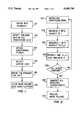
- FIGS. 1-2are flow charts of steps in accordance with the present method.
- FIGS. 3a-3dillustrate geometric details for obtaining the cross section from an image volume and a reference point according to the present method.
- FIG. 4is a graph demonstrating the variation of the average intensity function F over a varying radius r.
- FIG. 5is a 3-D plot of the average intensity function F varying with the position of the center points within a 9 ⁇ 9 neighborhood area.
- FIG. 6is a cross section of the left ventricle of the human heart showing a center point of the cross section determined in accordance with the present method.
- FIG. 7is a drawing illustrating the average intensity function F being derived for a cross section of a geometric object.
- CTcomputed tomography
- MRImagnetic resonance imaging
- PETphoton emission tomography
- SPECTsingle photon emission computed tomography
- the present methodreduces the problem of finding the true axis of a tube-like shaped object to defining a 3-D curve connecting the center points of cross sections of the tube-like shaped object.
- This new methodsearches for the center of a cross section by maximizing (where the object is bright) or minimizing (where the object is dark) a specially designed correlation function about a reference point.
- This new methodis efficient and reliable even for images that reveal only part of a full circular-shaped object, namely a sector or a number of disconnected arcs of the circular shaped cross section.
- the direct applications of the present method in the medical fieldinclude positron emission tomography (PET) or single photon emission computed tomography (SPECT) myocardial perfusion studies, curved determination for lung airway related diagnosis, and blood flow studies from 3-D vessel images using computed tomography (CT) or magnetic resonance imaging (MRI).
- PETpositron emission tomography
- SPECTsingle photon emission computed tomography
- CTcomputed tomography
- MRImagnetic resonance imaging
- the steps for automatically determining the true axis and radius of cross sections of tube-like shaped objectsare shown in the flow charts of FIGS. 1 and 2.
- the first stepis to obtain cross sections from a 3-D image volume and to provide the starting searching point of each slice for the curved axis determination.
- the entire image volumeis divided into several piece-wise volume segments in the direction of the long axis of the image volume 101.
- the number of segments or fragments developedwill vary with different applications.
- the overall computation timecan increase significantly with an increase in the number of piece wise volume segments.
- FIG. 3ashows a cylindrical shaped, transverse image volume 301 and a curved, tube-like shaped portion inside the image volume for which a curved axis is to be determined.
- the long axis of the middle transverse image of each individual segmentis detected 102.
- Piece-wise longitudinal planes 103are obtained by cutting the volume segments along this long axis. Then the straight center lines 104 of the longitudinal cuts are determined. These steps are detailed in FIGS. 3b and 3c.
- the transverse image of the tube-like shaped objectcan appear as an elliptical or horseshoe like image 307.
- the next step 105requires defining the straight-line axis of one piece-wise segment by the straight center line 306 of the longitudinal cut 305 of the piece-wise segment 303.
- the cross section or transverse image sliceis acquired by slicing the 3-D image volume perpendicular to the straight-line axis 106.
- the point where the straight-line axis and the transverse image slice intersectsis used as a starting point for obtaining the radius and the center point of the transverse image slice.
- These stepsare repeated for each transverse image (cross section) 201 to determine the radius or center points for defining the curve axis.
- the average intensity function Fwill have a distinct signature with a maximum or a minimum.
- the graph of FIG. 4shows a distinct average intensity function F having a maximum 401 at a radius approaching 10. At a radius less then 10 or above 10, the average intensity function decreases from a maximum to indicate that the test circle defined by the center point (x,y) and radius r chosen do not provide the best image intensity fit with the imaged cross section.
- FIG. 7Shown in FIG. 7 is a detail of the searching direction along radius r 703 about a selected neighborhood area (x,y) 702 for a cross section of an object 701. Due to the isotropic nature of the intensity function F, i.e. does not vary with direction, the searching result will not be biased by the non-uniformity of the intensity along the image circle.
- FIG. 7Shown in FIG. 7 is a detail of the searching direction along radius r 703 about a selected neighborhood area (x,y) 702 for a cross section of an object 701. Due to the isotropic nature of the intensity function F, i.e. does not vary with direction, the searching result will not be biased by the non-uniformity of the intensity along the image circle.
- the average intensity function Fwill generally yield an accurate determination of the center and the radius of the circle.
- the intensity function Fis first maximized/minimized again with respect to the position of the center points within the user defined neighborhood of the reference point as shown in FIG. 5, where the selected neighborhood area is a 9 ⁇ 9 area.
- the true center point of the imageis determined to be the one which results in the overall maximum value in F, shown in FIG. 5 to be the highest point 501 of the 3-D graph. Illustrated in FIG. 6 is a cross section of the left ventricle of a heart, where the hot spot in the center represents the center point of the cross section detected by the maximum of the intensity function F.
- the center point determined for a preceding cross section or transverse sliceis preferably used as the starting point (x,y) for the succeeding transverse slice to find the centers of a sequence of transverse slices of a tube-like shaped object.
- the radius r and center point (x,y) values which produce the maximum intensity function F valueare recorded for each transverse slice 204.
- the series of center points of the consecutive transverse slicesare connected to generate a curve axis for the tube-like shaped object of interest 206.
- the generated curve axisis then used for re-slicing the image volume of the object of interest for accurate diagnosis or inspection.
Landscapes
- Physics & Mathematics (AREA)
- Engineering & Computer Science (AREA)
- Geometry (AREA)
- Computer Vision & Pattern Recognition (AREA)
- General Physics & Mathematics (AREA)
- Theoretical Computer Science (AREA)
- Magnetic Resonance Imaging Apparatus (AREA)
- Apparatus For Radiation Diagnosis (AREA)
Abstract
Description
Claims (30)
Priority Applications (1)
| Application Number | Priority Date | Filing Date | Title |
|---|---|---|---|
| US08/622,076US5699799A (en) | 1996-03-26 | 1996-03-26 | Automatic determination of the curved axis of a 3-D tube-shaped object in image volume |
Applications Claiming Priority (1)
| Application Number | Priority Date | Filing Date | Title |
|---|---|---|---|
| US08/622,076US5699799A (en) | 1996-03-26 | 1996-03-26 | Automatic determination of the curved axis of a 3-D tube-shaped object in image volume |
Publications (1)
| Publication Number | Publication Date |
|---|---|
| US5699799Atrue US5699799A (en) | 1997-12-23 |
Family
ID=24492848
Family Applications (1)
| Application Number | Title | Priority Date | Filing Date |
|---|---|---|---|
| US08/622,076Expired - LifetimeUS5699799A (en) | 1996-03-26 | 1996-03-26 | Automatic determination of the curved axis of a 3-D tube-shaped object in image volume |
Country Status (1)
| Country | Link |
|---|---|
| US (1) | US5699799A (en) |
Cited By (115)
| Publication number | Priority date | Publication date | Assignee | Title |
|---|---|---|---|---|
| US5891030A (en)* | 1997-01-24 | 1999-04-06 | Mayo Foundation For Medical Education And Research | System for two dimensional and three dimensional imaging of tubular structures in the human body |
| US5908387A (en)* | 1996-06-21 | 1999-06-01 | Quinton Instrument Company | Device and method for improved quantitative coronary artery analysis |
| WO1999045844A1 (en)* | 1998-03-13 | 1999-09-16 | University Of Iowa Research Foundation | A curved cross section based system and method for gastrointestinal tract unraveling |
| WO2000019900A1 (en)* | 1998-10-06 | 2000-04-13 | Cardiac Mariners, Inc. | Image reconstruction method and apparatus |
| US6169917B1 (en)* | 1997-12-30 | 2001-01-02 | Leonardo Masotti | Method and device for reconstructing three-dimensional images of blood vessels, particularly coronary arteries, or other three-dimensional structures |
| US6178223B1 (en)* | 1998-10-06 | 2001-01-23 | Cardiac Mariners, Inc. | Image reconstruction method and apparatus |
| US6181764B1 (en)* | 1998-10-06 | 2001-01-30 | Cardiac Mariners, Inc. | Image reconstruction for wide depth of field images |
| WO2001037219A1 (en)* | 1999-11-19 | 2001-05-25 | General Electric Company | Method and apparatus for reformatting tubular volumetric bodies |
| US20010031920A1 (en)* | 1999-06-29 | 2001-10-18 | The Research Foundation Of State University Of New York | System and method for performing a three-dimensional virtual examination of objects, such as internal organs |
| WO2001080185A1 (en)* | 2000-04-14 | 2001-10-25 | General Electric Company | Method and apparatus for three-dimensional reconstruction of angiograms |
| US6331116B1 (en) | 1996-09-16 | 2001-12-18 | The Research Foundation Of State University Of New York | System and method for performing a three-dimensional virtual segmentation and examination |
| US6343936B1 (en) | 1996-09-16 | 2002-02-05 | The Research Foundation Of State University Of New York | System and method for performing a three-dimensional virtual examination, navigation and visualization |
| US6411852B1 (en)* | 1997-04-07 | 2002-06-25 | Broncus Technologies, Inc. | Modification of airways by application of energy |
| US6470070B2 (en) | 2000-12-20 | 2002-10-22 | Cedara Software Corp. | Image reconstruction using multiple X-ray projections |
| US6473488B2 (en) | 2000-12-20 | 2002-10-29 | Cedara Software Corp. | Three dimensional image reconstruction from single plane X-ray fluorograms |
| US6535623B1 (en) | 1999-04-15 | 2003-03-18 | Allen Robert Tannenbaum | Curvature based system for the segmentation and analysis of cardiac magnetic resonance images |
| US20030053669A1 (en)* | 2001-07-18 | 2003-03-20 | Marconi Medical Systems, Inc. | Magnetic resonance angiography method and apparatus |
| WO2003046835A1 (en)* | 2001-11-21 | 2003-06-05 | Koninklijke Philips Electronics Nv | Vessel tracking and tree extraction method and apparatus |
| US20030139660A1 (en)* | 2002-01-18 | 2003-07-24 | Isao Tatebayashi | Magnetic resonance imaging using technique of positioning multi-slabs to be imaged |
| US6634363B1 (en) | 1997-04-07 | 2003-10-21 | Broncus Technologies, Inc. | Methods of treating lungs having reversible obstructive pulmonary disease |
| US20030208116A1 (en)* | 2000-06-06 | 2003-11-06 | Zhengrong Liang | Computer aided treatment planning and visualization with image registration and fusion |
| WO2002041781A3 (en)* | 2000-11-27 | 2003-11-13 | Ge Med Sys Global Tech Co Llc | Method and apparatus for analysis of blood vessel images |
| US6650928B1 (en)* | 2000-11-27 | 2003-11-18 | Ge Medical Systems Global Technology Company, Llc | Color parametric and composite maps for CT perfusion |
| US20040015070A1 (en)* | 2001-02-05 | 2004-01-22 | Zhengrong Liang | Computer aided treatment planning |
| US20040064029A1 (en)* | 2002-09-30 | 2004-04-01 | The Government Of The Usa As Represented By The Secretary Of The Dept. Of Health & Human Services | Computer-aided classification of anomalies in anatomical structures |
| US6718054B1 (en) | 1999-06-23 | 2004-04-06 | Massachusetts Institute Of Technology | MRA segmentation using active contour models |
| US20040091143A1 (en)* | 2001-01-22 | 2004-05-13 | Qingmao Hu | Two and three dimensional skeletonization |
| US20040102697A1 (en)* | 2000-10-18 | 2004-05-27 | Rami Evron | Method and system for positioning a device in a tubular organ |
| US6782284B1 (en) | 2001-11-21 | 2004-08-24 | Koninklijke Philips Electronics, N.V. | Method and apparatus for semi-automatic aneurysm measurement and stent planning using volume image data |
| WO2004114220A1 (en)* | 2003-06-17 | 2004-12-29 | Brown University | Method and apparatus for model-based detection of structure in projection data |
| US6842638B1 (en) | 2001-11-13 | 2005-01-11 | Koninklijke Philips Electronics N.V. | Angiography method and apparatus |
| US20050008210A1 (en)* | 2000-05-09 | 2005-01-13 | Paieon, Inc. | System and method for three-dimensional reconstruction of an artery |
| US20050015003A1 (en)* | 2003-07-15 | 2005-01-20 | Rainer Lachner | Method and device for determining a three-dimensional form of a body from two-dimensional projection images |
| US20050074150A1 (en)* | 2003-10-03 | 2005-04-07 | Andrew Bruss | Systems and methods for emulating an angiogram using three-dimensional image data |
| US20050107679A1 (en)* | 2003-07-11 | 2005-05-19 | Bernhard Geiger | System and method for endoscopic path planning |
| US20050113664A1 (en)* | 2003-11-26 | 2005-05-26 | Laurent Stefani | Cardiac display methods and apparatus |
| US20050110791A1 (en)* | 2003-11-26 | 2005-05-26 | Prabhu Krishnamoorthy | Systems and methods for segmenting and displaying tubular vessels in volumetric imaging data |
| US6928314B1 (en) | 1998-01-23 | 2005-08-09 | Mayo Foundation For Medical Education And Research | System for two-dimensional and three-dimensional imaging of tubular structures in the human body |
| US20050180621A1 (en)* | 2002-11-27 | 2005-08-18 | Raghav Raman | Quantification of vascular irregularity |
| WO2005048816A3 (en)* | 2003-11-14 | 2006-02-23 | Univ New York | Automatic radial prescription of long-axis slices in mri examinations |
| US7020314B1 (en)* | 2001-11-13 | 2006-03-28 | Koninklijke Philips Electronics N.V. | Black blood angiography method and apparatus |
| US7024027B1 (en) | 2001-11-13 | 2006-04-04 | Koninklijke Philips Electronics N.V. | Method and apparatus for three-dimensional filtering of angiographic volume data |
| US20060074285A1 (en)* | 2004-09-24 | 2006-04-06 | Paieon Inc. | Apparatus and method for fusion and in-operating-room presentation of volumetric data and 3-D angiographic data |
| US7027869B2 (en) | 1998-01-07 | 2006-04-11 | Asthmatx, Inc. | Method for treating an asthma attack |
| US20060079746A1 (en)* | 2004-10-11 | 2006-04-13 | Perret Florence M | Apparatus and method for analysis of tissue classes along tubular structures |
| US20060098010A1 (en)* | 2004-03-09 | 2006-05-11 | Jeff Dwyer | Anatomical visualization and measurement system |
| US20060182327A1 (en)* | 2003-06-17 | 2006-08-17 | Brown Universtiy | Methods and apparatus for identifying subject matter in view data |
| US20060188135A1 (en)* | 2003-07-21 | 2006-08-24 | Michael Zarkh | Method and system for identifying optimal image within a series of images that depict a moving organ |
| US20060235669A1 (en)* | 1998-02-03 | 2006-10-19 | Charbel Fady T | Method and system for 3D blood vessel localization |
| US20060291705A1 (en)* | 2005-05-13 | 2006-12-28 | Rolf Baumann | Method and device for reconstructing two-dimensional sectional images |
| US20070103464A1 (en)* | 1999-06-29 | 2007-05-10 | Kaufman Arie E | System and method for performing a three-dimensional virtual examination of objects, such as internal organs |
| US20070116342A1 (en)* | 2003-09-25 | 2007-05-24 | Michael Zarkh | System and method for three-dimensional reconstruction of a tubular organ |
| US20070122019A1 (en)* | 2005-11-30 | 2007-05-31 | Kabushiki Kaisha Toshiba | Magnetic resonance imaging apparatus, method of making an imaging-plan, and method of imaging |
| US20070120845A1 (en)* | 2005-11-25 | 2007-05-31 | Kazuhiko Matsumoto | Image processing method and computer readable medium for image processing |
| US7324104B1 (en) | 2001-09-14 | 2008-01-29 | The Research Foundation Of State University Of New York | Method of centerline generation in virtual objects |
| US7425212B1 (en) | 1998-06-10 | 2008-09-16 | Asthmatx, Inc. | Devices for modification of airways by transfer of energy |
| US20080247621A1 (en)* | 2001-10-15 | 2008-10-09 | Michael Zarkh | Method and Apparatus for Positioning a Device in a Tubular Organ |
| US20080273781A1 (en)* | 2005-02-14 | 2008-11-06 | Mayo Foundation For Medical Education And Research | Electronic Stool Subtraction in Ct Colonography |
| US20080273777A1 (en)* | 2005-10-21 | 2008-11-06 | Vincent Luboz | Methods And Apparatus For Segmentation And Reconstruction For Endovascular And Endoluminal Anatomical Structures |
| US20090086912A1 (en)* | 2007-09-28 | 2009-04-02 | Takuya Sakaguchi | Image display apparatus and x-ray diagnostic apparatus |
| US20090161927A1 (en)* | 2006-05-02 | 2009-06-25 | National University Corporation Nagoya University | Medical Image Observation Assisting System |
| US7556624B2 (en) | 1997-04-07 | 2009-07-07 | Asthmatx, Inc. | Method of increasing gas exchange of a lung |
| US7574024B2 (en) | 2000-10-02 | 2009-08-11 | The Research Foundation Of State University Of New York | Centerline and tree branch skeleton determination for virtual objects |
| US7596256B1 (en) | 2001-09-14 | 2009-09-29 | The Research Foundation For The State University Of New York | Computer assisted detection of lesions in volumetric medical images |
| CN100571615C (en)* | 2003-09-24 | 2009-12-23 | 株式会社东芝 | The blood flow analysis device |
| US20100172566A1 (en)* | 2009-01-07 | 2010-07-08 | Takao Goto | Median plane determination apparatus and magnetic resonance imaging system |
| US20100177177A1 (en)* | 2007-06-07 | 2010-07-15 | Koninklijke Philips Electronics N.V. | Inspection of tubular-shaped structures |
| US20100208956A1 (en)* | 2005-11-30 | 2010-08-19 | The Research Foundation Of State University Of New York | Electronic colon cleansing method for virtual colonoscopy |
| US20100214283A1 (en)* | 2005-06-22 | 2010-08-26 | Koninklijke Philips Electronics, N.V. | Method to visualize cutplanes for curved elongated structures |
| US20100260390A1 (en)* | 2005-11-30 | 2010-10-14 | The Research Foundation Of State University Of New York | System and method for reduction of false positives during computer aided polyp detection |
| US7837679B2 (en) | 2000-10-17 | 2010-11-23 | Asthmatx, Inc. | Control system and process for application of energy to airway walls and other mediums |
| US20100312100A1 (en)* | 2005-03-31 | 2010-12-09 | Michael Zarkh | Method and apparatus for guiding a device in a totally occluded or partly occluded tubular organ |
| US7853331B2 (en) | 2004-11-05 | 2010-12-14 | Asthmatx, Inc. | Medical device with procedure improvement features |
| US7921855B2 (en) | 1998-01-07 | 2011-04-12 | Asthmatx, Inc. | Method for treating an asthma attack |
| US7931647B2 (en) | 2006-10-20 | 2011-04-26 | Asthmatx, Inc. | Method of delivering energy to a lung airway using markers |
| US7949407B2 (en) | 2004-11-05 | 2011-05-24 | Asthmatx, Inc. | Energy delivery devices and methods |
| US7992572B2 (en) | 1998-06-10 | 2011-08-09 | Asthmatx, Inc. | Methods of evaluating individuals having reversible obstructive pulmonary disease |
| US20110280366A1 (en)* | 2010-05-17 | 2011-11-17 | Toshiba Medical Systems Corporation | Image processing apparatus and x-ray ct system |
| US20120093278A1 (en)* | 2010-10-15 | 2012-04-19 | Shinsuke Tsukagoshi | Medical image processing apparatus and x-ray computed tomography apparatus |
| US8181656B2 (en) | 1998-06-10 | 2012-05-22 | Asthmatx, Inc. | Methods for treating airways |
| US20120163688A1 (en)* | 2010-12-22 | 2012-06-28 | Chevron U.S.A. Inc. | System and method for multi-phase segmentation of density images representing porous media |
| US8235983B2 (en) | 2007-07-12 | 2012-08-07 | Asthmatx, Inc. | Systems and methods for delivering energy to passageways in a patient |
| US8251070B2 (en) | 2000-03-27 | 2012-08-28 | Asthmatx, Inc. | Methods for treating airways |
| US8257413B2 (en) | 2000-10-17 | 2012-09-04 | Asthmatx, Inc. | Modification of airways by application of energy |
| US8483831B1 (en) | 2008-02-15 | 2013-07-09 | Holaira, Inc. | System and method for bronchial dilation |
| US20130243285A1 (en)* | 2010-08-30 | 2013-09-19 | Fujifilm Corporation | Medical image alignment apparatus, method, and program |
| US20140067106A1 (en)* | 2012-08-29 | 2014-03-06 | Prem Makeig | Computer-Implemented Methods for Generating 3D Models Suitable for 3D Printing |
| US8740895B2 (en) | 2009-10-27 | 2014-06-03 | Holaira, Inc. | Delivery devices with coolable energy emitting assemblies |
| US8808280B2 (en) | 2008-05-09 | 2014-08-19 | Holaira, Inc. | Systems, assemblies, and methods for treating a bronchial tree |
| US8911439B2 (en) | 2009-11-11 | 2014-12-16 | Holaira, Inc. | Non-invasive and minimally invasive denervation methods and systems for performing the same |
| US8920413B2 (en) | 2004-11-12 | 2014-12-30 | Asthmatx, Inc. | Energy delivery devices and methods |
| US9091628B2 (en) | 2012-12-21 | 2015-07-28 | L-3 Communications Security And Detection Systems, Inc. | 3D mapping with two orthogonal imaging views |
| US9149328B2 (en) | 2009-11-11 | 2015-10-06 | Holaira, Inc. | Systems, apparatuses, and methods for treating tissue and controlling stenosis |
| WO2015150320A1 (en)* | 2014-04-01 | 2015-10-08 | Universitetet I Oslo | Segmentation of tubular organ structures |
| US9272132B2 (en) | 2012-11-02 | 2016-03-01 | Boston Scientific Scimed, Inc. | Medical device for treating airways and related methods of use |
| US9283374B2 (en) | 2012-11-05 | 2016-03-15 | Boston Scientific Scimed, Inc. | Devices and methods for delivering energy to body lumens |
| US9339618B2 (en) | 2003-05-13 | 2016-05-17 | Holaira, Inc. | Method and apparatus for controlling narrowing of at least one airway |
| US9398933B2 (en) | 2012-12-27 | 2016-07-26 | Holaira, Inc. | Methods for improving drug efficacy including a combination of drug administration and nerve modulation |
| US9530219B2 (en) | 2014-07-02 | 2016-12-27 | Covidien Lp | System and method for detecting trachea |
| US9592086B2 (en) | 2012-07-24 | 2017-03-14 | Boston Scientific Scimed, Inc. | Electrodes for tissue treatment |
| US9603668B2 (en) | 2014-07-02 | 2017-03-28 | Covidien Lp | Dynamic 3D lung map view for tool navigation inside the lung |
| US9754367B2 (en) | 2014-07-02 | 2017-09-05 | Covidien Lp | Trachea marking |
| US9770216B2 (en) | 2014-07-02 | 2017-09-26 | Covidien Lp | System and method for navigating within the lung |
| US9770293B2 (en) | 2012-06-04 | 2017-09-26 | Boston Scientific Scimed, Inc. | Systems and methods for treating tissue of a passageway within a body |
| US9814618B2 (en) | 2013-06-06 | 2017-11-14 | Boston Scientific Scimed, Inc. | Devices for delivering energy and related methods of use |
| US9836848B2 (en) | 2014-07-02 | 2017-12-05 | Covidien Lp | System and method for segmentation of lung |
| US10478247B2 (en) | 2013-08-09 | 2019-11-19 | Boston Scientific Scimed, Inc. | Expandable catheter and related methods of manufacture and use |
| US10643371B2 (en) | 2014-08-11 | 2020-05-05 | Covidien Lp | Treatment procedure planning system and method |
| US10709352B2 (en) | 2015-10-27 | 2020-07-14 | Covidien Lp | Method of using lung airway carina locations to improve ENB registration |
| US10772532B2 (en) | 2014-07-02 | 2020-09-15 | Covidien Lp | Real-time automatic registration feedback |
| US10896498B2 (en) | 2018-07-23 | 2021-01-19 | The Boeing Company | Characterization of melted veil strand ratios in plies of fiber material |
| USD916750S1 (en) | 2014-07-02 | 2021-04-20 | Covidien Lp | Display screen or portion thereof with graphical user interface |
| US10986990B2 (en) | 2015-09-24 | 2021-04-27 | Covidien Lp | Marker placement |
| US11224392B2 (en) | 2018-02-01 | 2022-01-18 | Covidien Lp | Mapping disease spread |
| US12089902B2 (en) | 2019-07-30 | 2024-09-17 | Coviden Lp | Cone beam and 3D fluoroscope lung navigation |
Citations (18)
| Publication number | Priority date | Publication date | Assignee | Title |
|---|---|---|---|---|
| US4101961A (en)* | 1977-02-16 | 1978-07-18 | Nasa | Contour detector and data acquisition system for the left ventricular outline |
| US4630203A (en)* | 1983-12-27 | 1986-12-16 | Thomas Szirtes | Contour radiography: a system for determining 3-dimensional contours of an object from its 2-dimensional images |
| US4843629A (en)* | 1985-11-15 | 1989-06-27 | Thomson-Csf | Method of locating pairs of curved parallel lines in an image |
| US4920573A (en)* | 1988-05-09 | 1990-04-24 | Mpdi, Inc. | Method for generating perpendicular synthesized cross-sectional images |
| US4939646A (en)* | 1988-05-09 | 1990-07-03 | Mpdi, Inc. | Method for representing digitized image data |
| US5036463A (en)* | 1988-11-23 | 1991-07-30 | University Of Florida | Angioscopic system and method for dimensional measurement including measurement of the distance from angioscopic ends to designated planes |
| US5303706A (en)* | 1992-05-21 | 1994-04-19 | North American Philips Corporation | Directional interpolation for magnetic resonance angiography |
| US5323111A (en)* | 1992-03-11 | 1994-06-21 | Hitachi Medical Corp. | Magnetic resonance imaging method and apparatus |
| US5361763A (en)* | 1993-03-02 | 1994-11-08 | Wisconsin Alumni Research Foundation | Method for segmenting features in an image |
| US5368033A (en)* | 1993-04-20 | 1994-11-29 | North American Philips Corporation | Magnetic resonance angiography method and apparatus employing an integration projection |
| US5421330A (en)* | 1991-04-25 | 1995-06-06 | Inria Institut National De Recherche En Informatique Et En Automatique | Method and device for examining a body, particularly for tomography |
| US5427100A (en)* | 1992-02-07 | 1995-06-27 | Hitachi, Ltd. | Method for determining median line |
| US5457754A (en)* | 1990-08-02 | 1995-10-10 | University Of Cincinnati | Method for automatic contour extraction of a cardiac image |
| US5458111A (en)* | 1994-09-06 | 1995-10-17 | William C. Bond | Computed tomographic colonoscopy |
| US5501218A (en)* | 1991-10-30 | 1996-03-26 | Kabushiki Kaisha Toshiba | Method of scanning in MRI |
| US5570430A (en)* | 1994-05-31 | 1996-10-29 | University Of Washington | Method for determining the contour of an in vivo organ using multiple image frames of the organ |
| US5601084A (en)* | 1993-06-23 | 1997-02-11 | University Of Washington | Determining cardiac wall thickness and motion by imaging and three-dimensional modeling |
| US5617487A (en)* | 1993-09-22 | 1997-04-01 | Konica Corporation | Image cutout apparatus |
- 1996
- 1996-03-26USUS08/622,076patent/US5699799A/ennot_activeExpired - Lifetime
Patent Citations (18)
| Publication number | Priority date | Publication date | Assignee | Title |
|---|---|---|---|---|
| US4101961A (en)* | 1977-02-16 | 1978-07-18 | Nasa | Contour detector and data acquisition system for the left ventricular outline |
| US4630203A (en)* | 1983-12-27 | 1986-12-16 | Thomas Szirtes | Contour radiography: a system for determining 3-dimensional contours of an object from its 2-dimensional images |
| US4843629A (en)* | 1985-11-15 | 1989-06-27 | Thomson-Csf | Method of locating pairs of curved parallel lines in an image |
| US4920573A (en)* | 1988-05-09 | 1990-04-24 | Mpdi, Inc. | Method for generating perpendicular synthesized cross-sectional images |
| US4939646A (en)* | 1988-05-09 | 1990-07-03 | Mpdi, Inc. | Method for representing digitized image data |
| US5036463A (en)* | 1988-11-23 | 1991-07-30 | University Of Florida | Angioscopic system and method for dimensional measurement including measurement of the distance from angioscopic ends to designated planes |
| US5457754A (en)* | 1990-08-02 | 1995-10-10 | University Of Cincinnati | Method for automatic contour extraction of a cardiac image |
| US5421330A (en)* | 1991-04-25 | 1995-06-06 | Inria Institut National De Recherche En Informatique Et En Automatique | Method and device for examining a body, particularly for tomography |
| US5501218A (en)* | 1991-10-30 | 1996-03-26 | Kabushiki Kaisha Toshiba | Method of scanning in MRI |
| US5427100A (en)* | 1992-02-07 | 1995-06-27 | Hitachi, Ltd. | Method for determining median line |
| US5323111A (en)* | 1992-03-11 | 1994-06-21 | Hitachi Medical Corp. | Magnetic resonance imaging method and apparatus |
| US5303706A (en)* | 1992-05-21 | 1994-04-19 | North American Philips Corporation | Directional interpolation for magnetic resonance angiography |
| US5361763A (en)* | 1993-03-02 | 1994-11-08 | Wisconsin Alumni Research Foundation | Method for segmenting features in an image |
| US5368033A (en)* | 1993-04-20 | 1994-11-29 | North American Philips Corporation | Magnetic resonance angiography method and apparatus employing an integration projection |
| US5601084A (en)* | 1993-06-23 | 1997-02-11 | University Of Washington | Determining cardiac wall thickness and motion by imaging and three-dimensional modeling |
| US5617487A (en)* | 1993-09-22 | 1997-04-01 | Konica Corporation | Image cutout apparatus |
| US5570430A (en)* | 1994-05-31 | 1996-10-29 | University Of Washington | Method for determining the contour of an in vivo organ using multiple image frames of the organ |
| US5458111A (en)* | 1994-09-06 | 1995-10-17 | William C. Bond | Computed tomographic colonoscopy |
Cited By (271)
| Publication number | Priority date | Publication date | Assignee | Title |
|---|---|---|---|---|
| US5908387A (en)* | 1996-06-21 | 1999-06-01 | Quinton Instrument Company | Device and method for improved quantitative coronary artery analysis |
| US6331116B1 (en) | 1996-09-16 | 2001-12-18 | The Research Foundation Of State University Of New York | System and method for performing a three-dimensional virtual segmentation and examination |
| US7148887B2 (en) | 1996-09-16 | 2006-12-12 | The Research Foundation Of State University Of New York | System and method for performing a three-dimensional virtual segmentation and examination with optical texture mapping |
| US20070276225A1 (en)* | 1996-09-16 | 2007-11-29 | Kaufman Arie E | System and method for performing a three-dimensional virtual examination of objects, such as internal organs |
| US7474776B2 (en) | 1996-09-16 | 2009-01-06 | The Research Foundation Of State Of New York | System and method for performing a three-dimensional virtual examination of objects, such as internal organs |
| US7486811B2 (en) | 1996-09-16 | 2009-02-03 | The Research Foundation Of State University Of New York | System and method for performing a three-dimensional virtual examination of objects, such as internal organs |
| US6514082B2 (en) | 1996-09-16 | 2003-02-04 | The Research Foundation Of State University Of New York | System and method for performing a three-dimensional examination with collapse correction |
| US20020045153A1 (en)* | 1996-09-16 | 2002-04-18 | Kaufman Arie E. | System and method for performing a three-dimensional virtual segmentation and examination with optical texture mapping |
| US6343936B1 (en) | 1996-09-16 | 2002-02-05 | The Research Foundation Of State University Of New York | System and method for performing a three-dimensional virtual examination, navigation and visualization |
| US5891030A (en)* | 1997-01-24 | 1999-04-06 | Mayo Foundation For Medical Education And Research | System for two dimensional and three dimensional imaging of tubular structures in the human body |
| US8161978B2 (en) | 1997-04-07 | 2012-04-24 | Asthmatx, Inc. | Methods for treating asthma by damaging nerve tissue |
| US11033317B2 (en) | 1997-04-07 | 2021-06-15 | Boston Scientific Scimed, Inc. | Methods for treating a lung |
| US8944071B2 (en) | 1997-04-07 | 2015-02-03 | Asthmatx, Inc. | Method for treating an asthma attack |
| US9027564B2 (en) | 1997-04-07 | 2015-05-12 | Asthmatx, Inc. | Method for treating a lung |
| US6411852B1 (en)* | 1997-04-07 | 2002-06-25 | Broncus Technologies, Inc. | Modification of airways by application of energy |
| US7770584B2 (en) | 1997-04-07 | 2010-08-10 | Asthmatx, Inc. | Modification of airways by application of microwave energy |
| US7740017B2 (en) | 1997-04-07 | 2010-06-22 | Asthmatx, Inc. | Method for treating an asthma attack |
| US10058370B2 (en) | 1997-04-07 | 2018-08-28 | Boston Scientific Scimed, Inc. | Method for treating a lung |
| US8640711B2 (en) | 1997-04-07 | 2014-02-04 | Asthmatx, Inc. | Method for treating an asthma attack |
| US8267094B2 (en) | 1997-04-07 | 2012-09-18 | Asthmatx, Inc. | Modification of airways by application of ultrasound energy |
| US7556624B2 (en) | 1997-04-07 | 2009-07-07 | Asthmatx, Inc. | Method of increasing gas exchange of a lung |
| US9956023B2 (en) | 1997-04-07 | 2018-05-01 | Boston Scientific Scimed, Inc. | System for treating a lung |
| US7938123B2 (en) | 1997-04-07 | 2011-05-10 | Asthmatx, Inc. | Modification of airways by application of cryo energy |
| US6634363B1 (en) | 1997-04-07 | 2003-10-21 | Broncus Technologies, Inc. | Methods of treating lungs having reversible obstructive pulmonary disease |
| US6169917B1 (en)* | 1997-12-30 | 2001-01-02 | Leonardo Masotti | Method and device for reconstructing three-dimensional images of blood vessels, particularly coronary arteries, or other three-dimensional structures |
| US7921855B2 (en) | 1998-01-07 | 2011-04-12 | Asthmatx, Inc. | Method for treating an asthma attack |
| US9789331B2 (en) | 1998-01-07 | 2017-10-17 | Boston Scientific Scimed, Inc. | Methods of treating a lung |
| US8584681B2 (en) | 1998-01-07 | 2013-11-19 | Asthmatx, Inc. | Method for treating an asthma attack |
| US7027869B2 (en) | 1998-01-07 | 2006-04-11 | Asthmatx, Inc. | Method for treating an asthma attack |
| US6928314B1 (en) | 1998-01-23 | 2005-08-09 | Mayo Foundation For Medical Education And Research | System for two-dimensional and three-dimensional imaging of tubular structures in the human body |
| US20060235669A1 (en)* | 1998-02-03 | 2006-10-19 | Charbel Fady T | Method and system for 3D blood vessel localization |
| US7739090B2 (en)* | 1998-02-03 | 2010-06-15 | University Of Illinois, Board Of Trustees | Method and system for 3D blood vessel localization |
| WO1999045844A1 (en)* | 1998-03-13 | 1999-09-16 | University Of Iowa Research Foundation | A curved cross section based system and method for gastrointestinal tract unraveling |
| US6212420B1 (en) | 1998-03-13 | 2001-04-03 | University Of Iowa Research Foundation | Curved cross-section based system and method for gastrointestinal tract unraveling |
| US8443810B2 (en) | 1998-06-10 | 2013-05-21 | Asthmatx, Inc. | Methods of reducing mucus in airways |
| US7425212B1 (en) | 1998-06-10 | 2008-09-16 | Asthmatx, Inc. | Devices for modification of airways by transfer of energy |
| US8534291B2 (en) | 1998-06-10 | 2013-09-17 | Asthmatx, Inc. | Methods of treating inflammation in airways |
| US7264002B2 (en) | 1998-06-10 | 2007-09-04 | Asthmatx, Inc. | Methods of treating reversible obstructive pulmonary disease |
| US7273055B2 (en) | 1998-06-10 | 2007-09-25 | Asthmatx, Inc. | Methods of treating asthma |
| US7992572B2 (en) | 1998-06-10 | 2011-08-09 | Asthmatx, Inc. | Methods of evaluating individuals having reversible obstructive pulmonary disease |
| US8464723B2 (en) | 1998-06-10 | 2013-06-18 | Asthmatx, Inc. | Methods of evaluating individuals having reversible obstructive pulmonary disease |
| US7542802B2 (en) | 1998-06-10 | 2009-06-02 | Asthmatx, Inc. | Methods of regenerating tissue in airways |
| US8733367B2 (en) | 1998-06-10 | 2014-05-27 | Asthmatx, Inc. | Methods of treating inflammation in airways |
| US8181656B2 (en) | 1998-06-10 | 2012-05-22 | Asthmatx, Inc. | Methods for treating airways |
| WO2000019900A1 (en)* | 1998-10-06 | 2000-04-13 | Cardiac Mariners, Inc. | Image reconstruction method and apparatus |
| US6178223B1 (en)* | 1998-10-06 | 2001-01-23 | Cardiac Mariners, Inc. | Image reconstruction method and apparatus |
| US6181764B1 (en)* | 1998-10-06 | 2001-01-30 | Cardiac Mariners, Inc. | Image reconstruction for wide depth of field images |
| US6535623B1 (en) | 1999-04-15 | 2003-03-18 | Allen Robert Tannenbaum | Curvature based system for the segmentation and analysis of cardiac magnetic resonance images |
| US6721450B2 (en) | 1999-04-15 | 2004-04-13 | Allen Robert Tannenbaum | Curvature based system for the segmentation and analysis of image data |
| US6718054B1 (en) | 1999-06-23 | 2004-04-06 | Massachusetts Institute Of Technology | MRA segmentation using active contour models |
| US20070103464A1 (en)* | 1999-06-29 | 2007-05-10 | Kaufman Arie E | System and method for performing a three-dimensional virtual examination of objects, such as internal organs |
| US7477768B2 (en) | 1999-06-29 | 2009-01-13 | The Research Foundation Of State University Of New York | System and method for performing a three-dimensional virtual examination of objects, such as internal organs |
| US7194117B2 (en) | 1999-06-29 | 2007-03-20 | The Research Foundation Of State University Of New York | System and method for performing a three-dimensional virtual examination of objects, such as internal organs |
| US20010031920A1 (en)* | 1999-06-29 | 2001-10-18 | The Research Foundation Of State University Of New York | System and method for performing a three-dimensional virtual examination of objects, such as internal organs |
| WO2001037219A1 (en)* | 1999-11-19 | 2001-05-25 | General Electric Company | Method and apparatus for reformatting tubular volumetric bodies |
| US8459268B2 (en) | 2000-03-27 | 2013-06-11 | Asthmatx, Inc. | Methods for treating airways |
| US9358024B2 (en) | 2000-03-27 | 2016-06-07 | Asthmatx, Inc. | Methods for treating airways |
| US10561458B2 (en) | 2000-03-27 | 2020-02-18 | Boston Scientific Scimed, Inc. | Methods for treating airways |
| US10278766B2 (en) | 2000-03-27 | 2019-05-07 | Boston Scientific Scimed, Inc. | Methods for treating airways |
| US8251070B2 (en) | 2000-03-27 | 2012-08-28 | Asthmatx, Inc. | Methods for treating airways |
| WO2001080185A1 (en)* | 2000-04-14 | 2001-10-25 | General Electric Company | Method and apparatus for three-dimensional reconstruction of angiograms |
| US20050008210A1 (en)* | 2000-05-09 | 2005-01-13 | Paieon, Inc. | System and method for three-dimensional reconstruction of an artery |
| US20030208116A1 (en)* | 2000-06-06 | 2003-11-06 | Zhengrong Liang | Computer aided treatment planning and visualization with image registration and fusion |
| US7356367B2 (en) | 2000-06-06 | 2008-04-08 | The Research Foundation Of State University Of New York | Computer aided treatment planning and visualization with image registration and fusion |
| US7706600B2 (en) | 2000-10-02 | 2010-04-27 | The Research Foundation Of State University Of New York | Enhanced virtual navigation and examination |
| US7574024B2 (en) | 2000-10-02 | 2009-08-11 | The Research Foundation Of State University Of New York | Centerline and tree branch skeleton determination for virtual objects |
| US8888769B2 (en) | 2000-10-17 | 2014-11-18 | Asthmatx, Inc. | Control system and process for application of energy to airway walls and other mediums |
| US7837679B2 (en) | 2000-10-17 | 2010-11-23 | Asthmatx, Inc. | Control system and process for application of energy to airway walls and other mediums |
| US9931163B2 (en) | 2000-10-17 | 2018-04-03 | Boston Scientific Scimed, Inc. | Energy delivery devices |
| US7854734B2 (en) | 2000-10-17 | 2010-12-21 | Asthmatx, Inc. | Control system and process for application of energy to airway walls and other mediums |
| US8257413B2 (en) | 2000-10-17 | 2012-09-04 | Asthmatx, Inc. | Modification of airways by application of energy |
| US9033976B2 (en) | 2000-10-17 | 2015-05-19 | Asthmatx, Inc. | Modification of airways by application of energy |
| US8465486B2 (en) | 2000-10-17 | 2013-06-18 | Asthmatx, Inc. | Modification of airways by application of energy |
| US20040102697A1 (en)* | 2000-10-18 | 2004-05-27 | Rami Evron | Method and system for positioning a device in a tubular organ |
| US7778685B2 (en) | 2000-10-18 | 2010-08-17 | Paieon Inc. | Method and system for positioning a device in a tubular organ |
| US6650928B1 (en)* | 2000-11-27 | 2003-11-18 | Ge Medical Systems Global Technology Company, Llc | Color parametric and composite maps for CT perfusion |
| WO2002041781A3 (en)* | 2000-11-27 | 2003-11-13 | Ge Med Sys Global Tech Co Llc | Method and apparatus for analysis of blood vessel images |
| US6829379B1 (en) | 2000-11-27 | 2004-12-07 | Ge Medical Systems Global Technology Company, Llc | Methods and apparatus to assist and facilitate vessel analysis |
| US6470070B2 (en) | 2000-12-20 | 2002-10-22 | Cedara Software Corp. | Image reconstruction using multiple X-ray projections |
| US6473488B2 (en) | 2000-12-20 | 2002-10-29 | Cedara Software Corp. | Three dimensional image reconstruction from single plane X-ray fluorograms |
| US6661869B2 (en) | 2000-12-20 | 2003-12-09 | Cedara Software Corp. | Image reconstruction using multiple X-ray projections |
| US6587541B2 (en) | 2000-12-20 | 2003-07-01 | Cedara Software Corp. | Three dimensional image reconstruction from single plane x-ray fluorograms |
| US20040091143A1 (en)* | 2001-01-22 | 2004-05-13 | Qingmao Hu | Two and three dimensional skeletonization |
| US7630750B2 (en) | 2001-02-05 | 2009-12-08 | The Research Foundation For The State University Of New York | Computer aided treatment planning |
| US20040015070A1 (en)* | 2001-02-05 | 2004-01-22 | Zhengrong Liang | Computer aided treatment planning |
| US20030053669A1 (en)* | 2001-07-18 | 2003-03-20 | Marconi Medical Systems, Inc. | Magnetic resonance angiography method and apparatus |
| US7324104B1 (en) | 2001-09-14 | 2008-01-29 | The Research Foundation Of State University Of New York | Method of centerline generation in virtual objects |
| US7596256B1 (en) | 2001-09-14 | 2009-09-29 | The Research Foundation For The State University Of New York | Computer assisted detection of lesions in volumetric medical images |
| US8126241B2 (en) | 2001-10-15 | 2012-02-28 | Michael Zarkh | Method and apparatus for positioning a device in a tubular organ |
| US20080247621A1 (en)* | 2001-10-15 | 2008-10-09 | Michael Zarkh | Method and Apparatus for Positioning a Device in a Tubular Organ |
| US10016592B2 (en) | 2001-10-17 | 2018-07-10 | Boston Scientific Scimed, Inc. | Control system and process for application of energy to airway walls and other mediums |
| US7024027B1 (en) | 2001-11-13 | 2006-04-04 | Koninklijke Philips Electronics N.V. | Method and apparatus for three-dimensional filtering of angiographic volume data |
| US7020314B1 (en)* | 2001-11-13 | 2006-03-28 | Koninklijke Philips Electronics N.V. | Black blood angiography method and apparatus |
| US6842638B1 (en) | 2001-11-13 | 2005-01-11 | Koninklijke Philips Electronics N.V. | Angiography method and apparatus |
| WO2003046835A1 (en)* | 2001-11-21 | 2003-06-05 | Koninklijke Philips Electronics Nv | Vessel tracking and tree extraction method and apparatus |
| US6728566B1 (en) | 2001-11-21 | 2004-04-27 | Koninklijke Philips Electronics, N.V. | Vessel tracking and tree extraction method and apparatus |
| US6782284B1 (en) | 2001-11-21 | 2004-08-24 | Koninklijke Philips Electronics, N.V. | Method and apparatus for semi-automatic aneurysm measurement and stent planning using volume image data |
| US7190992B2 (en)* | 2002-01-18 | 2007-03-13 | Kabushiki Kaisha Toshiba | Magnetic resonance imaging using technique of positioning multi-slabs to be imaged |
| US7953468B2 (en) | 2002-01-18 | 2011-05-31 | Kabushiki Kaisha Toshiba | Magnetic resonance imaging using technique of positioning multi-slabs to be imaged |
| US20060122487A1 (en)* | 2002-01-18 | 2006-06-08 | Kabushiki Kaisha Toshiba | Magnetic resonance imaging using technique of positioning multi-slabs to be imaged |
| US9250306B2 (en) | 2002-01-18 | 2016-02-02 | Kabushiki Kaisha Toshiba | Magnetic resonance imaging using technique of positioning multi-slabs to be imaged |
| US20030139660A1 (en)* | 2002-01-18 | 2003-07-24 | Isao Tatebayashi | Magnetic resonance imaging using technique of positioning multi-slabs to be imaged |
| US20080015419A1 (en)* | 2002-09-30 | 2008-01-17 | The Gov. Of The U.S.A. As Represented By The Secretary Of The Dept. Of Health And Human Service | Computer-aided classification of anomalies in anatomical structures |
| US7260250B2 (en) | 2002-09-30 | 2007-08-21 | The United States Of America As Represented By The Secretary Of The Department Of Health And Human Services | Computer-aided classification of anomalies in anatomical structures |
| US20040064029A1 (en)* | 2002-09-30 | 2004-04-01 | The Government Of The Usa As Represented By The Secretary Of The Dept. Of Health & Human Services | Computer-aided classification of anomalies in anatomical structures |
| US20100074491A1 (en)* | 2002-09-30 | 2010-03-25 | The Government of the United States of America as represented by the Secretary of Health and Human | Computer-aided classification of anomalies in anatomical structures |
| US8189890B2 (en) | 2002-09-30 | 2012-05-29 | The United States Of America As Represented By The Secretary Of The Department Of Health And Human Services | Computer-aided classification of anomalies in anatomical structures |
| US7646904B2 (en) | 2002-09-30 | 2010-01-12 | The United States Of America As Represented By The Department Of Health And Human Services | Computer-aided classification of anomalies in anatomical structures |
| US7379574B2 (en)* | 2002-11-27 | 2008-05-27 | The Board Of Trustees Of The Leland Stanford Junior University | Quantification of vascular irregularity |
| US20050180621A1 (en)* | 2002-11-27 | 2005-08-18 | Raghav Raman | Quantification of vascular irregularity |
| US9339618B2 (en) | 2003-05-13 | 2016-05-17 | Holaira, Inc. | Method and apparatus for controlling narrowing of at least one airway |
| US10953170B2 (en) | 2003-05-13 | 2021-03-23 | Nuvaira, Inc. | Apparatus for treating asthma using neurotoxin |
| US20040267114A1 (en)* | 2003-06-17 | 2004-12-30 | Brown University | Methods and apparatus for model-based detection of structure in view data |
| US20060182327A1 (en)* | 2003-06-17 | 2006-08-17 | Brown Universtiy | Methods and apparatus for identifying subject matter in view data |
| US20150139525A1 (en)* | 2003-06-17 | 2015-05-21 | Brown University | Methods and apparatus for model-based detection of structure in view data |
| US7492934B2 (en)* | 2003-06-17 | 2009-02-17 | Brown University | Methods and apparatus for model-based detection of structure in view data |
| JP2007524445A (en)* | 2003-06-17 | 2007-08-30 | ブラウン ユニバーシティ | Method and apparatus for model-based detection of structures in projection data |
| US20090123053A1 (en)* | 2003-06-17 | 2009-05-14 | Brown University | Methods and apparatus for model-based detection of structure in view data |
| US9576198B2 (en) | 2003-06-17 | 2017-02-21 | Brown University | Methods and apparatus for identifying subject matter in view data |
| WO2004114220A1 (en)* | 2003-06-17 | 2004-12-29 | Brown University | Method and apparatus for model-based detection of structure in projection data |
| US8515145B2 (en) | 2003-06-17 | 2013-08-20 | Brown University | Methods and apparatus for identifying subject matter in view data |
| US7978887B2 (en) | 2003-06-17 | 2011-07-12 | Brown University | Methods and apparatus for identifying subject matter in view data |
| US20050107679A1 (en)* | 2003-07-11 | 2005-05-19 | Bernhard Geiger | System and method for endoscopic path planning |
| US7822461B2 (en)* | 2003-07-11 | 2010-10-26 | Siemens Medical Solutions Usa, Inc. | System and method for endoscopic path planning |
| US20050015003A1 (en)* | 2003-07-15 | 2005-01-20 | Rainer Lachner | Method and device for determining a three-dimensional form of a body from two-dimensional projection images |
| US7873403B2 (en)* | 2003-07-15 | 2011-01-18 | Brainlab Ag | Method and device for determining a three-dimensional form of a body from two-dimensional projection images |
| US20060188135A1 (en)* | 2003-07-21 | 2006-08-24 | Michael Zarkh | Method and system for identifying optimal image within a series of images that depict a moving organ |
| US7587074B2 (en) | 2003-07-21 | 2009-09-08 | Paieon Inc. | Method and system for identifying optimal image within a series of images that depict a moving organ |
| CN100571615C (en)* | 2003-09-24 | 2009-12-23 | 株式会社东芝 | The blood flow analysis device |
| US20070116342A1 (en)* | 2003-09-25 | 2007-05-24 | Michael Zarkh | System and method for three-dimensional reconstruction of a tubular organ |
| US7742629B2 (en) | 2003-09-25 | 2010-06-22 | Paieon Inc. | System and method for three-dimensional reconstruction of a tubular organ |
| US20050074150A1 (en)* | 2003-10-03 | 2005-04-07 | Andrew Bruss | Systems and methods for emulating an angiogram using three-dimensional image data |
| US20070063703A1 (en)* | 2003-11-14 | 2007-03-22 | Paj Vinay M | Method, system, storage medium and software arrangement for radial prescription of long-axis slices in magnetic resonance imaging examinations |
| US7345480B2 (en) | 2003-11-14 | 2008-03-18 | New York University | Method, system, storage medium and software arrangement for radial prescription of long-axis slices in magnetic resonance imaging examinations |
| WO2005048816A3 (en)* | 2003-11-14 | 2006-02-23 | Univ New York | Automatic radial prescription of long-axis slices in mri examinations |
| US20050113664A1 (en)* | 2003-11-26 | 2005-05-26 | Laurent Stefani | Cardiac display methods and apparatus |
| US20050110791A1 (en)* | 2003-11-26 | 2005-05-26 | Prabhu Krishnamoorthy | Systems and methods for segmenting and displaying tubular vessels in volumetric imaging data |
| WO2005055141A1 (en)* | 2003-11-26 | 2005-06-16 | Vital Images, Inc. | Segmenting and displaying tubular vessels in volumetric imaging data |
| US20060098010A1 (en)* | 2004-03-09 | 2006-05-11 | Jeff Dwyer | Anatomical visualization and measurement system |
| US20060074285A1 (en)* | 2004-09-24 | 2006-04-06 | Paieon Inc. | Apparatus and method for fusion and in-operating-room presentation of volumetric data and 3-D angiographic data |
| WO2006033113A3 (en)* | 2004-09-24 | 2006-08-17 | Paieon Inc | Apparatus and method for fusion and in-operating-room presentation of volumetric data and 3-d angiographic data |
| US20060079746A1 (en)* | 2004-10-11 | 2006-04-13 | Perret Florence M | Apparatus and method for analysis of tissue classes along tubular structures |
| US7949407B2 (en) | 2004-11-05 | 2011-05-24 | Asthmatx, Inc. | Energy delivery devices and methods |
| US7853331B2 (en) | 2004-11-05 | 2010-12-14 | Asthmatx, Inc. | Medical device with procedure improvement features |
| US10076380B2 (en) | 2004-11-05 | 2018-09-18 | Boston Scientific Scimed, Inc. | Energy delivery devices and methods |
| US8480667B2 (en) | 2004-11-05 | 2013-07-09 | Asthmatx, Inc. | Medical device with procedure improvement features |
| US10398502B2 (en) | 2004-11-05 | 2019-09-03 | Boston Scientific Scimed, Inc. | Energy delivery devices and methods |
| US8920413B2 (en) | 2004-11-12 | 2014-12-30 | Asthmatx, Inc. | Energy delivery devices and methods |
| US20080273781A1 (en)* | 2005-02-14 | 2008-11-06 | Mayo Foundation For Medical Education And Research | Electronic Stool Subtraction in Ct Colonography |
| US8031921B2 (en) | 2005-02-14 | 2011-10-04 | Mayo Foundation For Medical Education And Research | Electronic stool subtraction in CT colonography |
| US8295577B2 (en) | 2005-03-31 | 2012-10-23 | Michael Zarkh | Method and apparatus for guiding a device in a totally occluded or partly occluded tubular organ |
| US20100312100A1 (en)* | 2005-03-31 | 2010-12-09 | Michael Zarkh | Method and apparatus for guiding a device in a totally occluded or partly occluded tubular organ |
| US20060291705A1 (en)* | 2005-05-13 | 2006-12-28 | Rolf Baumann | Method and device for reconstructing two-dimensional sectional images |
| US7853304B2 (en)* | 2005-05-13 | 2010-12-14 | Tomtec Imaging Systems Gmbh | Method and device for reconstructing two-dimensional sectional images |
| US8253723B2 (en) | 2005-06-22 | 2012-08-28 | Koninklijke Philips Electronics N.V. | Method to visualize cutplanes for curved elongated structures |
| US20100214283A1 (en)* | 2005-06-22 | 2010-08-26 | Koninklijke Philips Electronics, N.V. | Method to visualize cutplanes for curved elongated structures |
| US20080273777A1 (en)* | 2005-10-21 | 2008-11-06 | Vincent Luboz | Methods And Apparatus For Segmentation And Reconstruction For Endovascular And Endoluminal Anatomical Structures |
| US20070120845A1 (en)* | 2005-11-25 | 2007-05-31 | Kazuhiko Matsumoto | Image processing method and computer readable medium for image processing |
| US7825924B2 (en)* | 2005-11-25 | 2010-11-02 | Ziosoft, Inc. | Image processing method and computer readable medium for image processing |
| US20100260390A1 (en)* | 2005-11-30 | 2010-10-14 | The Research Foundation Of State University Of New York | System and method for reduction of false positives during computer aided polyp detection |
| US20070122019A1 (en)* | 2005-11-30 | 2007-05-31 | Kabushiki Kaisha Toshiba | Magnetic resonance imaging apparatus, method of making an imaging-plan, and method of imaging |
| US8452061B2 (en) | 2005-11-30 | 2013-05-28 | The Research Foundation Of State University Of New York | Electronic colon cleansing method for virtual colonoscopy |
| US20100208956A1 (en)* | 2005-11-30 | 2010-08-19 | The Research Foundation Of State University Of New York | Electronic colon cleansing method for virtual colonoscopy |
| US7853059B2 (en)* | 2005-11-30 | 2010-12-14 | Kabushiki Kaisha Toshiba | Magnetic resonance imaging apparatus, method of making an imaging-plan, and method of imaging |
| US8199984B2 (en)* | 2006-05-02 | 2012-06-12 | National University Corporation Nagoya University | System that assists in observing a luminal organ using the structure of the luminal organ |
| US20090161927A1 (en)* | 2006-05-02 | 2009-06-25 | National University Corporation Nagoya University | Medical Image Observation Assisting System |
| US7931647B2 (en) | 2006-10-20 | 2011-04-26 | Asthmatx, Inc. | Method of delivering energy to a lung airway using markers |
| US20100177177A1 (en)* | 2007-06-07 | 2010-07-15 | Koninklijke Philips Electronics N.V. | Inspection of tubular-shaped structures |
| US12029476B2 (en) | 2007-07-12 | 2024-07-09 | Boston Scientific Scimed, Inc. | Systems and methods for delivering energy to passageways in a patient |
| US8235983B2 (en) | 2007-07-12 | 2012-08-07 | Asthmatx, Inc. | Systems and methods for delivering energy to passageways in a patient |
| US11478299B2 (en) | 2007-07-12 | 2022-10-25 | Boston Scientific Scimed, Inc. | Systems and methods for delivering energy to passageways in a patient |
| US10368941B2 (en) | 2007-07-12 | 2019-08-06 | Boston Scientific Scimed, Inc. | Systems and methods for delivering energy to passageways in a patient |
| US20090086912A1 (en)* | 2007-09-28 | 2009-04-02 | Takuya Sakaguchi | Image display apparatus and x-ray diagnostic apparatus |
| US8934604B2 (en)* | 2007-09-28 | 2015-01-13 | Kabushiki Kaisha Toshiba | Image display apparatus and X-ray diagnostic apparatus |
| US9125643B2 (en) | 2008-02-15 | 2015-09-08 | Holaira, Inc. | System and method for bronchial dilation |
| US8483831B1 (en) | 2008-02-15 | 2013-07-09 | Holaira, Inc. | System and method for bronchial dilation |
| US12357827B2 (en) | 2008-02-15 | 2025-07-15 | Nuvaira, Inc. | System and method for bronchial dilation |
| US11058879B2 (en) | 2008-02-15 | 2021-07-13 | Nuvaira, Inc. | System and method for bronchial dilation |
| US8731672B2 (en) | 2008-02-15 | 2014-05-20 | Holaira, Inc. | System and method for bronchial dilation |
| US8489192B1 (en) | 2008-02-15 | 2013-07-16 | Holaira, Inc. | System and method for bronchial dilation |
| US8961508B2 (en) | 2008-05-09 | 2015-02-24 | Holaira, Inc. | Systems, assemblies, and methods for treating a bronchial tree |
| US8961507B2 (en) | 2008-05-09 | 2015-02-24 | Holaira, Inc. | Systems, assemblies, and methods for treating a bronchial tree |
| US9668809B2 (en) | 2008-05-09 | 2017-06-06 | Holaira, Inc. | Systems, assemblies, and methods for treating a bronchial tree |
| US8808280B2 (en) | 2008-05-09 | 2014-08-19 | Holaira, Inc. | Systems, assemblies, and methods for treating a bronchial tree |
| US8821489B2 (en) | 2008-05-09 | 2014-09-02 | Holaira, Inc. | Systems, assemblies, and methods for treating a bronchial tree |
| US10149714B2 (en) | 2008-05-09 | 2018-12-11 | Nuvaira, Inc. | Systems, assemblies, and methods for treating a bronchial tree |
| US11937868B2 (en) | 2008-05-09 | 2024-03-26 | Nuvaira, Inc. | Systems, assemblies, and methods for treating a bronchial tree |
| US20100172566A1 (en)* | 2009-01-07 | 2010-07-08 | Takao Goto | Median plane determination apparatus and magnetic resonance imaging system |
| US8488858B2 (en) | 2009-01-07 | 2013-07-16 | Ge Medical Systems Global Technology Company, Llc | Median plane determination apparatus and magnetic resonance imaging system |
| US8740895B2 (en) | 2009-10-27 | 2014-06-03 | Holaira, Inc. | Delivery devices with coolable energy emitting assemblies |
| US8932289B2 (en) | 2009-10-27 | 2015-01-13 | Holaira, Inc. | Delivery devices with coolable energy emitting assemblies |
| US9931162B2 (en) | 2009-10-27 | 2018-04-03 | Nuvaira, Inc. | Delivery devices with coolable energy emitting assemblies |
| US9005195B2 (en) | 2009-10-27 | 2015-04-14 | Holaira, Inc. | Delivery devices with coolable energy emitting assemblies |
| US9017324B2 (en) | 2009-10-27 | 2015-04-28 | Holaira, Inc. | Delivery devices with coolable energy emitting assemblies |
| US8777943B2 (en) | 2009-10-27 | 2014-07-15 | Holaira, Inc. | Delivery devices with coolable energy emitting assemblies |
| US9675412B2 (en) | 2009-10-27 | 2017-06-13 | Holaira, Inc. | Delivery devices with coolable energy emitting assemblies |
| US9649153B2 (en) | 2009-10-27 | 2017-05-16 | Holaira, Inc. | Delivery devices with coolable energy emitting assemblies |
| US9149328B2 (en) | 2009-11-11 | 2015-10-06 | Holaira, Inc. | Systems, apparatuses, and methods for treating tissue and controlling stenosis |
| US11712283B2 (en) | 2009-11-11 | 2023-08-01 | Nuvaira, Inc. | Non-invasive and minimally invasive denervation methods and systems for performing the same |
| US10610283B2 (en) | 2009-11-11 | 2020-04-07 | Nuvaira, Inc. | Non-invasive and minimally invasive denervation methods and systems for performing the same |
| US8911439B2 (en) | 2009-11-11 | 2014-12-16 | Holaira, Inc. | Non-invasive and minimally invasive denervation methods and systems for performing the same |
| US12343060B2 (en) | 2009-11-11 | 2025-07-01 | Nuvaira, Inc. | Non-invasive and minimally invasive denervation methods and systems for performing the same |
| US9649154B2 (en) | 2009-11-11 | 2017-05-16 | Holaira, Inc. | Non-invasive and minimally invasive denervation methods and systems for performing the same |
| US11389233B2 (en) | 2009-11-11 | 2022-07-19 | Nuvaira, Inc. | Systems, apparatuses, and methods for treating tissue and controlling stenosis |
| US12290309B2 (en) | 2009-11-11 | 2025-05-06 | Nuvaira, Inc. | Systems, apparatuses, and methods for treating tissue and controlling stenosis |
| US8953739B2 (en)* | 2010-05-17 | 2015-02-10 | Kabushiki Kaisha Toshiba | Image processing apparatus and X-ray CT system |
| US20110280366A1 (en)* | 2010-05-17 | 2011-11-17 | Toshiba Medical Systems Corporation | Image processing apparatus and x-ray ct system |
| US20130243285A1 (en)* | 2010-08-30 | 2013-09-19 | Fujifilm Corporation | Medical image alignment apparatus, method, and program |
| US8798227B2 (en)* | 2010-10-15 | 2014-08-05 | Kabushiki Kaisha Toshiba | Medical image processing apparatus and X-ray computed tomography apparatus |
| US20120093278A1 (en)* | 2010-10-15 | 2012-04-19 | Shinsuke Tsukagoshi | Medical image processing apparatus and x-ray computed tomography apparatus |
| US8861814B2 (en)* | 2010-12-22 | 2014-10-14 | Chevron U.S.A. Inc. | System and method for multi-phase segmentation of density images representing porous media |
| US20120163688A1 (en)* | 2010-12-22 | 2012-06-28 | Chevron U.S.A. Inc. | System and method for multi-phase segmentation of density images representing porous media |
| US9770293B2 (en) | 2012-06-04 | 2017-09-26 | Boston Scientific Scimed, Inc. | Systems and methods for treating tissue of a passageway within a body |
| US9592086B2 (en) | 2012-07-24 | 2017-03-14 | Boston Scientific Scimed, Inc. | Electrodes for tissue treatment |
| US20140067106A1 (en)* | 2012-08-29 | 2014-03-06 | Prem Makeig | Computer-Implemented Methods for Generating 3D Models Suitable for 3D Printing |
| US9977840B2 (en)* | 2012-08-29 | 2018-05-22 | Prem Makeig | Computer-implemented methods for generating 3D models suitable for 3D printing |
| US9272132B2 (en) | 2012-11-02 | 2016-03-01 | Boston Scientific Scimed, Inc. | Medical device for treating airways and related methods of use |
| US9572619B2 (en) | 2012-11-02 | 2017-02-21 | Boston Scientific Scimed, Inc. | Medical device for treating airways and related methods of use |
| US9283374B2 (en) | 2012-11-05 | 2016-03-15 | Boston Scientific Scimed, Inc. | Devices and methods for delivering energy to body lumens |
| US9974609B2 (en) | 2012-11-05 | 2018-05-22 | Boston Scientific Scimed, Inc. | Devices and methods for delivering energy to body lumens |
| US10492859B2 (en) | 2012-11-05 | 2019-12-03 | Boston Scientific Scimed, Inc. | Devices and methods for delivering energy to body lumens |
| US9091628B2 (en) | 2012-12-21 | 2015-07-28 | L-3 Communications Security And Detection Systems, Inc. | 3D mapping with two orthogonal imaging views |
| US9398933B2 (en) | 2012-12-27 | 2016-07-26 | Holaira, Inc. | Methods for improving drug efficacy including a combination of drug administration and nerve modulation |
| US9814618B2 (en) | 2013-06-06 | 2017-11-14 | Boston Scientific Scimed, Inc. | Devices for delivering energy and related methods of use |
| US11801090B2 (en) | 2013-08-09 | 2023-10-31 | Boston Scientific Scimed, Inc. | Expandable catheter and related methods of manufacture and use |
| US10478247B2 (en) | 2013-08-09 | 2019-11-19 | Boston Scientific Scimed, Inc. | Expandable catheter and related methods of manufacture and use |
| WO2015150320A1 (en)* | 2014-04-01 | 2015-10-08 | Universitetet I Oslo | Segmentation of tubular organ structures |
| US9603668B2 (en) | 2014-07-02 | 2017-03-28 | Covidien Lp | Dynamic 3D lung map view for tool navigation inside the lung |
| US11607276B2 (en) | 2014-07-02 | 2023-03-21 | Covidien Lp | Dynamic 3D lung map view for tool navigation inside the lung |
| US10646277B2 (en) | 2014-07-02 | 2020-05-12 | Covidien Lp | Methods of providing a map view of a lung or luminal network using a 3D model |
| US10653485B2 (en) | 2014-07-02 | 2020-05-19 | Covidien Lp | System and method of intraluminal navigation using a 3D model |
| US10660708B2 (en) | 2014-07-02 | 2020-05-26 | Covidien Lp | Dynamic 3D lung map view for tool navigation inside the lung |
| US10062166B2 (en) | 2014-07-02 | 2018-08-28 | Covidien Lp | Trachea marking |
| US10776914B2 (en) | 2014-07-02 | 2020-09-15 | Covidien Lp | System and method for detecting trachea |
| US10772532B2 (en) | 2014-07-02 | 2020-09-15 | Covidien Lp | Real-time automatic registration feedback |
| US10799297B2 (en) | 2014-07-02 | 2020-10-13 | Covidien Lp | Dynamic 3D lung map view for tool navigation inside the lung |
| US10878573B2 (en) | 2014-07-02 | 2020-12-29 | Covidien Lp | System and method for segmentation of lung |
| US10074185B2 (en) | 2014-07-02 | 2018-09-11 | Covidien Lp | System and method for segmentation of lung |
| US9741115B2 (en) | 2014-07-02 | 2017-08-22 | Covidien Lp | System and method for detecting trachea |
| USD916750S1 (en) | 2014-07-02 | 2021-04-20 | Covidien Lp | Display screen or portion thereof with graphical user interface |
| USD916749S1 (en) | 2014-07-02 | 2021-04-20 | Covidien Lp | Display screen or portion thereof with graphical user interface |
| US10105185B2 (en) | 2014-07-02 | 2018-10-23 | Covidien Lp | Dynamic 3D lung map view for tool navigation |
| US11026644B2 (en) | 2014-07-02 | 2021-06-08 | Covidien Lp | System and method for navigating within the lung |
| US9990721B2 (en) | 2014-07-02 | 2018-06-05 | Covidien Lp | System and method for detecting trachea |
| US9607395B2 (en) | 2014-07-02 | 2017-03-28 | Covidien Lp | System and method for detecting trachea |
| US11172989B2 (en) | 2014-07-02 | 2021-11-16 | Covidien Lp | Dynamic 3D lung map view for tool navigation inside the lung |
| US9848953B2 (en) | 2014-07-02 | 2017-12-26 | Covidien Lp | Dynamic 3D lung map view for tool navigation inside the lung |
| US9836848B2 (en) | 2014-07-02 | 2017-12-05 | Covidien Lp | System and method for segmentation of lung |
| US11877804B2 (en) | 2014-07-02 | 2024-01-23 | Covidien Lp | Methods for navigation of catheters inside lungs |
| US11361439B2 (en) | 2014-07-02 | 2022-06-14 | Covidien Lp | System and method for detecting trachea |
| US9754367B2 (en) | 2014-07-02 | 2017-09-05 | Covidien Lp | Trachea marking |
| US11389247B2 (en) | 2014-07-02 | 2022-07-19 | Covidien Lp | Methods for navigation of a probe inside a lung |
| US9530219B2 (en) | 2014-07-02 | 2016-12-27 | Covidien Lp | System and method for detecting trachea |
| US11529192B2 (en) | 2014-07-02 | 2022-12-20 | Covidien Lp | Dynamic 3D lung map view for tool navigation inside the lung |
| US11547485B2 (en) | 2014-07-02 | 2023-01-10 | Covidien Lp | Dynamic 3D lung map view for tool navigation inside the lung |
| US11576556B2 (en) | 2014-07-02 | 2023-02-14 | Covidien Lp | System and method for navigating within the lung |
| US11823431B2 (en) | 2014-07-02 | 2023-11-21 | Covidien Lp | System and method for detecting trachea |
| US11583205B2 (en) | 2014-07-02 | 2023-02-21 | Covidien Lp | Real-time automatic registration feedback |
| US10460441B2 (en) | 2014-07-02 | 2019-10-29 | Covidien Lp | Trachea marking |
| US9770216B2 (en) | 2014-07-02 | 2017-09-26 | Covidien Lp | System and method for navigating within the lung |
| US11769292B2 (en) | 2014-08-11 | 2023-09-26 | Covidien Lp | Treatment procedure planning system and method |
| US10643371B2 (en) | 2014-08-11 | 2020-05-05 | Covidien Lp | Treatment procedure planning system and method |
| US11238642B2 (en) | 2014-08-11 | 2022-02-01 | Covidien Lp | Treatment procedure planning system and method |
| US11227427B2 (en) | 2014-08-11 | 2022-01-18 | Covidien Lp | Treatment procedure planning system and method |
| US11672415B2 (en) | 2015-09-24 | 2023-06-13 | Covidien Lp | Marker placement |
| US10986990B2 (en) | 2015-09-24 | 2021-04-27 | Covidien Lp | Marker placement |
| US11576588B2 (en) | 2015-10-27 | 2023-02-14 | Covidien Lp | Method of using lung airway carina locations to improve ENB registration |
| US10709352B2 (en) | 2015-10-27 | 2020-07-14 | Covidien Lp | Method of using lung airway carina locations to improve ENB registration |
| US11224392B2 (en) | 2018-02-01 | 2022-01-18 | Covidien Lp | Mapping disease spread |
| US10896498B2 (en) | 2018-07-23 | 2021-01-19 | The Boeing Company | Characterization of melted veil strand ratios in plies of fiber material |
| US12089902B2 (en) | 2019-07-30 | 2024-09-17 | Coviden Lp | Cone beam and 3D fluoroscope lung navigation |
Similar Documents
| Publication | Publication Date | Title |
|---|---|---|
| US5699799A (en) | Automatic determination of the curved axis of a 3-D tube-shaped object in image volume | |
| US6643533B2 (en) | Method and apparatus for displaying images of tubular structures | |
| EP2457216B1 (en) | Anatomy modeling for tumor region of interest definition | |
| Tschirren et al. | Intrathoracic airway trees: segmentation and airway morphology analysis from low-dose CT scans | |
| CN107111867B (en) | Multimodal imaging system and method | |
| JP5081390B2 (en) | Method and system for monitoring tumor burden | |
| US6278767B1 (en) | Methods for measuring curved distances on 3D and MIP images | |
| US6718193B2 (en) | Method and apparatus for analyzing vessels displayed as unfolded structures | |
| US5602891A (en) | Imaging apparatus and method with compensation for object motion | |
| CN100528078C (en) | Method and system for airway measurement | |
| US7072435B2 (en) | Methods and apparatus for anomaly detection | |
| US9135695B2 (en) | Method for creating attenuation correction maps for PET image reconstruction | |
| US7590270B2 (en) | Method and apparatus for visualizing deposits in blood vessels, particularly in coronary vessels | |
| US20040170248A1 (en) | Method and system for reconstructing an image from projection data acquired by a cone beam computed tomography system | |
| JP2002330958A (en) | Method and device for selecting and displaying medical image data | |
| US20080279435A1 (en) | Automated Calcium Scoring of the Aorta | |
| WO2009081317A1 (en) | Hardware tumor phantom for improved computer-aided diagnosis | |
| US20040161144A1 (en) | Method for producing an image | |
| Champier et al. | Delineation and quantitation of brain lesions by fuzzy clustering in positron emission tomography | |
| US20080069414A1 (en) | System and method for segmentation | |
| US7269244B2 (en) | Methods and apparatus for generating thick images in cone beam volumetric CT | |
| Le Goff-Rougetet et al. | Segmented MR images for brain attenuation correction in PET | |
| US5682887A (en) | Determining the position range of the heart from a sequence of projection images using 1-D pseudo motion analysis | |
| US20040171931A1 (en) | Method to produce a volume data set | |
| US20220180522A1 (en) | System and method for generating and displaying contours |
Legal Events
| Date | Code | Title | Description |
|---|---|---|---|
| AS | Assignment | Owner name:SIEMENS CORPORATE RESEARCH, INC., NEW JERSEY Free format text:ASSIGNMENT OF ASSIGNORS INTEREST;ASSIGNOR:QIAN, JIANZHONG;REEL/FRAME:007926/0911 Effective date:19960322 Owner name:SIEMENS CORPORATE RESEARCH, INC., NEW JERSEY Free format text:ASSIGNMENT OF ASSIGNORS INTEREST;ASSIGNOR:XU, BEILEI;REEL/FRAME:007926/0924 Effective date:19960322 | |
| STCF | Information on status: patent grant | Free format text:PATENTED CASE | |
| FEPP | Fee payment procedure | Free format text:PAYOR NUMBER ASSIGNED (ORIGINAL EVENT CODE: ASPN); ENTITY STATUS OF PATENT OWNER: LARGE ENTITY | |
| FPAY | Fee payment | Year of fee payment:4 | |
| FPAY | Fee payment | Year of fee payment:8 | |
| FPAY | Fee payment | Year of fee payment:12 | |
| AS | Assignment | Owner name:SIEMENS CORPORATION,NEW JERSEY Free format text:MERGER;ASSIGNOR:SIEMENS CORPORATE RESEARCH, INC.;REEL/FRAME:024185/0042 Effective date:20090902 | |
| AS | Assignment | Owner name:SIEMENS MEDICAL SOLUTIONS USA, INC., PENNSYLVANIA Free format text:ASSIGNMENT OF ASSIGNORS INTEREST;ASSIGNOR:SIEMENS CORPORATION;REEL/FRAME:037974/0022 Effective date:20150801 |