US5695457A - Cardioplegia catheter system - Google Patents
Cardioplegia catheter systemDownload PDFInfo
- Publication number
- US5695457A US5695457AUS08/351,850US35185094AUS5695457AUS 5695457 AUS5695457 AUS 5695457AUS 35185094 AUS35185094 AUS 35185094AUS 5695457 AUS5695457 AUS 5695457A
- Authority
- US
- United States
- Prior art keywords
- catheter
- branch
- lumen
- distal
- coronary
- Prior art date
- Legal status (The legal status is an assumption and is not a legal conclusion. Google has not performed a legal analysis and makes no representation as to the accuracy of the status listed.)
- Expired - Lifetime
Links
- 230000010412perfusionEffects0.000claimsabstractdescription175
- 210000004351coronary vesselAnatomy0.000claimsabstractdescription80
- 238000013022ventingMethods0.000claimsabstractdescription39
- 230000001101cardioplegic effectEffects0.000claimsabstractdescription36
- 239000008280bloodSubstances0.000claimsabstractdescription34
- 210000004369bloodAnatomy0.000claimsabstractdescription34
- 230000002612cardiopulmonary effectEffects0.000claimsabstractdescription33
- 230000002093peripheral effectEffects0.000claimsabstractdescription26
- 210000005240left ventricleAnatomy0.000claimsabstractdescription16
- 239000012530fluidSubstances0.000claimsabstractdescription10
- 230000001939inductive effectEffects0.000claimsabstractdescription8
- 238000001802infusionMethods0.000claimsabstract3
- 238000012544monitoring processMethods0.000claimsdescription24
- 210000001367arteryAnatomy0.000claimsdescription21
- 230000017531blood circulationEffects0.000claimsdescription5
- 239000000372cardioplegic agentSubstances0.000claimsdescription2
- 239000008148cardioplegic solutionSubstances0.000abstractdescription48
- 238000001356surgical procedureMethods0.000abstractdescription32
- 210000001105femoral arteryAnatomy0.000abstractdescription10
- 230000002861ventricularEffects0.000abstractdescription4
- 230000000747cardiac effectEffects0.000abstractdescription3
- 230000008337systemic blood flowEffects0.000abstractdescription3
- 238000000034methodMethods0.000description70
- 210000000709aortaAnatomy0.000description29
- 210000004165myocardiumAnatomy0.000description22
- 238000010009beatingMethods0.000description15
- 238000007675cardiac surgeryMethods0.000description12
- 210000001765aortic valveAnatomy0.000description10
- 210000000038chestAnatomy0.000description10
- 210000003748coronary sinusAnatomy0.000description10
- 230000001839systemic circulationEffects0.000description10
- 230000009885systemic effectEffects0.000description10
- 238000013459approachMethods0.000description9
- 230000002107myocardial effectEffects0.000description9
- 206010021113HypothermiaDiseases0.000description8
- 238000010276constructionMethods0.000description8
- 230000002631hypothermal effectEffects0.000description8
- QVGXLLKOCUKJST-UHFFFAOYSA-Natomic oxygenChemical compound[O]QVGXLLKOCUKJST-UHFFFAOYSA-N0.000description7
- 239000000463materialSubstances0.000description7
- 229910052760oxygenInorganic materials0.000description7
- 239000001301oxygenSubstances0.000description7
- FAPWRFPIFSIZLT-UHFFFAOYSA-MSodium chlorideChemical compound[Na+].[Cl-]FAPWRFPIFSIZLT-UHFFFAOYSA-M0.000description6
- 208000014674injuryDiseases0.000description6
- 239000000203mixtureSubstances0.000description6
- 239000000523sampleSubstances0.000description6
- 230000006378damageEffects0.000description5
- 239000000243solutionSubstances0.000description5
- 210000001519tissueAnatomy0.000description5
- 210000003462veinAnatomy0.000description5
- 208000027418Wounds and injuryDiseases0.000description4
- 238000010586diagramMethods0.000description4
- 238000002347injectionMethods0.000description4
- 239000007924injectionSubstances0.000description4
- 238000005086pumpingMethods0.000description4
- 208000031481Pathologic ConstrictionDiseases0.000description3
- 206010063837Reperfusion injuryDiseases0.000description3
- 238000002399angioplastyMethods0.000description3
- 239000012503blood componentSubstances0.000description3
- 230000002308calcificationEffects0.000description3
- 230000004087circulationEffects0.000description3
- 238000013152interventional procedureMethods0.000description3
- 230000037390scarringEffects0.000description3
- 239000011780sodium chlorideSubstances0.000description3
- 238000011144upstream manufacturingMethods0.000description3
- 206010050559Aortic valve calcificationDiseases0.000description2
- 206010002915Aortic valve incompetenceDiseases0.000description2
- 230000003872anastomosisEffects0.000description2
- 238000002583angiographyMethods0.000description2
- 201000002064aortic valve insufficiencyDiseases0.000description2
- 230000008901benefitEffects0.000description2
- 230000036772blood pressureEffects0.000description2
- 210000002302brachial arteryAnatomy0.000description2
- 210000001715carotid arteryAnatomy0.000description2
- 230000005779cell damageEffects0.000description2
- 230000003247decreasing effectEffects0.000description2
- 239000013536elastomeric materialSubstances0.000description2
- 230000005484gravityEffects0.000description2
- 230000023597hemostasisEffects0.000description2
- 230000000302ischemic effectEffects0.000description2
- 230000001706oxygenating effectEffects0.000description2
- BASFCYQUMIYNBI-UHFFFAOYSA-NplatinumChemical compound[Pt]BASFCYQUMIYNBI-UHFFFAOYSA-N0.000description2
- 229920002635polyurethanePolymers0.000description2
- 239000004814polyurethaneSubstances0.000description2
- 230000002028prematureEffects0.000description2
- 230000002787reinforcementEffects0.000description2
- 230000000250revascularizationEffects0.000description2
- 230000008733traumaEffects0.000description2
- 206010018910HaemolysisDiseases0.000description1
- 208000028389Nerve injuryDiseases0.000description1
- 239000000853adhesiveSubstances0.000description1
- 230000001070adhesive effectEffects0.000description1
- 210000003484anatomyAnatomy0.000description1
- 210000002376aorta thoracicAnatomy0.000description1
- 229910052788bariumInorganic materials0.000description1
- DSAJWYNOEDNPEQ-UHFFFAOYSA-Nbarium atomChemical compound[Ba]DSAJWYNOEDNPEQ-UHFFFAOYSA-N0.000description1
- 230000004888barrier functionEffects0.000description1
- 230000009286beneficial effectEffects0.000description1
- 150000001622bismuth compoundsChemical class0.000description1
- 230000000740bleeding effectEffects0.000description1
- 230000000903blocking effectEffects0.000description1
- 239000000306componentSubstances0.000description1
- 239000002872contrast mediaSubstances0.000description1
- 238000001816coolingMethods0.000description1
- 208000029078coronary artery diseaseDiseases0.000description1
- 239000003814drugSubstances0.000description1
- 230000010102embolizationEffects0.000description1
- 230000003511endothelial effectEffects0.000description1
- 210000003191femoral veinAnatomy0.000description1
- 239000003527fibrinolytic agentSubstances0.000description1
- 239000000945fillerSubstances0.000description1
- PCHJSUWPFVWCPO-UHFFFAOYSA-NgoldChemical compound[Au]PCHJSUWPFVWCPO-UHFFFAOYSA-N0.000description1
- 229910052737goldInorganic materials0.000description1
- 239000010931goldSubstances0.000description1
- 230000010247heart contractionEffects0.000description1
- 210000005003heart tissueAnatomy0.000description1
- 230000008588hemolysisEffects0.000description1
- 238000002955isolationMethods0.000description1
- 229920000126latexPolymers0.000description1
- 239000004816latexSubstances0.000description1
- 210000004072lungAnatomy0.000description1
- 239000003550markerSubstances0.000description1
- 229910052751metalInorganic materials0.000description1
- 239000002184metalSubstances0.000description1
- 230000005012migrationEffects0.000description1
- 238000013508migrationMethods0.000description1
- 238000002324minimally invasive surgeryMethods0.000description1
- 238000012986modificationMethods0.000description1
- 230000004048modificationEffects0.000description1
- 239000002991molded plasticSubstances0.000description1
- 230000003387muscularEffects0.000description1
- 210000005036nerveAnatomy0.000description1
- 230000008764nerve damageEffects0.000description1
- 230000004962physiological conditionEffects0.000description1
- 239000004033plasticSubstances0.000description1
- 229920003023plasticPolymers0.000description1
- 229910052697platinumInorganic materials0.000description1
- 229920000642polymerPolymers0.000description1
- 229920001296polysiloxanePolymers0.000description1
- 239000004800polyvinyl chlorideSubstances0.000description1
- 229920000915polyvinyl chloridePolymers0.000description1
- 230000008569processEffects0.000description1
- 210000001147pulmonary arteryAnatomy0.000description1
- 230000003134recirculating effectEffects0.000description1
- 238000011084recoveryMethods0.000description1
- 238000009877renderingMethods0.000description1
- 230000010410reperfusionEffects0.000description1
- 210000005245right atriumAnatomy0.000description1
- 238000007789sealingMethods0.000description1
- 230000035945sensitivityEffects0.000description1
- 239000007787solidSubstances0.000description1
- 238000010561standard procedureMethods0.000description1
- 230000036262stenosisEffects0.000description1
- 208000037804stenosisDiseases0.000description1
- 239000000126substanceSubstances0.000description1
- 229910052715tantalumInorganic materials0.000description1
- GUVRBAGPIYLISA-UHFFFAOYSA-Ntantalum atomChemical compound[Ta]GUVRBAGPIYLISA-UHFFFAOYSA-N0.000description1
- 229940124597therapeutic agentDrugs0.000description1
- 229920002725thermoplastic elastomerPolymers0.000description1
- 230000002537thrombolytic effectEffects0.000description1
- 238000002627tracheal intubationMethods0.000description1
- WFKWXMTUELFFGS-UHFFFAOYSA-NtungstenChemical compound[W]WFKWXMTUELFFGS-UHFFFAOYSA-N0.000description1
- 229910052721tungstenInorganic materials0.000description1
- 239000010937tungstenSubstances0.000description1
- 230000002792vascularEffects0.000description1
- 208000003663ventricular fibrillationDiseases0.000description1
- 238000012800visualizationMethods0.000description1
Images


Classifications
- A—HUMAN NECESSITIES
- A61—MEDICAL OR VETERINARY SCIENCE; HYGIENE
- A61M—DEVICES FOR INTRODUCING MEDIA INTO, OR ONTO, THE BODY; DEVICES FOR TRANSDUCING BODY MEDIA OR FOR TAKING MEDIA FROM THE BODY; DEVICES FOR PRODUCING OR ENDING SLEEP OR STUPOR
- A61M25/00—Catheters; Hollow probes
- A61M25/0067—Catheters; Hollow probes characterised by the distal end, e.g. tips
- A61M25/0068—Static characteristics of the catheter tip, e.g. shape, atraumatic tip, curved tip or tip structure
- A—HUMAN NECESSITIES
- A61—MEDICAL OR VETERINARY SCIENCE; HYGIENE
- A61F—FILTERS IMPLANTABLE INTO BLOOD VESSELS; PROSTHESES; DEVICES PROVIDING PATENCY TO, OR PREVENTING COLLAPSING OF, TUBULAR STRUCTURES OF THE BODY, e.g. STENTS; ORTHOPAEDIC, NURSING OR CONTRACEPTIVE DEVICES; FOMENTATION; TREATMENT OR PROTECTION OF EYES OR EARS; BANDAGES, DRESSINGS OR ABSORBENT PADS; FIRST-AID KITS
- A61F2/00—Filters implantable into blood vessels; Prostheses, i.e. artificial substitutes or replacements for parts of the body; Appliances for connecting them with the body; Devices providing patency to, or preventing collapsing of, tubular structures of the body, e.g. stents
- A61F2/02—Prostheses implantable into the body
- A61F2/24—Heart valves ; Vascular valves, e.g. venous valves; Heart implants, e.g. passive devices for improving the function of the native valve or the heart muscle; Transmyocardial revascularisation [TMR] devices; Valves implantable in the body
- A61F2/2427—Devices for manipulating or deploying heart valves during implantation
- A—HUMAN NECESSITIES
- A61—MEDICAL OR VETERINARY SCIENCE; HYGIENE
- A61M—DEVICES FOR INTRODUCING MEDIA INTO, OR ONTO, THE BODY; DEVICES FOR TRANSDUCING BODY MEDIA OR FOR TAKING MEDIA FROM THE BODY; DEVICES FOR PRODUCING OR ENDING SLEEP OR STUPOR
- A61M1/00—Suction or pumping devices for medical purposes; Devices for carrying-off, for treatment of, or for carrying-over, body-liquids; Drainage systems
- A61M1/36—Other treatment of blood in a by-pass of the natural circulatory system, e.g. temperature adaptation, irradiation ; Extra-corporeal blood circuits
- A61M1/3613—Reperfusion, e.g. of the coronary vessels, e.g. retroperfusion
- A—HUMAN NECESSITIES
- A61—MEDICAL OR VETERINARY SCIENCE; HYGIENE
- A61M—DEVICES FOR INTRODUCING MEDIA INTO, OR ONTO, THE BODY; DEVICES FOR TRANSDUCING BODY MEDIA OR FOR TAKING MEDIA FROM THE BODY; DEVICES FOR PRODUCING OR ENDING SLEEP OR STUPOR
- A61M1/00—Suction or pumping devices for medical purposes; Devices for carrying-off, for treatment of, or for carrying-over, body-liquids; Drainage systems
- A61M1/36—Other treatment of blood in a by-pass of the natural circulatory system, e.g. temperature adaptation, irradiation ; Extra-corporeal blood circuits
- A61M1/3621—Extra-corporeal blood circuits
- A61M1/3653—Interfaces between patient blood circulation and extra-corporal blood circuit
- A61M1/3659—Cannulae pertaining to extracorporeal circulation
- A—HUMAN NECESSITIES
- A61—MEDICAL OR VETERINARY SCIENCE; HYGIENE
- A61M—DEVICES FOR INTRODUCING MEDIA INTO, OR ONTO, THE BODY; DEVICES FOR TRANSDUCING BODY MEDIA OR FOR TAKING MEDIA FROM THE BODY; DEVICES FOR PRODUCING OR ENDING SLEEP OR STUPOR
- A61M25/00—Catheters; Hollow probes
- A61M25/0021—Catheters; Hollow probes characterised by the form of the tubing
- A61M25/0041—Catheters; Hollow probes characterised by the form of the tubing pre-formed, e.g. specially adapted to fit with the anatomy of body channels
- A—HUMAN NECESSITIES
- A61—MEDICAL OR VETERINARY SCIENCE; HYGIENE
- A61M—DEVICES FOR INTRODUCING MEDIA INTO, OR ONTO, THE BODY; DEVICES FOR TRANSDUCING BODY MEDIA OR FOR TAKING MEDIA FROM THE BODY; DEVICES FOR PRODUCING OR ENDING SLEEP OR STUPOR
- A61M25/00—Catheters; Hollow probes
- A61M25/01—Introducing, guiding, advancing, emplacing or holding catheters
- A61M25/06—Body-piercing guide needles or the like
- A61M25/0662—Guide tubes
- A—HUMAN NECESSITIES
- A61—MEDICAL OR VETERINARY SCIENCE; HYGIENE
- A61M—DEVICES FOR INTRODUCING MEDIA INTO, OR ONTO, THE BODY; DEVICES FOR TRANSDUCING BODY MEDIA OR FOR TAKING MEDIA FROM THE BODY; DEVICES FOR PRODUCING OR ENDING SLEEP OR STUPOR
- A61M25/00—Catheters; Hollow probes
- A61M25/10—Balloon catheters
- A—HUMAN NECESSITIES
- A61—MEDICAL OR VETERINARY SCIENCE; HYGIENE
- A61B—DIAGNOSIS; SURGERY; IDENTIFICATION
- A61B17/00—Surgical instruments, devices or methods
- A61B17/00234—Surgical instruments, devices or methods for minimally invasive surgery
- A61B2017/00238—Type of minimally invasive operation
- A61B2017/00243—Type of minimally invasive operation cardiac
- A—HUMAN NECESSITIES
- A61—MEDICAL OR VETERINARY SCIENCE; HYGIENE
- A61B—DIAGNOSIS; SURGERY; IDENTIFICATION
- A61B18/00—Surgical instruments, devices or methods for transferring non-mechanical forms of energy to or from the body
- A61B2018/00053—Mechanical features of the instrument of device
- A61B2018/00214—Expandable means emitting energy, e.g. by elements carried thereon
- A61B2018/0022—Balloons
- A61B2018/00232—Balloons having an irregular shape
- A—HUMAN NECESSITIES
- A61—MEDICAL OR VETERINARY SCIENCE; HYGIENE
- A61B—DIAGNOSIS; SURGERY; IDENTIFICATION
- A61B18/00—Surgical instruments, devices or methods for transferring non-mechanical forms of energy to or from the body
- A61B2018/00053—Mechanical features of the instrument of device
- A61B2018/00214—Expandable means emitting energy, e.g. by elements carried thereon
- A61B2018/0022—Balloons
- A61B2018/0025—Multiple balloons
- A61B2018/00261—Multiple balloons arranged in a line
- A—HUMAN NECESSITIES
- A61—MEDICAL OR VETERINARY SCIENCE; HYGIENE
- A61M—DEVICES FOR INTRODUCING MEDIA INTO, OR ONTO, THE BODY; DEVICES FOR TRANSDUCING BODY MEDIA OR FOR TAKING MEDIA FROM THE BODY; DEVICES FOR PRODUCING OR ENDING SLEEP OR STUPOR
- A61M25/00—Catheters; Hollow probes
- A61M25/0021—Catheters; Hollow probes characterised by the form of the tubing
- A61M25/0023—Catheters; Hollow probes characterised by the form of the tubing by the form of the lumen, e.g. cross-section, variable diameter
- A61M25/0026—Multi-lumen catheters with stationary elements
- A61M25/003—Multi-lumen catheters with stationary elements characterized by features relating to least one lumen located at the distal part of the catheter, e.g. filters, plugs or valves
- A61M2025/0031—Multi-lumen catheters with stationary elements characterized by features relating to least one lumen located at the distal part of the catheter, e.g. filters, plugs or valves characterized by lumina for withdrawing or delivering, i.e. used for extracorporeal circuit treatment
- A—HUMAN NECESSITIES
- A61—MEDICAL OR VETERINARY SCIENCE; HYGIENE
- A61M—DEVICES FOR INTRODUCING MEDIA INTO, OR ONTO, THE BODY; DEVICES FOR TRANSDUCING BODY MEDIA OR FOR TAKING MEDIA FROM THE BODY; DEVICES FOR PRODUCING OR ENDING SLEEP OR STUPOR
- A61M25/00—Catheters; Hollow probes
- A61M25/0021—Catheters; Hollow probes characterised by the form of the tubing
- A61M25/0023—Catheters; Hollow probes characterised by the form of the tubing by the form of the lumen, e.g. cross-section, variable diameter
- A61M25/0026—Multi-lumen catheters with stationary elements
- A61M2025/0034—Multi-lumen catheters with stationary elements characterized by elements which are assembled, connected or fused, e.g. splittable tubes, outer sheaths creating lumina or separate cores
- A—HUMAN NECESSITIES
- A61—MEDICAL OR VETERINARY SCIENCE; HYGIENE
- A61M—DEVICES FOR INTRODUCING MEDIA INTO, OR ONTO, THE BODY; DEVICES FOR TRANSDUCING BODY MEDIA OR FOR TAKING MEDIA FROM THE BODY; DEVICES FOR PRODUCING OR ENDING SLEEP OR STUPOR
- A61M25/00—Catheters; Hollow probes
- A61M25/0021—Catheters; Hollow probes characterised by the form of the tubing
- A61M25/0023—Catheters; Hollow probes characterised by the form of the tubing by the form of the lumen, e.g. cross-section, variable diameter
- A61M25/0026—Multi-lumen catheters with stationary elements
- A61M2025/0036—Multi-lumen catheters with stationary elements with more than four lumina
- A—HUMAN NECESSITIES
- A61—MEDICAL OR VETERINARY SCIENCE; HYGIENE
- A61M—DEVICES FOR INTRODUCING MEDIA INTO, OR ONTO, THE BODY; DEVICES FOR TRANSDUCING BODY MEDIA OR FOR TAKING MEDIA FROM THE BODY; DEVICES FOR PRODUCING OR ENDING SLEEP OR STUPOR
- A61M25/00—Catheters; Hollow probes
- A61M25/0021—Catheters; Hollow probes characterised by the form of the tubing
- A61M25/0023—Catheters; Hollow probes characterised by the form of the tubing by the form of the lumen, e.g. cross-section, variable diameter
- A61M25/0026—Multi-lumen catheters with stationary elements
- A61M2025/004—Multi-lumen catheters with stationary elements characterized by lumina being arranged circumferentially
- A—HUMAN NECESSITIES
- A61—MEDICAL OR VETERINARY SCIENCE; HYGIENE
- A61M—DEVICES FOR INTRODUCING MEDIA INTO, OR ONTO, THE BODY; DEVICES FOR TRANSDUCING BODY MEDIA OR FOR TAKING MEDIA FROM THE BODY; DEVICES FOR PRODUCING OR ENDING SLEEP OR STUPOR
- A61M25/00—Catheters; Hollow probes
- A61M25/0043—Catheters; Hollow probes characterised by structural features
- A61M2025/0057—Catheters delivering medicament other than through a conventional lumen, e.g. porous walls or hydrogel coatings
- A—HUMAN NECESSITIES
- A61—MEDICAL OR VETERINARY SCIENCE; HYGIENE
- A61M—DEVICES FOR INTRODUCING MEDIA INTO, OR ONTO, THE BODY; DEVICES FOR TRANSDUCING BODY MEDIA OR FOR TAKING MEDIA FROM THE BODY; DEVICES FOR PRODUCING OR ENDING SLEEP OR STUPOR
- A61M25/00—Catheters; Hollow probes
- A61M25/0067—Catheters; Hollow probes characterised by the distal end, e.g. tips
- A61M25/0074—Dynamic characteristics of the catheter tip, e.g. openable, closable, expandable or deformable
- A61M25/0075—Valve means
- A61M2025/0076—Unidirectional valves
- A61M2025/0078—Unidirectional valves for fluid inflow from the body into the catheter lumen
- A—HUMAN NECESSITIES
- A61—MEDICAL OR VETERINARY SCIENCE; HYGIENE
- A61M—DEVICES FOR INTRODUCING MEDIA INTO, OR ONTO, THE BODY; DEVICES FOR TRANSDUCING BODY MEDIA OR FOR TAKING MEDIA FROM THE BODY; DEVICES FOR PRODUCING OR ENDING SLEEP OR STUPOR
- A61M25/00—Catheters; Hollow probes
- A61M25/0067—Catheters; Hollow probes characterised by the distal end, e.g. tips
- A61M25/0082—Catheter tip comprising a tool
- A61M25/0084—Catheter tip comprising a tool being one or more injection needles
- A61M2025/0087—Multiple injection needles protruding laterally from the distal tip
- A—HUMAN NECESSITIES
- A61—MEDICAL OR VETERINARY SCIENCE; HYGIENE
- A61M—DEVICES FOR INTRODUCING MEDIA INTO, OR ONTO, THE BODY; DEVICES FOR TRANSDUCING BODY MEDIA OR FOR TAKING MEDIA FROM THE BODY; DEVICES FOR PRODUCING OR ENDING SLEEP OR STUPOR
- A61M25/00—Catheters; Hollow probes
- A61M25/01—Introducing, guiding, advancing, emplacing or holding catheters
- A61M25/02—Holding devices, e.g. on the body
- A61M2025/028—Holding devices, e.g. on the body having a mainly rigid support structure
- A—HUMAN NECESSITIES
- A61—MEDICAL OR VETERINARY SCIENCE; HYGIENE
- A61M—DEVICES FOR INTRODUCING MEDIA INTO, OR ONTO, THE BODY; DEVICES FOR TRANSDUCING BODY MEDIA OR FOR TAKING MEDIA FROM THE BODY; DEVICES FOR PRODUCING OR ENDING SLEEP OR STUPOR
- A61M25/00—Catheters; Hollow probes
- A61M25/10—Balloon catheters
- A61M2025/1043—Balloon catheters with special features or adapted for special applications
- A61M2025/1047—Balloon catheters with special features or adapted for special applications having centering means, e.g. balloons having an appropriate shape
- A—HUMAN NECESSITIES
- A61—MEDICAL OR VETERINARY SCIENCE; HYGIENE
- A61M—DEVICES FOR INTRODUCING MEDIA INTO, OR ONTO, THE BODY; DEVICES FOR TRANSDUCING BODY MEDIA OR FOR TAKING MEDIA FROM THE BODY; DEVICES FOR PRODUCING OR ENDING SLEEP OR STUPOR
- A61M25/00—Catheters; Hollow probes
- A61M25/10—Balloon catheters
- A61M2025/1043—Balloon catheters with special features or adapted for special applications
- A61M2025/1077—Balloon catheters with special features or adapted for special applications having a system for expelling the air out of the balloon before inflation and use
- A—HUMAN NECESSITIES
- A61—MEDICAL OR VETERINARY SCIENCE; HYGIENE
- A61M—DEVICES FOR INTRODUCING MEDIA INTO, OR ONTO, THE BODY; DEVICES FOR TRANSDUCING BODY MEDIA OR FOR TAKING MEDIA FROM THE BODY; DEVICES FOR PRODUCING OR ENDING SLEEP OR STUPOR
- A61M2202/00—Special media to be introduced, removed or treated
- A61M2202/04—Liquids
- A61M2202/0468—Liquids non-physiological
- A61M2202/047—Liquids non-physiological cardioplegic
- A—HUMAN NECESSITIES
- A61—MEDICAL OR VETERINARY SCIENCE; HYGIENE
- A61M—DEVICES FOR INTRODUCING MEDIA INTO, OR ONTO, THE BODY; DEVICES FOR TRANSDUCING BODY MEDIA OR FOR TAKING MEDIA FROM THE BODY; DEVICES FOR PRODUCING OR ENDING SLEEP OR STUPOR
- A61M2205/00—General characteristics of the apparatus
- A61M2205/11—General characteristics of the apparatus with means for preventing cross-contamination when used for multiple patients
- A—HUMAN NECESSITIES
- A61—MEDICAL OR VETERINARY SCIENCE; HYGIENE
- A61M—DEVICES FOR INTRODUCING MEDIA INTO, OR ONTO, THE BODY; DEVICES FOR TRANSDUCING BODY MEDIA OR FOR TAKING MEDIA FROM THE BODY; DEVICES FOR PRODUCING OR ENDING SLEEP OR STUPOR
- A61M2205/00—General characteristics of the apparatus
- A61M2205/36—General characteristics of the apparatus related to heating or cooling
- A61M2205/366—General characteristics of the apparatus related to heating or cooling by liquid heat exchangers
- A—HUMAN NECESSITIES
- A61—MEDICAL OR VETERINARY SCIENCE; HYGIENE
- A61M—DEVICES FOR INTRODUCING MEDIA INTO, OR ONTO, THE BODY; DEVICES FOR TRANSDUCING BODY MEDIA OR FOR TAKING MEDIA FROM THE BODY; DEVICES FOR PRODUCING OR ENDING SLEEP OR STUPOR
- A61M2210/00—Anatomical parts of the body
- A61M2210/12—Blood circulatory system
- A61M2210/125—Heart
- A—HUMAN NECESSITIES
- A61—MEDICAL OR VETERINARY SCIENCE; HYGIENE
- A61M—DEVICES FOR INTRODUCING MEDIA INTO, OR ONTO, THE BODY; DEVICES FOR TRANSDUCING BODY MEDIA OR FOR TAKING MEDIA FROM THE BODY; DEVICES FOR PRODUCING OR ENDING SLEEP OR STUPOR
- A61M25/00—Catheters; Hollow probes
- A61M25/0043—Catheters; Hollow probes characterised by structural features
- A61M25/005—Catheters; Hollow probes characterised by structural features with embedded materials for reinforcement, e.g. wires, coils, braids
- A—HUMAN NECESSITIES
- A61—MEDICAL OR VETERINARY SCIENCE; HYGIENE
- A61M—DEVICES FOR INTRODUCING MEDIA INTO, OR ONTO, THE BODY; DEVICES FOR TRANSDUCING BODY MEDIA OR FOR TAKING MEDIA FROM THE BODY; DEVICES FOR PRODUCING OR ENDING SLEEP OR STUPOR
- A61M25/00—Catheters; Hollow probes
- A61M25/0067—Catheters; Hollow probes characterised by the distal end, e.g. tips
- A61M25/008—Strength or flexibility characteristics of the catheter tip
- A—HUMAN NECESSITIES
- A61—MEDICAL OR VETERINARY SCIENCE; HYGIENE
- A61M—DEVICES FOR INTRODUCING MEDIA INTO, OR ONTO, THE BODY; DEVICES FOR TRANSDUCING BODY MEDIA OR FOR TAKING MEDIA FROM THE BODY; DEVICES FOR PRODUCING OR ENDING SLEEP OR STUPOR
- A61M25/00—Catheters; Hollow probes
- A61M25/01—Introducing, guiding, advancing, emplacing or holding catheters
- A61M25/0105—Steering means as part of the catheter or advancing means; Markers for positioning
- A61M25/0125—Catheters carried by the bloodstream, e.g. with parachutes; Balloon catheters specially designed for this purpose
- A—HUMAN NECESSITIES
- A61—MEDICAL OR VETERINARY SCIENCE; HYGIENE
- A61M—DEVICES FOR INTRODUCING MEDIA INTO, OR ONTO, THE BODY; DEVICES FOR TRANSDUCING BODY MEDIA OR FOR TAKING MEDIA FROM THE BODY; DEVICES FOR PRODUCING OR ENDING SLEEP OR STUPOR
- A61M25/00—Catheters; Hollow probes
- A61M25/10—Balloon catheters
- A61M25/1002—Balloon catheters characterised by balloon shape
Definitions
- the present inventionrelates generally to methods and devices for performing surgical procedures. More particularly, it relates to methods and devices for inducing cardioplegic arrest for myocardial protection during cardiac surgery by direct perfusion of the coronary arteries using a transluminal approach from a peripheral arterial entry point.
- Myocardial protectionis an essential part of almost every cardiac surgery procedure. Many cardiac surgery procedures cannot be effectively performed on a beating heart because the motion of the heart muscle would interfere with the intricate surgical manipulations. Also, for procedures where the coronary arteries or one of the chambers of the heart must be opened, the blood pressure in the beating heart would cause excessive bleeding that would endanger the patient and obscure the surgical site.
- the chestis opened using a median sternotomy to gain surgical access to the heart.
- Thisalso allows access to the aorta for cross clamping which is important for standard methods of administering cardioplegia.
- the patientBefore stopping the heart, the patient is prepared by placing an arterial cannula and a venous cannula which are connected to a cardiopulmonary bypass (CPB) system.
- the CPB systemtakes over the functions of the heart and the lungs of the patient by pumping and oxygenating the blood while the heart is stopped.
- the ascending aortacan be cross clamped to isolate the coronary arteries from the rest of the systemic arterial circulation.
- cardioplegic arrestis induced by injecting 500-1000 cc of cardioplegic solution into the aortic root using a needle or cannula which pierces the wall of the ascending aorta upstream of the cross clamp.
- the needle puncture in the aortamust be repaired before the heart is restarted.
- cardioplegic solutionbe perfused directly into the coronary arteries.
- An aortotomy incisionis first made in the aorta upstream of the cross clamp.
- One or two coronary perfusion cannul.ae butted.are then inserted into the aorta though the aortotomy incision, then into the coronary arteries. If two cannul.ae butted. are used, both the right and the left coronary arteries can be perfused simultaneously.
- the coronariesare perfused serially, usually starting with the left coronary artery because it supplies blood to the greater mass of myocardial tissue. Once the recommended dosage of cardioplegic solution has been injected, the perfusion cannul.ae butted. are withdrawn. The aortotomy incision must be repaired before the heart is restarted.
- the coronary arteriesbe reperfused with oxygenated cardioplegic solution or a mixture of oxygenated blood and cardioplegic solution every twenty to thirty minutes to prevent the build up of any oxygen debt in the myocardial tissue and to maintain cardioplegic arrest and hypothermia of the heart. If the perfusion is being done by injection into the aortic root or into the coronary arteries, this usually requires interrupting the surgery while the cardioplegic solution is infused. This lengthens the overall procedure and the amount of time that the patient must be kept on cardiopulmonary bypass.
- cardioplegia by retrograde perfusionhas a number of advantages over antegrade perfusion via the aortic root or coronary arteries.
- the heartcan be intermittently or constantly perfused with cardioplegic solution throughout the procedure without interrupting the surgery.
- retrograde perfusionis thought to more completely perfuse the heart muscle in the case of occlusive coronary artery disease, effectively delivering cardioplegic solution to myocardial tissue downstream of a tight stenosis or total occlusion that would not be adequately perfused by antegrade injection.
- Retroperfusion of cardioplegiais not without its disadvantages.
- Early experience with retroperfusionshowed that the coronary sinus is sensitive to mechanical and pressure injury. The catheter must be carefully placed to avoid injury to the coronary sinus and to avoid occluding the middle cardiac vein with the balloon cuff which would result in incomplete perfusion of the myocardium. Complete perfusion of the myocardium is also not assured when the coronary arteries are highly collateralized. Highly developed collaterals and vascular adhesions to the heart can provide escape routes for the cardioplegic solution before the heart is thoroughly perfused. Veno-venous shunting can be responsible for diverting as much as 40% of the cardioplegic solution before it ever reaches the capillary bed.
- the pressure sensitivity of the coronary sinusnecessitates keeping the perfusion pressure under 50 mmHg to avoid pressure injury, whereas the coronary arteries, being smaller in diameter and more muscular, can be safely perfused at pressures up to 150 mmHg.
- the lower perfusion pressuremeans that it can take up to thirty minutes to deliver the recommended 500-1000 ml of cardioplegic solution by retrograde perfusion and, consequently, it takes longer to induce cardioplegic arrest.
- usually, only about five minutesare required to deliver the same quantity of solution by antegrade perfusion and cardioplegic arrest is almost immediate.
- At least one studyhas suggested introducing cardioplegia by combining aortic root injection and retrograde coronary sinus perfusion.
- This methodachieves almost immediate cardioplegic arrest with a preliminary bolus of cardioplegic solution into the aortic root, and cardioplegic arrest and hypothermia are then maintained by continuous retrograde coronary sinus perfusion of cold cardioplegic solution.
- Thissolves the time delay problems that arise from the slower retrograde perfusion and the interruption of longer surgical procedures for repeated antegrade perfusion.
- this procedureintroduces additional complications and potentially increases the risk of mechanical injury to the vessels involved.
- Minimally invasive surgeryis a very important trend within the field of surgery today.
- minimally invasive surgical techniquesuse endoscopic or transluminal surgical approaches to minimize the trauma and morbidity of surgical procedures.
- cardiac surgerycould also be performed using minimally invasive surgical techniques.
- cardioplegia and establishing cardiopulmonary bypassdo not meet this need. All of the accepted methods for inducing cardioplegia, whether by antegrade or retrograde perfusion, still require cross clamping of the aorta to isolate the coronary arteries from the systemic circulation.
- femoral-to-femoral cardiopulmonary bypass systemshave been available for many years, these systems cannot achieve total bypass of the heart without aortic cross clamping to isolate the coronary arteries from the systemic circulation. Consequently, femoral-to-femoral cardiopulmonary bypass systems have mostly been used as support systems for patients at risk during procedures that do not require total cardiopulmonary bypass.
- the continuing need for invasive aortic cross clampingis a major obstacle to achieving the goal of performing cardiac surgery through minimally invasive surgical techniques.
- the methods and devicesshould allow these goals be achievable through a transluminal approach from a peripheral arterial access and should require no aortic cross clamping that would necessitate a median sternotomy or other grossly invasive surgical access to the heart.
- aortic cross clampingBy eliminating the need for aortic cross clamping, such a system would remove one of the major barriers to performing cardiac surgery through minimally invasive surgical techniques.
- Such a systemwould also allow total cardiopulmonary bypass using a femoral-to-femoral cardiopulmonary bypass system. Total cardiopulmonary bypass and myocardial protection could then be achieved through a minimally invasive transluminal approach.
- the present inventionprovides methods, systems and devices for inducing cardioplegic arrest and for isolating the coronary arteries from the systemic circulation to maintain cardioplegic arrest during cardiac surgery.
- the devicestake the form of one or more arterial catheters for introducing cardioplegic solution by direct antegrade perfusion into the coronary arteries and for occluding the coronary ostia so that blood from the systemic circulation cannot enter the coronary arteries.
- the catheterswhen combined with a cardiopulmonary bypass system, create a total cardiopulmonary bypass and myocardial protection system.
- a minimally invasive cardiopulmonary bypass systemsuch as a femoral-to-femoral cardiopulmonary bypass system, is used, total cardiopulmonary support is achieved without the need for grossly invasive surgical access to the heart.
- the inventionprovides a single arterial catheter for introduction through a peripheral artery access by arterial cutdown or by the Seldinger technique.
- the single catheterhas at least two distal branches which are adapted for selective intubation and occlusion of each of the coronary ostia. Since there is considerable variability in the normal coronary anatomy of humans, the single catheter can be made in a number of different versions. To accommodate most patients, the single catheter is made with two distal perfusion branches, one each for the right and left coronary ostia. A second variation with three distal perfusion branches can be made for the occasional patient who has three significant coronary ostia, such as when the left anterior descending coronary artery and the circumflex artery originate separately from the aortic root. For the rare cases in which a patient has all of the coronary arteries arising from a single coronary ostium, a single-ended catheter from one of the embodiments described below can be used.
- the length and diameter of the catheterare such that they allow the catheter to be introduced via the femoral artery.
- the cathetercan be adapted for introduction from the carotid or brachial artery or from another peripheral arterial access.
- each of the distal ends of the catheterhave an individually inflatable balloon for engaging and occluding each of the coronary ostia.
- an O-ring or cortically tapered occlusion devicecan be used in place of each of the inflatable occlusion balloons.
- Each distal end of the catheterhas a through lumen which exits the catheter distal to the occlusion device.
- the through lumenscan extend in parallel through the entire length of the catheter or they can branch from a single perfusion lumen within the proximal shaft of the catheter.
- the through lumensshould have a sufficient internal diameter to allow a flow rate of cardioplegic solution of about 100-200 ml/min or greater with a safe perfusion pressure which does not exceed 150 mmHg at the distal ends of the catheter.
- Each distal branch of the cathetercan be preshaped to selectively enter one of the coronary ostia.
- Such preshaped selective curves for coronary cathetersare well known in the art and are commonly employed on angiography catheters and angioplasty guiding catheters.
- each of the distal branchescan be individually directed into its respective coronary ostium using a steerable guidewire.
- the cathetermay be made with a separate distal branch for venting blood from the left ventricle.
- This venting branchdoes not have an occlusion device, but instead has an atraumatic distal tip with one or more vent holes which can be safely passed across the aortic valve to vent blood from the left ventricle through a venting lumen in the catheter.
- the atraumatic distal tipcan have a soft blunt end, a bulbous tip, or a curve such as a pigtail that can cross the valve without damaging it.
- the venting branchshould have a small enough outer diameter that it can positioned across the aortic valve without rendering it incompetent.
- the inventionprovides a system of separate arterial catheters, one for each coronary ostium and, optionally, a separate catheter for venting the left ventricle.
- Each of the coronary perfusion cathetershas an inflatable balloon cuff or other occlusion device at the distal end.
- a perfusion lumenexits each coronary perfusion catheter distal to the occlusion device.
- the distal ends of each of the coronary perfusion catheterscan be preshaped to selectively enter its respective coronary ostium, or each of the coronary catheters can be individually directed into its respective coronary ostium using a steerable guidewire support system.
- the catheter shaftsshould be reinforced or made rigid enough that they can be manipulated into the coronary ostia using selective catheterization techniques.
- the venting catheterhas an atraumatic distal tip and a port near the distal end that is connected to a venting lumen that runs the length of the catheter.
- the catheterscan all be introduced through a single arterial sheath placed in the femoral artery or another peripheral artery by arterial cutdown or by the Seldinger technique.
- the arterial sheathcan be made integral with the arterial cannula of the cardiopulmonary bypass system.
- One or more hemostasis valves in the arterial cannulaallow the coronary and venting catheters to pass through the blood flow lumen of the arterial cannula without leakage of blood.
- the inventionprovides a system of individual arterial catheters which are delivered to the ascending aorta through a single common guiding catheter.
- the arterial cathetersinclude one catheter for each coronary ostium and, optionally, a separate catheter for venting the left ventricle.
- the guiding catheteris preferably made with a reinforced shaft to give the system support, and the distal portion of the shaft is preferably curved to hold the distal end of the catheter in the ascending aorta close to the coronary ostia. Since the individual catheters are supported in the aorta by the common guiding catheter, the construction of the individual catheters can be made simpler without reinforcement, possibly reducing the overall expense of the system.
- the coronary perfusion cathetersare preferably combined with a cardiopulmonary bypass system to provide total cardiopulmonary support for the patient undergoing heart surgery.
- a femoral-to-femoral cardiopulmonary bypass system or other minimally invasive systemis preferred to reduce the necessity for invasive access to the heart.
- a perfusion pumpsuch as a roller pump, or a pressure cuff can be added to the system to pump the cardioplegic solution through the perfusion catheters, or a syringe can be used.
- One or more syringes filled with saline solution or with radiopaque contrast diluted with salinecan be used to inflate the occlusion balloon cuffs on the perfusion catheters.
- a stopcock attached to each syringecan be used to keep the balloons inflated for the duration of the surgical procedure.
- the patientis prepared for cardiopulmonary bypass by placing the arterial and venous cannul.ae butted. in a peripheral artery and peripheral vein respectively and connecting the cannul.ae butted. to the cardiopulmonary bypass system.
- the coronary perfusion cathetersare introduced into a peripheral artery, such as a femoral artery, advanced toward the heart until their ends are in the ascending aorta, and selectively intubated into each of the coronary ostia. If desired, the optional venting catheter should also be placed across the aortic valve at this time.
- systemic hypothermiais to be used in conjunction with the cardioplegic arrest
- the systemic circulationshould be precooled by pumping cooled blood through the entire circulatory system before total bypass is established. This will also lower the temperature of the myocardium. Precooling the myocardium this way will help to make the cold cardioplegia more effective.
- the occlusion balloon cuffsare inflated in each of the coronary ostia and immediately the oxygenated cold cardioplegic solution is pumped through each of the perfusion lumens at a rate of approximately 100-200 ml/min in each coronary artery. Cardioplegic arrest is almost immediate.
- the pressure in the aortawill exceed that in the ventricle and the aortic valve will close around the venting catheter.
- Blood and other fluidsmay be vented through the venting catheter by gravity or by a negative pressure applied to the vent lumen with a syringe or pump to depressurize the left ventricle.
- the flow ratecan be decreased to a constant 25-50 ml/min to maintain the cardioplegic arrest and the hypothermia of the myocardium.
- the coronary arteriescan be periodically reinfused with oxygenated cold cardioplegic solution to avoid either oxygen depletion, or premature rewarming or restarting of the heart.
- the occlusion balloon cuffsmust be kept inflated for the duration of the surgical procedure so that systemic blood does not enter the coronary arteries. If this happens the heart could start beating prematurely, which would interfere with the surgical procedure.
- the cardiac surgeryis performed during the period of cardioplegic arrest using minimally invasive techniques through thoracoscopic or transluminal access to the heart.
- the methodcan also be used in conjunction with traditional open chest surgical techniques when there is a contraindication to standard cardioplegia administration techniques, such as significant aortic valve insufficiency, or calcification, scarring or adhesions of the ascending aorta.
- the systemic circulationcan be rewarmed and the occlusion balloon cuffs deflated and withdrawn from the coronary ostia.
- a final bolus of oxygenated normothermic blood mixed with cardioplegic solutioncan be pumped through the perfusion catheters before the occlusion balloon cuffs are deflated to wash out the coronary arteries and prevent reperfusion injury according to the method described by Buckberg et al. in U.S. Pat. No. 5,011,469.
- a defibrillation pulsecan be applied to the heart to start it beating correctly.
- the heartis weaned from CPB, and then the perfusion catheters, vent catheter and bypass cannul.ae butted. can be withdrawn and the cutdowns or percutaneous punctures closed.
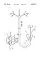
- FIG. 1Ais a side elevation view of a coronary perfusion catheter for use in the method of the present invention.
- FIGS. 1B-1Eare transverse cross sections of the coronary perfusion catheter of FIG. 1A taken through section lines 1B-1E, respectively.
- FIG. 1Fis a transverse cross section of an alternate construction to FIGS. 1C and 1D.
- FIG. 1Gis a transverse cross section of an alternate construction to FIG. 1E.
- FIG. 2Ais a side elevation view of a variation of the first embodiment of the coronary perfusion catheter of FIG. 1 with precurved distal ends.
- FIGS. 2B-2Eare transverse cross sections of the coronary perfusion catheter of FIG. 2A taken through section lines 2B-2E, respectively.
- FIG. 3Ais a side elevation view of a variation of the first embodiment of the coronary perfusion catheter of FIG. 1.
- FIGS. 3B-3Eare transverse cross sections of the coronary perfusion catheter of FIG. 3A taken through section lines 3B-3E, respectively.
- FIG. 4Ais a side elevation view of a second embodiment of a coronary perfusion catheter system for use in the method of the present invention.
- FIGS. 4B-4Eare transverse cross sections of the coronary perfusion catheter of FIG. 4A taken through section lines 4B-4E, respectively.
- FIG. 5Ais a side elevation view of a third embodiment of a coronary perfusion catheter system for use in the method of the present invention.
- FIG. 5Bis a transverse cross sections of the coronary perfusion catheter of FIG. 5A taken through section line 5B.
- FIG. 6shows a fourth embodiment of a coronary perfusion catheter system for use in the method of the present invention.
- FIGS. 7A-7Fshow the tip of the coronary perfusion catheter with six alternative embodiments of the occlusion means for blocking blood flow into the coronary arteries.
- FIG. 8is a diagram of the ascending aorta showing the placement of two coronary perfusion catheters and the left ventricular vent catheter.
- FIG. 9is a schematic diagram of the complete cardioplegia and cardiopulmonary bypass system for carrying out the method of the present invention.
- FIG. 10Ashows a fifth embodiment of a coronary perfusion catheter system for use in the method of the present invention.
- FIG. 10Bis a transverse cross section of the coronary perfusion catheter system of FIG. 10A taken through section lines 10B.
- FIG. 11shows a subselective catheter and coronary artery probe for use with the coronary perfusion catheter system of the present invention.
- FIGS. 1A-1Eillustrate a first embodiment of the coronary perfusion catheter for use in the method of the present invention.
- a single arterial catheter 20is used to deliver cardioplegic solution to all of the coronary arteries.
- a side elevation view of the catheter 20is shown in FIG. 1A.
- the single catheter 20has an elongated shaft 22.
- the catheterincludes a first distal branch 24 for engaging the left coronary ostium, and a second distal branch 26 for engaging the right coronary ostium.
- the catheteris also shown with an optional third distal branch 28 for venting blood from the left ventricle of the heart. All three of the distal branches join together to form a common proximal shaft 22.
- the first distal branch 24 of the catheterhas a first inflatable occlusion balloon cuff 30 at its distal tip.
- the balloon 30communicates with a first balloon inflation lumen 32 which extends through the catheter to a first balloon inflation port 34 on the proximal end of the catheter.
- a first perfusion lumen 36extends from a first perfusion fitting 46 at the proximal end of the catheter, through the common proximal shaft 22 and ends at a perfusion port 38 at the distal tip of the first distal branch 24 distal to the first occlusion balloon 30.
- the first branch 24may also include a first pressure monitoring lumen 40 for monitoring the perfusion pressure in the left coronary artery.
- the pressure lumen 40communicates with a first pressure port 42 near the distal tip of the first branch 24 distal to the occlusion balloon cuff 30 and with a first pressure monitoring fitting 44 at the proximal end of the catheter.
- the perfusion pressurecan be monitored by a pressure transducer, such as a piezoelectric transducer, placed in the distal end of the pressure lumen 40.
- Other sensing devicessuch as a thermocouple temperature transducer, could also be added to the catheter for monitoring other physiological conditions.
- the second distal branch 26 of the catheterhas a second inflatable occlusion balloon cuff 50 at its distal tip.
- the balloon 50communicates with a second balloon inflation lumen 52 which extends through the catheter to a second balloon inflation port 54 on the proximal end of the catheter.
- a second perfusion lumen 56extends from a second perfusion fitting 66 at the proximal end of the catheter, through the common proximal shaft 22 and ends at a perfusion port 58 at the distal tip of the second distal branch 26 distal to the second occlusion balloon 50.
- the second branch 26may also include a second pressure monitoring lumen 60 for monitoring the perfusion pressure in the right coronary artery.
- the pressure lumen 60communicates with a second pressure port 62 near the distal tip of the second branch 26 distal to the occlusion balloon cuff 50 and with a second pressure monitoring fitting 64 at the proximal end of the catheter.
- the first and second perfusion lumens 36, 56can run parallel to one another through the common proximal shaft 22, as illustrated in FIG. 1B, or the two distal perfusion lumens 36, 56 can join to form a single common perfusion lumen in the proximal shaft 22 which connects to a single perfusion fitting at the proximal end.
- the perfusion lumens 36, 56should have sufficient cross sectional area to allow a flow rate of cardioplegic solution of about 100-250 ml/min or greater with a safe perfusion pressure which does not exceed 150 mmHg at the distal ends of the catheter.
- a through lumen with an internal diameter of 1.7 mm or an equivalent areawill deliver 100 ml/min of blood/cardioplegia mixture or 250 ml/min of crystalloid cardioplegic solution at acceptable perfusion pressures.
- the optional third distal branch 28 for venting blood from the left ventricle of the hearthas a single venting lumen 68 which connects to a single venting hole 70 or multiple venting holes at the distal tip of the third distal branch 28.
- the third distal branch 28is shown in cross section in FIG. 1E.
- the venting lumen 68runs the length of the catheter shaft and connects to a venting port 72 at the proximal end of the catheter.
- the third distal branch 28has an atraumatic distal tip 74 which can be safely passed across the aortic valve to vent blood from the left ventricle without damaging the leaflets of the valve.
- the venting tiphas a pigtail curve 74 for crossing the aortic valve.
- atraumatic distal tipexamples include a soft blunt end, a bulbous catheter tip or various other atraumatic catheter curves.
- An alternate construction for the third distal branch 28is shown in cross section in FIG. 1G.
- the third distal branch 28has a second lumen 67 for monitoring the blood pressure in the patient's left ventricle.
- the pressure monitoring lumen 67would connect one or more pressure monitoring ports near the distal end of the third distal branch 28 to a pressure monitoring fitting at the proximal end of the catheter.
- the venting port 72ends in a Luer fitting or other standard catheter fitting for attaching it to a vacuum collection bottle or roller pump.
- Each of the perfusion fittings 46, 66ends in a Luer fitting or other standard catheter fitting for attaching it to a syringe or a perfusion pump.
- the balloon inflation ports 34, 54may have a stopcock, as illustrated, or a simple Luer fitting attached at the proximal end for attachment to a syringe or other balloon inflation device.
- the length and diameter of the catheterare such that they allow the catheter to be introduced via the femoral artery.
- the cathetercan be adapted for introduction from the carotid or brachial artery or from another peripheral arterial access.
- the length of the catheteris preferably 80-120 cm, 90-100 cm being the preferred length for introduction via a femoral artery.
- the diameter of each distal branch 24, 26, 28is preferably 6-7 French (Charriere scale) or 2.0-2.3 mm diameter.
- the diameter of common proximal portion 22 of the catheteris preferably about 12 French (Charriere scale) or 4.0 mm diameter.
- the straight flexible distal branches 24, 26 of the perfusion catheter 20are adapted to be directed into the left and right coronary ostia, respectively, using a steerable guidewire introduced through the perfusion lumens 36, 56 or other known selective catheterization technique.
- FIGS. 2A-2Eshow a variation of the first embodiment of the coronary perfusion catheter with preshaped curves 76, 78 on each of the distal branches 24, 26 to selectively enter one of the coronary ostia.
- the first distal branch 24is illustrated with a Judkins left curve 76 to enter the left coronary ostium
- the second distal branch 26is illustrated with a Judkins right curve 78 to enter the right coronary ostium.
- FIGS. 3A-3Eshow another variation of the first embodiment of the coronary perfusion catheter. Because it is an anatomical variation for humans to sometimes have three significant coronary ostia rather than two, this variation is made with three distal branches 24, 26, 48, each with an inflatable occlusion balloon cuff and a perfusion lumen which connects to the distal tip of the catheter.
- the third distal branch 48is used to selectively intubate the third coronary ostium, which may be a separate origin of the circumflex artery.
- This illustrative exampleis shown without the optional distal branch for venting the left ventricle. When this is the case, a separate ventricular venting catheter or a pulmonary artery venting catheter should be used to vent pressure from within the chambers of the heart.
- FIGS. 4A-4Eshow a second preferred embodiment of the invention which provides a system 80 of separate arterial catheters 82, 84, one for each coronary ostium and, optionally, a separate catheter (not shown) for venting the left ventricle.
- Each of the coronary perfusion catheters 82, 84has an inflatable balloon cuff 86, 88 or other occlusion device at the distal end.
- a perfusion lumen 90, 92exits each catheter distal to the occlusion device.
- each of the perfusion cathetersmay also include a pressure monitoring lumen 94, 95 for monitoring the perfusion pressure in the coronary arteries.
- the distal ends of each of the coronary perfusion catheterscan be preshaped to selectively enter its respective coronary ostium.
- Such selective curves for coronary cathetersare well known in the art and are commonly employed on angiography catheters and angioplasty guiding catheters.
- FIG. 4Athe left coronary perfusion catheter 82 is illustrated with a Judkins left curve
- the right coronary perfusion catheter 84is illustrated with a Judkins right curve.
- Other common selective curvesinclude Amplatz left and right curves, Sones curves, and specialized curves for selectively catheterizing the proximal anastomoses of coronary bypass grafts.
- each of the coronary catheterscan be individually directed into its respective coronary ostium using a steerable guidewire.
- the cathetersare made with the catheter shafts reinforced with wire braid 98, as illustrated in FIGS. 3C and 3E, to give the catheters sufficient torsional rigidity that they can be manipulated into the coronary ostia using selective catheterization techniques.
- the diameter of each catheteris preferably 6-7 French (Charriere scale) or 2.0-2.3 mm diameter.
- Each of the coronary perfusion cathetershas a three lumen shaft, as shown in FIGS. 4B and 4D.
- the balloon inflation lumens 100, 102communicate with the inflatable occlusion balloon cuffs 86, 88 and the perfusion lumens 90, 92 extend to the distal tips of the catheters distal to the occlusion balloons.
- each catheter 4B and 4Dwould include a single or double lumen inner shaft with an external or coaxial balloon lumen tube which communicates with the inflatable occlusion balloon cuff.
- the perfusion lumens of each cathetershould have a sufficient internal diameter to allow a flow rate of cardioplegic solution of about 100-250 ml/min or greater with a safe perfusion pressure which does not exceed 150 mmHg at the distal ends of the catheter.
- a perfusion lumen with an internal diameter of 1.7 mm or an equivalent areawill deliver 100 ml/min of blood/cardioplegia mixture or 250 ml/min of crystalloid cardioplegic solution at acceptable perfusion pressures.
- each of the perfusion cathetersalso includes a third, pressure monitoring lumen 94, 96 for monitoring the perfusion pressure in each of the coronary arteries.
- the cathetersare all introduced through a single arterial sheath 104 placed in the femoral artery or another peripheral artery by arterial cutdown or by the Seldinger technique.
- the arterial sheath 104can be made integral with the arterial cannula of the cardiopulmonary bypass system, as shown in FIG. 4A.
- One or more hemostasis valves 106, 108 on the arterial cannulaallow the coronary perfusion 82, 84 and venting catheters to pass through the blood flow lumen 110 of the arterial cannula 104 without significant leakage of blood.
- FIGS. 5A-5Bshow a third preferred embodiment of the invention which provides a system of individual arterial catheters which are delivered to the ascending aorta through a single common guiding catheter 118, one catheter for each coronary ostium 112, 114 and, optionally, a separate catheter 116 for venting the left ventricle.
- Each of the coronary perfusion catheters 112, 114has an inflatable balloon cuff 120, 122 or other occlusion device at the distal end.
- a perfusion lumenexits each catheter distal to the occlusion device.
- each of the perfusion cathetersmay also include a pressure monitoring lumen for monitoring the perfusion pressure in the coronary arteries.
- the guiding catheter 118is preferably made with a wire braid 124 reinforced shaft 126, as shown in FIG. 5B, to give the system support.
- the distal portion of the shaftis preferably curved to hold the distal end 128 of the catheter in the ascending aorta close to the coronary ostia. Since the individual catheters are supported in the aorta by the common guiding catheter 118, the construction of the individual catheters 112, 114 can be made simpler without reinforcement, as shown in cross section in FIG. 5B.
- FIG. 6shows a fourth preferred embodiment of the invention which provides a system of individual arterial catheters which are delivered to the ascending aorta through a multilumen guiding catheter 130.
- the distal portion 132 of the multilumen guiding catheter 130is curved to hold the distal end of the catheter in the ascending aorta proximate the coronary ostia in the aortic root.
- a first coronary perfusion catheter 140is introduced through a first internal lumen of the multilumen guiding catheter 130.
- the first lumen of the guiding catheterterminates in a first exit port 134 which is angled to direct the perfusion catheter toward the left coronary artery.
- a second coronary perfusion catheter 142is introduced through a second internal lumen of the multilumen guiding catheter 130.
- the second lumen of the guiding catheterterminates in a second exit port 136 which is angled to direct the perfusion catheter toward the right coronary artery.
- Each of the coronary perfusion cathetershas an inflatable balloon cuff 146, 148 or other occlusion device at the distal end.
- a perfusion lumen 150, 152exits each catheter distal to the occlusion device.
- each of the perfusion cathetersmay also include a pressure monitoring lumen for monitoring the perfusion pressure in the coronary arteries.
- a venting catheter 144is introduced through a third internal lumen of the multilumen guiding catheter 130.
- the third lumen of the guiding catheterterminates in a third exit port 138 which is angled to direct the venting catheter 144 across the aortic valve into the left ventricle.
- FIG. 10Ashows a fifth preferred embodiment of the invention which provides a catheter system including a multilumen combination guiding catheter and perfusion catheter 194 and at least one coaxial perfusion catheter 206 delivered through one of the guiding catheter lumens.
- the distal portion 198 of the multilumen guiding catheter 194is curved to direct an occlusion device 200 on the distal tip of the guiding catheter into a first coronary ostium, which in this illustrative example is the left coronary ostium 176.
- the multilumen guiding catheter 194is shown in cross section in FIG. 10B.
- the guiding catheter 194has at least two lumens: a perfusion lumen 202 connecting to the distal tip of the catheter for delivering cardioplegic solution into the first coronary artery, and a guiding lumen 204 for guiding a separate coaxial coronary perfusion catheter 206 to the ascending aorta and into the second coronary ostium 180.
- the guiding catheter 194may also include a separate inflation lumen and/or pressure monitoring lumen, as described in the previous embodiments.
- the coronary perfusion catheter 206is introduced through the guiding lumen 204 of the multilumen guiding catheter 194, and it exits the guiding catheter through a side port 208 which is positioned within the ascending aorta.
- An occlusion device 212 on the distal end of the coronary perfusion catheter 206is directed into the second coronary ostium, in this case the right coronary ostium 180, by an appropriately curved distal end or by using a guidewire.
- the coronary perfusion catheter 206has at least one internal lumen 210, as shown in FIG. 10B, for delivering cardioplegic solution into the second coronary artery.
- the coronary perfusion catheter 206may also include additional lumens for inflation and/or pressure monitoring.
- the coronary perfusion catheter 206can be introduced through the perfusion lumen 202 of the guiding catheter 194, and the coronary perfusion catheter 206 can be adapted to seal against the side port 208 of the guiding catheter 194.
- the distal portion of the coronary perfusion catheter 206can be tapered so that it seals against the side port 208 as it is advanced out of the guiding catheter 194.
- a sealing meanssuch as an O-ring seal can be provided in the side port 208.
- the guiding catheter 194 and the coronary perfusion catheter 206can be perfused separately through their respective lumens, or the flow channels can be joined by placing one or more holes through the sidewall of the coronary perfusion catheter 206 proximal to were it exits the side port 208 so its perfusion lumen 210 communicates with the perfusion lumen 202 of the guiding catheter 194.
- FIG. 11shows a subselective catheter and coronary artery probe 220 that can be used in conjunction with the coronary perfusion catheter system of the present invention.
- the subselective catheter 220is dimensioned so that it can be delivered to the coronary arteries through one of the perfusion lumens 216 of the perfusion catheter system 218.
- the subselective catheter 220can be used for subselectively delivering cardioplegic solution to one of the branches of the coronary arteries or for infusing therapeutic agents, such as thrombolytic drugs, into the coronary arteries while the heart is arrested.
- the distal tip 222 of the subselective catheter 220also serves as a coronary artery probe for helping to locate stenoses or occlusions in the coronary arteries.
- the distal tip 222 of the subselective catheter 220is advanced selectively into the coronary artery and its branches until it is stopped by a blockage in the artery.
- the distal tip 222is configured to have a small bulbous ring 224 which can be palpated through the walls of the coronary artery to help the surgeon to locate the stenoses and to pick the appropriate site for graft anastomosis during bypass surgery.
- the ring 224can be palpated directly by the surgeon with a gloved hand.
- an elongated handheld probe introduced through an access port in the patient's chestcan be used for locating the small bulbous ring 224 on the catheter tip 222.
- the ring 224is made of metal or of plastic with a radiopaque filler so that the distal probe tip 222 of the catheter 220 can also be located fluoroscopically.
- Another use of the coaxially placed subselective catheter and coronary artery probe 220is to create a recirculating heat exchanger circuit within the coronary arteries themselves to increase the effectiveness of the tissue cooling in cold cardioplegia methods.
- one of the cathetersfor example the subselective catheter 220, is used to infuse cold cardioplegic solution into the coronary arteries at a higher rate than would otherwise be necessary to establish cardioplegic arrest.
- the other catheterin this case the perfusion catheter 218, is used to vent the excess cardioplegic fluid so that the pressure in the coronary artery does not exceed the desired perfusion pressure.
- the advantage of this methodis that it increases the flow rate of cold cardioplegic solution which is pumped through the coronary arteries to better cool the cardiac tissue without increasing the perfusion pressure or pumping an excessive amount of cardioplegic solution through the patient's capillary bed.
- This methodwill be especially beneficial in the performance of closed-chest cardiac procedures where it would be inconvenient to bath the entire heart in cold saline solution or to wrap a heat exchanger around the outside of the heart.
- FIGS. 7A-7Fillustrate alternative occlusion means for the distal tip of the coronary perfusion catheters.
- FIG. 7Aillustrates an inflatable occlusion balloon cuff 154 as previously described.
- the occlusion balloon 154is approximately spherical when inflated.
- the maximum diameter of the inflated balloon 154can be about 5 mm, which is sufficient to occlude the coronary ostia 160 in most patients.
- Occlusion balloons as large as 7 or 8 mmmay occasionally be needed for occluding vein grafts which exceed 5 mm internal diameter.
- the balloonis preferably made from an elastomeric material such as latex, silicone or polyurethane or a blend of materials such as polyurethane and polyvinyl chloride.
- the ballooncan be adhesively bonded and/or tied to the catheter shaft. If a thermoplastic elastomer is used as the balloon material, the balloon can be heat bonded directly to a shaft of compatible material without adhesives. Using an elastomeric balloon material allows the deflated balloon to achieve an almost zero deflated profile and it makes the balloons somewhat self deflating from the elastic energy stored in the balloon material on inflation.
- the balloon inflation pressureshould be slightly higher than the maximum perfusion pressure that will be used to prevent migration of the balloon and to prevent cardioplegic solution or systemic blood from leaking past the inflated balloon. At the same time, the inflation pressure should be low enough that the inflated balloon does not inadvertently dilate the coronary ostia.
- An inflation pressure of 350 mmHghas been shown to be effective.
- the inflatable occlusion balloon cuffoccludes the coronary artery very gently with very little danger of damaging the ostium.
- Additional features, such as ribs or bumps,may be molded into the surface of the balloon to increase the friction with the coronary ostia to prevent slippage of the balloon without increasing the inflation pressure.
- FIG. 7Bshows a coronary perfusion catheter tip 156 with a conically tapered occlusion means 158 at the distal tip.
- This conical occlusion means 158is wedged into the coronary ostium 160 to occlude the flow around the catheter tip.
- FIG. 7Cshows a coronary perfusion catheter tip with an O-ring occlusion 162 means at the distal tip.
- the O-ring occlusion means 162is, likewise, wedged into the coronary ostium 160 to occlude the flow around the catheter tip.
- FIG. 7Dshows a coronary perfusion catheter tip with a wedge-shaped occlusion means 164 at the distal tip.
- the wedge 164is sized to occlude the coronary ostium 160 and isolate the coronary artery from the systemic blood flow.
- the wedge 164can be made from a solid or hollow, molded plastic or elastomeric material, or the wedge can be formed as a shaped occlusion balloon which achieves the wedge shape when inflated.
- FIGS. 7E and 7Fshow reverse wedge-shaped occlusion balloons 166, 170.
- the balloons 166, 170are preferably formed as shaped occlusion balloons which achieve the reverse wedge shape when inflated.
- FIG. 7Eillustrates a variation of the occlusion balloon 166 which is self-inflating.
- cardioplegic solutionWhen cardioplegic solution is infused through the perfusion lumen of the catheter, it first enters the occlusion balloon 166 which is bonded near the distal end of the catheter. The pressure of the cardioplegic solution inflates the balloon 166 and seals the sides of the balloon against the walls of the coronary ostium 160. The cardioplegic solution exits the occlusion balloon 166 through a distal orifice 168 into the coronary artery. The distal orifice 168 offers a slight resistance to the flow of the cardioplegic solution which pressurizes the occlusion balloon 166 and keeps it inflated as long as there is sufficient flow of cardioplegic solution through the catheter.
- FIGS. 7B-7EFor the embodiments of the occlusion means shown, in FIGS. 7B-7E, that do not require an inflation lumen the construction of the perfusion catheter can be much simpler than for the occlusion balloon embodiments previously described.
- a cross section of one possible embodiment of the shaft constructionis shown in FIG. 1F.
- the perfusion catheters (or the distal branches of a single perfusion catheter) 24have a perfusion lumen 36 and a pressure monitoring lumen 40 which connect the proximal and distal ends of the catheter. If other means are provided to monitor and control the perfusion pressure in the coronary arteries, the catheters can be further simplified with a single perfusion lumen from end to end, which could lower the overall cost of the catheter system and the overall diameter of the catheters.
- the catheter system of the present inventionis preferably supplied to the end user in a sterile ready to use condition.
- all of the components necessary for carrying out the proceduremay be packaged together in a single kit sterilized and ready for use.
- FIG. 8is a diagram of the ascending aorta 172, showing the placement of the coronary perfusion catheters 174, 178 and the left ventricular venting catheter 184.
- a first coronary perfusion catheter 174engages the left coronary ostium 176
- a second coronary perfusion catheter 178engages the right coronary ostium 180.
- Placement of the cathetersis generally done under fluoroscopic guidance.
- the cathetercan be provided with one or more radiopaque marker rings of a dense material, such as gold, platinum, tantalum or tungsten, at their distal tips.
- the catheter shaftsmay be made of a polymer compounded with a radiopaque material, such as barium or bismuth compounds, to increase the radiopacity.
- the perfusion catheters 174, 178can be placed with fluoroscopic guidance by either of two methods.
- the distal ends of the cathetersare preshaped with selective coronary curves.
- the curvesare straightened out with a stiff guidewire placed in the perfusion lumen, as shown by phantom lines 82', 84' in FIG. 4A, when the catheters are introduced into the femoral artery and advanced through the descending aorta and into the ascending aorta.
- the guidewiresare withdrawn the catheters resume their curved shapes which are adapted to be easily maneuvered into the coronary ostia.
- the distal ends of the cathetersare not precurved, but a curved steerable guidewire is used to direct each of the catheters into the respective coronary ostium.
- the curved guiding cathetercan assist the steerable guidewire in directing the perfusion catheters to their respective coronary ostia.
- the lumens of the guiding cathetercan be adapted to direct the perfusion catheters into the ostia, as illustrated in FIG. 6.
- the perfusion cathetersshould be inserted far enough into the coronary ostia so that the balloons will be entirely within the coronary arteries when they are inflated.
- the balloonsmust be positioned upstream of any side branches of the coronary arteries to insure complete isolation of the myocardium from the systemic circulation and complete perfusion of the coronary arteries with cardioplegic solution.
- Positioning the tip of each catheter about 3 to 5 mm downstream of the coronary ostium prior to inflationwill result in correct balloon placement in most cases.
- the occlusion balloonsare preferably inflated with sterile saline solution, or with a mixture of saline and a radiopaque contrast agent, to eliminate any danger of air embolization in the occurrence of a balloon leak or rupture.
- the positions of the occlusion balloonsshould be verified fluoroscopically after inflation.
- FIG. 9is a schematic diagram of the complete cardioplegia and cardiopulmonary bypass system for carrying out the method of the present invention.
- the patientis prepared for cardiopulmonary bypass by placing the arterial cannula 188 in a peripheral artery, such as the femoral artery and venous cannula 192 in a peripheral vein, such as the femoral vein, and connecting them to the cardiopulmonary bypass system 190.
- a femoral-to-femoral cardiopulmonary bypass system or other minimally invasive systemis preferred to reduce the necessity for invasive surgical access to the heart. Examples of suitable femoral-to-femoral cardiopulmonary bypass systems can be found in U.S. Pat. Nos.
- the coronary perfusion catheter or catheters 174, 178are introduced into a peripheral arterial access site, such as a femoral artery, by arterial cutdown or by the Seldinger technique, then directed to the coronary ostia using one of the various techniques described above.
- the optional venting catheter 184should also be placed across the aortic valve 182 at this time.
- the systemis used to administer cold cardioplegia to the patient for maximal protection of the myocardium.
- the cardioplegic fluidpreferably consists of an aqueous KCl solution mixed with oxygenated blood at a ratio of four parts blood to one part KCl solution.
- the aqueous KCl solutionconsists of crystalloid KCl mixed with saline to have a concentration in the range of 10-50 mEq K + /liter, preferably 15-30 mEq K + /liter.
- an aqueous KCl solution with a concentration in the range of 10-30 mEq K + /liter, without a blood componentmay be used.
- the occlusion balloon cuffsare inflated in each of the coronary ostia and immediately oxygenated cold cardioplegic solution, typically between 3° C. and 10° C., is pumped through each of the perfusion lumens at a rate of approximately 100-200 ml/min in each coronary artery.
- the perfusion pressure in the coronary arteriescan be monitored through the pressure monitoring lumens to be sure that the perfusion pressure does not exceed 150 mmHg at the distal ends of the catheters.
- the pump pressureshould not exceed 350 mmHg to avoid undue hemolysis in the blood component of the fluid mixture. If a cardioplegic solution without any blood component is used, a higher pump pressure may be used.
- the actual perfusion pressure in the coronary arteriesshould not be allowed to exceed 150 mmHg to avoid endothelial damage to the arterial walls.
- Cardioplegic arrestoccurs almost immediate when the cardioplegic solution is infused.
- the venting cathetercan be vented by gravity or a negative pressure can be applied to the vent lumen with a syringe or pump to depressurize the left ventricle.
- the venting cathetercan be connected to a blood recovery unit which reprocesses the blood and adds it to the bypass circuit.
- An initial bolus totaling 500-1000 ml of cardioplegic solutionis infused into the coronary arteries.
- the flow ratecan be decreased to a constant 25-50 ml/min to maintain the cardioplegic arrest and the hypothermia of the myocardium.
- the coronary arteriescan be periodically reinfused with oxygenated cold cardioplegic solution to avoid either oxygen depletion or premature restarting of the heart.
- the occlusion balloon cuffsmust be kept inflated for the duration of the surgical procedure so that systemic blood does not enter the coronary arteries. If this happens the heart could start beating prematurely, which would interfere with the surgical procedure.
- the method of the present inventioncan be used in conjunction with retrograde perfusion through the coronary sinus to maintain hypothermia and cardioplegic arrest.
- the initial bolus of cardioplegiais infused through the coronary perfusion catheters into the coronary arteries.
- the balloon of a retrograde perfusion catheteris inflated in the coronary sinus and further cardioplegic solution is perfused retrograde through the coronary circulation at a lower flow rate.
- the occlusion balloon cuffsare kept inflated to isolate the coronary arteries from the systemic blood circulation, and the cardioplegic solution is vented from the coronary arteries through the perfusion lumens of the coronary perfusion catheters.
- the cardiac surgeryis performed during the period of cardioplegic arrest using minimally invasive techniques through thoracoscopic or transluminal access to the heart.
- the methodcan also be used in conjunction with traditional open chest surgical techniques when there is a contraindication to standard cardioplegia administration techniques, such as significant aortic valve insufficiency or calcification, scarring or adhesions of the ascending aorta.
- the coronary perfusion catheters of the cardioplegia systemcan be used as guiding catheters for performing catheter based interventional procedures in the coronary arteries of the patient.
- catheter based interventional procedurescan be performed by inserting the interventional catheters through the perfusion lumens of the coronary perfusion catheters into the coronary arteries.
- Catheter based interventional procedurescan thus be combined with minimally invasive cardiac surgery to improve patient outcome.
- the coronary perfusion catheters of the cardioplegia system and the method of administering cardioplegiacan be used in conjunction with a laser system for transmyocardial revascularization such as any of the systems described in U.S. Pat. Nos. 4,658,817, 5,125,924 and 5,125,926, the specifications of which are hereby incorporated by reference in their entirety.
- the laser devicescan be introduced thoracoscopically or transluminally to maintain the minimally-invasive character of the procedure. The use of these systems in conjunction with the present system of achieving cardioplegic arrest solves the difficulties of applying laser energy to the beating heart described in each of the patents.
- performing transmyocardial revascularization on a still heartinsures that the laser energy will be applied to the myocardium when it is in the relaxed state, which has been shown to yield better results. It also avoids the possible complication of inducing ventricular fibrillation which can occur when laser energy is applied to the myocardium while it is contracting during systole.
- the laser induced channelscan be more carefully placed while the heart is still so that the ischemic myocardium can be more effectively revascularized and critical structures, such as nerves or coronary arteries, can be carefully avoided with the laser.
- the systemic circulationcan be rewarmed and the occlusion balloon cuffs deflated and withdrawn from the coronary ostia.
- a final bolus of oxygenated normothermic blood mixed with cardioplegic solutioncan be pumped through the perfusion catheters before the occlusion balloon cuffs are deflated to wash out the coronary arteries and prevent reperfusion injury according to the method described by Buckberg et al.
- the heartshould resume beating spontaneously. If the heart does not restart spontaneously or if it begins beating irregularly, a defibrillation pulse can be applied to the heart to start it beating correctly.
- the perfusion catheters, vent catheter and bypass cannul.ae butted.can now be withdrawn and the cutdowns or percutaneous punctures closed.
- the methods and devices described hereinprovide a system for inducing cardioplegic arrest and total cardiopulmonary support entirely through minimally invasive transluminal access.
- the present inventiontherefore provides an important link in making minimally invasive cardiac surgery a real possibility.
Landscapes
- Health & Medical Sciences (AREA)
- Life Sciences & Earth Sciences (AREA)
- Heart & Thoracic Surgery (AREA)
- Animal Behavior & Ethology (AREA)
- Veterinary Medicine (AREA)
- Public Health (AREA)
- Engineering & Computer Science (AREA)
- Biomedical Technology (AREA)
- General Health & Medical Sciences (AREA)
- Anesthesiology (AREA)
- Hematology (AREA)
- Vascular Medicine (AREA)
- Cardiology (AREA)
- Pulmonology (AREA)
- Biophysics (AREA)
- Transplantation (AREA)
- Oral & Maxillofacial Surgery (AREA)
- Child & Adolescent Psychology (AREA)
- Media Introduction/Drainage Providing Device (AREA)
- Surgical Instruments (AREA)
- Ultra Sonic Daignosis Equipment (AREA)
- Radiation-Therapy Devices (AREA)
- Infusion, Injection, And Reservoir Apparatuses (AREA)
Abstract
Description
Claims (23)
Priority Applications (11)
| Application Number | Priority Date | Filing Date | Title |
|---|---|---|---|
| US08/351,850US5695457A (en) | 1994-07-28 | 1994-12-07 | Cardioplegia catheter system |
| JP51760396AJP3650122B2 (en) | 1994-12-07 | 1995-11-21 | Cardiac arrest catheter system |
| PCT/US1995/015014WO1996017644A1 (en) | 1994-12-07 | 1995-11-21 | Cardioplegia catheter system |
| ES95941419TES2217288T3 (en) | 1994-12-07 | 1995-11-21 | CARDIOPLEGIA CATHETER SYSTEM. |
| CA002206091ACA2206091A1 (en) | 1994-12-07 | 1995-11-21 | Cardioplegia catheter system |
| AT95941419TATE261329T1 (en) | 1994-12-07 | 1995-11-21 | CARDIOPLEGIA CATHETER SYSTEM |
| DE69532686TDE69532686T2 (en) | 1994-12-07 | 1995-11-21 | Cardioplegia CATHETER SYSTEM |
| AU42847/96AAU696812B2 (en) | 1994-12-07 | 1995-11-21 | Cardioplegia catheter system |
| EP95941419AEP0841963B1 (en) | 1994-12-07 | 1995-11-21 | Cardioplegia catheter system |
| US08/615,152US5807318A (en) | 1994-07-28 | 1996-03-12 | Method of perfusing the coronary vasculature |
| US09/863,135US6913601B2 (en) | 1994-12-07 | 2001-05-22 | Method for delivering a fluid to the coronary ostia |
Applications Claiming Priority (2)
| Application Number | Priority Date | Filing Date | Title |
|---|---|---|---|
| US08/282,192US5584803A (en) | 1991-07-16 | 1994-07-28 | System for cardiac procedures |
| US08/351,850US5695457A (en) | 1994-07-28 | 1994-12-07 | Cardioplegia catheter system |
Related Parent Applications (1)
| Application Number | Title | Priority Date | Filing Date |
|---|---|---|---|
| US08/282,192Continuation-In-PartUS5584803A (en) | 1991-07-16 | 1994-07-28 | System for cardiac procedures |
Related Child Applications (1)
| Application Number | Title | Priority Date | Filing Date |
|---|---|---|---|
| US08/615,152DivisionUS5807318A (en) | 1994-07-28 | 1996-03-12 | Method of perfusing the coronary vasculature |
Publications (1)
| Publication Number | Publication Date |
|---|---|
| US5695457Atrue US5695457A (en) | 1997-12-09 |
Family
ID=23382681
Family Applications (3)
| Application Number | Title | Priority Date | Filing Date |
|---|---|---|---|
| US08/351,850Expired - LifetimeUS5695457A (en) | 1994-07-28 | 1994-12-07 | Cardioplegia catheter system |
| US08/615,152Expired - LifetimeUS5807318A (en) | 1994-07-28 | 1996-03-12 | Method of perfusing the coronary vasculature |
| US09/863,135Expired - Fee RelatedUS6913601B2 (en) | 1994-12-07 | 2001-05-22 | Method for delivering a fluid to the coronary ostia |
Family Applications After (2)
| Application Number | Title | Priority Date | Filing Date |
|---|---|---|---|
| US08/615,152Expired - LifetimeUS5807318A (en) | 1994-07-28 | 1996-03-12 | Method of perfusing the coronary vasculature |
| US09/863,135Expired - Fee RelatedUS6913601B2 (en) | 1994-12-07 | 2001-05-22 | Method for delivering a fluid to the coronary ostia |
Country Status (9)
| Country | Link |
|---|---|
| US (3) | US5695457A (en) |
| EP (1) | EP0841963B1 (en) |
| JP (1) | JP3650122B2 (en) |
| AT (1) | ATE261329T1 (en) |
| AU (1) | AU696812B2 (en) |
| CA (1) | CA2206091A1 (en) |
| DE (1) | DE69532686T2 (en) |
| ES (1) | ES2217288T3 (en) |
| WO (1) | WO1996017644A1 (en) |
Cited By (113)
| Publication number | Priority date | Publication date | Assignee | Title |
|---|---|---|---|---|
| US5800414A (en)* | 1996-10-18 | 1998-09-01 | Synthelabo | Catheter with flexible and elongate body |
| US5873366A (en)* | 1996-11-07 | 1999-02-23 | Chim; Nicholas | Method for transmyocardial revascularization |
| US5911702A (en)* | 1997-11-06 | 1999-06-15 | Heartport, Inc. | Methods and devices for cannulating a patient's blood vessel |
| WO1999035979A1 (en)* | 1998-01-13 | 1999-07-22 | Lumend, Inc. | Methods and apparatus for crossing total occlusions in blood vessels |
| US6077256A (en)* | 1998-10-06 | 2000-06-20 | Mann; Michael J. | Delivery of a composition to the lung |
| US6090096A (en)* | 1997-04-23 | 2000-07-18 | Heartport, Inc. | Antegrade cardioplegia catheter and method |
| US6149670A (en)* | 1999-03-11 | 2000-11-21 | Alsius Corporation | Method and system for treating cardiac arrest using hypothermia |
| US6152141A (en)* | 1994-07-28 | 2000-11-28 | Heartport, Inc. | Method for delivery of therapeutic agents to the heart |
| US6179766B1 (en)* | 1999-01-28 | 2001-01-30 | Gregg A. Dickerson | Methods of breast cancer treatment |
| US6203759B1 (en) | 1996-05-31 | 2001-03-20 | Packard Instrument Company | Microvolume liquid handling system |
| US6210363B1 (en) | 1999-02-23 | 2001-04-03 | Cardeon Corporation | Methods and devices for occluding a vessel and performing differential perfusion |
| US6210365B1 (en) | 1998-08-14 | 2001-04-03 | Cardiovention, Inc. | Perfusion catheter system having sutureless arteriotomy seal and methods of use |
| US6217527B1 (en) | 1998-09-30 | 2001-04-17 | Lumend, Inc. | Methods and apparatus for crossing vascular occlusions |
| US6221059B1 (en)* | 1996-06-21 | 2001-04-24 | Medtronic, Inc. | Flow-directed catheter system and method of use |
| US6241699B1 (en)* | 1998-07-22 | 2001-06-05 | Chase Medical, Inc. | Catheter system and method for posterior epicardial revascularization and intracardiac surgery on a beating heart |
| US6364868B1 (en)* | 1995-08-02 | 2002-04-02 | The Trustees Of Columbia University In The City Of New York | Ureteral catheter and tissue expander and method of megaureter creation |
| US6393320B2 (en) | 1999-02-19 | 2002-05-21 | Alsius Corporation | Method for treating cardiac arrest |
| WO2001082996A3 (en)* | 2000-04-28 | 2002-05-23 | Cirque Medical Inc | Multi-lumen intravenous extension |
| US20020099254A1 (en)* | 2001-01-23 | 2002-07-25 | Movahed M. Reza | Method and apparatus to remove substances from vessels of the heart and other parts of the body to minimize or aviod renal or other harm or dysfunction |
| US6432124B1 (en) | 1999-03-11 | 2002-08-13 | Alsius Corporation | Method and system treating heart malady such as cardiac arrest and heart attack using hypothermia |
| WO2002062236A1 (en)* | 2001-02-06 | 2002-08-15 | Edwards Lifesciences Corporation | Method and system for tissue repair using dual catheters |
| US6461336B1 (en)* | 2000-02-08 | 2002-10-08 | LARRé JORGE CASADO | Cardiological medical equipment |
| US6460544B1 (en) | 1999-03-11 | 2002-10-08 | Alsius Corporation | Method and apparatus for establishing and maintaining therapeutic hypothemia |
| US6482171B1 (en)* | 1991-07-16 | 2002-11-19 | Heartport, Inc. | Multi-lumen catheter |
| US6508777B1 (en) | 1998-05-08 | 2003-01-21 | Cardeon Corporation | Circulatory support system and method of use for isolated segmental perfusion |
| US6514226B1 (en) | 2000-02-10 | 2003-02-04 | Chf Solutions, Inc. | Method and apparatus for treatment of congestive heart failure by improving perfusion of the kidney |
| US6521187B1 (en) | 1996-05-31 | 2003-02-18 | Packard Instrument Company | Dispensing liquid drops onto porous brittle substrates |
| US6537817B1 (en) | 1993-05-31 | 2003-03-25 | Packard Instrument Company | Piezoelectric-drop-on-demand technology |
| US6554790B1 (en) | 1998-11-20 | 2003-04-29 | Intuitive Surgical, Inc. | Cardiopulmonary bypass device and method |
| US6572640B1 (en) | 2001-11-21 | 2003-06-03 | Alsius Corporation | Method and apparatus for cardiopulmonary bypass patient temperature control |
| US20030153946A1 (en)* | 2000-08-11 | 2003-08-14 | Edwards Lifesciences Ag | Device and method for treatment of atrioventricular regurgitation |
| US20030187467A1 (en)* | 2002-03-26 | 2003-10-02 | Stefan Schreck | Sequential heart valve leaflet repair device and method of use |
| US20030199805A1 (en)* | 2002-04-23 | 2003-10-23 | Mcweeney John O. | Drainage devices and methods |
| US6669674B1 (en)* | 1997-09-26 | 2003-12-30 | Cardeon Corporation | Introducer and perfusion cannula |
| US6673040B1 (en) | 1996-04-16 | 2004-01-06 | Cardeon Corporation | System and methods for catheter procedures with circulatory support in high risk patients |
| US6682551B1 (en) | 1999-03-11 | 2004-01-27 | Alsius Corporation | Method and system for treating cardiac arrest using hypothermia |
| US20040044365A1 (en)* | 2002-09-03 | 2004-03-04 | Bachman Alan B. | Single catheter mitral valve repair device and method for use |
| US6702773B1 (en)* | 2000-02-01 | 2004-03-09 | Cardeon Corporation | Methods and devices for performing differential perfusion |
| US6726651B1 (en) | 1999-08-04 | 2004-04-27 | Cardeon Corporation | Method and apparatus for differentially perfusing a patient during cardiopulmonary bypass |
| US6726710B2 (en) | 1999-08-16 | 2004-04-27 | Alsius Corporation | Method and system for treating cardiac arrest using hypothermia |
| US20040093023A1 (en)* | 1999-10-21 | 2004-05-13 | Allen William J. | Minimally invasive mitral valve repair method and apparatus |
| EP1105173A4 (en)* | 1998-08-27 | 2004-07-07 | A Med Systems Inc | Intravascular cannulation apparatus and methods of use |
| US20040159360A1 (en)* | 2001-03-16 | 2004-08-19 | Navis John A. | Peritoneal dialysis catheter |
| US20040181238A1 (en)* | 2003-03-14 | 2004-09-16 | David Zarbatany | Mitral valve repair system and method for use |
| US20040255956A1 (en)* | 2001-12-21 | 2004-12-23 | Jakob Vinten-Johansen | Post-conditioning for the reduction of ischemic-reperfusion injury in the heart and other organs |
| US6951555B1 (en) | 1998-03-16 | 2005-10-04 | Chase Medical, L.P. | Catheter having integral expandable/collapsible lumen |
| US20050234396A1 (en)* | 2004-04-20 | 2005-10-20 | Forsberg Andrew T | Method and apparatus for locating vascular punctures |
| US6969379B1 (en) | 1998-08-27 | 2005-11-29 | A-Med Systems, Inc. | Intravascular cannulation apparatus and methods of use |
| US6984238B2 (en) | 1995-02-24 | 2006-01-10 | Gifford Iii Hanson S | Devices and methods for performing avascular anastomosis |
| US7004173B2 (en) | 2000-12-05 | 2006-02-28 | Lumend, Inc. | Catheter system for vascular re-entry from a sub-intimal space |
| US20060064056A1 (en)* | 2004-09-17 | 2006-03-23 | James Coyle | Guiding catheter assembly for embolic protection by proximal occlusion |
| US20060074399A1 (en)* | 2004-10-05 | 2006-04-06 | Bates Mark C | Methods and apparatus for treating infarcted regions of tissue following acute myocardial infarction |
| US7087066B2 (en) | 1995-02-24 | 2006-08-08 | Bolduc Lee R | Surgical clips and methods for tissue approximation |
| US20060178622A1 (en)* | 2002-10-29 | 2006-08-10 | Constantz Brent R | Devices and methods for treating aortic valve stenosis |
| US20060206053A1 (en)* | 2005-03-10 | 2006-09-14 | Lary G B | Catheter for treatment of severe pulmonary emboli |
| US20060217660A1 (en)* | 2005-03-10 | 2006-09-28 | Lary Banning G | Catheter with semipermeable membrane for treatment of severe pulmonary emboli |
| USRE39451E1 (en) | 2000-06-08 | 2006-12-26 | Spire Biomedical, Inc. | Double-lumen catheter |
| US20070041960A1 (en)* | 2004-01-27 | 2007-02-22 | Ramot At Tel-Aviv University Ltd., Enzysurge Ltd. | Apparatus and methods for enzymatic debridement of skin lesions |
| US20070112369A1 (en)* | 2005-11-05 | 2007-05-17 | Crossman Arthur W | Expandable component guide wire system and related method of using the same |
| US20070160645A1 (en)* | 2001-10-25 | 2007-07-12 | Jakob Vinten-Johansen | PostConditioning System And Method For The Reduction Of Ischemic-Reperfusion Injury In The Heart And Other Organs |
| US20080065156A1 (en)* | 2006-09-08 | 2008-03-13 | Hauser David L | Expandable clip for tissue repair |
| US20080097385A1 (en)* | 2004-12-22 | 2008-04-24 | Jakob Vinten-Johansen | Therapeutic Adjuncts to Enhance the Organ Protective Effects of Postconditioning |
| US7393339B2 (en) | 2003-02-21 | 2008-07-01 | C. R. Bard, Inc. | Multi-lumen catheter with separate distal tips |
| US20080249420A1 (en)* | 2004-10-14 | 2008-10-09 | Crossman Arthur W | Vascular Catheter Device and Related Methods of Using the Same |
| US20090093801A1 (en)* | 2005-12-24 | 2009-04-09 | Crossman Arthur W | Circumferential ablation guide wire system and related method of using the same |
| US20090137982A1 (en)* | 2005-04-21 | 2009-05-28 | Medtronic Vascular, Inc. | Guiding Catheter with Resiliently Compressible Occluder |
| USRE40913E1 (en) | 2000-06-01 | 2009-09-08 | Medical Components, Inc. | Multilumen catheter assembly and methods for making and inserting the same |
| US20090254161A1 (en)* | 1999-12-14 | 2009-10-08 | Zoll Circulation, Inc. | Method for reducing myocardial infarct by application of intravascular hypothermia |
| US7651510B2 (en) | 1998-01-23 | 2010-01-26 | Heartport, Inc. | System for performing vascular anastomoses |
| US7654264B2 (en) | 2006-07-18 | 2010-02-02 | Nellcor Puritan Bennett Llc | Medical tube including an inflatable cuff having a notched collar |
| US7686781B2 (en) | 2001-10-25 | 2010-03-30 | Emory University | Catheter for modified perfusion |
| US7850676B2 (en) | 2004-04-19 | 2010-12-14 | The Invention Science Fund I, Llc | System with a reservoir for perfusion management |
| US7857767B2 (en) | 2004-04-19 | 2010-12-28 | Invention Science Fund I, Llc | Lumen-traveling device |
| US7879023B2 (en) | 2004-04-19 | 2011-02-01 | The Invention Science Fund I, Llc | System for perfusion management |
| US20110112625A1 (en)* | 2000-03-27 | 2011-05-12 | Neovasc Medical Ltd. | Varying diameter vascular implant and balloon |
| US7981093B2 (en) | 2000-06-01 | 2011-07-19 | Medical Components, Inc. | Methods of making a multilumen catheter assembly |
| US7998060B2 (en) | 2004-04-19 | 2011-08-16 | The Invention Science Fund I, Llc | Lumen-traveling delivery device |
| US8019413B2 (en) | 2007-03-19 | 2011-09-13 | The Invention Science Fund I, Llc | Lumen-traveling biological interface device and method of use |
| US8021321B2 (en) | 2002-02-07 | 2011-09-20 | C. R. Bard, Inc. | Split tip dialysis catheter |
| US8066660B2 (en) | 2007-10-26 | 2011-11-29 | C. R. Bard, Inc. | Split-tip catheter including lateral distal openings |
| US8092415B2 (en) | 2007-11-01 | 2012-01-10 | C. R. Bard, Inc. | Catheter assembly including triple lumen tip |
| US8092549B2 (en) | 2004-09-24 | 2012-01-10 | The Invention Science Fund I, Llc | Ciliated stent-like-system |
| US8206371B2 (en) | 2003-05-27 | 2012-06-26 | Bard Access Systems, Inc. | Methods and apparatus for inserting multi-lumen split-tip catheters into a blood vessel |
| US20120172838A1 (en)* | 1998-03-25 | 2012-07-05 | Medtronic Vascular, Inc. | Systems and Methods for Delivering Drugs to Selected Locations Within the Body |
| US8292841B2 (en) | 2007-10-26 | 2012-10-23 | C. R. Bard, Inc. | Solid-body catheter including lateral distal openings |
| US8353896B2 (en) | 2004-04-19 | 2013-01-15 | The Invention Science Fund I, Llc | Controllable release nasal system |
| US20130023852A1 (en)* | 2011-07-22 | 2013-01-24 | William Joseph Drasler | Flow Protection Device |
| US8361014B2 (en)* | 2004-04-19 | 2013-01-29 | The Invention Science Fund I, Llc | Telescoping perfusion management system |
| US8394218B2 (en) | 2009-07-20 | 2013-03-12 | Covidien Lp | Method for making a multi-lumen catheter having a separated tip section |
| US20130172677A1 (en)* | 2011-12-29 | 2013-07-04 | Cook Medical Technologies Llc | Space-optimized visualization catheter with camera train holder |
| US8500939B2 (en) | 2007-10-17 | 2013-08-06 | Bard Access Systems, Inc. | Manufacture of split tip catheters |
| US8512219B2 (en) | 2004-04-19 | 2013-08-20 | The Invention Science Fund I, Llc | Bioelectromagnetic interface system |
| US20130267959A1 (en)* | 2001-06-14 | 2013-10-10 | Alexandria Research Technologies, Inc. | Modular apparatus and method for sculpting the surface of a joint |
| US20130281979A1 (en)* | 2012-04-18 | 2013-10-24 | Edwards Lifesciences Llc | Retrograde cardioplegia delivery catheter and method for inducing cardioplegic arrest |
| US20140135786A1 (en)* | 2012-11-09 | 2014-05-15 | Naris Llc | Medical procedure access kit |
| US8753373B2 (en) | 2007-05-08 | 2014-06-17 | Edwards Lifesciences Corporation | Suture-fastening clip |
| US8876754B2 (en) | 2006-08-31 | 2014-11-04 | Bayer Medical Care Inc. | Catheter with filtering and sensing elements |
| US8992454B2 (en) | 2004-06-09 | 2015-03-31 | Bard Access Systems, Inc. | Splitable tip catheter with bioresorbable adhesive |
| US9011329B2 (en) | 2004-04-19 | 2015-04-21 | Searete Llc | Lumenally-active device |
| US20150119853A1 (en)* | 2013-10-29 | 2015-04-30 | Medwerks, Llc | Convertible Shape Catheter And Method Of Use |
| US9180274B2 (en) | 2010-09-09 | 2015-11-10 | W. L. G ore & Associates, Inc | Indwelling luminal devices |
| US9198563B2 (en) | 2006-04-12 | 2015-12-01 | The Invention Science Fund I, Llc | Temporal control of a lumen traveling device in a body tube tree |
| US20160008531A1 (en)* | 2009-08-11 | 2016-01-14 | W-Z Biotech, Llc | Dual lumen cannula for artificial lung and right ventricular assist device |
| USD748252S1 (en) | 2013-02-08 | 2016-01-26 | C. R. Bard, Inc. | Multi-lumen catheter tip |
| US9579485B2 (en) | 2007-11-01 | 2017-02-28 | C. R. Bard, Inc. | Catheter assembly including a multi-lumen configuration |
| US9668643B2 (en) | 2011-12-29 | 2017-06-06 | Cook Medical Technologies Llc | Space-optimized visualization catheter with oblong shape |
| US9849229B2 (en) | 2014-04-25 | 2017-12-26 | Covidien Lp | Split-tip catheter |
| US9895515B2 (en) | 2013-03-15 | 2018-02-20 | Corvivo Inc. | Perfusion catheter system for simultaneous delivery of cardioplegia to the left and right coronary artery |
| US10258768B2 (en) | 2014-07-14 | 2019-04-16 | C. R. Bard, Inc. | Apparatuses, systems, and methods for inserting catheters having enhanced stiffening and guiding features |
| US10517673B2 (en)* | 2014-10-29 | 2019-12-31 | The Spectranetics Corporation | Laser energy delivery devices including laser transmission detection systems and methods |
| WO2020107035A1 (en)* | 2018-11-25 | 2020-05-28 | Patient Shield Concepts, Llc | Catheter for fluid drainage and medication delivery from/to a patient |
| US20220001163A1 (en)* | 2014-01-14 | 2022-01-06 | CardioFlow Technologies, LLC | Apparatus and methods for optimizing intra cardiac filling pressures, heart rate, and cardiac output |
| US20220305184A1 (en)* | 2019-08-22 | 2022-09-29 | Istanbul Medipol Universitesi | Multiple perfusion cannula system for use in aortic surgery |
Families Citing this family (128)
| Publication number | Priority date | Publication date | Assignee | Title |
|---|---|---|---|---|
| US5843093A (en)* | 1994-02-09 | 1998-12-01 | University Of Iowa Research Foundation | Stereotactic electrode assembly |
| US6182664B1 (en) | 1996-02-19 | 2001-02-06 | Edwards Lifesciences Corporation | Minimally invasive cardiac valve surgery procedure |
| US6026814A (en) | 1997-03-06 | 2000-02-22 | Scimed Life Systems, Inc. | System and method for percutaneous coronary artery bypass |
| US6217546B1 (en) | 1997-05-19 | 2001-04-17 | United States Surgical Corporation | Catheter system |
| WO1999004845A2 (en)* | 1997-07-22 | 1999-02-04 | Chase Medical Inc. | Catheter having a lumen occluding balloon and method of use thereof |
| US6889082B2 (en) | 1997-10-09 | 2005-05-03 | Orqis Medical Corporation | Implantable heart assist system and method of applying same |
| US6491039B1 (en) | 1998-01-23 | 2002-12-10 | Innercool Therapies, Inc. | Medical procedure |
| US7371254B2 (en) | 1998-01-23 | 2008-05-13 | Innercool Therapies, Inc. | Medical procedure |
| US6129713A (en)* | 1998-08-11 | 2000-10-10 | Embol-X, Inc. | Slidable cannula and method of use |
| US6325813B1 (en) | 1998-08-18 | 2001-12-04 | Scimed Life Systems, Inc. | Method and apparatus for stabilizing vascular wall |
| US6383172B1 (en)* | 1999-04-02 | 2002-05-07 | Coaxia, Inc. | Retrograde venous perfusion with isolation of cerebral circulation |
| US7951071B2 (en) | 1999-06-02 | 2011-05-31 | Tyco Healthcare Group Lp | Moisture-detecting shaft for use with an electro-mechanical surgical device |
| US8025199B2 (en) | 2004-02-23 | 2011-09-27 | Tyco Healthcare Group Lp | Surgical cutting and stapling device |
| US6793652B1 (en) | 1999-06-02 | 2004-09-21 | Power Medical Interventions, Inc. | Electro-mechanical surgical device |
| US6962601B2 (en) | 1999-08-02 | 2005-11-08 | University Of Chicago Office Of Technology Transfer | Method for inducing hypothermia |
| US7422601B2 (en)* | 1999-08-02 | 2008-09-09 | University Of Chicago Office Of Technology Transfer | Method for inducing hypothermia |
| AU6756300A (en)* | 1999-08-02 | 2001-02-19 | Lance B. Becker | Method for inducing hypothermia |
| US6786884B1 (en) | 1999-10-29 | 2004-09-07 | Bard Access Systems, Inc. | Bolus tip design for a multi-lumen catheter |
| US6530894B1 (en)* | 1999-11-16 | 2003-03-11 | Coaxia, Inc. | Aortic shunt with spinal perfusion and cooling device |
| US7744599B2 (en) | 2000-02-16 | 2010-06-29 | Trans1 Inc. | Articulating spinal implant |
| US7014633B2 (en)* | 2000-02-16 | 2006-03-21 | Trans1, Inc. | Methods of performing procedures in the spine |
| US7717958B2 (en) | 2000-02-16 | 2010-05-18 | Trans1, Inc. | Prosthetic nucleus apparatus |
| US6899716B2 (en)* | 2000-02-16 | 2005-05-31 | Trans1, Inc. | Method and apparatus for spinal augmentation |
| US6558390B2 (en) | 2000-02-16 | 2003-05-06 | Axiamed, Inc. | Methods and apparatus for performing therapeutic procedures in the spine |
| WO2002018004A2 (en)* | 2000-08-28 | 2002-03-07 | C.R. Bard, Inc. | Multi-lumen catheter and tip configurations for use therewith |
| US6616684B1 (en)* | 2000-10-06 | 2003-09-09 | Myocor, Inc. | Endovascular splinting devices and methods |
| US6530914B1 (en)* | 2000-10-24 | 2003-03-11 | Scimed Life Systems, Inc. | Deflectable tip guide in guide system |
| US20020188167A1 (en) | 2001-06-06 | 2002-12-12 | Anthony Viole | Multilumen catheter for minimizing limb ischemia |
| GB0130745D0 (en)* | 2001-12-21 | 2002-02-06 | Cathnet Science Holding As | Balloon catheter assembly |
| CA2722645A1 (en) | 2002-07-31 | 2004-02-19 | Power Medical Interventions, Llc | Orifice introducer device |
| US6929635B2 (en) | 2002-08-20 | 2005-08-16 | Scimed Life Systems, Inc. | Reinforced multi-lumen medical shaft |
| EP1585572A4 (en)* | 2002-09-20 | 2010-02-24 | Flowmedica Inc | Method and apparatus for intra aortic substance delivery to a branch vessel |
| US9956377B2 (en)* | 2002-09-20 | 2018-05-01 | Angiodynamics, Inc. | Method and apparatus for intra-aortic substance delivery to a branch vessel |
| EP1539291A4 (en)* | 2002-09-20 | 2010-03-10 | Flowmedica Inc | Method and apparatus for selective material delivery via an intra-renal catheter |
| US7776042B2 (en) | 2002-12-03 | 2010-08-17 | Trans1 Inc. | Methods and apparatus for provision of therapy to adjacent motion segments |
| US7086918B2 (en)* | 2002-12-11 | 2006-08-08 | Applied Materials, Inc. | Low temperature process for passivation applications |
| US20050015048A1 (en)* | 2003-03-12 | 2005-01-20 | Chiu Jessica G. | Infusion treatment agents, catheters, filter devices, and occlusion devices, and use thereof |
| EP1644070A4 (en)* | 2003-06-17 | 2010-02-17 | Flowmedica Inc | Method and apparatus for intra aortic substance delivery to a branch vessel |
| DE10337138A1 (en)* | 2003-08-11 | 2005-03-17 | Freitag, Lutz, Dr. | Method and arrangement for the respiratory assistance of a patient as well as tracheal prosthesis and catheter |
| US7588033B2 (en)* | 2003-06-18 | 2009-09-15 | Breathe Technologies, Inc. | Methods, systems and devices for improving ventilation in a lung area |
| US20060269444A1 (en)* | 2003-07-11 | 2006-11-30 | Tomislav Mihaljevic | Cardioscopy |
| CN1905917B (en) | 2003-08-18 | 2011-08-03 | 门罗生命公司 | Method and device for non-invasive ventilation with nasal interface |
| US20050054990A1 (en)* | 2003-09-08 | 2005-03-10 | Joanna Graft | Split-tip catheter divider |
| AU2004283727A1 (en) | 2003-10-23 | 2005-05-06 | Trans1 Inc. | Tools and tool kits for performing minimally invasive procedures on the spine |
| EP1689478A4 (en)* | 2003-11-06 | 2007-01-24 | Radius Int Lp | Catheter and method of manufacture |
| US20050113631A1 (en)* | 2003-11-12 | 2005-05-26 | Bolling Steven F. | Cannulae having a redirecting tip |
| US20070073271A1 (en)* | 2003-11-15 | 2007-03-29 | Brucker Gregory G | Catheter for diagnostic imaging and therapeutic procedures |
| US20060100572A1 (en)* | 2004-02-12 | 2006-05-11 | Dimatteo Kristian | Dialysis catheter tip and method of manufacture |
| US20050182352A1 (en)* | 2004-02-12 | 2005-08-18 | Dimatteo Kristian | Dialysis catheter tip |
| US7722596B2 (en)* | 2004-02-26 | 2010-05-25 | Osprey Medical, Inc. | Regional cardiac tissue treatment |
| WO2005091910A2 (en)* | 2004-03-04 | 2005-10-06 | Flowmedica, Inc. | Sheath for use in peripheral interventions |
| US20060036302A1 (en)* | 2004-05-28 | 2006-02-16 | Kasza Kenneth E | Methods of inducing protective hypothermia of organs |
| US20060004316A1 (en) | 2004-07-02 | 2006-01-05 | Difiore Attilio E | Reduction of recirculation in catheters |
| US8323227B2 (en) | 2004-07-02 | 2012-12-04 | C. R. Bard, Inc. | Tip configurations for a multi-lumen catheter |
| US7678075B2 (en)* | 2004-12-30 | 2010-03-16 | Advanced Cardiovascular Systems, Inc. | Infusion catheter and use thereof |
| US20060161232A1 (en)* | 2005-01-18 | 2006-07-20 | Kasza, Oras and Son to The University of Chicago | Phase-change particulate ice slurry coolant medical delivery tubing and insertion devices |
| US7389653B2 (en)* | 2005-09-15 | 2008-06-24 | The University Of Chicago | Medical ice slurry production device |
| JP2009508645A (en) | 2005-09-20 | 2009-03-05 | ルッツ フレイテッグ, | System, method and apparatus for assisting patient breathing |
| WO2007119238A2 (en) | 2006-04-17 | 2007-10-25 | Hadasit Medical Research Services And Development Ltd. | Ptca guiding catheter with extra lumen for continuous medication infusion |
| JP5191005B2 (en) | 2006-05-18 | 2013-04-24 | ブリーズ テクノロジーズ, インコーポレイテッド | Method and device for tracheostomy |
| US8348889B2 (en)* | 2006-06-01 | 2013-01-08 | Arash Salemi | Self-retaining and fluid delivery catheter |
| US9375217B2 (en)* | 2006-07-18 | 2016-06-28 | Boston Scientific Scimed, Inc. | Catheterizing body lumens |
| EP2068992B1 (en) | 2006-08-03 | 2016-10-05 | Breathe Technologies, Inc. | Devices for minimally invasive respiratory support |
| US20080125746A1 (en)* | 2006-08-18 | 2008-05-29 | James Edward Shapland | Collection catheter and method |
| US8152786B2 (en)* | 2006-11-07 | 2012-04-10 | Osprey Medical, Inc. | Collection catheter and kit |
| WO2008144589A1 (en) | 2007-05-18 | 2008-11-27 | Breathe Technologies, Inc. | Methods and devices for sensing respiration and providing ventilation therapy |
| EP2203206A4 (en) | 2007-09-26 | 2017-12-06 | Breathe Technologies, Inc. | Methods and devices for treating sleep apnea |
| EP2200686A4 (en) | 2007-09-26 | 2017-11-01 | Breathe Technologies, Inc. | Methods and devices for providing inspiratory and expiratory flow relief during ventilation therapy |
| CA2711745C (en)* | 2007-10-19 | 2016-06-07 | Barts And The London Nhs Trust | Catheter apparatus |
| WO2009114826A2 (en)* | 2008-03-13 | 2009-09-17 | Angiodynamics, Inc. | Treatment systems and methods for renal-related diseases |
| US8776793B2 (en) | 2008-04-18 | 2014-07-15 | Breathe Technologies, Inc. | Methods and devices for sensing respiration and controlling ventilator functions |
| WO2009129506A1 (en) | 2008-04-18 | 2009-10-22 | Breathe Technologies, Inc. | Methods and devices for sensing respiration and controlling ventilator functions |
| WO2009152470A1 (en)* | 2008-06-13 | 2009-12-17 | The Foundry, Llc. | Methods and apparatus for joint distraction |
| WO2010014568A1 (en)* | 2008-07-28 | 2010-02-04 | William Beaumont Hospital | Multiple port introducer for thrombolysis |
| US9050400B2 (en)* | 2008-08-12 | 2015-06-09 | Osprey Medical, Inc. | Remote sensing catheter system and methods |
| US20100041984A1 (en)* | 2008-08-12 | 2010-02-18 | James Edward Shapland | Impedance sensing device and catheter system |
| CN102196837B (en) | 2008-08-22 | 2015-09-09 | 呼吸科技公司 | Methods and devices for providing mechanical ventilation utilizing an open airway interface |
| US10252020B2 (en) | 2008-10-01 | 2019-04-09 | Breathe Technologies, Inc. | Ventilator with biofeedback monitoring and control for improving patient activity and health |
| GB2464932B (en) | 2008-10-28 | 2013-07-03 | Braidlock Ltd | Methods and apparatus for securing a line |
| US20100152709A1 (en)* | 2008-12-15 | 2010-06-17 | Cathlogic, Inc. | Dysfunction Resistant Catheter Systems and Associated Methods |
| US9132250B2 (en) | 2009-09-03 | 2015-09-15 | Breathe Technologies, Inc. | Methods, systems and devices for non-invasive ventilation including a non-sealing ventilation interface with an entrainment port and/or pressure feature |
| SE533463C2 (en)* | 2009-02-26 | 2010-10-05 | Stroemsholmen Ab | Balancing device for balancing two relatively moving parts including a gas spring and method for balancing |
| US10426453B2 (en) | 2009-03-17 | 2019-10-01 | Pivot Medical, Inc. | Method and apparatus for distracting a joint |
| US12035902B2 (en) | 2009-03-17 | 2024-07-16 | Stryker Corporation | Method and apparatus for distracting a joint |
| CN111420208B (en) | 2009-04-02 | 2023-07-04 | 呼吸科技公司 | Methods, systems and devices for non-invasive open ventilation using a gas delivery nozzle within an outer tube |
| US9962512B2 (en) | 2009-04-02 | 2018-05-08 | Breathe Technologies, Inc. | Methods, systems and devices for non-invasive ventilation including a non-sealing ventilation interface with a free space nozzle feature |
| US8231519B2 (en)* | 2009-05-20 | 2012-07-31 | Thoratec Corporation | Multi-lumen cannula |
| US8747472B2 (en)* | 2009-08-14 | 2014-06-10 | Baxano Surgical, Inc. | Spinal therapy device with fixated distraction distance |
| WO2011029074A1 (en) | 2009-09-03 | 2011-03-10 | Breathe Technologies, Inc. | Methods, systems and devices for non-invasive ventilation including a non-sealing ventilation interface with an entrainment port and/or pressure feature |
| US9295816B2 (en)* | 2009-12-09 | 2016-03-29 | Osprey Medical, Inc. | Catheter with distal and proximal ports |
| CA2807416C (en) | 2010-08-16 | 2019-02-19 | Breathe Technologies, Inc. | Methods, systems and devices using lox to provide ventilatory support |
| CA2811423C (en) | 2010-09-30 | 2019-03-12 | Breathe Technologies, Inc. | Methods, systems and devices for humidifying a respiratory tract |
| US9072872B2 (en) | 2010-10-29 | 2015-07-07 | Medtronic, Inc. | Telescoping catheter delivery system for left heart endocardial device placement |
| US8948848B2 (en)* | 2011-01-07 | 2015-02-03 | Innovative Cardiovascular Solutions, Llc | Angiography catheter |
| US10434292B2 (en)* | 2011-06-24 | 2019-10-08 | Access Closure | Method and devices for flow occlusion during device exchanges |
| US20130046376A1 (en)* | 2011-06-24 | 2013-02-21 | Ali Hassan | Method and devices for flow occlusion during device exchanges |
| WO2013023126A2 (en) | 2011-08-11 | 2013-02-14 | Osprey Medical Inc. | Systems and methods for limb treatment |
| US8888692B1 (en) | 2011-08-26 | 2014-11-18 | Applied Medical Resources Corporation | Trocar cannula assembly and method of manufacture |
| WO2013067194A2 (en)* | 2011-11-01 | 2013-05-10 | Stinis Curtiss T | Aortic valve positioning systems, devices, and methods |
| US9439653B2 (en) | 2011-12-07 | 2016-09-13 | Traumatek Solutions B.V. | Devices and methods for endovascular access and therapy |
| US10118020B2 (en) | 2011-12-07 | 2018-11-06 | Traumatek Solutions B.V. | Devices and methods for endovascular access and therapy |
| US9168352B2 (en) | 2011-12-19 | 2015-10-27 | Cardiacassist, Inc. | Dual lumen cannula |
| EP2827935B1 (en)* | 2012-03-18 | 2018-01-10 | Traumatek Solutions B.V. | Devices for endovascular access and therapy |
| US9532785B2 (en) | 2012-05-09 | 2017-01-03 | Access Closure, Inc. | Method and devices for flow occlusion during device exchanges |
| US9227046B1 (en)* | 2012-05-15 | 2016-01-05 | Calvin Douglas | Intravenous line coupling apparatus |
| WO2014159225A2 (en) | 2013-03-14 | 2014-10-02 | Baxano Surgical, Inc. | Spinal implants and implantation system |
| KR102301914B1 (en) | 2013-03-15 | 2021-09-15 | 어플라이드 메디컬 리소시스 코포레이션 | Trocar cannula assembly with low profile insertion configuration and method of manufacture |
| WO2015057735A1 (en) | 2013-10-15 | 2015-04-23 | Cedars-Sinai Medical Center | Anatomically-orientated and self-positioning transcatheter mitral valve |
| US10543078B2 (en) | 2013-10-16 | 2020-01-28 | Cedars-Sinai Medical Center | Modular dis-assembly of transcatheter valve replacement devices and uses thereof |
| WO2015058001A1 (en) | 2013-10-17 | 2015-04-23 | Cedars-Sinai Medical Center | Device to percutaneously treat heart valve embolization |
| CN111991117B (en) | 2013-12-11 | 2023-10-24 | 雪松-西奈医学中心 | Device for transcatheter mitral valve replacement in a double-orifice mitral valve |
| US10507301B2 (en) | 2014-01-31 | 2019-12-17 | Cedars-Sinai Medical Center | Pigtail for optimal aortic valvular complex imaging and alignment |
| EP2918249B1 (en)* | 2014-03-14 | 2020-04-29 | Venus MedTech (HangZhou), Inc. | Supraclavicular catheter system for transseptal access to the left atrium and left ventricle |
| EP4193965A1 (en) | 2015-03-12 | 2023-06-14 | Cedars-Sinai Medical Center | Devices, systems, and methods to optimize annular orientation of transcatheter valves |
| CN104757973B (en)* | 2015-04-10 | 2018-05-29 | 张志辉 | A kind of diagnoses and treatment conduit |
| EP3313289B1 (en) | 2015-06-23 | 2021-03-17 | Traumatek Solutions B.V. | Vessel cannulation device |
| US10792449B2 (en) | 2017-10-03 | 2020-10-06 | Breathe Technologies, Inc. | Patient interface with integrated jet pump |
| WO2019070828A1 (en)* | 2017-10-03 | 2019-04-11 | Sanford Health | Bi-lateral catheter system and methods for use |
| SG11202008410YA (en) | 2018-03-07 | 2020-09-29 | Innovative Cardiovascular Solutions Llc | Embolic protection device |
| JP2021535778A (en)* | 2018-08-21 | 2021-12-23 | ボストン サイエンティフィック サイムド, インコーポレイテッドBoston Scientific Scimed, Inc. | A system to protect the cerebrovascular system |
| JP7215812B2 (en) | 2018-09-11 | 2023-01-31 | ザ クーパー ヘルス システム | Body cavity irrigation and drainage system and method |
| KR102268993B1 (en)* | 2019-05-16 | 2021-06-24 | (주) 타우피엔유메디칼 | A device for the treatment of tricuspid regurgitation with position fixing tube |
| US20210128299A1 (en)* | 2019-10-28 | 2021-05-06 | Bahram Khadivi | Systems and methods for deploying transcatheter heart valves |
| CN117337202A (en)* | 2021-02-22 | 2024-01-02 | 迪纳柯公司 | Locoregional perfusion of the kidneys |
| US20220323217A1 (en)* | 2021-04-09 | 2022-10-13 | Boston Scientific Scimed, Inc. | Rotational alignment of medical implant |
| WO2022251813A1 (en)* | 2021-05-24 | 2022-12-01 | B2M Medical, Inc. | Method and system of removing visceral fat |
| US12433657B2 (en) | 2022-01-04 | 2025-10-07 | B2M Medical, Inc. | Planar-shaped thermal balloon for mesenteric fat reduction |
| WO2025062183A1 (en)* | 2023-09-22 | 2025-03-27 | Whiteswell Limited | Reinforced flexible catheter and methods for use and manufacture |
Citations (53)
| Publication number | Priority date | Publication date | Assignee | Title |
|---|---|---|---|---|
| US3788328A (en)* | 1971-04-29 | 1974-01-29 | Sherwood Medical Ind Inc | Cardiovascular catheter |
| US3903895A (en)* | 1973-01-05 | 1975-09-09 | Sherwood Medical Ind Inc | Cardiovascular catheter |
| US4173981A (en)* | 1977-05-23 | 1979-11-13 | University Of Utah | Cannula for arterial and venous bypass cannulation |
| US4248224A (en)* | 1978-08-01 | 1981-02-03 | Jones James W | Double venous cannula |
| US4285341A (en)* | 1978-11-22 | 1981-08-25 | Pollack Charles N | Extracorporeal cannula apparatus with retractable intralumenal balloon and method for using same |
| US4287892A (en)* | 1980-03-03 | 1981-09-08 | Peter Schiff | Cannula for intra-aortic balloon devices and the like |
| US4327709A (en)* | 1978-03-06 | 1982-05-04 | Datascope Corp. | Apparatus and method for the percutaneous introduction of intra-aortic balloons into the human body |
| US4531936A (en)* | 1981-01-29 | 1985-07-30 | Gordon Robert T | Device and method for the selective delivery of drugs to the myocardium |
| US4540399A (en)* | 1983-02-01 | 1985-09-10 | Ken Litzie | Emergency bypass system |
| US4592340A (en)* | 1984-05-02 | 1986-06-03 | Boyles Paul W | Artificial catheter means |
| EP0218275A1 (en)* | 1985-08-30 | 1987-04-15 | Fijneman, Martinus Jacobus Antonius Johannes | Multi-purpose catheter |
| US4658817A (en)* | 1985-04-01 | 1987-04-21 | Children's Hospital Medical Center | Method and apparatus for transmyocardial revascularization using a laser |
| US4697574A (en)* | 1985-02-20 | 1987-10-06 | Medicorp Research Laboratories Corp. | Pump for assistance in circulation |
| US4712551A (en)* | 1986-10-14 | 1987-12-15 | Rayhanabad Simon B | Vascular shunt |
| US4714460A (en)* | 1983-07-29 | 1987-12-22 | Reynaldo Calderon | Methods and systems for retrograde perfusion in the body for curing it of the disease or immume deficiency |
| US4770652A (en)* | 1985-02-12 | 1988-09-13 | Mahurkar Sakharam D | Method and apparatus for using dual-lumen catheters for extracorporeal treatment |
| US4771777A (en)* | 1987-01-06 | 1988-09-20 | Advanced Cardiovascular Systems, Inc. | Perfusion type balloon dilatation catheter, apparatus and method |
| US4784639A (en)* | 1987-07-06 | 1988-11-15 | Patel Piyush V | Catheter and method of inserting catheter |
| US4790825A (en)* | 1986-09-05 | 1988-12-13 | Electro Catheter Corporation | Closed chest cannulation method and device for atrial-major artery bypass |
| US4804365A (en)* | 1987-02-13 | 1989-02-14 | C. R. Bard | Vascular cannulae for transfemoral cardiopulmonary bypass and method of use |
| US4804359A (en)* | 1987-10-23 | 1989-02-14 | Research Medical, Inc. | Cardiovascular cannula and obturator |
| US4820271A (en)* | 1986-03-17 | 1989-04-11 | Deutsch Larry Stuart | Guiding catheter system |
| US4832028A (en)* | 1987-02-27 | 1989-05-23 | Patel Piyush V | Catheter assembly and method of performing percutaneous transluminal coronary angioplasty |
| US4877035A (en)* | 1988-10-12 | 1989-10-31 | Trustees Of The University Of Pennsylvania | Measurement of the end-systolic pressure-volume relation using intraaortic balloon occlusion |
| US4902272A (en)* | 1987-06-17 | 1990-02-20 | Abiomed Cardiovascular, Inc. | Intra-arterial cardiac support system |
| US5000743A (en)* | 1987-02-27 | 1991-03-19 | Patel Piyush V | Catheter assembly and method of performing percutaneous transluminal coronary angioplasty |
| US5009636A (en)* | 1989-12-06 | 1991-04-23 | The Kendall Company | Dual-lumen catheter apparatus and method |
| US5011469A (en)* | 1988-08-29 | 1991-04-30 | Shiley, Inc. | Peripheral cardiopulmonary bypass and coronary reperfusion system |
| US5041098A (en)* | 1989-05-19 | 1991-08-20 | Strato Medical Corporation | Vascular access system for extracorporeal treatment of blood |
| US5106368A (en)* | 1990-04-20 | 1992-04-21 | Cook Incorporated | Collapsible lumen catheter for extracorporeal treatment |
| US5122125A (en)* | 1990-04-25 | 1992-06-16 | Ashridge A.G. | Catheter for angioplasty with soft centering tip |
| US5125924A (en)* | 1990-09-24 | 1992-06-30 | Laser Engineering, Inc. | Heart-synchronized vacuum-assisted pulsed laser system and method |
| US5125926A (en)* | 1990-09-24 | 1992-06-30 | Laser Engineering, Inc. | Heart-synchronized pulsed laser system |
| US5166305A (en)* | 1990-02-02 | 1992-11-24 | Korea Institute Of Science And Technology | Aromatic polysulfoneetherketone polymers |
| US5167628A (en)* | 1991-05-02 | 1992-12-01 | Boyles Paul W | Aortic balloon catheter assembly for indirect infusion of the coronary arteries |
| US5171232A (en)* | 1989-06-13 | 1992-12-15 | Cordis Corporation | Catheter having highly radiopaque, flexible tip |
| US5195942A (en)* | 1991-08-12 | 1993-03-23 | Institute Of Critical Care Medicine | Cardiac arrest treatment |
| US5250038A (en)* | 1992-10-09 | 1993-10-05 | Cook Incorporated | Multiple lumen vascular access introducer sheath |
| US5254097A (en)* | 1992-01-06 | 1993-10-19 | Datascope Investment Corp. | Combined percutaneous cardiopulmonary bypass (PBY) and intra-aortic balloon (IAB) access cannula |
| US5312344A (en)* | 1991-02-25 | 1994-05-17 | Grinfeld Roberto R | Arterial perfusion cannula for extracorporeal circulation and other uses |
| US5322509A (en)* | 1993-01-06 | 1994-06-21 | Iowa Methodist Medical Center | Cardiac catheter |
| US5324260A (en)* | 1992-04-27 | 1994-06-28 | Minnesota Mining And Manufacturing Company | Retrograde coronary sinus catheter |
| US5370640A (en)* | 1993-07-01 | 1994-12-06 | Kolff; Jack | Intracorporeal catheter placement apparatus and method |
| US5374245A (en)* | 1990-01-10 | 1994-12-20 | Mahurkar; Sakharam D. | Reinforced multiple-lumen catheter and apparatus and method for making the same |
| US5411027A (en)* | 1992-07-03 | 1995-05-02 | Wiklund; Lars | Equipment and method for treating circulatory arrest |
| US5433700A (en)* | 1992-12-03 | 1995-07-18 | Stanford Surgical Technologies, Inc. | Method for intraluminally inducing cardioplegic arrest and catheter for use therein |
| US5451207A (en)* | 1994-04-25 | 1995-09-19 | The Regents Of The University Of California | Method of coronary plaque removal with bypass and perfusion |
| US5458574A (en)* | 1994-03-16 | 1995-10-17 | Heartport, Inc. | System for performing a cardiac procedure |
| US5476453A (en)* | 1994-05-20 | 1995-12-19 | Mehta; Sameer | Catheter for simultaneous right and left coronary cannulization |
| US5478309A (en)* | 1994-05-27 | 1995-12-26 | William P. Sweezer, Jr. | Catheter system and method for providing cardiopulmonary bypass pump support during heart surgery |
| US5487730A (en)* | 1992-12-30 | 1996-01-30 | Medtronic, Inc. | Balloon catheter with balloon surface retention means |
| US5525388A (en)* | 1992-08-07 | 1996-06-11 | Advanced Cardiovascular Systems, Inc. | Dilatation balloon with constant wall thickness |
| US5527292A (en)* | 1990-10-29 | 1996-06-18 | Scimed Life Systems, Inc. | Intravascular device for coronary heart treatment |
Family Cites Families (26)
| Publication number | Priority date | Publication date | Assignee | Title |
|---|---|---|---|---|
| US4456000A (en)* | 1981-08-17 | 1984-06-26 | Angiomedics Corporation | Expandable occlusion apparatus |
| ZA85332B (en) | 1984-01-20 | 1985-09-25 | Cedars Sinai Medical Center | Method of treatment of the myocardium and catheter for retroinfusion of pharmacologic agents |
| US4610661A (en)* | 1984-06-13 | 1986-09-09 | Possis Medical, Incorporated | Perfusion device |
| FR2577424B1 (en)* | 1985-02-20 | 1989-04-28 | Gilles Karcher | CORONARY PERFUSION PUMP |
| US4593340A (en)* | 1985-08-13 | 1986-06-03 | Allied Corporation | Igniter with improved insulator support |
| EP0249338A3 (en)* | 1986-06-12 | 1988-12-14 | C.R. Bard, Inc. | Retroperfusion catheter |
| EP0266442B1 (en)* | 1986-11-03 | 1990-01-17 | RACO-Maschinenfabrik GmbH & Co. KG | Folding and laying apparatus |
| US4820349A (en)* | 1987-08-21 | 1989-04-11 | C. R. Bard, Inc. | Dilatation catheter with collapsible outer diameter |
| US4892519A (en)* | 1987-12-03 | 1990-01-09 | Advanced Cardiovascular Systems, Inc. | Steerable perfusion dilatation catheter |
| US5021045A (en)* | 1988-04-28 | 1991-06-04 | Research Medical, Inc. | Retrograde venous cardioplegia catheters and methods of use and manufacture |
| US5324560A (en) | 1988-08-29 | 1994-06-28 | Oblander M Duane | Method of molding a polyurethane wear ring |
| US5122115A (en)* | 1989-12-08 | 1992-06-16 | Temple University | Multilumen angiography catheter |
| DE4000552A1 (en)* | 1990-01-10 | 1991-07-11 | Asfar Sami K | Forked sealable catheter end for bile duct examination - with X=ray contrast medium introduced at opposite forked end |
| US5222941A (en)* | 1990-01-12 | 1993-06-29 | Don Michael T Anthony | Method of dissolving an obstruction in a vessel |
| US5116305A (en)* | 1990-02-01 | 1992-05-26 | Abiomed, Inc. | Curved intra aortic balloon with non-folding inflated balloon membrane |
| US5135484A (en)* | 1990-05-09 | 1992-08-04 | Pioneering Technologies, Inc. | Method of removing plaque from vessels |
| US5558644A (en)* | 1991-07-16 | 1996-09-24 | Heartport, Inc. | Retrograde delivery catheter and method for inducing cardioplegic arrest |
| US5584803A (en)* | 1991-07-16 | 1996-12-17 | Heartport, Inc. | System for cardiac procedures |
| EP0600940B1 (en)* | 1991-07-24 | 1999-02-24 | Advanced Cardiovascular Systems, Inc. | Low profile perfusion-type dilatation catheter |
| US5195971A (en)* | 1992-02-10 | 1993-03-23 | Advanced Cardiovascular Systems, Inc. | Perfusion type dilatation catheter |
| US5395331A (en) | 1992-04-27 | 1995-03-07 | Minnesota Mining And Manufacturing Company | Retrograde coronary sinus catheter having a ribbed balloon |
| US5330451A (en)* | 1992-12-17 | 1994-07-19 | Shelhigh, Inc. | Multi purpose perfusion cannula |
| US5320605A (en)* | 1993-01-22 | 1994-06-14 | Harvinder Sahota | Multi-wire multi-balloon catheter |
| US5395353A (en)* | 1993-11-02 | 1995-03-07 | Vascular Technologies, Inc. | Guiding catheter with controllable perfusion ports |
| US5902290A (en)* | 1994-03-14 | 1999-05-11 | Advanced Cardiovascular Systems, Inc. | Catheter providing intraluminal access |
| US5484412A (en)* | 1994-04-19 | 1996-01-16 | Pierpont; Brien E. | Angioplasty method and means for performing angioplasty |
- 1994
- 1994-12-07USUS08/351,850patent/US5695457A/ennot_activeExpired - Lifetime
- 1995
- 1995-11-21AUAU42847/96Apatent/AU696812B2/ennot_activeCeased
- 1995-11-21CACA002206091Apatent/CA2206091A1/ennot_activeAbandoned
- 1995-11-21WOPCT/US1995/015014patent/WO1996017644A1/enactiveIP Right Grant
- 1995-11-21JPJP51760396Apatent/JP3650122B2/ennot_activeExpired - Fee Related
- 1995-11-21DEDE69532686Tpatent/DE69532686T2/ennot_activeExpired - Fee Related
- 1995-11-21ESES95941419Tpatent/ES2217288T3/ennot_activeExpired - Lifetime
- 1995-11-21EPEP95941419Apatent/EP0841963B1/ennot_activeExpired - Lifetime
- 1995-11-21ATAT95941419Tpatent/ATE261329T1/ennot_activeIP Right Cessation
- 1996
- 1996-03-12USUS08/615,152patent/US5807318A/ennot_activeExpired - Lifetime
- 2001
- 2001-05-22USUS09/863,135patent/US6913601B2/ennot_activeExpired - Fee Related
Patent Citations (54)
| Publication number | Priority date | Publication date | Assignee | Title |
|---|---|---|---|---|
| US3788328A (en)* | 1971-04-29 | 1974-01-29 | Sherwood Medical Ind Inc | Cardiovascular catheter |
| US3903895A (en)* | 1973-01-05 | 1975-09-09 | Sherwood Medical Ind Inc | Cardiovascular catheter |
| US4173981A (en)* | 1977-05-23 | 1979-11-13 | University Of Utah | Cannula for arterial and venous bypass cannulation |
| US4327709A (en)* | 1978-03-06 | 1982-05-04 | Datascope Corp. | Apparatus and method for the percutaneous introduction of intra-aortic balloons into the human body |
| US4248224A (en)* | 1978-08-01 | 1981-02-03 | Jones James W | Double venous cannula |
| US4285341A (en)* | 1978-11-22 | 1981-08-25 | Pollack Charles N | Extracorporeal cannula apparatus with retractable intralumenal balloon and method for using same |
| US4287892A (en)* | 1980-03-03 | 1981-09-08 | Peter Schiff | Cannula for intra-aortic balloon devices and the like |
| US4531936A (en)* | 1981-01-29 | 1985-07-30 | Gordon Robert T | Device and method for the selective delivery of drugs to the myocardium |
| US4540399A (en)* | 1983-02-01 | 1985-09-10 | Ken Litzie | Emergency bypass system |
| US4714460A (en)* | 1983-07-29 | 1987-12-22 | Reynaldo Calderon | Methods and systems for retrograde perfusion in the body for curing it of the disease or immume deficiency |
| US4592340A (en)* | 1984-05-02 | 1986-06-03 | Boyles Paul W | Artificial catheter means |
| US4770652A (en)* | 1985-02-12 | 1988-09-13 | Mahurkar Sakharam D | Method and apparatus for using dual-lumen catheters for extracorporeal treatment |
| US4697574A (en)* | 1985-02-20 | 1987-10-06 | Medicorp Research Laboratories Corp. | Pump for assistance in circulation |
| US4658817A (en)* | 1985-04-01 | 1987-04-21 | Children's Hospital Medical Center | Method and apparatus for transmyocardial revascularization using a laser |
| EP0218275A1 (en)* | 1985-08-30 | 1987-04-15 | Fijneman, Martinus Jacobus Antonius Johannes | Multi-purpose catheter |
| US4820271A (en)* | 1986-03-17 | 1989-04-11 | Deutsch Larry Stuart | Guiding catheter system |
| US4790825A (en)* | 1986-09-05 | 1988-12-13 | Electro Catheter Corporation | Closed chest cannulation method and device for atrial-major artery bypass |
| US4712551A (en)* | 1986-10-14 | 1987-12-15 | Rayhanabad Simon B | Vascular shunt |
| US4771777A (en)* | 1987-01-06 | 1988-09-20 | Advanced Cardiovascular Systems, Inc. | Perfusion type balloon dilatation catheter, apparatus and method |
| US4804365A (en)* | 1987-02-13 | 1989-02-14 | C. R. Bard | Vascular cannulae for transfemoral cardiopulmonary bypass and method of use |
| US5000743A (en)* | 1987-02-27 | 1991-03-19 | Patel Piyush V | Catheter assembly and method of performing percutaneous transluminal coronary angioplasty |
| US4832028A (en)* | 1987-02-27 | 1989-05-23 | Patel Piyush V | Catheter assembly and method of performing percutaneous transluminal coronary angioplasty |
| US4902272A (en)* | 1987-06-17 | 1990-02-20 | Abiomed Cardiovascular, Inc. | Intra-arterial cardiac support system |
| US4784639A (en)* | 1987-07-06 | 1988-11-15 | Patel Piyush V | Catheter and method of inserting catheter |
| US4804359A (en)* | 1987-10-23 | 1989-02-14 | Research Medical, Inc. | Cardiovascular cannula and obturator |
| US5011469A (en)* | 1988-08-29 | 1991-04-30 | Shiley, Inc. | Peripheral cardiopulmonary bypass and coronary reperfusion system |
| US4877035A (en)* | 1988-10-12 | 1989-10-31 | Trustees Of The University Of Pennsylvania | Measurement of the end-systolic pressure-volume relation using intraaortic balloon occlusion |
| US5041098A (en)* | 1989-05-19 | 1991-08-20 | Strato Medical Corporation | Vascular access system for extracorporeal treatment of blood |
| US5171232B1 (en)* | 1989-06-13 | 1997-10-28 | Cordis Corp | Catheter having highly radiopaque flexible tip |
| US5171232A (en)* | 1989-06-13 | 1992-12-15 | Cordis Corporation | Catheter having highly radiopaque, flexible tip |
| US5009636A (en)* | 1989-12-06 | 1991-04-23 | The Kendall Company | Dual-lumen catheter apparatus and method |
| US5374245A (en)* | 1990-01-10 | 1994-12-20 | Mahurkar; Sakharam D. | Reinforced multiple-lumen catheter and apparatus and method for making the same |
| US5166305A (en)* | 1990-02-02 | 1992-11-24 | Korea Institute Of Science And Technology | Aromatic polysulfoneetherketone polymers |
| US5106368A (en)* | 1990-04-20 | 1992-04-21 | Cook Incorporated | Collapsible lumen catheter for extracorporeal treatment |
| US5122125A (en)* | 1990-04-25 | 1992-06-16 | Ashridge A.G. | Catheter for angioplasty with soft centering tip |
| US5125926A (en)* | 1990-09-24 | 1992-06-30 | Laser Engineering, Inc. | Heart-synchronized pulsed laser system |
| US5125924A (en)* | 1990-09-24 | 1992-06-30 | Laser Engineering, Inc. | Heart-synchronized vacuum-assisted pulsed laser system and method |
| US5527292A (en)* | 1990-10-29 | 1996-06-18 | Scimed Life Systems, Inc. | Intravascular device for coronary heart treatment |
| US5312344A (en)* | 1991-02-25 | 1994-05-17 | Grinfeld Roberto R | Arterial perfusion cannula for extracorporeal circulation and other uses |
| US5167628A (en)* | 1991-05-02 | 1992-12-01 | Boyles Paul W | Aortic balloon catheter assembly for indirect infusion of the coronary arteries |
| US5195942A (en)* | 1991-08-12 | 1993-03-23 | Institute Of Critical Care Medicine | Cardiac arrest treatment |
| US5254097A (en)* | 1992-01-06 | 1993-10-19 | Datascope Investment Corp. | Combined percutaneous cardiopulmonary bypass (PBY) and intra-aortic balloon (IAB) access cannula |
| US5324260A (en)* | 1992-04-27 | 1994-06-28 | Minnesota Mining And Manufacturing Company | Retrograde coronary sinus catheter |
| US5411027A (en)* | 1992-07-03 | 1995-05-02 | Wiklund; Lars | Equipment and method for treating circulatory arrest |
| US5525388A (en)* | 1992-08-07 | 1996-06-11 | Advanced Cardiovascular Systems, Inc. | Dilatation balloon with constant wall thickness |
| US5250038A (en)* | 1992-10-09 | 1993-10-05 | Cook Incorporated | Multiple lumen vascular access introducer sheath |
| US5433700A (en)* | 1992-12-03 | 1995-07-18 | Stanford Surgical Technologies, Inc. | Method for intraluminally inducing cardioplegic arrest and catheter for use therein |
| US5487730A (en)* | 1992-12-30 | 1996-01-30 | Medtronic, Inc. | Balloon catheter with balloon surface retention means |
| US5322509A (en)* | 1993-01-06 | 1994-06-21 | Iowa Methodist Medical Center | Cardiac catheter |
| US5370640A (en)* | 1993-07-01 | 1994-12-06 | Kolff; Jack | Intracorporeal catheter placement apparatus and method |
| US5458574A (en)* | 1994-03-16 | 1995-10-17 | Heartport, Inc. | System for performing a cardiac procedure |
| US5451207A (en)* | 1994-04-25 | 1995-09-19 | The Regents Of The University Of California | Method of coronary plaque removal with bypass and perfusion |
| US5476453A (en)* | 1994-05-20 | 1995-12-19 | Mehta; Sameer | Catheter for simultaneous right and left coronary cannulization |
| US5478309A (en)* | 1994-05-27 | 1995-12-26 | William P. Sweezer, Jr. | Catheter system and method for providing cardiopulmonary bypass pump support during heart surgery |
Non-Patent Citations (15)
| Title |
|---|
| Accessoires de Perfusion Coronaire, New Cardicorp S.A., Rue Saint Pierre 6, 1701 Fribourg, Switzerland.* |
| Accessoires de Perfusion Coronaire, New Cardicorp S.A., Rue Saint-Pierre 6, 1701 Fribourg, Switzerland. |
| Menasche et al. "Retrograde Coronary Sinus Perfusion: A Safe Alternative for Ensuring Cardioplegic Delivery in Aortic Valve Surgery," Ann Thorac Surg 34(6):647-658 (1982). |
| Menasche et al. Retrograde Coronary Sinus Perfusion: A Safe Alternative for Ensuring Cardioplegic Delivery in Aortic Valve Surgery, Ann Thorac Surg 34(6):647 658 (1982).* |
| Menasche, P. "Experimental Comparison of Manually Inflatable Versus Autoinflatable Retrograde Cardioplegia Catheters," Ann Thorac Surg 58:533-5 (1994). |
| Menasche, P. Experimental Comparison of Manually Inflatable Versus Autoinflatable Retrograde Cardioplegia Catheters, Ann Thorac Surg 58:533 5 (1994).* |
| Reed et al. "Cardiopulmonary Perfusion" Texas Medical Press, Inc., Houston, Texas, 1975. |
| Reed et al. Cardiopulmonary Perfusion Texas Medical Press, Inc., Houston, Texas, 1975.* |
| Uchida, et al., "Percutaneous fiberoptic angioscopy of the cardiac valves," American Heart Journal, Jun. 1991, pp. 1791-1798. |
| Uchida, et al., Percutaneous fiberoptic angioscopy of the cardiac valves, American Heart Journal, Jun. 1991, pp. 1791 1798.* |
| William Harvey DeBakey Coronary Perfusion Cannulae Type 1870, BARD Cardiopulmonary Division, p. B31.* |
| William Harvey Molina Cardioplegia Cannulae Type 1880 and Type 1881, BARD Cardiopulmonary Division, p. B53.* |
| William Harvey, Left Ventricular Vent Cannulae Type 1891/Type 1892/Type 1895, BARD Cardiopulmonary Division, p. B57.* |
| Yamaguchi et al. "A Case of a Reoperation Using a Balloon Catheter with Blocked Pars Ascendes Aortae" Kyobu Geka , 42(11):961-964 (1989). |
| Yamaguchi et al. A Case of a Reoperation Using a Balloon Catheter with Blocked Pars Ascendes Aortae Kyobu Geka , 42(11):961 964 (1989).* |
Cited By (225)
| Publication number | Priority date | Publication date | Assignee | Title |
|---|---|---|---|---|
| US6482171B1 (en)* | 1991-07-16 | 2002-11-19 | Heartport, Inc. | Multi-lumen catheter |
| US6537817B1 (en) | 1993-05-31 | 2003-03-25 | Packard Instrument Company | Piezoelectric-drop-on-demand technology |
| US6152141A (en)* | 1994-07-28 | 2000-11-28 | Heartport, Inc. | Method for delivery of therapeutic agents to the heart |
| US6984238B2 (en) | 1995-02-24 | 2006-01-10 | Gifford Iii Hanson S | Devices and methods for performing avascular anastomosis |
| US7087066B2 (en) | 1995-02-24 | 2006-08-08 | Bolduc Lee R | Surgical clips and methods for tissue approximation |
| US7935129B2 (en) | 1995-02-24 | 2011-05-03 | Heartport, Inc. | Device for engaging tissue having a preexisting opening |
| US8617190B2 (en) | 1995-02-24 | 2013-12-31 | Heartport, Inc. | Device for engaging tissue having a preexisting opening |
| US7763041B2 (en) | 1995-02-24 | 2010-07-27 | Heartport, Inc. | Surgical clips and methods for tissue approximation |
| US6364868B1 (en)* | 1995-08-02 | 2002-04-02 | The Trustees Of Columbia University In The City Of New York | Ureteral catheter and tissue expander and method of megaureter creation |
| US6673040B1 (en) | 1996-04-16 | 2004-01-06 | Cardeon Corporation | System and methods for catheter procedures with circulatory support in high risk patients |
| US6203759B1 (en) | 1996-05-31 | 2001-03-20 | Packard Instrument Company | Microvolume liquid handling system |
| US6592825B2 (en) | 1996-05-31 | 2003-07-15 | Packard Instrument Company, Inc. | Microvolume liquid handling system |
| US6422431B2 (en) | 1996-05-31 | 2002-07-23 | Packard Instrument Company, Inc. | Microvolume liquid handling system |
| US6521187B1 (en) | 1996-05-31 | 2003-02-18 | Packard Instrument Company | Dispensing liquid drops onto porous brittle substrates |
| US6221059B1 (en)* | 1996-06-21 | 2001-04-24 | Medtronic, Inc. | Flow-directed catheter system and method of use |
| US5800414A (en)* | 1996-10-18 | 1998-09-01 | Synthelabo | Catheter with flexible and elongate body |
| US5873366A (en)* | 1996-11-07 | 1999-02-23 | Chim; Nicholas | Method for transmyocardial revascularization |
| US6090096A (en)* | 1997-04-23 | 2000-07-18 | Heartport, Inc. | Antegrade cardioplegia catheter and method |
| US6932792B1 (en) | 1997-04-23 | 2005-08-23 | Frederick G. St. Goar | Antegrade cardioplegia catheter and method |
| US6669674B1 (en)* | 1997-09-26 | 2003-12-30 | Cardeon Corporation | Introducer and perfusion cannula |
| US5911702A (en)* | 1997-11-06 | 1999-06-15 | Heartport, Inc. | Methods and devices for cannulating a patient's blood vessel |
| US6235000B1 (en) | 1998-01-13 | 2001-05-22 | Lumend, Inc. | Apparatus for crossing total occlusion in blood vessels |
| US6231546B1 (en) | 1998-01-13 | 2001-05-15 | Lumend, Inc. | Methods and apparatus for crossing total occlusions in blood vessels |
| US6221049B1 (en) | 1998-01-13 | 2001-04-24 | Lumend, Inc. | Methods and apparatus for crossing vascular occlusions |
| US6514217B1 (en) | 1998-01-13 | 2003-02-04 | Lumend, Inc. | Methods and apparatus for treating vascular occlusions |
| US6511458B2 (en) | 1998-01-13 | 2003-01-28 | Lumend, Inc. | Vascular re-entry catheter |
| WO1999035979A1 (en)* | 1998-01-13 | 1999-07-22 | Lumend, Inc. | Methods and apparatus for crossing total occlusions in blood vessels |
| US7651510B2 (en) | 1998-01-23 | 2010-01-26 | Heartport, Inc. | System for performing vascular anastomoses |
| US6951555B1 (en) | 1998-03-16 | 2005-10-04 | Chase Medical, L.P. | Catheter having integral expandable/collapsible lumen |
| US20120172838A1 (en)* | 1998-03-25 | 2012-07-05 | Medtronic Vascular, Inc. | Systems and Methods for Delivering Drugs to Selected Locations Within the Body |
| US8540694B2 (en)* | 1998-03-25 | 2013-09-24 | Medtronic Vascular, Inc. | Systems and methods for delivering drugs to selected locations within the body |
| US6508777B1 (en) | 1998-05-08 | 2003-01-21 | Cardeon Corporation | Circulatory support system and method of use for isolated segmental perfusion |
| US6241699B1 (en)* | 1998-07-22 | 2001-06-05 | Chase Medical, Inc. | Catheter system and method for posterior epicardial revascularization and intracardiac surgery on a beating heart |
| US6210365B1 (en) | 1998-08-14 | 2001-04-03 | Cardiovention, Inc. | Perfusion catheter system having sutureless arteriotomy seal and methods of use |
| US6969379B1 (en) | 1998-08-27 | 2005-11-29 | A-Med Systems, Inc. | Intravascular cannulation apparatus and methods of use |
| EP1105173A4 (en)* | 1998-08-27 | 2004-07-07 | A Med Systems Inc | Intravascular cannulation apparatus and methods of use |
| US6217527B1 (en) | 1998-09-30 | 2001-04-17 | Lumend, Inc. | Methods and apparatus for crossing vascular occlusions |
| US6077256A (en)* | 1998-10-06 | 2000-06-20 | Mann; Michael J. | Delivery of a composition to the lung |
| US6979423B2 (en) | 1998-11-20 | 2005-12-27 | Intuitive Surgical | Cardiopulmonary bypass device and method |
| US20030129084A1 (en)* | 1998-11-20 | 2003-07-10 | Intuitive Surgical, Inc. | Cardiopulmonary bypass device and method |
| US6554790B1 (en) | 1998-11-20 | 2003-04-29 | Intuitive Surgical, Inc. | Cardiopulmonary bypass device and method |
| US6179766B1 (en)* | 1999-01-28 | 2001-01-30 | Gregg A. Dickerson | Methods of breast cancer treatment |
| US6393320B2 (en) | 1999-02-19 | 2002-05-21 | Alsius Corporation | Method for treating cardiac arrest |
| US6516224B2 (en) | 1999-02-19 | 2003-02-04 | Alsius Corporation | Method for treating cardiac arrest |
| US6210363B1 (en) | 1999-02-23 | 2001-04-03 | Cardeon Corporation | Methods and devices for occluding a vessel and performing differential perfusion |
| US20020193853A1 (en)* | 1999-03-11 | 2002-12-19 | Alsius Corp. | Method and system for treating cardiac arrest using hypothermia |
| US6149670A (en)* | 1999-03-11 | 2000-11-21 | Alsius Corporation | Method and system for treating cardiac arrest using hypothermia |
| US6432124B1 (en) | 1999-03-11 | 2002-08-13 | Alsius Corporation | Method and system treating heart malady such as cardiac arrest and heart attack using hypothermia |
| US6682551B1 (en) | 1999-03-11 | 2004-01-27 | Alsius Corporation | Method and system for treating cardiac arrest using hypothermia |
| US6454793B1 (en) | 1999-03-11 | 2002-09-24 | Alsius Corporation | Method and apparatus for establishing and maintaining therapeutic hypothermia |
| US6460544B1 (en) | 1999-03-11 | 2002-10-08 | Alsius Corporation | Method and apparatus for establishing and maintaining therapeutic hypothemia |
| US7014651B2 (en) | 1999-03-11 | 2006-03-21 | Alsius Corporation | Method and system for treating cardiac arrest using hypothermia |
| US6726651B1 (en) | 1999-08-04 | 2004-04-27 | Cardeon Corporation | Method and apparatus for differentially perfusing a patient during cardiopulmonary bypass |
| US6726710B2 (en) | 1999-08-16 | 2004-04-27 | Alsius Corporation | Method and system for treating cardiac arrest using hypothermia |
| US20060064115A1 (en)* | 1999-10-21 | 2006-03-23 | Allen William J | Minimally invasive mitral valve repair method and apparatus |
| US8361086B2 (en) | 1999-10-21 | 2013-01-29 | Edwards Lifesciences Corporation | Minimally invasive mitral valve repair method and apparatus |
| US7758595B2 (en) | 1999-10-21 | 2010-07-20 | Edwards Lifesciences Corporation | Minimally invasive mitral valve repair method and apparatus |
| US7744609B2 (en) | 1999-10-21 | 2010-06-29 | Edwards Lifesciences Corporation | Minimally invasive mitral valve repair method and apparatus |
| US20100234813A1 (en)* | 1999-10-21 | 2010-09-16 | Allen William J | Minimally invasive mitral valve repair method and apparatus |
| US20040093023A1 (en)* | 1999-10-21 | 2004-05-13 | Allen William J. | Minimally invasive mitral valve repair method and apparatus |
| US7112207B2 (en) | 1999-10-21 | 2006-09-26 | Edwards Lifesciences Corporation | Minimally invasive mitral valve repair method and apparatus |
| US20060064116A1 (en)* | 1999-10-21 | 2006-03-23 | Allen William J | Minimally invasive mitral valve repair method and apparatus |
| US8568464B2 (en)* | 1999-12-14 | 2013-10-29 | Zoll Circulation, Inc. | Method for reducing myocardial infarct by application of intravascular hypothermia |
| US20090254161A1 (en)* | 1999-12-14 | 2009-10-08 | Zoll Circulation, Inc. | Method for reducing myocardial infarct by application of intravascular hypothermia |
| US6702773B1 (en)* | 2000-02-01 | 2004-03-09 | Cardeon Corporation | Methods and devices for performing differential perfusion |
| US6461336B1 (en)* | 2000-02-08 | 2002-10-08 | LARRé JORGE CASADO | Cardiological medical equipment |
| US6514226B1 (en) | 2000-02-10 | 2003-02-04 | Chf Solutions, Inc. | Method and apparatus for treatment of congestive heart failure by improving perfusion of the kidney |
| US20130317593A1 (en)* | 2000-03-27 | 2013-11-28 | Neovasc Medical Ltd. | Varying diameter vascular implant and balloon |
| US20180021156A1 (en)* | 2000-03-27 | 2018-01-25 | Neovasc Medical Ltd. | Varying diameter vascular implant and balloon |
| US20110112625A1 (en)* | 2000-03-27 | 2011-05-12 | Neovasc Medical Ltd. | Varying diameter vascular implant and balloon |
| WO2001082996A3 (en)* | 2000-04-28 | 2002-05-23 | Cirque Medical Inc | Multi-lumen intravenous extension |
| US9314242B2 (en) | 2000-05-01 | 2016-04-19 | Edwards Lifesciences Corporation | Single catheter heart repair device and method for use |
| US9999419B2 (en) | 2000-05-01 | 2018-06-19 | Edwards Lifesciences Corporation | Single catheter heart repair device and method for use |
| US11058849B2 (en) | 2000-06-01 | 2021-07-13 | Medical Components, Inc. | Multi-lumen catheter |
| US7981093B2 (en) | 2000-06-01 | 2011-07-19 | Medical Components, Inc. | Methods of making a multilumen catheter assembly |
| USRE40913E1 (en) | 2000-06-01 | 2009-09-08 | Medical Components, Inc. | Multilumen catheter assembly and methods for making and inserting the same |
| USRE39451E1 (en) | 2000-06-08 | 2006-12-26 | Spire Biomedical, Inc. | Double-lumen catheter |
| US20030153946A1 (en)* | 2000-08-11 | 2003-08-14 | Edwards Lifesciences Ag | Device and method for treatment of atrioventricular regurgitation |
| US8062313B2 (en) | 2000-08-11 | 2011-11-22 | Edwards Lifesciences Corporation | Device and a method for treatment of atrioventricular regurgitation |
| US7011669B2 (en) | 2000-08-11 | 2006-03-14 | Edwards Lifesciences Corporation | Device and method for treatment of atrioventricular regurgitation |
| US7004173B2 (en) | 2000-12-05 | 2006-02-28 | Lumend, Inc. | Catheter system for vascular re-entry from a sub-intimal space |
| US7998102B2 (en) | 2001-01-23 | 2011-08-16 | Catharos Medical Systems, Inc. | Method and apparatus to remove substances from vessels of the heart and other parts of the body to minimize or avoid renal or other harm or dysfunction |
| US20090187101A1 (en)* | 2001-01-23 | 2009-07-23 | Movahed M Reza | Method and apparatus to remove substances from vessels of the heart and other parts of the body to minimize or avoid renal or other harm or dysfunction |
| US20020099254A1 (en)* | 2001-01-23 | 2002-07-25 | Movahed M. Reza | Method and apparatus to remove substances from vessels of the heart and other parts of the body to minimize or aviod renal or other harm or dysfunction |
| US20080021314A1 (en)* | 2001-01-23 | 2008-01-24 | Movahed M R | Method and apparatus to remove substances from vessels of the heart and other parts of the body to minimize or avoid renal or other harm or dysfunction |
| US7363072B2 (en)* | 2001-01-23 | 2008-04-22 | Catharos Medical Systems, Inc. | Method and apparatus to remove substances from vessels of the heart and other parts of the body to minimize or avoid renal or other harm or dysfunction |
| US8100849B2 (en) | 2001-01-23 | 2012-01-24 | Catharos Medical Systems, Inc. | Method and apparatus to remove substances from vessels of the heart and other parts of the body to minimize or avoid renal or other harm or dysfunction |
| US20050267493A1 (en)* | 2001-02-06 | 2005-12-01 | Schreck Stefan G | Method and system for tissue repair using dual catheters |
| WO2002062236A1 (en)* | 2001-02-06 | 2002-08-15 | Edwards Lifesciences Corporation | Method and system for tissue repair using dual catheters |
| US7013928B2 (en)* | 2001-03-16 | 2006-03-21 | John Navis | Peritoneal dialysis catheter |
| US20040159360A1 (en)* | 2001-03-16 | 2004-08-19 | Navis John A. | Peritoneal dialysis catheter |
| US6889713B2 (en)* | 2001-03-16 | 2005-05-10 | John A. Navis | Peritoneal dialysis catheter |
| US20050126650A1 (en)* | 2001-03-16 | 2005-06-16 | John Navis | Peritoneal dialysis catheter |
| US20130267959A1 (en)* | 2001-06-14 | 2013-10-10 | Alexandria Research Technologies, Inc. | Modular apparatus and method for sculpting the surface of a joint |
| US20070160645A1 (en)* | 2001-10-25 | 2007-07-12 | Jakob Vinten-Johansen | PostConditioning System And Method For The Reduction Of Ischemic-Reperfusion Injury In The Heart And Other Organs |
| US7686781B2 (en) | 2001-10-25 | 2010-03-30 | Emory University | Catheter for modified perfusion |
| US6572640B1 (en) | 2001-11-21 | 2003-06-03 | Alsius Corporation | Method and apparatus for cardiopulmonary bypass patient temperature control |
| US20040255956A1 (en)* | 2001-12-21 | 2004-12-23 | Jakob Vinten-Johansen | Post-conditioning for the reduction of ischemic-reperfusion injury in the heart and other organs |
| US8021321B2 (en) | 2002-02-07 | 2011-09-20 | C. R. Bard, Inc. | Split tip dialysis catheter |
| US20030187467A1 (en)* | 2002-03-26 | 2003-10-02 | Stefan Schreck | Sequential heart valve leaflet repair device and method of use |
| US7094244B2 (en) | 2002-03-26 | 2006-08-22 | Edwards Lifesciences Corporation | Sequential heart valve leaflet repair device and method of use |
| US20110093063A1 (en)* | 2002-03-26 | 2011-04-21 | Edwards Lifesciences Corporation | Sequential Heart Valve Leaflet Repair Device |
| US20090125120A1 (en)* | 2002-04-23 | 2009-05-14 | Mcweeney John O | Drainage Devices |
| US20030199805A1 (en)* | 2002-04-23 | 2003-10-23 | Mcweeney John O. | Drainage devices and methods |
| US8388691B2 (en) | 2002-04-23 | 2013-03-05 | Boston Scientific Scimed, Inc. | Drainage devices |
| US7485150B2 (en)* | 2002-04-23 | 2009-02-03 | Boston Scientific Scimed, Inc. | Drainage devices and methods |
| US7083628B2 (en) | 2002-09-03 | 2006-08-01 | Edwards Lifesciences Corporation | Single catheter mitral valve repair device and method for use |
| US20040044365A1 (en)* | 2002-09-03 | 2004-03-04 | Bachman Alan B. | Single catheter mitral valve repair device and method for use |
| US7887552B2 (en) | 2002-09-03 | 2011-02-15 | Edwards Lifesciences Corporation | Single catheter mitral valve repair device and method for use |
| US20060178622A1 (en)* | 2002-10-29 | 2006-08-10 | Constantz Brent R | Devices and methods for treating aortic valve stenosis |
| US8728026B2 (en)* | 2002-10-29 | 2014-05-20 | Cordis Corporation | Devices and methods for treating aortic valve stenosis |
| US7393339B2 (en) | 2003-02-21 | 2008-07-01 | C. R. Bard, Inc. | Multi-lumen catheter with separate distal tips |
| US8152951B2 (en) | 2003-02-21 | 2012-04-10 | C. R. Bard, Inc. | Multi-lumen catheter with separate distal tips |
| US9387304B2 (en) | 2003-02-21 | 2016-07-12 | C.R. Bard, Inc. | Multi-lumen catheter with separate distal tips |
| US8808227B2 (en) | 2003-02-21 | 2014-08-19 | C. R. Bard, Inc. | Multi-lumen catheter with separate distal tips |
| US8226666B2 (en) | 2003-03-14 | 2012-07-24 | Edwards Lifesciences Corporation | Mitral valve repair system and method for use |
| US8777991B2 (en) | 2003-03-14 | 2014-07-15 | David Zarbatany | Mitral valve repair system and method for use |
| US20040181238A1 (en)* | 2003-03-14 | 2004-09-16 | David Zarbatany | Mitral valve repair system and method for use |
| US20080228201A1 (en)* | 2003-03-14 | 2008-09-18 | Edwards Lifesciences Corporation | Mitral valve repair system and method for use |
| US7381210B2 (en) | 2003-03-14 | 2008-06-03 | Edwards Lifesciences Corporation | Mitral valve repair system and method for use |
| US10806895B2 (en) | 2003-05-27 | 2020-10-20 | Bard Access Systems, Inc. | Methods and apparatus for inserting multi-lumen split-tip catheters into a blood vessel |
| US8206371B2 (en) | 2003-05-27 | 2012-06-26 | Bard Access Systems, Inc. | Methods and apparatus for inserting multi-lumen split-tip catheters into a blood vessel |
| US8597275B2 (en) | 2003-05-27 | 2013-12-03 | Bard Access Systems, Inc. | Methods and apparatus for inserting multi-lumen split-tip catheters into a blood vessel |
| US10105514B2 (en) | 2003-05-27 | 2018-10-23 | Bard Access Systems, Inc. | Methods and apparatus for inserting multi-lumen split-tip catheters into a blood vessel |
| US9572956B2 (en) | 2003-05-27 | 2017-02-21 | Bard Access Systems, Inc. | Methods and apparatus for inserting multi-lumen split-tip catheters into a blood vessel |
| US9155828B2 (en) | 2004-01-27 | 2015-10-13 | Ramot At Tel-Aviv University Ltd. | Apparatus and methods for enzymatic debridement of skin lesions |
| US20070041960A1 (en)* | 2004-01-27 | 2007-02-22 | Ramot At Tel-Aviv University Ltd., Enzysurge Ltd. | Apparatus and methods for enzymatic debridement of skin lesions |
| US8128589B2 (en)* | 2004-01-27 | 2012-03-06 | Ramot At Tel-Aviv University Ltd. | Apparatus and methods for enzymatic debridement of skin lesions |
| US7857767B2 (en) | 2004-04-19 | 2010-12-28 | Invention Science Fund I, Llc | Lumen-traveling device |
| US8361056B2 (en) | 2004-04-19 | 2013-01-29 | The Invention Science Fund I, Llc | System with a reservoir for perfusion management |
| US9801527B2 (en) | 2004-04-19 | 2017-10-31 | Gearbox, Llc | Lumen-traveling biological interface device |
| US9173837B2 (en) | 2004-04-19 | 2015-11-03 | The Invention Science Fund I, Llc | Controllable release nasal system |
| US9011329B2 (en) | 2004-04-19 | 2015-04-21 | Searete Llc | Lumenally-active device |
| US8660642B2 (en) | 2004-04-19 | 2014-02-25 | The Invention Science Fund I, Llc | Lumen-traveling biological interface device and method of use |
| US7850676B2 (en) | 2004-04-19 | 2010-12-14 | The Invention Science Fund I, Llc | System with a reservoir for perfusion management |
| US8512219B2 (en) | 2004-04-19 | 2013-08-20 | The Invention Science Fund I, Llc | Bioelectromagnetic interface system |
| US7867217B2 (en) | 2004-04-19 | 2011-01-11 | The Invention Science Fund I, Llc | System with a reservoir for perfusion management |
| US8000784B2 (en) | 2004-04-19 | 2011-08-16 | The Invention Science Fund I, Llc | Lumen-traveling device |
| US7998060B2 (en) | 2004-04-19 | 2011-08-16 | The Invention Science Fund I, Llc | Lumen-traveling delivery device |
| US8372032B2 (en) | 2004-04-19 | 2013-02-12 | The Invention Science Fund I, Llc | Telescoping perfusion management system |
| US7879023B2 (en) | 2004-04-19 | 2011-02-01 | The Invention Science Fund I, Llc | System for perfusion management |
| US7871402B2 (en) | 2004-04-19 | 2011-01-18 | The Invention Science Fund I, Llc | System with a reservoir for perfusion management |
| US8323263B2 (en) | 2004-04-19 | 2012-12-04 | The Invention Science Fund I, Llc | System with a reservoir for perfusion management |
| US8337482B2 (en) | 2004-04-19 | 2012-12-25 | The Invention Science Fund I, Llc | System for perfusion management |
| US8353896B2 (en) | 2004-04-19 | 2013-01-15 | The Invention Science Fund I, Llc | Controllable release nasal system |
| US8361013B2 (en) | 2004-04-19 | 2013-01-29 | The Invention Science Fund I, Llc | Telescoping perfusion management system |
| US8361014B2 (en)* | 2004-04-19 | 2013-01-29 | The Invention Science Fund I, Llc | Telescoping perfusion management system |
| US7648493B2 (en)* | 2004-04-20 | 2010-01-19 | St. Jude Medical Puerto Rico Llc | Method and apparatus for locating vascular punctures |
| US20050234396A1 (en)* | 2004-04-20 | 2005-10-20 | Forsberg Andrew T | Method and apparatus for locating vascular punctures |
| US9669149B2 (en) | 2004-06-09 | 2017-06-06 | Bard Access Systems, Inc. | Splitable tip catheter with bioresorbable adhesive |
| US9782535B2 (en) | 2004-06-09 | 2017-10-10 | Bard Access Systems, Inc. | Splitable tip catheter with bioresorbable adhesive |
| US8992454B2 (en) | 2004-06-09 | 2015-03-31 | Bard Access Systems, Inc. | Splitable tip catheter with bioresorbable adhesive |
| US20060064056A1 (en)* | 2004-09-17 | 2006-03-23 | James Coyle | Guiding catheter assembly for embolic protection by proximal occlusion |
| US8092549B2 (en) | 2004-09-24 | 2012-01-10 | The Invention Science Fund I, Llc | Ciliated stent-like-system |
| US8062283B2 (en) | 2004-10-05 | 2011-11-22 | Nexeon Medsystems, Inc. | Methods and apparatus for treating infarcted regions of tissue following acute myocardial infarction |
| US7819856B2 (en)* | 2004-10-05 | 2010-10-26 | Nexeon Medical Systems, Inc. | Methods and apparatus for treating infarcted regions of tissue following acute myocardial infarction |
| US20060074399A1 (en)* | 2004-10-05 | 2006-04-06 | Bates Mark C | Methods and apparatus for treating infarcted regions of tissue following acute myocardial infarction |
| US20100010474A1 (en)* | 2004-10-05 | 2010-01-14 | Nexeon Medsystems, Inc. | Methods and apparatus for treating infarcted regions of tissue following acute myocardial infarction |
| US20080249420A1 (en)* | 2004-10-14 | 2008-10-09 | Crossman Arthur W | Vascular Catheter Device and Related Methods of Using the Same |
| US20080097385A1 (en)* | 2004-12-22 | 2008-04-24 | Jakob Vinten-Johansen | Therapeutic Adjuncts to Enhance the Organ Protective Effects of Postconditioning |
| US20060206053A1 (en)* | 2005-03-10 | 2006-09-14 | Lary G B | Catheter for treatment of severe pulmonary emboli |
| US7244243B2 (en)* | 2005-03-10 | 2007-07-17 | Banning Gray Lary | Catheter for treatment of severe pulmonary emboli |
| US20060217660A1 (en)* | 2005-03-10 | 2006-09-28 | Lary Banning G | Catheter with semipermeable membrane for treatment of severe pulmonary emboli |
| US7909810B2 (en)* | 2005-04-21 | 2011-03-22 | Medtronic Vascular, Inc | Guiding catheter with resiliently compressible occluder |
| US20090137982A1 (en)* | 2005-04-21 | 2009-05-28 | Medtronic Vascular, Inc. | Guiding Catheter with Resiliently Compressible Occluder |
| US7691081B2 (en) | 2005-11-05 | 2010-04-06 | Crossman Arthur W | Expandable component guide wire system and related method of using the same |
| US20070112369A1 (en)* | 2005-11-05 | 2007-05-17 | Crossman Arthur W | Expandable component guide wire system and related method of using the same |
| US20090093801A1 (en)* | 2005-12-24 | 2009-04-09 | Crossman Arthur W | Circumferential ablation guide wire system and related method of using the same |
| US8137342B2 (en) | 2005-12-24 | 2012-03-20 | Crossman Arthur W | Circumferential ablation guide wire system and related method of using the same |
| US9198563B2 (en) | 2006-04-12 | 2015-12-01 | The Invention Science Fund I, Llc | Temporal control of a lumen traveling device in a body tube tree |
| US8694092B2 (en) | 2006-04-12 | 2014-04-08 | The Invention Science Fund I, Llc | Lumen-traveling biological interface device and method of use |
| US9408530B2 (en) | 2006-04-12 | 2016-08-09 | Gearbox, Llc | Parameter-based navigation by a lumen traveling device |
| US8096299B2 (en) | 2006-07-18 | 2012-01-17 | Nellcor Puritan Bennett Llc | Medical tube including an inflatable cuff having a notched collar |
| US7654264B2 (en) | 2006-07-18 | 2010-02-02 | Nellcor Puritan Bennett Llc | Medical tube including an inflatable cuff having a notched collar |
| US8876754B2 (en) | 2006-08-31 | 2014-11-04 | Bayer Medical Care Inc. | Catheter with filtering and sensing elements |
| US9301749B2 (en) | 2006-09-08 | 2016-04-05 | Edwards Lifesciences Corporation | Expandable clip for tissue repair |
| US20080065156A1 (en)* | 2006-09-08 | 2008-03-13 | Hauser David L | Expandable clip for tissue repair |
| US8019413B2 (en) | 2007-03-19 | 2011-09-13 | The Invention Science Fund I, Llc | Lumen-traveling biological interface device and method of use |
| US8024036B2 (en) | 2007-03-19 | 2011-09-20 | The Invention Science Fund I, Llc | Lumen-traveling biological interface device and method of use |
| US8753373B2 (en) | 2007-05-08 | 2014-06-17 | Edwards Lifesciences Corporation | Suture-fastening clip |
| US8500939B2 (en) | 2007-10-17 | 2013-08-06 | Bard Access Systems, Inc. | Manufacture of split tip catheters |
| US10207043B2 (en) | 2007-10-26 | 2019-02-19 | C. R. Bard, Inc. | Solid-body catheter including lateral distal openings |
| US12076475B2 (en) | 2007-10-26 | 2024-09-03 | C. R. Bard, Inc. | Split-tip catheter including lateral distal openings |
| US9233200B2 (en) | 2007-10-26 | 2016-01-12 | C.R. Bard, Inc. | Split-tip catheter including lateral distal openings |
| US10258732B2 (en) | 2007-10-26 | 2019-04-16 | C. R. Bard, Inc. | Split-tip catheter including lateral distal openings |
| US8066660B2 (en) | 2007-10-26 | 2011-11-29 | C. R. Bard, Inc. | Split-tip catheter including lateral distal openings |
| US8292841B2 (en) | 2007-10-26 | 2012-10-23 | C. R. Bard, Inc. | Solid-body catheter including lateral distal openings |
| US11338075B2 (en) | 2007-10-26 | 2022-05-24 | C. R. Bard, Inc. | Split-tip catheter including lateral distal openings |
| US9174019B2 (en) | 2007-10-26 | 2015-11-03 | C. R. Bard, Inc. | Solid-body catheter including lateral distal openings |
| US8696614B2 (en) | 2007-10-26 | 2014-04-15 | C. R. Bard, Inc. | Split-tip catheter including lateral distal openings |
| US8540661B2 (en) | 2007-10-26 | 2013-09-24 | C. R. Bard, Inc. | Solid-body catheter including lateral distal openings |
| US11260161B2 (en) | 2007-10-26 | 2022-03-01 | C. R. Bard, Inc. | Solid-body catheter including lateral distal openings |
| US9579485B2 (en) | 2007-11-01 | 2017-02-28 | C. R. Bard, Inc. | Catheter assembly including a multi-lumen configuration |
| US9610422B2 (en) | 2007-11-01 | 2017-04-04 | C. R. Bard, Inc. | Catheter assembly |
| US8092415B2 (en) | 2007-11-01 | 2012-01-10 | C. R. Bard, Inc. | Catheter assembly including triple lumen tip |
| US11918758B2 (en) | 2007-11-01 | 2024-03-05 | C. R. Bard, Inc. | Catheter assembly including a multi-lumen configuration |
| US10518064B2 (en) | 2007-11-01 | 2019-12-31 | C. R. Bard, Inc. | Catheter assembly including a multi-lumen configuration |
| US8894601B2 (en) | 2007-11-01 | 2014-11-25 | C. R. Bard, Inc. | Catheter assembly including triple lumen tip |
| US8394218B2 (en) | 2009-07-20 | 2013-03-12 | Covidien Lp | Method for making a multi-lumen catheter having a separated tip section |
| US9089666B2 (en) | 2009-07-20 | 2015-07-28 | Covidien Lp | Method for making a multi-lumen catheter having a separated tip section |
| US20160008531A1 (en)* | 2009-08-11 | 2016-01-14 | W-Z Biotech, Llc | Dual lumen cannula for artificial lung and right ventricular assist device |
| US11571544B2 (en) | 2010-09-09 | 2023-02-07 | W. L. Gore & Associates, Inc. | Indwelling luminal devices |
| US10625049B2 (en) | 2010-09-09 | 2020-04-21 | W. L. Gore & Associates, Inc. | Indwelling luminal devices |
| US12397130B2 (en) | 2010-09-09 | 2025-08-26 | W. L. Gore & Associates, Inc. | Indwelling luminal devices |
| US9180274B2 (en) | 2010-09-09 | 2015-11-10 | W. L. G ore & Associates, Inc | Indwelling luminal devices |
| US20130023852A1 (en)* | 2011-07-22 | 2013-01-24 | William Joseph Drasler | Flow Protection Device |
| US9668643B2 (en) | 2011-12-29 | 2017-06-06 | Cook Medical Technologies Llc | Space-optimized visualization catheter with oblong shape |
| US20130172677A1 (en)* | 2011-12-29 | 2013-07-04 | Cook Medical Technologies Llc | Space-optimized visualization catheter with camera train holder |
| US10244927B2 (en)* | 2011-12-29 | 2019-04-02 | Cook Medical Technologies Llc | Space-optimized visualization catheter with camera train holder |
| US9314587B2 (en)* | 2012-04-18 | 2016-04-19 | Edwards Lifesciences Corporation | Retrograde cardioplegia delivery catheter and method for inducing cardioplegic arrest |
| US20130281979A1 (en)* | 2012-04-18 | 2013-10-24 | Edwards Lifesciences Llc | Retrograde cardioplegia delivery catheter and method for inducing cardioplegic arrest |
| US20140135786A1 (en)* | 2012-11-09 | 2014-05-15 | Naris Llc | Medical procedure access kit |
| USD748252S1 (en) | 2013-02-08 | 2016-01-26 | C. R. Bard, Inc. | Multi-lumen catheter tip |
| US9895515B2 (en) | 2013-03-15 | 2018-02-20 | Corvivo Inc. | Perfusion catheter system for simultaneous delivery of cardioplegia to the left and right coronary artery |
| US20150119853A1 (en)* | 2013-10-29 | 2015-04-30 | Medwerks, Llc | Convertible Shape Catheter And Method Of Use |
| US12121731B2 (en)* | 2014-01-14 | 2024-10-22 | CardioFlow Technologies, LLC | Apparatus and methods for optimizing intra cardiac filling pressures, heart rate, and cardiac output |
| US20220001163A1 (en)* | 2014-01-14 | 2022-01-06 | CardioFlow Technologies, LLC | Apparatus and methods for optimizing intra cardiac filling pressures, heart rate, and cardiac output |
| US9849229B2 (en) | 2014-04-25 | 2017-12-26 | Covidien Lp | Split-tip catheter |
| US10258768B2 (en) | 2014-07-14 | 2019-04-16 | C. R. Bard, Inc. | Apparatuses, systems, and methods for inserting catheters having enhanced stiffening and guiding features |
| US10857330B2 (en) | 2014-07-14 | 2020-12-08 | C. R. Bard, Inc. | Apparatuses, systems, and methods for inserting catheters having enhanced stiffening and guiding features |
| US10517673B2 (en)* | 2014-10-29 | 2019-12-31 | The Spectranetics Corporation | Laser energy delivery devices including laser transmission detection systems and methods |
| WO2020107035A1 (en)* | 2018-11-25 | 2020-05-28 | Patient Shield Concepts, Llc | Catheter for fluid drainage and medication delivery from/to a patient |
| US20220126059A1 (en)* | 2018-11-25 | 2022-04-28 | Patient Shield Concepts, Llc | Catheter and tube assembly and method of use |
| US20220305184A1 (en)* | 2019-08-22 | 2022-09-29 | Istanbul Medipol Universitesi | Multiple perfusion cannula system for use in aortic surgery |
| US12220514B2 (en)* | 2019-08-22 | 2025-02-11 | Istanbul Medipol Universitesi | Multiple perfusion cannula system for use in aortic surgery |
Also Published As
| Publication number | Publication date |
|---|---|
| US20010023334A1 (en) | 2001-09-20 |
| JP3650122B2 (en) | 2005-05-18 |
| WO1996017644A1 (en) | 1996-06-13 |
| US6913601B2 (en) | 2005-07-05 |
| AU696812B2 (en) | 1998-09-17 |
| DE69532686D1 (en) | 2004-04-15 |
| AU4284796A (en) | 1996-06-26 |
| US5807318A (en) | 1998-09-15 |
| EP0841963B1 (en) | 2004-03-10 |
| ATE261329T1 (en) | 2004-03-15 |
| JPH10510178A (en) | 1998-10-06 |
| EP0841963A4 (en) | 1998-08-19 |
| EP0841963A1 (en) | 1998-05-20 |
| CA2206091A1 (en) | 1996-06-13 |
| DE69532686T2 (en) | 2005-02-10 |
| ES2217288T3 (en) | 2004-11-01 |
Similar Documents
| Publication | Publication Date | Title |
|---|---|---|
| US5695457A (en) | Cardioplegia catheter system | |
| US6508777B1 (en) | Circulatory support system and method of use for isolated segmental perfusion | |
| US6241699B1 (en) | Catheter system and method for posterior epicardial revascularization and intracardiac surgery on a beating heart | |
| US5814016A (en) | Endovascular system for arresting the heart | |
| US6592567B1 (en) | Kidney perfusion catheter | |
| JP4334022B2 (en) | Method and apparatus for occluding a patient's ascending aorta | |
| US5810757A (en) | Catheter system and method for total isolation of the heart | |
| US20010044591A1 (en) | System for cardiac procedures | |
| WO1999004836A1 (en) | Catheter system and method for posterior epicardial revascularization and intracardiac surgery on a beating heart | |
| US20040162519A1 (en) | Aortic occlusion balloon cannula | |
| EP1374930A1 (en) | Circulatory support system for isolated segmental perfusion | |
| AU2007200672A1 (en) | Endovascular system for arresting the heart |
Legal Events
| Date | Code | Title | Description |
|---|---|---|---|
| AS | Assignment | Owner name:STANFORD SURGICAL TECHNOLOGIES, INC., CALIFORNIA Free format text:ASSIGNMENT OF ASSIGNORS INTEREST;ASSIGNOR:STEVENS, JOHN H.;REEL/FRAME:007270/0608 Effective date:19941123 Owner name:STANFORD SURGICAL TECHNOLOGIES, INC., CALIFORNIA Free format text:ASSIGNMENT OF ASSIGNORS INTEREST;ASSIGNORS:ST. GOAR, FREDERICK G.;PETERS, WILLIAM S.;EVARD, PHILIP C.;AND OTHERS;REEL/FRAME:007270/0606;SIGNING DATES FROM 19941012 TO 19941110 | |
| AS | Assignment | Owner name:HEARTPORT, INC., CALIFORNIA Free format text:ASSIGNMENT OF ASSIGNORS INTEREST;ASSIGNOR:STANFORD SURGICAL TECHNOLOGIES, INC.;REEL/FRAME:007577/0200 Effective date:19950714 | |
| STCF | Information on status: patent grant | Free format text:PATENTED CASE | |
| FEPP | Fee payment procedure | Free format text:PAT HLDR NO LONGER CLAIMS SMALL ENT STAT AS SMALL BUSINESS (ORIGINAL EVENT CODE: LSM2); ENTITY STATUS OF PATENT OWNER: LARGE ENTITY | |
| REMI | Maintenance fee reminder mailed | ||
| FPAY | Fee payment | Year of fee payment:4 | |
| SULP | Surcharge for late payment | ||
| FPAY | Fee payment | Year of fee payment:8 | |
| AS | Assignment | Owner name:EDWARDS LIFESCIENCES, LLC, CALIFORNIA Free format text:ASSIGNMENT OF ASSIGNORS INTEREST;ASSIGNOR:HEARTPORT, INC.;REEL/FRAME:020753/0153 Effective date:20071219 | |
| FEPP | Fee payment procedure | Free format text:PAYER NUMBER DE-ASSIGNED (ORIGINAL EVENT CODE: RMPN); ENTITY STATUS OF PATENT OWNER: LARGE ENTITY Free format text:PAYOR NUMBER ASSIGNED (ORIGINAL EVENT CODE: ASPN); ENTITY STATUS OF PATENT OWNER: LARGE ENTITY | |
| FPAY | Fee payment | Year of fee payment:12 |