US5682900A - Method and apparatus for obtaining heartbeat measurements from a ECG waveform - Google Patents
Method and apparatus for obtaining heartbeat measurements from a ECG waveformDownload PDFInfo
- Publication number
- US5682900A US5682900AUS08/564,774US56477495AUS5682900AUS 5682900 AUS5682900 AUS 5682900AUS 56477495 AUS56477495 AUS 56477495AUS 5682900 AUS5682900 AUS 5682900A
- Authority
- US
- United States
- Prior art keywords
- heartbeats
- dominant
- heartbeat
- representative
- obtaining
- Prior art date
- Legal status (The legal status is an assumption and is not a legal conclusion. Google has not performed a legal analysis and makes no representation as to the accuracy of the status listed.)
- Expired - Fee Related
Links
- 238000005259measurementMethods0.000titleclaimsabstractdescription94
- 238000000034methodMethods0.000titleclaimsabstractdescription35
- 238000001514detection methodMethods0.000claimsabstractdescription18
- 238000003745diagnosisMethods0.000abstractdescription6
- 238000012360testing methodMethods0.000abstractdescription4
- 208000029078coronary artery diseaseDiseases0.000abstractdescription3
- 230000000994depressogenic effectEffects0.000abstractdescription3
- 230000000694effectsEffects0.000description21
- 230000006870functionEffects0.000description17
- 206010047289Ventricular extrasystolesDiseases0.000description5
- 238000012935AveragingMethods0.000description4
- 238000010586diagramMethods0.000description4
- 238000000718qrs complexMethods0.000description4
- 206010015856ExtrasystolesDiseases0.000description3
- 206010042602Supraventricular extrasystolesDiseases0.000description3
- 238000007796conventional methodMethods0.000description3
- 230000003247decreasing effectEffects0.000description3
- 238000001914filtrationMethods0.000description3
- 238000009532heart rate measurementMethods0.000description2
- 230000002159abnormal effectEffects0.000description1
- 230000001154acute effectEffects0.000description1
- 206010003119arrhythmiaDiseases0.000description1
- 230000006793arrhythmiaEffects0.000description1
- 238000013461designMethods0.000description1
- 238000009499grossingMethods0.000description1
- 238000012545processingMethods0.000description1
- 238000005070samplingMethods0.000description1
Images


Classifications
- A—HUMAN NECESSITIES
- A61—MEDICAL OR VETERINARY SCIENCE; HYGIENE
- A61B—DIAGNOSIS; SURGERY; IDENTIFICATION
- A61B5/00—Measuring for diagnostic purposes; Identification of persons
- A61B5/24—Detecting, measuring or recording bioelectric or biomagnetic signals of the body or parts thereof
- A61B5/316—Modalities, i.e. specific diagnostic methods
- A61B5/318—Heart-related electrical modalities, e.g. electrocardiography [ECG]
- A61B5/346—Analysis of electrocardiograms
- A61B5/349—Detecting specific parameters of the electrocardiograph cycle
- A61B5/35—Detecting specific parameters of the electrocardiograph cycle by template matching
- A—HUMAN NECESSITIES
- A61—MEDICAL OR VETERINARY SCIENCE; HYGIENE
- A61B—DIAGNOSIS; SURGERY; IDENTIFICATION
- A61B5/00—Measuring for diagnostic purposes; Identification of persons
- A61B5/72—Signal processing specially adapted for physiological signals or for diagnostic purposes
- A61B5/7235—Details of waveform analysis
- A61B5/7264—Classification of physiological signals or data, e.g. using neural networks, statistical classifiers, expert systems or fuzzy systems
- A—HUMAN NECESSITIES
- A61—MEDICAL OR VETERINARY SCIENCE; HYGIENE
- A61B—DIAGNOSIS; SURGERY; IDENTIFICATION
- A61B5/00—Measuring for diagnostic purposes; Identification of persons
- A61B5/02—Detecting, measuring or recording for evaluating the cardiovascular system, e.g. pulse, heart rate, blood pressure or blood flow
- A61B5/024—Measuring pulse rate or heart rate
- A61B5/0245—Measuring pulse rate or heart rate by using sensing means generating electric signals, i.e. ECG signals
- G—PHYSICS
- G16—INFORMATION AND COMMUNICATION TECHNOLOGY [ICT] SPECIALLY ADAPTED FOR SPECIFIC APPLICATION FIELDS
- G16H—HEALTHCARE INFORMATICS, i.e. INFORMATION AND COMMUNICATION TECHNOLOGY [ICT] SPECIALLY ADAPTED FOR THE HANDLING OR PROCESSING OF MEDICAL OR HEALTHCARE DATA
- G16H50/00—ICT specially adapted for medical diagnosis, medical simulation or medical data mining; ICT specially adapted for detecting, monitoring or modelling epidemics or pandemics
- G16H50/20—ICT specially adapted for medical diagnosis, medical simulation or medical data mining; ICT specially adapted for detecting, monitoring or modelling epidemics or pandemics for computer-aided diagnosis, e.g. based on medical expert systems
Definitions
- This inventionrelates to the electronics circuitry field. More particularly, this invention is a method and apparatus for obtaining heartbeat measurements from an ECG waveform.
- EGG electrodes connected to a patientusually deliver ECG data to a cardiograph that comprise not only information showing the electrical activity of the patient's heart, but also electrical noise. This noise can make up most of the ECG data, and can corrupt and totally overwhelm the portion of the ECG data that contain information about the electrical activity of a patient's heart. This problem is especially acute in hostile environments, such as a patient undergoing a stress or exercise test, where the noise can be quite extreme.
- the cardiologist or other medical professionalwill find it difficult, if not impossible, to obtain information about a patient's heart, such as measurements of a representative heartbeat, useable in arriving at a correct diagnosis of the condition of the patient's heart.
- a method and apparatus for obtaining heartbeat measurementsobtains ECG data from a plurality of ECG waveforms, which are in turn obtained from signals received from a plurality of ECG electrodes.
- QRS detection logicdetects heartbeats in the ECG data.
- Classification logicclassifies heartbeats into categories based on shape and/or timing.
- Alignment logicaligns the heartbeats.
- Representative heartbeat creation logiccreates a representative heartbeat from the aligned heartbeats.
- Measurement logicmeasures various aspects of the representative heartbeat. This logic analyzes the ECG waveforms to determine an earliest QRS onset and latest QRS offset, and uses these values to perform a variety of measurements. This results in robust measurements even in very noisy environments.
- the representative heartbeatis displayed, either alone or with heart rate and/or other measurement information, to the cardiologist or medical professional for diagnosis of the condition of the patient's heart, such as a diagnosis of coronary artery disease, based on finding a depressed ST segment in the representative heartbeat of a patient undergoing a stress or exercise test.
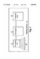
- FIG. 1shows a block diagram of the medical device of the preferred embodiment of the invention.
- FIG. 2shows a block diagram of the medical device of FIG. 1 in more detail.
- FIG. 3shows the processor of the computing unit of the medical device of FIG. 2 in more detail.
- FIGS. 4 and 12show flowcharts of the operation of the cardiograph of the preferred embodiment of the invention.
- FIG. 5shows a high level flowchart of the operation of the computing unit of the preferred embodiment of the invention.
- FIGS. 6A and 6Bshow a flowchart of the operation of the QRS detection logic of the preferred embodiment of the invention.
- FIGS. 7A and 7Bshow a flowchart of the operation of the heart rate calculation logic of the preferred embodiment of the invention.
- FIGS. 8A, 8B and 8Cshow a flowchart of the operation of the classification logic of the preferred embodiment of the invention.
- FIGS. 9A and 9Bshow a flowchart of the operation of the alignment logic of the preferred embodiment of the invention.
- FIGS. 10A and 10Bshow a flowchart of the operation of the representative heartbeat creation logic of the preferred embodiment of the invention.
- FIGS. 11A and 11Bshow a flowchart of the operation of the measurements logic of the preferred embodiment of the invention.
- FIG. 13shows a graph of three exemplary ECG waveforms used by the QRS detection logic of the preferred embodiment of the invention.
- FIG. 14shows a graph of an exemplary activity function used by the QRS detection logic of the preferred embodiment of the invention.
- FIG. 15shows a graph of exemplary classified heartbeats.
- FIG. 16shows a graph of exemplary heartbeats being aligned by the alignment logic of the preferred embodiment of the invention.
- FIG. 17shows a graph of exemplary aligned heartbeats being timesliced by the representative heartbeat creation logic of the preferred embodiment of the invention.
- FIG. 18shows a printout or display of a representative heartbeat without measurements.
- FIG. 19shows a printout or display of a representative heartbeat with measurements.
- FIG. 1shows a block diagram of the medical device of the preferred embodiment of the invention.
- Medical device 10comprises acquisition unit 20, electrodes 25, cardiograph 40, and computing unit 60.
- cardiograph 40 and acquisition unit 20are separate components of a PageWriter XLi, manufactured by the Hewlett-Packard company, modified to execute the flowcharts of FIGS. 4 and 12 of the preferred embodiment of the invention.
- Computing unit 60is a HP Vectra personal computer, suitably programmed to execute the flowcharts of FIGS. 5-11 of the preferred embodiment of the invention.
- FIG. 2shows a block diagram of medical device 10 in more detail.
- Cardiograph 40contains acquisition unit interface 41, processor 45, printer 47, and computing unit interface 49.
- Processor 45executes the flowcharts of FIGS. 4 and 12 of the preferred embodiment of the invention.
- Computing unit 60contains cardiograph interface 61, processor 65, display 66, input device 67, memory 68, and storage 69.
- Processor 65executes the flowcharts of FIGS. 5-11 of the preferred embodiment of the invention.
- FIG. 2shows medical device 10 as containing discrete components, those skilled in the art will appreciate that medical device 10 could be a single unit that contains each of the components shown in FIG. 2, or contain a different number of discrete components, and still fall within the spirit and scope of the invention.
- FIG. 3shows processor 65 of computing unit 60 of medical device 10 in more detail.
- Processor 65contains QRS detection logic 71, heart rate calculation logic 73, classification logic 74, alignment logic 75, representative heartbeat creation logic 77, and measurement logic 78.
- each of these logic blocksis performed by software written to perform the functions of relevant portions of the flowcharts shown in FIGS. 5-11, and this software is executed by processor 65.
- some or all of logic blocks 71-78could be special purpose hardware, such as contained in an application specific integrated circuit, designed to perform functions of relevant portions of the flowcharts shown in FIGS. 5-11.
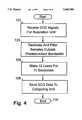
- FIG. 4shows a flowchart of the operation of cardiograph 40 of the preferred embodiment of the invention.
- ECG signalsare received from electrodes 25 of acquisition unit 20. In the preferred embodiment, these signals are digital signals sampled at a high sampling rate.
- Block 103decimates and filters the sampled ECG signals outside of a predetermined bandwidth. In the preferred embodiment, the predetermined bandwidth is 0.01 Hz to 150 Hz, and the decimation process reduces the number of samples to one eighth of the number of original samples.
- Block 105makes twelve ECG leads from the ten electrodes in a conventional manner. The signals contained on the twelve ECG leads will be referred to herein as "ECG waveforms", and the information contained thereon will be referred to herein as "ECG data”.
- Block 108sends the ECG data on the ECG waveforms to computing unit 60.
- the flowchartends in block 109.
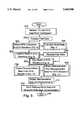
- FIG. 5shows a high level flowchart of the operation of computing unit 60 of the preferred embodiment of the invention.
- Block 201receives the ECG waveforms containing the ECG data from cardiograph 40.
- Block 203forward filters the ECG data.
- this filteris a high pass filter used as part of a forward/reverse filtering scheme to remove baseline wander while preserving low frequency information in the ECG data.
- Block 300calls a subroutine that detects the heartbeats (i.e. QRS complexes) in the ECG waveforms. This subroutine calculates an activity function from a subset of the ECG waveforms determined to be least noisy, and uses this activity function to search for heartbeats. This allows for true heartbeats to be detected while discarding false "noise" beats. The operation of this subroutine will be described in more detail later in conjunction with the discussion of FIG. 6.
- Block 400calls a subroutine that calculates the patient's heart rate. This logic determines the intervals between the heartbeats, discards a percentage of the shortest and longest intervals, and averages the remaining intervals to arrive at the patient's heart rate. This results in a robust calculation of the heart rate even in the presence of noise falsely detected as heartbeats and missed beats common in noisy environments. The operation of this subroutine will be described in more detail later in conjunction with the discussion of FIG. 7.
- Block 500calls a subroutine that classifies heartbeats. This classification is done by comparing each heartbeat against a group of templates corresponding to one or more heartbeat classifications. The templates are updated to track changes in the morphology of the heartbeats. The operation of this subroutine will be described in more detail later in conjunction with the discussion of FIG. 8.
- Block 205reverse filters the ECG data.
- this filteris a high pass filter used as part of a forward/reverse filtering scheme to remove baseline wander while preserving low frequency information in the ECG data.
- Block 600calls a subroutine that aligns heartbeats prior to representative heartbeat creation. This logic slides the heartbeats across an alignment template heartbeat to calculate when the heartbeats are aligned, and performs adjustments to reduce the effects of noise or jitter on the different ECG waveforms. The operation of this subroutine will be described in more detail later in conjunction with the discussion of FIG. 9.
- Block 700calls a subroutine that creates a representative heartbeat from the aligned heartbeats. This logic time slices through the aligned heartbeats, discarding a percentage of the smallest and largest magnitudes of the aligned heartbeats at each instance of time and averaging the remaining magnitudes to produce a representative heartbeat. This trimmed averaging technique results in a high quality representative beat, since samples from noise and misclassified beats are discarded. The operation of this subroutine will be described in more detail later in conjunction with the discussion of FIG. 10.
- Block 800calls a subroutine that measures various aspects of a representative heartbeat.
- This logicanalyzes the representative heartbeats from a group of ECG waveforms to determine an earliest QRS onset and latest QRS offset, and uses these values to perform a variety of measurements. This results in robust measurements even in very noisy environments. The operation of this subroutine will be described in more detail later in conjunction with the discussion of FIG. 11.
- Block 210displays the representative heartbeat created by subroutine 700 and, optionally, the measurements obtained by subroutines 800 and 400, on display 66 of computing unit 60 (FIG. 2). Examples of these displays are shown in FIGS. 18 and 19.
- Block 220sends the representative heartbeat and measurements for each ECG waveform, including the heart rate measurement calculated by subroutine 400, back to cardiograph 40. Cardiograph 40 processes this information in accordance with the flowchart of FIG. 12. The flowchart ends in block 249.
- FIG. 12shows how cardiograph 40 processes the information received from computing unit 60.
- Block 150receives the representative heartbeat and measurements, including the heart rate measurement, sent by block 220 of FIG. 5.
- Block 190prints the representative heartbeat created by subroutine 700 and, optionally, the measurements obtained by subroutines 800 and 400, on printer 47 of cardiograph 40 (FIG. 2). Examples of these printouts are shown in FIGS. 18 and 19.
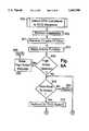
- FIG. 6shows a flowchart of the operation of subroutine 300, performed by QRS detection logic 71 of computing unit 60 of the preferred embodiment of the invention.
- Block 302performs an initialization process that learns about the ECG data. More specifically, the first time through the subroutine, this process analyzes the first few seconds of ECG data to determine a preliminary interval between peaks and the magnitude of an average peak. During routine processing (i.e. subsequent times through the subroutine) block 302 continues to update the information obtained the first time through the subroutine.
- Block 301receives ECG data from three ECG waveforms out of the twelve ECG waveforms received by computing unit 60 in block 201 (FIG. 4). A graph of three exemplary ECG waveforms is shown in FIG. 13. In the preferred embodiment, block 301 selects these three ECG waveforms as the three ECG waveforms that have optimal noise characteristics. This is done by continuously calculating the signal quality on the twelve ECG waveforms and ranking these waveforms from highest to lowest signal quality.
- Block 303calculates an activity function out of the three leads of ECG data.
- An activity functionis a signal mathematically derived from the ECG data which emphasizes characteristics of the heartbeat while minimizing the influence of noise to enable more accurate heartbeat detection.
- the activity functionis created by decimating the ECG data by one half, filtering the data using a bandpass filter and taking the absolute value of the first difference. The absolute first differences from the three ECG waveforms are then summed together, thresholded and smoothed to improved noise performance. Noise statistics (i.e., signal quality) across all twelve ECG waveforms are calculated and updated.
- a graph of an exemplary activity functionis shown in FIG. 14.
- Block 305checks the noise statistics to see if unacceptably high noise was detected. If so, block 306 issues a "high noise" message, which is displayed on display 66 of computing unit 60 (FIG. 2). This error causes the subroutine to terminate abnormally by ending in block 398. In the absence of high noise, block 305 is answered negatively. Block 309 looks to see if it is to detect another heartbeat. If not, the subroutine returns in block 399 to block 400 of FIG. 5.
- subroutine 300performs up to three different types of searches to find each heartbeat.
- the first searchis an on-time search. This search is normally used to detect heartbeats that appear within a small window of their expected time. If the on-time search fails to detect a heartbeat, a modified on-time search is used. The modified on-time search is similar in operation to the on-time search, but can detect heartbeats in low noise environments that the on-time search might miss, such as early beats. If both the on-time search and the modified on-time search fail to detect a heartbeat, a late beat search is performed. This search detects heartbeats that appear later than their expected time.
- Block 310performs an on-time search of the activity function for QRS complexes in the ECG data.
- block 310searches over 115% of the current interval length between heartbeats (as determined in block 302) with a threshold that starts at 80% of the average peak value of the activity function. The threshold is then linearly decreased over time to 40% at the end of the search window. Any local maxima discovered above this linearly decreasing threshold is considered a "peak".
- Block 315checks to see if any peaks were detected. If so, block 320 selects the true heartbeat (i.e., QRS peak) from the detected peaks by looking at the times each peak occurred.
- QRS peaki.e., QRS peak
- Block 320selects the true heartbeat (i.e., QRS peak) from the detected peaks by looking at the times each peak occurred.
- QRS peaki.e., QRS peak
- block 315checks negatively, block 330 checks to see if the noise level is low. If so, block 335 searches the activity function by using a modified on-time search. In the preferred embodiment, this search is performed by using a constant lower threshold, such as 54% of the average peak activity function value, over 115% of the current interval length. Block 335 checks to see if any peaks were detected. If so, block 338 selects the true heartbeat from the detected peaks by selecting the peak that occurred closest to the time the next heartbeat was expected. Information about the heartbeat selected in block 338, such as beat timing information, is stored in storage 69 of computing unit 60 (FIG. 2). Flow of control loops back to block 309 to look for another beat to detect.
- a constant lower thresholdsuch as 54% of the average peak activity function value
- block 350performs a late beat search.
- this blocksearches the activity function over three R--R interval lengths using a linearly decreasing threshold, as was done with the on-time search.
- Block 355checks to see if any peaks were detected. If so, block 358 selects the true heartbeat from the detected peaks by selecting the first peak that it finds. Information about the heartbeat selected in block 358, such as beat timing information, is stored in storage 69 of computing unit 60 (FIG. 2). Flow of control loops back to block 309 to look for another beat to detect. If block 355 is answered negatively, block 370 issues a "detector failed" message that is displayed on display 66 of computing unit 60. Since no beats were detected, this causes an abnormal termination of the subroutine in block 396.
- FIG. 7shows a flowchart of the operation of subroutine 400, performed by the heart rate calculation logic 73 of computing unit 60 of the preferred embodiment of the invention.
- Block 403reads and stores the R--R interval between the first heartbeat detected in the ECG waveform and the second heartbeat detected in the ECG waveform. In the preferred embodiment, this detection is done using information stored by QRS detection logic 71 in subroutine 300, although conventional methods of detecting R--R intervals in an ECG waveform could also be used.
- Block 405increments RR -- ctr.
- Block 410checks to see if the counter is less than a maximum counter value and there are additional heartbeats still available for analysis in the ECG waveform.
- block 403is a timer and where block 410 checks to see if a maximum time has elapsed. For example, if a maximum time was set for 10 seconds, only heartbeats occurring in the most recent 10 second period of time are used to calculate the heart rate.
- block 420checks to make sure at least a minimum number of heartbeats were analyzed by the loop made up of blocks 403-410.
- block 425sorts the R--R intervals from shortest to longest.
- Block 430then discards a percentage of the shortest and longest R--R intervals.
- the QRS detectormay incorrectly detect noise as a heartbeat, and may incorrectly miss a real heartbeat. These errors result in incorrect R--R intervals, both too short and too long.
- the trimmed average done hereresults in a robust and accurate heart rate calculation even in the presence of false detections and missed beats. With low noise and/or arrhythmias, this trimmed average technique also produces an accurate calculation of the heart rate.
- block 430discards 25% of the shortest and 25% of the longest R--R intervals, although different values could be used.
- Block 435then averages the remaining R--R intervals.
- Block 440converts this average R--R interval to a heart rate.
- Block 445smoothes the heart rate determined by block 440 by averaging it with a predetermined number of past heart rates. In the preferred embodiment, block 445 averages the current heart rate with the past two heart rates. In any event, the heart rate determined by block 445 (or by block 440, if the smoothing step of block 445 is not desired) is stored by block 450 in storage 69 of computing unit 60 (FIG. 2). The subroutine returns in block 499 to block 500 of FIG. 5.
- block 460simply computes the average R--R interval of the small number of R--R intervals that were read and stored in block 403. This average R--R interval is converted to a heart rate in block 440, and the heart rate is stored by block 450 in storage 69 of computing unit 60 (FIG. 2). As before, the subroutine returns in block 499 to block 500 of FIG. 5.
- FIG. 8shows a flowchart of the operation subroutine 500, performed by classification logic 74 of computing unit 60 of the preferred embodiment of the invention.
- subroutine 500is used to classify beats as "D" (dominant), "V” (ventricular ectopic), “S” (supraventricular ectopic), or "Q” (questionable), although other classifications could be used.
- Block 501checks to see if there are more heartbeats to classify. If so, Block 502 gets beat timing information for the next heartbeat. In the preferred embodiment, this information is obtained from QRS detection logic 71 in a manner that has already been discussed, although conventional methods of obtaining this information could also be used.
- Block 503normalizes the portion of the activity function (from QRS detection logic 71 or conventional means) around the beat to classify.
- Block 505performs timing and physiologic measurements on both the activity function and on the beat to classify. These measurements are used in blocks 510 and 550 to assist in beat classification, as will be discussed later.
- physiologic limitse.g. within a predetermined width and height.
- Block 542uses timing and physiologic information to classify this beat and template as either "D" (Dominant), "V” (ventricular ectopic), “S” (supraventricular ectopic), or “Q” (questionable). Most commonly, this beat will be classified as D and the template classified as D/S, for "Dominant/supraventricular ectopic", since the vast majority of beats classified will be classified this way, and since both D and S beats have the same morphology and thus would match the same template, but vary by timing information--the S beat being earlier than the D beat. The method of distinguishing between D and S beats is performed by blocks 545 to 558, as will be discussed in more detail later.
- Block 542increments Num -- Template to one to indicate one stored template. Flow of control moves to block 545, the operation of which will be discussed later.
- block 518is answered negatively, and block 520 slides the beat to classify across the first, stationary template.
- the first templateusually corresponds to a first heartbeat classification of D/S, meaning the template for dominant and supraventricular waveforms.
- Block 520slides this beat across the first template, the minimum area difference between the beat to classify and the first template is calculated.
- Block 525asks if this minimum area difference is less than a threshold. If it is, the beat matches the first (D/S) template, and block 530 averages the new beat data into the template it matched. In the preferred embodiment, a weighted average is used, where the existing template is given more weight in the averaging process than the new beat.
- Block 530also keeps track of the number of times a beat matched this template, as well as when a beat most recently matched this template.
- Block 535increments the template counter.
- Block 538verifies that the maximum number of templates to check has not been exceeded, which would indicate all templates have been checked. If block 538 is answered affirmatively, flow of control loops back to blocks 518 and 520, where the beat is slid across the second template.
- the second templateexemplarily corresponds to a classification of ventricular ectopic ("V").
- Block 520again computes the minimum area difference, and block 525 again asks if this minimum area difference is less than a threshold. If it is, the beat matches the second (V) template, and block 530 averages the new beat data into the template it matched. If no match was found, the subroutine loops through blocks 535, 538, 518, 520, and 525 until a match is found or until block 538 is answered negatively, indicating that all existing templates have been checked for matches.
- Block 537verifies the classification of the template. As new beats are averaged into the existing templates, it is possible for the classification of the template to change. For example, a template originally classified as "V" may be reclassified to "D" as more beats are averaged into it. Block 545 checks to see if the beat matched a D/S template. If so, an additional inquiry about the beat must be made before the beat can be classified. This is done in block 550, which asks if the beat was early. If so, the beat is classified as supraventricular ("S") in block 555, and flow of control loops back to block 501 to look to see if there are more beats to classify.
- Ssupraventricular
- the beatis classified as dominant ("D") in block 558, and flow of control loops back to block 501 to look to see if there are more beats to classify. If block 545 determined that the beat matched a template other than the D/S template, block 560 classifies the beat as the classification corresponding to the template it matched. For example, if the beat matched the ventricular ectopic ("V") template, the beat would be classified as ventricular ectopic. Flow of control loops back to block 501, as previously discussed.
- Vventricular ectopic
- Block 539asks if Num -- template is less than Max -- template--a counter indicating the maximum number of templates. If not, block 540 creates a new template for this beat. Block 542 classifies the new template and beat and increments Num -- templates, as discussed previously.
- Block 541overwrites the template with the least recent update.
- block 543classifies the new template, but does not increment Num -- templates, since the number of templates did not change. Those skilled in the art will appreciate that the number of templates actually created can vary, depending on the amount of noise in the environment and whether any ectopic beats are detected.
- block 590displays the classified heartbeats on display 66 of computing unit 60 (FIG. 2).
- FIG. 15One such exemplary display is shown in FIG. 15.
- the subroutinereturns in block 599 to block 205 of FIG. 5.
- FIG. 9shows a flowchart of the operation of subroutine 600, performed by alignment logic 75 of Computing unit 60 of the preferred embodiment of the invention.
- Block 601determines which 3 ECG waveforms are the least noisy. In the preferred embodiment, this is done by using the information obtained in block 301 of the Detect QRS Complexes in ECG Waveform subroutine 300, although this determination could be made directly in this subroutine by continuously calculating the signal to noise ratio or other indication of signal quality on the twelve ECG waveforms and ranking these waveforms from highest to lowest, or by using some other technique.
- Block 603sets a lead counter to look at the first of the three least noisy ECG waveforms.
- Block 605receives ECG data from the ECG waveform determined by the lead counter.
- block 610determines the dominant template amongst the beats to be used to construct the representative heartbeat. In the preferred embodiment, this is done by looking at the number of times the templates used by the classification logic were matched by a beat, as determined in block 530 in FIG. 8. By definition, this will be a D/S template.
- Block 620creates and stores an alignment template in storage 69 of computing unit 60.
- the alignment templateis created with a beat which matched the dominant template determined in block 610, where a portion of the dominant template around the QRS is normalized.
- An alternate embodimenthas been contemplated where step 610 is skipped, and block 620 creates an alignment template by finding the first beat classified as D (via beat classification logic 74 or a conventional method of beat classification) and using this beat as the alignment template.
- Block 630sets a beat counter to 1.
- Block 640gets the next dominant beat for this ECG waveform and normalizes a portion of this beat around the QRS complex.
- beats classified as "D”are referred to herein as “dominant”
- beats classified as "V”, "S”, or "Q”are referred to as “non-dominant”.
- non-dominant beatsare excluded from alignment and from the determination of a representative heartbeat, since these beats can contaminate the representative heartbeat.
- Block 650slides this beat across the stationary alignment template, while computing the value of the sum of the absolute values of the difference between the beat and the alignment template. This value is referred to herein as the area difference.
- the position where the area difference is the minimumis the position where the beat is best aligned with the alignment template, and this position is saved for this beat in storage 69 of computing unit 60.
- Block 655increments the beat counter.
- Block 660checks to see if the beat counter is greater than or equal to the number of beats to align for this ECG waveform. If not, flow of control loops back to block 640 to get the next dominant beat. If so, block 670 increments the lead counter.
- Block 675checks to see if the lead counter is greater than or equal to 3--the number of least noisy ECG waveforms selected in block 601. If block 675 is answered negatively, flow of control loops back to block 605, where the alignment process is repeated for the next ECG waveform. If block 675 is answered affirmatively, block 680 corrects and stores each beat time on each ECG waveform with the median value of the alignment times just determined independently on the three least noisy leads. This is done to minimize the effect of noise which results in beats on different ECG waveforms best aligning at slightly different times (i.e. jitter). The subroutine returns in block 699 to block 700 in FIG. 5. FIG. 16 shows a new beat being slid across a stationary template.
- FIG. 10shows a flowchart of the operation of subroutine 700, performed by representative heartbeat creation logic 77 of computing unit 60 of the preferred embodiment of the invention.
- Block 701determines which beats to use in creating the representative heartbeat. In the preferred embodiment, only “dominant" beats, aligned via the beat alignment steps discussed above, are used. If more "dominant" beats are available than the number needed to construct a representative heartbeat, those with the most similar morphology are used. For example, if two or more dominant templates were created in classification subroutine 500, only the beats that match the dominant template that contains the most beats will preferably be used.
- Block 705sets a counter which keeps track of which ECG waveform the representative heartbeat is being created for to one.
- Block 710checks to see if the lead number counter is greater than or equal to the maximum number of ECG waveforms. If not, block 715 reads the aligned "dominant" beats for this ECG waveform. Block 720 sets a time pointer to zero.
- Block 725gets a time slice of data for each aligned beat at the instance of time identified by the time pointer.
- this datais the magnitude of each of the aligned dominant heartbeats at this moment of time.
- Block 730sorts the magnitudes for this slice of time from smallest to largest.
- Block 735discards a percentage of the smallest and largest magnitudes.
- the beat classification subroutinemay incorrectly classify beats as being dominant. These errors result in misclassified beats being incorrectly included in the aligned beats.
- the trimmed average done hereresults in a robust and accurate representative heartbeat even in the presence of misclassified beats and high noise present on dominant beats.
- Block 740averages the remaining magnitudes for this time slice.
- Block 750stores the average magnitude for this time slice in a representative beat array in storage 69 of computing unit 60.
- Block 755increments the time pointer to the next slice of time, and block 760 checks to see if the time pointer has reached its maximum value. If not, flow of control loops back to block 725 to determine the average magnitude of the other slices of time to complete the representative heartbeat array.
- FIG. 17shows exemplary aligned beats being time sliced using the process described above.
- block 765forward and reverse filters the representative heartbeat stored in the representative heartbeat array, and stores the result back into the array in block 770.
- Block 775increments the ECG waveform counter, and flow of control loops back to block 710 to create a representative heartbeat for each of the other ECG waveforms.
- block 780displays the representative heartbeats on display 66 of computing unit 60. An exemplary display of a representative heartbeat is shown in FIG. 18. The subroutine returns in block 799 to block 800 of FIG. 5.
- FIG. 11shows a flowchart of the operation of subroutine 800, performed by measurements logic 78 of computing unit 60 of the preferred embodiment of the invention.
- Block 801obtains the representative heartbeats for each of the ECG waveforms. In the preferred embodiment, this is done by reading the representative heartbeat array stored in step 770 of FIG. 10.
- representative heartbeats created using a different methodincluding conventionally known methods for creating representative heartbeats, could be used.
- Block 803measures the earliest QRS onset and the latest QRS offset. These values are from an activity function derived from a subset of the representative heartbeats obtained in block 801. These values are used for many of the measurements that will be made for these representative heartbeats, as will soon be discussed.
- Block 805sets a counter that keeps track of the ECG waveform for which the representative heartbeat is being measured. Block 810 gets the representative heartbeat for this ECG waveform.
- Block 815determines the isoelectric level of the representative heartbeat. In the preferred embodiment, this is the average level of the 16 msec of data prior to the earliest QRS onset.
- Block 820determines the R wave amplitude for this representative heartbeat.
- thisis the maximum positive value between the earliest QRS onset and the latest QRS offset, with an adjustment made to correct for elevated ST segments at the latest QRS offset, if the "T" wave is so large that it impacts the determination of the R wave amplitude.
- Block 825determines the ST level. In the preferred embodiment, this is the average of 10 msec around the user-determined ST measurement point of the representative heartbeat.
- Block 830determines the ST slope. In the preferred embodiment, this is determined by using a best line fit between the latest QRS offset and the ST measurement point of the representative heartbeat.
- Block 835determines the ST integral. In the preferred embodiment, this is determined by computing the sum of the negative area between the latest QRS offset and the ST measurement point of the representative heartbeat.
- Block 850updates a measurement confidence flag for each measurement taken.
- historical information and physiologic limitsare used to set these measurement flags to either a "low” or “high” confidence.
- These confidence flagscan be displayed to a user in a variety of ways, including the term “low” or “high” displayed next to a measurement, changing the color of the measurement on the display (e.g., green means high, red means low), etc.
- a "low” confidence flagwould indicate to the cardiologist or other medical professional that a measurement is not physiologic or has changed in a non-physiologic manner and should be manually reviewed for correctness.
- Block 855increments the ECG waveform counter.
- Block 860checks to see if the ECG waveform counter exceeds the maximum number of ECG waveforms.
- block 880displays the measurements on display 66 of computing unit 60 (FIG. 2).
- a cardiologist looking at the representative heartbeat and the measurements shown in FIG. 19would see that there is a depressed ST segment, indicating that the patient undergoing a stress test has coronary artery disease.
- the subroutinereturns in block 899 to block 210 of FIG. 5.
Landscapes
- Health & Medical Sciences (AREA)
- Life Sciences & Earth Sciences (AREA)
- Engineering & Computer Science (AREA)
- Cardiology (AREA)
- Physics & Mathematics (AREA)
- Biophysics (AREA)
- Public Health (AREA)
- Artificial Intelligence (AREA)
- Veterinary Medicine (AREA)
- General Health & Medical Sciences (AREA)
- Animal Behavior & Ethology (AREA)
- Surgery (AREA)
- Molecular Biology (AREA)
- Pathology (AREA)
- Biomedical Technology (AREA)
- Heart & Thoracic Surgery (AREA)
- Medical Informatics (AREA)
- Evolutionary Computation (AREA)
- Fuzzy Systems (AREA)
- Signal Processing (AREA)
- Psychiatry (AREA)
- Mathematical Physics (AREA)
- Physiology (AREA)
- Computer Vision & Pattern Recognition (AREA)
- Measurement And Recording Of Electrical Phenomena And Electrical Characteristics Of The Living Body (AREA)
- Measuring Pulse, Heart Rate, Blood Pressure Or Blood Flow (AREA)
Abstract
Description
______________________________________ Title Ser. No. ______________________________________ Method And Apparatus For Detecting Heartbeats In An 08/564,889 ECG Waveform Method And Apparatus For Calculating A Heart Rate In An 08/564,749 ECG Waveform Method and Apparatus For Classifying Heartbeats In An 08/564,768 ECG Waveform Method And Apparatus For Creating A Representative 08/565,504 Heartbeat From An ECG Waveform ______________________________________
Claims (16)
Priority Applications (4)
| Application Number | Priority Date | Filing Date | Title |
|---|---|---|---|
| US08/564,774US5682900A (en) | 1995-11-29 | 1995-11-29 | Method and apparatus for obtaining heartbeat measurements from a ECG waveform |
| JP8299164AJPH09168520A (en) | 1995-11-29 | 1996-11-12 | Method and apparatus for obtaining measured value of heart beat from ecg wave shape |
| EP96308432AEP0776630B1 (en) | 1995-11-29 | 1996-11-21 | Apparatus for obtaining heartbeat measurements in an ECG waveform |
| DE69626178TDE69626178T2 (en) | 1995-11-29 | 1996-11-21 | Heart rate monitor in an ECG waveform |
Applications Claiming Priority (1)
| Application Number | Priority Date | Filing Date | Title |
|---|---|---|---|
| US08/564,774US5682900A (en) | 1995-11-29 | 1995-11-29 | Method and apparatus for obtaining heartbeat measurements from a ECG waveform |
Publications (1)
| Publication Number | Publication Date |
|---|---|
| US5682900Atrue US5682900A (en) | 1997-11-04 |
Family
ID=24255832
Family Applications (1)
| Application Number | Title | Priority Date | Filing Date |
|---|---|---|---|
| US08/564,774Expired - Fee RelatedUS5682900A (en) | 1995-11-29 | 1995-11-29 | Method and apparatus for obtaining heartbeat measurements from a ECG waveform |
Country Status (4)
| Country | Link |
|---|---|
| US (1) | US5682900A (en) |
| EP (1) | EP0776630B1 (en) |
| JP (1) | JPH09168520A (en) |
| DE (1) | DE69626178T2 (en) |
Cited By (36)
| Publication number | Priority date | Publication date | Assignee | Title |
|---|---|---|---|---|
| US20020049474A1 (en)* | 1999-03-12 | 2002-04-25 | Cardiac Pacemakers, Inc. | Method and system for verifying the integrity of normal sinus rhythm templates |
| US20020091333A1 (en)* | 1999-02-12 | 2002-07-11 | Cardiac Pacemakers, Inc. | System and method for arrhythmia discrimination |
| US6438410B2 (en)* | 1999-02-12 | 2002-08-20 | Cardiac Pacemakers, Inc. | System and method for a classifying cardiac complexes |
| US20030060849A1 (en)* | 1999-07-14 | 2003-03-27 | Cardiac Pacemakers, Inc. | Classification of supraventricular and ventricular cardiac rhythms using cross channel timing algorithm |
| US20040219600A1 (en)* | 2002-12-13 | 2004-11-04 | Williams Robert Wood | Method for determining sensitivity to environmental toxins and susceptibility to parkinson's disease |
| US20040267143A1 (en)* | 2003-06-27 | 2004-12-30 | Sweeney Robert J. | Signal compression based on curvature parameters |
| US6950702B2 (en) | 2002-07-15 | 2005-09-27 | Cardiac Pacemakers, Inc. | Use of curvature based features for beat detection |
| US6978177B1 (en) | 2000-11-14 | 2005-12-20 | Cardiac Pacemakers, Inc. | Method and apparatus for using atrial discrimination algorithms to determine optimal pacing therapy and therapy timing |
| US7039463B2 (en) | 1999-03-12 | 2006-05-02 | Cardiac Pacemakers, Inc. | Discrimination of supraventricular tachycardia and ventricular tachycardia events |
| US7113824B2 (en) | 1997-04-30 | 2006-09-26 | Cardiac Pacemakers, Inc. | Apparatus and method for treating ventricular tachyarrhythmias |
| US20070055165A1 (en)* | 2003-09-26 | 2007-03-08 | D Aubioul Jan A R | Heart beat signal analysis |
| US7203535B1 (en) | 1999-04-01 | 2007-04-10 | Cardiac Pacemakers, Inc. | System and method for classifying tachycardia arrhythmias having 1:1 atrial-to-ventricular rhythms |
| US7212849B2 (en) | 2004-10-28 | 2007-05-01 | Cardiac Pacemakers, Inc. | Methods and apparatuses for arrhythmia detection and classification using wireless ECG |
| US20070208266A1 (en)* | 2006-03-03 | 2007-09-06 | Cardiac Science Corporation | Methods for quantifying the risk of cardiac death using exercise induced heart rate variability metrics |
| US20070249949A1 (en)* | 2006-04-21 | 2007-10-25 | Cardiac Science Corporation | Methods and apparatus for quantifying the risk of cardiac death using exercise induced heart rate recovery metrics |
| US7289845B2 (en) | 2000-10-31 | 2007-10-30 | Cardiac Pacemakers, Inc. | Curvature based method for selecting features from an electrophysiologic signal for purpose of complex identification and classification |
| US20080091080A1 (en)* | 2003-04-22 | 2008-04-17 | Patrick Leahy | Device and Method for Use in Surgery |
| US7430446B2 (en) | 2005-01-20 | 2008-09-30 | Cardiac Pacemakers, Inc. | Methods and apparatuses for cardiac arrhythmia classification using morphology stability |
| US20080281371A1 (en)* | 2007-05-08 | 2008-11-13 | Kenknight Bruce | Method For Controlling Pacemaker Therapy |
| US20080281369A1 (en)* | 2007-05-08 | 2008-11-13 | Kenknight Bruce | System And Method For Determining The Origin Of A Sensed Beat |
| US7515956B2 (en) | 2004-05-12 | 2009-04-07 | Cardiac Pacemakers, Inc. | Template based AV/VA interval comparison for the discrimination of cardiac arrhythmias |
| US7610084B2 (en) | 2001-06-05 | 2009-10-27 | Cardiac Pacemakers, Inc. | System and method for classifying cardiac depolarization complexes with multi-dimensional correlation |
| US7792571B2 (en) | 2003-06-27 | 2010-09-07 | Cardiac Pacemakers, Inc. | Tachyarrhythmia detection and discrimination based on curvature parameters |
| CN101869476B (en)* | 2009-04-22 | 2013-07-17 | 财团法人工业技术研究院 | Detect heartbeat method |
| US9050014B2 (en) | 2011-12-14 | 2015-06-09 | Siemens Medical Solutions Usa, Inc. | System for cardiac arrhythmia detection and characterization |
| US9314210B2 (en) | 2005-06-13 | 2016-04-19 | Cardiac Pacemakers, Inc. | Method and apparatus for rate-dependent morphology-based cardiac arrhythmia classification |
| US9504427B2 (en) | 2011-05-04 | 2016-11-29 | Cardioinsight Technologies, Inc. | Signal averaging |
| US9592391B2 (en) | 2014-01-10 | 2017-03-14 | Cardiac Pacemakers, Inc. | Systems and methods for detecting cardiac arrhythmias |
| US9669230B2 (en) | 2015-02-06 | 2017-06-06 | Cardiac Pacemakers, Inc. | Systems and methods for treating cardiac arrhythmias |
| US10449361B2 (en) | 2014-01-10 | 2019-10-22 | Cardiac Pacemakers, Inc. | Systems and methods for treating cardiac arrhythmias |
| US10463866B2 (en) | 2014-07-11 | 2019-11-05 | Cardiac Pacemakers, Inc. | Systems and methods for treating cardiac arrhythmias |
| US10758737B2 (en) | 2016-09-21 | 2020-09-01 | Cardiac Pacemakers, Inc. | Using sensor data from an intracardially implanted medical device to influence operation of an extracardially implantable cardioverter |
| CN111657905A (en)* | 2020-06-23 | 2020-09-15 | 中国医学科学院生物医学工程研究所 | Feature point detection method, device, equipment and storage medium |
| CN113647959A (en)* | 2021-07-27 | 2021-11-16 | 东软集团股份有限公司 | Method, device and equipment for identifying waveform of electrocardiographic waveform signal |
| RU2783147C1 (en)* | 2021-12-09 | 2022-11-09 | Публичное акционерное общество энергетики и электрификации "Мосэнерго" (ПАО "Мосэнерго") | Method for automated heart rate determination |
| US20230414149A1 (en)* | 2020-11-23 | 2023-12-28 | Prevayl Innovations Limited | Method and System for Measuring and Displaying Biosignal Data to a Wearer of a Wearable Article |
Families Citing this family (3)
| Publication number | Priority date | Publication date | Assignee | Title |
|---|---|---|---|---|
| EP1377913A4 (en)* | 2001-03-19 | 2008-02-20 | B S P Biolog Signal Proc Ltd | METHOD AND DEVICE FOR EFFICIENTLY PRESENTING PERIODIC AND NEARLY PERIODIC SIGNALS FOR ANALYSIS |
| US7031764B2 (en) | 2002-11-08 | 2006-04-18 | Cardiac Pacemakers, Inc. | Cardiac rhythm management systems and methods using multiple morphology templates for discriminating between rhythms |
| US7277747B2 (en) | 2004-11-23 | 2007-10-02 | Cardiac Pacemakers, Inc. | Arrhythmia memory for tachyarrhythmia discrimination |
Citations (2)
| Publication number | Priority date | Publication date | Assignee | Title |
|---|---|---|---|---|
| US4630204A (en)* | 1984-02-21 | 1986-12-16 | Mortara Instrument Inc. | High resolution ECG waveform processor |
| US5284152A (en)* | 1992-02-28 | 1994-02-08 | Hewlett-Packard Company | Method for displaying superimposed heartbeat waveforms |
Family Cites Families (2)
| Publication number | Priority date | Publication date | Assignee | Title |
|---|---|---|---|---|
| US4589420A (en)* | 1984-07-13 | 1986-05-20 | Spacelabs Inc. | Method and apparatus for ECG rhythm analysis |
| US4930075A (en)* | 1987-07-13 | 1990-05-29 | The Board Of Trustees Of The Leland Stanford Junior University | Technique to evaluate myocardial ischemia from ECG parameters |
- 1995
- 1995-11-29USUS08/564,774patent/US5682900A/ennot_activeExpired - Fee Related
- 1996
- 1996-11-12JPJP8299164Apatent/JPH09168520A/ennot_activeWithdrawn
- 1996-11-21EPEP96308432Apatent/EP0776630B1/ennot_activeExpired - Lifetime
- 1996-11-21DEDE69626178Tpatent/DE69626178T2/ennot_activeExpired - Fee Related
Patent Citations (2)
| Publication number | Priority date | Publication date | Assignee | Title |
|---|---|---|---|---|
| US4630204A (en)* | 1984-02-21 | 1986-12-16 | Mortara Instrument Inc. | High resolution ECG waveform processor |
| US5284152A (en)* | 1992-02-28 | 1994-02-08 | Hewlett-Packard Company | Method for displaying superimposed heartbeat waveforms |
Non-Patent Citations (4)
| Title |
|---|
| "Fast And Reliable QRS Alignment Technique For High-Frequency Analysis of Signal-Averaged ECG", by O. J. Escalona, R.H. Mitchell, D.E. Balderson, And D.W.G. Harron, MBEC Kyoto World Congress Supplement, Jul. 1993, pp. S137-S146. |
| "Influence Of Noise On Wave Boundary Recognition By ECG Measurement Programs", by J.L. Willems, Chr. Zywietz, P. Arnaud, J. H. Van Bemmel, R. Degani, and P. W. MacFarlane, Computers & Biomedical Research, 1987, pp. 543-562. |
| Fast And Reliable QRS Alignment Technique For High Frequency Analysis of Signal Averaged ECG , by O. J. Escalona, R.H. Mitchell, D.E. Balderson, And D.W.G. Harron, MBEC Kyoto World Congress Supplement, Jul. 1993, pp. S137 S146.* |
| Influence Of Noise On Wave Boundary Recognition By ECG Measurement Programs , by J.L. Willems, Chr. Zywietz, P. Arnaud, J. H. Van Bemmel, R. Degani, and P. W. MacFarlane, Computers & Biomedical Research, 1987, pp. 543 562.* |
Cited By (69)
| Publication number | Priority date | Publication date | Assignee | Title |
|---|---|---|---|---|
| US7522956B2 (en) | 1997-04-30 | 2009-04-21 | Cardiac Pacemakers, Inc. | Apparatus and method for treating ventricular tachyarrhythmias |
| US8046068B2 (en) | 1997-04-30 | 2011-10-25 | Cardiac Pacemakers, Inc. | Apparatus and method for treating ventricular tachyarrhythmias |
| US8306619B2 (en) | 1997-04-30 | 2012-11-06 | Cardiac Pacemakers, Inc. | Apparatus and method for treating ventricular tachyarrhythmias |
| US7113824B2 (en) | 1997-04-30 | 2006-09-26 | Cardiac Pacemakers, Inc. | Apparatus and method for treating ventricular tachyarrhythmias |
| US6959212B2 (en) | 1999-02-12 | 2005-10-25 | Cardiac Pacemakers, Inc. | System and method for arrhythmia discrimination |
| US20020091333A1 (en)* | 1999-02-12 | 2002-07-11 | Cardiac Pacemakers, Inc. | System and method for arrhythmia discrimination |
| US6438410B2 (en)* | 1999-02-12 | 2002-08-20 | Cardiac Pacemakers, Inc. | System and method for a classifying cardiac complexes |
| US6728572B2 (en) | 1999-02-12 | 2004-04-27 | Cardiac Pacemakers, Inc. | System and method for classifying cardiac complexes |
| US8019408B2 (en) | 1999-03-12 | 2011-09-13 | Cardiac Pacemakers, Inc. | System for verifying the integrity of cardiac complex templates |
| US7953476B2 (en) | 1999-03-12 | 2011-05-31 | Cardiac Pacemakers, Inc. | Discrimination of supraventricular tachycardia and ventricular tachycardia events |
| US7653430B2 (en) | 1999-03-12 | 2010-01-26 | Cardiac Pacemakers, Inc. | Method and system for verifying the integrity of normal sinus rhythm templates |
| US6996434B2 (en) | 1999-03-12 | 2006-02-07 | Cardiac Pacemakers, Inc. | Method and system for verifying the integrity of normal sinus rhythm templates |
| US7039463B2 (en) | 1999-03-12 | 2006-05-02 | Cardiac Pacemakers, Inc. | Discrimination of supraventricular tachycardia and ventricular tachycardia events |
| US20110034817A1 (en)* | 1999-03-12 | 2011-02-10 | Marcovecchio Alan F | Discrimination of supraventricular tachycardia and ventricular tachycardia events |
| US8229552B2 (en) | 1999-03-12 | 2012-07-24 | Cardiac Pacemakers, Inc. | Discrimination of supraventricular tachycardia and ventricular tachycardia events |
| US8548575B2 (en) | 1999-03-12 | 2013-10-01 | Cardiac Pacemakers, Inc. | System for verifying the integrity of cardiac complex templates |
| US7991457B2 (en) | 1999-03-12 | 2011-08-02 | Cardiac Pacemakers, Inc. | Discrimination of supraventricular tachycardia and ventricular tachycardia events |
| US20020049474A1 (en)* | 1999-03-12 | 2002-04-25 | Cardiac Pacemakers, Inc. | Method and system for verifying the integrity of normal sinus rhythm templates |
| US7203535B1 (en) | 1999-04-01 | 2007-04-10 | Cardiac Pacemakers, Inc. | System and method for classifying tachycardia arrhythmias having 1:1 atrial-to-ventricular rhythms |
| US20030060849A1 (en)* | 1999-07-14 | 2003-03-27 | Cardiac Pacemakers, Inc. | Classification of supraventricular and ventricular cardiac rhythms using cross channel timing algorithm |
| US8315697B2 (en) | 1999-07-14 | 2012-11-20 | Cardiac Pacemakers, Inc. | Classification of supraventricular and ventricular cardiac rhythms using cross channel timing algorithm |
| US8050757B2 (en) | 1999-07-14 | 2011-11-01 | Cardiac Pacemakers, Inc. | Classification of supraventricular and ventricular cardiac rhythms using cross channel timing algorithm |
| US6889081B2 (en) | 1999-07-14 | 2005-05-03 | Cardiac Pacemakers, Inc. | Classification of supraventricular and ventricular cardiac rhythms using cross channel timing algorithm |
| US7580744B2 (en) | 1999-07-14 | 2009-08-25 | Cardiac Pacemakers, Inc. | Classification of supraventricular and ventricular cardiac rhythms using cross channel timing algorithm |
| US7289845B2 (en) | 2000-10-31 | 2007-10-30 | Cardiac Pacemakers, Inc. | Curvature based method for selecting features from an electrophysiologic signal for purpose of complex identification and classification |
| US7764997B2 (en) | 2000-11-14 | 2010-07-27 | Cardiac Pacemakers, Inc. | Method and apparatus for using atrial discrimination algorithms to determine optimal pacing therapy and therapy timing |
| US6978177B1 (en) | 2000-11-14 | 2005-12-20 | Cardiac Pacemakers, Inc. | Method and apparatus for using atrial discrimination algorithms to determine optimal pacing therapy and therapy timing |
| US7610084B2 (en) | 2001-06-05 | 2009-10-27 | Cardiac Pacemakers, Inc. | System and method for classifying cardiac depolarization complexes with multi-dimensional correlation |
| US6950702B2 (en) | 2002-07-15 | 2005-09-27 | Cardiac Pacemakers, Inc. | Use of curvature based features for beat detection |
| US20040219600A1 (en)* | 2002-12-13 | 2004-11-04 | Williams Robert Wood | Method for determining sensitivity to environmental toxins and susceptibility to parkinson's disease |
| US20080091080A1 (en)* | 2003-04-22 | 2008-04-17 | Patrick Leahy | Device and Method for Use in Surgery |
| US7792571B2 (en) | 2003-06-27 | 2010-09-07 | Cardiac Pacemakers, Inc. | Tachyarrhythmia detection and discrimination based on curvature parameters |
| US8280508B2 (en) | 2003-06-27 | 2012-10-02 | Cardiac Pacemakers, Inc. | Signal compression based on curvature parameters |
| US7500955B2 (en) | 2003-06-27 | 2009-03-10 | Cardiac Pacemaker, Inc. | Signal compression based on curvature parameters |
| US8409107B2 (en) | 2003-06-27 | 2013-04-02 | Cardiac Pacemakers, Inc. | Tachyarrhythmia detection and discrimination based on curvature parameters |
| US20040267143A1 (en)* | 2003-06-27 | 2004-12-30 | Sweeney Robert J. | Signal compression based on curvature parameters |
| US20070055165A1 (en)* | 2003-09-26 | 2007-03-08 | D Aubioul Jan A R | Heart beat signal analysis |
| US7515956B2 (en) | 2004-05-12 | 2009-04-07 | Cardiac Pacemakers, Inc. | Template based AV/VA interval comparison for the discrimination of cardiac arrhythmias |
| US9138590B2 (en) | 2004-10-28 | 2015-09-22 | Cardiac Pacemakers, Inc. | Implantable medical device sensing and selecting wireless ECG and intracardiac electrogram |
| US7212849B2 (en) | 2004-10-28 | 2007-05-01 | Cardiac Pacemakers, Inc. | Methods and apparatuses for arrhythmia detection and classification using wireless ECG |
| US8239020B2 (en) | 2004-10-28 | 2012-08-07 | Cardiac Pacemakers, Inc. | Implantable medical device sensing wireless ECG as substitute for intracardiac electrogram |
| US7430446B2 (en) | 2005-01-20 | 2008-09-30 | Cardiac Pacemakers, Inc. | Methods and apparatuses for cardiac arrhythmia classification using morphology stability |
| US8244348B2 (en) | 2005-01-20 | 2012-08-14 | Cardiac Pacemakers, Inc. | Method and apparatus for cardiac arrhythmia classification using template band-based morphology analysis |
| US9314210B2 (en) | 2005-06-13 | 2016-04-19 | Cardiac Pacemakers, Inc. | Method and apparatus for rate-dependent morphology-based cardiac arrhythmia classification |
| US20070208266A1 (en)* | 2006-03-03 | 2007-09-06 | Cardiac Science Corporation | Methods for quantifying the risk of cardiac death using exercise induced heart rate variability metrics |
| US7708683B2 (en) | 2006-03-03 | 2010-05-04 | Cardiac Science Corporation | Methods for quantifying the risk of cardiac death using exercise induced heart rate variability metrics |
| WO2007103744A3 (en)* | 2006-03-03 | 2008-05-22 | Cardiac Science Corp | Methods for quantifying the risk of cardiac death using exercise induced heart rate variability metrics |
| US9149195B2 (en) | 2006-04-21 | 2015-10-06 | Mortara Instrument, Inc. | Methods and apparatus for quantifying the risk of cardiac death using exercise induced heart rate recovery metrics |
| US20070249949A1 (en)* | 2006-04-21 | 2007-10-25 | Cardiac Science Corporation | Methods and apparatus for quantifying the risk of cardiac death using exercise induced heart rate recovery metrics |
| US8224443B2 (en) | 2007-05-08 | 2012-07-17 | Cardiac Pacemakers, Inc. | Method for controlling pacemaker therapy |
| US20080281369A1 (en)* | 2007-05-08 | 2008-11-13 | Kenknight Bruce | System And Method For Determining The Origin Of A Sensed Beat |
| US8442631B2 (en) | 2007-05-08 | 2013-05-14 | Cardiac Pacemakers, Inc. | System and method for determining the origin of a sensed beat |
| US20080281371A1 (en)* | 2007-05-08 | 2008-11-13 | Kenknight Bruce | Method For Controlling Pacemaker Therapy |
| US8781581B2 (en) | 2007-05-08 | 2014-07-15 | Cardiac Pacemakers, Inc. | System and method for determining the origin of a sensed beat |
| CN101869476B (en)* | 2009-04-22 | 2013-07-17 | 财团法人工业技术研究院 | Detect heartbeat method |
| US9504427B2 (en) | 2011-05-04 | 2016-11-29 | Cardioinsight Technologies, Inc. | Signal averaging |
| US9050014B2 (en) | 2011-12-14 | 2015-06-09 | Siemens Medical Solutions Usa, Inc. | System for cardiac arrhythmia detection and characterization |
| US9592391B2 (en) | 2014-01-10 | 2017-03-14 | Cardiac Pacemakers, Inc. | Systems and methods for detecting cardiac arrhythmias |
| US10449361B2 (en) | 2014-01-10 | 2019-10-22 | Cardiac Pacemakers, Inc. | Systems and methods for treating cardiac arrhythmias |
| US10463866B2 (en) | 2014-07-11 | 2019-11-05 | Cardiac Pacemakers, Inc. | Systems and methods for treating cardiac arrhythmias |
| US9669230B2 (en) | 2015-02-06 | 2017-06-06 | Cardiac Pacemakers, Inc. | Systems and methods for treating cardiac arrhythmias |
| US10238882B2 (en) | 2015-02-06 | 2019-03-26 | Cardiac Pacemakers | Systems and methods for treating cardiac arrhythmias |
| US11020595B2 (en) | 2015-02-06 | 2021-06-01 | Cardiac Pacemakers, Inc. | Systems and methods for treating cardiac arrhythmias |
| US10758737B2 (en) | 2016-09-21 | 2020-09-01 | Cardiac Pacemakers, Inc. | Using sensor data from an intracardially implanted medical device to influence operation of an extracardially implantable cardioverter |
| CN111657905A (en)* | 2020-06-23 | 2020-09-15 | 中国医学科学院生物医学工程研究所 | Feature point detection method, device, equipment and storage medium |
| US20230414149A1 (en)* | 2020-11-23 | 2023-12-28 | Prevayl Innovations Limited | Method and System for Measuring and Displaying Biosignal Data to a Wearer of a Wearable Article |
| CN113647959A (en)* | 2021-07-27 | 2021-11-16 | 东软集团股份有限公司 | Method, device and equipment for identifying waveform of electrocardiographic waveform signal |
| CN113647959B (en)* | 2021-07-27 | 2024-01-12 | 东软集团股份有限公司 | Waveform identification method, device and equipment for electrocardiographic waveform signals |
| RU2783147C1 (en)* | 2021-12-09 | 2022-11-09 | Публичное акционерное общество энергетики и электрификации "Мосэнерго" (ПАО "Мосэнерго") | Method for automated heart rate determination |
Also Published As
| Publication number | Publication date |
|---|---|
| EP0776630A1 (en) | 1997-06-04 |
| DE69626178D1 (en) | 2003-03-20 |
| DE69626178T2 (en) | 2003-12-04 |
| JPH09168520A (en) | 1997-06-30 |
| EP0776630B1 (en) | 2003-02-12 |
Similar Documents
| Publication | Publication Date | Title |
|---|---|---|
| US5682900A (en) | Method and apparatus for obtaining heartbeat measurements from a ECG waveform | |
| US5817027A (en) | Method and apparatus for classifying heartbeats in an ECG waveform | |
| US5628326A (en) | Calculating a heart rate from an ECG waveform by discarding a percentage of R-R intervals prior to averaging | |
| JPH09173310A5 (en) | ||
| JPH09168520A5 (en) | ||
| JPH09173312A5 (en) | ||
| US5715829A (en) | Method and apparatus for detecting heartbeats in an ECG waveform using an activity function and on-time search | |
| US5613496A (en) | Method and apparatus for creating a representative heartbeat from an ECG waveform | |
| US5323783A (en) | Dynamic ST segment estimation and adjustment | |
| US7074192B2 (en) | Method and apparatus for measuring blood pressure using relaxed matching criteria | |
| US8024030B2 (en) | System and method for analyzing an electrocardiogram signal | |
| US5159932A (en) | Myocardial ischemia detection system | |
| US20060178591A1 (en) | Methods and systems for real time breath rate determination with limited processor resources | |
| JPH09168521A5 (en) | ||
| US6491629B1 (en) | Method for determining at least one diagnostic piece of information from signal patterns of medical sensor systems | |
| Nygårds et al. | An automated system for ECG monitoring | |
| JP2001517520A (en) | R wave detection method and apparatus | |
| CA2283860A1 (en) | Method and apparatus for arbitrating to obtain best estimates for blood constituent values and rejecting harmonics | |
| CA2357059A1 (en) | Signal processing apparatus | |
| JPS5867235A (en) | Method and apparatus for measuring cardiac pulse | |
| JP2635079B2 (en) | Method of removing pace pulse signal from ECG wave signal and apparatus for removing pace pulse signal from ECG wave signal | |
| JPH09173311A5 (en) | ||
| CN110769748B (en) | Artifact Tolerant Pulse Rate Variability Measurement | |
| US6668189B2 (en) | Method and system for measuring T-wave alternans by alignment of alternating median beats to a cubic spline | |
| US20240197211A1 (en) | System and method for blood glucose monitoring based on heart rate variability |
Legal Events
| Date | Code | Title | Description |
|---|---|---|---|
| AS | Assignment | Owner name:HEWLETT-PACKARD COMPANY, CALIFORNIA Free format text:ASSIGNMENT OF ASSIGNORS INTEREST;ASSIGNORS:ARAND, PATRICIA A.;POST, WILLIAM L.;REEL/FRAME:007868/0346 Effective date:19951128 | |
| AS | Assignment | Owner name:HEWLETT-PACKARD COMPANY, A DELAWARE CORPORATION, C Free format text:MERGER;ASSIGNOR:HEWLETT-PACKARD COMPANY, A CALIFORNIA CORPORATION;REEL/FRAME:010841/0649 Effective date:19980520 | |
| AS | Assignment | Owner name:AGILENT TECHNOLOGIES INC, CALIFORNIA Free format text:ASSIGNMENT OF ASSIGNORS INTEREST;ASSIGNOR:HEWLETT-PACKARD COMPANY;REEL/FRAME:010977/0540 Effective date:19991101 | |
| FEPP | Fee payment procedure | Free format text:PAYOR NUMBER ASSIGNED (ORIGINAL EVENT CODE: ASPN); ENTITY STATUS OF PATENT OWNER: LARGE ENTITY | |
| FPAY | Fee payment | Year of fee payment:4 | |
| AS | Assignment | Owner name:KONINKLIJKE PHILIPS ELECTRONICS N.V., NETHERLANDS Free format text:ASSIGNMENT OF ASSIGNORS INTEREST;ASSIGNOR:AGILENT TECHNOLOGIES, INC.;REEL/FRAME:014662/0179 Effective date:20010801 | |
| FPAY | Fee payment | Year of fee payment:8 | |
| REMI | Maintenance fee reminder mailed | ||
| AS | Assignment | Owner name:KONINKLIJKE PHILIPS ELECTRONICS N V, NETHERLANDS Free format text:ASSIGNMENT OF ASSIGNORS INTEREST;ASSIGNOR:AGILENT TECHNOLOGIES, INC.;REEL/FRAME:022835/0572 Effective date:20090610 | |
| LAPS | Lapse for failure to pay maintenance fees | ||
| STCH | Information on status: patent discontinuation | Free format text:PATENT EXPIRED DUE TO NONPAYMENT OF MAINTENANCE FEES UNDER 37 CFR 1.362 | |
| FP | Lapsed due to failure to pay maintenance fee | Effective date:20091104 |