US5673694A - Method and apparatus for continuous measurement of central venous oxygen saturation - Google Patents
Method and apparatus for continuous measurement of central venous oxygen saturationDownload PDFInfo
- Publication number
- US5673694A US5673694AUS08/512,462US51246295AUS5673694AUS 5673694 AUS5673694 AUS 5673694AUS 51246295 AUS51246295 AUS 51246295AUS 5673694 AUS5673694 AUS 5673694A
- Authority
- US
- United States
- Prior art keywords
- fiber optic
- optic bundle
- catheter
- oxygen saturation
- locking
- Prior art date
- Legal status (The legal status is an assumption and is not a legal conclusion. Google has not performed a legal analysis and makes no representation as to the accuracy of the status listed.)
- Expired - Lifetime
Links
Images

Classifications
- A—HUMAN NECESSITIES
- A61—MEDICAL OR VETERINARY SCIENCE; HYGIENE
- A61B—DIAGNOSIS; SURGERY; IDENTIFICATION
- A61B5/00—Measuring for diagnostic purposes; Identification of persons
- A61B5/145—Measuring characteristics of blood in vivo, e.g. gas concentration or pH-value ; Measuring characteristics of body fluids or tissues, e.g. interstitial fluid or cerebral tissue
- A61B5/1455—Measuring characteristics of blood in vivo, e.g. gas concentration or pH-value ; Measuring characteristics of body fluids or tissues, e.g. interstitial fluid or cerebral tissue using optical sensors, e.g. spectral photometrical oximeters
- A61B5/1459—Measuring characteristics of blood in vivo, e.g. gas concentration or pH-value ; Measuring characteristics of body fluids or tissues, e.g. interstitial fluid or cerebral tissue using optical sensors, e.g. spectral photometrical oximeters invasive, e.g. introduced into the body by a catheter
- A—HUMAN NECESSITIES
- A61—MEDICAL OR VETERINARY SCIENCE; HYGIENE
- A61B—DIAGNOSIS; SURGERY; IDENTIFICATION
- A61B5/00—Measuring for diagnostic purposes; Identification of persons
- A61B5/145—Measuring characteristics of blood in vivo, e.g. gas concentration or pH-value ; Measuring characteristics of body fluids or tissues, e.g. interstitial fluid or cerebral tissue
- A61B5/1495—Calibrating or testing of in-vivo probes
Definitions
- This inventionrelates to a method and apparatus for measuring central venous oxygen saturation and treatment during human cardiopulmonary resuscitation (cardiac arrest) and clinical shock, and more particularly to an apparatus and method for measuring central venous oxygen saturation using a standard central venous catheter.
- Cardiac arrest and shockare some of the most dynamic pathophysiological events in clinical medicine.
- An immediate cascade of pathologic processesis triggered in response to a decrease in oxygen delivery. Since oxygen is not stored in sufficient quantities in the body, inadequate oxygen transport to the cells for even very brief periods of time can result in organ failure and death.
- SvO 2Mixed venous oxygen saturation
- SvO 2should be drawn from a pulmonary artery catheter which is approximately 65 centimeters long and is placed into a vein that assesses the right side of the heart and then into the pulmonary artery.
- a pulmonary artery catheteris extremely difficult and can be impractical during cardiac arrest and severe shock due to low blood pressure.
- the central venous systemis located much closer to the skin and can be more easily accessed during shock and cardiac arrest.
- central venousright atrial or superior vena cava oxygen saturation (ScvO 2 )
- pulmonary artery blood oxygen saturation (SvO 2 )during spontaneous circulation, circulatory failure, and closed chest CPR.
- the central venous bloodcan be obtained much more easily than blood from the pulmonary artery under conditions of shock and cardiac arrest.
- CPPcoronary perfusion pressure
- ECO 2end-tidal carbon dioxide concentration
- ETCO 2has been studied in animals and humans and has been proposed as a prognostic and therapeutic guide during CPR. Although ETCO 2 has the advantage of being non-invasive, it is influenced by multiple variables (i.e. aspiration, pre-existing pulmonary disease) which may limit its true reflection of blood flow and CPP in the cardiac arrest setting.
- Fiber optic technologyhas previously been utilized in measuring ScvO 2 .
- the catheterincludes a catheter body having a fiber optic bundle disposed therein. In operation, this catheter is inserted into the subclavian vein or internal jugular vein with the aid of a catheter introducer or guide wire.
- the guide wireWhen placing the catheter using a guide wire, the guide wire is placed into a vein. The catheter is then threaded over the guide wire and guided into the vein. When the catheter is correctly positioned within the vein, it has to be secured to the skin with stitches or sutures to avoid movement. Movement will decrease the quality of the signal or dislodge the catheter from the patient.
- the tip of the catheterWhen the catheter goes through the skin over the guide wire into the vein, the tip of the catheter, where the fiber optic bundle exits, can be damaged by the surrounding tissue as it penetrates. This damage can cause alteration in the fiber optic bundle which could lead to erroneous measurement of ScvO 2 .
- This cathetercan also be performed through an introducer.
- An introduceris a small plastic tube that is placed through the skin into the vein and serves as a tunnel or passageway. The catheter is then guided through the introducer into the vein. Once the catheter is correctly positioned within the vein, it is also sutured or stitched to the skin to prevent movement of the catheter which could cause decreased signal quality and to avoid dislodging of the catheter from the patient. Whether the catheter is placed over a guide wire or inserted through an introducer, both techniques require an x-ray of the patient to determine if placement inside the patient is correct.
- the present inventionutilizes an adaptor which allows a fiber optic bundle to be placed into any standard central venous catheter and, thereby provides a substantial improvement over known prior art devices.
- an oxygen saturation measurement apparatusfor use with a central venous catheter including a fiber optic bundle having a distal end and a proximal end.
- the fiber optic bundleincludes afferent and efferent light-conducting fibers for sending signals and receiving signals for generating oxygen saturation measurements.
- a sheath disposed about the fiber optic bundleencapsulates and protects the fiber optic bundle and exposes the distal end of the fiber optic bundle.
- a locking deviceis provided for locking the fiber optic bundle relative to a catheter into which the fiber optic bundle is inserted to fix the relative relationship between the fiber optic bundle and the catheter when disposed in situ during an oxygen saturation measurement procedure.
- the present inventionfurther provides a method for measurement of oxygen saturation of venous blood which includes the steps of inserting a catheter having both distal and proximal ends into a central venous blood vessel. Connecting a fitting to the centra venous catheter which allows for both insertion of a fiber optic bundle and fixing of the fiber optic bundle relative to a catheter into which the fiber optic bundle is inserted.
- the methodfurther includes the steps of inserting the fiber optic bundle, including afferent and efferent light-conducting fibers for sending signals and receiving signals for generating oxygen saturation measurements, into the catheter and locking the position of the fiber optic bundle relative to the catheter to fix the relative relationship between the fiber optic bundle and the catheter when disposed in situ during an oxygen saturation measurement procedure and continuously monitoring the oxygen saturation of the venous blood.
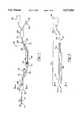
- FIG. 1is a schematic diagram illustrating the apparatus of the present invention
- FIG. 2is a cross-sectional side view of the invention of the present invention showing the apparatus of the present invention inserted into a catheter;
- FIG. 3is a sectional view of the apparatus of the present invention taken along line 3--3 in FIG. 2;
- FIG. 4is a sectional view of the locking device of the present invention.
- FIG. 5is a sectional view of the catheter taken along line 5--5 of FIG. 1.
- an apparatus generally indicated at 10for use with a central venous catheter 11 for measuring central venous oxygen saturation (ScvO 2 ).
- the apparatus 10comprises a fiber optic bundle generally shown at 12 having a distal end 14 and a proximal end 16.
- the fiber optic bundle 12includes afferent and efferent light-conducting fibers 18 for sending signals and receiving signals for generating oxygen saturation measurements.
- a sheath 28is disposed about the fiber optic bundle 12 and encapsulates and protects the fiber optic bundle 12 and exposes the distal end 14 of the fiber optic bundle 12.
- the fiber optic bundle 12includes a distal portion 20 and a proximal portion 22.
- the distal portion 20includes the region of the fiber optic bundle 12 that is inserted into the standard venous catheter 11 such as an Product No. CS-17702, Arrow International, Inc., Reading, Pa.
- the proximal portion 22 of the fiber optic bundle 12includes the region that transmits light signals to and from a light generator/detector where the light is analyzed yielding a measurement of venous oxygen saturation.
- the fibers 18spectrophotometrically reflect light transmitted therethrough. The light is transmitted from a light source through the fiber optic bundle 12 into the blood.
- the fiber optic bundle 12is disposed in a sheath or cover 28 which both encapsulates and protects the fiber optic bundle 12 as shown in FIG. 3.
- the sheath 28can also define a lumen 26 disposed therein extending longitudinally the entire length of the distal portion 20 of the fiber optic bundle 12.
- a preferred embodiment of the present inventionprovides a sheath 28 which extends along the distal portion 20 of the fiber optic bundle 12 from the distal end 14 of the bundle 12 to an optional junction 29. At the distal end 14 of the distal portion of the fiber optic bundle 12 as shown in FIG.
- the sheath 28is absent, thereby allowing for the fiber optic bundle 12 to be in direct contact with the blood for taking oxygen saturation measurements and also to expose the distal or open end 27 of the lumen 26 thereby facilitating sampling, pressure measurement, or fluid/pharmaceutical administration.
- the sheath 12includes markings or delineations which are located at preselected distances from the distal end 14 of the bundle 12 which provide a visual indication of fiber optic bundle insertion depth. This indication of fiber optic bundle insertion depth aids the practitioner in proper placement of the bundle within a patient.
- the apparatus 10includes the optional junction 29.
- the sheath 28defines a single lumen 26 which extends from the junction 29 the entire length of the distal portion 20 of the fiber optic bundle 12.
- the junction 29permits access to the lumen(s) 26 through a port 34 and also is the location where the distal portion 20 of the fiber optic bundle 12 becomes separated from the lumen 26.
- the proximal portion 22 of the fiber optic bundle 12extends to a connector body 36 which is then connected to the light generator/detector (not shown), such as an Oximetry 3 Oximetry System, Abbott Critical Care Systems, Mountainview, Calif., which can be computer controlled.
- the porn 34 extending from the junction 29can include a tube having a connector 35 attached thereto.
- the outer diameter of the sheath 28 containing the fiber optic bundle 12should be less than the inner diameter of the lumen or port of the catheter 11 into which the fiber optic bundle 12 is inserted.
- the sheath 28is preferably constructed of a self-lubricating material to facilitate insertion and removal of the 28 encapsulated fiber optic bundle 12 from a catheter 11. Such a self-lubricating material could include silicone.
- the locking device 40is adapted to both connect the apparatus 10 of the present invention to a standard venous catheter 11 and to fix or retain the distal portion 20 of the fiber optic bundle 12 with respect to the catheter 11 when the fiber optic bundle 12 is inserted into the catheter 11.
- the locking device 40enables more accurate oxygen saturation measurement to be taken since it prevents movement of the fiber optic bundle 12 once it has be properly positioned in the blood vessel. Additionally, the locking device 40 does not require stitching of the fiber optic bundle 12 to the patient's skin to prevent movement of the fiber optic bundle 12, thereby eliminating a potential source of infection.
- the locking device 40includes a connector 42 operatively connected in fluid communication to the locking device 40 for attaching the locking device 40 to the catheter 11.
- the connector 42can be any suitable connector, such as a quick-release or Luer-type connector, or other screw down-type connector known to those skilled in the art.
- the locking device 40can include a threaded male insert 44 in mating engagement with a female locking member 46.
- the locking member 46includes a flange 47 having a resilient insert 48 abutting thereto.
- the flange 47 and the inserteach have an opening 50,52, respectively, to allow for the insertion and support of the distal portion 22 of the fiber optic bundle 12.
- the resilient insert 48can be constructed of any suitable material which, when the locking member 46 is matingly engaged with the male insert 44, compresses the resilient insert 48 causing its opening to become smaller 52.
- the insert 48compresses and engages the sheath 28 over the fiber optic bundle 12 thereby preventing movement of the distal portion 20 of the fiber optic bundle 12 within the locking device 40 and within the catheter 11.
- the region of the distal portion 20 of the fiber optic bundle 12 to which the locking device 40 engagescan include a reinforcement 54 disposed about the sheath 28 or integrally formed with the sheath 28 to prevent damage to the sheath 28 and fiber optic bundle 12 caused by the compression forces of the locking device 40.
- This reinforcement 54can include a concentric layer of a harder or higher durometer plastic material such as silicone, PVC, polypropylene, metal or metal alloy, etc.
- the locking device 40can further include a protective sleeve 56 fixedly attached to the locking member 46 for protecting and maintaining sterility of the sheath 28.
- the protective sleeve 56allows the sheath 28 encapsulated fiber optic bundle 12 to pass therethrough.
- the protective sleeve 52can be formed of any suitable material, such as cellophane or polyvinylidene chloride which allows the sleeve 52 to contract or expand in an "accordion-like" fashion when the fiber optic bundle 12 is inserted or removed to constantly cover and maintain sterile the distal portion 20 of the fiber optic bundle 12 and sheath 28.
- the catheter 11 used in conjunction with the present inventionincludes a body 62 forming at least one and preferably more than one lumen 64 therein.
- the catheter body 62has a distal 66 and a proximal 68 end.
- the lumen(s) 64extend(s) longitudinally within the catheter body 62 from a distal opening(s) 70 to a proximal port(s) 72.
- the catheter sheath 28can also include markings or delineations which provide a practitioner with information about the depth of the catheter insertion into a patient. The information provided to the practitioner by these markings aids in the proper insertion and placement of the catheter 11 into the patient.
- the catheter body 62includes a blood vessel insertion portion 74 and a lumen junction 76.
- the blood vessel insertion portion 74is adapted to be inserted into a blood vessel, such as the subclavian vein, supraclavicular vein, and internal jugular vein, and preferably has a length of at least 20 cm.
- the blood vessel insertion portion 74should be capable of flexing to facilitate insertion of the catheter 11 into the blood vessel but should also have sufficient rigidity such that it will not be unduly flexed under the force of turbulent blood flow.
- the blood vessel insertion portion 74also includes an opening 75 located at the distal end 66 of the catheter body 62. This opening 75 permits the distal end 20 of the fiber optic bundle 12 to exit or protrude beyond the blood vessel insertion portion 74 of the catheter body 62.
- the blood vessel insertion portion 74 of the catheter body 62can be constructed of any suitable biologically compatible material, such as polyurethane, known to those skilled in the art.
- the lumen junction 76is the portion of the catheter body 62 at which point each lumen 64 included in the blood vessel insertion portion 74 of the catheter body 62 is translated into separate port(s) or tubes 72 such as for a sampling port or an insertion port for the fiber optic bundle 12.
- a two lumen catheter 11has two ports which proximally extend from the lumen junction 76 each port permitting access to each individual lumen 64 for inserting or withdrawing fluids therethrough and/or inserting or withdrawing the fiber optic bundle 12.
- Each port 72has a connector 73 which allows the port 72 to be connected to other devices or fittings such as a hypodermic needle or intravenous fluid injection port or to a fitting which allows connection and insertion of a fiber optic bundle 12.
- the connector 73can be any suitable type connector known in the art, such as a quick-release or Luer-type connector or a screw down-type connector.
- a standard central venous catheter 11 having at least one lumenis percutaneously inserted, properly positioned and secured into the central venous system through the subclavian vein.
- the connector 42 of the locking device 40would then be connected to a fitting 73 on the catheter 11 to allow for the insertion of the fiber optic bundle 12 within the catheter 11.
- the fiber optic bundle 12would then be inserted through the locking device into the catheter 11 and positioned such that the distal tip of the fiber optic bundle 12 extends beyond the distal opening of the catheter 11 preferably into the right atrium.
- the markings or delineations on the catheter sheath 28would provide information about the depth of the catheter insertion within the patient.
- the location of the fiber optic bundle 12is then fixed relative to the catheter 11 by applying the locking member to the sheath covering the fiber optic bundle 12.
- the apparatus 10Prior to insertion of the fiber optic bundle 12 in situ (in a blood vessel), the apparatus 10 is electronically calibrated. Then, using a sample of venous blood taken from the patient, the fiber optic bundle 12 can be re-calibrated in situ, if necessary.
- the fiber optic bundle 12can be removed and the catheter 11 used for other purposes. Utilizing the apparatus 10 of the present invention, a fiber optic bundle 12 can be reinserted into the original catheter 11 thereby eliminating the expense of replacing the catheter 11. This would also reduce the delay in time, associated expense, and patient discomfort which would have been caused by having to replace the catheter 11 and take an x-ray to ascertain proper placement of the catheter 11.
Landscapes
- Health & Medical Sciences (AREA)
- Physics & Mathematics (AREA)
- Life Sciences & Earth Sciences (AREA)
- Medical Informatics (AREA)
- Surgery (AREA)
- Biophysics (AREA)
- Pathology (AREA)
- Engineering & Computer Science (AREA)
- Biomedical Technology (AREA)
- Heart & Thoracic Surgery (AREA)
- Spectroscopy & Molecular Physics (AREA)
- Molecular Biology (AREA)
- Optics & Photonics (AREA)
- Animal Behavior & Ethology (AREA)
- General Health & Medical Sciences (AREA)
- Public Health (AREA)
- Veterinary Medicine (AREA)
- Measurement Of The Respiration, Hearing Ability, Form, And Blood Characteristics Of Living Organisms (AREA)
- Investigating Or Analysing Materials By Optical Means (AREA)
- Investigating Or Analysing Biological Materials (AREA)
Abstract
Description
Claims (8)
Priority Applications (10)
| Application Number | Priority Date | Filing Date | Title |
|---|---|---|---|
| US08/512,462US5673694A (en) | 1995-08-08 | 1995-08-08 | Method and apparatus for continuous measurement of central venous oxygen saturation |
| EP96926215AEP0825827B1 (en) | 1995-08-08 | 1996-07-31 | Device for central venous oxygen measurement |
| DE69636596TDE69636596T2 (en) | 1995-08-08 | 1996-07-31 | DEVICE FOR MEASURING THE CENTRAL VENTILATED OXYGEN SATURATION |
| CA002202038ACA2202038C (en) | 1995-08-08 | 1996-07-31 | Central venous oxygen saturation measurement |
| AU66442/96AAU709716B2 (en) | 1995-08-08 | 1996-07-31 | Central venous oxygen saturation measurement |
| AT96926215TATE341272T1 (en) | 1995-08-08 | 1996-07-31 | DEVICE FOR MEASURING CENTRAL VENOUS OXYGEN SATURATION |
| JP9508529AJPH11501553A (en) | 1995-08-08 | 1996-07-31 | Central venous oxygen saturation measurement |
| PCT/US1996/012571WO1997005820A1 (en) | 1995-08-08 | 1996-07-31 | Central venous oxygen saturation measurement |
| CA002634810ACA2634810A1 (en) | 1995-08-08 | 1996-07-31 | Central venous oxygen saturation measurement |
| EP05013649AEP1579800A1 (en) | 1995-08-08 | 2005-06-24 | Central venous oxygen saturation measurement |
Applications Claiming Priority (1)
| Application Number | Priority Date | Filing Date | Title |
|---|---|---|---|
| US08/512,462US5673694A (en) | 1995-08-08 | 1995-08-08 | Method and apparatus for continuous measurement of central venous oxygen saturation |
Publications (1)
| Publication Number | Publication Date |
|---|---|
| US5673694Atrue US5673694A (en) | 1997-10-07 |
Family
ID=24039200
Family Applications (1)
| Application Number | Title | Priority Date | Filing Date |
|---|---|---|---|
| US08/512,462Expired - LifetimeUS5673694A (en) | 1995-08-08 | 1995-08-08 | Method and apparatus for continuous measurement of central venous oxygen saturation |
Country Status (8)
| Country | Link |
|---|---|
| US (1) | US5673694A (en) |
| EP (2) | EP0825827B1 (en) |
| JP (1) | JPH11501553A (en) |
| AT (1) | ATE341272T1 (en) |
| AU (1) | AU709716B2 (en) |
| CA (2) | CA2634810A1 (en) |
| DE (1) | DE69636596T2 (en) |
| WO (1) | WO1997005820A1 (en) |
Cited By (50)
| Publication number | Priority date | Publication date | Assignee | Title |
|---|---|---|---|---|
| US6005658A (en)* | 1997-04-18 | 1999-12-21 | Hewlett-Packard Company | Intermittent measuring of arterial oxygen saturation of hemoglobin |
| US6144444A (en)* | 1998-11-06 | 2000-11-07 | Medtronic Avecor Cardiovascular, Inc. | Apparatus and method to determine blood parameters |
| US6231514B1 (en) | 1996-06-26 | 2001-05-15 | Tobo, Llc | Device for use in temporary insertion of a sensor within a patient's body |
| US6415851B1 (en)* | 1999-12-21 | 2002-07-09 | Visteon Global Technologies, Inc. | Multi-zone temperature control system for HVAC air-handling assembly |
| US20040015061A1 (en)* | 2002-07-16 | 2004-01-22 | Clifford Currier | Central venous catheter having a soft tip and fiber optics |
| US20040064021A1 (en)* | 2002-09-28 | 2004-04-01 | Pulsion Medical Systems Ag | Catheter system |
| US6716176B1 (en) | 2002-09-30 | 2004-04-06 | Tobo, Llc | Device for use in temporary insertion of a sensor within a patient's body |
| US20040082841A1 (en)* | 2002-10-24 | 2004-04-29 | Furnary Anthony P. | Method and apparatus for monitoring blood condition and cardiopulmonary function |
| US20040220455A1 (en)* | 1996-06-26 | 2004-11-04 | Lowe Robert I. | Method for monitoring blood characteristics and cardiopulmonary function |
| US6819951B2 (en) | 2002-09-24 | 2004-11-16 | Mayo Foundation For Medical Education And Research | Peripherally inserted central catheter with continuous central venous oximetry and proximal high flow port |
| US6976986B2 (en) | 2000-04-12 | 2005-12-20 | Afx, Inc. | Electrode arrangement for use in a medical instrument |
| US20060009740A1 (en)* | 2001-08-28 | 2006-01-12 | Michael Higgins | Multiple lumen catheter having a soft tip |
| US7029467B2 (en) | 2002-07-16 | 2006-04-18 | Edwards Lifesciences Corporation | Multiple lumen catheter having a soft tip |
| US7052491B2 (en) | 1998-10-23 | 2006-05-30 | Afx, Inc. | Vacuum-assisted securing apparatus for a microwave ablation instrument |
| US7099717B2 (en) | 2002-01-03 | 2006-08-29 | Afx Inc. | Catheter having improved steering |
| US20060224053A1 (en)* | 2005-03-30 | 2006-10-05 | Skyline Biomedical, Inc. | Apparatus and method for non-invasive and minimally-invasive sensing of venous oxygen saturation and pH levels |
| US7192427B2 (en) | 2002-02-19 | 2007-03-20 | Afx, Inc. | Apparatus and method for assessing transmurality of a tissue ablation |
| US7226446B1 (en) | 1999-05-04 | 2007-06-05 | Dinesh Mody | Surgical microwave ablation assembly |
| WO2007097754A1 (en)* | 2006-02-22 | 2007-08-30 | Dexcom, Inc. | Analyte sensor |
| US7303560B2 (en) | 2000-12-29 | 2007-12-04 | Afx, Inc. | Method of positioning a medical instrument |
| US7346399B2 (en) | 1999-05-28 | 2008-03-18 | Afx, Inc. | Monopole tip for ablation catheter |
| US7613491B2 (en) | 2002-05-22 | 2009-11-03 | Dexcom, Inc. | Silicone based membranes for use in implantable glucose sensors |
| EP2157462A1 (en)* | 2008-08-22 | 2010-02-24 | Pulsion Medical Systems AG | Fiber-optic probe |
| JP2010524598A (en)* | 2007-05-02 | 2010-07-22 | セント ヴィンセンツ ホスピタル(メルボルン)リミテッド | Noninvasive measurement of blood oxygen saturation |
| US7885697B2 (en) | 2004-07-13 | 2011-02-08 | Dexcom, Inc. | Transcutaneous analyte sensor |
| US20110118577A1 (en)* | 2007-07-31 | 2011-05-19 | Up Management Gmbh | Catheter system having an optical probe and method for the application of an optical probe in a catheter system |
| US20110201908A1 (en)* | 2010-02-17 | 2011-08-18 | Farkas Joshua D | Intermittent extracorporeal spectrophotometry |
| US8133178B2 (en) | 2006-02-22 | 2012-03-13 | Dexcom, Inc. | Analyte sensor |
| US8290559B2 (en) | 2007-12-17 | 2012-10-16 | Dexcom, Inc. | Systems and methods for processing sensor data |
| US8364229B2 (en) | 2003-07-25 | 2013-01-29 | Dexcom, Inc. | Analyte sensors having a signal-to-noise ratio substantially unaffected by non-constant noise |
| US8417312B2 (en) | 2007-10-25 | 2013-04-09 | Dexcom, Inc. | Systems and methods for processing sensor data |
| US8562558B2 (en) | 2007-06-08 | 2013-10-22 | Dexcom, Inc. | Integrated medicament delivery device for use with continuous analyte sensor |
| US8615282B2 (en) | 2004-07-13 | 2013-12-24 | Dexcom, Inc. | Analyte sensor |
| US9135402B2 (en) | 2007-12-17 | 2015-09-15 | Dexcom, Inc. | Systems and methods for processing sensor data |
| US9247900B2 (en) | 2004-07-13 | 2016-02-02 | Dexcom, Inc. | Analyte sensor |
| WO2017123764A1 (en)* | 2016-01-12 | 2017-07-20 | Bloodworks, Llc | Peripheral fiberoptic intravascular blood metric probe modular device and method |
| US9717446B2 (en) | 2010-07-09 | 2017-08-01 | St. Vincent's Hospital (Melbourne) Limited | Non-invasive measurement of blood oxygen saturation |
| US9737213B1 (en)* | 2009-03-24 | 2017-08-22 | Vioptix, Inc. | Using an oximeter probe to detect intestinal ischemia |
| US9763609B2 (en) | 2003-07-25 | 2017-09-19 | Dexcom, Inc. | Analyte sensors having a signal-to-noise ratio substantially unaffected by non-constant noise |
| US9986942B2 (en) | 2004-07-13 | 2018-06-05 | Dexcom, Inc. | Analyte sensor |
| US10610136B2 (en) | 2005-03-10 | 2020-04-07 | Dexcom, Inc. | System and methods for processing analyte sensor data for sensor calibration |
| US10791928B2 (en) | 2007-05-18 | 2020-10-06 | Dexcom, Inc. | Analyte sensors having a signal-to-noise ratio substantially unaffected by non-constant noise |
| US10835672B2 (en) | 2004-02-26 | 2020-11-17 | Dexcom, Inc. | Integrated insulin delivery system with continuous glucose sensor |
| US10966609B2 (en) | 2004-02-26 | 2021-04-06 | Dexcom, Inc. | Integrated medicament delivery device for use with continuous analyte sensor |
| US11246990B2 (en) | 2004-02-26 | 2022-02-15 | Dexcom, Inc. | Integrated delivery device for continuous glucose sensor |
| US11331022B2 (en) | 2017-10-24 | 2022-05-17 | Dexcom, Inc. | Pre-connected analyte sensors |
| US11350862B2 (en) | 2017-10-24 | 2022-06-07 | Dexcom, Inc. | Pre-connected analyte sensors |
| US11918782B2 (en) | 2006-06-30 | 2024-03-05 | Abbott Diabetes Care Inc. | Integrated analyte sensor and infusion device and methods therefor |
| US12315630B2 (en) | 2009-08-31 | 2025-05-27 | Abbott Diabetes Care Inc. | Medical devices and methods |
| US12369821B2 (en) | 2018-06-07 | 2025-07-29 | Abbott Diabetes Care Inc. | Focused sterilization and sterilized sub-assemblies for analyte monitoring systems |
Families Citing this family (2)
| Publication number | Priority date | Publication date | Assignee | Title |
|---|---|---|---|---|
| US8553223B2 (en) | 2010-03-31 | 2013-10-08 | Covidien Lp | Biodegradable fibers for sensing |
| US9180260B2 (en) | 2013-08-30 | 2015-11-10 | Covidien Lp | Systems and methods for monitoring an injection procedure |
Citations (4)
| Publication number | Priority date | Publication date | Assignee | Title |
|---|---|---|---|---|
| US5271398A (en)* | 1991-10-09 | 1993-12-21 | Optex Biomedical, Inc. | Intra-vessel measurement of blood parameters |
| US5284138A (en)* | 1991-07-09 | 1994-02-08 | C. R. Bard, Inc. | Apparatus and method for positioning a sensor away from the blood vessel wall |
| US5315995A (en)* | 1992-03-19 | 1994-05-31 | Henry Ford Hospital | Method and apparatus for continuous measurement of central venous oxygen saturation during human cardiopulmonary resuscitation and clinical shock |
| US5435308A (en)* | 1992-07-16 | 1995-07-25 | Abbott Laboratories | Multi-purpose multi-parameter cardiac catheter |
Family Cites Families (4)
| Publication number | Priority date | Publication date | Assignee | Title |
|---|---|---|---|---|
| DE3215879A1 (en)* | 1982-04-29 | 1983-11-03 | Fa. Carl Zeiss, 7920 Heidenheim | DEVICE FOR SPECTRUM MEASUREMENT IN THE BLOOD RAIL |
| US4718423A (en)* | 1986-10-17 | 1988-01-12 | Spectramed, Inc. | Multiple-function cardiovascular catheter system with very high lumenal efficiency and no crossovers |
| US4830013A (en)* | 1987-01-30 | 1989-05-16 | Minnesota Mining And Manufacturing Co. | Intravascular blood parameter measurement system |
| DE4024677A1 (en)* | 1990-08-03 | 1992-02-13 | Winter & Ibe Olympus | Medical clamp for monomode optical fibre light conductor - uses clamp elements acting against sleeve enclosing optical fibre conductor |
- 1995
- 1995-08-08USUS08/512,462patent/US5673694A/ennot_activeExpired - Lifetime
- 1996
- 1996-07-31DEDE69636596Tpatent/DE69636596T2/ennot_activeExpired - Lifetime
- 1996-07-31EPEP96926215Apatent/EP0825827B1/ennot_activeExpired - Lifetime
- 1996-07-31ATAT96926215Tpatent/ATE341272T1/ennot_activeIP Right Cessation
- 1996-07-31AUAU66442/96Apatent/AU709716B2/ennot_activeExpired
- 1996-07-31WOPCT/US1996/012571patent/WO1997005820A1/enactiveIP Right Grant
- 1996-07-31CACA002634810Apatent/CA2634810A1/ennot_activeAbandoned
- 1996-07-31CACA002202038Apatent/CA2202038C/ennot_activeExpired - Lifetime
- 1996-07-31JPJP9508529Apatent/JPH11501553A/enactivePending
- 2005
- 2005-06-24EPEP05013649Apatent/EP1579800A1/ennot_activeWithdrawn
Patent Citations (4)
| Publication number | Priority date | Publication date | Assignee | Title |
|---|---|---|---|---|
| US5284138A (en)* | 1991-07-09 | 1994-02-08 | C. R. Bard, Inc. | Apparatus and method for positioning a sensor away from the blood vessel wall |
| US5271398A (en)* | 1991-10-09 | 1993-12-21 | Optex Biomedical, Inc. | Intra-vessel measurement of blood parameters |
| US5315995A (en)* | 1992-03-19 | 1994-05-31 | Henry Ford Hospital | Method and apparatus for continuous measurement of central venous oxygen saturation during human cardiopulmonary resuscitation and clinical shock |
| US5435308A (en)* | 1992-07-16 | 1995-07-25 | Abbott Laboratories | Multi-purpose multi-parameter cardiac catheter |
Non-Patent Citations (30)
| Title |
|---|
| Ander et al., "Continuous central venous oxygen saturation monitoring as an adjunct in the treatment of cardiac arrest . . ." Clinical Intensive Care, 5:323-240 (1994). |
| Ander et al., "Continuous central venous oxygen saturation monitoring in the resuscitation of hemodynamically . ." Abstracts of Posters 14th Int'l Sym. on Intensive Care and Emerg. Med., vol. 6, No. 2 (1995). |
| Ander et al., Continuous central venous oxygen saturation monitoring as an adjunct in the treatment of cardiac arrest . . . Clinical Intensive Care , 5:323 240 (1994).* |
| Ander et al., Continuous central venous oxygen saturation monitoring in the resuscitation of hemodynamically . . Abstracts of Posters 14th Int l Sym. on Intensive Care and Emerg. Med. , vol. 6, No. 2 (1995).* |
| Jaggi et al., "Occult cardiogenic shock in end-stage heart failure patients presenting to the Emergency Department" Supplement to Clinical Intensive Care, vol. 6, No. 2, p. 104 (1995). |
| Jaggi et al., Occult cardiogenic shock in end stage heart failure patients presenting to the Emergency Department Supplement to Clinical Intensive Care , vol. 6, No. 2, p. 104 (1995).* |
| Kowalenko et al., "Continuous central venous oxygen saturation monitoring during the resuscitation . . ." SAIM 1994 Ann. Mtg. Abstracts Academic Emergency Medincine, vol. 1, No. 2 A69 (1994). |
| Kowalenko et al., Continuous central venous oxygen saturation monitoring during the resuscitation . . . SAIM 1994 Ann. Mtg. Abstracts Academic Emergency Medincine , vol. 1, No. 2 A69 (1994).* |
| Rady et al., "Continuous central venous oximetry and shock index in the emergency department --use in the evaluation of clinical shock" American Journal of Emerg. Med., vol. 10, No. 6, pp. 538-541 (1992). |
| Rady et al., "Continuous central venous oximety for the evaluation and treatment of acute cardiac failure in the emergency department" International: Journal of Intensive Care, vol. 1:64-65 Summer (1994). |
| Rady et al., "The responses of blood pressure, heart rate, shock index, central venous . ." Critical Care Medicine, A138 Jan. (1995). |
| Rady et al., Continuous central venous oximetry and shock index in the emergency department use in the evaluation of clinical shock American Journal of Emerg. Med. , vol. 10, No. 6, pp. 538 541 (1992).* |
| Rady et al., Continuous central venous oximety for the evaluation and treatment of acute cardiac failure in the emergency department International: Journal of Intensive Care , vol. 1:64 65 Summer (1994).* |
| Rady et al., The responses of blood pressure, heart rate, shock index, central venous . . Critical Care Medicine , A138 Jan. (1995).* |
| Rady, "The role of central venous oximetry, lactic acid concentration and shock index in the evaluation of clinical shock: a review" Resuscitation, 24:55-60 (1992). |
| Rady, The role of central venous oximetry, lactic acid concentration and shock index in the evaluation of clinical shock: a review Resuscitation , 24:55 60 (1992).* |
| Rivers et al., "Continuous central venous oxygen saturation . . ." Michigan Emergency Physician, News & Views, vol. XIII, No. 3 May and continued in vol. XIII, No. 4 Jun./Jul. (1994). |
| Rivers et al., "Coronary perfusion pressure, end-tidal carbon dioxide concentration and continuous central venous oxygen . ." Supplement to Clinical Intensive Care, vol. 3, No. 2, p. 100 (1992). |
| Rivers et al., "The clinical implications of continuous central venous oxygen saturation during human CPR" Annals of Emergency Medicine, 21:9 Sep. (1992). |
| Rivers et al., "The clinical implications of continuous central venous oxygen saturation during human CPR" In Emergency Medicine, Chapter 1 -Acute Systems Pathophysiology, pp. 16-17. |
| Rivers et al., "The effect of the total cumulative epinerphrine dose administered during human CPR . . ." Chest, 106:5, pp. 1499-1507 Nov. (1994). |
| Rivers et al., "Venous hyperoxia after cardiac arrest" Chest, vol. 102, pp. 1787-1793, Dec. (1992). |
| Rivers et al., Continuous central venous oxygen saturation . . . Michigan Emergency Physician , News & Views, vol. XIII, No. 3 May and continued in vol. XIII, No. 4 Jun./Jul. (1994).* |
| Rivers et al., Coronary perfusion pressure, end tidal carbon dioxide concentration and continuous central venous oxygen . . Supplement to Clinical Intensive Care , vol. 3, No. 2, p. 100 (1992).* |
| Rivers et al., The clinical implications of continuous central venous oxygen saturation during human CPR Annals of Emergency Medicine , 21:9 Sep. (1992).* |
| Rivers et al., The clinical implications of continuous central venous oxygen saturation during human CPR In Emergency Medicine , Chapter 1 Acute Systems Pathophysiology, pp. 16 17.* |
| Rivers et al., The effect of the total cumulative epinerphrine dose administered during human CPR . . . Chest , 106:5, pp. 1499 1507 Nov. (1994).* |
| Rivers et al., Venous hyperoxia after cardiac arrest Chest , vol. 102, pp. 1787 1793, Dec. (1992).* |
| Rivers, et al., "Coronary perfusion pressure, end-tidal carbon dioxide concentration and continuous central venous oxygen . ." Abstracts of Papers, Presentations Cardiopulmonary Resuscitation S85. |
| Rivers, et al., Coronary perfusion pressure, end tidal carbon dioxide concentration and continuous central venous oxygen . . Abstracts of Papers, Presentations Cardiopulmonary Resuscitation S85.* |
Cited By (148)
| Publication number | Priority date | Publication date | Assignee | Title |
|---|---|---|---|---|
| US6231514B1 (en) | 1996-06-26 | 2001-05-15 | Tobo, Llc | Device for use in temporary insertion of a sensor within a patient's body |
| US20040220455A1 (en)* | 1996-06-26 | 2004-11-04 | Lowe Robert I. | Method for monitoring blood characteristics and cardiopulmonary function |
| US6005658A (en)* | 1997-04-18 | 1999-12-21 | Hewlett-Packard Company | Intermittent measuring of arterial oxygen saturation of hemoglobin |
| US7052491B2 (en) | 1998-10-23 | 2006-05-30 | Afx, Inc. | Vacuum-assisted securing apparatus for a microwave ablation instrument |
| US7387627B2 (en) | 1998-10-23 | 2008-06-17 | Maquet Cardiovascular Llc | Vacuum-assisted securing apparatus for a microwave ablation instrument |
| US7115126B2 (en) | 1998-10-23 | 2006-10-03 | Afx Inc. | Directional microwave ablation instrument with off-set energy delivery portion |
| US6144444A (en)* | 1998-11-06 | 2000-11-07 | Medtronic Avecor Cardiovascular, Inc. | Apparatus and method to determine blood parameters |
| US7226446B1 (en) | 1999-05-04 | 2007-06-05 | Dinesh Mody | Surgical microwave ablation assembly |
| US7346399B2 (en) | 1999-05-28 | 2008-03-18 | Afx, Inc. | Monopole tip for ablation catheter |
| US6415851B1 (en)* | 1999-12-21 | 2002-07-09 | Visteon Global Technologies, Inc. | Multi-zone temperature control system for HVAC air-handling assembly |
| US7156841B2 (en) | 2000-04-12 | 2007-01-02 | Afx, Inc. | Electrode arrangement for use in a medical instrument |
| US6976986B2 (en) | 2000-04-12 | 2005-12-20 | Afx, Inc. | Electrode arrangement for use in a medical instrument |
| US7303560B2 (en) | 2000-12-29 | 2007-12-04 | Afx, Inc. | Method of positioning a medical instrument |
| US20060009740A1 (en)* | 2001-08-28 | 2006-01-12 | Michael Higgins | Multiple lumen catheter having a soft tip |
| US7099717B2 (en) | 2002-01-03 | 2006-08-29 | Afx Inc. | Catheter having improved steering |
| US7192427B2 (en) | 2002-02-19 | 2007-03-20 | Afx, Inc. | Apparatus and method for assessing transmurality of a tissue ablation |
| US8064977B2 (en) | 2002-05-22 | 2011-11-22 | Dexcom, Inc. | Silicone based membranes for use in implantable glucose sensors |
| US7613491B2 (en) | 2002-05-22 | 2009-11-03 | Dexcom, Inc. | Silicone based membranes for use in implantable glucose sensors |
| US10052051B2 (en) | 2002-05-22 | 2018-08-21 | Dexcom, Inc. | Silicone based membranes for use in implantable glucose sensors |
| US11020026B2 (en) | 2002-05-22 | 2021-06-01 | Dexcom, Inc. | Silicone based membranes for use in implantable glucose sensors |
| US9549693B2 (en) | 2002-05-22 | 2017-01-24 | Dexcom, Inc. | Silicone based membranes for use in implantable glucose sensors |
| US8543184B2 (en) | 2002-05-22 | 2013-09-24 | Dexcom, Inc. | Silicone based membranes for use in implantable glucose sensors |
| US7029467B2 (en) | 2002-07-16 | 2006-04-18 | Edwards Lifesciences Corporation | Multiple lumen catheter having a soft tip |
| US6999809B2 (en) | 2002-07-16 | 2006-02-14 | Edwards Lifesciences Corporation | Central venous catheter having a soft tip and fiber optics |
| US20040015061A1 (en)* | 2002-07-16 | 2004-01-22 | Clifford Currier | Central venous catheter having a soft tip and fiber optics |
| US20050054975A1 (en)* | 2002-09-24 | 2005-03-10 | Bhavesh Patel | Peripherally inserted central catheter with continuous central venous oximetry and proximal high flow port |
| US7458938B2 (en) | 2002-09-24 | 2008-12-02 | Mayo Foundation For Medical Education & Research | Peripherally inserted central catheter with continuous central venous oximetry and proximal high flow port |
| US6819951B2 (en) | 2002-09-24 | 2004-11-16 | Mayo Foundation For Medical Education And Research | Peripherally inserted central catheter with continuous central venous oximetry and proximal high flow port |
| EP1402917A3 (en)* | 2002-09-28 | 2004-05-12 | Pulsion Medical Systems AG | Catheter comprising an optical fiber |
| US20040064021A1 (en)* | 2002-09-28 | 2004-04-01 | Pulsion Medical Systems Ag | Catheter system |
| US6954665B2 (en) | 2002-09-28 | 2005-10-11 | Pulsion Medical Systems Ag | Catheter system |
| US6716176B1 (en) | 2002-09-30 | 2004-04-06 | Tobo, Llc | Device for use in temporary insertion of a sensor within a patient's body |
| US20060149145A1 (en)* | 2002-10-24 | 2006-07-06 | Furnary Anthony P | Method and apparatus for monitoring blood condition and cardiopulmonary function |
| US7010337B2 (en) | 2002-10-24 | 2006-03-07 | Furnary Anthony P | Method and apparatus for monitoring blood condition and cardiopulmonary function |
| US8078249B2 (en) | 2002-10-24 | 2011-12-13 | Sensicor, Llc | Method and apparatus for monitoring blood condition and cardiopulmonary function |
| US20040082841A1 (en)* | 2002-10-24 | 2004-04-29 | Furnary Anthony P. | Method and apparatus for monitoring blood condition and cardiopulmonary function |
| US9763609B2 (en) | 2003-07-25 | 2017-09-19 | Dexcom, Inc. | Analyte sensors having a signal-to-noise ratio substantially unaffected by non-constant noise |
| US10376143B2 (en) | 2003-07-25 | 2019-08-13 | Dexcom, Inc. | Analyte sensors having a signal-to-noise ratio substantially unaffected by non-constant noise |
| US8364229B2 (en) | 2003-07-25 | 2013-01-29 | Dexcom, Inc. | Analyte sensors having a signal-to-noise ratio substantially unaffected by non-constant noise |
| US12226617B2 (en) | 2004-02-26 | 2025-02-18 | Dexcom, Inc. | Integrated delivery device for continuous glucose sensor |
| US12102410B2 (en) | 2004-02-26 | 2024-10-01 | Dexcom, Inc | Integrated medicament delivery device for use with continuous analyte sensor |
| US10966609B2 (en) | 2004-02-26 | 2021-04-06 | Dexcom, Inc. | Integrated medicament delivery device for use with continuous analyte sensor |
| US11246990B2 (en) | 2004-02-26 | 2022-02-15 | Dexcom, Inc. | Integrated delivery device for continuous glucose sensor |
| US12115357B2 (en) | 2004-02-26 | 2024-10-15 | Dexcom, Inc. | Integrated delivery device for continuous glucose sensor |
| US10835672B2 (en) | 2004-02-26 | 2020-11-17 | Dexcom, Inc. | Integrated insulin delivery system with continuous glucose sensor |
| US10918314B2 (en) | 2004-07-13 | 2021-02-16 | Dexcom, Inc. | Analyte sensor |
| US10932700B2 (en) | 2004-07-13 | 2021-03-02 | Dexcom, Inc. | Analyte sensor |
| US10918315B2 (en) | 2004-07-13 | 2021-02-16 | Dexcom, Inc. | Analyte sensor |
| US10918313B2 (en) | 2004-07-13 | 2021-02-16 | Dexcom, Inc. | Analyte sensor |
| US8615282B2 (en) | 2004-07-13 | 2013-12-24 | Dexcom, Inc. | Analyte sensor |
| US11883164B2 (en) | 2004-07-13 | 2024-01-30 | Dexcom, Inc. | System and methods for processing analyte sensor data for sensor calibration |
| US8792953B2 (en) | 2004-07-13 | 2014-07-29 | Dexcom, Inc. | Transcutaneous analyte sensor |
| US10827956B2 (en) | 2004-07-13 | 2020-11-10 | Dexcom, Inc. | Analyte sensor |
| US10813576B2 (en) | 2004-07-13 | 2020-10-27 | Dexcom, Inc. | Analyte sensor |
| US10799158B2 (en) | 2004-07-13 | 2020-10-13 | Dexcom, Inc. | Analyte sensor |
| US11064917B2 (en) | 2004-07-13 | 2021-07-20 | Dexcom, Inc. | Analyte sensor |
| US10799159B2 (en) | 2004-07-13 | 2020-10-13 | Dexcom, Inc. | Analyte sensor |
| US9247900B2 (en) | 2004-07-13 | 2016-02-02 | Dexcom, Inc. | Analyte sensor |
| US11045120B2 (en) | 2004-07-13 | 2021-06-29 | Dexcom, Inc. | Analyte sensor |
| US9414777B2 (en) | 2004-07-13 | 2016-08-16 | Dexcom, Inc. | Transcutaneous analyte sensor |
| US7885697B2 (en) | 2004-07-13 | 2011-02-08 | Dexcom, Inc. | Transcutaneous analyte sensor |
| US9668677B2 (en) | 2004-07-13 | 2017-06-06 | Dexcom, Inc. | Analyte sensor |
| US11026605B1 (en) | 2004-07-13 | 2021-06-08 | Dexcom, Inc. | Analyte sensor |
| US10722152B2 (en) | 2004-07-13 | 2020-07-28 | Dexcom, Inc. | Analyte sensor |
| US10709362B2 (en) | 2004-07-13 | 2020-07-14 | Dexcom, Inc. | Analyte sensor |
| US10709363B2 (en) | 2004-07-13 | 2020-07-14 | Dexcom, Inc. | Analyte sensor |
| US10993642B2 (en) | 2004-07-13 | 2021-05-04 | Dexcom, Inc. | Analyte sensor |
| US10524703B2 (en) | 2004-07-13 | 2020-01-07 | Dexcom, Inc. | Transcutaneous analyte sensor |
| US10314525B2 (en) | 2004-07-13 | 2019-06-11 | Dexcom, Inc. | Analyte sensor |
| US10993641B2 (en) | 2004-07-13 | 2021-05-04 | Dexcom, Inc. | Analyte sensor |
| US10980452B2 (en) | 2004-07-13 | 2021-04-20 | Dexcom, Inc. | Analyte sensor |
| US9986942B2 (en) | 2004-07-13 | 2018-06-05 | Dexcom, Inc. | Analyte sensor |
| US10918318B2 (en) | 2005-03-10 | 2021-02-16 | Dexcom, Inc. | System and methods for processing analyte sensor data for sensor calibration |
| US10610136B2 (en) | 2005-03-10 | 2020-04-07 | Dexcom, Inc. | System and methods for processing analyte sensor data for sensor calibration |
| US10918317B2 (en) | 2005-03-10 | 2021-02-16 | Dexcom, Inc. | System and methods for processing analyte sensor data for sensor calibration |
| US10898114B2 (en) | 2005-03-10 | 2021-01-26 | Dexcom, Inc. | System and methods for processing analyte sensor data for sensor calibration |
| US10925524B2 (en) | 2005-03-10 | 2021-02-23 | Dexcom, Inc. | System and methods for processing analyte sensor data for sensor calibration |
| US10856787B2 (en) | 2005-03-10 | 2020-12-08 | Dexcom, Inc. | System and methods for processing analyte sensor data for sensor calibration |
| US10743801B2 (en) | 2005-03-10 | 2020-08-18 | Dexcom, Inc. | System and methods for processing analyte sensor data for sensor calibration |
| US10918316B2 (en) | 2005-03-10 | 2021-02-16 | Dexcom, Inc. | System and methods for processing analyte sensor data for sensor calibration |
| US10716498B2 (en) | 2005-03-10 | 2020-07-21 | Dexcom, Inc. | System and methods for processing analyte sensor data for sensor calibration |
| US11000213B2 (en) | 2005-03-10 | 2021-05-11 | Dexcom, Inc. | System and methods for processing analyte sensor data for sensor calibration |
| US10610137B2 (en) | 2005-03-10 | 2020-04-07 | Dexcom, Inc. | System and methods for processing analyte sensor data for sensor calibration |
| US10610135B2 (en) | 2005-03-10 | 2020-04-07 | Dexcom, Inc. | System and methods for processing analyte sensor data for sensor calibration |
| US10617336B2 (en) | 2005-03-10 | 2020-04-14 | Dexcom, Inc. | System and methods for processing analyte sensor data for sensor calibration |
| US10709364B2 (en) | 2005-03-10 | 2020-07-14 | Dexcom, Inc. | System and methods for processing analyte sensor data for sensor calibration |
| US11051726B2 (en) | 2005-03-10 | 2021-07-06 | Dexcom, Inc. | System and methods for processing analyte sensor data for sensor calibration |
| US20060224053A1 (en)* | 2005-03-30 | 2006-10-05 | Skyline Biomedical, Inc. | Apparatus and method for non-invasive and minimally-invasive sensing of venous oxygen saturation and pH levels |
| US10813577B2 (en) | 2005-06-21 | 2020-10-27 | Dexcom, Inc. | Analyte sensor |
| US8968198B2 (en) | 2006-02-22 | 2015-03-03 | Dexcom, Inc. | Analyte sensor |
| WO2007097754A1 (en)* | 2006-02-22 | 2007-08-30 | Dexcom, Inc. | Analyte sensor |
| US9724028B2 (en) | 2006-02-22 | 2017-08-08 | Dexcom, Inc. | Analyte sensor |
| US8133178B2 (en) | 2006-02-22 | 2012-03-13 | Dexcom, Inc. | Analyte sensor |
| US11918782B2 (en) | 2006-06-30 | 2024-03-05 | Abbott Diabetes Care Inc. | Integrated analyte sensor and infusion device and methods therefor |
| US20100198027A1 (en)* | 2007-05-02 | 2010-08-05 | Barry Dixon | Non-invasive measurement of blood oxygen saturation |
| JP2010524598A (en)* | 2007-05-02 | 2010-07-22 | セント ヴィンセンツ ホスピタル(メルボルン)リミテッド | Noninvasive measurement of blood oxygen saturation |
| US8417305B2 (en) | 2007-05-02 | 2013-04-09 | St. Vincents Hospital (Melbourne) Limited | Non-invasive measurement of blood oxygen saturation |
| US10791928B2 (en) | 2007-05-18 | 2020-10-06 | Dexcom, Inc. | Analyte sensors having a signal-to-noise ratio substantially unaffected by non-constant noise |
| US12433485B2 (en) | 2007-05-18 | 2025-10-07 | Dexcom, Inc. | Analyte sensors having a signal-to-noise ratio substantially unaffected by non-constant noise |
| US12394120B2 (en) | 2007-06-08 | 2025-08-19 | Dexcom, Inc. | Integrated medicament delivery device for use with continuous analyte sensor |
| US11373347B2 (en) | 2007-06-08 | 2022-06-28 | Dexcom, Inc. | Integrated medicament delivery device for use with continuous analyte sensor |
| US10403012B2 (en) | 2007-06-08 | 2019-09-03 | Dexcom, Inc. | Integrated medicament delivery device for use with continuous analyte sensor |
| US9741139B2 (en) | 2007-06-08 | 2017-08-22 | Dexcom, Inc. | Integrated medicament delivery device for use with continuous analyte sensor |
| US8562558B2 (en) | 2007-06-08 | 2013-10-22 | Dexcom, Inc. | Integrated medicament delivery device for use with continuous analyte sensor |
| US20110118577A1 (en)* | 2007-07-31 | 2011-05-19 | Up Management Gmbh | Catheter system having an optical probe and method for the application of an optical probe in a catheter system |
| US8849366B2 (en) | 2007-07-31 | 2014-09-30 | Up-Med Gmbh | Catheter system having an optical probe and method for the application of an optical probe in a catheter system |
| US12246166B2 (en) | 2007-10-09 | 2025-03-11 | Dexcom, Inc. | Integrated insulin delivery system with continuous glucose sensor |
| US12397113B2 (en) | 2007-10-09 | 2025-08-26 | Dexcom, Inc. | Integrated insulin delivery system with continuous glucose sensor |
| US11160926B1 (en) | 2007-10-09 | 2021-11-02 | Dexcom, Inc. | Pre-connected analyte sensors |
| US12397110B2 (en) | 2007-10-09 | 2025-08-26 | Dexcom, Inc. | Integrated insulin delivery system with continuous glucose sensor |
| US11744943B2 (en) | 2007-10-09 | 2023-09-05 | Dexcom, Inc. | Integrated insulin delivery system with continuous glucose sensor |
| US8417312B2 (en) | 2007-10-25 | 2013-04-09 | Dexcom, Inc. | Systems and methods for processing sensor data |
| US10182751B2 (en) | 2007-10-25 | 2019-01-22 | Dexcom, Inc. | Systems and methods for processing sensor data |
| US11272869B2 (en) | 2007-10-25 | 2022-03-15 | Dexcom, Inc. | Systems and methods for processing sensor data |
| US9717449B2 (en) | 2007-10-25 | 2017-08-01 | Dexcom, Inc. | Systems and methods for processing sensor data |
| US9149234B2 (en) | 2007-12-17 | 2015-10-06 | Dexcom, Inc. | Systems and methods for processing sensor data |
| US12165757B2 (en) | 2007-12-17 | 2024-12-10 | Dexcom, Inc. | Systems and methods for processing sensor data |
| US10827980B2 (en) | 2007-12-17 | 2020-11-10 | Dexcom, Inc. | Systems and methods for processing sensor data |
| US9149233B2 (en) | 2007-12-17 | 2015-10-06 | Dexcom, Inc. | Systems and methods for processing sensor data |
| US9839395B2 (en) | 2007-12-17 | 2017-12-12 | Dexcom, Inc. | Systems and methods for processing sensor data |
| US9135402B2 (en) | 2007-12-17 | 2015-09-15 | Dexcom, Inc. | Systems and methods for processing sensor data |
| US9901307B2 (en) | 2007-12-17 | 2018-02-27 | Dexcom, Inc. | Systems and methods for processing sensor data |
| US9339238B2 (en) | 2007-12-17 | 2016-05-17 | Dexcom, Inc. | Systems and methods for processing sensor data |
| US11342058B2 (en) | 2007-12-17 | 2022-05-24 | Dexcom, Inc. | Systems and methods for processing sensor data |
| US8290559B2 (en) | 2007-12-17 | 2012-10-16 | Dexcom, Inc. | Systems and methods for processing sensor data |
| US10506982B2 (en) | 2007-12-17 | 2019-12-17 | Dexcom, Inc. | Systems and methods for processing sensor data |
| EP2157462A1 (en)* | 2008-08-22 | 2010-02-24 | Pulsion Medical Systems AG | Fiber-optic probe |
| US20100049019A1 (en)* | 2008-08-22 | 2010-02-25 | Pulsion Medical Systems Ag | Fiber-optic probe |
| US8521248B2 (en) | 2008-08-22 | 2013-08-27 | Pulsion Medical Systems Se | Fiber-optic probe |
| RU2510720C2 (en)* | 2008-08-22 | 2014-04-10 | Пульзион Медикал Системз Аг | Optical fibre probe |
| US12097008B1 (en)* | 2009-03-24 | 2024-09-24 | Vioptix, Inc. | Using an oximeter probe to detect intestinal ischemia |
| US11457812B1 (en)* | 2009-03-24 | 2022-10-04 | Vioptix, Inc. | Using an oximeter probe to detect intestinal ischemia |
| US10368749B1 (en)* | 2009-03-24 | 2019-08-06 | Vioptix, Inc. | Using an oximeter probe to detect intestinal ischemia |
| US12076109B1 (en)* | 2009-03-24 | 2024-09-03 | Vioptix, Inc. | Using an oximeter probe to detect intestinal ischemia |
| US11457844B1 (en)* | 2009-03-24 | 2022-10-04 | Vioptix, Inc. | Using an oximeter probe to detect intestinal ischemia |
| US10335070B1 (en) | 2009-03-24 | 2019-07-02 | Vioptix, Inc. | Using an oximeter probe to detect intestinal ischemia |
| US9737213B1 (en)* | 2009-03-24 | 2017-08-22 | Vioptix, Inc. | Using an oximeter probe to detect intestinal ischemia |
| US12315630B2 (en) | 2009-08-31 | 2025-05-27 | Abbott Diabetes Care Inc. | Medical devices and methods |
| US20110201908A1 (en)* | 2010-02-17 | 2011-08-18 | Farkas Joshua D | Intermittent extracorporeal spectrophotometry |
| US9717446B2 (en) | 2010-07-09 | 2017-08-01 | St. Vincent's Hospital (Melbourne) Limited | Non-invasive measurement of blood oxygen saturation |
| WO2017123764A1 (en)* | 2016-01-12 | 2017-07-20 | Bloodworks, Llc | Peripheral fiberoptic intravascular blood metric probe modular device and method |
| US11331022B2 (en) | 2017-10-24 | 2022-05-17 | Dexcom, Inc. | Pre-connected analyte sensors |
| US11706876B2 (en) | 2017-10-24 | 2023-07-18 | Dexcom, Inc. | Pre-connected analyte sensors |
| US12150250B2 (en) | 2017-10-24 | 2024-11-19 | Dexcom, Inc. | Pre-connected analyte sensors |
| US11350862B2 (en) | 2017-10-24 | 2022-06-07 | Dexcom, Inc. | Pre-connected analyte sensors |
| US11382540B2 (en) | 2017-10-24 | 2022-07-12 | Dexcom, Inc. | Pre-connected analyte sensors |
| US11943876B2 (en) | 2017-10-24 | 2024-03-26 | Dexcom, Inc. | Pre-connected analyte sensors |
| US12369821B2 (en) | 2018-06-07 | 2025-07-29 | Abbott Diabetes Care Inc. | Focused sterilization and sterilized sub-assemblies for analyte monitoring systems |
Also Published As
| Publication number | Publication date |
|---|---|
| EP1579800A1 (en) | 2005-09-28 |
| CA2202038A1 (en) | 1997-02-20 |
| EP0825827A1 (en) | 1998-03-04 |
| CA2202038C (en) | 2008-09-23 |
| AU6644296A (en) | 1997-03-05 |
| EP0825827B1 (en) | 2006-10-04 |
| DE69636596T2 (en) | 2007-08-16 |
| WO1997005820A1 (en) | 1997-02-20 |
| JPH11501553A (en) | 1999-02-09 |
| ATE341272T1 (en) | 2006-10-15 |
| EP0825827A4 (en) | 1999-02-24 |
| DE69636596D1 (en) | 2006-11-16 |
| AU709716B2 (en) | 1999-09-02 |
| CA2634810A1 (en) | 1997-02-20 |
Similar Documents
| Publication | Publication Date | Title |
|---|---|---|
| US5673694A (en) | Method and apparatus for continuous measurement of central venous oxygen saturation | |
| EP0562408B1 (en) | Apparatus for measuring central venous oxygen saturation during human cardiopulmonary resuscitation and clinical shock, and for treating the patient | |
| JP2972251B2 (en) | Long-term measurement system for internal pressure | |
| US6819951B2 (en) | Peripherally inserted central catheter with continuous central venous oximetry and proximal high flow port | |
| EP0607682B1 (en) | Apparatus for implantation of sensors | |
| US7833157B2 (en) | Multilumen catheter | |
| US20150282747A1 (en) | Oxidation measurement system and related method thereof | |
| US6954665B2 (en) | Catheter system | |
| US5676145A (en) | Cerebral hemodynamic monitoring system | |
| JP3114994B2 (en) | Cardiac blood output deep needle assembly | |
| Hála et al. | Tachycardia-induced cardiomyopathy as a chronic heart failure model in swine | |
| US20150031977A1 (en) | Perfusion cannula with integrated sensor technology | |
| Amend et al. | Hemodynamic studies in conscious domestic ponies | |
| Filler et al. | Muscle pH, pO2, pCO2 monitoring: A review of laboratory and clinical evaluations | |
| Drew et al. | Experimental approach to visual intracardiac surgery, using an extracorporeal circulation | |
| Andersen et al. | Long-term carotid access in the goat: Observations on application of a totally implantable catheter system | |
| Haberstroh et al. | A technique of chronic intravascular catheterization for monitoring haemodynamic and laboratory values in conscious swine | |
| Milton | Circulation and invasive monitoring: back to basics | |
| JP2000005202A (en) | Animal vascular procedures |
Legal Events
| Date | Code | Title | Description |
|---|---|---|---|
| AS | Assignment | Owner name:HENRY FORD HEALTH SYSTEM, MICHIGAN Free format text:ASSIGNMENT OF ASSIGNORS INTEREST;ASSIGNOR:RIVERS, EMANUEL PHILLIP;REEL/FRAME:007604/0510 Effective date:19950725 | |
| STCF | Information on status: patent grant | Free format text:PATENTED CASE | |
| FEPP | Fee payment procedure | Free format text:PAT HLDR NO LONGER CLAIMS SMALL ENT STAT AS NONPROFIT ORG (ORIGINAL EVENT CODE: LSM3); ENTITY STATUS OF PATENT OWNER: SMALL ENTITY | |
| FPAY | Fee payment | Year of fee payment:4 | |
| AS | Assignment | Owner name:RIVERS, EMANUEL, MICHIGAN Free format text:ASSIGNMENT OF ASSIGNORS INTEREST;ASSIGNOR:HENRY FORD HEALTH SYSTEM;REEL/FRAME:014662/0130 Effective date:20030228 | |
| FEPP | Fee payment procedure | Free format text:PAT HOLDER CLAIMS SMALL ENTITY STATUS, ENTITY STATUS SET TO SMALL (ORIGINAL EVENT CODE: LTOS); ENTITY STATUS OF PATENT OWNER: SMALL ENTITY | |
| REFU | Refund | Free format text:REFUND - PAYMENT OF MAINTENANCE FEE, 8TH YEAR, LARGE ENTITY (ORIGINAL EVENT CODE: R1552); ENTITY STATUS OF PATENT OWNER: SMALL ENTITY | |
| FPAY | Fee payment | Year of fee payment:8 | |
| FEPP | Fee payment procedure | Free format text:ENTITY STATUS SET TO SMALL (ORIGINAL EVENT CODE: SMAL); ENTITY STATUS OF PATENT OWNER: SMALL ENTITY | |
| AS | Assignment | Owner name:EDWARDS LIFESCIENCES CORPORATION, CALIFORNIA Free format text:ASSIGNMENT OF ASSIGNORS INTEREST;ASSIGNOR:RIVERS, EMANUEL P.;REEL/FRAME:016621/0818 Effective date:20051003 | |
| FEPP | Fee payment procedure | Free format text:PAYER NUMBER DE-ASSIGNED (ORIGINAL EVENT CODE: RMPN); ENTITY STATUS OF PATENT OWNER: SMALL ENTITY Free format text:PAYOR NUMBER ASSIGNED (ORIGINAL EVENT CODE: ASPN); ENTITY STATUS OF PATENT OWNER: SMALL ENTITY | |
| FPAY | Fee payment | Year of fee payment:12 |