US5647368A - Imaging system for detecting diseased tissue using native fluorsecence in the gastrointestinal and respiratory tract - Google Patents
Imaging system for detecting diseased tissue using native fluorsecence in the gastrointestinal and respiratory tractDownload PDFInfo
- Publication number
- US5647368A US5647368AUS08/608,185US60818596AUS5647368AUS 5647368 AUS5647368 AUS 5647368AUS 60818596 AUS60818596 AUS 60818596AUS 5647368 AUS5647368 AUS 5647368A
- Authority
- US
- United States
- Prior art keywords
- autofluorescence
- light
- tissue
- color
- excitation light
- Prior art date
- Legal status (The legal status is an assumption and is not a legal conclusion. Google has not performed a legal analysis and makes no representation as to the accuracy of the status listed.)
- Expired - Lifetime
Links
Images


Classifications
- A—HUMAN NECESSITIES
- A61—MEDICAL OR VETERINARY SCIENCE; HYGIENE
- A61B—DIAGNOSIS; SURGERY; IDENTIFICATION
- A61B5/00—Measuring for diagnostic purposes; Identification of persons
- A61B5/42—Detecting, measuring or recording for evaluating the gastrointestinal, the endocrine or the exocrine systems
- A—HUMAN NECESSITIES
- A61—MEDICAL OR VETERINARY SCIENCE; HYGIENE
- A61B—DIAGNOSIS; SURGERY; IDENTIFICATION
- A61B1/00—Instruments for performing medical examinations of the interior of cavities or tubes of the body by visual or photographical inspection, e.g. endoscopes; Illuminating arrangements therefor
- A61B1/00002—Operational features of endoscopes
- A61B1/00004—Operational features of endoscopes characterised by electronic signal processing
- A61B1/00009—Operational features of endoscopes characterised by electronic signal processing of image signals during a use of endoscope
- A—HUMAN NECESSITIES
- A61—MEDICAL OR VETERINARY SCIENCE; HYGIENE
- A61B—DIAGNOSIS; SURGERY; IDENTIFICATION
- A61B1/00—Instruments for performing medical examinations of the interior of cavities or tubes of the body by visual or photographical inspection, e.g. endoscopes; Illuminating arrangements therefor
- A61B1/00163—Optical arrangements
- A61B1/00186—Optical arrangements with imaging filters
- A—HUMAN NECESSITIES
- A61—MEDICAL OR VETERINARY SCIENCE; HYGIENE
- A61B—DIAGNOSIS; SURGERY; IDENTIFICATION
- A61B1/00—Instruments for performing medical examinations of the interior of cavities or tubes of the body by visual or photographical inspection, e.g. endoscopes; Illuminating arrangements therefor
- A61B1/04—Instruments for performing medical examinations of the interior of cavities or tubes of the body by visual or photographical inspection, e.g. endoscopes; Illuminating arrangements therefor combined with photographic or television appliances
- A61B1/042—Instruments for performing medical examinations of the interior of cavities or tubes of the body by visual or photographical inspection, e.g. endoscopes; Illuminating arrangements therefor combined with photographic or television appliances characterised by a proximal camera, e.g. a CCD camera
- A—HUMAN NECESSITIES
- A61—MEDICAL OR VETERINARY SCIENCE; HYGIENE
- A61B—DIAGNOSIS; SURGERY; IDENTIFICATION
- A61B1/00—Instruments for performing medical examinations of the interior of cavities or tubes of the body by visual or photographical inspection, e.g. endoscopes; Illuminating arrangements therefor
- A61B1/04—Instruments for performing medical examinations of the interior of cavities or tubes of the body by visual or photographical inspection, e.g. endoscopes; Illuminating arrangements therefor combined with photographic or television appliances
- A61B1/043—Instruments for performing medical examinations of the interior of cavities or tubes of the body by visual or photographical inspection, e.g. endoscopes; Illuminating arrangements therefor combined with photographic or television appliances for fluorescence imaging
- A—HUMAN NECESSITIES
- A61—MEDICAL OR VETERINARY SCIENCE; HYGIENE
- A61B—DIAGNOSIS; SURGERY; IDENTIFICATION
- A61B1/00—Instruments for performing medical examinations of the interior of cavities or tubes of the body by visual or photographical inspection, e.g. endoscopes; Illuminating arrangements therefor
- A61B1/06—Instruments for performing medical examinations of the interior of cavities or tubes of the body by visual or photographical inspection, e.g. endoscopes; Illuminating arrangements therefor with illuminating arrangements
- A61B1/0646—Instruments for performing medical examinations of the interior of cavities or tubes of the body by visual or photographical inspection, e.g. endoscopes; Illuminating arrangements therefor with illuminating arrangements with illumination filters
- A—HUMAN NECESSITIES
- A61—MEDICAL OR VETERINARY SCIENCE; HYGIENE
- A61B—DIAGNOSIS; SURGERY; IDENTIFICATION
- A61B5/00—Measuring for diagnostic purposes; Identification of persons
- A61B5/0059—Measuring for diagnostic purposes; Identification of persons using light, e.g. diagnosis by transillumination, diascopy, fluorescence
- A61B5/0071—Measuring for diagnostic purposes; Identification of persons using light, e.g. diagnosis by transillumination, diascopy, fluorescence by measuring fluorescence emission
- A—HUMAN NECESSITIES
- A61—MEDICAL OR VETERINARY SCIENCE; HYGIENE
- A61B—DIAGNOSIS; SURGERY; IDENTIFICATION
- A61B5/00—Measuring for diagnostic purposes; Identification of persons
- A61B5/0059—Measuring for diagnostic purposes; Identification of persons using light, e.g. diagnosis by transillumination, diascopy, fluorescence
- A61B5/0082—Measuring for diagnostic purposes; Identification of persons using light, e.g. diagnosis by transillumination, diascopy, fluorescence adapted for particular medical purposes
- A61B5/0084—Measuring for diagnostic purposes; Identification of persons using light, e.g. diagnosis by transillumination, diascopy, fluorescence adapted for particular medical purposes for introduction into the body, e.g. by catheters
- A—HUMAN NECESSITIES
- A61—MEDICAL OR VETERINARY SCIENCE; HYGIENE
- A61B—DIAGNOSIS; SURGERY; IDENTIFICATION
- A61B5/00—Measuring for diagnostic purposes; Identification of persons
- A61B5/42—Detecting, measuring or recording for evaluating the gastrointestinal, the endocrine or the exocrine systems
- A61B5/4222—Evaluating particular parts, e.g. particular organs
- A61B5/4255—Intestines, colon or appendix
Definitions
- the present inventionrelates to cancer detection systems, and in particular to cancer detection systems that measure the native fluorescence response of normal, precancerous and cancerous tissue.
- fluorescence imagingThis imaging mode provides different information than the conventionally used reflectance imaging mode.
- the physicianinserts an endoscope or fiber optic bundle into the body cavity which conducts illumination into the cavity from a light source.
- the light sourceis typically white light whereas in fluorescence, a specific excitation wavelength(s) is used.
- the reflectance or fluorescence imageis captured by the imaging fiber bundle of the endoscope and viewed by the physician through the ocular of the endoscope.
- a cameracan be attached to the ocular so that the image can be displayed on a monitor.
- a physiciantypically prepares a patient by administering a photosensitive drug that binds to cancerous tissue.
- the photosensitive drugscause the cancerous tissue to contrast with the surrounding tissue, thereby allowing the physician to visually detect the presence of cancer.
- the problem with the use of photosensitive drugs to detect canceris that most drugs have significant side effects. The most serious side effect is that the patients become hyper sun sensitive while the drugs are active. Therefore, patients who receive these drugs must be kept in darkened rooms for several days after the administration of the drugs. This method of cancer detection is therefore not suited for screening applications where it is desired to test as many patients as possible in the least amount of time.
- Another cancer detection method based on fluorescence imaging that eliminates the need to administer photosensitive drugsis based on the fact that cancerous or precancerous tissue responds differently to applied light than does normal tissue.
- monochromatic lightWhen monochromatic light is applied to living tissue, a portion of the absorbed light will be re-emitted at different wavelengths in a process termed autofluorescence (also referred to as native fluorescence).
- autofluorescencealso referred to as native fluorescence.
- the intensity or number of autofluorescence photons released by the abnormal tissue in the green portion of the visible light spectrumdiffers relative to the intensity or number of photons released by healthy tissue. In the red region of the visible light spectrum, the number of photons released by the healthy and abnormal tissue are closer to being similar.
- Examples of prior art systems which utilize the difference in fluorescence and autofluorescence intensity to detect the presence of cancer cellsinclude U.S. Pat. No. 4,786,813 to Svanberg et al.; U.S. Pat. No. 4,930,516 ('516) to Alfano et al.; U.S. Pat. No. 5,042,494 ('494) to Alfano; U.K. Patent Application No. 2,203,831, by Zeng Kun; U.S. Pat. No. 5,131,398 ('398) to Alfano et al.; and PCT application No. WZ 90/10219 by Andersson-Engles.
- the systems described in each of these patentsgenerally fall into two categories in order to discriminate between cancerous and non-cancerous cells.
- the first category oneutilizes single point, narrow band spectroscopy measurements to detect cancerous tissue, while the second utilizes broad band imaging systems.
- Examples of the systems in the first categoryare the Alfano patents, '516, '494 and '398, which describe single point measurement systems. These systems use a ratiometric comparison of the narrow band intensity of autofluorescence light produced by healthy and suspect tissue, as well as a detection of a change in spectral peaks (i.e., a shift towards the blue) to indicate the presence of cancerous cells.
- these narrow band intensity measurements and the associated discrimination algorithmis based on an autofluorescence spectra derived from in vitro measurements of rat tumors that have not been found to be applicable to cancer in humans.
- the Zeng systemsimply looks for a change in the spectral envelope of the autofluorescence light to detect malignant tissue.
- this methodhas not proven sufficiently reliable to identify suspicious tissue in different patients due to changes in the amount of autofluoresence light produced as the probe is moved from one location to another or one patient to another.
- Examples of the second categoryare illustrated by the Svanberg et al. and Andersson-Engles patents, which disclose systems for producing images of diseased tissue.
- the fluorescence or autofluorescence light produced by the tissueis divided into four beams that are filtered with a broadband filter before being applied to an intensified CCD camera.
- the output of the intensified CCD camerais used to compute ratios of the intensity of the four broad spectral bands.
- the ratio calculationsare then displayed, typically as pseudo colors, on a monitor so that a physician can visually detect the presence of cancerous lesions.
- the calculation of ratios or the requirement for image processingis not desirable in a medical diagnostic aid system because the physician loses information that is, in our experience, necessary for diagnostic judgment. Such information is only retained by presenting the physician with a view of the tissue directly and not a mathematical representation of the tissue.
- the present inventionis directed to address the problems of prior art cancer imaging systems and to improve the systems disclosed in the '494 and '662 applications in order to facilitate the detection of cancerous or precancerous tissue in the respiratory and gastrointestinal tracts.
- the present inventionis an imaging system for detecting the presence of cancerous or precancerous lesions in vivo.
- a light sourceproduces blue excitation light that is directed into an illumination guide of a fiber optic endoscope in order to illuminate a portion of tissue to be examined.
- the endoscopereturns some reflected excitation light as well as autofluorescence light produced by the tissue under examination.
- Light received from the endoscope, through the imaging fiber bundle,is divided into two beams by a dichroic mirror.
- the first beamcomprises autofluorescence light in the green portion of the visible light spectrum as well as any reflected blue excitation light.
- the second beamcomprises autofluorescence light in the red portion of the spectrum.
- Light in the two beamsis filtered and applied to a pair of image intensified CCD cameras.
- Real time images(i.e., 30 frames/s), produced by the CCD cameras, are applied to the red, green and blue color inputs of a color video monitor. Still images can be captured from the real time images and stored by means of a frame grabber in a computer.
- the blue and green color inputs of the video monitorare connected to receive the images produced by the camera that detects green autofluorescence light, while the red color input of the color video monitor is connected to receive the image produced by the camera that detects the red autofluorescence light.
- the video monitorproduces a display whereby normal tissue appears as approximately cyan in color, while potentially cancerous tissue appears reddish in color. It turns out that the perceived colors on the monitor represent the ratio of the two acquisition channels. This is an important aspect of the present invention since the ratio is displayed without calculations or digital image processing but by simply mixing the three color channels.
- a further aspect of the inventionis a pair of controls that allow a physician to adjust the brightness and color contrast of the video display image by adjusting the gain of the red and green intensified CCD cameras in a specific proportion to compensate for the changes in autofluorescence from patient to patient, from organ to organ, and from location to location.
- a mercury arc lampis used to produce the blue excitation light at the strong atomic line of 436 nanometers.
- Light from the arc lampis gathered by an elliptical mirror and filtered by a dichroic mirror and a bandpass filter before it is directed into the illumination guide of the endoscope.
- This filtered light sourcegives high illumination power and is inexpensive to manufacture and is relatively easy to maintain.
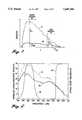
- FIG. 1is a graph showing the difference in autofluorescence intensity between normal and cancerous tissue
- FIG. 2is a graph showing the difference in autofluorescence spectral shape between normal and cancerous tissue in the spectral bands utilized by the present invention
- FIG. 3is a block diagram of a cancer detection system according to the present invention.
- FIG. 4is a block diagram of the image acquisition/processing units used with the cancer detection system of the present invention.
- FIG. 5is a chromaticity diagram showing how the perceived color for normal and cancerous tissue are displayed by the cancer detection system of the present invention
- FIG. 6is a diagram of an excitation light source according to another aspect of the present invention.
- FIG. 7is a pictorial diagram of an image produced by the cancer detection system of the present invention.
- the present inventionis a system for detecting cancerous or precancerous tissues using autofluorescence images that does not require the use of tumor enhancing drugs or intensive image processing.
- FIG. 1is a graph showing the differences in autofluorescence light intensity for normal and cancerous or precancerous tissue in humans. As indicated above, when blue excitation light is shined on tissue, some of the light will be absorbed and reemitted as autofluorescence light. The autofluorescence light has an intensity and spectral shape that is indicative of whether the tissue is normal or diseased. A spectral curve 10 represents the response of normal, healthy tissue, while a spectral curve 20 represents the response of tissue that is cancerous or precancerous. As can be seen, there is a significant difference in the intensity of the two curves in a green channel 30, while the differences between the healthy and abnormal tissue in a red channel 40 is small.
- the magnitude of the curves 10 and 20 shown in FIG. 1not only vary with wavelength, but also with the distance between a probe and the tissue under examination as well as the intensity of the light source. Therefore, in a practical sense it is not possible to determine whether tissue is cancerous by simply detecting the magnitude of the autofluorescence light in the green channel. However, the ratios of the autofluorescence light intensities taken at two different wavelengths remain constant despite variations in the distance of the fiber optic probe to the tissue and the angle of the probe to the tissue. This is true since the intensities of the green and red channel change proportionally for the same kind of tissue.
- the intensity of red autofluorescence light for normal tissue R n divided by the intensity of the green autofluorescence light for normal tissue G nremains substantially constant despite variations in the distance and angle of the tip of the endoscope to the tissue under consideration.
- the ratio of the intensity of the red autofluorescence light R CIS (CIS for carcinoma in situ) divided by the intensity of the green autofluorescence light G CISremains substantially constant despite variations in the position and orientation of the endoscope.
- the inventorshave found that the ratio of the response of the red channel to the green channel is approximately 0.54 for colon tissue and 0.16 for lung tissue, while for carcinoma in situ, the ratio of the red to green channel is approximately 1.6 for colon tissue and 0.6 for lung tissue. These values are given as examples only and they are highly dependent on the system, i.e., the spectral response of the imaging system. However, by calculating or by representing the ratio of the red channel autofluorescence light and the green channel autofluorescence light, a determination can be made whether the tissue under consideration is healthy or potentially cancerous when interpreted along with the contextual information of the image.
- FIG. 2shows the range of wavelengths used by the present invention to determine whether tissue is potentially cancerous or precancerous.
- the red channelis limited to wavelengths longer than 630 nanometers, while the green channel comprises wavelengths extending from approximately 490 to 560 nanometers.
- the areas in the red bandhave been normalized so that, in this part of the spectrum, the ratio of the normal and cancerous tissue is one. This normalization still results in a significant difference in the green band. Therefore, the ratio or a representation of the ratio will discriminate between normal and cancerous tissue.
- the present inventionallows a physician to determine whether the tissue under consideration is potentially cancerous or precancerous.
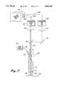
- the endoscope autofluorescence imaging system of the present inventionincludes an excitation light source 100 and a fiber optic endoscope 102, which may be a gastroscope, duodenoscope, choledochoscope, bronchoscope or colonoscope depending on the type of tissue to be examined.
- the endoscopemay be an Olympus BF-20D for the respiratory tract, an Olympus GIF-XQ30 for the esophagus and stomach and an Olympus CF-30L for the rectum and colon.
- the light source 100produces blue excitation light 104 which is fed through the incoherent illumination guide 106 of the fiber optic endoscope.
- the excitation lightis focused by an outer lens 108 that is disposed at the distal end of the endoscope.
- the blue excitation lightilluminates a portion of tissue 110 that is to be examined.
- the tissueis excited by the blue light and emits photons (autofluorescence light) 112 having a longer wavelength in the visible portion of the light spectrum.
- the autofluorescence light emitted by the tissue along with some reflected blue excitation lightis gathered by a lens 114 at the distal end of the endoscope and channeled through an imaging guide portion 116 of the endoscope through a number of conventional imaging lenses (not shown).
- autofluorescence lightis applied to a dichroic mirror 120.
- the dichroic mirror 120has a cutoff wavelength between 565 and 575 nanometers.
- the dichroic mirrordivides the autofluorescence light from the imaging guide into two spectral bands.
- the autofluorescence light having wavelengths longer than the cutoff wavelength of the dichroic mirror 120is directed toward a reflection mirror 122 that directs the autofluorescence light into a filter 124.
- the filter 124is positioned in front of a red channel, image intensified CCD camera 126.
- Autofluorescence light having wavelengths less than the cutoff wavelength of the dichroic mirror 120passes through the dichroic mirror toward a filter 128 that is positioned in front of a second image intensified, green channel CCD camera 130.
- the intensity of the red autofluorescence wavelength band that is applied to the camera 126is substantially the same for normal tissue and abnormal tissue.
- the intensity of the green autofluorescence wavelength band applied to the camera 130is substantially different for normal and abnormal tissue.
- the images produced by both CCD cameras 126 and 130are combined on a color video monitor 150 that allows any abnormal tissue to be easily identified. Because a high power blue excitation light is employed and all the photons in the spectral bands are applied to the image intensified CCD cameras 126 and 130, the images produced by the cameras 126 and 130 are bright enough to distinguish between normal and abnormal tissue, even in the larger organs such as in the gastrointestinal tract, without the need for tumor enhancing photosensitive drugs.
- FIG. 4is a more detailed block diagram of the imaging portion of the cancer detection system according to the present invention.
- the lightis directed to the filters 124 and 128.
- the filter 124is a long pass filter that blocks light having wavelengths less than 630 nanometers.
- the red autofluorescence lightis directed onto the surface of a GEN II image intensifier 132 such as a DEP XX 1700 manufactured by Delft Instruments.
- the output of the image intensifier 132is coupled through a fiber optic bundle 134 to the surface of a CCD image sensor 136 within the CCD camera 126.
- the green autofluorescence lightpasses through a bandpass filter 128 that removes any blue excitation light that may have been reflected off the surface of the tissue 110.
- the bandpass filter 128removes light having wavelengths less than 490 nanometers or greater than 560 nanometers.
- the out of band attenuation of the filters 124 and 128must be sufficient to ensure that virtually all of the reflected blue excitation light is blocked from reaching the CCD cameras.
- the green autofluorescence lightis directed to a GEN II image intensifier 140.
- the output of the image intensifier 140is coupled via a fiber optic bundle 142 to the surface of a CCD image sensor 144 within the green CCD camera 130.
- a genlock 149 within the camera 130maintains the synchronization of the red CCD camera 126 to the green CCD camera 130.
- the analog output signals of the red and green image intensified CCD cameras 126 and 130are fed to a pair of amplifiers 152, 154.
- the output of the amplifiersis fed into a video image processing board 155 that is part of a controlling computer 200.
- the controlling computer 200includes an Intel 486/100 MHz CPU 202 and 16 MB of internal memory 204. However, other high speed digital computers could also be used.
- the video image processing board 155includes a pair of analog-to-digital converters 156, 157, that receive the amplified video signals from the red and green CCD cameras, respectively.
- the analog-to-digital convertersconvert the analog video signals into a corresponding digital format.
- a pair of look up tables 158 and 160are used to modify the relative gain of the digital video signals in a manner that will be described below. For each discrete value of the digital video signal, the CPU 202 reads up a corresponding digital value having the correct relative gain from the look up tables.
- the values of the digitized video signalsread from the look up tables 158, 160, that are part of a random access memory 162 where they can be retrieved by the central processing unit for analysis at a later time.
- the digital video signalsare continuously retrieved from the random access memory 162 and reconverted into an analog format by a pair of digital-to-analog converters 164 and 166.
- the two converterscorrespond to the green and red channel images.
- the outputs of the digital to analog converters 164, 166,are applied directly to the red, green and blue inputs of the color monitor 150 shown in FIG. 3. This means that the video signal from the green CCD camera 130 is coupled to a green input 150G of the color monitor as well as a blue input 150B.
- a red input 150R of the color monitorreceives the video signal from the red CCD camera 126.
- the color video monitor 150receives the video signals and produces a color image, whereby the healthy tissue is approximately cyan in color and any abnormal or potentially cancerous tissue appears reddish in color and is therefore easily identifiable by a trained physician.
- the direct coupling of the video signals from the video input/output board 152 and 154 through the video image processing board 155 to the color video monitor 150is accomplished without any significant digital signal processing other than a minor change in the gain as will be described below.
- Thisallows a physician to view as true a representation of the data from the intensified CCD cameras as possible. In other words, the structure (e.g., contrast and texture) of the image are maintained. Therefore, the physician is able to consider subtleties in the video image that may otherwise be removed or masked as a result of any digital image processing that takes place before the image is displayed.
- the two autofluorescence image signals produced by the CCD camerasare representative of the true autofluorescence image of the tissue in the respective wavelength bands, allowing lesions to be seen at video rates within their context. This aspect is essential for the physician to recognize potential lesions which he/she can then biopsy through the biopsy channel of the endoscope for pathology confirmation.
- the fluorescence imaging system of the present inventionis unique in that it does not perform calculations to compensate for distance and angle effects of the endoscope with respect to the tissue under examination. Instead, the images from the red and green cameras are superimposed upon each other and displayed as distinct colors. This allows the physician to interpret the image using the visual experience they have acquired over their lifetime. In addition, the system circumvents the problem of an algorithm making a clinical decision as to what tissue may or may not be cancerous as well as allows the tissue to be observed in real time.
- the present inventionprovides a set of controls 220 that allow the physician to adjust the "brightness” and "color balance” of the color video image produced.
- the controls 220can be realized by hardware or software control of the camera gain.
- the brightness of the displayed imageis related to the gain of both the red and green channels while the color balance is related to the relative gain of the green and red channels.
- the gain of the red channel camera 126is related to the gain of the green channel camera 130 according to the equation:
- Ris the gain of the red channel image intensifier and G is the gain of the green channel image intensifier
- a, b and care constant.
- the particular values selected for the constants a, b and cdepend on the type of intensified CCD camera used, the intensity of the light source, the properties of the tissue and the human eye's ability to perceive color.
- the constant "a”should be selected in the range of 0.0 to 0.1
- "b”should be in the range of 0.5 to 1.5
- "c”should be selected from the range of 0.0 to 0.5.
- the constant "c”provides for an offset in color between the two channels while the linear term “b” ensures that as the overall brightness is changed, the gain of both the green and red channels is related.
- the quadratic term “a”provides a second order connection to ensure that the perceived color on the monitor remains the same despite variations to the overall brightness of the display. However, in the present embodiment of the invention, the "a" term is set to zero.
- the physicianadjusts the position of a knob 222 which adjusts the gain of the green channel. Movement of this knob 222 produces signals on a lead 224 that are read by the CPU 202.
- the CPU 202then adjusts the numbers stored in the look up tables 158 and 160 according to Equation 1 without changing the values of the constants a, b and c.
- the CPUcan adjust the gain of the red and green intensified CCD cameras by supplying gain signals from the CPU to a pair of gain control boards 146, 148, which control the high voltage of the image intensifiers 132, 140.
- the gain of the green channelis increased or decreased, the gain of the red channel is increased or decreased proportionally.
- the physicianadjusts the position of a contrast knob 226. Movement of the knob 226 produces signals on a lead 228 that are read by the CPU 202.
- the CPUthen adjusts the multiplier b that relates the gain of the red channel to the gain of the green channel.
- the constant "b"is varied linearly in the range described above as the contrast control knob 226 is moved between its minimum and maximum positions.
- the CPUthen calculates a gain of the red channel by recalculating the values stored in the look up table 158 or adjusts the gain of the red channel image intensifier camera 132 by supplying a new gain signal to the gain control board 146.
- the physicianadjusts the brightness control knob 222 until an image of sufficient intensity of the tissue is produced on the video monitor 150. If the physician believes he or she is viewing a malignant lesion, the position of the color balance knob 226 is adjusted until the entire video image appears red. The color balance knob can be readjusted until no red is apparent in the background of the video image. If the area of interest still appears reddish in color, the physician can have greater certainty that the red portion of the video image is a cancerous or precancerous lesion. The color balance control knob 226 can also be adjusted so that the displayed color is what the physician wants to see, thereby giving the physician some flexibility in setting the display to be something that is easier for the individual physician to interpret.
- the present inventiondisplays tissue on the color monitor as a color that is not dependent upon the position or orientation of the endoscope with respect to the tissue under examination.
- the rationale as to why this is true for the present inventionis stated below.
- any RGB color monitorcan be modeled according to the following equation: ##EQU1##
- the values R, G and Bare the magnitudes of the video signals applied to the color inputs of the monitor and the 3 ⁇ 3 matrix comprises a series of fixed coefficients that are primarily determined by the phosphor used on the monitor.
- the values, X, Y and Zare referred to as tristimulus values.
- Every color that can be displayed on the monitorcan be represented as a pair of color coordinates x, y, on a chromaticity diagram 250 as shown in FIG. 5.
- Thisis a widely used conventional diagram used to represent colors that can be displayed on a video monitor.
- the coordinates x and yare related to the tristimulus values by the equations: ##EQU2##
- the ratio of the red to green autofluorescence light for healthy colon tissueis approximately 0.54.
- the color coordinates x and y, of the chromaticity graphare computed and plotted for Equations 3 and 4 given that for a constant R/G ratio of 0.54, as shown in FIG. 5, it results in a point 252 having coordinates x 1 , y 1 with a greenish hue.
- the ratio of the red to green autofluorescence light for potentially cancerous colon tissueis approximately 1.6 which, when plotted, results in a point 254 having coordinates x 2 , y 2 with a red-brown hue.
- each point on the chromaticity graphlies an area 256, 258, 264 and 266 about which the human eye cannot perceive changes in color. It has further been determined that these areas tend to decrease in size as the value of x, y decreases, i.e., the colors shown in the blue to red region of the chromaticity diagram.
- the position of point 252moves to a point 260, having coordinates x 3 , y 3 , in the blue region of the chromaticity graph.
- the point 254is moved to a point 262, having coordinates x 4 , y 4 , near the red region of the graph. Because the colors displayed are in an area where the human eye is more perceptive of slight color changes, a physician is more easily able to detect slight variations in color and, hence, can more readily detect potentially cancerous tissue.
- the R/G ratioas a color is an important part of the present invention.
- the display of a calculated ratiodoes not provide contextual information.
- Contextual informationis, in our experience, essential to allow a physician to determine whether a suspicious lesion requires a biopsy or whether or not an artifact is observed (e.g., blood).
- the color display system of the present inventionis fast because no calculations are performed and the resulting image retains the tissue contextual information.
- FIG. 6is a schematic diagram of the excitation light source 100 shown in FIG. 1.
- the light source 100produces an intense blue light at the 436 nanometer mercury atomic line that is used to illuminate large internal body cavities.
- the light sourceincludes a one hundred watt mercury arc lamp 300 that produces light having a significant blue spectral content.
- the inventorshave found that in order to couple the blue light produced by the lamp 300 into the illumination guide 106 of the endoscope, the arc size of the lamp must be smaller than the diameter of the illumination guide of the endoscope.
- the arc lamp usedis an Ushio model number USH-102D having arc dimensions of approximately 0.25 millimeters.
- a collecting mirror 302Surrounding the arc lamp 300 is a collecting mirror 302, which in the present embodiment of the invention is elliptical in shape.
- the arc lamp 300is positioned at one foci of the mirror 302 so that light from the sides and rear of the lamp 300 is gathered by the mirror and directed towards a dichroic mirror 303. In this way, as much as 80% of the radiation energy of the lamp is collected.
- the dichroic mirror 303is preferably mounted below the are lamp 300 at a position closer than the second foci of the elliptical mirror. Light having wavelengths between 350 and 450 nanometers is reflected at forty-five degrees from the face of the dichroic mirror 303 and through a 399 nanometer, low fluorescence, long pass filter 306.
- the dichroic mirror 303In addition to reflecting light in the desired spectral band, the dichroic mirror 303 also serves to pass longer wavelengths of light to a beam stop 305 allowing one to filter out the infrared radiation so that components of the light source do not suffer thermal damage.
- a shutter 304 positioned between the dichroic mirror 303 and a long pass filter 306is used to control whether the excitation input is directed into the endoscope.
- the long pass filter 306attenuates the strong spectral lines produced by the mercury arc lamp in the ultraviolet range so that a coating on an interference bandpass filter 310 described below is protected from damage. After passing through the long pass filter 306, the light is directed to a one hundred millimeter focal length plano convex lens 308 that creates an optically homogenous, substantially parallel light beam, which is necessary for high out-of-band rejection filtering by a color filter 310.
- the color filterwhich preferably comprises an interference bandpass filter, is positioned perpendicular to the light beam produced by the lens 308 to attenuate light having wavelengths less than 400 nanometers and greater than 450 nanometers.
- the dichroic mirror 303reflects about 10% of the light outside the band of 350-450 nanometer band. Because a very pure excitation light is required for tissue fluorescence imaging, the interference bandpass filter 310 is used to further reduce the amount of light outside of the interested wavelength band (420 nanometers to 450 nanometers).
- the bandpass filter 310has an out of band transmittance of smaller than 5 ⁇ 10 -5 so that light with wavelengths less than 420 nanometers or greater than 450 nanometers is weaker than the autofluorescence light emitted from the tissue.
- the remaining lightAfter passing through the interference bandpass filter 310, the remaining light is directed to a 65 millimeter focal length lens 312 that focuses the light onto an end of the illumination guide 106 (FIG. 3).
- the light source 100 shown in FIG. 6provides approximately 500 milliwatts of blue light into the illumination guide of the endoscope. This light is sufficient to illuminate large internal body cavities such as the stomach or colon.
- the light sourcedelivers approximately 50-80 milliwatts of excitation light at the end of the endoscope in contrast to the 15 milliwatts that are typically delivered with a laser light source.
- the light source 100is easier to manufacture and maintain than a laser.
- FIG. 7is a pictorial representation of an image produced by the cancer detection system of the present invention.
- the video monitor 150produces a color display, wherein healthy tissue appears cyan (white-blue) in color and potentially cancerous or precancerous lesions appear reddish brown.
- the video display producedthereby allows physicians to readily detect the presence of cancerous or precancerous lesions without the use of photosensitive drugs.
- the color displayretains the contextual information of the tissue, thereby allowing the physician to interpret subtleties in the image that are traditionally lost in digital camera systems that employ significant digital signal processing.
- the cancer detection system of the present inventionis ideally suited for cancer screening applications whereby it is desired to test as many patients as possible for the presence of abnormal tissue without need to administer photosensitive drugs.
Landscapes
- Health & Medical Sciences (AREA)
- Life Sciences & Earth Sciences (AREA)
- Surgery (AREA)
- Engineering & Computer Science (AREA)
- Veterinary Medicine (AREA)
- Animal Behavior & Ethology (AREA)
- Public Health (AREA)
- General Health & Medical Sciences (AREA)
- Pathology (AREA)
- Biophysics (AREA)
- Physics & Mathematics (AREA)
- Biomedical Technology (AREA)
- Heart & Thoracic Surgery (AREA)
- Medical Informatics (AREA)
- Molecular Biology (AREA)
- Radiology & Medical Imaging (AREA)
- Optics & Photonics (AREA)
- Nuclear Medicine, Radiotherapy & Molecular Imaging (AREA)
- Endocrinology (AREA)
- Gastroenterology & Hepatology (AREA)
- Physiology (AREA)
- Signal Processing (AREA)
- Endoscopes (AREA)
- Investigating, Analyzing Materials By Fluorescence Or Luminescence (AREA)
- Investigating Or Analysing Materials By The Use Of Chemical Reactions (AREA)
- Investigating Or Analysing Biological Materials (AREA)
Abstract
Description
R.sub.(G) =aG.sup.2 +bG+c (1)
Claims (13)
Priority Applications (5)
| Application Number | Priority Date | Filing Date | Title |
|---|---|---|---|
| US08/608,185US5647368A (en) | 1996-02-28 | 1996-02-28 | Imaging system for detecting diseased tissue using native fluorsecence in the gastrointestinal and respiratory tract |
| EP03010552AEP1369077A3 (en) | 1996-02-28 | 1997-01-31 | Imaging system for detecting diseased tissue using native fluorescence in the gastrointestinal or respiratory tract |
| DE69725361TDE69725361T2 (en) | 1996-02-28 | 1997-01-31 | Image acquisition system for the detection of diseased tissue with native fluorescence in the digestive and respiratory tract |
| EP97300654AEP0792618B1 (en) | 1996-02-28 | 1997-01-31 | Imaging system for detecting diseased tissue using native fluorescence in the gastrointestinal and respiratory tract |
| JP9046303AJP3022377B2 (en) | 1996-02-28 | 1997-02-28 | Imaging system to detect diseased tissue using unique fluorescence in gastrointestinal and respiratory organs |
Applications Claiming Priority (1)
| Application Number | Priority Date | Filing Date | Title |
|---|---|---|---|
| US08/608,185US5647368A (en) | 1996-02-28 | 1996-02-28 | Imaging system for detecting diseased tissue using native fluorsecence in the gastrointestinal and respiratory tract |
Publications (1)
| Publication Number | Publication Date |
|---|---|
| US5647368Atrue US5647368A (en) | 1997-07-15 |
Family
ID=24435430
Family Applications (1)
| Application Number | Title | Priority Date | Filing Date |
|---|---|---|---|
| US08/608,185Expired - LifetimeUS5647368A (en) | 1996-02-28 | 1996-02-28 | Imaging system for detecting diseased tissue using native fluorsecence in the gastrointestinal and respiratory tract |
Country Status (4)
| Country | Link |
|---|---|
| US (1) | US5647368A (en) |
| EP (2) | EP1369077A3 (en) |
| JP (1) | JP3022377B2 (en) |
| DE (1) | DE69725361T2 (en) |
Cited By (198)
| Publication number | Priority date | Publication date | Assignee | Title |
|---|---|---|---|---|
| US5865754A (en)* | 1995-08-24 | 1999-02-02 | Purdue Research Foundation Office Of Technology Transfer | Fluorescence imaging system and method |
| EP0935132A3 (en)* | 1998-02-05 | 1999-09-15 | Herolab GmbH | Apparatus for optical analysis of samples |
| WO1999053832A1 (en) | 1998-04-20 | 1999-10-28 | Xillix Technologies Corp. | Imaging system with automatic gain control for reflectance and fluorescence endoscopy |
| US5984861A (en)* | 1997-09-29 | 1999-11-16 | Boston Scientific Corporation | Endofluorescence imaging module for an endoscope |
| US6002137A (en)* | 1997-02-13 | 1999-12-14 | Fuji Photo Film Co., Ltd. | Fluorescence detecting system |
| US6008889A (en)* | 1997-04-16 | 1999-12-28 | Zeng; Haishan | Spectrometer system for diagnosis of skin disease |
| US6021344A (en)* | 1996-12-04 | 2000-02-01 | Derma Technologies, Inc. | Fluorescence scope system for dermatologic diagnosis |
| US6026319A (en)* | 1997-02-13 | 2000-02-15 | Fuji Photo Film Co., Ltd. | Fluorescence detecting system |
| US6032071A (en)* | 1994-12-01 | 2000-02-29 | Norbert Artner | Skin examination device |
| US6032070A (en)* | 1995-06-07 | 2000-02-29 | University Of Arkansas | Method and apparatus for detecting electro-magnetic reflection from biological tissue |
| WO2000013578A1 (en) | 1998-09-03 | 2000-03-16 | Hypermed Imaging, Inc. | Infrared endoscopic balloon probes |
| US6055451A (en) | 1997-12-12 | 2000-04-25 | Spectrx, Inc. | Apparatus and method for determining tissue characteristics |
| US6070096A (en)* | 1996-03-06 | 2000-05-30 | Fuji Photo Film Co., Ltd. | Fluorescence detecting apparatus |
| US6096065A (en)* | 1997-09-29 | 2000-08-01 | Boston Scientific Corporation | Sheath for tissue spectroscopy |
| US6110106A (en)* | 1998-06-24 | 2000-08-29 | Biomax Technologies, Inc. | Endoscopes and methods relating to direct viewing of a target tissue |
| US6119031A (en)* | 1996-11-21 | 2000-09-12 | Boston Scientific Corporation | Miniature spectrometer |
| US6144791A (en)* | 1995-11-20 | 2000-11-07 | Cirrex Corp. | Beam steering for optical fibers and other related devices |
| US6148227A (en)* | 1998-01-07 | 2000-11-14 | Richard Wolf Gmbh | Diagnosis apparatus for the picture providing recording of fluorescing biological tissue regions |
| US6167297A (en)* | 1999-05-05 | 2000-12-26 | Benaron; David A. | Detecting, localizing, and targeting internal sites in vivo using optical contrast agents |
| US6174291B1 (en) | 1998-03-09 | 2001-01-16 | Spectrascience, Inc. | Optical biopsy system and methods for tissue diagnosis |
| US6179777B1 (en) | 1997-11-27 | 2001-01-30 | Asahi Kogaku Kogyo Kabushiki Kaisha | Fluorescent diagnosing apparatus including optical path switching member |
| US6185443B1 (en) | 1997-09-29 | 2001-02-06 | Boston Scientific Corporation | Visible display for an interventional device |
| US6201989B1 (en) | 1997-03-13 | 2001-03-13 | Biomax Technologies Inc. | Methods and apparatus for detecting the rejection of transplanted tissue |
| US6208783B1 (en) | 1997-03-13 | 2001-03-27 | Cirrex Corp. | Optical filtering device |
| US20010003800A1 (en)* | 1996-11-21 | 2001-06-14 | Steven J. Frank | Interventional photonic energy emitter system |
| WO2001045557A1 (en)* | 1999-12-22 | 2001-06-28 | Xillix Technologies Corporation | Portable system for detecting skin abnormalities |
| US6256530B1 (en) | 1998-09-15 | 2001-07-03 | Denvu, L.L.C. | Optical instrument and technique for cancer diagnosis using in-vivo fluorescence emission of test tissue |
| US6289236B1 (en)* | 1997-10-10 | 2001-09-11 | The General Hospital Corporation | Methods and apparatus for distinguishing inflamed and tumorous bladder tissue |
| US6289229B1 (en) | 1998-01-20 | 2001-09-11 | Scimed Life Systems, Inc. | Readable probe array for in vivo use |
| WO2001072214A1 (en)* | 2000-03-28 | 2001-10-04 | Foundation For Research And Technology-Hellas | Method and system for characterization and mapping of tissue lesions |
| US6317624B1 (en) | 1997-05-05 | 2001-11-13 | The General Hospital Corporation | Apparatus and method for demarcating tumors |
| US6324418B1 (en)* | 1997-09-29 | 2001-11-27 | Boston Scientific Corporation | Portable tissue spectroscopy apparatus and method |
| US6364829B1 (en) | 1999-01-26 | 2002-04-02 | Newton Laboratories, Inc. | Autofluorescence imaging system for endoscopy |
| US6370406B1 (en) | 1995-11-20 | 2002-04-09 | Cirrex Corp. | Method and apparatus for analyzing a test material by inducing and detecting light-matter interactions |
| US6405074B1 (en)* | 1997-08-29 | 2002-06-11 | Bhaskar Banerjee | Detection of cancer using cellular autofluorescence |
| US6405073B1 (en) | 1997-07-22 | 2002-06-11 | Scimed Life Systems, Inc. | Miniature spectrometer system and method |
| US6411838B1 (en) | 1998-12-23 | 2002-06-25 | Medispectra, Inc. | Systems and methods for optical examination of samples |
| US6422994B1 (en) | 1997-09-24 | 2002-07-23 | Olympus Optical Co., Ltd. | Fluorescent diagnostic system and method providing color discrimination enhancement |
| US20020103439A1 (en)* | 2000-12-19 | 2002-08-01 | Haishan Zeng | Methods and apparatus for fluorescence and reflectance imaging and spectroscopy and for contemporaneous measurements of electromagnetic radiation with multiple measuring devices |
| US20020105505A1 (en)* | 2000-06-06 | 2002-08-08 | Fuji Photo Film Co., Ltd. | Fluorescent-light image display method and apparatus therefor |
| US6444970B1 (en) | 1998-06-26 | 2002-09-03 | Scimed Life Systems, Inc. | Miniature low-noise photodiode system |
| US20020147383A1 (en)* | 2001-04-04 | 2002-10-10 | Richard Wolf Gmbh | Device for the picture-providing diagnosis of tissue |
| EP1256310A2 (en) | 2001-05-07 | 2002-11-13 | Fuji Photo Film Co., Ltd. | Fluorescence image display apparatus |
| US20020175993A1 (en)* | 2001-05-16 | 2002-11-28 | Olympus Optical Co., Ltd. | Endoscope system using normal light and fluorescence |
| US20020197728A1 (en)* | 1999-12-15 | 2002-12-26 | Howard Kaufman | Methods of monitoring effects of chemical agents on a sample |
| US20030001104A1 (en)* | 2001-06-29 | 2003-01-02 | Fuji Photo Film Co., Ltd | Method and apparatus for obtaining fluorescence images, and computer executable program therefor |
| US6516217B1 (en)* | 1999-07-09 | 2003-02-04 | Fuji Photo Film Co., Ltd. | Fluorescence diagnosis system |
| US6529768B1 (en)* | 1999-11-18 | 2003-03-04 | Fuji Photo Film Co., Ltd. | Method and apparatus for acquiring fluorescence images |
| US6571119B2 (en)* | 1996-03-06 | 2003-05-27 | Fuji Photo Film Co., Ltd. | Fluorescence detecting apparatus |
| US6574502B2 (en) | 1999-12-02 | 2003-06-03 | Fuji Photo Film Co., Ltd. | Apparatus for displaying fluorescence images |
| US6580935B1 (en) | 1999-03-12 | 2003-06-17 | Cirrex Corp. | Method and system for stabilizing reflected light |
| US6603552B1 (en) | 1999-12-22 | 2003-08-05 | Xillix Technologies Corp. | Portable system for detecting skin abnormalities based on characteristic autofluorescence |
| US20030220574A1 (en)* | 2002-03-18 | 2003-11-27 | Sarcos Investments Lc. | Miniaturized imaging device including utility aperture and SSID |
| US20030222325A1 (en)* | 2002-03-18 | 2003-12-04 | Sarcos Investments Lc. | Miniaturized imaging device with integrated circuit connector system |
| US20040010192A1 (en)* | 2000-06-15 | 2004-01-15 | Spectros Corporation | Optical imaging of induced signals in vivo under ambient light conditions |
| US20040037454A1 (en)* | 2002-06-26 | 2004-02-26 | Olympus Optical Co., Ltd. | Image processing device for fluorescence observation |
| US6766184B2 (en) | 2000-03-28 | 2004-07-20 | Board Of Regents, The University Of Texas System | Methods and apparatus for diagnostic multispectral digital imaging |
| US6768918B2 (en) | 2002-07-10 | 2004-07-27 | Medispectra, Inc. | Fluorescent fiberoptic probe for tissue health discrimination and method of use thereof |
| RU2234242C2 (en)* | 2002-03-19 | 2004-08-20 | Федеральное государственное унитарное предприятие Научно-исследовательский институт "Полюс" | Method for determining biological tissue condition |
| US20040225222A1 (en)* | 2003-05-08 | 2004-11-11 | Haishan Zeng | Real-time contemporaneous multimodal imaging and spectroscopy uses thereof |
| US6818903B2 (en) | 2002-07-09 | 2004-11-16 | Medispectra, Inc. | Method and apparatus for identifying spectral artifacts |
| EP1477103A1 (en)* | 2002-01-15 | 2004-11-17 | Xillix Technologies Corp. | Fluorescence endoscopy video systems with no moving parts in the camera |
| US6821245B2 (en) | 2000-07-14 | 2004-11-23 | Xillix Technologies Corporation | Compact fluorescence endoscopy video system |
| US6826422B1 (en) | 1997-01-13 | 2004-11-30 | Medispectra, Inc. | Spectral volume microprobe arrays |
| US20040249274A1 (en)* | 2003-03-18 | 2004-12-09 | Yaroslavsky Anna N. | Polarized light imaging devices and methods |
| US20040245350A1 (en)* | 2003-06-03 | 2004-12-09 | Haishan Zeng | Methods and apparatus for fluorescence imaging using multiple excitation-emission pairs and simultaneous multi-channel image detection |
| US6839661B2 (en) | 2000-12-15 | 2005-01-04 | Medispectra, Inc. | System for normalizing spectra |
| US6847490B1 (en) | 1997-01-13 | 2005-01-25 | Medispectra, Inc. | Optical probe accessory device for use in vivo diagnostic procedures |
| US20050030407A1 (en)* | 2002-03-12 | 2005-02-10 | Dan Davidovici | Optical image recording and image evaluation system |
| US20050065440A1 (en)* | 2003-09-23 | 2005-03-24 | Richard Levenson | Spectral imaging of deep tissue |
| US20050085732A1 (en)* | 2003-06-20 | 2005-04-21 | Sevick-Muraca Eva M. | Method and system for near-infrared fluorescence contrast-enhanced imaging with area illumination and area detection |
| US20050094147A1 (en)* | 2003-09-19 | 2005-05-05 | Yaroslavsky Anna N. | Fluorescence polarization imaging devices and methods |
| US20050154261A1 (en)* | 2000-04-03 | 2005-07-14 | Ohline Robert M. | Tendon-driven endoscope and methods of insertion |
| US6933154B2 (en) | 2002-07-09 | 2005-08-23 | Medispectra, Inc. | Optimal windows for obtaining optical data for characterization of tissue samples |
| US6937885B1 (en) | 1997-10-30 | 2005-08-30 | Hypermed, Inc. | Multispectral/hyperspectral medical instrument |
| US6975899B2 (en) | 1998-09-11 | 2005-12-13 | Spectrx, Inc. | Multi-modal optical tissue diagnostic system |
| US7054002B1 (en) | 1999-10-08 | 2006-05-30 | The Texas A&M University System | Characterization of luminescence in a scattering medium |
| US7062311B1 (en) | 1999-07-07 | 2006-06-13 | Fuji Photo Film Co., Ltd. | Fluorescence observing apparatus |
| US20060146172A1 (en)* | 2002-03-18 | 2006-07-06 | Jacobsen Stephen C | Miniaturized utility device having integrated optical capabilities |
| US7103401B2 (en) | 2002-07-10 | 2006-09-05 | Medispectra, Inc. | Colonic polyp discrimination by tissue fluorescence and fiberoptic probe |
| US20060217594A1 (en)* | 2005-03-24 | 2006-09-28 | Ferguson Gary W | Endoscopy device with removable tip |
| US7127282B2 (en) | 1998-12-23 | 2006-10-24 | Medispectra, Inc. | Optical methods and systems for rapid screening of the cervix |
| US20060241496A1 (en)* | 2002-01-15 | 2006-10-26 | Xillix Technologies Corp. | Filter for use with imaging endoscopes |
| US7136518B2 (en) | 2003-04-18 | 2006-11-14 | Medispectra, Inc. | Methods and apparatus for displaying diagnostic data |
| US7187810B2 (en) | 1999-12-15 | 2007-03-06 | Medispectra, Inc. | Methods and systems for correcting image misalignment |
| WO2007079943A1 (en)* | 2005-12-21 | 2007-07-19 | Fachhochschule Mannheim | Fibre-optic fluorescence sensor system |
| US7260248B2 (en) | 1999-12-15 | 2007-08-21 | Medispectra, Inc. | Image processing using measures of similarity |
| US7282723B2 (en) | 2002-07-09 | 2007-10-16 | Medispectra, Inc. | Methods and apparatus for processing spectral data for use in tissue characterization |
| US20070276230A1 (en)* | 2003-11-20 | 2007-11-29 | Mitsuharu Miwa | Lymph Node Detecting Apparatus |
| US7309867B2 (en) | 2003-04-18 | 2007-12-18 | Medispectra, Inc. | Methods and apparatus for characterization of tissue samples |
| US7328059B2 (en) | 1996-08-23 | 2008-02-05 | The Texas A & M University System | Imaging of light scattering tissues with fluorescent contrast agents |
| US20080058649A1 (en)* | 2006-04-12 | 2008-03-06 | Searete Llc., A Limited Liability Corporation Of The State Of Delaware | Systems for autofluorescent imaging and target ablation |
| US20080118578A1 (en)* | 1997-12-11 | 2008-05-22 | Dees H Craig | Topical Medicaments and Methods for Photodynamic Treatment of Disease |
| US20080118567A1 (en)* | 1998-08-06 | 2008-05-22 | Provectus Pharmatech. Inc, | Medicaments for Chemotherapeutic Treatment of Disease |
| US20080177140A1 (en)* | 2007-01-23 | 2008-07-24 | Xillix Technologies Corp. | Cameras for fluorescence and reflectance imaging |
| US20080294032A1 (en)* | 2003-09-23 | 2008-11-27 | Cambridge Research And Instrumentation, Inc. | Spectral Imaging of Biological Samples |
| US7459696B2 (en) | 2003-04-18 | 2008-12-02 | Schomacker Kevin T | Methods and apparatus for calibrating spectral data |
| US20080303898A1 (en)* | 2007-06-06 | 2008-12-11 | Olympus Medical Systems Corp. | Endoscopic image processing apparatus |
| US7469160B2 (en) | 2003-04-18 | 2008-12-23 | Banks Perry S | Methods and apparatus for evaluating image focus |
| US7505807B1 (en) | 1997-05-15 | 2009-03-17 | Regents Of The University Of Minnesota | Magnetic resonance apparatus for use with active electrode and drug deliver catheter |
| US20090117199A1 (en)* | 1998-08-06 | 2009-05-07 | Scott Timothy C | Method of treatment of cancer |
| US20090156900A1 (en)* | 2007-12-13 | 2009-06-18 | Robertson David W | Extended spectral sensitivity endoscope system and method of using the same |
| US20090192349A1 (en)* | 2008-01-24 | 2009-07-30 | Lifeguard Surgical Systems | Common bile duct surgical imaging system |
| US20090192390A1 (en)* | 2008-01-24 | 2009-07-30 | Lifeguard Surgical Systems | Common bile duct surgical imaging system |
| US20090216085A1 (en)* | 2008-02-27 | 2009-08-27 | Olympus Medical Systems Corp. | Fluorescent endoscopic device and method of creating fluorescent endoscopic image |
| US20100073731A1 (en)* | 2007-06-20 | 2010-03-25 | Olympus Medical Systems Corp. | Image generating apparatus |
| US20100145844A1 (en)* | 2007-01-17 | 2010-06-10 | Steidlmayer Pete | Method for scheduling future orders on an electronic commodity trading system |
| US20100166272A1 (en)* | 2003-06-12 | 2010-07-01 | Eli Horn | System and method to detect a transition in an image stream |
| US20100268025A1 (en)* | 2007-11-09 | 2010-10-21 | Amir Belson | Apparatus and methods for capsule endoscopy of the esophagus |
| US7835074B2 (en) | 2007-06-05 | 2010-11-16 | Sterling Lc | Mini-scope for multi-directional imaging |
| US7846091B2 (en) | 1999-01-26 | 2010-12-07 | Newton Laboratories, Inc. | Autofluorescence imaging system for endoscopy |
| US7865230B1 (en) | 1997-02-07 | 2011-01-04 | Texas A&M University System | Method and system for detecting sentinel lymph nodes |
| US20110028788A1 (en)* | 2008-03-24 | 2011-02-03 | The Regents Of The University Of Michigan | Non-Contact Infrared Fiber-Optic Device for Monitoring Esophageal Temperature to Prevent Thermal Injury During Radiofrequency Catheter Ablation or Cryoablation |
| US20110060189A1 (en)* | 2004-06-30 | 2011-03-10 | Given Imaging Ltd. | Apparatus and Methods for Capsule Endoscopy of the Esophagus |
| US20110117025A1 (en)* | 2008-05-20 | 2011-05-19 | Ralph Sebastian Dacosta | Device and method for fluorescence-based imaging and monitoring |
| US20110137179A1 (en)* | 2008-08-21 | 2011-06-09 | University Of Florida Research Foundation, Inc. | Differential laser-induced perturbation (dlip) for bioimaging and chemical sensing |
| US7969659B2 (en) | 2008-01-11 | 2011-06-28 | Sterling Lc | Grin lens microscope system |
| US8024027B2 (en) | 1998-09-03 | 2011-09-20 | Hyperspectral Imaging, Inc. | Infrared endoscopic balloon probes |
| US8046055B2 (en) | 2004-06-30 | 2011-10-25 | Hamamatsu Photonics K.K. | Lymph node detector |
| US8062212B2 (en) | 2000-04-03 | 2011-11-22 | Intuitive Surgical Operations, Inc. | Steerable endoscope and improved method of insertion |
| US8083879B2 (en) | 2005-11-23 | 2011-12-27 | Intuitive Surgical Operations, Inc. | Non-metallic, multi-strand control cable for steerable instruments |
| DE102010033825A1 (en)* | 2010-08-09 | 2012-02-09 | Carl Zeiss Meditec Ag | Filter set for use in fluorescence tracking system to carry out fluorescence observation of object, has illuminating light filter whose transmission characteristic is sum of two partial characteristics |
| US8182418B2 (en) | 2008-02-25 | 2012-05-22 | Intuitive Surgical Operations, Inc. | Systems and methods for articulating an elongate body |
| US20120179010A1 (en)* | 2002-04-04 | 2012-07-12 | Maynard John D | Determination of a Measure of a Glycation End-Product or Disease State Using Tissue Fluorescence of Various Sites |
| EP2478827A1 (en)* | 2011-01-19 | 2012-07-25 | Fujifilm Corporation | Endoscope system |
| EP2478826A1 (en)* | 2011-01-19 | 2012-07-25 | Fujifilm Corporation | Endoscope system |
| US8280140B2 (en) | 2005-01-27 | 2012-10-02 | Cambridge Research & Instrumentation, Inc. | Classifying image features |
| DE102011016138A1 (en)* | 2011-03-30 | 2012-10-04 | Karl Storz Gmbh & Co. Kg | Device for fluorescence diagnosis |
| DE102011100507A1 (en)* | 2011-04-29 | 2012-10-31 | Fraunhofer-Gesellschaft zur Förderung der angewandten Forschung e.V. | Mobile optical analyzer |
| US8328877B2 (en) | 2002-03-19 | 2012-12-11 | Boston Scientific Scimed, Inc. | Stent retention element and related methods |
| US8361090B2 (en) | 2002-01-09 | 2013-01-29 | Intuitive Surgical Operations, Inc. | Apparatus and method for endoscopic colectomy |
| US8486735B2 (en) | 2008-07-30 | 2013-07-16 | Raytheon Company | Method and device for incremental wavelength variation to analyze tissue |
| DE102012002086A1 (en)* | 2012-02-06 | 2013-08-08 | Carl Zeiss Meditec Ag | A method of examining biological tissue and devices for examining and treating the tissue |
| US8509880B1 (en)* | 2008-02-06 | 2013-08-13 | Remicalm, Llc | Handheld portable examination device for diagnostic use |
| US8517923B2 (en) | 2000-04-03 | 2013-08-27 | Intuitive Surgical Operations, Inc. | Apparatus and methods for facilitating treatment of tissue via improved delivery of energy based and non-energy based modalities |
| US8562802B1 (en) | 2006-02-13 | 2013-10-22 | Life Technologies Corporation | Transilluminator base and scanner for imaging fluorescent gels, charging devices and portable electrophoresis systems |
| US8568299B2 (en) | 2006-05-19 | 2013-10-29 | Intuitive Surgical Operations, Inc. | Methods and apparatus for displaying three-dimensional orientation of a steerable distal tip of an endoscope |
| US8614768B2 (en) | 2002-03-18 | 2013-12-24 | Raytheon Company | Miniaturized imaging device including GRIN lens optically coupled to SSID |
| US8690762B2 (en) | 2008-06-18 | 2014-04-08 | Raytheon Company | Transparent endoscope head defining a focal length |
| EP2724661A1 (en)* | 2005-10-17 | 2014-04-30 | Novadaq Technologies Inc. | Device for short wavelength visible reflectance endoscopy using broadband illumination |
| US8717428B2 (en) | 2009-10-01 | 2014-05-06 | Raytheon Company | Light diffusion apparatus |
| US20140225992A1 (en)* | 2011-08-12 | 2014-08-14 | Intuitive Surgical Operations, Inc. | Increased resolution and dynamic range image capture unit in a surgical instrument and method |
| US8828028B2 (en) | 2009-11-03 | 2014-09-09 | Raytheon Company | Suture device and method for closing a planar opening |
| US20140276102A1 (en)* | 2013-03-14 | 2014-09-18 | Lumicell, Inc. | Medical imaging device and methods of use |
| US8845524B2 (en) | 2000-04-03 | 2014-09-30 | Intuitive Surgical Operations, Inc. | Steerable segmented endoscope and method of insertion |
| US8882657B2 (en) | 2003-03-07 | 2014-11-11 | Intuitive Surgical Operations, Inc. | Instrument having radio frequency identification systems and methods for use |
| US8888688B2 (en) | 2000-04-03 | 2014-11-18 | Intuitive Surgical Operations, Inc. | Connector device for a controllable instrument |
| US8922633B1 (en) | 2010-09-27 | 2014-12-30 | Given Imaging Ltd. | Detection of gastrointestinal sections and transition of an in-vivo device there between |
| DE102013108189A1 (en)* | 2013-07-31 | 2015-02-05 | Endress + Hauser Conducta Gesellschaft für Mess- und Regeltechnik mbH + Co. KG | Arrangement for the optical measurement of a process variable and measuring device comprising such |
| US8965079B1 (en) | 2010-09-28 | 2015-02-24 | Given Imaging Ltd. | Real time detection of gastrointestinal sections and transitions of an in-vivo device therebetween |
| US8974651B2 (en) | 2010-04-17 | 2015-03-10 | C.C. Imex | Illuminator for visualization of fluorophores |
| US9060704B2 (en) | 2008-11-04 | 2015-06-23 | Sarcos Lc | Method and device for wavelength shifted imaging |
| EP2888989A1 (en) | 2013-12-31 | 2015-07-01 | Karl Storz Imaging, Inc. | Switching between white light imaging and excitation light imaging leaving last video frame displayed |
| US9144664B2 (en) | 2009-10-01 | 2015-09-29 | Sarcos Lc | Method and apparatus for manipulating movement of a micro-catheter |
| US9220398B2 (en) | 2007-10-11 | 2015-12-29 | Intuitive Surgical Operations, Inc. | System for managing Bowden cables in articulating instruments |
| US9314304B2 (en) | 2010-12-08 | 2016-04-19 | Lumicell, Inc. | Methods and system for image guided cell ablation with microscopic resolution |
| US9324145B1 (en) | 2013-08-08 | 2016-04-26 | Given Imaging Ltd. | System and method for detection of transitions in an image stream of the gastrointestinal tract |
| RU2582460C1 (en)* | 2015-04-06 | 2016-04-27 | Государственное бюджетное учреждение здравоохранения Московской области "Московский областной научно-исследовательский клинический институт им. М.Ф. Владимирского" (ГБУЗ МО МОНИКИ им. М.Ф. Владимирского) | Method for diagnosing muscle tissue non-specific inflammation in laboratory animals |
| US9386909B2 (en) | 2006-07-28 | 2016-07-12 | Novadaq Technologies Inc. | System and method for deposition and removal of an optical element on an endoscope objective |
| DE102015011441A1 (en)* | 2015-09-01 | 2017-03-02 | Carl Zeiss Meditec Ag | Fluorescence light detection system and microscopy system |
| US9610021B2 (en) | 2008-01-25 | 2017-04-04 | Novadaq Technologies Inc. | Method for evaluating blush in myocardial tissue |
| US9642532B2 (en) | 2008-03-18 | 2017-05-09 | Novadaq Technologies Inc. | Imaging system for combined full-color reflectance and near-infrared imaging |
| US9661996B2 (en) | 2009-10-01 | 2017-05-30 | Sarcos Lc | Needle delivered imaging device |
| US9816930B2 (en) | 2014-09-29 | 2017-11-14 | Novadaq Technologies Inc. | Imaging a target fluorophore in a biological material in the presence of autofluorescence |
| US9814378B2 (en) | 2011-03-08 | 2017-11-14 | Novadaq Technologies Inc. | Full spectrum LED illuminator having a mechanical enclosure and heatsink |
| US9835587B2 (en) | 2014-04-01 | 2017-12-05 | C.C. Imex | Electrophoresis running tank assembly |
| US20170367559A1 (en)* | 2015-03-26 | 2017-12-28 | Sony Corporation | Surgical system, information processing device, and method |
| US9877654B2 (en) | 2006-02-07 | 2018-01-30 | Novadaq Technologies Inc. | Near infrared imaging |
| US10041042B2 (en) | 2008-05-02 | 2018-08-07 | Novadaq Technologies ULC | Methods for production and use of substance-loaded erythrocytes (S-IEs) for observation and treatment of microvascular hemodynamics |
| DE102017203452A1 (en) | 2017-03-02 | 2018-09-06 | Carl Zeiss Meditec Ag | Fluorescence observation system |
| US10070793B2 (en) | 2010-11-27 | 2018-09-11 | Securus Medical Group, Inc. | Ablation and temperature measurement devices |
| CN109044278A (en)* | 2018-08-16 | 2018-12-21 | 济南显微智能科技有限公司 | A kind of double fluorescent tracing imaging devices |
| US10219742B2 (en) | 2008-04-14 | 2019-03-05 | Novadaq Technologies ULC | Locating and analyzing perforator flaps for plastic and reconstructive surgery |
| US10265419B2 (en) | 2005-09-02 | 2019-04-23 | Novadaq Technologies ULC | Intraoperative determination of nerve location |
| US10278585B2 (en) | 2012-06-21 | 2019-05-07 | Novadaq Technologies ULC | Quantification and analysis of angiography and perfusion |
| US10293122B2 (en) | 2016-03-17 | 2019-05-21 | Novadaq Technologies ULC | Endoluminal introducer with contamination avoidance |
| US10434190B2 (en) | 2006-09-07 | 2019-10-08 | Novadaq Technologies ULC | Pre-and-intra-operative localization of penile sentinel nodes |
| US10438356B2 (en) | 2014-07-24 | 2019-10-08 | University Health Network | Collection and analysis of data for diagnostic purposes |
| US10492671B2 (en) | 2009-05-08 | 2019-12-03 | Novadaq Technologies ULC | Near infra red fluorescence imaging for visualization of blood vessels during endoscopic harvest |
| US10512392B2 (en) | 2008-02-06 | 2019-12-24 | Intuitive Surgical Operations, Inc. | Segmented instrument having braking capabilities |
| US10602917B2 (en) | 2013-12-31 | 2020-03-31 | Karl Storz Imaging, Inc. | Switching between white light imaging and excitation light imaging leaving last video frame displayed |
| US10631746B2 (en) | 2014-10-09 | 2020-04-28 | Novadaq Technologies ULC | Quantification of absolute blood flow in tissue using fluorescence-mediated photoplethysmography |
| US10694151B2 (en) | 2006-12-22 | 2020-06-23 | Novadaq Technologies ULC | Imaging system with a single color image sensor for simultaneous fluorescence and color video endoscopy |
| US10869645B2 (en) | 2016-06-14 | 2020-12-22 | Stryker European Operations Limited | Methods and systems for adaptive imaging for low light signal enhancement in medical visualization |
| US20200405135A1 (en)* | 2018-03-15 | 2020-12-31 | Sony Olympus Medical Solutions Inc. | Medical observation system |
| US10973398B2 (en) | 2011-08-12 | 2021-04-13 | Intuitive Surgical Operations, Inc. | Image capture unit in a surgical instrument |
| USD916294S1 (en) | 2016-04-28 | 2021-04-13 | Stryker European Operations Limited | Illumination and imaging device |
| US10980420B2 (en) | 2016-01-26 | 2021-04-20 | Stryker European Operations Limited | Configurable platform |
| US10992848B2 (en) | 2017-02-10 | 2021-04-27 | Novadaq Technologies ULC | Open-field handheld fluorescence imaging systems and methods |
| US20210183059A1 (en)* | 2019-12-17 | 2021-06-17 | Schölly Fiberoptic GmbH | Image acquisition method using a color transformation and associated medical image acquisition system |
| US11096563B2 (en) | 2005-11-22 | 2021-08-24 | Intuitive Surgical Operations, Inc. | Method of determining the shape of a bendable instrument |
| US20210307597A1 (en)* | 2018-07-23 | 2021-10-07 | The Regents Of The University Of California | Oral and oropharyngeal cancer screening system and methods of use |
| US11426075B1 (en) | 2017-08-23 | 2022-08-30 | Lumicell, Inc. | System and method for residual cancer cell detection |
| US11592396B2 (en) | 2009-05-27 | 2023-02-28 | Lumicell, Inc. | Methods and systems for spatially identifying abnormal cells |
| US11930278B2 (en) | 2015-11-13 | 2024-03-12 | Stryker Corporation | Systems and methods for illumination and imaging of a target |
| DE102022125852A1 (en)* | 2022-10-06 | 2024-04-11 | Karl Storz Se & Co. Kg | Medical imaging device and method for medical imaging |
| US12075970B2 (en)* | 2019-03-01 | 2024-09-03 | Sony Olympus Medical Solutions Inc. | Medical control device and medical observation system |
| DE102023134457A1 (en)* | 2023-12-08 | 2025-06-12 | Karl Storz Se & Co. Kg | Imaging device, in particular endoscopic, exoscopic and/or microscopic imaging device |
Families Citing this family (12)
| Publication number | Priority date | Publication date | Assignee | Title |
|---|---|---|---|---|
| JP4008184B2 (en)* | 1996-03-06 | 2007-11-14 | 富士フイルム株式会社 | Fluorescence detection device |
| JPH11271453A (en)* | 1998-03-25 | 1999-10-08 | Toshiba Corp | Radiation discrimination measurement method and radiation discrimination measurement device |
| US6487440B2 (en) | 1998-07-08 | 2002-11-26 | Lifespex, Inc. | Optical probe having and methods for difuse and uniform light irradiation |
| US6332092B1 (en) | 1998-07-08 | 2001-12-18 | Lifespex, Incorporated | Optical probe having and methods for uniform light irradiation and/or light collection over a volume |
| JP3309276B2 (en) | 1999-03-17 | 2002-07-29 | エーカポット・パンナチェート | Fluorescent endoscope system |
| US20040202356A1 (en)* | 2003-04-10 | 2004-10-14 | Stookey George K. | Optical detection of dental caries |
| US7270543B2 (en) | 2004-06-29 | 2007-09-18 | Therametric Technologies, Inc. | Handpiece for caries detection |
| US8360771B2 (en) | 2006-12-28 | 2013-01-29 | Therametric Technologies, Inc. | Handpiece for detection of dental demineralization |
| CN102361748A (en)* | 2009-01-23 | 2012-02-22 | 德雷克塞尔大学 | Apparatus and methods for detecting inflammation using quantum dots |
| KR101514204B1 (en) | 2013-07-12 | 2015-04-23 | 한국전기연구원 | Apparatus and method for detecting NIR fluorescence at Sentinel Lymph Node |
| US10165972B2 (en) | 2013-07-12 | 2019-01-01 | Inthesmart Co., Ltd. | Apparatus and method for detecting NIR fluorescence at sentinel lymph node |
| EP3834702A1 (en) | 2014-03-17 | 2021-06-16 | Intuitive Surgical Operations, Inc. | System and method for tissue contact detection and for auto-exposure and illumination control |
Citations (45)
| Publication number | Priority date | Publication date | Assignee | Title |
|---|---|---|---|---|
| JPS58222331A (en)* | 1982-06-21 | 1983-12-24 | Sony Corp | Reproducer of character information |
| US4473841A (en)* | 1981-10-20 | 1984-09-25 | Fuji Photo Film Co., Ltd. | Video signal transmission system for endoscope using solid state image sensor |
| US4541438A (en)* | 1983-06-02 | 1985-09-17 | The Johns Hopkins University | Localization of cancerous tissue by monitoring infrared fluorescence emitted by intravenously injected porphyrin tumor-specific markers excited by long wavelength light |
| US4556057A (en)* | 1982-08-31 | 1985-12-03 | Hamamatsu Tv Co., Ltd. | Cancer diagnosis device utilizing laser beam pulses |
| WO1986002730A1 (en)* | 1984-10-22 | 1986-05-09 | Hightech Network Sci Ab | A fluorescence imaging system |
| EP0215772A2 (en)* | 1985-09-16 | 1987-03-25 | AVL Medical Instruments AG | Method and device for diagnosing tumours using sera |
| US4719508A (en)* | 1985-10-02 | 1988-01-12 | Olympus Optical Co., Ltd. | Endoscopic photographing apparatus |
| US4718417A (en)* | 1985-03-22 | 1988-01-12 | Massachusetts Institute Of Technology | Visible fluorescence spectral diagnostic for laser angiosurgery |
| US4768513A (en)* | 1986-04-21 | 1988-09-06 | Agency Of Industrial Science And Technology | Method and device for measuring and processing light |
| US4773097A (en)* | 1984-05-31 | 1988-09-20 | Omron Tateisi Electronics Co. | Image analyzing apparatus |
| US4774568A (en)* | 1986-01-27 | 1988-09-27 | Kabushiki Kaisha Toshiba | Endoscopic apparatus |
| GB2203831A (en)* | 1986-07-07 | 1988-10-26 | Academy Of Applied Sciences | Diagnosis of malignant tumours by fluorescence |
| US4805597A (en)* | 1986-09-30 | 1989-02-21 | Kabushiki Kaisha Toshiba | Endoscopic apparatus |
| US4821117A (en)* | 1986-11-12 | 1989-04-11 | Kabushiki Kaisha Toshiba | Endoscopic system for producing fluorescent and visible images |
| US4827908A (en)* | 1987-03-30 | 1989-05-09 | Kabushiki Kaisha Toshiba | Endoscopic apparatus |
| US4852579A (en)* | 1987-04-20 | 1989-08-01 | Karl Storz Endoscopy Gmbh And Company | Photocharacterization and treatment of normal abnormal and ectopic endometrium |
| US4858001A (en)* | 1987-10-08 | 1989-08-15 | High-Tech Medical Instrumentation, Inc. | Modular endoscopic apparatus with image rotation |
| US4860731A (en)* | 1987-12-17 | 1989-08-29 | Olympus Optical Co., Ltd. | Endoscope |
| US4867137A (en)* | 1987-03-19 | 1989-09-19 | Olympus Optical Co., Ltd. | Electronic endoscope |
| US4868647A (en)* | 1987-09-14 | 1989-09-19 | Olympus Optical Co., Ltd. | Electronic endoscopic apparatus isolated by differential type drive means |
| US4900934A (en)* | 1987-07-15 | 1990-02-13 | University Of Utah | Apparatus for simultaneous visualization and measurement of fluorescence from fluorescent dye-treated cell preparations and solutions |
| US4930516A (en)* | 1985-11-13 | 1990-06-05 | Alfano Robert R | Method for detecting cancerous tissue using visible native luminescence |
| US4938205A (en)* | 1988-05-27 | 1990-07-03 | The University Of Connecticut | Endoscope with traced raster and elemental photodetectors |
| WO1990010219A1 (en)* | 1989-02-22 | 1990-09-07 | Spectraphos Ab | Improvements in diagnosis by means of fluorescent light emission from tissue |
| US4957114A (en)* | 1985-04-01 | 1990-09-18 | Kun Zeng | Diagnostic apparatus for intrinsic fluorescence of malignant tumor |
| WO1990012536A1 (en)* | 1989-04-14 | 1990-11-01 | Massachusetts Institute Of Technology | Spectral diagnosis of diseased tissue |
| US4993404A (en)* | 1989-06-26 | 1991-02-19 | Lane Timothy G | Fluoroscopy switching device |
| US4998972A (en)* | 1988-04-28 | 1991-03-12 | Thomas J. Fogarty | Real time angioscopy imaging system |
| US5003977A (en)* | 1988-03-31 | 1991-04-02 | Agency Of Industrial Science And Technology | Device for analyzing fluorescent light signals |
| US5042494A (en)* | 1985-11-13 | 1991-08-27 | Alfano Robert R | Method and apparatus for detecting cancerous tissue using luminescence excitation spectra |
| US5071417A (en)* | 1990-06-15 | 1991-12-10 | Rare Earth Medical Lasers, Inc. | Laser fusion of biological materials |
| US5078150A (en)* | 1988-05-02 | 1992-01-07 | Olympus Optical Co., Ltd. | Spectral diagnosing apparatus with endoscope |
| US5091652A (en)* | 1990-01-12 | 1992-02-25 | The Regents Of The University Of California | Laser excited confocal microscope fluorescence scanner and method |
| US5090400A (en)* | 1987-03-31 | 1992-02-25 | Kabushiki Kaisha Toshiba | Measuring endoscope |
| US5117466A (en)* | 1991-04-30 | 1992-05-26 | The United States Of America As Represented By The United States Department Of Energy | Integrated fluorescence analysis system |
| US5125404A (en)* | 1985-03-22 | 1992-06-30 | Massachusetts Institute Of Technology | Apparatus and method for obtaining spectrally resolved spatial images of tissue |
| US5131398A (en)* | 1990-01-22 | 1992-07-21 | Mediscience Technology Corp. | Method and apparatus for distinguishing cancerous tissue from benign tumor tissue, benign tissue or normal tissue using native fluorescence |
| EP0512965A1 (en)* | 1991-05-08 | 1992-11-11 | Xillix Technologies Corporation | Endoscopic imaging system for diseased tissue |
| US5196928A (en)* | 1991-04-02 | 1993-03-23 | Olympus Optical Co., Ltd. | Endoscope system for simultaneously displaying two endoscopic images on a shared monitor |
| US5318024A (en)* | 1985-03-22 | 1994-06-07 | Massachusetts Institute Of Technology | Laser endoscope for spectroscopic imaging |
| US5318023A (en)* | 1991-04-03 | 1994-06-07 | Cedars-Sinai Medical Center | Apparatus and method of use for a photosensitizer enhanced fluorescence based biopsy needle |
| US5377676A (en)* | 1991-04-03 | 1995-01-03 | Cedars-Sinai Medical Center | Method for determining the biodistribution of substances using fluorescence spectroscopy |
| WO1995000171A1 (en)* | 1993-06-25 | 1995-01-05 | Novo Nordisk A/S | A stabilised phenylalanine ammonia lyase |
| US5421339A (en)* | 1993-05-12 | 1995-06-06 | Board Of Regents, The University Of Texas System | Diagnosis of dysplasia using laser induced fluoroescence |
| US5421337A (en)* | 1989-04-14 | 1995-06-06 | Massachusetts Institute Of Technology | Spectral diagnosis of diseased tissue |
Family Cites Families (2)
| Publication number | Priority date | Publication date | Assignee | Title |
|---|---|---|---|---|
| JP2717402B2 (en)* | 1987-02-20 | 1998-02-18 | 日本分光工業株式会社 | Intracellular calcium measurement device |
| US5590660A (en)* | 1994-03-28 | 1997-01-07 | Xillix Technologies Corp. | Apparatus and method for imaging diseased tissue using integrated autofluorescence |
- 1996
- 1996-02-28USUS08/608,185patent/US5647368A/ennot_activeExpired - Lifetime
- 1997
- 1997-01-31EPEP03010552Apatent/EP1369077A3/ennot_activeWithdrawn
- 1997-01-31DEDE69725361Tpatent/DE69725361T2/ennot_activeExpired - Lifetime
- 1997-01-31EPEP97300654Apatent/EP0792618B1/ennot_activeExpired - Lifetime
- 1997-02-28JPJP9046303Apatent/JP3022377B2/ennot_activeExpired - Fee Related
Patent Citations (50)
| Publication number | Priority date | Publication date | Assignee | Title |
|---|---|---|---|---|
| US4473841A (en)* | 1981-10-20 | 1984-09-25 | Fuji Photo Film Co., Ltd. | Video signal transmission system for endoscope using solid state image sensor |
| JPS58222331A (en)* | 1982-06-21 | 1983-12-24 | Sony Corp | Reproducer of character information |
| US4556057A (en)* | 1982-08-31 | 1985-12-03 | Hamamatsu Tv Co., Ltd. | Cancer diagnosis device utilizing laser beam pulses |
| US4541438A (en)* | 1983-06-02 | 1985-09-17 | The Johns Hopkins University | Localization of cancerous tissue by monitoring infrared fluorescence emitted by intravenously injected porphyrin tumor-specific markers excited by long wavelength light |
| US4773097A (en)* | 1984-05-31 | 1988-09-20 | Omron Tateisi Electronics Co. | Image analyzing apparatus |
| US4786813A (en)* | 1984-10-22 | 1988-11-22 | Hightech Network Sci Ab | Fluorescence imaging system |
| WO1986002730A1 (en)* | 1984-10-22 | 1986-05-09 | Hightech Network Sci Ab | A fluorescence imaging system |
| US4718417A (en)* | 1985-03-22 | 1988-01-12 | Massachusetts Institute Of Technology | Visible fluorescence spectral diagnostic for laser angiosurgery |
| US5125404A (en)* | 1985-03-22 | 1992-06-30 | Massachusetts Institute Of Technology | Apparatus and method for obtaining spectrally resolved spatial images of tissue |
| US5318024A (en)* | 1985-03-22 | 1994-06-07 | Massachusetts Institute Of Technology | Laser endoscope for spectroscopic imaging |
| US4957114A (en)* | 1985-04-01 | 1990-09-18 | Kun Zeng | Diagnostic apparatus for intrinsic fluorescence of malignant tumor |
| EP0215772A2 (en)* | 1985-09-16 | 1987-03-25 | AVL Medical Instruments AG | Method and device for diagnosing tumours using sera |
| US4719508A (en)* | 1985-10-02 | 1988-01-12 | Olympus Optical Co., Ltd. | Endoscopic photographing apparatus |
| US5042494A (en)* | 1985-11-13 | 1991-08-27 | Alfano Robert R | Method and apparatus for detecting cancerous tissue using luminescence excitation spectra |
| US4930516A (en)* | 1985-11-13 | 1990-06-05 | Alfano Robert R | Method for detecting cancerous tissue using visible native luminescence |
| US4930516B1 (en)* | 1985-11-13 | 1998-08-04 | Laser Diagnostic Instr Inc | Method for detecting cancerous tissue using visible native luminescence |
| US4774568A (en)* | 1986-01-27 | 1988-09-27 | Kabushiki Kaisha Toshiba | Endoscopic apparatus |
| US4768513A (en)* | 1986-04-21 | 1988-09-06 | Agency Of Industrial Science And Technology | Method and device for measuring and processing light |
| GB2203831A (en)* | 1986-07-07 | 1988-10-26 | Academy Of Applied Sciences | Diagnosis of malignant tumours by fluorescence |
| US4805597A (en)* | 1986-09-30 | 1989-02-21 | Kabushiki Kaisha Toshiba | Endoscopic apparatus |
| US4821117A (en)* | 1986-11-12 | 1989-04-11 | Kabushiki Kaisha Toshiba | Endoscopic system for producing fluorescent and visible images |
| US4867137A (en)* | 1987-03-19 | 1989-09-19 | Olympus Optical Co., Ltd. | Electronic endoscope |
| US4827908A (en)* | 1987-03-30 | 1989-05-09 | Kabushiki Kaisha Toshiba | Endoscopic apparatus |
| US5090400A (en)* | 1987-03-31 | 1992-02-25 | Kabushiki Kaisha Toshiba | Measuring endoscope |
| US4852579A (en)* | 1987-04-20 | 1989-08-01 | Karl Storz Endoscopy Gmbh And Company | Photocharacterization and treatment of normal abnormal and ectopic endometrium |
| US4900934A (en)* | 1987-07-15 | 1990-02-13 | University Of Utah | Apparatus for simultaneous visualization and measurement of fluorescence from fluorescent dye-treated cell preparations and solutions |
| US4868647A (en)* | 1987-09-14 | 1989-09-19 | Olympus Optical Co., Ltd. | Electronic endoscopic apparatus isolated by differential type drive means |
| US4858001A (en)* | 1987-10-08 | 1989-08-15 | High-Tech Medical Instrumentation, Inc. | Modular endoscopic apparatus with image rotation |
| US4858001B1 (en)* | 1987-10-08 | 1992-06-30 | High Tech Medical Instrumentat | |
| US4860731A (en)* | 1987-12-17 | 1989-08-29 | Olympus Optical Co., Ltd. | Endoscope |
| US5003977A (en)* | 1988-03-31 | 1991-04-02 | Agency Of Industrial Science And Technology | Device for analyzing fluorescent light signals |
| US4998972A (en)* | 1988-04-28 | 1991-03-12 | Thomas J. Fogarty | Real time angioscopy imaging system |
| US5078150A (en)* | 1988-05-02 | 1992-01-07 | Olympus Optical Co., Ltd. | Spectral diagnosing apparatus with endoscope |
| US4938205A (en)* | 1988-05-27 | 1990-07-03 | The University Of Connecticut | Endoscope with traced raster and elemental photodetectors |
| US5115137A (en)* | 1989-02-22 | 1992-05-19 | Spectraphos Ab | Diagnosis by means of fluorescent light emission from tissue |
| WO1990010219A1 (en)* | 1989-02-22 | 1990-09-07 | Spectraphos Ab | Improvements in diagnosis by means of fluorescent light emission from tissue |
| WO1990012536A1 (en)* | 1989-04-14 | 1990-11-01 | Massachusetts Institute Of Technology | Spectral diagnosis of diseased tissue |
| US5421337A (en)* | 1989-04-14 | 1995-06-06 | Massachusetts Institute Of Technology | Spectral diagnosis of diseased tissue |
| US4993404A (en)* | 1989-06-26 | 1991-02-19 | Lane Timothy G | Fluoroscopy switching device |
| US5091652A (en)* | 1990-01-12 | 1992-02-25 | The Regents Of The University Of California | Laser excited confocal microscope fluorescence scanner and method |
| US5131398A (en)* | 1990-01-22 | 1992-07-21 | Mediscience Technology Corp. | Method and apparatus for distinguishing cancerous tissue from benign tumor tissue, benign tissue or normal tissue using native fluorescence |
| US5071417A (en)* | 1990-06-15 | 1991-12-10 | Rare Earth Medical Lasers, Inc. | Laser fusion of biological materials |
| US5196928A (en)* | 1991-04-02 | 1993-03-23 | Olympus Optical Co., Ltd. | Endoscope system for simultaneously displaying two endoscopic images on a shared monitor |
| US5318023A (en)* | 1991-04-03 | 1994-06-07 | Cedars-Sinai Medical Center | Apparatus and method of use for a photosensitizer enhanced fluorescence based biopsy needle |
| US5377676A (en)* | 1991-04-03 | 1995-01-03 | Cedars-Sinai Medical Center | Method for determining the biodistribution of substances using fluorescence spectroscopy |
| US5117466A (en)* | 1991-04-30 | 1992-05-26 | The United States Of America As Represented By The United States Department Of Energy | Integrated fluorescence analysis system |
| EP0512965A1 (en)* | 1991-05-08 | 1992-11-11 | Xillix Technologies Corporation | Endoscopic imaging system for diseased tissue |
| US5507287A (en)* | 1991-05-08 | 1996-04-16 | Xillix Technologies Corporation | Endoscopic imaging system for diseased tissue |
| US5421339A (en)* | 1993-05-12 | 1995-06-06 | Board Of Regents, The University Of Texas System | Diagnosis of dysplasia using laser induced fluoroescence |
| WO1995000171A1 (en)* | 1993-06-25 | 1995-01-05 | Novo Nordisk A/S | A stabilised phenylalanine ammonia lyase |
Non-Patent Citations (66)
| Title |
|---|
| Alfano et al., "Fluorescence Spectra from Cancerous and Normal Human Breast and Lung Tissues," IEEE Journal of Quantum Electronics, vol. QE-23, No. 10, 1987, pp. 1806-1811. |
| Alfano et al., Fluorescence Spectra from Cancerous and Normal Human Breast and Lung Tissues, IEEE Journal of Quantum Electronics, vol. QE 23, No. 10, 1987, pp. 1806 1811.* |
| Andersson Engels et al., Fluorescence Characteristics of Atherosclorotic Plaque and Malignant Tumors, SPIE, vol. 1426, 1991, pp. 31 43.* |
| Andersson Engels et al., Tissue Diagnostics Using Laser Induced Fluorescence, Ber Bunsenges, Phys. Chem., vol. 93, 1989, pp. 335 342.* |
| Andersson-Engels et al., "Fluorescence Characteristics of Atherosclorotic Plaque and Malignant Tumors," SPIE, vol. 1426, 1991, pp. 31-43. |
| Andersson-Engels et al., "Tissue Diagnostics Using Laser-Induced Fluorescence," Ber Bunsenges, Phys. Chem., vol. 93, 1989, pp. 335-342. |
| Coffey et al., "Evaluation of Visual Acuity During Laser Photoradiation Therapy of Cancer," Lasers in Surgery and Medicine, vol. 4, pp. 65-71. |
| Coffey et al., Evaluation of Visual Acuity During Laser Photoradiation Therapy of Cancer, Lasers in Surgery and Medicine, vol. 4, pp. 65 71.* |
| Cothren et al., "Gastrointestinal Tissue Diagnosis by Laser-Induced Fluorescence Spectroscopy at Endoscopy," Gastrointestinal Endoscopy, vol. 36, No. 2, 1990, pp. 105-111. |
| Cothren et al., Gastrointestinal Tissue Diagnosis by Laser Induced Fluorescence Spectroscopy at Endoscopy, Gastrointestinal Endoscopy, vol. 36, No. 2, 1990, pp. 105 111.* |
| Dougherty et al., "Cutaneous Phototoxic Occurences in Patients Receiving Photofrin," Lasers in Surgery and Medicine, vol. 10, 1990, pp. 485-488. |
| Dougherty et al., Cutaneous Phototoxic Occurences in Patients Receiving Photofrin, Lasers in Surgery and Medicine, vol. 10, 1990, pp. 485 488.* |
| Hayata et al., "Fiberoptic Bronchoscopic Laser Photoradiation for Tumor Localization in Lung Cancer," Chest, vol. 82, 1982, pp. 10-14. |
| Hayata et al., Fiberoptic Bronchoscopic Laser Photoradiation for Tumor Localization in Lung Cancer, Chest, vol. 82, 1982, pp. 10 14.* |
| Hirano et al., "Photodynamic Cancer Diagnosis and Treatment System Consisting of Pulse Lasers and an Endoscopic Spectro-Image Analyzer," Laser in Life Sciences, vol. 3(1), 1989, pp. 1-18. |
| Hirano et al., Photodynamic Cancer Diagnosis and Treatment System Consisting of Pulse Lasers and an Endoscopic Spectro Image Analyzer, Laser in Life Sciences, vol. 3(1), 1989, pp. 1 18.* |
| Hung et al., "Autofluorescence of Normal and Malignant Bronchial Tissue," Lasers in Surgery and Medicine, vol. 11, 1991, pp. 99-105. |
| Hung et al., Autofluorescence of Normal and Malignant Bronchial Tissue, Lasers in Surgery and Medicine, vol. 11, 1991, pp. 99 105.* |
| Ikeda, New Bronichial TV Endoscopy System, Elsevier Science Publishers B.V. Biomedical Press, 1988.* |
| Kapadia et al., "Laser-Induced Fluorescence Spectroscopy of Human Colonic Mucosa," Gastroenterology, vol. 99, 1990, pp. 150-157. |
| Kapadia et al., Laser Induced Fluorescence Spectroscopy of Human Colonic Mucosa, Gastroenterology, vol. 99, 1990, pp. 150 157.* |
| Kato et al., "Early Detection of Lung Cancer by Means of Hematoporphyrin Derivative Fluorescence and Laser Photoradiation," Clinics in Chest Medicine, vol. 6, No. 2, 1985, pp. 237-253. |
| Kato et al., "Photodynamic Diagnosis in Respiratory Tract Malignancy Using an Excimer Dye Laser System", Journal of Photochemistry and Photobiology, B. Biology, vol. 6, 1990, pp. 189-196. |
| Kato et al., Early Detection of Lung Cancer by Means of Hematoporphyrin Derivative Fluorescence and Laser Photoradiation, Clinics in Chest Medicine, vol. 6, No. 2, 1985, pp. 237 253.* |
| Kato et al., Photodynamic Diagnosis in Respiratory Tract Malignancy Using an Excimer Dye Laser System , Journal of Photochemistry and Photobiology, B. Biology, vol. 6, 1990, pp. 189 196.* |
| Lam et al., "Detection of Early Lung Cancer Using Low Dose Photofrin II", Chest, vol. 97, 1990, pp. 333-337. |
| Lam et al., "Detection of Lung Cancer by Ratio Fluorometry With and Without Photofrin II," SPIE Proc. vol. 1201, 1990, pp. 561-568. |
| Lam et al., "Mechanism of Detection of Early Lung Cancer by Ratio Fluorometry," Lasers in Life Sciences, vol. 4(2), 1991, pp. 67-73. |
| Lam et al., Detection of Early Lung Cancer Using Low Dose Photofrin II , Chest, vol. 97, 1990, pp. 333 337.* |
| Lam et al., Detection of Lung Cancer by Ratio Fluorometry With and Without Photofrin II, SPIE Proc. vol. 1201, 1990, pp. 561 568.* |
| Lam et al., Fluorescence Detection, Advances in the Diagnosis and Therapy of Lung Cancer, Blackwell Scientific Publications Inc.* |
| Lam et al., Fluorescence Imaging of Early Lung Cancer, IEEE Eng. Med. Biology, vol. 12, 1990.* |
| Lam et al., Mechanism of Detection of Early Lung Cancer by Ratio Fluorometry, Lasers in Life Sciences, vol. 4(2), 1991, pp. 67 73.* |
| Montan et al., "Multicolor Imaging and Contrast Enhancement in Cancer-Tumor Localization Using Laser-Induced Fluorescence in Hematoporphyrin-derivative-bearing Tissue," Optics Letters, vol. 10(2), 1985, pp. 56-58. |
| Montan et al., Multicolor Imaging and Contrast Enhancement in Cancer Tumor Localization Using Laser Induced Fluorescence in Hematoporphyrin derivative bearing Tissue, Optics Letters, vol. 10(2), 1985, pp. 56 58.* |
| Mullooly et al., "Dihematoporphyrin Ether-Induced Photosensitivity in Laryngeal Papilloma Patients," Lasers in Surgery and Medicine, vol. 10, 1990, pp. 349-356. |
| Mullooly et al., Dihematoporphyrin Ether Induced Photosensitivity in Laryngeal Papilloma Patients, Lasers in Surgery and Medicine, vol. 10, 1990, pp. 349 356.* |
| Palcic et al. Lung Imaging Fluorescence Endoscope: A Device for Detection of Occult Lung Cancer, Medical Design and Material, 1991.* |
| Palcic et al., "Detection and Localization of Early Lung Cancer by Imaging Techniques," Chest, vol. 99, 1991, pp. 742-743. |
| Palcic et al., "Development of a Lung Imaging Fluorescence Endoscope," Proceedings of the 12th Annual Int'l Conference of the IEEE Engineering in Medicine and Biology Society, vol. 12, No. 1, 1990. |
| Palcic et al., "Lung Imaging Fluorescence Endoscope: Developement and Experimental Prototype," SPIE, vol. 1448, 1991, pp. 113-117. |
| Palcic et al., "The Importance of Image Quality for Computing Texture Features in Biomedical Specimens," SPIE Proc., vol. 1205, 1990,pp. 155-162. |
| Palcic et al., Detection and Localization of Early Lung Cancer by Imaging Techniques, Chest, vol. 99, 1991, pp. 742 743.* |
| Palcic et al., Development of a Lung Imaging Fluorescence Endoscope, Proceedings of the 12th Annual Int l Conference of the IEEE Engineering in Medicine and Biology Society, vol. 12, No. 1, 1990.* |
| Palcic et al., Lung Imaging Fluorescence Endoscope: Developement and Experimental Prototype, SPIE, vol. 1448, 1991, pp. 113 117.* |
| Palcic et al., The Importance of Image Quality for Computing Texture Features in Biomedical Specimens, SPIE Proc., vol. 1205, 1990,pp. 155 162.* |
| Peak et al., "DNA-to-Protein Crosslinks and Backbone Breaks Caused by FAR-and NEAR-Ultraviolet and Visible Light Radiations in Mammalian Cells," Mechanism of DNA Damage and Repair, Implications for Carcinogenesis and Risk Assessment, 1986, pp. 193-202. |
| Peak et al., DNA to Protein Crosslinks and Backbone Breaks Caused by FAR and NEAR Ultraviolet and Visible Light Radiations in Mammalian Cells, Mechanism of DNA Damage and Repair, Implications for Carcinogenesis and Risk Assessment, 1986, pp. 193 202.* |
| Profio et al., "Digital Background Subtraction for Fluorescence Imaging," Medical Physics, vol. 13(5), 1988, pp. 717-727. |
| Profio et al., "Endoscopic Fluorescence Detection of Early Lung Cancer," SPIE, vol. 1426, 1991, pp. 44-46. |
| Profio et al., "Fluorometer for Endoscopic Diagnosis of Tumors," Medical Physics, vol. 11(4), 1984, pp. 516-520. |
| Profio et al., "Laser Fluorescence Bronchoscope for Localization of Occult Lung Tumors," Medical Physics, vol. 6, 1979, pp. 523-525. |
| Profio et al., Digital Background Subtraction for Fluorescence Imaging, Medical Physics, vol. 13(5), 1988, pp. 717 727.* |
| Profio et al., Endoscopic Fluorescence Detection of Early Lung Cancer, SPIE, vol. 1426, 1991, pp. 44 46.* |
| Profio et al., Fluorometer for Endoscopic Diagnosis of Tumors, Medical Physics, vol. 11(4), 1984, pp. 516 520.* |
| Profio et al., Laser Fluorescence Bronchoscope for Localization of Occult Lung Tumors, Medical Physics, vol. 6, 1979, pp. 523 525.* |
| Rava et al., "Early Detection of Dysplasia in Colon and Bladder Tissue Using Laser Induced Fluorescence," SPIE, vol. 1426, 1991, pp. 68-78. |
| Rava et al., Early Detection of Dysplasia in Colon and Bladder Tissue Using Laser Induced Fluorescence, SPIE, vol. 1426, 1991, pp. 68 78.* |
| Razum et al., "Skin Photosensitivity: Duration and Intensity Following Intravenous Hematoporphyrin Derivatives, Hp D and DHE," Photochemistry and Photobiology, vol. 46, No. 5, 1987, pp. 925-928. |
| Razum et al., Skin Photosensitivity: Duration and Intensity Following Intravenous Hematoporphyrin Derivatives, H p D and DHE, Photochemistry and Photobiology, vol. 46, No. 5, 1987, pp. 925 928.* |
| Richards Kortum et al., Spectroscopic Diagnosis of Colonic Dysplasia: Spectroscopic Analysis, Biochemistry and Photobiology, vol. 53, No. 6, 1991, pp. 777 786.* |
| Richards-Kortum et al., "Spectroscopic Diagnosis of Colonic Dysplasia: Spectroscopic Analysis," Biochemistry and Photobiology, vol. 53, No. 6, 1991, pp. 777-786. |
| Tang et al., "Spectroscopic Differences Between Human Cancer and Normal Lung and Breast Tissues," Lasers in Surgery and Medicine, vol. 9, 1989, pp. 290-295. |
| Tang et al., Spectroscopic Differences Between Human Cancer and Normal Lung and Breast Tissues, Lasers in Surgery and Medicine, vol. 9, 1989, pp. 290 295.* |
| Wagnieres et al., "Photodetection of Early Cancer by Laser Induced Fluorescence of a Tumor-Selective Dye: Apparatus Design and Realization," SPIE Proc., vol. 1203, 1990, pp. 43-52. |
| Wagnieres et al., Photodetection of Early Cancer by Laser Induced Fluorescence of a Tumor Selective Dye: Apparatus Design and Realization, SPIE Proc., vol. 1203, 1990, pp. 43 52.* |
Cited By (399)
| Publication number | Priority date | Publication date | Assignee | Title |
|---|---|---|---|---|
| US6032071A (en)* | 1994-12-01 | 2000-02-29 | Norbert Artner | Skin examination device |
| US6032070A (en)* | 1995-06-07 | 2000-02-29 | University Of Arkansas | Method and apparatus for detecting electro-magnetic reflection from biological tissue |
| US5865754A (en)* | 1995-08-24 | 1999-02-02 | Purdue Research Foundation Office Of Technology Transfer | Fluorescence imaging system and method |
| US6222970B1 (en) | 1995-11-20 | 2001-04-24 | Cirrex Corp. | Methods and apparatus for filtering an optical fiber |
| US6144791A (en)* | 1995-11-20 | 2000-11-07 | Cirrex Corp. | Beam steering for optical fibers and other related devices |
| US6370406B1 (en) | 1995-11-20 | 2002-04-09 | Cirrex Corp. | Method and apparatus for analyzing a test material by inducing and detecting light-matter interactions |
| US6571119B2 (en)* | 1996-03-06 | 2003-05-27 | Fuji Photo Film Co., Ltd. | Fluorescence detecting apparatus |
| US6070096A (en)* | 1996-03-06 | 2000-05-30 | Fuji Photo Film Co., Ltd. | Fluorescence detecting apparatus |
| US7328059B2 (en) | 1996-08-23 | 2008-02-05 | The Texas A & M University System | Imaging of light scattering tissues with fluorescent contrast agents |
| US6343227B1 (en) | 1996-11-21 | 2002-01-29 | Boston Scientific Corporation | Miniature spectrometer |
| US8126531B2 (en) | 1996-11-21 | 2012-02-28 | Boston Scientific Scimed, Inc. | Miniature spectrometer |
| US20010003800A1 (en)* | 1996-11-21 | 2001-06-14 | Steven J. Frank | Interventional photonic energy emitter system |
| US6119031A (en)* | 1996-11-21 | 2000-09-12 | Boston Scientific Corporation | Miniature spectrometer |
| US20020115918A1 (en)* | 1996-11-21 | 2002-08-22 | Crowley Robert J. | Miniature spectrometer |
| US8660637B2 (en) | 1996-11-21 | 2014-02-25 | Boston Scientific Scimed, Inc. | Miniature spectrometer |
| US6021344A (en)* | 1996-12-04 | 2000-02-01 | Derma Technologies, Inc. | Fluorescence scope system for dermatologic diagnosis |
| US6826422B1 (en) | 1997-01-13 | 2004-11-30 | Medispectra, Inc. | Spectral volume microprobe arrays |
| US6847490B1 (en) | 1997-01-13 | 2005-01-25 | Medispectra, Inc. | Optical probe accessory device for use in vivo diagnostic procedures |
| US20080175790A1 (en)* | 1997-02-07 | 2008-07-24 | The Texas A&M University System | Imaging of Light Scattering Tissues with Fluorescent Contrast Agents |
| US7865230B1 (en) | 1997-02-07 | 2011-01-04 | Texas A&M University System | Method and system for detecting sentinel lymph nodes |
| US6026319A (en)* | 1997-02-13 | 2000-02-15 | Fuji Photo Film Co., Ltd. | Fluorescence detecting system |
| US6002137A (en)* | 1997-02-13 | 1999-12-14 | Fuji Photo Film Co., Ltd. | Fluorescence detecting system |
| US6201989B1 (en) | 1997-03-13 | 2001-03-13 | Biomax Technologies Inc. | Methods and apparatus for detecting the rejection of transplanted tissue |
| US6208783B1 (en) | 1997-03-13 | 2001-03-27 | Cirrex Corp. | Optical filtering device |
| US6069689A (en)* | 1997-04-16 | 2000-05-30 | Derma Technologies, Inc. | Apparatus and methods relating to optical systems for diagnosis of skin diseases |
| US6008889A (en)* | 1997-04-16 | 1999-12-28 | Zeng; Haishan | Spectrometer system for diagnosis of skin disease |
| US6317624B1 (en) | 1997-05-05 | 2001-11-13 | The General Hospital Corporation | Apparatus and method for demarcating tumors |
| US7505807B1 (en) | 1997-05-15 | 2009-03-17 | Regents Of The University Of Minnesota | Magnetic resonance apparatus for use with active electrode and drug deliver catheter |
| US6405073B1 (en) | 1997-07-22 | 2002-06-11 | Scimed Life Systems, Inc. | Miniature spectrometer system and method |
| US6405074B1 (en)* | 1997-08-29 | 2002-06-11 | Bhaskar Banerjee | Detection of cancer using cellular autofluorescence |
| US6422994B1 (en) | 1997-09-24 | 2002-07-23 | Olympus Optical Co., Ltd. | Fluorescent diagnostic system and method providing color discrimination enhancement |
| US6185443B1 (en) | 1997-09-29 | 2001-02-06 | Boston Scientific Corporation | Visible display for an interventional device |
| US6364831B1 (en) | 1997-09-29 | 2002-04-02 | Boston Scientific Corporation | Endofluorescence imaging module for an endoscope |
| US6096065A (en)* | 1997-09-29 | 2000-08-01 | Boston Scientific Corporation | Sheath for tissue spectroscopy |
| US6324418B1 (en)* | 1997-09-29 | 2001-11-27 | Boston Scientific Corporation | Portable tissue spectroscopy apparatus and method |
| US6882875B1 (en) | 1997-09-29 | 2005-04-19 | Boston Scientific Corporation | Visible display for an interventional device |
| US6383209B1 (en) | 1997-09-29 | 2002-05-07 | Boston Scientific Corporation | Sheath for tissue spectroscopy |
| US5984861A (en)* | 1997-09-29 | 1999-11-16 | Boston Scientific Corporation | Endofluorescence imaging module for an endoscope |
| US6289236B1 (en)* | 1997-10-10 | 2001-09-11 | The General Hospital Corporation | Methods and apparatus for distinguishing inflamed and tumorous bladder tissue |
| US8175688B2 (en) | 1997-10-30 | 2012-05-08 | Hypermed Imaging, Inc. | Multispectral/hyperspectral medical instrument |
| US20060241497A1 (en)* | 1997-10-30 | 2006-10-26 | Lewis Edgar N | Multispectral/hyperspectral medical instrument |
| US6937885B1 (en) | 1997-10-30 | 2005-08-30 | Hypermed, Inc. | Multispectral/hyperspectral medical instrument |
| US9084533B2 (en) | 1997-10-30 | 2015-07-21 | Hypermed Imaging, Inc. | Multispectral/hyperspectral medical instrument |
| US6179777B1 (en) | 1997-11-27 | 2001-01-30 | Asahi Kogaku Kogyo Kabushiki Kaisha | Fluorescent diagnosing apparatus including optical path switching member |
| US20080118578A1 (en)* | 1997-12-11 | 2008-05-22 | Dees H Craig | Topical Medicaments and Methods for Photodynamic Treatment of Disease |
| US8974363B2 (en) | 1997-12-11 | 2015-03-10 | Provectus Pharmatech, Inc. | Topical medicaments and methods for photodynamic treatment of disease |
| US6055451A (en) | 1997-12-12 | 2000-04-25 | Spectrx, Inc. | Apparatus and method for determining tissue characteristics |
| US6148227A (en)* | 1998-01-07 | 2000-11-14 | Richard Wolf Gmbh | Diagnosis apparatus for the picture providing recording of fluorescing biological tissue regions |
| US8140148B2 (en) | 1998-01-20 | 2012-03-20 | Boston Scientific Scimed Ltd. | Readable probe array for in vivo use |
| US7302289B2 (en) | 1998-01-20 | 2007-11-27 | Scimed Life Systems, Inc. | Readable probe array for in-vivo use |
| US6289229B1 (en) | 1998-01-20 | 2001-09-11 | Scimed Life Systems, Inc. | Readable probe array for in vivo use |
| EP0935132A3 (en)* | 1998-02-05 | 1999-09-15 | Herolab GmbH | Apparatus for optical analysis of samples |
| US6174291B1 (en) | 1998-03-09 | 2001-01-16 | Spectrascience, Inc. | Optical biopsy system and methods for tissue diagnosis |
| US6462770B1 (en) | 1998-04-20 | 2002-10-08 | Xillix Technologies Corp. | Imaging system with automatic gain control for reflectance and fluorescence endoscopy |
| WO1999053832A1 (en) | 1998-04-20 | 1999-10-28 | Xillix Technologies Corp. | Imaging system with automatic gain control for reflectance and fluorescence endoscopy |
| EP1073365B1 (en)* | 1998-04-20 | 2005-07-13 | Xillix Technologies Corp. | Imaging system with automatic gain control for reflectance and fluorescence endoscopy |
| US6110106A (en)* | 1998-06-24 | 2000-08-29 | Biomax Technologies, Inc. | Endoscopes and methods relating to direct viewing of a target tissue |
| US6444970B1 (en) | 1998-06-26 | 2002-09-03 | Scimed Life Systems, Inc. | Miniature low-noise photodiode system |
| US8557298B2 (en) | 1998-08-06 | 2013-10-15 | Provectus Pharmatech, Inc. | Medicaments for chemotherapeutic treatment of disease |
| US20090117199A1 (en)* | 1998-08-06 | 2009-05-07 | Scott Timothy C | Method of treatment of cancer |
| US20080118567A1 (en)* | 1998-08-06 | 2008-05-22 | Provectus Pharmatech. Inc, | Medicaments for Chemotherapeutic Treatment of Disease |
| US8024027B2 (en) | 1998-09-03 | 2011-09-20 | Hyperspectral Imaging, Inc. | Infrared endoscopic balloon probes |
| WO2000013578A1 (en) | 1998-09-03 | 2000-03-16 | Hypermed Imaging, Inc. | Infrared endoscopic balloon probes |
| US8538507B2 (en) | 1998-09-03 | 2013-09-17 | Hypermed Imaging, Inc. | Infrared endoscopic balloon probes |
| US6975899B2 (en) | 1998-09-11 | 2005-12-13 | Spectrx, Inc. | Multi-modal optical tissue diagnostic system |
| US6256530B1 (en) | 1998-09-15 | 2001-07-03 | Denvu, L.L.C. | Optical instrument and technique for cancer diagnosis using in-vivo fluorescence emission of test tissue |
| US6411838B1 (en) | 1998-12-23 | 2002-06-25 | Medispectra, Inc. | Systems and methods for optical examination of samples |
| US7127282B2 (en) | 1998-12-23 | 2006-10-24 | Medispectra, Inc. | Optical methods and systems for rapid screening of the cervix |
| US6760613B2 (en) | 1998-12-23 | 2004-07-06 | Medispectra, Inc. | Substantially monostatic, substantially confocal optical systems for examination of samples |
| US7846091B2 (en) | 1999-01-26 | 2010-12-07 | Newton Laboratories, Inc. | Autofluorescence imaging system for endoscopy |
| US20110213252A1 (en)* | 1999-01-26 | 2011-09-01 | Fulghum Stephen F | Autofluorescence imaging system for endoscopy |
| US6364829B1 (en) | 1999-01-26 | 2002-04-02 | Newton Laboratories, Inc. | Autofluorescence imaging system for endoscopy |
| US8764643B2 (en) | 1999-01-26 | 2014-07-01 | Hoya Corporation | Autofluorescence imaging system for endoscopy |
| US6580935B1 (en) | 1999-03-12 | 2003-06-17 | Cirrex Corp. | Method and system for stabilizing reflected light |
| US6246901B1 (en)* | 1999-05-05 | 2001-06-12 | David A. Benaron | Detecting, localizing, and targeting internal sites in vivo using optical contrast agents |
| US6167297A (en)* | 1999-05-05 | 2000-12-26 | Benaron; David A. | Detecting, localizing, and targeting internal sites in vivo using optical contrast agents |
| EP2048504A1 (en) | 1999-05-05 | 2009-04-15 | Spectros Corporation | Detecting, localizing, and targeting internal sites in vivo using optical contrast agents |
| US7062311B1 (en) | 1999-07-07 | 2006-06-13 | Fuji Photo Film Co., Ltd. | Fluorescence observing apparatus |
| US6516217B1 (en)* | 1999-07-09 | 2003-02-04 | Fuji Photo Film Co., Ltd. | Fluorescence diagnosis system |
| US7054002B1 (en) | 1999-10-08 | 2006-05-30 | The Texas A&M University System | Characterization of luminescence in a scattering medium |
| US6529768B1 (en)* | 1999-11-18 | 2003-03-04 | Fuji Photo Film Co., Ltd. | Method and apparatus for acquiring fluorescence images |
| US6574502B2 (en) | 1999-12-02 | 2003-06-03 | Fuji Photo Film Co., Ltd. | Apparatus for displaying fluorescence images |
| US6902935B2 (en) | 1999-12-15 | 2005-06-07 | Medispectra, Inc. | Methods of monitoring effects of chemical agents on a sample |
| US7187810B2 (en) | 1999-12-15 | 2007-03-06 | Medispectra, Inc. | Methods and systems for correcting image misalignment |
| US20020197728A1 (en)* | 1999-12-15 | 2002-12-26 | Howard Kaufman | Methods of monitoring effects of chemical agents on a sample |
| US7260248B2 (en) | 1999-12-15 | 2007-08-21 | Medispectra, Inc. | Image processing using measures of similarity |
| US6603552B1 (en) | 1999-12-22 | 2003-08-05 | Xillix Technologies Corp. | Portable system for detecting skin abnormalities based on characteristic autofluorescence |
| US20040196463A1 (en)* | 1999-12-22 | 2004-10-07 | Xillix Technologies Corporation | Portable system for detecting skin abnormalities based on characteristic autofluorescence |
| WO2001045557A1 (en)* | 1999-12-22 | 2001-06-28 | Xillix Technologies Corporation | Portable system for detecting skin abnormalities |
| US20030206301A1 (en)* | 1999-12-22 | 2003-11-06 | Xillix Technologies Corporation | Portable system for detecting skin abnormalities based on characteristic autofluorescence |
| GR20000100102A (en)* | 2000-03-28 | 2001-11-30 | ����������� ����� ��������� (����) | Method and system for characterization and mapping of tissue lesions |
| US20060141633A1 (en)* | 2000-03-28 | 2006-06-29 | The Foundation Of Research And Technology Hellas | Method and system for characterization and mapping of tissue lesions |
| WO2001072214A1 (en)* | 2000-03-28 | 2001-10-04 | Foundation For Research And Technology-Hellas | Method and system for characterization and mapping of tissue lesions |
| US20050090751A1 (en)* | 2000-03-28 | 2005-04-28 | Foundation For Research And Technology | Method and system for characterization and mapping of tissue lesions |
| US7974683B2 (en) | 2000-03-28 | 2011-07-05 | Forth Photonics Ltd. | Method and system for characterization and mapping of tissue lesions via light and special chemical agents |
| US20020007123A1 (en)* | 2000-03-28 | 2002-01-17 | Constantinos Balas | Method and system for characterization and mapping of tissue lesions |
| US20090253991A1 (en)* | 2000-03-28 | 2009-10-08 | Forth Photonics Limited | Method and system for characterization and mapping of tissue lesions |
| US6766184B2 (en) | 2000-03-28 | 2004-07-20 | Board Of Regents, The University Of Texas System | Methods and apparatus for diagnostic multispectral digital imaging |
| US7598088B2 (en) | 2000-03-28 | 2009-10-06 | Forth Photonics Ltd. | Optical imaging method and system for characterization and mapping of tissue lesions |
| EP2057936A1 (en)* | 2000-03-28 | 2009-05-13 | Forth Photonics Limited | Method and system for characterization and mapping of tissue lesions |
| US7515952B2 (en) | 2000-03-28 | 2009-04-07 | Forth Photonics Limited | System for characterization and mapping of tissue lesions |
| US8173432B2 (en) | 2000-03-28 | 2012-05-08 | Forth Photonics Ltd. | Method and system for characterization and mapping of tissue lesions |
| US10105036B2 (en) | 2000-04-03 | 2018-10-23 | Intuitive Surgical Operations, Inc. | Connector device for a controllable instrument |
| US12076102B2 (en) | 2000-04-03 | 2024-09-03 | Intuitive Surgical Operations, Inc. | Connector device for a controllable instrument |
| US11026564B2 (en) | 2000-04-03 | 2021-06-08 | Intuitive Surgical Operations, Inc. | Apparatus and methods for facilitating treatment of tissue via improved delivery of energy based and non-energy based modalities |
| US8834354B2 (en) | 2000-04-03 | 2014-09-16 | Intuitive Surgical Operations, Inc. | Steerable endoscope and improved method of insertion |
| US10893794B2 (en) | 2000-04-03 | 2021-01-19 | Intuitive Surgical Operations, Inc. | Steerable endoscope and improved method of insertion |
| US8845524B2 (en) | 2000-04-03 | 2014-09-30 | Intuitive Surgical Operations, Inc. | Steerable segmented endoscope and method of insertion |
| US8888688B2 (en) | 2000-04-03 | 2014-11-18 | Intuitive Surgical Operations, Inc. | Connector device for a controllable instrument |
| US8062212B2 (en) | 2000-04-03 | 2011-11-22 | Intuitive Surgical Operations, Inc. | Steerable endoscope and improved method of insertion |
| US9138132B2 (en) | 2000-04-03 | 2015-09-22 | Intuitive Surgical Operations, Inc. | Steerable endoscope and improved method of insertion |
| US8827894B2 (en) | 2000-04-03 | 2014-09-09 | Intuitive Surgical Operations, Inc. | Steerable endoscope and improved method of insertion |
| US10327625B2 (en) | 2000-04-03 | 2019-06-25 | Intuitive Surgical Operations, Inc. | Apparatus and methods for facilitating treatment of tissue via improved delivery of energy based and non-energy based modalities |
| US8641602B2 (en) | 2000-04-03 | 2014-02-04 | Intuitive Surgical Operations, Inc. | Steerable endoscope and improved method of insertion |
| US20050154261A1 (en)* | 2000-04-03 | 2005-07-14 | Ohline Robert M. | Tendon-driven endoscope and methods of insertion |
| US9427282B2 (en) | 2000-04-03 | 2016-08-30 | Intuitive Surgical Operations, Inc. | Apparatus and methods for facilitating treatment of tissue via improved delivery of energy based and non-energy based modalities |
| US8517923B2 (en) | 2000-04-03 | 2013-08-27 | Intuitive Surgical Operations, Inc. | Apparatus and methods for facilitating treatment of tissue via improved delivery of energy based and non-energy based modalities |
| US10736490B2 (en) | 2000-04-03 | 2020-08-11 | Intuitive Surgical Operations, Inc. | Connector device for a controllable instrument |
| US9808140B2 (en) | 2000-04-03 | 2017-11-07 | Intuitive Surgical Operations, Inc. | Steerable segmented endoscope and method of insertion |
| US8721530B2 (en) | 2000-04-03 | 2014-05-13 | Intuitive Surgical Operations, Inc. | Tendon-driven endoscope and methods of use |
| US20020105505A1 (en)* | 2000-06-06 | 2002-08-08 | Fuji Photo Film Co., Ltd. | Fluorescent-light image display method and apparatus therefor |
| US7283858B2 (en)* | 2000-06-06 | 2007-10-16 | Fujifilm Corporation | Fluorescent-light image display method and apparatus therefor |
| US7668587B2 (en) | 2000-06-15 | 2010-02-23 | Spectros Corporation | Optical imaging of induced signals in vivo under ambient light conditions |
| US6748259B1 (en) | 2000-06-15 | 2004-06-08 | Spectros Corporation | Optical imaging of induced signals in vivo under ambient light conditions |
| US20040010192A1 (en)* | 2000-06-15 | 2004-01-15 | Spectros Corporation | Optical imaging of induced signals in vivo under ambient light conditions |
| US20100106026A1 (en)* | 2000-06-15 | 2010-04-29 | Benaron David A | Optical imaging of induced signals in vivo under ambient light conditions |
| US6821245B2 (en) | 2000-07-14 | 2004-11-23 | Xillix Technologies Corporation | Compact fluorescence endoscopy video system |
| US7341557B2 (en) | 2000-07-14 | 2008-03-11 | Novadaq Technologies Inc. | Compact fluorescence endoscopy video system |
| US7722534B2 (en) | 2000-07-14 | 2010-05-25 | Novadaq Technologies, Inc. | Compact fluorescence endoscopy video system |
| US9968244B2 (en) | 2000-07-14 | 2018-05-15 | Novadaq Technologies ULC | Compact fluorescence endoscopy video system |
| EP1731087A2 (en) | 2000-07-14 | 2006-12-13 | Xillix Technologies Corp. | Compact fluorescent endoscopy video system |
| US20100198010A1 (en)* | 2000-07-14 | 2010-08-05 | Novadaq Technologies Inc. | Compact fluorescence endoscopy video system |
| US20050065406A1 (en)* | 2000-07-14 | 2005-03-24 | Xillix Technologies Corporation | Compact fluorescence endoscopy video system |
| US20100210904A1 (en)* | 2000-07-14 | 2010-08-19 | Novadaq Technologies Inc. | Compact fluorescence endoscopy video system |
| US8961403B2 (en) | 2000-07-14 | 2015-02-24 | Novadaq Technologies Inc. | Compact fluorescence endoscopy video system |
| US6839661B2 (en) | 2000-12-15 | 2005-01-04 | Medispectra, Inc. | System for normalizing spectra |
| US7253894B2 (en) | 2000-12-19 | 2007-08-07 | Perceptronix Medical, Inc. | Image detection apparatus for fluorescence and reflectance imaging and spectroscopy and for contemporaneous measurements of electromagnetic radiation with multiple measuring devices |
| US20050203421A1 (en)* | 2000-12-19 | 2005-09-15 | Haishan Zeng | Image detection apparatus for fluorescence and reflectance imaging and spectroscopy and for contemporaneous measurements of electromagnetic radiation with multiple measuring devices |
| US6826424B1 (en) | 2000-12-19 | 2004-11-30 | Haishan Zeng | Methods and apparatus for fluorescence and reflectance imaging and spectroscopy and for contemporaneous measurements of electromagnetic radiation with multiple measuring devices |
| US20050167621A1 (en)* | 2000-12-19 | 2005-08-04 | Haishan Zeng | Imaging methods for fluorescence and reflectance imaging and spectroscopy and for contemporaneous measurements of electromagnetic radiation with multiple measuring devices |
| US20050203423A1 (en)* | 2000-12-19 | 2005-09-15 | Haishan Zeng | Imaging systems for fluorescence and reflectance imaging and spectroscopy and for contemporaneous measurements of electromagnetic radiation with multiple measuring devices |
| US20020103439A1 (en)* | 2000-12-19 | 2002-08-01 | Haishan Zeng | Methods and apparatus for fluorescence and reflectance imaging and spectroscopy and for contemporaneous measurements of electromagnetic radiation with multiple measuring devices |
| US6898458B2 (en) | 2000-12-19 | 2005-05-24 | Haishan Zeng | Methods and apparatus for fluorescence and reflectance imaging and spectroscopy and for contemporaneous measurements of electromagnetic radiation with multiple measuring devices |
| US7115841B2 (en) | 2000-12-19 | 2006-10-03 | Perceptronix Medical, Inc. | Imaging methods for fluorescence and reflectance imaging and spectroscopy and for contemporaneous measurements of electromagnetic radiation with multiple measuring devices |
| US7190452B2 (en) | 2000-12-19 | 2007-03-13 | Perceptronix Medical, Inc. | Imaging systems for fluorescence and reflectance imaging and spectroscopy and for contemporaneous measurements of electromagnetic radiation with multiple measuring devices |
| US8019405B2 (en)* | 2001-04-04 | 2011-09-13 | Richard Wolf Gmbh | Device for the picture-providing diagnosis of tissue using one of at least two diagnosis modes |
| US20020147383A1 (en)* | 2001-04-04 | 2002-10-10 | Richard Wolf Gmbh | Device for the picture-providing diagnosis of tissue |
| EP1256310A2 (en) | 2001-05-07 | 2002-11-13 | Fuji Photo Film Co., Ltd. | Fluorescence image display apparatus |
| US7043291B2 (en)* | 2001-05-07 | 2006-05-09 | Fuji Photo Film Co., Ltd. | Fluorescence image display apparatus |
| US20020177780A1 (en)* | 2001-05-07 | 2002-11-28 | Fuji Photo Film Co., Ltd. | Fluorescence image display apparatus |
| US20060058684A1 (en)* | 2001-05-07 | 2006-03-16 | Fuji Photo Film Co., Ltd. | Fluorescence image display apparatus |
| EP1256310A3 (en)* | 2001-05-07 | 2003-09-03 | Fuji Photo Film Co., Ltd. | Fluorescence image display apparatus |
| US7583993B2 (en) | 2001-05-07 | 2009-09-01 | Fujifilm Corporation | Fluorescence image display apparatus |
| EP1258221B1 (en)* | 2001-05-16 | 2008-01-02 | Olympus Corporation | Endoscope system using normal light and fluorescence |
| US20040215060A1 (en)* | 2001-05-16 | 2004-10-28 | Olympus Corporation | Endoscope system using normal light and fluorescence |
| US7172553B2 (en) | 2001-05-16 | 2007-02-06 | Olympus Corporation | Endoscope system using normal light and fluorescence |
| US7226412B2 (en) | 2001-05-16 | 2007-06-05 | Olympus Corporation | Endoscope system using normal light and fluorescence |
| US20020175993A1 (en)* | 2001-05-16 | 2002-11-28 | Olympus Optical Co., Ltd. | Endoscope system using normal light and fluorescence |
| US20030001104A1 (en)* | 2001-06-29 | 2003-01-02 | Fuji Photo Film Co., Ltd | Method and apparatus for obtaining fluorescence images, and computer executable program therefor |
| US9421016B2 (en) | 2002-01-09 | 2016-08-23 | Intuitive Surgical Operations, Inc. | Apparatus and method for endoscopic colectomy |
| US8361090B2 (en) | 2002-01-09 | 2013-01-29 | Intuitive Surgical Operations, Inc. | Apparatus and method for endoscopic colectomy |
| US10349816B2 (en) | 2002-01-09 | 2019-07-16 | Intuitive Surgical Operations, Inc. | Apparatus and method for endoscopic colectomy |
| US8696694B2 (en) | 2002-01-09 | 2014-04-15 | Intuitive Surgical Operations, Inc. | Apparatus and method for endoscopic colectomy |
| US10182709B2 (en) | 2002-01-15 | 2019-01-22 | Novadaq Technologies ULC | Filter for use with imaging endoscopes |
| US20050143627A1 (en)* | 2002-01-15 | 2005-06-30 | Xillix Technologies Corporation | Fluorescence endoscopy video systems with no moving parts in the camera |
| US6899675B2 (en) | 2002-01-15 | 2005-05-31 | Xillix Technologies Corp. | Fluorescence endoscopy video systems with no moving parts in the camera |
| US20060241496A1 (en)* | 2002-01-15 | 2006-10-26 | Xillix Technologies Corp. | Filter for use with imaging endoscopes |
| EP1477103A1 (en)* | 2002-01-15 | 2004-11-17 | Xillix Technologies Corp. | Fluorescence endoscopy video systems with no moving parts in the camera |
| EP1520508A1 (en)* | 2002-01-15 | 2005-04-06 | Xillix Technologies Corp. | Fluorescence endoscopy video systems with no moving parts in the camera |
| US20050030407A1 (en)* | 2002-03-12 | 2005-02-10 | Dan Davidovici | Optical image recording and image evaluation system |
| US7929824B2 (en) | 2002-03-12 | 2011-04-19 | Carl Zeiss Microimaging Gmbh | Optical image recording and image evaluation system |
| US20030220574A1 (en)* | 2002-03-18 | 2003-11-27 | Sarcos Investments Lc. | Miniaturized imaging device including utility aperture and SSID |
| US7629659B2 (en) | 2002-03-18 | 2009-12-08 | Sterling Lc | Miniaturized imaging device with integrated circuit connector system |
| US7591780B2 (en) | 2002-03-18 | 2009-09-22 | Sterling Lc | Miniaturized imaging device with integrated circuit connector system |
| US20060146172A1 (en)* | 2002-03-18 | 2006-07-06 | Jacobsen Stephen C | Miniaturized utility device having integrated optical capabilities |
| US8614768B2 (en) | 2002-03-18 | 2013-12-24 | Raytheon Company | Miniaturized imaging device including GRIN lens optically coupled to SSID |
| US7787939B2 (en) | 2002-03-18 | 2010-08-31 | Sterling Lc | Miniaturized imaging device including utility aperture and SSID |
| US20050158899A1 (en)* | 2002-03-18 | 2005-07-21 | Sarcos Investments Lc | Miniaturized imaging device with integrated circuit connector system |
| US20030222325A1 (en)* | 2002-03-18 | 2003-12-04 | Sarcos Investments Lc. | Miniaturized imaging device with integrated circuit connector system |
| US20080185672A1 (en)* | 2002-03-18 | 2008-08-07 | Jacobsen Stephen C | Miniaturized imaging device with integrated circuit connector system |
| US7166537B2 (en) | 2002-03-18 | 2007-01-23 | Sarcos Investments Lc | Miniaturized imaging device with integrated circuit connector system |
| US8328877B2 (en) | 2002-03-19 | 2012-12-11 | Boston Scientific Scimed, Inc. | Stent retention element and related methods |
| RU2234242C2 (en)* | 2002-03-19 | 2004-08-20 | Федеральное государственное унитарное предприятие Научно-исследовательский институт "Полюс" | Method for determining biological tissue condition |
| US20120179010A1 (en)* | 2002-04-04 | 2012-07-12 | Maynard John D | Determination of a Measure of a Glycation End-Product or Disease State Using Tissue Fluorescence of Various Sites |
| US7324674B2 (en)* | 2002-06-26 | 2008-01-29 | Olympus Corporation | Image processing device for fluorescence observation |
| US20040037454A1 (en)* | 2002-06-26 | 2004-02-26 | Olympus Optical Co., Ltd. | Image processing device for fluorescence observation |
| US7282723B2 (en) | 2002-07-09 | 2007-10-16 | Medispectra, Inc. | Methods and apparatus for processing spectral data for use in tissue characterization |
| US6933154B2 (en) | 2002-07-09 | 2005-08-23 | Medispectra, Inc. | Optimal windows for obtaining optical data for characterization of tissue samples |
| US6818903B2 (en) | 2002-07-09 | 2004-11-16 | Medispectra, Inc. | Method and apparatus for identifying spectral artifacts |
| US8005527B2 (en) | 2002-07-10 | 2011-08-23 | Luma Imaging Corporation | Method of determining a condition of a tissue |
| US6768918B2 (en) | 2002-07-10 | 2004-07-27 | Medispectra, Inc. | Fluorescent fiberoptic probe for tissue health discrimination and method of use thereof |
| US7310547B2 (en) | 2002-07-10 | 2007-12-18 | Medispectra, Inc. | Fluorescent fiberoptic probe for tissue health discrimination |
| US7103401B2 (en) | 2002-07-10 | 2006-09-05 | Medispectra, Inc. | Colonic polyp discrimination by tissue fluorescence and fiberoptic probe |
| US10959807B2 (en) | 2003-03-07 | 2021-03-30 | Intuitive Surgical Operations, Inc. | Systems and methods for determining the state of motion of an instrument |
| US8882657B2 (en) | 2003-03-07 | 2014-11-11 | Intuitive Surgical Operations, Inc. | Instrument having radio frequency identification systems and methods for use |
| US9980778B2 (en) | 2003-03-07 | 2018-05-29 | Intuitive Surgical Operations, Inc. | Instrument having radio frequency identification systems and methods for use |
| US7627363B2 (en) | 2003-03-18 | 2009-12-01 | The General Hospital Corporation | Polarized light imaging devices and methods |
| US20040249274A1 (en)* | 2003-03-18 | 2004-12-09 | Yaroslavsky Anna N. | Polarized light imaging devices and methods |
| US7469160B2 (en) | 2003-04-18 | 2008-12-23 | Banks Perry S | Methods and apparatus for evaluating image focus |
| US7136518B2 (en) | 2003-04-18 | 2006-11-14 | Medispectra, Inc. | Methods and apparatus for displaying diagnostic data |
| US7459696B2 (en) | 2003-04-18 | 2008-12-02 | Schomacker Kevin T | Methods and apparatus for calibrating spectral data |
| US7309867B2 (en) | 2003-04-18 | 2007-12-18 | Medispectra, Inc. | Methods and apparatus for characterization of tissue samples |
| US20040225222A1 (en)* | 2003-05-08 | 2004-11-11 | Haishan Zeng | Real-time contemporaneous multimodal imaging and spectroscopy uses thereof |
| US20040245350A1 (en)* | 2003-06-03 | 2004-12-09 | Haishan Zeng | Methods and apparatus for fluorescence imaging using multiple excitation-emission pairs and simultaneous multi-channel image detection |
| US7697975B2 (en) | 2003-06-03 | 2010-04-13 | British Colombia Cancer Agency | Methods and apparatus for fluorescence imaging using multiple excitation-emission pairs and simultaneous multi-channel image detection |
| WO2004106896A3 (en)* | 2003-06-03 | 2005-02-03 | Spectravu Medical Inc | Methods and apparatus for fluorescence imaging using multiple excitation-emission pairs and simultaneous multi-channel image detection |
| US7885446B2 (en)* | 2003-06-12 | 2011-02-08 | Given Imaging Ltd. | System and method to detect a transition in an image stream |
| US20100166272A1 (en)* | 2003-06-12 | 2010-07-01 | Eli Horn | System and method to detect a transition in an image stream |
| US7599732B2 (en) | 2003-06-20 | 2009-10-06 | The Texas A&M University System | Method and system for near-infrared fluorescence contrast-enhanced imaging with area illumination and area detection |
| US20050085732A1 (en)* | 2003-06-20 | 2005-04-21 | Sevick-Muraca Eva M. | Method and system for near-infrared fluorescence contrast-enhanced imaging with area illumination and area detection |
| US20100053607A1 (en)* | 2003-09-19 | 2010-03-04 | The General Hospital Corporation | Fluorescence polarizatio imaging device and method |
| US20050094147A1 (en)* | 2003-09-19 | 2005-05-05 | Yaroslavsky Anna N. | Fluorescence polarization imaging devices and methods |
| US7564550B2 (en) | 2003-09-19 | 2009-07-21 | The General Hospital Corporation | Fluorescence polarization imaging devices and methods |
| US7289205B2 (en) | 2003-09-19 | 2007-10-30 | The General Hospital Corporation | Fluorescence polarization imaging devices and methods |
| US8139211B2 (en) | 2003-09-19 | 2012-03-20 | The General Hospital Corporation | Fluorescence polarization imaging device and method |
| US8879812B2 (en) | 2003-09-23 | 2014-11-04 | Cambridge Research & Instrumentation, Inc. | Spectral imaging of biological samples |
| US20050065440A1 (en)* | 2003-09-23 | 2005-03-24 | Richard Levenson | Spectral imaging of deep tissue |
| US20070016082A1 (en)* | 2003-09-23 | 2007-01-18 | Richard Levenson | Spectral imaging |
| US8634607B2 (en) | 2003-09-23 | 2014-01-21 | Cambridge Research & Instrumentation, Inc. | Spectral imaging of biological samples |
| US20090245605A1 (en)* | 2003-09-23 | 2009-10-01 | Richard Levenson | Spectral imaging of biological samples |
| US20080294032A1 (en)* | 2003-09-23 | 2008-11-27 | Cambridge Research And Instrumentation, Inc. | Spectral Imaging of Biological Samples |
| US8391961B2 (en) | 2003-09-23 | 2013-03-05 | Cambridge Research & Instrumentation, Inc. | Spectral imaging |
| US9588099B2 (en) | 2003-09-23 | 2017-03-07 | Cambridge Research & Intrumentation, Inc. | Spectral imaging |
| US7321791B2 (en)* | 2003-09-23 | 2008-01-22 | Cambridge Research And Instrumentation, Inc. | Spectral imaging of deep tissue |
| US8385615B2 (en) | 2003-09-23 | 2013-02-26 | Cambridge Research & Instrumentation, Inc. | Spectral imaging of biological samples |
| US20070276230A1 (en)* | 2003-11-20 | 2007-11-29 | Mitsuharu Miwa | Lymph Node Detecting Apparatus |
| US8190231B2 (en) | 2003-11-20 | 2012-05-29 | Hamamatsu Photonics K.K. | Lymph node detecting apparatus |
| EP1688083A4 (en)* | 2003-11-20 | 2010-07-07 | Hamamatsu Photonics Kk | LYMPHATIC GANGLION DETECTOR |
| US20110060189A1 (en)* | 2004-06-30 | 2011-03-10 | Given Imaging Ltd. | Apparatus and Methods for Capsule Endoscopy of the Esophagus |
| US9968290B2 (en) | 2004-06-30 | 2018-05-15 | Given Imaging Ltd. | Apparatus and methods for capsule endoscopy of the esophagus |
| US8046055B2 (en) | 2004-06-30 | 2011-10-25 | Hamamatsu Photonics K.K. | Lymph node detector |
| US8280140B2 (en) | 2005-01-27 | 2012-10-02 | Cambridge Research & Instrumentation, Inc. | Classifying image features |
| US8639043B2 (en) | 2005-01-27 | 2014-01-28 | Cambridge Research & Instrumentation, Inc. | Classifying image features |
| US20060217594A1 (en)* | 2005-03-24 | 2006-09-28 | Ferguson Gary W | Endoscopy device with removable tip |
| US20070015963A1 (en)* | 2005-05-04 | 2007-01-18 | Xillix Technologies Corp. | Filter for use with imaging endoscopes |
| US8630698B2 (en) | 2005-05-04 | 2014-01-14 | Novadaq Technologies, Inc. | Filter for use with imaging endoscopes |
| US10265419B2 (en) | 2005-09-02 | 2019-04-23 | Novadaq Technologies ULC | Intraoperative determination of nerve location |
| EP2724661A1 (en)* | 2005-10-17 | 2014-04-30 | Novadaq Technologies Inc. | Device for short wavelength visible reflectance endoscopy using broadband illumination |
| EP3461392A1 (en)* | 2005-10-17 | 2019-04-03 | Novadaq Technologies ULC | Device for short wavelength visible reflectance endoscopy using broadband illumination |
| US11617499B2 (en) | 2005-11-22 | 2023-04-04 | Intuitive Surgical Operations, Inc. | System for determining the shape of a bendable instrument |
| US11096563B2 (en) | 2005-11-22 | 2021-08-24 | Intuitive Surgical Operations, Inc. | Method of determining the shape of a bendable instrument |
| US8083879B2 (en) | 2005-11-23 | 2011-12-27 | Intuitive Surgical Operations, Inc. | Non-metallic, multi-strand control cable for steerable instruments |
| WO2007079943A1 (en)* | 2005-12-21 | 2007-07-19 | Fachhochschule Mannheim | Fibre-optic fluorescence sensor system |
| US9877654B2 (en) | 2006-02-07 | 2018-01-30 | Novadaq Technologies Inc. | Near infrared imaging |
| US8562802B1 (en) | 2006-02-13 | 2013-10-22 | Life Technologies Corporation | Transilluminator base and scanner for imaging fluorescent gels, charging devices and portable electrophoresis systems |
| US20080058649A1 (en)* | 2006-04-12 | 2008-03-06 | Searete Llc., A Limited Liability Corporation Of The State Of Delaware | Systems for autofluorescent imaging and target ablation |
| US8160680B2 (en) | 2006-04-12 | 2012-04-17 | The Invention Science Fund I, Llc | Autofluorescent imaging and target ablation |
| US8180436B2 (en) | 2006-04-12 | 2012-05-15 | The Invention Science Fund I, Llc | Systems for autofluorescent imaging and target ablation |
| US8145295B2 (en) | 2006-04-12 | 2012-03-27 | The Invention Science Fund I, Llc | Methods and systems for untethered autofluorescent imaging, target ablation, and movement of untethered device in a lumen |
| US8936629B2 (en) | 2006-04-12 | 2015-01-20 | Invention Science Fund I Llc | Autofluorescent imaging and target ablation |
| US9220917B2 (en) | 2006-04-12 | 2015-12-29 | The Invention Science Fund I, Llc | Systems for autofluorescent imaging and target ablation |
| US9357901B2 (en) | 2006-05-19 | 2016-06-07 | Intuitive Surgical Operations, Inc. | Methods and apparatus for displaying three-dimensional orientation of a steerable distal tip of an endoscope |
| US10426412B2 (en) | 2006-05-19 | 2019-10-01 | Intuitive Surgical Operations, Inc. | Methods and apparatus for displaying three-dimensional orientation of a steerable distal tip of an endoscope |
| US8568299B2 (en) | 2006-05-19 | 2013-10-29 | Intuitive Surgical Operations, Inc. | Methods and apparatus for displaying three-dimensional orientation of a steerable distal tip of an endoscope |
| US12256891B2 (en) | 2006-05-19 | 2025-03-25 | Intuitive Surgical Operations, Inc. | Methods and apparatus for displaying three-dimensional orientation of a steerable distal tip of an endoscope |
| US9386909B2 (en) | 2006-07-28 | 2016-07-12 | Novadaq Technologies Inc. | System and method for deposition and removal of an optical element on an endoscope objective |
| US10434190B2 (en) | 2006-09-07 | 2019-10-08 | Novadaq Technologies ULC | Pre-and-intra-operative localization of penile sentinel nodes |
| US10694152B2 (en) | 2006-12-22 | 2020-06-23 | Novadaq Technologies ULC | Imaging systems and methods for displaying fluorescence and visible images |
| US10694151B2 (en) | 2006-12-22 | 2020-06-23 | Novadaq Technologies ULC | Imaging system with a single color image sensor for simultaneous fluorescence and color video endoscopy |
| US11025867B2 (en) | 2006-12-22 | 2021-06-01 | Stryker European Operations Limited | Imaging systems and methods for displaying fluorescence and visible images |
| US11770503B2 (en) | 2006-12-22 | 2023-09-26 | Stryker European Operations Limited | Imaging systems and methods for displaying fluorescence and visible images |
| US20100145844A1 (en)* | 2007-01-17 | 2010-06-10 | Steidlmayer Pete | Method for scheduling future orders on an electronic commodity trading system |
| US20080177140A1 (en)* | 2007-01-23 | 2008-07-24 | Xillix Technologies Corp. | Cameras for fluorescence and reflectance imaging |
| US8358462B2 (en) | 2007-06-05 | 2013-01-22 | Jacobsen Stephen C | Mini-scope for multi-directional imaging |
| US7835074B2 (en) | 2007-06-05 | 2010-11-16 | Sterling Lc | Mini-scope for multi-directional imaging |
| US20080303898A1 (en)* | 2007-06-06 | 2008-12-11 | Olympus Medical Systems Corp. | Endoscopic image processing apparatus |
| US20100073731A1 (en)* | 2007-06-20 | 2010-03-25 | Olympus Medical Systems Corp. | Image generating apparatus |
| US8542272B2 (en)* | 2007-06-20 | 2013-09-24 | Olympus Medical Systems Corp. | Image generating apparatus |
| US9220398B2 (en) | 2007-10-11 | 2015-12-29 | Intuitive Surgical Operations, Inc. | System for managing Bowden cables in articulating instruments |
| US20100268025A1 (en)* | 2007-11-09 | 2010-10-21 | Amir Belson | Apparatus and methods for capsule endoscopy of the esophagus |
| US20090156900A1 (en)* | 2007-12-13 | 2009-06-18 | Robertson David W | Extended spectral sensitivity endoscope system and method of using the same |
| US8280496B2 (en) | 2007-12-13 | 2012-10-02 | Boston Scientific Scimed, Inc. | Extended spectral sensitivity endoscope system and method of using the same |
| WO2009076590A3 (en)* | 2007-12-13 | 2009-09-11 | Boston Scientific Scimed, Inc. | Extended spectral sensitivity endoscope system and method of using the same |
| US7969659B2 (en) | 2008-01-11 | 2011-06-28 | Sterling Lc | Grin lens microscope system |
| US9072445B2 (en) | 2008-01-24 | 2015-07-07 | Lifeguard Surgical Systems Inc. | Common bile duct surgical imaging system |
| US20090192349A1 (en)* | 2008-01-24 | 2009-07-30 | Lifeguard Surgical Systems | Common bile duct surgical imaging system |
| US20090192390A1 (en)* | 2008-01-24 | 2009-07-30 | Lifeguard Surgical Systems | Common bile duct surgical imaging system |
| US9936887B2 (en) | 2008-01-25 | 2018-04-10 | Novadaq Technologies ULC | Method for evaluating blush in myocardial tissue |
| US11564583B2 (en) | 2008-01-25 | 2023-01-31 | Stryker European Operations Limited | Method for evaluating blush in myocardial tissue |
| US10835138B2 (en) | 2008-01-25 | 2020-11-17 | Stryker European Operations Limited | Method for evaluating blush in myocardial tissue |
| US9610021B2 (en) | 2008-01-25 | 2017-04-04 | Novadaq Technologies Inc. | Method for evaluating blush in myocardial tissue |
| US8509880B1 (en)* | 2008-02-06 | 2013-08-13 | Remicalm, Llc | Handheld portable examination device for diagnostic use |
| US10512392B2 (en) | 2008-02-06 | 2019-12-24 | Intuitive Surgical Operations, Inc. | Segmented instrument having braking capabilities |
| US10952594B2 (en) | 2008-02-06 | 2021-03-23 | Intuitive Surgical Operations, Inc. | Segmented instrument having braking capabilities |
| US8608647B2 (en) | 2008-02-25 | 2013-12-17 | Intuitive Surgical Operations, Inc. | Systems and methods for articulating an elongate body |
| US8182418B2 (en) | 2008-02-25 | 2012-05-22 | Intuitive Surgical Operations, Inc. | Systems and methods for articulating an elongate body |
| US20090216085A1 (en)* | 2008-02-27 | 2009-08-27 | Olympus Medical Systems Corp. | Fluorescent endoscopic device and method of creating fluorescent endoscopic image |
| US8105232B2 (en)* | 2008-02-27 | 2012-01-31 | Olympus Medical Systems Corp. | Fluorescent endoscopic device and method of creating fluorescent endoscopic image |
| US9642532B2 (en) | 2008-03-18 | 2017-05-09 | Novadaq Technologies Inc. | Imaging system for combined full-color reflectance and near-infrared imaging |
| US10779734B2 (en) | 2008-03-18 | 2020-09-22 | Stryker European Operations Limited | Imaging system for combine full-color reflectance and near-infrared imaging |
| US20110028788A1 (en)* | 2008-03-24 | 2011-02-03 | The Regents Of The University Of Michigan | Non-Contact Infrared Fiber-Optic Device for Monitoring Esophageal Temperature to Prevent Thermal Injury During Radiofrequency Catheter Ablation or Cryoablation |
| US8971997B2 (en) | 2008-03-24 | 2015-03-03 | The Regents Of The University Of Michigan | Non-contact infrared fiber-optic device for measuring temperature in a vessel |
| US10219742B2 (en) | 2008-04-14 | 2019-03-05 | Novadaq Technologies ULC | Locating and analyzing perforator flaps for plastic and reconstructive surgery |
| US10041042B2 (en) | 2008-05-02 | 2018-08-07 | Novadaq Technologies ULC | Methods for production and use of substance-loaded erythrocytes (S-IEs) for observation and treatment of microvascular hemodynamics |
| US11375898B2 (en) | 2008-05-20 | 2022-07-05 | University Health Network | Method and system with spectral filtering and thermal mapping for imaging and collection of data for diagnostic purposes from bacteria |
| US20110117025A1 (en)* | 2008-05-20 | 2011-05-19 | Ralph Sebastian Dacosta | Device and method for fluorescence-based imaging and monitoring |
| US12251191B2 (en) | 2008-05-20 | 2025-03-18 | University Health Network | Diagnostic method and system with optical and temperature sensors for imaging and mapping fluorescence intensities of tissue |
| US9042967B2 (en) | 2008-05-20 | 2015-05-26 | University Health Network | Device and method for wound imaging and monitoring |
| US11284800B2 (en) | 2008-05-20 | 2022-03-29 | University Health Network | Devices, methods, and systems for fluorescence-based endoscopic imaging and collection of data with optical filters with corresponding discrete spectral bandwidth |
| US12226186B2 (en) | 2008-05-20 | 2025-02-18 | University Health Network | Devices, methods, and systems with spectral filtering for detecting wound and identifying bacteria based on fluorescence signature |
| US11154198B2 (en) | 2008-05-20 | 2021-10-26 | University Health Network | Method and system for imaging and collection of data for diagnostic purposes |
| US8690762B2 (en) | 2008-06-18 | 2014-04-08 | Raytheon Company | Transparent endoscope head defining a focal length |
| US9521946B2 (en) | 2008-06-18 | 2016-12-20 | Sarcos Lc | Transparent endoscope head defining a focal length |
| US9259142B2 (en) | 2008-07-30 | 2016-02-16 | Sarcos Lc | Method and device for incremental wavelength variation to analyze tissue |
| US8486735B2 (en) | 2008-07-30 | 2013-07-16 | Raytheon Company | Method and device for incremental wavelength variation to analyze tissue |
| US20110137179A1 (en)* | 2008-08-21 | 2011-06-09 | University Of Florida Research Foundation, Inc. | Differential laser-induced perturbation (dlip) for bioimaging and chemical sensing |
| US8939966B2 (en)* | 2008-08-21 | 2015-01-27 | University Of Florida Research Foundation, Inc. | Differential laser-induced perturbation (DLIP) for bioimaging and chemical sensing |
| US9717418B2 (en) | 2008-11-04 | 2017-08-01 | Sarcos Lc | Method and device for wavelength shifted imaging |
| US9060704B2 (en) | 2008-11-04 | 2015-06-23 | Sarcos Lc | Method and device for wavelength shifted imaging |
| US10492671B2 (en) | 2009-05-08 | 2019-12-03 | Novadaq Technologies ULC | Near infra red fluorescence imaging for visualization of blood vessels during endoscopic harvest |
| US11592396B2 (en) | 2009-05-27 | 2023-02-28 | Lumicell, Inc. | Methods and systems for spatially identifying abnormal cells |
| US12163887B2 (en) | 2009-05-27 | 2024-12-10 | Lumicell, Inc. | Methods and systems for spatially identifying abnormal cells |
| US9144664B2 (en) | 2009-10-01 | 2015-09-29 | Sarcos Lc | Method and apparatus for manipulating movement of a micro-catheter |
| US8717428B2 (en) | 2009-10-01 | 2014-05-06 | Raytheon Company | Light diffusion apparatus |
| US9661996B2 (en) | 2009-10-01 | 2017-05-30 | Sarcos Lc | Needle delivered imaging device |
| US8828028B2 (en) | 2009-11-03 | 2014-09-09 | Raytheon Company | Suture device and method for closing a planar opening |
| US8974651B2 (en) | 2010-04-17 | 2015-03-10 | C.C. Imex | Illuminator for visualization of fluorophores |
| DE102010033825B4 (en) | 2010-08-09 | 2023-11-09 | Carl Zeiss Meditec Ag | Fluorescence observation system and filter set |
| DE102010033825A1 (en)* | 2010-08-09 | 2012-02-09 | Carl Zeiss Meditec Ag | Filter set for use in fluorescence tracking system to carry out fluorescence observation of object, has illuminating light filter whose transmission characteristic is sum of two partial characteristics |
| DE102010033825B9 (en) | 2010-08-09 | 2024-04-18 | Carl Zeiss Meditec Ag | Fluorescence observation system and filter set |
| US8730601B2 (en) | 2010-08-09 | 2014-05-20 | Carl Zeiss Meditec Ag | Fluorescence observation system and set of filters |
| US8922633B1 (en) | 2010-09-27 | 2014-12-30 | Given Imaging Ltd. | Detection of gastrointestinal sections and transition of an in-vivo device there between |
| US8965079B1 (en) | 2010-09-28 | 2015-02-24 | Given Imaging Ltd. | Real time detection of gastrointestinal sections and transitions of an in-vivo device therebetween |
| US10070793B2 (en) | 2010-11-27 | 2018-09-11 | Securus Medical Group, Inc. | Ablation and temperature measurement devices |
| US10285759B2 (en) | 2010-12-08 | 2019-05-14 | Lumicell, Inc. | Methods and system for image guided cell ablation with microscopic resolution |
| US9314304B2 (en) | 2010-12-08 | 2016-04-19 | Lumicell, Inc. | Methods and system for image guided cell ablation with microscopic resolution |
| US11446088B2 (en) | 2010-12-08 | 2022-09-20 | Lumicell, Inc. | Methods and system for image guided cell ablation |
| US10039603B2 (en) | 2010-12-08 | 2018-08-07 | Lumicell, Inc. | Methods and system for image guided cell ablation |
| US9532835B2 (en) | 2010-12-08 | 2017-01-03 | Lumicell, Inc. | Methods and system for image guided cell ablation with microscopic resolution |
| US12318139B2 (en) | 2010-12-08 | 2025-06-03 | Lumicell, Inc. | Methods and system for image guided cell ablation |
| EP2478826A1 (en)* | 2011-01-19 | 2012-07-25 | Fujifilm Corporation | Endoscope system |
| EP2478827A1 (en)* | 2011-01-19 | 2012-07-25 | Fujifilm Corporation | Endoscope system |
| US9814378B2 (en) | 2011-03-08 | 2017-11-14 | Novadaq Technologies Inc. | Full spectrum LED illuminator having a mechanical enclosure and heatsink |
| DE102011016138A1 (en)* | 2011-03-30 | 2012-10-04 | Karl Storz Gmbh & Co. Kg | Device for fluorescence diagnosis |
| DE102011100507B4 (en) | 2011-04-29 | 2020-05-14 | Fraunhofer-Gesellschaft zur Förderung der angewandten Forschung e.V. | Portable optical analyzer |
| DE102011100507A1 (en)* | 2011-04-29 | 2012-10-31 | Fraunhofer-Gesellschaft zur Förderung der angewandten Forschung e.V. | Mobile optical analyzer |
| US10254533B2 (en)* | 2011-08-12 | 2019-04-09 | Intuitive Surgical Operations, Inc. | Increased resolution and dynamic range image capture unit in a surgical instrument and method |
| US10809519B2 (en) | 2011-08-12 | 2020-10-20 | Kitagawa Industries Co., Ltd. | Increased resolution and dynamic range image capture unit in a surgical instrument and method |
| US10973398B2 (en) | 2011-08-12 | 2021-04-13 | Intuitive Surgical Operations, Inc. | Image capture unit in a surgical instrument |
| US20140225992A1 (en)* | 2011-08-12 | 2014-08-14 | Intuitive Surgical Operations, Inc. | Increased resolution and dynamic range image capture unit in a surgical instrument and method |
| DE102012002086A1 (en)* | 2012-02-06 | 2013-08-08 | Carl Zeiss Meditec Ag | A method of examining biological tissue and devices for examining and treating the tissue |
| US9883804B2 (en) | 2012-02-06 | 2018-02-06 | Carl Zeiss Meditec Ag | Method and device for examining a biological tissue by analysing fluorescence response to illumination and for treating the tissue |
| US10973411B2 (en) | 2012-02-06 | 2021-04-13 | Carl Zeiss Meditec Ag | Method and device for examining a biological tissue by analysing fluorescence response to illumination and for treating the tissue |
| US12186055B2 (en) | 2012-06-21 | 2025-01-07 | Stryker Corporation | Quantification and analysis of angiography and perfusion |
| US10278585B2 (en) | 2012-06-21 | 2019-05-07 | Novadaq Technologies ULC | Quantification and analysis of angiography and perfusion |
| US11284801B2 (en) | 2012-06-21 | 2022-03-29 | Stryker European Operations Limited | Quantification and analysis of angiography and perfusion |
| US9763577B2 (en) | 2013-03-14 | 2017-09-19 | Lumicell, Inc. | Imaging agent for detection of diseased cells |
| US11471056B2 (en) | 2013-03-14 | 2022-10-18 | Lumicell, Inc. | Medical imaging device and methods of use |
| US10813554B2 (en) | 2013-03-14 | 2020-10-27 | Lumicell, Inc. | Medical imaging device and methods of use |
| US20140276102A1 (en)* | 2013-03-14 | 2014-09-18 | Lumicell, Inc. | Medical imaging device and methods of use |
| US10791937B2 (en)* | 2013-03-14 | 2020-10-06 | Lumicell, Inc. | Medical imaging device and methods of use |
| WO2014152797A3 (en)* | 2013-03-14 | 2015-01-08 | Lumicell, Inc. | Medical imaging device and methods of use |
| DE102013108189A1 (en)* | 2013-07-31 | 2015-02-05 | Endress + Hauser Conducta Gesellschaft für Mess- und Regeltechnik mbH + Co. KG | Arrangement for the optical measurement of a process variable and measuring device comprising such |
| US9488581B2 (en) | 2013-07-31 | 2016-11-08 | Endress+Hauser Conducta Gmbh+Co. Kg | Arrangement for optical measurement of a process variable and measuring device comprising such an arrangement |
| US9324145B1 (en) | 2013-08-08 | 2016-04-26 | Given Imaging Ltd. | System and method for detection of transitions in an image stream of the gastrointestinal tract |
| US10602918B2 (en) | 2013-12-31 | 2020-03-31 | Karl Storz Imaging, Inc. | Switching between white light imaging and excitation light imaging leaving last video frame displayed |
| US10602917B2 (en) | 2013-12-31 | 2020-03-31 | Karl Storz Imaging, Inc. | Switching between white light imaging and excitation light imaging leaving last video frame displayed |
| EP2888989A1 (en) | 2013-12-31 | 2015-07-01 | Karl Storz Imaging, Inc. | Switching between white light imaging and excitation light imaging leaving last video frame displayed |
| US10641731B2 (en) | 2014-04-01 | 2020-05-05 | C.C. Imex | Electrophoresis running tank assembly |
| US9835587B2 (en) | 2014-04-01 | 2017-12-05 | C.C. Imex | Electrophoresis running tank assembly |
| US10438356B2 (en) | 2014-07-24 | 2019-10-08 | University Health Network | Collection and analysis of data for diagnostic purposes |
| US12387335B2 (en) | 2014-07-24 | 2025-08-12 | University Health Network | Systems, devices, and methods for visualization of tissue and collection and analysis of data regarding same |
| US12169935B2 (en) | 2014-07-24 | 2024-12-17 | University Health Network | Systems, devices, and methods for visualization of tissue and collection and analysis of data regarding same |
| US11676276B2 (en) | 2014-07-24 | 2023-06-13 | University Health Network | Collection and analysis of data for diagnostic purposes |
| US11961236B2 (en) | 2014-07-24 | 2024-04-16 | University Health Network | Collection and analysis of data for diagnostic purposes |
| US11954861B2 (en) | 2014-07-24 | 2024-04-09 | University Health Network | Systems, devices, and methods for visualization of tissue and collection and analysis of data regarding same |
| US9816930B2 (en) | 2014-09-29 | 2017-11-14 | Novadaq Technologies Inc. | Imaging a target fluorophore in a biological material in the presence of autofluorescence |
| US10488340B2 (en) | 2014-09-29 | 2019-11-26 | Novadaq Technologies ULC | Imaging a target fluorophore in a biological material in the presence of autofluorescence |
| US10631746B2 (en) | 2014-10-09 | 2020-04-28 | Novadaq Technologies ULC | Quantification of absolute blood flow in tissue using fluorescence-mediated photoplethysmography |
| US20170367559A1 (en)* | 2015-03-26 | 2017-12-28 | Sony Corporation | Surgical system, information processing device, and method |
| US10966590B2 (en)* | 2015-03-26 | 2021-04-06 | Sony Corporation | Surgical system, information processing device, and method |
| RU2582460C1 (en)* | 2015-04-06 | 2016-04-27 | Государственное бюджетное учреждение здравоохранения Московской области "Московский областной научно-исследовательский клинический институт им. М.Ф. Владимирского" (ГБУЗ МО МОНИКИ им. М.Ф. Владимирского) | Method for diagnosing muscle tissue non-specific inflammation in laboratory animals |
| DE102015011441A1 (en)* | 2015-09-01 | 2017-03-02 | Carl Zeiss Meditec Ag | Fluorescence light detection system and microscopy system |
| US11930278B2 (en) | 2015-11-13 | 2024-03-12 | Stryker Corporation | Systems and methods for illumination and imaging of a target |
| US10980420B2 (en) | 2016-01-26 | 2021-04-20 | Stryker European Operations Limited | Configurable platform |
| US11298024B2 (en) | 2016-01-26 | 2022-04-12 | Stryker European Operations Limited | Configurable platform |
| US10293122B2 (en) | 2016-03-17 | 2019-05-21 | Novadaq Technologies ULC | Endoluminal introducer with contamination avoidance |
| USD1065550S1 (en) | 2016-04-28 | 2025-03-04 | Stryker Corporation | Device for illumination and imaging of a target |
| USD977480S1 (en) | 2016-04-28 | 2023-02-07 | Stryker European Operations Limited | Device for illumination and imaging of a target |
| USD916294S1 (en) | 2016-04-28 | 2021-04-13 | Stryker European Operations Limited | Illumination and imaging device |
| US11756674B2 (en) | 2016-06-14 | 2023-09-12 | Stryker European Operations Limited | Methods and systems for adaptive imaging for low light signal enhancement in medical visualization |
| US10869645B2 (en) | 2016-06-14 | 2020-12-22 | Stryker European Operations Limited | Methods and systems for adaptive imaging for low light signal enhancement in medical visualization |
| US11140305B2 (en) | 2017-02-10 | 2021-10-05 | Stryker European Operations Limited | Open-field handheld fluorescence imaging systems and methods |
| US10992848B2 (en) | 2017-02-10 | 2021-04-27 | Novadaq Technologies ULC | Open-field handheld fluorescence imaging systems and methods |
| US12028600B2 (en) | 2017-02-10 | 2024-07-02 | Stryker Corporation | Open-field handheld fluorescence imaging systems and methods |
| DE102017203452B9 (en) | 2017-03-02 | 2021-12-09 | Carl Zeiss Meditec Ag | Fluorescence observation system |
| DE102017203452A1 (en) | 2017-03-02 | 2018-09-06 | Carl Zeiss Meditec Ag | Fluorescence observation system |
| DE102017203452B4 (en) | 2017-03-02 | 2021-09-09 | Carl Zeiss Meditec Ag | Fluorescence observation system |
| US11426075B1 (en) | 2017-08-23 | 2022-08-30 | Lumicell, Inc. | System and method for residual cancer cell detection |
| US11771324B1 (en) | 2017-08-23 | 2023-10-03 | Lumicell, Inc. | System and method for residual cancer cell detection |
| US20200405135A1 (en)* | 2018-03-15 | 2020-12-31 | Sony Olympus Medical Solutions Inc. | Medical observation system |
| US12133637B2 (en)* | 2018-03-15 | 2024-11-05 | Sony Olympus Medical Solutions Inc. | Medical observation system |
| US12161288B2 (en)* | 2018-07-23 | 2024-12-10 | The Regents Of The University Of California | Oral and oropharyngeal cancer screening system and methods of use |
| US20210307597A1 (en)* | 2018-07-23 | 2021-10-07 | The Regents Of The University Of California | Oral and oropharyngeal cancer screening system and methods of use |
| CN109044278A (en)* | 2018-08-16 | 2018-12-21 | 济南显微智能科技有限公司 | A kind of double fluorescent tracing imaging devices |
| US12075970B2 (en)* | 2019-03-01 | 2024-09-03 | Sony Olympus Medical Solutions Inc. | Medical control device and medical observation system |
| US20210183059A1 (en)* | 2019-12-17 | 2021-06-17 | Schölly Fiberoptic GmbH | Image acquisition method using a color transformation and associated medical image acquisition system |
| DE102022125852A1 (en)* | 2022-10-06 | 2024-04-11 | Karl Storz Se & Co. Kg | Medical imaging device and method for medical imaging |
| DE102023134457A1 (en)* | 2023-12-08 | 2025-06-12 | Karl Storz Se & Co. Kg | Imaging device, in particular endoscopic, exoscopic and/or microscopic imaging device |
Also Published As
| Publication number | Publication date |
|---|---|
| EP1369077A2 (en) | 2003-12-10 |
| EP0792618B1 (en) | 2003-10-08 |
| DE69725361T2 (en) | 2004-07-15 |
| EP0792618A1 (en) | 1997-09-03 |
| EP1369077A3 (en) | 2004-11-17 |
| JP3022377B2 (en) | 2000-03-21 |
| JPH09327433A (en) | 1997-12-22 |
| DE69725361D1 (en) | 2003-11-13 |
Similar Documents
| Publication | Publication Date | Title |
|---|---|---|
| US5647368A (en) | Imaging system for detecting diseased tissue using native fluorsecence in the gastrointestinal and respiratory tract | |
| US6603552B1 (en) | Portable system for detecting skin abnormalities based on characteristic autofluorescence | |
| US5769792A (en) | Endoscopic imaging system for diseased tissue | |
| EP0752825B1 (en) | Apparatus and method for imaging diseased tissue using integrated autofluorescence | |
| US5507287A (en) | Endoscopic imaging system for diseased tissue | |
| US5986271A (en) | Fluorescence imaging system | |
| US20190082963A1 (en) | Compact fluorescence endoscopy video system | |
| JP3621704B2 (en) | Photodynamic diagnostic equipment | |
| Zeng et al. | Real‐time endoscopic fluorescence imaging for early cancer detection in the gastrointestinal tract | |
| US20080177140A1 (en) | Cameras for fluorescence and reflectance imaging | |
| JP4855728B2 (en) | Illumination device and observation device | |
| EP1239771B1 (en) | Portable system for detecting skin abnormalities | |
| JP2021035549A (en) | Endoscope system | |
| JP3823096B2 (en) | Imaging device | |
| HK1098938A (en) | Compact fluorescent endoscopy video system |
Legal Events
| Date | Code | Title | Description |
|---|---|---|---|
| AS | Assignment | Owner name:XILLIX TECHNOLOGIES CORP., CANADA Free format text:ASSIGNMENT OF ASSIGNORS INTEREST;ASSIGNORS:ZENG, HAISHAN;CLINE, RICHARD W.;MACAULAY, CALUM E.;AND OTHERS;REEL/FRAME:007901/0775;SIGNING DATES FROM 19960216 TO 19960223 | |
| STCF | Information on status: patent grant | Free format text:PATENTED CASE | |
| FPAY | Fee payment | Year of fee payment:4 | |
| FEPP | Fee payment procedure | Free format text:PAYOR NUMBER ASSIGNED (ORIGINAL EVENT CODE: ASPN); ENTITY STATUS OF PATENT OWNER: LARGE ENTITY | |
| FPAY | Fee payment | Year of fee payment:8 | |
| AS | Assignment | Owner name:NOVADAQ TECHNOLOGIES INC., CANADA Free format text:ASSIGNMENT OF ASSIGNORS INTEREST;ASSIGNOR:XILLIX TECHNOLOGIES CORP.;REEL/FRAME:019297/0691 Effective date:20070502 | |
| FPAY | Fee payment | Year of fee payment:12 | |
| REMI | Maintenance fee reminder mailed | ||
| AS | Assignment | Owner name:NOVADAQ TECHNOLOGIES INC., CANADA Free format text:RELEASE BY SECURED PARTY;ASSIGNOR:MIDCAP FUNDING IV TRUST (AS SUCCESSOR AGENT TO MIDCAP FINANCIAL TRUST);REEL/FRAME:043786/0344 Effective date:20170901 Owner name:NOVADAQ CORP., CANADA Free format text:RELEASE BY SECURED PARTY;ASSIGNOR:MIDCAP FUNDING IV TRUST (AS SUCCESSOR AGENT TO MIDCAP FINANCIAL TRUST);REEL/FRAME:043786/0344 Effective date:20170901 Owner name:NOVADAQ TECHNOLOGIES INC., CANADA Free format text:RELEASE BY SECURED PARTY;ASSIGNOR:MIDCAP FINANCIAL TRUST;REEL/FRAME:043788/0799 Effective date:20170901 Owner name:NOVADAQ CORP., CANADA Free format text:RELEASE BY SECURED PARTY;ASSIGNOR:MIDCAP FINANCIAL TRUST;REEL/FRAME:043788/0799 Effective date:20170901 |