US5645849A - Hemostatic patch - Google Patents
Hemostatic patchDownload PDFInfo
- Publication number
- US5645849A US5645849AUS08/487,659US48765995AUS5645849AUS 5645849 AUS5645849 AUS 5645849AUS 48765995 AUS48765995 AUS 48765995AUS 5645849 AUS5645849 AUS 5645849A
- Authority
- US
- United States
- Prior art keywords
- patch
- thrombin
- hemostatic
- wound
- eaca
- Prior art date
- Legal status (The legal status is an assumption and is not a legal conclusion. Google has not performed a legal analysis and makes no representation as to the accuracy of the status listed.)
- Expired - Fee Related
Links
- 230000002439hemostatic effectEffects0.000titleclaimsabstractdescription45
- SLXKOJJOQWFEFD-UHFFFAOYSA-N6-aminohexanoic acidChemical compoundNCCCCCC(O)=OSLXKOJJOQWFEFD-UHFFFAOYSA-N0.000claimsabstractdescription107
- 108090000190ThrombinProteins0.000claimsabstractdescription85
- 229960004072thrombinDrugs0.000claimsabstractdescription85
- 108010010803GelatinProteins0.000claimsabstractdescription31
- 239000008273gelatinSubstances0.000claimsabstractdescription31
- 229920000159gelatinPolymers0.000claimsabstractdescription31
- 235000019322gelatineNutrition0.000claimsabstractdescription31
- 235000011852gelatine dessertsNutrition0.000claimsabstractdescription31
- 230000000740bleeding effectEffects0.000claimsabstractdescription26
- 229940030225antihemorrhagicsDrugs0.000claimsabstractdescription24
- 239000002874hemostatic agentSubstances0.000claimsabstractdescription24
- 239000006260foamSubstances0.000claimsabstractdescription21
- 230000004913activationEffects0.000claimsabstractdescription16
- UXVMQQNJUSDDNG-UHFFFAOYSA-LCalcium chlorideChemical compound[Cl-].[Cl-].[Ca+2]UXVMQQNJUSDDNG-UHFFFAOYSA-L0.000claimsabstractdescription13
- 229960002684aminocaproic acidDrugs0.000claimsabstractdescription13
- 239000001110calcium chlorideSubstances0.000claimsabstractdescription12
- 229910001628calcium chlorideInorganic materials0.000claimsabstractdescription12
- 238000003825pressingMethods0.000claimsabstractdescription4
- 239000011159matrix materialSubstances0.000claimsdescription60
- BHPQYMZQTOCNFJ-UHFFFAOYSA-NCalcium cationChemical compound[Ca+2]BHPQYMZQTOCNFJ-UHFFFAOYSA-N0.000claimsdescription31
- 230000035602clottingEffects0.000claimsdescription19
- 238000000034methodMethods0.000claimsdescription17
- 102000008186CollagenHuman genes0.000claimsdescription16
- 108010035532CollagenProteins0.000claimsdescription16
- 229920001436collagenPolymers0.000claimsdescription16
- IYMAXBFPHPZYIK-BQBZGAKWSA-NArg-Gly-AspChemical compoundNC(N)=NCCC[C@H](N)C(=O)NCC(=O)N[C@@H](CC(O)=O)C(O)=OIYMAXBFPHPZYIK-BQBZGAKWSA-N0.000claimsdescription13
- 239000000648calcium alginateSubstances0.000claimsdescription12
- 235000010410calcium alginateNutrition0.000claimsdescription12
- 229960002681calcium alginateDrugs0.000claimsdescription12
- OYPRJOBELJOOCE-UHFFFAOYSA-NCalciumChemical compound[Ca]OYPRJOBELJOOCE-UHFFFAOYSA-N0.000claimsdescription11
- 229910052791calciumInorganic materials0.000claimsdescription11
- 102000007327ProtaminesHuman genes0.000claimsdescription10
- 108010007568ProtaminesProteins0.000claimsdescription10
- 229950008679protamine sulfateDrugs0.000claimsdescription10
- 239000000872bufferSubstances0.000claimsdescription8
- OKHHGHGGPDJQHR-YMOPUZKJSA-Lcalcium;(2s,3s,4s,5s,6r)-6-[(2r,3s,4r,5s,6r)-2-carboxy-6-[(2r,3s,4r,5s,6r)-2-carboxylato-4,5,6-trihydroxyoxan-3-yl]oxy-4,5-dihydroxyoxan-3-yl]oxy-3,4,5-trihydroxyoxane-2-carboxylateChemical compound[Ca+2].O[C@@H]1[C@H](O)[C@H](O)O[C@@H](C([O-])=O)[C@H]1O[C@H]1[C@@H](O)[C@@H](O)[C@H](O[C@H]2[C@H]([C@@H](O)[C@H](O)[C@H](O2)C([O-])=O)O)[C@H](C(O)=O)O1OKHHGHGGPDJQHR-YMOPUZKJSA-L0.000claimsdescription8
- NNRFRJQMBSBXGO-CIUDSAMLSA-N(3s)-3-[[2-[[(2s)-2-amino-5-(diaminomethylideneamino)pentanoyl]amino]acetyl]amino]-4-[[(1s)-1-carboxy-2-hydroxyethyl]amino]-4-oxobutanoic acidChemical compoundNC(N)=NCCC[C@H](N)C(=O)NCC(=O)N[C@@H](CC(O)=O)C(=O)N[C@@H](CO)C(O)=ONNRFRJQMBSBXGO-CIUDSAMLSA-N0.000claimsdescription7
- 239000011575calciumSubstances0.000claimsdescription7
- 235000001465calciumNutrition0.000claimsdescription7
- 241000283690Bos taurusSpecies0.000claimsdescription6
- 229910001424calcium ionInorganic materials0.000claimsdescription6
- 108010072041arginyl-glycyl-aspartic acidProteins0.000claimsdescription4
- DGAQECJNVWCQMB-PUAWFVPOSA-MIlexoside XXIXChemical compoundC[C@@H]1CC[C@@]2(CC[C@@]3(C(=CC[C@H]4[C@]3(CC[C@@H]5[C@@]4(CC[C@@H](C5(C)C)OS(=O)(=O)[O-])C)C)[C@@H]2[C@]1(C)O)C)C(=O)O[C@H]6[C@@H]([C@H]([C@@H]([C@H](O6)CO)O)O)O.[Na+]DGAQECJNVWCQMB-PUAWFVPOSA-M0.000claimsdescription3
- 108010089975arginyl-glycyl-aspartyl-serineProteins0.000claimsdescription3
- 229910052708sodiumInorganic materials0.000claimsdescription3
- 239000011734sodiumSubstances0.000claimsdescription3
- 206010053567CoagulopathiesDiseases0.000claimsdescription2
- 239000004627regenerated celluloseSubstances0.000claimsdescription2
- 239000008280bloodSubstances0.000abstractdescription30
- 210000004369bloodAnatomy0.000abstractdescription30
- 230000003902lesionEffects0.000abstractdescription24
- 239000000758substrateSubstances0.000abstractdescription16
- 210000000056organAnatomy0.000abstractdescription11
- 239000000203mixtureSubstances0.000abstractdescription6
- 210000004738parenchymal cellAnatomy0.000abstractdescription6
- 230000002378acidificating effectEffects0.000abstract1
- 230000023555blood coagulationEffects0.000abstract1
- 239000012530fluidSubstances0.000abstract1
- 208000032843HemorrhageDiseases0.000description30
- 208000027418Wounds and injuryDiseases0.000description30
- 206010052428WoundDiseases0.000description29
- 239000000243solutionSubstances0.000description21
- 108010049003FibrinogenProteins0.000description19
- 102000008946FibrinogenHuman genes0.000description19
- 229940012952fibrinogenDrugs0.000description19
- 239000000654additiveSubstances0.000description17
- 108010034963tachocombProteins0.000description17
- 230000023597hemostasisEffects0.000description13
- 230000000694effectsEffects0.000description12
- 238000012360testing methodMethods0.000description12
- 241000588724Escherichia coliSpecies0.000description11
- 150000001875compoundsChemical class0.000description11
- 210000001519tissueAnatomy0.000description11
- 230000005764inhibitory processEffects0.000description9
- 210000003734kidneyAnatomy0.000description9
- 210000004185liverAnatomy0.000description9
- 241001465754MetazoaSpecies0.000description8
- FAPWRFPIFSIZLT-UHFFFAOYSA-MSodium chlorideChemical compound[Na+].[Cl-]FAPWRFPIFSIZLT-UHFFFAOYSA-M0.000description8
- 239000003795chemical substances by applicationSubstances0.000description8
- 229940106780human fibrinogenDrugs0.000description8
- 230000000996additive effectEffects0.000description7
- 238000002474experimental methodMethods0.000description7
- 230000001965increasing effectEffects0.000description7
- 239000000463materialSubstances0.000description7
- 235000018102proteinsNutrition0.000description7
- 102000004169proteins and genesHuman genes0.000description7
- 108090000623proteins and genesProteins0.000description7
- 238000001356surgical procedureMethods0.000description7
- 108010080379Fibrin Tissue AdhesiveProteins0.000description6
- 230000000844anti-bacterial effectEffects0.000description6
- 230000020764fibrinolysisEffects0.000description6
- 238000002360preparation methodMethods0.000description6
- 238000005057refrigerationMethods0.000description6
- 229920001817AgarPolymers0.000description5
- 108090000790EnzymesProteins0.000description5
- 102000004190EnzymesHuman genes0.000description5
- 241000282898Sus scrofaSpecies0.000description5
- 239000008272agarSubstances0.000description5
- 238000011109contaminationMethods0.000description5
- 229940088598enzymeDrugs0.000description5
- 230000002401inhibitory effectEffects0.000description5
- 239000000661sodium alginateSubstances0.000description5
- 229940005550sodium alginateDrugs0.000description5
- 239000011780sodium chlorideSubstances0.000description5
- 239000008279solSubstances0.000description5
- 210000000952spleenAnatomy0.000description5
- 230000000699topical effectEffects0.000description5
- UCTWMZQNUQWSLP-VIFPVBQESA-N(R)-adrenalineChemical compoundCNC[C@H](O)C1=CC=C(O)C(O)=C1UCTWMZQNUQWSLP-VIFPVBQESA-N0.000description4
- 229930182837(R)-adrenalineNatural products0.000description4
- IXPNQXFRVYWDDI-UHFFFAOYSA-N1-methyl-2,4-dioxo-1,3-diazinane-5-carboximidamideChemical compoundCN1CC(C(N)=N)C(=O)NC1=OIXPNQXFRVYWDDI-UHFFFAOYSA-N0.000description4
- 108010073385FibrinProteins0.000description4
- 102000009123FibrinHuman genes0.000description4
- BWGVNKXGVNDBDI-UHFFFAOYSA-NFibrin monomerChemical compoundCNC(=O)CNC(=O)CNBWGVNKXGVNDBDI-UHFFFAOYSA-N0.000description4
- HTTJABKRGRZYRN-UHFFFAOYSA-NHeparinChemical compoundOC1C(NC(=O)C)C(O)OC(COS(O)(=O)=O)C1OC1C(OS(O)(=O)=O)C(O)C(OC2C(C(OS(O)(=O)=O)C(OC3C(C(O)C(O)C(O3)C(O)=O)OS(O)(=O)=O)C(CO)O2)NS(O)(=O)=O)C(C(O)=O)O1HTTJABKRGRZYRN-UHFFFAOYSA-N0.000description4
- 101000829980Homo sapiens Ral guanine nucleotide dissociation stimulatorProteins0.000description4
- 102100023320Ral guanine nucleotide dissociation stimulatorHuman genes0.000description4
- 230000008901benefitEffects0.000description4
- 230000015556catabolic processEffects0.000description4
- 238000001035dryingMethods0.000description4
- 229960005139epinephrineDrugs0.000description4
- 229950003499fibrinDrugs0.000description4
- 229960002897heparinDrugs0.000description4
- 229920000669heparinPolymers0.000description4
- 239000004615ingredientSubstances0.000description4
- 150000002500ionsChemical class0.000description4
- 229940012957plasminDrugs0.000description4
- 235000010413sodium alginateNutrition0.000description4
- IAZDPXIOMUYVGZ-UHFFFAOYSA-NDimethylsulphoxideChemical compoundCS(C)=OIAZDPXIOMUYVGZ-UHFFFAOYSA-N0.000description3
- AUNGANRZJHBGPY-SCRDCRAPSA-NRiboflavinChemical compoundOC[C@@H](O)[C@@H](O)[C@@H](O)CN1C=2C=C(C)C(C)=CC=2N=C2C1=NC(=O)NC2=OAUNGANRZJHBGPY-SCRDCRAPSA-N0.000description3
- 206010054829Splenic lesionDiseases0.000description3
- 239000007983Tris bufferSubstances0.000description3
- 239000000853adhesiveSubstances0.000description3
- 230000001070adhesive effectEffects0.000description3
- 230000003872anastomosisEffects0.000description3
- 238000006731degradation reactionMethods0.000description3
- 239000003814drugSubstances0.000description3
- 230000002708enhancing effectEffects0.000description3
- 238000010348incorporationMethods0.000description3
- 230000001939inductive effectEffects0.000description3
- 238000001802infusionMethods0.000description3
- 210000004072lungAnatomy0.000description3
- 230000003287optical effectEffects0.000description3
- 108090000765processed proteins & peptidesProteins0.000description3
- 230000004936stimulating effectEffects0.000description3
- 238000002560therapeutic procedureMethods0.000description3
- 238000011282treatmentMethods0.000description3
- LENZDBCJOHFCAS-UHFFFAOYSA-NtrisChemical compoundOCC(N)(CO)COLENZDBCJOHFCAS-UHFFFAOYSA-N0.000description3
- DHMQDGOQFOQNFH-UHFFFAOYSA-NGlycineChemical compoundNCC(O)=ODHMQDGOQFOQNFH-UHFFFAOYSA-N0.000description2
- 241000282412HomoSpecies0.000description2
- 241000124008MammaliaSpecies0.000description2
- 241000282887SuidaeSpecies0.000description2
- 230000009471actionEffects0.000description2
- 239000012670alkaline solutionSubstances0.000description2
- 229940127219anticoagulant drugDrugs0.000description2
- 230000015572biosynthetic processEffects0.000description2
- 210000004204blood vesselAnatomy0.000description2
- 210000001124body fluidAnatomy0.000description2
- 239000010839body fluidSubstances0.000description2
- 230000008859changeEffects0.000description2
- 238000006243chemical reactionMethods0.000description2
- 230000015271coagulationEffects0.000description2
- 238000005345coagulationMethods0.000description2
- 230000006378damageEffects0.000description2
- 229960001912dicoumarolDrugs0.000description2
- DOBMPNYZJYQDGZ-UHFFFAOYSA-NdicoumarolChemical compoundC1=CC=CC2=C1OC(=O)C(CC=1C(OC3=CC=CC=C3C=1O)=O)=C2ODOBMPNYZJYQDGZ-UHFFFAOYSA-N0.000description2
- HIZKPJUTKKJDGA-UHFFFAOYSA-NdicumarolNatural productsO=C1OC2=CC=CC=C2C(=O)C1CC1C(=O)C2=CC=CC=C2OC1=OHIZKPJUTKKJDGA-UHFFFAOYSA-N0.000description2
- 231100000673dose–response relationshipToxicity0.000description2
- 229940079593drugDrugs0.000description2
- 230000009977dual effectEffects0.000description2
- 238000004108freeze dryingMethods0.000description2
- 208000014674injuryDiseases0.000description2
- 238000004519manufacturing processMethods0.000description2
- 238000012856packingMethods0.000description2
- 210000000496pancreasAnatomy0.000description2
- 239000000843powderSubstances0.000description2
- 230000008569processEffects0.000description2
- 230000001737promoting effectEffects0.000description2
- 230000005855radiationEffects0.000description2
- 238000007789sealingMethods0.000description2
- 238000002791soakingMethods0.000description2
- 230000003381solubilizing effectEffects0.000description2
- 235000012461spongesNutrition0.000description2
- 238000005507sprayingMethods0.000description2
- 239000000126substanceSubstances0.000description2
- 230000002792vascularEffects0.000description2
- 230000003612virological effectEffects0.000description2
- 239000011800void materialSubstances0.000description2
- 230000029663wound healingEffects0.000description2
- JKMHFZQWWAIEOD-UHFFFAOYSA-N2-[4-(2-hydroxyethyl)piperazin-1-yl]ethanesulfonic acidChemical compoundOCC[NH+]1CCN(CCS([O-])(=O)=O)CC1JKMHFZQWWAIEOD-UHFFFAOYSA-N0.000description1
- IYMAXBFPHPZYIK-UHFFFAOYSA-N2-[[2-[[2-azaniumyl-5-(diaminomethylideneazaniumyl)pentanoyl]amino]acetyl]amino]butanedioateChemical compoundNC(N)=NCCCC(N)C(=O)NCC(=O)NC(CC(O)=O)C(O)=OIYMAXBFPHPZYIK-UHFFFAOYSA-N0.000description1
- GKFPPCXIBHQRQT-UHFFFAOYSA-N6-(2-carboxy-4,5-dihydroxy-6-methoxyoxan-3-yl)oxy-4,5-dihydroxy-3-methoxyoxane-2-carboxylic acidChemical compoundOC1C(O)C(OC)OC(C(O)=O)C1OC1C(O)C(O)C(OC)C(C(O)=O)O1GKFPPCXIBHQRQT-UHFFFAOYSA-N0.000description1
- XDOLZJYETYVRKV-UHFFFAOYSA-N7-Aminoheptanoic acidChemical compoundNCCCCCCC(O)=OXDOLZJYETYVRKV-UHFFFAOYSA-N0.000description1
- UQXNEWQGGVUVQA-UHFFFAOYSA-N8-aminooctanoic acidChemical compoundNCCCCCCCC(O)=OUQXNEWQGGVUVQA-UHFFFAOYSA-N0.000description1
- 108010039627AprotininProteins0.000description1
- 239000004475ArginineSubstances0.000description1
- 108010001478BacitracinProteins0.000description1
- LSNNMFCWUKXFEE-UHFFFAOYSA-MBisulfiteChemical compoundOS([O-])=OLSNNMFCWUKXFEE-UHFFFAOYSA-M0.000description1
- 102000015081Blood Coagulation FactorsHuman genes0.000description1
- 108010039209Blood Coagulation FactorsProteins0.000description1
- 241000282472Canis lupus familiarisSpecies0.000description1
- AUNGANRZJHBGPY-UHFFFAOYSA-ND-LyxoflavinNatural productsOCC(O)C(O)C(O)CN1C=2C=C(C)C(C)=CC=2N=C2C1=NC(=O)NC2=OAUNGANRZJHBGPY-UHFFFAOYSA-N0.000description1
- 229920004934Dacron®Polymers0.000description1
- 108010067306FibronectinsProteins0.000description1
- 102000016359FibronectinsHuman genes0.000description1
- 239000004471GlycineSubstances0.000description1
- 206010019799Hepatitis viralDiseases0.000description1
- 102000003839Human ProteinsHuman genes0.000description1
- 108090000144Human ProteinsProteins0.000description1
- 206010021138Hypovolaemic shockDiseases0.000description1
- ODKSFYDXXFIFQN-BYPYZUCNSA-PL-argininium(2+)Chemical compoundNC(=[NH2+])NCCC[C@H]([NH3+])C(O)=OODKSFYDXXFIFQN-BYPYZUCNSA-P0.000description1
- CKLJMWTZIZZHCS-REOHCLBHSA-NL-aspartic acidChemical compoundOC(=O)[C@@H](N)CC(O)=OCKLJMWTZIZZHCS-REOHCLBHSA-N0.000description1
- FYYHWMGAXLPEAU-UHFFFAOYSA-NMagnesiumChemical compound[Mg]FYYHWMGAXLPEAU-UHFFFAOYSA-N0.000description1
- 229930193140NeomycinNatural products0.000description1
- MTCFGRXMJLQNBG-UHFFFAOYSA-NSerineNatural productsOCC(N)C(O)=OMTCFGRXMJLQNBG-UHFFFAOYSA-N0.000description1
- 241000700605VirusesSpecies0.000description1
- VEUACKUBDLVUAC-UHFFFAOYSA-N[Na].[Ca]Chemical compound[Na].[Ca]VEUACKUBDLVUAC-UHFFFAOYSA-N0.000description1
- 230000003187abdominal effectEffects0.000description1
- 238000010521absorption reactionMethods0.000description1
- 239000002253acidSubstances0.000description1
- 230000003213activating effectEffects0.000description1
- 239000004480active ingredientSubstances0.000description1
- 230000001154acute effectEffects0.000description1
- 239000002390adhesive tapeSubstances0.000description1
- XAGFODPZIPBFFR-UHFFFAOYSA-NaluminiumChemical compound[Al]XAGFODPZIPBFFR-UHFFFAOYSA-N0.000description1
- 229910052782aluminiumInorganic materials0.000description1
- 230000002924anti-infective effectEffects0.000description1
- 239000003146anticoagulant agentSubstances0.000description1
- 229960004405aprotininDrugs0.000description1
- ODKSFYDXXFIFQN-UHFFFAOYSA-NarginineNatural productsOC(=O)C(N)CCCNC(N)=NODKSFYDXXFIFQN-UHFFFAOYSA-N0.000description1
- 230000004872arterial blood pressureEffects0.000description1
- 235000003704aspartic acidNutrition0.000description1
- 238000003556assayMethods0.000description1
- 208000010668atopic eczemaDiseases0.000description1
- 229960003071bacitracinDrugs0.000description1
- 229930184125bacitracinNatural products0.000description1
- CLKOFPXJLQSYAH-ABRJDSQDSA-Nbacitracin AChemical compoundC1SC([C@@H](N)[C@@H](C)CC)=N[C@@H]1C(=O)N[C@@H](CC(C)C)C(=O)N[C@H](CCC(O)=O)C(=O)N[C@@H]([C@@H](C)CC)C(=O)N[C@@H]1C(=O)N[C@H](CCCN)C(=O)N[C@@H]([C@@H](C)CC)C(=O)N[C@H](CC=2C=CC=CC=2)C(=O)N[C@@H](CC=2N=CNC=2)C(=O)N[C@H](CC(O)=O)C(=O)N[C@@H](CC(N)=O)C(=O)NCCCC1CLKOFPXJLQSYAH-ABRJDSQDSA-N0.000description1
- 239000003899bactericide agentSubstances0.000description1
- OQFSQFPPLPISGP-UHFFFAOYSA-Nbeta-carboxyaspartic acidNatural productsOC(=O)C(N)C(C(O)=O)C(O)=OOQFSQFPPLPISGP-UHFFFAOYSA-N0.000description1
- 235000000332black boxNutrition0.000description1
- 239000010836blood and blood productSubstances0.000description1
- 239000003114blood coagulation factorSubstances0.000description1
- 229940125691blood productDrugs0.000description1
- 210000000988bone and boneAnatomy0.000description1
- 230000003139buffering effectEffects0.000description1
- 229960002713calcium chlorideDrugs0.000description1
- 125000002843carboxylic acid groupChemical group0.000description1
- 238000003776cleavage reactionMethods0.000description1
- 239000012568clinical materialSubstances0.000description1
- 239000011248coating agentSubstances0.000description1
- 238000000576coating methodMethods0.000description1
- 230000000052comparative effectEffects0.000description1
- 238000007796conventional methodMethods0.000description1
- 238000001816coolingMethods0.000description1
- 230000002498deadly effectEffects0.000description1
- 230000003247decreasing effectEffects0.000description1
- 208000002925dental cariesDiseases0.000description1
- 230000001419dependent effectEffects0.000description1
- 238000001514detection methodMethods0.000description1
- 238000011161developmentMethods0.000description1
- 239000012153distilled waterSubstances0.000description1
- 239000012154double-distilled waterSubstances0.000description1
- -1e.g.Chemical compound0.000description1
- 230000007613environmental effectEffects0.000description1
- 208000001780epistaxisDiseases0.000description1
- 238000000605extractionMethods0.000description1
- 230000019305fibroblast migrationEffects0.000description1
- 239000011888foilSubstances0.000description1
- 230000006870functionEffects0.000description1
- 125000000524functional groupChemical group0.000description1
- 239000000417fungicideSubstances0.000description1
- BTIJJDXEELBZFS-QDUVMHSLSA-KheminChemical compoundCC1=C(CCC(O)=O)C(C=C2C(CCC(O)=O)=C(C)\C(N2[Fe](Cl)N23)=C\4)=N\C1=C/C2=C(C)C(C=C)=C3\C=C/1C(C)=C(C=C)C/4=N\1BTIJJDXEELBZFS-QDUVMHSLSA-K0.000description1
- 229940025294heminDrugs0.000description1
- 230000028993immune responseEffects0.000description1
- 230000006872improvementEffects0.000description1
- 238000011534incubationMethods0.000description1
- 239000003112inhibitorSubstances0.000description1
- ZPNFWUPYTFPOJU-LPYSRVMUSA-NiniprolChemical compoundC([C@H]1C(=O)NCC(=O)NCC(=O)N[C@H]2CSSC[C@H]3C(=O)N[C@@H](CCCCN)C(=O)N[C@@H](C)C(=O)N[C@@H](CCCNC(N)=N)C(=O)N[C@H](C(N[C@H](C(=O)N[C@@H](CCCNC(N)=N)C(=O)N[C@@H](CC=4C=CC(O)=CC=4)C(=O)N[C@@H](CC=4C=CC=CC=4)C(=O)N[C@@H](CC=4C=CC(O)=CC=4)C(=O)N[C@@H](CC(N)=O)C(=O)N[C@@H](C)C(=O)N[C@@H](CCCCN)C(=O)N[C@@H](C)C(=O)NCC(=O)N[C@@H](CC(C)C)C(=O)N[C@@H](CSSC[C@H](NC(=O)[C@H](CC(O)=O)NC(=O)[C@H](CCC(O)=O)NC(=O)[C@H](C)NC(=O)[C@H](CO)NC(=O)[C@H](CCCCN)NC(=O)[C@H](CC=4C=CC=CC=4)NC(=O)[C@H](CC(N)=O)NC(=O)[C@H](CC(N)=O)NC(=O)[C@H](CCCNC(N)=N)NC(=O)[C@H](CCCCN)NC(=O)[C@H](C)NC(=O)[C@H](CCCNC(N)=N)NC2=O)C(=O)N[C@@H](CCSC)C(=O)N[C@@H](CCCNC(N)=N)C(=O)N[C@@H]([C@@H](C)O)C(=O)N[C@@H](CSSC[C@H](NC(=O)[C@H](CC=2C=CC=CC=2)NC(=O)[C@H](CC(O)=O)NC(=O)[C@H]2N(CCC2)C(=O)[C@@H](N)CCCNC(N)=N)C(=O)N[C@@H](CC(C)C)C(=O)N[C@@H](CCC(O)=O)C(=O)N2[C@@H](CCC2)C(=O)N2[C@@H](CCC2)C(=O)N[C@@H](CC=2C=CC(O)=CC=2)C(=O)N[C@@H]([C@@H](C)O)C(=O)NCC(=O)N2[C@@H](CCC2)C(=O)N3)C(=O)NCC(=O)NCC(=O)N[C@@H](C)C(O)=O)C(=O)N[C@@H](CCC(N)=O)C(=O)N[C@H](C(=O)N[C@@H](CC=2C=CC=CC=2)C(=O)N[C@H](C(=O)N1)C(C)C)[C@@H](C)O)[C@@H](C)CC)=O)[C@@H](C)CC)C1=CC=C(O)C=C1ZPNFWUPYTFPOJU-LPYSRVMUSA-N0.000description1
- 239000007924injectionSubstances0.000description1
- 238000002347injectionMethods0.000description1
- 230000003993interactionEffects0.000description1
- 244000144972livestockSpecies0.000description1
- 239000011777magnesiumSubstances0.000description1
- 229910052749magnesiumInorganic materials0.000description1
- 238000005259measurementMethods0.000description1
- 230000001404mediated effectEffects0.000description1
- 244000005700microbiomeSpecies0.000description1
- 238000002156mixingMethods0.000description1
- 229960004927neomycinDrugs0.000description1
- 230000000926neurological effectEffects0.000description1
- 239000005022packaging materialSubstances0.000description1
- 230000000149penetrating effectEffects0.000description1
- ABLZXFCXXLZCGV-UHFFFAOYSA-Nphosphonic acid groupChemical groupP(O)(O)=OABLZXFCXXLZCGV-UHFFFAOYSA-N0.000description1
- 239000011148porous materialSubstances0.000description1
- 230000002980postoperative effectEffects0.000description1
- 102000004196processed proteins & peptidesHuman genes0.000description1
- 239000000047productSubstances0.000description1
- 238000000746purificationMethods0.000description1
- 210000002254renal arteryAnatomy0.000description1
- 238000011160researchMethods0.000description1
- 239000002151riboflavinSubstances0.000description1
- 229960002477riboflavinDrugs0.000description1
- 235000019192riboflavinNutrition0.000description1
- 230000007017scissionEffects0.000description1
- 210000002966serumAnatomy0.000description1
- 206010040560shockDiseases0.000description1
- 239000007790solid phaseSubstances0.000description1
- 241000894007speciesSpecies0.000description1
- 239000007921spraySubstances0.000description1
- 239000008174sterile solutionSubstances0.000description1
- BUUPQKDIAURBJP-UHFFFAOYSA-Nsulfinic acidChemical compoundOS=OBUUPQKDIAURBJP-UHFFFAOYSA-N0.000description1
- 239000013589supplementSubstances0.000description1
- 229960003766thrombin (human)Drugs0.000description1
- 230000001971thyroidal effectEffects0.000description1
- 230000008733traumaEffects0.000description1
- 210000003462veinAnatomy0.000description1
- 201000001862viral hepatitisDiseases0.000description1
- 239000011716vitamin B2Substances0.000description1
- 229940019333vitamin k antagonistsDrugs0.000description1
- XLYOFNOQVPJJNP-UHFFFAOYSA-NwaterChemical compoundOXLYOFNOQVPJJNP-UHFFFAOYSA-N0.000description1
- 239000003357wound healing promoting agentSubstances0.000description1
Images


Classifications
- A—HUMAN NECESSITIES
- A61—MEDICAL OR VETERINARY SCIENCE; HYGIENE
- A61L—METHODS OR APPARATUS FOR STERILISING MATERIALS OR OBJECTS IN GENERAL; DISINFECTION, STERILISATION OR DEODORISATION OF AIR; CHEMICAL ASPECTS OF BANDAGES, DRESSINGS, ABSORBENT PADS OR SURGICAL ARTICLES; MATERIALS FOR BANDAGES, DRESSINGS, ABSORBENT PADS OR SURGICAL ARTICLES
- A61L26/00—Chemical aspects of, or use of materials for, wound dressings or bandages in liquid, gel or powder form
- A61L26/0061—Use of materials characterised by their function or physical properties
- A61L26/009—Materials resorbable by the body
- A—HUMAN NECESSITIES
- A61—MEDICAL OR VETERINARY SCIENCE; HYGIENE
- A61F—FILTERS IMPLANTABLE INTO BLOOD VESSELS; PROSTHESES; DEVICES PROVIDING PATENCY TO, OR PREVENTING COLLAPSING OF, TUBULAR STRUCTURES OF THE BODY, e.g. STENTS; ORTHOPAEDIC, NURSING OR CONTRACEPTIVE DEVICES; FOMENTATION; TREATMENT OR PROTECTION OF EYES OR EARS; BANDAGES, DRESSINGS OR ABSORBENT PADS; FIRST-AID KITS
- A61F13/00—Bandages or dressings; Absorbent pads
- A61F13/02—Adhesive bandages or dressings
- A61F13/0203—Adhesive bandages or dressings with fluid retention members
- A—HUMAN NECESSITIES
- A61—MEDICAL OR VETERINARY SCIENCE; HYGIENE
- A61L—METHODS OR APPARATUS FOR STERILISING MATERIALS OR OBJECTS IN GENERAL; DISINFECTION, STERILISATION OR DEODORISATION OF AIR; CHEMICAL ASPECTS OF BANDAGES, DRESSINGS, ABSORBENT PADS OR SURGICAL ARTICLES; MATERIALS FOR BANDAGES, DRESSINGS, ABSORBENT PADS OR SURGICAL ARTICLES
- A61L15/00—Chemical aspects of, or use of materials for, bandages, dressings or absorbent pads
- A61L15/16—Bandages, dressings or absorbent pads for physiological fluids such as urine or blood, e.g. sanitary towels, tampons
- A61L15/42—Use of materials characterised by their function or physical properties
- A61L15/44—Medicaments
- A—HUMAN NECESSITIES
- A61—MEDICAL OR VETERINARY SCIENCE; HYGIENE
- A61L—METHODS OR APPARATUS FOR STERILISING MATERIALS OR OBJECTS IN GENERAL; DISINFECTION, STERILISATION OR DEODORISATION OF AIR; CHEMICAL ASPECTS OF BANDAGES, DRESSINGS, ABSORBENT PADS OR SURGICAL ARTICLES; MATERIALS FOR BANDAGES, DRESSINGS, ABSORBENT PADS OR SURGICAL ARTICLES
- A61L15/00—Chemical aspects of, or use of materials for, bandages, dressings or absorbent pads
- A61L15/16—Bandages, dressings or absorbent pads for physiological fluids such as urine or blood, e.g. sanitary towels, tampons
- A61L15/42—Use of materials characterised by their function or physical properties
- A61L15/64—Use of materials characterised by their function or physical properties specially adapted to be resorbable inside the body
- A—HUMAN NECESSITIES
- A61—MEDICAL OR VETERINARY SCIENCE; HYGIENE
- A61L—METHODS OR APPARATUS FOR STERILISING MATERIALS OR OBJECTS IN GENERAL; DISINFECTION, STERILISATION OR DEODORISATION OF AIR; CHEMICAL ASPECTS OF BANDAGES, DRESSINGS, ABSORBENT PADS OR SURGICAL ARTICLES; MATERIALS FOR BANDAGES, DRESSINGS, ABSORBENT PADS OR SURGICAL ARTICLES
- A61L24/00—Surgical adhesives or cements; Adhesives for colostomy devices
- A61L24/001—Use of materials characterised by their function or physical properties
- A61L24/0015—Medicaments; Biocides
- A—HUMAN NECESSITIES
- A61—MEDICAL OR VETERINARY SCIENCE; HYGIENE
- A61L—METHODS OR APPARATUS FOR STERILISING MATERIALS OR OBJECTS IN GENERAL; DISINFECTION, STERILISATION OR DEODORISATION OF AIR; CHEMICAL ASPECTS OF BANDAGES, DRESSINGS, ABSORBENT PADS OR SURGICAL ARTICLES; MATERIALS FOR BANDAGES, DRESSINGS, ABSORBENT PADS OR SURGICAL ARTICLES
- A61L24/00—Surgical adhesives or cements; Adhesives for colostomy devices
- A61L24/001—Use of materials characterised by their function or physical properties
- A61L24/0042—Materials resorbable by the body
- A—HUMAN NECESSITIES
- A61—MEDICAL OR VETERINARY SCIENCE; HYGIENE
- A61F—FILTERS IMPLANTABLE INTO BLOOD VESSELS; PROSTHESES; DEVICES PROVIDING PATENCY TO, OR PREVENTING COLLAPSING OF, TUBULAR STRUCTURES OF THE BODY, e.g. STENTS; ORTHOPAEDIC, NURSING OR CONTRACEPTIVE DEVICES; FOMENTATION; TREATMENT OR PROTECTION OF EYES OR EARS; BANDAGES, DRESSINGS OR ABSORBENT PADS; FIRST-AID KITS
- A61F13/00—Bandages or dressings; Absorbent pads
- A61F2013/00089—Wound bandages
- A61F2013/00217—Wound bandages not adhering to the wound
- A61F2013/00221—Wound bandages not adhering to the wound biodegradable, non-irritating
- A—HUMAN NECESSITIES
- A61—MEDICAL OR VETERINARY SCIENCE; HYGIENE
- A61F—FILTERS IMPLANTABLE INTO BLOOD VESSELS; PROSTHESES; DEVICES PROVIDING PATENCY TO, OR PREVENTING COLLAPSING OF, TUBULAR STRUCTURES OF THE BODY, e.g. STENTS; ORTHOPAEDIC, NURSING OR CONTRACEPTIVE DEVICES; FOMENTATION; TREATMENT OR PROTECTION OF EYES OR EARS; BANDAGES, DRESSINGS OR ABSORBENT PADS; FIRST-AID KITS
- A61F13/00—Bandages or dressings; Absorbent pads
- A61F2013/00361—Plasters
- A61F2013/00365—Plasters use
- A61F2013/00463—Plasters use haemostatic
- A61F2013/00472—Plasters use haemostatic with chemical means
- A—HUMAN NECESSITIES
- A61—MEDICAL OR VETERINARY SCIENCE; HYGIENE
- A61L—METHODS OR APPARATUS FOR STERILISING MATERIALS OR OBJECTS IN GENERAL; DISINFECTION, STERILISATION OR DEODORISATION OF AIR; CHEMICAL ASPECTS OF BANDAGES, DRESSINGS, ABSORBENT PADS OR SURGICAL ARTICLES; MATERIALS FOR BANDAGES, DRESSINGS, ABSORBENT PADS OR SURGICAL ARTICLES
- A61L2300/00—Biologically active materials used in bandages, wound dressings, absorbent pads or medical devices
- A61L2300/40—Biologically active materials used in bandages, wound dressings, absorbent pads or medical devices characterised by a specific therapeutic activity or mode of action
- A61L2300/418—Agents promoting blood coagulation, blood-clotting agents, embolising agents
- A—HUMAN NECESSITIES
- A61—MEDICAL OR VETERINARY SCIENCE; HYGIENE
- A61L—METHODS OR APPARATUS FOR STERILISING MATERIALS OR OBJECTS IN GENERAL; DISINFECTION, STERILISATION OR DEODORISATION OF AIR; CHEMICAL ASPECTS OF BANDAGES, DRESSINGS, ABSORBENT PADS OR SURGICAL ARTICLES; MATERIALS FOR BANDAGES, DRESSINGS, ABSORBENT PADS OR SURGICAL ARTICLES
- A61L2300/00—Biologically active materials used in bandages, wound dressings, absorbent pads or medical devices
- A61L2300/40—Biologically active materials used in bandages, wound dressings, absorbent pads or medical devices characterised by a specific therapeutic activity or mode of action
- A61L2300/432—Inhibitors, antagonists
- A61L2300/434—Inhibitors, antagonists of enzymes
- A—HUMAN NECESSITIES
- A61—MEDICAL OR VETERINARY SCIENCE; HYGIENE
- A61L—METHODS OR APPARATUS FOR STERILISING MATERIALS OR OBJECTS IN GENERAL; DISINFECTION, STERILISATION OR DEODORISATION OF AIR; CHEMICAL ASPECTS OF BANDAGES, DRESSINGS, ABSORBENT PADS OR SURGICAL ARTICLES; MATERIALS FOR BANDAGES, DRESSINGS, ABSORBENT PADS OR SURGICAL ARTICLES
- A61L2400/00—Materials characterised by their function or physical properties
- A61L2400/04—Materials for stopping bleeding
Definitions
- a hemorrhage of a blood vessel, body tissue, organ or bonecan result in blood loss leading to hypovolemic shock and death.
- hemophiliacs and patients receiving anticoagulant medication, such as often prescribed post-operatively for heart surgerythe problem of rapid blood loss is even more acute.
- Fibrin glueis composed of a mixture of human fibrinogen and bovine thrombin. It is sold as a kit containing separate vials of fibrinogen and thrombin solutions. These solutions are mixed together and applied to the wound in various ways, including as a paste, as a spray or on a patch.
- Fibrin glueis an inconsistent and ineffective therapy for hemostasis.
- the mixing, soaking, and coating of a patch with fibrin gluerequires time-consuming and cumbersome procedures. Each of the preparation steps introduces potential errors and thus their efficacy varies with the experience of operating room personnel. Moreover, during the preparation of such solution, further hemorrhage occurs and the solutions are washed away by intense bleeding. Despite the headway made in fibrinogen compositions and surgical techniques, these pitfalls in achieving hemostasis underscore the need for development of a suitable product.
- TAFfibrin glue
- An improvement over fibrin glue, marketed in Europeconsists of a biodegradable collagen patch onto which is impregnated bovine thrombin, aprotinin and human fibrinogen (the "TAF" patch).
- TAFa biodegradable collagen patch onto which is impregnated bovine thrombin, aprotinin and human fibrinogen
- An example of a TAF patchis the TachoComb® patch marketed in Europe by Hafslund Nycomed Pharma, DE.
- the patchalso contains calcium chloride to enhance coagulation. In use, this patch is removed from its package, dipped into saline solution and applied to the bleeding organ with light pressure for at least five minutes. When the bleeding has stopped, the patch is left in place by the surgeon and the cavity closed.
- a major drawback to the use of fibrin glue and the TAF patchis that both contain human fibrinogen, a protein purified from human blood. Because of the high risk of HIV and hepatitis viral contamination, the Food and Drug Administration revoked the use of human fibrinogen in the United States in 1978. In addition to the safety concerns, human fibrinogen purified from human plasma is very expensive.
- a TAF patchalso requires refrigeration in order to stabilize the coagulation-enhancing agents contained in the patch. This requirement prohibits certain field applications of the patch, where refrigeration facilities are unavailable.
- Another problem with a TAF patch that surgeons citeis its inflexibility, that is, the patch does not conform easily to the shape of the body surface to which it is applied.
- a hemorrhage of a parenchymal organsuch as the spleen, liver, lung or pancreas, which can result from trauma or surgery, is particularly difficult to treat. Parenchymal organs are difficult to ligate because the tissue is easily torn, pulverized or crumbled. As a result, surgeons often resort to the use of electrocautery, which can lead to further destruction of the patient's tissues.
- an effective hemostatic patchis desired which is safe from deadly viral contamination and even stops bleeding in the problematic hemorrhages of parenchymal organs.
- a patchis further desired that is inexpensive, easy to use and that molds easily to body contours. Also a need exists for a patch that withstands elevated temperatures without requiring refrigeration and retains hemostatic efficacy.
- an effective hemostatic patchcomprising a matrix and at least one hemostatic agent, epsilon aminocaproic acid.
- the patchdoes not require as an ingredient any exogenous human protein, such as fibrinogen, which thereby avoids introduction of unsafe contaminating viruses.
- the present hemostatic patchis inexpensive, easy to use, thermally stable, and antibacterial, as well.
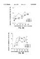
- FIG. 1displays a control experiment showing the thrombin activation at 37° C. and physiological pH.
- FIG. 2demonstrates the effects of pH on thrombin activation at 37° C.
- FIG. 3A and FIG. 3Beach show inhibition by EACA of Staph. aureus growth in the presence of various concentrations of EACA.
- FIGS. 4A and FIG. 4Beach show inhibition by EACA of E. coli growth in the presence of various concentrations of EACA.
- a hemostatic patchcomprising a shaped structural element that is a biodegradable matrix, such as absorbable gelatin sponge or calcium alginate, to which is applied a hemostatic agent that contains epsilon aminocaproic acid, "EACA.”
- EACAis an inhibitor of clot degradation. In the body, clot formation and clot breakdown are competing processes. EACA inhibits the production of plasmin, an enzyme that degrades clots. Plasmin degrades clots by solubilizing fibrin, an important component of clots, in a process called fibrinolysis.
- a hemostatic patch according to the inventionthus comprises an amount of EACA effective for inhibiting fibrinolysis.
- EACAfunctions as a hemostatic agent in a patch in a manner that approximates the effectiveness of fibrinogen, a coagulation factor that, in solution, converts to fibrin in the presence of thrombin.
- Fibrinogenis an active ingredient found in other hemostatic patches.
- EACAis devoid of the hazards that accompany use of fibrinogen.
- EACA in the matrix of a patchprovides an alkaline environment that accelerates the activation of thrombin.
- EACAgreatly increases thrombin's activity FIG. 2. This phenomenon holds true whether the EACA acts on thrombin present in the blood endogenously or on thrombin that is supplied externally in a patch.
- a patch comprising EACAexerts a dual hemostatic action by (1) slowing clot degradation by inhibiting plasmin formation and (2) accelerating clot formation by activating thrombin.
- a methodfor accelerating the activity of thrombin by increasing the pH of the local environment of a patch according to the invention.
- a patchcomprises a matrix and a "thrombin enhancing compound" capable of raising the pH in a solution in the local environment of the patch sufficient to increase the activation of thrombin.
- a compoundis capable of raising the pH of the local environment to a pH in the range of 7.0-9.0 inclusive, more advantageously between pH 7.02-8.02 inclusively, and even further advantageously, between pH 7.62-8.02, inclusively.
- an alkaline solutionis created in the local environment of the patch as the thrombin enhancing compound solubilizes upon its contact with blood. Then, thrombin present in the blood and, optionally, thrombin provided as an exogenous ingredient of the patch mixes with the alkaline solution in the local environment of a patch and thereby is activated.
- the thrombin-enhancing compound provided in the patch for increasing pHis EACA.
- a "sterile buffer”which is pharmaceutically acceptable and capable of buffering the local pH in the patch to alkaline conditions, (i.e., between a pH of 7.0-9.0, more advantageously pH 7.02-8.02, and even further advantageously, 7.62-8.02), is suitable as a thrombin-enhancing compound, as well.
- Tris bufferis an effective thrombin-enhancing sterile buffer, as shown in FIG. 2, open diamond-shaped graphical plot.
- Other sterile buffers that buffer the pH in this rangeare contemplated, such as Hepes buffer, for example. Accordingly, in a more advantageous patch, EACA and Tris (or other) buffer both are provided in the matrix of the patch.
- EACApossesses antibacterial properties. According to the present invention, it has been demonstrated that EACA exerts dose-dependent inhibition of both S. aureus and E. coli growth (FIGS. 3A, 3B and 4A, 4B, respectively). Therefore, the EACA/matrix patch according to the present invention is very desirable for its antibacterial effects on microorganisms present at the wound site where a patch is applied.
- EACAcontains no foreign peptides of animal origin. For example, a non-human fibrinogen hemostatic agent in some humans will trigger an immune response or allergic-like reaction.
- a patch according to the inventioncan contain as a sole hemostatic agent EACA dispersed within a matrix or applied to a surface of a matrix in an amount effective for inhibiting fibrinolysis and thereby stimulating clot formation.
- a biodegradable "matrix” as referred to herein, and as employed in any of the present embodiments of the invention,is selected from, but not limited to, the group consisting of absorbable gelatin sponge, calcium alginate, calcium/sodium alginate, collagen, and oxidized regenerated cellulose.
- EACA4,390,519 to Sawyer, and other conventional matrices utilized in hemostatic patches, are contemplated for use with EACA according to the present invention.
- Four matrices that are advantageous for use with EACAinclude absorbable gelatin sponge, calcium alginate, calcium/sodium alginate, and collagen.
- a first embodiment of the inventiontherefore provides a patch comprising a matrix of absorbable gelatin sponge "G” and a hemostatic agent, EACA "E.”
- the GE or G(Ca++)E patchneed not contain or fibrinogen to function effectively to control hemorrhage of a parenahymal organ.
- both GE and G(Ca++)Ehave good thermal stability and can be stored for months to a few years without refrigeration and losing effectiveness.
- the GE and G(Ca++)E patchesare useful for field and emergency use, since each may be stored in a ready-to-use state for a lengthy period, even in absence of refrigeration. Both also are much less expensive to make than patches which contain fibrinogen.
- a GE or G(Ca++)E patchfurther comprises an effective amount of thrombin for stimulating hemostasis and thus is designated as "GTE” or "GT(Ca++)E.”
- GTEthrombin
- GT(Ca++)Ethrombin molecule
- a thrombin moleculeis most stable at temperatures between 2°-8° C. However, these patches can be stored for a limited period of time at room temperature. In fact, because addition of thrombin enhances the GE and G(Ca++)E patches' effectiveness, these patches are very useful outside the clinic for field use, such as for emergency or military purposes.
- the matrixis advantageously a flat layer of gelatin foam, more advantageously, Gelfoam®, and even more advantageously, compressed gelatin foam or compressed GelFoam®.
- the effectiveness of patches of the present invention in promoting clot formationis enhanced by the lattice structure of the gelatin foam, which promotes enzyme substrate interactions.
- the gelatin foam structureenhances contact between thrombin provided exogenously in the patch with endogenous fibrinogen present in the blood exuding from the wound.
- Additional hemostatic agentscan be applied to the GE patch in amounts effective for stimulating hemostasis, including, but not limited to: thrombin "T", an enzyme which converts fibrinogen to fibrin; calcium, sodium, magnesium or other ions that stimulate hemostasis; and optionally, fibrinogen, "F".
- thrombinan enzyme which converts fibrinogen to fibrin
- calcium, sodium, magnesium or other ionsthat stimulate hemostasis
- fibrinogenfibrinogen
- calcium chlorideis generally a preferred additive for introducing a calcium ion into the patch.
- EACA analogsor compounds that possess a similar hemostatic activity and a chemical structure to that of EACA, can be used instead of, or in addition to, EACA in a patch according to the invention.
- Possible EACA analogs contemplated for addition to a matrixinclude EACA derivatives having bioisosteric functional groups.
- EACA's carboxylic acid groupcan be substituted, for example, by sulfonic or sulfinic acid (--SO 2 H and --SO 3 H) or phosphonic acid groups.
- Examples of analogsinclude, but are not limited, to 5-aminopentanoic a acid, 7-aminoheptanoic acid, 8-aminooctanoic acid, provided that these compounds exert a hemostatic activity.
- thrombin and fibrinogenas defined herein are meant to include natural thrombin and fibrinogen molecules derived from an animal or human origin, a synthetic form or a recombinant form of the molecules, including functionally active analogs that effectively maintain the enzyme's clot promoting activity in an animal or human.
- the species of animal from which the molecule is derivedcan vary and depends on the intended use of the patch. For example, a patch intended for human use for safety reasons contains non-human thrombin, and preferred in this context is bovine thrombin.
- thrombinBy avoiding use of human fibrinogen, risks associated with viral contamination of purified blood products (particularly with fibrinogen) are minimized. Indeed, the ingredients EACA, thrombin and GelFoam® all are approved by the U.S. Food and Drug Administration for human use.
- a patchhaving a matrix composed of calcium-sodium alginate "CVA” or calcium alginate "CA,” and a hemostatic layer of EACA "E.” It is understood that calcium alginate can be substituted for calcium/sodium alginate in the discussion and examples hereafter, without substantial differences in results.
- CVAEadvantageously contains calcium ion and thrombin as well. It also is less expensive as compared with a patch that contains fibrinogen. Similar to the GE patch, the CAE patch can include additional hemostatic agents including, but not limited to, thrombin, calcium or sodium or other ions in amounts that are effective to stimulate or accelerate hemostasis. These patches further can contain additives as described herein, as well.
- an effective amount of the active peptide, RGD, "R” or RGDS effective to stimulate wound healingis added to a patch comprising GE or CAE, and thus such a patch is designated as GER or CAER.
- the tripeptide RGDis composed of arginine, glycine and aspartic acid, and optionally serine “RGDS,” and is the active site of fibrinogen and fibronectin. RGD accelerates wound healing and is believed to stimulate fibroblast migration.
- the RGD additiveis also much less expensive than fibrinogen.
- RGDcan be synthesized easily using conventional solid phase chemistry at a fraction of the cost of obtaining fibrinogen, which currently must be obtained by purification from a natural source.
- an amount of the agent protamine sulfate "P" effective to neutralize heparin present in the local environment of the patchis added to any of the aforementioned patches comprising EACA and a matrix.
- Protamine sulfateneutralizes heparin or vitamin K antagonists that are present in the blood of certain patients or animals being treated with a hemostatic patch.
- a patch comprising GEP or CAEP, for example,is prescribed for persons undergoing parenteral therapy with heparin.
- a patch that further contains thrombinwould be effective in patients taking dicumarol.
- a patch containing protamine sulfateis preferably stored at refrigerated temperatures of 2-8 degrees Celsius to maintain the activity of protamine sulfate.
- an additional advantage of the patches according to the present inventionis that the matrices, such as absorbable gelatin sponge or calcium alginate, and the hemostatic agents, especially EACA and thrombin, and the additive, RGD, all are relatively inexpensive. It is estimated that production of a "standard-size" rectangular patch of about 9.5 ⁇ 4.8 cm, having a thickness of about 2.5 mm would cost substantially less than a TAF patch of the same size.
- Patches according to the present inventionexhibited efficacy in inducing hemostasis in freely bleeding lesions of the spleen, liver and kidney of an anesthetized pig.
- Surgical lesions induced in parenchymal organs of pigsprovide a good model system for hemostasis in the analogous human organs as evidenced by preclinical studies performed on pigs and dogs for the TachoComb® patch.
- patches according to the present inventionperformed better than TachoComb® in the liver, while in the kidneys, the patch containing a matrix of GelFoam®, thrombin and 100 mg/cm 2 EACA performed equally as well as the TachoComb® patch.
- the results of that comparative experimentare presented in Example 3 herein.
- Another important advantage of the present inventionis its flexibility, that is, a patch is provided that easily conforms to the contours of an organ or biological surface, making the manipulation of applying the patch quicker to perform. As a result, there is less overall blood loss to the patient and less time is spent in surgery.
- a hemostatic patch according to the present inventionin employed by applying a "wound-contacting" surface of the patch, a surface intended to contact the wound and containing hemostatic agent(s) and optionally additives, to a bleeding wound. Then, the patch is maintained in contact with the wound for a period of time sufficient for clotting to occur at the interface between the hemostatic patch and the wound and for bleeding to be substantially arrested. Preferably the patch is maintained in contact with the wound surface for a period of about 3-20 minutes, advantageously 3-10 minutes, and more advantageously, 3-5 minutes. Where EACA, thrombin, and calcium chloride all are present on/in the matrix, the time period is preferably about 5 minutes.
- the patchis held in place against the biological surface preferably with light pressure, preferably by means of a sterile saline soaked sponge.
- the patchmay be held in place simply by applying pressure to the patch by means of a gauze or other dry sterile material.
- a bandageincluding an elasticized bandage, can be wrapped around the patch so as to provide light pressure on the wound site.
- a patch according to the present inventionis useful for hermetically sealing body tissue. For example, when air leaks from a wound in the lungs, a patch is applied to the surface surrounding the wound, held in place with light pressure for a period of time adequate to induce hemostasis, as discussed above. During that time, in addition to hemostasis, a hermetic seal forms.
- a hemostatic patch according to the inventionPrior to applying the patch, it is preferable to soak the patch in sterile saline solution. Such a step is not required, however.
- Use of a hemostatic patch according to the invention, without first soaking in saline solutionpermits quick and simple application of the patch in field situations, such as may be encountered by an emergency medical technician or a military health-care worker.
- the patchis contained within a sealed sterile package which facilitates removal of the patch without contamination.
- a sealed sterile packagefor example, can be an aluminum foil pouch or other conventional material that is easily sterilized. Radiation, advantageously gamma radiation, is applied to sterilize the patch and packaging material together.
- a container having dual compartmentsis provided.
- a first compartmentcontains distilled water, sterile saline or a sterile buffer, while the second compartment contains a patch according to the invention.
- the patch of the second compartmentcan be readily dipped into an opened first compartment and subsequently applied to the wound.
- a preferred use of a patch according to the present inventionis to inhibit or completely stop bleeding of a parenchymal organ, such as the liver, kidney, spleen, pancreas or lungs. Additional uses for such a patch include curbing bleeding of tissues during types of surgery such as, but not limited to, internal/abdominal, vascular (particularly for anastomosis), ufological, gynecological (particularly for an episiotomy), thyroidal, neurological, ENT, tissue transplant uses, and dental surgeries.
- a hemostatic patchincludes topical treatment, such as for burn or tissue transplants.
- a patch intended for topical use according to the inventionpreferably contains additives, such as anti-infection medicaments. Bactericides, fungicides and wound healing agents can be added, as well. Neomycin and bacitracin are examples of certain additives that are incorporated into a patch intended for topical use, in addition to the antibacterial properties of EACA discussed above.
- a hemostatic patch of the inventionalso is useful for treating animals, preferably humans or other mammals.
- animalspreferably humans or other mammals.
- both companion, livestock and wild animalscan be treated with a hemostatic patch.
- a patch in size and shape according to the intended usemay be cut to size with a pair of scissors.
- gelatin foampreferably provided in a compressed form. More preferably, a GelFoam® matrix that is compressed to at least one-half its original thickness.
- a patchmay be spherically, conically, cuboidally or cylindrically-shaped or prefabricated into small squares, such as for packing into a body cavity. Such an embodiment is useful for example, for a dental cavity resulting from tooth extraction. Additionally, the patch can be configured into a tampon, for example, for epistaxis (profusely bleeding nostril) or other void.
- a patch intended for topical applicationsadditionally can be applied with an adhesive tape, as a band-aid form, where the patch is adhered to an adhesive backing.
- the adhesive used to secure the patchis porous in areas which contact the skin.
- One or more additional layers of wound dressing materialcan be applied to a patch.
- Such an additional layercan be made as an integral part of the patch, thereby creating a thicker patch.
- the layermay be applied as a supplement to the backside (non-wound contacting surface) of a patch according to the invention.
- the layer(s)can contain superabsorbents to wick exudant solution from the wound site. It is advised that for patches intended for internal-surgical applications, where an added layer(s) is integral with the patch, the layer(s) should be both biodegradable and pharmaceutically acceptable.
- the patchcan be designed to facilitate its application to anastomose or fuse ends of a blood vessel or other body lumen having been severed surgically or otherwise.
- a rectangular GETR patchfor example, is wrapped around the external surface of the ends of a Dacron® graft. When the graft is positioned into place, the patch accelerates fibrin growth into the graft to seal the graft in place (hemostatically and hermetically).
- kitsthat contains a graft and a patch according to the present invention that is designed for fitting with the ends of the graft.
- a kitis provided having a patch of the present invention pre-fitted onto at least one end of a graft.
- a wound-contacting surface of the patchis coated with a color indicator to assist the user, such as yellow vitamin B 2 (riboflavin) or a suitable dye, for example, hemin.
- a color indicatorto assist the user, such as yellow vitamin B 2 (riboflavin) or a suitable dye, for example, hemin.
- a patch according to the inventionis made by applying to a matrix, an amount of EACA effective for inhibiting fibrinolysis in the local environment of the matrix.
- an amount of EACAeffective for inhibiting fibrinolysis in the local environment of the matrix.
- about 10-100 mg/cm 2 of EACAis applied to a wound-contacting surface of the matrix, more advantageously 60-70 mg/cm 2 .
- EACAhemostatic agents or additives described as components of a patch according to the invention
- EACAcan be applied to the matrix by any of several methods which all would be performed most advantageously under sterile conditions. It is understood that conventional methods of applying the hemostatic agents and additives to a matrix comprising EACA besides those described herein can be performed as well.
- EACAis applied as a layer to a particular surface or side of the matrix, which surface is then designated as the wound-contacting surface.
- EACAelectroactive polymer
- a solution of EACAcan be coated onto a matrix and dried by lyophilization or by conventional means.
- a matrixis dipped completely or partially into a sterile solution of EACA such that a sufficient amount of EACA accumulates within the matrix effective to inhibit fibrinolysis in a mammal, such that similar effectiveness to the Hemarrest patch is demonstrated.
- the matrix/EACAis coated with a protein layer that facilitates EACA's adherence to the matrix.
- this proteinis thrombin, although other proteinaceous or gelatin compound which facilitates such adherence could be utilized, as well.
- the matrixis coated with a protein layer prior to application of EACA.
- the matrixis treated before and after addition of EACA with a protein, preferably which is in solution with an ion additive, such as calcium (i.e., calcium chloride solution).
- embodiments such as GT(Ca++)E or CT(Ca++)Eare made by applying to a wound contacting surface of a matrix of gelatin foam or collagen, a first solution of thrombin dissolved in calcium chloride, the thrombin present at an amount, for example, between 1-1000 IU/cm 2 , advantageously 1-100 IU/cm 2 , and more advantageously 1-4 IU/cm 2 , or 1.25 IU/cm 2 .
- the thrombinis dissolved in 20-60 mM calcium chloride, preferably about 40 mM, such that an amount between 25-150 micrograms/cm 2 , preferably 50-100 micrograms/cm 2 , is deposited onto that surface.
- the next stepcomprises applying to the thrombin-coated matrix surface, 10-100 mg/cm 2 of epsilon aminocaproic acid, preferably 60-70 mg/cm 2 , and preferably in a powder form; then, applying a second solution of thrombin in calcium chloride, which, for example contains the amounts of thrombin and calcium as described in the first solution; and then drying the thrombin, calcium chloride and epsilon aminocaproic acid on the patch.
- the amount of thrombin applied in the first and second solutionscan vary, or, a single thrombin solution sealing step can be applied after addition of EACA.
- the total amount of thrombin applied to the wound-contacting surface of the patch by the two stepsis 2-10 IU/cm 2 .
- the drying stepis accomplished by lyophilization, preferably.
- Other drying procedures appropriate for a material containing an active protein ingredientcan also be employed, so long as the drying treatment does not denature the proteins or render them inactive when exposed to animal blood.
- the patchis conventionally dried, by maintaining it at room temperature for a period of 1-3 hours, followed by refrigeration overnight.
- an agent added to a matrixin addition to EACA, thrombin, calcium chloride, includes an amount of protamine sulfate effective to neutralize heparin in the local environment of the patch.
- Protamine sulfateis added in an amount between 1-15 mg/cm 2 of said matrix, preferably in an amount between 2-5 mg/cm 2 of a wound contacting surface of the matrix.
- RGD or RGDS peptidecan be dissolved in double distilled water and sprayed onto a wound-contacting surface of the patch.
- a patchadvantageously contains an amount of RGD effective to enhance clot formation.
- RGD or RGDSis applied to a patch advantageously in an amount between 110-130 mg/cm 2 .
- a standard size patchwould contain about 1-10 mg/patch or about 5-7 mg/patch of RGD or RGDS.
- the hemostatic agents or additives described in the foregoing paragraphscan be applied to a matrix as a layer, for example, by spraying them onto the wound-contacting surface of the matrix in dried forms.
- a matrixcan be dipped or coated with a solution containing the hemostatic agent/additive. It is desirable that the matrix and agents commingle, particularly when the patch is exposed to a body fluid such as blood, which permits the dried agents to solubilize and mix.
- a patchcan be provided wherein the hemostatic agent or mixture of hemostatic agents are absorbed into the pores or interstices of the matrix, or, the agents can be layered on a surface of the matrix and upon solubilizing the agents by addition of body fluid, the desired commingling is achieved.
- the matrixcan be coated with appropriate hemostatic agents described in the above embodiments on one or all surfaces.
- the hemostatic agents and additivesare coated on only one surface (the wound-contacting surface). Such an arrangement avoids inducing hemostasis between the wound and a non-wounded tissue in the vicinity of the patch.
- the patchis coated with hemostatic agent(s)/additive(s) on all surfaces.
- a kit according to the inventioncomprises any of the above described hemostatic patch embodiments (which vary in ways including hemostatic agent(s) and additive(s) utilized, shape or size) according to the invention and a package, wherein the patch is contained within a sealed sterile package which facilitates removal of the patch without contamination.
- the kitcan contain multiple patches, preferably wherein each patch is contained within a separate sealed sterile package.
- a kit designed for field/military usecan, in addition to a hemostatic patch, further include disposable pre-sterilized surgical instruments, such as a scalpel, clamp, tourniquet, elastic or inelastic bandage, or the like.
- kitscomprises a patch containing agents added to the matrix including thrombin, EACA calcium chloride, and protamine sulfate.
- agents added to the matrixincluding thrombin, EACA calcium chloride, and protamine sulfate.
- a kitcan be prescribed, for example, to patients requiring anticoagulant therapy, to avert the risk of serious bleeding which can occur from a minor injury.
- the availability of such a patchcan reduce postoperative hospitalization for patients on dicumarol who underwent surgery.
- the first part of this studyexamined activation of thrombin and its degradation in H 2 O after incubation at 37° C.
- the assay usedwas a colorimetric cleaving of a tripeptide, TFA-phe-pro-arg-AFC, where the AFC is the colorometric tag. Seventeen mg of this substrate was dissolved in 200 ⁇ l DMSO. Thrombin was made up as 10 units/mi.
- the "TEST" solutioncontained 100 ⁇ l substrate and 200 ⁇ l of the thrombin solution; a blank contained the same amount c. substrate and 200 ⁇ l of H 2 O.
- FIG. 1 labeled as "ACTIVATION OF THROMBIN SOLUTION AT 37° C.”shows the results of that experiment.
- the optical density in all of these experimentsis an indication of the color and therefore the amount of cleavage of the enzyme that has taken place.
- the slope of the black-box lineindicates that thrombin activation of thrombin dissolved in H 2 O takes place slowly over a 172 minute time period.
- EACAwas shown to inhibit both Staph. aureus and E. coli in a dose-dependent manner by the following method.
- EACACulture plates and EACA discs were prepared as follows: Whatman filter paper discs of 5.4 cm in diameter and 22.9 cm 2 in area were placed in beakers of almost the same diameter. EACA (229 mg) was dissolved in 250 ⁇ l of double distilled H 2 O and used to make the final concentrations. All concentrations of EACA were applied in 250 ⁇ l of H 2 O. Concentrations of 0, 10, 20, 30, 40, 50, 60, 70, 80, 90 and 100 mg/cm 2 were prepared. After application of EACA solutions, the discs were allowed to dry and frozen to ensure stability.
- Discs for application to agar plateswere made with a paper punch, at a size of about 6.35 mm. Agar plates were poured in two increments.
- Brain-Heart Infusion agarwas prepared and autoclaved. After cooling to approximately 55° C., 12 mls were added to each 100 mm ⁇ 15 mm petri dish. Plates were allowed to cool to room temperature, wrapped in parafilm and refrigerated. Brain-Heart Infusion broth was prepared and autoclaved. When the temperature was cooled to room temperature, a 1 ml aliquot of Staph. aureus or E. coli was added and the broth incubated overnight at 37° C.
- FIG. 3A and FIG. 3Beach show inhibition by EACA of Staph. aureus growth graphically, for each set of various concentrations of EACA, while FIGS. 4A and FIG. 4B each show inhibition by EACA of E. coli growth for each set of varying concentrations of EACA.
- TachoComb® patcheswere obtained and applied according to the manufacturer's instructions. That is, prior to preparation, the TachoComb® patches were dipped in sterile saline and applied to bleeding organs with light pressure for five minutes.
- a lobe of pig liverwas surgically isolated and three lesions approximately 1 ⁇ 1.5 cm in size were created. Blood flowed freely from each of the lesions.
- Each of the patches discussed in part A. (above)were applied and kept under pressure by a saline soaked sponge for five minutes and the pressure was released. Patches were evaluated by their ability to control hemorrhage in terms of (a) leakage, (b) ability to withstand increased vascular pressure, (c) the resistance offered when attempting to peel the patch from the lesion, and (d) events of clot formation in the lesion.
- both poles (ends) of the kidneywere surgically removed to a depth of approximately 0.5 cm, while the renal artery was clamped.
- the clampwas removed after the test patches were placed and pressure applied with a saline soaked surgical sponge for five minutes.
- both patches according to the inventionshowed good control of hemorrhage, with only a little bleeding from the edge in the 100 mg patch and no bleeding from the 10 mg patch.
- the TachoComb® patchwas the only patch leaking or bleeding from the edge.
- a small amount of bloodwas present on the surface of the 100 mg patch, while none was present on the 10 mg patch.
- the TachoComb® patchWhen epinephrine was injected, the TachoComb® patch still dripped blood from the edges after 18 minutes. The peel test after 20 minutes showed the TachoComb® patch with minimal adhesion, the clot stuck to the patch, and the wound continued to bleed. In the lesion with the 100 mg patch, blood also flowed, but not as much as the TachoComb® patch. The 10 mg patch had the least bleeding of any of the patches and had both good incorporation of the patch into the lesion and good clot formation, with some minimal adhesion to the periphery.
- the TachoComb®In the kidney, there was not much difference between the TachoComb® and the 100 mg patch lesions. There was no bleeding before or after epinephrine injections. When the patches were peeled at 20 minutes, the TachoComb®patch had very good adhesive qualities, good clot formation, but some free blood. The 100 mg patch did not have as good adhesiveness, but had a well-formed clot and no hemorrhage. When a blank gelatin patch and 10 mg patches according to the invention were compared, the 10 mg patch definitely was better. Five minutes after the pressure release, there was free blood under the 100 mg patch while there was some bleeding around the edge of the 10 mg patch.
- a lesion on the opposite pole of the same kidneywas covered with a TachoComb® patch.
- the latter patchwas darker, which indicates that more blood was coming through the patch matrix.
- the lower edges of that patchwere looser as compared to the hemarrest patch.
- Fresh bloodcould be seen on a dry sponge held against the organ for the purpose of aiding in detection of fresh blood.
- the studyhas both parts I and II.
- Part 1Patches CTR, CTE(f), GT(f)E(f) and a plain gelfoam (G) patch were applied to lesions made on the spleen of an anesthetized pig.
- the symbol "(f)”denotes the compound immediately preceding it as a freshly-applied compound. That is, E(f) denotes EACA that is freshly applied to a patch very soon (less than about three hours) after it is made.
- Patch GStrong adhesion to surrounding tissue, no adhesion to lesion, much leaking.
- Patch CTE(f)RNo adhesion, good clot formation, little leakage.
- Patch GT(f)E(f)Good adhesion to lesion, good clot formation, little leakage.
- Patch CTE(f)No adhesion, good clot formation, little leakage.
- Patch CTRDry to the blot test (placing a dry surgical sponge on the lesion), clot is developed, no collagen incorporated into lesion.
- Patch GDry to the blot test, gelatin foam incorporated into the lesion.
- Patch CTE(f)RAbsolutely no blood elements on sponge after blotting; clot is excellent, filling lesion and extending onto the surrounding normal area.
- Patch GT(f)E(f))Serum staining on sponge, but good clot and gelatin sponge incorporated into the lesion.
- Patch CTE(f) Dry to blot testsimilar to CTE(f)R.
Landscapes
- Health & Medical Sciences (AREA)
- Animal Behavior & Ethology (AREA)
- Engineering & Computer Science (AREA)
- Veterinary Medicine (AREA)
- Public Health (AREA)
- General Health & Medical Sciences (AREA)
- Life Sciences & Earth Sciences (AREA)
- Epidemiology (AREA)
- Chemical & Material Sciences (AREA)
- Materials Engineering (AREA)
- Surgery (AREA)
- Hematology (AREA)
- Biomedical Technology (AREA)
- Heart & Thoracic Surgery (AREA)
- Vascular Medicine (AREA)
- Materials For Medical Uses (AREA)
- Medicinal Preparation (AREA)
- Medicines That Contain Protein Lipid Enzymes And Other Medicines (AREA)
- Acyclic And Carbocyclic Compounds In Medicinal Compositions (AREA)
- Pharmaceuticals Containing Other Organic And Inorganic Compounds (AREA)
Abstract
Description
TABLE 1 ______________________________________ PATCH COMPONENT CODES: ______________________________________ G = gelatin foam patch alone, e.g., Gelfoam ® CA = calcium alginate CVA = calcium/sodium alginate, e.g., Kaltostat ® C or CVC = collagen or collagen (Helistat ®), respectively E = EACA (Ca++) = calcium T = thrombin R = RGD peptide P = protamine sulfate F = Fibrinogen (f) = freshly applied compound (Example 7) GT (Ca++) E = "Hemarrest ™" patch ______________________________________
TABLE 2 __________________________________________________________________________Results: Inhibition of E. coli (Plates 1-6) and Staph. aureus (Plates 7-12) Growth by EACA Conc. of DIAMETER Plate EACA in OF % > % OF Date Number Organism mg/cm2 INHIBITION CONTROL MAXIMUM __________________________________________________________________________10/22/93 1 E. coli control 6.35 0.00 77.00 10 6.95 9.40 84.20 30 7.65 20.50 92.70 50 7.75 22.00 93.90 70 8.25 29.90 100.00 90 7.50 18.10 90.90 10/22/93 2 E. coli control 6.35 0.00 70.20 10 6.70 5.50 74.00 30 8.55 34.60 94.50 50 8.60 35.40 95.00 70 9.05 42.50 100.00 90 8.35 31.50 92.30 10/22/93 3 E. coli control 6.35 0.00 70.60 10 6.70 5.50 74.40 30 7.05 11.00 78.30 50 7.10 11.80 78.80 70 9.00 41.70 100.00 90 8.25 29.90 91.70 10/22/93 4 E. coli control 6.35 0.00 77.40 20 7.05 11.00 86.00 40 7.70 21.30 93.90 60 7.70 21.30 93.90 80 8.20 29.10 100.00 100 7.75 22.00 94.50 10/22/93 5 E. coli control 6.35 0.00 78.40 20 7.70 21.30 95.10 40 7.75 22.00 95.70 60 7.45 17.30 92.00 80 8.10 27.60 100.00 100 8.10 27.60 100.00 10/22/93 6 E. coli control 6.35 0.00 76.50 20 7.60 19.70 91.60 40 8.10 27.60 97.60 60 7.90 24.40 95.20 80 8.25 29.90 99.40 100 8.30 30.70 100.00 10/22/93 7 S. aureus control 6.60 0.00 77.20 10 7.55 14.40 88.30 30 8.20 24.20 95.90 50 7.65 15.90 89.50 70 8.55 29.50 100.00 90 7.90 19.70 92.40 10/22/93 8 S. aureus control 6.55 0.00 80.90 10 7.10 8.40 87.70 30 7.00 6.90 86.40 50 7.15 9.20 88.30 70 7.75 18.30 95.70 90 8.10 23.70 100.00 10/22/93 9 S. aureus control 6.55 0.00 81.90 10 7.00 6.90 87.50 30 7.20 9.90 90.00 50 7.45 13.70 93.10 70 7.85 19.80 98.10 90 8.00 22.10 100.00 10/22/93 10 S. aureus control 6.60 0.00 79.00 20 8.05 21.90 96.40 40 8.30 25.80 99.40 60 8.10 22.70 97.00 80 7.55 14.40 90.40 100 8.35 26.50 100.00 10/22/93 11 S. aureus control 6.50 0.00 83.90 20 7.20 10.80 92.90 40 7.70 18.50 99.40 60 7.30 12.30 94.20 80 7.40 13.80 95.50 100 7.75 19.20 100.00 10/22/93 12 S. aureus control 6.50 0.00 79.30 20 6.75 3.80 82.30 40 8.05 23.80 98.20 60 8.00 23.10 97.60 80 8.20 26.20 100.00 100 7.20 10.80 87.80 __________________________________________________________________________
Claims (17)
Priority Applications (1)
| Application Number | Priority Date | Filing Date | Title |
|---|---|---|---|
| US08/487,659US5645849A (en) | 1993-11-03 | 1995-06-07 | Hemostatic patch |
Applications Claiming Priority (2)
| Application Number | Priority Date | Filing Date | Title |
|---|---|---|---|
| US14636093A | 1993-11-03 | 1993-11-03 | |
| US08/487,659US5645849A (en) | 1993-11-03 | 1995-06-07 | Hemostatic patch |
Related Parent Applications (1)
| Application Number | Title | Priority Date | Filing Date |
|---|---|---|---|
| US14636093AContinuation | 1993-11-03 | 1993-11-03 |
Publications (1)
| Publication Number | Publication Date |
|---|---|
| US5645849Atrue US5645849A (en) | 1997-07-08 |
Family
ID=22517031
Family Applications (2)
| Application Number | Title | Priority Date | Filing Date |
|---|---|---|---|
| US08/487,659Expired - Fee RelatedUS5645849A (en) | 1993-11-03 | 1995-06-07 | Hemostatic patch |
| US08/474,127Expired - LifetimeUS5643596A (en) | 1993-11-03 | 1995-06-07 | Hemostatic patch |
Family Applications After (1)
| Application Number | Title | Priority Date | Filing Date |
|---|---|---|---|
| US08/474,127Expired - LifetimeUS5643596A (en) | 1993-11-03 | 1995-06-07 | Hemostatic patch |
Country Status (9)
| Country | Link |
|---|---|
| US (2) | US5645849A (en) |
| EP (1) | EP0726749B1 (en) |
| JP (1) | JPH09504719A (en) |
| AT (1) | ATE273035T1 (en) |
| AU (1) | AU1086795A (en) |
| CA (1) | CA2175203A1 (en) |
| DE (1) | DE69433939T2 (en) |
| ES (1) | ES2227542T3 (en) |
| WO (1) | WO1995012371A1 (en) |
Cited By (94)
| Publication number | Priority date | Publication date | Assignee | Title |
|---|---|---|---|---|
| US5968001A (en)* | 1996-05-14 | 1999-10-19 | Bristol-Myers Squibb Company | Wound dressings with leak prevention seal |
| US6162241A (en)* | 1997-08-06 | 2000-12-19 | Focal, Inc. | Hemostatic tissue sealants |
| US6187347B1 (en) | 2000-02-09 | 2001-02-13 | Ecosafe, Llc. | Composition for arresting the flow of blood and method |
| WO2001054735A3 (en)* | 2000-01-28 | 2001-12-20 | Orthogene Inc | Gel-infused sponges for tissue repair and augmentation |
| US20020049471A1 (en)* | 2000-09-22 | 2002-04-25 | Perlei Medical Produkte Gmbh | Method and hemostatic patch for effecting local hemostasis |
| US20020062104A1 (en)* | 1999-09-23 | 2002-05-23 | Mark Ashby | Depth and puncture control for blood vessel hemostasis system |
| US20020138025A1 (en)* | 2001-03-09 | 2002-09-26 | Scimed Life Systems, Inc. | Medical slings |
| US6475514B1 (en) | 1998-12-03 | 2002-11-05 | Andrew Blitzer | Athletic patch |
| DE10135507A1 (en)* | 2001-07-20 | 2003-02-06 | Henkel Kgaa | Hemostatic skin plaster, especially for covering cuts or scratches, comprises polymer film containing dissolved or dispersed astringent and/or hemostatic agent, e.g. aluminum hydroxychloride |
| US20030078234A1 (en)* | 2001-02-12 | 2003-04-24 | Marine Polymer Technologies Inc. | Methods for treating a breach or puncture in a blood vessel |
| US20030162708A1 (en)* | 2001-12-21 | 2003-08-28 | Jorgen Wolff | Haemostatic kit, a method of preparing a haemostatic agent and a method of promoting haemostatis |
| US20030163074A1 (en)* | 2002-02-15 | 2003-08-28 | Mcgowan Jeremy | Wound dressing impervious to chemical and biological agents |
| US20030175327A1 (en)* | 2001-12-31 | 2003-09-18 | Cochrum Kent C. | Hemostatic compositions and methods for controlling bleeding |
| US20030224056A1 (en)* | 2002-05-31 | 2003-12-04 | Sanjay Kotha | Hemostatic composition |
| US6666817B2 (en) | 2001-10-05 | 2003-12-23 | Scimed Life Systems, Inc. | Expandable surgical implants and methods of using them |
| US20040001879A1 (en)* | 2002-06-28 | 2004-01-01 | Guo Jian Xin | Hemostatic wound dressing and method of making same |
| US20040005350A1 (en)* | 2002-06-28 | 2004-01-08 | Looney Dwayne Lee | Hemostatic wound dressings and methods of making same |
| US20040019328A1 (en)* | 2001-11-08 | 2004-01-29 | Sing Eduardo Chi | System and method for delivering hemostasis promoting material to a blood vessel puncture site by fluid pressure |
| US6689047B2 (en) | 2000-11-15 | 2004-02-10 | Scimed Life Systems, Inc. | Treating urinary incontinence |
| US20040039246A1 (en)* | 2001-07-27 | 2004-02-26 | Barry Gellman | Medical slings |
| US20040101546A1 (en)* | 2002-11-26 | 2004-05-27 | Gorman Anne Jessica | Hemostatic wound dressing containing aldehyde-modified polysaccharide and hemostatic agents |
| US20040101547A1 (en)* | 2002-11-26 | 2004-05-27 | Pendharkar Sanyog Manohar | Wound dressing containing aldehyde-modified regenerated polysaccharide |
| US20040101548A1 (en)* | 2002-11-26 | 2004-05-27 | Pendharkar Sanyog Manohar | Hemostatic wound dressing containing aldehyde-modified polysaccharide |
| US20040105980A1 (en)* | 2002-11-25 | 2004-06-03 | Sudarshan Tirumalai S. | Multifunctional particulate material, fluid, and composition |
| US20040122350A1 (en)* | 2002-12-20 | 2004-06-24 | Sheng-Ping Zhong | Puncture hole sealing device |
| US20040120993A1 (en)* | 2002-12-20 | 2004-06-24 | Guanghui Zhang | Hemostatic wound dressing and fabric and methods of making and using same |
| US20040122349A1 (en)* | 2002-12-20 | 2004-06-24 | Lafontaine Daniel M. | Closure device with textured surface |
| US6755781B2 (en) | 2001-07-27 | 2004-06-29 | Scimed Life Systems, Inc. | Medical slings |
| WO2004054632A1 (en)* | 2002-12-16 | 2004-07-01 | Langer, Hans-Georg | Haemostatic wound dressing |
| US20040243044A1 (en)* | 2003-06-02 | 2004-12-02 | Penegor Stephen A. | Hemostatic wound dressing |
| US20040241212A1 (en)* | 2003-05-30 | 2004-12-02 | Pendharkar Sanyog Manohar | Biodegradable hemostatic wound dressings |
| US20040265371A1 (en)* | 2003-06-25 | 2004-12-30 | Looney Dwayne Lee | Hemostatic devices and methods of making same |
| US20050010248A1 (en)* | 2003-07-10 | 2005-01-13 | Scimed Life Systems, Inc. | System for closing an opening in a body cavity |
| US20050130884A1 (en)* | 2000-02-07 | 2005-06-16 | Brandt Curtis R. | Pharmacologically active antiviral peptides and methods of their use |
| US20050203024A1 (en)* | 2003-12-01 | 2005-09-15 | Wisconsin Alumni Research Foundation | Peptides and methods for inducing cellular resistance to infection |
| US20050226916A1 (en)* | 1998-11-12 | 2005-10-13 | Cochrum Kent C | Hemostatic polymer useful for RAPID blood coagulation and hemostasis |
| US6982501B1 (en) | 2003-05-19 | 2006-01-03 | Materials Modification, Inc. | Magnetic fluid power generator device and method for generating power |
| US20060002918A1 (en)* | 2004-06-22 | 2006-01-05 | Shan Jiang | Thrombin compositions |
| US20060034935A1 (en)* | 2004-07-22 | 2006-02-16 | Pronovost Allan D | Compositions and methods for treating excessive bleeding |
| US7007972B1 (en) | 2003-03-10 | 2006-03-07 | Materials Modification, Inc. | Method and airbag inflation apparatus employing magnetic fluid |
| US7019191B2 (en) | 2003-03-25 | 2006-03-28 | Ethicon, Inc. | Hemostatic wound dressings and methods of making same |
| US7025772B2 (en) | 2001-03-09 | 2006-04-11 | Scimed Life Systems, Inc. | System for implanting an implant and method thereof |
| US20060084930A1 (en)* | 2004-10-20 | 2006-04-20 | Sridevi Dhanaraj | Reinforced absorbable multilayered fabric for use in medical devices |
| US7048710B1 (en) | 1998-05-01 | 2006-05-23 | Sub-Q, Inc. | System and method for facilitating hemostasis of blood vessel punctures with absorbable sponge |
| US20060115805A1 (en)* | 2002-12-11 | 2006-06-01 | Hansen John E | Gelatine-based materials as swabs |
| US20060258995A1 (en)* | 2004-10-20 | 2006-11-16 | Pendharkar Sanyog M | Method for making a reinforced absorbable multilayered fabric for use in medical devices |
| US20060257457A1 (en)* | 2004-10-20 | 2006-11-16 | Gorman Anne J | Method for making a reinforced absorbable multilayered hemostatic wound dressing |
| US20060257458A1 (en)* | 2004-10-20 | 2006-11-16 | Gorman Anne J | Reinforced absorbable multilayered hemostatis wound dressing |
| US20070009578A1 (en)* | 2004-07-09 | 2007-01-11 | Lene Moller | Haemostatic composition comprising hyaluronic acid |
| US7175646B2 (en) | 1995-09-15 | 2007-02-13 | Boston Scientific Scimed, Inc. | Apparatus and method for percutaneous sealing of blood vessel punctures |
| EP1673134A4 (en)* | 2003-09-12 | 2007-03-21 | Marinepolymer Tech Inc | PRESERVATION OF VASCULAR ACCESS IN HEMODIALYSIS PATIENTS |
| US7200956B1 (en) | 2003-07-23 | 2007-04-10 | Materials Modification, Inc. | Magnetic fluid cushioning device for a footwear or shoe |
| US20070160543A1 (en)* | 2004-01-30 | 2007-07-12 | Lene Moller | Haemostatic sprays and compositions |
| US20070248653A1 (en)* | 2006-04-20 | 2007-10-25 | Cochrum Kent C | Hemostatic compositions and methods for controlling bleeding |
| US7335219B1 (en)* | 2002-11-04 | 2008-02-26 | Sub-Q, Inc. | Hemostatic device including a capsule |
| US7361138B2 (en) | 2003-07-31 | 2008-04-22 | Scimed Life Systems, Inc. | Bioabsorbable casing for surgical sling assembly |
| US20080138416A1 (en)* | 2006-06-13 | 2008-06-12 | Fmc Biopolymer As | Method and systems for using biopolymer-based beads and hydrogels |
| US7402133B2 (en) | 2002-12-17 | 2008-07-22 | Boston Scientific Scimed, Inc. | Spacer for sling delivery system |
| US7448389B1 (en) | 2003-10-10 | 2008-11-11 | Materials Modification, Inc. | Method and kit for inducing hypoxia in tumors through the use of a magnetic fluid |
| US20080304970A1 (en)* | 2003-07-14 | 2008-12-11 | Cooper Paul V | Pump with rotating inlet |
| US20090093550A1 (en)* | 2007-09-19 | 2009-04-09 | Rolfes Emily R | Biocompatible foams, systems, and methods |
| EP2078532A1 (en) | 2001-12-21 | 2009-07-15 | Ferrosan A/S | Medical device comprising a haemostatic agent and haemostatic kit comprising the medical device |
| US20090246238A1 (en)* | 2004-10-20 | 2009-10-01 | Anne Jessica Gorman | Reinforced absorbable multilayered hemostatic wound dressing |
| US7611479B2 (en) | 1998-05-01 | 2009-11-03 | Sub-Q, Inc. | System and method for facilitating hemostasis of blood vessel punctures with absorbable sponge |
| US20090275904A1 (en)* | 2008-05-02 | 2009-11-05 | Sardesai Neil Rajendra | Sheet assemblies with releasable medicaments |
| US7618567B2 (en) | 1998-05-01 | 2009-11-17 | Boston Scientific Scimed, Inc. | Absorbable sponge with contrasting agent |
| US7875043B1 (en) | 2003-12-09 | 2011-01-25 | Sub-Q, Inc. | Cinching loop |
| US20110021964A1 (en)* | 2008-02-29 | 2011-01-27 | Ferrosan Medical Devices A/S | Device for Promotion of Hemostasis and/or Wound Healing |
| US20110045047A1 (en)* | 2008-10-17 | 2011-02-24 | Confluent Surgical, Inc. | Hemostatic implant |
| US20110081397A1 (en)* | 2009-10-01 | 2011-04-07 | Tyco Healthcare Group Lp | Mesh Implant |
| US8033983B2 (en) | 2001-03-09 | 2011-10-11 | Boston Scientific Scimed, Inc. | Medical implant |
| US8050741B2 (en) | 1998-05-01 | 2011-11-01 | Boston Scientific Scimed, Inc. | Device and method for facilitating hemostasis of a biopsy tract |
| US8187625B2 (en) | 2001-03-12 | 2012-05-29 | Boston Scientific Scimed, Inc. | Cross-linked gelatin composition comprising a wetting agent |
| US8518063B2 (en) | 2001-04-24 | 2013-08-27 | Russell A. Houser | Arteriotomy closure devices and techniques |
| US8961541B2 (en) | 2007-12-03 | 2015-02-24 | Cardio Vascular Technologies Inc. | Vascular closure devices, systems, and methods of use |
| US8992567B1 (en) | 2001-04-24 | 2015-03-31 | Cardiovascular Technologies Inc. | Compressible, deformable, or deflectable tissue closure devices and method of manufacture |
| US9084602B2 (en) | 2011-01-26 | 2015-07-21 | Covidien Lp | Buttress film with hemostatic action for surgical stapling apparatus |
| US9265858B2 (en) | 2012-06-12 | 2016-02-23 | Ferrosan Medical Devices A/S | Dry haemostatic composition |
| US9271706B2 (en) | 2008-08-12 | 2016-03-01 | Covidien Lp | Medical device for wound closure and method of use |
| US9345460B2 (en) | 2001-04-24 | 2016-05-24 | Cardiovascular Technologies, Inc. | Tissue closure devices, device and systems for delivery, kits and methods therefor |
| WO2016171633A1 (en)* | 2015-04-21 | 2016-10-27 | Ertan Mevlut | Regenerated oxidized celulose based hemostatic materialcontaining antifibrolytic agents |
| WO2017077525A1 (en) | 2015-11-08 | 2017-05-11 | Omrix Biopharmaceuticals Ltd. | Hemostatic composition |
| US9724078B2 (en) | 2013-06-21 | 2017-08-08 | Ferrosan Medical Devices A/S | Vacuum expanded dry composition and syringe for retaining same |
| US20180043054A1 (en)* | 2016-08-15 | 2018-02-15 | Guangzhou Bioseal Biotech Co. Ltd. | Hemostatic compositions and methods of making thereof |
| US10034957B2 (en) | 2015-11-06 | 2018-07-31 | Ethicon Llc | Compacted hemostatic cellulosic aggregates |
| US10111980B2 (en) | 2013-12-11 | 2018-10-30 | Ferrosan Medical Devices A/S | Dry composition comprising an extrusion enhancer |
| WO2019053579A1 (en) | 2017-09-12 | 2019-03-21 | Shilpa Medicare Limited | Tranexamic acid spray for knee arthroplasty |
| US10653837B2 (en) | 2014-12-24 | 2020-05-19 | Ferrosan Medical Devices A/S | Syringe for retaining and mixing first and second substances |
| US10918796B2 (en) | 2015-07-03 | 2021-02-16 | Ferrosan Medical Devices A/S | Syringe for mixing two components and for retaining a vacuum in a storage condition |
| US11046818B2 (en) | 2014-10-13 | 2021-06-29 | Ferrosan Medical Devices A/S | Dry composition for use in haemostasis and wound healing |
| US11109849B2 (en) | 2012-03-06 | 2021-09-07 | Ferrosan Medical Devices A/S | Pressurized container containing haemostatic paste |
| US11382731B2 (en) | 2015-02-27 | 2022-07-12 | Covidien Lp | Medical devices with sealing properties |
| US11413335B2 (en) | 2018-02-13 | 2022-08-16 | Guangzhou Bioseal Biotech Co. Ltd | Hemostatic compositions and methods of making thereof |
| US11801324B2 (en) | 2018-05-09 | 2023-10-31 | Ferrosan Medical Devices A/S | Method for preparing a haemostatic composition |
Families Citing this family (54)
| Publication number | Priority date | Publication date | Assignee | Title |
|---|---|---|---|---|
| WO1998000161A1 (en)* | 1996-05-24 | 1998-01-08 | Buford Biomedical, Inc. | Fibrin-based systems for the controlled release of medicinals |
| US6706690B2 (en)* | 1999-06-10 | 2004-03-16 | Baxter Healthcare Corporation | Hemoactive compositions and methods for their manufacture and use |
| US7871637B2 (en) | 1996-08-27 | 2011-01-18 | Baxter International Inc. | Dry hemostatic compositions and methods for their preparation |
| DK0936903T3 (en)* | 1996-09-07 | 2004-02-09 | Guenther Beisel | Delayed action agent |
| EP1009291B8 (en)* | 1997-03-20 | 2006-01-11 | Genzyme Corporation | Biodegradable tissue retractor |
| US6979307B2 (en) | 1997-06-24 | 2005-12-27 | Cascade Medical Enterprises Llc | Systems and methods for preparing autologous fibrin glue |
| IT1292410B1 (en)* | 1997-06-24 | 1999-02-08 | Roberto Beretta | READY-TO-USE CONTAINER TO OBTAIN AUTOLOGOUS FIBRIN GLUE |
| US6042592A (en) | 1997-08-04 | 2000-03-28 | Meadox Medicals, Inc. | Thin soft tissue support mesh |
| US6056970A (en)* | 1998-05-07 | 2000-05-02 | Genzyme Corporation | Compositions comprising hemostatic compounds and bioabsorbable polymers |
| US6463335B1 (en) | 1999-10-04 | 2002-10-08 | Medtronic, Inc. | Temporary medical electrical lead having electrode mounting pad with biodegradable adhesive |
| WO2002030479A1 (en)* | 2000-10-13 | 2002-04-18 | On Site Gas Systems, Inc. | Bandage using molecular sieves |
| RU2186572C1 (en)* | 2001-01-09 | 2002-08-10 | Павлов Валентин Николаевич | Method for obtaining a material to cover injured renal surface |
| US8741335B2 (en) | 2002-06-14 | 2014-06-03 | Hemcon Medical Technologies, Inc. | Hemostatic compositions, assemblies, systems, and methods employing particulate hemostatic agents formed from hydrophilic polymer foam such as Chitosan |
| US7371403B2 (en)* | 2002-06-14 | 2008-05-13 | Providence Health System-Oregon | Wound dressing and method for controlling severe, life-threatening bleeding |
| DK1401352T3 (en)* | 2001-06-14 | 2012-06-25 | Kenton W Gregory | PROCEDURE FOR PREPARING A CHITOSANOUS COMPOUND |
| US6998510B2 (en)* | 2002-02-04 | 2006-02-14 | Damage Control Surgical Technologies, Inc. | Method and apparatus for improved hemostasis and damage control operations |
| WO2004001023A2 (en)* | 2002-06-20 | 2003-12-31 | Bionethos Holding Gmbh | Method and device for multiplying and differentiating cells in the presence of growth factors and of a biological matrix or of a supporting structure |
| US20040106344A1 (en)* | 2002-06-28 | 2004-06-03 | Looney Dwayne Lee | Hemostatic wound dressings containing proteinaceous polymers |
| US7943810B2 (en) | 2003-02-04 | 2011-05-17 | Buckman Robert F | Method and apparatus for hemostasis |
| US8834864B2 (en) | 2003-06-05 | 2014-09-16 | Baxter International Inc. | Methods for repairing and regenerating human dura mater |
| EP1631262A4 (en)* | 2003-06-12 | 2010-05-05 | Boston Scient Ltd An Irish Co | IMPROVED SYSTEM AND METHOD FOR FACILITATING HEMOSTASE USING AN ABSORBABLE SPONGE |
| US7927626B2 (en) | 2003-08-07 | 2011-04-19 | Ethicon, Inc. | Process of making flowable hemostatic compositions and devices containing such compositions |
| JP2005075815A (en)* | 2003-09-03 | 2005-03-24 | Masao Tanihara | Hemostatic tissue-repairing material |
| CA2597940A1 (en) | 2005-02-15 | 2006-08-24 | Virginia Commonwealth University | Mineral technologies (mt) for acute hemostasis and for the treatment of acute wounds and chronic ulcers |
| US9204957B2 (en) | 2005-03-17 | 2015-12-08 | Hemcon Medical Technologies, Inc. | Systems and methods for hemorrhage control and or tissue repair |
| CN1321702C (en)* | 2005-10-26 | 2007-06-20 | 陕西杨凌岱鹰生物技术研究所 | Wound dressing for first aid |
| US8938898B2 (en) | 2006-04-27 | 2015-01-27 | Z-Medica, Llc | Devices for the identification of medical products |
| JP2009538192A (en)* | 2006-05-23 | 2009-11-05 | プロビデンス ヘルス システム−オレゴン ディー/ビー/エー プロビデンス セント ビンセント メディカル センター | System and method for introducing and applying a bandage structure within a body lumen or body hollow organ |
| US7604819B2 (en) | 2006-05-26 | 2009-10-20 | Z-Medica Corporation | Clay-based hemostatic agents and devices for the delivery thereof |
| TWI436793B (en) | 2006-08-02 | 2014-05-11 | Baxter Int | Rapidly acting dry sealant and methods for use and manufacture |
| EP2252335B1 (en) | 2008-03-03 | 2013-04-24 | Omrix Biopharmaceuticals Ltd. | A gelatin sponge comprising an active ingredient, its preparation and use |
| EP2269594B1 (en) | 2008-04-16 | 2016-03-02 | The Chemo-Sero-Therapeutic Research Institute | Method of producing thrombin-immobilized bioabsorbable sheet preparation |
| AU2009241687A1 (en)* | 2008-05-02 | 2009-11-05 | Providence Health System-Oregon D/B/A Providence St. Vincent Medical Center | Wound dressing devices and methods |
| RU2396984C2 (en)* | 2008-09-25 | 2010-08-20 | Леонид Прокофьевич Истранов | Antimicrobial haemostatic sponge |
| EP2340002B1 (en) | 2008-10-06 | 2015-03-25 | Providence Health System - Oregon | Foam medical devices and methods |
| US9039783B2 (en) | 2009-05-18 | 2015-05-26 | Baxter International, Inc. | Method for the improvement of mesh implant biocompatibility |
| AU2010262058B2 (en) | 2009-06-16 | 2013-08-29 | Baxter Healthcare S.A. | Hemostatic sponge |
| MX2012007056A (en) | 2009-12-16 | 2012-09-28 | Baxter Healthcare Sa | Hemostatic sponge. |
| SA111320355B1 (en) | 2010-04-07 | 2015-01-08 | Baxter Heathcare S A | Hemostatic sponge |
| KR102002755B1 (en) | 2010-06-01 | 2019-07-23 | 백스터 인터내셔널 인코포레이티드 | Process for making dry and stable hemostatic compositions |
| US9084728B2 (en) | 2010-06-01 | 2015-07-21 | Baxter International Inc. | Process for making dry and stable hemostatic compositions |
| CA2801120C (en) | 2010-06-01 | 2019-08-20 | Baxter International Inc. | Process for making dry and stable hemostatic compositions |
| US8858969B2 (en) | 2010-09-22 | 2014-10-14 | Z-Medica, Llc | Hemostatic compositions, devices, and methods |
| US20130090291A1 (en) | 2011-10-11 | 2013-04-11 | Baxter Healthcare S.A. | Hemostatic compositions |
| MX356185B (en) | 2011-10-11 | 2018-05-17 | Baxter Int | Hemostatic compositions. |
| CA2853356C (en) | 2011-10-27 | 2021-03-23 | Baxter International Inc. | Hemostatic compositions |
| DE102012002209A1 (en)* | 2012-02-07 | 2013-08-08 | Carl Freudenberg Kg | Biodegradable nonwoven for medical purposes |
| US9168178B2 (en) | 2012-05-22 | 2015-10-27 | Covidien Lp | Energy-delivery system and method for controlling blood loss from wounds |
| MX376779B (en) | 2012-06-22 | 2025-03-07 | Teleflex Tech Llc | HEMOSTATIC DEVICES. |
| RU2741304C2 (en)* | 2015-11-08 | 2021-01-25 | Омрикс Биофармасьютикалс Лтд. | Haemostatic composition |
| CN105521520B (en)* | 2015-12-10 | 2018-12-25 | 苏州大学 | Preparation method of silkworm fibroin hemostatic material |
| CN107349458B (en)* | 2017-07-14 | 2019-09-13 | 北京化工大学 | A kind of preparation method of gelatin/plant polysaccharide composite hemostatic sponge |
| CN108159494A (en)* | 2017-12-30 | 2018-06-15 | 孙祎 | A kind of preparation method of toughening modifying cellulose base skin regeneration material |
| US12251491B2 (en) | 2019-03-20 | 2025-03-18 | Astellas Pharma Inc. | Thrombin-carrying hemostatic sheet |
Citations (43)
| Publication number | Priority date | Publication date | Assignee | Title |
|---|---|---|---|---|
| US2610625A (en)* | 1947-10-14 | 1952-09-16 | Armour & Co | Surgical sponge and the preparation thereof |
| US3157524A (en)* | 1960-10-25 | 1964-11-17 | Ethicon Inc | Preparation of collagen sponge |
| DE1960614A1 (en)* | 1969-12-03 | 1971-06-09 | Dental Ges Pfandl & Co | Foaming agent for medicines esp toothpaste |
| US3731683A (en)* | 1971-06-04 | 1973-05-08 | Alza Corp | Bandage for the controlled metering of topical drugs to the skin |
| US3797494A (en)* | 1969-04-01 | 1974-03-19 | Alza Corp | Bandage for the administration of drug by controlled metering through microporous materials |
| US3854480A (en)* | 1969-04-01 | 1974-12-17 | Alza Corp | Drug-delivery system |
| US3908018A (en)* | 1968-05-03 | 1975-09-23 | Choay Sa | Therapeutical and cosmetic composition including N-propionyl-epsilon-amino-caproic acid |
| US4006220A (en)* | 1975-06-04 | 1977-02-01 | Gottlieb Sheldon K | Compositions and methods useful for repairing depressed cutaneous scars |
| JPS5498091A (en)* | 1978-01-18 | 1979-08-02 | Unitika Ltd | Wound protecting material |
| US4167945A (en)* | 1977-01-31 | 1979-09-18 | Gottlieb Sheldon K | Method for enhancing the healing of grafted tissue |
| SU700129A1 (en)* | 1978-06-14 | 1979-11-30 | Киевский Научно-Исследовательский Институт Гематологии И Переливания Крови | Method of obtaining hemostatic diseases |
| US4191751A (en)* | 1975-06-04 | 1980-03-04 | Gottlieb Sheldon K | Method for promoting growth of new connective tissue over surface wounds |
| DE2928405A1 (en)* | 1979-07-13 | 1981-01-29 | Bayer Ag | NEW N-CARBOXYLATED CARBAMATES, METHOD FOR THEIR PRODUCTION AND THEIR USE |
| US4265233A (en)* | 1978-04-12 | 1981-05-05 | Unitika Ltd. | Material for wound healing |
| SU908357A1 (en)* | 1978-09-19 | 1982-02-28 | Московская Ордена Трудового Красного Знамени Ветеринарная Академия Им. К.И.Скрябина | Method of producing collagenic material |
| SU921540A1 (en)* | 1980-09-24 | 1982-04-23 | Киевский государственный институт усовершенствования врачей | Sternotomy method |
| EP0059265A1 (en)* | 1981-02-16 | 1982-09-08 | Hormon-Chemie München GmbH | Material for filling and healing wounds, and method for its preparation |
| US4362567A (en)* | 1979-02-15 | 1982-12-07 | Immuno Aktiengesellschaft Fur Chemisch-Medizinische Produkte | Tissue adhesive |
| US4363319A (en)* | 1980-06-30 | 1982-12-14 | Applied Medical Devices, Inc. | Ready-to-use bandage incorporating a coagulant composition and method of preparing same |
| US4390519A (en)* | 1978-05-19 | 1983-06-28 | Sawyer Philip Nicholas | Bandage with hemostatic agent and methods for preparing and employing the same |
| EP0090997A2 (en)* | 1982-04-02 | 1983-10-12 | Dr. Ruhland Nachf. GmbH | Adhesive wound covering material comprising collagen |
| US4427650A (en)* | 1981-06-25 | 1984-01-24 | Serapharm Michael Stroetmann | Enriched plasma derivative for advancement of wound closure and healing |
| US4427651A (en)* | 1981-06-25 | 1984-01-24 | Serapharm Michael Stroetmann | Enriched plasma derivative for enhancement of wound closure and coverage |
| US4515637A (en)* | 1983-11-16 | 1985-05-07 | Seton Company | Collagen-thrombin compositions |
| US4600574A (en)* | 1984-03-21 | 1986-07-15 | Immuno Aktiengesellschaft Fur Chemisch-Medizinische Produkte | Method of producing a tissue adhesive |
| US4606337A (en)* | 1982-04-19 | 1986-08-19 | Serapharm Gmbh & Co. Kg | Resorptive sheet material for closing and healing wounds and method of making the same |
| US4627879A (en)* | 1984-09-07 | 1986-12-09 | The Trustees Of Columbia University In The City Of New York | Fibrin adhesive prepared as a concentrate from single donor fresh frozen plasma |
| US4637815A (en)* | 1985-08-06 | 1987-01-20 | Lemole Gerald M | Irrigational hemostatic solution |
| US4642118A (en)* | 1984-06-11 | 1987-02-10 | Koken Co., Ltd. | Man-made skin composed of two layers: collagen and a poly-alpha-amino acid |
| US4650678A (en)* | 1982-02-04 | 1987-03-17 | Behringwerke Aktiengesellschaft | Readily dissolvable lyophilized fibrinogen formulation |
| US4738849A (en)* | 1984-06-28 | 1988-04-19 | Interface Biomedical Laboratories Corp. | Composite medical articles for application to wounds and method for producing same |
| US4743337A (en)* | 1986-10-03 | 1988-05-10 | Usm Corporation | Jaws for a bag former |
| JPH0199565A (en)* | 1987-10-12 | 1989-04-18 | Green Cross Corp:The | Fibrin glue preparation kit |
| US4891359A (en)* | 1988-12-08 | 1990-01-02 | Johnson & Johnson Patient Care, Inc. | Hemostatic collagen paste composition |
| US4957902A (en)* | 1988-12-20 | 1990-09-18 | Board Of Regents, The University Of Texas System | Peptide inhibitors of wound contraction |
| WO1990013320A1 (en)* | 1989-05-05 | 1990-11-15 | Ferrosan A/S | Haemostatic sponge |
| US5057414A (en)* | 1987-07-03 | 1991-10-15 | Behringwerke Aktiengesellschaft | Method for the determination of the activity of serine proteases or serine protease inhibitors |
| US5100429A (en)* | 1989-04-28 | 1992-03-31 | C. R. Bard, Inc. | Endovascular stent and delivery system |
| US5201745A (en)* | 1988-03-15 | 1993-04-13 | Imedex | Visceral surgery patch |
| WO1993006855A1 (en)* | 1991-10-11 | 1993-04-15 | Novo Nordisk A/S | Hemostatic composition for local hemostasis |
| US5266327A (en)* | 1991-02-08 | 1993-11-30 | Agrez Michael V | Enhanced suturing and means therefor |
| US5290552A (en)* | 1988-05-02 | 1994-03-01 | Matrix Pharmaceutical, Inc./Project Hear | Surgical adhesive material |
| US5330974A (en)* | 1993-03-01 | 1994-07-19 | Fibratek, Inc. | Therapeutic fibrinogen compositions |
Family Cites Families (3)
| Publication number | Priority date | Publication date | Assignee | Title |
|---|---|---|---|---|
| EP0341007B1 (en)* | 1988-05-02 | 1994-09-14 | Project Hear | Surgical adhesive material |
| EP0365705A1 (en)* | 1988-10-26 | 1990-05-02 | Zentralna Problemna Laboratoria Po Kryobiologia I Lyophilisazia | Biopreparation for the treatment of wounds |
| DD292840A5 (en)* | 1990-03-15 | 1991-08-14 | Veb Lederwerk "August Apfelbaum",De | METHOD FOR PRODUCING WOUND-ADHESIVE HAEMOSTYPTICS |
- 1994
- 1994-11-03JPJP7513376Apatent/JPH09504719A/enactivePending
- 1994-11-03CACA002175203Apatent/CA2175203A1/ennot_activeAbandoned
- 1994-11-03AUAU10867/95Apatent/AU1086795A/ennot_activeAbandoned
- 1994-11-03WOPCT/US1994/012574patent/WO1995012371A1/enactiveIP Right Grant
- 1994-11-03EPEP95901740Apatent/EP0726749B1/ennot_activeExpired - Lifetime
- 1994-11-03ATAT95901740Tpatent/ATE273035T1/enactive
- 1994-11-03ESES95901740Tpatent/ES2227542T3/ennot_activeExpired - Lifetime
- 1994-11-03DEDE69433939Tpatent/DE69433939T2/ennot_activeExpired - Lifetime
- 1995
- 1995-06-07USUS08/487,659patent/US5645849A/ennot_activeExpired - Fee Related
- 1995-06-07USUS08/474,127patent/US5643596A/ennot_activeExpired - Lifetime
Patent Citations (46)
| Publication number | Priority date | Publication date | Assignee | Title |
|---|---|---|---|---|
| US2610625A (en)* | 1947-10-14 | 1952-09-16 | Armour & Co | Surgical sponge and the preparation thereof |
| US3157524A (en)* | 1960-10-25 | 1964-11-17 | Ethicon Inc | Preparation of collagen sponge |
| US3908018A (en)* | 1968-05-03 | 1975-09-23 | Choay Sa | Therapeutical and cosmetic composition including N-propionyl-epsilon-amino-caproic acid |
| US3797494A (en)* | 1969-04-01 | 1974-03-19 | Alza Corp | Bandage for the administration of drug by controlled metering through microporous materials |
| US3854480A (en)* | 1969-04-01 | 1974-12-17 | Alza Corp | Drug-delivery system |
| DE1960614A1 (en)* | 1969-12-03 | 1971-06-09 | Dental Ges Pfandl & Co | Foaming agent for medicines esp toothpaste |
| US3731683A (en)* | 1971-06-04 | 1973-05-08 | Alza Corp | Bandage for the controlled metering of topical drugs to the skin |
| US4006220A (en)* | 1975-06-04 | 1977-02-01 | Gottlieb Sheldon K | Compositions and methods useful for repairing depressed cutaneous scars |
| US4191751A (en)* | 1975-06-04 | 1980-03-04 | Gottlieb Sheldon K | Method for promoting growth of new connective tissue over surface wounds |
| US4167945A (en)* | 1977-01-31 | 1979-09-18 | Gottlieb Sheldon K | Method for enhancing the healing of grafted tissue |
| JPS5498091A (en)* | 1978-01-18 | 1979-08-02 | Unitika Ltd | Wound protecting material |
| US4265233A (en)* | 1978-04-12 | 1981-05-05 | Unitika Ltd. | Material for wound healing |
| US4390519A (en)* | 1978-05-19 | 1983-06-28 | Sawyer Philip Nicholas | Bandage with hemostatic agent and methods for preparing and employing the same |
| SU700129A1 (en)* | 1978-06-14 | 1979-11-30 | Киевский Научно-Исследовательский Институт Гематологии И Переливания Крови | Method of obtaining hemostatic diseases |
| SU908357A1 (en)* | 1978-09-19 | 1982-02-28 | Московская Ордена Трудового Красного Знамени Ветеринарная Академия Им. К.И.Скрябина | Method of producing collagenic material |
| US4362567A (en)* | 1979-02-15 | 1982-12-07 | Immuno Aktiengesellschaft Fur Chemisch-Medizinische Produkte | Tissue adhesive |
| US4414976A (en)* | 1979-02-15 | 1983-11-15 | Immuno Aktiengesellschaft Fur Chemischmedizinische Produkte | Tissue adhesive |
| DE2928405A1 (en)* | 1979-07-13 | 1981-01-29 | Bayer Ag | NEW N-CARBOXYLATED CARBAMATES, METHOD FOR THEIR PRODUCTION AND THEIR USE |
| US4363319A (en)* | 1980-06-30 | 1982-12-14 | Applied Medical Devices, Inc. | Ready-to-use bandage incorporating a coagulant composition and method of preparing same |
| SU921540A1 (en)* | 1980-09-24 | 1982-04-23 | Киевский государственный институт усовершенствования врачей | Sternotomy method |
| EP0059265A1 (en)* | 1981-02-16 | 1982-09-08 | Hormon-Chemie München GmbH | Material for filling and healing wounds, and method for its preparation |
| US4453939A (en)* | 1981-02-16 | 1984-06-12 | Hormon-Chemie Munchen Gmbh | Composition for sealing and healing wounds |
| US4427650A (en)* | 1981-06-25 | 1984-01-24 | Serapharm Michael Stroetmann | Enriched plasma derivative for advancement of wound closure and healing |
| US4427651A (en)* | 1981-06-25 | 1984-01-24 | Serapharm Michael Stroetmann | Enriched plasma derivative for enhancement of wound closure and coverage |
| US4650678A (en)* | 1982-02-04 | 1987-03-17 | Behringwerke Aktiengesellschaft | Readily dissolvable lyophilized fibrinogen formulation |
| EP0090997A2 (en)* | 1982-04-02 | 1983-10-12 | Dr. Ruhland Nachf. GmbH | Adhesive wound covering material comprising collagen |
| US4683142A (en)* | 1982-04-19 | 1987-07-28 | Eberhard Zimmermann | Resorptive sheet material for closing and healing wounds and method of making the same |
| US4606337A (en)* | 1982-04-19 | 1986-08-19 | Serapharm Gmbh & Co. Kg | Resorptive sheet material for closing and healing wounds and method of making the same |
| US4515637A (en)* | 1983-11-16 | 1985-05-07 | Seton Company | Collagen-thrombin compositions |
| US4600574A (en)* | 1984-03-21 | 1986-07-15 | Immuno Aktiengesellschaft Fur Chemisch-Medizinische Produkte | Method of producing a tissue adhesive |
| US4642118A (en)* | 1984-06-11 | 1987-02-10 | Koken Co., Ltd. | Man-made skin composed of two layers: collagen and a poly-alpha-amino acid |
| US4738849A (en)* | 1984-06-28 | 1988-04-19 | Interface Biomedical Laboratories Corp. | Composite medical articles for application to wounds and method for producing same |
| US4627879A (en)* | 1984-09-07 | 1986-12-09 | The Trustees Of Columbia University In The City Of New York | Fibrin adhesive prepared as a concentrate from single donor fresh frozen plasma |
| US4637815A (en)* | 1985-08-06 | 1987-01-20 | Lemole Gerald M | Irrigational hemostatic solution |
| US4743337A (en)* | 1986-10-03 | 1988-05-10 | Usm Corporation | Jaws for a bag former |
| US5057414A (en)* | 1987-07-03 | 1991-10-15 | Behringwerke Aktiengesellschaft | Method for the determination of the activity of serine proteases or serine protease inhibitors |
| JPH0199565A (en)* | 1987-10-12 | 1989-04-18 | Green Cross Corp:The | Fibrin glue preparation kit |
| US5201745A (en)* | 1988-03-15 | 1993-04-13 | Imedex | Visceral surgery patch |
| US5290552A (en)* | 1988-05-02 | 1994-03-01 | Matrix Pharmaceutical, Inc./Project Hear | Surgical adhesive material |
| US4891359A (en)* | 1988-12-08 | 1990-01-02 | Johnson & Johnson Patient Care, Inc. | Hemostatic collagen paste composition |
| US4957902A (en)* | 1988-12-20 | 1990-09-18 | Board Of Regents, The University Of Texas System | Peptide inhibitors of wound contraction |
| US5100429A (en)* | 1989-04-28 | 1992-03-31 | C. R. Bard, Inc. | Endovascular stent and delivery system |
| WO1990013320A1 (en)* | 1989-05-05 | 1990-11-15 | Ferrosan A/S | Haemostatic sponge |
| US5266327A (en)* | 1991-02-08 | 1993-11-30 | Agrez Michael V | Enhanced suturing and means therefor |
| WO1993006855A1 (en)* | 1991-10-11 | 1993-04-15 | Novo Nordisk A/S | Hemostatic composition for local hemostasis |
| US5330974A (en)* | 1993-03-01 | 1994-07-19 | Fibratek, Inc. | Therapeutic fibrinogen compositions |
Non-Patent Citations (38)
| Title |
|---|
| "Amicar", Physician's Desk Ref., pp. 1194-1195. |
| "Antifibrinolytic Activities of alpha-N-actyl-L-lysine . . .", Abstract, Anonick et al., Arrerioscler Thromb., vol. 12(6), Jun. 1992, pp. 708-716. |
| "Application of Fibrinogen-Thrombin-Collagen-Based Hemostyptic Agent . . .", Schelling et al., T. Journal of Trauma, vol. 28(4), Apr. 1988, pp. 472-475. |
| "Autologous Fibrin Glue in Full-Thickness Skin Grafting", Abstract, Chakravorty et al., Ann. Plast, Surg., vol. 23(6), Dec. 1989, pp. 488-491. |
| "Autologous Fibrin Tissue Adhesive Biodegration and Systemic Effects", Abstract, Harris et al., Laryngoscope, vol. 97(10), Oct. 1987, pp. 1141-1144. |
| "Biotech Gets a Grip on Cell Adhesion", Travis, Science, vol. 260, May 1993, pp. 906-908. |
| "Fibrin Glue: A Review of the Preparation, Efficacy, and Adverse . . .", Abstract, Thompson et al., Drug Intell. Clin. Pharm., vol. 22(12), Dec. 1988, pp. 946-952. |
| "Fibrin Glue: The Perfect Operative Sealant?", Gibble et al., Transfusion, vol. 30(8), 1990, pp. 741-747. |
| "Gelfoam", Physician's Desk Ref., pp. 2338-2340. |
| "Haemostatic Activity of Ethamsylate and Aminocaproic Acid . . .", Abstract, Horak et al., Biomaterials, vol. 13(8), 1992, pp. 521-526. |
| "Haemostyptic Preparations on the Basis of Collagen Alone . . .", Schiele et al., Clinical Materials, vol. 9, 1992, pp. 169-177. |
| "HELISTAT, Absorbable Collagen Hemostatic Sponge", Ad. (4 pgs.), Calgon. |
| "Hemopad", Physician's Desk Ref., pp. 627-628. |
| "KALTOSTAT Wound Dressing", Ad. (2 pgs.), Calgon Vestal Labs. |
| "Pepsin Fibrinolysis of Artificial Clots . . . the Effect of pH and . . .", Abstract, Surg. Endosc., vol. 3(3), 1989, pp. 148-151. |
| "Surgicel and Surgical Nu-Knit", Physician's Desk Ref., pp. 1151-1153. |
| "The Effectiveness of a Fibrinogen-Thrombin-Collagen-based Hemostatic . . .", Schelling et al., Annals of Surgery, vol. 205(4), Apr. 1987, pp. 432-435. |
| "Thrombostat", Physician's Desk Ref., pp. 1765-1766. |
| "Topical USP Thrombogen", Physician's Desk Ref., pp. 1153-1154. |
| Amicar , Physician s Desk Ref., pp. 1194 1195.* |
| Antifibrinolytic Activities of alpha N actyl L lysine . . . , Abstract, Anonick et al., Arrerioscler Thromb., vol. 12(6), Jun. 1992, pp. 708 716.* |
| Application of Fibrinogen Thrombin Collagen Based Hemostyptic Agent . . . , Schelling et al., T. Journal of Trauma, vol. 28(4), Apr. 1988, pp. 472 475.* |
| Autologous Fibrin Glue in Full Thickness Skin Grafting , Abstract, Chakravorty et al., Ann. Plast, Surg., vol. 23(6), Dec. 1989, pp. 488 491.* |
| Autologous Fibrin Tissue Adhesive Biodegration and Systemic Effects , Abstract, Harris et al., Laryngoscope, vol. 97(10), Oct. 1987, pp. 1141 1144.* |
| Biotech Gets a Grip on Cell Adhesion , Travis, Science, vol. 260, May 1993, pp. 906 908.* |
| Fibrin Glue: A Review of the Preparation, Efficacy, and Adverse . . . , Abstract, Thompson et al., Drug Intell. Clin. Pharm., vol. 22(12), Dec. 1988, pp. 946 952.* |
| Fibrin Glue: The Perfect Operative Sealant , Gibble et al., Transfusion, vol. 30(8), 1990, pp. 741 747.* |
| Gelfoam , Physician s Desk Ref., pp. 2338 2340.* |
| Haemostatic Activity of Ethamsylate and Aminocaproic Acid . . . , Abstract, Horak et al., Biomaterials, vol. 13(8), 1992, pp. 521 526.* |
| Haemostyptic Preparations on the Basis of Collagen Alone . . . , Schiele et al., Clinical Materials, vol. 9, 1992, pp. 169 177.* |
| HELISTAT, Absorbable Collagen Hemostatic Sponge , Ad. (4 pgs.), Calgon.* |
| Hemopad , Physician s Desk Ref., pp. 627 628.* |
| KALTOSTAT Wound Dressing , Ad. (2 pgs.), Calgon Vestal Labs.* |
| Pepsin Fibrinolysis of Artificial Clots . . . the Effect of pH and . . . , Abstract, Surg. Endosc., vol. 3(3), 1989, pp. 148 151.* |
| Surgicel and Surgical Nu Knit , Physician s Desk Ref., pp. 1151 1153.* |
| The Effectiveness of a Fibrinogen Thrombin Collagen based Hemostatic . . . , Schelling et al., Annals of Surgery, vol. 205(4), Apr. 1987, pp. 432 435.* |
| Thrombostat , Physician s Desk Ref., pp. 1765 1766.* |
| Topical USP Thrombogen , Physician s Desk Ref., pp. 1153 1154.* |
Cited By (190)
| Publication number | Priority date | Publication date | Assignee | Title |
|---|---|---|---|---|
| US7175646B2 (en) | 1995-09-15 | 2007-02-13 | Boston Scientific Scimed, Inc. | Apparatus and method for percutaneous sealing of blood vessel punctures |
| US5968001A (en)* | 1996-05-14 | 1999-10-19 | Bristol-Myers Squibb Company | Wound dressings with leak prevention seal |
| US6162241A (en)* | 1997-08-06 | 2000-12-19 | Focal, Inc. | Hemostatic tissue sealants |
| US8050741B2 (en) | 1998-05-01 | 2011-11-01 | Boston Scientific Scimed, Inc. | Device and method for facilitating hemostasis of a biopsy tract |
| US20100029908A1 (en)* | 1998-05-01 | 2010-02-04 | Boston Scientific Scimed, Inc. | Absorbable sponge with contrasting agent |
| US7618567B2 (en) | 1998-05-01 | 2009-11-17 | Boston Scientific Scimed, Inc. | Absorbable sponge with contrasting agent |
| US7048710B1 (en) | 1998-05-01 | 2006-05-23 | Sub-Q, Inc. | System and method for facilitating hemostasis of blood vessel punctures with absorbable sponge |
| US7611479B2 (en) | 1998-05-01 | 2009-11-03 | Sub-Q, Inc. | System and method for facilitating hemostasis of blood vessel punctures with absorbable sponge |
| US20050226916A1 (en)* | 1998-11-12 | 2005-10-13 | Cochrum Kent C | Hemostatic polymer useful for RAPID blood coagulation and hemostasis |
| US20070255238A1 (en)* | 1998-11-12 | 2007-11-01 | Cochrum Kent C | Hemostatic Polymer Useful for Rapid Blood Coagulation and Hemostasis |
| US20030054026A1 (en)* | 1998-12-03 | 2003-03-20 | Andrew Blitzer | Athletic patch |
| US6475514B1 (en) | 1998-12-03 | 2002-11-05 | Andrew Blitzer | Athletic patch |
| US6893656B2 (en) | 1998-12-03 | 2005-05-17 | Vita-Patch, Llc | Athletic patch |
| US6984219B2 (en) | 1999-09-23 | 2006-01-10 | Mark Ashby | Depth and puncture control for blood vessel hemostasis system |
| US20020062104A1 (en)* | 1999-09-23 | 2002-05-23 | Mark Ashby | Depth and puncture control for blood vessel hemostasis system |
| WO2001054735A3 (en)* | 2000-01-28 | 2001-12-20 | Orthogene Inc | Gel-infused sponges for tissue repair and augmentation |
| US7371809B2 (en) | 2000-02-07 | 2008-05-13 | Wisconsin Alumni Research Foundation | Pharmacologically active antiviral peptides |
| US8748566B2 (en) | 2000-02-07 | 2014-06-10 | Wisconsin Alumni Research Foundation | Pharmacologically active antiviral peptides and methods of use |
| US8748565B2 (en) | 2000-02-07 | 2014-06-10 | Wisconsin Alumni Reseach Foundation | Pharmacologically active antiviral peptides and methods of their use |
| US20050130884A1 (en)* | 2000-02-07 | 2005-06-16 | Brandt Curtis R. | Pharmacologically active antiviral peptides and methods of their use |
| US20090253624A1 (en)* | 2000-02-07 | 2009-10-08 | Wisconsin Alumni Research Foundation | Pharmacologically active antiviral peptides and methods of use |
| US6187347B1 (en) | 2000-02-09 | 2001-02-13 | Ecosafe, Llc. | Composition for arresting the flow of blood and method |
| US7022125B2 (en)* | 2000-09-22 | 2006-04-04 | Perlei Medical Incorporated | Method and hemostatic patch for effecting local hemostasis |
| US20020049471A1 (en)* | 2000-09-22 | 2002-04-25 | Perlei Medical Produkte Gmbh | Method and hemostatic patch for effecting local hemostasis |
| US20070060788A1 (en)* | 2000-11-15 | 2007-03-15 | Boston Scientific Scimed Inc. | Systems and methods for delivering a medical implant to an anatomical location in a patient |
| US6689047B2 (en) | 2000-11-15 | 2004-02-10 | Scimed Life Systems, Inc. | Treating urinary incontinence |
| US7014607B2 (en) | 2000-11-15 | 2006-03-21 | Boston Scientific Scimed, Inc. | Treating urinary incontinence |
| US20040106846A1 (en)* | 2000-11-15 | 2004-06-03 | Scimed Life Systems, Inc. | Treating urinary incontinence |
| US20030078234A1 (en)* | 2001-02-12 | 2003-04-24 | Marine Polymer Technologies Inc. | Methods for treating a breach or puncture in a blood vessel |
| US20070072826A1 (en)* | 2001-02-12 | 2007-03-29 | Vournokis John N | Compositions and methods for modulation of vascular structure and/or function |
| US8859528B2 (en) | 2001-02-12 | 2014-10-14 | Marine Polymer Technologies, Inc. | Compositions and methods for modulation of vascular structure and/or function |
| US8481512B2 (en) | 2001-02-12 | 2013-07-09 | Marine Polymer Technologies, Inc. | Compositions and methods for modulation of vascular structure and/or function |
| US7115588B2 (en) | 2001-02-12 | 2006-10-03 | Marine Polymer Technologies Inc. | Methods for treating a breach or puncture in a blood vessel |
| US7762969B2 (en) | 2001-03-09 | 2010-07-27 | Boston Scientific Scimed, Inc. | Medical slings |
| US7235043B2 (en) | 2001-03-09 | 2007-06-26 | Boston Scientific Scimed Inc. | System for implanting an implant and method thereof |
| US8033983B2 (en) | 2001-03-09 | 2011-10-11 | Boston Scientific Scimed, Inc. | Medical implant |
| US8617048B2 (en) | 2001-03-09 | 2013-12-31 | Boston Scientific Scimed, Inc. | System for implanting an implant and method thereof |
| US20020138025A1 (en)* | 2001-03-09 | 2002-09-26 | Scimed Life Systems, Inc. | Medical slings |
| US8162816B2 (en) | 2001-03-09 | 2012-04-24 | Boston Scientific Scimed, Inc. | System for implanting an implant and method thereof |
| US6936052B2 (en) | 2001-03-09 | 2005-08-30 | Boston Scientific Scimed, Inc. | System for implanting an implant and method thereof |
| US9433488B2 (en) | 2001-03-09 | 2016-09-06 | Boston Scientific Scimed, Inc. | Medical slings |
| US6953428B2 (en) | 2001-03-09 | 2005-10-11 | Boston Scientific Scimed, Inc. | Medical slings |
| US7025772B2 (en) | 2001-03-09 | 2006-04-11 | Scimed Life Systems, Inc. | System for implanting an implant and method thereof |
| US6991597B2 (en) | 2001-03-09 | 2006-01-31 | Boston Scientific Scimed, Inc. | System for implanting an implant and method thereof |
| US8187625B2 (en) | 2001-03-12 | 2012-05-29 | Boston Scientific Scimed, Inc. | Cross-linked gelatin composition comprising a wetting agent |
| US8821918B2 (en) | 2001-03-12 | 2014-09-02 | Boston Scientific Scimed Inc. | Cross-linked gelatin composition comprising a wetting agent |
| US8524270B2 (en) | 2001-03-12 | 2013-09-03 | Boston Scientific Scimed, Inc. | Cross-linked gelatin composition coated with a wetting agent |
| US8518063B2 (en) | 2001-04-24 | 2013-08-27 | Russell A. Houser | Arteriotomy closure devices and techniques |
| US8992567B1 (en) | 2001-04-24 | 2015-03-31 | Cardiovascular Technologies Inc. | Compressible, deformable, or deflectable tissue closure devices and method of manufacture |
| US9345460B2 (en) | 2001-04-24 | 2016-05-24 | Cardiovascular Technologies, Inc. | Tissue closure devices, device and systems for delivery, kits and methods therefor |
| DE10135507A1 (en)* | 2001-07-20 | 2003-02-06 | Henkel Kgaa | Hemostatic skin plaster, especially for covering cuts or scratches, comprises polymer film containing dissolved or dispersed astringent and/or hemostatic agent, e.g. aluminum hydroxychloride |
| US6755781B2 (en) | 2001-07-27 | 2004-06-29 | Scimed Life Systems, Inc. | Medical slings |
| US7070558B2 (en) | 2001-07-27 | 2006-07-04 | Boston Scientific Scimed, Inc. | Medical slings |
| US7981022B2 (en) | 2001-07-27 | 2011-07-19 | Boston Scientific Scimed, Inc. | Medical slings |
| US20040039246A1 (en)* | 2001-07-27 | 2004-02-26 | Barry Gellman | Medical slings |
| US8764622B2 (en) | 2001-07-27 | 2014-07-01 | Boston Scientific Scimed, Inc. | Medical slings |
| US9549803B2 (en) | 2001-07-27 | 2017-01-24 | Boston Scientific Scimed, Inc. | Medical slings |
| US20060195013A1 (en)* | 2001-07-27 | 2006-08-31 | Boston Scientific Scimed, Inc. | Medical slings |
| US8376928B2 (en) | 2001-10-05 | 2013-02-19 | Boston Scientific Scimed, Inc. | Expandable surgical implants and methods of using them |
| US7465270B2 (en) | 2001-10-05 | 2008-12-16 | Boston Scientific Scimed, Inc. | Expandable surgical implants and methods of using them |
| US6666817B2 (en) | 2001-10-05 | 2003-12-23 | Scimed Life Systems, Inc. | Expandable surgical implants and methods of using them |
| US20090076318A1 (en)* | 2001-10-05 | 2009-03-19 | Boston Scientific Scimed, Inc. | Expandable surgical implants and methods of using them |
| US20040019328A1 (en)* | 2001-11-08 | 2004-01-29 | Sing Eduardo Chi | System and method for delivering hemostasis promoting material to a blood vessel puncture site by fluid pressure |
| US7008440B2 (en) | 2001-11-08 | 2006-03-07 | Sub-Q, Inc. | System and method for delivering hemostasis promoting material to a blood vessel puncture site by fluid pressure |
| US20030162708A1 (en)* | 2001-12-21 | 2003-08-28 | Jorgen Wolff | Haemostatic kit, a method of preparing a haemostatic agent and a method of promoting haemostatis |
| US8283320B2 (en) | 2001-12-21 | 2012-10-09 | Ferrosan Medical Devices A/S | Haemostatic kit, a method of preparing a haemostatic agent and a method of promoting haemostasis |
| EP2078532A1 (en) | 2001-12-21 | 2009-07-15 | Ferrosan A/S | Medical device comprising a haemostatic agent and haemostatic kit comprising the medical device |
| US7923431B2 (en) | 2001-12-21 | 2011-04-12 | Ferrosan Medical Devices A/S | Haemostatic kit, a method of preparing a haemostatic agent and a method of promoting haemostatis |
| EP2289566A1 (en) | 2001-12-21 | 2011-03-02 | Ferrosan Medical Devices A/S | Medical device comprising a haemostatic agent and haemostatic kit comprising the medical device |
| US7101862B2 (en) | 2001-12-31 | 2006-09-05 | Area Laboratories, Llc | Hemostatic compositions and methods for controlling bleeding |
| US20060141018A1 (en)* | 2001-12-31 | 2006-06-29 | Crosslink-D, Incorporated, A Delaware Corporation | Hemostatic compositions and methods for controlling bleeding |
| US20030175327A1 (en)* | 2001-12-31 | 2003-09-18 | Cochrum Kent C. | Hemostatic compositions and methods for controlling bleeding |
| US20030163074A1 (en)* | 2002-02-15 | 2003-08-28 | Mcgowan Jeremy | Wound dressing impervious to chemical and biological agents |
| US6787680B2 (en)* | 2002-02-15 | 2004-09-07 | Jeremy D. McGowan | Wound dressing impervious to chemical and biological agents |
| WO2003101429A1 (en) | 2002-05-31 | 2003-12-11 | Materials Modification, Inc. | A hemostatic composition |
| US20030224056A1 (en)* | 2002-05-31 | 2003-12-04 | Sanjay Kotha | Hemostatic composition |
| US7670623B2 (en) | 2002-05-31 | 2010-03-02 | Materials Modification, Inc. | Hemostatic composition |
| US7252837B2 (en) | 2002-06-28 | 2007-08-07 | Ethicon, Inc. | Hemostatic wound dressing and method of making same |
| US7279177B2 (en) | 2002-06-28 | 2007-10-09 | Ethicon, Inc. | Hemostatic wound dressings and methods of making same |
| US20040005350A1 (en)* | 2002-06-28 | 2004-01-08 | Looney Dwayne Lee | Hemostatic wound dressings and methods of making same |
| US20040001879A1 (en)* | 2002-06-28 | 2004-01-01 | Guo Jian Xin | Hemostatic wound dressing and method of making same |
| US7335219B1 (en)* | 2002-11-04 | 2008-02-26 | Sub-Q, Inc. | Hemostatic device including a capsule |
| US20040105980A1 (en)* | 2002-11-25 | 2004-06-03 | Sudarshan Tirumalai S. | Multifunctional particulate material, fluid, and composition |
| US7560160B2 (en) | 2002-11-25 | 2009-07-14 | Materials Modification, Inc. | Multifunctional particulate material, fluid, and composition |
| US20040101547A1 (en)* | 2002-11-26 | 2004-05-27 | Pendharkar Sanyog Manohar | Wound dressing containing aldehyde-modified regenerated polysaccharide |
| US20040101546A1 (en)* | 2002-11-26 | 2004-05-27 | Gorman Anne Jessica | Hemostatic wound dressing containing aldehyde-modified polysaccharide and hemostatic agents |
| US20040101548A1 (en)* | 2002-11-26 | 2004-05-27 | Pendharkar Sanyog Manohar | Hemostatic wound dressing containing aldehyde-modified polysaccharide |
| US20060159733A1 (en)* | 2002-11-26 | 2006-07-20 | Pendharkar Sanyog M | Method of providing hemostasis to a wound |
| US7955288B2 (en) | 2002-12-11 | 2011-06-07 | Ferrosan Medical Devices A/S | Gelatine-based materials as swabs |
| US20060115805A1 (en)* | 2002-12-11 | 2006-06-01 | Hansen John E | Gelatine-based materials as swabs |
| WO2004054632A1 (en)* | 2002-12-16 | 2004-07-01 | Langer, Hans-Georg | Haemostatic wound dressing |
| US7402133B2 (en) | 2002-12-17 | 2008-07-22 | Boston Scientific Scimed, Inc. | Spacer for sling delivery system |
| US8632453B2 (en) | 2002-12-17 | 2014-01-21 | Boston Scientific Scimed, Inc. | Spacer for sling delivery system |
| US20040122349A1 (en)* | 2002-12-20 | 2004-06-24 | Lafontaine Daniel M. | Closure device with textured surface |
| US20100234884A1 (en)* | 2002-12-20 | 2010-09-16 | Boston Scientific Scimed, Inc. | Closure device with textured surface |
| US20040122350A1 (en)* | 2002-12-20 | 2004-06-24 | Sheng-Ping Zhong | Puncture hole sealing device |
| US20040120993A1 (en)* | 2002-12-20 | 2004-06-24 | Guanghui Zhang | Hemostatic wound dressing and fabric and methods of making and using same |
| US8398677B2 (en) | 2002-12-20 | 2013-03-19 | Boston Scientific Scimed, Inc. | Closure device with textured surface |
| US8709038B2 (en) | 2002-12-20 | 2014-04-29 | Boston Scientific Scimed, Inc. | Puncture hole sealing device |
| US7007972B1 (en) | 2003-03-10 | 2006-03-07 | Materials Modification, Inc. | Method and airbag inflation apparatus employing magnetic fluid |
| US7019191B2 (en) | 2003-03-25 | 2006-03-28 | Ethicon, Inc. | Hemostatic wound dressings and methods of making same |
| US6982501B1 (en) | 2003-05-19 | 2006-01-03 | Materials Modification, Inc. | Magnetic fluid power generator device and method for generating power |
| US20040241212A1 (en)* | 2003-05-30 | 2004-12-02 | Pendharkar Sanyog Manohar | Biodegradable hemostatic wound dressings |
| US20040243044A1 (en)* | 2003-06-02 | 2004-12-02 | Penegor Stephen A. | Hemostatic wound dressing |
| US20040265371A1 (en)* | 2003-06-25 | 2004-12-30 | Looney Dwayne Lee | Hemostatic devices and methods of making same |
| US20060233869A1 (en)* | 2003-06-25 | 2006-10-19 | Looney Dwayne L | Hemostatic devices and methods of making same |
| US8709463B2 (en) | 2003-06-25 | 2014-04-29 | Ethicon, Inc. | Hemostatic devices and methods of making same |
| US7942897B2 (en) | 2003-07-10 | 2011-05-17 | Boston Scientific Scimed, Inc. | System for closing an opening in a body cavity |
| US20050010248A1 (en)* | 2003-07-10 | 2005-01-13 | Scimed Life Systems, Inc. | System for closing an opening in a body cavity |
| US8075837B2 (en) | 2003-07-14 | 2011-12-13 | Cooper Paul V | Pump with rotating inlet |
| US20080304970A1 (en)* | 2003-07-14 | 2008-12-11 | Cooper Paul V | Pump with rotating inlet |
| US7200956B1 (en) | 2003-07-23 | 2007-04-10 | Materials Modification, Inc. | Magnetic fluid cushioning device for a footwear or shoe |
| US7824326B2 (en) | 2003-07-31 | 2010-11-02 | Boston Scientific Scimed, Inc. | Bioabsorbable casing for surgical sling assembly |
| US7361138B2 (en) | 2003-07-31 | 2008-04-22 | Scimed Life Systems, Inc. | Bioabsorbable casing for surgical sling assembly |
| EP1673134A4 (en)* | 2003-09-12 | 2007-03-21 | Marinepolymer Tech Inc | PRESERVATION OF VASCULAR ACCESS IN HEMODIALYSIS PATIENTS |
| US7448389B1 (en) | 2003-10-10 | 2008-11-11 | Materials Modification, Inc. | Method and kit for inducing hypoxia in tumors through the use of a magnetic fluid |
| US20050203024A1 (en)* | 2003-12-01 | 2005-09-15 | Wisconsin Alumni Research Foundation | Peptides and methods for inducing cellular resistance to infection |
| US7432045B2 (en) | 2003-12-01 | 2008-10-07 | Wisconsin Alumni Research Foundation | Method of inhibiting influenza infection with antiviral peptides |
| US7875043B1 (en) | 2003-12-09 | 2011-01-25 | Sub-Q, Inc. | Cinching loop |
| US7923031B2 (en) | 2004-01-30 | 2011-04-12 | Ferrosan Medical Devices A/S | Haemostatic sprays and compositions |
| US20070160543A1 (en)* | 2004-01-30 | 2007-07-12 | Lene Moller | Haemostatic sprays and compositions |
| US7393674B2 (en) | 2004-06-22 | 2008-07-01 | Zymogenetics, Inc. | Thrombin compositions |
| US20060002918A1 (en)* | 2004-06-22 | 2006-01-05 | Shan Jiang | Thrombin compositions |
| US20070104705A1 (en)* | 2004-06-22 | 2007-05-10 | Zymogenetics, Inc. | Thrombin compositions |
| US7473543B2 (en) | 2004-06-22 | 2009-01-06 | Zymogenetics, Inc. | Thrombin compositions |
| US8021684B2 (en) | 2004-07-09 | 2011-09-20 | Ferrosan Medical Devices A/S | Haemostatic composition comprising hyaluronic acid |
| US20070009578A1 (en)* | 2004-07-09 | 2007-01-11 | Lene Moller | Haemostatic composition comprising hyaluronic acid |
| US20100047352A1 (en)* | 2004-07-22 | 2010-02-25 | Hemo Nanoscience, Llc | Compositions and methods for treating excessive bleeding |
| US20060034935A1 (en)* | 2004-07-22 | 2006-02-16 | Pronovost Allan D | Compositions and methods for treating excessive bleeding |
| US20060257458A1 (en)* | 2004-10-20 | 2006-11-16 | Gorman Anne J | Reinforced absorbable multilayered hemostatis wound dressing |
| US9439997B2 (en) | 2004-10-20 | 2016-09-13 | Ethicon, Inc. | Reinforced absorbable multilayered hemostatis wound dressing |
| US20090246238A1 (en)* | 2004-10-20 | 2009-10-01 | Anne Jessica Gorman | Reinforced absorbable multilayered hemostatic wound dressing |
| US7749204B2 (en) | 2004-10-20 | 2010-07-06 | Ethicon, Inc. | Reinforced absorbable multilayered fabric for use in tissue repair and regeneration |
| US20060257457A1 (en)* | 2004-10-20 | 2006-11-16 | Gorman Anne J | Method for making a reinforced absorbable multilayered hemostatic wound dressing |
| US20060258995A1 (en)* | 2004-10-20 | 2006-11-16 | Pendharkar Sanyog M | Method for making a reinforced absorbable multilayered fabric for use in medical devices |
| US20080260810A1 (en)* | 2004-10-20 | 2008-10-23 | Guanghui Zhang | Hemostat |
| US20060084338A1 (en)* | 2004-10-20 | 2006-04-20 | Shetty Dhanuraj S | Reinforced absorbable multilayered fabric for use in medical devices |
| US9358318B2 (en) | 2004-10-20 | 2016-06-07 | Ethicon, Inc. | Method of making a reinforced absorbable multilayered hemostatic wound dressing |
| US7666803B2 (en) | 2004-10-20 | 2010-02-23 | Ethicon, Inc. | Reinforced absorbable multilayered fabric for use in medical devices |
| US20060084930A1 (en)* | 2004-10-20 | 2006-04-20 | Sridevi Dhanaraj | Reinforced absorbable multilayered fabric for use in medical devices |
| US20090098193A1 (en)* | 2006-04-20 | 2009-04-16 | Crosslink-D, A California Corporation | Hemostatic compositions and methods for controlling bleeding |
| US20070248653A1 (en)* | 2006-04-20 | 2007-10-25 | Cochrum Kent C | Hemostatic compositions and methods for controlling bleeding |
| US20080138416A1 (en)* | 2006-06-13 | 2008-06-12 | Fmc Biopolymer As | Method and systems for using biopolymer-based beads and hydrogels |
| WO2007147014A3 (en)* | 2006-06-13 | 2008-07-31 | Fmc Biopolymer As | Method and systems for using biopolymer-based beads and hydrogels |
| US8999377B2 (en) | 2007-09-19 | 2015-04-07 | Surmodics, Inc. | System for forming a biocompatible foam using polymerizable alpha(1-4)glucopyranose polymers and gas-producing component |
| US20090093550A1 (en)* | 2007-09-19 | 2009-04-09 | Rolfes Emily R | Biocompatible foams, systems, and methods |
| US8961541B2 (en) | 2007-12-03 | 2015-02-24 | Cardio Vascular Technologies Inc. | Vascular closure devices, systems, and methods of use |
| US20110021964A1 (en)* | 2008-02-29 | 2011-01-27 | Ferrosan Medical Devices A/S | Device for Promotion of Hemostasis and/or Wound Healing |
| US9533069B2 (en) | 2008-02-29 | 2017-01-03 | Ferrosan Medical Devices A/S | Device for promotion of hemostasis and/or wound healing |
| US8642831B2 (en) | 2008-02-29 | 2014-02-04 | Ferrosan Medical Devices A/S | Device for promotion of hemostasis and/or wound healing |
| US20090275904A1 (en)* | 2008-05-02 | 2009-11-05 | Sardesai Neil Rajendra | Sheet assemblies with releasable medicaments |
| US9271706B2 (en) | 2008-08-12 | 2016-03-01 | Covidien Lp | Medical device for wound closure and method of use |
| US10722224B2 (en) | 2008-08-12 | 2020-07-28 | Covidien Lp | Medical device for wound closure and method of use |
| US20110045047A1 (en)* | 2008-10-17 | 2011-02-24 | Confluent Surgical, Inc. | Hemostatic implant |
| US9889230B2 (en) | 2008-10-17 | 2018-02-13 | Covidien Lp | Hemostatic implant |
| US20110081397A1 (en)* | 2009-10-01 | 2011-04-07 | Tyco Healthcare Group Lp | Mesh Implant |
| US8470355B2 (en) | 2009-10-01 | 2013-06-25 | Covidien Lp | Mesh implant |
| US9084602B2 (en) | 2011-01-26 | 2015-07-21 | Covidien Lp | Buttress film with hemostatic action for surgical stapling apparatus |
| US11109849B2 (en) | 2012-03-06 | 2021-09-07 | Ferrosan Medical Devices A/S | Pressurized container containing haemostatic paste |
| US9265858B2 (en) | 2012-06-12 | 2016-02-23 | Ferrosan Medical Devices A/S | Dry haemostatic composition |
| US9999703B2 (en) | 2012-06-12 | 2018-06-19 | Ferrosan Medical Devices A/S | Dry haemostatic composition |
| US10799611B2 (en) | 2012-06-12 | 2020-10-13 | Ferrosan Medical Devices A/S | Dry haemostatic composition |
| US9724078B2 (en) | 2013-06-21 | 2017-08-08 | Ferrosan Medical Devices A/S | Vacuum expanded dry composition and syringe for retaining same |
| US10595837B2 (en) | 2013-06-21 | 2020-03-24 | Ferrosan Medical Devices A/S | Vacuum expanded dry composition and syringe for retaining same |
| US10111980B2 (en) | 2013-12-11 | 2018-10-30 | Ferrosan Medical Devices A/S | Dry composition comprising an extrusion enhancer |
| US11103616B2 (en) | 2013-12-11 | 2021-08-31 | Ferrosan Medical Devices A/S | Dry composition comprising an extrusion enhancer |
| US11046818B2 (en) | 2014-10-13 | 2021-06-29 | Ferrosan Medical Devices A/S | Dry composition for use in haemostasis and wound healing |
| US10653837B2 (en) | 2014-12-24 | 2020-05-19 | Ferrosan Medical Devices A/S | Syringe for retaining and mixing first and second substances |
| US11382731B2 (en) | 2015-02-27 | 2022-07-12 | Covidien Lp | Medical devices with sealing properties |
| WO2016171633A1 (en)* | 2015-04-21 | 2016-10-27 | Ertan Mevlut | Regenerated oxidized celulose based hemostatic materialcontaining antifibrolytic agents |
| US10918796B2 (en) | 2015-07-03 | 2021-02-16 | Ferrosan Medical Devices A/S | Syringe for mixing two components and for retaining a vacuum in a storage condition |
| US10034957B2 (en) | 2015-11-06 | 2018-07-31 | Ethicon Llc | Compacted hemostatic cellulosic aggregates |
| US11235085B2 (en) | 2015-11-06 | 2022-02-01 | Cilag Gmbh International | Compacted hemostatic cellulosic aggregates |
| US12201743B2 (en) | 2015-11-06 | 2025-01-21 | Cilag Gmbh International | Compacted hemostatic cellulosic aggregates |
| US11896732B2 (en) | 2015-11-06 | 2024-02-13 | Cilag Gmbh International | Compacted hemostatic cellulosic aggregates |
| US10137221B2 (en) | 2015-11-08 | 2018-11-27 | Omrix Biopharmaceuticals Ltd. | Hemostatic mixture of cellulose-based short and long fibers |
| US11007300B2 (en) | 2015-11-08 | 2021-05-18 | Omrix Biopharmaceuticals Ltd. | Hemostatic composition |
| US11007301B2 (en) | 2015-11-08 | 2021-05-18 | Omrix Biopharmaceuticals Ltd. | Hemostatic mixture of cellulose-based short and long fibers |
| US11712495B2 (en) | 2015-11-08 | 2023-08-01 | Omrix Biopharmaceuticals Ltd. | Hemostatic mixture of cellulose-based short and long fibers |
| US10960105B2 (en) | 2015-11-08 | 2021-03-30 | Omrix Biopharmaceuticals Ltd. | Hemostatic composition |
| WO2017077525A1 (en) | 2015-11-08 | 2017-05-11 | Omrix Biopharmaceuticals Ltd. | Hemostatic composition |
| US10137220B2 (en) | 2015-11-08 | 2018-11-27 | Omrix Biopharmaceuticals Ltd. | Hemostatic composition |
| US11229720B2 (en)* | 2016-08-15 | 2022-01-25 | Guangzhou Bioseal Biotech Co., Ltd. | Hemostatic compositions and methods of making thereof |
| CN107754005A (en)* | 2016-08-15 | 2018-03-06 | 广州倍绣生物技术有限公司 | Hemostatic composition and its manufacture method |
| US20180043054A1 (en)* | 2016-08-15 | 2018-02-15 | Guangzhou Bioseal Biotech Co. Ltd. | Hemostatic compositions and methods of making thereof |
| US11998656B2 (en) | 2016-08-15 | 2024-06-04 | Guangzhou Bioseal Co., Ltd. | Hemostatic compositions and methods of making thereof |
| US10980740B2 (en) | 2017-09-12 | 2021-04-20 | Shilpa Medicare Limited | Tranexamic acid spray for knee arthroplasty |
| WO2019053579A1 (en) | 2017-09-12 | 2019-03-21 | Shilpa Medicare Limited | Tranexamic acid spray for knee arthroplasty |
| US11413335B2 (en) | 2018-02-13 | 2022-08-16 | Guangzhou Bioseal Biotech Co. Ltd | Hemostatic compositions and methods of making thereof |
| US11801324B2 (en) | 2018-05-09 | 2023-10-31 | Ferrosan Medical Devices A/S | Method for preparing a haemostatic composition |
Also Published As
| Publication number | Publication date |
|---|---|
| DE69433939T2 (en) | 2005-08-11 |
| AU1086795A (en) | 1995-05-23 |
| ES2227542T3 (en) | 2005-04-01 |
| EP0726749B1 (en) | 2004-08-11 |
| US5643596A (en) | 1997-07-01 |
| CA2175203A1 (en) | 1995-05-11 |
| WO1995012371A1 (en) | 1995-05-11 |
| ATE273035T1 (en) | 2004-08-15 |
| DE69433939D1 (en) | 2004-09-16 |
| EP0726749A4 (en) | 2000-03-29 |
| EP0726749A1 (en) | 1996-08-21 |
| JPH09504719A (en) | 1997-05-13 |
Similar Documents
| Publication | Publication Date | Title |
|---|---|---|
| US5645849A (en) | Hemostatic patch | |
| WO1996040033A1 (en) | Non-biological patch for hemostasis | |
| AU2004206150B2 (en) | Hemostatic materials | |
| US5702715A (en) | Reinforced biological sealants | |
| US6762336B1 (en) | Hemostatic sandwich bandage | |
| KR100830294B1 (en) | Carriers with Solid Fibrinogen and Solid Thrombin | |
| CA2351341C (en) | Collagen hemostatic foam | |
| US6054122A (en) | Supplemented and unsupplemented tissue sealants, methods of their production and use | |
| AU755486B2 (en) | Hemostatic sandwich bandage | |
| Blair et al. | Comparison of absorbable materials for surgical haemostasis | |
| Jackson et al. | Hemostatic efficacy of a fibrin sealant–based topical agent in a femoral artery injury model: A randomized, blinded, placebo-controlled study | |
| US6891077B2 (en) | Fibrinogen bandages and arterial bleeding models and methods of making and using thereof | |
| JPH07500095A (en) | Hemostatic composition for local hemostasis | |
| US7022125B2 (en) | Method and hemostatic patch for effecting local hemostasis | |
| Rothwell et al. | Addition of a propyl gallate-based procoagulant to a fibrin bandage improves hemostatic performance in a swine arterial bleeding model | |
| Reiner | Fibrin glue increasingly popular for topical surgical hemostasis | |
| WO2004010913A1 (en) | Fibrinogen bandages and methods | |
| US20250256005A1 (en) | Flowable paste with ellagic acid-nickel complex and uses thereof for activating coagulation | |
| Revelli et al. | Topical hemostatic agents | |
| DeRisi et al. | Attempts to bypass the need for splenectomy in splenic injury | |
| KR20240151476A (en) | hemostatic material |
Legal Events
| Date | Code | Title | Description |
|---|---|---|---|
| CC | Certificate of correction | ||
| AS | Assignment | Owner name:CLARION FUND, INC., WISCONSIN Free format text:SECURITY AGREEMENT;ASSIGNOR:CLARION PHARMACEUTICALS, INC.;REEL/FRAME:010579/0336 Effective date:20000207 | |
| REMI | Maintenance fee reminder mailed | ||
| FPAY | Fee payment | Year of fee payment:4 | |
| SULP | Surcharge for late payment | ||
| AS | Assignment | Owner name:CLARION FUND, INC., WISCONSIN Free format text:AFFIDAVIT OF FORECLOSURE;ASSIGNOR:CLARION PHARMACEUTICALS, INC.;REEL/FRAME:011934/0572 Effective date:20010618 | |
| AS | Assignment | Owner name:DEWITT ROSS & STEVENS S.C., WISCONSIN Free format text:ATTORNEY'S LIEN;ASSIGNOR:CLARION PHARMACEUTICALS, INC.;REEL/FRAME:012665/0871 Effective date:20020220 | |
| AS | Assignment | Owner name:CLARION PHARMACEUTICALS, INC., WISCONSIN Free format text:REMOVAL OF ATTORNEY'S LIEN;ASSIGNOR:DEWITT ROSS & STEVENS S.C.;REEL/FRAME:013117/0681 Effective date:20020711 Owner name:CLARION FUND, INC., WISCONSIN Free format text:REMOVAL OF ATTORNEY'S LIEN;ASSIGNOR:DEWITT ROSS & STEVENS S.C.;REEL/FRAME:013117/0681 Effective date:20020711 | |
| FPAY | Fee payment | Year of fee payment:8 | |
| SULP | Surcharge for late payment | Year of fee payment:7 | |
| FEPP | Fee payment procedure | Free format text:PAYOR NUMBER ASSIGNED (ORIGINAL EVENT CODE: ASPN); ENTITY STATUS OF PATENT OWNER: SMALL ENTITY | |
| REMI | Maintenance fee reminder mailed | ||
| LAPS | Lapse for failure to pay maintenance fees | ||
| STCH | Information on status: patent discontinuation | Free format text:PATENT EXPIRED DUE TO NONPAYMENT OF MAINTENANCE FEES UNDER 37 CFR 1.362 | |
| FP | Lapsed due to failure to pay maintenance fee | Effective date:20090708 |