US5645566A - Apparatus and method for percutaneous sealing of blood vessel punctures - Google Patents
Apparatus and method for percutaneous sealing of blood vessel puncturesDownload PDFInfo
- Publication number
- US5645566A US5645566AUS08/528,892US52889295AUS5645566AUS 5645566 AUS5645566 AUS 5645566AUS 52889295 AUS52889295 AUS 52889295AUS 5645566 AUS5645566 AUS 5645566A
- Authority
- US
- United States
- Prior art keywords
- catheter
- introducer
- puncture
- distal end
- vessel
- Prior art date
- Legal status (The legal status is an assumption and is not a legal conclusion. Google has not performed a legal analysis and makes no representation as to the accuracy of the status listed.)
- Expired - Lifetime
Links
Images


Classifications
- A—HUMAN NECESSITIES
- A61—MEDICAL OR VETERINARY SCIENCE; HYGIENE
- A61B—DIAGNOSIS; SURGERY; IDENTIFICATION
- A61B17/00—Surgical instruments, devices or methods
- A61B17/0057—Implements for plugging an opening in the wall of a hollow or tubular organ, e.g. for sealing a vessel puncture or closing a cardiac septal defect
- A—HUMAN NECESSITIES
- A61—MEDICAL OR VETERINARY SCIENCE; HYGIENE
- A61B—DIAGNOSIS; SURGERY; IDENTIFICATION
- A61B17/00—Surgical instruments, devices or methods
- A61B17/08—Wound clamps or clips, i.e. not or only partly penetrating the tissue ; Devices for bringing together the edges of a wound
- A61B17/085—Wound clamps or clips, i.e. not or only partly penetrating the tissue ; Devices for bringing together the edges of a wound with adhesive layer
- A—HUMAN NECESSITIES
- A61—MEDICAL OR VETERINARY SCIENCE; HYGIENE
- A61B—DIAGNOSIS; SURGERY; IDENTIFICATION
- A61B17/00—Surgical instruments, devices or methods
- A61B2017/00535—Surgical instruments, devices or methods pneumatically or hydraulically operated
- A61B2017/00557—Surgical instruments, devices or methods pneumatically or hydraulically operated inflatable
- A—HUMAN NECESSITIES
- A61—MEDICAL OR VETERINARY SCIENCE; HYGIENE
- A61B—DIAGNOSIS; SURGERY; IDENTIFICATION
- A61B17/00—Surgical instruments, devices or methods
- A61B17/0057—Implements for plugging an opening in the wall of a hollow or tubular organ, e.g. for sealing a vessel puncture or closing a cardiac septal defect
- A61B2017/00672—Locating means therefor, e.g. bleed back lumen
- A—HUMAN NECESSITIES
- A61—MEDICAL OR VETERINARY SCIENCE; HYGIENE
- A61B—DIAGNOSIS; SURGERY; IDENTIFICATION
- A61B90/00—Instruments, implements or accessories specially adapted for surgery or diagnosis and not covered by any of the groups A61B1/00 - A61B50/00, e.g. for luxation treatment or for protecting wound edges
- A61B90/39—Markers, e.g. radio-opaque or breast lesions markers
Definitions
- the present inventionrelates generally to the field of apparatus and methods for sealing wounds in the blood vessels of humans or animals. More specifically, the invention relates to a guided vascular compression device for percutaneously sealing arterial or venous punctures subsequent to surgical procedures, by promoting in situ hemostasis.
- PTCApercutaneous transluminal coronary angioplasty
- percutaneous coronary angioplastymost often involving access to the femoral artery, is performed hundreds of thousands of times annually, and the number of other such vessel-piercing procedures performed, e.g., percutaneous coronary angiography and atherectomy, has exceeded two million per year.
- external pressure devicessuch as femoral compression systems
- femoral compression systemsmay be unsuitable for patients with substantial amounts of subcutaneous adipose tissue, since the skin surface may be a considerable distance from the vascular puncture site, thereby rendering skin compression inaccurate and thus less effective.
- One class of such puncture sealing devicesfeatures intraluminal plugs, as disclosed in U.S. Pat. Nos. 4,852,568--Kensey; 4,890,612 Kensey; 5,021,059--Kensey et al.; and 5,061,274 Kensey.
- This class of deviceis characterized by the placement of an object within the bloodstream of the vessel to close the puncture.
- the Janzen et al. plug materialgenerally resorbable, is not removed from the patient once installed. Such permanent placement of foreign material into the body may result in inflammation or scar formation in the long term.
- the present inventioninvolves a method for sealing a puncture site in a blood vessel, and apparatus for performing that method, wherein use is made of an introducer sheath (commonly referred to in the medical community as an "introducer") which is usually already in place inside the puncture site when a medical practitioner has completed a procedure that requires intravascular access.
- Locatormeans, preferably either a locator tube (having an inflatable locating balloon), or a standard guidewire, is passed through the introducer and into the lumen of the vessel.
- a semi-rigid catheterincluding an expandable compression element at its distal end, and either two axial lumens (used in a compression balloon embodiment) or a single axial lumen (used in other embodiments), is inserted along the locator means fully into the introducer so that the expandable compression element at the distal end of the catheter is contained in an unexpanded state within the distal end of the introducer when the introducer is in a first or distal position relative to the catheter.
- the introducer and the catheterare partially withdrawn together (moved proximally) from the puncture site until a preferred location above the vessel is achieved, the relative axial positions of the introducer and the catheter remaining unchanged, so that the introducer remains in its first or distal position relative to the catheter.
- This locationis chosen to provide for a layer of fatty tissue above the puncture site between the compression element and the vessel.
- the extent of partial withdrawalis determined by the tactile sense of the practitioner, aided by a marker on a locator tube for the embodiment employing a locating balloon as the locator means, or by fluoroscopic viewing of a contrast medium, for the embodiment employing a guidewire as the locating means.
- the introducerWhen the location is achieved, the introducer is moved to a second or proximal position relative to the catheter until the expandable compression element is revealed and expanded to bear on the fatty tissue layer.
- the expandable compression elementcomprises an expandable prong assembly including a resilient spanning sheet for compressing the fatty tissue layer.
- the expandable compression elementcomprises a foam pad element bearing directly on the fatty tissue layer upon expansion when deployed from the introducer.
- a lightweight holding arrangementis employed to maintain hemostatic pressure.
- the holding arrangementcomprises an adhesive skin patch and fastener strips or bands bringing downward pressure on a sheath cuff clamped to the introducer.
- the locator meanslocated balloon tube or guidewire
- the expandable distal end elementis collapsed, and the introducer and catheter are permanently removed from the patient.
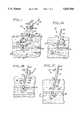
- FIG. 1is an elevational view, partially in cross section, illustrating a first preferred embodiment of the present invention
- FIG. 1Ais an elevational view, partially in cross section, illustrating the initial position in a puncture site of the distal portion of the apparatus of FIG. 1;
- FIG. 1Bis an elevational view, partially in cross section, illustrating the apparatus of FIG. 1A in a preferred operational position
- FIG. 1Cis an elevational view, partially in cross section, illustrating the apparatus of FIG. 1A with the compression balloon revealed and not yet inflated;
- FIG. 1Dis a cross sectional view taken along lines 1D--1D of FIG. 1, illustrating the dual lumen configuration of a catheter element of the apparatus of FIG. 1;
- FIG. 2is an elevational view, partially in cross section, of a second preferred embodiment of the present invention, showing the compression mechanism of this embodiment in a retracted state near a vascular puncture site;
- FIG. 2Ais a perspective view of the embodiment of FIG. 2, showing the compression mechanism in an expanded state;
- FIG. 2Bis a view similar to that of FIG. 2, showing the compression mechanism deployed, in its expanded state, at a vascular puncture site;
- FIG. 3is an elevational view, partially in cross section, of a third preferred embodiment of the present invention, showing the compression mechanism of this embodiment in a retracted state near a vascular puncture site;
- FIG. 3Ais a view, similar to that of FIG. 3, illustrating the compression mechanism in an expanded state
- FIG. 4is a perspective view of a fourth preferred embodiment of the present invention.
- FIG. 4Ais an elevational view, partially in cross section, illustrating the initial position in a puncture site of the introducer and guidewire elements of the apparatus of FIG. 4;
- FIG. 4Bis a view similar to that of FIG. 4A, but showing a a catheter contained within introducer when the introducer is in a first axial position relative to the catheter;
- FIG. 4Cis an elevational view, partially in cross section, illustrating the apparatus of FIG. 4A in a preferred operational position
- FIG. 4Dis an elevational view, partially in cross section, illustrating the apparatus of FIG. 4A with the compression balloon revealed and not yet inflated, the introducer having been moved to a second axial position relative to the catheter;
- FIG. 4Eis a perspective view, partially in cross section, illustrating the compression balloon of the apparatus of FIG. 4D in an inflated state
- FIG. 4Fis an elevational view, partially in cross section, illustrating the apparatus of FIG. 4E with the guidewire element withdrawn.
- FIG. 5is an elevational view, partially in cross section, illustrating a modification of the embodiment of FIG. 1.
- FIG. 1A percutaneous blood vessel sealing device, or percutaneous hemostatic device 10, which applies hemostatic sealing pressure directly to tissue adjacent a vascular puncture site, without employing implanted materials, is shown in FIG. 1.
- an introducer sheath(introducer) 12, well known in the art, is present in an incision 14 that extends from the skin surface 16 to a blood vessel (artery or vein) 18 of a patient at the site of a blood vessel puncture 20.
- the introducer 12has normally been inserted previously to provide access to the vessel 18 for instrumentation (not shown) used in performing a vascular procedure immediately preceding the need to seal the puncture 20.
- the initial position of an introducer 12 so insertedis most clearly illustrated in FIG. 4A, which shows a tapered distal end 22 of the introducer 12 at a puncture site 24, inserted within a vascular puncture 20.
- the introducer 12will have a size of approximately 7 French (2.3 mm in diameter), and a length of approximately 130 mm, although a size as large as 14 French (4.7 mm in diameter) may be used for larger punctures.
- a hollow locator tube 30extends coaxially through the introducer 12 and into the vessel 18 through the puncture 20.
- Guided by the locator tube 30 into the introducer working channel 26is a semi-rigid catheter 32 having a catheter proximal end 33, and a catheter distal end 34 (FIG. 1A).
- the introducer 12is movable axially with respect to the catheter 32, and is disposed initially at a first axial position, or distal position, in which the catheter distal end 34 is enclosed or sheathed within the distal end 22 of the introducer 12.
- the catheter 32is a dual-lumen device having a first axial lumen 36 (FIG. 1D) which encompasses the locator tube 30 when the catheter 32 is inserted into the working channel 26 of the introducer 12.
- a second axial lumen 38is provided with an inflation orifice 40 near its distal end, the inflation orifice communicating with the interior of a compression balloon 42 that concentrically surrounds a portion of the length of the catheter 32 extending proximally from its distal end 34.
- the compression balloon 42is initially enclosed, in an uninflated state, within the distal end 22 of the introducer 12, as illustrated in FIG. 1A.
- the opposite (proximal) end of the second axial lumen 38communicates with a compression balloon inflation port 44 through an inflation tube 45, as shown in FIGS. 1 and 4.
- the catheter 32has an outer diameter sufficiently small to be freely insertable into the introducer 12, and a length that is greater than that of the introducer 12, i.e., in the range of about 130 mm to about 750 mm
- the locator tube 30has an inflatable intravascular locating balloon 50 at its distal end portion, shown in FIG. 1A in an uninflated state.
- the interior of the locating balloon 50is in fluid communication with the hollow interior of the locator tube 30 through a suitable inflation orifice (not shown), as is well known in conventional balloon catheters and the like.
- luer locks 46, 48may be employed for both the locator balloon embodiment (FIGS. 1 through 1D) and for embodiments (described below) featuring expandable compression elements other than the compression balloon 42, a version using no luer locks will be described below that is specifically adapted for use with the compression balloon 42. Both the luer and non-luer versions are suitable for embodiments employing either the inflatable locating balloon 50 or a guidewire locating means, to be described below.
- FIG. 1Ashows the locator tube 30, having the uninflated locating balloon 50 near its distal end, inserted into the vessel 18 through the introducer 12 and the vascular puncture 20. It is advantageous to construct the locator tube 30 so that a length of tube extends distally beyond the location of the locating balloon 50 into the vessel 18 to facilitate re-access through the vascular puncture 20, if required.
- the entire apparatus 10(including the introducer 12 and the catheter 32) is in its initial position relative to the vessel; that is, the distal tip 22 of the introducer 12 is located adjacent to or within the puncture 20, while the introducer 12 is in its above-described first axial position or distal position relative to the catheter 32, in which the catheter distal end 34 and the uninflated compression balloon 42 are enclosed within the distal end 22 of the introducer 12.
- FIG. 1Billustrates the device 10 after the locating balloon 50 has been inflated by fluid introduced into it via the locator tube 30.
- the entire device 10(including the introducer 12 and the catheter 32) has been partially withdrawn from the puncture site 24 in the direction of the arrow 52 (i.e., in the proximal direction), to a "preferred operational position", in which the locating balloon 50 is lodged against an interior wall 54 of the vessel 18.
- the introducer 12remains in its first or distal position, in which the portion of the catheter 32 carrying the uninflated compression balloon 42 is enclosed within the distal end 22 of the introducer 12.
- the introducer 12has been moved axially, relative to the catheter 32, in the direction of the arrow 52 (i.e., proximally), to its second axial position, or proximal position.
- the movement of the introducer 12 to this second or proximal positionuncovers the uninflated compression balloon 42.
- the compression stage of the device 10is illustrated next in FIG. 1.
- the compression balloon 42inflated via the second axial lumen 38 (FIG. 1D), rests in an optimal position to effect natural hemostasis, viz., above a laminar portion 56 of the fatty tissue adjacent the puncture site 24.
- An optimal distance from the vessel 18 to the catheter distal end 34is in the range of 2 mm to 10 mm. This distance will dispose a layer of fatty tissue 56 between the vessel 18 and the catheter 32, minimizing the potential for pseudo-aneurysm.
- the introducer luer lock 46is shown engaged with the catheter luer lock 48, assuring that a holding force applied to the introducer 12 will be transmitted as well to the catheter 32.
- a visible marker band 57 on the exterior of the locating tubing 30may advantageously be provided to align the proximal ends of the introducer 12 and the catheter 32 in correspondence with the location of the distal ends 22, 34 thereof when the locator balloon 50 is lodged against the inner wall 54 of vessel 18.
- FIGS. 1 and 4An adhesive skin patch 58 with a sheath cuff 60 clamped onto the external portion of the introducer 12 to apply downward force (in the direction of the arrow 62, i.e., distally) on the introducer 12 is shown in FIGS. 1 and 4.
- Fastener strips 64secure the adhesive patch 58 to the sheath cuff 60.
- the fastener strips 64may be elastic bands with suitable adhesive areas, or hook and loop strips (such as the type marketed under the trademark VELCRO) that adhere to areas of complementary material on the patch 58.
- Pressure maintained by the introducer sheath cuff 60 on the catheter 32provides hemostatic pressure on the compression balloon 42 to bear on the tissue layer 56 for a first period of time, whereupon the locating tube 30 is withdrawn (the locator balloon 50 having first been deflated), and a second period of time elapses, after which all instrumentation is removed from the patient as will be noted when the method for sealing the puncture 20 is described in detail below.
- FIGS. 2, 2A, and 2Bshow a collapsible prong assembly compression element 66 attached to the catheter distal end 34.
- the prong assembly 66is radially compressed or collapsed when enclosed within the introducer 12, when the introducer is in its first or distal position.
- the prong assembly 66expands radially when the introducer 12 is partially withdrawn from the vessel 18 (FIGS. 2A and 2B), by moving the introducer 12 to its second or proximal position in a manner similar to the partial withdrawal of introducer 12 in the direction of arrow 52 as described previously in connection with the compression balloon embodiment.
- the prong assembly 66comprises a plurality of spaced-apart resilient prongs 68, the proximal ends of which are attached to the catheter 32, and the distal ends of which are attached to a collapsible spanning film sheet or dam 70, shown expanded in FIGS. 2A and 2B.
- the sheet or dam 70allows the application of hemostatic pressure on the tissue 56 above the vessel 18.
- a central aperture 72 in the sheet or dam 70permits the locator tube (not shown) to project through the catheter 32 into the vessel 18 as described previously. Since there is no compression balloon to be inflated, a catheter with a single axial lumen 36 is adequate for this application.
- Materials for the spanning sheet or dam 70may include polyurethane and polyethyleneterephthalate (PET).
- FIGS. 3 and 3Ashow a foam pad compression element 74 attached to the catheter distal end 34.
- the foam pad element 74is compressed when enclosed within the introducer 12 when the introducer is in its first or distal position.
- the foam pad compression element 74then expands when the introducer 12 is partially withdrawn from the vessel 18, as shown in FIG. 3A, by moving the introducer 12 to its second or proximal position, as described above with respect to the first and second embodiments. Hemostatic pressure is similarly exerted on the tissue 56 above the vessel 18.
- An axial channel 76 in the foam pad 74permits the locator tube (not shown) to project through the catheter 32 into the vessel 18, as described previously.
- Materials for the foam pad 74may include various polymeric foams, such as polyurethanes, as are well-known in the art.
- the foam pad 74may be impregnated with a coagulant such as thrombin or protamine to effect local hemostasis.
- a locator tube 30 with a locating balloon 50to determine the optimal operational location for the apparatus 10.
- a guidewire 78may be utilized for the location determination of the apparatus 10, as illustrated in FIGS. 4 through 4F.
- a standard guidewire 78typically 3 French (1 mm in diameter), shown coaxially located within the introducer 12, has a distal end 82 extending out of the introducer distal end 22 into the puncture 20 of the vessel 18.
- the catheter 32is shown in FIG. 4B having been inserted into the introducer 12 and guided to the distal end 22 of the introducer by the guidewire 78.
- a radiopaque marker 84for viewing under fluoroscopy, as shown in FIG. 4D.
- FIG. 4Cshows an optimal location for catheter distal end 34, radiopaque contrast medium (not shown) having been introduced into the catheter lumen 36, and the apparatus 10 having been partially withdrawn from the vessel 18 in the direction of the arrow 52 (i.e., proximally).
- An extravasation 85 of the radiopaque contrast mediumis shown marking the desired distance between the vessel 18 and the catheter distal end 34, as will be explained when the method for sealing the puncture is described below.
- FIG. 4DThe introducer 12 is shown in FIG. 4D having been moved, in the direction of the arrow 52, to its second or proximal position to reveal the uninflated compression balloon 42 in position for inflating.
- FIG. 4Eillustrates the apparatus 10 with the compression balloon 42 inflated and in place above the fatty layer 56 to apply hemostatic pressure for a first period of time in order to effect initial closure of puncture site 24.
- FIG. 4Fshows the apparatus 10 after the guidewire 78 has been removed from the apparatus 10 and pressure is applied for a second period of time to close the puncture 20.
- the guidewire 78 and radiopaque positioning of an expandable compression element at the distal end 34 of the catheter 32may be employed with the prong assembly and foam pad embodiments described above in connection with the locator tube 30.
- a standard hemostatic "Y" 86is used, as shown in FIG. 4.
- the "Y" 86has a main leg 88 for receiving the guidewire 78 into the axial lumen 36 of the catheter 32, while a side port 90 of the "Y" 86 is used for introducing the contrast medium into the same lumen.
- FIG. 5A modification of the first (compression balloon) embodiment of the present invention is shown in FIG. 5, where an apparatus 110 has an introducer 112 having no luer connection with a catheter 132. Since the cuff 60 applies downward force in the direction of the arrow 62 only to the introducer 112, and not to the catheter 132, the distal end 122 of the introducer 112 must bear directly on the compression balloon 42 to exert hemostatic pressure on the balloon 42.
- this modificationis suitable only for the compression balloon embodiment of this invention, both the locator tube 30 and the guidewire 78 may be utilized in this modification for optimal positioning of the catheter distal end 34.
- a piercing cannulais inserted into the skin of a patient at an angle of from 25 to 45 degrees until it punctures a blood vessel, e.g., the femoral artery.
- the vesselmay be located one centimeter or more beneath the surface of the skin.
- a guidewireis inserted through the cannula into the vessel, the cannula is withdrawn, and a catheter introducer sheath is inserted over the guidewire into the puncture site.
- the practitionerthen uses the introducer to gain access to the vascular lumen for the instrumentation used to perform the particular procedure.
- the introduceris the last device remaining in the puncture, which must then be sealed.
- the method of the present inventionprovides a rapid, permanent, inexpensive sealing of a puncture in a blood vessel, with no foreign implants remaining in the patient.
- the methodcan be understood with reference to the drawing figures and the previous description of the apparatus of this invention.
- an introducer sheath 12is shown in a puncture site 24 at the conclusion of a vascular procedure.
- a locator tube 30 having an inflatable locating balloon 50 adjacent its distal endis inserted axially through the introducer 12, into a puncture 20 and extending the uninflated locating balloon 50 into the lumen of a vessel 18.
- a dual lumen catheter 32is passed over the locator tube 30 so that a first lumen 36 (FIG. 1D) of the catheter 32 receives the locator tube 30.
- the locator tube 30maintains alignment of the catheter 32 with the puncture 20 and allows repeated access into the vessel 18, if necessary.
- the catheter 32having an inflatable compression balloon 42 at its distal end 34, is inserted fully into the introducer 12 until its distal end 34, including the uninflated compression balloon 42, is at the distal end 22 of the introducer 12.
- the locator tube 30is pushed or pulled until a marker band 57 (shown in FIG. 1) is aligned with the proximal end 33 of the catheter 32.
- the marker band 57is preselected to establish a fixed relationship with the catheter 32 so that a preferred distance may be maintained between the vessel 18 and the distal end 34 of catheter 32 as will be explained below.
- the introducer 12being in its first or distal position, the uninflated compression balloon 42 is fully enclosed and contained within the working channel 26 of the introducer 12, as described above.
- the practitionerthen inflates the locating balloon 50 via the locator tube 30, partially withdrawing the introducer 12, the catheter 32 and the locator tube 30 from the puncture 20 in the direction of the arrow 52, until the locating balloon 50 lodges against the inner wall of the vessel 18 at the puncture 20, as illustrated in FIG. 1B. Since the position of the catheter distal end 34 relative to the introducer distal end 22 remains unchanged, the distal end 34 of the catheter is now at the location predetermined by the placement of the marker band 57, preferably about 5 mm to 15 mm from the puncture 20. This distance will allow a layer of fatty subcutaneous tissue 56 to lie between the catheter distal end 34 and the puncture 20.
- the introducer 12is further withdrawn in the direction of the arrow 52, by moving it to its second or proximal position relative to the catheter 32, as described above, to expose the uninflated compression balloon 42, as shown in FIG. 1C.
- the luer fittings 46, 48 at the proximal ends of the catheter 32 and the introducer 12, respectively,are now connected to each other to lock the catheter 32 and the introducer 12 into a fixed position relative to one another, and the compression balloon 42 is then inflated, as illustrated in FIG. 1, via a second catheter lumen 38 (FIG. 1D).
- the compression balloon 42is then pressed down against the fatty layer 56 above the puncture site 24, while gentle traction is maintained on the locating balloon 50, thus compressing the extravascular fatty tissue 56 between the balloons 42, 50.
- the fatty tissue 56advantageously minimizes the potential of pseudo-aneurysm formation and promotes efficient hemostasis.
- an introducer cuff 60is clamped onto the introducer 12 and secured to an adhesive patch 58 by means of elastic or hook and loop fastening strips 64 (FIGS. 1 and 4).
- the introducer 12is locked with the catheter 32 by the luer fittings 46, 48, the downward force provided by the fastening strips 64 is transmitted from the introducer 12 through the semi-rigid catheter 32 to the compression balloon 42, maintaining hemostatic pressure on the puncture site 24 through fatty tissue 56.
- first period of timeapproximately 5 to 15 minutes
- initial clotting of the puncture 20will have occurred.
- the locating balloon 50is then deflated and the locator tube 30 withdrawn from the apparatus 10, leaving only a small (e.g., approximately 1 mm in diameter) portion of the original puncture 20 to clot.
- the compression balloon 42remains in place for an additional (second) period of time (approximately 5 to 25 minutes), providing hemostasis to the puncture 20, after which the compression balloon 42 is deflated and retracted proximally into the introducer 12, the luer fittings 46, 48 having first been disconnected.
- the sealing processhaving been completed, the apparatus 10 is completely removed from the patient.
- the foregoing methoduses an introducer 12 that is already positioned at the access site so that position is not lost in changing instruments, bleeding does not occur while devices are positioned, and the locator tube 30 maintains the access location for re-access if needed during the initial clotting of the puncture 20.
- employment of the present inventionrequires minimal physician time and greatly reduces staff time and involvement previously devoted to maintaining supradermal pressure for long periods of hemostasis.
- the need for operating room timemay be reduced by the removal of the locator tube 30, the introducer 12 and the catheter 32 after the patient is returned to the patient's room. Overall, patient discomfort is significantly lessened through the use of the foregoing method as compared with the traditional manual external compression techniques.
- the compression element at catheter distal end 34comprises the collapsible prong assembly 66, as shown in FIGS. 2, 2A, and 2B.
- the movement of the introducer 12 to its second or proximal positionreleases the prong assembly 66 from confinement within the introducer 12, allowing the individual prongs 68 of the prong assembly 66 to expand, as illustrated in FIG. 2A.
- a resilient spanning sheet or dam 70supported by the ends of the prongs 68, then allows the application of hemostatic pressure on the fatty tissue layer 56, as described earlier in connection with the compression balloon embodiment.
- the locator tube(not shown) passes through and is withdrawn from the aperture 72 in the spanning film 70.
- a third embodiment of the methodinvolves the use of the compressible foam pad 74 shown in FIGS. 3 and 3A as the compression element at the distal end 34 of the catheter 32.
- the introducer 12when the catheter 32 is in the preferred location as shown in FIG. 3, the introducer 12 is moved from its first or distal position to its second or proximal position (in the direction of the arrow 52) to uncover the foam pad 74, allowing it to expand, as illustrated in FIG. 3A.
- the expanded foam pad 74exerts hemostatic pressure upon the fatty tissue layer 56, as described previously.
- the locator tube(not shown) passes through and is withdrawn from the pad channel 76 formed axially in the foam pad 74.
- a coagulant agentsuch as collagen, thrombin or protamine may be delivered to the vicinity of the puncture site through the pad channel 76 which communicates with the catheter axial lumen 36.
- the foam pad 74may be saturated with the agent prior to deployment.
- the method employed with the apparatus described abovemay also use a guidewire 78 (FIG. 4) to perform the locating functions provided by the locator tube 30 in the previous embodiments.
- All three of the compression elementsviz., the compression balloon 42, the expandable prong element 66 and the foam pad 74, may be utilized with the guidewire 78.
- FIGS. 4 through 4Fshowing only the compression balloon 42 alternative, may be viewed with the understanding that the method to be described in conjunction therewith applies to all three guidewire 78 embodiments.
- the introducer 12is shown as it remains in the puncture 20 after a vascular access procedure.
- a conventional surgical guidewire 78is extended through the introducer 12 so that its distal end 82 extends into the lumen of the vessel 18.
- the dual lumen catheter 32is passed over the guidewire 78 so that a first lumen 36 (FIG. 1D) of the catheter 32 receives the guidewire 78.
- the guidewire 78maintains alignment of the catheter 32 with the puncture 20 and allows re-access into the vessel 18 if it becomes necessary.
- the catheter 32having an inflatable compression balloon 42 at its distal end 34, is inserted fully into the introducer 12 until its distal end 34, including the uninflated compression balloon 42, is enclosed within the working channel 26 at the distal end 22 of the introducer 12, as shown in FIG. 4B.
- a radiopaque contrast medium(not shown) is introduced into the catheter first lumen 36, as illustrated in FIG. 4.
- a main leg 88 of a conventional hemostasis "Y" 86may be passed over the guidewire 78 and attached to the proximal end 33 of the catheter lumen 36.
- the contrast mediumis then introduced into the catheter lumen 36 via a side port 90 of the "Y" 86, and viewed by the practitioner using conventional fluoroscopic techniques.
- a radiopaque marker 84may be provided at the tip of the catheter distal end 34 (FIG. 4D).
- the introducer 12 with the catheter 32is partially withdrawn in the direction of the arrow 52 from the puncture 20. Withdrawal is continued until contrast medium in the catheter lumen 36 escaping from around the guidewire 78 into the vessel 18 is observed to form an extravasation cloud 85, signifying that the introducer 12 and the catheter 32 have exited the puncture 20.
- the catheter distal end element 34is the preferred distance of about 5 to 15 mm from the vessel 18, withdrawal of the catheter 32 is halted, as shown in FIG. 4C.
- the remainder of the closure procedureis essentially the same as described above after the preferred position of the catheter 32 was determined through the locator tube 30 method.
- the introducer 12is moved from its first or distal position relative to the catheter 32 to its second or proximal position, to expose the uninflated compression balloon 42, as shown in FIG. 4D.
- the compression balloon 42is then inflated to bear on the fatty tissue layer 56 as shown in FIG. 4E.
- the locating meansin this embodiment guidewire 78
- the locating meansis then withdrawn from the apparatus after an initial period of clotting (FIG. 4F).
- the method employing the guidewire 78may be effectively adapted for use with the expandable prong element and foam tip embodiments of the present invention.
- FIG. 5shows the position of the catheter 132 aligned with a visible marker band 57 on the locator tube 30, just as in the first embodiment described above. It will be readily understood that the method of this "luerless" apparatus 110 may be equally utilized with the guidewire 78 as with the locator tube 30 for the compression balloon embodiment of this invention.
Landscapes
- Health & Medical Sciences (AREA)
- Surgery (AREA)
- Life Sciences & Earth Sciences (AREA)
- Medical Informatics (AREA)
- Nuclear Medicine, Radiotherapy & Molecular Imaging (AREA)
- Engineering & Computer Science (AREA)
- Biomedical Technology (AREA)
- Heart & Thoracic Surgery (AREA)
- Cardiology (AREA)
- Molecular Biology (AREA)
- Animal Behavior & Ethology (AREA)
- General Health & Medical Sciences (AREA)
- Public Health (AREA)
- Veterinary Medicine (AREA)
- Media Introduction/Drainage Providing Device (AREA)
- Surgical Instruments (AREA)
Abstract
Description
Claims (53)
Priority Applications (10)
| Application Number | Priority Date | Filing Date | Title |
|---|---|---|---|
| US08/528,892US5645566A (en) | 1995-09-15 | 1995-09-15 | Apparatus and method for percutaneous sealing of blood vessel punctures |
| CA002231560ACA2231560C (en) | 1995-09-15 | 1996-09-10 | Apparatus and method for percutaneous sealing of blood vessel punctures |
| JP9512040AJP2000507838A (en) | 1995-09-15 | 1996-09-10 | Apparatus and method for percutaneously sealing a vascular puncture |
| EP96932191AEP0852478A1 (en) | 1995-09-15 | 1996-09-10 | Apparatus and method for percutaneous sealing of blood vessel punctures |
| PCT/US1996/014486WO1997009934A1 (en) | 1995-09-15 | 1996-09-10 | Apparatus and method for percutaneous sealing of blood vessel punctures |
| US08/888,851US6071300A (en) | 1995-09-15 | 1997-07-07 | Apparatus and method for percutaneous sealing of blood vessel punctures |
| US09/365,674US6371974B1 (en) | 1995-09-15 | 1999-08-02 | Apparatus and method for percutaneous sealing of blood vessel punctures |
| US10/107,539US7175646B2 (en) | 1995-09-15 | 2002-03-25 | Apparatus and method for percutaneous sealing of blood vessel punctures |
| US11/537,024US20070021770A1 (en) | 1995-09-15 | 2006-09-29 | Apparatus and Method for Percutaneous Sealing of Blood Vessel Punctures |
| US12/887,945US20110014290A1 (en) | 1995-09-15 | 2010-09-22 | System and method for facilitating hemostasis with an absorbable sponge |
Applications Claiming Priority (1)
| Application Number | Priority Date | Filing Date | Title |
|---|---|---|---|
| US08/528,892US5645566A (en) | 1995-09-15 | 1995-09-15 | Apparatus and method for percutaneous sealing of blood vessel punctures |
Related Child Applications (1)
| Application Number | Title | Priority Date | Filing Date |
|---|---|---|---|
| US08/888,851Continuation-In-PartUS6071300A (en) | 1995-09-15 | 1997-07-07 | Apparatus and method for percutaneous sealing of blood vessel punctures |
Publications (1)
| Publication Number | Publication Date |
|---|---|
| US5645566Atrue US5645566A (en) | 1997-07-08 |
Family
ID=24107636
Family Applications (1)
| Application Number | Title | Priority Date | Filing Date |
|---|---|---|---|
| US08/528,892Expired - LifetimeUS5645566A (en) | 1995-09-15 | 1995-09-15 | Apparatus and method for percutaneous sealing of blood vessel punctures |
Country Status (5)
| Country | Link |
|---|---|
| US (1) | US5645566A (en) |
| EP (1) | EP0852478A1 (en) |
| JP (1) | JP2000507838A (en) |
| CA (1) | CA2231560C (en) |
| WO (1) | WO1997009934A1 (en) |
Cited By (222)
| Publication number | Priority date | Publication date | Assignee | Title |
|---|---|---|---|---|
| WO1998005259A1 (en)* | 1996-08-06 | 1998-02-12 | Quinton Instrument Company | Insertion assembly and method of inserting a hemostatic closure device into an incision |
| US5910155A (en)* | 1998-06-05 | 1999-06-08 | United States Surgical Corporation | Vascular wound closure system |
| US6004341A (en)* | 1996-12-05 | 1999-12-21 | Loma Linda University Medical Center | Vascular wound closure device |
| WO2000002488A1 (en)* | 1998-07-13 | 2000-01-20 | Cardiodex Ltd. | Methods and apparatus for hemostasis following arterial catheterization |
| US6030365A (en)* | 1998-06-10 | 2000-02-29 | Laufer; Michael D. | Minimally invasive sterile surgical access device and method |
| US6068599A (en)* | 1997-07-14 | 2000-05-30 | Matsushita Electric Industrial Co., Ltd. | Blood vessel puncturing device using ultrasound |
| US6071301A (en)* | 1998-05-01 | 2000-06-06 | Sub Q., Inc. | Device and method for facilitating hemostasis of a biopsy tract |
| US6126675A (en)* | 1999-01-11 | 2000-10-03 | Ethicon, Inc. | Bioabsorbable device and method for sealing vascular punctures |
| US6162192A (en)* | 1998-05-01 | 2000-12-19 | Sub Q, Inc. | System and method for facilitating hemostasis of blood vessel punctures with absorbable sponge |
| WO2000038579A3 (en)* | 1998-12-24 | 2000-12-21 | Vivant Medical Inc | Device and method for safe location and marking of a cavity and sentinel lymph nodes |
| US6183497B1 (en) | 1998-05-01 | 2001-02-06 | Sub-Q, Inc. | Absorbable sponge with contrasting agent |
| US6200328B1 (en) | 1998-05-01 | 2001-03-13 | Sub Q, Incorporated | Device and method for facilitating hemostasis of a biopsy tract |
| US6287322B1 (en) | 1995-12-07 | 2001-09-11 | Loma Linda University Medical Center | Tissue opening locator and everter and method |
| US6306154B1 (en) | 1997-06-18 | 2001-10-23 | Bhk Holding | Hemostatic system for body cavities |
| US20010034528A1 (en)* | 1994-09-16 | 2001-10-25 | Foerster Seth A. | Methods and devices for defining and marking tissue |
| US6315753B1 (en) | 1998-05-01 | 2001-11-13 | Sub-Q, Inc. | System and method for facilitating hemostasis of blood vessel punctures with absorbable sponge |
| US6328757B1 (en)* | 1999-04-23 | 2001-12-11 | Robert G. Matheny | Device and method for performing surgery without impeding organ function |
| US6356782B1 (en) | 1998-12-24 | 2002-03-12 | Vivant Medical, Inc. | Subcutaneous cavity marking device and method |
| US20020058882A1 (en)* | 1998-06-22 | 2002-05-16 | Artemis Medical, Incorporated | Biopsy localization method and device |
| US6494848B1 (en)* | 1996-12-19 | 2002-12-17 | St. Jude Medical Puerto Rico B.V. | Measuring device for use with a hemostatic puncture closure device |
| US6524326B1 (en) | 1995-12-07 | 2003-02-25 | Loma Linda University Medical Center | Tissue opening locator and everter and method |
| US20030055454A1 (en)* | 2001-03-14 | 2003-03-20 | Cardiodex Ltd. | Balloon method and apparatus for vascular closure following arterial catheterization |
| US6540735B1 (en) | 2000-05-12 | 2003-04-01 | Sub-Q, Inc. | System and method for facilitating hemostasis of blood vessel punctures with absorbable sponge |
| US6544236B1 (en) | 1999-02-10 | 2003-04-08 | Sub-Q, Incorporated | Device, system and method for improving delivery of hemostatic material |
| US20030093097A1 (en)* | 2001-11-15 | 2003-05-15 | Ernesto Avellanet | Aneurysm embolic device with an occlusive member |
| US6565527B1 (en)* | 1996-04-26 | 2003-05-20 | Medtronic, Inc. | Intravascular balloon occlusion device |
| US20030105483A1 (en)* | 2000-05-09 | 2003-06-05 | Hudson John Overton | Nasal packing device |
| US6605047B2 (en) | 2001-09-10 | 2003-08-12 | Vivant Medical, Inc. | Biopsy marker delivery system |
| US6610026B2 (en) | 1998-05-01 | 2003-08-26 | Sub-Q, Inc. | Method of hydrating a sponge material for delivery to a body |
| US20030199924A1 (en)* | 2000-09-08 | 2003-10-23 | James Coleman | Surgical stapler |
| US20030229308A1 (en)* | 2002-06-05 | 2003-12-11 | Israil Tsals | Injector adapter and combination thereof |
| US20040010216A1 (en)* | 2000-02-24 | 2004-01-15 | Zhu Yong Hua | Device for closing tissue openings |
| US20040028502A1 (en)* | 2001-06-07 | 2004-02-12 | Christy Cummins | Surgical staple |
| US6706051B2 (en) | 1998-04-08 | 2004-03-16 | Bhk Holding, Ltd. | Hemostatic system for body cavities |
| US20040054346A1 (en)* | 2000-08-02 | 2004-03-18 | Zhu Yong Hua | Vascular wound closure device and method |
| US20040068228A1 (en)* | 2002-10-04 | 2004-04-08 | Jon Cunningham | Device and method for stabilizing catheters |
| US20040073299A1 (en)* | 2000-12-16 | 2004-04-15 | Hudson John Overton | Hemostatic device |
| US20040092879A1 (en)* | 2000-11-06 | 2004-05-13 | Medamicus, Inc. | Safety introducer apparatus and method therefor |
| US20040102730A1 (en)* | 2002-10-22 | 2004-05-27 | Davis Thomas P. | System and method for facilitating hemostasis of blood vessel punctures with absorbable sponge |
| US20040122362A1 (en)* | 2002-09-10 | 2004-06-24 | Houser Russell A. | Pseudo aneurysm repair system |
| US20040138674A1 (en)* | 2003-01-14 | 2004-07-15 | Radi Medical Systems Ab | Introducer sheath |
| US20040153054A1 (en)* | 2003-02-04 | 2004-08-05 | Cardiodex Ltd. | Methods and apparatus for hemostasis following arterial catheterization |
| US20040153060A1 (en)* | 2003-02-04 | 2004-08-05 | Cardiodex Ltd. | Methods and apparatus for hemostasis following arterial catheterization |
| US20040158287A1 (en)* | 2000-07-14 | 2004-08-12 | Cragg Andrew H. | Sheath-mounted arterial plug delivery device |
| US20040192767A1 (en)* | 2003-01-29 | 2004-09-30 | Oxford Alexander W. | EP4 receptor antagonists |
| US20040236355A1 (en)* | 2001-08-09 | 2004-11-25 | Thomas Anthony | Surgical stapling device |
| US20040249391A1 (en)* | 2001-08-09 | 2004-12-09 | Christy Cummins | Surgical stapling device and method |
| US6846320B2 (en) | 1998-05-01 | 2005-01-25 | Sub-Q, Inc. | Device and method for facilitating hemostasis of a biopsy tract |
| US6849232B2 (en) | 2001-03-12 | 2005-02-01 | Sub-Q, Inc. | Methods for sterilizing cross-linked gelatin compositions |
| US6863680B2 (en) | 2001-11-08 | 2005-03-08 | Sub-Q, Inc. | System and method for delivering hemostasis promoting material to a blood vessel puncture site by fluid pressure |
| US20050085773A1 (en)* | 2003-10-15 | 2005-04-21 | Forsberg Andrew T. | Method and apparatus for locating vascular punctures |
| US20050085855A1 (en)* | 2003-10-17 | 2005-04-21 | Forsberg Andrew T. | Automatic suture locking device |
| US20050095275A1 (en)* | 2003-09-05 | 2005-05-05 | Zhu Yong H. | Dressing delivery system for internal wounds |
| US6890342B2 (en) | 2000-08-02 | 2005-05-10 | Loma Linda University | Method and apparatus for closing vascular puncture using hemostatic material |
| US20050107827A1 (en)* | 2003-11-13 | 2005-05-19 | Loran Paprocki | Method and apparatus for sealing an internal tissue puncture incorporating a block and tackle |
| US20050125031A1 (en)* | 2003-12-03 | 2005-06-09 | Pipenhagen Catherine A. | Vascular sealing device with high surface area sealing plug |
| US20050125030A1 (en)* | 2003-12-03 | 2005-06-09 | Forsberg Andrew T. | Vascular puncture seal anchor nest |
| US20050175580A1 (en)* | 2001-01-05 | 2005-08-11 | Daniela Salvemini | Compositions and methods for enhancing cytokine activity and treating hypotension associated with the administration of cytokines |
| US20050228430A1 (en)* | 2003-03-18 | 2005-10-13 | Medtronic, Inc. | Intravascular balloon occlusion device and method for using the same |
| US20050228443A1 (en)* | 2004-04-09 | 2005-10-13 | Cardiva Medical, Inc. | Device and method for sealing blood vessels |
| US20050234396A1 (en)* | 2004-04-20 | 2005-10-20 | Forsberg Andrew T | Method and apparatus for locating vascular punctures |
| US20050256537A1 (en)* | 2002-07-03 | 2005-11-17 | Christy Cummins | Surgical stapling device |
| US20050267521A1 (en)* | 2004-05-13 | 2005-12-01 | St. Jude Medical Puerto Rico B.V. | Collagen sponge for arterial sealing |
| US20050267522A1 (en)* | 2004-05-27 | 2005-12-01 | Cardiva Medical, Inc. | Self-tensioning vascular occlusion device and method for its use |
| US20050288706A1 (en)* | 2004-05-07 | 2005-12-29 | Nmt Medical, Inc. | Inflatable occluder |
| US6984219B2 (en) | 1999-09-23 | 2006-01-10 | Mark Ashby | Depth and puncture control for blood vessel hemostasis system |
| US20060009802A1 (en)* | 2004-07-10 | 2006-01-12 | Modesitt D B | Biological tissue closure device and method |
| US7008440B2 (en) | 2001-11-08 | 2006-03-07 | Sub-Q, Inc. | System and method for delivering hemostasis promoting material to a blood vessel puncture site by fluid pressure |
| US20060058844A1 (en)* | 2004-09-13 | 2006-03-16 | St. Jude Medical Puerto Rico B.V. | Vascular sealing device with locking system |
| US7018392B2 (en) | 1998-04-08 | 2006-03-28 | Arthrocare Corporation | Hemostatic system for body cavities |
| US7025748B2 (en) | 2001-11-08 | 2006-04-11 | Boston Scientific Scimed, Inc. | Sheath based blood vessel puncture locator and depth indicator |
| US7025776B1 (en) | 2001-04-24 | 2006-04-11 | Advanced Catheter Engineering, Inc. | Arteriotomy closure devices and techniques |
| US7029489B1 (en) | 2001-05-18 | 2006-04-18 | Sub-Q, Inc. | System and method for delivering hemostasis promoting material to a blood vessel puncture site |
| US7037323B2 (en) | 2001-11-08 | 2006-05-02 | Sub-Q, Inc. | Pledget-handling system and method for delivering hemostasis promoting material to a blood vessel puncture site by fluid pressure |
| US7037322B1 (en) | 2001-11-08 | 2006-05-02 | Sub-Q, Inc. | System and method for delivering hemostasis promoting material to a blood vessel puncture with a staging tube |
| US20060106402A1 (en)* | 2004-11-18 | 2006-05-18 | Mclucas Bruce | Endovascular surgical method |
| US20060116635A1 (en)* | 2004-11-29 | 2006-06-01 | Med Enclosure L.L.C. | Arterial closure device |
| US20060142798A1 (en)* | 2004-12-27 | 2006-06-29 | Holman Thomas J | Device and method for closing an opening in a body cavity or lumen |
| US20060154513A1 (en)* | 2001-07-26 | 2006-07-13 | Barker Jed M | Angled patch panel with cable support bar for network cable racks |
| US20060253049A1 (en)* | 2003-01-14 | 2006-11-09 | Radi Medical Systems Ab | Method for introducer replacement |
| US7175646B2 (en) | 1995-09-15 | 2007-02-13 | Boston Scientific Scimed, Inc. | Apparatus and method for percutaneous sealing of blood vessel punctures |
| US7192436B2 (en) | 2001-11-08 | 2007-03-20 | Sub-Q, Inc. | Pledget-handling system and method for delivering hemostasis promoting material to a blood vessel puncture site by fluid pressure |
| US7201725B1 (en)* | 2000-09-25 | 2007-04-10 | Sub-Q, Inc. | Device and method for determining a depth of an incision |
| US20070123816A1 (en)* | 2005-10-05 | 2007-05-31 | Zhu Yong H | Vascular wound closure device and method |
| US20070135503A1 (en)* | 2003-10-16 | 2007-06-14 | Clark David E | Furan derivatives as ep4 receptor antagonists |
| US20070149998A1 (en)* | 2005-12-22 | 2007-06-28 | Ethicon, Inc. | Systems and methods for closing a vessel wound |
| US20070149999A1 (en)* | 2005-12-23 | 2007-06-28 | Ethicon, Inc. | Systems and methods for closing a vessel wound |
| US20070150002A1 (en)* | 2005-12-23 | 2007-06-28 | Ethicon, Inc. | Systems and methods for closing a vessel wound |
| US20070156084A1 (en)* | 2006-01-04 | 2007-07-05 | St. Jude Medical Puerto Rico B.V. | Balloon insertion apparatus and method of sealing a tissue puncture |
| US7335219B1 (en) | 2002-11-04 | 2008-02-26 | Sub-Q, Inc. | Hemostatic device including a capsule |
| US20080071311A1 (en)* | 2006-09-18 | 2008-03-20 | St. Jude Medical Puerto Rico B.V. | Flexible tamping device |
| US20080086109A1 (en)* | 2004-11-29 | 2008-04-10 | Paul Shabty | Arterial closure device |
| US20090054926A1 (en)* | 2007-08-21 | 2009-02-26 | St. Jude Medical Puerto Rico B.V. | Extra-vascular sealing device and method |
| US20090088794A1 (en)* | 2007-10-01 | 2009-04-02 | Boston Scientific Scimed, Inc | Connective Tissue Closure Device and Method |
| US20090125056A1 (en)* | 2007-08-15 | 2009-05-14 | Cardiodex Ltd. | Systems and methods for puncture closure |
| US20090171281A1 (en)* | 2007-12-31 | 2009-07-02 | Pipenhagen Catherine A | Systems and methods for locating and closing a tissue puncture |
| US20090171282A1 (en)* | 2007-12-31 | 2009-07-02 | Pipenhagen Catherine A | Vascuar closure device having a flowable sealing material |
| US20090234374A1 (en)* | 2008-03-17 | 2009-09-17 | Ethicon, Inc. | Applicator instruments for the delivery, deployment, and tamponade of hemostats and methods therefor |
| US20090248056A1 (en)* | 2008-03-28 | 2009-10-01 | Ethicon, Inc. | Applicator instruments for controlling bleeding at surgical sites and methods therefor |
| US20090275978A1 (en)* | 2004-05-27 | 2009-11-05 | Cardiva Medical, Inc. | Self-tensioning vascular occlusion device and method for its use |
| US7618436B2 (en) | 2005-04-12 | 2009-11-17 | St. Jude Medical Puerto Rico Llc | Tissue puncture closure device with scroll gear transmission tamping system |
| US7625352B1 (en)* | 1998-05-01 | 2009-12-01 | Sub-Q, Inc. | Depth and puncture control for system for hemostasis of blood vessel |
| US20100010449A1 (en)* | 2008-07-09 | 2010-01-14 | Kyphon Sarl | Cannula Stabilization Device, System, And Method Of Use |
| USD611144S1 (en) | 2006-06-28 | 2010-03-02 | Abbott Laboratories | Apparatus for delivering a closure element |
| US7695492B1 (en) | 1999-09-23 | 2010-04-13 | Boston Scientific Scimed, Inc. | Enhanced bleed back system |
| US20100152748A1 (en)* | 2008-12-12 | 2010-06-17 | E-Pacing, Inc. | Devices, Systems, and Methods Providing Body Lumen Access |
| US7799048B2 (en) | 2000-05-09 | 2010-09-21 | Arthrocare Corporation | Nasal packing device |
| US7806904B2 (en) | 2000-12-07 | 2010-10-05 | Integrated Vascular Systems, Inc. | Closure device |
| US7806910B2 (en) | 2002-11-26 | 2010-10-05 | Abbott Laboratories | Multi-element biased suture clip |
| US7819895B2 (en) | 2000-01-05 | 2010-10-26 | Integrated Vascular Systems, Inc. | Vascular sheath with bioabsorbable puncture site closure apparatus and methods of use |
| US7828817B2 (en) | 2000-01-05 | 2010-11-09 | Integrated Vascular Systems, Inc. | Apparatus and methods for delivering a closure device |
| US7842068B2 (en) | 2000-12-07 | 2010-11-30 | Integrated Vascular Systems, Inc. | Apparatus and methods for providing tactile feedback while delivering a closure device |
| US7841502B2 (en) | 2007-12-18 | 2010-11-30 | Abbott Laboratories | Modular clip applier |
| US7850709B2 (en) | 2002-06-04 | 2010-12-14 | Abbott Vascular Inc. | Blood vessel closure clip and delivery device |
| US7850797B2 (en) | 2002-12-31 | 2010-12-14 | Integrated Vascular Systems, Inc. | Methods for manufacturing a clip and clip |
| US7867249B2 (en) | 2003-01-30 | 2011-01-11 | Integrated Vascular Systems, Inc. | Clip applier and methods of use |
| US20110009900A1 (en)* | 2009-07-13 | 2011-01-13 | Tom Holman | Metal vascular aperture closure device |
| US7875043B1 (en) | 2003-12-09 | 2011-01-25 | Sub-Q, Inc. | Cinching loop |
| US7879071B2 (en) | 2000-12-07 | 2011-02-01 | Integrated Vascular Systems, Inc. | Closure device and methods for making and using them |
| US20110046665A1 (en)* | 2007-09-12 | 2011-02-24 | Transluminal Technologies, Llc | Closure Device, Deployment Apparatus, and Method of Deploying a Closure Device |
| US7909873B2 (en) | 2006-12-15 | 2011-03-22 | Soteira, Inc. | Delivery apparatus and methods for vertebrostenting |
| US7931669B2 (en) | 2000-01-05 | 2011-04-26 | Integrated Vascular Systems, Inc. | Integrated vascular device with puncture site closure component and sealant and methods of use |
| US7955353B1 (en) | 2002-11-04 | 2011-06-07 | Sub-Q, Inc. | Dissolvable closure device |
| US20110137338A1 (en)* | 2009-12-08 | 2011-06-09 | Victor Matthew Phillips | Hemostatic Device and Its Methods of Use |
| US7959634B2 (en) | 2004-03-29 | 2011-06-14 | Soteira Inc. | Orthopedic surgery access devices |
| US7998169B2 (en) | 2004-05-12 | 2011-08-16 | Arstasis, Inc. | Access and closure device and method |
| US8002794B2 (en) | 2005-05-12 | 2011-08-23 | Arstasis, Inc. | Access and closure device and method |
| US8007512B2 (en) | 2002-02-21 | 2011-08-30 | Integrated Vascular Systems, Inc. | Plunger apparatus and methods for delivering a closure device |
| US8012167B2 (en) | 2003-08-14 | 2011-09-06 | Loma Linda University Medical Center | Vascular wound closure device and method |
| US8048108B2 (en) | 2005-08-24 | 2011-11-01 | Abbott Vascular Inc. | Vascular closure methods and apparatuses |
| US20110301618A1 (en)* | 2010-06-07 | 2011-12-08 | Samuel Lichtenstein | Closing openings in anatomical tissue |
| US8137380B2 (en) | 2007-09-12 | 2012-03-20 | Transluminal Technologies, Llc | Closure device, deployment apparatus, and method of deploying a closure device |
| US20120123512A1 (en)* | 2010-11-11 | 2012-05-17 | Wilson T. Asfora | Sutureless vascular anastomosis connection |
| US8187625B2 (en) | 2001-03-12 | 2012-05-29 | Boston Scientific Scimed, Inc. | Cross-linked gelatin composition comprising a wetting agent |
| US8202293B2 (en) | 2003-01-30 | 2012-06-19 | Integrated Vascular Systems, Inc. | Clip applier and methods of use |
| US8202294B2 (en) | 2003-01-30 | 2012-06-19 | Integrated Vascular Systems, Inc. | Clip applier and methods of use |
| US8226681B2 (en) | 2007-06-25 | 2012-07-24 | Abbott Laboratories | Methods, devices, and apparatus for managing access through tissue |
| US8262694B2 (en)* | 2004-01-30 | 2012-09-11 | W.L. Gore & Associates, Inc. | Devices, systems, and methods for closure of cardiac openings |
| US8292822B2 (en) | 1998-06-22 | 2012-10-23 | Devicor Medical Products, Inc. | Biopsy localization method and device |
| US8303624B2 (en) | 2010-03-15 | 2012-11-06 | Abbott Cardiovascular Systems, Inc. | Bioabsorbable plug |
| US8313497B2 (en) | 2005-07-01 | 2012-11-20 | Abbott Laboratories | Clip applier and methods of use |
| US8317821B1 (en) | 2002-11-04 | 2012-11-27 | Boston Scientific Scimed, Inc. | Release mechanism |
| US8323312B2 (en) | 2008-12-22 | 2012-12-04 | Abbott Laboratories | Closure device |
| US20130006281A1 (en)* | 1999-04-05 | 2013-01-03 | Medtronic, Inc. | Apparatus and Methods for Anastomosis |
| US20130018414A1 (en)* | 2004-01-30 | 2013-01-17 | W.L. Gore & Associates | Devices, Systems and Methods for Closure of Cardiac Openings |
| US8398676B2 (en) | 2008-10-30 | 2013-03-19 | Abbott Vascular Inc. | Closure device |
| US8398656B2 (en) | 2003-01-30 | 2013-03-19 | Integrated Vascular Systems, Inc. | Clip applier and methods of use |
| US8435236B2 (en) | 2004-11-22 | 2013-05-07 | Cardiodex, Ltd. | Techniques for heat-treating varicose veins |
| US8506592B2 (en) | 2008-08-26 | 2013-08-13 | St. Jude Medical, Inc. | Method and system for sealing percutaneous punctures |
| US8518063B2 (en) | 2001-04-24 | 2013-08-27 | Russell A. Houser | Arteriotomy closure devices and techniques |
| US20130245680A1 (en)* | 2012-03-16 | 2013-09-19 | Covidien Lp | Closure Tape Dispenser |
| US8556930B2 (en) | 2006-06-28 | 2013-10-15 | Abbott Laboratories | Vessel closure device |
| US8556932B2 (en) | 2011-05-19 | 2013-10-15 | Abbott Cardiovascular Systems, Inc. | Collapsible plug for tissue closure |
| US8590760B2 (en) | 2004-05-25 | 2013-11-26 | Abbott Vascular Inc. | Surgical stapler |
| US8603116B2 (en) | 2010-08-04 | 2013-12-10 | Abbott Cardiovascular Systems, Inc. | Closure device with long tines |
| US8617184B2 (en) | 2011-02-15 | 2013-12-31 | Abbott Cardiovascular Systems, Inc. | Vessel closure system |
| US8672953B2 (en) | 2007-12-17 | 2014-03-18 | Abbott Laboratories | Tissue closure system and methods of use |
| US8690910B2 (en) | 2000-12-07 | 2014-04-08 | Integrated Vascular Systems, Inc. | Closure device and methods for making and using them |
| US8758398B2 (en) | 2006-09-08 | 2014-06-24 | Integrated Vascular Systems, Inc. | Apparatus and method for delivering a closure element |
| US8758400B2 (en) | 2000-01-05 | 2014-06-24 | Integrated Vascular Systems, Inc. | Closure system and methods of use |
| US8758399B2 (en) | 2010-08-02 | 2014-06-24 | Abbott Cardiovascular Systems, Inc. | Expandable bioabsorbable plug apparatus and method |
| US8808310B2 (en) | 2006-04-20 | 2014-08-19 | Integrated Vascular Systems, Inc. | Resettable clip applier and reset tools |
| US8821534B2 (en) | 2010-12-06 | 2014-09-02 | Integrated Vascular Systems, Inc. | Clip applier having improved hemostasis and methods of use |
| US8840640B2 (en) | 2007-12-31 | 2014-09-23 | St. Jude Medical Puerto Rico Llc | Vascular closure device having an improved plug |
| US8845682B2 (en) | 2009-10-13 | 2014-09-30 | E-Pacing, Inc. | Vasculature closure devices and methods |
| US8858594B2 (en) | 2008-12-22 | 2014-10-14 | Abbott Laboratories | Curved closure device |
| US8876862B2 (en) | 2011-04-14 | 2014-11-04 | Phillips Medical Llc | Hemostatic device and its methods of use |
| US8893947B2 (en) | 2007-12-17 | 2014-11-25 | Abbott Laboratories | Clip applier and methods of use |
| US20140358076A1 (en)* | 2013-05-28 | 2014-12-04 | Smh Device Corp. | Tunneled Catheter with Hemostasis Mechanism |
| US20140358075A1 (en)* | 2013-05-28 | 2014-12-04 | Smh Device Corp. | Tunneled Catheter with Hemostasis Mechanism |
| US8905937B2 (en) | 2009-02-26 | 2014-12-09 | Integrated Vascular Systems, Inc. | Methods and apparatus for locating a surface of a body lumen |
| US8920442B2 (en) | 2005-08-24 | 2014-12-30 | Abbott Vascular Inc. | Vascular opening edge eversion methods and apparatuses |
| US8926633B2 (en) | 2005-06-24 | 2015-01-06 | Abbott Laboratories | Apparatus and method for delivering a closure element |
| US8940002B2 (en) | 2010-09-30 | 2015-01-27 | Kardium Inc. | Tissue anchor system |
| US8961541B2 (en) | 2007-12-03 | 2015-02-24 | Cardio Vascular Technologies Inc. | Vascular closure devices, systems, and methods of use |
| US8979882B2 (en) | 2008-07-21 | 2015-03-17 | Arstasis, Inc. | Devices, methods, and kits for forming tracts in tissue |
| US8992567B1 (en) | 2001-04-24 | 2015-03-31 | Cardiovascular Technologies Inc. | Compressible, deformable, or deflectable tissue closure devices and method of manufacture |
| US9072511B2 (en) | 2011-03-25 | 2015-07-07 | Kardium Inc. | Medical kit for constricting tissue or a bodily orifice, for example, a mitral valve |
| US9089674B2 (en) | 2000-10-06 | 2015-07-28 | Integrated Vascular Systems, Inc. | Apparatus and methods for positioning a vascular sheath |
| US9089311B2 (en) | 2009-01-09 | 2015-07-28 | Abbott Vascular Inc. | Vessel closure devices and methods |
| US9149276B2 (en) | 2011-03-21 | 2015-10-06 | Abbott Cardiovascular Systems, Inc. | Clip and deployment apparatus for tissue closure |
| US9155530B2 (en) | 2010-11-09 | 2015-10-13 | Transluminal Technologies, Llc | Specially designed magnesium-aluminum alloys and medical uses thereof in a hemodynamic environment |
| US20150297875A1 (en)* | 2013-03-04 | 2015-10-22 | Vascular Solutions,Inc. | Vascular dilator systems, kits, and methods |
| US9173644B2 (en) | 2009-01-09 | 2015-11-03 | Abbott Vascular Inc. | Closure devices, systems, and methods |
| US9192397B2 (en) | 2006-12-15 | 2015-11-24 | Gmedelaware 2 Llc | Devices and methods for fracture reduction |
| US9192468B2 (en) | 2006-06-28 | 2015-11-24 | Kardium Inc. | Method for anchoring a mitral valve |
| US9204964B2 (en) | 2009-10-01 | 2015-12-08 | Kardium Inc. | Medical device, kit and method for constricting tissue or a bodily orifice, for example, a mitral valve |
| US9282965B2 (en) | 2008-05-16 | 2016-03-15 | Abbott Laboratories | Apparatus and methods for engaging tissue |
| US9314230B2 (en) | 2009-01-09 | 2016-04-19 | Abbott Vascular Inc. | Closure device with rapidly eroding anchor |
| US9332976B2 (en) | 2011-11-30 | 2016-05-10 | Abbott Cardiovascular Systems, Inc. | Tissue closure device |
| US9345460B2 (en) | 2001-04-24 | 2016-05-24 | Cardiovascular Technologies, Inc. | Tissue closure devices, device and systems for delivery, kits and methods therefor |
| US9364209B2 (en) | 2012-12-21 | 2016-06-14 | Abbott Cardiovascular Systems, Inc. | Articulating suturing device |
| US9414824B2 (en) | 2009-01-16 | 2016-08-16 | Abbott Vascular Inc. | Closure devices, systems, and methods |
| US9414820B2 (en) | 2009-01-09 | 2016-08-16 | Abbott Vascular Inc. | Closure devices, systems, and methods |
| US9456816B2 (en) | 2007-09-12 | 2016-10-04 | Transluminal Technologies, Llc | Closure device, deployment apparatus, and method of deploying a closure device |
| US9456811B2 (en) | 2005-08-24 | 2016-10-04 | Abbott Vascular Inc. | Vascular closure methods and apparatuses |
| US9468428B2 (en) | 2012-06-13 | 2016-10-18 | Phillips Medical Llc | Hemostatic device and its methods of use |
| US9480485B2 (en) | 2006-12-15 | 2016-11-01 | Globus Medical, Inc. | Devices and methods for vertebrostenting |
| US9486191B2 (en) | 2009-01-09 | 2016-11-08 | Abbott Vascular, Inc. | Closure devices |
| US9572557B2 (en) | 2006-02-21 | 2017-02-21 | Kardium Inc. | Method and device for closing holes in tissue |
| US9579091B2 (en) | 2000-01-05 | 2017-02-28 | Integrated Vascular Systems, Inc. | Closure system and methods of use |
| US9585647B2 (en) | 2009-08-26 | 2017-03-07 | Abbott Laboratories | Medical device for repairing a fistula |
| US9642604B2 (en) | 2012-04-12 | 2017-05-09 | Phillips Medical Llc | Hemostatic system and its methods of use |
| US9669113B1 (en) | 1998-12-24 | 2017-06-06 | Devicor Medical Products, Inc. | Device and method for safe location and marking of a biopsy cavity |
| US9724081B2 (en) | 2013-06-04 | 2017-08-08 | Phillips Medical Llc | Hemostatic system and its methods of use |
| US20170224464A1 (en)* | 2016-02-08 | 2017-08-10 | Terumo Kabushiki Kaisha | Treatment method and medical device |
| US9744038B2 (en) | 2008-05-13 | 2017-08-29 | Kardium Inc. | Medical device for constricting tissue or a bodily orifice, for example a mitral valve |
| US9839416B2 (en) | 2013-07-12 | 2017-12-12 | Phillips Medical, LLC | Hemostatic device and its methods of use |
| US9993236B2 (en) | 2009-12-08 | 2018-06-12 | Phillips Medical, LLC | Hemostatic device and its methods of use |
| US10085730B2 (en) | 2013-07-12 | 2018-10-02 | Phillips Medical, LLC | Hemostatic device and its methods of use |
| US10085731B2 (en) | 2013-07-15 | 2018-10-02 | E-Pacing, Inc. | Vasculature closure devices and methods |
| US10159526B2 (en) | 2013-02-21 | 2018-12-25 | Stryker Corporation | Tissue ablation cannula assembly |
| US10159485B2 (en) | 2010-11-11 | 2018-12-25 | Asfora Ip, Llc | Deployment tool for sutureless vascular anastomosis connection |
| US10286186B2 (en)* | 2013-04-01 | 2019-05-14 | Terumo Kabushiki Kaisha | Guiding catheter assembly and method of using the same |
| US10441753B2 (en) | 2012-05-25 | 2019-10-15 | Arstasis, Inc. | Vascular access configuration |
| US10675447B2 (en) | 2012-05-25 | 2020-06-09 | Arstasis, Inc. | Vascular access configuration |
| US11033392B2 (en) | 2006-08-02 | 2021-06-15 | Kardium Inc. | System for improving diastolic dysfunction |
| CN114521930A (en)* | 2022-03-18 | 2022-05-24 | 安徽省立医院(中国科学技术大学附属第一医院) | Suturing device and method for closing blood vessel opening |
| US11864750B2 (en) | 2020-04-29 | 2024-01-09 | Medos International Sarl | Knotless anchor insertion |
| USD1019945S1 (en) | 2021-12-30 | 2024-03-26 | Medos International Sarl | Suture anchor insertion device |
| USD1028232S1 (en) | 2021-04-27 | 2024-05-21 | Medos International Sarl | Suture anchor insertion device |
| US12137894B2 (en) | 2021-12-30 | 2024-11-12 | Medos International Sarl | Knotless anchor inserter tool extraction |
Families Citing this family (8)
| Publication number | Priority date | Publication date | Assignee | Title |
|---|---|---|---|---|
| US7335220B2 (en) | 2004-11-05 | 2008-02-26 | Access Closure, Inc. | Apparatus and methods for sealing a vascular puncture |
| DE60311268T2 (en) | 2002-08-01 | 2007-12-27 | Abbott Laboratories Vascular Enterprises Ltd. | Device for sealing puncture wounds by reducing the circumference of the puncture |
| DE102004022780A1 (en)* | 2004-05-03 | 2005-12-01 | Michel DOARÉ | System for vascular occlusion, especially after arterial catheter intervention |
| US8262693B2 (en)* | 2004-11-05 | 2012-09-11 | Accessclosure, Inc. | Apparatus and methods for sealing a vascular puncture |
| EP2793990A4 (en)* | 2011-12-21 | 2016-03-02 | Walkill Concepts Inc | Self-sealing catheters |
| US10966697B2 (en) | 2013-05-06 | 2021-04-06 | Caveomed Gmbh | Vascular closure device and method of positioning vascular closure device |
| EP2801325B1 (en)* | 2013-05-06 | 2016-03-16 | CaveoMed GmbH | Vascular closure device |
| JP7714671B2 (en)* | 2021-03-04 | 2025-07-29 | テルモ メディカル コーポレーション | Device and method for achieving hemostasis in blood vessels |
Citations (25)
| Publication number | Priority date | Publication date | Assignee | Title |
|---|---|---|---|---|
| US3411505A (en)* | 1965-12-15 | 1968-11-19 | Paul D. Nobis | Device for interrupting arterial flow |
| US4219026A (en)* | 1978-09-15 | 1980-08-26 | The Kendall Company | Bladder hemostatic catheter |
| US4224945A (en)* | 1978-08-30 | 1980-09-30 | Jonathan Cohen | Inflatable expansible surgical pressure dressing |
| US4619261A (en)* | 1984-08-09 | 1986-10-28 | Frederico Guerriero | Hydrostatic pressure device for bleeding control through an inflatable, stitchable and retrievable balloon-net system |
| US4699616A (en)* | 1986-06-13 | 1987-10-13 | Hollister Incorporated | Catheter retention device and method |
| US4832688A (en)* | 1986-04-09 | 1989-05-23 | Terumo Kabushiki Kaisha | Catheter for repair of blood vessel |
| US4836204A (en)* | 1987-07-06 | 1989-06-06 | Landymore Roderick W | Method for effecting closure of a perforation in the septum of the heart |
| US4852568A (en)* | 1987-02-17 | 1989-08-01 | Kensey Nash Corporation | Method and apparatus for sealing an opening in tissue of a living being |
| US4890612A (en)* | 1987-02-17 | 1990-01-02 | Kensey Nash Corporation | Device for sealing percutaneous puncture in a vessel |
| US4929246A (en)* | 1988-10-27 | 1990-05-29 | C. R. Bard, Inc. | Method for closing and sealing an artery after removing a catheter |
| US5021059A (en)* | 1990-05-07 | 1991-06-04 | Kensey Nash Corporation | Plug device with pulley for sealing punctures in tissue and methods of use |
| US5053046A (en)* | 1988-08-22 | 1991-10-01 | Woodrow W. Janese | Dural sealing needle and method of use |
| US5061274A (en)* | 1989-12-04 | 1991-10-29 | Kensey Nash Corporation | Plug device for sealing openings and method of use |
| US5192290A (en)* | 1990-08-29 | 1993-03-09 | Applied Medical Resources, Inc. | Embolectomy catheter |
| US5221259A (en)* | 1990-12-27 | 1993-06-22 | Novoste Corporation | Wound treating device and method of using same |
| US5232453A (en)* | 1989-07-14 | 1993-08-03 | E. R. Squibb & Sons, Inc. | Catheter holder |
| US5275616A (en)* | 1990-10-01 | 1994-01-04 | Quinton Instrument Company | Insertion assembly and method of inserting a vessel plug into the body of a patient |
| WO1994002072A1 (en)* | 1992-07-16 | 1994-02-03 | Sherwood Medical Company | Device for sealing hemostatic incisions |
| US5334216A (en)* | 1992-12-10 | 1994-08-02 | Howmedica Inc. | Hemostatic plug |
| US5342388A (en)* | 1993-03-25 | 1994-08-30 | Sonia Toller | Method and apparatus for sealing luminal tissue |
| US5352211A (en)* | 1993-07-11 | 1994-10-04 | Louisville Laboratories | External stability device |
| US5383899A (en)* | 1993-09-28 | 1995-01-24 | Hammerslag; Julius G. | Method of using a surface opening adhesive sealer |
| US5391183A (en)* | 1990-09-21 | 1995-02-21 | Datascope Investment Corp | Device and method sealing puncture wounds |
| US5419765A (en)* | 1990-12-27 | 1995-05-30 | Novoste Corporation | Wound treating device and method for treating wounds |
| US5486195A (en)* | 1993-07-26 | 1996-01-23 | Myers; Gene | Method and apparatus for arteriotomy closure |
Family Cites Families (2)
| Publication number | Priority date | Publication date | Assignee | Title |
|---|---|---|---|---|
| CA2089999A1 (en)* | 1992-02-24 | 1993-08-25 | H. Jonathan Tovey | Resilient arm mesh deployer |
| US5462561A (en)* | 1993-08-05 | 1995-10-31 | Voda; Jan K. | Suture device |
- 1995
- 1995-09-15USUS08/528,892patent/US5645566A/ennot_activeExpired - Lifetime
- 1996
- 1996-09-10CACA002231560Apatent/CA2231560C/ennot_activeExpired - Fee Related
- 1996-09-10EPEP96932191Apatent/EP0852478A1/ennot_activeWithdrawn
- 1996-09-10JPJP9512040Apatent/JP2000507838A/enactivePending
- 1996-09-10WOPCT/US1996/014486patent/WO1997009934A1/ennot_activeApplication Discontinuation
Patent Citations (27)
| Publication number | Priority date | Publication date | Assignee | Title |
|---|---|---|---|---|
| US3411505A (en)* | 1965-12-15 | 1968-11-19 | Paul D. Nobis | Device for interrupting arterial flow |
| US4224945A (en)* | 1978-08-30 | 1980-09-30 | Jonathan Cohen | Inflatable expansible surgical pressure dressing |
| US4219026A (en)* | 1978-09-15 | 1980-08-26 | The Kendall Company | Bladder hemostatic catheter |
| US4619261A (en)* | 1984-08-09 | 1986-10-28 | Frederico Guerriero | Hydrostatic pressure device for bleeding control through an inflatable, stitchable and retrievable balloon-net system |
| US4832688A (en)* | 1986-04-09 | 1989-05-23 | Terumo Kabushiki Kaisha | Catheter for repair of blood vessel |
| US4699616A (en)* | 1986-06-13 | 1987-10-13 | Hollister Incorporated | Catheter retention device and method |
| US4852568A (en)* | 1987-02-17 | 1989-08-01 | Kensey Nash Corporation | Method and apparatus for sealing an opening in tissue of a living being |
| US4890612A (en)* | 1987-02-17 | 1990-01-02 | Kensey Nash Corporation | Device for sealing percutaneous puncture in a vessel |
| US4836204A (en)* | 1987-07-06 | 1989-06-06 | Landymore Roderick W | Method for effecting closure of a perforation in the septum of the heart |
| US5053046A (en)* | 1988-08-22 | 1991-10-01 | Woodrow W. Janese | Dural sealing needle and method of use |
| US4929246A (en)* | 1988-10-27 | 1990-05-29 | C. R. Bard, Inc. | Method for closing and sealing an artery after removing a catheter |
| US5232453A (en)* | 1989-07-14 | 1993-08-03 | E. R. Squibb & Sons, Inc. | Catheter holder |
| US5061274A (en)* | 1989-12-04 | 1991-10-29 | Kensey Nash Corporation | Plug device for sealing openings and method of use |
| US5021059A (en)* | 1990-05-07 | 1991-06-04 | Kensey Nash Corporation | Plug device with pulley for sealing punctures in tissue and methods of use |
| US5192290A (en)* | 1990-08-29 | 1993-03-09 | Applied Medical Resources, Inc. | Embolectomy catheter |
| US5391183A (en)* | 1990-09-21 | 1995-02-21 | Datascope Investment Corp | Device and method sealing puncture wounds |
| US5275616A (en)* | 1990-10-01 | 1994-01-04 | Quinton Instrument Company | Insertion assembly and method of inserting a vessel plug into the body of a patient |
| US5275616B1 (en)* | 1990-10-01 | 1996-01-23 | Quinton Instr | Insertion assembly and method of inserting a vessel plug into the body of a patient |
| US5221259A (en)* | 1990-12-27 | 1993-06-22 | Novoste Corporation | Wound treating device and method of using same |
| US5419765A (en)* | 1990-12-27 | 1995-05-30 | Novoste Corporation | Wound treating device and method for treating wounds |
| WO1994002072A1 (en)* | 1992-07-16 | 1994-02-03 | Sherwood Medical Company | Device for sealing hemostatic incisions |
| US5540715A (en)* | 1992-07-16 | 1996-07-30 | Sherwood Medical Company | Device for sealing hemostatic incisions |
| US5334216A (en)* | 1992-12-10 | 1994-08-02 | Howmedica Inc. | Hemostatic plug |
| US5342388A (en)* | 1993-03-25 | 1994-08-30 | Sonia Toller | Method and apparatus for sealing luminal tissue |
| US5352211A (en)* | 1993-07-11 | 1994-10-04 | Louisville Laboratories | External stability device |
| US5486195A (en)* | 1993-07-26 | 1996-01-23 | Myers; Gene | Method and apparatus for arteriotomy closure |
| US5383899A (en)* | 1993-09-28 | 1995-01-24 | Hammerslag; Julius G. | Method of using a surface opening adhesive sealer |
Cited By (431)
| Publication number | Priority date | Publication date | Assignee | Title |
|---|---|---|---|---|
| US7044957B2 (en) | 1994-09-16 | 2006-05-16 | Ethicon Endo-Surgery, Inc. | Devices for defining and marking tissue |
| US7229417B2 (en) | 1994-09-16 | 2007-06-12 | Ethicon Endo-Surgery, Inc. | Methods for marking a biopsy site |
| US20010034528A1 (en)* | 1994-09-16 | 2001-10-25 | Foerster Seth A. | Methods and devices for defining and marking tissue |
| US20020193815A1 (en)* | 1994-09-16 | 2002-12-19 | Foerster Seth A. | Methods and devices for defining and marking tissue |
| US8277391B2 (en) | 1994-09-16 | 2012-10-02 | Devicor Medical Products, Inc. | Methods and devices for defining and marking tissue |
| US20020026201A1 (en)* | 1994-09-16 | 2002-02-28 | Foerster Seth A. | Methods for defining and marking tissue |
| US7625397B2 (en) | 1994-09-16 | 2009-12-01 | Ethicon Endo-Surgery, Inc. | Methods for defining and marking tissue |
| US7175646B2 (en) | 1995-09-15 | 2007-02-13 | Boston Scientific Scimed, Inc. | Apparatus and method for percutaneous sealing of blood vessel punctures |
| US6524326B1 (en) | 1995-12-07 | 2003-02-25 | Loma Linda University Medical Center | Tissue opening locator and everter and method |
| US6964675B2 (en) | 1995-12-07 | 2005-11-15 | Loma Linda University Medical Center | Tissue opening locator and everter and method |
| US6287322B1 (en) | 1995-12-07 | 2001-09-11 | Loma Linda University Medical Center | Tissue opening locator and everter and method |
| US6565527B1 (en)* | 1996-04-26 | 2003-05-20 | Medtronic, Inc. | Intravascular balloon occlusion device |
| US6966903B2 (en) | 1996-04-26 | 2005-11-22 | Medtronic, Inc. | Intravascular balloon occlusion device and method for using the same |
| US20040158316A1 (en)* | 1996-04-26 | 2004-08-12 | Medtronic, Inc. | Intravascular balloon occlusion device and method for using the same |
| WO1998005259A1 (en)* | 1996-08-06 | 1998-02-12 | Quinton Instrument Company | Insertion assembly and method of inserting a hemostatic closure device into an incision |
| EP0863725A4 (en)* | 1996-08-06 | 2000-03-22 | Tyco Group Sarl | Insertion assembly and method of inserting a hemostatic closure device into an incision |
| US6004341A (en)* | 1996-12-05 | 1999-12-21 | Loma Linda University Medical Center | Vascular wound closure device |
| US6494848B1 (en)* | 1996-12-19 | 2002-12-17 | St. Jude Medical Puerto Rico B.V. | Measuring device for use with a hemostatic puncture closure device |
| US6306154B1 (en) | 1997-06-18 | 2001-10-23 | Bhk Holding | Hemostatic system for body cavities |
| US6068599A (en)* | 1997-07-14 | 2000-05-30 | Matsushita Electric Industrial Co., Ltd. | Blood vessel puncturing device using ultrasound |
| US6706051B2 (en) | 1998-04-08 | 2004-03-16 | Bhk Holding, Ltd. | Hemostatic system for body cavities |
| US7018392B2 (en) | 1998-04-08 | 2006-03-28 | Arthrocare Corporation | Hemostatic system for body cavities |
| US8050741B2 (en) | 1998-05-01 | 2011-11-01 | Boston Scientific Scimed, Inc. | Device and method for facilitating hemostasis of a biopsy tract |
| US7625352B1 (en)* | 1998-05-01 | 2009-12-01 | Sub-Q, Inc. | Depth and puncture control for system for hemostasis of blood vessel |
| US6440153B2 (en) | 1998-05-01 | 2002-08-27 | Sub-Q, Inc. | Device and method for facilitating hemostasis of a biopsy tract |
| US6447534B2 (en) | 1998-05-01 | 2002-09-10 | Sub-Q, Inc. | Device and method for facilitating hemostasis of a biopsy tract |
| US6315753B1 (en) | 1998-05-01 | 2001-11-13 | Sub-Q, Inc. | System and method for facilitating hemostasis of blood vessel punctures with absorbable sponge |
| US6846320B2 (en) | 1998-05-01 | 2005-01-25 | Sub-Q, Inc. | Device and method for facilitating hemostasis of a biopsy tract |
| US6200328B1 (en) | 1998-05-01 | 2001-03-13 | Sub Q, Incorporated | Device and method for facilitating hemostasis of a biopsy tract |
| US6527734B2 (en) | 1998-05-01 | 2003-03-04 | Sub-Q, Inc. | System and method for facilitating hemostasis of blood vessel punctures with absorbable sponge |
| US6162192A (en)* | 1998-05-01 | 2000-12-19 | Sub Q, Inc. | System and method for facilitating hemostasis of blood vessel punctures with absorbable sponge |
| US7753872B2 (en) | 1998-05-01 | 2010-07-13 | Boston Scientific Scimed, Inc. | Device, system and method for improving delivery of hemostatic material |
| US6071301A (en)* | 1998-05-01 | 2000-06-06 | Sub Q., Inc. | Device and method for facilitating hemostasis of a biopsy tract |
| US6183497B1 (en) | 1998-05-01 | 2001-02-06 | Sub-Q, Inc. | Absorbable sponge with contrasting agent |
| US7048710B1 (en) | 1998-05-01 | 2006-05-23 | Sub-Q, Inc. | System and method for facilitating hemostasis of blood vessel punctures with absorbable sponge |
| US6440151B1 (en) | 1998-05-01 | 2002-08-27 | Sub-Q, Inc. | Device and method for facilitating hemostasis of a biopsy tract |
| US20030135237A1 (en)* | 1998-05-01 | 2003-07-17 | Cragg Andrew H. | Device, system and method for improving delivery of hemostatic material |
| US7618567B2 (en) | 1998-05-01 | 2009-11-17 | Boston Scientific Scimed, Inc. | Absorbable sponge with contrasting agent |
| US6610026B2 (en) | 1998-05-01 | 2003-08-26 | Sub-Q, Inc. | Method of hydrating a sponge material for delivery to a body |
| US7611479B2 (en) | 1998-05-01 | 2009-11-03 | Sub-Q, Inc. | System and method for facilitating hemostasis of blood vessel punctures with absorbable sponge |
| US5910155A (en)* | 1998-06-05 | 1999-06-08 | United States Surgical Corporation | Vascular wound closure system |
| US6030365A (en)* | 1998-06-10 | 2000-02-29 | Laufer; Michael D. | Minimally invasive sterile surgical access device and method |
| US20020058882A1 (en)* | 1998-06-22 | 2002-05-16 | Artemis Medical, Incorporated | Biopsy localization method and device |
| US8292822B2 (en) | 1998-06-22 | 2012-10-23 | Devicor Medical Products, Inc. | Biopsy localization method and device |
| US10010380B2 (en) | 1998-06-22 | 2018-07-03 | Devicor Medical Products, Inc. | Biopsy localization method and device |
| US6048358A (en)* | 1998-07-13 | 2000-04-11 | Barak; Shlomo | Method and apparatus for hemostasis following arterial catheterization |
| WO2000002488A1 (en)* | 1998-07-13 | 2000-01-20 | Cardiodex Ltd. | Methods and apparatus for hemostasis following arterial catheterization |
| US8320993B2 (en) | 1998-12-24 | 2012-11-27 | Devicor Medical Products, Inc. | Subcutaneous cavity marking device |
| US9492570B2 (en) | 1998-12-24 | 2016-11-15 | Devicor Medical Products, Inc. | Device and method for safe location and marking of a biopsy cavity |
| US6356782B1 (en) | 1998-12-24 | 2002-03-12 | Vivant Medical, Inc. | Subcutaneous cavity marking device and method |
| US8306602B2 (en) | 1998-12-24 | 2012-11-06 | Devicor Medical Products, Inc. | Biopsy cavity marking device |
| US9669113B1 (en) | 1998-12-24 | 2017-06-06 | Devicor Medical Products, Inc. | Device and method for safe location and marking of a biopsy cavity |
| US8320994B2 (en) | 1998-12-24 | 2012-11-27 | Devicor Medical Products, Inc. | Biopsy cavity marking device and method |
| US9380998B2 (en) | 1998-12-24 | 2016-07-05 | Devicor Medical Products, Inc. | Subcutaneous cavity marking device and method |
| US6371904B1 (en) | 1998-12-24 | 2002-04-16 | Vivant Medical, Inc. | Subcutaneous cavity marking device and method |
| US8600481B2 (en) | 1998-12-24 | 2013-12-03 | Devicor Medical Products, Inc. | Subcutaneous cavity marking device |
| US9986974B2 (en) | 1998-12-24 | 2018-06-05 | Devicor Medical Products, Inc. | Biopsy cavity marking device |
| US7668582B2 (en) | 1998-12-24 | 2010-02-23 | Ethicon Endo-Surgery, Inc. | Biopsy site marker |
| WO2000038579A3 (en)* | 1998-12-24 | 2000-12-21 | Vivant Medical Inc | Device and method for safe location and marking of a cavity and sentinel lymph nodes |
| US6126675A (en)* | 1999-01-11 | 2000-10-03 | Ethicon, Inc. | Bioabsorbable device and method for sealing vascular punctures |
| US6544236B1 (en) | 1999-02-10 | 2003-04-08 | Sub-Q, Incorporated | Device, system and method for improving delivery of hemostatic material |
| US20130006281A1 (en)* | 1999-04-05 | 2013-01-03 | Medtronic, Inc. | Apparatus and Methods for Anastomosis |
| US6328757B1 (en)* | 1999-04-23 | 2001-12-11 | Robert G. Matheny | Device and method for performing surgery without impeding organ function |
| US7695492B1 (en) | 1999-09-23 | 2010-04-13 | Boston Scientific Scimed, Inc. | Enhanced bleed back system |
| US6984219B2 (en) | 1999-09-23 | 2006-01-10 | Mark Ashby | Depth and puncture control for blood vessel hemostasis system |
| US7931669B2 (en) | 2000-01-05 | 2011-04-26 | Integrated Vascular Systems, Inc. | Integrated vascular device with puncture site closure component and sealant and methods of use |
| US8956388B2 (en) | 2000-01-05 | 2015-02-17 | Integrated Vascular Systems, Inc. | Integrated vascular device with puncture site closure component and sealant |
| US7901428B2 (en) | 2000-01-05 | 2011-03-08 | Integrated Vascular Systems, Inc. | Vascular sheath with bioabsorbable puncture site closure apparatus and methods of use |
| US8758400B2 (en) | 2000-01-05 | 2014-06-24 | Integrated Vascular Systems, Inc. | Closure system and methods of use |
| US7828817B2 (en) | 2000-01-05 | 2010-11-09 | Integrated Vascular Systems, Inc. | Apparatus and methods for delivering a closure device |
| US10111664B2 (en) | 2000-01-05 | 2018-10-30 | Integrated Vascular Systems, Inc. | Closure system and methods of use |
| US8758396B2 (en) | 2000-01-05 | 2014-06-24 | Integrated Vascular Systems, Inc. | Vascular sheath with bioabsorbable puncture site closure apparatus and methods of use |
| US9579091B2 (en) | 2000-01-05 | 2017-02-28 | Integrated Vascular Systems, Inc. | Closure system and methods of use |
| US7819895B2 (en) | 2000-01-05 | 2010-10-26 | Integrated Vascular Systems, Inc. | Vascular sheath with bioabsorbable puncture site closure apparatus and methods of use |
| US9050087B2 (en) | 2000-01-05 | 2015-06-09 | Integrated Vascular Systems, Inc. | Integrated vascular device with puncture site closure component and sealant and methods of use |
| US7931628B2 (en) | 2000-02-24 | 2011-04-26 | Loma Linda University Medical Center | Device for closing tissue openings |
| US20040010216A1 (en)* | 2000-02-24 | 2004-01-15 | Zhu Yong Hua | Device for closing tissue openings |
| US8137375B2 (en) | 2000-05-09 | 2012-03-20 | Arthrocare Corporation | Nasal packing device |
| US7799048B2 (en) | 2000-05-09 | 2010-09-21 | Arthrocare Corporation | Nasal packing device |
| US20100324534A1 (en)* | 2000-05-09 | 2010-12-23 | Arthrocare Corporation | Nasal packing device |
| US20030105483A1 (en)* | 2000-05-09 | 2003-06-05 | Hudson John Overton | Nasal packing device |
| US6964658B2 (en) | 2000-05-12 | 2005-11-15 | Sub-Q, Inc. | System and method for facilitating hemostasis of blood vessel punctures with absorbable sponge |
| US6540735B1 (en) | 2000-05-12 | 2003-04-01 | Sub-Q, Inc. | System and method for facilitating hemostasis of blood vessel punctures with absorbable sponge |
| US20100049245A1 (en)* | 2000-07-14 | 2010-02-25 | Boston Scientific Scimed, Inc. | Sheath-mounted arterial plug delivery device |
| US20040158287A1 (en)* | 2000-07-14 | 2004-08-12 | Cragg Andrew H. | Sheath-mounted arterial plug delivery device |
| US8696702B2 (en) | 2000-07-14 | 2014-04-15 | Boston Scientific Scimed, Inc. | Sheath-mounted arterial plug delivery device |
| US7621936B2 (en) | 2000-07-14 | 2009-11-24 | Boston Scientific Scimed, Inc. | Sheath-mounted arterial plug delivery device |
| US8491628B2 (en) | 2000-08-01 | 2013-07-23 | Loma Linda University Medical Center | Vascular wound closure device and method |
| US8425552B2 (en) | 2000-08-02 | 2013-04-23 | Loma Linda University Medical Center | Apparatus for closing vascular puncture |
| US9320505B2 (en) | 2000-08-02 | 2016-04-26 | Loma Linda University | Apparatus for closing vascular puncture |
| US9101731B2 (en) | 2000-08-02 | 2015-08-11 | Loma Linda University Medical Center | Vascular wound closure device and method |
| US20040054346A1 (en)* | 2000-08-02 | 2004-03-18 | Zhu Yong Hua | Vascular wound closure device and method |
| US6890342B2 (en) | 2000-08-02 | 2005-05-10 | Loma Linda University | Method and apparatus for closing vascular puncture using hemostatic material |
| US20050209637A1 (en)* | 2000-08-02 | 2005-09-22 | Zhu Yong H | Apparatus for closing vascular puncture |
| US8702750B2 (en) | 2000-08-02 | 2014-04-22 | Loma Linda University | Apparatus for closing vascular puncture |
| US9060769B2 (en) | 2000-09-08 | 2015-06-23 | Abbott Vascular Inc. | Surgical stapler |
| US6926731B2 (en) | 2000-09-08 | 2005-08-09 | James Coleman | Surgical stapler |
| US9402625B2 (en) | 2000-09-08 | 2016-08-02 | Abbott Vascular Inc. | Surgical stapler |
| US8784447B2 (en) | 2000-09-08 | 2014-07-22 | Abbott Vascular Inc. | Surgical stapler |
| US20030199924A1 (en)* | 2000-09-08 | 2003-10-23 | James Coleman | Surgical stapler |
| US7201725B1 (en)* | 2000-09-25 | 2007-04-10 | Sub-Q, Inc. | Device and method for determining a depth of an incision |
| US9089674B2 (en) | 2000-10-06 | 2015-07-28 | Integrated Vascular Systems, Inc. | Apparatus and methods for positioning a vascular sheath |
| US20040092879A1 (en)* | 2000-11-06 | 2004-05-13 | Medamicus, Inc. | Safety introducer apparatus and method therefor |
| US8690910B2 (en) | 2000-12-07 | 2014-04-08 | Integrated Vascular Systems, Inc. | Closure device and methods for making and using them |
| US7806904B2 (en) | 2000-12-07 | 2010-10-05 | Integrated Vascular Systems, Inc. | Closure device |
| US8128644B2 (en) | 2000-12-07 | 2012-03-06 | Integrated Vascular Systems, Inc. | Closure device and methods for making and using them |
| US8257390B2 (en) | 2000-12-07 | 2012-09-04 | Integrated Vascular Systems, Inc. | Closure device and methods for making and using them |
| US7887555B2 (en) | 2000-12-07 | 2011-02-15 | Integrated Vascular Systems, Inc. | Closure device and methods for making and using them |
| US7879071B2 (en) | 2000-12-07 | 2011-02-01 | Integrated Vascular Systems, Inc. | Closure device and methods for making and using them |
| US8182497B2 (en) | 2000-12-07 | 2012-05-22 | Integrated Vascular Systems, Inc. | Closure device |
| US8236026B2 (en) | 2000-12-07 | 2012-08-07 | Integrated Vascular Systems, Inc. | Closure device and methods for making and using them |
| US8603136B2 (en) | 2000-12-07 | 2013-12-10 | Integrated Vascular Systems, Inc. | Apparatus and methods for providing tactile feedback while delivering a closure device |
| US8597325B2 (en) | 2000-12-07 | 2013-12-03 | Integrated Vascular Systems, Inc. | Apparatus and methods for providing tactile feedback while delivering a closure device |
| US9585646B2 (en) | 2000-12-07 | 2017-03-07 | Integrated Vascular Systems, Inc. | Closure device and methods for making and using them |
| US8486108B2 (en) | 2000-12-07 | 2013-07-16 | Integrated Vascular Systems, Inc. | Closure device and methods for making and using them |
| US9554786B2 (en) | 2000-12-07 | 2017-01-31 | Integrated Vascular Systems, Inc. | Closure device and methods for making and using them |
| US10245013B2 (en) | 2000-12-07 | 2019-04-02 | Integrated Vascular Systems, Inc. | Closure device and methods for making and using them |
| US7842068B2 (en) | 2000-12-07 | 2010-11-30 | Integrated Vascular Systems, Inc. | Apparatus and methods for providing tactile feedback while delivering a closure device |
| US8486092B2 (en) | 2000-12-07 | 2013-07-16 | Integrated Vascular Systems, Inc. | Closure device and methods for making and using them |
| US9320522B2 (en) | 2000-12-07 | 2016-04-26 | Integrated Vascular Systems, Inc. | Closure device and methods for making and using them |
| US20040073299A1 (en)* | 2000-12-16 | 2004-04-15 | Hudson John Overton | Hemostatic device |
| US20050175580A1 (en)* | 2001-01-05 | 2005-08-11 | Daniela Salvemini | Compositions and methods for enhancing cytokine activity and treating hypotension associated with the administration of cytokines |
| US8524270B2 (en) | 2001-03-12 | 2013-09-03 | Boston Scientific Scimed, Inc. | Cross-linked gelatin composition coated with a wetting agent |
| US20050106063A1 (en)* | 2001-03-12 | 2005-05-19 | Mark Ashby | Methods for sterilizing cross-linked gelatin compositions |
| US6849232B2 (en) | 2001-03-12 | 2005-02-01 | Sub-Q, Inc. | Methods for sterilizing cross-linked gelatin compositions |
| US7264772B2 (en) | 2001-03-12 | 2007-09-04 | Boston Scientific Scimed, Inc. | Methods for sterilizing cross-linked gelatin compositions |
| US8821918B2 (en) | 2001-03-12 | 2014-09-02 | Boston Scientific Scimed Inc. | Cross-linked gelatin composition comprising a wetting agent |
| US8187625B2 (en) | 2001-03-12 | 2012-05-29 | Boston Scientific Scimed, Inc. | Cross-linked gelatin composition comprising a wetting agent |
| US20030055454A1 (en)* | 2001-03-14 | 2003-03-20 | Cardiodex Ltd. | Balloon method and apparatus for vascular closure following arterial catheterization |
| US6743195B2 (en) | 2001-03-14 | 2004-06-01 | Cardiodex | Balloon method and apparatus for vascular closure following arterial catheterization |
| US7008441B2 (en) | 2001-03-14 | 2006-03-07 | Cardiodex | Balloon method and apparatus for vascular closure following arterial catheterization |
| US8518063B2 (en) | 2001-04-24 | 2013-08-27 | Russell A. Houser | Arteriotomy closure devices and techniques |
| US8992567B1 (en) | 2001-04-24 | 2015-03-31 | Cardiovascular Technologies Inc. | Compressible, deformable, or deflectable tissue closure devices and method of manufacture |
| US7025776B1 (en) | 2001-04-24 | 2006-04-11 | Advanced Catheter Engineering, Inc. | Arteriotomy closure devices and techniques |
| US9345460B2 (en) | 2001-04-24 | 2016-05-24 | Cardiovascular Technologies, Inc. | Tissue closure devices, device and systems for delivery, kits and methods therefor |
| US7029489B1 (en) | 2001-05-18 | 2006-04-18 | Sub-Q, Inc. | System and method for delivering hemostasis promoting material to a blood vessel puncture site |
| US7887563B2 (en) | 2001-06-07 | 2011-02-15 | Abbott Vascular Inc. | Surgical staple |
| US20040028502A1 (en)* | 2001-06-07 | 2004-02-12 | Christy Cummins | Surgical staple |
| US7918873B2 (en) | 2001-06-07 | 2011-04-05 | Abbott Vascular Inc. | Surgical staple |
| US7108709B2 (en)* | 2001-06-07 | 2006-09-19 | Christy Cummins | Surgical staple |
| US8728119B2 (en) | 2001-06-07 | 2014-05-20 | Abbott Vascular Inc. | Surgical staple |
| US20060154513A1 (en)* | 2001-07-26 | 2006-07-13 | Barker Jed M | Angled patch panel with cable support bar for network cable racks |
| US7008435B2 (en) | 2001-08-09 | 2006-03-07 | Christy Cummins | Surgical stapling device and method |
| US20040236355A1 (en)* | 2001-08-09 | 2004-11-25 | Thomas Anthony | Surgical stapling device |
| US7163551B2 (en) | 2001-08-09 | 2007-01-16 | Thomas Anthony | Surgical stapling device |
| US20040249391A1 (en)* | 2001-08-09 | 2004-12-09 | Christy Cummins | Surgical stapling device and method |
| US6605047B2 (en) | 2001-09-10 | 2003-08-12 | Vivant Medical, Inc. | Biopsy marker delivery system |
| US20040049126A1 (en)* | 2001-09-10 | 2004-03-11 | Vivant Medical, Inc. | Biopsy marker delivery system |
| US7083576B2 (en) | 2001-09-10 | 2006-08-01 | Sascha Zarins | Biopsy marker delivery system |
| US7025748B2 (en) | 2001-11-08 | 2006-04-11 | Boston Scientific Scimed, Inc. | Sheath based blood vessel puncture locator and depth indicator |
| US7037322B1 (en) | 2001-11-08 | 2006-05-02 | Sub-Q, Inc. | System and method for delivering hemostasis promoting material to a blood vessel puncture with a staging tube |
| US7037323B2 (en) | 2001-11-08 | 2006-05-02 | Sub-Q, Inc. | Pledget-handling system and method for delivering hemostasis promoting material to a blood vessel puncture site by fluid pressure |
| US7192436B2 (en) | 2001-11-08 | 2007-03-20 | Sub-Q, Inc. | Pledget-handling system and method for delivering hemostasis promoting material to a blood vessel puncture site by fluid pressure |
| US7008440B2 (en) | 2001-11-08 | 2006-03-07 | Sub-Q, Inc. | System and method for delivering hemostasis promoting material to a blood vessel puncture site by fluid pressure |
| US6863680B2 (en) | 2001-11-08 | 2005-03-08 | Sub-Q, Inc. | System and method for delivering hemostasis promoting material to a blood vessel puncture site by fluid pressure |
| US20030093097A1 (en)* | 2001-11-15 | 2003-05-15 | Ernesto Avellanet | Aneurysm embolic device with an occlusive member |
| US7083632B2 (en)* | 2001-11-15 | 2006-08-01 | Cordis Neurovascular, Inc. | Aneurysm embolic device with an occlusive member |
| US8579932B2 (en) | 2002-02-21 | 2013-11-12 | Integrated Vascular Systems, Inc. | Sheath apparatus and methods for delivering a closure device |
| US9498196B2 (en) | 2002-02-21 | 2016-11-22 | Integrated Vascular Systems, Inc. | Sheath apparatus and methods for delivering a closure device |
| US10201340B2 (en) | 2002-02-21 | 2019-02-12 | Integrated Vascular Systems, Inc. | Sheath apparatus and methods for delivering a closure device |
| US8007512B2 (en) | 2002-02-21 | 2011-08-30 | Integrated Vascular Systems, Inc. | Plunger apparatus and methods for delivering a closure device |
| US8469995B2 (en) | 2002-06-04 | 2013-06-25 | Abbott Vascular Inc. | Blood vessel closure clip and delivery device |
| US9980728B2 (en) | 2002-06-04 | 2018-05-29 | Abbott Vascular Inc | Blood vessel closure clip and delivery device |
| US8192459B2 (en) | 2002-06-04 | 2012-06-05 | Abbott Vascular Inc. | Blood vessel closure clip and delivery device |
| US9295469B2 (en) | 2002-06-04 | 2016-03-29 | Abbott Vascular Inc. | Blood vessel closure clip and delivery device |
| US7850709B2 (en) | 2002-06-04 | 2010-12-14 | Abbott Vascular Inc. | Blood vessel closure clip and delivery device |
| US20030229308A1 (en)* | 2002-06-05 | 2003-12-11 | Israil Tsals | Injector adapter and combination thereof |
| US7111768B2 (en) | 2002-07-03 | 2006-09-26 | Christy Cummins | Surgical stapling device |
| US20050256537A1 (en)* | 2002-07-03 | 2005-11-17 | Christy Cummins | Surgical stapling device |
| US20040122362A1 (en)* | 2002-09-10 | 2004-06-24 | Houser Russell A. | Pseudo aneurysm repair system |
| US20040068228A1 (en)* | 2002-10-04 | 2004-04-08 | Jon Cunningham | Device and method for stabilizing catheters |
| US20040102730A1 (en)* | 2002-10-22 | 2004-05-27 | Davis Thomas P. | System and method for facilitating hemostasis of blood vessel punctures with absorbable sponge |
| WO2004037315A3 (en)* | 2002-10-22 | 2004-07-08 | Sub Q Inc | Method for facilitating blood vessel hemostasis |
| US7455680B1 (en) | 2002-11-04 | 2008-11-25 | Boston Scientific Scimed, Inc. | Apparatus and method for inhibiting blood loss |
| US7955353B1 (en) | 2002-11-04 | 2011-06-07 | Sub-Q, Inc. | Dissolvable closure device |
| US8317821B1 (en) | 2002-11-04 | 2012-11-27 | Boston Scientific Scimed, Inc. | Release mechanism |
| US7335219B1 (en) | 2002-11-04 | 2008-02-26 | Sub-Q, Inc. | Hemostatic device including a capsule |
| US7806910B2 (en) | 2002-11-26 | 2010-10-05 | Abbott Laboratories | Multi-element biased suture clip |
| US7850797B2 (en) | 2002-12-31 | 2010-12-14 | Integrated Vascular Systems, Inc. | Methods for manufacturing a clip and clip |
| US8585836B2 (en) | 2002-12-31 | 2013-11-19 | Integrated Vascular Systems, Inc. | Methods for manufacturing a clip and clip |
| US7854810B2 (en) | 2002-12-31 | 2010-12-21 | Integrated Vascular Systems, Inc. | Methods for manufacturing a clip and clip |
| US8202283B2 (en) | 2002-12-31 | 2012-06-19 | Integrated Vascular Systems, Inc. | Methods for manufacturing a clip and clip |
| US8734366B2 (en)* | 2003-01-14 | 2014-05-27 | Radi Medical Systems Ab | Guide rod for introducer replacement |
| US8382793B2 (en)* | 2003-01-14 | 2013-02-26 | Radi Medical Systems Ab | Introducer sheath |
| US20040138674A1 (en)* | 2003-01-14 | 2004-07-15 | Radi Medical Systems Ab | Introducer sheath |
| US20060253049A1 (en)* | 2003-01-14 | 2006-11-09 | Radi Medical Systems Ab | Method for introducer replacement |
| US7528157B2 (en) | 2003-01-29 | 2009-05-05 | Asterand Uk Limited | EP4 receptor antagonists |
| US20040192767A1 (en)* | 2003-01-29 | 2004-09-30 | Oxford Alexander W. | EP4 receptor antagonists |
| US7507754B2 (en) | 2003-01-29 | 2009-03-24 | Asterand Uk Limited | EP4 receptor antagonists |
| US7196089B2 (en) | 2003-01-29 | 2007-03-27 | Asterand Uk Limited | EP4 receptor antagonists |
| US7858644B2 (en) | 2003-01-29 | 2010-12-28 | Asterand Uk Limited | EP4 receptor antagonists |
| US20070123575A1 (en)* | 2003-01-29 | 2007-05-31 | Oxford Alexander W | Ep4 receptor antagonists |
| US9398914B2 (en) | 2003-01-30 | 2016-07-26 | Integrated Vascular Systems, Inc. | Methods of use of a clip applier |
| US8202293B2 (en) | 2003-01-30 | 2012-06-19 | Integrated Vascular Systems, Inc. | Clip applier and methods of use |
| US8529587B2 (en) | 2003-01-30 | 2013-09-10 | Integrated Vascular Systems, Inc. | Methods of use of a clip applier |
| US11589856B2 (en) | 2003-01-30 | 2023-02-28 | Integrated Vascular Systems, Inc. | Clip applier and methods of use |
| US8926656B2 (en) | 2003-01-30 | 2015-01-06 | Integated Vascular Systems, Inc. | Clip applier and methods of use |
| US10398418B2 (en) | 2003-01-30 | 2019-09-03 | Integrated Vascular Systems, Inc. | Clip applier and methods of use |
| US9271707B2 (en) | 2003-01-30 | 2016-03-01 | Integrated Vascular Systems, Inc. | Clip applier and methods of use |
| US7905900B2 (en) | 2003-01-30 | 2011-03-15 | Integrated Vascular Systems, Inc. | Clip applier and methods of use |
| US7867249B2 (en) | 2003-01-30 | 2011-01-11 | Integrated Vascular Systems, Inc. | Clip applier and methods of use |
| US8398656B2 (en) | 2003-01-30 | 2013-03-19 | Integrated Vascular Systems, Inc. | Clip applier and methods of use |
| US8202294B2 (en) | 2003-01-30 | 2012-06-19 | Integrated Vascular Systems, Inc. | Clip applier and methods of use |
| US20040153060A1 (en)* | 2003-02-04 | 2004-08-05 | Cardiodex Ltd. | Methods and apparatus for hemostasis following arterial catheterization |
| US7223266B2 (en) | 2003-02-04 | 2007-05-29 | Cardiodex Ltd. | Methods and apparatus for hemostasis following arterial catheterization |
| US7115127B2 (en) | 2003-02-04 | 2006-10-03 | Cardiodex, Ltd. | Methods and apparatus for hemostasis following arterial catheterization |
| US20040153054A1 (en)* | 2003-02-04 | 2004-08-05 | Cardiodex Ltd. | Methods and apparatus for hemostasis following arterial catheterization |
| US8372072B2 (en) | 2003-02-04 | 2013-02-12 | Cardiodex Ltd. | Methods and apparatus for hemostasis following arterial catheterization |
| US20050228430A1 (en)* | 2003-03-18 | 2005-10-13 | Medtronic, Inc. | Intravascular balloon occlusion device and method for using the same |
| US8012167B2 (en) | 2003-08-14 | 2011-09-06 | Loma Linda University Medical Center | Vascular wound closure device and method |
| US9364205B2 (en) | 2003-08-14 | 2016-06-14 | Loma Linda University Medical Center | Wound closure device and method |
| US8187627B2 (en) | 2003-09-05 | 2012-05-29 | Loma Linda University Medical Center | Dressing delivery system for internal wounds |
| US20050095275A1 (en)* | 2003-09-05 | 2005-05-05 | Zhu Yong H. | Dressing delivery system for internal wounds |
| US20050085773A1 (en)* | 2003-10-15 | 2005-04-21 | Forsberg Andrew T. | Method and apparatus for locating vascular punctures |
| US20080306117A1 (en)* | 2003-10-16 | 2008-12-11 | Asterand Uk Limited | Ep4 receptor antagonists |
| US20070135503A1 (en)* | 2003-10-16 | 2007-06-14 | Clark David E | Furan derivatives as ep4 receptor antagonists |
| US7417068B2 (en) | 2003-10-16 | 2008-08-26 | Asterand Uk Limited | EP4 receptor antagonists |
| US7569602B2 (en) | 2003-10-16 | 2009-08-04 | Asterand Uk Limited | Furan derivatives as EP4 receptor antagonists |
| US20050085855A1 (en)* | 2003-10-17 | 2005-04-21 | Forsberg Andrew T. | Automatic suture locking device |
| US8007514B2 (en) | 2003-10-17 | 2011-08-30 | St. Jude Medical Puerto Rico Llc | Automatic suture locking device |
| US8128652B2 (en) | 2003-11-13 | 2012-03-06 | St. Jude Medical Puerto Rico Llc | Method and apparatus for sealing an internal tissue puncture incorporating a block and tackle |
| US20050107827A1 (en)* | 2003-11-13 | 2005-05-19 | Loran Paprocki | Method and apparatus for sealing an internal tissue puncture incorporating a block and tackle |
| US20090312790A1 (en)* | 2003-12-03 | 2009-12-17 | St. Jude Medical Puerto Rico Llc | Vascular puncture seal anchor nest |
| US7597705B2 (en) | 2003-12-03 | 2009-10-06 | St. Jude Medical Puerto Rico Llc | Vascular puncture seal anchor nest |
| US8382795B2 (en) | 2003-12-03 | 2013-02-26 | St. Jude Medical Puerto Rico Llc | Vascular puncture seal anchor nest |
| US20050125031A1 (en)* | 2003-12-03 | 2005-06-09 | Pipenhagen Catherine A. | Vascular sealing device with high surface area sealing plug |
| US7621937B2 (en) | 2003-12-03 | 2009-11-24 | St. Jude Medical Puerto Rico LC | Vascular sealing device with high surface area sealing plug |
| US20050125030A1 (en)* | 2003-12-03 | 2005-06-09 | Forsberg Andrew T. | Vascular puncture seal anchor nest |
| US9750489B2 (en) | 2003-12-03 | 2017-09-05 | Terumo Puerto Rico, L.L.C. | Vascular sealing device with high surface area sealing plug |
| US9039738B2 (en) | 2003-12-03 | 2015-05-26 | St. Jude Medical Puerto Rico Llc | Vascular sealing device with high surface area sealing plug |
| US7875043B1 (en) | 2003-12-09 | 2011-01-25 | Sub-Q, Inc. | Cinching loop |
| US20130018414A1 (en)* | 2004-01-30 | 2013-01-17 | W.L. Gore & Associates | Devices, Systems and Methods for Closure of Cardiac Openings |
| US8262694B2 (en)* | 2004-01-30 | 2012-09-11 | W.L. Gore & Associates, Inc. | Devices, systems, and methods for closure of cardiac openings |
| US7959634B2 (en) | 2004-03-29 | 2011-06-14 | Soteira Inc. | Orthopedic surgery access devices |
| US20050228443A1 (en)* | 2004-04-09 | 2005-10-13 | Cardiva Medical, Inc. | Device and method for sealing blood vessels |
| US9017374B2 (en) | 2004-04-09 | 2015-04-28 | Cardiva Medical, Inc. | Device and method for sealing blood vessels |
| US7648493B2 (en) | 2004-04-20 | 2010-01-19 | St. Jude Medical Puerto Rico Llc | Method and apparatus for locating vascular punctures |
| US20050234396A1 (en)* | 2004-04-20 | 2005-10-20 | Forsberg Andrew T | Method and apparatus for locating vascular punctures |
| US20050288706A1 (en)* | 2004-05-07 | 2005-12-29 | Nmt Medical, Inc. | Inflatable occluder |
| US7842069B2 (en)* | 2004-05-07 | 2010-11-30 | Nmt Medical, Inc. | Inflatable occluder |
| US8002793B2 (en) | 2004-05-12 | 2011-08-23 | Arstasis, Inc. | Access and closure device and method |
| US7998169B2 (en) | 2004-05-12 | 2011-08-16 | Arstasis, Inc. | Access and closure device and method |
| US8002792B2 (en) | 2004-05-12 | 2011-08-23 | Arstasis, Inc. | Access and closure device and method |
| US8002791B2 (en) | 2004-05-12 | 2011-08-23 | Arstasis, Inc. | Access and closure device and method |
| US8012168B2 (en) | 2004-05-12 | 2011-09-06 | Arstasis, Inc. | Access and closure device and method |
| US20050267521A1 (en)* | 2004-05-13 | 2005-12-01 | St. Jude Medical Puerto Rico B.V. | Collagen sponge for arterial sealing |
| US8590760B2 (en) | 2004-05-25 | 2013-11-26 | Abbott Vascular Inc. | Surgical stapler |
| US8439944B2 (en) | 2004-05-27 | 2013-05-14 | Cardiva Medical, Inc. | Self-tensioning vascular occlusion device and method for its use |
| US20090275978A1 (en)* | 2004-05-27 | 2009-11-05 | Cardiva Medical, Inc. | Self-tensioning vascular occlusion device and method for its use |
| US7993366B2 (en)* | 2004-05-27 | 2011-08-09 | Cardiva Medical, Inc. | Self-tensioning vascular occlusion device and method for its use |
| US20050267522A1 (en)* | 2004-05-27 | 2005-12-01 | Cardiva Medical, Inc. | Self-tensioning vascular occlusion device and method for its use |
| US7678133B2 (en) | 2004-07-10 | 2010-03-16 | Arstasis, Inc. | Biological tissue closure device and method |
| US20060009802A1 (en)* | 2004-07-10 | 2006-01-12 | Modesitt D B | Biological tissue closure device and method |
| US20060058844A1 (en)* | 2004-09-13 | 2006-03-16 | St. Jude Medical Puerto Rico B.V. | Vascular sealing device with locking system |
| US20060106402A1 (en)* | 2004-11-18 | 2006-05-18 | Mclucas Bruce | Endovascular surgical method |
| US8435236B2 (en) | 2004-11-22 | 2013-05-07 | Cardiodex, Ltd. | Techniques for heat-treating varicose veins |
| US20080086109A1 (en)* | 2004-11-29 | 2008-04-10 | Paul Shabty | Arterial closure device |
| US20060116635A1 (en)* | 2004-11-29 | 2006-06-01 | Med Enclosure L.L.C. | Arterial closure device |
| US20060142798A1 (en)* | 2004-12-27 | 2006-06-29 | Holman Thomas J | Device and method for closing an opening in a body cavity or lumen |
| US7618436B2 (en) | 2005-04-12 | 2009-11-17 | St. Jude Medical Puerto Rico Llc | Tissue puncture closure device with scroll gear transmission tamping system |
| US8241325B2 (en) | 2005-05-12 | 2012-08-14 | Arstasis, Inc. | Access and closure device and method |
| US8002794B2 (en) | 2005-05-12 | 2011-08-23 | Arstasis, Inc. | Access and closure device and method |
| US8083767B2 (en) | 2005-05-12 | 2011-12-27 | Arstasis, Inc. | Access and closure device and method |
| US8926633B2 (en) | 2005-06-24 | 2015-01-06 | Abbott Laboratories | Apparatus and method for delivering a closure element |
| US8313497B2 (en) | 2005-07-01 | 2012-11-20 | Abbott Laboratories | Clip applier and methods of use |
| US8518057B2 (en) | 2005-07-01 | 2013-08-27 | Abbott Laboratories | Clip applier and methods of use |
| US12070214B2 (en) | 2005-07-01 | 2024-08-27 | Abbott Laboratories | Clip applier and methods of use |
| US9050068B2 (en) | 2005-07-01 | 2015-06-09 | Abbott Laboratories | Clip applier and methods of use |
| US10085753B2 (en) | 2005-07-01 | 2018-10-02 | Abbott Laboratories | Clip applier and methods of use |
| US11344304B2 (en) | 2005-07-01 | 2022-05-31 | Abbott Laboratories | Clip applier and methods of use |
| US8920442B2 (en) | 2005-08-24 | 2014-12-30 | Abbott Vascular Inc. | Vascular opening edge eversion methods and apparatuses |
| US9456811B2 (en) | 2005-08-24 | 2016-10-04 | Abbott Vascular Inc. | Vascular closure methods and apparatuses |
| US8048108B2 (en) | 2005-08-24 | 2011-11-01 | Abbott Vascular Inc. | Vascular closure methods and apparatuses |
| US8088145B2 (en) | 2005-10-05 | 2012-01-03 | Loma Linda University Medical Center | Vascular wound closure device and method |
| US9179902B2 (en) | 2005-10-05 | 2015-11-10 | Loma Linda University Medical Center | Vascular wound closure device and method |
| US20070123816A1 (en)* | 2005-10-05 | 2007-05-31 | Zhu Yong H | Vascular wound closure device and method |
| US8617253B2 (en) | 2005-10-05 | 2013-12-31 | Loma Linda University Medical Center | Vascular wound closure device and method |
| US20070149998A1 (en)* | 2005-12-22 | 2007-06-28 | Ethicon, Inc. | Systems and methods for closing a vessel wound |
| US8317822B2 (en) | 2005-12-22 | 2012-11-27 | Ethicon, Inc. | Systems and methods for closing a vessel wound |
| US8267942B2 (en) | 2005-12-23 | 2012-09-18 | Ethicon, Inc. | Systems and methods for closing a vessel wound |
| US9504457B2 (en) | 2005-12-23 | 2016-11-29 | CARDINAL HEALTH SWITZERLAND 515 GmbH | Systems and methods for closing a vessel wound |
| US20070150002A1 (en)* | 2005-12-23 | 2007-06-28 | Ethicon, Inc. | Systems and methods for closing a vessel wound |
| US9427216B2 (en) | 2005-12-23 | 2016-08-30 | CARDINAL HEALTH SWITZERLAND 515 GmbH | Systems and methods for closing a vessel wound |
| US20070149999A1 (en)* | 2005-12-23 | 2007-06-28 | Ethicon, Inc. | Systems and methods for closing a vessel wound |
| US20070156084A1 (en)* | 2006-01-04 | 2007-07-05 | St. Jude Medical Puerto Rico B.V. | Balloon insertion apparatus and method of sealing a tissue puncture |
| US8382794B2 (en) | 2006-01-04 | 2013-02-26 | St. Jude Medical Puerto Rico Llc | Balloon insertion apparatus and method of sealing a tissue puncture |
| US9572557B2 (en) | 2006-02-21 | 2017-02-21 | Kardium Inc. | Method and device for closing holes in tissue |
| US8808310B2 (en) | 2006-04-20 | 2014-08-19 | Integrated Vascular Systems, Inc. | Resettable clip applier and reset tools |
| USD611144S1 (en) | 2006-06-28 | 2010-03-02 | Abbott Laboratories | Apparatus for delivering a closure element |
| US8556930B2 (en) | 2006-06-28 | 2013-10-15 | Abbott Laboratories | Vessel closure device |
| US9192468B2 (en) | 2006-06-28 | 2015-11-24 | Kardium Inc. | Method for anchoring a mitral valve |
| US9962144B2 (en) | 2006-06-28 | 2018-05-08 | Abbott Laboratories | Vessel closure device |
| US11033392B2 (en) | 2006-08-02 | 2021-06-15 | Kardium Inc. | System for improving diastolic dysfunction |
| US8758398B2 (en) | 2006-09-08 | 2014-06-24 | Integrated Vascular Systems, Inc. | Apparatus and method for delivering a closure element |
| US7749248B2 (en) | 2006-09-18 | 2010-07-06 | St. Jude Medical Puerto Rico Llc | Flexible tamping device |
| US20100234883A1 (en)* | 2006-09-18 | 2010-09-16 | St. Jude Medical Puerto Rico Llc | Flexible tamping device |
| US20080071311A1 (en)* | 2006-09-18 | 2008-03-20 | St. Jude Medical Puerto Rico B.V. | Flexible tamping device |
| US8465517B2 (en) | 2006-09-18 | 2013-06-18 | St. Jude Medical Puerto Rico Llc | Flexible tamping device |
| US9237916B2 (en) | 2006-12-15 | 2016-01-19 | Gmedeleware 2 Llc | Devices and methods for vertebrostenting |
| US8623025B2 (en) | 2006-12-15 | 2014-01-07 | Gmedelaware 2 Llc | Delivery apparatus and methods for vertebrostenting |
| US7909873B2 (en) | 2006-12-15 | 2011-03-22 | Soteira, Inc. | Delivery apparatus and methods for vertebrostenting |
| US9480485B2 (en) | 2006-12-15 | 2016-11-01 | Globus Medical, Inc. | Devices and methods for vertebrostenting |
| US9192397B2 (en) | 2006-12-15 | 2015-11-24 | Gmedelaware 2 Llc | Devices and methods for fracture reduction |
| US8226681B2 (en) | 2007-06-25 | 2012-07-24 | Abbott Laboratories | Methods, devices, and apparatus for managing access through tissue |
| US8366706B2 (en) | 2007-08-15 | 2013-02-05 | Cardiodex, Ltd. | Systems and methods for puncture closure |
| US20090125056A1 (en)* | 2007-08-15 | 2009-05-14 | Cardiodex Ltd. | Systems and methods for puncture closure |
| US9913635B2 (en) | 2007-08-21 | 2018-03-13 | St. Jude Medical Puerto Rico Llc | Vascular closure device having a flowable sealing material |
| US20090054926A1 (en)* | 2007-08-21 | 2009-02-26 | St. Jude Medical Puerto Rico B.V. | Extra-vascular sealing device and method |
| US8568445B2 (en) | 2007-08-21 | 2013-10-29 | St. Jude Medical Puerto Rico Llc | Extra-vascular sealing device and method |
| US9254346B2 (en) | 2007-08-21 | 2016-02-09 | St. Jude Medical Puerto Rico Llc | Vascular closure device having a flowable sealing material |
| US8137380B2 (en) | 2007-09-12 | 2012-03-20 | Transluminal Technologies, Llc | Closure device, deployment apparatus, and method of deploying a closure device |
| US8876861B2 (en) | 2007-09-12 | 2014-11-04 | Transluminal Technologies, Inc. | Closure device, deployment apparatus, and method of deploying a closure device |
| US20110046665A1 (en)* | 2007-09-12 | 2011-02-24 | Transluminal Technologies, Llc | Closure Device, Deployment Apparatus, and Method of Deploying a Closure Device |
| US9456816B2 (en) | 2007-09-12 | 2016-10-04 | Transluminal Technologies, Llc | Closure device, deployment apparatus, and method of deploying a closure device |
| US20110087275A1 (en)* | 2007-10-01 | 2011-04-14 | Boston Scientific Scimed, Inc. | Connective tissue closure device and method |
| US8469994B2 (en) | 2007-10-01 | 2013-06-25 | Boston Scientific Scimed, Inc. | Connective tissue closure device and method |
| US7875054B2 (en)* | 2007-10-01 | 2011-01-25 | Boston Scientific Scimed, Inc. | Connective tissue closure device and method |
| US20090088794A1 (en)* | 2007-10-01 | 2009-04-02 | Boston Scientific Scimed, Inc | Connective Tissue Closure Device and Method |
| US8961541B2 (en) | 2007-12-03 | 2015-02-24 | Cardio Vascular Technologies Inc. | Vascular closure devices, systems, and methods of use |
| US8672953B2 (en) | 2007-12-17 | 2014-03-18 | Abbott Laboratories | Tissue closure system and methods of use |
| US8893947B2 (en) | 2007-12-17 | 2014-11-25 | Abbott Laboratories | Clip applier and methods of use |
| US8820602B2 (en) | 2007-12-18 | 2014-09-02 | Abbott Laboratories | Modular clip applier |
| US7841502B2 (en) | 2007-12-18 | 2010-11-30 | Abbott Laboratories | Modular clip applier |
| US9282953B2 (en) | 2007-12-31 | 2016-03-15 | St. Jude Medical Puerto Rico Llc | Systems and methods for locating and closing a tissue puncture |
| US8840640B2 (en) | 2007-12-31 | 2014-09-23 | St. Jude Medical Puerto Rico Llc | Vascular closure device having an improved plug |
| US20090171281A1 (en)* | 2007-12-31 | 2009-07-02 | Pipenhagen Catherine A | Systems and methods for locating and closing a tissue puncture |
| US20090171282A1 (en)* | 2007-12-31 | 2009-07-02 | Pipenhagen Catherine A | Vascuar closure device having a flowable sealing material |
| US8333787B2 (en) | 2007-12-31 | 2012-12-18 | St. Jude Medical Puerto Rico Llc | Vascular closure device having a flowable sealing material |
| US8372092B2 (en) | 2008-03-17 | 2013-02-12 | Ethicon, Inc. | Applicator instruments having protective carriers for hemostats and methods therefor |
| US8361100B2 (en) | 2008-03-17 | 2013-01-29 | Ethicon, Inc. | Applicator instruments for the delivery, deployment, and tamponade of hemostats and methods therefor |
| US20090234374A1 (en)* | 2008-03-17 | 2009-09-17 | Ethicon, Inc. | Applicator instruments for the delivery, deployment, and tamponade of hemostats and methods therefor |
| US20090234380A1 (en)* | 2008-03-17 | 2009-09-17 | Ethicon, Inc. | Applicator instruments having protective carriers for hemostats and methods therefor |
| US8366733B2 (en)* | 2008-03-28 | 2013-02-05 | Ethicon, Inc. | Applicator instruments for controlling bleeding at surgical sites and methods therefor |
| US20090248056A1 (en)* | 2008-03-28 | 2009-10-01 | Ethicon, Inc. | Applicator instruments for controlling bleeding at surgical sites and methods therefor |
| US9744038B2 (en) | 2008-05-13 | 2017-08-29 | Kardium Inc. | Medical device for constricting tissue or a bodily orifice, for example a mitral valve |
| US10413295B2 (en) | 2008-05-16 | 2019-09-17 | Abbott Laboratories | Engaging element for engaging tissue |
| US9282965B2 (en) | 2008-05-16 | 2016-03-15 | Abbott Laboratories | Apparatus and methods for engaging tissue |
| US9687255B2 (en) | 2008-06-17 | 2017-06-27 | Globus Medical, Inc. | Device and methods for fracture reduction |
| US10588646B2 (en) | 2008-06-17 | 2020-03-17 | Globus Medical, Inc. | Devices and methods for fracture reduction |
| US20100010449A1 (en)* | 2008-07-09 | 2010-01-14 | Kyphon Sarl | Cannula Stabilization Device, System, And Method Of Use |
| US8979882B2 (en) | 2008-07-21 | 2015-03-17 | Arstasis, Inc. | Devices, methods, and kits for forming tracts in tissue |
| US8845683B2 (en) | 2008-08-26 | 2014-09-30 | St. Jude Medical, Inc. | Method and system for sealing percutaneous punctures |
| US8506592B2 (en) | 2008-08-26 | 2013-08-13 | St. Jude Medical, Inc. | Method and system for sealing percutaneous punctures |
| US9241696B2 (en) | 2008-10-30 | 2016-01-26 | Abbott Vascular Inc. | Closure device |
| US8657852B2 (en) | 2008-10-30 | 2014-02-25 | Abbott Vascular Inc. | Closure device |
| US8398676B2 (en) | 2008-10-30 | 2013-03-19 | Abbott Vascular Inc. | Closure device |
| US20100152748A1 (en)* | 2008-12-12 | 2010-06-17 | E-Pacing, Inc. | Devices, Systems, and Methods Providing Body Lumen Access |
| US8323312B2 (en) | 2008-12-22 | 2012-12-04 | Abbott Laboratories | Closure device |
| US8858594B2 (en) | 2008-12-22 | 2014-10-14 | Abbott Laboratories | Curved closure device |
| US9089311B2 (en) | 2009-01-09 | 2015-07-28 | Abbott Vascular Inc. | Vessel closure devices and methods |
| US9486191B2 (en) | 2009-01-09 | 2016-11-08 | Abbott Vascular, Inc. | Closure devices |
| US11439378B2 (en) | 2009-01-09 | 2022-09-13 | Abbott Cardiovascular Systems, Inc. | Closure devices and methods |
| US12383247B2 (en) | 2009-01-09 | 2025-08-12 | Abbott Vascular, Inc. | Closure devices and methods |
| US10537313B2 (en) | 2009-01-09 | 2020-01-21 | Abbott Vascular, Inc. | Closure devices and methods |
| US9414820B2 (en) | 2009-01-09 | 2016-08-16 | Abbott Vascular Inc. | Closure devices, systems, and methods |
| US9173644B2 (en) | 2009-01-09 | 2015-11-03 | Abbott Vascular Inc. | Closure devices, systems, and methods |
| US9314230B2 (en) | 2009-01-09 | 2016-04-19 | Abbott Vascular Inc. | Closure device with rapidly eroding anchor |
| US9414824B2 (en) | 2009-01-16 | 2016-08-16 | Abbott Vascular Inc. | Closure devices, systems, and methods |
| US8905937B2 (en) | 2009-02-26 | 2014-12-09 | Integrated Vascular Systems, Inc. | Methods and apparatus for locating a surface of a body lumen |
| US8252022B2 (en) | 2009-07-13 | 2012-08-28 | Vascular Solutions, Inc. | Metal vascular aperture closure device |
| US20110009900A1 (en)* | 2009-07-13 | 2011-01-13 | Tom Holman | Metal vascular aperture closure device |
| US8192456B2 (en) | 2009-07-13 | 2012-06-05 | Vascular Solutions, Inc. | Metal vascular aperture closure device |
| US20110009901A1 (en)* | 2009-07-13 | 2011-01-13 | Tom Holman | Metal vascular aperture closure device |
| US9585647B2 (en) | 2009-08-26 | 2017-03-07 | Abbott Laboratories | Medical device for repairing a fistula |
| US10813758B2 (en) | 2009-10-01 | 2020-10-27 | Kardium Inc. | Medical device, kit and method for constricting tissue or a bodily orifice, for example, a mitral valve |
| US10687941B2 (en) | 2009-10-01 | 2020-06-23 | Kardium Inc. | Medical device, kit and method for constricting tissue or a bodily orifice, for example, a mitral valve |
| US9867703B2 (en) | 2009-10-01 | 2018-01-16 | Kardium Inc. | Medical device, kit and method for constricting tissue or a bodily orifice, for example, a mitral valve |
| US9204964B2 (en) | 2009-10-01 | 2015-12-08 | Kardium Inc. | Medical device, kit and method for constricting tissue or a bodily orifice, for example, a mitral valve |
| US8845682B2 (en) | 2009-10-13 | 2014-09-30 | E-Pacing, Inc. | Vasculature closure devices and methods |
| US10765415B2 (en) | 2009-12-08 | 2020-09-08 | Phillips Medical, LLC | Hemostatic device and its methods of use |
| US9993236B2 (en) | 2009-12-08 | 2018-06-12 | Phillips Medical, LLC | Hemostatic device and its methods of use |
| US20110137338A1 (en)* | 2009-12-08 | 2011-06-09 | Victor Matthew Phillips | Hemostatic Device and Its Methods of Use |
| US9179900B2 (en) | 2009-12-08 | 2015-11-10 | Phillips Medical Llc | Hemostatic device and its methods of use |
| US8303624B2 (en) | 2010-03-15 | 2012-11-06 | Abbott Cardiovascular Systems, Inc. | Bioabsorbable plug |
| US9050066B2 (en)* | 2010-06-07 | 2015-06-09 | Kardium Inc. | Closing openings in anatomical tissue |
| US9918706B2 (en) | 2010-06-07 | 2018-03-20 | Kardium Inc. | Closing openings in anatomical tissue |
| US10603022B2 (en) | 2010-06-07 | 2020-03-31 | Kardium Inc. | Closing openings in anatomical tissue |
| US20110301618A1 (en)* | 2010-06-07 | 2011-12-08 | Samuel Lichtenstein | Closing openings in anatomical tissue |
| US8758399B2 (en) | 2010-08-02 | 2014-06-24 | Abbott Cardiovascular Systems, Inc. | Expandable bioabsorbable plug apparatus and method |
| US8603116B2 (en) | 2010-08-04 | 2013-12-10 | Abbott Cardiovascular Systems, Inc. | Closure device with long tines |
| US8940002B2 (en) | 2010-09-30 | 2015-01-27 | Kardium Inc. | Tissue anchor system |
| US9155530B2 (en) | 2010-11-09 | 2015-10-13 | Transluminal Technologies, Llc | Specially designed magnesium-aluminum alloys and medical uses thereof in a hemodynamic environment |
| US10111667B2 (en) | 2010-11-11 | 2018-10-30 | Asfora Ip, Llc | Sutureless vascular anastomosis connection |
| US9820743B2 (en) | 2010-11-11 | 2017-11-21 | Asfora Ip, Llc | Sutureless vascular anastomosis connection |
| US20120123512A1 (en)* | 2010-11-11 | 2012-05-17 | Wilson T. Asfora | Sutureless vascular anastomosis connection |
| US10159485B2 (en) | 2010-11-11 | 2018-12-25 | Asfora Ip, Llc | Deployment tool for sutureless vascular anastomosis connection |
| US9386989B2 (en) | 2010-11-11 | 2016-07-12 | Asfora Ip, Llc | Sutureless vascular anastomosis connection |
| US9566068B2 (en) | 2010-11-11 | 2017-02-14 | Asfora Ip, Llc | Sutureless vascular anastomosis connection |
| US9498220B2 (en) | 2010-11-11 | 2016-11-22 | Asfora Ip, Llc | Sutureless vascular anastomosis connection |
| US8821534B2 (en) | 2010-12-06 | 2014-09-02 | Integrated Vascular Systems, Inc. | Clip applier having improved hemostasis and methods of use |
| US8617184B2 (en) | 2011-02-15 | 2013-12-31 | Abbott Cardiovascular Systems, Inc. | Vessel closure system |
| US9149276B2 (en) | 2011-03-21 | 2015-10-06 | Abbott Cardiovascular Systems, Inc. | Clip and deployment apparatus for tissue closure |
| US10058318B2 (en) | 2011-03-25 | 2018-08-28 | Kardium Inc. | Medical kit for constricting tissue or a bodily orifice, for example, a mitral valve |
| US9072511B2 (en) | 2011-03-25 | 2015-07-07 | Kardium Inc. | Medical kit for constricting tissue or a bodily orifice, for example, a mitral valve |
| US8876862B2 (en) | 2011-04-14 | 2014-11-04 | Phillips Medical Llc | Hemostatic device and its methods of use |
| US8556932B2 (en) | 2011-05-19 | 2013-10-15 | Abbott Cardiovascular Systems, Inc. | Collapsible plug for tissue closure |
| US9332976B2 (en) | 2011-11-30 | 2016-05-10 | Abbott Cardiovascular Systems, Inc. | Tissue closure device |
| US20130245680A1 (en)* | 2012-03-16 | 2013-09-19 | Covidien Lp | Closure Tape Dispenser |
| US9161756B2 (en)* | 2012-03-16 | 2015-10-20 | Covidien Lp | Closure tape dispenser |
| US9642604B2 (en) | 2012-04-12 | 2017-05-09 | Phillips Medical Llc | Hemostatic system and its methods of use |
| US10441753B2 (en) | 2012-05-25 | 2019-10-15 | Arstasis, Inc. | Vascular access configuration |
| US10675447B2 (en) | 2012-05-25 | 2020-06-09 | Arstasis, Inc. | Vascular access configuration |
| US9468428B2 (en) | 2012-06-13 | 2016-10-18 | Phillips Medical Llc | Hemostatic device and its methods of use |
| US9364209B2 (en) | 2012-12-21 | 2016-06-14 | Abbott Cardiovascular Systems, Inc. | Articulating suturing device |
| US10537312B2 (en) | 2012-12-21 | 2020-01-21 | Abbott Cardiovascular Systems, Inc. | Articulating suturing device |
| US11672518B2 (en) | 2012-12-21 | 2023-06-13 | Abbott Cardiovascular Systems, Inc. | Articulating suturing device |
| US12133676B2 (en) | 2013-02-21 | 2024-11-05 | Stryker Corporation | Tissue ablation cannula assembly |
| US10159526B2 (en) | 2013-02-21 | 2018-12-25 | Stryker Corporation | Tissue ablation cannula assembly |
| US11452561B2 (en) | 2013-02-21 | 2022-09-27 | Stryker Corporation | Tissue ablation cannula assembly |
| US20150297875A1 (en)* | 2013-03-04 | 2015-10-22 | Vascular Solutions,Inc. | Vascular dilator systems, kits, and methods |
| US10286186B2 (en)* | 2013-04-01 | 2019-05-14 | Terumo Kabushiki Kaisha | Guiding catheter assembly and method of using the same |
| US20140358075A1 (en)* | 2013-05-28 | 2014-12-04 | Smh Device Corp. | Tunneled Catheter with Hemostasis Mechanism |
| US9308351B2 (en)* | 2013-05-28 | 2016-04-12 | Smh Device Corp. | Tunneled catheter with hemostasis mechanism |
| US20140358076A1 (en)* | 2013-05-28 | 2014-12-04 | Smh Device Corp. | Tunneled Catheter with Hemostasis Mechanism |
| US9067043B2 (en)* | 2013-05-28 | 2015-06-30 | Smh Device Corp. | Tunneled catheter with hemostasis mechanism |
| US9724081B2 (en) | 2013-06-04 | 2017-08-08 | Phillips Medical Llc | Hemostatic system and its methods of use |
| US10722226B2 (en) | 2013-07-12 | 2020-07-28 | Phillips Medical, LLC | Hemostatic device and its methods of use |
| US9839416B2 (en) | 2013-07-12 | 2017-12-12 | Phillips Medical, LLC | Hemostatic device and its methods of use |
| US10772615B2 (en) | 2013-07-12 | 2020-09-15 | Phillips Medical, LLC | Hemostatic device and its methods of use |
| US10085730B2 (en) | 2013-07-12 | 2018-10-02 | Phillips Medical, LLC | Hemostatic device and its methods of use |
| US10085731B2 (en) | 2013-07-15 | 2018-10-02 | E-Pacing, Inc. | Vasculature closure devices and methods |
| US20170224464A1 (en)* | 2016-02-08 | 2017-08-10 | Terumo Kabushiki Kaisha | Treatment method and medical device |
| US10646211B2 (en)* | 2016-02-08 | 2020-05-12 | Terumo Kabushiki Kaisha | Treatment method and medical device |
| US11864750B2 (en) | 2020-04-29 | 2024-01-09 | Medos International Sarl | Knotless anchor insertion |
| US11890002B2 (en) | 2020-04-29 | 2024-02-06 | Medos International Sarl | Knotless anchor insertion |
| US12349895B2 (en) | 2020-04-29 | 2025-07-08 | Medos International Sarl | Knotless anchor insertion |
| USD1028232S1 (en) | 2021-04-27 | 2024-05-21 | Medos International Sarl | Suture anchor insertion device |
| USD1019945S1 (en) | 2021-12-30 | 2024-03-26 | Medos International Sarl | Suture anchor insertion device |
| US12137894B2 (en) | 2021-12-30 | 2024-11-12 | Medos International Sarl | Knotless anchor inserter tool extraction |
| CN114521930B (en)* | 2022-03-18 | 2023-07-25 | 安徽省立医院(中国科学技术大学附属第一医院) | A suturing device and method for sealing blood vessel opening |
| CN114521930A (en)* | 2022-03-18 | 2022-05-24 | 安徽省立医院(中国科学技术大学附属第一医院) | Suturing device and method for closing blood vessel opening |
Also Published As
| Publication number | Publication date |
|---|---|
| WO1997009934A1 (en) | 1997-03-20 |
| JP2000507838A (en) | 2000-06-27 |
| CA2231560C (en) | 2001-05-01 |
| CA2231560A1 (en) | 1997-03-20 |
| EP0852478A1 (en) | 1998-07-15 |
Similar Documents
| Publication | Publication Date | Title |
|---|---|---|
| US5645566A (en) | Apparatus and method for percutaneous sealing of blood vessel punctures | |
| US7175646B2 (en) | Apparatus and method for percutaneous sealing of blood vessel punctures | |
| JP3253892B2 (en) | Fixation device for sealing percutaneous holes in blood vessels | |
| US6048357A (en) | Anchoring device and method for sealing punctures in vessels | |
| US6017359A (en) | Vascular sealing apparatus | |
| US8597325B2 (en) | Apparatus and methods for providing tactile feedback while delivering a closure device | |
| US5626601A (en) | Vascular sealing apparatus and method | |
| EP0493810B1 (en) | Wound clotting device | |
| AU2002249775B2 (en) | System and method for establishing vascular access | |
| US9089674B2 (en) | Apparatus and methods for positioning a vascular sheath | |
| US5478352A (en) | Insertion assembly and method of inserting a vessel plug into the body of a patient | |
| US8758396B2 (en) | Vascular sheath with bioabsorbable puncture site closure apparatus and methods of use | |
| US10952710B2 (en) | Balloon closure device | |
| KR20080110741A (en) | Wound Closure Device | |
| WO2008033628A1 (en) | Method and apparatus for promoting hemostasis of a blood vessel puncture | |
| EP1194074A1 (en) | System and method for establishing vascular access | |
| US12213656B2 (en) | Balloon closure device |
Legal Events
| Date | Code | Title | Description |
|---|---|---|---|
| STCF | Information on status: patent grant | Free format text:PATENTED CASE | |
| FPAY | Fee payment | Year of fee payment:4 | |
| SULP | Surcharge for late payment | ||
| REMI | Maintenance fee reminder mailed | ||
| AS | Assignment | Owner name:BOSTON SCIENTIFIC CORPORATION, MASSACHUSETTS Free format text:NOTE AND WARRANT PURCHASE AGREEMENT;ASSIGNOR:SUB-Q, INC.;REEL/FRAME:015156/0484 Effective date:20040831 Owner name:PACIFIC VENTURE GROUP II, L.P., CALIFORNIA Free format text:NOTE AND WARRANT PURCHASE AGREEMENT;ASSIGNOR:SUB-Q, INC.;REEL/FRAME:015156/0484 Effective date:20040831 Owner name:PVG ASSOCIATES II, L.P., CALIFORNIA Free format text:NOTE AND WARRANT PURCHASE AGREEMENT;ASSIGNOR:SUB-Q, INC.;REEL/FRAME:015156/0484 Effective date:20040831 Owner name:PEQUOT PRIVATE EQUITY FUND II, L.P., CONNECTICUT Free format text:NOTE AND WARRANT PURCHASE AGREEMENT;ASSIGNOR:SUB-Q, INC.;REEL/FRAME:015156/0484 Effective date:20040831 Owner name:KINGSBURY CAPITAL PARTNERS, L.P. III, CALIFORNIA Free format text:NOTE AND WARRANT PURCHASE AGREEMENT;ASSIGNOR:SUB-Q, INC.;REEL/FRAME:015156/0484 Effective date:20040831 Owner name:KINGSBURY CAPITAL PARTNERS, L.P. IV, CALIFORNIA Free format text:NOTE AND WARRANT PURCHASE AGREEMENT;ASSIGNOR:SUB-Q, INC.;REEL/FRAME:015156/0484 Effective date:20040831 Owner name:HARTZLER, GEOFFREY O., KANSAS Free format text:NOTE AND WARRANT PURCHASE AGREEMENT;ASSIGNOR:SUB-Q, INC.;REEL/FRAME:015156/0484 Effective date:20040831 Owner name:WALLACE, GEORGE, CALIFORNIA Free format text:NOTE AND WARRANT PURCHASE AGREEMENT;ASSIGNOR:SUB-Q, INC.;REEL/FRAME:015156/0484 Effective date:20040831 Owner name:CRAGG, ANDREW H., MINNESOTA Free format text:NOTE AND WARRANT PURCHASE AGREEMENT;ASSIGNOR:SUB-Q, INC.;REEL/FRAME:015156/0484 Effective date:20040831 Owner name:ALLEN FAMILY TRUST DATED 10/12/81, CALIFORNIA Free format text:NOTE AND WARRANT PURCHASE AGREEMENT;ASSIGNOR:SUB-Q, INC.;REEL/FRAME:015156/0484 Effective date:20040831 Owner name:FEUCHTER, BRUCE, CALIFORNIA Free format text:NOTE AND WARRANT PURCHASE AGREEMENT;ASSIGNOR:SUB-Q, INC.;REEL/FRAME:015156/0484 Effective date:20040831 Owner name:BOSTON SCIENTIFIC CORPORATION,MASSACHUSETTS Free format text:NOTE AND WARRANT PURCHASE AGREEMENT;ASSIGNOR:SUB-Q, INC.;REEL/FRAME:015156/0484 Effective date:20040831 Owner name:PACIFIC VENTURE GROUP II, L.P.,CALIFORNIA Free format text:NOTE AND WARRANT PURCHASE AGREEMENT;ASSIGNOR:SUB-Q, INC.;REEL/FRAME:015156/0484 Effective date:20040831 Owner name:PVG ASSOCIATES II, L.P.,CALIFORNIA Free format text:NOTE AND WARRANT PURCHASE AGREEMENT;ASSIGNOR:SUB-Q, INC.;REEL/FRAME:015156/0484 Effective date:20040831 Owner name:PEQUOT PRIVATE EQUITY FUND II, L.P.,CONNECTICUT Free format text:NOTE AND WARRANT PURCHASE AGREEMENT;ASSIGNOR:SUB-Q, INC.;REEL/FRAME:015156/0484 Effective date:20040831 Owner name:KINGSBURY CAPITAL PARTNERS, L.P. III,CALIFORNIA Free format text:NOTE AND WARRANT PURCHASE AGREEMENT;ASSIGNOR:SUB-Q, INC.;REEL/FRAME:015156/0484 Effective date:20040831 Owner name:KINGSBURY CAPITAL PARTNERS, L.P. IV,CALIFORNIA Free format text:NOTE AND WARRANT PURCHASE AGREEMENT;ASSIGNOR:SUB-Q, INC.;REEL/FRAME:015156/0484 Effective date:20040831 Owner name:HARTZLER, GEOFFREY O.,KANSAS Free format text:NOTE AND WARRANT PURCHASE AGREEMENT;ASSIGNOR:SUB-Q, INC.;REEL/FRAME:015156/0484 Effective date:20040831 Owner name:WALLACE, GEORGE,CALIFORNIA Free format text:NOTE AND WARRANT PURCHASE AGREEMENT;ASSIGNOR:SUB-Q, INC.;REEL/FRAME:015156/0484 Effective date:20040831 Owner name:CRAGG, ANDREW H.,MINNESOTA Free format text:NOTE AND WARRANT PURCHASE AGREEMENT;ASSIGNOR:SUB-Q, INC.;REEL/FRAME:015156/0484 Effective date:20040831 Owner name:ALLEN FAMILY TRUST DATED 10/12/81,CALIFORNIA Free format text:NOTE AND WARRANT PURCHASE AGREEMENT;ASSIGNOR:SUB-Q, INC.;REEL/FRAME:015156/0484 Effective date:20040831 Owner name:FEUCHTER, BRUCE,CALIFORNIA Free format text:NOTE AND WARRANT PURCHASE AGREEMENT;ASSIGNOR:SUB-Q, INC.;REEL/FRAME:015156/0484 Effective date:20040831 | |
| FEPP | Fee payment procedure | Free format text:PAT HOLDER NO LONGER CLAIMS SMALL ENTITY STATUS, ENTITY STATUS SET TO UNDISCOUNTED (ORIGINAL EVENT CODE: STOL); ENTITY STATUS OF PATENT OWNER: LARGE ENTITY | |
| REFU | Refund | Free format text:REFUND - PAYMENT OF MAINTENANCE FEE, 8TH YR, SMALL ENTITY (ORIGINAL EVENT CODE: R2552); ENTITY STATUS OF PATENT OWNER: LARGE ENTITY Free format text:REFUND - 7.5 YR SURCHARGE - LATE PMT W/IN 6 MO, SMALL ENTITY (ORIGINAL EVENT CODE: R2555); ENTITY STATUS OF PATENT OWNER: LARGE ENTITY | |
| FPAY | Fee payment | Year of fee payment:8 | |
| SULP | Surcharge for late payment | Year of fee payment:7 | |
| AS | Assignment | Owner name:BOSTON SCIENTIFIC SCIMED, INC.,MINNESOTA Free format text:ASSIGNMENT OF ASSIGNORS INTEREST;ASSIGNOR:SUB-Q, INC.;REEL/FRAME:018420/0029 Effective date:20050504 Owner name:BOSTON SCIENTIFIC SCIMED, INC., MINNESOTA Free format text:ASSIGNMENT OF ASSIGNORS INTEREST;ASSIGNOR:SUB-Q, INC.;REEL/FRAME:018420/0029 Effective date:20050504 | |
| FPAY | Fee payment | Year of fee payment:12 |