US5645062A - Biomedical electrode device - Google Patents
Biomedical electrode deviceDownload PDFInfo
- Publication number
- US5645062A US5645062AUS08/196,465US19646594AUS5645062AUS 5645062 AUS5645062 AUS 5645062AUS 19646594 AUS19646594 AUS 19646594AUS 5645062 AUS5645062 AUS 5645062A
- Authority
- US
- United States
- Prior art keywords
- electrode
- substrate
- bioadhesive layer
- cup
- layer
- Prior art date
- Legal status (The legal status is an assumption and is not a legal conclusion. Google has not performed a legal analysis and makes no representation as to the accuracy of the status listed.)
- Expired - Lifetime
Links
- 239000000758substrateSubstances0.000claimsabstractdescription47
- 239000000227bioadhesiveSubstances0.000claimsabstractdescription26
- XLYOFNOQVPJJNP-UHFFFAOYSA-NwaterSubstancesOXLYOFNOQVPJJNP-UHFFFAOYSA-N0.000claimsabstractdescription12
- 239000000203mixtureSubstances0.000claimsdescription13
- 229920000642polymerPolymers0.000claimsdescription12
- 239000011810insulating materialSubstances0.000claimsdescription8
- 229920001577copolymerPolymers0.000claimsdescription5
- 229920000036polyvinylpyrrolidonePolymers0.000claimsdescription4
- 239000001267polyvinylpyrrolidoneSubstances0.000claimsdescription4
- 235000013855polyvinylpyrrolidoneNutrition0.000claimsdescription4
- FPYJFEHAWHCUMM-UHFFFAOYSA-Nmaleic anhydrideChemical compoundO=C1OC(=O)C=C1FPYJFEHAWHCUMM-UHFFFAOYSA-N0.000claimsdescription3
- XJRBAMWJDBPFIM-UHFFFAOYSA-Nmethyl vinyl etherChemical compoundCOC=CXJRBAMWJDBPFIM-UHFFFAOYSA-N0.000claimsdescription3
- 239000003795chemical substances by applicationSubstances0.000claimsdescription2
- 239000004020conductorSubstances0.000claimsdescription2
- 230000001605fetal effectEffects0.000claims1
- 239000010410layerSubstances0.000description31
- PEDCQBHIVMGVHV-UHFFFAOYSA-NGlycerineChemical compoundOCC(O)COPEDCQBHIVMGVHV-UHFFFAOYSA-N0.000description29
- 230000001070adhesive effectEffects0.000description18
- 239000000853adhesiveSubstances0.000description17
- UPBDXRPQPOWRKR-UHFFFAOYSA-Nfuran-2,5-dione;methoxyetheneChemical compoundCOC=C.O=C1OC(=O)C=C1UPBDXRPQPOWRKR-UHFFFAOYSA-N0.000description13
- FAPWRFPIFSIZLT-UHFFFAOYSA-MSodium chlorideChemical compound[Na+].[Cl-]FAPWRFPIFSIZLT-UHFFFAOYSA-M0.000description12
- HEMHJVSKTPXQMS-UHFFFAOYSA-MSodium hydroxideChemical compound[OH-].[Na+]HEMHJVSKTPXQMS-UHFFFAOYSA-M0.000description12
- 239000000463materialSubstances0.000description12
- 235000002639sodium chlorideNutrition0.000description11
- 238000001035dryingMethods0.000description10
- 235000011187glycerolNutrition0.000description10
- 238000012544monitoring processMethods0.000description9
- 239000000243solutionSubstances0.000description9
- 239000004820Pressure-sensitive adhesiveSubstances0.000description8
- 210000004761scalpAnatomy0.000description8
- 210000003128headAnatomy0.000description7
- 238000013461designMethods0.000description6
- 239000000976inkSubstances0.000description6
- 239000011780sodium chlorideSubstances0.000description6
- LYCAIKOWRPUZTN-UHFFFAOYSA-NEthylene glycolChemical compoundOCCOLYCAIKOWRPUZTN-UHFFFAOYSA-N0.000description5
- 238000009472formulationMethods0.000description5
- 239000004014plasticizerSubstances0.000description5
- 150000003839saltsChemical class0.000description5
- BQCADISMDOOEFD-UHFFFAOYSA-NSilverChemical compound[Ag]BQCADISMDOOEFD-UHFFFAOYSA-N0.000description4
- 239000007864aqueous solutionSubstances0.000description4
- 230000035587bioadhesionEffects0.000description4
- 230000000694effectsEffects0.000description4
- 239000012530fluidSubstances0.000description4
- 239000000499gelSubstances0.000description4
- 239000000017hydrogelSubstances0.000description4
- 208000014674injuryDiseases0.000description4
- 238000000034methodMethods0.000description4
- 230000008569processEffects0.000description4
- 229910052709silverInorganic materials0.000description4
- 239000004332silverSubstances0.000description4
- 230000008733traumaEffects0.000description4
- DNIAPMSPPWPWGF-UHFFFAOYSA-NPropylene glycolChemical compoundCC(O)CODNIAPMSPPWPWGF-UHFFFAOYSA-N0.000description3
- 238000005266castingMethods0.000description3
- 238000000576coating methodMethods0.000description3
- MTHSVFCYNBDYFN-UHFFFAOYSA-Ndiethylene glycolChemical compoundOCCOCCOMTHSVFCYNBDYFN-UHFFFAOYSA-N0.000description3
- 229910052751metalInorganic materials0.000description3
- 239000002184metalSubstances0.000description3
- 239000000126substanceSubstances0.000description3
- 238000012360testing methodMethods0.000description3
- NIQCNGHVCWTJSM-UHFFFAOYSA-NDimethyl phthalateChemical compoundCOC(=O)C1=CC=CC=C1C(=O)OCNIQCNGHVCWTJSM-UHFFFAOYSA-N0.000description2
- 206010013786Dry skinDiseases0.000description2
- WCUXLLCKKVVCTQ-UHFFFAOYSA-MPotassium chlorideChemical compound[Cl-].[K+]WCUXLLCKKVVCTQ-UHFFFAOYSA-M0.000description2
- 229910021607Silver chlorideInorganic materials0.000description2
- 230000001464adherent effectEffects0.000description2
- 239000011248coating agentSubstances0.000description2
- 210000000806cranial fontanelleAnatomy0.000description2
- FLKPEMZONWLCSK-UHFFFAOYSA-Ndiethyl phthalateChemical compoundCCOC(=O)C1=CC=CC=C1C(=O)OCCFLKPEMZONWLCSK-UHFFFAOYSA-N0.000description2
- ALOUNLDAKADEEB-UHFFFAOYSA-Ndimethyl sebacateChemical compoundCOC(=O)CCCCCCCCC(=O)OCALOUNLDAKADEEB-UHFFFAOYSA-N0.000description2
- 230000037336dry skinEffects0.000description2
- ZANNOFHADGWOLI-UHFFFAOYSA-Nethyl 2-hydroxyacetateChemical compoundCCOC(=O)COZANNOFHADGWOLI-UHFFFAOYSA-N0.000description2
- 238000000338in vitroMethods0.000description2
- 239000012774insulation materialSubstances0.000description2
- 210000003097mucusAnatomy0.000description2
- 210000003205muscleAnatomy0.000description2
- 230000000474nursing effectEffects0.000description2
- 239000002245particleSubstances0.000description2
- 229920000728polyesterPolymers0.000description2
- HKZLPVFGJNLROG-UHFFFAOYSA-Msilver monochlorideChemical compound[Cl-].[Ag+]HKZLPVFGJNLROG-UHFFFAOYSA-M0.000description2
- 238000009966trimmingMethods0.000description2
- LNAZSHAWQACDHT-XIYTZBAFSA-N(2r,3r,4s,5r,6s)-4,5-dimethoxy-2-(methoxymethyl)-3-[(2s,3r,4s,5r,6r)-3,4,5-trimethoxy-6-(methoxymethyl)oxan-2-yl]oxy-6-[(2r,3r,4s,5r,6r)-4,5,6-trimethoxy-2-(methoxymethyl)oxan-3-yl]oxyoxaneChemical compoundCO[C@@H]1[C@@H](OC)[C@H](OC)[C@@H](COC)O[C@H]1O[C@H]1[C@H](OC)[C@@H](OC)[C@H](O[C@H]2[C@@H]([C@@H](OC)[C@H](OC)O[C@@H]2COC)OC)O[C@@H]1COCLNAZSHAWQACDHT-XIYTZBAFSA-N0.000description1
- DYBIGIADVHIODH-UHFFFAOYSA-N2-nonylphenol;oxiraneChemical classC1CO1.CCCCCCCCCC1=CC=CC=C1ODYBIGIADVHIODH-UHFFFAOYSA-N0.000description1
- 206010002091AnaesthesiaDiseases0.000description1
- FBPFZTCFMRRESA-FSIIMWSLSA-ND-GlucitolNatural productsOC[C@H](O)[C@H](O)[C@@H](O)[C@H](O)COFBPFZTCFMRRESA-FSIIMWSLSA-N0.000description1
- FBPFZTCFMRRESA-JGWLITMVSA-ND-glucitolChemical compoundOC[C@H](O)[C@@H](O)[C@H](O)[C@H](O)COFBPFZTCFMRRESA-JGWLITMVSA-N0.000description1
- MQIUGAXCHLFZKX-UHFFFAOYSA-NDi-n-octyl phthalateNatural productsCCCCCCCCOC(=O)C1=CC=CC=C1C(=O)OCCCCCCCCMQIUGAXCHLFZKX-UHFFFAOYSA-N0.000description1
- 229920003134Eudragit® polymerPolymers0.000description1
- 206010016855Foetal distress syndromeDiseases0.000description1
- 102000003886GlycoproteinsHuman genes0.000description1
- 108090000288GlycoproteinsProteins0.000description1
- 241000282412HomoSpecies0.000description1
- 229920000663Hydroxyethyl cellulosePolymers0.000description1
- 239000004354Hydroxyethyl celluloseSubstances0.000description1
- 229920002153Hydroxypropyl cellulosePolymers0.000description1
- 238000012404In vitro experimentMethods0.000description1
- CERQOIWHTDAKMF-UHFFFAOYSA-MMethacrylateChemical compoundCC(=C)C([O-])=OCERQOIWHTDAKMF-UHFFFAOYSA-M0.000description1
- VVQNEPGJFQJSBK-UHFFFAOYSA-NMethyl methacrylateChemical compoundCOC(=O)C(C)=CVVQNEPGJFQJSBK-UHFFFAOYSA-N0.000description1
- 102000015728MucinsHuman genes0.000description1
- 108010063954MucinsProteins0.000description1
- 229920002125Sokalan®Polymers0.000description1
- YSMRWXYRXBRSND-UHFFFAOYSA-NTOTPChemical compoundCC1=CC=CC=C1OP(=O)(OC=1C(=CC=CC=1)C)OC1=CC=CC=C1CYSMRWXYRXBRSND-UHFFFAOYSA-N0.000description1
- 206010046543Urinary incontinenceDiseases0.000description1
- DPXJVFZANSGRMM-UHFFFAOYSA-Nacetic acid;2,3,4,5,6-pentahydroxyhexanal;sodiumChemical compound[Na].CC(O)=O.OCC(O)C(O)C(O)C(O)C=ODPXJVFZANSGRMM-UHFFFAOYSA-N0.000description1
- 239000002253acidSubstances0.000description1
- 239000003513alkaliSubstances0.000description1
- 210000004381amniotic fluidAnatomy0.000description1
- 238000001949anaesthesiaMethods0.000description1
- 230000037005anaesthesiaEffects0.000description1
- 239000012237artificial materialSubstances0.000description1
- 239000002585baseSubstances0.000description1
- 230000008901benefitEffects0.000description1
- BJQHLKABXJIVAM-UHFFFAOYSA-Nbis(2-ethylhexyl) phthalateChemical compoundCCCCC(CC)COC(=O)C1=CC=CC=C1C(=O)OCC(CC)CCCCBJQHLKABXJIVAM-UHFFFAOYSA-N0.000description1
- 229920001400block copolymerPolymers0.000description1
- 238000006664bond formation reactionMethods0.000description1
- 239000001768carboxy methyl celluloseSubstances0.000description1
- 230000000747cardiac effectEffects0.000description1
- 210000000170cell membraneAnatomy0.000description1
- 239000001913celluloseSubstances0.000description1
- 229920002678cellulosePolymers0.000description1
- 235000010980celluloseNutrition0.000description1
- 238000013329compoundingMethods0.000description1
- 150000001875compoundsChemical class0.000description1
- 238000010276constructionMethods0.000description1
- 239000006071creamSubstances0.000description1
- 238000007872degassingMethods0.000description1
- 238000000151depositionMethods0.000description1
- 238000011161developmentMethods0.000description1
- FBSAITBEAPNWJG-UHFFFAOYSA-Ndimethyl phthalateNatural productsCC(=O)OC1=CC=CC=C1OC(C)=OFBSAITBEAPNWJG-UHFFFAOYSA-N0.000description1
- 229940014772dimethyl sebacateDrugs0.000description1
- 229960001826dimethylphthalateDrugs0.000description1
- 150000002009diolsChemical class0.000description1
- ZPWVASYFFYYZEW-UHFFFAOYSA-Ldipotassium hydrogen phosphateChemical compound[K+].[K+].OP([O-])([O-])=OZPWVASYFFYYZEW-UHFFFAOYSA-L0.000description1
- LOKCTEFSRHRXRJ-UHFFFAOYSA-Idipotassium trisodium dihydrogen phosphate hydrogen phosphate dichlorideChemical compoundP(=O)(O)(O)[O-].[K+].P(=O)(O)([O-])[O-].[Na+].[Na+].[Cl-].[K+].[Cl-].[Na+]LOKCTEFSRHRXRJ-UHFFFAOYSA-I0.000description1
- 201000010099diseaseDiseases0.000description1
- 208000037265diseases, disorders, signs and symptomsDiseases0.000description1
- 239000003814drugSubstances0.000description1
- 229920001971elastomerPolymers0.000description1
- 239000000806elastomerSubstances0.000description1
- 238000005516engineering processMethods0.000description1
- 239000003623enhancerSubstances0.000description1
- 210000000981epitheliumAnatomy0.000description1
- ZCRZCMUDOWDGOB-UHFFFAOYSA-Nethanesulfonimidic acidChemical classCCS(N)(=O)=OZCRZCMUDOWDGOB-UHFFFAOYSA-N0.000description1
- 208000006454hepatitisDiseases0.000description1
- 231100000283hepatitisToxicity0.000description1
- 150000002433hydrophilic moleculesChemical class0.000description1
- 229920013821hydroxy alkyl cellulosePolymers0.000description1
- WGCNASOHLSPBMP-UHFFFAOYSA-NhydroxyacetaldehydeNatural productsOCC=OWGCNASOHLSPBMP-UHFFFAOYSA-N0.000description1
- 235000019447hydroxyethyl celluloseNutrition0.000description1
- 239000001863hydroxypropyl celluloseSubstances0.000description1
- 235000010977hydroxypropyl celluloseNutrition0.000description1
- 239000001866hydroxypropyl methyl celluloseSubstances0.000description1
- 229920003088hydroxypropyl methyl cellulosePolymers0.000description1
- 235000010979hydroxypropyl methyl celluloseNutrition0.000description1
- UFVKGYZPFZQRLF-UHFFFAOYSA-Nhydroxypropyl methyl celluloseChemical compoundOC1C(O)C(OC)OC(CO)C1OC1C(O)C(O)C(OC2C(C(O)C(OC3C(C(O)C(O)C(CO)O3)O)C(CO)O2)O)C(CO)O1UFVKGYZPFZQRLF-UHFFFAOYSA-N0.000description1
- 238000001727in vivoMethods0.000description1
- 238000010348incorporationMethods0.000description1
- 238000007373indentationMethods0.000description1
- 238000003780insertionMethods0.000description1
- 230000037431insertionEffects0.000description1
- 238000007689inspectionMethods0.000description1
- 238000009413insulationMethods0.000description1
- 150000002500ionsChemical class0.000description1
- 238000002955isolationMethods0.000description1
- 239000000314lubricantSubstances0.000description1
- 238000007567mass-production techniqueMethods0.000description1
- 239000012528membraneSubstances0.000description1
- 229920000609methyl cellulosePolymers0.000description1
- 239000001923methylcelluloseSubstances0.000description1
- 235000010981methylcelluloseNutrition0.000description1
- 238000012806monitoring deviceMethods0.000description1
- 229910000402monopotassium phosphateInorganic materials0.000description1
- 235000019796monopotassium phosphateNutrition0.000description1
- 229940051875mucinsDrugs0.000description1
- 210000003455parietal boneAnatomy0.000description1
- 239000006072pasteSubstances0.000description1
- 230000002688persistenceEffects0.000description1
- 239000002953phosphate buffered salineSubstances0.000description1
- PJNZPQUBCPKICU-UHFFFAOYSA-Nphosphoric acid;potassiumChemical compound[K].OP(O)(O)=OPJNZPQUBCPKICU-UHFFFAOYSA-N0.000description1
- 230000010399physical interactionEffects0.000description1
- 229920000191poly(N-vinyl pyrrolidone)Polymers0.000description1
- 229920000058polyacrylatePolymers0.000description1
- 229920000515polycarbonatePolymers0.000description1
- 239000004417polycarbonateSubstances0.000description1
- 229920005862polyolPolymers0.000description1
- 150000003077polyolsChemical class0.000description1
- 229920002503polyoxyethylene-polyoxypropylenePolymers0.000description1
- 229920001289polyvinyl etherPolymers0.000description1
- -1polyvinylal--coholsPolymers0.000description1
- 239000001103potassium chlorideSubstances0.000description1
- 235000011164potassium chlorideNutrition0.000description1
- 239000001508potassium citrateSubstances0.000description1
- 229960002635potassium citrateDrugs0.000description1
- QEEAPRPFLLJWCF-UHFFFAOYSA-Kpotassium citrate (anhydrous)Chemical compound[K+].[K+].[K+].[O-]C(=O)CC(O)(CC([O-])=O)C([O-])=OQEEAPRPFLLJWCF-UHFFFAOYSA-K0.000description1
- 235000011082potassium citratesNutrition0.000description1
- 230000035935pregnancyEffects0.000description1
- 238000002360preparation methodMethods0.000description1
- 230000002035prolonged effectEffects0.000description1
- 230000001681protective effectEffects0.000description1
- 239000011241protective layerSubstances0.000description1
- 238000009877renderingMethods0.000description1
- 239000011347resinSubstances0.000description1
- 229920005989resinPolymers0.000description1
- 238000007650screen-printingMethods0.000description1
- 238000007789sealingMethods0.000description1
- 235000019812sodium carboxymethyl celluloseNutrition0.000description1
- 229920001027sodium carboxymethylcellulosePolymers0.000description1
- 239000000600sorbitolSubstances0.000description1
- 238000003756stirringMethods0.000description1
- 238000003860storageMethods0.000description1
- 230000008961swellingEffects0.000description1
- 238000010998test methodMethods0.000description1
- 231100000331toxicToxicity0.000description1
- 230000002588toxic effectEffects0.000description1
- 238000002646transcutaneous electrical nerve stimulationMethods0.000description1
- 230000003144traumatizing effectEffects0.000description1
- ZIBGPFATKBEMQZ-UHFFFAOYSA-Ntriethylene glycolChemical compoundOCCOCCOCCOZIBGPFATKBEMQZ-UHFFFAOYSA-N0.000description1
Images


Classifications
- C—CHEMISTRY; METALLURGY
- C09—DYES; PAINTS; POLISHES; NATURAL RESINS; ADHESIVES; COMPOSITIONS NOT OTHERWISE PROVIDED FOR; APPLICATIONS OF MATERIALS NOT OTHERWISE PROVIDED FOR
- C09J—ADHESIVES; NON-MECHANICAL ASPECTS OF ADHESIVE PROCESSES IN GENERAL; ADHESIVE PROCESSES NOT PROVIDED FOR ELSEWHERE; USE OF MATERIALS AS ADHESIVES
- C09J9/00—Adhesives characterised by their physical nature or the effects produced, e.g. glue sticks
- A—HUMAN NECESSITIES
- A61—MEDICAL OR VETERINARY SCIENCE; HYGIENE
- A61B—DIAGNOSIS; SURGERY; IDENTIFICATION
- A61B5/00—Measuring for diagnostic purposes; Identification of persons
- A61B5/24—Detecting, measuring or recording bioelectric or biomagnetic signals of the body or parts thereof
- A61B5/25—Bioelectric electrodes therefor
- A61B5/279—Bioelectric electrodes therefor specially adapted for particular uses
- A61B5/28—Bioelectric electrodes therefor specially adapted for particular uses for electrocardiography [ECG]
- A61B5/283—Invasive
- A61B5/288—Invasive for foetal cardiography, e.g. scalp electrodes
- A—HUMAN NECESSITIES
- A61—MEDICAL OR VETERINARY SCIENCE; HYGIENE
- A61B—DIAGNOSIS; SURGERY; IDENTIFICATION
- A61B5/00—Measuring for diagnostic purposes; Identification of persons
- A61B5/43—Detecting, measuring or recording for evaluating the reproductive systems
- A61B5/4306—Detecting, measuring or recording for evaluating the reproductive systems for evaluating the female reproductive systems, e.g. gynaecological evaluations
- A61B5/4343—Pregnancy and labour monitoring, e.g. for labour onset detection
- A61B5/4362—Assessing foetal parameters
- A—HUMAN NECESSITIES
- A61—MEDICAL OR VETERINARY SCIENCE; HYGIENE
- A61N—ELECTROTHERAPY; MAGNETOTHERAPY; RADIATION THERAPY; ULTRASOUND THERAPY
- A61N1/00—Electrotherapy; Circuits therefor
- A61N1/02—Details
- A61N1/04—Electrodes
- A61N1/05—Electrodes for implantation or insertion into the body, e.g. heart electrode
- A61N1/0551—Spinal or peripheral nerve electrodes
Definitions
- This inventionrelates to a biomedical electrode.
- a biomedical electrodefor use in the prolonged monitoring of a bio-electrical signal in a wet environment.
- a biomedical electrodefor use in the continuous monitoring of a foetal heart rate during labour.
- Biomedical electrodes for external monitoring of electrical signals in humansare known in the art.
- Adhesives suitable for securing such electrodes to human skinare also known in the art. These adhesives are electrically conductive creams, pastes or gels applied directly to dry skin, thus forming an interface with the electrode.
- the Patent literaturediscloses several electrode designs incorporating the conducting adhesive as a coating on the electrode surface (for example, U.S. Pat. Nos. 4,674,512, 4,391,278, 4,125,110 and 4, 391,278). Removal of a release paper or liner allows direct attachment of the electrode to dry skin by application of light pressure. Thus, this type of design requires a pressure-sensitive adhesive.
- Pressure-sensitive adhesivesmay be produced conventionally by compounding an elastomer with a tackifying resin, or alternatively by using polymers which are inherently pressure-sensitive such as the polyacrylates and polyvinylether adhesives.
- a compoundsuch as sodium chloride is also required in order to confer electrical conductivity on the adhesive.
- a further requirementis that the cohesive force of the adhesive, a measure of its structural integrity, should be greater than its adhesive force so that the electrode can be removed from the substrate without leaving an unacceptable residue.
- Biomedical electrode systems with pressure-sensitive adhesive interfacesare used, for example, in the monitoring of electrical activity of underlying muscles or a heart.
- the resulting signal being recordedis known as an electromyogram; in the case of the heart, the signal is known as an electrocardiogram.
- a disadvantage of pressure-sensitive electrode adhesives known in the artis their inability to cope with exposure to significant amounts of moisture. Under very wet conditions adhesion is lost as the adhesive absorbs moisture and swells, with a consequent failure of signal monitoring.
- Moisture activated adhesivesare known in the art. Such adhesives are presented in the dry state and only exhibit adhesive properties when moistened, for example, paper labels or postage stamps coated with a dried gum. A disadvantage of such wet-stick adhesives is that they must be pre-moistened before being applied to the site of use.
- a particular example of a biomedical electrode system which is required to function in a wet environmentis an electrode used in the monitoring of human foetal heart rate during labour.
- the human foetus in the birth canalis surrounded by a considerable volume of aqueous fluid, about one liter.
- the foetal skinis also lightly coated with a protective material, the vernix. Under these conditions, an electrical sensor cannot be secured to the foetal skin by means of a conventional, pressure-sensitive adhesive.
- a foetal scalp electrodein which the interface between the foetal skin and the signal conducting substance is made by means of a metal clip secured to the foetal head.
- the use of such electrodes, which detect the electrical energy produced during each cardiac cycle,is well-known in the art.
- the designsuffers from several disadvantages:
- the inventiontherefore, provides a biomedical electrode device comprising an electrically insulating substrate, an electrode on the substrate, and a moisture-activated electrically conductive bioadhesive layer on the electrode, the bioadhesive layer having an adhesion of between 50 and 500 g/cm 2 and a water content of less than 25% w/w.
- adhesionmay be defined as the state in which two bodies, in the form of condensed phases, are held together for extended periods by interfacial forces. These forces may involve covalent bond formation, mechanical, chemical or physical interactions.
- adherentsare of a biological nature the process is known as bioadhesion.
- Type III bioadhesionin which artificial material is made to adhere to a biological substrate.
- Bioadhesionis a relatively new area of study. Since there is no overall accepted theory of bioadhesion, the development of bioadhesives has tended to be empirical. In many cases adhesion is to epithelium coated with a thin gel layer of mucus.
- Mucinsviscoelastic glycoproteins
- mucoadhesionAdhesion to this mucosal gel layer. Therefore, viscoelastic polymer formulations for use as bioadhesives have to be tailored to the nature of the biological substrate.
- hydrogelsare cross-linked hydrophilic molecules, either synthetic or naturally occurring, which have the ability to swell in water without dissolving, and to retain water within their structure. These properties confer a high degree of biocompatibility on hydrogel polymers, hence their extensive applications in medicine.
- the bioadhesive, moisture-activated electrically-conducting interfaceis formed from one or more hydrogels, together with the addition of a conducting material known in the art, typically an ionic salt, and with the further addition, where necessary, of a plasticising agent capable of rendering the dried interface pliant and conformable. If the bioadhesive formulation possesses sufficient inherent electrical conductivity, the salt component may be omitted.
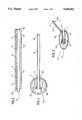
- FIG. 1is a plan view of a first embodiment of a biomedical electrode device according to the invention
- FIG. 2is an elevation of the electrode device of FIG. 1 of the drawings
- FIG. 3is a perspective view of the electrode device of FIG. 1 of the drawings.
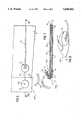
- FIG. 4is a plan view of a second embodiment of a biomedical electrode device according to the invention.
- FIG. 5is a cross-sectional view of the electrode device taken along the line V--V of FIG. 4 of the drawings and viewed in the direction of the associated arrows;
- FiG. 6is a plan view of a third embodiment of biomedical electrode device according to the invention.
- FIG. 7is a cross-sectional view of the electrode device taken along the line VII--VII of FIG. 6 of the drawings and viewed in the direction of the associated arrows;
- FIG. 8is a perspective view of the electrode device of FIG. 6 of the drawings.
- FIGS. 9 to 12, 13a, 13b, 14a, and 14bare cross-sectional views of further embodiments of electrode devices according to the invention.
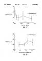
- FIG. 15demonstrates the in vitro adhesion properties of 10% w/w Gantrez AN 139 films containing variable amounts of glycerol as plasticizer with respect to initial adhesion and on first and second restick. All points represent the mean ⁇ SD adhesion results for nine replicates. All tests were carried out using wet hairy piglet skin.
- FIG. 16demonstrates pH-adhesion relationships for 10% w/w Gantrez AN 139 in water with varying amounts of glycerol added.
- FIG. 17demonstrates the effect of salt concentration on adhesion in respect of 10% w/w Gantrez AN 139, 5% w/w glycerol and pH adjusted to 5.3 ⁇ 0.18 with concentrated NaOH. Error bars represent the SD of the mean for six replicates.
- FIG. 18demonstrates the effect of thickness of film on adhesion in respect of 10% w/w Gantrez AN 139, 5% w/w glycerol and pH adjusted to 5.3 ⁇ 0.18 with concentrated NaOH. Error bars represent the SD of the mean for nine replicates. The mean thickness values were obtained from ten replicates.
- FIG. 19is a trace of foetal heart using the electrode device according to the invention.
- an electrode device 10which comprises a relatively thin electrically insulating substrate 11 having an obverse side 21; a reverse side 22; a proximal end 23; and a distal end 24.
- a foetal electrode sensor 12 and associated lead 13is deposited onto the obverse side 21 of the substrate 11 at the proximal end 23 thereof.
- a reference or indifferent electrode sensor 14 and associated lead 15is deposited onto the reverse side 22 of the substrate 11 also at the proximal end 23 thereof.
- the foetal electrode sensor 12 and the reference electrode sensor 14are in substantially parallel spaced apart relationship.
- a means of making electrical connection to each of the two leads 13, 15is provided by extending the leads 13, 15 from the proximal end 23 to the distal end 24.
- An insulating layer 16is deposited over the foetal electrode lead 13 so as to leave the sensor 12 and the free end of the lead 13 located at the distal end 24 exposed, the latter being necessary for making electrical connection to a monitoring device (not shown).
- the lead 15 from the sensor 14may also be covered by an insulating layer. This is not essential as the sensor 14 and the lead 15 may both serve to make electrical contact with surrounding fluids etc. of the environment in which the electrode device 10 will be used
- the electrode device 10should preferably be relatively thin, lightweight and flexible in order to minimise the strength of adhesion required; to maximise electrical contact; to minimise or avoid trauma to the foetal head; and to minimise or avoid discomfort to the mother both during electrode application and the delivery process itself.
- the substrate 11a non-woven polyester sold under the trade name "Melinex" and available in a range of film thicknesses.
- the inventionis not limited in its scope by the use of any given substrate material which meets the necessary design requirements.
- the sensors 12, 14 and the leads 13, 15may be deposited by, for example, screenprinting thin layers of conducting serographic ink onto a suitable polyester or polycarbonate substrate 11. Suitable inks include those optimally loaded with either silver or a combination of silver and silver chloride particles.
- the sensors 12, 14may be printed or otherwise deposited using a different ink to that used to print or deposit their connecting leads 13, 15. Inks loaded with silver and silver chloride particles are found to be suitable for the sensors 12, 14 as they give rise to good electrode-tissue interface electrical characteristics. Silver-loaded inks, on the other hand, give rise to layers with high conductivity characteristics and hence are suitable for depositing connecting leads 13, 15.
- the connecting leads 13, 15make contact centrally with the electrode sensors 12, 14.
- a pair of circularly shaped openings 25, 26is provided in the substrate 11 at the proximal end 23. Contiguous with the opening 25 and in parallel spaced apart relationship relative to the leads 13, 15 is a slit 27; similarly, a slit 28 is provided which is contiguous with the opening 26.
- the provision of the holes 25, 26 and associated respective slits 27, 28enables the leads 13, 15 to move in the direction of the arrows 29 relative to the proximal end 23 of the electrode device 10.
- the foetal electrode sensor 12is coated with a film-forming layer 17.
- the layer 17 on dryingis non-adherent on dry surfaces, contrary to the conventional pressure-sensitive adhesives. It is, therefor, readily handled and has excellent storage properties.
- the layer 17is activated by moisture when the electrode 10 is offered to a moist surface, such as a foetal scalp. It does not, therefore, require a pre-moistening step.
- a further advantage of the layer 17is its ability to maintain sufficient strength of adhesion to secure the electrode device 10 to a surface such as the foetal scalp, even in the presence of a substantial amount of aqueous fluid. In these respects, the invention differs from known wet-stick adhesives.
- the film-forming layer 17may be prepared using a bioadhesive gel or viscous solution by conventional means known in the art and drying to a low water content, drying being complete when all tack is lost from the resulting dried film.
- the layer 17may be made from polymeric components in an aqueous base and at a total polymer concentration by weight in the wet state of between 0.01% and 50%, but preferably between 1 and 20% by weight of the total mixture, suitable polymers including polyvinylpyrrolidone, polyvinylal--cohols, carbomers, cellulose derivatives such as methyl cellulose and sodium carboxymethylcellulose, hydroxyalkyl celluloses such as hydroxyethylcellulose, hydroxypropylcellulose and hydroxypropylmethylcellulose, polyoxyethylene polyoxypropylene diol block copolymers, poly (methyl vinyl ether/maleic anhydride) copolymers, vinylpyrrolidone copolymers such as vinylpyrrolidone/vinyl acetate copo
- Suitable plasticisers for incorporation into the layer 17 at a total plasticiser concentration by a weight of between 0.25% and 30%, but preferably between 2 and 25% by weight of the total mixtureinclude dimethyl phthalate, diethyl phthalate, dioctyl phthalate, glycerin, polyols such as ethylene glycol, diethylene glycol, triethylene glycol, polethethylene glycol, propylene glycol, sorbitol and glycerol, nonylphenol--ethylene oxide adducts sold under the trade name Antarox, ethyl glycolate and ethyl sulphonamide derivatives sold under the trade name Santicizer, tricresyl phosphate, dimethylsebacate, ethyl glycolate.
- Suitable electrically conducting salts for enabling the layer 17 to be electrically conductive and which may be added to the polymerlc componentsinclude at a total concentration by weight of between 0% and 10% but preferably between 0.01% and 0.25% by weight of the total mixture sodium chloride, potassium chloride, potassium dihydrogen phosphate, dipotassium hydrogen phosphate, potassium citrate. Electrical conduction may also be achieved by addition of alkali or acid to the polymeric components.
- the polymeric componentis a poly (methyl vinyl ether/maleic anhydride) copolymer sold under the trade names Gantrez AN, Gantrez S and Gantrez ES.
- the polymeric componentis Gantrez AN 139 or Gantrez AN 139 in combination with polyvinylpyrrolidone.
- a further preferred embodiment of the inventionprovides for the addition to Gantrez AN 139 aqueous solution or Gantrez AN 139/polyvinylpyrrolidone aqueous solution of glycerol as a plasticiser and sodium chloride as an enhancer of electrical conduction.
- Glycerolis present in a final concentration in the aqueous solution, before drying, of between 1 and 40% w/w, preferably 5% w/w and sodium chloride is present in a final concentration in the aqueous solution, before drying, of between 0.001 and 5% w/w, preferably 0.1% w/w.
- the solutionsare formed by conventional means known in the art, using slow stirring at room temperature.
- the resulting solutionsare relatively viscous. Entrained air bubbles may be removed by subjecting the solutions to degassing under vacuum, preferably overnight, or by centrifuging the solutions, for example, at 4000 r.p.m. for 20 minutes.
- the layermay be formed by casting the polymer solution such as is disclosed in Example 1 or Example 2 below, directly onto a head 20 of the electrode device 10 and drying to remove water, thus forming a pliable, non-tacky, non-adhesive, film.
- the head 20 of the electrode device 10is that part of the device 10 containing the sensors 12, 14 and the proximal ends of the leads 13, 15.
- Castingmay be done by conventional means known in the art, including the use of a casting knife with a variable aperture.
- the layer 17may be directly deposited, using a suitable template, onto the head 20. Drying may be achieved by conventional means known in the art, including use of a tray drier, vacuum oven or tunnel dryer. Drying may be carried out at temperatures between ambient and 150° C., preferably 60° C.
- the drying timevaries depending on the solution formulation and the type of drying process used.
- the water content of the final, dried filmis preferably less than 25% w/w.
- the adhesion properties of electrode 10have been determined in vitro using neonate, hairy porcine skin as a model.
- the test methodis known in the art and described in Woolfson, A. D et al. International Journal of Pharmaceutics, volume 84, pages 69-76 (1992).
- variables studiedincluded the effect on adhesion of plasticiser concentration, pH, salt concentration and thickness of deposited film.
- Results of the in vitro experimentsare shown in FIGS. 16-19.
- a particular feature of the conducting bioadhesive wet-stick interfaceis its ability to allow removal and subsequent repositioning/replacement of the electrode, with adherence characteristics maintained. This is demonstrated by the results shown in FIG. 15.
- the ability of the electrode 10 to persist in adhering to skinwas studied by adhering it to neonate porcine skin and immersing the whole in one liter of phosphate-buffered saline at 37° C. Persistence of adherence was determined visually by removal and inspection of the test samples at 15 minute intervals. Testing was discontinued after 6 hours, at which time the electrode 10 remained firmly adhered to the porcine skin substrate.
- the electrode 10was tested in vivo for adherence to foetal skin and quality of electrical signals obtained.
- the electrode 10adhered immediately to unwashed or otherwise pretreated foetal skin and remained firmly attached throughout the duration of the assessment.
- a sample of the trace obtainedis shown in FIG. 19.
- FIGS. 4 and 5 of the drawingsthere is shown a second embodiment of an electrode device 100 according to the invention which is similar in many respects to the electrode device 10 and like numerals for equivalent components are used in the drawings.
- FIG. 4shows a "blank" from which the device will ultimately be formed by trimming surplus substrate material from around the electrodes and leads.
- the insulating layer 16 and the film-forming layer 17are omitted from FIG. 4 of the drawing.
- the composition of the layer 17is similar to that described with reference to the electrode device 10.
- the essential differences between the electrode device 100 when compared with the electrode device 10are as follows.
- both electrodes 12, 14 of the electrode 100are located on the same side of the substrate i.e. either the obverse side 21 or the reverse side 22.
- the electrode sensors 12, 14are in linear relationship so that the reference electrode sensor 14 is located at the extreme proximal end 23 of the substrate 11 with the foetal electrode sensor 12 located albeit at the proximal end 23 but in the direction of the distal end 24 of the electrode 100.
- the respective associated leads 13 and 15are also located on the same side of the substrate 1l as the sensors 12, 14 but to enable the lead 15 to be electrically isolated from the lead 13, the lead 15 has a curved path or portion 15a which circumlocates the sensor 12.
- a fold line 101Located between the sensor 12 and the sensor 14 is a fold line 101 so that part of the substrate 11 containing the sensor 14 may be folded in the direction of the arrows 103 between 100° and 160° as shown in FIG. 5 of the drawings.
- the relative positions of the sensors 12, 14will now be clearly seen in FIG. 5 of the drawings being at an angle of between 20° and 80°.
- a pair of side walls 102may connect the sides of the curved part of the substrate 11 with the linear part of the substrate 11 to form a housing which constitutes a finger cradle 104.
- the side walls 102may be formed from laterally extending flaps of the substrate material which are left attached to the device on either side of the electrodes after trimming the blank as referred to above, or they may separate pieces which are individually attached.
- the provision of the cradle 104enables the electrode 100 device to be positioned relatively easily by the insertion of, for example, an index finger into the cradle 104, so as to carry the electrode device 100 to the site of use, e.g. the head or scalp of the baby being delivered and placement of the electrode 100 by the application of suitable pressure so that the layer 17 and hence the foetal electrode sensor 12 is in contact with the head of a baby with relatively long leads trailing along the leg of the mother to which they may be taped by conventional means.
- the leads 13, 15can then easily be connected to suitable monitoring equipment.
- FIG. 6-8show a third embodiment of an electrode device 200 according to the invention which is substantially similar to the electrode device 100 except as follows. [For reasons of clarity the insulating layer 16 and the film-forming layer 17 are omitted from FIG. 6 of the drawings.] Whereas the leads 13, 15 of the electrode device 100 are on the same side of the substrate 11, in the electrode device 200, the lead 13 is on the same or obverse side 21 of the substrate 11 as the sensors 12, 14 and the lead 15 is on the reverse side 22 of the substrate 11. This arrangement allows for a relatively simpler construction of electrode device 200 in that the curve path 15a is not required. To enable electrical contact to be made between the lead 15 and the sensor 14, an opening 201 is provided centrally in the sensor 14 so that electrical connection may be made by the provision of, for example, electrically conductive ink in the opening in electrical contact with the sensor 14 and the lead 15.
- FIG. 9 of the drawingsshows a fourth embodiment of an electrode device 300 according to the invention which is substantially similar to the device 10 except as follows.
- the proximal end 23 of the substrate 11has attached thereto by suitable means a piece of substrate material 11a which projects towards the distal end 24 of the substrate 11.
- a pair of side walls 102connects the material 11a with the substrate 11 to form, in a manner similar to that described with respect to FIGS. 4-5 of the drawings, a finger cradle 104.
- FIG. 10 of the drawingsthere is shown a fifth embodiment of an electrode device 400 according to the invention which is substantially similar to the electrode device 100 of FIG. 5 except as follows. Instead of providing a single curve of between 100° and 160°, a double curve is employed so that the reference electrode 14 is in substantially parallel spaced apart relationship relative to the electrode 12 having been moved through an angle of about 180°.
- the use of surface electrodescan result in an inadequate ECG trace due to electrical shorting between electrodes. This can be mitigated by surrounding the adhesive 17 and sensor 12 with an annulus or band of insulating material.
- FIGS. 11 and 12Embodiments of such a design are shown in FIGS. 11 and 12.
- the band of insulating material 50extends to the edges of the substrate 11, whereas in FIG. 12, the band of insulating material is itself surrounded by a further band of moisture activated bioadhesive material 51.
- the bioadesive material 51ensures firm mechanical contact between the insulation material 50 and the foetal scalp, and even firmer contact can be achieved by making the thickness of the outer bioadhesive layer 51 less than that of the insulation material 50.
- the outer layer 51may be less conductive than the main bioadhesive layer 17. In the case of both FIG. 11 and FIG.
- the insulating material 50be made of a material substantially more rigid than the bioadhesive layer so that the band 50 can penetrate the vernix coating to make firm contact with the foetal scalp, yet not so rigid that it cannot deform to conform with body contours without traumatizing the foetal skin.
- the band 50can penetrate the vernix coating to make firm contact with the foetal scalp, yet not so rigid that it cannot deform to conform with body contours without traumatizing the foetal skin.
- FIGS. 11 and 12there may be more than one band arranged concentrically within one another. In such a case the multiple bands may be the same or different composition and may be located immediately adjacent one another or separated by intermediate regions of bioadhesive.
- FIGS. 13A and 13BA further embodiment of the invention is shown in FIGS. 13A and 13B.
- the substrate 11is formed or moulded, along with the electrodes 12 and 14 and the insulating layer 16, to form a cup-shaped housing containing the bioadhesive material 17.
- Thisavoids the need for a surrounding band of insulation 50 as described above.
- Applying the device using moderate pressure, as shown in FIG. 13Awill ensure that the "rim" or edge of the cup 11', which is substantially more rigid than the bioadhesive layer 17, penetrates the vernix covering to make firm mechanical contact with the foetal scalp. This will greatly reduce the possibility of electrical shorting between the two electrodes 12 and 14.
- suctionmay be applied to the interior of the cup 11' by using a vacuum pump, a suction "bulb” or a syringe, for example.
- a vacuum pumpa suction "bulb” or a syringe
- One end 53 of a suction tube 52is inserted through a self-sealing membrane 54 in the cup 11', FIG. 14A, and its other end is attached to the vacuum pump or other suction device. Once the device has been firmly attached, the suction tube 52 can be withdrawn, FIG. 14B.
- a finger cradlecan be provided in the case of the embodiments of FIGS. 11 to 14. However, these are not shown in order to avoid overcomplicating the Figures.
- a finger cradlein all embodiments of the invention described above, one can provide a slot in the substrate 11 for engagement by an external applicator.
- a release liner(not shown) may be provided which forms a protective layer over the obverse side of the electrode device and which is removable immediately prior to use.
- the electrode device according to the inventionmay also be used for transcutaneous electrical nerve stimulation for the treatment of, for example, urinary incontinence, or for electrodental anaesthesia or as referred to above, for the electrical monitoring of a foetal heart.
Landscapes
- Health & Medical Sciences (AREA)
- Life Sciences & Earth Sciences (AREA)
- Biomedical Technology (AREA)
- Cardiology (AREA)
- Veterinary Medicine (AREA)
- Public Health (AREA)
- General Health & Medical Sciences (AREA)
- Animal Behavior & Ethology (AREA)
- Heart & Thoracic Surgery (AREA)
- Engineering & Computer Science (AREA)
- Surgery (AREA)
- Pathology (AREA)
- Medical Informatics (AREA)
- Molecular Biology (AREA)
- Biophysics (AREA)
- Physics & Mathematics (AREA)
- Pediatric Medicine (AREA)
- Neurology (AREA)
- Pregnancy & Childbirth (AREA)
- Gynecology & Obstetrics (AREA)
- Neurosurgery (AREA)
- Orthopedic Medicine & Surgery (AREA)
- Reproductive Health (AREA)
- Nuclear Medicine, Radiotherapy & Molecular Imaging (AREA)
- Radiology & Medical Imaging (AREA)
- Chemical & Material Sciences (AREA)
- Organic Chemistry (AREA)
- Measurement And Recording Of Electrical Phenomena And Electrical Characteristics Of The Living Body (AREA)
- Electrotherapy Devices (AREA)
Abstract
Description
______________________________________ Gantrez AN 139 10% w/w Glycerol 5% w/w Sodium Chloride 0.1% w/w Concentrated Sodium Hydroxide sufficient to adjust pH of final solution to 5 Water q.s. ______________________________________
______________________________________ Gantrez AN 139 15% w/w Glycerol 7.5% w/w Kollidon 90 5% w/w Sodium Chloride 0.1% w/w Concentrated Sodium Hydroxide sufficient to adjust pH of final solution to 5 Water q.s. ______________________________________
Claims (13)
Applications Claiming Priority (2)
| Application Number | Priority Date | Filing Date | Title |
|---|---|---|---|
| IE930103 | 1993-02-15 | ||
| IE930103 | 1993-02-15 |
Publications (1)
| Publication Number | Publication Date |
|---|---|
| US5645062Atrue US5645062A (en) | 1997-07-08 |
Family
ID=11039875
Family Applications (1)
| Application Number | Title | Priority Date | Filing Date |
|---|---|---|---|
| US08/196,465Expired - LifetimeUS5645062A (en) | 1993-02-15 | 1994-02-15 | Biomedical electrode device |
Country Status (5)
| Country | Link |
|---|---|
| US (1) | US5645062A (en) |
| JP (1) | JP3636374B2 (en) |
| DE (1) | DE4404762A1 (en) |
| FR (1) | FR2701381A1 (en) |
| GB (1) | GB2274995B (en) |
Cited By (49)
| Publication number | Priority date | Publication date | Assignee | Title |
|---|---|---|---|---|
| US6129751A (en)* | 1998-07-28 | 2000-10-10 | Intermedics Inc. | Cardiac lead with active fixation and biocompatible lubricant |
| WO2002098272A2 (en) | 2001-06-05 | 2002-12-12 | Barnev Ltd. | Probe anchor |
| US6594515B2 (en)* | 2000-01-10 | 2003-07-15 | Richard L. Watson | Noninvasive, intrauterine fetal ECG strip electrode |
| US20030170308A1 (en)* | 2001-05-01 | 2003-09-11 | Cleary Gary W. | Hydrogel compositions |
| US20040000663A1 (en)* | 2002-04-10 | 2004-01-01 | Segall Daniel P. | Hydro-insensitive alternating current responsive composites |
| US6687524B1 (en)* | 1999-08-24 | 2004-02-03 | Cas Medical Systems, Inc | Disposable neonatal electrode for use in a high humidity environment |
| US20040230179A1 (en)* | 2003-02-07 | 2004-11-18 | Alfred E. Mann Institute For Biomedical Engineering | Surgical drain with sensors for monitoring fluid lumen |
| US6842636B2 (en) | 2002-09-27 | 2005-01-11 | Axelgaard Manufacturing Co., Ltd. | Medical electrode |
| US20050277998A1 (en)* | 2004-02-11 | 2005-12-15 | Tracey Michael R | System and method for nerve stimulation |
| US20060182788A1 (en)* | 2005-01-27 | 2006-08-17 | Parminder Singh | Hydrophilic biocompatible adhesive formulations and uses |
| US20060195153A1 (en)* | 2004-02-11 | 2006-08-31 | Diubaldi Anthony | System and method for selectively stimulating different body parts |
| US20060195146A1 (en)* | 2004-02-11 | 2006-08-31 | Tracey Michael R | System and method for selectively stimulating different body parts |
| US20070150034A1 (en)* | 2005-06-09 | 2007-06-28 | Medtronic, Inc. | Implantable medical lead |
| US20070185541A1 (en)* | 2004-02-11 | 2007-08-09 | Diubaldi Anthony | Conductive mesh for neurostimulation |
| US20080125729A1 (en)* | 2005-05-03 | 2008-05-29 | Universitat Rostock | Dressing for the Treatment of Chronic Wounds |
| US20080167701A1 (en)* | 2007-01-09 | 2008-07-10 | Angel Medical Systems, Inc. | Adaptive conductive lead systems |
| US20080243217A1 (en)* | 2000-05-30 | 2008-10-02 | Michael Peter Wildon | Cardiac stimulation apparatus |
| WO2008150974A1 (en)* | 2007-06-01 | 2008-12-11 | Med-El Elektromedizinische Geraete Gmbh | Biodegradable flexible coating for implantable medical devices |
| US20090093858A1 (en)* | 2007-10-03 | 2009-04-09 | Ethicon, Inc. | Implantable pulse generators and methods for selective nerve stimulation |
| US20090227857A1 (en)* | 2008-03-06 | 2009-09-10 | Chuck Rowe | Biomedical electrode |
| US20090299437A1 (en)* | 2008-06-03 | 2009-12-03 | Med-El Elektromedizinische Geraete Gmbh | Conductive Coating of Implants with Inductive Link |
| US20090317451A1 (en)* | 2008-08-26 | 2009-12-24 | Hauser Ray L | Pressure-sensitive adhesive for skin surface and/or transdermal substance delivery |
| US20090318568A1 (en)* | 2009-08-26 | 2009-12-24 | Hauser Ray L | Adherent coating for tissue surface and/or trans-tissue surface substance delivery |
| US20100016702A1 (en)* | 2008-07-18 | 2010-01-21 | Flexcon Company, Inc. | High Impedance Signal Detection Systems and Methods for Use in Electrocardiogram Detection Systems |
| US20100036230A1 (en)* | 2008-08-06 | 2010-02-11 | Flexcon Company, Inc. | Multiple Electrode Composite Systems and Methods for Use in Electrocardiogram Detection Systems |
| US20100087905A1 (en)* | 2008-10-08 | 2010-04-08 | Med-El Elektromedizinische Geraete Gmbh | Cochlear Tissue Protection from Electrode Trauma |
| US20100249677A1 (en)* | 2005-06-07 | 2010-09-30 | Ethicon, Inc. | Piezoelectric stimulation device |
| US20110202120A1 (en)* | 2010-02-16 | 2011-08-18 | Med-El Elektromedizinische Geraete Gmbh | Electrode Design for Reduced Trauma Insertion |
| US8206738B2 (en) | 2001-05-01 | 2012-06-26 | Corium International, Inc. | Hydrogel compositions with an erodible backing member |
| US8273405B2 (en) | 2001-05-01 | 2012-09-25 | A.V. Topcheiv Institute of Petrochemical Synthesis, Russian Academy of Sciences | Water-absorbent adhesive compositions and associated methods of manufacture and use |
| USRE44145E1 (en) | 2000-07-07 | 2013-04-09 | A.V. Topchiev Institute Of Petrochemical Synthesis | Preparation of hydrophilic pressure sensitive adhesives having optimized adhesive properties |
| US8541021B2 (en) | 2001-05-01 | 2013-09-24 | A.V. Topchiev Institute Of Petrochemical Synthesis | Hydrogel compositions demonstrating phase separation on contact with aqueous media |
| US8548558B2 (en) | 2008-03-06 | 2013-10-01 | Covidien Lp | Electrode capable of attachment to a garment, system, and methods of manufacturing |
| US8658201B2 (en) | 2004-01-30 | 2014-02-25 | Corium International, Inc. | Rapidly dissolving film for delivery of an active agent |
| WO2014069853A1 (en)* | 2012-10-29 | 2014-05-08 | Kolon Industries, Inc. | Aramid fiber product with excellent conductivity and method of manufacturing the same |
| US8753669B2 (en) | 2001-05-01 | 2014-06-17 | A.V. Topchiev Institute Of Petrochemical Synthesis, Russian Academy Of Sciences | Two-phase, water-absorbent bioadhesive composition |
| US8784879B2 (en) | 2009-01-14 | 2014-07-22 | Corium International, Inc. | Transdermal administration of tamsulosin |
| US8821901B2 (en) | 2001-05-01 | 2014-09-02 | A.V. Topchiev Institute Of Petrochemical Synthesis Russian Academy Of Sciences | Method of preparing polymeric adhesive compositions utilizing the mechanism of interaction between the polymer components |
| US8840918B2 (en) | 2001-05-01 | 2014-09-23 | A. V. Topchiev Institute of Petrochemical Synthesis, Russian Academy of Sciences | Hydrogel compositions for tooth whitening |
| EP2789314A2 (en) | 2003-10-06 | 2014-10-15 | 3F Therapeutics, Inc | Minimally invasive valve replacement system |
| US8868216B2 (en) | 2008-11-21 | 2014-10-21 | Covidien Lp | Electrode garment |
| US9242021B2 (en) | 2004-08-05 | 2016-01-26 | Corium International, Inc. | Adhesive composition |
| US9393416B2 (en) | 2005-06-09 | 2016-07-19 | Medtronic, Inc. | Peripheral nerve field stimulation and spinal cord stimulation |
| US10300273B2 (en) | 2005-06-09 | 2019-05-28 | Medtronic, Inc. | Combination therapy including peripheral nerve field stimulation |
| CN111050643A (en)* | 2017-09-07 | 2020-04-21 | 美敦力施美德公司 | Endotracheal tube with tube coating |
| US20200383640A1 (en)* | 2019-06-07 | 2020-12-10 | InBody Co., Ltd. | Electrode patch |
| EP3821852A2 (en) | 2003-10-06 | 2021-05-19 | Medtronic 3F Therapeutics, Inc. | Minimally invasive valve replacement system |
| WO2022011431A1 (en)* | 2020-07-17 | 2022-01-20 | Seer Medical Pty Ltd | Adhesive pad for retaining an electrode against a patient's skin |
| US12011911B2 (en) | 2020-03-25 | 2024-06-18 | Flexcon Company, Inc. | Isotropic non-aqueous electrode sensing material |
Families Citing this family (6)
| Publication number | Priority date | Publication date | Assignee | Title |
|---|---|---|---|---|
| US5833622A (en)* | 1994-04-04 | 1998-11-10 | Graphic Controls Corporation | Non-invasive fetal probe having improved mechanical and electrical properties |
| AUPQ788400A0 (en)* | 2000-05-30 | 2000-06-22 | Wildon, Michael Peter | Epicardial stimulation lead |
| EP1904173B8 (en) | 2005-06-09 | 2016-06-08 | Medtronic, Inc. | Implantable medical device with electrodes on multiple housing surfaces |
| US9643004B2 (en) | 2006-10-31 | 2017-05-09 | Medtronic, Inc. | Implantable medical elongated member with adhesive elements |
| GB2467533A (en)* | 2009-02-04 | 2010-08-11 | Rebecca Lynn Pilditch | Conductive ink for use on the living human body |
| US9682229B2 (en)* | 2012-06-29 | 2017-06-20 | Medtronic, Inc. | Drug-eluting polymer coated implantable electrode |
Citations (17)
| Publication number | Priority date | Publication date | Assignee | Title |
|---|---|---|---|---|
| GB1520351A (en)* | 1975-04-14 | 1978-08-09 | Medtronic Inc | Electromedical electrodes |
| GB2034184A (en)* | 1978-10-12 | 1980-06-04 | Hymes A | An improved monitoring or stimulation electrode |
| GB1574363A (en)* | 1976-02-05 | 1980-09-03 | Johnson & Johnson | Medical electrode |
| US4539996A (en)* | 1980-01-23 | 1985-09-10 | Minnesota Mining And Manufacturing Company | Conductive adhesive and biomedical electrode |
| US4543958A (en)* | 1979-04-30 | 1985-10-01 | Ndm Corporation | Medical electrode assembly |
| EP0085327B1 (en)* | 1982-01-18 | 1986-04-30 | Medtronic, Inc. | Electrically conductive compositions and electrodes utilizing same |
| US4699146A (en)* | 1982-02-25 | 1987-10-13 | Valleylab, Inc. | Hydrophilic, elastomeric, pressure-sensitive adhesive |
| EP0255241A2 (en)* | 1986-06-30 | 1988-02-03 | Nepera, Inc. | Medical electrode |
| US4766005A (en)* | 1986-10-30 | 1988-08-23 | Opi Products, Inc. | Material and method for obtaining strong adhesive bonding to proteinaceous substrates |
| US4848353A (en)* | 1986-09-05 | 1989-07-18 | Minnesota Mining And Manufacturing Company | Electrically-conductive, pressure-sensitive adhesive and biomedical electrodes |
| DE3816190C1 (en)* | 1988-05-11 | 1989-08-10 | Dieter Von 8000 Muenchen De Zeppelin | Scalp electrode |
| US4860754A (en)* | 1987-04-01 | 1989-08-29 | E. R. Squibb & Sons, Inc. | Electrically conductive adhesive materials |
| EP0409067A2 (en)* | 1989-07-21 | 1991-01-23 | Iomed, Inc. | Hydratable bioelectrode |
| US4989607A (en)* | 1989-03-30 | 1991-02-05 | Preston Keusch | Highly conductive non-stringy adhesive hydrophilic gels and medical electrode assemblies manufactured therefrom |
| US5003978A (en)* | 1985-08-21 | 1991-04-02 | Technology 21, Inc. | Non-polarizable dry biomedical electrode |
| US5143071A (en)* | 1989-03-30 | 1992-09-01 | Nepera, Inc. | Non-stringy adhesive hydrophilic gels |
| US5178143A (en)* | 1991-07-24 | 1993-01-12 | Isp Investments Inc. | Electrically conductive gel composition |
- 1994
- 1994-02-11GBGB9402719Apatent/GB2274995B/ennot_activeExpired - Fee Related
- 1994-02-14FRFR9401635Apatent/FR2701381A1/enactivePending
- 1994-02-15JPJP01859094Apatent/JP3636374B2/ennot_activeExpired - Fee Related
- 1994-02-15USUS08/196,465patent/US5645062A/ennot_activeExpired - Lifetime
- 1994-02-15DEDE4404762Apatent/DE4404762A1/ennot_activeCeased
Patent Citations (17)
| Publication number | Priority date | Publication date | Assignee | Title |
|---|---|---|---|---|
| GB1520351A (en)* | 1975-04-14 | 1978-08-09 | Medtronic Inc | Electromedical electrodes |
| GB1574363A (en)* | 1976-02-05 | 1980-09-03 | Johnson & Johnson | Medical electrode |
| GB2034184A (en)* | 1978-10-12 | 1980-06-04 | Hymes A | An improved monitoring or stimulation electrode |
| US4543958A (en)* | 1979-04-30 | 1985-10-01 | Ndm Corporation | Medical electrode assembly |
| US4539996A (en)* | 1980-01-23 | 1985-09-10 | Minnesota Mining And Manufacturing Company | Conductive adhesive and biomedical electrode |
| EP0085327B1 (en)* | 1982-01-18 | 1986-04-30 | Medtronic, Inc. | Electrically conductive compositions and electrodes utilizing same |
| US4699146A (en)* | 1982-02-25 | 1987-10-13 | Valleylab, Inc. | Hydrophilic, elastomeric, pressure-sensitive adhesive |
| US5003978A (en)* | 1985-08-21 | 1991-04-02 | Technology 21, Inc. | Non-polarizable dry biomedical electrode |
| EP0255241A2 (en)* | 1986-06-30 | 1988-02-03 | Nepera, Inc. | Medical electrode |
| US4848353A (en)* | 1986-09-05 | 1989-07-18 | Minnesota Mining And Manufacturing Company | Electrically-conductive, pressure-sensitive adhesive and biomedical electrodes |
| US4766005A (en)* | 1986-10-30 | 1988-08-23 | Opi Products, Inc. | Material and method for obtaining strong adhesive bonding to proteinaceous substrates |
| US4860754A (en)* | 1987-04-01 | 1989-08-29 | E. R. Squibb & Sons, Inc. | Electrically conductive adhesive materials |
| DE3816190C1 (en)* | 1988-05-11 | 1989-08-10 | Dieter Von 8000 Muenchen De Zeppelin | Scalp electrode |
| US4989607A (en)* | 1989-03-30 | 1991-02-05 | Preston Keusch | Highly conductive non-stringy adhesive hydrophilic gels and medical electrode assemblies manufactured therefrom |
| US5143071A (en)* | 1989-03-30 | 1992-09-01 | Nepera, Inc. | Non-stringy adhesive hydrophilic gels |
| EP0409067A2 (en)* | 1989-07-21 | 1991-01-23 | Iomed, Inc. | Hydratable bioelectrode |
| US5178143A (en)* | 1991-07-24 | 1993-01-12 | Isp Investments Inc. | Electrically conductive gel composition |
Non-Patent Citations (4)
| Title |
|---|
| Comprehensive Maternity Nursing, Nursing Process and the Childbearing Family, second edition, J.B. Lippincott Company, pp. 676 687.* |
| Comprehensive Maternity Nursing, Nursing Process and the Childbearing Family, second edition, J.B. Lippincott Company, pp. 676-687. |
| Handbook of Pressure Sensitive Adhesive Technology, Donatas Satas, Van Nostrand Reinhold Company, New York, 1982, pp. 1 25.* |
| Handbook of Pressure-Sensitive Adhesive Technology, Donatas Satas, Van Nostrand Reinhold Company, New York, 1982, pp. 1-25. |
Cited By (113)
| Publication number | Priority date | Publication date | Assignee | Title |
|---|---|---|---|---|
| US6129751A (en)* | 1998-07-28 | 2000-10-10 | Intermedics Inc. | Cardiac lead with active fixation and biocompatible lubricant |
| US6687524B1 (en)* | 1999-08-24 | 2004-02-03 | Cas Medical Systems, Inc | Disposable neonatal electrode for use in a high humidity environment |
| US6594515B2 (en)* | 2000-01-10 | 2003-07-15 | Richard L. Watson | Noninvasive, intrauterine fetal ECG strip electrode |
| US20040015067A1 (en)* | 2000-01-10 | 2004-01-22 | Watson Richard L. | Noninvasive, intrauterine fetal egg strip electrode |
| US6973341B2 (en) | 2000-01-10 | 2005-12-06 | Watson Richard L | Noninvasive, intrauterine fetal ECG strip electrode |
| US20080243217A1 (en)* | 2000-05-30 | 2008-10-02 | Michael Peter Wildon | Cardiac stimulation apparatus |
| USRE45666E1 (en) | 2000-07-07 | 2015-09-08 | A.V. Topchiev Institute Of Petrochemical Synthesis | Preparation of hydrophilic pressure sensitive adhesives having optimized adhesive properties |
| USRE44145E1 (en) | 2000-07-07 | 2013-04-09 | A.V. Topchiev Institute Of Petrochemical Synthesis | Preparation of hydrophilic pressure sensitive adhesives having optimized adhesive properties |
| US9127140B2 (en) | 2001-05-01 | 2015-09-08 | A. V. Topchiev Institute of Petrochemical Synthesis, Russian Academy of Sciences | Water-absorbent adhesive compositions and associated methods of manufacture and use |
| US8481071B2 (en) | 2001-05-01 | 2013-07-09 | Corium International, Inc. | Hydrogel compositions with an erodible backing member |
| US9687428B2 (en) | 2001-05-01 | 2017-06-27 | A. V. Topchiev Institute of Petrochemical Synthesis, Russian Academy of Sciences | Hydrogel compositions for tooth whitening |
| US8206738B2 (en) | 2001-05-01 | 2012-06-26 | Corium International, Inc. | Hydrogel compositions with an erodible backing member |
| US9532935B2 (en) | 2001-05-01 | 2017-01-03 | A. V. Topchiev Institute of Petrochemical Synthesis, Russian Academy of Sciences | Hydrogel compositions for tooth whitening |
| US9259504B2 (en) | 2001-05-01 | 2016-02-16 | A. V. Topchiev Institute of Petrochemical Synthesis, Russian Academy of Sciences | Non-electrically conductive hydrogel composition |
| US10835454B2 (en) | 2001-05-01 | 2020-11-17 | Corium, Inc. | Hydrogel compositions with an erodible backing member |
| US8273405B2 (en) | 2001-05-01 | 2012-09-25 | A.V. Topcheiv Institute of Petrochemical Synthesis, Russian Academy of Sciences | Water-absorbent adhesive compositions and associated methods of manufacture and use |
| US10869947B2 (en) | 2001-05-01 | 2020-12-22 | Corium, Inc. | Hydrogel compositions |
| US9089481B2 (en) | 2001-05-01 | 2015-07-28 | A. V. Topchiev Institute of Petrochemical Synthesis, Russian Academy of Sciences | Hydrogel compositions demonstrating phase separation on contact with aqueous media |
| US9084723B2 (en) | 2001-05-01 | 2015-07-21 | A. V. Topchiev Institute of Petrochemical Synthesis, Russian Academy of Sciences | Hydrogel compositions with an erodible backing member |
| US8840918B2 (en) | 2001-05-01 | 2014-09-23 | A. V. Topchiev Institute of Petrochemical Synthesis, Russian Academy of Sciences | Hydrogel compositions for tooth whitening |
| US8821901B2 (en) | 2001-05-01 | 2014-09-02 | A.V. Topchiev Institute Of Petrochemical Synthesis Russian Academy Of Sciences | Method of preparing polymeric adhesive compositions utilizing the mechanism of interaction between the polymer components |
| US8753669B2 (en) | 2001-05-01 | 2014-06-17 | A.V. Topchiev Institute Of Petrochemical Synthesis, Russian Academy Of Sciences | Two-phase, water-absorbent bioadhesive composition |
| US20030170308A1 (en)* | 2001-05-01 | 2003-09-11 | Cleary Gary W. | Hydrogel compositions |
| US8741331B2 (en) | 2001-05-01 | 2014-06-03 | A. V. Topchiev Institute of Petrochemicals Synthesis, Russian Academy of Sciences | Hydrogel compositions with an erodible backing member |
| US8728445B2 (en) | 2001-05-01 | 2014-05-20 | A.V. Topchiev Institute Of Petrochemical Synthesis, Russian Academy Of Sciences | Hydrogel Compositions |
| US8617647B2 (en) | 2001-05-01 | 2013-12-31 | A.V. Topchiev Institutes of Petrochemical Synthesis, Russian Academy of Sciences | Water-absorbent adhesive compositions and associated methods of manufacture and use |
| US8541021B2 (en) | 2001-05-01 | 2013-09-24 | A.V. Topchiev Institute Of Petrochemical Synthesis | Hydrogel compositions demonstrating phase separation on contact with aqueous media |
| US10179096B2 (en) | 2001-05-01 | 2019-01-15 | Corium International, Inc. | Hydrogel compositions for tooth whitening |
| US8481059B2 (en) | 2001-05-01 | 2013-07-09 | A.V. Topchiev Institute Of Petrochemical Synthesis, Russian Academy Of Sciences | Hydrogel compositions |
| US7207941B2 (en) | 2001-06-05 | 2007-04-24 | Barnev Ltd. | Birth monitoring system |
| US20040153008A1 (en)* | 2001-06-05 | 2004-08-05 | Yehuda Sharf | Probe anchor |
| US20040236193A1 (en)* | 2001-06-05 | 2004-11-25 | Yehuda Sharf | Birth monitoring system |
| WO2002098272A2 (en) | 2001-06-05 | 2002-12-12 | Barnev Ltd. | Probe anchor |
| US7998574B2 (en) | 2002-04-10 | 2011-08-16 | Flexcon Company Inc. | Biomedical sensing methods employing hydro-insensitive alternating current responsive composites |
| US20110105875A1 (en)* | 2002-04-10 | 2011-05-05 | Flexcon Company, Inc. | Biomedical sensing methods employing hydro-insensitive alternating current responsive composites |
| US20080221424A1 (en)* | 2002-04-10 | 2008-09-11 | Flexcon Company, Inc. | Methods of detecting signals from subjects using hydro-insensitive alternating current responsive composites |
| US7867611B2 (en) | 2002-04-10 | 2011-01-11 | Flexcon Company, Inc. | Biomedical sensors employing hydro-insensitive alternating current responsive composites |
| US20040000663A1 (en)* | 2002-04-10 | 2004-01-01 | Segall Daniel P. | Hydro-insensitive alternating current responsive composites |
| US20100189952A1 (en)* | 2002-04-10 | 2010-07-29 | Flexcon Company, Inc. | Biomedical sensors employing hydro-insensitive alternating current responsive composites |
| US7713447B2 (en) | 2002-04-10 | 2010-05-11 | Flexcon Company, Inc. | Methods of detecting signals from subjects using hydro-insensitive alternating current responsive composites |
| US7651638B2 (en) | 2002-04-10 | 2010-01-26 | Flexcon Company, Inc. | Hydro-insensitive alternating current responsive composites |
| US20050085706A1 (en)* | 2002-09-27 | 2005-04-21 | Perrault James J. | Medical electrode |
| US7252792B2 (en)* | 2002-09-27 | 2007-08-07 | Axelgaard Manufacturing Company, Ltd. | Medical electrode |
| US6842636B2 (en) | 2002-09-27 | 2005-01-11 | Axelgaard Manufacturing Co., Ltd. | Medical electrode |
| US20040230118A1 (en)* | 2003-02-07 | 2004-11-18 | Alfred E. Mann Institute For Biomedical Engineering At The University Of Southern Ca | Surgical drain with sensors for monitoring internal tissue condition |
| US20040230132A1 (en)* | 2003-02-07 | 2004-11-18 | Alfred E. Mann Institute For Biomedical Engineering At The | Surgical drain with positioning and protective features |
| US20060217684A1 (en)* | 2003-02-07 | 2006-09-28 | Alfred E. Mann Institute For Biomedical Engineering | Surgical Drain with Sensors for Monitoring Internal Tissue Condition |
| US20040254432A1 (en)* | 2003-02-07 | 2004-12-16 | Alfred E. Mann Institute For Biomedical Engineering At The Univ. Of S. California | Surgical drain with sensors for differential monitoring of internal condition |
| US20060217685A1 (en)* | 2003-02-07 | 2006-09-28 | Alfred E. Mann Institute For Biomedical Engineering | Surgical Drain with Sensors for Monitoring Internal Tissue Condition |
| US7241287B2 (en) | 2003-02-07 | 2007-07-10 | Alfred E. Mann Institute For Biomedical Engineering At The University Of Southern California | Implanted surgical drain with drain holes for monitoring internal tissue condition |
| US7244251B2 (en) | 2003-02-07 | 2007-07-17 | Alfred E. Mann Institute For Biomedical Engineering | Implanted surgical drain with multiple sensing elements for monitoring internal tissue condition |
| US7264616B2 (en) | 2003-02-07 | 2007-09-04 | Alfred E. Mann Institute For Biomedical Engineering At The University Of Southern California | Method of utilizing a surgical drain with sensors for monitoring internal tissue condition |
| US7267671B2 (en) | 2003-02-07 | 2007-09-11 | Alfred E. Mann Institute For Biomedical Research At The University Of Southern California | Surgical drain with sensors for monitoring fluid lumen |
| US20040230179A1 (en)* | 2003-02-07 | 2004-11-18 | Alfred E. Mann Institute For Biomedical Engineering | Surgical drain with sensors for monitoring fluid lumen |
| US7322971B2 (en) | 2003-02-07 | 2008-01-29 | Alfred E. Mann Institute For Biomedical Engineering At The University Of Southern California | Surgical drain with sensors for monitoring internal tissue condition by transmittance |
| US7419483B2 (en) | 2003-02-07 | 2008-09-02 | Alfred E. Mann Institute For Biomedical Engineering At The University Of Southern California | Surgical drain with positioning and protective features |
| US7252659B2 (en) | 2003-02-07 | 2007-08-07 | Alfred E. Mann Institute For Biomedical Engineering At The University Of Southern California | Implanted surgical drain with sensing and transmitting elements for monitoring internal tissue condition |
| EP3821852A2 (en) | 2003-10-06 | 2021-05-19 | Medtronic 3F Therapeutics, Inc. | Minimally invasive valve replacement system |
| EP2789314A2 (en) | 2003-10-06 | 2014-10-15 | 3F Therapeutics, Inc | Minimally invasive valve replacement system |
| US8658201B2 (en) | 2004-01-30 | 2014-02-25 | Corium International, Inc. | Rapidly dissolving film for delivery of an active agent |
| US9144552B2 (en) | 2004-01-30 | 2015-09-29 | A.V. Topchiev Institute Of Petrochemical Synthesis, Russian Academy Of Sciences | Rapidly dissolving film for delivery of an active agent |
| US20060195153A1 (en)* | 2004-02-11 | 2006-08-31 | Diubaldi Anthony | System and method for selectively stimulating different body parts |
| US8751003B2 (en) | 2004-02-11 | 2014-06-10 | Ethicon, Inc. | Conductive mesh for neurostimulation |
| US20050277998A1 (en)* | 2004-02-11 | 2005-12-15 | Tracey Michael R | System and method for nerve stimulation |
| US20070185541A1 (en)* | 2004-02-11 | 2007-08-09 | Diubaldi Anthony | Conductive mesh for neurostimulation |
| US20060195146A1 (en)* | 2004-02-11 | 2006-08-31 | Tracey Michael R | System and method for selectively stimulating different body parts |
| US7979137B2 (en) | 2004-02-11 | 2011-07-12 | Ethicon, Inc. | System and method for nerve stimulation |
| US7647112B2 (en) | 2004-02-11 | 2010-01-12 | Ethicon, Inc. | System and method for selectively stimulating different body parts |
| US8165695B2 (en) | 2004-02-11 | 2012-04-24 | Ethicon, Inc. | System and method for selectively stimulating different body parts |
| US8583256B2 (en) | 2004-02-11 | 2013-11-12 | Ethicon, Inc. | System and method for nerve stimulation |
| US9242021B2 (en) | 2004-08-05 | 2016-01-26 | Corium International, Inc. | Adhesive composition |
| US20060182788A1 (en)* | 2005-01-27 | 2006-08-17 | Parminder Singh | Hydrophilic biocompatible adhesive formulations and uses |
| US20080125729A1 (en)* | 2005-05-03 | 2008-05-29 | Universitat Rostock | Dressing for the Treatment of Chronic Wounds |
| US8588930B2 (en) | 2005-06-07 | 2013-11-19 | Ethicon, Inc. | Piezoelectric stimulation device |
| US20100249677A1 (en)* | 2005-06-07 | 2010-09-30 | Ethicon, Inc. | Piezoelectric stimulation device |
| US20070150034A1 (en)* | 2005-06-09 | 2007-06-28 | Medtronic, Inc. | Implantable medical lead |
| US10300273B2 (en) | 2005-06-09 | 2019-05-28 | Medtronic, Inc. | Combination therapy including peripheral nerve field stimulation |
| US9393416B2 (en) | 2005-06-09 | 2016-07-19 | Medtronic, Inc. | Peripheral nerve field stimulation and spinal cord stimulation |
| US11154709B2 (en) | 2005-06-09 | 2021-10-26 | Medtronic, Inc. | Combination therapy including peripheral nerve field stimulation |
| US8204607B2 (en) | 2005-06-09 | 2012-06-19 | Medtronic, Inc. | Implantable medical lead |
| WO2007090047A1 (en)* | 2006-01-31 | 2007-08-09 | Ethicon, Inc. | System and method for selectively stimulating different body parts |
| US20080167701A1 (en)* | 2007-01-09 | 2008-07-10 | Angel Medical Systems, Inc. | Adaptive conductive lead systems |
| US8538552B2 (en) | 2007-01-09 | 2013-09-17 | Angel Medical Systems, Inc. | Adaptive conductive lead systems |
| WO2008150974A1 (en)* | 2007-06-01 | 2008-12-11 | Med-El Elektromedizinische Geraete Gmbh | Biodegradable flexible coating for implantable medical devices |
| US20090043369A1 (en)* | 2007-06-01 | 2009-02-12 | Med-El Elektromedizinische Geraete Gmbh | Flexible Biodegradable Coating For Implantable Medical Devices |
| US20090093858A1 (en)* | 2007-10-03 | 2009-04-09 | Ethicon, Inc. | Implantable pulse generators and methods for selective nerve stimulation |
| US8352026B2 (en) | 2007-10-03 | 2013-01-08 | Ethicon, Inc. | Implantable pulse generators and methods for selective nerve stimulation |
| US20090227857A1 (en)* | 2008-03-06 | 2009-09-10 | Chuck Rowe | Biomedical electrode |
| US8548558B2 (en) | 2008-03-06 | 2013-10-01 | Covidien Lp | Electrode capable of attachment to a garment, system, and methods of manufacturing |
| US20090299437A1 (en)* | 2008-06-03 | 2009-12-03 | Med-El Elektromedizinische Geraete Gmbh | Conductive Coating of Implants with Inductive Link |
| US9833160B2 (en) | 2008-07-18 | 2017-12-05 | Flexcon Company, Inc. | High impedance signal detection systems and methods for use in electrocardiogram detection systems |
| US20100016702A1 (en)* | 2008-07-18 | 2010-01-21 | Flexcon Company, Inc. | High Impedance Signal Detection Systems and Methods for Use in Electrocardiogram Detection Systems |
| US8788009B2 (en) | 2008-07-18 | 2014-07-22 | Flexcon Company, Inc. | High impedance signal detection systems and methods for use in electrocardiogram detection systems |
| US8792957B2 (en) | 2008-08-06 | 2014-07-29 | Flexcon Company, Inc. | Multiple electrode composite systems in electrocardiogram detection systems |
| US9788743B2 (en) | 2008-08-06 | 2017-10-17 | Flexcon Company, Inc. | Methods for using a multiple electrode composite system in electrocardiogram detection systems |
| US20100036230A1 (en)* | 2008-08-06 | 2010-02-11 | Flexcon Company, Inc. | Multiple Electrode Composite Systems and Methods for Use in Electrocardiogram Detection Systems |
| WO2010027876A1 (en)* | 2008-08-26 | 2010-03-11 | Hauser Ray L | Substance delivery to skin and other tissues |
| US20090317451A1 (en)* | 2008-08-26 | 2009-12-24 | Hauser Ray L | Pressure-sensitive adhesive for skin surface and/or transdermal substance delivery |
| US20100087905A1 (en)* | 2008-10-08 | 2010-04-08 | Med-El Elektromedizinische Geraete Gmbh | Cochlear Tissue Protection from Electrode Trauma |
| US8718785B2 (en) | 2008-10-08 | 2014-05-06 | Med-El Elektromedizinische Geraete Gmbh | Cochlear tissue protection from electrode trauma |
| US8868216B2 (en) | 2008-11-21 | 2014-10-21 | Covidien Lp | Electrode garment |
| US9610253B2 (en) | 2009-01-14 | 2017-04-04 | Corium International, Inc. | Transdermal administration of tamsulosin |
| US10238612B2 (en) | 2009-01-14 | 2019-03-26 | Corium International, Inc. | Transdermal administration of tamsulosin |
| US8784879B2 (en) | 2009-01-14 | 2014-07-22 | Corium International, Inc. | Transdermal administration of tamsulosin |
| US20090318568A1 (en)* | 2009-08-26 | 2009-12-24 | Hauser Ray L | Adherent coating for tissue surface and/or trans-tissue surface substance delivery |
| US20110202120A1 (en)* | 2010-02-16 | 2011-08-18 | Med-El Elektromedizinische Geraete Gmbh | Electrode Design for Reduced Trauma Insertion |
| WO2014069853A1 (en)* | 2012-10-29 | 2014-05-08 | Kolon Industries, Inc. | Aramid fiber product with excellent conductivity and method of manufacturing the same |
| CN111050643A (en)* | 2017-09-07 | 2020-04-21 | 美敦力施美德公司 | Endotracheal tube with tube coating |
| US20200383640A1 (en)* | 2019-06-07 | 2020-12-10 | InBody Co., Ltd. | Electrode patch |
| US11583230B2 (en)* | 2019-06-07 | 2023-02-21 | InBody Co., Ltd. | Electrode patch |
| US12011911B2 (en) | 2020-03-25 | 2024-06-18 | Flexcon Company, Inc. | Isotropic non-aqueous electrode sensing material |
| WO2022011431A1 (en)* | 2020-07-17 | 2022-01-20 | Seer Medical Pty Ltd | Adhesive pad for retaining an electrode against a patient's skin |
| US20230240578A1 (en)* | 2020-07-17 | 2023-08-03 | Seer Medical Pty Ltd | Adhesive pad for retaining an electrode against a patient's skin |
Also Published As
| Publication number | Publication date |
|---|---|
| GB2274995A (en) | 1994-08-17 |
| FR2701381A1 (en) | 1994-08-19 |
| GB9402719D0 (en) | 1994-04-06 |
| GB2274995B (en) | 1996-10-09 |
| JP3636374B2 (en) | 2005-04-06 |
| DE4404762A1 (en) | 1994-09-15 |
| JPH06285030A (en) | 1994-10-11 |
Similar Documents
| Publication | Publication Date | Title |
|---|---|---|
| US5645062A (en) | Biomedical electrode device | |
| US5622168A (en) | Conductive hydrogels and physiological electrodes and electrode assemblies therefrom | |
| AU652717B2 (en) | Non-stringy adhesive hydrophilic gels | |
| US5665477A (en) | Hydrogel adhesive for attaching medical device to patient | |
| EP0255241B1 (en) | Medical electrode | |
| US4989607A (en) | Highly conductive non-stringy adhesive hydrophilic gels and medical electrode assemblies manufactured therefrom | |
| US4273135A (en) | Biomedical electrode | |
| US4352359A (en) | Biomedical electrode | |
| US7076282B2 (en) | Bioadhesive compositions and biomedical electrodes containing them | |
| US4066078A (en) | Disposable electrode | |
| US6121508A (en) | Polar, lipophilic pressure-sensitive adhesive compositions and medical devices using same | |
| US8897853B2 (en) | Quick-release self-contained medical electrode | |
| JPS6332452B2 (en) | ||
| CA2081527A1 (en) | Solid state conductive polymer compositions, biomedical electrodes containing such compositions, and method of preparing same | |
| JPS6230769B2 (en) | ||
| US20030045788A1 (en) | Corrosion prevention in biomedical electrodes | |
| JPH029074B2 (en) | ||
| CN115820199B (en) | Modified black talc antibacterial conductive pressure-sensitive adhesive, medical electrode sheet and preparation method thereof | |
| CN214965708U (en) | Improved electrode plate | |
| CN217162125U (en) | Microneedle recording electrode paste | |
| EP1559437A2 (en) | Bioadhesive compositions comprising hydrophobic polymers | |
| JPS6330011B2 (en) | ||
| JPS6330010B2 (en) | ||
| CN110811604A (en) | Flexible electrocardiogram electrode patch and preparation method thereof | |
| JPH0479254B2 (en) |
Legal Events
| Date | Code | Title | Description |
|---|---|---|---|
| AS | Assignment | Owner name:INTERNATIONALE NEDERLANDEN (U.S.) CAPITAL CORPORAT Free format text:COLLATERAL ASSIGNMENT AND SECURITY AGREEMENT (PATE;ASSIGNOR:BRUNSWICK BIOMEDICAL CORPORATION;REEL/FRAME:007894/0004 Effective date:19960415 | |
| FEPP | Fee payment procedure | Free format text:PAYOR NUMBER ASSIGNED (ORIGINAL EVENT CODE: ASPN); ENTITY STATUS OF PATENT OWNER: SMALL ENTITY | |
| STCF | Information on status: patent grant | Free format text:PATENTED CASE | |
| FPAY | Fee payment | Year of fee payment:4 | |
| AS | Assignment | Owner name:ING (U.S.) CAPITAL CORPORATION, GEORGIA Free format text:SECURITY INTEREST;ASSIGNOR:MERIDIAN MEDICAL TECHNOLOGIES, INC.;REEL/FRAME:013634/0663 Effective date:19961120 Owner name:MERIDIAN MEDICAL TECHNOLOGIES, INC. (FORMERLY KNOW Free format text:SECURITY RELEASE;ASSIGNOR:ING (U.S.) CAPITAL CORPORATION (FORMERLY KNOWN AS INTERNATIONALE NEDERLANDEN (U.S.) CAPITAL CORPORATION);REEL/FRAME:013634/0484 Effective date:20020130 | |
| FPAY | Fee payment | Year of fee payment:8 | |
| AS | Assignment | Owner name:CREDIT SUISSE, AS AGENT, NEW YORK Free format text:SECURITY AGREEMENT;ASSIGNOR:MERIDIAN MEDICAL TECHNOLOGIES, INC.;REEL/FRAME:022034/0922 Effective date:20081229 Owner name:CREDIT SUISSE, AS AGENT,NEW YORK Free format text:SECURITY AGREEMENT;ASSIGNOR:MERIDIAN MEDICAL TECHNOLOGIES, INC.;REEL/FRAME:022034/0922 Effective date:20081229 | |
| REMI | Maintenance fee reminder mailed | ||
| FPAY | Fee payment | Year of fee payment:12 | |
| SULP | Surcharge for late payment | Year of fee payment:11 | |
| AS | Assignment | Owner name:CREDIT SUISSE AG,NEW YORK Free format text:RELEASE OF SECURITY INTEREST;ASSIGNOR:MERIDIAN MEDICAL TECHNOLOGIES, INC.;REEL/FRAME:024369/0032 Effective date:20100511 Owner name:CREDIT SUISSE AG, NEW YORK Free format text:RELEASE OF SECURITY INTEREST;ASSIGNOR:MERIDIAN MEDICAL TECHNOLOGIES, INC.;REEL/FRAME:024369/0032 Effective date:20100511 | |
| AS | Assignment | Owner name:CREDIT SUISSE AG,NEW YORK Free format text:SECURITY AGREEMENT;ASSIGNOR:MERIDIAN MEDICAL TECHNOLOGIES, INC.;REEL/FRAME:024379/0816 Effective date:20100511 Owner name:CREDIT SUISSE AG, NEW YORK Free format text:SECURITY AGREEMENT;ASSIGNOR:MERIDIAN MEDICAL TECHNOLOGIES, INC.;REEL/FRAME:024379/0816 Effective date:20100511 | |
| AS | Assignment | Owner name:MERIDIAN MEDICAL TECHNOLOGIES, INC., MARYLAND Free format text:RELEASE BY SECURED PARTY;ASSIGNOR:CREDIT SUISSE AG;REEL/FRAME:025741/0482 Effective date:20110131 |