US5643175A - Sterilizable endoscope with separable disposable tube assembly - Google Patents
Sterilizable endoscope with separable disposable tube assemblyDownload PDFInfo
- Publication number
- US5643175A US5643175AUS08/596,775US59677596AUS5643175AUS 5643175 AUS5643175 AUS 5643175AUS 59677596 AUS59677596 AUS 59677596AUS 5643175 AUS5643175 AUS 5643175A
- Authority
- US
- United States
- Prior art keywords
- endoscope
- capsule
- segment
- separable
- channel section
- Prior art date
- Legal status (The legal status is an assumption and is not a legal conclusion. Google has not performed a legal analysis and makes no representation as to the accuracy of the status listed.)
- Expired - Fee Related
Links
- 239000002775capsuleSubstances0.000claimsabstractdescription82
- 239000012530fluidSubstances0.000claimsabstractdescription27
- 239000000835fiberSubstances0.000claimsabstractdescription10
- 230000013011matingEffects0.000claimsdescription12
- 238000004891communicationMethods0.000claimsdescription5
- 230000003287optical effectEffects0.000abstractdescription8
- 238000011835investigationMethods0.000abstractdescription6
- CWYNVVGOOAEACU-UHFFFAOYSA-NFe2+Chemical compound[Fe+2]CWYNVVGOOAEACU-UHFFFAOYSA-N0.000abstractdescription4
- 239000007788liquidSubstances0.000abstractdescription4
- 239000007789gasSubstances0.000description24
- 238000000034methodMethods0.000description15
- 230000001954sterilising effectEffects0.000description12
- 238000004659sterilization and disinfectionMethods0.000description10
- 238000010276constructionMethods0.000description7
- 239000000645desinfectantSubstances0.000description7
- 239000013307optical fiberSubstances0.000description4
- 230000005540biological transmissionEffects0.000description3
- CURLTUGMZLYLDI-UHFFFAOYSA-NCarbon dioxideChemical compoundO=C=OCURLTUGMZLYLDI-UHFFFAOYSA-N0.000description2
- 241000725303Human immunodeficiency virusSpecies0.000description2
- 230000001427coherent effectEffects0.000description2
- 210000001072colonAnatomy0.000description2
- 235000012489doughnutsNutrition0.000description2
- 238000005516engineering processMethods0.000description2
- 230000002496gastric effectEffects0.000description2
- 238000010438heat treatmentMethods0.000description2
- 238000003780insertionMethods0.000description2
- 230000037431insertionEffects0.000description2
- 210000000936intestineAnatomy0.000description2
- 238000012986modificationMethods0.000description2
- 230000004048modificationEffects0.000description2
- 238000007789sealingMethods0.000description2
- 230000000472traumatic effectEffects0.000description2
- 241000894006BacteriaSpecies0.000description1
- 208000035473Communicable diseaseDiseases0.000description1
- 206010011409Cross infectionDiseases0.000description1
- IAYPIBMASNFSPL-UHFFFAOYSA-NEthylene oxideChemical compoundC1CO1IAYPIBMASNFSPL-UHFFFAOYSA-N0.000description1
- 206010029803Nosocomial infectionDiseases0.000description1
- 239000004677NylonSubstances0.000description1
- 206010033372Pain and discomfortDiseases0.000description1
- 241000589517Pseudomonas aeruginosaSpecies0.000description1
- 241000293871Salmonella enterica subsp. enterica serovar TyphiSpecies0.000description1
- 241000607715Serratia marcescensSpecies0.000description1
- 241000700605VirusesSpecies0.000description1
- 229910002092carbon dioxideInorganic materials0.000description1
- 239000001569carbon dioxideSubstances0.000description1
- KPLQYGBQNPPQGA-UHFFFAOYSA-Ncobalt samariumChemical compound[Co].[Sm]KPLQYGBQNPPQGA-UHFFFAOYSA-N0.000description1
- 230000000295complement effectEffects0.000description1
- 239000004020conductorSubstances0.000description1
- 238000011109contaminationMethods0.000description1
- 238000012864cross contaminationMethods0.000description1
- 230000000249desinfective effectEffects0.000description1
- 238000007598dipping methodMethods0.000description1
- 238000001035dryingMethods0.000description1
- 230000000694effectsEffects0.000description1
- 238000002674endoscopic surgeryMethods0.000description1
- 239000002654heat shrinkable materialSubstances0.000description1
- 208000002672hepatitis BDiseases0.000description1
- 238000003384imaging methodMethods0.000description1
- 238000007654immersionMethods0.000description1
- 208000014674injuryDiseases0.000description1
- 239000000463materialSubstances0.000description1
- 229910052751metalInorganic materials0.000description1
- 239000002184metalSubstances0.000description1
- 229920001778nylonPolymers0.000description1
- 239000004033plasticSubstances0.000description1
- 238000012545processingMethods0.000description1
- 229910000938samarium–cobalt magnetInorganic materials0.000description1
- 238000002791soakingMethods0.000description1
- 210000002784stomachAnatomy0.000description1
- 238000001356surgical procedureMethods0.000description1
- 230000009747swallowingEffects0.000description1
- 230000008733traumaEffects0.000description1
Images


Classifications
- A—HUMAN NECESSITIES
- A61—MEDICAL OR VETERINARY SCIENCE; HYGIENE
- A61B—DIAGNOSIS; SURGERY; IDENTIFICATION
- A61B1/00—Instruments for performing medical examinations of the interior of cavities or tubes of the body by visual or photographical inspection, e.g. endoscopes; Illuminating arrangements therefor
- A61B1/00147—Holding or positioning arrangements
- A61B1/00151—Holding or positioning arrangements using everted tubes
- A—HUMAN NECESSITIES
- A61—MEDICAL OR VETERINARY SCIENCE; HYGIENE
- A61B—DIAGNOSIS; SURGERY; IDENTIFICATION
- A61B1/00—Instruments for performing medical examinations of the interior of cavities or tubes of the body by visual or photographical inspection, e.g. endoscopes; Illuminating arrangements therefor
- A61B1/00064—Constructional details of the endoscope body
- A61B1/00071—Insertion part of the endoscope body
- A61B1/00073—Insertion part of the endoscope body with externally grooved shaft
- A—HUMAN NECESSITIES
- A61—MEDICAL OR VETERINARY SCIENCE; HYGIENE
- A61B—DIAGNOSIS; SURGERY; IDENTIFICATION
- A61B1/00—Instruments for performing medical examinations of the interior of cavities or tubes of the body by visual or photographical inspection, e.g. endoscopes; Illuminating arrangements therefor
- A61B1/00064—Constructional details of the endoscope body
- A61B1/00071—Insertion part of the endoscope body
- A61B1/0008—Insertion part of the endoscope body characterised by distal tip features
- A61B1/00082—Balloons
- A—HUMAN NECESSITIES
- A61—MEDICAL OR VETERINARY SCIENCE; HYGIENE
- A61B—DIAGNOSIS; SURGERY; IDENTIFICATION
- A61B1/00—Instruments for performing medical examinations of the interior of cavities or tubes of the body by visual or photographical inspection, e.g. endoscopes; Illuminating arrangements therefor
- A61B1/00064—Constructional details of the endoscope body
- A61B1/00071—Insertion part of the endoscope body
- A61B1/0008—Insertion part of the endoscope body characterised by distal tip features
- A61B1/00101—Insertion part of the endoscope body characterised by distal tip features the distal tip features being detachable
- A—HUMAN NECESSITIES
- A61—MEDICAL OR VETERINARY SCIENCE; HYGIENE
- A61B—DIAGNOSIS; SURGERY; IDENTIFICATION
- A61B1/00—Instruments for performing medical examinations of the interior of cavities or tubes of the body by visual or photographical inspection, e.g. endoscopes; Illuminating arrangements therefor
- A61B1/00064—Constructional details of the endoscope body
- A61B1/00105—Constructional details of the endoscope body characterised by modular construction
- A—HUMAN NECESSITIES
- A61—MEDICAL OR VETERINARY SCIENCE; HYGIENE
- A61B—DIAGNOSIS; SURGERY; IDENTIFICATION
- A61B1/00—Instruments for performing medical examinations of the interior of cavities or tubes of the body by visual or photographical inspection, e.g. endoscopes; Illuminating arrangements therefor
- A61B1/00142—Instruments for performing medical examinations of the interior of cavities or tubes of the body by visual or photographical inspection, e.g. endoscopes; Illuminating arrangements therefor with means for preventing contamination, e.g. by using a sanitary sheath
- A—HUMAN NECESSITIES
- A61—MEDICAL OR VETERINARY SCIENCE; HYGIENE
- A61B—DIAGNOSIS; SURGERY; IDENTIFICATION
- A61B1/00—Instruments for performing medical examinations of the interior of cavities or tubes of the body by visual or photographical inspection, e.g. endoscopes; Illuminating arrangements therefor
- A61B1/00147—Holding or positioning arrangements
- A61B1/00158—Holding or positioning arrangements using magnetic field
- A—HUMAN NECESSITIES
- A61—MEDICAL OR VETERINARY SCIENCE; HYGIENE
- A61B—DIAGNOSIS; SURGERY; IDENTIFICATION
- A61B1/00—Instruments for performing medical examinations of the interior of cavities or tubes of the body by visual or photographical inspection, e.g. endoscopes; Illuminating arrangements therefor
- A61B1/00163—Optical arrangements
- A61B1/00174—Optical arrangements characterised by the viewing angles
- A—HUMAN NECESSITIES
- A61—MEDICAL OR VETERINARY SCIENCE; HYGIENE
- A61B—DIAGNOSIS; SURGERY; IDENTIFICATION
- A61B1/00—Instruments for performing medical examinations of the interior of cavities or tubes of the body by visual or photographical inspection, e.g. endoscopes; Illuminating arrangements therefor
- A61B1/012—Instruments for performing medical examinations of the interior of cavities or tubes of the body by visual or photographical inspection, e.g. endoscopes; Illuminating arrangements therefor characterised by internal passages or accessories therefor
- A61B1/015—Control of fluid supply or evacuation
- A—HUMAN NECESSITIES
- A61—MEDICAL OR VETERINARY SCIENCE; HYGIENE
- A61B—DIAGNOSIS; SURGERY; IDENTIFICATION
- A61B1/00—Instruments for performing medical examinations of the interior of cavities or tubes of the body by visual or photographical inspection, e.g. endoscopes; Illuminating arrangements therefor
- A61B1/012—Instruments for performing medical examinations of the interior of cavities or tubes of the body by visual or photographical inspection, e.g. endoscopes; Illuminating arrangements therefor characterised by internal passages or accessories therefor
- A61B1/018—Instruments for performing medical examinations of the interior of cavities or tubes of the body by visual or photographical inspection, e.g. endoscopes; Illuminating arrangements therefor characterised by internal passages or accessories therefor for receiving instruments
- A—HUMAN NECESSITIES
- A61—MEDICAL OR VETERINARY SCIENCE; HYGIENE
- A61B—DIAGNOSIS; SURGERY; IDENTIFICATION
- A61B1/00—Instruments for performing medical examinations of the interior of cavities or tubes of the body by visual or photographical inspection, e.g. endoscopes; Illuminating arrangements therefor
- A61B1/04—Instruments for performing medical examinations of the interior of cavities or tubes of the body by visual or photographical inspection, e.g. endoscopes; Illuminating arrangements therefor combined with photographic or television appliances
- A61B1/042—Instruments for performing medical examinations of the interior of cavities or tubes of the body by visual or photographical inspection, e.g. endoscopes; Illuminating arrangements therefor combined with photographic or television appliances characterised by a proximal camera, e.g. a CCD camera
- A—HUMAN NECESSITIES
- A61—MEDICAL OR VETERINARY SCIENCE; HYGIENE
- A61B—DIAGNOSIS; SURGERY; IDENTIFICATION
- A61B1/00—Instruments for performing medical examinations of the interior of cavities or tubes of the body by visual or photographical inspection, e.g. endoscopes; Illuminating arrangements therefor
- A61B1/04—Instruments for performing medical examinations of the interior of cavities or tubes of the body by visual or photographical inspection, e.g. endoscopes; Illuminating arrangements therefor combined with photographic or television appliances
- A61B1/05—Instruments for performing medical examinations of the interior of cavities or tubes of the body by visual or photographical inspection, e.g. endoscopes; Illuminating arrangements therefor combined with photographic or television appliances characterised by the image sensor, e.g. camera, being in the distal end portion
- A—HUMAN NECESSITIES
- A61—MEDICAL OR VETERINARY SCIENCE; HYGIENE
- A61B—DIAGNOSIS; SURGERY; IDENTIFICATION
- A61B1/00—Instruments for performing medical examinations of the interior of cavities or tubes of the body by visual or photographical inspection, e.g. endoscopes; Illuminating arrangements therefor
- A61B1/12—Instruments for performing medical examinations of the interior of cavities or tubes of the body by visual or photographical inspection, e.g. endoscopes; Illuminating arrangements therefor with cooling or rinsing arrangements
Definitions
- This inventionrelates to a sterilizable and/or "fluid immersable" endoscope and particularly to a tethered endoscope with a separable disposable tube assembly which can be replaced after each use and resterilization of the endoscope.
- multiple usemeans that the endoscope must be sterilized or at least disinfected after use with each patient prior to use with the next patient.
- One protocol for sterilizationinvolves immersing the endoscope in a disinfectant solution for a predetermined period of time. It is also important to flush the channels which carry gases or fluids and those channels which are used for receiving operative instruments.
- Another protocolis to heat sterilize the endoscope by placing it in an autoclave.
- the optics and electronics of many endoscopeswill not permit them to be subjected to heat sterilization.
- the disinfectantsometimes the endoscope is not placed in the disinfecting solution for a sufficient length of time nor are the channels flushed out completely, because of the urgency to get the endoscope back into service as soon as possible. Over time, the disinfectant solution may loose some of its strength, thereby limiting its effectiveness.
- an improved endoscope in one configurationwhich has a sterilizable optical section and a disposable or throwaway separable channel section or tube assembly.
- the endoscopeincludes an elongated capsule, of the size of a medicinal capsule.
- the capsuleincludes a substantially cylindrical housing with a transparent window at the distal end thereof, for containing the endoscope optics.
- An image sensoris mounted adjacent the window within the capsule.
- An image transmitting cablewith multiple conductors each has a distal end connected to the image sensor circuit board and a proximal end connected to a video control unit. From the video control unit signals are transmitted to the video monitor which displays the image in black and white or color.
- Light transmitting fiberseach have a distal end adjacent to the window within the capsule and extends proximally from the capsule for transmitting light to a site under investigation from a remote light source.
- the separable channel sectionis removably attached to the capsule in fixed relationship and has at least one longitudinal channel for transmitting fluids or for receiving an operative instrument.
- a flexible tubeis correspondingly connected to the proximal end of the channel for supplying fluid or for manipulating the operative instrument from a remote location.
- the separable channel sectionis disposable after use on a patient and the capsule is sterilizable for reuse with another separable channel section on the next patient.
- the image transmitting filaments and the light transmitting fibersare housed in a common conduit connected to the proximal end of the capsule.
- the capsulemay have a flat relief (i.e., a first substantially planar mating surface) which is engageable with a flat side (i.e., a second substantially planar mating surface) on the separable channel section.
- the separable sectionfurther includes a cylindrical section along its exterior surface with substantially the same radius as the radius of the housing, so that when the capsule and the separable channel section are fastened together, they form the complete cylinder having a substantially circular cross-sectional shape. They may also be configured to have an elliptical cross-sectional shape.
- the separable channel sectionmay include a plurality of longitudinal channels which comprises at least one fluid transmitting channel and at least one operative channel connected to corresponding tubes for supplying gases or operative instruments from a remote location.
- the separable channel sectionincludes a distal housing releasably connected to the distal end of the capsule and a proximal housing releasably connected to the proximal end of the capsule with tubes interconnecting longitudinal channels in each housing. These channels and interconnecting tubes provide passageways through which gases or instruments can be passed.
- an umbilicated balloon catheterhaving a tubular wall with an inflatable balloon adjacent the distal end thereof receives the capsule portion of the endoscope of this invention therein so that the capsule is positioned within the distal end of the inflatable balloon so that the balloon is in contact with the body channel that it is passing through rather than the distal end of the endoscope.
- An inflation conduitis connected to the capsule and extends through the catheter so that the balloon can be inflated while in the body channel or in the passageway.
- the ballooncan either be donut shaped or elongated.
- the tubular wall of the umbilicated balloon cathetercan have a plurality of longitudinal channels for introducing any one of a variety of tools to the site under investigation.
- a telescopic catheterin another form of the invention, can be used which has an outer tubular wall folded back upon itself at its distal end.
- the folded portion of the catheterforms an inner tubular wall telescoped within the outer wall and within a cylindrical housing for storage before expansion.
- the capsule portion of the endoscopeis placed just distally beyond the distal end of the telescopic catheter.
- the cylindrical housinghas means for admitting gas under pressure into the space between the inner and outer tubular walls to expand the catheter so that the distal end moves in a distal direction to push the capsule in the distal direction.
- This deviceis generally referred to as a toposcopic catheter developed by the National Institute of Health (NIH).
- the capsulecan be provided with at least three gas vents equally spaced around the periphery thereof for selectively receiving gas from a gas delivery tube to guide the capsule through the passageway.
- a strong magnetcan be used for manipulating the capsule along a passageway with the magnet located exteriorly thereof.
- the capsulemay be configured into a side viewing device, with the optical window facing sideways.
- the image sensoris positioned behind the prism, and the detachable and disposable channel section is stair-stepped to one side and behind the laterally facing window.
- a novel methodis provided wherein a sterile disposable tube assembly or channel section is attached to a sterilized endoscope having suitable optics for transmitting light to a site under investigation and reproducing an image of the site at a remote location.
- the endoscopeis inserted with the attached tube assembly into a passageway to the desired site.
- the siteis investigated through the endoscope and the necessary operative procedures are carried out through the tube assembly.
- the endoscopeis then removed from the passageway whereupon the separable channel section is separated from the endoscope and thrown away and the endoscope is sterilized. After sterilization a new sterile separable channel section is attached to the sterilized endoscope. This greatly minimized possible cross infection between patients.
- the separable channel sectionhas a concave mating surface to fit snugly around the rounded body of the steerable endoscope which serves as a complementary convex mating surface.
- the sheathhas incorporated therein a nylon string extending longitudinally therealong which allows easy tearing of the sheath after use for removal. A new sterile sheath can be heat shrunk onto the endoscope for use on the next patient.
- the endoscope of this inventionbecause of its very small size substantially reduces the size and weight compared to conventional devices resulting in considerably more comfort for the patient.
- One of the most traumatic experiences for patientsis the insertion of very large diameter and very heavy endoscopes into the colon, stomach or bronchial tree.
- the methodincludes placing a sterile heat shrinkable sheath on the body of the endoscope and applying heat to the sheath to shrink it into sealing engagement with the endoscope body.
- the separable channel sectionis releasably attached to the distal end of the endoscope body around the sheath in a fixed position.
- the operative siteis investigated and the necessary, operative procedures are performed.
- the endoscopeis then removed, the separable channel section is separated from the endoscope and thrown away and the sheath is removed from the endoscope and thrown away.
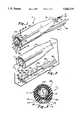
- FIG. 1is a perspective view of an endoscope having a sterilizable optical capsule section with a disposable separable tube assembly or disposable separable channel section;
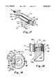
- FIG. 2is a fragmentary exploded view of the endoscope of FIG. 1;
- FIG. 3is an enlarged vertical section, taken along line 3--3 of FIG. 1;
- FIG. 4is a perspective view of the endoscope of FIG. 1 connected through a control member to suitable gas supplying and instrument supplying channels;
- FIG. 5is an enlarged vertical section, taken along line 5--5 of FIG. 4, showing the interior construction of the conduits for the capsule and the separable channel section;
- FIG. 6is a perspective view of a steerable electronic endoscope having a separable channel section
- FIG. 7is a perspective view showing the use of the invention in an umbilicated balloon catheter
- FIG. 8is an enlarged horizontal section, taken along line 8--8 of FIG. 7, showing details of the balloon construction
- FIG. 9is a perspective view of the endoscope of this invention used with an alternative umbilicated balloon catheter
- FIG. 10is an enlarged horizontal section, taken along line 10--10 of FIG. 9, showing details of the balloon construction
- FIG. 11is a perspective view of a telescopic catheter (also known as toposcopic catheter) used with the endoscope of this invention.
- FIG. 12is an enlarged longitudinal section, taken along line 12--12 of FIG. 11, showing details of the telescopic catheter;
- FIG. 13is a perspective view of an alternative endoscope having gas vents for guiding it along a bodily passageway;
- FIG. 14is an enlarged vertical section, taken along line 14--14 of FIG. 13, showing the gas passageways connected to the vents;
- FIG. 15is a section through a bodily passageway showing the catheter of FIG. 13 therein;
- FIG. 16is a section through the intestines of a patient showing the endoscope of this invention being drawn therealong by means of a strong magnet;
- FIG. 17is a fragmentary perspective view of another still further embodiment of the endoscope of this invention utilizing a prism for side viewing;
- FIG. 18is an enlarged end view of the endoscope of FIG. 17;
- FIG. 19is a fragmentary horizontal section, taken along line 19--19 of FIG. 18 showing further details of the endoscope construction
- FIG. 20is a fragmentary perspective view of another alternative embodiment of the endoscope wherein the separable channel section is magnetically attached;
- FIG. 21is an exploded view of the endoscope of FIG. 20;
- FIG. 22is an enlarged vertical section, taken along line 22--22 of FIG. 20;
- FIG. 23is a fragmentary perspective view of yet another embodiment wherein the sterilizable capsule and the separable channel section are held together by an elastic band;
- FIG. 24is an enlarged vertical section, taken along line 24--24 of FIG. 23;
- FIG. 25is a fragmentary exploded view of the endoscope of FIG. 23;
- FIG. 26is a perspective view of a prior art steerable endoscope having a removable sterile sheath
- FIG. 27is an enlarged section, taken along line 27--27 of FIG. 26, showing the construction of the sheath;
- FIG. 28is a perspective view of the prior art endoscope with the sheath in place, showing it being heat shrunk onto the endoscope;
- FIG. 29is an enlarged fragmentary horizontal section, taken along line 29--29 of FIG. 28, showing the heat shrunk sheath in place on the end of the endoscope;
- FIG. 30is a fragmentary exploded perspective view of the end of the endoscope of FIG. 26 with the heat shrunk sheath in place and showing the separable channel section and an elastic band for holding it in place;
- FIG. 31is a fragmentary perspective view of the apparatus of FIG. 30 when assembled.
- FIG. 32is an exploded fragmentary perspective view of the endoscope assembly being dissembled after use.
- a tethered endoscope Eis provided as best seen in FIGS. 1-4 which includes a sterilizable capsule C to which a disposable separable channel section S is releasably attached.
- Capsule Cwhich is easily sterilizable by heat in an autoclave, by gas sterilization or by dipping it in a disinfectant, has a window 10 at the distal end thereof.
- a lens system 12Positioned directly behind the window is a lens system 12 for focusing an image at a site under investigation on to an image receptor, such as CCD chip 14.
- image receptorssuch as a CCD chip 14.
- the CCD chipis the only electronic receptor of sufficiently small size to be placed in a capsule of the type contemplated by this invention.
- the capsuleis the size of a medicinal capsule, being 0.7 cm in diameter and 1.5 cm in length.
- the CCDis connected to electronic cable 16, shown in FIG. 1 which extend through a conduit 18 to a remote location, as will be more fully described below.
- the CCDis at least partially surrounded by optical fibers 20 for transmitting light to the operative site for viewing by means of the CCD 14. These optical fibers also extend through conduit 18.
- conduit 18will have a diameter of only 1/16 of an inch or smaller. This structure can be sterilized easily by immersing it in a disinfectant solution or by placing it in an autoclave for heat sterilization or by using ethylene oxide for gas sterilization.
- the separable channel section Sis removably connectable to capsule C in a fixed position and includes a channel member 22 having a plurality of channels, such as channels 24 and 26 for introduction of gas, such as carbon dioxide gas, or for drawing a suction to remove fluid or tissue from the operative site.
- a channel member 22having a plurality of channels, such as channels 24 and 26 for introduction of gas, such as carbon dioxide gas, or for drawing a suction to remove fluid or tissue from the operative site.
- one channelcould be used to direct fluid under pressure to clean the end of the optics to assure precise viewing.
- Another channelcould be used to direct a flow of warm gas over the optics for drying the optics.
- a larger central channel 28is provided through which an operative tool, such as tool 30 shown in FIG. 4, can be provided. Although the tool is illustrated as being forceps, it will be understood that other instruments may be utilized.
- separable channel section Shas a flat surface 32 which engages a flat surface 34 of capsule C.
- Channel section Sfurther includes a central longitudinally extending guide or key 36 which is engageable in a longitudinal guideway or keyway 38 formed in flat surface 34 on capsule C.
- the shape of capsule Cis defined by an exterior surface which includes surface 34, keyway 38 and outer edge 39.
- separable channel section Shas a shape defined by an exterior surface which includes flat 32, key 36 and arcuate outer edge 41.
- Central axis A--Ais provided showing the appropriate geometric center of the cross-sectional shapes wherein the attachment of the capsule C to the separable section S results in either the substantially circular (e.g. FIG. 1) or oval (FIG. 31) shape.
- a sterilized capsuleis attached to a new sterile separable channel section S whereupon endoscope E can be introduced into a passageway of the body of a patient by one of many methods to be described, so that suitable investigative and/or operative procedures can be undertaken.
- the endoscopecan be withdrawn. Section S is then separated from capsule C and thrown away and the capsule is sterilized by immersion in disinfectant or by placing it in an autoclave for heat sterilization or gas sterilization over an appropriate period of time. In this manner, the expensive optical portion of the endoscope E is preserved for multiple uses whereas the inexpensive, easily contaminated and difficult to sterilize separable channel section S can be separated from capsule C and thrown away.
- separable channel section Scan be provided with an additional channel 40 as shown for the introduction of a guide wire which has been previously positioned in the body of the patient by a catheter in a conventional manner.
- the capsulecan be placed on to the guide wire and slid therealong over channel 40 until it is also properly positioned.
- tubesare connected to the proximal end of section S which are in communication with the channels therein.
- a tube 42is connected to channel 24 and a tube 44 is connected to channel 26.
- a larger tube 46is connected to larger operating channel 28.
- Each tubecan be connected to a suitable connector, not shown, for introducing an appropriate fluid or instrument.
- conduit 18extends distally to a housing 50 to which it is releasably connected by fitting 52.
- the housing 50has a removable section 54 to which tubes 42, 44 and 46 are connected.
- a conduit 56is provided for connecting the light fibers to a suitable source of light through fitting 58.
- a conduit 60is provided for conducting the electrical signals to an image processing device through connector 62.
- a tube 64which is in further communication with tube 42 and is connected to a luer lock 66.
- a second tube 68is in fluid communication with tube 44 and is provided with a luer lock 70.
- a third tube 72is in communication with operating tube 46 and is adapted to receive an instrument, such as instrument 30, which has a handle 74 connected directly to it.
- This endoscopeincludes a barrel 80 with light fibers 82, for providing light to a site under investigation, and electronic cables 84 attached to a CCD (not shown) for transmitting an image which is viewed through lens 86.
- light fibers 82are connected through a fitting 88 to a light source 90.
- the CCDis connected to a camera 92, having controls 94, and supplying an image through conduit 96 to a video control unit 97 and then to video monitor 98 via cable 99.
- the steering mechanism within barrel 80is controlled, as by knob 100 and may be of the type shown in my U.S. Pat. No.
- the imaging system of the steerable endoscopemay be a coherent optical fiber bundle instead of a CCD.
- a separable channel section 102is removably connected to the distal end of barrel 80 in the same manner in which separable section S is attached to capsule C in FIG. 2.
- Section 102has a plurality of longitudinal channels, such as three, which are connected to a vacuum hose 104, a fluid hose 106 and an instrument tube 108. These are connected to a proximal housing 110 which also is of similar construction to separable channel section S, shown in FIG. 2, and is releasably connected to barrel 80 in a similar manner.
- Housing 110includes longitudinal channels, one of which respectively communicates vacuum hose 104 with vacuum hose 112. That is, hose 112 is attached between the proximal end of housing 110 and a vacuum pump 114.
- Another channel in housing 110communicates fluid hose 106 with fluid hose 116 which is connected to a fluid pump 118.
- a third central channel in housing 110communicates instrument tube 108 with instrument tube 120 for receiving an operative instrument, such as forceps 122.
- steerable endoscope E'can be initially sterilized in a sterilizing solution or by heating it in an autoclave or by gas sterilization for a suitable period of time to kill any viruses or bacteria remaining on the endoscope from its previous use.
- the endoscopecan then be used in a conventional manner and, after use, separable channel section 102 can be separated from the endoscope and thrown away.
- the endoscope E'can be resterilized by putting it in an autoclave or by soaking it in a suitable disinfectant. Thereafter, endoscope E' can be reconnected to a new sterile separable channel section for reuse.
- endoscope Eis of very small size, being approximately the same size as a medicinal capsule. Thus, it can be swallowed and if necessary a metallic weight can be imbedded into the terminal end to aid in propulsion of the capsule through the gastrointestinal track.
- a second method of insertion and propulsionis by use of an umbilicated balloon catheter 130, of the type shown in FIGS. 7 and 8, in which endoscope E can be preloaded. Preloading is of great help to the endoscopist because it saves him or her the time and trouble of inserting the devices over long distances inside exceptionally long operating channels.
- conduit 18 as well as tubes 42, 44 and 46pass through a central passageway 132 in the catheter and are attached to a connector 134 which has an optics portion 136 and a separable tube connector 138 which can be attached to the appropriate instrumentation, as previously described.
- the distal end of catheter 130is provided with an inflatable balloon 140 which can be used to center the catheter in a body passageway 142 as best seen in FIG. 8.
- the distal end of the endoscope Eis positioned within the distal end of the balloon 140 so that when the catheter 130 is introduced through a body passage, the distal end of the balloon 140 acts as a cushion and the endoscope E does not come into contact with body tissue.
- the balloonprovides a very safe non-traumatic guide when used in the bowel or colon. When lubricated, the balloon offers little resistance to the passage of the catheter to its desired location.
- the balloonis inflated through a tube 144 which is connected to a gas supply tube 146 whose proximal end is connected to a suitable connector 148.
- the umbilicated balloon catheter 130can be provided with an additional channel 150 for introduction of a guide wire (not shown) to introduce umbilicated catheter 130 through a trochar (not shown) to the desired location within a patient's body in a conventional manner.
- FIGS. 9 and 10Another umbilicated balloon catheter 152 is shown in FIGS. 9 and 10 wherein the balloon 154 has a generally donut configuration, because it has less contact surface with the passageway it can be more easily manipulated therealong. As with the catheter 130 shown in FIGS. 7 and 8, the distal end of the balloon 154 extends beyond the distal end of the endoscope E to provide the necessary cushioning effect.
- the endoscope Ecan also be inserted by use of a telescopic catheter 160, as shown in FIGS. 11 and 12.
- the generally cylindrical housing 162is provided which has a distal cylindrical portion 164 to which an outer tubular wall is securely attached. This wall is folded back upon itself to create a distal end 168 and to form an inner tubular wall 170 which is fan folded within housing 162 to form a folded proximal end 172, as shown in FIG. 12.
- This catheter 160can be preloaded with endoscope E, having capsule C extending beyond the distal end thereof and conduit 18 and tubes 42, 44 and 46 extending through the inner tubular wall 170 and out the proximal end, as shown.
- Housing 162has a gas inlet 174 to which a gas supply hose 176 is connected.
- gasWhen gas is introduced under pressure, the distal end 168 of the catheter will be pushed in a distal direction, thereby moving capsule C in that same distal direction.
- the capsulecan be moved into its appropriate location by the expansion of telescopic catheter 160.
- FIGS. 13-15An alternative form of endoscope E" is illustrated in FIGS. 13-15.
- This endoscopehas a capsule C' with circumferentially and equally spaced fluid vents 176.
- a similar set of fluid vents 178are provided adjacent the proximal end thereof.
- Fluid vents 176are supplied with a gas or liquid through supply tubes 180, whereas fluid vents 178 are supplied with a gas or liquid through supply tubes 182.
- the fluidcan be selectively controlled through remote control valves (not shown) connected to supply tubes 180 and 182 so that jets of gas or liquid can be ejected selectively through any one of vents 176 or vents 178 in any combination so as to manipulate and guide the endoscope through the passageway.
- remote control valvesnot shown
- FIG. 16shows the use of endoscope E within the intestines of patient wherein the endoscope is moved along the desired path by a strong magnet 185.
- a strong magnet 185is one made of Samarium Cobalt which is available through the Hitachi Corporation of Midland, Mich. U.S.A.
- the endoscope housingis made of ferrous material.
- FIGS. 17-19A further embodiment is shown in FIGS. 17-19 wherein an endoscope E a is provided which has a capsule C a with a prism therein for viewing at right angles the instruments introduced through separable section S.
- capsule C ahas a CCD chip 190 mounted adjacent the distal end thereof for receiving an image projected through a lens system 192 mounted distally thereof.
- Attached to the lens systemis a prism 194 for viewing an image projected through a window 196, as shown.
- Optical fibers 198surround the CCD and project light onto the operative site by means of prism 194.
- Separable channel section Sis identical to section S of FIG. 1. It has a flat 32, which engages a flat surface 200 on capsule C a , and a central longitudinally extending guide or key 36 which is engageable in a longitudinal guideway or keyway 202 formed in flat surface 200 on capsule C a .
- Guideway 36can be provided with an additional channel 40 for introduction of a guide wire, as best seen in FIG. 18.
- an endoscope E bhas a sterilizable capsule C b to which a separable section S b is attached by means of a magnetic strip.
- Capsule C bis substantially identical to capsule C of FIG. 1.
- the capsuleincludes a CCD chip 208 having a lens 210 for focusing an image at the operative site thereon.
- Light transmitting fibers 212are positioned around the CCD and lens structure for directing light to the operative site.
- Separable channel section S bhas fluid passageways 214 and 216 with an instrument passageway 215 of larger size therebetween. Passageway 214 is connected to a tube 218; passageway 216 is connected to tube 220 and instrument passageway 215 is connected to tube 222.
- capsule of C bis provided with a ferrous metal plate 226 having depending flanges 228 on opposite sides thereof.
- Separable section S bhas a magnetic strip 230 which is held in place against plate 228 by magnetic attraction and is sized to fit within the flanges 228 to hold the separable section in a fixed but removable position.
- FIGS. 23-25Another embodiment is shown in FIGS. 23-25 wherein an endoscope E c has a capsule C c and a separable channel section S c which are held together by an elastic band.
- Capsule C cis substantially identical to capsule C b in the previous embodiment except that it includes a recess 234 around its periphery.
- Section S calso has a corresponding recess 236 around its periphery for receiving and positioning an elastic band 238, thereby holding capsule C c and Section S c together in a fixed position during use.
- FIGS. 26-32A final embodiment is shown in FIGS. 26-32 wherein a conventional prior art steerable endoscope E d is provided with a sterile sheath 240 which is made of heat shrinkable material which is transparent and has a pull string 242 extending substantially the full length of the sheath for ripping it open after use, as will be explained more fully below.
- the steerable endoscope E dhas a flexible body portion 244 whose proximal end is connected to a handle 246 having controls 248 and 250 for manipulating the distal end 252. It also includes one or more channels, not shown, which are used for passage of fluids and instruments when the endoscope is used in the conventional manner.
- the sterile sheath 240is placed over the end of the endoscope and drawn up over body 244, thereby covering the channels and body, and heat is applied thereto, as by a forced air dryer 254, which causes the sheath 240 to seal tightly around endoscope body 244.
- the sheath 240is transparent so as not to interfere with light or image transmission through the encased endoscope.
- a separable section S dis attached to the distal end 252 of endoscope E d over the sheath 240 and is fixedly held in place, as by a sterile elastic band 256.
- Separable section S dis similar to the separable sections of the previous embodiments except that it has a concave surface 258 which mates with the outer surface 39 of distal end 252 of endoscope E d .
- the apparatusWhen assembled, the apparatus will have the configuration shown in FIG. 31 whereupon it can be inserted through a body passageway and used for its intended purpose.
- band 256is removed, as shown in FIG.
- the sheath 240is removed by pulling on string 242 which longitudinally rips the sheath apart so that it can be separated from endoscope E d .
- the present applicationcan be adapted for use with conventional endoscopes without modification thereof. This provides a means and method of reusing a conventional endoscope without encountering the expense, time delay and uncertainty associated with conventional resterilization procedures.
- a steerable endoscopehas been illustrated and described, the sheath can be used also with a conventional rigid endoscope.
- An endoscopehas been provided which is of very small size having a sterilizable optical section and a disposable sterile separable channel section which is removably but fixedly attached to the capsule so that after initial use, the separable channel section can be separated from the optical section and thrown away.
- the capsulethen is resterilized by heating it in an autoclave or by immersing it in a sterilizing solution for a suitable length of time. After the capsule is resterilized, a new sterile separable channel section can be attached to the resterilized capsule and the endoscope reused.
- the devicecan be end viewing or, by use of a prism, side viewing.
- a similar structurecan be provided on a steerable endoscope wherein the channel section is separable from the capsule which is steerable. Additionally, the endoscope, because of its small size can be introduced into the body by swallowing since it is no larger than a medicinal capsule. Other ways of introducing the device are by umbilicated balloon catheters which offer almost no resistance in bodily passageways and serve to center the catheter within the passageway and protect the body tissue. Another alternative is the use of a telescopic catheter for placement of the endoscope in the desired location. The endoscope can also be provided with fluid vents which are selectively supplied with fluid to steer the endoscope as it is moved into a bodily passageway.
- a strong magnetcan be used outside the bodily passageway for pulling the endoscope, having a ferrous housing, into the desired location.
- a separable channel sectioncan be fixedly attached to the capsule by an elastic band or magnetically.
- the separable channel sectioncan be used with a conventional endoscope which has been sealed in a sterile sheath.
Landscapes
- Health & Medical Sciences (AREA)
- Life Sciences & Earth Sciences (AREA)
- Surgery (AREA)
- Nuclear Medicine, Radiotherapy & Molecular Imaging (AREA)
- Biomedical Technology (AREA)
- Optics & Photonics (AREA)
- Pathology (AREA)
- Radiology & Medical Imaging (AREA)
- Biophysics (AREA)
- Engineering & Computer Science (AREA)
- Physics & Mathematics (AREA)
- Heart & Thoracic Surgery (AREA)
- Medical Informatics (AREA)
- Molecular Biology (AREA)
- Animal Behavior & Ethology (AREA)
- General Health & Medical Sciences (AREA)
- Public Health (AREA)
- Veterinary Medicine (AREA)
- Endoscopes (AREA)
Abstract
Description
This application is a continuation-in-part of U.S. Patent application Ser. No. 08/333,360 filed Nov. 2, 1994, now U.S. Pat. No. 5,489,256, which is a continuation of U.S. patent application Ser. No. 07/938,629 filed Sep. 1, 1992, now abandoned.
This invention relates to a sterilizable and/or "fluid immersable" endoscope and particularly to a tethered endoscope with a separable disposable tube assembly which can be replaced after each use and resterilization of the endoscope.
In recent years the popularity of endoscopic surgery has proliferated. This occurred because of the advances in technology which allow smaller and smaller endoscopes to be used, thereby permitting operative procedures to be undertaken in a less invasive manner for the patient than was previously possible. Thus, the patient suffers less trauma, recuperates much more rapidly and experiences less pain and discomfort than with more conventional surgical procedures.
Because of the sophisticated optics and electro-optics contained in modern endoscopes, they generally are very expensive. In order for this expense to be justified, they must be reused with a large number of patients.
Of course, multiple use means that the endoscope must be sterilized or at least disinfected after use with each patient prior to use with the next patient. One protocol for sterilization involves immersing the endoscope in a disinfectant solution for a predetermined period of time. It is also important to flush the channels which carry gases or fluids and those channels which are used for receiving operative instruments. Another protocol is to heat sterilize the endoscope by placing it in an autoclave. However, the optics and electronics of many endoscopes will not permit them to be subjected to heat sterilization. When using the disinfectant, sometimes the endoscope is not placed in the disinfecting solution for a sufficient length of time nor are the channels flushed out completely, because of the urgency to get the endoscope back into service as soon as possible. Over time, the disinfectant solution may loose some of its strength, thereby limiting its effectiveness.
Because of these shortcomings, many studies indicate that transmission of infectious diseases often occurs from one patient to another. By way of example, transmission of salmonella typhi has been reported. In addition, pseudomonas aeruginosa has been linked to improperly sterilized endoscopes. Also, an outbreak of serratia marcescens has been associated with the use of improperly sterilized bronchoscopes. Furthermore, hepatitis B has been transmitted by endoscopes when the endoscopes were processed in an inappropriate manner between patients. Finally, with respect to endoscope used on AID's patients, it has been found that the sterilizing procedures have not always removed contamination of the human immunodeficiency virus (HIV). This list is not exhaustive by any means.
A high-level of disinfection failures among gastrointestinal endoscopes have been noted, as well as failures in bronchoscopes, laryngoscopes and other devices. This may be due to the fact that they are long and narrow and have long and narrow channels which are difficult to clean.
From the foregoing, it is apparent that endoscopes which can be more easily and effectively sterilized are needed.
In accordance with the present invention, an improved endoscope in one configuration has been provided which has a sterilizable optical section and a disposable or throwaway separable channel section or tube assembly. The endoscope includes an elongated capsule, of the size of a medicinal capsule. The capsule includes a substantially cylindrical housing with a transparent window at the distal end thereof, for containing the endoscope optics. An image sensor is mounted adjacent the window within the capsule. An image transmitting cable with multiple conductors each has a distal end connected to the image sensor circuit board and a proximal end connected to a video control unit. From the video control unit signals are transmitted to the video monitor which displays the image in black and white or color. Light transmitting fibers each have a distal end adjacent to the window within the capsule and extends proximally from the capsule for transmitting light to a site under investigation from a remote light source. The separable channel section is removably attached to the capsule in fixed relationship and has at least one longitudinal channel for transmitting fluids or for receiving an operative instrument. A flexible tube is correspondingly connected to the proximal end of the channel for supplying fluid or for manipulating the operative instrument from a remote location. The separable channel section is disposable after use on a patient and the capsule is sterilizable for reuse with another separable channel section on the next patient.
More particularly, the image transmitting filaments and the light transmitting fibers are housed in a common conduit connected to the proximal end of the capsule. Conveniently, the capsule may have a flat relief (i.e., a first substantially planar mating surface) which is engageable with a flat side (i.e., a second substantially planar mating surface) on the separable channel section. The separable section further includes a cylindrical section along its exterior surface with substantially the same radius as the radius of the housing, so that when the capsule and the separable channel section are fastened together, they form the complete cylinder having a substantially circular cross-sectional shape. They may also be configured to have an elliptical cross-sectional shape. The separable channel section may include a plurality of longitudinal channels which comprises at least one fluid transmitting channel and at least one operative channel connected to corresponding tubes for supplying gases or operative instruments from a remote location.
Other means of attachment of the disposable channel section can be utilized. This can include a magnetic attachment or the use of a very strong elastic band. Those schooled in the construction of such disposable tubing devices may envision other methods of holding the separable channel section in the releasable, yet fixed, relationship to the capsule.
This same inventive concept can be used with a conventional endoscope, such as a rigid endoscope or a steerable endoscope having a sterilizable steerable capsule, and a separable channel section releasably connected in a fixed position to the capsule. In this structure, the separable channel section includes a distal housing releasably connected to the distal end of the capsule and a proximal housing releasably connected to the proximal end of the capsule with tubes interconnecting longitudinal channels in each housing. These channels and interconnecting tubes provide passageways through which gases or instruments can be passed.
In another form of the invention, an umbilicated balloon catheter having a tubular wall with an inflatable balloon adjacent the distal end thereof receives the capsule portion of the endoscope of this invention therein so that the capsule is positioned within the distal end of the inflatable balloon so that the balloon is in contact with the body channel that it is passing through rather than the distal end of the endoscope. An inflation conduit is connected to the capsule and extends through the catheter so that the balloon can be inflated while in the body channel or in the passageway. The balloon can either be donut shaped or elongated. Also, the tubular wall of the umbilicated balloon catheter can have a plurality of longitudinal channels for introducing any one of a variety of tools to the site under investigation.
In another form of the invention, a telescopic catheter can be used which has an outer tubular wall folded back upon itself at its distal end. The folded portion of the catheter forms an inner tubular wall telescoped within the outer wall and within a cylindrical housing for storage before expansion. The capsule portion of the endoscope is placed just distally beyond the distal end of the telescopic catheter. The cylindrical housing has means for admitting gas under pressure into the space between the inner and outer tubular walls to expand the catheter so that the distal end moves in a distal direction to push the capsule in the distal direction. This device is generally referred to as a toposcopic catheter developed by the National Institute of Health (NIH).
In a still further embodiment, the capsule can be provided with at least three gas vents equally spaced around the periphery thereof for selectively receiving gas from a gas delivery tube to guide the capsule through the passageway. Alternatively, a strong magnet can be used for manipulating the capsule along a passageway with the magnet located exteriorly thereof.
In another embodiment, the capsule may be configured into a side viewing device, with the optical window facing sideways. Here the image sensor is positioned behind the prism, and the detachable and disposable channel section is stair-stepped to one side and behind the laterally facing window.
By utilizing the endoscope of this invention, a novel method is provided wherein a sterile disposable tube assembly or channel section is attached to a sterilized endoscope having suitable optics for transmitting light to a site under investigation and reproducing an image of the site at a remote location. The endoscope is inserted with the attached tube assembly into a passageway to the desired site. The site is investigated through the endoscope and the necessary operative procedures are carried out through the tube assembly. The endoscope is then removed from the passageway whereupon the separable channel section is separated from the endoscope and thrown away and the endoscope is sterilized. After sterilization a new sterile separable channel section is attached to the sterilized endoscope. This greatly minimized possible cross infection between patients.
Existing endoscopes with built-in operating channels, optics and electronics can be modified for use with a separable channel section. This is a accomplished by encapsulating the entire steerable endoscope in a translucent sterile covering of thin, heat shrinkable plastic tubing closed at the distal end. This condom shaped covering can be placed over the endoscope and heat shrunk down to a very tight fitting covering encasing and sealing all of the endoscope including the hard to clean operating channels therein. This effectively isolates the endoscope in a sterile sheath and bars any cross contamination to the patient from the endoscope. The separable channel section is then mounted onto the sheath enclosed steerable endoscope adjacent the distal end thereof with an elastic band holding it in proper position. The separable channel section has a concave mating surface to fit snugly around the rounded body of the steerable endoscope which serves as a complementary convex mating surface. The sheath has incorporated therein a nylon string extending longitudinally therealong which allows easy tearing of the sheath after use for removal. A new sterile sheath can be heat shrunk onto the endoscope for use on the next patient.
The endoscope of this invention, because of its very small size substantially reduces the size and weight compared to conventional devices resulting in considerably more comfort for the patient. One of the most traumatic experiences for patients is the insertion of very large diameter and very heavy endoscopes into the colon, stomach or bronchial tree.
With this last-described apparatus, a method has been provided for utilizing a separable channel section on a conventional endoscope. The method includes placing a sterile heat shrinkable sheath on the body of the endoscope and applying heat to the sheath to shrink it into sealing engagement with the endoscope body. The separable channel section is releasably attached to the distal end of the endoscope body around the sheath in a fixed position. The operative site is investigated and the necessary, operative procedures are performed. The endoscope is then removed, the separable channel section is separated from the endoscope and thrown away and the sheath is removed from the endoscope and thrown away.
FIG. 1 is a perspective view of an endoscope having a sterilizable optical capsule section with a disposable separable tube assembly or disposable separable channel section;
FIG. 2 is a fragmentary exploded view of the endoscope of FIG. 1;
FIG. 3 is an enlarged vertical section, taken along line 3--3 of FIG. 1;
FIG. 4 is a perspective view of the endoscope of FIG. 1 connected through a control member to suitable gas supplying and instrument supplying channels;
FIG. 5 is an enlarged vertical section, taken alongline 5--5 of FIG. 4, showing the interior construction of the conduits for the capsule and the separable channel section;
FIG. 6 is a perspective view of a steerable electronic endoscope having a separable channel section;
FIG. 7 is a perspective view showing the use of the invention in an umbilicated balloon catheter;
FIG. 8 is an enlarged horizontal section, taken along line 8--8 of FIG. 7, showing details of the balloon construction;
FIG. 9 is a perspective view of the endoscope of this invention used with an alternative umbilicated balloon catheter;
FIG. 10 is an enlarged horizontal section, taken alongline 10--10 of FIG. 9, showing details of the balloon construction;
FIG. 11 is a perspective view of a telescopic catheter (also known as toposcopic catheter) used with the endoscope of this invention;
FIG. 12 is an enlarged longitudinal section, taken alongline 12--12 of FIG. 11, showing details of the telescopic catheter;
FIG. 13 is a perspective view of an alternative endoscope having gas vents for guiding it along a bodily passageway;
FIG. 14 is an enlarged vertical section, taken alongline 14--14 of FIG. 13, showing the gas passageways connected to the vents;
FIG. 15 is a section through a bodily passageway showing the catheter of FIG. 13 therein;
FIG. 16 is a section through the intestines of a patient showing the endoscope of this invention being drawn therealong by means of a strong magnet;
FIG. 17 is a fragmentary perspective view of another still further embodiment of the endoscope of this invention utilizing a prism for side viewing;
FIG. 18 is an enlarged end view of the endoscope of FIG. 17;
FIG. 19 is a fragmentary horizontal section, taken alongline 19--19 of FIG. 18 showing further details of the endoscope construction;
FIG. 20 is a fragmentary perspective view of another alternative embodiment of the endoscope wherein the separable channel section is magnetically attached;
FIG. 21 is an exploded view of the endoscope of FIG. 20;
FIG. 22 is an enlarged vertical section, taken alongline 22--22 of FIG. 20;
FIG. 23 is a fragmentary perspective view of yet another embodiment wherein the sterilizable capsule and the separable channel section are held together by an elastic band;
FIG. 24 is an enlarged vertical section, taken alongline 24--24 of FIG. 23;
FIG. 25 is a fragmentary exploded view of the endoscope of FIG. 23;
FIG. 26 is a perspective view of a prior art steerable endoscope having a removable sterile sheath;
FIG. 27 is an enlarged section, taken alongline 27--27 of FIG. 26, showing the construction of the sheath;
FIG. 28 is a perspective view of the prior art endoscope with the sheath in place, showing it being heat shrunk onto the endoscope;
FIG. 29 is an enlarged fragmentary horizontal section, taken alongline 29--29 of FIG. 28, showing the heat shrunk sheath in place on the end of the endoscope;
FIG. 30 is a fragmentary exploded perspective view of the end of the endoscope of FIG. 26 with the heat shrunk sheath in place and showing the separable channel section and an elastic band for holding it in place;
FIG. 31 is a fragmentary perspective view of the apparatus of FIG. 30 when assembled; and
FIG. 32 is an exploded fragmentary perspective view of the endoscope assembly being dissembled after use.
In accordance with one form of this invention, a tethered endoscope E is provided as best seen in FIGS. 1-4 which includes a sterilizable capsule C to which a disposable separable channel section S is releasably attached. Capsule C, which is easily sterilizable by heat in an autoclave, by gas sterilization or by dipping it in a disinfectant, has awindow 10 at the distal end thereof. Positioned directly behind the window is alens system 12 for focusing an image at a site under investigation on to an image receptor, such asCCD chip 14. It will be understood that other types of image receptors, such as a CID and MOS device or coherent optic fibers for guiding an image to a remotely located camera can be provided. However, under present technology, the CCD chip is the only electronic receptor of sufficiently small size to be placed in a capsule of the type contemplated by this invention. In this regard, the capsule is the size of a medicinal capsule, being 0.7 cm in diameter and 1.5 cm in length. Thus, it is small enough to be swallowed by a patient or otherwise introduced into small bodily passageways. The CCD is connected toelectronic cable 16, shown in FIG. 1 which extend through aconduit 18 to a remote location, as will be more fully described below. The CCD is at least partially surrounded byoptical fibers 20 for transmitting light to the operative site for viewing by means of theCCD 14. These optical fibers also extend throughconduit 18. Conveniently,conduit 18 will have a diameter of only 1/16 of an inch or smaller. This structure can be sterilized easily by immersing it in a disinfectant solution or by placing it in an autoclave for heat sterilization or by using ethylene oxide for gas sterilization.
Conveniently, the separable channel section S is removably connectable to capsule C in a fixed position and includes achannel member 22 having a plurality of channels, such aschannels central channel 28 is provided through which an operative tool, such astool 30 shown in FIG. 4, can be provided. Although the tool is illustrated as being forceps, it will be understood that other instruments may be utilized. Conveniently, separable channel section S has aflat surface 32 which engages a flat surface 34 of capsule C. Channel section S further includes a central longitudinally extending guide or key 36 which is engageable in a longitudinal guideway or keyway 38 formed in flat surface 34 on capsule C. In order to better illustrate how capsule C and separable section S are attached, it will be understood that the shape of capsule C is defined by an exterior surface which includes surface 34, keyway 38 andouter edge 39. Similarly, separable channel section S has a shape defined by an exterior surface which includes flat 32, key 36 and arcuateouter edge 41. Common to all the embodiments disclosed herein is that when capsule C attaches to section S, the exterior surfaces of each element form a substantially circular or oval cross section. Central axis A--A is provided showing the appropriate geometric center of the cross-sectional shapes wherein the attachment of the capsule C to the separable section S results in either the substantially circular (e.g. FIG. 1) or oval (FIG. 31) shape. In use, a sterilized capsule is attached to a new sterile separable channel section S whereupon endoscope E can be introduced into a passageway of the body of a patient by one of many methods to be described, so that suitable investigative and/or operative procedures can be undertaken.
Once the procedures are completed, the endoscope can be withdrawn. Section S is then separated from capsule C and thrown away and the capsule is sterilized by immersion in disinfectant or by placing it in an autoclave for heat sterilization or gas sterilization over an appropriate period of time. In this manner, the expensive optical portion of the endoscope E is preserved for multiple uses whereas the inexpensive, easily contaminated and difficult to sterilize separable channel section S can be separated from capsule C and thrown away.
Conveniently, separable channel section S can be provided with anadditional channel 40 as shown for the introduction of a guide wire which has been previously positioned in the body of the patient by a catheter in a conventional manner. Thus, after removal of the catheter over the guide wire, the capsule can be placed on to the guide wire and slid therealong overchannel 40 until it is also properly positioned.
Conveniently, tubes are connected to the proximal end of section S which are in communication with the channels therein. For example, atube 42 is connected to channel 24 and atube 44 is connected to channel 26. Similarly, alarger tube 46 is connected tolarger operating channel 28. Each tube can be connected to a suitable connector, not shown, for introducing an appropriate fluid or instrument.
As best seen in FIG. 4, an alternative arrangement is shown wherein theconduit 18 extends distally to ahousing 50 to which it is releasably connected by fitting 52. Conveniently, thehousing 50 has aremovable section 54 to whichtubes housing 50, aconduit 56 is provided for connecting the light fibers to a suitable source of light throughfitting 58. Similarly, aconduit 60 is provided for conducting the electrical signals to an image processing device throughconnector 62.
Similarly, connected to the back side ofremovable section 54 is a tube 64 which is in further communication withtube 42 and is connected to aluer lock 66. A second tube 68 is in fluid communication withtube 44 and is provided with a luer lock 70. Athird tube 72 is in communication withoperating tube 46 and is adapted to receive an instrument, such asinstrument 30, which has ahandle 74 connected directly to it.
The same inventive concepts just described can be applied to a steerable endoscope E', as best seen in FIG. 6. This endoscope includes abarrel 80 withlight fibers 82, for providing light to a site under investigation, andelectronic cables 84 attached to a CCD (not shown) for transmitting an image which is viewed throughlens 86. As illustrated,light fibers 82 are connected through a fitting 88 to alight source 90. The CCD is connected to acamera 92, having controls 94, and supplying an image through conduit 96 to avideo control unit 97 and then to video monitor 98 viacable 99. The steering mechanism withinbarrel 80 is controlled, as byknob 100 and may be of the type shown in my U.S. Pat. No. 5,325,845 entitled "Steerable Sheath For Use With Selected Removable Optical Catheter", or any other well-known steering device. In an alternative configuration, the imaging system of the steerable endoscope may be a coherent optical fiber bundle instead of a CCD.
As seen in FIG. 6, aseparable channel section 102 is removably connected to the distal end ofbarrel 80 in the same manner in which separable section S is attached to capsule C in FIG. 2.Section 102 has a plurality of longitudinal channels, such as three, which are connected to avacuum hose 104, afluid hose 106 and aninstrument tube 108. These are connected to aproximal housing 110 which also is of similar construction to separable channel section S, shown in FIG. 2, and is releasably connected tobarrel 80 in a similar manner.Housing 110 includes longitudinal channels, one of which respectively communicatesvacuum hose 104 with vacuum hose 112. That is, hose 112 is attached between the proximal end ofhousing 110 and a vacuum pump 114. Another channel inhousing 110 communicatesfluid hose 106 withfluid hose 116 which is connected to afluid pump 118. A third central channel inhousing 110 communicatesinstrument tube 108 with instrument tube 120 for receiving an operative instrument, such asforceps 122.
As with endoscope E, steerable endoscope E' can be initially sterilized in a sterilizing solution or by heating it in an autoclave or by gas sterilization for a suitable period of time to kill any viruses or bacteria remaining on the endoscope from its previous use. The endoscope can then be used in a conventional manner and, after use,separable channel section 102 can be separated from the endoscope and thrown away. The endoscope E' can be resterilized by putting it in an autoclave or by soaking it in a suitable disinfectant. Thereafter, endoscope E' can be reconnected to a new sterile separable channel section for reuse.
Various methods are available for introducing endoscope E into the body. As previously explained, it is of very small size, being approximately the same size as a medicinal capsule. Thus, it can be swallowed and if necessary a metallic weight can be imbedded into the terminal end to aid in propulsion of the capsule through the gastrointestinal track.
A second method of insertion and propulsion is by use of anumbilicated balloon catheter 130, of the type shown in FIGS. 7 and 8, in which endoscope E can be preloaded. Preloading is of great help to the endoscopist because it saves him or her the time and trouble of inserting the devices over long distances inside exceptionally long operating channels. As can be seen in FIG. 7,conduit 18 as well astubes central passageway 132 in the catheter and are attached to aconnector 134 which has anoptics portion 136 and aseparable tube connector 138 which can be attached to the appropriate instrumentation, as previously described. The distal end ofcatheter 130 is provided with aninflatable balloon 140 which can be used to center the catheter in abody passageway 142 as best seen in FIG. 8. As shown, the distal end of the endoscope E is positioned within the distal end of theballoon 140 so that when thecatheter 130 is introduced through a body passage, the distal end of theballoon 140 acts as a cushion and the endoscope E does not come into contact with body tissue. The balloon provides a very safe non-traumatic guide when used in the bowel or colon. When lubricated, the balloon offers little resistance to the passage of the catheter to its desired location. The balloon is inflated through atube 144 which is connected to a gas supply tube 146 whose proximal end is connected to asuitable connector 148. Theumbilicated balloon catheter 130 can be provided with anadditional channel 150 for introduction of a guide wire (not shown) to introduceumbilicated catheter 130 through a trochar (not shown) to the desired location within a patient's body in a conventional manner.
Anotherumbilicated balloon catheter 152 is shown in FIGS. 9 and 10 wherein theballoon 154 has a generally donut configuration, because it has less contact surface with the passageway it can be more easily manipulated therealong. As with thecatheter 130 shown in FIGS. 7 and 8, the distal end of theballoon 154 extends beyond the distal end of the endoscope E to provide the necessary cushioning effect.
The endoscope E can also be inserted by use of a telescopic catheter 160, as shown in FIGS. 11 and 12. The generallycylindrical housing 162 is provided which has a distal cylindrical portion 164 to which an outer tubular wall is securely attached. This wall is folded back upon itself to create adistal end 168 and to form an inner tubular wall 170 which is fan folded withinhousing 162 to form a folded proximal end 172, as shown in FIG. 12. This catheter 160 can be preloaded with endoscope E, having capsule C extending beyond the distal end thereof andconduit 18 andtubes Housing 162 has agas inlet 174 to which agas supply hose 176 is connected. When gas is introduced under pressure, thedistal end 168 of the catheter will be pushed in a distal direction, thereby moving capsule C in that same distal direction. Thus, the capsule can be moved into its appropriate location by the expansion of telescopic catheter 160.
An alternative form of endoscope E" is illustrated in FIGS. 13-15. This endoscope has a capsule C' with circumferentially and equally spaced fluid vents 176. A similar set offluid vents 178 are provided adjacent the proximal end thereof. Fluid vents 176 are supplied with a gas or liquid throughsupply tubes 180, whereas fluid vents 178 are supplied with a gas or liquid throughsupply tubes 182. When the endoscope E" is introduced into a bodily passageway, such aspassageway 184, shown in FIG. 15, the fluid can be selectively controlled through remote control valves (not shown) connected to supplytubes vents 176 orvents 178 in any combination so as to manipulate and guide the endoscope through the passageway.
FIG. 16 shows the use of endoscope E within the intestines of patient wherein the endoscope is moved along the desired path by a strong magnet 185. An example of such a magnet is one made of Samarium Cobalt which is available through the Hitachi Corporation of Midland, Mich. U.S.A. In this application, the endoscope housing is made of ferrous material.
A further embodiment is shown in FIGS. 17-19 wherein an endoscope Ea is provided which has a capsule Ca with a prism therein for viewing at right angles the instruments introduced through separable section S. In this regard, capsule Ca has aCCD chip 190 mounted adjacent the distal end thereof for receiving an image projected through alens system 192 mounted distally thereof. Attached to the lens system is aprism 194 for viewing an image projected through awindow 196, as shown.Optical fibers 198 surround the CCD and project light onto the operative site by means ofprism 194.
Separable channel section S is identical to section S of FIG. 1. It has a flat 32, which engages aflat surface 200 on capsule Ca, and a central longitudinally extending guide or key 36 which is engageable in a longitudinal guideway orkeyway 202 formed inflat surface 200 on capsule Ca. Guideway 36 can be provided with anadditional channel 40 for introduction of a guide wire, as best seen in FIG. 18.
Yet another embodiment is shown in FIGS. 20-22, wherein an endoscope Eb has a sterilizable capsule Cb to which a separable section Sb is attached by means of a magnetic strip. Capsule Cb is substantially identical to capsule C of FIG. 1. The capsule includes aCCD chip 208 having alens 210 for focusing an image at the operative site thereon.Light transmitting fibers 212 are positioned around the CCD and lens structure for directing light to the operative site. Separable channel section Sb hasfluid passageways instrument passageway 215 of larger size therebetween.Passageway 214 is connected to atube 218;passageway 216 is connected totube 220 andinstrument passageway 215 is connected totube 222. The mating surface of capsule of Cb is provided with aferrous metal plate 226 having dependingflanges 228 on opposite sides thereof. Separable section Sb has amagnetic strip 230 which is held in place againstplate 228 by magnetic attraction and is sized to fit within theflanges 228 to hold the separable section in a fixed but removable position.
Another embodiment is shown in FIGS. 23-25 wherein an endoscope Ec has a capsule Cc and a separable channel section Sc which are held together by an elastic band. Capsule Cc is substantially identical to capsule Cb in the previous embodiment except that it includes arecess 234 around its periphery. Section Sc also has acorresponding recess 236 around its periphery for receiving and positioning anelastic band 238, thereby holding capsule Cc and Section Sc together in a fixed position during use.
A final embodiment is shown in FIGS. 26-32 wherein a conventional prior art steerable endoscope Ed is provided with asterile sheath 240 which is made of heat shrinkable material which is transparent and has apull string 242 extending substantially the full length of the sheath for ripping it open after use, as will be explained more fully below. The steerable endoscope Ed has aflexible body portion 244 whose proximal end is connected to ahandle 246 havingcontrols distal end 252. It also includes one or more channels, not shown, which are used for passage of fluids and instruments when the endoscope is used in the conventional manner.
When the conventional prior art endoscope is to be used in accordance with this invention, thesterile sheath 240 is placed over the end of the endoscope and drawn up overbody 244, thereby covering the channels and body, and heat is applied thereto, as by a forcedair dryer 254, which causes thesheath 240 to seal tightly aroundendoscope body 244. Thesheath 240 is transparent so as not to interfere with light or image transmission through the encased endoscope.
Next, a separable section Sd is attached to thedistal end 252 of endoscope Ed over thesheath 240 and is fixedly held in place, as by a sterileelastic band 256. Separable section Sd is similar to the separable sections of the previous embodiments except that it has aconcave surface 258 which mates with theouter surface 39 ofdistal end 252 of endoscope Ed. When assembled, the apparatus will have the configuration shown in FIG. 31 whereupon it can be inserted through a body passageway and used for its intended purpose. After use,band 256 is removed, as shown in FIG. 32, and thesheath 240 is removed by pulling onstring 242 which longitudinally rips the sheath apart so that it can be separated from endoscope Ed. Thus, the present application can be adapted for use with conventional endoscopes without modification thereof. This provides a means and method of reusing a conventional endoscope without encountering the expense, time delay and uncertainty associated with conventional resterilization procedures. Although a steerable endoscope has been illustrated and described, the sheath can be used also with a conventional rigid endoscope.
From the foregoing, the advantages of this invention are readily apparent. An endoscope has been provided which is of very small size having a sterilizable optical section and a disposable sterile separable channel section which is removably but fixedly attached to the capsule so that after initial use, the separable channel section can be separated from the optical section and thrown away. The capsule then is resterilized by heating it in an autoclave or by immersing it in a sterilizing solution for a suitable length of time. After the capsule is resterilized, a new sterile separable channel section can be attached to the resterilized capsule and the endoscope reused. The device can be end viewing or, by use of a prism, side viewing. A similar structure can be provided on a steerable endoscope wherein the channel section is separable from the capsule which is steerable. Additionally, the endoscope, because of its small size can be introduced into the body by swallowing since it is no larger than a medicinal capsule. Other ways of introducing the device are by umbilicated balloon catheters which offer almost no resistance in bodily passageways and serve to center the catheter within the passageway and protect the body tissue. Another alternative is the use of a telescopic catheter for placement of the endoscope in the desired location. The endoscope can also be provided with fluid vents which are selectively supplied with fluid to steer the endoscope as it is moved into a bodily passageway. Finally, a strong magnet can be used outside the bodily passageway for pulling the endoscope, having a ferrous housing, into the desired location. In other embodiments, a separable channel section can be fixedly attached to the capsule by an elastic band or magnetically. Finally, the separable channel section can be used with a conventional endoscope which has been sealed in a sterile sheath.
This invention has been described in detail with reference to particular embodiments thereof, but it will be understood that various other modifications can be effected within the spirit and scope of this invention.
Claims (6)
1. An improved endoscope comprising:
a first segment comprising a substantially cylindrical capsule including distal and proximal ends and a first curved exterior surface, said capsule further including a transparent window at said distal end and a first substantially planar mating surface opposite said first exterior surface;
an image sensor mounted adjacent said window and within said first segment;
at least one image transmitting cable connectable to said image sensor and extending proximally from said first segment for transmitting an image signal from said image sensor to a video control unit and then to a remote image display device;
at least one light transmitting fiber having a distal end adjacent said window within said first segment and extending proximally from said first segment for transmitting light to a surgical site from a remote light source;
a second segment comprising a separable disposable channel section having at least one longitudinal opening for transmitting fluids or for receiving an operative instrument and further having a second curved exterior surface and a second substantially planar mating surface opposite said second exterior surface, said second substantially planar mating surface being releasably attachable to said first substantially planar mating surface of said capsule such that there is formed a substantially planar interface between said first and second segments;
an umbilicated balloon catheter having a tubular wall with an inflatable balloon adjacent a distal end thereof, said distal end of said balloon catheter being positioned distally beyond said distal end of said first segment; and
wherein said first and second segments, when attached along said first and second mating surfaces, together form a cylindrical endoscope having a substantially contiguous smooth curved exterior surface and a substantially circular cross-sectional shape, such that after use on a patient, said second segment is disposable while said first segment is sterilizable for reuse with a new second segment on another patient.
2. The endoscope, as claimed in claim 1, wherein:
said first substantially planar mating surface comprises a longitudinal keyway having a recessed portion, and said second substantially planar mating surface includes a longitudinal key which is engageable with said longitudinal keyway.
3. The endoscope, as claimed in claim 1, wherein:
said second segment is shaped such that said first segment and said second segment, when attached have a substantially common radius as defined from a central axis extending through the approximate geometric center of said endoscope.
4. The endoscope, as claimed in claim 1, further including:
at least one longitudinal opening extending longitudinally within said tubular wall in fluid communication with said balloon for inflation of said balloon to center said endoscope in a passageway of a patient into which said catheter has been introduced to protect the passageway when said catheter is moved along the passageway.
5. The endoscope, as claimed in claim 1, wherein:
said umbilicated balloon catheter has a substantially cylindrical shape.
6. The endoscope, as claimed in claim 1, wherein:
said umbilicated balloon catheter has a donut-like shape.
Priority Applications (1)
| Application Number | Priority Date | Filing Date | Title |
|---|---|---|---|
| US08/596,775US5643175A (en) | 1992-09-01 | 1996-02-05 | Sterilizable endoscope with separable disposable tube assembly |
Applications Claiming Priority (3)
| Application Number | Priority Date | Filing Date | Title |
|---|---|---|---|
| US93862992A | 1992-09-01 | 1992-09-01 | |
| US08/333,360US5489256A (en) | 1992-09-01 | 1994-11-02 | Sterilizable endoscope with separable disposable tube assembly |
| US08/596,775US5643175A (en) | 1992-09-01 | 1996-02-05 | Sterilizable endoscope with separable disposable tube assembly |
Related Parent Applications (1)
| Application Number | Title | Priority Date | Filing Date |
|---|---|---|---|
| US08/333,360Continuation-In-PartUS5489256A (en) | 1992-09-01 | 1994-11-02 | Sterilizable endoscope with separable disposable tube assembly |
Publications (1)
| Publication Number | Publication Date |
|---|---|
| US5643175Atrue US5643175A (en) | 1997-07-01 |
Family
ID=26988688
Family Applications (1)
| Application Number | Title | Priority Date | Filing Date |
|---|---|---|---|
| US08/596,775Expired - Fee RelatedUS5643175A (en) | 1992-09-01 | 1996-02-05 | Sterilizable endoscope with separable disposable tube assembly |
Country Status (1)
| Country | Link |
|---|---|
| US (1) | US5643175A (en) |
Cited By (208)
| Publication number | Priority date | Publication date | Assignee | Title |
|---|---|---|---|---|
| US5868667A (en)* | 1998-03-27 | 1999-02-09 | Ethicon, Inc. | Pressure-equalizing cap |
| US5891013A (en)* | 1996-02-07 | 1999-04-06 | Pinotage, Llc | System for single-puncture endoscopic surgery |
| US5980450A (en)* | 1997-05-07 | 1999-11-09 | Pinotage, Llc | Coupling device for use in an imaging system |
| WO2000048506A1 (en)* | 1999-02-15 | 2000-08-24 | Herrmann Ingo F | Deformable fiberscope with a displaceable supplementary device |
| WO2000042900A3 (en)* | 1999-01-21 | 2000-11-02 | Vision Sciences Inc | Forming thin-walled elastic components from an elastomeric material |
| US6234958B1 (en)* | 1998-11-30 | 2001-05-22 | Medical Access Systems, Llc | Medical device introduction system including medical introducer having a plurality of access ports and methods of performing medical procedures with same |
| WO2001054564A1 (en)* | 2000-01-28 | 2001-08-02 | Xion Gmbh | Flexible endoscope |
| EP1142530A1 (en)* | 2000-03-21 | 2001-10-10 | Rainer Dr. Zotz | Pneumatic drive for an endoscope |
| US6305536B1 (en)* | 1998-12-17 | 2001-10-23 | Nix Company Ltd. | Method for covering a mouth insert portion of an intraoral camera with a film-made sheath and the combination thereof |
| US20010041843A1 (en)* | 1999-02-02 | 2001-11-15 | Mark Modell | Spectral volume microprobe arrays |
| US6346073B1 (en) | 1997-05-07 | 2002-02-12 | Pinotage, Llc | Imaging system and components thereof |
| US20020093484A1 (en)* | 2000-12-07 | 2002-07-18 | Michael Skala | Method and system for use of a pointing device with moving images |
| US20020109774A1 (en)* | 2001-01-16 | 2002-08-15 | Gavriel Meron | System and method for wide field imaging of body lumens |
| US20020197728A1 (en)* | 1999-12-15 | 2002-12-26 | Howard Kaufman | Methods of monitoring effects of chemical agents on a sample |
| US6530881B1 (en) | 1999-01-21 | 2003-03-11 | Vision Sciences, Inc. | Sheath apparatus for endoscopes and methods for forming same |
| WO2003015604A3 (en)* | 2001-08-16 | 2003-05-08 | Syntheon Llc | Apparatus for delivering instrument over endoscope |
| US6572536B1 (en) | 1999-11-05 | 2003-06-03 | Visionary Biomedical, Inc. | Autoclavable flexible fiberscope |
| US6582360B1 (en)* | 1999-05-19 | 2003-06-24 | Fuji Photo Optical Co., Ltd. | Tube connection structure in endoscope |
| WO2004021868A3 (en)* | 2002-09-06 | 2004-05-06 | Bard Inc C R | External endoscopic accessory control system |
| EP1426073A1 (en)* | 2002-12-05 | 2004-06-09 | Ethicon Endo-Surgery, Inc. | Catheter with extendable track |
| WO2004047626A1 (en)* | 2002-11-22 | 2004-06-10 | Jiang, Shoumei | Endoscope assembly with disposable sheath |
| US20040111019A1 (en)* | 2002-12-05 | 2004-06-10 | Long Gary L. | Locally-propelled, intraluminal device with cable loop track and method of use |
| EP1421894A3 (en)* | 2002-11-22 | 2004-07-28 | STM Medizintechnik Starnberg GmbH | Endoscope head |
| US6786864B2 (en)* | 2001-02-06 | 2004-09-07 | Olympus Corporation | Endoscopic system and method for positioning an indwelling tube |
| US20040206914A1 (en)* | 2003-04-18 | 2004-10-21 | Medispectra, Inc. | Methods and apparatus for calibrating spectral data |
| US20040206913A1 (en)* | 2003-04-18 | 2004-10-21 | Medispectra, Inc. | Methods and apparatus for characterization of tissue samples |
| US20040215058A1 (en)* | 2002-09-06 | 2004-10-28 | Zirps Christopher T | Endoscopic accessory mounting adaptor |
| EP1477105A1 (en)* | 2003-05-16 | 2004-11-17 | Ethicon Endo-Surgery | Endcap for use with an endoscope |
| US20040230096A1 (en)* | 2003-05-16 | 2004-11-18 | David Stefanchik | Method of guiding medical devices |
| US20040249247A1 (en)* | 2003-05-01 | 2004-12-09 | Iddan Gavriel J. | Endoscope with panoramic view |
| US20050004474A1 (en)* | 2001-01-16 | 2005-01-06 | Iddan Gavriel J. | Method and device for imaging body lumens |
| US20050004434A1 (en)* | 2003-05-05 | 2005-01-06 | Konstantin Bob | Endoscope shaft |
| US6847490B1 (en) | 1997-01-13 | 2005-01-25 | Medispectra, Inc. | Optical probe accessory device for use in vivo diagnostic procedures |
| WO2005032354A1 (en)* | 2003-10-06 | 2005-04-14 | Romeiro Sapienza Jose Carlos | An additional conductor for a bronchofibroscope and a conductor-bronchofibroscope assembly |
| US20050085694A1 (en)* | 2003-10-16 | 2005-04-21 | Nakao Naomi L. | Endoscope with open channels |
| US20050171398A1 (en)* | 2002-12-26 | 2005-08-04 | Given Imaging Ltd. | In vivo imaging device and method of manufacture thereof |
| US6933154B2 (en) | 2002-07-09 | 2005-08-23 | Medispectra, Inc. | Optimal windows for obtaining optical data for characterization of tissue samples |
| US6939290B2 (en) | 2002-02-11 | 2005-09-06 | Given Imaging Ltd | Self propelled device having a magnetohydrodynamic propulsion system |
| US6958034B2 (en) | 2002-02-11 | 2005-10-25 | Given Imaging Ltd. | Self propelled device |
| US20050256504A1 (en)* | 2004-05-14 | 2005-11-17 | Ethicon Endo-Surgery, Inc. | Medical instrument having a catheter and a medical guidewire |
| US20050259942A1 (en)* | 2004-04-08 | 2005-11-24 | Burak Temelkuran | Photonic crystal fibers and medical systems including photonic crystal fibers |
| US20050259934A1 (en)* | 2004-04-08 | 2005-11-24 | Burak Temelkuran | Photonic crystal fibers and medical systems including photonic crystal fibers |
| WO2005122866A1 (en)* | 2004-06-21 | 2005-12-29 | Korea Institute Of Science And Technology | Capsule type endoscope control system |
| US20060004276A1 (en)* | 2004-06-30 | 2006-01-05 | Iddan Gavriel J | Motor for an in-vivo device |
| US20060015013A1 (en)* | 2004-06-30 | 2006-01-19 | Zvika Gilad | Device and method for in vivo illumination |
| WO2006027136A1 (en)* | 2004-09-07 | 2006-03-16 | Christian Thiel | Modular endoscope |
| US20060074274A1 (en)* | 2002-10-15 | 2006-04-06 | Friedman Marc D | Medical device sheath apparatus and method of making and using same |
| US20060082648A1 (en)* | 2000-03-08 | 2006-04-20 | Given Imaging Ltd. | Device and system for in vivo imaging |
| WO2006063497A1 (en)* | 2004-12-14 | 2006-06-22 | Kerang Jiang | A endoscope assembly with a disposable sheath |
| WO2006059321A3 (en)* | 2004-11-30 | 2006-07-06 | Sphereview Ltd | A compact gynecological observation system for examination, imaging, image analysis and treatment |
| US20060184039A1 (en)* | 2001-07-26 | 2006-08-17 | Dov Avni | Apparatus and method for light control in an in-vivo imaging device |
| US20060195014A1 (en)* | 2005-02-28 | 2006-08-31 | University Of Washington | Tethered capsule endoscope for Barrett's Esophagus screening |
| US7127282B2 (en) | 1998-12-23 | 2006-10-24 | Medispectra, Inc. | Optical methods and systems for rapid screening of the cervix |
| US7136518B2 (en) | 2003-04-18 | 2006-11-14 | Medispectra, Inc. | Methods and apparatus for displaying diagnostic data |
| US20060258908A1 (en)* | 2005-05-13 | 2006-11-16 | David Stefanchik | Sheath for use with an endoscope |
| US20060258907A1 (en)* | 2005-05-13 | 2006-11-16 | David Stefanchik | Track for medical devices |
| US20060258910A1 (en)* | 2005-05-13 | 2006-11-16 | David Stefanchik | Method of positioning a device on an endoscope |
| US20060258904A1 (en)* | 2005-05-13 | 2006-11-16 | David Stefanchik | Feeding tube and track |
| US20060258903A1 (en)* | 2005-05-13 | 2006-11-16 | David Stefanchik | Method of inserting a feeding tube |
| US20060280258A1 (en)* | 2005-06-14 | 2006-12-14 | Ido Bettesh | Modulator and method for producing a modulated signal |
| US7187810B2 (en) | 1999-12-15 | 2007-03-06 | Medispectra, Inc. | Methods and systems for correcting image misalignment |
| US7189247B1 (en) | 2002-09-06 | 2007-03-13 | Conmed Endoscopic Technologies, Inc. | Endoscopic band ligator |
| US20070078298A1 (en)* | 2003-07-02 | 2007-04-05 | Arkady Glukhovsky | Imaging sensor array and device and method for use thereof |
| US20070106113A1 (en)* | 2005-11-07 | 2007-05-10 | Biagio Ravo | Combination endoscopic operative delivery system |
| US20070106112A1 (en)* | 2003-12-24 | 2007-05-10 | Daniel Gat | Device, system and method for in-vivo imaging of a body lumen |
| US20070129624A1 (en)* | 2003-12-24 | 2007-06-07 | Zvika Gilad | Device, system and method for in-vivo imaging of a body lumen |
| US20070133002A1 (en)* | 2005-10-11 | 2007-06-14 | Duke University | Systems and methods for endoscopic angle-resolved low coherence interferometry |
| US20070149850A1 (en)* | 2005-12-22 | 2007-06-28 | Spivey James T | Endoscope endcap attachment tool |
| US7260248B2 (en) | 1999-12-15 | 2007-08-21 | Medispectra, Inc. | Image processing using measures of similarity |
| US20070225694A1 (en)* | 2006-03-22 | 2007-09-27 | Ethicon Endo-Surgery, Inc. | Intubation device for enteral feeding |
| US20070225728A1 (en)* | 2006-03-22 | 2007-09-27 | Ethicon Endo-Surgery, Inc. | Intubation system for use with an endoscope |
| US20070229954A1 (en)* | 2006-04-03 | 2007-10-04 | Pourang Bral | Means and method to prevent liquids and flying debris from blocking the viewing pathway of an optical element |
| US20070239137A1 (en)* | 2006-03-30 | 2007-10-11 | Ethicon Endo-Surgery, Inc. | Intubation device for colonic decompression |
| US20070239171A1 (en)* | 2006-03-30 | 2007-10-11 | Ethicon Endo-Surgery, Inc. | Medical snaring device |
| US7282723B2 (en) | 2002-07-09 | 2007-10-16 | Medispectra, Inc. | Methods and apparatus for processing spectral data for use in tissue characterization |
| US20070255273A1 (en)* | 2006-04-29 | 2007-11-01 | Board Of Regents, The University Of Texas System | Devices for use in Transluminal and Endoluminal Surgery |
| US7310547B2 (en) | 2002-07-10 | 2007-12-18 | Medispectra, Inc. | Fluorescent fiberoptic probe for tissue health discrimination |
| US20080009672A1 (en)* | 2004-08-19 | 2008-01-10 | Beat Krattiger | Connector Unit For Endoscopes |
| US20080021276A1 (en)* | 2006-07-21 | 2008-01-24 | Oncoscope, Inc. | Protective probe tip, particularly for use on a fiber-optic probe used in an endoscopic application |
| US20080045863A1 (en)* | 2006-08-17 | 2008-02-21 | Ethicon Endo-Surgery, Inc. | Guidewire structure including a medical guidewire |
| US20080058679A1 (en)* | 2006-08-17 | 2008-03-06 | Ethicon Endo-Surgery, Inc. | Guidewire structure including a medical guidewire and method for using a medical instrument |
| US20080064920A1 (en)* | 2006-09-08 | 2008-03-13 | Ethicon Endo-Surgery, Inc. | Medical drive system for providing motion to at least a portion of a medical apparatus |
| US20080086157A1 (en)* | 2006-06-30 | 2008-04-10 | Depuy Spine, Inc. | Disc Nucleus Removal Devices and Methods |
| US20080097331A1 (en)* | 2006-09-05 | 2008-04-24 | Ethicon Endo-Surgery, Inc. | Guidewire structure including a medical guidewire and method for using |
| US20080132758A1 (en)* | 2006-12-05 | 2008-06-05 | Ethicon Endo-Surgery, Inc. | Independent Articulating Accessory Channel |
| US20080161647A1 (en)* | 2006-12-27 | 2008-07-03 | Amit Pascal | Device and method for multiple illumination fields of an in-vivo imaging device |
| US20080167521A1 (en)* | 2007-01-09 | 2008-07-10 | Sheetz Jane A | Method of in vivo monitoring using an imaging system including scanned beam imaging unit |
| US20080183035A1 (en)* | 2007-01-26 | 2008-07-31 | Ethicon Endo-Surgery, Inc. | Endoscopic Accessory Control Mechanism |
| US20080207999A1 (en)* | 2005-07-08 | 2008-08-28 | Klaus Abraham-Fuchs | Endoscopic Capsule |
| US20080221388A1 (en)* | 2007-03-09 | 2008-09-11 | University Of Washington | Side viewing optical fiber endoscope |
| US20080269779A1 (en)* | 2003-12-02 | 2008-10-30 | Board Of Regents, The University Of Texas System | Surgical anchor and system |
| US7469160B2 (en) | 2003-04-18 | 2008-12-23 | Banks Perry S | Methods and apparatus for evaluating image focus |
| US20090028407A1 (en)* | 2005-11-23 | 2009-01-29 | University Of Washington | Scanning beam with variable sequential framing using interrupted scanning resonance |
| US20090073456A1 (en)* | 2007-09-13 | 2009-03-19 | Duke University | Apparatuses, systems, and methods for low-coherence interferometry (lci) |
| US20090105532A1 (en)* | 2007-10-22 | 2009-04-23 | Zvika Gilad | In vivo imaging device and method of manufacturing thereof |
| US7527620B2 (en) | 2004-05-14 | 2009-05-05 | Ethicon Endo-Surgery, Inc. | Medical instrument having a medical guidewire |
| WO2009069837A1 (en)* | 2007-11-28 | 2009-06-04 | Top R & D Co., Ltd | Suction catheter for surgical |
| US20090143648A1 (en)* | 2007-11-30 | 2009-06-04 | Tyco Healthcare Group Lp | Endoscope system for gastrostomy catheter placement |
| US20090177094A1 (en)* | 2008-01-08 | 2009-07-09 | Oncoscope, Inc. | Systems and methods for tissue examination, diagnostic, treatment, and/or monitoring |
| WO2010004570A1 (en)* | 2008-07-10 | 2010-01-14 | Superdimension Ltd. | Integrated multi-functional endoscopic tool |
| US20100073512A1 (en)* | 2004-05-17 | 2010-03-25 | Alf Olsen | Real-time exposure control for automatic light control |
| EP2193715A1 (en) | 2007-04-25 | 2010-06-09 | Sygenta Participations AG. | Fungicidal Compositions |
| US20100160733A1 (en)* | 2002-04-17 | 2010-06-24 | Pinhas Gilboa | Endoscope Structures And Techniques For Navigating To A Target In Branched Structure |
| US7805178B1 (en) | 2005-07-25 | 2010-09-28 | Given Imaging Ltd. | Device, system and method of receiving and recording and displaying in-vivo data with user entered data |
| US20100268030A1 (en)* | 2009-04-16 | 2010-10-21 | Tyco Healthcare Group Lp | Self-Steering Endoscopic Device |
| US20100298640A1 (en)* | 2006-02-16 | 2010-11-25 | Vision - Sciences Inc. | Endoscope With Imaging Capsule |
| US7857754B2 (en) | 2005-05-13 | 2010-12-28 | Ethicon Endo-Surgery, Inc. | Apparatus useful for positioning a device on an endoscope |
| US7866322B2 (en) | 2002-10-15 | 2011-01-11 | Given Imaging Ltd. | Device, system and method for transfer of signals to a moving device |
| US7907986B2 (en) | 2001-09-24 | 2011-03-15 | Given Imaging Ltd. | System and method for controlling a device in vivo |
| US20110087224A1 (en)* | 2009-10-09 | 2011-04-14 | Cadeddu Jeffrey A | Magnetic surgical sled with variable arm |
| US7952718B2 (en) | 2007-05-03 | 2011-05-31 | University Of Washington | High resolution optical coherence tomography based imaging for intraluminal and interstitial use implemented with a reduced form factor |
| USRE42497E1 (en) | 2003-05-06 | 2011-06-28 | Duke University | Fourier domain low-coherence interferometry for light scattering spectroscopy apparatus and method |
| US20110160532A1 (en)* | 2009-12-29 | 2011-06-30 | Richard Wolf Gmbh | Endoscopic instrument |
| US20110157596A1 (en)* | 2005-10-11 | 2011-06-30 | Duke University | Systems and methods for endoscopic angle-resolved low coherence interferometry |
| US7998065B2 (en) | 2001-06-18 | 2011-08-16 | Given Imaging Ltd. | In vivo sensing device with a circuit board having rigid sections and flexible sections |
| US20110208308A1 (en)* | 2006-11-28 | 2011-08-25 | Columna Pty Ltd | Tissue prosthesis insertion system and method |
| WO2012013246A1 (en)* | 2010-07-30 | 2012-02-02 | Ethicon Endo-Surgery, Inc. | A system and method for submucosal tunneling of the gi tract for the diversion of bodily fluids |
| US8273015B2 (en) | 2007-01-09 | 2012-09-25 | Ethicon Endo-Surgery, Inc. | Methods for imaging the anatomy with an anatomically secured scanner assembly |
| US8382662B2 (en) | 2003-12-12 | 2013-02-26 | University Of Washington | Catheterscope 3D guidance and interface system |
| US8396535B2 (en) | 2000-06-19 | 2013-03-12 | University Of Washington | Integrated optical scanning image acquisition and display |
| US20130096385A1 (en)* | 2011-10-14 | 2013-04-18 | Intuitive Surgical Operations, Inc. | Vision probe and catheter systems |
| US8452068B2 (en) | 2008-06-06 | 2013-05-28 | Covidien Lp | Hybrid registration method |
| US8473032B2 (en) | 2008-06-03 | 2013-06-25 | Superdimension, Ltd. | Feature-based registration method |
| US8611984B2 (en) | 2009-04-08 | 2013-12-17 | Covidien Lp | Locatable catheter |
| US8663088B2 (en) | 2003-09-15 | 2014-03-04 | Covidien Lp | System of accessories for use with bronchoscopes |
| US8764725B2 (en) | 2004-02-09 | 2014-07-01 | Covidien Lp | Directional anchoring mechanism, method and applications thereof |
| US20140257417A1 (en)* | 2005-08-15 | 2014-09-11 | Spinecell Private Limited | Systems, methods and apparatuses for formation and insertion of tissue prosthesis |
| US8840566B2 (en) | 2007-04-02 | 2014-09-23 | University Of Washington | Catheter with imaging capability acts as guidewire for cannula tools |
| US8905920B2 (en) | 2007-09-27 | 2014-12-09 | Covidien Lp | Bronchoscope adapter and method |
| US9055881B2 (en) | 2004-04-26 | 2015-06-16 | Super Dimension Ltd. | System and method for image-based alignment of an endoscope |
| US9063299B2 (en) | 2009-12-15 | 2015-06-23 | Omni Guide, Inc. | Two-part surgical waveguide |
| US9113846B2 (en) | 2001-07-26 | 2015-08-25 | Given Imaging Ltd. | In-vivo imaging device providing data compression |
| US9149175B2 (en) | 2001-07-26 | 2015-10-06 | Given Imaging Ltd. | Apparatus and method for light control in an in-vivo imaging device |
| US9179831B2 (en) | 2009-11-30 | 2015-11-10 | King Systems Corporation | Visualization instrument |
| US9274001B2 (en) | 2010-01-22 | 2016-03-01 | Duke University | Dual window processing schemes for spectroscopic optical coherence tomography (OCT) and fourier domain low coherence interferometry |
| DE102007017517B4 (en)* | 2007-04-13 | 2016-03-10 | Siemens Aktiengesellschaft | Navigable endoscopy capsule |
| US20160143510A1 (en)* | 2013-08-08 | 2016-05-26 | Sharp Kabushiki Kaisha | In-body monitoring camera system and support tube for in-body monitoring-camera-system |
| US20160234408A1 (en)* | 2013-10-31 | 2016-08-11 | Sharp Kabushiki Kaisha | Intracorporeal-monitoring camera system, support tube for intracorporeal-monitoring camera system, and cable holder for intracorporeal-monitoring camera system |
| US9561078B2 (en) | 2006-03-03 | 2017-02-07 | University Of Washington | Multi-cladding optical fiber scanner |
| US9566115B2 (en) | 2009-07-28 | 2017-02-14 | Neuwave Medical, Inc. | Energy delivery systems and uses thereof |
| US9575140B2 (en) | 2008-04-03 | 2017-02-21 | Covidien Lp | Magnetic interference detection system and method |
| WO2017155130A1 (en)* | 2016-03-08 | 2017-09-14 | (의료)길의료재단 | Endoscope device having plurality of coupling parts, and cap connected to same |
| US9820642B2 (en) | 2007-08-04 | 2017-11-21 | King Systems Corporation | Airway intubation device |
| US9823127B2 (en) | 2010-01-22 | 2017-11-21 | Duke University | Systems and methods for deep spectroscopic imaging of biological samples with use of an interferometer and spectrometer |
| WO2017201424A1 (en)* | 2016-05-19 | 2017-11-23 | Metamodix, Inc. | Pyloric anchor retrieval tools and methods |
| US9861440B2 (en)* | 2010-05-03 | 2018-01-09 | Neuwave Medical, Inc. | Energy delivery systems and uses thereof |
| US10111767B2 (en) | 2010-10-29 | 2018-10-30 | Abbott Cardiovascular Systems Inc. | Sheaths used in polymer scaffold delivery systems |
| US10172669B2 (en) | 2009-10-09 | 2019-01-08 | Ethicon Llc | Surgical instrument comprising an energy trigger lockout |
| US10232147B2 (en) | 2011-05-27 | 2019-03-19 | Abbott Cardiovascular Systems Inc. | Method for assembling a scaffold-balloon catheter |
| US10314638B2 (en) | 2015-04-07 | 2019-06-11 | Ethicon Llc | Articulating radio frequency (RF) tissue seal with articulating state sensing |
| US10363092B2 (en) | 2006-03-24 | 2019-07-30 | Neuwave Medical, Inc. | Transmission line with heat transfer ability |
| US10376314B2 (en) | 2006-07-14 | 2019-08-13 | Neuwave Medical, Inc. | Energy delivery systems and uses thereof |
| CN110215180A (en)* | 2019-07-04 | 2019-09-10 | 上海英诺伟医疗器械有限公司 | Rigid endoscope device |
| US10418705B2 (en) | 2016-10-28 | 2019-09-17 | Covidien Lp | Electromagnetic navigation antenna assembly and electromagnetic navigation system including the same |
| US10426555B2 (en) | 2015-06-03 | 2019-10-01 | Covidien Lp | Medical instrument with sensor for use in a system and method for electromagnetic navigation |
| US10433996B2 (en) | 2014-03-13 | 2019-10-08 | Abbott Cardiovascular Systems Inc. | Striped sheaths for medical devices |
| US10446931B2 (en) | 2016-10-28 | 2019-10-15 | Covidien Lp | Electromagnetic navigation antenna assembly and electromagnetic navigation system including the same |
| US20190321076A1 (en)* | 2002-05-30 | 2019-10-24 | Intuitive Surgical Operations, Inc. | Catheter Systems with Imaging Assemblies |
| CN110446450A (en)* | 2017-03-17 | 2019-11-12 | 国立大学法人大阪大学 | Flexible endoscope and endoscopic system with the flexible endoscope |
| US10470907B2 (en) | 2011-06-21 | 2019-11-12 | Abbott Cardiovascular Systems Inc. | Sheaths used with polymer scaffolds |
| US10478254B2 (en) | 2016-05-16 | 2019-11-19 | Covidien Lp | System and method to access lung tissue |
| US10517505B2 (en) | 2016-10-28 | 2019-12-31 | Covidien Lp | Systems, methods, and computer-readable media for optimizing an electromagnetic navigation system |
| US10531917B2 (en) | 2016-04-15 | 2020-01-14 | Neuwave Medical, Inc. | Systems and methods for energy delivery |
| EP3610775A1 (en)* | 2018-08-17 | 2020-02-19 | Acclarent, Inc. | Endoscope with anatomy elevation assembly |
| US10582834B2 (en) | 2010-06-15 | 2020-03-10 | Covidien Lp | Locatable expandable working channel and method |
| US10603117B2 (en) | 2017-06-28 | 2020-03-31 | Ethicon Llc | Articulation state detection mechanisms |
| US10615500B2 (en) | 2016-10-28 | 2020-04-07 | Covidien Lp | System and method for designing electromagnetic navigation antenna assemblies |
| US10638952B2 (en) | 2016-10-28 | 2020-05-05 | Covidien Lp | Methods, systems, and computer-readable media for calibrating an electromagnetic navigation system |
| US10653866B2 (en) | 2011-10-14 | 2020-05-19 | Intuitive Surgical Operations, Inc. | Catheter with removable vision probe |
| US10667860B2 (en) | 2011-12-21 | 2020-06-02 | Neuwave Medical, Inc. | Energy delivery systems and uses thereof |
| US10682070B2 (en) | 2011-10-14 | 2020-06-16 | Intuitive Surgical Operations, Inc. | Electromagnetic sensor with probe and guide sensing elements |
| US10722311B2 (en) | 2016-10-28 | 2020-07-28 | Covidien Lp | System and method for identifying a location and/or an orientation of an electromagnetic sensor based on a map |
| US10744303B2 (en) | 2011-10-14 | 2020-08-18 | Intuitive Surgical Operations, Inc. | Catheters with control modes for interchangeable probes |
| US10743750B2 (en) | 2014-04-28 | 2020-08-18 | Massachusetts Institute Of Technology | Multi-link modular continuum robotic endoscope system |
| US10751109B2 (en) | 2014-12-22 | 2020-08-25 | Ethicon Llc | High power battery powered RF amplifier topology |
| US10751117B2 (en) | 2016-09-23 | 2020-08-25 | Ethicon Llc | Electrosurgical instrument with fluid diverter |
| US10750932B1 (en)* | 2019-06-11 | 2020-08-25 | Steris Instrument Management Services, Inc. | Method of assembling endoscope optical components |
| US10751126B2 (en) | 2016-10-28 | 2020-08-25 | Covidien Lp | System and method for generating a map for electromagnetic navigation |
| US10779876B2 (en) | 2011-10-24 | 2020-09-22 | Ethicon Llc | Battery powered surgical instrument |
| US10792106B2 (en) | 2016-10-28 | 2020-10-06 | Covidien Lp | System for calibrating an electromagnetic navigation system |
| US10799284B2 (en) | 2017-03-15 | 2020-10-13 | Ethicon Llc | Electrosurgical instrument with textured jaws |
| US20200352598A1 (en)* | 2019-05-12 | 2020-11-12 | EvolutionMedVentures LLC | Add-on sheath |
| US10856934B2 (en) | 2016-04-29 | 2020-12-08 | Ethicon Llc | Electrosurgical instrument with electrically conductive gap setting and tissue engaging members |
| US10952792B2 (en) | 2015-10-26 | 2021-03-23 | Neuwave Medical, Inc. | Energy delivery systems and uses thereof |
| US10952593B2 (en) | 2014-06-10 | 2021-03-23 | Covidien Lp | Bronchoscope adapter |
| US10959806B2 (en) | 2015-12-30 | 2021-03-30 | Ethicon Llc | Energized medical device with reusable handle |
| US10959771B2 (en) | 2015-10-16 | 2021-03-30 | Ethicon Llc | Suction and irrigation sealing grasper |
| US10987156B2 (en) | 2016-04-29 | 2021-04-27 | Ethicon Llc | Electrosurgical instrument with electrically conductive gap setting member and electrically insulative tissue engaging members |
| US11033323B2 (en) | 2017-09-29 | 2021-06-15 | Cilag Gmbh International | Systems and methods for managing fluid and suction in electrosurgical systems |
| US11033325B2 (en) | 2017-02-16 | 2021-06-15 | Cilag Gmbh International | Electrosurgical instrument with telescoping suction port and debris cleaner |
| US11090103B2 (en) | 2010-05-21 | 2021-08-17 | Cilag Gmbh International | Medical device |
| US11219489B2 (en) | 2017-10-31 | 2022-01-11 | Covidien Lp | Devices and systems for providing sensors in parallel with medical tools |
| CN114159009A (en)* | 2021-12-31 | 2022-03-11 | 上海博方医疗科技有限公司 | Capsule endoscope system and operation method |
| US11324395B2 (en)* | 2003-04-01 | 2022-05-10 | Boston Scientific Scimed, Inc. | Endoscopic imaging system |
| US11389235B2 (en) | 2006-07-14 | 2022-07-19 | Neuwave Medical, Inc. | Energy delivery systems and uses thereof |
| US11439800B2 (en) | 2020-11-15 | 2022-09-13 | EvolutionMedVentures, LLC | Sheath stepwise dilation |
| US11464611B2 (en)* | 2012-04-03 | 2022-10-11 | Donovan Berkely | Adapters with light sources for dental air/water syringes |
| US11484358B2 (en) | 2017-09-29 | 2022-11-01 | Cilag Gmbh International | Flexible electrosurgical instrument |
| US11490951B2 (en) | 2017-09-29 | 2022-11-08 | Cilag Gmbh International | Saline contact with electrodes |
| US11497546B2 (en) | 2017-03-31 | 2022-11-15 | Cilag Gmbh International | Area ratios of patterned coatings on RF electrodes to reduce sticking |
| US11659981B2 (en)* | 2018-07-13 | 2023-05-30 | i-PRO Co., Ltd. | Endoscope with a holder including a lens and an image sensor |
| US11672596B2 (en) | 2018-02-26 | 2023-06-13 | Neuwave Medical, Inc. | Energy delivery devices with flexible and adjustable tips |
| US11672424B2 (en) | 2019-01-19 | 2023-06-13 | Marek Sekowski | Microsurgical imaging system |
| US11832879B2 (en) | 2019-03-08 | 2023-12-05 | Neuwave Medical, Inc. | Systems and methods for energy delivery |
| US20230404374A1 (en)* | 2020-11-09 | 2023-12-21 | Changchun Tang | Multi-Directional Image Photographing Apparatus |
| US11957342B2 (en) | 2021-11-01 | 2024-04-16 | Cilag Gmbh International | Devices, systems, and methods for detecting tissue and foreign objects during a surgical operation |
| US12089902B2 (en) | 2019-07-30 | 2024-09-17 | Coviden Lp | Cone beam and 3D fluoroscope lung navigation |
| US12127797B2 (en) | 2011-10-14 | 2024-10-29 | Intuitive Surgical Operations, Inc. | Catheter sensor systems |
Citations (16)
| Publication number | Priority date | Publication date | Assignee | Title |
|---|---|---|---|---|
| US3043309A (en)* | 1959-09-29 | 1962-07-10 | Avco Corp | Method of performing intestinal intubation |
| US4646722A (en)* | 1984-12-10 | 1987-03-03 | Opielab, Inc. | Protective endoscope sheath and method of installing same |
| US4737142A (en)* | 1984-11-28 | 1988-04-12 | Richard Wolf Gmbh | Instrument for examination and treatment of bodily passages |
| US4869238A (en)* | 1988-04-22 | 1989-09-26 | Opielab, Inc. | Endoscope for use with a disposable sheath |
| US4878485A (en)* | 1989-02-03 | 1989-11-07 | Adair Edwin Lloyd | Rigid video endoscope with heat sterilizable sheath |
| US4984563A (en)* | 1987-06-29 | 1991-01-15 | Croisy Renaud | Multiple function observation and treatment endoscope and preparation process |
| US4991564A (en)* | 1989-07-04 | 1991-02-12 | Asahi Kogaku Kogyo Kabushiki Kaisha | Sheath device for endoscope |
| US5125143A (en)* | 1989-07-19 | 1992-06-30 | Asahi Kogaku Kogyo Kabushiki Kaisha | Device for securing skin tube to bendable tube portion of endoscope |
| US5154164A (en)* | 1990-02-01 | 1992-10-13 | Machida Endoscope Co., Ltd. | Anchoring structure for endoscope cover |
| US5167220A (en)* | 1990-08-09 | 1992-12-01 | Brown Cathy K | Systems and methods for maintaining a clear visual field during endoscopic procedures |
| US5188596A (en)* | 1990-09-27 | 1993-02-23 | Mentor Corporation | Transparent prostate dilation balloon and scope |
| US5188094A (en)* | 1991-09-30 | 1993-02-23 | Adair Edwin Lloyd | Heat sterilizable electronic video endoscope |
| US5193525A (en)* | 1990-11-30 | 1993-03-16 | Vision Sciences | Antiglare tip in a sheath for an endoscope |
| US5251613A (en)* | 1991-05-06 | 1993-10-12 | Adair Edwin Lloyd | Method of cervical videoscope with detachable camera |
| US5386817A (en)* | 1991-06-10 | 1995-02-07 | Endomedical Technologies, Inc. | Endoscope sheath and valve system |
| US5489256A (en)* | 1992-09-01 | 1996-02-06 | Adair; Edwin L. | Sterilizable endoscope with separable disposable tube assembly |
- 1996
- 1996-02-05USUS08/596,775patent/US5643175A/ennot_activeExpired - Fee Related
Patent Citations (16)
| Publication number | Priority date | Publication date | Assignee | Title |
|---|---|---|---|---|
| US3043309A (en)* | 1959-09-29 | 1962-07-10 | Avco Corp | Method of performing intestinal intubation |
| US4737142A (en)* | 1984-11-28 | 1988-04-12 | Richard Wolf Gmbh | Instrument for examination and treatment of bodily passages |
| US4646722A (en)* | 1984-12-10 | 1987-03-03 | Opielab, Inc. | Protective endoscope sheath and method of installing same |
| US4984563A (en)* | 1987-06-29 | 1991-01-15 | Croisy Renaud | Multiple function observation and treatment endoscope and preparation process |
| US4869238A (en)* | 1988-04-22 | 1989-09-26 | Opielab, Inc. | Endoscope for use with a disposable sheath |
| US4878485A (en)* | 1989-02-03 | 1989-11-07 | Adair Edwin Lloyd | Rigid video endoscope with heat sterilizable sheath |
| US4991564A (en)* | 1989-07-04 | 1991-02-12 | Asahi Kogaku Kogyo Kabushiki Kaisha | Sheath device for endoscope |
| US5125143A (en)* | 1989-07-19 | 1992-06-30 | Asahi Kogaku Kogyo Kabushiki Kaisha | Device for securing skin tube to bendable tube portion of endoscope |
| US5154164A (en)* | 1990-02-01 | 1992-10-13 | Machida Endoscope Co., Ltd. | Anchoring structure for endoscope cover |
| US5167220A (en)* | 1990-08-09 | 1992-12-01 | Brown Cathy K | Systems and methods for maintaining a clear visual field during endoscopic procedures |
| US5188596A (en)* | 1990-09-27 | 1993-02-23 | Mentor Corporation | Transparent prostate dilation balloon and scope |
| US5193525A (en)* | 1990-11-30 | 1993-03-16 | Vision Sciences | Antiglare tip in a sheath for an endoscope |
| US5251613A (en)* | 1991-05-06 | 1993-10-12 | Adair Edwin Lloyd | Method of cervical videoscope with detachable camera |
| US5386817A (en)* | 1991-06-10 | 1995-02-07 | Endomedical Technologies, Inc. | Endoscope sheath and valve system |
| US5188094A (en)* | 1991-09-30 | 1993-02-23 | Adair Edwin Lloyd | Heat sterilizable electronic video endoscope |
| US5489256A (en)* | 1992-09-01 | 1996-02-06 | Adair; Edwin L. | Sterilizable endoscope with separable disposable tube assembly |
Cited By (391)
| Publication number | Priority date | Publication date | Assignee | Title |
|---|---|---|---|---|
| US5891013A (en)* | 1996-02-07 | 1999-04-06 | Pinotage, Llc | System for single-puncture endoscopic surgery |
| US6348034B1 (en) | 1996-02-07 | 2002-02-19 | Pinotage, Llc | System for single-puncture endoscopic surgery |
| US6847490B1 (en) | 1997-01-13 | 2005-01-25 | Medispectra, Inc. | Optical probe accessory device for use in vivo diagnostic procedures |
| US6346073B1 (en) | 1997-05-07 | 2002-02-12 | Pinotage, Llc | Imaging system and components thereof |
| US5980450A (en)* | 1997-05-07 | 1999-11-09 | Pinotage, Llc | Coupling device for use in an imaging system |
| US5868667A (en)* | 1998-03-27 | 1999-02-09 | Ethicon, Inc. | Pressure-equalizing cap |
| US6234958B1 (en)* | 1998-11-30 | 2001-05-22 | Medical Access Systems, Llc | Medical device introduction system including medical introducer having a plurality of access ports and methods of performing medical procedures with same |
| US6305536B1 (en)* | 1998-12-17 | 2001-10-23 | Nix Company Ltd. | Method for covering a mouth insert portion of an intraoral camera with a film-made sheath and the combination thereof |
| US7127282B2 (en) | 1998-12-23 | 2006-10-24 | Medispectra, Inc. | Optical methods and systems for rapid screening of the cervix |
| US6530881B1 (en) | 1999-01-21 | 2003-03-11 | Vision Sciences, Inc. | Sheath apparatus for endoscopes and methods for forming same |
| US6733440B2 (en) | 1999-01-21 | 2004-05-11 | Vision Sciences, Inc. | Apparatus and method for forming thin-walled elastic components from an elastomeric material |
| US6350231B1 (en) | 1999-01-21 | 2002-02-26 | Vision Sciences, Inc. | Apparatus and method for forming thin-walled elastic components from an elastomeric material |
| WO2000042900A3 (en)* | 1999-01-21 | 2000-11-02 | Vision Sciences Inc | Forming thin-walled elastic components from an elastomeric material |
| AU765296B2 (en)* | 1999-01-21 | 2003-09-11 | Vision Sciences, Inc. | Apparatus and method for forming thin-walled elastic components from an elastomeric material |
| US20010041843A1 (en)* | 1999-02-02 | 2001-11-15 | Mark Modell | Spectral volume microprobe arrays |
| US6878106B1 (en)* | 1999-02-15 | 2005-04-12 | Ingo F. Herrmann | Deformable fiberscope with a displaceable supplementary device |
| WO2000048506A1 (en)* | 1999-02-15 | 2000-08-24 | Herrmann Ingo F | Deformable fiberscope with a displaceable supplementary device |
| US6582360B1 (en)* | 1999-05-19 | 2003-06-24 | Fuji Photo Optical Co., Ltd. | Tube connection structure in endoscope |
| US6572536B1 (en) | 1999-11-05 | 2003-06-03 | Visionary Biomedical, Inc. | Autoclavable flexible fiberscope |
| US6902935B2 (en) | 1999-12-15 | 2005-06-07 | Medispectra, Inc. | Methods of monitoring effects of chemical agents on a sample |
| US7260248B2 (en) | 1999-12-15 | 2007-08-21 | Medispectra, Inc. | Image processing using measures of similarity |
| US7187810B2 (en) | 1999-12-15 | 2007-03-06 | Medispectra, Inc. | Methods and systems for correcting image misalignment |
| US20020197728A1 (en)* | 1999-12-15 | 2002-12-26 | Howard Kaufman | Methods of monitoring effects of chemical agents on a sample |
| WO2001054564A1 (en)* | 2000-01-28 | 2001-08-02 | Xion Gmbh | Flexible endoscope |
| US7872667B2 (en) | 2000-03-08 | 2011-01-18 | Given Imaging Ltd. | Device and system for in vivo imaging |
| US9386208B2 (en) | 2000-03-08 | 2016-07-05 | Given Imaging Ltd. | Device and system for in vivo imaging |
| US20060082648A1 (en)* | 2000-03-08 | 2006-04-20 | Given Imaging Ltd. | Device and system for in vivo imaging |
| US20080106596A1 (en)* | 2000-03-08 | 2008-05-08 | Iddan Gavriel J | Device and system for in vivo imaging |
| US20060132599A1 (en)* | 2000-03-08 | 2006-06-22 | Given Imaging Ltd. | Device and system for in vivo imaging |
| US8125516B2 (en) | 2000-03-08 | 2012-02-28 | Given Imaging, Ltd. | Device and system for in vivo imaging |
| US8194123B2 (en) | 2000-03-08 | 2012-06-05 | Given Imaging Ltd. | Device and system for in vivo imaging |
| US20060158512A1 (en)* | 2000-03-08 | 2006-07-20 | Given Imaging Ltd. | Device and system for in vivo imaging |
| US9432562B2 (en) | 2000-03-08 | 2016-08-30 | Given Imaging Ltd. | Device and system for in vivo imaging |
| EP1142530A1 (en)* | 2000-03-21 | 2001-10-10 | Rainer Dr. Zotz | Pneumatic drive for an endoscope |
| US8396535B2 (en) | 2000-06-19 | 2013-03-12 | University Of Washington | Integrated optical scanning image acquisition and display |
| US20020093484A1 (en)* | 2000-12-07 | 2002-07-18 | Michael Skala | Method and system for use of a pointing device with moving images |
| WO2002045567A3 (en)* | 2000-12-07 | 2002-10-10 | Given Imaging Ltd | Method and system for use of a pointing device with moving images |
| WO2002054932A3 (en)* | 2001-01-16 | 2003-01-09 | Given Imaging Ltd | System and method for wide field imaging of body lumens |
| US20020109774A1 (en)* | 2001-01-16 | 2002-08-15 | Gavriel Meron | System and method for wide field imaging of body lumens |
| US20050004474A1 (en)* | 2001-01-16 | 2005-01-06 | Iddan Gavriel J. | Method and device for imaging body lumens |
| US7553276B2 (en) | 2001-01-16 | 2009-06-30 | Given Imaging Ltd. | Method and device for imaging body lumens |
| US6786864B2 (en)* | 2001-02-06 | 2004-09-07 | Olympus Corporation | Endoscopic system and method for positioning an indwelling tube |
| US7998065B2 (en) | 2001-06-18 | 2011-08-16 | Given Imaging Ltd. | In vivo sensing device with a circuit board having rigid sections and flexible sections |
| US9113846B2 (en) | 2001-07-26 | 2015-08-25 | Given Imaging Ltd. | In-vivo imaging device providing data compression |
| US9149175B2 (en) | 2001-07-26 | 2015-10-06 | Given Imaging Ltd. | Apparatus and method for light control in an in-vivo imaging device |
| US20060184039A1 (en)* | 2001-07-26 | 2006-08-17 | Dov Avni | Apparatus and method for light control in an in-vivo imaging device |
| US6569085B2 (en)* | 2001-08-16 | 2003-05-27 | Syntheon, Llc | Methods and apparatus for delivering a medical instrument over an endoscope while the endoscope is in a body lumen |
| WO2003015604A3 (en)* | 2001-08-16 | 2003-05-08 | Syntheon Llc | Apparatus for delivering instrument over endoscope |
| US20030195387A1 (en)* | 2001-08-16 | 2003-10-16 | Syntheon, Llc | Methods and appartus for delivering a medical instrument over an endoscope while the endoscope is in a body lumen |
| US7907986B2 (en) | 2001-09-24 | 2011-03-15 | Given Imaging Ltd. | System and method for controlling a device in vivo |
| US6939290B2 (en) | 2002-02-11 | 2005-09-06 | Given Imaging Ltd | Self propelled device having a magnetohydrodynamic propulsion system |
| US6958034B2 (en) | 2002-02-11 | 2005-10-25 | Given Imaging Ltd. | Self propelled device |
| US20060030754A1 (en)* | 2002-02-11 | 2006-02-09 | Given Imaging Ltd. | Self propelled device |
| US9642514B2 (en) | 2002-04-17 | 2017-05-09 | Covidien Lp | Endoscope structures and techniques for navigating to a target in a branched structure |
| US20100160733A1 (en)* | 2002-04-17 | 2010-06-24 | Pinhas Gilboa | Endoscope Structures And Techniques For Navigating To A Target In Branched Structure |
| US10743748B2 (en) | 2002-04-17 | 2020-08-18 | Covidien Lp | Endoscope structures and techniques for navigating to a target in branched structure |
| US8696685B2 (en) | 2002-04-17 | 2014-04-15 | Covidien Lp | Endoscope structures and techniques for navigating to a target in branched structure |
| US8696548B2 (en) | 2002-04-17 | 2014-04-15 | Covidien Lp | Endoscope structures and techniques for navigating to a target in branched structure |
| US20190321076A1 (en)* | 2002-05-30 | 2019-10-24 | Intuitive Surgical Operations, Inc. | Catheter Systems with Imaging Assemblies |
| US11633213B2 (en) | 2002-05-30 | 2023-04-25 | Intuitive Surgical Operations, Inc. | Catheter systems with imaging assemblies |
| US11058458B2 (en)* | 2002-05-30 | 2021-07-13 | Intuitive Surgical Operations, Inc. | Catheter systems with imaging assemblies |
| US7282723B2 (en) | 2002-07-09 | 2007-10-16 | Medispectra, Inc. | Methods and apparatus for processing spectral data for use in tissue characterization |
| US6933154B2 (en) | 2002-07-09 | 2005-08-23 | Medispectra, Inc. | Optimal windows for obtaining optical data for characterization of tissue samples |
| US8005527B2 (en) | 2002-07-10 | 2011-08-23 | Luma Imaging Corporation | Method of determining a condition of a tissue |
| US7310547B2 (en) | 2002-07-10 | 2007-12-18 | Medispectra, Inc. | Fluorescent fiberoptic probe for tissue health discrimination |
| US20040220449A1 (en)* | 2002-09-06 | 2004-11-04 | Zirps Christopher T. | External endoscopic accessory control system |
| US20070265493A1 (en)* | 2002-09-06 | 2007-11-15 | Conmed Endoscopic Technologies, Inc. | Endoscopic band ligator |
| US20040215058A1 (en)* | 2002-09-06 | 2004-10-28 | Zirps Christopher T | Endoscopic accessory mounting adaptor |
| US20070191674A1 (en)* | 2002-09-06 | 2007-08-16 | C. R. Bard, Inc. | External endoscopic acessory control system |
| US7223230B2 (en) | 2002-09-06 | 2007-05-29 | C. R. Bard, Inc. | External endoscopic accessory control system |
| US7717846B2 (en) | 2002-09-06 | 2010-05-18 | C.R. Bard, Inc. | External endoscopic accessory control system |
| WO2004021868A3 (en)* | 2002-09-06 | 2004-05-06 | Bard Inc C R | External endoscopic accessory control system |
| US7204804B2 (en) | 2002-09-06 | 2007-04-17 | C.R. Bard, Inc. | Endoscopic accessory mounting adaptor |
| US7189247B1 (en) | 2002-09-06 | 2007-03-13 | Conmed Endoscopic Technologies, Inc. | Endoscopic band ligator |
| US7866322B2 (en) | 2002-10-15 | 2011-01-11 | Given Imaging Ltd. | Device, system and method for transfer of signals to a moving device |
| US20060074274A1 (en)* | 2002-10-15 | 2006-04-06 | Friedman Marc D | Medical device sheath apparatus and method of making and using same |
| CN100337579C (en)* | 2002-11-22 | 2007-09-19 | 姜克让 | Endoscope system of having one-off sheath |
| EP1421894A3 (en)* | 2002-11-22 | 2004-07-28 | STM Medizintechnik Starnberg GmbH | Endoscope head |
| WO2004047626A1 (en)* | 2002-11-22 | 2004-06-10 | Jiang, Shoumei | Endoscope assembly with disposable sheath |
| US7371209B2 (en) | 2002-11-22 | 2008-05-13 | Stm Medizintechnik Starnberg Gmbh | Endoscope head |
| US20040147807A1 (en)* | 2002-11-22 | 2004-07-29 | Thomas Viebach | Endoscope head |
| EP1426073A1 (en)* | 2002-12-05 | 2004-06-09 | Ethicon Endo-Surgery, Inc. | Catheter with extendable track |
| US20040111020A1 (en)* | 2002-12-05 | 2004-06-10 | Long Gary L. | Medical device with track and method of use |
| US7351202B2 (en) | 2002-12-05 | 2008-04-01 | Ethicon Endo-Surgery, Inc. | Medical device with track and method of use |
| US7226410B2 (en) | 2002-12-05 | 2007-06-05 | Ethicon-Endo Surgery, Inc. | Locally-propelled, intraluminal device with cable loop track and method of use |
| US20040111019A1 (en)* | 2002-12-05 | 2004-06-10 | Long Gary L. | Locally-propelled, intraluminal device with cable loop track and method of use |
| US20050171398A1 (en)* | 2002-12-26 | 2005-08-04 | Given Imaging Ltd. | In vivo imaging device and method of manufacture thereof |
| US7833151B2 (en) | 2002-12-26 | 2010-11-16 | Given Imaging Ltd. | In vivo imaging device with two imagers |
| US11324395B2 (en)* | 2003-04-01 | 2022-05-10 | Boston Scientific Scimed, Inc. | Endoscopic imaging system |
| US7469160B2 (en) | 2003-04-18 | 2008-12-23 | Banks Perry S | Methods and apparatus for evaluating image focus |
| US7309867B2 (en) | 2003-04-18 | 2007-12-18 | Medispectra, Inc. | Methods and apparatus for characterization of tissue samples |
| US20040206913A1 (en)* | 2003-04-18 | 2004-10-21 | Medispectra, Inc. | Methods and apparatus for characterization of tissue samples |
| US20040206914A1 (en)* | 2003-04-18 | 2004-10-21 | Medispectra, Inc. | Methods and apparatus for calibrating spectral data |
| US7136518B2 (en) | 2003-04-18 | 2006-11-14 | Medispectra, Inc. | Methods and apparatus for displaying diagnostic data |
| US7459696B2 (en) | 2003-04-18 | 2008-12-02 | Schomacker Kevin T | Methods and apparatus for calibrating spectral data |
| US20040249247A1 (en)* | 2003-05-01 | 2004-12-09 | Iddan Gavriel J. | Endoscope with panoramic view |
| US20050004434A1 (en)* | 2003-05-05 | 2005-01-06 | Konstantin Bob | Endoscope shaft |
| US7311659B2 (en)* | 2003-05-05 | 2007-12-25 | Stm Medizintechnik Stamberg Gmbh | Endoscope shaft |
| USRE42497E1 (en) | 2003-05-06 | 2011-06-28 | Duke University | Fourier domain low-coherence interferometry for light scattering spectroscopy apparatus and method |
| US7815565B2 (en) | 2003-05-16 | 2010-10-19 | Ethicon Endo-Surgery, Inc. | Endcap for use with an endoscope |
| US7431694B2 (en) | 2003-05-16 | 2008-10-07 | Ethicon Endo-Surgery, Inc. | Method of guiding medical devices |
| US7615005B2 (en) | 2003-05-16 | 2009-11-10 | Ethicon Endo-Surgery, Inc. | Medical apparatus for use with an endoscope |
| US20040230096A1 (en)* | 2003-05-16 | 2004-11-18 | David Stefanchik | Method of guiding medical devices |
| EP1477105A1 (en)* | 2003-05-16 | 2004-11-17 | Ethicon Endo-Surgery | Endcap for use with an endoscope |
| US7650180B2 (en) | 2003-07-02 | 2010-01-19 | Given Imaging Ltd. | Imaging sensor array and device and method for use therefor |
| US20070078298A1 (en)* | 2003-07-02 | 2007-04-05 | Arkady Glukhovsky | Imaging sensor array and device and method for use thereof |
| US10383509B2 (en) | 2003-09-15 | 2019-08-20 | Covidien Lp | System of accessories for use with bronchoscopes |
| US9089261B2 (en) | 2003-09-15 | 2015-07-28 | Covidien Lp | System of accessories for use with bronchoscopes |
| US8663088B2 (en) | 2003-09-15 | 2014-03-04 | Covidien Lp | System of accessories for use with bronchoscopes |
| WO2005032354A1 (en)* | 2003-10-06 | 2005-04-14 | Romeiro Sapienza Jose Carlos | An additional conductor for a bronchofibroscope and a conductor-bronchofibroscope assembly |
| US7762949B2 (en)* | 2003-10-16 | 2010-07-27 | Granit Medical Innovation, Llc | Endoscope with open channels |
| US20050085694A1 (en)* | 2003-10-16 | 2005-04-21 | Nakao Naomi L. | Endoscope with open channels |
| US20080269779A1 (en)* | 2003-12-02 | 2008-10-30 | Board Of Regents, The University Of Texas System | Surgical anchor and system |
| US9033957B2 (en) | 2003-12-02 | 2015-05-19 | Board Of Regents, The University Of Texas System | Surgical anchor and system |
| US8382662B2 (en) | 2003-12-12 | 2013-02-26 | University Of Washington | Catheterscope 3D guidance and interface system |
| US9226687B2 (en) | 2003-12-12 | 2016-01-05 | University Of Washington | Catheterscope 3D guidance and interface system |
| US9554729B2 (en) | 2003-12-12 | 2017-01-31 | University Of Washington | Catheterscope 3D guidance and interface system |
| US20070129624A1 (en)* | 2003-12-24 | 2007-06-07 | Zvika Gilad | Device, system and method for in-vivo imaging of a body lumen |
| US20070106112A1 (en)* | 2003-12-24 | 2007-05-10 | Daniel Gat | Device, system and method for in-vivo imaging of a body lumen |
| US8639314B2 (en) | 2003-12-24 | 2014-01-28 | Given Imaging Ltd. | Device, system and method for in-vivo imaging of a body lumen |
| US20110034795A9 (en)* | 2003-12-24 | 2011-02-10 | Zvika Gilad | Device, system and method for in-vivo imaging of a body lumen |
| US8764725B2 (en) | 2004-02-09 | 2014-07-01 | Covidien Lp | Directional anchoring mechanism, method and applications thereof |
| US7998062B2 (en) | 2004-03-29 | 2011-08-16 | Superdimension, Ltd. | Endoscope structures and techniques for navigating to a target in branched structure |
| US8320725B2 (en) | 2004-04-08 | 2012-11-27 | Omniguide, Inc. | Photonic crystal fibers and medical systems including photonic crystal fibers |
| US8761561B2 (en) | 2004-04-08 | 2014-06-24 | Omniguide, Inc. | Medical system including a flexible waveguide mechanically coupled to an actuator |
| US20050259942A1 (en)* | 2004-04-08 | 2005-11-24 | Burak Temelkuran | Photonic crystal fibers and medical systems including photonic crystal fibers |
| US7991258B2 (en) | 2004-04-08 | 2011-08-02 | Omniguide, Inc. | Photonic crystal fibers and medical systems including photonic crystal fibers |
| US7349589B2 (en) | 2004-04-08 | 2008-03-25 | Omniguide, Inc. | Photonic crystal fibers and medical systems including photonic crystal fibers |
| US20050259934A1 (en)* | 2004-04-08 | 2005-11-24 | Burak Temelkuran | Photonic crystal fibers and medical systems including photonic crystal fibers |
| US20070122096A1 (en)* | 2004-04-08 | 2007-05-31 | Omniguide, Inc., A Massachusetts Corporation | Photonic crystal fibers and medical systems including photonic crystal fibers |
| US7331954B2 (en) | 2004-04-08 | 2008-02-19 | Omniguide, Inc. | Photonic crystal fibers and medical systems including photonic crystal fibers |
| US9055881B2 (en) | 2004-04-26 | 2015-06-16 | Super Dimension Ltd. | System and method for image-based alignment of an endoscope |
| US10321803B2 (en) | 2004-04-26 | 2019-06-18 | Covidien Lp | System and method for image-based alignment of an endoscope |
| US7785269B2 (en) | 2004-05-14 | 2010-08-31 | Ethicon Endo-Surgery, Inc. | Medical instrument having a guidewire and an add-to catheter |
| US7527620B2 (en) | 2004-05-14 | 2009-05-05 | Ethicon Endo-Surgery, Inc. | Medical instrument having a medical guidewire |
| US7896862B2 (en) | 2004-05-14 | 2011-03-01 | Ethicon Endo-Surgery, Inc. | Medical instrument having a controlled guidewire drive |
| US20050256504A1 (en)* | 2004-05-14 | 2005-11-17 | Ethicon Endo-Surgery, Inc. | Medical instrument having a catheter and a medical guidewire |
| US7758564B2 (en) | 2004-05-14 | 2010-07-20 | Ethicon Endo-Surgery, Inc. | Medical instrument having a catheter and a medical guidewire |
| US7828791B2 (en) | 2004-05-14 | 2010-11-09 | Ethicon Endo-Surgery, Inc. | Medical instrument having a guidewire and articulated catheter |
| US8100882B2 (en) | 2004-05-14 | 2012-01-24 | Ethicon Endo-Surgery, Inc. | Guidewire structure |
| US20100073512A1 (en)* | 2004-05-17 | 2010-03-25 | Alf Olsen | Real-time exposure control for automatic light control |
| US8547476B2 (en) | 2004-05-17 | 2013-10-01 | Micron Technology, Inc. | Image sensor including real-time automatic exposure control and swallowable pill including the same |
| US8149326B2 (en) | 2004-05-17 | 2012-04-03 | Micron Technology, Inc. | Real-time exposure control for automatic light control |
| US9071762B2 (en) | 2004-05-17 | 2015-06-30 | Micron Technology, Inc. | Image sensor including real-time automatic exposure control and swallowable pill including the same |
| WO2005122866A1 (en)* | 2004-06-21 | 2005-12-29 | Korea Institute Of Science And Technology | Capsule type endoscope control system |
| US20080300458A1 (en)* | 2004-06-21 | 2008-12-04 | Byung Kyu Kim | Capsule Type Endoscope Control System |
| US7643865B2 (en) | 2004-06-30 | 2010-01-05 | Given Imaging Ltd. | Autonomous in-vivo device |
| US20060004276A1 (en)* | 2004-06-30 | 2006-01-05 | Iddan Gavriel J | Motor for an in-vivo device |
| US20060015013A1 (en)* | 2004-06-30 | 2006-01-19 | Zvika Gilad | Device and method for in vivo illumination |
| US8734332B2 (en)* | 2004-08-19 | 2014-05-27 | Storz Endoskop Produktions Gmbh | Connector unit for endoscopes |
| US20080009672A1 (en)* | 2004-08-19 | 2008-01-10 | Beat Krattiger | Connector Unit For Endoscopes |
| WO2006027136A1 (en)* | 2004-09-07 | 2006-03-16 | Christian Thiel | Modular endoscope |
| WO2006059321A3 (en)* | 2004-11-30 | 2006-07-06 | Sphereview Ltd | A compact gynecological observation system for examination, imaging, image analysis and treatment |
| US20080045791A1 (en)* | 2004-11-30 | 2008-02-21 | Ehud Gal | Compact gynecological observation system for examination, imaging analysis and treatment |
| WO2006063497A1 (en)* | 2004-12-14 | 2006-06-22 | Kerang Jiang | A endoscope assembly with a disposable sheath |
| US20080249362A1 (en)* | 2004-12-14 | 2008-10-09 | Kerang Jiang | Endoscope System with a Disposal Sheath |
| US7530948B2 (en)* | 2005-02-28 | 2009-05-12 | University Of Washington | Tethered capsule endoscope for Barrett's Esophagus screening |
| US20060195014A1 (en)* | 2005-02-28 | 2006-08-31 | University Of Washington | Tethered capsule endoscope for Barrett's Esophagus screening |
| US9872613B2 (en) | 2005-02-28 | 2018-01-23 | University Of Washington | Monitoring disposition of tethered capsule endoscope in esophagus |
| US9161684B2 (en)* | 2005-02-28 | 2015-10-20 | University Of Washington | Monitoring disposition of tethered capsule endoscope in esophagus |
| US20070299309A1 (en)* | 2005-02-28 | 2007-12-27 | University Of Washington | Monitoring disposition of tethered capsule endoscope in esophagus |
| US20060258908A1 (en)* | 2005-05-13 | 2006-11-16 | David Stefanchik | Sheath for use with an endoscope |
| US20060258903A1 (en)* | 2005-05-13 | 2006-11-16 | David Stefanchik | Method of inserting a feeding tube |
| US7648457B2 (en) | 2005-05-13 | 2010-01-19 | Ethicon Endo-Surgery, Inc. | Method of positioning a device on an endoscope |
| US7615003B2 (en) | 2005-05-13 | 2009-11-10 | Ethicon Endo-Surgery, Inc. | Track for medical devices |
| US7857754B2 (en) | 2005-05-13 | 2010-12-28 | Ethicon Endo-Surgery, Inc. | Apparatus useful for positioning a device on an endoscope |
| US20060258910A1 (en)* | 2005-05-13 | 2006-11-16 | David Stefanchik | Method of positioning a device on an endoscope |
| US20060258907A1 (en)* | 2005-05-13 | 2006-11-16 | David Stefanchik | Track for medical devices |
| US7905830B2 (en) | 2005-05-13 | 2011-03-15 | Ethicon Endo-Surgery, Inc. | Sheath for use with an endoscope |
| US20060258904A1 (en)* | 2005-05-13 | 2006-11-16 | David Stefanchik | Feeding tube and track |
| US20060280258A1 (en)* | 2005-06-14 | 2006-12-14 | Ido Bettesh | Modulator and method for producing a modulated signal |
| US7778356B2 (en) | 2005-06-14 | 2010-08-17 | Given Imaging Ltd. | Modulator and method for producing a modulated signal |
| DE102005032368B4 (en)* | 2005-07-08 | 2016-01-28 | Siemens Aktiengesellschaft | endoscopy capsule |
| US20080207999A1 (en)* | 2005-07-08 | 2008-08-28 | Klaus Abraham-Fuchs | Endoscopic Capsule |
| US7805178B1 (en) | 2005-07-25 | 2010-09-28 | Given Imaging Ltd. | Device, system and method of receiving and recording and displaying in-vivo data with user entered data |
| US9492291B2 (en)* | 2005-08-15 | 2016-11-15 | Kunovus Pty Ltd. | Systems, methods and apparatuses for formation and insertion of tissue prosthesis |
| US20140257417A1 (en)* | 2005-08-15 | 2014-09-11 | Spinecell Private Limited | Systems, methods and apparatuses for formation and insertion of tissue prosthesis |
| US20100014090A1 (en)* | 2005-10-11 | 2010-01-21 | Duke University | Systems and methods for endoscopic angle-resolved low coherence interferometry |
| US8860945B2 (en) | 2005-10-11 | 2014-10-14 | Duke University | Systems and methods for endoscopic angle-resolved low coherence interferometry |
| US20110157596A1 (en)* | 2005-10-11 | 2011-06-30 | Duke University | Systems and methods for endoscopic angle-resolved low coherence interferometry |
| US7903254B2 (en) | 2005-10-11 | 2011-03-08 | Duke University | Systems and methods for endoscopic angle-resolved low coherence interferometry |
| US8537366B2 (en) | 2005-10-11 | 2013-09-17 | Duke University | Systems and methods for endoscopic angle-resolved low coherence interferometry |
| US9687157B2 (en) | 2005-10-11 | 2017-06-27 | Duke University | Systems and methods for endoscopic angle-resolved low coherence interferometry |
| US10292595B2 (en) | 2005-10-11 | 2019-05-21 | Duke University | Systems and methods for endoscopic angle-resolved low coherence interferometry |
| US20070133002A1 (en)* | 2005-10-11 | 2007-06-14 | Duke University | Systems and methods for endoscopic angle-resolved low coherence interferometry |
| US7595889B2 (en) | 2005-10-11 | 2009-09-29 | Duke University | Systems and methods for endoscopic angle-resolved low coherence interferometry |
| US20070106113A1 (en)* | 2005-11-07 | 2007-05-10 | Biagio Ravo | Combination endoscopic operative delivery system |
| WO2007056374A3 (en)* | 2005-11-07 | 2007-11-29 | Biagio Ravo | Combination endoscopic operative delivery system |
| US20090028407A1 (en)* | 2005-11-23 | 2009-01-29 | University Of Washington | Scanning beam with variable sequential framing using interrupted scanning resonance |
| US8537203B2 (en) | 2005-11-23 | 2013-09-17 | University Of Washington | Scanning beam with variable sequential framing using interrupted scanning resonance |
| US20070149850A1 (en)* | 2005-12-22 | 2007-06-28 | Spivey James T | Endoscope endcap attachment tool |
| EP1983926A4 (en)* | 2006-02-16 | 2011-05-11 | Vision Sciences Inc | ENDOSCOPE COMPRISING IMAGING CAPSULE |
| US20100298640A1 (en)* | 2006-02-16 | 2010-11-25 | Vision - Sciences Inc. | Endoscope With Imaging Capsule |
| US9295377B2 (en) | 2006-02-16 | 2016-03-29 | Cogentix Medical, Inc. | Endoscope with imaging capsule |
| US9561078B2 (en) | 2006-03-03 | 2017-02-07 | University Of Washington | Multi-cladding optical fiber scanner |
| US20070225694A1 (en)* | 2006-03-22 | 2007-09-27 | Ethicon Endo-Surgery, Inc. | Intubation device for enteral feeding |
| US20070225728A1 (en)* | 2006-03-22 | 2007-09-27 | Ethicon Endo-Surgery, Inc. | Intubation system for use with an endoscope |
| US7771396B2 (en) | 2006-03-22 | 2010-08-10 | Ethicon Endo-Surgery, Inc. | Intubation device for enteral feeding |
| CN101040770B (en)* | 2006-03-22 | 2010-08-18 | 伊西康内外科公司 | Intubation system for use with an endoscope |
| US7803137B2 (en)* | 2006-03-22 | 2010-09-28 | Ethicon Endo-Surgery, Inc. | Intubation system for use with an endoscope |
| US11944376B2 (en) | 2006-03-24 | 2024-04-02 | Neuwave Medical, Inc. | Transmission line with heat transfer ability |
| US10363092B2 (en) | 2006-03-24 | 2019-07-30 | Neuwave Medical, Inc. | Transmission line with heat transfer ability |
| US20070239171A1 (en)* | 2006-03-30 | 2007-10-11 | Ethicon Endo-Surgery, Inc. | Medical snaring device |
| US20070239137A1 (en)* | 2006-03-30 | 2007-10-11 | Ethicon Endo-Surgery, Inc. | Intubation device for colonic decompression |
| US7726821B2 (en)* | 2006-04-03 | 2010-06-01 | Pourang Bral | Means and method to prevent liquids and flying debris from blocking the viewing pathway of an optical element |
| US20070229954A1 (en)* | 2006-04-03 | 2007-10-04 | Pourang Bral | Means and method to prevent liquids and flying debris from blocking the viewing pathway of an optical element |
| US8480668B2 (en) | 2006-04-29 | 2013-07-09 | Board Of Regents Of The University Of Texas System | Devices for use in transluminal and endoluminal surgery |
| US20070255273A1 (en)* | 2006-04-29 | 2007-11-01 | Board Of Regents, The University Of Texas System | Devices for use in Transluminal and Endoluminal Surgery |
| US20100256636A1 (en)* | 2006-04-29 | 2010-10-07 | Raul Fernandez | Devices for Use in Transluminal and Endoluminal Surgery |
| US7691103B2 (en) | 2006-04-29 | 2010-04-06 | Board Of Regents, The University Of Texas System | Devices for use in transluminal and endoluminal surgery |
| US10052123B2 (en) | 2006-06-30 | 2018-08-21 | DePuy Synthes Products, Inc. | Disc nucleus removal devices and methods |
| US8109957B2 (en) | 2006-06-30 | 2012-02-07 | Depuy Spine, Inc. | Disc nucleus removal devices and methods |
| US9173673B2 (en) | 2006-06-30 | 2015-11-03 | DePuy Synthes Products, Inc. | Disc nucleus removal devices and methods |
| US11638592B2 (en) | 2006-06-30 | 2023-05-02 | DePuy Synthes Products, Inc. | Disc nucleus removal devices and methods |
| US20080086157A1 (en)* | 2006-06-30 | 2008-04-10 | Depuy Spine, Inc. | Disc Nucleus Removal Devices and Methods |
| US11576722B2 (en) | 2006-07-14 | 2023-02-14 | Neuwave Medical, Inc. | Energy delivery systems and uses thereof |
| US11576723B2 (en) | 2006-07-14 | 2023-02-14 | Neuwave Medical, Inc. | Energy delivery systems and uses thereof |
| US11596474B2 (en) | 2006-07-14 | 2023-03-07 | Neuwave Medical, Inc. | Energy delivery systems and uses thereof |
| US11389235B2 (en) | 2006-07-14 | 2022-07-19 | Neuwave Medical, Inc. | Energy delivery systems and uses thereof |
| US10376314B2 (en) | 2006-07-14 | 2019-08-13 | Neuwave Medical, Inc. | Energy delivery systems and uses thereof |
| US20080021276A1 (en)* | 2006-07-21 | 2008-01-24 | Oncoscope, Inc. | Protective probe tip, particularly for use on a fiber-optic probe used in an endoscopic application |
| US20080058679A1 (en)* | 2006-08-17 | 2008-03-06 | Ethicon Endo-Surgery, Inc. | Guidewire structure including a medical guidewire and method for using a medical instrument |
| US20080045863A1 (en)* | 2006-08-17 | 2008-02-21 | Ethicon Endo-Surgery, Inc. | Guidewire structure including a medical guidewire |
| US8002714B2 (en) | 2006-08-17 | 2011-08-23 | Ethicon Endo-Surgery, Inc. | Guidewire structure including a medical guidewire and method for using a medical instrument |
| US20080097331A1 (en)* | 2006-09-05 | 2008-04-24 | Ethicon Endo-Surgery, Inc. | Guidewire structure including a medical guidewire and method for using |
| US20080064920A1 (en)* | 2006-09-08 | 2008-03-13 | Ethicon Endo-Surgery, Inc. | Medical drive system for providing motion to at least a portion of a medical apparatus |
| US20110208308A1 (en)* | 2006-11-28 | 2011-08-25 | Columna Pty Ltd | Tissue prosthesis insertion system and method |
| US9433512B2 (en)* | 2006-11-28 | 2016-09-06 | Kunovns Party Ltd. | Tissue prosthesis insertion system and method |
| US20160074177A1 (en)* | 2006-11-28 | 2016-03-17 | Kunovus Pty. Ltd (formerly Spinecell Pty. Ltd) | Tissue prosthesis insertion system and method |
| US20140249634A1 (en)* | 2006-11-28 | 2014-09-04 | Spinecell Party, Ltd | Tissue prosthesis insertion system and method |
| US9192485B2 (en)* | 2006-11-28 | 2015-11-24 | Spincell Private Ltd | Tissue prosthesis insertion system and method |
| US8728161B2 (en)* | 2006-11-28 | 2014-05-20 | Spinecell Private Ltd | Tissue prosthesis insertion system and method |
| US9662227B2 (en)* | 2006-11-28 | 2017-05-30 | Kunovus Party Ltd | Tissue prosthesis insertion system and method |
| US7976458B2 (en) | 2006-12-05 | 2011-07-12 | Ethicon Endo-Surgery, Inc. | Independent articulating accessory channel |
| US20080132758A1 (en)* | 2006-12-05 | 2008-06-05 | Ethicon Endo-Surgery, Inc. | Independent Articulating Accessory Channel |
| US20080161647A1 (en)* | 2006-12-27 | 2008-07-03 | Amit Pascal | Device and method for multiple illumination fields of an in-vivo imaging device |
| US8801606B2 (en)* | 2007-01-09 | 2014-08-12 | Ethicon Endo-Surgery, Inc. | Method of in vivo monitoring using an imaging system including scanned beam imaging unit |
| US8273015B2 (en) | 2007-01-09 | 2012-09-25 | Ethicon Endo-Surgery, Inc. | Methods for imaging the anatomy with an anatomically secured scanner assembly |
| US20080167521A1 (en)* | 2007-01-09 | 2008-07-10 | Sheetz Jane A | Method of in vivo monitoring using an imaging system including scanned beam imaging unit |
| US8007432B2 (en) | 2007-01-26 | 2011-08-30 | Ethicon Endo-Surgery, Inc. | Endoscopic accessory control mechanism |
| US20080183035A1 (en)* | 2007-01-26 | 2008-07-31 | Ethicon Endo-Surgery, Inc. | Endoscopic Accessory Control Mechanism |
| US20080221388A1 (en)* | 2007-03-09 | 2008-09-11 | University Of Washington | Side viewing optical fiber endoscope |
| US8840566B2 (en) | 2007-04-02 | 2014-09-23 | University Of Washington | Catheter with imaging capability acts as guidewire for cannula tools |
| DE102007017517B4 (en)* | 2007-04-13 | 2016-03-10 | Siemens Aktiengesellschaft | Navigable endoscopy capsule |
| EP2193715A1 (en) | 2007-04-25 | 2010-06-09 | Sygenta Participations AG. | Fungicidal Compositions |
| US7952718B2 (en) | 2007-05-03 | 2011-05-31 | University Of Washington | High resolution optical coherence tomography based imaging for intraluminal and interstitial use implemented with a reduced form factor |
| US9820642B2 (en) | 2007-08-04 | 2017-11-21 | King Systems Corporation | Airway intubation device |
| US20090073456A1 (en)* | 2007-09-13 | 2009-03-19 | Duke University | Apparatuses, systems, and methods for low-coherence interferometry (lci) |
| US9668639B2 (en) | 2007-09-27 | 2017-06-06 | Covidien Lp | Bronchoscope adapter and method |
| US8905920B2 (en) | 2007-09-27 | 2014-12-09 | Covidien Lp | Bronchoscope adapter and method |
| US9986895B2 (en) | 2007-09-27 | 2018-06-05 | Covidien Lp | Bronchoscope adapter and method |
| US10390686B2 (en) | 2007-09-27 | 2019-08-27 | Covidien Lp | Bronchoscope adapter and method |
| US10980400B2 (en) | 2007-09-27 | 2021-04-20 | Covidien Lp | Bronchoscope adapter and method |
| US20090105532A1 (en)* | 2007-10-22 | 2009-04-23 | Zvika Gilad | In vivo imaging device and method of manufacturing thereof |
| WO2009069837A1 (en)* | 2007-11-28 | 2009-06-04 | Top R & D Co., Ltd | Suction catheter for surgical |
| US20090143648A1 (en)* | 2007-11-30 | 2009-06-04 | Tyco Healthcare Group Lp | Endoscope system for gastrostomy catheter placement |
| US8679002B2 (en) | 2007-11-30 | 2014-03-25 | Covidien Lp | Endoscope system for gastrostomy catheter placement |
| US20090177094A1 (en)* | 2008-01-08 | 2009-07-09 | Oncoscope, Inc. | Systems and methods for tissue examination, diagnostic, treatment, and/or monitoring |
| US9575140B2 (en) | 2008-04-03 | 2017-02-21 | Covidien Lp | Magnetic interference detection system and method |
| US11074702B2 (en) | 2008-06-03 | 2021-07-27 | Covidien Lp | Feature-based registration method |
| US10096126B2 (en) | 2008-06-03 | 2018-10-09 | Covidien Lp | Feature-based registration method |
| US9117258B2 (en) | 2008-06-03 | 2015-08-25 | Covidien Lp | Feature-based registration method |
| US8473032B2 (en) | 2008-06-03 | 2013-06-25 | Superdimension, Ltd. | Feature-based registration method |
| US9659374B2 (en) | 2008-06-03 | 2017-05-23 | Covidien Lp | Feature-based registration method |
| US11783498B2 (en) | 2008-06-03 | 2023-10-10 | Covidien Lp | Feature-based registration method |
| US8452068B2 (en) | 2008-06-06 | 2013-05-28 | Covidien Lp | Hybrid registration method |
| US10478092B2 (en) | 2008-06-06 | 2019-11-19 | Covidien Lp | Hybrid registration method |
| US8467589B2 (en) | 2008-06-06 | 2013-06-18 | Covidien Lp | Hybrid registration method |
| US9271803B2 (en) | 2008-06-06 | 2016-03-01 | Covidien Lp | Hybrid registration method |
| US10674936B2 (en) | 2008-06-06 | 2020-06-09 | Covidien Lp | Hybrid registration method |
| US11931141B2 (en) | 2008-06-06 | 2024-03-19 | Covidien Lp | Hybrid registration method |
| US10285623B2 (en) | 2008-06-06 | 2019-05-14 | Covidien Lp | Hybrid registration method |
| US8932207B2 (en) | 2008-07-10 | 2015-01-13 | Covidien Lp | Integrated multi-functional endoscopic tool |
| US10912487B2 (en) | 2008-07-10 | 2021-02-09 | Covidien Lp | Integrated multi-function endoscopic tool |
| WO2010004570A1 (en)* | 2008-07-10 | 2010-01-14 | Superdimension Ltd. | Integrated multi-functional endoscopic tool |
| US10070801B2 (en) | 2008-07-10 | 2018-09-11 | Covidien Lp | Integrated multi-functional endoscopic tool |
| US11234611B2 (en) | 2008-07-10 | 2022-02-01 | Covidien Lp | Integrated multi-functional endoscopic tool |
| US11241164B2 (en) | 2008-07-10 | 2022-02-08 | Covidien Lp | Integrated multi-functional endoscopic tool |
| US9113813B2 (en) | 2009-04-08 | 2015-08-25 | Covidien Lp | Locatable catheter |
| US10154798B2 (en) | 2009-04-08 | 2018-12-18 | Covidien Lp | Locatable catheter |
| US8611984B2 (en) | 2009-04-08 | 2013-12-17 | Covidien Lp | Locatable catheter |
| US8444549B2 (en) | 2009-04-16 | 2013-05-21 | Covidien Lp | Self-steering endoscopic device |
| US20100268030A1 (en)* | 2009-04-16 | 2010-10-21 | Tyco Healthcare Group Lp | Self-Steering Endoscopic Device |
| US9877783B2 (en) | 2009-07-28 | 2018-01-30 | Neuwave Medical, Inc. | Energy delivery systems and uses thereof |
| US10357312B2 (en) | 2009-07-28 | 2019-07-23 | Neuwave Medical, Inc. | Energy delivery systems and uses thereof |
| US11013557B2 (en) | 2009-07-28 | 2021-05-25 | Neuwave Medical, Inc. | Energy delivery systems and uses thereof |
| US9566115B2 (en) | 2009-07-28 | 2017-02-14 | Neuwave Medical, Inc. | Energy delivery systems and uses thereof |
| US10172669B2 (en) | 2009-10-09 | 2019-01-08 | Ethicon Llc | Surgical instrument comprising an energy trigger lockout |
| US20110087224A1 (en)* | 2009-10-09 | 2011-04-14 | Cadeddu Jeffrey A | Magnetic surgical sled with variable arm |
| US9854962B2 (en) | 2009-11-30 | 2018-01-02 | King Systems Corporation | Visualization instrument |
| US9179831B2 (en) | 2009-11-30 | 2015-11-10 | King Systems Corporation | Visualization instrument |
| US9063299B2 (en) | 2009-12-15 | 2015-06-23 | Omni Guide, Inc. | Two-part surgical waveguide |
| US20110160532A1 (en)* | 2009-12-29 | 2011-06-30 | Richard Wolf Gmbh | Endoscopic instrument |
| US9823127B2 (en) | 2010-01-22 | 2017-11-21 | Duke University | Systems and methods for deep spectroscopic imaging of biological samples with use of an interferometer and spectrometer |
| US9274001B2 (en) | 2010-01-22 | 2016-03-01 | Duke University | Dual window processing schemes for spectroscopic optical coherence tomography (OCT) and fourier domain low coherence interferometry |
| US12376903B2 (en) | 2010-05-03 | 2025-08-05 | Neuwave Medical, Inc. | Energy delivery systems and uses thereof |
| US9861440B2 (en)* | 2010-05-03 | 2018-01-09 | Neuwave Medical, Inc. | Energy delivery systems and uses thereof |
| US10524862B2 (en) | 2010-05-03 | 2020-01-07 | Neuwave Medical, Inc. | Energy delivery systems and uses thereof |
| US9872729B2 (en)* | 2010-05-03 | 2018-01-23 | Neuwave Medical, Inc. | Energy delivery systems and uses thereof |
| US11490960B2 (en) | 2010-05-03 | 2022-11-08 | Neuwave Medical, Inc. | Energy delivery systems and uses thereof |
| US10603106B2 (en) | 2010-05-03 | 2020-03-31 | Neuwave Medical, Inc. | Energy delivery systems and uses thereof |
| US11090103B2 (en) | 2010-05-21 | 2021-08-17 | Cilag Gmbh International | Medical device |
| US10582834B2 (en) | 2010-06-15 | 2020-03-10 | Covidien Lp | Locatable expandable working channel and method |
| WO2012013246A1 (en)* | 2010-07-30 | 2012-02-02 | Ethicon Endo-Surgery, Inc. | A system and method for submucosal tunneling of the gi tract for the diversion of bodily fluids |
| US10111767B2 (en) | 2010-10-29 | 2018-10-30 | Abbott Cardiovascular Systems Inc. | Sheaths used in polymer scaffold delivery systems |
| US10232147B2 (en) | 2011-05-27 | 2019-03-19 | Abbott Cardiovascular Systems Inc. | Method for assembling a scaffold-balloon catheter |
| US10470907B2 (en) | 2011-06-21 | 2019-11-12 | Abbott Cardiovascular Systems Inc. | Sheaths used with polymer scaffolds |
| US11918340B2 (en) | 2011-10-14 | 2024-03-05 | Intuitive Surgical Opeartions, Inc. | Electromagnetic sensor with probe and guide sensing elements |
| US11684758B2 (en) | 2011-10-14 | 2023-06-27 | Intuitive Surgical Operations, Inc. | Catheter with removable vision probe |
| US10653866B2 (en) | 2011-10-14 | 2020-05-19 | Intuitive Surgical Operations, Inc. | Catheter with removable vision probe |
| US12127797B2 (en) | 2011-10-14 | 2024-10-29 | Intuitive Surgical Operations, Inc. | Catheter sensor systems |
| US10682070B2 (en) | 2011-10-14 | 2020-06-16 | Intuitive Surgical Operations, Inc. | Electromagnetic sensor with probe and guide sensing elements |
| US12390618B2 (en) | 2011-10-14 | 2025-08-19 | Intuitive Surgical Operations, Inc. | Catheters with control modes for interchangeable probes |
| US10744303B2 (en) | 2011-10-14 | 2020-08-18 | Intuitive Surgical Operations, Inc. | Catheters with control modes for interchangeable probes |
| US20130096385A1 (en)* | 2011-10-14 | 2013-04-18 | Intuitive Surgical Operations, Inc. | Vision probe and catheter systems |
| US10779876B2 (en) | 2011-10-24 | 2020-09-22 | Ethicon Llc | Battery powered surgical instrument |
| US11638607B2 (en) | 2011-12-21 | 2023-05-02 | Neuwave Medical, Inc. | Energy delivery systems and uses thereof |
| US10667860B2 (en) | 2011-12-21 | 2020-06-02 | Neuwave Medical, Inc. | Energy delivery systems and uses thereof |
| US11464611B2 (en)* | 2012-04-03 | 2022-10-11 | Donovan Berkely | Adapters with light sources for dental air/water syringes |
| US20160143510A1 (en)* | 2013-08-08 | 2016-05-26 | Sharp Kabushiki Kaisha | In-body monitoring camera system and support tube for in-body monitoring-camera-system |
| US10307040B2 (en)* | 2013-08-08 | 2019-06-04 | Sharp Kabushiki Kaisha | In-body monitoring camera system and support tube for in-body monitoring-camera-system |
| US20160234408A1 (en)* | 2013-10-31 | 2016-08-11 | Sharp Kabushiki Kaisha | Intracorporeal-monitoring camera system, support tube for intracorporeal-monitoring camera system, and cable holder for intracorporeal-monitoring camera system |
| US10264166B2 (en)* | 2013-10-31 | 2019-04-16 | Sharp Kabushiki Kaisha | Intracorporeal-monitoring camera system, support tube for intracorporeal-monitoring camera system, and cable holder for intracorporeal-monitoring camera system |
| US10433996B2 (en) | 2014-03-13 | 2019-10-08 | Abbott Cardiovascular Systems Inc. | Striped sheaths for medical devices |
| US10449074B2 (en) | 2014-03-13 | 2019-10-22 | Abbott Cardiovascular Systems Inc. | Striped sheaths for medical devices |
| US10743750B2 (en) | 2014-04-28 | 2020-08-18 | Massachusetts Institute Of Technology | Multi-link modular continuum robotic endoscope system |
| US10952593B2 (en) | 2014-06-10 | 2021-03-23 | Covidien Lp | Bronchoscope adapter |
| US10751109B2 (en) | 2014-12-22 | 2020-08-25 | Ethicon Llc | High power battery powered RF amplifier topology |
| US10314638B2 (en) | 2015-04-07 | 2019-06-11 | Ethicon Llc | Articulating radio frequency (RF) tissue seal with articulating state sensing |
| US10426555B2 (en) | 2015-06-03 | 2019-10-01 | Covidien Lp | Medical instrument with sensor for use in a system and method for electromagnetic navigation |
| US10959771B2 (en) | 2015-10-16 | 2021-03-30 | Ethicon Llc | Suction and irrigation sealing grasper |
| US10952792B2 (en) | 2015-10-26 | 2021-03-23 | Neuwave Medical, Inc. | Energy delivery systems and uses thereof |
| US11678935B2 (en) | 2015-10-26 | 2023-06-20 | Neuwave Medical, Inc. | Energy delivery systems and uses thereof |
| US10959806B2 (en) | 2015-12-30 | 2021-03-30 | Ethicon Llc | Energized medical device with reusable handle |
| WO2017155130A1 (en)* | 2016-03-08 | 2017-09-14 | (의료)길의료재단 | Endoscope device having plurality of coupling parts, and cap connected to same |
| US11395699B2 (en) | 2016-04-15 | 2022-07-26 | Neuwave Medical, Inc. | Systems and methods for energy delivery |
| US10531917B2 (en) | 2016-04-15 | 2020-01-14 | Neuwave Medical, Inc. | Systems and methods for energy delivery |
| US10987156B2 (en) | 2016-04-29 | 2021-04-27 | Ethicon Llc | Electrosurgical instrument with electrically conductive gap setting member and electrically insulative tissue engaging members |
| US10856934B2 (en) | 2016-04-29 | 2020-12-08 | Ethicon Llc | Electrosurgical instrument with electrically conductive gap setting and tissue engaging members |
| US11160617B2 (en) | 2016-05-16 | 2021-11-02 | Covidien Lp | System and method to access lung tissue |
| US11786317B2 (en) | 2016-05-16 | 2023-10-17 | Covidien Lp | System and method to access lung tissue |
| US10478254B2 (en) | 2016-05-16 | 2019-11-19 | Covidien Lp | System and method to access lung tissue |
| WO2017201424A1 (en)* | 2016-05-19 | 2017-11-23 | Metamodix, Inc. | Pyloric anchor retrieval tools and methods |
| US10751209B2 (en) | 2016-05-19 | 2020-08-25 | Metamodix, Inc. | Pyloric anchor retrieval tools and methods |
| US11666470B2 (en) | 2016-05-19 | 2023-06-06 | Metamodix, Inc | Pyloric anchor retrieval tools and methods |
| US10751117B2 (en) | 2016-09-23 | 2020-08-25 | Ethicon Llc | Electrosurgical instrument with fluid diverter |
| US11839422B2 (en) | 2016-09-23 | 2023-12-12 | Cilag Gmbh International | Electrosurgical instrument with fluid diverter |
| US12295644B2 (en) | 2016-09-23 | 2025-05-13 | Cilag Gmbh International | Electrosurgical instrument with fluid diverter |
| US10751126B2 (en) | 2016-10-28 | 2020-08-25 | Covidien Lp | System and method for generating a map for electromagnetic navigation |
| US10638952B2 (en) | 2016-10-28 | 2020-05-05 | Covidien Lp | Methods, systems, and computer-readable media for calibrating an electromagnetic navigation system |
| US10517505B2 (en) | 2016-10-28 | 2019-12-31 | Covidien Lp | Systems, methods, and computer-readable media for optimizing an electromagnetic navigation system |
| US11786314B2 (en) | 2016-10-28 | 2023-10-17 | Covidien Lp | System for calibrating an electromagnetic navigation system |
| US10615500B2 (en) | 2016-10-28 | 2020-04-07 | Covidien Lp | System and method for designing electromagnetic navigation antenna assemblies |
| US11759264B2 (en) | 2016-10-28 | 2023-09-19 | Covidien Lp | System and method for identifying a location and/or an orientation of an electromagnetic sensor based on a map |
| US10418705B2 (en) | 2016-10-28 | 2019-09-17 | Covidien Lp | Electromagnetic navigation antenna assembly and electromagnetic navigation system including the same |
| US11672604B2 (en) | 2016-10-28 | 2023-06-13 | Covidien Lp | System and method for generating a map for electromagnetic navigation |
| US10792106B2 (en) | 2016-10-28 | 2020-10-06 | Covidien Lp | System for calibrating an electromagnetic navigation system |
| US10446931B2 (en) | 2016-10-28 | 2019-10-15 | Covidien Lp | Electromagnetic navigation antenna assembly and electromagnetic navigation system including the same |
| US10722311B2 (en) | 2016-10-28 | 2020-07-28 | Covidien Lp | System and method for identifying a location and/or an orientation of an electromagnetic sensor based on a map |
| US11033325B2 (en) | 2017-02-16 | 2021-06-15 | Cilag Gmbh International | Electrosurgical instrument with telescoping suction port and debris cleaner |
| US10799284B2 (en) | 2017-03-15 | 2020-10-13 | Ethicon Llc | Electrosurgical instrument with textured jaws |
| US12023087B2 (en) | 2017-03-15 | 2024-07-02 | Cilag Gmbh International | Electrosurgical instrument with textured jaws |
| CN110446450B (en)* | 2017-03-17 | 2021-10-01 | 国立大学法人大阪大学 | Flexible endoscope and endoscope system having the same |
| CN110446450A (en)* | 2017-03-17 | 2019-11-12 | 国立大学法人大阪大学 | Flexible endoscope and endoscopic system with the flexible endoscope |
| US11497546B2 (en) | 2017-03-31 | 2022-11-15 | Cilag Gmbh International | Area ratios of patterned coatings on RF electrodes to reduce sticking |
| US10603117B2 (en) | 2017-06-28 | 2020-03-31 | Ethicon Llc | Articulation state detection mechanisms |
| US11484358B2 (en) | 2017-09-29 | 2022-11-01 | Cilag Gmbh International | Flexible electrosurgical instrument |
| US11033323B2 (en) | 2017-09-29 | 2021-06-15 | Cilag Gmbh International | Systems and methods for managing fluid and suction in electrosurgical systems |
| US12390264B2 (en) | 2017-09-29 | 2025-08-19 | Cilag Gmbh International | Systems and methods for managing fluid and suction in electrosurgical systems |
| US11490951B2 (en) | 2017-09-29 | 2022-11-08 | Cilag Gmbh International | Saline contact with electrodes |
| US11219489B2 (en) | 2017-10-31 | 2022-01-11 | Covidien Lp | Devices and systems for providing sensors in parallel with medical tools |
| US12171490B2 (en) | 2018-02-26 | 2024-12-24 | Neuwave Medical, Inc. | Energy delivery devices with flexible and adjustable tips |
| US11672596B2 (en) | 2018-02-26 | 2023-06-13 | Neuwave Medical, Inc. | Energy delivery devices with flexible and adjustable tips |
| US11659981B2 (en)* | 2018-07-13 | 2023-05-30 | i-PRO Co., Ltd. | Endoscope with a holder including a lens and an image sensor |
| CN110833454A (en)* | 2018-08-17 | 2020-02-25 | 阿克拉伦特公司 | Endoscope with anatomical lift assembly |
| US20200054243A1 (en)* | 2018-08-17 | 2020-02-20 | Acclarent, Inc. | Endoscope with anatomy elevation assembly |
| EP3610775A1 (en)* | 2018-08-17 | 2020-02-19 | Acclarent, Inc. | Endoscope with anatomy elevation assembly |
| US11672424B2 (en) | 2019-01-19 | 2023-06-13 | Marek Sekowski | Microsurgical imaging system |
| US11832879B2 (en) | 2019-03-08 | 2023-12-05 | Neuwave Medical, Inc. | Systems and methods for energy delivery |
| US20200352598A1 (en)* | 2019-05-12 | 2020-11-12 | EvolutionMedVentures LLC | Add-on sheath |
| US12042170B2 (en)* | 2019-05-12 | 2024-07-23 | EvolutionMedVentures LLC | Add-on sheath |
| US10750932B1 (en)* | 2019-06-11 | 2020-08-25 | Steris Instrument Management Services, Inc. | Method of assembling endoscope optical components |
| US11154182B2 (en)* | 2019-06-11 | 2021-10-26 | Steris Instrument Management Services, Inc. | Method of assembling endoscope optical components |
| CN110215180B (en)* | 2019-07-04 | 2024-06-07 | 上海安清医疗器械有限公司 | Rigid endoscope device |
| CN110215180A (en)* | 2019-07-04 | 2019-09-10 | 上海英诺伟医疗器械有限公司 | Rigid endoscope device |
| US12089902B2 (en) | 2019-07-30 | 2024-09-17 | Coviden Lp | Cone beam and 3D fluoroscope lung navigation |
| US20230404374A1 (en)* | 2020-11-09 | 2023-12-21 | Changchun Tang | Multi-Directional Image Photographing Apparatus |
| US11998709B2 (en) | 2020-11-15 | 2024-06-04 | EvolutionMedVentures LLC | Sheath stepwise dilation |
| US11439800B2 (en) | 2020-11-15 | 2022-09-13 | EvolutionMedVentures, LLC | Sheath stepwise dilation |
| US11957342B2 (en) | 2021-11-01 | 2024-04-16 | Cilag Gmbh International | Devices, systems, and methods for detecting tissue and foreign objects during a surgical operation |
| CN114159009A (en)* | 2021-12-31 | 2022-03-11 | 上海博方医疗科技有限公司 | Capsule endoscope system and operation method |
Similar Documents
| Publication | Publication Date | Title |
|---|---|---|
| US5643175A (en) | Sterilizable endoscope with separable disposable tube assembly | |
| US5489256A (en) | Sterilizable endoscope with separable disposable tube assembly | |
| US5817015A (en) | Endoscope with reusable core and disposable sheath with passageways | |
| US5630782A (en) | Sterilizable endoscope with separable auxiliary assembly | |
| US5402768A (en) | Endoscope with reusable core and disposable sheath with passageways | |
| US5704892A (en) | Endoscope with reusable core and disposable sheath with passageways | |
| EP0456761B1 (en) | Rigid video endoscope with heat sterilizable sheath | |
| US4836189A (en) | Video hysteroscope | |
| US5846182A (en) | Esophageal overtube for smoke evacuation | |
| US5891013A (en) | System for single-puncture endoscopic surgery | |
| US6855107B2 (en) | Method for insertion of an endoscope into the colon | |
| US6340344B1 (en) | Endoscope with a removable suction tube | |
| US20040143161A1 (en) | Double sleeve endoscope | |
| JPS61179128A (en) | Flexible elongated endoscope and endoscope sheath | |
| KR20070110830A (en) | Endoscope with protective sleeve | |
| WO1997028735A9 (en) | System for single-puncture endoscopic surgery | |
| WO1992010969A1 (en) | Surgical device | |
| US11980407B1 (en) | Method of cryotherapy with an endoscopy tube | |
| JP2005131107A (en) | External channel and endoscope apparatus equipped with the same | |
| WO1998047422A2 (en) | Flexible oesophagoscope | |
| JPH0663008U (en) | Protective cover member with channel for endoscope |
Legal Events
| Date | Code | Title | Description |
|---|---|---|---|
| FPAY | Fee payment | Year of fee payment:4 | |
| AS | Assignment | Owner name:MICRO-MEDICAL DEVICES, INC., COLORADO Free format text:ASSIGNMENT OF ASSIGNORS INTEREST;ASSIGNOR:ADAIR, EDWIN L.;REEL/FRAME:013616/0130 Effective date:20021129 | |
| FPAY | Fee payment | Year of fee payment:8 | |
| REMI | Maintenance fee reminder mailed | ||
| LAPS | Lapse for failure to pay maintenance fees | ||
| STCH | Information on status: patent discontinuation | Free format text:PATENT EXPIRED DUE TO NONPAYMENT OF MAINTENANCE FEES UNDER 37 CFR 1.362 | |
| FP | Expired due to failure to pay maintenance fee | Effective date:20090701 |