US5628744A - Treatment beam handpiece - Google Patents
Treatment beam handpieceDownload PDFInfo
- Publication number
- US5628744A US5628744AUS08/171,593US17159393AUS5628744AUS 5628744 AUS5628744 AUS 5628744AUS 17159393 AUS17159393 AUS 17159393AUS 5628744 AUS5628744 AUS 5628744A
- Authority
- US
- United States
- Prior art keywords
- treatment
- optical energy
- energy source
- signal
- reflected light
- Prior art date
- Legal status (The legal status is an assumption and is not a legal conclusion. Google has not performed a legal analysis and makes no representation as to the accuracy of the status listed.)
- Expired - Lifetime
Links
- 238000011282treatmentMethods0.000titleclaimsabstractdescription103
- 230000003287optical effectEffects0.000claimsabstractdescription68
- 230000003902lesionEffects0.000claimsabstractdescription35
- 239000000523sampleSubstances0.000claimsdescription48
- 238000001514detection methodMethods0.000claimsdescription10
- 230000037311normal skinEffects0.000claimsdescription6
- 230000005856abnormalityEffects0.000claims10
- 230000036074healthy skinEffects0.000abstractdescription4
- 238000011179visual inspectionMethods0.000abstractdescription3
- 210000001519tissueAnatomy0.000description21
- XUMBMVFBXHLACL-UHFFFAOYSA-NMelaninChemical compoundO=C1C(=O)C(C2=CNC3=C(C(C(=O)C4=C32)=O)C)=C2C4=CNC2=C1CXUMBMVFBXHLACL-UHFFFAOYSA-N0.000description14
- 238000000034methodMethods0.000description10
- 239000013307optical fiberSubstances0.000description9
- 210000003491skinAnatomy0.000description9
- 102000001554HemoglobinsHuman genes0.000description7
- 108010054147HemoglobinsProteins0.000description7
- 230000003321amplificationEffects0.000description7
- 238000003199nucleic acid amplification methodMethods0.000description7
- 238000010586diagramMethods0.000description6
- 239000000049pigmentSubstances0.000description6
- 238000010521absorption reactionMethods0.000description5
- 208000006787Port-Wine StainDiseases0.000description4
- 238000013532laser treatmentMethods0.000description3
- 230000004048modificationEffects0.000description3
- 238000012986modificationMethods0.000description3
- 208000009056telangiectasisDiseases0.000description3
- XKRFYHLGVUSROY-UHFFFAOYSA-NArgonChemical compound[Ar]XKRFYHLGVUSROY-UHFFFAOYSA-N0.000description2
- RYGMFSIKBFXOCR-UHFFFAOYSA-NCopperChemical compound[Cu]RYGMFSIKBFXOCR-UHFFFAOYSA-N0.000description2
- XUIMIQQOPSSXEZ-UHFFFAOYSA-NSiliconChemical compound[Si]XUIMIQQOPSSXEZ-UHFFFAOYSA-N0.000description2
- 229910052802copperInorganic materials0.000description2
- 239000010949copperSubstances0.000description2
- 239000000835fiberSubstances0.000description2
- 238000005259measurementMethods0.000description2
- 230000007246mechanismEffects0.000description2
- 230000008569processEffects0.000description2
- 230000035945sensitivityEffects0.000description2
- 229910052710siliconInorganic materials0.000description2
- 239000010703siliconSubstances0.000description2
- 208000000453Skin NeoplasmsDiseases0.000description1
- 206010043189TelangiectasiaDiseases0.000description1
- 238000000862absorption spectrumMethods0.000description1
- 230000003213activating effectEffects0.000description1
- 201000009431angiokeratomaDiseases0.000description1
- 229910052786argonInorganic materials0.000description1
- 239000008280bloodSubstances0.000description1
- 210000004369bloodAnatomy0.000description1
- 238000002316cosmetic surgeryMethods0.000description1
- 210000004207dermisAnatomy0.000description1
- 230000002828effect on organs or tissueEffects0.000description1
- 210000004209hairAnatomy0.000description1
- 210000003780hair follicleAnatomy0.000description1
- 238000001727in vivoMethods0.000description1
- 230000003993interactionEffects0.000description1
- 229910052743kryptonInorganic materials0.000description1
- DNNSSWSSYDEUBZ-UHFFFAOYSA-Nkrypton atomChemical compound[Kr]DNNSSWSSYDEUBZ-UHFFFAOYSA-N0.000description1
- 230000036564melanin contentEffects0.000description1
- 238000006213oxygenation reactionMethods0.000description1
- 238000002428photodynamic therapyMethods0.000description1
- 238000012545processingMethods0.000description1
- 238000011002quantificationMethods0.000description1
- 230000004044responseEffects0.000description1
- 201000000849skin cancerDiseases0.000description1
- 231100000444skin lesionToxicity0.000description1
- 206010040882skin lesionDiseases0.000description1
- 238000002798spectrophotometry methodMethods0.000description1
- 238000001228spectrumMethods0.000description1
- 230000006496vascular abnormalityEffects0.000description1
Images


Classifications
- A—HUMAN NECESSITIES
- A61—MEDICAL OR VETERINARY SCIENCE; HYGIENE
- A61B—DIAGNOSIS; SURGERY; IDENTIFICATION
- A61B18/00—Surgical instruments, devices or methods for transferring non-mechanical forms of energy to or from the body
- A61B18/18—Surgical instruments, devices or methods for transferring non-mechanical forms of energy to or from the body by applying electromagnetic radiation, e.g. microwaves
- A61B18/20—Surgical instruments, devices or methods for transferring non-mechanical forms of energy to or from the body by applying electromagnetic radiation, e.g. microwaves using laser
- A61B18/203—Surgical instruments, devices or methods for transferring non-mechanical forms of energy to or from the body by applying electromagnetic radiation, e.g. microwaves using laser applying laser energy to the outside of the body
- A—HUMAN NECESSITIES
- A61—MEDICAL OR VETERINARY SCIENCE; HYGIENE
- A61B—DIAGNOSIS; SURGERY; IDENTIFICATION
- A61B18/00—Surgical instruments, devices or methods for transferring non-mechanical forms of energy to or from the body
- A61B18/18—Surgical instruments, devices or methods for transferring non-mechanical forms of energy to or from the body by applying electromagnetic radiation, e.g. microwaves
- A61B18/20—Surgical instruments, devices or methods for transferring non-mechanical forms of energy to or from the body by applying electromagnetic radiation, e.g. microwaves using laser
- A—HUMAN NECESSITIES
- A61—MEDICAL OR VETERINARY SCIENCE; HYGIENE
- A61B—DIAGNOSIS; SURGERY; IDENTIFICATION
- A61B17/00—Surgical instruments, devices or methods
- A61B2017/00017—Electrical control of surgical instruments
- A61B2017/00022—Sensing or detecting at the treatment site
- A61B2017/00057—Light
- A—HUMAN NECESSITIES
- A61—MEDICAL OR VETERINARY SCIENCE; HYGIENE
- A61B—DIAGNOSIS; SURGERY; IDENTIFICATION
- A61B17/00—Surgical instruments, devices or methods
- A61B2017/00743—Type of operation; Specification of treatment sites
- A61B2017/00747—Dermatology
- A—HUMAN NECESSITIES
- A61—MEDICAL OR VETERINARY SCIENCE; HYGIENE
- A61B—DIAGNOSIS; SURGERY; IDENTIFICATION
- A61B18/00—Surgical instruments, devices or methods for transferring non-mechanical forms of energy to or from the body
- A61B2018/00315—Surgical instruments, devices or methods for transferring non-mechanical forms of energy to or from the body for treatment of particular body parts
- A61B2018/00452—Skin
- A61B2018/00476—Hair follicles
- A—HUMAN NECESSITIES
- A61—MEDICAL OR VETERINARY SCIENCE; HYGIENE
- A61B—DIAGNOSIS; SURGERY; IDENTIFICATION
- A61B18/00—Surgical instruments, devices or methods for transferring non-mechanical forms of energy to or from the body
- A61B2018/00636—Sensing and controlling the application of energy
- A61B2018/00666—Sensing and controlling the application of energy using a threshold value
- A—HUMAN NECESSITIES
- A61—MEDICAL OR VETERINARY SCIENCE; HYGIENE
- A61B—DIAGNOSIS; SURGERY; IDENTIFICATION
- A61B18/00—Surgical instruments, devices or methods for transferring non-mechanical forms of energy to or from the body
- A61B2018/00636—Sensing and controlling the application of energy
- A61B2018/00904—Automatic detection of target tissue
- A—HUMAN NECESSITIES
- A61—MEDICAL OR VETERINARY SCIENCE; HYGIENE
- A61B—DIAGNOSIS; SURGERY; IDENTIFICATION
- A61B18/00—Surgical instruments, devices or methods for transferring non-mechanical forms of energy to or from the body
- A61B18/18—Surgical instruments, devices or methods for transferring non-mechanical forms of energy to or from the body by applying electromagnetic radiation, e.g. microwaves
- A61B18/20—Surgical instruments, devices or methods for transferring non-mechanical forms of energy to or from the body by applying electromagnetic radiation, e.g. microwaves using laser
- A61B2018/2015—Miscellaneous features
- A61B2018/2025—Miscellaneous features with a pilot laser
Definitions
- This inventionrelates generally to devices that deliver energy to a treatment site, and more particularly, a dermatology handpiece for treating contrasting pigments in the skin.
- Lasershave found utility in the treatment of skin lesions such as port wine stains and telangiectasia. They exert their effects on tissue when the high-intensity incident beam of photons is absorbed by a pigment, chromophore, with an appropriate absorption spectrum, releasing heat at the site of this photo-thermal interaction.
- one techniqueis to irradiate a large area of the lesion. Tissue absorption characteristics are used to deposit the laser energy in the desired location. This is, however, difficult because the difference in absorption between the target and surrounding tissue varies with each patient.
- An alternativeis for the physician to track the target by following the outline of the treatment area by hand, thereby leaving the surrounding tissue unaffected.
- Some targetsare too small to be lased by such a manual tracking technique. Complicated scanning mechanisms and impractical robotic schemes have been proposed and built to facilitate the laser treatment process. These have proven to be too expensive and clumsy to use.
- Cutaneous spectrophotometrya technique that measures reflected monochromatic light from skin, has been employed as a noninvasive method to characterize in vivo pigments on port wine stains, S. V. Tang, et al., The Journal of Investigative Dermatology, 1983, 80, pages 420 to 423.
- Port wine stainshave been treated with yellow 578 nm light from a copper vapor laser.
- lightwas applied by scanning a 1 mm optical fiber approximately 2 mm above a lesion.
- a maximum scan rate of 3s/cm2was used, J. W. Pickering, et al., British Journal of Plastic Surgery, 1990, 43, pages 273 to 282.
- Each patientwas assigned to a particular class of vascular abnormality.
- the periphery of the area to be treatedwas marked with a green pen which contrasted with the color of the lesion. The green outline provided a finishing point for the scan because the true edge of the lesion was not easily discerned.
- the color of healthy skinis determined largely by the quantity and degree of oxygenation of blood in the dermis and the presence or absence of the brown/black epidermal pigment, melanin.
- a specially designed skin reflectance spectrophotometer, the Haemelometerhas been developed for the quantification of cutaneous hemoglobin and melanin by Feather et al., Phys. Med. Biol., 1988. Vol. 33, No. 6, pages 711 to 722.
- the Haemelometerconsists of a power supply, an electronic drive with signal processing capability and a skin reflectance measuring head.
- Nine LED's and a silicon photodiode detectorare positioned in a hollow hemisphere measuring head. Reflectance signals generated in the silicon photodiode detector are amplified and separated into three channels, corresponding to each of three wavelengths.
- the measuring headis held lightly over an area of skin being studied and provides simultaneous measurement of the hemoglobin and melanin indices.
- None of the preceding devices or methodsprovide a dermatology handpiece which establishes a threshold signal to differentiate between normal skin tissue and a lesion in such a manner that a treatment beam of optical energy is delivered only when the threshold signal is exceeded.
- Current devicesdo not provide selectivity between normal tissue and the lesion, other than by visual inspection. Such devices deliver optical energy to lesions and normal tissue without distinguishing between the two.
- the present inventionis a dermatology handpiece that delivers a treatment beam of optical energy to a lesion.
- An optical delivery deviceis positioned at least partially in a housing. The optical delivery device receives the treatment beam from an optical light source.
- a treatment beam selectivity deviceis responsive to the optical delivery device. It provides selective delivery of the treatment beam to the lesion in response to a reflectance signal from a reflectance beam that is reflected from the lesion.
- the treatment beamdelivers optical energy only when it is positioned above the lesion and not over healthy tissue.
- a threshold signalis created by taking a reading of healthy skin with a low power probe beam.
- the dermatology handpieceis adjusted so that the treatment beam is not delivered until the threshold signal is exceeded.
- Both the treatment and probe beamsare in the same general location but the probe beam has a different wavelength.
- the treatment beamis turned on and off, depending on whether or not a reflectance beam, received from the precise site where the treatment beam will be delivered in the next sequence, exceeds the threshold. This maximizes the effective treatment of the lesion. Substantially all of the lesion receives the proper amount of optical energy in the treatment beam, while healthy tissue does not receive a dosage of optical energy.
- the present inventionis particularly useful in treating a wide variety of individuals, each with different levels of melanin.
- the threshold signalis determined based on the level of melanin present in each individual patient. There is no set threshold signal that is employed for everyone.
- the present inventiontakes into consideration the fact that individuals have different levels of melanin present, and hence, the determination as to whether or not a lesion is present is based upon a prior measurement of that individual's healthy skin melanin content.
- the present inventioncan be used to treat only certain lesions.
- a patientmay have a variety of lesions with varying degrees of severity. It may desirable to only treat the most severe lesions.
- the physiciancan select a particular class of lesion, based on melanin or hemoglobin content. Again, a significant of selectivity is provided.
- the present inventionenables the physician to accurately distinguish between lesions and healthy tissue.
- the present inventionrecognizes that individual pigments and hemoglobin contents differ and that there is not one set threshold suitable for all patients. Additionally, the invention permits the physician to treat only certain lesions, depending on their degree of severity.
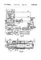
- FIG. 1is a side perspective view of the dermatology handpiece.
- FIG. 2is a top perspective view of the dermatology handpiece.
- FIG. 3is a block diagram of the dermatology handpiece system with a single optical source.
- FIG. 4is a block diagram of the dermatology handpiece system with different probe beam and treatment beam optical sources.
- FIG. 5is a block diagram of the dermatology handpiece system with pulsed and CW laser treatment beam sources.
- FIG. 6is a side sectional view of the handpiece illustrated in FIG. 1.
- FIG. 7is a top sectional view of the handpiece illustrated in FIG. 2.
- FIG. 8is a diagram illustrating a scanning system that can be used with the dermatology handpiece.
- FIG. 9is a circuit diagram for the dermatology handpiece system.
- FIGS. 1 and 2illustrate exterior views of a dermatology handpiece 10.
- a cannula 11retains a dermatology delivery device 12, such as the MicronSpot Handpiece, commercially available from Laserscope, San Jose, Calif.
- a scanning systemis positioned within housing 14. The scanning system, as will be more fully explained later, provides movement of an optical fiber that delivers optical energy from a laser source to a dermatology lesion.
- Dermatology delivery device 12is connected optically, either directly or indirectly, to a laser source, not shown.
- a laser sourcecan be used with the dermatology handpiece of the invention, including but not limited to the following KTP 532/YAG, copper vapor, flash lamp dye, argon, krypton, dye and YAG. Additionally, a concentrated light source, such as an arc lamp, can be used.
- a focused beam of optical energy 16emerges from a distal end 18 of cannula 11.
- a window 20is formed at distal end 18 and provides the ability to view by the physician.
- a switch 22is hand activated by the physician to turn the optical energy from the dermatology handpiece on or off.
- FIGS. 3, 4 and 5illustrate further elements of the invention.
- Optical source 24is preferably a laser, and more particularly a KTP 532 wavelength laser.
- Optical source 24produces a beam 26 of optical energy.
- the embodiment shown in FIG. 3has optical source 24 producing both a treatment beam 26 which is delivered to a tissue site, and a probe beam 27 (FIG. 4).
- Probe beam 27is used to establish a threshold value. When the threshold value is exceeded then treatment beam 26 delivers beam 26 to a treatment site 28.
- Probe beam 27 and treatment beam 26can be of different wavelengths.
- probe beamcan be in the yellow region of the spectrum, created from a HeNe laser, while the source of treatment beam 26 may by an IR laser.
- both beamsare preferably delivered along the same optical fiber, or alternatively, different fibers may be employed.
- Treatment site 28is preferably a dermatology lesion. It can also be portwine stains, tattoos, spider veins, telangiectasias, angiokeratomas, hair removal, application in photo dynamic therapy detection and for the treatment of skin cancer. Beam 26 is reflected from treatment site 28 and produces a reflectance/remittance signal 30 that is received at a detector 32 and amplified.
- a suitable detectoris any photo cell, photo detector/fluorescence detector or photo diodes.
- One detector that can be usedis a Model No. DT 25, commercially available from EG&G.
- a signalis produced at detector 32. It is then compared at comparator 34 with a threshold signal that is pre-set by the physician by a threshold adjustment 36 and a sensitivity adjustment 38. Probe beam 27 is reflected from treatment site 28 and reflectance/remittance signal 30 is generated.
- Sensitivity and threshold adjustments 38 and 36are set by the physician so that healthy tissue, or tissue representative of a person's normal pigment, is set to a base line of "0".
- the treatment beam 26will deliver optical energy to treatment site 28. Below the base line treatment beam 26 is not activated.
- Probe beam 37also provides a method of tracing the delivery of the optical energy.
- dermatology handpiece 10places on normal skin near treatment site 28. Dermatology handpiece 10 is then calibrated by scanning to look at the reflectance of probe beam 27. An input signal is produced, stored in an analog or digital processor and that level then becomes the baseline or threshold because of the modification of the gain in the detection circuitry.
- a trigger-generator device 40receives the comparison signal, which determines whether or not the base line has been exceeded, and then produces an indicator signal 42 to establish that the base line has been exceeded.
- Trigger-generator device 40sends a signal 46 to optical source 24 indicating that the base line has been exceeded.
- Optical source 24then emits treatment beam 26. This process continues until the base line is not exceeded.
- indicator signal 42can be an audio device 44 such as Model No. SC628E, commercially available from Mallory.
- a suitable detection and amplification device 32is OP Model 17.
- Comparator 34can be a Model No. LM2903 Op Amp.
- Trigger-generator 40can be a Model No. CD4538BE. All are commercially from R&D Electronics, San Jose, Calif.
- the physiciancan interrupt treatment beam 26 by activating enabling switch 22.
- Thisprovides great flexibility.
- the base linecan be exceeded but the physician may notice something at treatment site 28 which requires him to deactivate optical source 24. This is readily achieved with enabling switch 22, positioned conveniently at the exterior of cannula 11.
- Optical source 24can be separated into a treatment beam source 24 and a probe beam source 48 (FIG. 4).
- Probe beam source 48can be a HeNe or a diode laser such as a HeNe, green, yellow, red, orange, Model No. 1125 MP, commercially available from Uniphase.
- probe beam source 48can also be a diode laser, such as diode laser Model No. 9424, commercially available from Toshiba.
- Different colored probe beamscan be used.
- a green probe beamis excellent for hemoglobin absorption.
- a red probe beamis useful in treating hair follicles.
- Changing probe beam 37is one way of providing different applications of dermatology handpiece 10. However, greater contrast with hemoglobin is obtained with a green probe beam because of absorption characteristics.
- Treatment beam source 24can be a KTP 532/1064 laser commercially available from Laserscope, San Jose, Calif. Its output characteristics are 0.5 to 40 watts of 532 nm, 5 watts to 100 watts of 1064 nm at exposure time levels of about 0.01 seconds when operated in the cw mode.
- treatment beam sourcecan be either a cw laser 24(a) or a pulsed laser 24(b). Both lasers can be incorporated in the same system, or either one can be employed.
- cw laser 24(a)When cw laser 24(a) is utilized a shutter box and beam dump 50 is also included. This is achieved, in one embodiment, with treatment beam source 24 operating in a lo RF mode on the internal Q-switch, permitting small leakage of light in the form of probe beam 27.
- the Q-switchacts as a shutter and suppresses the cavity of laser 24(a) from lasing completely.
- Treatment beam 26 and probe beam 27can be modulated in order to increase the signal to noise ratio and permit operation in ambient light.
- a lens 52is positioned adjacent to dermatology delivery device 12 at its distal end 54 nearest distal end 18 of cannula 11.
- Lens 52may be made to vary the spot size from 25 microns to about 5 mm.
- lens 52has a focal length of about 15 mm, and cannula 11 can be adjusted up and down to vary spot size.
- a spot size of about 25 microns to 5 mmcan be produced at treatment site 28.
- Detection and amplification device 32can be an IR detector coupled with an amplifier.
- a suitable deviceis Model No. DT 25, commercially available from EG&G.
- Detection and amplification device 32is positioned preferably within the cannula 11, so that it looks where probe beam 27 is projected and detects not only total scatter at treatment site 28, but also at the spot where probe beam 27 is projected. In one embodiment, detection and amplification device 32 is approximately 20 degrees off from the optical axis of probe beam 27.
- Ambient lightcan interfere with reflectance/remittance signal 30.
- probe beam 27can be modulated at a frequency above normal background ambient light. This may be, in one example, about 60 Hz.
- Detection and amplification device 32can include a high pass filter to remove all of the light except that of probe beam 27.
- a notch filtercan also be used.
- a suitable oneis green notch filter available from Burleigh. With either element the physician can visually see probe beam 27 through window 20 (FIG. 1) without interference from ambient light.
- a sensor 58which detects motion of cannula 11. This provides another indicator to the physician that energy is being delivered to treatment site 28 in a even matter.
- Sensor 58can be a an IR detector. It provides feedback as to the relative speed of dermatology handpiece 10 motion, and hence assists in the even distribution of optical energy to treatment site 28.
- dermatology handpiece 10is moved by the physician around substantially the entire surface of treatment site 28. In this embodiment it is useful to include sensor 58. In another embodiment, a scanning device 60 is positioned in dermatology handpiece 10, preferably in housing 14.
- probe beam 27can scan once across treatment beam 26, and a determination made at comparator 34. A level of treatment power is then formulated. As the absorption of treatment site 28 increases the power of treatment beam 26 will also increase when probe beam 27 scans across the same location.
- Scanning device 60moves an optical fiber 62 which is coupled to optical source 24.
- Optical fiber 62is caused to rotate about an optical axis 64 and deliver energy to tissue site 28.
- An optical focusing element 66such as a 100 mm fiber with a 15 mm lens, focuses the optical output from optical fiber at treatment site 28.
- Connecting and crank systems 68 and 70respectively work in tandem, imparting a rotational movement of optical fiber 62.
- Connecting and crank systems 68 and 70are positioned on a frame 72 that includes a platform 74.
- a bracket 76holds optical fiber 62 and is supported by platform 74.
- Motors 78 and 80provide the power sources to impart movement of connecting and crank systems 68 and 70, as well as connecting rods 82 and 84.
- a common joint 86completes the mechanism for rotating optical fiber 62 about optical axis 64.
- Detector 88may be an op amplifier.
- Switch 22, positioned at the exterior of dermatology handpiece 10allows the electronics to be enabled. In the event that the physician has not activated switch 22 then the electronics will not sense anything.
- the second element of detection and amplification device 32is an amplifier 90, preferably an op seventeen op amplifier, that amplifies and in certain instances inverts a signal received from detector 88. Associated with amplifier 90 is in integrated circuit 92. The signal from amplifier 90 is received at comparator 34, another op amp.
- Comparator 34is used by the physician to establish the base line.
- a potentiometer 94is used by the physician to determine a base line skin level. Potentiometer 94 is adjusted until an LED 96 is turned on, indicating base line or normal skin color. Once the LED is on the physician turns potentiometer 94 one-quarter to one-half of a turn.
- thresholdThis establishes the base line, or threshold. Whenever the signal from detector 88 drops below a certain threshold, established by comparator 34, trigger generator 40 triggers treatment beam source 24 or it causes the signal to go high. If treatment beam 26 is absorbed by the hemoglobin in the skin, or any kind of treatable tissue, the signal to detector 88 goes low. If it goes sufficiently low enough to a point where the skin threshold is accepted then comparator 34 is activated to a high signal.
- a switch 98positioned on the exterior of dermatologic handpiece 10, permits the physician to turn potentiometer 94 until LED 96 emits light, and then turned back slightly in order to scan over treatment site 28.
- a Q-Switch driver circuitshown generally as 100, provides for the output of a certain length of pulse from pulsed laser 24(b).
- Circuit 100turns off detector 88 off for a slightly longer amount of time than the Q-switch of pulsed laser 24(b). Regardless of how long circuit 100 sees reflectance/remittance signal 30, it will only output a certain length of pulse from pulsed laser 24(b).
Landscapes
- Physics & Mathematics (AREA)
- Health & Medical Sciences (AREA)
- Surgery (AREA)
- Optics & Photonics (AREA)
- Life Sciences & Earth Sciences (AREA)
- Heart & Thoracic Surgery (AREA)
- Molecular Biology (AREA)
- Nuclear Medicine, Radiotherapy & Molecular Imaging (AREA)
- Engineering & Computer Science (AREA)
- Biomedical Technology (AREA)
- Electromagnetism (AREA)
- Medical Informatics (AREA)
- Otolaryngology (AREA)
- Animal Behavior & Ethology (AREA)
- General Health & Medical Sciences (AREA)
- Public Health (AREA)
- Veterinary Medicine (AREA)
- Radiation-Therapy Devices (AREA)
- Surgical Instruments (AREA)
- Laser Surgery Devices (AREA)
- Treatments Of Macromolecular Shaped Articles (AREA)
Abstract
Description
Claims (9)
Priority Applications (6)
| Application Number | Priority Date | Filing Date | Title |
|---|---|---|---|
| US08/171,593US5628744A (en) | 1993-12-21 | 1993-12-21 | Treatment beam handpiece |
| AU14465/95AAU1446595A (en) | 1993-12-21 | 1994-12-21 | Treatment beam handpiece |
| CA002179600ACA2179600A1 (en) | 1993-12-21 | 1994-12-21 | Treatment beam handpiece |
| PCT/US1994/014951WO1995017130A1 (en) | 1993-12-21 | 1994-12-21 | Treatment beam handpiece |
| EP95906132AEP0735841A1 (en) | 1993-12-21 | 1994-12-21 | Treatment beam handpiece |
| NZ278275ANZ278275A (en) | 1993-12-21 | 1994-12-21 | Dermatology device; handpiece delivers optical energy to a lesion with an energy source having two different monochromatic wavelengths |
Applications Claiming Priority (1)
| Application Number | Priority Date | Filing Date | Title |
|---|---|---|---|
| US08/171,593US5628744A (en) | 1993-12-21 | 1993-12-21 | Treatment beam handpiece |
Publications (1)
| Publication Number | Publication Date |
|---|---|
| US5628744Atrue US5628744A (en) | 1997-05-13 |
Family
ID=22624366
Family Applications (1)
| Application Number | Title | Priority Date | Filing Date |
|---|---|---|---|
| US08/171,593Expired - LifetimeUS5628744A (en) | 1993-12-21 | 1993-12-21 | Treatment beam handpiece |
Country Status (6)
| Country | Link |
|---|---|
| US (1) | US5628744A (en) |
| EP (1) | EP0735841A1 (en) |
| AU (1) | AU1446595A (en) |
| CA (1) | CA2179600A1 (en) |
| NZ (1) | NZ278275A (en) |
| WO (1) | WO1995017130A1 (en) |
Cited By (97)
| Publication number | Priority date | Publication date | Assignee | Title |
|---|---|---|---|---|
| US5860968A (en)* | 1995-11-03 | 1999-01-19 | Luxar Corporation | Laser scanning method and apparatus |
| US5868731A (en)* | 1996-03-04 | 1999-02-09 | Innotech Usa, Inc. | Laser surgical device and method of its use |
| US6027496A (en)* | 1997-03-25 | 2000-02-22 | Abbott Laboratories | Removal of stratum corneum by means of light |
| US6066129A (en)* | 1998-01-29 | 2000-05-23 | Larson; Dean W. | Medical laser control system |
| US6074382A (en)* | 1997-08-29 | 2000-06-13 | Asah Medico A/S | Apparatus for tissue treatment |
| US6139554A (en)* | 1999-06-10 | 2000-10-31 | Karkar; Maurice N. | Multipurpose tissue resurfacing handpiece |
| WO2000071045A1 (en)* | 1999-05-25 | 2000-11-30 | International Technologies (Lasers), Ltd. | Laser for skin treatment |
| US6171302B1 (en)* | 1997-03-19 | 2001-01-09 | Gerard Talpalriu | Apparatus and method including a handpiece for synchronizing the pulsing of a light source |
| WO2001008578A1 (en)* | 1999-07-30 | 2001-02-08 | Vivant Medical, Inc. | Device and method for safe location and marking of a cavity and sentinel lymph nodes |
| US6228074B1 (en)* | 1998-10-15 | 2001-05-08 | Stephen Almeida | Multiple pulse photo-epilator |
| GB2364567A (en)* | 2000-07-07 | 2002-01-30 | Astron Clinica Ltd | Determining distribution of chromophores in skin or other epithelial tissue |
| WO2002009813A1 (en)* | 2000-07-31 | 2002-02-07 | El. En. S.P.A. | Method and device for epilation by ultrasound |
| US6356782B1 (en) | 1998-12-24 | 2002-03-12 | Vivant Medical, Inc. | Subcutaneous cavity marking device and method |
| US6371904B1 (en) | 1998-12-24 | 2002-04-16 | Vivant Medical, Inc. | Subcutaneous cavity marking device and method |
| US6413268B1 (en) | 2000-08-11 | 2002-07-02 | Raymond A. Hartman | Apparatus and method for targeted UV phototherapy of skin disorders |
| US6436094B1 (en) | 2000-03-16 | 2002-08-20 | Laserscope, Inc. | Electromagnetic and laser treatment and cooling device |
| US6447537B1 (en)* | 2000-06-21 | 2002-09-10 | Raymond A. Hartman | Targeted UV phototherapy apparatus and method |
| US20020156471A1 (en)* | 1999-03-09 | 2002-10-24 | Stern Roger A. | Method for treatment of tissue |
| US20020173782A1 (en)* | 2001-04-20 | 2002-11-21 | Cense Abraham Josephus | Skin treating device with protection against rediation pulse overdose |
| US6554824B2 (en) | 2000-12-15 | 2003-04-29 | Laserscope | Methods for laser treatment of soft tissue |
| US6572637B1 (en)* | 1999-03-12 | 2003-06-03 | Ya-Man Ltd. | Handbreadth-sized laser beam projecting probe for beauty treatment |
| US20030130649A1 (en)* | 2000-12-15 | 2003-07-10 | Murray Steven C. | Method and system for treatment of benign prostatic hypertrophy (BPH) |
| US20030135205A1 (en)* | 2000-12-15 | 2003-07-17 | Davenport Scott A. | Method and system for photoselective vaporization of the prostate, and other tissue |
| US6595986B2 (en) | 1998-10-15 | 2003-07-22 | Stephen Almeida | Multiple pulse photo-dermatological device |
| US6607523B1 (en) | 1999-03-19 | 2003-08-19 | Asah Medico A/S | Apparatus for tissue treatment |
| US20030199866A1 (en)* | 1996-01-05 | 2003-10-23 | Stern Roger A. | Method and kit for treatment of tissue |
| US20030212393A1 (en)* | 1996-01-05 | 2003-11-13 | Knowlton Edward W. | Handpiece with RF electrode and non-volatile memory |
| US20030216717A1 (en)* | 2002-02-22 | 2003-11-20 | Laserscope | Method and system for photoselective vaporization for gynecological treatments |
| US20030216719A1 (en)* | 2001-12-12 | 2003-11-20 | Len Debenedictis | Method and apparatus for treating skin using patterns of optical energy |
| US6653618B2 (en)* | 2000-04-28 | 2003-11-25 | Palomar Medical Technologies, Inc. | Contact detecting method and apparatus for an optical radiation handpiece |
| US20040000316A1 (en)* | 1996-01-05 | 2004-01-01 | Knowlton Edward W. | Methods for creating tissue effect utilizing electromagnetic energy and a reverse thermal gradient |
| US20040002705A1 (en)* | 1996-01-05 | 2004-01-01 | Knowlton Edward W. | Methods for creating tissue effect utilizing electromagnetic energy and a reverse thermal gradient |
| US20040002704A1 (en)* | 1996-01-05 | 2004-01-01 | Knowlton Edward W. | Treatment apparatus with electromagnetic energy delivery device and non-volatile memory |
| US6676654B1 (en) | 1997-08-29 | 2004-01-13 | Asah Medico A/S | Apparatus for tissue treatment and having a monitor for display of tissue features |
| US20040030332A1 (en)* | 1996-01-05 | 2004-02-12 | Knowlton Edward W. | Handpiece with electrode and non-volatile memory |
| US20040034346A1 (en)* | 1996-01-05 | 2004-02-19 | Stern Roger A. | RF device with thermo-electric cooler |
| US6702808B1 (en)* | 2000-09-28 | 2004-03-09 | Syneron Medical Ltd. | Device and method for treating skin |
| US20040092802A1 (en)* | 2000-07-07 | 2004-05-13 | Cane Michael Roger | Epithelial diagnostic aid |
| US20040111087A1 (en)* | 1999-03-09 | 2004-06-10 | Stern Roger A. | Handpiece for treatment of tissue |
| US20040167499A1 (en)* | 2003-02-25 | 2004-08-26 | Grove Robert E. | Eye-safe dermatologic treatment apparatus and method |
| US20040167500A1 (en)* | 2003-02-25 | 2004-08-26 | Weckwerth Mark V. | Self-contained, diode-laser-based dermatologic treatment apparatus and method |
| US20040167502A1 (en)* | 2003-02-25 | 2004-08-26 | Weckwerth Mark V. | Optical sensor and method for identifying the presence of skin |
| US20040176824A1 (en)* | 2003-03-04 | 2004-09-09 | Weckwerth Mark V. | Method and apparatus for the repigmentation of human skin |
| US20040176823A1 (en)* | 2003-02-25 | 2004-09-09 | Island Tobin C. | Acne treatment device and method |
| US20040176754A1 (en)* | 2003-03-06 | 2004-09-09 | Island Tobin C. | Method and device for sensing skin contact |
| US20040186535A1 (en)* | 1999-06-30 | 2004-09-23 | Knowlton Edward W. | Fluid delivery apparatus |
| US20050048436A1 (en)* | 2003-08-29 | 2005-03-03 | Udi Fishman | Handpiece apparatus and method for dispensing media |
| US20050049582A1 (en)* | 2001-12-12 | 2005-03-03 | Debenedictis Leonard C. | Method and apparatus for fractional photo therapy of skin |
| US20060020260A1 (en)* | 2004-07-22 | 2006-01-26 | Dover Jeffrey S | Method and apparatus of treating tissue |
| US7022121B2 (en) | 1999-03-09 | 2006-04-04 | Thermage, Inc. | Handpiece for treatment of tissue |
| US20060155266A1 (en)* | 2003-03-27 | 2006-07-13 | Dieter Manstein | Method and apparatus for dermatological treatment and fractional skin resurfacing |
| US20060194164A1 (en)* | 2004-12-09 | 2006-08-31 | Palomar Medical Technologies, Inc. | Oral appliance with heat transfer mechanism |
| US20060247740A1 (en)* | 2003-08-18 | 2006-11-02 | Koninklijke Philips Electronics N.V. | Device and method for low intensity optical hair growth control |
| US7189230B2 (en) | 1996-01-05 | 2007-03-13 | Thermage, Inc. | Method for treating skin and underlying tissue |
| US20070078501A1 (en)* | 2002-05-23 | 2007-04-05 | Palomar Medical Technologies, Inc. | Phototreatment device for use with coolants and topical substances |
| US20070093798A1 (en)* | 2005-08-29 | 2007-04-26 | Reliant Technologies, Inc. | Method and Apparatus for Monitoring and Controlling Thermally Induced Tissue Treatment |
| US7250045B2 (en) | 2003-02-25 | 2007-07-31 | Spectragenics, Inc. | Self-contained, eye-safe hair-regrowth-inhibition apparatus and method |
| US20070244526A1 (en)* | 2006-04-14 | 2007-10-18 | Asa S.R.L. | Laser apparatus for therapeutic applications |
| US20080058782A1 (en)* | 2006-08-29 | 2008-03-06 | Reliant Technologies, Inc. | Method and apparatus for monitoring and controlling density of fractional tissue treatments |
| US20080172047A1 (en)* | 2000-12-28 | 2008-07-17 | Palomar Medical Technologies, Inc. | Methods And Devices For Fractional Ablation Of Tissue |
| US20080208104A1 (en)* | 2005-04-18 | 2008-08-28 | Pantec Biosolutions Ag | Laser Microporator |
| US20080269590A1 (en)* | 2007-04-24 | 2008-10-30 | Matthias Wedel | Medical instrument for performing a medical intervention |
| US7452358B2 (en) | 1996-01-05 | 2008-11-18 | Thermage, Inc. | RF electrode assembly for handpiece |
| US20080287934A1 (en)* | 2007-05-15 | 2008-11-20 | Hunter Lowell D | Laser Handle and Fiber Guard |
| US20080287940A1 (en)* | 2007-05-14 | 2008-11-20 | Hunter Lowell D | Fiber Pole Tip |
| US20080294152A1 (en)* | 1996-12-02 | 2008-11-27 | Palomar Medical Technologies, Inc. | Cooling System For A Photocosmetic Device |
| US20090018531A1 (en)* | 2007-06-08 | 2009-01-15 | Cynosure, Inc. | Coaxial suction system for laser lipolysis |
| US20090043294A1 (en)* | 2003-02-25 | 2009-02-12 | Spectragenics, Inc. | Capacitive Sensing Method and Device for Detecting Skin |
| US20090099559A1 (en)* | 2007-10-05 | 2009-04-16 | The Research Foundation Of The State University Of New York | Coherent imaging fiber based hair removal device |
| WO2009053499A1 (en)* | 2007-10-25 | 2009-04-30 | Pantec Biosolutions Ag | Laser device and method for ablating biological tissue |
| US20090118720A1 (en)* | 2001-12-12 | 2009-05-07 | Reliant Technologies, Inc. | Dermatological Apparatus and Method |
| US20090125006A1 (en)* | 2004-02-25 | 2009-05-14 | Spectragenics, Inc. | Optical Sensor and Method for Identifying the Presence of Skin |
| US20090137994A1 (en)* | 2004-06-14 | 2009-05-28 | Rellant Technologies, Inc, | Adaptive control of optical pulses for laser medicine |
| US20090204109A1 (en)* | 2003-02-25 | 2009-08-13 | Tria Beauty, Inc. | Eye-Safe Dermatologic Treatment Apparatus and Method |
| US20090270848A1 (en)* | 2008-04-25 | 2009-10-29 | Tria Beauty, Inc. | Optical Sensor and Method for Identifying the Presence of Skin and the Pigmentation of Skin |
| US20100069898A1 (en)* | 2003-02-25 | 2010-03-18 | Tria Beauty, Inc. | Acne Treatment Method, System and Device |
| US20100130969A1 (en)* | 2008-11-25 | 2010-05-27 | Apogen Technologies, Inc. | System and method for dermatological treatment |
| US7758621B2 (en) | 1997-05-15 | 2010-07-20 | Palomar Medical Technologies, Inc. | Method and apparatus for therapeutic EMR treatment on the skin |
| US7763016B2 (en) | 1997-05-15 | 2010-07-27 | Palomar Medical Technologies, Inc. | Light energy delivery head |
| US20100210995A1 (en)* | 2006-05-02 | 2010-08-19 | Cook Incorporated | Systems and methods for treating superficial venous malformations like spider veins |
| US20110130747A1 (en)* | 2009-12-01 | 2011-06-02 | Ceramoptec Industries Inc. | Contact free and perforation safe endoluminal laser treatment device and method |
| US7981111B2 (en) | 2003-02-25 | 2011-07-19 | Tria Beauty, Inc. | Method and apparatus for the treatment of benign pigmented lesions |
| USRE42594E1 (en) | 1998-10-16 | 2011-08-02 | Reliant Technologies, Inc. | Tissue cooling rod for laser surgery |
| US8182473B2 (en) | 1999-01-08 | 2012-05-22 | Palomar Medical Technologies | Cooling system for a photocosmetic device |
| US8328794B2 (en) | 1996-12-02 | 2012-12-11 | Palomar Medical Technologies, Inc. | System for electromagnetic radiation dermatology and head for use therewith |
| US8346347B2 (en) | 2005-09-15 | 2013-01-01 | Palomar Medical Technologies, Inc. | Skin optical characterization device |
| US8535360B2 (en) | 2006-05-02 | 2013-09-17 | Green Medical, Ltd. | Systems and methods for treating superficial venous malformations like spider veins |
| US20140180368A1 (en)* | 2011-04-07 | 2014-06-26 | Altech Corporation | Handpiece for laser treatment device |
| US8915948B2 (en) | 2002-06-19 | 2014-12-23 | Palomar Medical Technologies, Llc | Method and apparatus for photothermal treatment of tissue at depth |
| US9028536B2 (en) | 2006-08-02 | 2015-05-12 | Cynosure, Inc. | Picosecond laser apparatus and methods for its operation and use |
| US9669113B1 (en) | 1998-12-24 | 2017-06-06 | Devicor Medical Products, Inc. | Device and method for safe location and marking of a biopsy cavity |
| US9780518B2 (en) | 2012-04-18 | 2017-10-03 | Cynosure, Inc. | Picosecond laser apparatus and methods for treating target tissues with same |
| US9919168B2 (en) | 2009-07-23 | 2018-03-20 | Palomar Medical Technologies, Inc. | Method for improvement of cellulite appearance |
| US10245107B2 (en) | 2013-03-15 | 2019-04-02 | Cynosure, Inc. | Picosecond optical radiation systems and methods of use |
| US10434324B2 (en) | 2005-04-22 | 2019-10-08 | Cynosure, Llc | Methods and systems for laser treatment using non-uniform output beam |
| US11007373B1 (en) | 2002-12-20 | 2021-05-18 | James Andrew Ohneck | Photobiostimulation device and method of using same |
| US11418000B2 (en) | 2018-02-26 | 2022-08-16 | Cynosure, Llc | Q-switched cavity dumped sub-nanosecond laser |
Families Citing this family (1)
| Publication number | Priority date | Publication date | Assignee | Title |
|---|---|---|---|---|
| DE19734732A1 (en)* | 1996-12-10 | 1998-06-18 | Wavelight Laser Technologie Gm | Arrangement for treating bodily substances |
Citations (6)
| Publication number | Priority date | Publication date | Assignee | Title |
|---|---|---|---|---|
| US4316467A (en)* | 1980-06-23 | 1982-02-23 | Lorenzo P. Maun | Control for laser hemangioma treatment system |
| US4768513A (en)* | 1986-04-21 | 1988-09-06 | Agency Of Industrial Science And Technology | Method and device for measuring and processing light |
| US5071417A (en)* | 1990-06-15 | 1991-12-10 | Rare Earth Medical Lasers, Inc. | Laser fusion of biological materials |
| US5275594A (en)* | 1990-11-09 | 1994-01-04 | C. R. Bard, Inc. | Angioplasty system having means for identification of atherosclerotic plaque |
| US5334191A (en)* | 1992-05-21 | 1994-08-02 | Dix Phillip Poppas | Laser tissue welding control system |
| US5350376A (en)* | 1993-04-16 | 1994-09-27 | Ceramoptec, Inc. | Optical controller device |
- 1993
- 1993-12-21USUS08/171,593patent/US5628744A/ennot_activeExpired - Lifetime
- 1994
- 1994-12-21CACA002179600Apatent/CA2179600A1/ennot_activeAbandoned
- 1994-12-21AUAU14465/95Apatent/AU1446595A/ennot_activeAbandoned
- 1994-12-21NZNZ278275Apatent/NZ278275A/enunknown
- 1994-12-21WOPCT/US1994/014951patent/WO1995017130A1/ennot_activeApplication Discontinuation
- 1994-12-21EPEP95906132Apatent/EP0735841A1/ennot_activeWithdrawn
Patent Citations (6)
| Publication number | Priority date | Publication date | Assignee | Title |
|---|---|---|---|---|
| US4316467A (en)* | 1980-06-23 | 1982-02-23 | Lorenzo P. Maun | Control for laser hemangioma treatment system |
| US4768513A (en)* | 1986-04-21 | 1988-09-06 | Agency Of Industrial Science And Technology | Method and device for measuring and processing light |
| US5071417A (en)* | 1990-06-15 | 1991-12-10 | Rare Earth Medical Lasers, Inc. | Laser fusion of biological materials |
| US5275594A (en)* | 1990-11-09 | 1994-01-04 | C. R. Bard, Inc. | Angioplasty system having means for identification of atherosclerotic plaque |
| US5334191A (en)* | 1992-05-21 | 1994-08-02 | Dix Phillip Poppas | Laser tissue welding control system |
| US5350376A (en)* | 1993-04-16 | 1994-09-27 | Ceramoptec, Inc. | Optical controller device |
Non-Patent Citations (6)
| Title |
|---|
| Feather et al., "A portable reflectometer for the rapid quantification of cutaneous haemoglobin and melanin", Phys. Med. Biol., 1988, vol. 33, No. 6, 711-722. |
| Feather et al., A portable reflectometer for the rapid quantification of cutaneous haemoglobin and melanin , Phys. Med. Biol., 1988, vol. 33, No. 6, 711 722.* |
| Pickering, et al. "Copper vapour laser treatment of port-wine stains and other vascular malformations", British Journal of Plastic Surgery (1990), 43:273-282. |
| Pickering, et al. Copper vapour laser treatment of port wine stains and other vascular malformations , British Journal of Plastic Surgery (1990), 43:273 282.* |
| Tang et al., "In Vivo Spectrophotometric Evaluation of Normal, Lesional, and Laser-Treated Skin in Patients with Port-Wine Stains", The Journal of Investigative Dermatology, 80:420-423, 1983. |
| Tang et al., In Vivo Spectrophotometric Evaluation of Normal, Lesional, and Laser Treated Skin in Patients with Port Wine Stains , The Journal of Investigative Dermatology, 80:420 423, 1983.* |
Cited By (195)
| Publication number | Priority date | Publication date | Assignee | Title |
|---|---|---|---|---|
| US5860968A (en)* | 1995-11-03 | 1999-01-19 | Luxar Corporation | Laser scanning method and apparatus |
| US7473251B2 (en) | 1996-01-05 | 2009-01-06 | Thermage, Inc. | Methods for creating tissue effect utilizing electromagnetic energy and a reverse thermal gradient |
| US20040000316A1 (en)* | 1996-01-05 | 2004-01-01 | Knowlton Edward W. | Methods for creating tissue effect utilizing electromagnetic energy and a reverse thermal gradient |
| US7267675B2 (en) | 1996-01-05 | 2007-09-11 | Thermage, Inc. | RF device with thermo-electric cooler |
| US7229436B2 (en) | 1996-01-05 | 2007-06-12 | Thermage, Inc. | Method and kit for treatment of tissue |
| US7189230B2 (en) | 1996-01-05 | 2007-03-13 | Thermage, Inc. | Method for treating skin and underlying tissue |
| US7452358B2 (en) | 1996-01-05 | 2008-11-18 | Thermage, Inc. | RF electrode assembly for handpiece |
| US20030212393A1 (en)* | 1996-01-05 | 2003-11-13 | Knowlton Edward W. | Handpiece with RF electrode and non-volatile memory |
| US20040034346A1 (en)* | 1996-01-05 | 2004-02-19 | Stern Roger A. | RF device with thermo-electric cooler |
| US20030199866A1 (en)* | 1996-01-05 | 2003-10-23 | Stern Roger A. | Method and kit for treatment of tissue |
| US20040002705A1 (en)* | 1996-01-05 | 2004-01-01 | Knowlton Edward W. | Methods for creating tissue effect utilizing electromagnetic energy and a reverse thermal gradient |
| US20040002704A1 (en)* | 1996-01-05 | 2004-01-01 | Knowlton Edward W. | Treatment apparatus with electromagnetic energy delivery device and non-volatile memory |
| US7006874B2 (en) | 1996-01-05 | 2006-02-28 | Thermage, Inc. | Treatment apparatus with electromagnetic energy delivery device and non-volatile memory |
| US20040030332A1 (en)* | 1996-01-05 | 2004-02-12 | Knowlton Edward W. | Handpiece with electrode and non-volatile memory |
| US7115123B2 (en) | 1996-01-05 | 2006-10-03 | Thermage, Inc. | Handpiece with electrode and non-volatile memory |
| US5868731A (en)* | 1996-03-04 | 1999-02-09 | Innotech Usa, Inc. | Laser surgical device and method of its use |
| US8328794B2 (en) | 1996-12-02 | 2012-12-11 | Palomar Medical Technologies, Inc. | System for electromagnetic radiation dermatology and head for use therewith |
| US20080294152A1 (en)* | 1996-12-02 | 2008-11-27 | Palomar Medical Technologies, Inc. | Cooling System For A Photocosmetic Device |
| US6171302B1 (en)* | 1997-03-19 | 2001-01-09 | Gerard Talpalriu | Apparatus and method including a handpiece for synchronizing the pulsing of a light source |
| US6245060B1 (en) | 1997-03-25 | 2001-06-12 | Abbott Laboratories | Removal of stratum corneum by means of light |
| US6027496A (en)* | 1997-03-25 | 2000-02-22 | Abbott Laboratories | Removal of stratum corneum by means of light |
| US8002768B1 (en) | 1997-05-15 | 2011-08-23 | Palomar Medical Technologies, Inc. | Light energy delivery head |
| US7763016B2 (en) | 1997-05-15 | 2010-07-27 | Palomar Medical Technologies, Inc. | Light energy delivery head |
| US7935107B2 (en) | 1997-05-15 | 2011-05-03 | Palomar Medical Technologies, Inc. | Heads for dermatology treatment |
| US8109924B2 (en) | 1997-05-15 | 2012-02-07 | Palomar Medical Technologies, Inc. | Heads for dermatology treatment |
| US8328796B2 (en) | 1997-05-15 | 2012-12-11 | Palomar Medical Technologies, Inc. | Light energy delivery head |
| US7758621B2 (en) | 1997-05-15 | 2010-07-20 | Palomar Medical Technologies, Inc. | Method and apparatus for therapeutic EMR treatment on the skin |
| US6074382A (en)* | 1997-08-29 | 2000-06-13 | Asah Medico A/S | Apparatus for tissue treatment |
| EP1009485A1 (en)* | 1997-08-29 | 2000-06-21 | Asah Medico A/S | An apparatus for tissue treatment |
| USRE38670E1 (en)* | 1997-08-29 | 2004-12-14 | Asah Medico A/S | Apparatus for tissue treatment |
| US6383177B1 (en)* | 1997-08-29 | 2002-05-07 | Asah Medico A/S | Apparatus for tissue treatment |
| US6676654B1 (en) | 1997-08-29 | 2004-01-13 | Asah Medico A/S | Apparatus for tissue treatment and having a monitor for display of tissue features |
| US6066129A (en)* | 1998-01-29 | 2000-05-23 | Larson; Dean W. | Medical laser control system |
| US6595986B2 (en) | 1998-10-15 | 2003-07-22 | Stephen Almeida | Multiple pulse photo-dermatological device |
| US6228074B1 (en)* | 1998-10-15 | 2001-05-08 | Stephen Almeida | Multiple pulse photo-epilator |
| US7097639B1 (en) | 1998-10-15 | 2006-08-29 | Zian Medical, Llc | Dual filter multiple pulse photo-dermatological device with pre/post optical heating, quasi-logarithmic spacing, and laser rod spectrum infusion |
| USRE43881E1 (en) | 1998-10-16 | 2012-12-25 | Reliant Technologies, Inc. | Tissue cooling rod for laser surgery |
| USRE42594E1 (en) | 1998-10-16 | 2011-08-02 | Reliant Technologies, Inc. | Tissue cooling rod for laser surgery |
| USRE46208E1 (en) | 1998-10-16 | 2016-11-22 | Reliant Technologies, Llc | Method for cryogenically treating tissue below the skin surface |
| US6371904B1 (en) | 1998-12-24 | 2002-04-16 | Vivant Medical, Inc. | Subcutaneous cavity marking device and method |
| US9986974B2 (en) | 1998-12-24 | 2018-06-05 | Devicor Medical Products, Inc. | Biopsy cavity marking device |
| US8600481B2 (en) | 1998-12-24 | 2013-12-03 | Devicor Medical Products, Inc. | Subcutaneous cavity marking device |
| US20060079770A1 (en)* | 1998-12-24 | 2006-04-13 | Sirimanne D L | Biopsy site marker |
| US20060036159A1 (en)* | 1998-12-24 | 2006-02-16 | Sirimanne D L | Biopsy cavity marking device |
| US9669113B1 (en) | 1998-12-24 | 2017-06-06 | Devicor Medical Products, Inc. | Device and method for safe location and marking of a biopsy cavity |
| US9492570B2 (en) | 1998-12-24 | 2016-11-15 | Devicor Medical Products, Inc. | Device and method for safe location and marking of a biopsy cavity |
| US7668582B2 (en) | 1998-12-24 | 2010-02-23 | Ethicon Endo-Surgery, Inc. | Biopsy site marker |
| US6356782B1 (en) | 1998-12-24 | 2002-03-12 | Vivant Medical, Inc. | Subcutaneous cavity marking device and method |
| US9380998B2 (en) | 1998-12-24 | 2016-07-05 | Devicor Medical Products, Inc. | Subcutaneous cavity marking device and method |
| US8320994B2 (en) | 1998-12-24 | 2012-11-27 | Devicor Medical Products, Inc. | Biopsy cavity marking device and method |
| US8320993B2 (en) | 1998-12-24 | 2012-11-27 | Devicor Medical Products, Inc. | Subcutaneous cavity marking device |
| US20020035324A1 (en)* | 1998-12-24 | 2002-03-21 | Sirimanne D. Laksen | Subcutaneous cavity marking device and method |
| US20100234726A1 (en)* | 1998-12-24 | 2010-09-16 | Sirimanne D Laksen | Device and method for safe location and marking of a biopsy cavity |
| US20020107437A1 (en)* | 1998-12-24 | 2002-08-08 | Sirimanne D. Laksen | Subcutaneous cavity marking device and method |
| US20050085724A1 (en)* | 1998-12-24 | 2005-04-21 | Vivant Medical, Inc. | Biopsy cavity marking device and method |
| US8306602B2 (en) | 1998-12-24 | 2012-11-06 | Devicor Medical Products, Inc. | Biopsy cavity marking device |
| US20050059888A1 (en)* | 1998-12-24 | 2005-03-17 | Sirimanne D. Laksen | Biopsy cavity marking device and method |
| US20050080338A1 (en)* | 1998-12-24 | 2005-04-14 | Sirimanne D. Laksen | Biopsy cavity marking device and method |
| US20050080337A1 (en)* | 1998-12-24 | 2005-04-14 | Vivant Medical, Inc. | Biopsy site marker |
| US20050080339A1 (en)* | 1998-12-24 | 2005-04-14 | Vivant Medical, Inc. | Biopsy cavity marking device |
| US8182473B2 (en) | 1999-01-08 | 2012-05-22 | Palomar Medical Technologies | Cooling system for a photocosmetic device |
| US7141049B2 (en) | 1999-03-09 | 2006-11-28 | Thermage, Inc. | Handpiece for treatment of tissue |
| US20020156471A1 (en)* | 1999-03-09 | 2002-10-24 | Stern Roger A. | Method for treatment of tissue |
| US20040111087A1 (en)* | 1999-03-09 | 2004-06-10 | Stern Roger A. | Handpiece for treatment of tissue |
| US7022121B2 (en) | 1999-03-09 | 2006-04-04 | Thermage, Inc. | Handpiece for treatment of tissue |
| US6572637B1 (en)* | 1999-03-12 | 2003-06-03 | Ya-Man Ltd. | Handbreadth-sized laser beam projecting probe for beauty treatment |
| US6607523B1 (en) | 1999-03-19 | 2003-08-19 | Asah Medico A/S | Apparatus for tissue treatment |
| WO2000071045A1 (en)* | 1999-05-25 | 2000-11-30 | International Technologies (Lasers), Ltd. | Laser for skin treatment |
| US7101365B1 (en) | 1999-05-25 | 2006-09-05 | I.T.L. Optronics, Ltd. | Laser for skin treatment |
| US6139554A (en)* | 1999-06-10 | 2000-10-31 | Karkar; Maurice N. | Multipurpose tissue resurfacing handpiece |
| US20040186535A1 (en)* | 1999-06-30 | 2004-09-23 | Knowlton Edward W. | Fluid delivery apparatus |
| WO2001008578A1 (en)* | 1999-07-30 | 2001-02-08 | Vivant Medical, Inc. | Device and method for safe location and marking of a cavity and sentinel lymph nodes |
| US6436094B1 (en) | 2000-03-16 | 2002-08-20 | Laserscope, Inc. | Electromagnetic and laser treatment and cooling device |
| US6653618B2 (en)* | 2000-04-28 | 2003-11-25 | Palomar Medical Technologies, Inc. | Contact detecting method and apparatus for an optical radiation handpiece |
| US6447537B1 (en)* | 2000-06-21 | 2002-09-10 | Raymond A. Hartman | Targeted UV phototherapy apparatus and method |
| US20040092802A1 (en)* | 2000-07-07 | 2004-05-13 | Cane Michael Roger | Epithelial diagnostic aid |
| GB2364567A (en)* | 2000-07-07 | 2002-01-30 | Astron Clinica Ltd | Determining distribution of chromophores in skin or other epithelial tissue |
| WO2002009813A1 (en)* | 2000-07-31 | 2002-02-07 | El. En. S.P.A. | Method and device for epilation by ultrasound |
| US6413268B1 (en) | 2000-08-11 | 2002-07-02 | Raymond A. Hartman | Apparatus and method for targeted UV phototherapy of skin disorders |
| US6702808B1 (en)* | 2000-09-28 | 2004-03-09 | Syneron Medical Ltd. | Device and method for treating skin |
| US20040236319A1 (en)* | 2000-12-15 | 2004-11-25 | Laserscope | Method and system for photoselective vaporization of the prostate, and other tissue |
| US20030135205A1 (en)* | 2000-12-15 | 2003-07-17 | Davenport Scott A. | Method and system for photoselective vaporization of the prostate, and other tissue |
| US20080262485A1 (en)* | 2000-12-15 | 2008-10-23 | Laserscope | Method and system for photoselective vaporization of the prostate, and other tissue |
| US20060084959A1 (en)* | 2000-12-15 | 2006-04-20 | Laserscope | Method and system for photoselective vaporization of the prostate, and other tissue |
| US6986764B2 (en) | 2000-12-15 | 2006-01-17 | Laserscope | Method and system for photoselective vaporization of the prostate, and other tissue |
| US20030130649A1 (en)* | 2000-12-15 | 2003-07-10 | Murray Steven C. | Method and system for treatment of benign prostatic hypertrophy (BPH) |
| US20050256513A1 (en)* | 2000-12-15 | 2005-11-17 | Laserscope | Method and system for vaporization of tissue using direct visualization |
| US10653482B2 (en) | 2000-12-15 | 2020-05-19 | Boston Scientific Scimed, Inc. | System for vaporization of tissue |
| US20070225696A1 (en)* | 2000-12-15 | 2007-09-27 | Davenport Scott A | Surgical apparatus for laser ablation of soft tissue |
| US6554824B2 (en) | 2000-12-15 | 2003-04-29 | Laserscope | Methods for laser treatment of soft tissue |
| US20040236318A1 (en)* | 2000-12-15 | 2004-11-25 | Laserscope | Method and system for photoselective vaporization of the prostate, and other tissue |
| US20080172047A1 (en)* | 2000-12-28 | 2008-07-17 | Palomar Medical Technologies, Inc. | Methods And Devices For Fractional Ablation Of Tissue |
| US20020173782A1 (en)* | 2001-04-20 | 2002-11-21 | Cense Abraham Josephus | Skin treating device with protection against rediation pulse overdose |
| US6955672B2 (en)* | 2001-04-20 | 2005-10-18 | Koninklijke Philips Electronics N.V. | Skin treating device with protection against radiation pulse overdose |
| US20090118720A1 (en)* | 2001-12-12 | 2009-05-07 | Reliant Technologies, Inc. | Dermatological Apparatus and Method |
| US20050049582A1 (en)* | 2001-12-12 | 2005-03-03 | Debenedictis Leonard C. | Method and apparatus for fractional photo therapy of skin |
| US20030216719A1 (en)* | 2001-12-12 | 2003-11-20 | Len Debenedictis | Method and apparatus for treating skin using patterns of optical energy |
| US20030216717A1 (en)* | 2002-02-22 | 2003-11-20 | Laserscope | Method and system for photoselective vaporization for gynecological treatments |
| US20050177145A1 (en)* | 2002-02-22 | 2005-08-11 | Laserscope | Method and system for photoselective vaporization for gynecological treatments |
| US7063694B2 (en) | 2002-02-22 | 2006-06-20 | Laserscope | Method and system for photoselective vaporization for gynecological treatments |
| US20070078501A1 (en)* | 2002-05-23 | 2007-04-05 | Palomar Medical Technologies, Inc. | Phototreatment device for use with coolants and topical substances |
| US7942916B2 (en) | 2002-05-23 | 2011-05-17 | Palomar Medical Technologies, Inc. | Phototreatment device for use with coolants and topical substances |
| US7942915B2 (en) | 2002-05-23 | 2011-05-17 | Palomar Medical Technologies, Inc. | Phototreatment device for use with coolants |
| US10556123B2 (en) | 2002-06-19 | 2020-02-11 | Palomar Medical Technologies, Llc | Method and apparatus for treatment of cutaneous and subcutaneous conditions |
| US8915948B2 (en) | 2002-06-19 | 2014-12-23 | Palomar Medical Technologies, Llc | Method and apparatus for photothermal treatment of tissue at depth |
| US10500413B2 (en) | 2002-06-19 | 2019-12-10 | Palomar Medical Technologies, Llc | Method and apparatus for treatment of cutaneous and subcutaneous conditions |
| US11007373B1 (en) | 2002-12-20 | 2021-05-18 | James Andrew Ohneck | Photobiostimulation device and method of using same |
| US10342617B2 (en) | 2003-02-25 | 2019-07-09 | Channel Investments, Llc | Phototherapy device thermal control apparatus and method |
| US20100069898A1 (en)* | 2003-02-25 | 2010-03-18 | Tria Beauty, Inc. | Acne Treatment Method, System and Device |
| US10342618B2 (en) | 2003-02-25 | 2019-07-09 | Channel Investments, Llc | Self-contained, eye-safe hair-regrowth-inhibition apparatus and method |
| US7118563B2 (en) | 2003-02-25 | 2006-10-10 | Spectragenics, Inc. | Self-contained, diode-laser-based dermatologic treatment apparatus |
| US8551104B2 (en) | 2003-02-25 | 2013-10-08 | Tria Beauty, Inc. | Self-contained, diode-laser-based dermatologic treatment apparatus |
| US20080027518A1 (en)* | 2003-02-25 | 2008-01-31 | Spectragenics, Inc. | Self-contained, eye-safe hair-regrowth-inhibition apparatus and method |
| US20090204109A1 (en)* | 2003-02-25 | 2009-08-13 | Tria Beauty, Inc. | Eye-Safe Dermatologic Treatment Apparatus and Method |
| US20040176823A1 (en)* | 2003-02-25 | 2004-09-09 | Island Tobin C. | Acne treatment device and method |
| US20040167502A1 (en)* | 2003-02-25 | 2004-08-26 | Weckwerth Mark V. | Optical sensor and method for identifying the presence of skin |
| US20090043294A1 (en)* | 2003-02-25 | 2009-02-12 | Spectragenics, Inc. | Capacitive Sensing Method and Device for Detecting Skin |
| US20040167500A1 (en)* | 2003-02-25 | 2004-08-26 | Weckwerth Mark V. | Self-contained, diode-laser-based dermatologic treatment apparatus and method |
| US20040167499A1 (en)* | 2003-02-25 | 2004-08-26 | Grove Robert E. | Eye-safe dermatologic treatment apparatus and method |
| US20070032847A1 (en)* | 2003-02-25 | 2007-02-08 | Spectragenics, Inc. | Self-contained, diode-laser-based dermatologic treatment apparatus |
| US7452356B2 (en) | 2003-02-25 | 2008-11-18 | Tria Beauty, Inc. | Eye-safe dermatologic treatment apparatus |
| US8709003B2 (en) | 2003-02-25 | 2014-04-29 | Tria Beauty, Inc. | Capacitive sensing method and device for detecting skin |
| US7981111B2 (en) | 2003-02-25 | 2011-07-19 | Tria Beauty, Inc. | Method and apparatus for the treatment of benign pigmented lesions |
| US7250045B2 (en) | 2003-02-25 | 2007-07-31 | Spectragenics, Inc. | Self-contained, eye-safe hair-regrowth-inhibition apparatus and method |
| US7413567B2 (en) | 2003-02-25 | 2008-08-19 | Spectragenics, Inc. | Optical sensor and method for identifying the presence of skin |
| US20040176824A1 (en)* | 2003-03-04 | 2004-09-09 | Weckwerth Mark V. | Method and apparatus for the repigmentation of human skin |
| US20040176754A1 (en)* | 2003-03-06 | 2004-09-09 | Island Tobin C. | Method and device for sensing skin contact |
| US9351792B2 (en) | 2003-03-27 | 2016-05-31 | The General Hospital Corporation | Method and apparatus for dermatological treatment and fractional skin resurfacing |
| US20060155266A1 (en)* | 2003-03-27 | 2006-07-13 | Dieter Manstein | Method and apparatus for dermatological treatment and fractional skin resurfacing |
| US9132279B2 (en)* | 2003-08-18 | 2015-09-15 | Koninklijke Philips N.V. | Device and method for low intensity optical hair growth control |
| US20060247740A1 (en)* | 2003-08-18 | 2006-11-02 | Koninklijke Philips Electronics N.V. | Device and method for low intensity optical hair growth control |
| US20050048436A1 (en)* | 2003-08-29 | 2005-03-03 | Udi Fishman | Handpiece apparatus and method for dispensing media |
| US20090125006A1 (en)* | 2004-02-25 | 2009-05-14 | Spectragenics, Inc. | Optical Sensor and Method for Identifying the Presence of Skin |
| US8777935B2 (en)* | 2004-02-25 | 2014-07-15 | Tria Beauty, Inc. | Optical sensor and method for identifying the presence of skin |
| US20090137994A1 (en)* | 2004-06-14 | 2009-05-28 | Rellant Technologies, Inc, | Adaptive control of optical pulses for laser medicine |
| US8291913B2 (en) | 2004-06-14 | 2012-10-23 | Reliant Technologies, Inc. | Adaptive control of optical pulses for laser medicine |
| US20110082446A1 (en)* | 2004-07-22 | 2011-04-07 | Shaser, Inc. | Method and Apparatus of Treating Tissue |
| US8246613B2 (en) | 2004-07-22 | 2012-08-21 | Shaser, Inc. | Method and apparatus of treating tissue |
| US20060020260A1 (en)* | 2004-07-22 | 2006-01-26 | Dover Jeffrey S | Method and apparatus of treating tissue |
| US7837675B2 (en) | 2004-07-22 | 2010-11-23 | Shaser, Inc. | Method and device for skin treatment with replaceable photosensitive window |
| US20060194164A1 (en)* | 2004-12-09 | 2006-08-31 | Palomar Medical Technologies, Inc. | Oral appliance with heat transfer mechanism |
| US9283037B2 (en) | 2005-04-18 | 2016-03-15 | Pantec Biosolutions Ag | Laser microporator |
| US20080208104A1 (en)* | 2005-04-18 | 2008-08-28 | Pantec Biosolutions Ag | Laser Microporator |
| US10434324B2 (en) | 2005-04-22 | 2019-10-08 | Cynosure, Llc | Methods and systems for laser treatment using non-uniform output beam |
| US20070093798A1 (en)* | 2005-08-29 | 2007-04-26 | Reliant Technologies, Inc. | Method and Apparatus for Monitoring and Controlling Thermally Induced Tissue Treatment |
| US7824395B2 (en) | 2005-08-29 | 2010-11-02 | Reliant Technologies, Inc. | Method and apparatus for monitoring and controlling thermally induced tissue treatment |
| US20070093797A1 (en)* | 2005-08-29 | 2007-04-26 | Reliant Technologies, Inc. | Method and Apparatus for Monitoring and Controlling Thermally Induced Tissue Treatment |
| US8346347B2 (en) | 2005-09-15 | 2013-01-01 | Palomar Medical Technologies, Inc. | Skin optical characterization device |
| US8251982B2 (en)* | 2006-04-14 | 2012-08-28 | Asa S.R.L. | Laser apparatus for therapeutic applications |
| US20070244526A1 (en)* | 2006-04-14 | 2007-10-18 | Asa S.R.L. | Laser apparatus for therapeutic applications |
| US8535360B2 (en) | 2006-05-02 | 2013-09-17 | Green Medical, Ltd. | Systems and methods for treating superficial venous malformations like spider veins |
| US8470010B2 (en)* | 2006-05-02 | 2013-06-25 | Green Medical, Inc. | Systems and methods for treating superficial venous malformations like spider veins |
| US20100210995A1 (en)* | 2006-05-02 | 2010-08-19 | Cook Incorporated | Systems and methods for treating superficial venous malformations like spider veins |
| US9028536B2 (en) | 2006-08-02 | 2015-05-12 | Cynosure, Inc. | Picosecond laser apparatus and methods for its operation and use |
| US11712299B2 (en) | 2006-08-02 | 2023-08-01 | Cynosure, LLC. | Picosecond laser apparatus and methods for its operation and use |
| US10849687B2 (en) | 2006-08-02 | 2020-12-01 | Cynosure, Llc | Picosecond laser apparatus and methods for its operation and use |
| US10966785B2 (en) | 2006-08-02 | 2021-04-06 | Cynosure, Llc | Picosecond laser apparatus and methods for its operation and use |
| US20080058782A1 (en)* | 2006-08-29 | 2008-03-06 | Reliant Technologies, Inc. | Method and apparatus for monitoring and controlling density of fractional tissue treatments |
| US20080269590A1 (en)* | 2007-04-24 | 2008-10-30 | Matthias Wedel | Medical instrument for performing a medical intervention |
| DE102007019333A1 (en)* | 2007-04-24 | 2008-11-06 | Siemens Ag | Medical instrument for performing a medical procedure |
| US20080287940A1 (en)* | 2007-05-14 | 2008-11-20 | Hunter Lowell D | Fiber Pole Tip |
| US20080287934A1 (en)* | 2007-05-15 | 2008-11-20 | Hunter Lowell D | Laser Handle and Fiber Guard |
| US8419718B2 (en) | 2007-05-15 | 2013-04-16 | Ams Research Corporation | Laser handle and fiber guard |
| US20090018531A1 (en)* | 2007-06-08 | 2009-01-15 | Cynosure, Inc. | Coaxial suction system for laser lipolysis |
| US8190243B2 (en) | 2007-06-08 | 2012-05-29 | Cynosure, Inc. | Thermal surgical monitoring |
| US20090024023A1 (en)* | 2007-06-08 | 2009-01-22 | Cynosure, Inc. | Thermal surgical monitoring |
| US20090099559A1 (en)* | 2007-10-05 | 2009-04-16 | The Research Foundation Of The State University Of New York | Coherent imaging fiber based hair removal device |
| US9474576B2 (en) | 2007-10-05 | 2016-10-25 | The Research Foundation For The State University Of New York | Coherent imaging fiber based hair removal device |
| US8753332B2 (en) | 2007-10-25 | 2014-06-17 | Pantec Biosolutions Ag | Laser device and method for ablating biological tissue |
| US20100292680A1 (en)* | 2007-10-25 | 2010-11-18 | Pantec Biosolutions Ag | Laser Device and Method for Ablating Biological Tissue |
| WO2009053499A1 (en)* | 2007-10-25 | 2009-04-30 | Pantec Biosolutions Ag | Laser device and method for ablating biological tissue |
| WO2009052866A1 (en)* | 2007-10-25 | 2009-04-30 | Pantec Biosolutions Ag | Laser device and method for ablating biological tissue |
| JP2011501992A (en)* | 2007-10-25 | 2011-01-20 | パンテック バイオソリューションズ アクチェンゲゼルシャフト | Laser apparatus and method for cauterizing biological tissue |
| US9687671B2 (en) | 2008-04-25 | 2017-06-27 | Channel Investments, Llc | Optical sensor and method for identifying the presence of skin and the pigmentation of skin |
| US20090270848A1 (en)* | 2008-04-25 | 2009-10-29 | Tria Beauty, Inc. | Optical Sensor and Method for Identifying the Presence of Skin and the Pigmentation of Skin |
| US20100130969A1 (en)* | 2008-11-25 | 2010-05-27 | Apogen Technologies, Inc. | System and method for dermatological treatment |
| US9919168B2 (en) | 2009-07-23 | 2018-03-20 | Palomar Medical Technologies, Inc. | Method for improvement of cellulite appearance |
| US9149335B2 (en)* | 2009-12-01 | 2015-10-06 | Biolitec Pharma Marketing Ltd | Contact free and perforation safe endoluminal laser treatment device and method |
| US20110130747A1 (en)* | 2009-12-01 | 2011-06-02 | Ceramoptec Industries Inc. | Contact free and perforation safe endoluminal laser treatment device and method |
| US8926678B2 (en)* | 2011-04-07 | 2015-01-06 | Altech Corporation | Handpiece for laser treatment device |
| US20140180368A1 (en)* | 2011-04-07 | 2014-06-26 | Altech Corporation | Handpiece for laser treatment device |
| US11095087B2 (en) | 2012-04-18 | 2021-08-17 | Cynosure, Llc | Picosecond laser apparatus and methods for treating target tissues with same |
| US11664637B2 (en) | 2012-04-18 | 2023-05-30 | Cynosure, Llc | Picosecond laser apparatus and methods for treating target tissues with same |
| US12431683B2 (en) | 2012-04-18 | 2025-09-30 | Cynosure, Llc | Picosecond laser apparatus and methods for treating target tissues with same |
| US10305244B2 (en) | 2012-04-18 | 2019-05-28 | Cynosure, Llc | Picosecond laser apparatus and methods for treating target tissues with same |
| US9780518B2 (en) | 2012-04-18 | 2017-10-03 | Cynosure, Inc. | Picosecond laser apparatus and methods for treating target tissues with same |
| US12068571B2 (en) | 2012-04-18 | 2024-08-20 | Cynosure, Llc | Picosecond laser apparatus and methods for treating target tissues with same |
| US10581217B2 (en) | 2012-04-18 | 2020-03-03 | Cynosure, Llc | Picosecond laser apparatus and methods for treating target tissues with same |
| US11446086B2 (en) | 2013-03-15 | 2022-09-20 | Cynosure, Llc | Picosecond optical radiation systems and methods of use |
| US10765478B2 (en) | 2013-03-15 | 2020-09-08 | Cynosurce, Llc | Picosecond optical radiation systems and methods of use |
| US10245107B2 (en) | 2013-03-15 | 2019-04-02 | Cynosure, Inc. | Picosecond optical radiation systems and methods of use |
| US12193734B2 (en) | 2013-03-15 | 2025-01-14 | Cynosure, Llc | Picosecond optical radiation systems and methods of use |
| US10285757B2 (en) | 2013-03-15 | 2019-05-14 | Cynosure, Llc | Picosecond optical radiation systems and methods of use |
| US11418000B2 (en) | 2018-02-26 | 2022-08-16 | Cynosure, Llc | Q-switched cavity dumped sub-nanosecond laser |
| US11791603B2 (en) | 2018-02-26 | 2023-10-17 | Cynosure, LLC. | Q-switched cavity dumped sub-nanosecond laser |
Also Published As
| Publication number | Publication date |
|---|---|
| AU1446595A (en) | 1995-07-10 |
| EP0735841A1 (en) | 1996-10-09 |
| WO1995017130A1 (en) | 1995-06-29 |
| CA2179600A1 (en) | 1995-06-29 |
| NZ278275A (en) | 1997-02-24 |
Similar Documents
| Publication | Publication Date | Title |
|---|---|---|
| US5628744A (en) | Treatment beam handpiece | |
| US6491715B1 (en) | Device for treating growing, dilated or malformed blood vessels and method for treating biological material | |
| US4973848A (en) | Laser apparatus for concurrent analysis and treatment | |
| EP1414361B1 (en) | Skin treating device comprising a processor for determination of the radiation pulse dose | |
| EP3013213B1 (en) | Measurement device for skin properties and non-invasive treatment device | |
| US5820627A (en) | Real-time optical feedback control of laser lithotripsy | |
| EP1075854B1 (en) | Laser apparatus and method of use thereof | |
| US7309335B2 (en) | Dermatological treatment with visualization | |
| US5653706A (en) | Dermatological laser treatment system with electronic visualization of the area being treated | |
| US20070264625A1 (en) | Apparatus and Method for Ablation-Related Dermatological Treatment of Selected Targets | |
| US20060253176A1 (en) | Dermatological treatment device with deflector optic | |
| US20070264626A1 (en) | Apparatus and Method for a Combination of Ablative and Nonablative Dermatological Treatment | |
| CA2234455A1 (en) | Radiation-delivery device | |
| CN1233454A (en) | Laser dermablator and dermablation | |
| US20090099559A1 (en) | Coherent imaging fiber based hair removal device | |
| WO2004010884A1 (en) | Hand-held apparatus for skin treatment with intensive light | |
| JP2000245525A (en) | Laser therapy instrument | |
| EP1771121B1 (en) | Manipulation of hair growth | |
| WO1999032193A1 (en) | Apparatus for therapeutic electromagnetic treatment | |
| Walsh et al. | Laser-Tissue Interactions and Their Clinical Applications¹ | |
| Hædersdal et al. | Risk assessment of side effects from copper vapor and argon laser treatment: the importance of skin pigmentation | |
| Viator et al. | Photoacoustic depth determination and imaging of port wine stain birthmarks | |
| US20170035508A1 (en) | Coherent imaging fiber based hair removal device | |
| Sebern et al. | Tissue modification with feedback: the smart scalpel | |
| Sebern et al. | Laser treatment of nevus flammus (port-wine stain) with spectroscopic feedback: the smart scalpel |
Legal Events
| Date | Code | Title | Description |
|---|---|---|---|
| AS | Assignment | Owner name:LASERSCOPE, CALIFORNIA Free format text:ASSIGNMENT OF ASSIGNORS INTEREST;ASSIGNORS:COLEMAN, TONY D.;DAVENPORT, SCOTT A.;REEL/FRAME:006832/0109 Effective date:19931220 | |
| STCF | Information on status: patent grant | Free format text:PATENTED CASE | |
| FEPP | Fee payment procedure | Free format text:PAYOR NUMBER ASSIGNED (ORIGINAL EVENT CODE: ASPN); ENTITY STATUS OF PATENT OWNER: LARGE ENTITY | |
| AS | Assignment | Owner name:SILICON VALLEY BANK, CALIFORNIA Free format text:SECURITY INTEREST;ASSIGNOR:LASERSCOPE;REEL/FRAME:010404/0394 Effective date:19991129 | |
| FPAY | Fee payment | Year of fee payment:4 | |
| FPAY | Fee payment | Year of fee payment:8 | |
| AS | Assignment | Owner name:CIT HEALTHCARE LLC, NEW YORK Free format text:SECURITY AGREEMENT;ASSIGNOR:LASERSCOPE;REEL/FRAME:018132/0682 Effective date:20060720 | |
| AS | Assignment | Owner name:LASERSCOPE, CALIFORNIA Free format text:RELEASE BY SECURED PARTY;ASSIGNOR:SILICON VALLEY BANK;REEL/FRAME:018471/0846 Effective date:20061103 | |
| FEPP | Fee payment procedure | Free format text:PAT HOLDER NO LONGER CLAIMS SMALL ENTITY STATUS, ENTITY STATUS SET TO UNDISCOUNTED (ORIGINAL EVENT CODE: STOL); ENTITY STATUS OF PATENT OWNER: LARGE ENTITY | |
| FPAY | Fee payment | Year of fee payment:12 | |
| SULP | Surcharge for late payment | ||
| AS | Assignment | Owner name:LASERSCOPE, MINNESOTA Free format text:RELEASE BY SECURED PARTY;ASSIGNOR:CIT HEALTHCARE LLC;REEL/FRAME:026142/0093 Effective date:20110412 | |
| AS | Assignment | Owner name:MORGAN STANLEY SENIOR FUNDING, INC., AS ADMINISTRA Free format text:SECURITY AGREEMENT;ASSIGNOR:LASERSCOPE;REEL/FRAME:026628/0340 Effective date:20110617 | |
| AS | Assignment | Owner name:LASERSCOPE, CALIFORNIA Free format text:RELEASE OF PATENT SECURITY INTEREST;ASSIGNOR:MORGAN STANLEY SENIOR FUNDING, INC., AS ADMINISTRATIVE AGENT;REEL/FRAME:032380/0324 Effective date:20140228 Owner name:AMS RESEARCH CORPORATION, MINNESOTA Free format text:RELEASE OF PATENT SECURITY INTEREST;ASSIGNOR:MORGAN STANLEY SENIOR FUNDING, INC., AS ADMINISTRATIVE AGENT;REEL/FRAME:032380/0053 Effective date:20140228 | |
| AS | Assignment | Owner name:DEUTSCHE BANK AG NEW YORK BRANCH, AS COLLATERAL AGENT, NEW YORK Free format text:GRANT OF SECURITY INTEREST IN PATENTS;ASSIGNORS:ENDO PHARMACEUTICALS SOLUTIONS, INC.;ENDO PHARMACEUTICALS, INC.;AMS RESEARCH CORPORATION;AND OTHERS;REEL/FRAME:032491/0440 Effective date:20140228 Owner name:DEUTSCHE BANK AG NEW YORK BRANCH, AS COLLATERAL AG Free format text:GRANT OF SECURITY INTEREST IN PATENTS;ASSIGNORS:ENDO PHARMACEUTICALS SOLUTIONS, INC.;ENDO PHARMACEUTICALS, INC.;AMS RESEARCH CORPORATION;AND OTHERS;REEL/FRAME:032491/0440 Effective date:20140228 | |
| AS | Assignment | Owner name:LASERSCOPE, CALIFORNIA Free format text:RELEASE BY SECURED PARTY;ASSIGNOR:DEUTSCHE BANK AG NEW YORK BRANCH;REEL/FRAME:036285/0146 Effective date:20150803 Owner name:AMERICAN MEDICAL SYSTEMS, LLC, MINNESOTA Free format text:RELEASE BY SECURED PARTY;ASSIGNOR:DEUTSCHE BANK AG NEW YORK BRANCH;REEL/FRAME:036285/0146 Effective date:20150803 Owner name:AMS RESEARCH, LLC, MINNESOTA Free format text:RELEASE BY SECURED PARTY;ASSIGNOR:DEUTSCHE BANK AG NEW YORK BRANCH;REEL/FRAME:036285/0146 Effective date:20150803 | |
| AS | Assignment | Owner name:BOSTON SCIENTIFIC SCIMED, INC., MINNESOTA Free format text:ASSIGNMENT OF ASSIGNORS INTEREST;ASSIGNOR:LASERSCOPE;REEL/FRAME:037814/0609 Effective date:20151210 Owner name:BOSTON SCIENTIFIC SCIMED, INC., MINNESOTA Free format text:ASSIGNMENT OF ASSIGNORS INTEREST;ASSIGNOR:AMERICAN MEDICAL SYSTEMS, LLC;REEL/FRAME:037901/0739 Effective date:20151210 |