US5621830A - Rotatable fiber optic joint - Google Patents
Rotatable fiber optic jointDownload PDFInfo
- Publication number
- US5621830A US5621830AUS08/475,900US47590095AUS5621830AUS 5621830 AUS5621830 AUS 5621830AUS 47590095 AUS47590095 AUS 47590095AUS 5621830 AUS5621830 AUS 5621830A
- Authority
- US
- United States
- Prior art keywords
- optical fibers
- handle
- outer member
- distal end
- joint
- Prior art date
- Legal status (The legal status is an assumption and is not a legal conclusion. Google has not performed a legal analysis and makes no representation as to the accuracy of the status listed.)
- Expired - Lifetime
Links
Images


Classifications
- A—HUMAN NECESSITIES
- A61—MEDICAL OR VETERINARY SCIENCE; HYGIENE
- A61B—DIAGNOSIS; SURGERY; IDENTIFICATION
- A61B1/00—Instruments for performing medical examinations of the interior of cavities or tubes of the body by visual or photographical inspection, e.g. endoscopes; Illuminating arrangements therefor
- A61B1/00163—Optical arrangements
- A61B1/00174—Optical arrangements characterised by the viewing angles
- A61B1/00179—Optical arrangements characterised by the viewing angles for off-axis viewing
- A—HUMAN NECESSITIES
- A61—MEDICAL OR VETERINARY SCIENCE; HYGIENE
- A61B—DIAGNOSIS; SURGERY; IDENTIFICATION
- A61B1/00—Instruments for performing medical examinations of the interior of cavities or tubes of the body by visual or photographical inspection, e.g. endoscopes; Illuminating arrangements therefor
- A61B1/00163—Optical arrangements
- A61B1/00174—Optical arrangements characterised by the viewing angles
- A61B1/00183—Optical arrangements characterised by the viewing angles for variable viewing angles
- A—HUMAN NECESSITIES
- A61—MEDICAL OR VETERINARY SCIENCE; HYGIENE
- A61B—DIAGNOSIS; SURGERY; IDENTIFICATION
- A61B1/00—Instruments for performing medical examinations of the interior of cavities or tubes of the body by visual or photographical inspection, e.g. endoscopes; Illuminating arrangements therefor
- A61B1/04—Instruments for performing medical examinations of the interior of cavities or tubes of the body by visual or photographical inspection, e.g. endoscopes; Illuminating arrangements therefor combined with photographic or television appliances
- A61B1/05—Instruments for performing medical examinations of the interior of cavities or tubes of the body by visual or photographical inspection, e.g. endoscopes; Illuminating arrangements therefor combined with photographic or television appliances characterised by the image sensor, e.g. camera, being in the distal end portion
- A—HUMAN NECESSITIES
- A61—MEDICAL OR VETERINARY SCIENCE; HYGIENE
- A61B—DIAGNOSIS; SURGERY; IDENTIFICATION
- A61B1/00—Instruments for performing medical examinations of the interior of cavities or tubes of the body by visual or photographical inspection, e.g. endoscopes; Illuminating arrangements therefor
- A61B1/313—Instruments for performing medical examinations of the interior of cavities or tubes of the body by visual or photographical inspection, e.g. endoscopes; Illuminating arrangements therefor for introducing through surgical openings, e.g. laparoscopes
- A61B1/317—Instruments for performing medical examinations of the interior of cavities or tubes of the body by visual or photographical inspection, e.g. endoscopes; Illuminating arrangements therefor for introducing through surgical openings, e.g. laparoscopes for bones or joints, e.g. osteoscopes, arthroscopes
- G—PHYSICS
- G02—OPTICS
- G02B—OPTICAL ELEMENTS, SYSTEMS OR APPARATUS
- G02B23/00—Telescopes, e.g. binoculars; Periscopes; Instruments for viewing the inside of hollow bodies; Viewfinders; Optical aiming or sighting devices
- G02B23/24—Instruments or systems for viewing the inside of hollow bodies, e.g. fibrescopes
- G02B23/26—Instruments or systems for viewing the inside of hollow bodies, e.g. fibrescopes using light guides
- G—PHYSICS
- G02—OPTICS
- G02B—OPTICAL ELEMENTS, SYSTEMS OR APPARATUS
- G02B6/00—Light guides; Structural details of arrangements comprising light guides and other optical elements, e.g. couplings
- G02B6/24—Coupling light guides
- G02B6/36—Mechanical coupling means
- G02B6/3604—Rotary joints allowing relative rotational movement between opposing fibre or fibre bundle ends
Definitions
- This inventionrelates to endoscopes which use optical fibers to provide illumination.
- Endoscopesare widely used to inspect regions of the body (e.g., joint spaces) through a small puncture wound.
- the endoscopeincludes an elongated insertion tube equipped with a set of optical fibers which extend from a proximal handle, through the insertion tube to the distal viewing tip of the endoscope.
- a cable that rigidly attaches to the handlecarries light from an external light source to the proximal end of the optical fibers, and the light is transmitted through the optical fibers to the distal viewing end, where the light is emitted to illuminate the region under inspection.
- Received light representing an optical image of the body regionis collected by a lens assembly mounted in the distal viewing end and is passed to, e.g., a solid-state image pickup device (such as a charge-coupled-device, or CCD).
- a solid-state image pickup devicesuch as a charge-coupled-device, or CCD.
- the CCDconverts the received optical image to electrical signals that are processed for viewing on a display.
- Some endoscopeshave a direction of view (i.e., the direction along which the endoscope emits and receives light) along the longitudinal axis of the insertion tube.
- the distal viewing ends of other endoscopesare constructed to provide an off-axis direction of view (e.g., at 30° or at 70°).
- the insertion tube of the latter type of endoscopeis often rotatable with respect to the handle to allow the user to view different areas without requiring that the entire endoscope be rotated.
- This inventionfeatures a rotatable joint between sets of optical fibers.
- the inventionis particularly, but by no means exclusively, useful in an endoscope having an insertion tube rotatably mounted to the endoscope handle.
- one set of optical fibersis carried by the insertion tube, with another set of optical fibers being disposed in the handle.
- the rotatable joint between the sets of optical fibersallows the insertion tube to be rotated (e.g., to change the viewing angle of the endoscope) without also rotating either the optical fibers in the handle or an external fiber optic cable that connects the handle to a light source.
- the fiber optic cablethus remains stationary and does not become twisted or wrapped around the handle when the insertion tube is rotated.
- the maneuverability of the endoscopeis enhanced and the risk of damaging the fiber optic cable (and its associated connectors) is reduced substantially.
- each of the first and second sets of optical fibersextends along a longitudinal axis, and the joint is constructed to allow relative rotation between the sets of optical fibers about the longitudinal axis.
- Preferred embodimentsinclude the following features.
- the ends of the first and second sets of optical fibersare spring-biased together. This improves light transmission efficiency by reducing the spacing between the ends as much as possible.
- Light transfer efficiency at the jointis further enhanced by a light transmitting substance, such as an immersion oil, placed between the ends of the respective sets of fibers.
- a light transmitting substancesuch as an immersion oil
- the substancehas an index of refraction substantially equal to that of at least one of sets of optical fibers.
- the substancealso lubricates the joint. The joint is sealed to avoid leakage of the substance.
- Another aspect of the inventionfeatures an endoscope, equipped with the rotatable joint.
- the endoscopeincludes an insertion member rotatably mounted to a handle.
- the handle and insertion membercarry the first and second set of optical fibers, respectively.
- Individual fibers of the first and second sets of optical fibersare arranged in first and second annular rings, respectively. These annular rings are adjacent to one another, are centered about the longitudinal axis, and are spring-biased together.
- the width of the first annular ringis greater than the width of the second annular ring, thereby maintaining efficient light transfer even if the rings become misaligned.
- the endoscopealso includes an optical element and an image detector mounted in the distal end of the insertion tube.
- the optical element(which includes at least one lens) is configured to provide a direction of view at a nonzero angle with respect to the longitudinal axis.
- the image detectoris a solid-state image pickup device, such as a CCD.
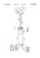
- FIG. 1is a diagrammatic view of an endoscope equipped with a rotatable joint between sets of optical fibers in the handle and in the insertion tube of the endoscope.
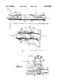
- FIG. 2is a cross-sectional view of the endoscope of FIG. 1.
- FIG. 3shows one of the components of the rotatable joint in detail.
- FIG. 4is a cross-section of a portion of the component of FIG. 3, taken along line 4--4.
- FIG. 5is an enlarged cross-sectional view of the rotatable joint.
- FIG. 6is an enlarged cross-sectional view of the insertion tube of the endoscope of FIG. 1 and of another component of the rotatable joint.
- FIG. 7is a cross-section of a portion of the component of FIG. 6, taken along line 7--7.
- FIG. 8is a cross-section, taken along line 8--8 of FIG. 6, showing the distal viewing tip of the endoscope.
- FIG. 9illustrates the actuator of the rotatable joint.
- endoscope 10suitable for viewing into a body cavity, joint space, or passageway during arthroscopic surgery, includes an elongated insertion tube 12 rotatably mounted to handle 14.
- Actuator 16which is also rotatably mounted to handle 14, is manipulated by a user to rotate insertion tube 12 about a longitudinal axis 18 to rotatably orient insertion tube 12 with respect to an area to be viewed, e.g., area A or area B.
- External light source 1provides the light used to illuminate areas A and B via a light cable connected to a proximal surface 5 of handle 14 at coupler 4.
- a first set 20 of optical fibersextending through handle 14 from light coupler 4 to joint 30 (shown schematically), transmits light received from source 1 to a second set 40 of optical fibers, which meet optical fiber set 20 at joint 30.
- the optical fibers in set 40extend from joint 30 through insertion tube 12 to distal viewing tip 41, to transmit light out distal viewing tip 41 to area A or area B.
- joint 30enables relative rotation between optical fiber sets 20, 40 about axis 18 when insertion member 12 is rotated with respect to handle 14 by actuator 16, while also providing efficient light transmission between them.
- Endoscope 10has an off-axis direction of view, i.e., a direction of view oriented at a nonzero angle with respect to longitudinal axis 18.
- the direction of viewis determined by the orientation of a lens assembly 50 (FIG. 2) at distal viewing tip 41 (i.e., the angle formed between longitudinal axis 18 and a vector 21 that is perpendicular to the lens at distal viewing tip 41) and is typically 30°, 45°, or 70°.
- the field of view, angle ⁇is an angle within which the endoscope receives light from external objects, i.e., the angle over which the endoscope "sees," and is equidistant on either side of vector 21.
- Endoscope 10illuminates (and views, as described below) area A when insertion tube is oriented as shown in FIG. 1.
- a userrotates actuator 16, which rotates insertion tube 12 with respect to handle 14 about longitudinal axis 18, to orient distal viewing tip 41 to view area B along vector 21' (shown in dashed lines).
- Rotatable joint 30allows set 20 of optical fibers and external cable 2 to remain stationary when insertion tube 12 is rotated. As a result, cable 2 does not become twisted or wound about handle 14 when insertion tube is rotated. This makes endoscope 10 easier to use and manipulate in the body. Moreover, the stresses imposed on optical fiber set 20 and cable 2 are significantly reduced, particularly at light coupler 4, which increases reliability and operating lifetime.
- CCDcharge-coupled-device
- FIG. 2light from objects illuminated by endoscope 10 is collected by lens assembly 50 mounted in distal viewing tip and passed to a charge-coupled-device (CCD) 52 or other suitable image detector supported proximally to lens assembly 50 at the distal end of a CCD tube 54.
- CCD 52converts the light into electrical signals representing an image of the objects.
- the electrical signalsare transmitted (by wiring which extends through CCD tube 54 and through handle 14) to a camera control unit 6 for processing in the usual manner.
- the resultant imageis displayed on display screen 8 (FIG. 1).
- CCD 52including its associated electrical and optical systems, are of the type described in detail in copending U.S. patent application Ser. No. 07/958,688, filed Oct. 9, 1992, incorporated herein by reference.
- CCD tube 54 and CCD 52are moved axially along longitudinal axis 18 with respect to lens assembly 50. This motion changes the spacing between lens assembly 50 and CCD 52, thereby adjusting the focus of the image.
- rotatable joint 30will be described in detail.
- the individual optical fibers of sets 20, 40are arranged in a pair of annular fiber rings 26, 46, each of which surrounds CCD tube 54, is centered about longitudinal axis 18, and extends for a short distance along longitudinal axis 18.
- Annular fiber rings 26, 46abut each other at joint 30 for efficient light transmission therebetween, and are mounted within handle 14 for relative rotation, as will be described.
- FIGS. 3 and 4show the construction of set 20 of optical fibers and a stationary annulus 22 that defines annular fiber ring 26.
- Sheath 56carries optical fiber set 20 from light coupler 4 to joint 30.
- the distal ends of individual glass fibers 19 of set 20are removed from sheath 56 and are arranged evenly around the exterior surface of an inner ring 23.
- a slightly larger, outer ring 24surrounds inner ring 23 and captures fibers 19 therebetween.
- Inner and outer rings 23, 24each extend along longitudinal axis 18 and are sized such that individual optical fibers 19 are densely arranged between inner ring 23 and outer ring 24. Fibers 19 are epoxied in place between rings 23, 24 and are cut off at the face 28 of rings 23, 24.
- the ends of optical fibers 19are polished flat to form annular fiber ring 26.
- annular fiber ring 46likewise is formed by securing the proximal ends of individual optical fibers 39 of set 40 between an elongated inner ring 43 and an elongated outer ring 44, which together form a rotatable annulus 42.
- Optical fibers 39are arranged evenly around the exterior of ring 43, and are surrounded by outer ring 44 to form annular ring 46 of optical fibers at a face 48 of rings 43, 44.
- Inner and outer rings 43, 44are sized such that individual optical fibers 39 are densely arranged therebetween for maximum light transfer.
- Individual optical fibers 39 of optical fibers 40are secured between inner and outer rings 43, 44 by, for example, an epoxy, and are also arranged such that at ring face 48, the individual optical fibers are parallel to longitudinal axis 18 and are polished flat at the ends, for efficient light transmission.
- Channel 58carries optical fibers 39 from joint 30 to distal viewing tip 41.
- annular fiber rings 26, 46are each centered about longitudinal axis 18 and are in radial alignment.
- the radial thickness 27 of annular fiber ring 26is, however, greater than the radial thickness 47 of annular fiber ring 46.
- radial thickness 27is 0.3 mm
- radial thickness 47is 0.2 mm (and as a result, fiber ring 26 extends 0.05 mm radially inwardly and outwardly of fiber ring 46).
- the individual optical fibers 19, 39are each about 2.0 mil in diameter.
- annuli 22, 42are each disposed over a sleeve 60 within handle 14 (CCD tube 54 passes through sleeve 60) so that faces 28, 48 (and hence fiber rings 26, 46) abut each other.
- Sleeve 60is bonded to rotating annulus inner ring 43 by a retaining compound, such as, for example, Loctite 620.
- Sleeve 60centers annular fiber rings 26, 46 with respect to longitudinal axis. The radial overlap between fiber rings 26, 46 discussed above helps ensure efficient light transfer between fibers 19, 39, even if annular fiber rings 26, 46 are slightly radially misaligned.
- a light transmissive fluid 62such as immersion oil, e.g., type A manufactured by Cargille Labs
- Fluid 62has index of refraction substantially equal to that of one, or preferably both, sets 20, 40 of optical fibers 20, 40.
- fluid 62lubricates joint 30 for ease of use.
- a seal 64preferably made from rubber, retains fluid 62 within joint 30 between faces 28, 48. Seal 64 is sized to securely fit over the abutting portions of outer rings 24, 44. The proximal and distal ends of seal 64 are enlarged and snugly disposed in corresponding grooves 66, 68 machined in outer rings 24, 44, respectively.
- 0-ring 70provides a seal between stationary annulus inner ring 23 and sleeve 60 to prevent fluid 62 from leaking from the proximal end of joint 30 into handle 14.
- Stationary annulus 22is biased toward rotating annulus 42 by coil spring 72, which exerts approximately a one pound force, to urge stationary ring face 28 against rotating ring face 48. This further improves light transfer by reducing the gap between fiber rings 26, 46 to as small as possible.
- One end of spring 72abuts an inner wall 80 of handle 14.
- the other end of spring 72engages one end of a cylindrical spring retainer 78.
- the opposite (proximal) end of spring retainerengages a retaining ring 74 secured within a groove 76 machined in outer ring 24.
- retaining ring 74acts as a flange surface for spring retainer 78.
- Cylindrical spring retainer 78surrounds seal 64 and the abutting faces 28, 48 of annuli 22, 42.
- the force of spring 72urges spring retainer 78 distally away from wall 80, thereby biasing stationary annulus 22 (via retaining ring 74) against rotating annulus 42.
- insertion tube 12includes inner tube 90 and outer tube 92 (CCD tube 54 is disposed within inner tube 90).
- Inner tube 90 and outer tube 92are aligned along different axes--longitudinal axis 18 and longitudinal axis 88, respectively.
- the offset between longitudinal axis 18 and longitudinal axis 88which is preferably about 0.013 inches, creates the above-described channel 58 between inner tube 92 and outer tube 90 for optical fibers 40.
- CCD tube 54, CCD 52, and lens assembly 50are aligned along longitudinal axis 18.
- window lens 94 of lens assembly 50is oriented with respect to longitudinal axis 18 to determine the direction of view of endoscope 10 and could be recessed from the distal end of outer tube 92 to protect its exposed distal surface.
- Wedge 96at distal viewing tip 41, deflects optical fibers 40 such that light is directed along the direction of view in accordance with the orientation of window lens 94, e.g., at 30° with respect to axis 18.
- individual optical fibers 39 of optical fibers 40are arranged in a crescent shape, corresponding to the shape of channel 58 formed between inner tube 90 and outer tube 92, to disperse the light across the width of insertion tube 12.
- Lens assembly 50, wedge 96, and optical fibers 40are all bonded within the distal end of outer tube 92 by, for example, an epoxy. Lens assembly 50 is also epoxied to the distal end of inner tube 90. CCD tube 54 is axially movable within inner tube 90 for focussing, as discussed above.
- outer tube 92The proximal end of outer tube 92 is inserted into an eccentric bore 98, aligned about longitudinal axis 88, within the distal end of rotating annulus outer ring 44.
- Outer tube 92is attached to the distal end of outer ring 44 by, for example, weld 86.
- Inner tube 90is attached to inner wall 99 of rotating annulus inner ring 43, by, for example, an epoxy.
- Actuator 16is inserted onto the distal end of handle 14 over tubes 90, 92.
- Thrust washer 100, wave spring 101, threaded retainer 102, and thrust washer 104are radially mounted over the proximal end of actuator 16.
- Thrust washers 100, 104provide radial bearings and are preferably made from a plastic having exceptional bearing characteristics, such as, for example, Torlon 4301.
- Retainer 106threaded into rotating annulus outer ring 44, holds tubes 90, 92 (and the components of joint 30) and actuator 16, respectively, within handle 14.
- Seal 105preferably a quad ring, seals the interface between actuator 16 and handle 14. Seals 107 avoid contaminants from entering handle 14 at the interface between actuator 16 and ring 43.
- the proximal end of actuator 16includes tabs 108 (only one of which is shown) which engage corresponding slots 110 (FIG. 7) formed in rotating annulus outer ring 44. Accordingly, when actuator 16 is rotated, tabs 108 engage slots 110 to rotate annulus outer ring 44, thereby also rotating inner ring 43, optical fibers 40, and tubes 90, 92 (which are attached to inner and outer rings 43, 44, respectively) about longitudinal axis 18.
- Anti-rotation tabs 112(see FIG. 3) on stationary annulus outer ring 24 engage throat 113 in handle 14 to prevent the frictional forces between stationary annulus 22 and rotating annulus 44 to cause stationary annulus 22 to rotate.
- CCD tube 54which is coupled to handle 14 at the focusing mechanism (not shown), including CCD 52 and its associated electrical and optical systems, also remains stationary.
- FIG. 2shows the connection between external fiber optic cable 2 (FIG. 1) and optical fiber set 20 at the proximal end 5 of handle 14.
- the individual fibers 19 of set 20are epoxied into threaded bushing 34, and the proximal ends 36 of optical fibers 29 are uniformly polished for efficiently receiving light from light coupler 4.
- Light coupler 4is designed to change the numerical aperture of light cable 2, which has a relatively low value (e.g., 0.54) for efficient light transfer, to a higher value (e.g., 0.81), which is desirable for adequately illuminating area A (FIG. 1). This is done by providing light coupler 4 with different inside diameters 4a, 4b at its ends.
- diameter 4ais relatively large (51/4 mm) where cable enters coupler 4, and is reduced substantially (to a diameter, 4b, of 31/2 mm) at the junction between cable 2 and optical fiber set 28.
- the ratio between diameters 4a and 4b multiplied by the numerical aperture of cable 2(0.54) provides the desired numerical aperture (0.81) of optical fiber set 28.
- insertion tube 12 of endoscope 10is inserted into a body cavity, joint space, or passageway during arthroscopic surgery.
- Distal viewing tip 41is oriented such that the desired area (e.g., area A, FIG. 1) within the body cavity is within the field of view of endoscope 10 and is illuminated by light emitted from optical fibers 39 at distal viewing tip 41.
- Light from objects in field of viewis collected by lens assembly 50 and passed to CCD 52.
- CCD 52converts the received optical image to electrical signals, which are processed by CCU 6 to display an image of the objects on display screen 8.
- actuator 16To view a different area (such as area B), the user rotates actuator 16 while holding handle 14 stationary. As discussed, the rotation of actuator 16 causes tubes 90, 92, and hence also lens assembly 50, to rotate about longitudinal axis. In addition, actuator 16 rotates annulus 42--and hence annular fiber ring 46--with respect to annulus 22 and annular fiber ring 26. The rotation is not conveyed to either set 20 of optical fibers or external light cable 2.
- inner tube 90 and outer tube 92may be coaxial so that the channel created for optical fibers 39 is annular.
- other arrangements of individual optical fibers 39 at distal viewing tip 41can be used.
- optical fibers 99can be arranged in an annular ring about the perimeter of window lens 94.
- endoscopecan include the CCD rotation feature described in a patent application entitled “Endoscope” filed concurrently herewith, assigned to the present assignee, and incorporated herein by reference.
Landscapes
- Health & Medical Sciences (AREA)
- Life Sciences & Earth Sciences (AREA)
- Physics & Mathematics (AREA)
- Surgery (AREA)
- Optics & Photonics (AREA)
- Biomedical Technology (AREA)
- Public Health (AREA)
- Biophysics (AREA)
- Pathology (AREA)
- Radiology & Medical Imaging (AREA)
- Veterinary Medicine (AREA)
- Engineering & Computer Science (AREA)
- Nuclear Medicine, Radiotherapy & Molecular Imaging (AREA)
- Heart & Thoracic Surgery (AREA)
- Medical Informatics (AREA)
- Molecular Biology (AREA)
- Animal Behavior & Ethology (AREA)
- General Health & Medical Sciences (AREA)
- General Physics & Mathematics (AREA)
- Orthopedic Medicine & Surgery (AREA)
- Physical Education & Sports Medicine (AREA)
- Astronomy & Astrophysics (AREA)
- Endoscopes (AREA)
- Instruments For Viewing The Inside Of Hollow Bodies (AREA)
Abstract
Description
This invention relates to endoscopes which use optical fibers to provide illumination.
Endoscopes are widely used to inspect regions of the body (e.g., joint spaces) through a small puncture wound. Typically, the endoscope includes an elongated insertion tube equipped with a set of optical fibers which extend from a proximal handle, through the insertion tube to the distal viewing tip of the endoscope. A cable that rigidly attaches to the handle (e.g., at a post on the side of the handle) carries light from an external light source to the proximal end of the optical fibers, and the light is transmitted through the optical fibers to the distal viewing end, where the light is emitted to illuminate the region under inspection.
Received light representing an optical image of the body region is collected by a lens assembly mounted in the distal viewing end and is passed to, e.g., a solid-state image pickup device (such as a charge-coupled-device, or CCD). The CCD converts the received optical image to electrical signals that are processed for viewing on a display.
Some endoscopes have a direction of view (i.e., the direction along which the endoscope emits and receives light) along the longitudinal axis of the insertion tube. The distal viewing ends of other endoscopes are constructed to provide an off-axis direction of view (e.g., at 30° or at 70°). The insertion tube of the latter type of endoscope is often rotatable with respect to the handle to allow the user to view different areas without requiring that the entire endoscope be rotated.
This invention features a rotatable joint between sets of optical fibers. The invention is particularly, but by no means exclusively, useful in an endoscope having an insertion tube rotatably mounted to the endoscope handle. In this case, one set of optical fibers is carried by the insertion tube, with another set of optical fibers being disposed in the handle.
The rotatable joint between the sets of optical fibers allows the insertion tube to be rotated (e.g., to change the viewing angle of the endoscope) without also rotating either the optical fibers in the handle or an external fiber optic cable that connects the handle to a light source. The fiber optic cable thus remains stationary and does not become twisted or wrapped around the handle when the insertion tube is rotated. The maneuverability of the endoscope is enhanced and the risk of damaging the fiber optic cable (and its associated connectors) is reduced substantially.
In one aspect of the invention, a portion of each of the first and second sets of optical fibers extends along a longitudinal axis, and the joint is constructed to allow relative rotation between the sets of optical fibers about the longitudinal axis.
Preferred embodiments include the following features.
The ends of the first and second sets of optical fibers are spring-biased together. This improves light transmission efficiency by reducing the spacing between the ends as much as possible.
Light transfer efficiency at the joint is further enhanced by a light transmitting substance, such as an immersion oil, placed between the ends of the respective sets of fibers. Preferably, the substance has an index of refraction substantially equal to that of at least one of sets of optical fibers. The substance also lubricates the joint. The joint is sealed to avoid leakage of the substance.
Another aspect of the invention features an endoscope, equipped with the rotatable joint.
Preferred embodiments include the following features. The endoscope includes an insertion member rotatably mounted to a handle. The handle and insertion member carry the first and second set of optical fibers, respectively. Individual fibers of the first and second sets of optical fibers are arranged in first and second annular rings, respectively. These annular rings are adjacent to one another, are centered about the longitudinal axis, and are spring-biased together. The width of the first annular ring is greater than the width of the second annular ring, thereby maintaining efficient light transfer even if the rings become misaligned.
The endoscope also includes an optical element and an image detector mounted in the distal end of the insertion tube. Preferably, the optical element (which includes at least one lens) is configured to provide a direction of view at a nonzero angle with respect to the longitudinal axis. The image detector is a solid-state image pickup device, such as a CCD.
Other features and advantages of the invention will become apparent from the following detailed description, and from the claims.
FIG. 1 is a diagrammatic view of an endoscope equipped with a rotatable joint between sets of optical fibers in the handle and in the insertion tube of the endoscope.
FIG. 2 is a cross-sectional view of the endoscope of FIG. 1.
FIG. 3 shows one of the components of the rotatable joint in detail.
FIG. 4 is a cross-section of a portion of the component of FIG. 3, taken alongline 4--4.
FIG. 5 is an enlarged cross-sectional view of the rotatable joint.
FIG. 6 is an enlarged cross-sectional view of the insertion tube of the endoscope of FIG. 1 and of another component of the rotatable joint.
FIG. 7 is a cross-section of a portion of the component of FIG. 6, taken along line 7--7.
FIG. 8 is a cross-section, taken alongline 8--8 of FIG. 6, showing the distal viewing tip of the endoscope.
FIG. 9 illustrates the actuator of the rotatable joint.
Referring to FIG. 1,endoscope 10, suitable for viewing into a body cavity, joint space, or passageway during arthroscopic surgery, includes anelongated insertion tube 12 rotatably mounted to handle 14.Actuator 16, which is also rotatably mounted to handle 14, is manipulated by a user to rotateinsertion tube 12 about alongitudinal axis 18 to rotatablyorient insertion tube 12 with respect to an area to be viewed, e.g., area A or area B.
The construction and operation ofjoint 30 are described in detail below. Suffice it here to say thatjoint 30 enables relative rotation betweenoptical fiber sets axis 18 wheninsertion member 12 is rotated with respect to handle 14 byactuator 16, while also providing efficient light transmission between them.
Referring to FIG. 2, light from objects illuminated byendoscope 10 is collected bylens assembly 50 mounted in distal viewing tip and passed to a charge-coupled-device (CCD) 52 or other suitable image detector supported proximally tolens assembly 50 at the distal end of aCCD tube 54.CCD 52 converts the light into electrical signals representing an image of the objects. The electrical signals are transmitted (by wiring which extends throughCCD tube 54 and through handle 14) to acamera control unit 6 for processing in the usual manner. The resultant image is displayed on display screen 8 (FIG. 1).
Referring also to FIGS. 3-6, rotatable joint 30 will be described in detail. At joint 30, the individual optical fibers ofsets CCD tube 54, is centered aboutlongitudinal axis 18, and extends for a short distance alonglongitudinal axis 18. Annular fiber rings 26, 46 abut each other at joint 30 for efficient light transmission therebetween, and are mounted withinhandle 14 for relative rotation, as will be described.
FIGS. 3 and 4 show the construction ofset 20 of optical fibers and a stationary annulus 22 that definesannular fiber ring 26.Sheath 56 carries optical fiber set 20 fromlight coupler 4 to joint 30. At joint 30, the distal ends ofindividual glass fibers 19 ofset 20 are removed fromsheath 56 and are arranged evenly around the exterior surface of aninner ring 23. A slightly larger,outer ring 24 surroundsinner ring 23 and capturesfibers 19 therebetween. Inner andouter rings longitudinal axis 18 and are sized such that individualoptical fibers 19 are densely arranged betweeninner ring 23 andouter ring 24.Fibers 19 are epoxied in place betweenrings face 28 ofrings optical fibers 19 are polished flat to formannular fiber ring 26.
Referring as well to FIGS. 6-7,annular fiber ring 46 likewise is formed by securing the proximal ends of individualoptical fibers 39 ofset 40 between an elongatedinner ring 43 and an elongatedouter ring 44, which together form arotatable annulus 42.Optical fibers 39 are arranged evenly around the exterior ofring 43, and are surrounded byouter ring 44 to formannular ring 46 of optical fibers at aface 48 ofrings outer rings optical fibers 39 are densely arranged therebetween for maximum light transfer. Individualoptical fibers 39 ofoptical fibers 40 are secured between inner andouter rings ring face 48, the individual optical fibers are parallel tolongitudinal axis 18 and are polished flat at the ends, for efficient light transmission.
Referring in particular to FIGS. 4 and 7, annular fiber rings 26, 46 are each centered aboutlongitudinal axis 18 and are in radial alignment. Theradial thickness 27 ofannular fiber ring 26 is, however, greater than theradial thickness 47 ofannular fiber ring 46. For example,radial thickness 27 is 0.3 mm, whileradial thickness 47 is 0.2 mm (and as a result,fiber ring 26 extends 0.05 mm radially inwardly and outwardly of fiber ring 46). (The individualoptical fibers
Referring to FIG. 5,annuli 22, 42 are each disposed over asleeve 60 within handle 14 (CCD tube 54 passes through sleeve 60) so that faces 28, 48 (and hence fiber rings 26, 46) abut each other.Sleeve 60 is bonded to rotating annulusinner ring 43 by a retaining compound, such as, for example, Loctite 620.Sleeve 60 centers annular fiber rings 26, 46 with respect to longitudinal axis. The radial overlap between fiber rings 26, 46 discussed above helps ensure efficient light transfer betweenfibers
Aseal 64, preferably made from rubber, retainsfluid 62 within joint 30 betweenfaces Seal 64 is sized to securely fit over the abutting portions ofouter rings seal 64 are enlarged and snugly disposed in correspondinggrooves 66, 68 machined inouter rings ring 70 provides a seal between stationary annulusinner ring 23 andsleeve 60 to prevent fluid 62 from leaking from the proximal end of joint 30 intohandle 14.
Stationary annulus 22 is biased towardrotating annulus 42 bycoil spring 72, which exerts approximately a one pound force, to urgestationary ring face 28 againstrotating ring face 48. This further improves light transfer by reducing the gap between fiber rings 26, 46 to as small as possible. One end ofspring 72 abuts aninner wall 80 ofhandle 14. The other end ofspring 72 engages one end of acylindrical spring retainer 78. The opposite (proximal) end of spring retainer engages a retainingring 74 secured within a groove 76 machined inouter ring 24. Thus, retainingring 74 acts as a flange surface forspring retainer 78. (Adiscrete retaining ring 74 is used instead of an integral flange because during manufacturing it is necessary to grip stationary annulus 22 to polish the ends of the individualoptical fibers 19 atring face 28; an integral flange would make this manufacturing step more difficult.)
Referring to FIGS. 2, 6, and 8,insertion tube 12 includesinner tube 90 and outer tube 92 (CCD tube 54 is disposed within inner tube 90).Inner tube 90 andouter tube 92 are aligned along different axes--longitudinal axis 18 andlongitudinal axis 88, respectively. The offset betweenlongitudinal axis 18 andlongitudinal axis 88, which is preferably about 0.013 inches, creates the above-describedchannel 58 betweeninner tube 92 andouter tube 90 foroptical fibers 40.CCD tube 54,CCD 52, andlens assembly 50 are aligned alonglongitudinal axis 18.
Atdistal viewing tip 41,window lens 94 oflens assembly 50 is oriented with respect tolongitudinal axis 18 to determine the direction of view ofendoscope 10 and could be recessed from the distal end ofouter tube 92 to protect its exposed distal surface.Wedge 96, atdistal viewing tip 41, deflectsoptical fibers 40 such that light is directed along the direction of view in accordance with the orientation ofwindow lens 94, e.g., at 30° with respect toaxis 18. As shown in FIG. 8, individualoptical fibers 39 ofoptical fibers 40 are arranged in a crescent shape, corresponding to the shape ofchannel 58 formed betweeninner tube 90 andouter tube 92, to disperse the light across the width ofinsertion tube 12.
The proximal end ofouter tube 92 is inserted into aneccentric bore 98, aligned aboutlongitudinal axis 88, within the distal end of rotating annulusouter ring 44.Outer tube 92 is attached to the distal end ofouter ring 44 by, for example,weld 86.Inner tube 90 is attached toinner wall 99 of rotating annulusinner ring 43, by, for example, an epoxy.Actuator 16 is inserted onto the distal end ofhandle 14 overtubes Thrust washer 100,wave spring 101, threadedretainer 102, and thrustwasher 104 are radially mounted over the proximal end ofactuator 16. Thrustwashers
Referring as well to FIG. 9, the proximal end ofactuator 16 includes tabs 108 (only one of which is shown) which engage corresponding slots 110 (FIG. 7) formed in rotating annulusouter ring 44. Accordingly, whenactuator 16 is rotated,tabs 108 engageslots 110 to rotate annulusouter ring 44, thereby also rotatinginner ring 43,optical fibers 40, andtubes 90, 92 (which are attached to inner andouter rings longitudinal axis 18. Anti-rotation tabs 112 (see FIG. 3) on stationary annulusouter ring 24 engagethroat 113 inhandle 14 to prevent the frictional forces between stationary annulus 22 androtating annulus 44 to cause stationary annulus 22 to rotate.CCD tube 54, which is coupled to handle 14 at the focusing mechanism (not shown), includingCCD 52 and its associated electrical and optical systems, also remains stationary.
FIG. 2 shows the connection between external fiber optic cable 2 (FIG. 1) and optical fiber set 20 at theproximal end 5 ofhandle 14. Theindividual fibers 19 ofset 20 are epoxied into threadedbushing 34, and the proximal ends 36 of optical fibers 29 are uniformly polished for efficiently receiving light fromlight coupler 4.Light coupler 4 is designed to change the numerical aperture oflight cable 2, which has a relatively low value (e.g., 0.54) for efficient light transfer, to a higher value (e.g., 0.81), which is desirable for adequately illuminating area A (FIG. 1). This is done by providinglight coupler 4 with differentinside diameters diameter 4a is relatively large (51/4 mm) where cable enterscoupler 4, and is reduced substantially (to a diameter, 4b, of 31/2 mm) at the junction betweencable 2 and optical fiber set 28. The ratio betweendiameters
In use,insertion tube 12 ofendoscope 10 is inserted into a body cavity, joint space, or passageway during arthroscopic surgery.Distal viewing tip 41 is oriented such that the desired area (e.g., area A, FIG. 1) within the body cavity is within the field of view ofendoscope 10 and is illuminated by light emitted fromoptical fibers 39 atdistal viewing tip 41. Light from objects in field of view is collected bylens assembly 50 and passed toCCD 52.CCD 52 converts the received optical image to electrical signals, which are processed byCCU 6 to display an image of the objects ondisplay screen 8.
To view a different area (such as area B), the user rotatesactuator 16 while holdinghandle 14 stationary. As discussed, the rotation ofactuator 16causes tubes lens assembly 50, to rotate about longitudinal axis. In addition,actuator 16 rotatesannulus 42--and henceannular fiber ring 46--with respect to annulus 22 andannular fiber ring 26. The rotation is not conveyed to either set 20 of optical fibers or externallight cable 2.
Other embodiments are within the scope of the following claims.
For example,inner tube 90 andouter tube 92 may be coaxial so that the channel created foroptical fibers 39 is annular. Likewise, other arrangements of individualoptical fibers 39 atdistal viewing tip 41 can be used. For example,optical fibers 99 can be arranged in an annular ring about the perimeter ofwindow lens 94.
Other features may be added to the endoscope. For example, focussing capability may be provided, as described in the aforementioned '668 patent application. In addition, the endoscope can include the CCD rotation feature described in a patent application entitled "Endoscope" filed concurrently herewith, assigned to the present assignee, and incorporated herein by reference.
Claims (21)
1. An apparatus for viewing a remote region, comprising:
a handle;
an elongated outer member configured to be inserted into the remote region and having a proximal end rotatably mounted to said handle;
a device disposed in a distal end of said outer member for viewing the remote region, a portion of said device being coupled to said handle by an inner member disposed along an axis within said outer member;
a first set of optical fibers disposed in said handle and having a distal end at which individual fibers thereof are arranged around said inner member;
a second set of optical fibers disposed in said outer member and having a proximal end at which individual fibers thereof are arranged around said inner member; and
a joint between said distal end of said first set of optical fibers and said proximal end of said second set of optical fibers, said inner member extending through said joint, said joint being constructed to allow relative rotation between said first and second sets of optical fibers about said axis of said inner member in response to relative rotation between said outer member and said handle.
2. The apparatus of claim 1 wherein said joint is constructed to maintain said distal end of said first set of optical fibers and said proximal end of said second set of optical fibers adjacent to one another for light transmission therebetween.
3. An apparatus for viewing a remote region, comprising:
a handle;
an elongated outer member configured to be inserted into the remote region and having a proximal end rotatably mounted to said handle;
a device disposed in a distal end of said outer member for viewing the remote region, a portion of said device being coupled to said handle by an inner member disposed along an axis within said outer member;
a first set of optical fibers disposed in said handle and having a distal end at which individual fibers thereof are arranged around said inner member;
a second set of optical fibers disposed in said outer member and having a proximal end at which individual fibers thereof are arranged around said inner member;
a joint between said distal end of said first set of optical fibers and said proximal end of said second set of optical fibers, said inner member extending through said joint, said joint being constructed to allow relative rotation between said first and second sets of optical fibers about said axis of said inner member in response to relative rotation between said outer member and said handle; and
a spring mounted to bias said distal end of said first set of optical fibers and said proximal end of said second set of optical fibers together to maintain said ends of said first and second sets of optical fibers adjacent to one another for light transmission therebetween.
4. The apparatus of claim 3 further comprising a light-transmitting substance disposed between said distal end of said first set of optical fibers and said proximal end of said second set of optical fibers.
5. An apparatus comprising:
a first set of optical fibers having at least a portion extending along a longitudinal axis;
a second set of optical fibers having at least a portion extending along said longitudinal axis;
a joint between an end of said portion of said first set of optical fibers and an end of said portion of said second set of optical fibers constructed to allow relative rotation between said portions about said longitudinal axis;
a spring mounted to bias said ends of said portions of said first set of optical fibers and said second set of optical fibers together to maintain said ends adjacent to one another for light transmission therebetween; and
a light-transmitting substance disposed between said ends of said portions of said first set of optical fibers and said second set of optical fibers, said substance having an index of refraction substantially equal to an index of refraction of at least one of said first and said second sets of optical fibers.
6. An apparatus comprising:
a first set of optical fibers having at least a portion extending along a longitudinal axis;
a second set of optical fibers having at least a portion extending along said longitudinal axis;
a joint between an end of said portion of said first set of optical fibers and an end of said portion of said second set of optical fibers constructed to allow relative rotation between said portions about said longitudinal axis;
a spring mounted to bias said ends of said portions of said first set of optical fibers and said second set of optical fibers together to maintain said ends adjacent to one another for light transmission therebetween; and
immersion oil disposed between said ends of said portions of said first set of optical fibers and said second set of optical fibers.
7. The apparatus of claim 5 wherein said substance is a liquid and said joint is constructed to contain said liquid between said ends of said portions of said first and said second sets of optical fibers.
8. An apparatus for delivering light from a source to a region remote from the source and viewing the region, comprising:
a handle;
an elongated outer member having a proximal end rotatably mounted to said handle, said outer member configured to be inserted into the remote region;
a device disposed in a distal end of said outer member for viewing the remote region, a portion of said device being coupled to said handle by an inner member disposed along an axis within said outer member;
a first set of optical fibers carried within said handle and having a proximal end configured to receive light from the source and a distal end at which individual fibers thereof are arranged around said inner member; and
a second set of optical fibers carried within said outer member and having a proximal end and a distal end, said distal end of said second set of optical fibers being configured to transmit light from said outer member into said remote region, individual fibers of said second set of optical fibers being arranged around said inner members at said proximal end of said second set of fibers; and
said outer member being rotatably mounted to said handle at a joint between said distal end of said first set of optical fibers and said proximal end of said second set of optical fibers, said inner member extending through said joint, said joint being constructed to allow relative rotation between said first and second sets of optical fibers about said axis of said inner member in response to relative rotation between said outer member and said handle.
9. An apparatus for delivering light from a source to a region remote from the source and viewing the region, comprising:
a handle;
an elongated outer member having a proximal end rotatably mounted to said handle, said outer member configured to be inserted into the remote region;
a device disposed in a distal end of said outer member, a portion of said device being coupled to said handle by an inner member disposed along a longitudinal axis within said outer member;
a first set of optical fibers carried within said handle and having a proximal end and a distal end, said proximal end being configured to receive light from the source, said distal end of said first set of optical fibers being configured so that individual fibers thereof are arranged in a first annular ring around said inner member;
a second set of optical fibers carried within outer member, said second set of optical fibers having a proximal end and a distal end, said distal end configured to transmit light from said outer member into said remote region, said proximal end of said second set of optical fibers being configured so that individual fibers thereof are arranged in a second annular ring around said inner member;
said outer member being rotatably mounted to said handle at a joint, said inner member extending through said joint, said joint being constructed to position said first and said second annular rings adjacent to one another and centered about said longitudinal axis and to allow relative rotation between said first and second sets of optical fibers about said longitudinal axis of said inner member in response to relative rotation between said outer member and said handle.
10. The apparatus of claim 9 wherein said first annular ring is configured with a width greater than a width of said second annular ring.
11. The apparatus of claim 9 further comprising a substance disposed between said first annular ring and said second annular ring, said joint being constructed to contain said substance between said first and said second annular rings.
12. The apparatus of claim 9 wherein said joint includes a spring to bias said first annular ring and said second annular ring together.
13. An endoscope comprising:
a handle that carries a first set of optical fibers having a proximal end and a distal end, said proximal end being configured to receive light from a light source,
an elongated outer member configured to be inserted into a remote region and rotatably mounted to said handle, said outer member carrying a second set of optical fibers having a proximal end and a distal end configured to transmit light from said distal end of said outer member to the remote region,
a device disposed at said distal end of said outer member for viewing the remote region, said device including an optical element and an image detector disposed adjacent to said optical element, said image detector being coupled to said handle by an inner member disposed along an axis within said outer member,
said distal end of said first set of optical fibers including a first ring of individual fibers arranged around said inner member,
said proximal end of said second set of optical fibers including a second ring of individual fibers arranged around said inner member,
said first ring of optical fibers and said second ring of optical fibers being disposed adjacent to each other at a rotatable joint between said outer member and said handle, said inner member extending through said joint, said joint being constructed to allow relative rotation between said first and second sets of optical fibers about said axis of said inner member in response to relative rotation between said outer member and said handle.
14. The endoscope of claim 13 wherein said optical element is configured to provide a direction of view at a nonzero angle with respect to said longitudinal axis.
15. The endoscope of claim 13 wherein said optical element includes at least one lens.
16. The endoscope of claim 13 wherein said image detector is a solid-state image pickup device.
17. The apparatus of claim 1 wherein said individual fibers at said distal end of said first set of optical fibers are symmetrically arranged about said axis, and said individual fibers at said proximal end of said second set of optical fibers are symmetrically arranged about said axis.
18. The apparatus of claim 1 wherein said individual fibers of said distal end of said first set of optical fibers are arranged in an annular ring about said axis, and said individual fibers of said proximal end of said second set of optical fibers are arranged in an annular ring about said axis.
19. The apparatus of claim 8 wherein said individual fibers at said distal end of said first set of optical fibers are symmetrically arranged about said axis, and said individual fibers at said proximal end of said second set of optical fibers are symmetrically arranged about said axis.
20. The apparatus of claim 8 wherein said individual fibers of said distal end of said first set of optical fibers are arranged in an annular ring about said axis, and said individual fibers of said proximal end of said second set of optical fibers are arranged in an annular ring about said axis.
21. An endoscope comprising:
a handle that carries a first set of optical fibers having a distal end;
an elongated outer member rotatably mounted to said handle, said outer member being configured to be inserted into a body and carrying a second set of optical fibers having a proximal end;
a device disposed in a distal end of said outer member for viewing the body, a portion of said device being coupled to said handle by an inner member disposed along an axis within said outer member, individual fibers at said distal end of said first set of optical fibers being arranged around said inner member, individual fibers at said proximal end of said second set of optical fibers being arranged around said inner member; and
a joint between said distal end of said first set of optical fibers and said proximal end of said second set of optical fibers, said inner member extending through said joint, said joint being constructed to allow relative rotation between said first and second sets of optical fibers about said axis of said inner member in response to relative rotation between said outer member and said handle.
Priority Applications (3)
| Application Number | Priority Date | Filing Date | Title |
|---|---|---|---|
| US08/475,900US5621830A (en) | 1995-06-07 | 1995-06-07 | Rotatable fiber optic joint |
| PCT/US1996/008821WO1996041225A1 (en) | 1995-06-07 | 1996-06-05 | Rotatable fibre optic joint |
| AU61515/96AAU6151596A (en) | 1995-06-07 | 1996-06-05 | Rotatable fibre optic joint |
Applications Claiming Priority (1)
| Application Number | Priority Date | Filing Date | Title |
|---|---|---|---|
| US08/475,900US5621830A (en) | 1995-06-07 | 1995-06-07 | Rotatable fiber optic joint |
Publications (1)
| Publication Number | Publication Date |
|---|---|
| US5621830Atrue US5621830A (en) | 1997-04-15 |
Family
ID=23889637
Family Applications (1)
| Application Number | Title | Priority Date | Filing Date |
|---|---|---|---|
| US08/475,900Expired - LifetimeUS5621830A (en) | 1995-06-07 | 1995-06-07 | Rotatable fiber optic joint |
Country Status (3)
| Country | Link |
|---|---|
| US (1) | US5621830A (en) |
| AU (1) | AU6151596A (en) |
| WO (1) | WO1996041225A1 (en) |
Cited By (136)
| Publication number | Priority date | Publication date | Assignee | Title |
|---|---|---|---|---|
| US5872879A (en)* | 1996-11-25 | 1999-02-16 | Boston Scientific Corporation | Rotatable connecting optical fibers |
| WO2001098801A3 (en)* | 2000-06-20 | 2002-06-06 | Schleifring Und Appbau Gmbh | Device for transmitting optical signals |
| US6432047B1 (en) | 1999-02-25 | 2002-08-13 | Micro Medical Devices, Inc. | Endoscopic surgical procedures and endoscopic apparatus comprising segmented fiber optic cables |
| US6464383B1 (en) | 1998-12-09 | 2002-10-15 | Steris Inc. | Fiber optic ceiling supported surgical task light system with optical commutator and manual zoom lens |
| US6478730B1 (en)* | 1998-09-09 | 2002-11-12 | Visionscope, Inc. | Zoom laparoscope |
| US20020198457A1 (en)* | 2001-04-30 | 2002-12-26 | Tearney Guillermo J. | Method and apparatus for improving image clarity and sensitivity in optical coherence tomography using dynamic feedback to control focal properties and coherence gating |
| US20030159471A1 (en)* | 2002-02-27 | 2003-08-28 | Wamin Optocomm Mfg. Corporation | Method for fabricating fiber optic joints |
| US20050004453A1 (en)* | 2003-01-24 | 2005-01-06 | Tearney Guillermo J. | System and method for identifying tissue using low-coherence interferometry |
| US20050018201A1 (en)* | 2002-01-24 | 2005-01-27 | De Boer Johannes F | Apparatus and method for ranging and noise reduction of low coherence interferometry lci and optical coherence tomography oct signals by parallel detection of spectral bands |
| US20050035295A1 (en)* | 2003-06-06 | 2005-02-17 | Brett Bouma | Process and apparatus for a wavelength tuning source |
| US20050089286A1 (en)* | 2003-10-16 | 2005-04-28 | Olympus Corporation | Endoscope |
| US20050128488A1 (en)* | 2003-11-28 | 2005-06-16 | Dvir Yelin | Method and apparatus for three-dimensional spectrally encoded imaging |
| US20050133373A1 (en)* | 2000-05-15 | 2005-06-23 | Ryoji Inaba | Capillary array electrophoresis apparatus and electrophoresis method |
| US20050228230A1 (en)* | 2004-04-07 | 2005-10-13 | Schara Nathan J | Gravity referenced endoscopic image orientation |
| US20060039004A1 (en)* | 2004-08-06 | 2006-02-23 | The General Hospital Corporation | Process, system and software arrangement for determining at least one location in a sample using an optical coherence tomography |
| US20060069307A1 (en)* | 2004-09-30 | 2006-03-30 | Scimed Life Systems, Inc. | Selectively rotatable shaft coupler |
| US20060067620A1 (en)* | 2004-09-29 | 2006-03-30 | The General Hospital Corporation | System and method for optical coherence imaging |
| US20060109478A1 (en)* | 2004-11-24 | 2006-05-25 | The General Hospital Corporation | Devices and arrangements for performing coherence range imaging using a common path interferometer |
| EP1685791A1 (en) | 2005-01-26 | 2006-08-02 | Karl Storz Development Corp. | Illumination system for variable direction of view instruments |
| US20060244973A1 (en)* | 2003-10-27 | 2006-11-02 | Seok-Hyun Yun | Method and apparatus for performing optical imaging using frequency-domain interferometry |
| EP1726254A1 (en)* | 2005-05-27 | 2006-11-29 | Karl Storz GmbH & Co. KG | Medical instrument for endoscopic operations |
| JP2007509710A (en)* | 2003-10-27 | 2007-04-19 | ダレル・アンド・ジテリス・インコーポレイテッド | Variable-field arthroscope using CCD |
| US20070112254A1 (en)* | 2005-11-16 | 2007-05-17 | Richard Wolf Gmbh | Optical instrument |
| US20070208400A1 (en)* | 2006-03-01 | 2007-09-06 | The General Hospital Corporation | System and method for providing cell specific laser therapy of atherosclerotic plaques by targeting light absorbers in macrophages |
| US20070229801A1 (en)* | 2005-09-29 | 2007-10-04 | The General Hospital Corporation | Arrangements and methods for providing multimodality microscopic imaging of one or more biological structures |
| US20070249899A1 (en)* | 2006-04-05 | 2007-10-25 | Alexander Seifert | Deflectable tip videoarthroscope |
| US20070274650A1 (en)* | 2006-02-01 | 2007-11-29 | The General Hospital Corporation | Apparatus for controlling at least one of at least two sections of at least one fiber |
| US20080013960A1 (en)* | 2000-11-10 | 2008-01-17 | The General Hospital Corporation | Apparatus and method for providing information for at least one structure |
| US20080094637A1 (en)* | 2003-01-24 | 2008-04-24 | The General Hospital Corporation | Apparatus and method for ranging and noise reduction of low coherence interferometry lci and optical coherence tomography oct signals by parallel detection of spectral bands |
| US7382949B2 (en) | 2004-11-02 | 2008-06-03 | The General Hospital Corporation | Fiber-optic rotational device, optical system and method for imaging a sample |
| US20080132834A1 (en)* | 2006-12-04 | 2008-06-05 | University Of Washington | Flexible endoscope tip bending mechanism using optical fibers as tension members |
| US20080175536A1 (en)* | 2007-01-24 | 2008-07-24 | Schleifring Und Apparatebau Gmbh | Two-Channel Multimode Rotary Joint |
| US20080175535A1 (en)* | 2007-01-24 | 2008-07-24 | Schleifring Und Apparatebau Gmbh | Two-Channel Multimode Rotary Joint |
| US20080206804A1 (en)* | 2007-01-19 | 2008-08-28 | The General Hospital Corporation | Arrangements and methods for multidimensional multiplexed luminescence imaging and diagnosis |
| US20080214892A1 (en)* | 2007-02-16 | 2008-09-04 | Irion Klaus M | Video Endoscope |
| US20080221388A1 (en)* | 2007-03-09 | 2008-09-11 | University Of Washington | Side viewing optical fiber endoscope |
| US20080234586A1 (en)* | 2007-03-19 | 2008-09-25 | The General Hospital Corporation | System and method for providing noninvasive diagnosis of compartment syndrome using exemplary laser speckle imaging procedure |
| US20080243030A1 (en)* | 2007-04-02 | 2008-10-02 | University Of Washington | Multifunction cannula tools |
| US7447408B2 (en) | 2004-07-02 | 2008-11-04 | The General Hospital Corproation | Imaging system and related techniques |
| US20080300456A1 (en)* | 2007-05-31 | 2008-12-04 | Irion Klaus M | Video Endoscope |
| US20090030283A1 (en)* | 2007-07-11 | 2009-01-29 | Scholly Fiberoptic Gmbh | Endoscope |
| US20090036782A1 (en)* | 2007-07-31 | 2009-02-05 | The General Hospital Corporation | Systems and methods for providing beam scan patterns for high speed doppler optical frequency domain imaging |
| US20090122302A1 (en)* | 2007-10-30 | 2009-05-14 | The General Hospital Corporation | System and method for cladding mode detection |
| US7538859B2 (en) | 2006-02-01 | 2009-05-26 | The General Hospital Corporation | Methods and systems for monitoring and obtaining information of at least one portion of a sample using conformal laser therapy procedures, and providing electromagnetic radiation thereto |
| US7567349B2 (en) | 2003-03-31 | 2009-07-28 | The General Hospital Corporation | Speckle reduction in optical coherence tomography by path length encoded angular compounding |
| US20090192358A1 (en)* | 2008-01-28 | 2009-07-30 | The General Hospital Corporation | Systems, processes and computer-accessible medium for providing hybrid flourescence and optical coherence tomography imaging |
| US20090208143A1 (en)* | 2008-02-19 | 2009-08-20 | University Of Washington | Efficient automated urothelial imaging using an endoscope with tip bending |
| US20090225324A1 (en)* | 2008-01-17 | 2009-09-10 | The General Hospital Corporation | Apparatus for providing endoscopic high-speed optical coherence tomography |
| US20090235396A1 (en)* | 2000-06-19 | 2009-09-17 | University Of Washington | Integrated optical scanning image acquisition and display |
| US20090323056A1 (en)* | 2007-05-04 | 2009-12-31 | The General Hospital Corporation | Methods, arrangements and systems for obtaining information associated with a sample using optical microscopy |
| US7742173B2 (en) | 2006-04-05 | 2010-06-22 | The General Hospital Corporation | Methods, arrangements and systems for polarization-sensitive optical frequency domain imaging of a sample |
| US7782464B2 (en) | 2006-05-12 | 2010-08-24 | The General Hospital Corporation | Processes, arrangements and systems for providing a fiber layer thickness map based on optical coherence tomography images |
| US7796270B2 (en) | 2006-01-10 | 2010-09-14 | The General Hospital Corporation | Systems and methods for generating data based on one or more spectrally-encoded endoscopy techniques |
| US7859679B2 (en) | 2005-05-31 | 2010-12-28 | The General Hospital Corporation | System, method and arrangement which can use spectral encoding heterodyne interferometry techniques for imaging |
| US7865231B2 (en) | 2001-05-01 | 2011-01-04 | The General Hospital Corporation | Method and apparatus for determination of atherosclerotic plaque type by measurement of tissue optical properties |
| US7911621B2 (en) | 2007-01-19 | 2011-03-22 | The General Hospital Corporation | Apparatus and method for controlling ranging depth in optical frequency domain imaging |
| US7920271B2 (en) | 2006-08-25 | 2011-04-05 | The General Hospital Corporation | Apparatus and methods for enhancing optical coherence tomography imaging using volumetric filtering techniques |
| US7949019B2 (en) | 2007-01-19 | 2011-05-24 | The General Hospital | Wavelength tuning source based on a rotatable reflector |
| US7982879B2 (en) | 2006-02-24 | 2011-07-19 | The General Hospital Corporation | Methods and systems for performing angle-resolved fourier-domain optical coherence tomography |
| US20110201924A1 (en)* | 2002-04-30 | 2011-08-18 | The General Hospital Corporation | Method and Apparatus for Improving Image Clarity and Sensitivity in Optical Tomography Using Dynamic Feedback to Control Focal Properties and Coherence Gating |
| US8018598B2 (en) | 2004-05-29 | 2011-09-13 | The General Hospital Corporation | Process, system and software arrangement for a chromatic dispersion compensation using reflective layers in optical coherence tomography (OCT) imaging |
| US20110237892A1 (en)* | 2008-07-14 | 2011-09-29 | The General Hospital Corporation | Apparatus and methods for color endoscopy |
| US8032200B2 (en) | 2000-10-30 | 2011-10-04 | The General Hospital Corporation | Methods and systems for tissue analysis |
| US8040608B2 (en) | 2007-08-31 | 2011-10-18 | The General Hospital Corporation | System and method for self-interference fluorescence microscopy, and computer-accessible medium associated therewith |
| US8045177B2 (en) | 2007-04-17 | 2011-10-25 | The General Hospital Corporation | Apparatus and methods for measuring vibrations using spectrally-encoded endoscopy |
| US20110292195A1 (en)* | 2010-06-01 | 2011-12-01 | Jan Dahmen | Visual field apparatus for an endoscope |
| US8097864B2 (en) | 2009-01-26 | 2012-01-17 | The General Hospital Corporation | System, method and computer-accessible medium for providing wide-field superresolution microscopy |
| US8145018B2 (en) | 2006-01-19 | 2012-03-27 | The General Hospital Corporation | Apparatus for obtaining information for a structure using spectrally-encoded endoscopy techniques and methods for producing one or more optical arrangements |
| US8175685B2 (en) | 2006-05-10 | 2012-05-08 | The General Hospital Corporation | Process, arrangements and systems for providing frequency domain imaging of a sample |
| DE102010063230A1 (en)* | 2010-12-16 | 2012-06-21 | Karl Storz Gmbh & Co. Kg | Endoscope with adjustable viewing direction |
| US8208995B2 (en) | 2004-08-24 | 2012-06-26 | The General Hospital Corporation | Method and apparatus for imaging of vessel segments |
| US8351665B2 (en) | 2005-04-28 | 2013-01-08 | The General Hospital Corporation | Systems, processes and software arrangements for evaluating information associated with an anatomical structure by an optical coherence ranging technique |
| DE102011089157A1 (en)* | 2011-12-20 | 2013-06-20 | Olympus Winter & Ibe Gmbh | Video endoscope with lateral viewing direction and method for mounting a video endoscope |
| US8593619B2 (en) | 2008-05-07 | 2013-11-26 | The General Hospital Corporation | System, method and computer-accessible medium for tracking vessel motion during three-dimensional coronary artery microscopy |
| US20140128679A1 (en)* | 2011-07-11 | 2014-05-08 | Olympus Winter & Ibe Gmbh | Endoscope having a sideways viewing direction |
| US8721077B2 (en) | 2011-04-29 | 2014-05-13 | The General Hospital Corporation | Systems, methods and computer-readable medium for determining depth-resolved physical and/or optical properties of scattering media by analyzing measured data over a range of depths |
| US20140142385A1 (en)* | 2012-11-22 | 2014-05-22 | Jan Dahmen | Endoscope with an adjustable viewing direction |
| US8804126B2 (en) | 2010-03-05 | 2014-08-12 | The General Hospital Corporation | Systems, methods and computer-accessible medium which provide microscopic images of at least one anatomical structure at a particular resolution |
| US8838213B2 (en) | 2006-10-19 | 2014-09-16 | The General Hospital Corporation | Apparatus and method for obtaining and providing imaging information associated with at least one portion of a sample, and effecting such portion(s) |
| US8861910B2 (en) | 2008-06-20 | 2014-10-14 | The General Hospital Corporation | Fused fiber optic coupler arrangement and method for use thereof |
| US8922781B2 (en) | 2004-11-29 | 2014-12-30 | The General Hospital Corporation | Arrangements, devices, endoscopes, catheters and methods for performing optical imaging by simultaneously illuminating and detecting multiple points on a sample |
| US8937724B2 (en) | 2008-12-10 | 2015-01-20 | The General Hospital Corporation | Systems and methods for extending imaging depth range of optical coherence tomography through optical sub-sampling |
| US8965487B2 (en) | 2004-08-24 | 2015-02-24 | The General Hospital Corporation | Process, system and software arrangement for measuring a mechanical strain and elastic properties of a sample |
| US9069130B2 (en) | 2010-05-03 | 2015-06-30 | The General Hospital Corporation | Apparatus, method and system for generating optical radiation from biological gain media |
| US9087368B2 (en) | 2006-01-19 | 2015-07-21 | The General Hospital Corporation | Methods and systems for optical imaging or epithelial luminal organs by beam scanning thereof |
| US9107573B2 (en) | 2012-10-17 | 2015-08-18 | Karl Storz Endovision, Inc. | Detachable shaft flexible endoscope |
| US9178330B2 (en) | 2009-02-04 | 2015-11-03 | The General Hospital Corporation | Apparatus and method for utilization of a high-speed optical wavelength tuning source |
| US9176319B2 (en) | 2007-03-23 | 2015-11-03 | The General Hospital Corporation | Methods, arrangements and apparatus for utilizing a wavelength-swept laser using angular scanning and dispersion procedures |
| CN105338883A (en)* | 2013-03-15 | 2016-02-17 | 奥利弗医疗公司 | Mechanical image rotation for rigidly coupled image sensor and endoscope |
| US9330092B2 (en) | 2011-07-19 | 2016-05-03 | The General Hospital Corporation | Systems, methods, apparatus and computer-accessible-medium for providing polarization-mode dispersion compensation in optical coherence tomography |
| US9341783B2 (en) | 2011-10-18 | 2016-05-17 | The General Hospital Corporation | Apparatus and methods for producing and/or providing recirculating optical delay(s) |
| US9351642B2 (en) | 2009-03-12 | 2016-05-31 | The General Hospital Corporation | Non-contact optical system, computer-accessible medium and method for measurement at least one mechanical property of tissue using coherent speckle technique(s) |
| US20160193012A1 (en)* | 2013-08-15 | 2016-07-07 | Intuitive Surgical Operations, Inc. | Surgical instruments and methods of cleaning surgical instruments |
| US9408527B2 (en)* | 2012-11-01 | 2016-08-09 | Karl Storz Imaging, Inc. | Solid state variable direction of view endoscope with rotatable wide-angle field for maximal image performance |
| US9415550B2 (en) | 2012-08-22 | 2016-08-16 | The General Hospital Corporation | System, method, and computer-accessible medium for fabrication miniature endoscope using soft lithography |
| US9441948B2 (en) | 2005-08-09 | 2016-09-13 | The General Hospital Corporation | Apparatus, methods and storage medium for performing polarization-based quadrature demodulation in optical coherence tomography |
| US9510758B2 (en) | 2010-10-27 | 2016-12-06 | The General Hospital Corporation | Apparatus, systems and methods for measuring blood pressure within at least one vessel |
| US9557154B2 (en) | 2010-05-25 | 2017-01-31 | The General Hospital Corporation | Systems, devices, methods, apparatus and computer-accessible media for providing optical imaging of structures and compositions |
| US9629528B2 (en) | 2012-03-30 | 2017-04-25 | The General Hospital Corporation | Imaging system, method and distal attachment for multidirectional field of view endoscopy |
| US9668652B2 (en) | 2013-07-26 | 2017-06-06 | The General Hospital Corporation | System, apparatus and method for utilizing optical dispersion for fourier-domain optical coherence tomography |
| US9733460B2 (en) | 2014-01-08 | 2017-08-15 | The General Hospital Corporation | Method and apparatus for microscopic imaging |
| US9777053B2 (en) | 2006-02-08 | 2017-10-03 | The General Hospital Corporation | Methods, arrangements and systems for obtaining information associated with an anatomical sample using optical microscopy |
| US9784681B2 (en) | 2013-05-13 | 2017-10-10 | The General Hospital Corporation | System and method for efficient detection of the phase and amplitude of a periodic modulation associated with self-interfering fluorescence |
| US9795301B2 (en) | 2010-05-25 | 2017-10-24 | The General Hospital Corporation | Apparatus, systems, methods and computer-accessible medium for spectral analysis of optical coherence tomography images |
| US20170360288A1 (en)* | 2015-07-06 | 2017-12-21 | Werd, Llc | Temporary tubes and a system for placing same in a patient |
| US9907457B2 (en) | 2013-02-01 | 2018-03-06 | Deka Products Limited Partnership | Endoscope with pannable camera |
| US10117576B2 (en) | 2013-07-19 | 2018-11-06 | The General Hospital Corporation | System, method and computer accessible medium for determining eye motion by imaging retina and providing feedback for acquisition of signals from the retina |
| US10228556B2 (en) | 2014-04-04 | 2019-03-12 | The General Hospital Corporation | Apparatus and method for controlling propagation and/or transmission of electromagnetic radiation in flexible waveguide(s) |
| US10241028B2 (en) | 2011-08-25 | 2019-03-26 | The General Hospital Corporation | Methods, systems, arrangements and computer-accessible medium for providing micro-optical coherence tomography procedures |
| US10285568B2 (en) | 2010-06-03 | 2019-05-14 | The General Hospital Corporation | Apparatus and method for devices for imaging structures in or at one or more luminal organs |
| US10426548B2 (en) | 2006-02-01 | 2019-10-01 | The General Hosppital Corporation | Methods and systems for providing electromagnetic radiation to at least one portion of a sample using conformal laser therapy procedures |
| US10478072B2 (en) | 2013-03-15 | 2019-11-19 | The General Hospital Corporation | Methods and system for characterizing an object |
| US10534129B2 (en) | 2007-03-30 | 2020-01-14 | The General Hospital Corporation | System and method providing intracoronary laser speckle imaging for the detection of vulnerable plaque |
| WO2020042887A1 (en)* | 2018-08-31 | 2020-03-05 | 微创(上海)医疗机器人有限公司 | Electronic endoscope and electronic endoscope system |
| US10616491B2 (en) | 2013-02-01 | 2020-04-07 | Deka Products Limited Partnership | Endoscope with pannable camera and related method |
| EP3673786A1 (en)* | 2009-07-10 | 2020-07-01 | Trice Medical, Inc. | Hand-held minimally dimensioned diagnostic device having integrated distal end visualization |
| US10736494B2 (en) | 2014-01-31 | 2020-08-11 | The General Hospital Corporation | System and method for facilitating manual and/or automatic volumetric imaging with real-time tension or force feedback using a tethered imaging device |
| US10893806B2 (en) | 2013-01-29 | 2021-01-19 | The General Hospital Corporation | Apparatus, systems and methods for providing information regarding the aortic valve |
| US10912462B2 (en) | 2014-07-25 | 2021-02-09 | The General Hospital Corporation | Apparatus, devices and methods for in vivo imaging and diagnosis |
| US10945588B2 (en) | 2015-08-11 | 2021-03-16 | Trice Medical, Inc. | Fully integrated, disposable tissue visualization device |
| US20210219816A1 (en)* | 2020-01-16 | 2021-07-22 | Meditrina, Inc. | Endoscopic imaging and control system |
| US11123047B2 (en) | 2008-01-28 | 2021-09-21 | The General Hospital Corporation | Hybrid systems and methods for multi-modal acquisition of intravascular imaging data and counteracting the effects of signal absorption in blood |
| US11147437B1 (en)* | 2017-06-23 | 2021-10-19 | Nanosurgery Technology Corporation | Trimmed imager probe |
| US11160444B1 (en)* | 2017-06-23 | 2021-11-02 | Nanosurgery Technology Corporation | Slotted imaging probe |
| US11179028B2 (en) | 2013-02-01 | 2021-11-23 | The General Hospital Corporation | Objective lens arrangement for confocal endomicroscopy |
| US11452433B2 (en) | 2013-07-19 | 2022-09-27 | The General Hospital Corporation | Imaging apparatus and method which utilizes multidirectional field of view endoscopy |
| US20220304551A1 (en)* | 2019-10-08 | 2022-09-29 | Medos International Sarl | Endoscope handpiece |
| US11490826B2 (en) | 2009-07-14 | 2022-11-08 | The General Hospital Corporation | Apparatus, systems and methods for measuring flow and pressure within a vessel |
| US11490797B2 (en) | 2012-05-21 | 2022-11-08 | The General Hospital Corporation | Apparatus, device and method for capsule microscopy |
| US11547446B2 (en) | 2014-01-13 | 2023-01-10 | Trice Medical, Inc. | Fully integrated, disposable tissue visualization device |
| US11553838B2 (en)* | 2018-03-20 | 2023-01-17 | Sony Corporation | Endoscope and arm system |
| DE102022124927A1 (en) | 2021-09-30 | 2023-03-30 | Fujifilm Corporation | ENDOSCOPE |
| JP2023051679A (en)* | 2021-09-30 | 2023-04-11 | 富士フイルム株式会社 | Endoscope |
| DE102022131736A1 (en) | 2021-11-30 | 2023-06-01 | Fujifilm Corporation | ACTUATION UNIT AND ENDOSCOPE |
| CN116195955A (en)* | 2021-11-30 | 2023-06-02 | 富士胶片株式会社 | endoscope |
| US11986162B2 (en) | 2018-04-26 | 2024-05-21 | Deka Products Limited Partnership | Endoscope with rotatable camera and related methods |
Citations (21)
| Publication number | Priority date | Publication date | Assignee | Title |
|---|---|---|---|---|
| GB2030313A (en)* | 1978-06-29 | 1980-04-02 | Wolf Gmbh Richard | Endoscopes |
| US4253447A (en)* | 1978-10-16 | 1981-03-03 | Welch Allyn, Inc. | Color endoscope with charge coupled device and television viewing |
| US4261344A (en)* | 1979-09-24 | 1981-04-14 | Welch Allyn, Inc. | Color endoscope |
| US4273109A (en)* | 1976-07-06 | 1981-06-16 | Cavitron Corporation | Fiber optic light delivery apparatus and medical instrument utilizing same |
| US4294511A (en)* | 1978-07-10 | 1981-10-13 | Olympus Optical Co., Ltd. | Connecting optical system for light guides |
| JPS5858509A (en)* | 1981-10-01 | 1983-04-07 | Toshiba Corp | optical connector |
| USRE31290E (en)* | 1978-10-16 | 1983-06-28 | Welch Allyn, Inc. | Color endoscope |
| USRE31289E (en)* | 1978-10-16 | 1983-06-28 | Welch Allyn, Inc. | Color endoscope with charge coupled device and television viewing |
| US4401365A (en)* | 1980-02-08 | 1983-08-30 | Hitachi, Ltd. | Rotary type optical switch |
| US4491865A (en)* | 1982-09-29 | 1985-01-01 | Welch Allyn, Inc. | Image sensor assembly |
| US4534339A (en)* | 1983-10-17 | 1985-08-13 | Warner-Lambert Technologies, Inc. | Endoscope |
| JPS60164707A (en)* | 1984-02-07 | 1985-08-27 | Machida Oputo Giken:Kk | Rotary type optical coupling joint |
| US4736733A (en)* | 1987-02-25 | 1988-04-12 | Medical Dynamics, Inc. | Endoscope with removable eyepiece |
| US4854302A (en)* | 1987-11-12 | 1989-08-08 | Welch Allyn, Inc. | Video equipped endoscope with needle probe |
| DE3840389A1 (en)* | 1988-11-30 | 1990-05-31 | Henke Sass Wolf Gmbh | TECHNICAL ENDOSCOPE |
| US4953932A (en)* | 1989-04-25 | 1990-09-04 | Hughes Aircraft Company | Optical slip ring |
| US4998182A (en)* | 1990-02-08 | 1991-03-05 | Welch Allyn, Inc. | Connector for optical sensor |
| US5222477A (en)* | 1991-09-30 | 1993-06-29 | Welch Allyn, Inc. | Endoscope or borescope stereo viewing system |
| US5331950A (en)* | 1991-10-22 | 1994-07-26 | Welch Allyn, Inc. | Video laparoscope with high-illuminance low-wattage light source |
| US5379756A (en)* | 1992-09-11 | 1995-01-10 | Welch Allyn, Inc. | Replaceable lens assembly for video laparoscope |
| US5438638A (en)* | 1994-04-05 | 1995-08-01 | Tektronix, Inc. | Electrically controlled mechanical optical switch |
- 1995
- 1995-06-07USUS08/475,900patent/US5621830A/ennot_activeExpired - Lifetime
- 1996
- 1996-06-05WOPCT/US1996/008821patent/WO1996041225A1/enactiveApplication Filing
- 1996-06-05AUAU61515/96Apatent/AU6151596A/ennot_activeAbandoned
Patent Citations (21)
| Publication number | Priority date | Publication date | Assignee | Title |
|---|---|---|---|---|
| US4273109A (en)* | 1976-07-06 | 1981-06-16 | Cavitron Corporation | Fiber optic light delivery apparatus and medical instrument utilizing same |
| GB2030313A (en)* | 1978-06-29 | 1980-04-02 | Wolf Gmbh Richard | Endoscopes |
| US4294511A (en)* | 1978-07-10 | 1981-10-13 | Olympus Optical Co., Ltd. | Connecting optical system for light guides |
| US4253447A (en)* | 1978-10-16 | 1981-03-03 | Welch Allyn, Inc. | Color endoscope with charge coupled device and television viewing |
| USRE31290E (en)* | 1978-10-16 | 1983-06-28 | Welch Allyn, Inc. | Color endoscope |
| USRE31289E (en)* | 1978-10-16 | 1983-06-28 | Welch Allyn, Inc. | Color endoscope with charge coupled device and television viewing |
| US4261344A (en)* | 1979-09-24 | 1981-04-14 | Welch Allyn, Inc. | Color endoscope |
| US4401365A (en)* | 1980-02-08 | 1983-08-30 | Hitachi, Ltd. | Rotary type optical switch |
| JPS5858509A (en)* | 1981-10-01 | 1983-04-07 | Toshiba Corp | optical connector |
| US4491865A (en)* | 1982-09-29 | 1985-01-01 | Welch Allyn, Inc. | Image sensor assembly |
| US4534339A (en)* | 1983-10-17 | 1985-08-13 | Warner-Lambert Technologies, Inc. | Endoscope |
| JPS60164707A (en)* | 1984-02-07 | 1985-08-27 | Machida Oputo Giken:Kk | Rotary type optical coupling joint |
| US4736733A (en)* | 1987-02-25 | 1988-04-12 | Medical Dynamics, Inc. | Endoscope with removable eyepiece |
| US4854302A (en)* | 1987-11-12 | 1989-08-08 | Welch Allyn, Inc. | Video equipped endoscope with needle probe |
| DE3840389A1 (en)* | 1988-11-30 | 1990-05-31 | Henke Sass Wolf Gmbh | TECHNICAL ENDOSCOPE |
| US4953932A (en)* | 1989-04-25 | 1990-09-04 | Hughes Aircraft Company | Optical slip ring |
| US4998182A (en)* | 1990-02-08 | 1991-03-05 | Welch Allyn, Inc. | Connector for optical sensor |
| US5222477A (en)* | 1991-09-30 | 1993-06-29 | Welch Allyn, Inc. | Endoscope or borescope stereo viewing system |
| US5331950A (en)* | 1991-10-22 | 1994-07-26 | Welch Allyn, Inc. | Video laparoscope with high-illuminance low-wattage light source |
| US5379756A (en)* | 1992-09-11 | 1995-01-10 | Welch Allyn, Inc. | Replaceable lens assembly for video laparoscope |
| US5438638A (en)* | 1994-04-05 | 1995-08-01 | Tektronix, Inc. | Electrically controlled mechanical optical switch |
Cited By (264)
| Publication number | Priority date | Publication date | Assignee | Title |
|---|---|---|---|---|
| US5949929A (en)* | 1996-11-25 | 1999-09-07 | Boston Scientific Corporation | Rotatably connecting optical fibers |
| US5872879A (en)* | 1996-11-25 | 1999-02-16 | Boston Scientific Corporation | Rotatable connecting optical fibers |
| US6478730B1 (en)* | 1998-09-09 | 2002-11-12 | Visionscope, Inc. | Zoom laparoscope |
| US6464383B1 (en) | 1998-12-09 | 2002-10-15 | Steris Inc. | Fiber optic ceiling supported surgical task light system with optical commutator and manual zoom lens |
| US6432047B1 (en) | 1999-02-25 | 2002-08-13 | Micro Medical Devices, Inc. | Endoscopic surgical procedures and endoscopic apparatus comprising segmented fiber optic cables |
| US20050133373A1 (en)* | 2000-05-15 | 2005-06-23 | Ryoji Inaba | Capillary array electrophoresis apparatus and electrophoresis method |
| US8396535B2 (en) | 2000-06-19 | 2013-03-12 | University Of Washington | Integrated optical scanning image acquisition and display |
| US20090235396A1 (en)* | 2000-06-19 | 2009-09-17 | University Of Washington | Integrated optical scanning image acquisition and display |
| US20040017984A1 (en)* | 2000-06-20 | 2004-01-29 | Hans Thiele | Device for transmitting optical signals |
| US7246949B2 (en) | 2000-06-20 | 2007-07-24 | Schleifring Und Apparatebau Gmbh | Device for transmitting optical signals |
| WO2001098801A3 (en)* | 2000-06-20 | 2002-06-06 | Schleifring Und Appbau Gmbh | Device for transmitting optical signals |
| US8032200B2 (en) | 2000-10-30 | 2011-10-04 | The General Hospital Corporation | Methods and systems for tissue analysis |
| US9282931B2 (en) | 2000-10-30 | 2016-03-15 | The General Hospital Corporation | Methods for tissue analysis |
| US20080013960A1 (en)* | 2000-11-10 | 2008-01-17 | The General Hospital Corporation | Apparatus and method for providing information for at least one structure |
| US20020198457A1 (en)* | 2001-04-30 | 2002-12-26 | Tearney Guillermo J. | Method and apparatus for improving image clarity and sensitivity in optical coherence tomography using dynamic feedback to control focal properties and coherence gating |
| US9897538B2 (en) | 2001-04-30 | 2018-02-20 | The General Hospital Corporation | Method and apparatus for improving image clarity and sensitivity in optical coherence tomography using dynamic feedback to control focal properties and coherence gating |
| US7865231B2 (en) | 2001-05-01 | 2011-01-04 | The General Hospital Corporation | Method and apparatus for determination of atherosclerotic plaque type by measurement of tissue optical properties |
| US8050747B2 (en) | 2001-05-01 | 2011-11-01 | The General Hospital Corporation | Method and apparatus for determination of atherosclerotic plaque type by measurement of tissue optical properties |
| US8150496B2 (en) | 2001-05-01 | 2012-04-03 | The General Hospital Corporation | Method and apparatus for determination of atherosclerotic plaque type by measurement of tissue optical properties |
| US7355716B2 (en) | 2002-01-24 | 2008-04-08 | The General Hospital Corporation | Apparatus and method for ranging and noise reduction of low coherence interferometry LCI and optical coherence tomography OCT signals by parallel detection of spectral bands |
| US7903257B2 (en) | 2002-01-24 | 2011-03-08 | The General Hospital Corporation | Apparatus and method for ranging and noise reduction of low coherence interferometry (LCI) and optical coherence tomography (OCT) signals by parallel detection of spectral bands |
| US7630083B2 (en) | 2002-01-24 | 2009-12-08 | The General Hospital Corporation | Apparatus and method for ranging and noise reduction of low coherence interferometry LCI and optical coherence tomography OCT signals by parallel detection of spectral bands |
| US7643152B2 (en) | 2002-01-24 | 2010-01-05 | The General Hospital Corporation | Apparatus and method for ranging and noise reduction of low coherence interferometry LCI and optical coherence tomography OCT signals by parallel detection of spectral bands |
| US20080170225A1 (en)* | 2002-01-24 | 2008-07-17 | The General Hospital Corporation | Apparatus and method for ranging and noise reduction of low coherence interferometry lci and optical coherence tomography oct signals by parallel detection of spectral bands |
| US20100094576A1 (en)* | 2002-01-24 | 2010-04-15 | The General Hospital Corporation | Apparatus and method for ranging and noise reduction of low coherence interferometry lci and optical coherence tomography oct signals by parallel detection of spectral bands |
| US20080097709A1 (en)* | 2002-01-24 | 2008-04-24 | The General Hospital Corporation | Apparatus and method for rangings and noise reduction of low coherence interferometry lci and optical coherence tomography oct signals by parallel detection of spectral bands |
| US7797119B2 (en) | 2002-01-24 | 2010-09-14 | The General Hospital Corporation | Apparatus and method for rangings and noise reduction of low coherence interferometry LCI and optical coherence tomography OCT signals by parallel detection of spectral bands |
| US20050018201A1 (en)* | 2002-01-24 | 2005-01-27 | De Boer Johannes F | Apparatus and method for ranging and noise reduction of low coherence interferometry lci and optical coherence tomography oct signals by parallel detection of spectral bands |
| US7872757B2 (en) | 2002-01-24 | 2011-01-18 | The General Hospital Corporation | Apparatus and method for ranging and noise reduction of low coherence interferometry LCI and optical coherence tomography OCT signals by parallel detection of spectral bands |
| US20030159471A1 (en)* | 2002-02-27 | 2003-08-28 | Wamin Optocomm Mfg. Corporation | Method for fabricating fiber optic joints |
| US20110201924A1 (en)* | 2002-04-30 | 2011-08-18 | The General Hospital Corporation | Method and Apparatus for Improving Image Clarity and Sensitivity in Optical Tomography Using Dynamic Feedback to Control Focal Properties and Coherence Gating |
| US20100157309A1 (en)* | 2003-01-24 | 2010-06-24 | The General Hospital Corporation | Speckle reduction in optical coherence tomography by path length encoded angular compounding |
| US7761139B2 (en) | 2003-01-24 | 2010-07-20 | The General Hospital Corporation | System and method for identifying tissue using low-coherence interferometry |
| US8559012B2 (en) | 2003-01-24 | 2013-10-15 | The General Hospital Corporation | Speckle reduction in optical coherence tomography by path length encoded angular compounding |
| US20050004453A1 (en)* | 2003-01-24 | 2005-01-06 | Tearney Guillermo J. | System and method for identifying tissue using low-coherence interferometry |
| US8174702B2 (en) | 2003-01-24 | 2012-05-08 | The General Hospital Corporation | Speckle reduction in optical coherence tomography by path length encoded angular compounding |
| US20080094637A1 (en)* | 2003-01-24 | 2008-04-24 | The General Hospital Corporation | Apparatus and method for ranging and noise reduction of low coherence interferometry lci and optical coherence tomography oct signals by parallel detection of spectral bands |
| US9226665B2 (en) | 2003-01-24 | 2016-01-05 | The General Hospital Corporation | Speckle reduction in optical coherence tomography by path length encoded angular compounding |
| US7643153B2 (en) | 2003-01-24 | 2010-01-05 | The General Hospital Corporation | Apparatus and method for ranging and noise reduction of low coherence interferometry LCI and optical coherence tomography OCT signals by parallel detection of spectral bands |
| US8054468B2 (en) | 2003-01-24 | 2011-11-08 | The General Hospital Corporation | Apparatus and method for ranging and noise reduction of low coherence interferometry LCI and optical coherence tomography OCT signals by parallel detection of spectral bands |
| US7567349B2 (en) | 2003-03-31 | 2009-07-28 | The General Hospital Corporation | Speckle reduction in optical coherence tomography by path length encoded angular compounding |
| US7519096B2 (en) | 2003-06-06 | 2009-04-14 | The General Hospital Corporation | Process and apparatus for a wavelength tuning source |
| USRE47675E1 (en) | 2003-06-06 | 2019-10-29 | The General Hospital Corporation | Process and apparatus for a wavelength tuning source |
| US7995627B2 (en) | 2003-06-06 | 2011-08-09 | The General Hospital Corporation | Process and apparatus for a wavelength tuning source |
| US20080181263A1 (en)* | 2003-06-06 | 2008-07-31 | The General Hospital Corporation | Process and apparatus for a wavelength tuning source |
| US20050035295A1 (en)* | 2003-06-06 | 2005-02-17 | Brett Bouma | Process and apparatus for a wavelength tuning source |
| US7724786B2 (en) | 2003-06-06 | 2010-05-25 | The General Hospital Corporation | Process and apparatus for a wavelength tuning source |
| US8416818B2 (en) | 2003-06-06 | 2013-04-09 | The General Hospital Corporation | Process and apparatus for a wavelength tuning source |
| US20050089286A1 (en)* | 2003-10-16 | 2005-04-28 | Olympus Corporation | Endoscope |
| US7409130B2 (en)* | 2003-10-16 | 2008-08-05 | Olympus Corporation | Endoscope |
| US8384909B2 (en) | 2003-10-27 | 2013-02-26 | The General Hospital Corporation | Method and apparatus for performing optical imaging using frequency-domain interferometry |
| US7969578B2 (en) | 2003-10-27 | 2011-06-28 | The General Hospital Corporation | Method and apparatus for performing optical imaging using frequency-domain interferometry |
| US7733497B2 (en) | 2003-10-27 | 2010-06-08 | The General Hospital Corporation | Method and apparatus for performing optical imaging using frequency-domain interferometry |
| US8705046B2 (en) | 2003-10-27 | 2014-04-22 | The General Hospital Corporation | Method and apparatus for performing optical imaging using frequency-domain interferometry |
| JP2007509710A (en)* | 2003-10-27 | 2007-04-19 | ダレル・アンド・ジテリス・インコーポレイテッド | Variable-field arthroscope using CCD |
| US9377290B2 (en) | 2003-10-27 | 2016-06-28 | The General Hospital Corporation | Method and apparatus for performing optical imaging using frequency-domain interferometry |
| US9812846B2 (en) | 2003-10-27 | 2017-11-07 | The General Hospital Corporation | Method and apparatus for performing optical imaging using frequency-domain interferometry |
| US20060244973A1 (en)* | 2003-10-27 | 2006-11-02 | Seok-Hyun Yun | Method and apparatus for performing optical imaging using frequency-domain interferometry |
| US8355138B2 (en) | 2003-10-27 | 2013-01-15 | The General Hospital Corporation | Method and apparatus for performing optical imaging using frequency-domain interferometry |
| EP1681989A4 (en)* | 2003-10-27 | 2009-09-16 | Durell & Gitelis Inc | Variable view arthroscope with charge coupled device |
| US20100309477A1 (en)* | 2003-10-27 | 2010-12-09 | The General Hospital Corporation | Method and apparatus for performing optical imaging using frequency-domain interferometry |
| US7551293B2 (en) | 2003-11-28 | 2009-06-23 | The General Hospital Corporation | Method and apparatus for three-dimensional spectrally encoded imaging |
| US20050128488A1 (en)* | 2003-11-28 | 2005-06-16 | Dvir Yelin | Method and apparatus for three-dimensional spectrally encoded imaging |
| US9033871B2 (en)* | 2004-04-07 | 2015-05-19 | Karl Storz Imaging, Inc. | Gravity referenced endoscopic image orientation |
| US20050228230A1 (en)* | 2004-04-07 | 2005-10-13 | Schara Nathan J | Gravity referenced endoscopic image orientation |
| US8018598B2 (en) | 2004-05-29 | 2011-09-13 | The General Hospital Corporation | Process, system and software arrangement for a chromatic dispersion compensation using reflective layers in optical coherence tomography (OCT) imaging |
| US7809225B2 (en) | 2004-07-02 | 2010-10-05 | The General Hospital Corporation | Imaging system and related techniques |
| US8369669B2 (en) | 2004-07-02 | 2013-02-05 | The General Hospital Corporation | Imaging system and related techniques |
| US7809226B2 (en) | 2004-07-02 | 2010-10-05 | The General Hospital Corporation | Imaging system and related techniques |
| US7447408B2 (en) | 2004-07-02 | 2008-11-04 | The General Hospital Corproation | Imaging system and related techniques |
| US8676013B2 (en) | 2004-07-02 | 2014-03-18 | The General Hospital Corporation | Imaging system using and related techniques |
| US7925133B2 (en) | 2004-07-02 | 2011-04-12 | The General Hospital Corporation | Imaging system and related techniques |
| US9664615B2 (en) | 2004-07-02 | 2017-05-30 | The General Hospital Corporation | Imaging system and related techniques |
| US20060039004A1 (en)* | 2004-08-06 | 2006-02-23 | The General Hospital Corporation | Process, system and software arrangement for determining at least one location in a sample using an optical coherence tomography |
| US8081316B2 (en) | 2004-08-06 | 2011-12-20 | The General Hospital Corporation | Process, system and software arrangement for determining at least one location in a sample using an optical coherence tomography |
| US9226660B2 (en) | 2004-08-06 | 2016-01-05 | The General Hospital Corporation | Process, system and software arrangement for determining at least one location in a sample using an optical coherence tomography |
| US8965487B2 (en) | 2004-08-24 | 2015-02-24 | The General Hospital Corporation | Process, system and software arrangement for measuring a mechanical strain and elastic properties of a sample |
| US9254102B2 (en) | 2004-08-24 | 2016-02-09 | The General Hospital Corporation | Method and apparatus for imaging of vessel segments |
| US8208995B2 (en) | 2004-08-24 | 2012-06-26 | The General Hospital Corporation | Method and apparatus for imaging of vessel segments |
| US9763623B2 (en) | 2004-08-24 | 2017-09-19 | The General Hospital Corporation | Method and apparatus for imaging of vessel segments |
| US20060067620A1 (en)* | 2004-09-29 | 2006-03-30 | The General Hospital Corporation | System and method for optical coherence imaging |
| USRE43875E1 (en) | 2004-09-29 | 2012-12-25 | The General Hospital Corporation | System and method for optical coherence imaging |
| US7366376B2 (en) | 2004-09-29 | 2008-04-29 | The General Hospital Corporation | System and method for optical coherence imaging |
| USRE45512E1 (en) | 2004-09-29 | 2015-05-12 | The General Hospital Corporation | System and method for optical coherence imaging |
| US8197400B2 (en) | 2004-09-30 | 2012-06-12 | Boston Scientific Scimed, Inc. | Selectively rotatable shaft coupler |
| WO2006039261A3 (en)* | 2004-09-30 | 2006-07-13 | Boston Scient Scimed Inc | Selectively rotatable shaft coupler |
| US20070238926A1 (en)* | 2004-09-30 | 2007-10-11 | Scimed Life Systems, Inc. | Selectively rotatable shaft coupler |
| US20060069307A1 (en)* | 2004-09-30 | 2006-03-30 | Scimed Life Systems, Inc. | Selectively rotatable shaft coupler |
| US7382949B2 (en) | 2004-11-02 | 2008-06-03 | The General Hospital Corporation | Fiber-optic rotational device, optical system and method for imaging a sample |
| US20060109478A1 (en)* | 2004-11-24 | 2006-05-25 | The General Hospital Corporation | Devices and arrangements for performing coherence range imaging using a common path interferometer |
| US7995210B2 (en) | 2004-11-24 | 2011-08-09 | The General Hospital Corporation | Devices and arrangements for performing coherence range imaging using a common path interferometer |
| US8922781B2 (en) | 2004-11-29 | 2014-12-30 | The General Hospital Corporation | Arrangements, devices, endoscopes, catheters and methods for performing optical imaging by simultaneously illuminating and detecting multiple points on a sample |
| US20060256431A1 (en)* | 2005-01-26 | 2006-11-16 | Hoeg Hans D | Illumination system for variable direction of view instruments |
| US10231608B2 (en) | 2005-01-26 | 2019-03-19 | Karl Storz Imaging | Illumination system for variable direction of view instruments |
| US7909756B2 (en) | 2005-01-26 | 2011-03-22 | Karl Storz Imaging, Inc. | Illumination system for variable direction of view instruments |
| US20110046447A1 (en)* | 2005-01-26 | 2011-02-24 | Hans David Hoeg | Illumination System For Variable Direction Of View Instruments |
| EP1685791A1 (en) | 2005-01-26 | 2006-08-02 | Karl Storz Development Corp. | Illumination system for variable direction of view instruments |
| US9326682B2 (en) | 2005-04-28 | 2016-05-03 | The General Hospital Corporation | Systems, processes and software arrangements for evaluating information associated with an anatomical structure by an optical coherence ranging technique |
| US8351665B2 (en) | 2005-04-28 | 2013-01-08 | The General Hospital Corporation | Systems, processes and software arrangements for evaluating information associated with an anatomical structure by an optical coherence ranging technique |
| EP1726254A1 (en)* | 2005-05-27 | 2006-11-29 | Karl Storz GmbH & Co. KG | Medical instrument for endoscopic operations |
| US20060270904A1 (en)* | 2005-05-27 | 2006-11-30 | Markus Kupferschmid | Medical instrument for endoscopic interventions |
| US7857755B2 (en) | 2005-05-27 | 2010-12-28 | Karl Storz Gmbh & Co. Kg | Medical instrument for endoscopic interventions |
| US7859679B2 (en) | 2005-05-31 | 2010-12-28 | The General Hospital Corporation | System, method and arrangement which can use spectral encoding heterodyne interferometry techniques for imaging |
| US9441948B2 (en) | 2005-08-09 | 2016-09-13 | The General Hospital Corporation | Apparatus, methods and storage medium for performing polarization-based quadrature demodulation in optical coherence tomography |
| US20110144504A1 (en)* | 2005-09-29 | 2011-06-16 | The General Hospital Corporation | Method and apparatus for optical imaging via spectral encoding |
| US7847949B2 (en) | 2005-09-29 | 2010-12-07 | The General Hospital Corporation | Method and apparatus for optical imaging via spectral encoding |
| US9513276B2 (en) | 2005-09-29 | 2016-12-06 | The General Hospital Corporation | Method and apparatus for optical imaging via spectral encoding |
| US8149418B2 (en) | 2005-09-29 | 2012-04-03 | The General Hospital Corporation | Method and apparatus for optical imaging via spectral encoding |
| US7872759B2 (en) | 2005-09-29 | 2011-01-18 | The General Hospital Corporation | Arrangements and methods for providing multimodality microscopic imaging of one or more biological structures |
| US7843572B2 (en) | 2005-09-29 | 2010-11-30 | The General Hospital Corporation | Method and apparatus for optical imaging via spectral encoding |
| US8384907B2 (en) | 2005-09-29 | 2013-02-26 | The General Hospital Corporation | Method and apparatus for optical imaging via spectral encoding |
| US20070229801A1 (en)* | 2005-09-29 | 2007-10-04 | The General Hospital Corporation | Arrangements and methods for providing multimodality microscopic imaging of one or more biological structures |
| US9304121B2 (en) | 2005-09-29 | 2016-04-05 | The General Hospital Corporation | Method and apparatus for optical imaging via spectral encoding |
| US8289522B2 (en) | 2005-09-29 | 2012-10-16 | The General Hospital Corporation | Arrangements and methods for providing multimodality microscopic imaging of one or more biological structures |
| US8760663B2 (en) | 2005-09-29 | 2014-06-24 | The General Hospital Corporation | Method and apparatus for optical imaging via spectral encoding |
| US8928889B2 (en) | 2005-09-29 | 2015-01-06 | The General Hospital Corporation | Arrangements and methods for providing multimodality microscopic imaging of one or more biological structures |
| EP1787570A1 (en)* | 2005-11-16 | 2007-05-23 | Richard Wolf GmbH | Optical instrument |
| US20070112254A1 (en)* | 2005-11-16 | 2007-05-17 | Richard Wolf Gmbh | Optical instrument |
| US8016751B2 (en) | 2005-11-16 | 2011-09-13 | Richard Wolf Gmbh | Optical instrument |
| US7796270B2 (en) | 2006-01-10 | 2010-09-14 | The General Hospital Corporation | Systems and methods for generating data based on one or more spectrally-encoded endoscopy techniques |
| US9646377B2 (en) | 2006-01-19 | 2017-05-09 | The General Hospital Corporation | Methods and systems for optical imaging or epithelial luminal organs by beam scanning thereof |
| US9087368B2 (en) | 2006-01-19 | 2015-07-21 | The General Hospital Corporation | Methods and systems for optical imaging or epithelial luminal organs by beam scanning thereof |
| US8818149B2 (en) | 2006-01-19 | 2014-08-26 | The General Hospital Corporation | Spectrally-encoded endoscopy techniques, apparatus and methods |
| US8145018B2 (en) | 2006-01-19 | 2012-03-27 | The General Hospital Corporation | Apparatus for obtaining information for a structure using spectrally-encoded endoscopy techniques and methods for producing one or more optical arrangements |
| US9791317B2 (en) | 2006-01-19 | 2017-10-17 | The General Hospital Corporation | Spectrally-encoded endoscopy techniques and methods |
| US9516997B2 (en) | 2006-01-19 | 2016-12-13 | The General Hospital Corporation | Spectrally-encoded endoscopy techniques, apparatus and methods |
| US10987000B2 (en) | 2006-01-19 | 2021-04-27 | The General Hospital Corporation | Methods and systems for optical imaging or epithelial luminal organs by beam scanning thereof |
| US9186067B2 (en) | 2006-02-01 | 2015-11-17 | The General Hospital Corporation | Apparatus for applying a plurality of electro-magnetic radiations to a sample |
| US7538859B2 (en) | 2006-02-01 | 2009-05-26 | The General Hospital Corporation | Methods and systems for monitoring and obtaining information of at least one portion of a sample using conformal laser therapy procedures, and providing electromagnetic radiation thereto |
| US9186066B2 (en) | 2006-02-01 | 2015-11-17 | The General Hospital Corporation | Apparatus for applying a plurality of electro-magnetic radiations to a sample |
| US20070274650A1 (en)* | 2006-02-01 | 2007-11-29 | The General Hospital Corporation | Apparatus for controlling at least one of at least two sections of at least one fiber |
| US7418169B2 (en)* | 2006-02-01 | 2008-08-26 | The General Hospital Corporation | Apparatus for controlling at least one of at least two sections of at least one fiber |
| US10426548B2 (en) | 2006-02-01 | 2019-10-01 | The General Hosppital Corporation | Methods and systems for providing electromagnetic radiation to at least one portion of a sample using conformal laser therapy procedures |
| US9777053B2 (en) | 2006-02-08 | 2017-10-03 | The General Hospital Corporation | Methods, arrangements and systems for obtaining information associated with an anatomical sample using optical microscopy |
| USRE46412E1 (en) | 2006-02-24 | 2017-05-23 | The General Hospital Corporation | Methods and systems for performing angle-resolved Fourier-domain optical coherence tomography |
| US7982879B2 (en) | 2006-02-24 | 2011-07-19 | The General Hospital Corporation | Methods and systems for performing angle-resolved fourier-domain optical coherence tomography |
| US20070208400A1 (en)* | 2006-03-01 | 2007-09-06 | The General Hospital Corporation | System and method for providing cell specific laser therapy of atherosclerotic plaques by targeting light absorbers in macrophages |
| US7742173B2 (en) | 2006-04-05 | 2010-06-22 | The General Hospital Corporation | Methods, arrangements and systems for polarization-sensitive optical frequency domain imaging of a sample |
| US20070249899A1 (en)* | 2006-04-05 | 2007-10-25 | Alexander Seifert | Deflectable tip videoarthroscope |
| US8647262B2 (en)* | 2006-04-05 | 2014-02-11 | Arthrex, Inc. | Deflectable tip videoarthroscope |
| US10413175B2 (en) | 2006-05-10 | 2019-09-17 | The General Hospital Corporation | Process, arrangements and systems for providing frequency domain imaging of a sample |
| US8175685B2 (en) | 2006-05-10 | 2012-05-08 | The General Hospital Corporation | Process, arrangements and systems for providing frequency domain imaging of a sample |
| US9364143B2 (en) | 2006-05-10 | 2016-06-14 | The General Hospital Corporation | Process, arrangements and systems for providing frequency domain imaging of a sample |
| US7782464B2 (en) | 2006-05-12 | 2010-08-24 | The General Hospital Corporation | Processes, arrangements and systems for providing a fiber layer thickness map based on optical coherence tomography images |
| US7920271B2 (en) | 2006-08-25 | 2011-04-05 | The General Hospital Corporation | Apparatus and methods for enhancing optical coherence tomography imaging using volumetric filtering techniques |
| US9968245B2 (en) | 2006-10-19 | 2018-05-15 | The General Hospital Corporation | Apparatus and method for obtaining and providing imaging information associated with at least one portion of a sample, and effecting such portion(s) |
| US8838213B2 (en) | 2006-10-19 | 2014-09-16 | The General Hospital Corporation | Apparatus and method for obtaining and providing imaging information associated with at least one portion of a sample, and effecting such portion(s) |
| US20080132834A1 (en)* | 2006-12-04 | 2008-06-05 | University Of Washington | Flexible endoscope tip bending mechanism using optical fibers as tension members |
| US20080206804A1 (en)* | 2007-01-19 | 2008-08-28 | The General Hospital Corporation | Arrangements and methods for multidimensional multiplexed luminescence imaging and diagnosis |
| US7949019B2 (en) | 2007-01-19 | 2011-05-24 | The General Hospital | Wavelength tuning source based on a rotatable reflector |
| US7911621B2 (en) | 2007-01-19 | 2011-03-22 | The General Hospital Corporation | Apparatus and method for controlling ranging depth in optical frequency domain imaging |
| US20080175535A1 (en)* | 2007-01-24 | 2008-07-24 | Schleifring Und Apparatebau Gmbh | Two-Channel Multimode Rotary Joint |
| US7724996B2 (en)* | 2007-01-24 | 2010-05-25 | Schleifring Und Apparatebau Gmbh | Two-channel multimode rotary joint |
| US20080175536A1 (en)* | 2007-01-24 | 2008-07-24 | Schleifring Und Apparatebau Gmbh | Two-Channel Multimode Rotary Joint |
| US8187171B2 (en)* | 2007-02-16 | 2012-05-29 | Karl Storz Gmbh & Co. Kg | Video endoscope |
| US20080214892A1 (en)* | 2007-02-16 | 2008-09-04 | Irion Klaus M | Video Endoscope |
| US20080221388A1 (en)* | 2007-03-09 | 2008-09-11 | University Of Washington | Side viewing optical fiber endoscope |
| US20080234586A1 (en)* | 2007-03-19 | 2008-09-25 | The General Hospital Corporation | System and method for providing noninvasive diagnosis of compartment syndrome using exemplary laser speckle imaging procedure |
| US9176319B2 (en) | 2007-03-23 | 2015-11-03 | The General Hospital Corporation | Methods, arrangements and apparatus for utilizing a wavelength-swept laser using angular scanning and dispersion procedures |
| US10534129B2 (en) | 2007-03-30 | 2020-01-14 | The General Hospital Corporation | System and method providing intracoronary laser speckle imaging for the detection of vulnerable plaque |
| US20080243030A1 (en)* | 2007-04-02 | 2008-10-02 | University Of Washington | Multifunction cannula tools |
| US8045177B2 (en) | 2007-04-17 | 2011-10-25 | The General Hospital Corporation | Apparatus and methods for measuring vibrations using spectrally-encoded endoscopy |
| US20090323056A1 (en)* | 2007-05-04 | 2009-12-31 | The General Hospital Corporation | Methods, arrangements and systems for obtaining information associated with a sample using optical microscopy |
| US8115919B2 (en) | 2007-05-04 | 2012-02-14 | The General Hospital Corporation | Methods, arrangements and systems for obtaining information associated with a sample using optical microscopy |
| US20080300456A1 (en)* | 2007-05-31 | 2008-12-04 | Irion Klaus M | Video Endoscope |
| US20090030283A1 (en)* | 2007-07-11 | 2009-01-29 | Scholly Fiberoptic Gmbh | Endoscope |
| US20090036782A1 (en)* | 2007-07-31 | 2009-02-05 | The General Hospital Corporation | Systems and methods for providing beam scan patterns for high speed doppler optical frequency domain imaging |
| US9375158B2 (en) | 2007-07-31 | 2016-06-28 | The General Hospital Corporation | Systems and methods for providing beam scan patterns for high speed doppler optical frequency domain imaging |
| US8040608B2 (en) | 2007-08-31 | 2011-10-18 | The General Hospital Corporation | System and method for self-interference fluorescence microscopy, and computer-accessible medium associated therewith |
| US20090122302A1 (en)* | 2007-10-30 | 2009-05-14 | The General Hospital Corporation | System and method for cladding mode detection |
| US7933021B2 (en) | 2007-10-30 | 2011-04-26 | The General Hospital Corporation | System and method for cladding mode detection |
| US20090225324A1 (en)* | 2008-01-17 | 2009-09-10 | The General Hospital Corporation | Apparatus for providing endoscopic high-speed optical coherence tomography |
| US11123047B2 (en) | 2008-01-28 | 2021-09-21 | The General Hospital Corporation | Hybrid systems and methods for multi-modal acquisition of intravascular imaging data and counteracting the effects of signal absorption in blood |
| US9332942B2 (en) | 2008-01-28 | 2016-05-10 | The General Hospital Corporation | Systems, processes and computer-accessible medium for providing hybrid flourescence and optical coherence tomography imaging |
| US20090192358A1 (en)* | 2008-01-28 | 2009-07-30 | The General Hospital Corporation | Systems, processes and computer-accessible medium for providing hybrid flourescence and optical coherence tomography imaging |
| US20090208143A1 (en)* | 2008-02-19 | 2009-08-20 | University Of Washington | Efficient automated urothelial imaging using an endoscope with tip bending |
| US8593619B2 (en) | 2008-05-07 | 2013-11-26 | The General Hospital Corporation | System, method and computer-accessible medium for tracking vessel motion during three-dimensional coronary artery microscopy |
| US9173572B2 (en) | 2008-05-07 | 2015-11-03 | The General Hospital Corporation | System, method and computer-accessible medium for tracking vessel motion during three-dimensional coronary artery microscopy |
| US8861910B2 (en) | 2008-06-20 | 2014-10-14 | The General Hospital Corporation | Fused fiber optic coupler arrangement and method for use thereof |
| US10835110B2 (en) | 2008-07-14 | 2020-11-17 | The General Hospital Corporation | Apparatus and method for facilitating at least partial overlap of dispersed ration on at least one sample |
| US20110237892A1 (en)* | 2008-07-14 | 2011-09-29 | The General Hospital Corporation | Apparatus and methods for color endoscopy |
| US9254089B2 (en) | 2008-07-14 | 2016-02-09 | The General Hospital Corporation | Apparatus and methods for facilitating at least partial overlap of dispersed ration on at least one sample |
| US8937724B2 (en) | 2008-12-10 | 2015-01-20 | The General Hospital Corporation | Systems and methods for extending imaging depth range of optical coherence tomography through optical sub-sampling |
| US8097864B2 (en) | 2009-01-26 | 2012-01-17 | The General Hospital Corporation | System, method and computer-accessible medium for providing wide-field superresolution microscopy |
| US9178330B2 (en) | 2009-02-04 | 2015-11-03 | The General Hospital Corporation | Apparatus and method for utilization of a high-speed optical wavelength tuning source |
| US9351642B2 (en) | 2009-03-12 | 2016-05-31 | The General Hospital Corporation | Non-contact optical system, computer-accessible medium and method for measurement at least one mechanical property of tissue using coherent speckle technique(s) |
| EP3673786A1 (en)* | 2009-07-10 | 2020-07-01 | Trice Medical, Inc. | Hand-held minimally dimensioned diagnostic device having integrated distal end visualization |
| US11490826B2 (en) | 2009-07-14 | 2022-11-08 | The General Hospital Corporation | Apparatus, systems and methods for measuring flow and pressure within a vessel |
| US9081148B2 (en) | 2010-03-05 | 2015-07-14 | The General Hospital Corporation | Systems, methods and computer-accessible medium which provide microscopic images of at least one anatomical structure at a particular resolution |
| US8896838B2 (en) | 2010-03-05 | 2014-11-25 | The General Hospital Corporation | Systems, methods and computer-accessible medium which provide microscopic images of at least one anatomical structure at a particular resolution |
| US9642531B2 (en) | 2010-03-05 | 2017-05-09 | The General Hospital Corporation | Systems, methods and computer-accessible medium which provide microscopic images of at least one anatomical structure at a particular resolution |
| US9408539B2 (en) | 2010-03-05 | 2016-08-09 | The General Hospital Corporation | Systems, methods and computer-accessible medium which provide microscopic images of at least one anatomical structure at a particular resolution |
| US8804126B2 (en) | 2010-03-05 | 2014-08-12 | The General Hospital Corporation | Systems, methods and computer-accessible medium which provide microscopic images of at least one anatomical structure at a particular resolution |
| US10463254B2 (en) | 2010-03-05 | 2019-11-05 | The General Hospital Corporation | Light tunnel and lens which provide extended focal depth of at least one anatomical structure at a particular resolution |
| US9069130B2 (en) | 2010-05-03 | 2015-06-30 | The General Hospital Corporation | Apparatus, method and system for generating optical radiation from biological gain media |
| US9951269B2 (en) | 2010-05-03 | 2018-04-24 | The General Hospital Corporation | Apparatus, method and system for generating optical radiation from biological gain media |
| US9557154B2 (en) | 2010-05-25 | 2017-01-31 | The General Hospital Corporation | Systems, devices, methods, apparatus and computer-accessible media for providing optical imaging of structures and compositions |
| US9795301B2 (en) | 2010-05-25 | 2017-10-24 | The General Hospital Corporation | Apparatus, systems, methods and computer-accessible medium for spectral analysis of optical coherence tomography images |
| US10939825B2 (en) | 2010-05-25 | 2021-03-09 | The General Hospital Corporation | Systems, devices, methods, apparatus and computer-accessible media for providing optical imaging of structures and compositions |
| US20110292195A1 (en)* | 2010-06-01 | 2011-12-01 | Jan Dahmen | Visual field apparatus for an endoscope |
| US9625700B2 (en)* | 2010-06-01 | 2017-04-18 | Karl Storz Gmbh & Co. Kg | Visual field apparatus and image transmission apparatus for an endoscope |
| US10285568B2 (en) | 2010-06-03 | 2019-05-14 | The General Hospital Corporation | Apparatus and method for devices for imaging structures in or at one or more luminal organs |
| US9510758B2 (en) | 2010-10-27 | 2016-12-06 | The General Hospital Corporation | Apparatus, systems and methods for measuring blood pressure within at least one vessel |
| DE102010063230A1 (en)* | 2010-12-16 | 2012-06-21 | Karl Storz Gmbh & Co. Kg | Endoscope with adjustable viewing direction |
| US8721077B2 (en) | 2011-04-29 | 2014-05-13 | The General Hospital Corporation | Systems, methods and computer-readable medium for determining depth-resolved physical and/or optical properties of scattering media by analyzing measured data over a range of depths |
| US10085621B2 (en)* | 2011-07-11 | 2018-10-02 | Olympus Winter & Ibe Gmbh | Endoscope having a sideways viewing direction |
| US20140128679A1 (en)* | 2011-07-11 | 2014-05-08 | Olympus Winter & Ibe Gmbh | Endoscope having a sideways viewing direction |
| US9330092B2 (en) | 2011-07-19 | 2016-05-03 | The General Hospital Corporation | Systems, methods, apparatus and computer-accessible-medium for providing polarization-mode dispersion compensation in optical coherence tomography |
| US10241028B2 (en) | 2011-08-25 | 2019-03-26 | The General Hospital Corporation | Methods, systems, arrangements and computer-accessible medium for providing micro-optical coherence tomography procedures |
| US9341783B2 (en) | 2011-10-18 | 2016-05-17 | The General Hospital Corporation | Apparatus and methods for producing and/or providing recirculating optical delay(s) |
| DE102011089157A1 (en)* | 2011-12-20 | 2013-06-20 | Olympus Winter & Ibe Gmbh | Video endoscope with lateral viewing direction and method for mounting a video endoscope |
| US9717396B2 (en) | 2011-12-20 | 2017-08-01 | Olympus Winter & Ibe Gmbh | Video endoscope with sideways viewing direction and method for mounting a video endoscope |
| US9629528B2 (en) | 2012-03-30 | 2017-04-25 | The General Hospital Corporation | Imaging system, method and distal attachment for multidirectional field of view endoscopy |
| US11490797B2 (en) | 2012-05-21 | 2022-11-08 | The General Hospital Corporation | Apparatus, device and method for capsule microscopy |
| US9415550B2 (en) | 2012-08-22 | 2016-08-16 | The General Hospital Corporation | System, method, and computer-accessible medium for fabrication miniature endoscope using soft lithography |
| US9107573B2 (en) | 2012-10-17 | 2015-08-18 | Karl Storz Endovision, Inc. | Detachable shaft flexible endoscope |
| US9408527B2 (en)* | 2012-11-01 | 2016-08-09 | Karl Storz Imaging, Inc. | Solid state variable direction of view endoscope with rotatable wide-angle field for maximal image performance |
| US9307892B2 (en)* | 2012-11-22 | 2016-04-12 | Karl Storz, Gmbh & Co. Kg | Endoscope with an adjustable viewing direction |
| US20140142385A1 (en)* | 2012-11-22 | 2014-05-22 | Jan Dahmen | Endoscope with an adjustable viewing direction |
| US10893806B2 (en) | 2013-01-29 | 2021-01-19 | The General Hospital Corporation | Apparatus, systems and methods for providing information regarding the aortic valve |
| US10616491B2 (en) | 2013-02-01 | 2020-04-07 | Deka Products Limited Partnership | Endoscope with pannable camera and related method |
| US10863888B2 (en) | 2013-02-01 | 2020-12-15 | Deka Products Limited Partnership | Endoscope with pannable camera |
| US10362927B2 (en) | 2013-02-01 | 2019-07-30 | Deka Products Limited Partnership | Endoscope with pannable camera |
| US9907457B2 (en) | 2013-02-01 | 2018-03-06 | Deka Products Limited Partnership | Endoscope with pannable camera |
| US11179028B2 (en) | 2013-02-01 | 2021-11-23 | The General Hospital Corporation | Objective lens arrangement for confocal endomicroscopy |
| US12075975B2 (en) | 2013-02-01 | 2024-09-03 | Deka Products Limited Partnership | Endoscope with pannable camera |
| US12200363B2 (en) | 2013-02-01 | 2025-01-14 | Deka Products Limited Partnership | Endoscope with pannable camera and related method |
| US10478072B2 (en) | 2013-03-15 | 2019-11-19 | The General Hospital Corporation | Methods and system for characterizing an object |
| CN105338883A (en)* | 2013-03-15 | 2016-02-17 | 奥利弗医疗公司 | Mechanical image rotation for rigidly coupled image sensor and endoscope |
| JP2016518880A (en)* | 2013-03-15 | 2016-06-30 | オリーブ・メディカル・コーポレイションOlive Medical Corporation | Rigidly coupled image sensor and endoscope mechanical image rotation |
| EP2967295A4 (en)* | 2013-03-15 | 2016-11-16 | Olive Medical Corp | Mechanical image rotation for rigidly coupled image sensor and endoscope |
| US9784681B2 (en) | 2013-05-13 | 2017-10-10 | The General Hospital Corporation | System and method for efficient detection of the phase and amplitude of a periodic modulation associated with self-interfering fluorescence |
| US10117576B2 (en) | 2013-07-19 | 2018-11-06 | The General Hospital Corporation | System, method and computer accessible medium for determining eye motion by imaging retina and providing feedback for acquisition of signals from the retina |
| US11452433B2 (en) | 2013-07-19 | 2022-09-27 | The General Hospital Corporation | Imaging apparatus and method which utilizes multidirectional field of view endoscopy |
| US9668652B2 (en) | 2013-07-26 | 2017-06-06 | The General Hospital Corporation | System, apparatus and method for utilizing optical dispersion for fourier-domain optical coherence tomography |
| US10058250B2 (en) | 2013-07-26 | 2018-08-28 | The General Hospital Corporation | System, apparatus and method for utilizing optical dispersion for fourier-domain optical coherence tomography |
| US11806198B2 (en) | 2013-08-15 | 2023-11-07 | Intuitive Surgical Operations, Inc. | Surgical instruments and methods of cleaning surgical instruments |
| US20160193012A1 (en)* | 2013-08-15 | 2016-07-07 | Intuitive Surgical Operations, Inc. | Surgical instruments and methods of cleaning surgical instruments |
| US9733460B2 (en) | 2014-01-08 | 2017-08-15 | The General Hospital Corporation | Method and apparatus for microscopic imaging |
| US20230255459A1 (en)* | 2014-01-13 | 2023-08-17 | Trice Medical, Inc. | Fully integrated, disposable tissue visualization device |
| US11547446B2 (en) | 2014-01-13 | 2023-01-10 | Trice Medical, Inc. | Fully integrated, disposable tissue visualization device |
| US10736494B2 (en) | 2014-01-31 | 2020-08-11 | The General Hospital Corporation | System and method for facilitating manual and/or automatic volumetric imaging with real-time tension or force feedback using a tethered imaging device |
| US10228556B2 (en) | 2014-04-04 | 2019-03-12 | The General Hospital Corporation | Apparatus and method for controlling propagation and/or transmission of electromagnetic radiation in flexible waveguide(s) |
| US10912462B2 (en) | 2014-07-25 | 2021-02-09 | The General Hospital Corporation | Apparatus, devices and methods for in vivo imaging and diagnosis |
| US11000452B2 (en)* | 2015-07-06 | 2021-05-11 | Werd, Llc | Temporary tubes and a system for placing same in a patient |
| US20170360288A1 (en)* | 2015-07-06 | 2017-12-21 | Werd, Llc | Temporary tubes and a system for placing same in a patient |
| US10945588B2 (en) | 2015-08-11 | 2021-03-16 | Trice Medical, Inc. | Fully integrated, disposable tissue visualization device |
| US20220265133A1 (en)* | 2017-06-23 | 2022-08-25 | Nanosurgery Technology Corporation | Slotted imaging probe |
| US11160444B1 (en)* | 2017-06-23 | 2021-11-02 | Nanosurgery Technology Corporation | Slotted imaging probe |
| US11147437B1 (en)* | 2017-06-23 | 2021-10-19 | Nanosurgery Technology Corporation | Trimmed imager probe |
| US11553838B2 (en)* | 2018-03-20 | 2023-01-17 | Sony Corporation | Endoscope and arm system |
| US11986162B2 (en) | 2018-04-26 | 2024-05-21 | Deka Products Limited Partnership | Endoscope with rotatable camera and related methods |
| WO2020042887A1 (en)* | 2018-08-31 | 2020-03-05 | 微创(上海)医疗机器人有限公司 | Electronic endoscope and electronic endoscope system |
| US11766164B2 (en)* | 2019-10-08 | 2023-09-26 | Medos International Sarl | Endoscope handpiece |
| US20220304551A1 (en)* | 2019-10-08 | 2022-09-29 | Medos International Sarl | Endoscope handpiece |
| US20210219816A1 (en)* | 2020-01-16 | 2021-07-22 | Meditrina, Inc. | Endoscopic imaging and control system |
| US11832786B2 (en)* | 2020-01-16 | 2023-12-05 | Meditrina, Inc. | Endoscopic imaging and control systems and methods for use in diagnostic and therapeutic medical procedures |
| US12295542B2 (en) | 2020-01-16 | 2025-05-13 | Meditrina, Inc. | Endoscope imaging and control systems and methods for use in diagnostic and therapeutic medical procedures |
| JP2023051679A (en)* | 2021-09-30 | 2023-04-11 | 富士フイルム株式会社 | Endoscope |
| DE102022124927A1 (en) | 2021-09-30 | 2023-03-30 | Fujifilm Corporation | ENDOSCOPE |
| CN116195954A (en)* | 2021-11-30 | 2023-06-02 | 富士胶片株式会社 | Operation unit and endoscope |
| JP2023080969A (en)* | 2021-11-30 | 2023-06-09 | 富士フイルム株式会社 | Endoscope |
| CN116195955A (en)* | 2021-11-30 | 2023-06-02 | 富士胶片株式会社 | endoscope |
| DE102022131736A1 (en) | 2021-11-30 | 2023-06-01 | Fujifilm Corporation | ACTUATION UNIT AND ENDOSCOPE |
Also Published As
| Publication number | Publication date |
|---|---|
| WO1996041225A1 (en) | 1996-12-19 |
| AU6151596A (en) | 1996-12-30 |
Similar Documents
| Publication | Publication Date | Title |
|---|---|---|
| US5621830A (en) | Rotatable fiber optic joint | |
| US5347988A (en) | Endoscope coupler with liquid interface | |
| US5797836A (en) | Endoscope with relative rotation and axial motion between an optical element and an imaging device | |
| US5630788A (en) | Endoscope with curved end image guide | |
| US7063663B2 (en) | Endoscopic system with a solid-state light source | |
| US4860732A (en) | Endoscope apparatus provided with endoscope insertion aid | |
| EP0454325B1 (en) | Magnetically coupled actuator | |
| US4740058A (en) | Coupler for optical instruments | |
| AU670815B2 (en) | Focusing endoscope | |
| US6398724B1 (en) | Focusable optical instrument with a sealed optical system having no internal optical moving parts | |
| EP0343558B1 (en) | Image picking-up and processing apparatus | |
| US3089484A (en) | Flexible optical instrument particularly for carrying out surgical procedures | |
| US20050197533A1 (en) | Endoscope and camera mount | |
| US20180125344A1 (en) | Method and apparatus for fiberscope employing single fiber bundle for co-propagation of image and illumination | |
| US6582359B2 (en) | Endoscope apparatus for inside wall of blood vessel | |
| GB2357856B (en) | Annular light source in borescopes and endoscopes | |
| JPH04276231A (en) | Portable hand joint mirror having periscope optical system | |
| US6855106B2 (en) | Endoscope and camera mount | |
| US5665051A (en) | Endoscope with axially movable optical fiber guide to compensate changes in length | |
| EP4046563B1 (en) | Endoscope system having a rotatable shaft | |
| JPH0713087A (en) | Hard endoscope device | |
| JPH0627393A (en) | Observation device | |
| US5243399A (en) | Alignment tool for endoscopes | |
| US20230094202A1 (en) | Endoscope | |
| JPH02100013A (en) | Optical adapter for endoscope |
Legal Events
| Date | Code | Title | Description |
|---|---|---|---|
| AS | Assignment | Owner name:SMITH & NEPHEW DYONICS INC., MASSACHUSETTS Free format text:ASSIGNMENT OF ASSIGNORS INTEREST;ASSIGNORS:LUCEY, PAUL V.;GREELEY, ROGER D.;REEL/FRAME:007612/0026;SIGNING DATES FROM 19950804 TO 19950808 | |
| STCF | Information on status: patent grant | Free format text:PATENTED CASE | |
| AS | Assignment | Owner name:SMITH & NEPHEW ENDOSCOPY, INC., MASSACHUSETTS Free format text:CHANGE OF NAME;ASSIGNOR:SMITH & NEPHEW DYONICS, INC.;REEL/FRAME:008693/0407 Effective date:19951010 Owner name:SMITH & NEPHEW, INC., MASSACHUSETTS Free format text:MERGER;ASSIGNOR:SMITH & NEPHEW ENDOSCOPY, INC.;REEL/FRAME:008693/0295 Effective date:19961126 | |
| FPAY | Fee payment | Year of fee payment:4 | |
| FPAY | Fee payment | Year of fee payment:8 | |
| REMI | Maintenance fee reminder mailed | ||
| FPAY | Fee payment | Year of fee payment:12 |