US5613982A - Method of preparing transplant tissue to reduce immunogenicity upon implantation - Google Patents
Method of preparing transplant tissue to reduce immunogenicity upon implantationDownload PDFInfo
- Publication number
- US5613982A US5613982AUS08/463,455US46345595AUS5613982AUS 5613982 AUS5613982 AUS 5613982AUS 46345595 AUS46345595 AUS 46345595AUS 5613982 AUS5613982 AUS 5613982A
- Authority
- US
- United States
- Prior art keywords
- tissue
- cells
- matrix
- fibroblast
- human
- Prior art date
- Legal status (The legal status is an assumption and is not a legal conclusion. Google has not performed a legal analysis and makes no representation as to the accuracy of the status listed.)
- Expired - Lifetime
Links
Images


Classifications
- A—HUMAN NECESSITIES
- A61—MEDICAL OR VETERINARY SCIENCE; HYGIENE
- A61L—METHODS OR APPARATUS FOR STERILISING MATERIALS OR OBJECTS IN GENERAL; DISINFECTION, STERILISATION OR DEODORISATION OF AIR; CHEMICAL ASPECTS OF BANDAGES, DRESSINGS, ABSORBENT PADS OR SURGICAL ARTICLES; MATERIALS FOR BANDAGES, DRESSINGS, ABSORBENT PADS OR SURGICAL ARTICLES
- A61L27/00—Materials for grafts or prostheses or for coating grafts or prostheses
- A61L27/36—Materials for grafts or prostheses or for coating grafts or prostheses containing ingredients of undetermined constitution or reaction products thereof, e.g. transplant tissue, natural bone, extracellular matrix
- A61L27/3683—Materials for grafts or prostheses or for coating grafts or prostheses containing ingredients of undetermined constitution or reaction products thereof, e.g. transplant tissue, natural bone, extracellular matrix subjected to a specific treatment prior to implantation, e.g. decellularising, demineralising, grinding, cellular disruption/non-collagenous protein removal, anti-calcification, crosslinking, supercritical fluid extraction, enzyme treatment
- A61L27/3687—Materials for grafts or prostheses or for coating grafts or prostheses containing ingredients of undetermined constitution or reaction products thereof, e.g. transplant tissue, natural bone, extracellular matrix subjected to a specific treatment prior to implantation, e.g. decellularising, demineralising, grinding, cellular disruption/non-collagenous protein removal, anti-calcification, crosslinking, supercritical fluid extraction, enzyme treatment characterised by the use of chemical agents in the treatment, e.g. specific enzymes, detergents, capping agents, crosslinkers, anticalcification agents
- A—HUMAN NECESSITIES
- A61—MEDICAL OR VETERINARY SCIENCE; HYGIENE
- A61L—METHODS OR APPARATUS FOR STERILISING MATERIALS OR OBJECTS IN GENERAL; DISINFECTION, STERILISATION OR DEODORISATION OF AIR; CHEMICAL ASPECTS OF BANDAGES, DRESSINGS, ABSORBENT PADS OR SURGICAL ARTICLES; MATERIALS FOR BANDAGES, DRESSINGS, ABSORBENT PADS OR SURGICAL ARTICLES
- A61L27/00—Materials for grafts or prostheses or for coating grafts or prostheses
- A61L27/36—Materials for grafts or prostheses or for coating grafts or prostheses containing ingredients of undetermined constitution or reaction products thereof, e.g. transplant tissue, natural bone, extracellular matrix
- A61L27/3604—Materials for grafts or prostheses or for coating grafts or prostheses containing ingredients of undetermined constitution or reaction products thereof, e.g. transplant tissue, natural bone, extracellular matrix characterised by the human or animal origin of the biological material, e.g. hair, fascia, fish scales, silk, shellac, pericardium, pleura, renal tissue, amniotic membrane, parenchymal tissue, fetal tissue, muscle tissue, fat tissue, enamel
- A61L27/3625—Vascular tissue, e.g. heart valves
- A—HUMAN NECESSITIES
- A61—MEDICAL OR VETERINARY SCIENCE; HYGIENE
- A61L—METHODS OR APPARATUS FOR STERILISING MATERIALS OR OBJECTS IN GENERAL; DISINFECTION, STERILISATION OR DEODORISATION OF AIR; CHEMICAL ASPECTS OF BANDAGES, DRESSINGS, ABSORBENT PADS OR SURGICAL ARTICLES; MATERIALS FOR BANDAGES, DRESSINGS, ABSORBENT PADS OR SURGICAL ARTICLES
- A61L27/00—Materials for grafts or prostheses or for coating grafts or prostheses
- A61L27/36—Materials for grafts or prostheses or for coating grafts or prostheses containing ingredients of undetermined constitution or reaction products thereof, e.g. transplant tissue, natural bone, extracellular matrix
- A61L27/3683—Materials for grafts or prostheses or for coating grafts or prostheses containing ingredients of undetermined constitution or reaction products thereof, e.g. transplant tissue, natural bone, extracellular matrix subjected to a specific treatment prior to implantation, e.g. decellularising, demineralising, grinding, cellular disruption/non-collagenous protein removal, anti-calcification, crosslinking, supercritical fluid extraction, enzyme treatment
- A—HUMAN NECESSITIES
- A61—MEDICAL OR VETERINARY SCIENCE; HYGIENE
- A61L—METHODS OR APPARATUS FOR STERILISING MATERIALS OR OBJECTS IN GENERAL; DISINFECTION, STERILISATION OR DEODORISATION OF AIR; CHEMICAL ASPECTS OF BANDAGES, DRESSINGS, ABSORBENT PADS OR SURGICAL ARTICLES; MATERIALS FOR BANDAGES, DRESSINGS, ABSORBENT PADS OR SURGICAL ARTICLES
- A61L27/00—Materials for grafts or prostheses or for coating grafts or prostheses
- A61L27/36—Materials for grafts or prostheses or for coating grafts or prostheses containing ingredients of undetermined constitution or reaction products thereof, e.g. transplant tissue, natural bone, extracellular matrix
- A61L27/38—Materials for grafts or prostheses or for coating grafts or prostheses containing ingredients of undetermined constitution or reaction products thereof, e.g. transplant tissue, natural bone, extracellular matrix containing added animal cells
- A61L27/3804—Materials for grafts or prostheses or for coating grafts or prostheses containing ingredients of undetermined constitution or reaction products thereof, e.g. transplant tissue, natural bone, extracellular matrix containing added animal cells characterised by specific cells or progenitors thereof, e.g. fibroblasts, connective tissue cells, kidney cells
- A—HUMAN NECESSITIES
- A61—MEDICAL OR VETERINARY SCIENCE; HYGIENE
- A61L—METHODS OR APPARATUS FOR STERILISING MATERIALS OR OBJECTS IN GENERAL; DISINFECTION, STERILISATION OR DEODORISATION OF AIR; CHEMICAL ASPECTS OF BANDAGES, DRESSINGS, ABSORBENT PADS OR SURGICAL ARTICLES; MATERIALS FOR BANDAGES, DRESSINGS, ABSORBENT PADS OR SURGICAL ARTICLES
- A61L27/00—Materials for grafts or prostheses or for coating grafts or prostheses
- A61L27/36—Materials for grafts or prostheses or for coating grafts or prostheses containing ingredients of undetermined constitution or reaction products thereof, e.g. transplant tissue, natural bone, extracellular matrix
- A61L27/38—Materials for grafts or prostheses or for coating grafts or prostheses containing ingredients of undetermined constitution or reaction products thereof, e.g. transplant tissue, natural bone, extracellular matrix containing added animal cells
- A61L27/3895—Materials for grafts or prostheses or for coating grafts or prostheses containing ingredients of undetermined constitution or reaction products thereof, e.g. transplant tissue, natural bone, extracellular matrix containing added animal cells using specific culture conditions, e.g. stimulating differentiation of stem cells, pulsatile flow conditions
- A—HUMAN NECESSITIES
- A61—MEDICAL OR VETERINARY SCIENCE; HYGIENE
- A61L—METHODS OR APPARATUS FOR STERILISING MATERIALS OR OBJECTS IN GENERAL; DISINFECTION, STERILISATION OR DEODORISATION OF AIR; CHEMICAL ASPECTS OF BANDAGES, DRESSINGS, ABSORBENT PADS OR SURGICAL ARTICLES; MATERIALS FOR BANDAGES, DRESSINGS, ABSORBENT PADS OR SURGICAL ARTICLES
- A61L27/00—Materials for grafts or prostheses or for coating grafts or prostheses
- A61L27/50—Materials characterised by their function or physical properties, e.g. injectable or lubricating compositions, shape-memory materials, surface modified materials
- A61L27/507—Materials characterised by their function or physical properties, e.g. injectable or lubricating compositions, shape-memory materials, surface modified materials for artificial blood vessels
- A—HUMAN NECESSITIES
- A61—MEDICAL OR VETERINARY SCIENCE; HYGIENE
- A61L—METHODS OR APPARATUS FOR STERILISING MATERIALS OR OBJECTS IN GENERAL; DISINFECTION, STERILISATION OR DEODORISATION OF AIR; CHEMICAL ASPECTS OF BANDAGES, DRESSINGS, ABSORBENT PADS OR SURGICAL ARTICLES; MATERIALS FOR BANDAGES, DRESSINGS, ABSORBENT PADS OR SURGICAL ARTICLES
- A61L2430/00—Materials or treatment for tissue regeneration
- A61L2430/40—Preparation and treatment of biological tissue for implantation, e.g. decellularisation, cross-linking
- Y—GENERAL TAGGING OF NEW TECHNOLOGICAL DEVELOPMENTS; GENERAL TAGGING OF CROSS-SECTIONAL TECHNOLOGIES SPANNING OVER SEVERAL SECTIONS OF THE IPC; TECHNICAL SUBJECTS COVERED BY FORMER USPC CROSS-REFERENCE ART COLLECTIONS [XRACs] AND DIGESTS
- Y10—TECHNICAL SUBJECTS COVERED BY FORMER USPC
- Y10S—TECHNICAL SUBJECTS COVERED BY FORMER USPC CROSS-REFERENCE ART COLLECTIONS [XRACs] AND DIGESTS
- Y10S623/00—Prosthesis, i.e. artificial body members, parts thereof, or aids and accessories therefor
- Y10S623/92—Method or apparatus for preparing or treating prosthetic
- Y10S623/921—Blood vessel
Definitions
- Surgical heart valve replacementmay involve implantation of one of three distinct prosthesis types; mechanical (synthetic), bioprosthetic (chemically-fixed porcine valve or bovine pericardium), or human allograft.
- mechanicalsynthetic
- bioprostheticchemically-fixed porcine valve or bovine pericardium
- human allograftThese prostheses provide effective hemodynamic improvement for replacement of native aortic valves that are either congenitally malformed or have been damaged by degenerative changes or disease resulting in either aortic insufficiency or aortic stenosis.
- mechanical valvesOf the approximately 55,000 aortic valve implants annually in the U.S., 75% are mechanical valves. The remainder of the replacements are of transplanted tissues.
- porcine bioprosthesesthe relatively small number of allografts (2,500 per year) is primarily due to their limited availability.
- the criteria for an ideal prosthesiswould include natural hemodynamics, long-term durability, low incidence of thromboembolic complications, freedom from calcification, proven lack of immunogenicity and no inappropriate hyperplastic responses following implantation. Even in autologous transplant situations, the surgical handling of the tissue, such as vein grafts, may itself be a stimulus for tissue hyperplasia and subsequent failure of the graft.
- heart valves, pulmonary and aorticmay be prepared having advantageous properties with respect to wear, tendency to calcify, stimulation of immune responses, and reduced difficulty in acquisition. It is also applicable to other forms of tissues particularly those composed of structural interstitial collagens.
- bioprosthetic heart valvesare prepared from valve tissues of porcine or bovine origin. Because these are species discordant immunologically from man, they are rapidly rejected by the implant recipient despite the use of immunosuppression drug therapy that would otherwise maintain an allograft. Significantly, these tissues are liable to hyperacute rejection by the recipient because of the presence in the recipient of preformed natural antibodies which recognize antigens on the surface of foreign cells, particularly those of the endothelial lining of heart valves and blood vessels. While bovine or porcine valve tissues are structurally and biomechanically appropriate for use in humans, the potential of such foreign tissue to stimulate immune rejection in the recipient has in the past dictated treatments with chemical cross-linking agents such as glutaraldehyde.
- tissue graftsare non-viable, there is no biosynthetic mechanism to repair structural proteins broken down during the operation of the tissue in the recipient.
- tissue graftstend to calcify with time, increasing the risk of structural damage and consequential failure. While occurring with less frequency relative to mechanical grafts, thromboembolism is also a patient management issue for recipients of these grafts.
- organs such as kidneyshave been transplanted allogeneically from one sibling to another in an effort to minimize immunologically mediated reactions in the transplant recipient, which would result in organ rejection.
- These patients, as well as patients receiving transplant organs from donors other than a sibling,are frequently administered drugs to suppress their immune system. While the immunological response to transplant tissue or organs may be suppressed through the use of immunosuppressant drugs to minimize rejection, immunosuppressant therapy is general in nature. Hence, immunosuppressant drugs also tend to suppress the immune response generally, which reduces the transplant recipient's ability to combat infection.
- This inventionprovides new and advantageous processes for generating implant tissue suitable for implant in humans or other mammals.
- the process of this inventiongenerally relates to treatment of xenogeneic or allogeneic tissue to generate a viable bioprothesis which does not produce an adverse immune response by the recipient upon implant, and possesses the regenerative capabilities of allografts, while exhibiting only limited propensity to calcify and little stimulation of thromboembolism.
- the process of this inventionincludes the steps of preparing a xenogeneic (or allogeneic) tissue matrix for further processing by removing native cells and other antigens and cellular debris from the decellularized tissue matrix, and treating the matrix to inhibit generation of new immunological sites.
- This tissue matrixis then treated with the cellular adhesion factors described below to enhance attachment of cells to the matrix during the process of repopulating the tissue matrix with such new cells.
- hybrid bioprosthesismay be obtainable, such as the ability to synthesize proteins otherwise atypical for the natural tissue at the site of implantation or unique to certain age groups.
- These hybrid graftswould combine the structural advantages of bioprosthetic grafts with the functional and regenerative capabilities of allografts as well as display attenuated or no immune response, limited propensity to calcify, and little stimulation of thromboembolism.
- these modified tissueswould not be supply limited and would afford the functionality of the graft to more recipients.
- these graftswould not be necessarily chemically altered to make them stable to the recipient's immune system; therefore, such materials would display biomechanical properties more like those of the tissue they are used to replace.
- the invention described hereinis useful for generating bioprosthetic xenografts suitable for human implantation. It is particularly well suited for generating xenogeneic grafts in which the major structural component is connective tissue matrix, such as heart valves, particularly heart valves of porcine or bovine origin.
- connective tissue matrixsuch as heart valves, particularly heart valves of porcine or bovine origin.
- Other tissues suitable for use in this inventionmay be, but are not limited to, aortic heart valves, pulmonary heart valves, fascia lata, dura mater, pericardium, meniscus, skin, ligament, tendon, and other connective tissue structures.
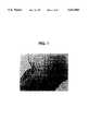
- FIG. 1Photomicrograph of a fresh aortic leaflet.
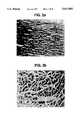
- FIG. 2aPhotomicrograph showing pulmonary valve conduit after decellularization.
- FIG. 2bPhotomicrograph showing myocardium after decellularization.
- FIG. 3aPhotomicrograph of decellularized aortic leaflet.
- FIG. 3bPhotomicrograph of repopulated aortic leaflet.
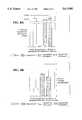
- FIG. 4aTotal Protein Synthesis in Porcine Aortic Heart Valve Leaflets
- FIG. 4bCollagen Protein Synthesis in Porcine Aortic Valve Leaflets.
- FIG. 4cTotal Protein Synthesis in Porcine Aortic Valve Leaflets.
- FIG. 4dCollagen Protein Synthesis in Porcine Aortic Valve Leaflets.
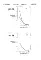
- FIG. 5aReactivity of Rabbit IgG Antisera Raised to Extracts of Cryopreserved or Depopulated Porcine Heart Valve Leaflets--Antibody Capture.
- FIG. 5bReactivity of Rabbit IgM Antisera Raised to Extracts of Cryopreserved or Depopulated Porcine Heart Valve Leaflets--Antibody Capture.
- FIG. 6aPhotomicrograph showing the minimal cellular response triggered by decellularized porcine heart valve leaflets after implantation.
- FIG. 6bPhotomicrograph showing cellular response triggered by cryopreserved porcine aortic valve leaflet after implantation.
- FIG. 7aPhotomicrograph of fresh porcine aortic valve leaflet assayed for pig cell associated antigen.
- FIG. 7bPhotomicrograph of decellularized porcine aortic valve leaflet assayed for pig cell associated antigen.
- FIG. 8aStress-Strain Relations of Porcine Aortic Valve Leaflets.
- FIG. 8bStress-Strain relations of Porcine Aortic Valve Leaflets.
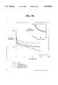
- FIG. 9aStress Relaxation Curves of Porcine Aortic Leaflets After Preconditioning.
- FIG. 9bStress Relaxation Curves of Porcine Aortic Leaflets After Preconditioning.
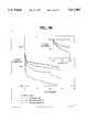
- FIG. 10aTensile Failure Data for Porcine Aortic Valve Leaflets.
- FIG. 10bTensile Failure Data for Porcine Aortic Valve Leaflets.
- the initial transplant tissue or organmay be of non-human origin. These tissues or organs may be obtained at approved slaughterhouses from animals fit for human consumption or from herds of domesticated animals maintained for the purpose of providing these tissues or organs. The tissues or organs are handled in a sterile manner, and any further dissection of the tissue or organs is carried out under aseptic conditions.
- Transplant tissue originating from a non-human sources and intended for use in a human recipientmay be processed to generate a hybrid xenograft or xenogeneic implant tissue, which is formed from a non-human tissue matrix, free of native cells and other antigenic components and which is populated with viable human cells.
- a hybrid xenograft or xenogeneic implant tissuewhich is formed from a non-human tissue matrix, free of native cells and other antigenic components and which is populated with viable human cells.
- the transplant tissuemay be sterilized by incubating it in a sterile buffered nutrient solution containing antimicrobial agents, for example an antibacterial, an antifungal, or a sterilant compatible with the transplant tissue.
- antimicrobial agentsfor example an antibacterial, an antifungal, or a sterilant compatible with the transplant tissue.
- the sterilized transplant tissuemay then be cryopreserved for further processing at a later time or may immediately be further processed according to the next steps of this process including a later cryopreservation of the tissue matrix or other tissue products of the process.
- a preliminary step of this inventioncalls for elimination of native viable cells as well as other cellular and acellular structures or components which may elicit an adverse immune response by the implant recipient.
- Physical forcesfor example the formation of intracellular ice, can be used to decellularize transplant tissues.
- vapor phase freezingslow rate of temperature decline
- rapid freezing processesin the absence of cryoprotectant, may result in tissue disruption such as the cracking of heart valve conduits.
- Colloid-forming materialsmay be added during freeze-thaw cycles to alter ice formation patterns in the tissue.
- Polyvinylpyrrolidone (10% w/v) and dialyzed hydroxyethyl starch (10% w/v)may be added to standard cryopreservation solutions (DMEM, 10% DMSO, 10% fetal bovine serum) to reduce extracellular ice formation while permitting formation of intracellular ice. This allows a measure of decellularization while affording the collagenase tissue matrix some protection from ice damage. Additionally, it is noted, that tissues, particularly heart valve conduits, will crack if rapidly frozen irrespective of the presence of cryoprotectant.
- various enzymatic or other chemical treatments to eliminate viable native cells from implant tissues or organsmay be used. For instance, extended exposure of cells to proteases such as trypsin result in cell death.
- proteasessuch as trypsin
- thismay not be the approach of choice for collagenous grafts intended for implant in high mechanical stress locations.
- Combinations of different classes of detergentsmay disrupt cell membranes and aid in the removal of cellular debris from tissue.
- stepsshould be taken to eliminate any residual detergent levels in the tissue matrix, so as to avoid interference with the later repopulating of the tissue matrix with viable cells.
- the decellularization of the transplant tissueis preferably accomplished by the administration of a solution effective to lyse native cells in the transplant tissue.
- the solutionmay be an aqueous hypotonic or low ionic strength solution formulated to effectively lyse the native tissue cells.
- Such an aqueous hypotonic solutionmay be de-ionized water or an aqueous hypotonic buffer.
- the aqueous hypotonic buffermay contain additives that provide suboptimal conditions for the activity of selected proteases, for example collagenase, which may be released as a result of cellular lysis.
- metal ion chelatorsfor example 1,10-phenanthroline and ethylenediaminetetraacetic acid (EDTA)
- EDTAethylenediaminetetraacetic acid
- Providing sub-optimal conditions for proteases such as collagenasemay assist in protecting the tissue matrix from degradation during the lysis step.
- suboptimal conditions for proteasesmay be achieved by formulating the hypotonic lysis solution to eliminate or limit the amount of calcium and zinc ions available in solution.
- Many proteasesare active in the presence of calcium and zinc ions and lose much of their activity in calcium and zinc ion free environments.
- the hypotonic lysis solutionwill be prepared selecting conditions of pH, reduced availability of calcium and zinc ions, presence of metal ion chelators and the use of proteolytic inhibitors specific for collagenase, such as ⁇ 1 -anticollagenase, such that the solution will optimally lyse the native cells while protecting the underlying tissue matrix from adverse proteolytic degradation.
- a hypotonic lysis solutionmay include a buffered solution of water, pH 5.5 to 8, preferably pH 7 to 8, free from calcium and zinc ions and including a metal ion chelator such as EDTA.
- control of the temperature and time parameters during the treatment of the tissue matrix with the hypotonic lysis solutionmay also be employed to limit the activity of proteases.
- a preferable step of this processincludes treatment of the tissue with enzymes, such as nucleases, effective to inhibit cellular metabolism, protein production and cell division without degrading the underlying collagen matrix.
- Nucleases that can be used for digestion of native cell DNA and RNAinclude both exonucleases and endonucleases. A wide variety of which are suitable for use in this step of the process and are commercially available.
- exonucleases that effectively inhibit cellular activityinclude DNAase I (SIGMA Chemical Company, St.
- RNAase ASIGMA Chemical Company, St. Louis, Mo.
- endonucleases that effectively inhibit cellular activityinclude EcoR I (SIGMA Chemical Company, St. Louis, Mo.) and Hind III (SIGMA Chemical Company, St. Louis, Mo.).
- the selected nucleasesare applied in a physiological buffer solution which contains ions which are optimal for the activity of the nuclease, such ions include magnesium and calcium salts. It is also preferred that the ionic concentration of the buffered solution, the treatment temperature and the length of treatment are selected to assure the desired level of effective nuclease activity.
- the bufferis preferably hypotonic to promote access of the nucleases to the cell interiors.
- tissueis preferably treated with a physiologically buffered medium comprised of nucleases DNAase I and RNAase A.
- the nuclease degradation solutioncontains about 0.1 ⁇ g/ml to 50 ⁇ g/ml, preferably 10 ⁇ g/ml, of the nuclease DNAase I, and 0.1 ⁇ g/ml to 10 ⁇ g/ml, preferably 1.0 ⁇ g/ml, of RNAase A.
- the tissuemay be decellularized by application of the foregoing at a temperature of about 20° C. to 38° C., preferably at about 37° C. for about 30 minutes to 6 hours, while at the same time the generation of new immunological sites as a result of collagen degradation is limited.
- enzymatic digestionsmay be suitable for use herein, for example, enzymes that will disrupt the function of native cells in a transplant tissue may be used.
- phospholipaseparticularly phospholipases A or C
- a buffered solutionmay be used to inhibit cellular function by disrupting cellular membranes of endogenous cells.
- the enzyme employedshould not have a detrimental effect on the tissue matrix protein.
- the enzymes suitable for usemay also be selected with respect to inhibition of cellular integrity, and also include enzymes which may interfere with cellular protein production.
- the pH of the vehicle, as well as the composition of the vehiclewill also be adjusted with respect to the pH activity profile of the enzyme chosen for use.
- the temperature applied during application of the enzyme to the tissueshould be adjusted in order to optimize enzymatic activity.
- the resultant transplant tissue matrixis washed to assure removal of cell debris which may include cellular protein, cellular lipids, and cellular nucleic acid, as well as any extracellular debris such as extracellular soluble proteins, lipids and proteoglycans. Removal of this cellular and extracellular debris reduces the likelihood of the transplant tissue matrix eliciting an adverse immune response from the recipient upon implant.
- the tissuemay be incubated in a balanced salt solution such as Hanks' Balanced Salt Solution (HBSS).
- HBSSHanks' Balanced Salt Solution
- the composition of the balanced salt solution wash, and the conditions under which it is applied to the transplant tissue matrixmay be selected to diminish or eliminate the activity of the nuclease or other enzyme utilized during the decellularization process.
- Such a balanced salt wash solutionwould preferably not contain magnesium or calcium salts, and the washing process may include incubation at a temperature of between about 2° C. and 42° C., with 4° C. most preferable.
- the transplant tissue matrixmay be incubated in the balanced salt wash solution for up to 10 to 12 days, with changes in wash solution every second or third day.
- an antibacterial, an antifungal or a sterilant or a combination thereofmay be included in the balanced salt wash solution to protect the transplant tissue matrix from contamination with environmental pathogens.
- the decellularized tissue matrixmay be utilized without the subsequent steps of in vitro cellular repopulation.
- the decellularized and substantially non-immunogenic tissue matrixis suitable for implant into a recipient.
- the reduced immunogenicity of the skin tissue matrixwill increase the tolerance of the transplanted skin by the recipient's immune system.
- the implantmay then be repopulated in vivo by the implant recipient's own cells.
- the skin matrixPrior to use as a skin graft, the skin matrix may be treated to enhance the ingrowth and attachment of the recipient's own skin cells. For instance, the matrix may be treated with adhesion factors or growth factors, such as keratinoctye growth factor, or both, to enhance the infusion of keratinocytes.
- tissue matrix prepared in accordance with the aboveis free of its native cells, and additionally cellular and extra-cellular antigen components have been washed out of the tissue matrix.
- tissue matrixhas been treated in a manner which limits the generation of new immunological sites in the collagen matrix.
- the tissue matrixretains the essential biomechanical strength necessary to provide the framework for further processing of the matrix.
- tissue matrix processed according to this inventionmay be cryopreserved for later use. Cryopreservation of decellularized transplant tissue would assure a supply or inventory of substantially non-immunogenic tissue matrices which, upon thawing, would be ready for further treatment according to the subsequent steps of this invention, or further processed as desired to provide an implant tissue product.
- tissue matricesmay be inventoried until such time as the particular cells to be employed during repopulation are identified. This may be of particular utility when the tissue matrix is to be repopulated with cells derived from the recipient or other cells selected for use based on their immunological compatibility with a specific recipient.
- native transplant tissuemay be cryopreserved prior to undergoing any of the processes of this invention. Tissues which are not decellularized retain their native cells in conjunction with the collagenous tissue matrix. Upon thawing these tissues may be further processed. Beneficially, the cryopreservation of intact transplant tissue may also aid in the depopulation of the tissue as a result of cellular death brought on by cryopreservation.
- the tissue matrixmay be next treated to enhance the adhesion and inward migration of the allogeneic or autologous cells, in vitro, which will be used to repopulate the transplant tissue.
- fibroblastsWhile not wishing to be bound by theory, there are several factors believed to effect the attachment of cells to tissue. For example, the adherence of fibroblasts to tissue surfaces involves interactions between the cell membrane and extracellular matrix components of native tissue such as fibrillar and sheet-forming collagen, proteoglycans, and glycoproteins.
- dermal fibroblasts cultured without serumattach rapidly and equally to types I and IV collagen. The extent of attachment is increased by the addition of serum (human or fetal bovine, maximal binding with 1% serum) and by purified fibronectin to the culture medium.
- serumhuman or fetal bovine, maximal binding with 1% serum
- purified fibronectinto the culture medium.
- Each of the two homologous subunits of fibronectinhas two cell recognition regions, the most important of which has the Arg-Gly-Asp (RGD) sequence.
- a second site, binding glycosaminoglycansacts synergistically and appears to stabilize the fibronectin-cell interactions mediated by the RGD sequence.
- Heparin sulfate along with chondroitin sulfateare the two glycosaminoglycans identified on cell surfaces. Heparin sulfate is linked to core proteins (syndecan or hyaluronectin) which can either be integral or membrane spanning.
- core proteinsseyndecan or hyaluronectin
- integrinsCellular binding sites for extracellular matrix glycoproteins are called integrins and these mediate tight binding of cells to the adhesion factors. Each adhesion factor appears to have a specialized integrin although a single integrin may bind to several extracellular matrix factors.
- Fibroblastswhen adherent to intact fibronectin (cell and heparin-binding domains) display a contracted morphology with focal adhesions. Without the heparin binding domain, fibroblasts will spread but fail to develop focal adhesions.
- a combination of factorsmay determine the rate at which cells can bind to a tissue surface.
- Many of theseare fibroblast products, although some like fibronectin may be derived from serum supplementation as well.
- the rate at which these factors are expressed and secreted by cellswill affect the attachment of cells to surfaces, and cytokines such as fibroblast growth factor and transforming growth factor- ⁇ are positive regulators of fibroblast collagen and fibronectin production.
- tissue matrixis promoted by the interaction between the cell membrane, and extracellular components associated with the corresponding implant tissue.
- appropriate treatments which promote cellular attachment to the decellularized tissue matrixinclude treatment with extracellular tissue components and, particularly, extracellular proteins, such as glycoproteins and/or proteoglycans or glycosaminoglycans which are effective to promote attachment of cells to the decellularized tissue matrix.
- extracellular tissue componentssuch as glycoproteins and/or proteoglycans or glycosaminoglycans which are effective to promote attachment of cells to the decellularized tissue matrix.
- a preferred technique for repopulating the tissue matrix with cellsis carried out by first treating the decellularized tissue matrix with cellular attachment factor effective to promote the attachment of the repopulating cells to the decellularized matrix.
- the decellularized tissue matrixmay be incubated in nutrient solution containing extracellular matrix protein such as fibronectin and a glycosaminoglycan for a period effective for binding of the fibronectin to surfaces of the transplant tissue matrix to be repopulated.
- extracellular matrix proteinsuch as fibronectin and a glycosaminoglycan
- Preferred buffers for use with fibronectin/glycosaminoglycaninclude sodium phosphate/glycerin/bovine serum albumin (Fetal Bovine Serum, BIO-WHITTAKER) and Dulbecco's Modified Eagle's Medium (DMEM), (GIBCO). These buffers typically are used to provide a physiological acceptable pH of about 7.0 to 7.6.
- the presence of the extracellular matrix proteinsestablish a surface on the tissue matrix to which the cells that have been chosen to repopulate the matrix attach. The stimulus of the extracellular matrix protein promotes cell repopulation in the graft.
- the preferred extracellular matrix protein for use hereinis the intact molecular form of fibronectin (Human Plasma Fibronectin, UPSTATE Biotechnology, Inc.). This heterofunctional glycoprotein has affinity for extracellular matrix proteins, proteoglycans, and certain cell types.
- the fibronectin treatment solutionpreferably also contains a proteoglycan which may be which may be one of the glycosaminoglycans heparin, heparin sulfate, chondroitin, chondroitin sulfate, dermarin, or dermarin sulfate. It is believed that the glycosaminoglycan promotes and stabilizes the binding between fibronectin and the tissue matrix associated collagen.
- the matrixadvantageously is capable of interaction with fibronectin because it is not chemically cross-linked.
- a source of fibronectinis from human blood, processed to limit contamination with virus.
- the preferred glycosaminoglycanis heparin.
- the concentration of glycoprotein used as the adhesion factor to treat the tissue matrixmay range from about 1 to about 100 ⁇ g/ml, with a fibronectin concentration of 10 ⁇ g/ml being preferred.
- the preferred weight ratio of fibronectin to heparinis about 10 parts fibronectin to about 1 part glycosaminoglycan, e.g. heparin. This is optimal for repopulation of porcine heart valve leaflets, but may range from about 0.1:1 to about 10:0.1 depending on the tissue used.
- the components of the nutrient solution containing the adhesion factorsare selected such that the solution is compatible with growth factors which are later added to the nutrient medium that the transplant tissue matrix is incubating in. These growth factors are employed to facilitate cell growth and repopulation of the tissue matrix.
- the decellularized transplant tissue matrixmay be repopulated with cells in vitro.
- the cells employed to repopulate the decellularized matrixmay be allogeneic cells cultured from the same species as the intended implant recipient, or may be autologous cells cultured from the implant recipient.
- the autologous or allogeneic cells in the repopulated tissue matrixknown as a heterograft or chimeric graft, will elicit less of an adverse immune response than an unprocessed xenogeneic transplant tissue.
- cells employed to repopulate the decellularized matrixmay be cells that have been genetically manipulated to enhance the production of specific proteins.
- Numerous recombinant DNA technologiesare known in the art for altering, enhancing, and modifying cell metabolism.
- Repopulationmay be accomplished by incubating the tissue matrix treated with cell adhesion factors in a nutrient medium containing the cells and growth factors active to promote cell proliferation and, hence, repopulation of the tissue matrix.
- a preferred cell type for use hereinis fibroblast cells.
- a variety of substancesmay be employed to enhance cell chemotaxis, increasing the rate of directional movement along a concentration gradient of the substance in solution.
- fibroblast growth factorfibroblast growth factor, platelet-derived growth factor, transforming growth factor- ⁇ , and the substrate-adhesion molecules, fibrillar collagens, collagen fragments, and fibronectin are chemotactic for fibroblasts.
- fibroblast migrationrequires de novo protein synthesis; protein synthesis in normal fibroblastic cells is stimulated by adhesion of cells to fibronectin, so the processes of cell adhesion and cell migration during repopulation are believed to be interrelated.
- Cellular migrationalso allows cells to move through the tissue matrix repopulating interior interstitial spaces as well as the surfaces of the tissue transplant matrix.
- the number of cells required to fully repopulate particular transplant tissue matricesdepends upon the volume of the tissue used and the types of cells provided. However, concentrations of 20,000 to 125,000 fibroblasts per milliliter may provide suitable coverage of the heart valve leaflet and aortic conduit tissue.
- the steps of cellular repopulation of the tissue matrixpreferably are conducted in the presence of growth factors effective to promote proliferation of the cultured cells employed to repopulate the matrix.
- a growth factor for use hereinmay be fibroblast growth factor (FGF), most preferably basic fibroblast growth factor (bFGF) (Human Recombinant bFGF, UPSTATE Biotechnology, Inc.).
- the fibroblast growth factorsare a family of mitogens active on mesenchymal cells. FGFs are not detected free in conditioned medium, instead the FGFs are found in the extracellular matrix in association with heparin sulfate, localizing in the fibronectin-heparin layer prebound to the transplant tissue matrix.
- the glycosaminoglycansstabilize FGF activity and are required for FGF binding to cell surface receptors where they stimulate autocrine/paracrine growth.
- the matrix and cellsare preferably exposed to bFGF continuously during the repopulation step to provide for stimulation of cell replication and expression of collagen protein synthesis, as required for normal valve function.
- Culture times of the matrix with growth factorrange from 10 to 21 days.
- the concentration of growth factor used to treat the tissue matrixmay range from 100 ng/ml to 10 ⁇ g/ml with a growth factor concentration for bFGF of 2.5 ⁇ g/ml being preferred.
- the culture mediummay include Dulbecco's Modified Eagle Medium (GIBCO) with 5-15% added serum.
- GEBCODulbecco's Modified Eagle Medium
- bovine serumis preferred but experience with implantation of allograft heart valves suggests that bovine serum may be utilized in the growth medium without adverse immunologic consequences to the implant recipient.
- Continual stimulation of the cells with serum and medium conditioned by the repopulating cellsmay provoke a more rapid cellular repopulation of the tissue matrix.
- the matrix, cells and growth factorsmay be incubated in a humidified atmosphere at 37° C. provided with a 95% air and 5% CO 2 mixture throughout the culture period.
- the transplant tissue matrixis cultured for a time sufficient to produce a repopulated graft with interstitial histology similar to that of fresh tissue.
- the tissueUpon conclusion of the cellular repopulation process, the tissue will also preferably display metabolic parameters similar to those of fresh tissue.
- transplant tissuesare functioning and viable prior to implantation in addition to being immunologically acceptable to the implant recipient or substantially non-immunogenic.
- Various assays exist to measure cellular activity and application of these assays to the implant tissues of this processprovide a method of monitoring and quantifying the viability of the cells which repopulate the implant tissue.
- an assaybe selected that measures a cellular activity which bears a relation to the intended function of the transplant tissue.
- the production of collagenis important in maintaining a functioning heart valve.
- collagenIn heart valve leaflets 5 to 15% of the total protein produced is collagen. Of that at least 75% will be type I collagen. Therefore in assaying a repopulated heart valve, it is preferable to assay for total collagen produced by the repopulating cells as an accurate measure of cellular viability.
- Assaying for cellular activity by measuring collagen productionare well known in the art. Examples of references discussing assays for cellular collagen production are Buckley, A. et al. 1980 Collagen Metabolism., Methods of Enzymology, 163:674-69, and Hayashi, T. and Nagai, Y. 1979. Separation of the ⁇ chains of Type I and III Collagens by SDS-polyacrylamide Gel Electrophoresis, Journal of Biochemistry. 86:453-459, and are hereby incorporated by reference.
- any assay that quantifiably measures a cellular function indicative of viable cellsmay be used.
- Fibroblast cellsare responsible for the production of most connective tissue components. They synthesize different collagen types, and the phenotype appears to be imposed by specific tissue environments; i.e., cultured fibroblasts synthesize collagen types according to their site of origin. Fibroblasts also produce various glycosaminoglycans and fibronectin and growth factors. It is the ability of dermal fibroblasts to synthesize types I, III, and V collagens in the proportion present in the matrix of the heart valve leaflet which makes them appropriate cells for repopulating the transplant tissue matrix and forming the hybrid graft described herein.
- the cells which are used to repopulate the particular graftcan be varied within wide limits, and different types of cells can be used in different circumstances, depending upon the function of the transplant, the nature of the tissue being replaced or augmented, the allergic sensitivity of the recipient in addition to other factors.
- a preferred embodiment of the inventionuses autologous cells in the process described herein.
- a tissue sampleis taken from the recipient prior to transplant or implant surgery.
- the tissueis treated, in accordance with the methods described herein below, to produce fibroblasts or other cells which are then used to repopulate the allogeneic or xonogeneic tissue matrix, in accordance with this process.
- fibroblasts or other cellswhich are then used to repopulate the allogeneic or xonogeneic tissue matrix, in accordance with this process.
- the cell sourcecan be selected to match the tissue to be transplanted. For example, if a blood vessel is to be transplanted, cells can be taken from a recipient's healthy blood vessel and used as the source of cells for graft repopulation. In this fashion, the healthy graft can be very closely matched to the recipient's diseased tissue.
- This aspect of the inventionis particularly useful when the transplant recipient is highly allergic, or if the tissue is highly immunogenic, such as with respect to transplantable blood vessels.
- allogeneic cell lineswhich are not likely to cause an unacceptable immune response upon implant may be used to repopulate the tissue matrix.
- Cells with no more than a weak or tolerable allergic responsemay be used to repopulate the tissue matrix to provide a substantially non-immunogenic implant.
- These cellsmay be naturally weakly immunogenic or be designed by virtue of recombinant cell technology to be weakly immunogenic.
- the tissue used to provide the fibroblast cell linkfor example skin (buttocks, thigh, or back) or heart valve leaflets, is recovered sterilely and provided to a processor in buffered nutrient medium.
- the tissueis cut into 1 mm 3 pieces using a sterile dissection technique. Groups of 10 pieces are then placed in 35 mm tissue culture dishes with a limiting amount of culture medium (DMEM plus 10% fetal bovine serum) sufficient to wet the tissue but not float the pieces. Incubate for one week at 37° C. in a humidified culture incubator in a 5% CO 2 atmosphere in air. After one week of incubation, each piece of tissue is surrounded by a dense outgrowth of fibroblasts.
- DMEMfetal bovine serum
- Epithelial cellsmay also be present but are lost during subsequent cell culturing.
- the fibroblastsare removed by standard trypsin digestion after rinsing the cells with a calcium and magnesium-free sterile buffered salt solution, and placed in larger cell culture vessels with fresh culture medium.
- the cell culturescan be expanded in this manner.
- the contents of one flaskcan be divided and placed into three larger vessels, and this process can be repeated about once a week.
- Cells recovered from these flasksare used as the source of repopulating cells.
- Cells obtained in this mannerare preferable to commercially available cell lines, because most cell lines are phenotypically altered and are no longer responsive in a normal manner to growth regulators (such as bFGF). Additionally most commercially available cell lines do not produce natural amounts and proportions of important protein products (such as collagen).
- a preferred embodiment of the inventionencompasses a xenograft treated to remove native cells and soluble proteins, the conditions are chosen to be non-toxic to cells used to repopulate the xenograft and chosen to render the final xenograft tissue matrix biomechanically sound and intact.
- the depopulated xenograft tissue matrixis then treated with extracellular matrix glycoprotein factor and glycosaminoglycan.
- the xenograftis then treated with a growth factor and glycosaminoglycan and incubated with exogenous cells that adhere to the graft. These exogenous cell migrate into the graft, proliferate within the graft, and express essential proteins and other factors critical to the function of the graft.
- the repopulated xenograftis rendered biologically functional by the repopulating cells and displays reduced or minimal immunogenicity as compared to either untreated xenograft or chemically-fixed xenograft.
- the reduced antigenicityis a consequence of the removal of xenoantigens or alloantigens during the initial depopulation of the tissue, and by the presence of repopulating cells which are not recognized as foreign by the recipient.
- These chimeric graftsshould also display a reduced tendency for calcification because cell debris, which forms either as the result of cell death during procurement of tissue or during the decellularization of the tissue, is removed by the washing regimen.
- Pig heartswere obtained within two hours of slaughter, in order to limit the effects of uncontrolled cellular degradation on tissue structure, and returned to the processing facility at 4° C. in a sterile solution of DMEM.
- Aortic heart valveswere excised under sterile conditions, incubated in antibiotic mixture for 16 hr at 37° C. in nutrient medium in 5% CO 2 atmosphere, and cryopreserved (cooling rate ⁇ -1° C./min) in DMEM containing 10% DMSO and 10% fetal bovine serum. After storage at -179° C., the valves were thawed rapidly at 37° C.
- tissueThe leaflets, aortic conduit, and myocardium were cut from the valve and divided portions of each tissue type were either placed directly into 10% buffered formalin for later histologic analysis or were processed for depopulation. After washing in lactated Ringers-5% dextrose solution three times for 15 min each at room temperature, tissues were incubated in 18 M ⁇ water for 2 hr at room temperature followed by digestion in 10 ⁇ g/ml DNAase I and 1 ⁇ g/ml RNAase A in 10 Mm Tris-Cl, pH 7.6, containing 3 mM magnesium and 1 mM calcium salts at 37° C. for 120 min.
- FIG. 1shows the pattern of cellularity of a fresh aortic leaflet with an endothelial layer on both the fibrosa and ventricularis surfaces and fibroblasts throughout the full thickness of the tissue.
- FIGS. 2a and 2bshow micrographs of pulmonary valve conduit and myocardium, respectively, after depopulation with essentially acellular appearance.
- Porcine aortic leafletswere recovered and depopulated as defined in EXAMPLE 1.
- Leafletswere incubated in 5 ml NaH 2 PO 4 /glycerin/BSA buffer at 37° C.
- Human plasma fibronectinwas added to the buffer to a concentration of 10 ⁇ g/ml along with 1 ⁇ g/ml heparin for 16 hr followed with addition of human recombinant bFGF to a concentration of 2.5 ⁇ g/ml along with 0.83 ⁇ g/ml heparin for an additional 6 hr.
- bovine dermal fibroblastspreviously isolated by standard explant culture techniques, were added to the heart valve leaflets at 2 ⁇ 10 4 cell/ml.
- FIG. 3ais a representative micrograph of a decellularized aortic leaflet.
- FIG. 3bis a representative micrograph of a decellularized aortic leaflet treated with both fibronectin and bFGF showing repopulation with exogenous fibroblasts.
- Biochemical activity of fibroblasts in repopulated as compared to fresh and depopulated tissuesBiochemical activity of fibroblasts in repopulated as compared to fresh and depopulated tissues.
- Porcine aortic heart valve leafletswere depopulated as in EXAMPLE 1 and repopulated with two different isolates of sheep dermal fibroblasts according to the technique in EXAMPLE 2. After 10 days of repopulation, leaflets were moved to fresh vessels and incubated for 48 hr in 1.0 ⁇ Ci/ml [ 3 H] proline in DMEM containing gentamycin and 50 ⁇ g/ml ascorbic acid, 50 ⁇ g/ml ⁇ -aminopropionitrile.
- Total proteins synthesizedwere determined by 10% trichloroacetic acid precipitated radioactivity recovered from medium and tissue extracts made 10 mM in N-ethylmaleimide, 25 mM in EDTA, and 10 mM in phenylmethylsulfonyl fluoride to prevent proteolysis; precipitated protein was further analyzed by digestion with Clostridial collagenase free of non-specific protease activity to define the collagen content. As shown in FIGS. 4a, 4b, 4c and 4d, protein synthetic activity of depopulated tissue was nil.
- porcine leaflets rendered treated by the depopulation procedures described in the preferred embodiment of the inventiondisplay no protein synthetic capacity in general and no collagen synthesis in particular.
- the successful application of repopulation proceduresis indicated by the ability to impart the cellular function of protein synthesis to the depopulated leaflet by the provision of exogenous fibroblasts during the repopulation procedure.
- FIGS. 5a and 5bThe results of humoral immune response studies are presented in FIGS. 5a and 5b.
- the humoral immune responsewas assessed using an antibody capture technique in which antigen extracts in 0.1 M NaCl of unmodified cryopreserved leaflets were used to screen sera from rabbits immunized with NaCl extracts of either modified cell depopulated leaflets or control leaflets.
- Emulsions of such extractswere made in 50% (v/v) Freund's complete adjuvant; 0.1 ml portions of these emulsions were placed at ten intradermal sites along the backs of separate New Zealand white male rabbits.
- FIG. 6shows that depopulated leaflets engendered minimal cellular responses compared with cryopreserved controls.
- cryopreserved tissuewhich contains both an endothelial cell layer as well as fibroblasts stimulated significant immune and inflammatory cell response with large numbers of heterophils and lymphocytes and plasma cells in the implant area as well as within the implants themselves. In depopulated tissue implants, both inflammatory and immune cells were fewer in number and more limited in distribution.
- modified proteinsare the sites of binding of preformed natural antiporcine antibodies that initiate hyperacute rejection responses of porcine tissues in primates (including man). Proteins binding this lectin were detected by then reacting the sections with biotin-conjugated horseradish peroxidase-avidin mixture, hydrogen peroxide, and 3,3'-diaminobenzidine. Presence of porcine antigens is detected by color development.
- FIG. 7amicroscopic analysis of sections demonstrates cell associated antigen in cryopreserved aortic leaflet.
- FIG. 7bdemonstrates markedly diminished antigen binding after decellularization of the aortic leaflet. This study shows that the depopulation procedure as presented in the preferred embodiment of the invention reduces critical antigens likely to cause rejection of a porcine graft by a human recipient.
- Porcine aortic heart valvesfreshly obtained, were sterilized with antibiotic mixture and cryopreserved under conditions that maintain cellular viability. For depopulation, aortic valves were thawed rapidly at 37° C., then treated with low hypotonicity solution, nucleases, and balanced salt solution for 10 days.
- leafletswere cut into circumferential or radial strips, mounted onto an Instron Model 1011 Materials Tester under calibrated clamping pressure. Tissue was bathed in 37° C. or 4° C. Hanks balanced salt solution during testing. After determination of gauge length and 20 preconditioning cycles (100 g load for circumferential strips and 180 g for radial strips), each specimen was tested as follows:
- FIGS. 8a and 8bA single load versus elongation test, results of which are shown in FIGS. 8a and 8b. Radial strips were more extensible than circumferential under all conditions. Tissue modulus was unaffected by any treatment, but depopulated radial strips were significantly more extensible than fresh tissue (113 ⁇ 11.8 vs. 85.9 ⁇ 8.6 mm/mm);
- FIGS. 9a and 9bA stress relaxation test, results of which are shown in FIGS. 9a and 9b. For circumferential and radial strips approximately 10% of the original stress was dissipated in the first 10 sec. Overall, the rate of loss of stress appeared greater in radial strips in general and was fastest in cryopreserved radial strips. At the termination of the test, stress remaining in 37° C. depopulated circumferential strips was significantly greater than in fresh tissue; there were no significant differences in the radial strips; and
- FIGS. 10a and 10bA tensile failure test, results of which are shown in FIGS. 10a and 10b.
- circumferential stripswere stronger, stiffer, and tougher than radial strips. Cryopreservation and depopulation did not affect these parameters measured in the circumferential direction. Radial strips cut from tissue depopulated at either temperature did show greater toughness as compared to fresh.
Landscapes
- Health & Medical Sciences (AREA)
- Life Sciences & Earth Sciences (AREA)
- Chemical & Material Sciences (AREA)
- Engineering & Computer Science (AREA)
- Biomedical Technology (AREA)
- Chemical Kinetics & Catalysis (AREA)
- General Health & Medical Sciences (AREA)
- Public Health (AREA)
- Veterinary Medicine (AREA)
- Animal Behavior & Ethology (AREA)
- Dermatology (AREA)
- Medicinal Chemistry (AREA)
- Oral & Maxillofacial Surgery (AREA)
- Transplantation (AREA)
- Epidemiology (AREA)
- Botany (AREA)
- Zoology (AREA)
- Molecular Biology (AREA)
- Urology & Nephrology (AREA)
- Cell Biology (AREA)
- Vascular Medicine (AREA)
- Heart & Thoracic Surgery (AREA)
- Cardiology (AREA)
- Developmental Biology & Embryology (AREA)
- General Chemical & Material Sciences (AREA)
- Materials For Medical Uses (AREA)
- Prostheses (AREA)
- Medicines Containing Material From Animals Or Micro-Organisms (AREA)
- Acyclic And Carbocyclic Compounds In Medicinal Compositions (AREA)
- Chemical Or Physical Treatment Of Fibers (AREA)
- Saccharide Compounds (AREA)
- Micro-Organisms Or Cultivation Processes Thereof (AREA)
- Game Rules And Presentations Of Slot Machines (AREA)
Abstract
Description
Claims (20)
Priority Applications (1)
| Application Number | Priority Date | Filing Date | Title |
|---|---|---|---|
| US08/463,455US5613982A (en) | 1994-03-14 | 1995-06-05 | Method of preparing transplant tissue to reduce immunogenicity upon implantation |
Applications Claiming Priority (2)
| Application Number | Priority Date | Filing Date | Title |
|---|---|---|---|
| US21375494A | 1994-03-14 | 1994-03-14 | |
| US08/463,455US5613982A (en) | 1994-03-14 | 1995-06-05 | Method of preparing transplant tissue to reduce immunogenicity upon implantation |
Related Parent Applications (1)
| Application Number | Title | Priority Date | Filing Date |
|---|---|---|---|
| US21375494ADivision | 1994-03-14 | 1994-03-14 |
Publications (1)
| Publication Number | Publication Date |
|---|---|
| US5613982Atrue US5613982A (en) | 1997-03-25 |
Family
ID=22796373
Family Applications (4)
| Application Number | Title | Priority Date | Filing Date |
|---|---|---|---|
| US08/463,643Expired - LifetimeUS5632778A (en) | 1994-03-14 | 1995-06-05 | Treated tissue for implantation and methods of preparation |
| US08/463,455Expired - LifetimeUS5613982A (en) | 1994-03-14 | 1995-06-05 | Method of preparing transplant tissue to reduce immunogenicity upon implantation |
| US08/463,171Expired - LifetimeUS5899936A (en) | 1994-03-14 | 1995-06-05 | Treated tissue for implantation and methods of preparation |
| US08/791,450Expired - LifetimeUS5843182A (en) | 1994-03-14 | 1997-01-27 | Treated tissue for implantation and methods of preparation |
Family Applications Before (1)
| Application Number | Title | Priority Date | Filing Date |
|---|---|---|---|
| US08/463,643Expired - LifetimeUS5632778A (en) | 1994-03-14 | 1995-06-05 | Treated tissue for implantation and methods of preparation |
Family Applications After (2)
| Application Number | Title | Priority Date | Filing Date |
|---|---|---|---|
| US08/463,171Expired - LifetimeUS5899936A (en) | 1994-03-14 | 1995-06-05 | Treated tissue for implantation and methods of preparation |
| US08/791,450Expired - LifetimeUS5843182A (en) | 1994-03-14 | 1997-01-27 | Treated tissue for implantation and methods of preparation |
Country Status (12)
| Country | Link |
|---|---|
| US (4) | US5632778A (en) |
| EP (2) | EP0871414B1 (en) |
| JP (1) | JPH09510108A (en) |
| KR (1) | KR100365573B1 (en) |
| AT (1) | ATE265191T1 (en) |
| AU (1) | AU1931495A (en) |
| CA (1) | CA2185447C (en) |
| DE (1) | DE69532976T2 (en) |
| DK (1) | DK0871414T3 (en) |
| ES (1) | ES2219660T3 (en) |
| PT (1) | PT871414E (en) |
| WO (1) | WO1995024873A1 (en) |
Cited By (166)
| Publication number | Priority date | Publication date | Assignee | Title |
|---|---|---|---|---|
| US5782915A (en)* | 1995-09-15 | 1998-07-21 | Stone; Kevin R. | Articular cartilage heterografts |
| WO1998046165A1 (en)* | 1997-04-11 | 1998-10-22 | Cryolife, Inc. | Tissue decellularization |
| US5855620A (en)* | 1995-04-19 | 1999-01-05 | St. Jude Medical, Inc. | Matrix substrate for a viable body tissue-derived prosthesis and method for making the same |
| US5865849A (en)* | 1995-06-07 | 1999-02-02 | Crosscart, Inc. | Meniscal heterografts |
| WO1998049972A3 (en)* | 1997-05-02 | 1999-04-08 | St Jude Medical | Differential treatment of prosthetic devices |
| US5902338A (en)* | 1995-09-15 | 1999-05-11 | Crosscart, Inc. | Anterior cruciate ligament heterograft |
| US5913900A (en)* | 1995-06-07 | 1999-06-22 | Corsscart, Inc. | Substantially native meniscal cartilage heterografts |
| US5916264A (en)* | 1997-05-14 | 1999-06-29 | Jomed Implantate Gmbh | Stent graft |
| US5944755A (en)* | 1995-09-15 | 1999-08-31 | Crosscart, Inc. | Articular cartilage xenografts |
| US5961549A (en)* | 1997-04-03 | 1999-10-05 | Baxter International Inc. | Multi-leaflet bioprosthetic heart valve |
| US5984858A (en)* | 1995-06-07 | 1999-11-16 | Crosscart, Inc. | Meniscal xenografts |
| US6046379A (en)* | 1995-06-07 | 2000-04-04 | Stone; Kevin R. | Meniscal xenografts |
| US6049025A (en)* | 1995-09-15 | 2000-04-11 | Stone; Kevin R. | Articular cartilage xenografts |
| WO2000047131A1 (en)* | 1999-02-11 | 2000-08-17 | Crosscart, Inc. | Proteoglycan-reduced soft tissue xenografts |
| WO2000047132A1 (en)* | 1999-02-11 | 2000-08-17 | Crosscart, Inc. | Aldehyde and glycosidase-treated soft and bone tissue xenografts |
| US6110206A (en)* | 1995-09-15 | 2000-08-29 | Crosscart, Inc. | Anterior cruciate ligament xenografts |
| DE19919625A1 (en)* | 1999-04-29 | 2000-11-30 | Hoerstrup Simon Philipp | In vitro production of heart valve by providing carrier with fibroblasts and/or myofibroblasts and then endothelial cells, followed by treatment in pulsating flow chamber of bioreactor |
| US6210440B1 (en) | 1995-09-15 | 2001-04-03 | Kevin R. Stone | Anterior cruciate ligament xenografts |
| DE19947718A1 (en)* | 1999-10-05 | 2001-05-17 | Ctm Handels Und Beratungsgesel | Stitch support for fixing artificial or biological heart valve, reconstruction ring or ring-free heart valve reconstruction to annulus has base layer coated with active substance |
| US6290718B1 (en) | 1998-02-02 | 2001-09-18 | Regeneration Technologies, Inc. | Luminal graft, stent or conduit made of cortical bone |
| US6376244B1 (en) | 1999-12-29 | 2002-04-23 | Children's Medical Center Corporation | Methods and compositions for organ decellularization |
| US6378221B1 (en) | 2000-02-29 | 2002-04-30 | Edwards Lifesciences Corporation | Systems and methods for mapping and marking the thickness of bioprosthetic sheet |
| US6383732B1 (en) | 1999-02-11 | 2002-05-07 | Crosscart, Inc. | Method of preparing xenograft heart valves |
| WO2002007584A3 (en)* | 2000-07-20 | 2002-05-23 | Univ Colorado State Res Found | Method for recellularization of a decellularized heart valve and heart valve produced thereby |
| DE10058240A1 (en)* | 2000-11-17 | 2002-05-29 | Auto Tissue Gmbh | Method and device for the production of biological prostheses |
| US6416995B1 (en)* | 1999-11-22 | 2002-07-09 | Bio Science Consultants, L.L.C. | Bioreactor mediated recellularization of natural and tissue engineered vascular grafts |
| WO2002014480A3 (en)* | 2000-08-16 | 2002-08-01 | Univ Duke | Decellularized tissue engineered constructs and tissues |
| US6482645B2 (en) | 1999-12-29 | 2002-11-19 | Children's Medical Center Corporation | Methods and compositions for producing artificial fascia |
| WO2002053015A3 (en)* | 2001-01-05 | 2002-11-21 | Jun Yang | Vascular tissue composition |
| US6528483B2 (en) | 1995-06-07 | 2003-03-04 | André Beaulieu | Method of producing concentrated non-buffered solutions of fibronectin |
| US20030083741A1 (en)* | 2001-10-26 | 2003-05-01 | Yi-Ren Woo | Valved prosthesis with porous substrate |
| US20030083752A1 (en)* | 1998-06-30 | 2003-05-01 | Lloyd Wolfinbarger | Plasticized bone and soft tissue grafts and methods of making and using same |
| US20030093147A1 (en)* | 2001-11-13 | 2003-05-15 | Ogle Matthew F. | Medical devices that stimulate growth factor production |
| US20030100948A1 (en)* | 2000-11-24 | 2003-05-29 | Swabey Ogilvy Renault | Connective tissue substitutes, method of preparation and uses thereof |
| US20030124099A1 (en)* | 1999-12-29 | 2003-07-03 | Anthony Atala | Augmentation of organ function |
| US20030135284A1 (en)* | 1998-06-30 | 2003-07-17 | Katrina Crouch | Plasticized bone grafts and methods of making and using same |
| US20030187515A1 (en)* | 2002-03-26 | 2003-10-02 | Hariri Robert J. | Collagen biofabric and methods of preparing and using the collagen biofabric |
| US20030217415A1 (en)* | 1998-06-30 | 2003-11-27 | Katrina Crouch | Plasticized bone grafts and methods of making and using same |
| US20030225355A1 (en)* | 1998-10-01 | 2003-12-04 | Butler Charles E. | Composite material for wound repair |
| US6689161B2 (en) | 2000-04-28 | 2004-02-10 | Baylor College Of Medicine | Decellularized vascular prostheses resistant to thrombus occlusion and immunologic rejection |
| US20040034435A1 (en)* | 1997-10-31 | 2004-02-19 | Anthony Atala | Organ reconstruction |
| US20040067582A1 (en)* | 2000-09-12 | 2004-04-08 | Lloyd Wolfinbarger | Process for devitalizing soft-tissue engineered medical implants, and devitalized soft-tissue medical implants produced |
| US20040076657A1 (en)* | 1999-06-07 | 2004-04-22 | Lifenet. | Process for decellularizing soft-tissue engineered medical implants, and decellularized soft-tissue medical implants produced |
| US6726718B1 (en)* | 1999-12-13 | 2004-04-27 | St. Jude Medical, Inc. | Medical articles prepared for cell adhesion |
| EP1059891A4 (en)* | 1998-03-06 | 2004-05-19 | Crosscart Inc | SOFT TISSUE XENOGRAPHS |
| US6743574B1 (en) | 2000-09-12 | 2004-06-01 | Lifenet | Process for devitalizing soft-tissue engineered medical implants, and devitalized soft-tissue medical implants produced |
| US20040122516A1 (en)* | 2002-12-20 | 2004-06-24 | Fogarty Thomas J. | Biologically implantable prosthesis and methods of using the same |
| WO2004058952A1 (en)* | 2002-12-27 | 2004-07-15 | Hee-Young Lee | Method of culturing autologous cell using injectable solid powder |
| US20040167634A1 (en)* | 1999-05-26 | 2004-08-26 | Anthony Atala | Prosthetic kidney and its use for treating kidney disease |
| US20040210305A1 (en)* | 2002-07-16 | 2004-10-21 | Medtronic, Inc. | Suture locking assembly and method of use |
| US20040220610A1 (en)* | 1999-11-08 | 2004-11-04 | Kreidler Marc S. | Thin film composite lamination |
| US20040234507A1 (en)* | 2001-05-07 | 2004-11-25 | Stone Kevin R | Submucosal xenografts |
| US20050013872A1 (en)* | 2003-07-17 | 2005-01-20 | Toby Freyman | Decellularized bone marrow extracellular matrix |
| US20050013870A1 (en)* | 2003-07-17 | 2005-01-20 | Toby Freyman | Decellularized extracellular matrix of conditioned body tissues and uses thereof |
| US20050043760A1 (en)* | 2003-08-22 | 2005-02-24 | Fogarty Thomas J. | Prosthesis fixturing device and methods of using the same |
| US6866686B2 (en) | 2000-01-28 | 2005-03-15 | Cryolife, Inc. | Tissue graft |
| US6933326B1 (en) | 1998-06-19 | 2005-08-23 | Lifecell Coporation | Particulate acellular tissue matrix |
| US6972041B1 (en) | 1998-03-16 | 2005-12-06 | Crosscart, Inc. | Bone xenografts |
| WO2006044512A1 (en)* | 2004-10-15 | 2006-04-27 | Cook Biotech Incorporated | Fibronectin-modified ecm tissue graft constructs and methods for preparation and use thereof |
| US7112320B1 (en) | 1995-06-07 | 2006-09-26 | Andre Beaulieu | Solid wound healing formulations containing fibronectin |
| WO2004091370A3 (en)* | 2003-04-10 | 2006-12-21 | Univ Boston | Method for stimulating angiogenesis and wound healing |
| US20070005129A1 (en)* | 2000-02-28 | 2007-01-04 | Christoph Damm | Anchoring system for implantable heart valve prostheses |
| US20070010897A1 (en)* | 2005-01-06 | 2007-01-11 | Stone Kevin R | Immunochemically modified and sterilized xenografts and allografts |
| US20070100440A1 (en)* | 2005-10-28 | 2007-05-03 | Jen.Cardiotec Gmbh | Device for the implantation and fixation of prosthetic valves |
| US20070142906A1 (en)* | 2005-11-04 | 2007-06-21 | Jen. Cardiotec Gmbh | Self-expandable medical instrument for treating defects in a patient's heart |
| US20070178137A1 (en)* | 2006-02-01 | 2007-08-02 | Toby Freyman | Local control of inflammation |
| US20070225801A1 (en)* | 2006-03-10 | 2007-09-27 | Drews Michael J | Valve introducers and methods for making and using them |
| US20070255423A1 (en)* | 1998-09-21 | 2007-11-01 | Carpentier Alain F | Treating biological tissues to mitigate post-implantation calcification |
| US20080102439A1 (en)* | 2006-10-27 | 2008-05-01 | Bin Tian | Biological tissue for surgical implantation |
| US20080131966A1 (en)* | 2001-02-14 | 2008-06-05 | Hariri Robert J | Renovation and repopulation of decellularized tissues and cadaveric organs by stem cells |
| WO2008048663A3 (en)* | 2006-10-18 | 2008-06-05 | Bacterin Int Inc | Novel process for devitalized/acellular tissue for transplantation |
| US20080206343A1 (en)* | 2007-02-12 | 2008-08-28 | Edinger James W | Hepatocytes and Chondrocytes from Adherent Placental StemCells; And CD34+ ,CD45- Placental Stem Cell-Enriched Cell Populations |
| US20080255660A1 (en)* | 2007-04-13 | 2008-10-16 | Volker Guyenot | Medical device for treating a heart valve insufficiency |
| US20080255661A1 (en)* | 2007-04-13 | 2008-10-16 | Helmut Straubinger | Medical device for treating a heart valve insufficiency or stenosis |
| US20080294270A1 (en)* | 2007-05-24 | 2008-11-27 | Zimmer Orthobiologics, Inc. | Differentially processed tissue and processing methods thereof |
| US20080302372A1 (en)* | 2007-06-11 | 2008-12-11 | Edwards Lifesciences Corporation | Methods for pre-stressing and capping bioprosthetic tissue |
| US20080305146A1 (en)* | 2007-06-08 | 2008-12-11 | Wake Forest University Health Sciences, | Selective cell therapy for the treatment of renal failure |
| US20090030269A1 (en)* | 2002-09-27 | 2009-01-29 | Board Of Regents, The University Of Texas System | Cell-free tissue replacement for tissue engineering |
| US20090054968A1 (en)* | 2001-08-03 | 2009-02-26 | Jenavalve Technology Inc. | Implant implantation unit and procedure for implanting the unit |
| US20090164005A1 (en)* | 2007-12-21 | 2009-06-25 | Edwards Lifesciences Corporation | Capping Bioprosthetic Tissue to Reduce Calcification |
| US20090171447A1 (en)* | 2005-12-22 | 2009-07-02 | Von Segesser Ludwig K | Stent-valves for valve replacement and associated methods and systems for surgery |
| US20090181807A1 (en)* | 2008-01-16 | 2009-07-16 | Jason Miguel De Los Santos | Golfing aid |
| US20090192599A1 (en)* | 2005-04-08 | 2009-07-30 | Arbor Surgical Technologies, Inc. | Two-piece prosthetic valves with snap-in connection and methods for use |
| US20090216313A1 (en)* | 2008-02-26 | 2009-08-27 | Helmut Straubinger | Stent for the positioning and anchoring of a valvular prosthesis |
| US20090216310A1 (en)* | 2008-02-26 | 2009-08-27 | Helmut Straubinger | Stent for the positioning and anchoring of a valvular prosthesis in an implantation site in the heart of a patient |
| US20090216312A1 (en)* | 2008-02-26 | 2009-08-27 | Helmut Straubinger | Stent for the Positioning and Anchoring of a Valvular Prosthesis in an Implantation Site in the Heart of a Patient |
| US20100010616A1 (en)* | 2003-10-08 | 2010-01-14 | Arbor Surgical Technologies, Inc. | Attachment device and methods of using the same |
| US20100030340A1 (en)* | 1998-06-30 | 2010-02-04 | Wolfinbarger Jr Lloyd | Plasticized Grafts and Methods of Making and Using Same |
| US20100047214A1 (en)* | 2008-08-22 | 2010-02-25 | Abramson Sascha D | Methods and Compositions for Treatment of Bone Defects with Placental Cell Populations |
| US7704222B2 (en) | 1998-09-10 | 2010-04-27 | Jenavalve Technology, Inc. | Methods and conduits for flowing blood from a heart chamber to a blood vessel |
| US20100104544A1 (en)* | 2007-06-08 | 2010-04-29 | Anthony Atala | Selective cell therapy for the treatment of renal failure |
| US20100112062A1 (en)* | 2007-06-08 | 2010-05-06 | Anthony Atala | Kidney structures and methods of forming the same |
| US7722671B1 (en)* | 1998-01-27 | 2010-05-25 | St. Jude Medical, Inc. | Medical devices with associated growth factors |
| US20100172830A1 (en)* | 2007-03-29 | 2010-07-08 | Cellx Inc. | Extraembryonic Tissue cells and method of use thereof |
| US20100292780A1 (en)* | 2009-05-15 | 2010-11-18 | Helmut Straubinger | Device for compressing a stent as well as system and method for loading a stent into a medical delivery system |
| US20110003387A1 (en)* | 2009-07-02 | 2011-01-06 | Abbot Stewart | Method of producing erythrocytes without feeder cells |
| US20110015616A1 (en)* | 2007-04-13 | 2011-01-20 | Helmut Straubinger | Handle for manipulating a catheter tip, catheter system and medical insertion system for inserting a self-expandable heart valve stent |
| WO2011019361A1 (en)* | 2009-08-11 | 2011-02-17 | Tissue Banks International | Acellular dermal allografts and method of preparation |
| WO2011051574A1 (en) | 2009-10-15 | 2011-05-05 | Olivier Schussler | Method for producing implantable medical bioprostheses having reduced calcification properties |
| US7967857B2 (en) | 2006-01-27 | 2011-06-28 | Medtronic, Inc. | Gasket with spring collar for prosthetic heart valves and methods for making and using them |
| US7972377B2 (en) | 2001-12-27 | 2011-07-05 | Medtronic, Inc. | Bioprosthetic heart valve |
| US20110208290A1 (en)* | 2008-02-26 | 2011-08-25 | Helmut Straubinger | Stent for the positioning and anchoring of a valvular prosthesis in an implantation site in the heart of a patient |
| US20110238167A1 (en)* | 2010-03-23 | 2011-09-29 | Dove Jeffrey S | Methods of conditioning sheet bioprosthetic tissue |
| US8211169B2 (en) | 2005-05-27 | 2012-07-03 | Medtronic, Inc. | Gasket with collar for prosthetic heart valves and methods for using them |
| US8398704B2 (en) | 2008-02-26 | 2013-03-19 | Jenavalve Technology, Inc. | Stent for the positioning and anchoring of a valvular prosthesis in an implantation site in the heart of a patient |
| WO2013060103A1 (en) | 2011-10-25 | 2013-05-02 | 上海微创医疗器械(集团)有限公司 | Method for treating animal-derived collagen fibre materials |
| US8562973B2 (en) | 2010-04-08 | 2013-10-22 | Anthrogenesis Corporation | Treatment of sarcoidosis using placental stem cells |
| US8591930B2 (en) | 2007-04-27 | 2013-11-26 | Cook Biotech Incorporated | Growth factor modified extracellular matrix material preparation and methods for preparation and use thereof |
| US20130324791A1 (en)* | 2010-11-25 | 2013-12-05 | Magnus Murphy | Method and device for treating pelvic organ prolapse |
| US8632608B2 (en) | 2002-01-03 | 2014-01-21 | Edwards Lifesciences Corporation | Treatment of bioprosthetic tissues to mitigate post implantation calcification |
| US8679174B2 (en) | 2005-01-20 | 2014-03-25 | JenaValve Technology, GmbH | Catheter for the transvascular implantation of prosthetic heart valves |
| US8753406B2 (en) | 2010-08-31 | 2014-06-17 | Zimmer Inc. | Osteochondral graft delivery device and uses thereof |
| WO2014127170A1 (en) | 2013-02-13 | 2014-08-21 | Wake Forest University Health Sciences | Bioengineered liver constructs and methods relating thereto |
| US8821569B2 (en) | 2006-04-29 | 2014-09-02 | Medtronic, Inc. | Multiple component prosthetic heart valve assemblies and methods for delivering them |
| USRE45130E1 (en) | 2000-02-28 | 2014-09-09 | Jenavalve Technology Gmbh | Device for fastening and anchoring cardiac valve prostheses |
| US8906601B2 (en) | 2010-06-17 | 2014-12-09 | Edwardss Lifesciences Corporation | Methods for stabilizing a bioprosthetic tissue by chemical modification of antigenic carbohydrates |
| US8969315B2 (en) | 2010-12-31 | 2015-03-03 | Anthrogenesis Corporation | Enhancement of placental stem cell potency using modulatory RNA molecules |
| US9040035B2 (en) | 2011-06-01 | 2015-05-26 | Anthrogenesis Corporation | Treatment of pain using placental stem cells |
| US9051550B2 (en) | 2009-04-09 | 2015-06-09 | Arizona Board Of Regents, On Behalf Of The University Of Arizona | Cellular seeding and co-culture of a three dimensional fibroblast construct |
| US9089523B2 (en) | 2011-07-28 | 2015-07-28 | Lifecell Corporation | Natural tissue scaffolds as tissue fillers |
| US9150318B1 (en) | 2009-01-02 | 2015-10-06 | Lifecell Corporation | Method for sterilizing an acellular tissue matrix |
| US9200253B1 (en) | 2007-08-06 | 2015-12-01 | Anthrogenesis Corporation | Method of producing erythrocytes |
| US9271821B2 (en) | 2012-01-24 | 2016-03-01 | Lifecell Corporation | Elongated tissue matrices |
| US9351829B2 (en) | 2010-11-17 | 2016-05-31 | Edwards Lifesciences Corporation | Double cross-linkage process to enhance post-implantation bioprosthetic tissue durability |
| US9358107B2 (en) | 2011-06-30 | 2016-06-07 | Edwards Lifesciences Corporation | Systems, dies, and methods for processing pericardial tissue |
| US9370536B2 (en) | 2012-09-26 | 2016-06-21 | Lifecell Corporation | Processed adipose tissue |
| US9375017B2 (en) | 2009-01-02 | 2016-06-28 | Lifecell Corporation | Method for debristling animal skin |
| US9375513B2 (en) | 2011-04-14 | 2016-06-28 | Lifecell Corporation | Regenerative materials |
| US9510947B2 (en) | 2011-10-21 | 2016-12-06 | Jenavalve Technology, Inc. | Catheter system for introducing an expandable heart valve stent into the body of a patient |
| US9532863B2 (en) | 2011-12-20 | 2017-01-03 | Lifecell Corporation | Sheet tissue products |
| US9549805B2 (en) | 2011-12-20 | 2017-01-24 | Lifecell Corporation | Flowable tissue products |
| US9592254B2 (en) | 2013-02-06 | 2017-03-14 | Lifecell Corporation | Methods for localized modification of tissue products |
| US9597182B2 (en) | 2010-05-20 | 2017-03-21 | Jenavalve Technology Inc. | Catheter system for introducing an expandable stent into the body of a patient |
| US9615922B2 (en) | 2013-09-30 | 2017-04-11 | Edwards Lifesciences Corporation | Method and apparatus for preparing a contoured biological tissue |
| US9744043B2 (en) | 2007-07-16 | 2017-08-29 | Lifenet Health | Crafting of cartilage |
| US9744031B2 (en) | 2010-05-25 | 2017-08-29 | Jenavalve Technology, Inc. | Prosthetic heart valve and endoprosthesis comprising a prosthetic heart valve and a stent |
| US9782436B2 (en) | 2012-04-24 | 2017-10-10 | Lifecell Corporation | Flowable tissue matrices |
| US9814573B2 (en) | 2013-03-08 | 2017-11-14 | St. Jude Medical, Cardiology Division, Inc. | Method of preparing a tissue swatch for a bioprosthetic device |
| US9867699B2 (en) | 2008-02-26 | 2018-01-16 | Jenavalve Technology, Inc. | Endoprosthesis for implantation in the heart of a patient |
| US9867694B2 (en) | 2013-08-30 | 2018-01-16 | Jenavalve Technology Inc. | Radially collapsible frame for a prosthetic valve and method for manufacturing such a frame |
| US9878127B2 (en) | 2012-05-16 | 2018-01-30 | Jenavalve Technology, Inc. | Catheter delivery system for heart valve prosthesis |
| US10149755B2 (en)* | 2013-12-31 | 2018-12-11 | Kingstronbio (Changshu) Co., Ltd. | Method for preparing an artificial heart valve leaflet |
| US10238771B2 (en) | 2012-11-08 | 2019-03-26 | Edwards Lifesciences Corporation | Methods for treating bioprosthetic tissue using a nucleophile/electrophile in a catalytic system |
| US10709555B2 (en) | 2015-05-01 | 2020-07-14 | Jenavalve Technology, Inc. | Device and method with reduced pacemaker rate in heart valve replacement |
| US10792394B2 (en) | 2016-06-03 | 2020-10-06 | Lifecell Corporation | Methods for localized modification of tissue products |
| US10821205B2 (en) | 2017-10-18 | 2020-11-03 | Lifecell Corporation | Adipose tissue products and methods of production |
| US10959839B2 (en) | 2013-10-08 | 2021-03-30 | Edwards Lifesciences Corporation | Method for directing cellular migration patterns on a biological tissue |
| US11045583B2 (en) | 2016-12-22 | 2021-06-29 | Lifecell Corporation | Devices and methods for tissue cryomilling |
| US11065138B2 (en) | 2016-05-13 | 2021-07-20 | Jenavalve Technology, Inc. | Heart valve prosthesis delivery system and method for delivery of heart valve prosthesis with introducer sheath and loading system |
| US11090338B2 (en) | 2012-07-13 | 2021-08-17 | Lifecell Corporation | Methods for improved treatment of adipose tissue |
| US11123375B2 (en) | 2017-10-18 | 2021-09-21 | Lifecell Corporation | Methods of treating tissue voids following removal of implantable infusion ports using adipose tissue products |
| US11197754B2 (en) | 2017-01-27 | 2021-12-14 | Jenavalve Technology, Inc. | Heart valve mimicry |
| US11246994B2 (en) | 2017-10-19 | 2022-02-15 | Lifecell Corporation | Methods for introduction of flowable acellular tissue matrix products into a hand |
| US11278406B2 (en) | 2010-05-20 | 2022-03-22 | Jenavalve Technology, Inc. | Catheter system for introducing an expandable heart valve stent into the body of a patient, insertion system with a catheter system and medical device for treatment of a heart valve defect |
| US11338059B2 (en)* | 2014-05-12 | 2022-05-24 | Gholam A. Peyman | Method of corneal and scleral inlay crosslinking and preservation |
| US11517428B2 (en) | 2018-11-01 | 2022-12-06 | Edwards Lifesciences Corporation | Transcatheter pulmonic regenerative valve |
| US11565023B2 (en) | 2014-05-12 | 2023-01-31 | Gholam A. Peyman | Method of corneal transplantation or corneal inlay implantation with cross-linking |
| US11633521B2 (en) | 2019-05-30 | 2023-04-25 | Lifecell Corporation | Biologic breast implant |
| US11666777B2 (en) | 2014-05-12 | 2023-06-06 | Gholam A. Peyman | Photodynamic therapy technique for preventing damage to the fovea of the eye or another body portion of a patient |
| US11826488B2 (en) | 2017-10-19 | 2023-11-28 | Lifecell Corporation | Flowable acellular tissue matrix products and methods of production |
| US12023416B2 (en) | 2017-05-31 | 2024-07-02 | Edwards Lifesciences Corporation | Collagen fibers and articles formed therefrom |
| US12023417B2 (en) | 2018-01-23 | 2024-07-02 | Edwards Lifesciences Corporation | Method for pre-stretching implantable biocompatible materials, and materials, and devices produced thereby |
| US12115280B2 (en) | 2010-06-17 | 2024-10-15 | Edwards Lifesciences Corporation | Methods for stabilizing a bioprosthetic tissue by chemical modification of antigenic carbohydrates |
| US12121461B2 (en) | 2015-03-20 | 2024-10-22 | Jenavalve Technology, Inc. | Heart valve prosthesis delivery system and method for delivery of heart valve prosthesis with introducer sheath |
| US12171658B2 (en) | 2022-11-09 | 2024-12-24 | Jenavalve Technology, Inc. | Catheter system for sequential deployment of an expandable implant |
| US12396889B2 (en) | 2014-05-12 | 2025-08-26 | Gholam A. Peyman | Lamellar corneal autologous or homologous graft in refractive surgery |
Families Citing this family (150)
| Publication number | Priority date | Publication date | Assignee | Title |
|---|---|---|---|---|
| US6039749A (en) | 1994-02-10 | 2000-03-21 | Endovascular Systems, Inc. | Method and apparatus for deploying non-circular stents and graftstent complexes |
| CA2186374A1 (en)* | 1994-04-29 | 1995-11-09 | William Carl Bruchman | Improved blood contact surfaces employing natural subendothelial matrix and method for making and using the same |
| US5906827A (en)* | 1994-06-03 | 1999-05-25 | Creative Biomolecules, Inc. | Matrix for the manufacture of autogenous replacement body parts |
| USRE40570E1 (en) | 1994-07-29 | 2008-11-11 | Edwards Lifesciences Corporation | Apparatuses and methods for treating biological tissue to mitigate calcification |
| DE69523074T2 (en) | 1994-07-29 | 2002-06-06 | Edwards Lifesciences Corp., Irvine | METHOD FOR TREATING IMPLANTABLE BIOLOGICAL TISSUES TO REDUCE CALCIFICATION |
| US5931969A (en) | 1994-07-29 | 1999-08-03 | Baxter International Inc. | Methods and apparatuses for treating biological tissue to mitigate calcification |
| US6974571B2 (en)* | 1995-03-28 | 2005-12-13 | Thomas Jefferson University | Isolated stromal cells and methods of using the same |
| US5928916A (en)* | 1996-04-25 | 1999-07-27 | Medtronic, Inc. | Ionic attachment of biomolecules with a guanidino moiety to medical device surfaces |
| AU6662698A (en) | 1997-02-20 | 1998-09-09 | Gregory S. Keller | Augmentation and repair of dermal, subcutaneous, and vocal cord tissue defects |
| US6221997B1 (en) | 1997-04-28 | 2001-04-24 | Kimberly Ann Woodhouse | Biodegradable polyurethanes |
| DE19828726A1 (en)* | 1997-06-27 | 1999-01-07 | Augustinus Dr Bader | Preparation of bio-artificial transplant |
| US6070589A (en) | 1997-08-01 | 2000-06-06 | Teramed, Inc. | Methods for deploying bypass graft stents |
| JP3657413B2 (en)* | 1997-12-02 | 2005-06-08 | 慶彦 清水 | Artificial tooth and manufacturing method thereof |
| US6371992B1 (en)* | 1997-12-19 | 2002-04-16 | The Regents Of The University Of California | Acellular matrix grafts: preparation and use |
| US6254635B1 (en)* | 1998-02-02 | 2001-07-03 | St. Jude Medical, Inc. | Calcification-resistant medical articles |
| EP1056335A1 (en)* | 1998-02-20 | 2000-12-06 | Lifecell Corporation | Method of processing and preserving collagen based tissues |
| US6887268B2 (en) | 1998-03-30 | 2005-05-03 | Cordis Corporation | Extension prosthesis for an arterial repair |
| US6290731B1 (en) | 1998-03-30 | 2001-09-18 | Cordis Corporation | Aortic graft having a precursor gasket for repairing an abdominal aortic aneurysm |
| US6626938B1 (en) | 2000-11-16 | 2003-09-30 | Cordis Corporation | Stent graft having a pleated graft member |
| US6663617B1 (en) | 1998-05-28 | 2003-12-16 | Georgia Tech Research Corporation | Devices for creating vascular grafts by vessel distension using fixed post and moveable driver elements |
| US6641576B1 (en)* | 1998-05-28 | 2003-11-04 | Georgia Tech Research Corporation | Devices for creating vascular grafts by vessel distension using rotatable elements |
| WO1999060952A1 (en) | 1998-05-28 | 1999-12-02 | Georgia Tech Research Corporation | Autologous vascular grafts created by vessel distension |
| US6524339B1 (en)* | 1999-01-27 | 2003-02-25 | David H. Adams | Cryopreserved homografts and other stentless bioprosthetic heart valves having natural tissue sewing rings |
| US6287340B1 (en)* | 1999-05-14 | 2001-09-11 | Trustees Of Tufts College | Bioengineered anterior cruciate ligament |
| WO2001002031A2 (en)* | 1999-07-01 | 2001-01-11 | Biomedical Design, Inc. | Targeted anticalcification treatment |
| US6652872B2 (en) | 1999-07-06 | 2003-11-25 | Ramat At Tel Aviv University Ltd. | Scaffold formed of tissue treated to eliminate cellular and cytosolic elements |
| US6632651B1 (en) | 1999-07-06 | 2003-10-14 | Ramot At Tel Aviv University Ltd. | Tissue maintenance system that applies rhythmic pulses of pressure |
| US6579538B1 (en) | 1999-12-22 | 2003-06-17 | Acell, Inc. | Tissue regenerative compositions for cardiac applications, method of making, and method of use thereof |
| US20040043006A1 (en)* | 2002-08-27 | 2004-03-04 | Badylak Stephen F. | Tissue regenerative composition |
| US6576265B1 (en) | 1999-12-22 | 2003-06-10 | Acell, Inc. | Tissue regenerative composition, method of making, and method of use thereof |
| US6245100B1 (en) | 2000-02-01 | 2001-06-12 | Cordis Corporation | Method for making a self-expanding stent-graft |
| US6296661B1 (en) | 2000-02-01 | 2001-10-02 | Luis A. Davila | Self-expanding stent-graft |
| US6652583B2 (en)* | 2000-04-07 | 2003-11-25 | Rhode Island Hospital | Cardiac valve replacement |
| US7585323B2 (en)* | 2000-05-05 | 2009-09-08 | Medlden, Llc | In vitro mechanical loading of musculoskeletal tissues |
| DE10026442A1 (en)* | 2000-05-29 | 2001-12-13 | Augustinus Bader | Method of making a recipient-specific tissue graft |
| DE10026480A1 (en)* | 2000-05-29 | 2001-12-13 | Augustinus Bader | Method of making a recipient-specific tissue graft or implant |
| DE10026482A1 (en)* | 2000-05-29 | 2001-12-13 | Augustinus Bader | Process for the production of a bioartificial graft |
| DE10034583A1 (en)* | 2000-07-14 | 2002-01-31 | Axel Haverich | Individual venous valve prosthesis |
| US8251986B2 (en) | 2000-08-17 | 2012-08-28 | Angiodynamics, Inc. | Method of destroying tissue cells by eletroporation |
| US6599526B2 (en)* | 2000-08-18 | 2003-07-29 | The University Of North Texas Health Science Center At Fort Worth | Pericardial anti-adhesion patch |
| US6743435B2 (en)* | 2000-08-28 | 2004-06-01 | Collagen Matrix Technologies, Inc. | Processing animal tissues by decellularizing, increasing surface area and acylating |
| WO2002024244A2 (en)* | 2000-09-20 | 2002-03-28 | Regeneration Technologies, Inc. | Method of preparing and processing transplant tissue |
| US20020119437A1 (en)* | 2000-09-20 | 2002-08-29 | Grooms Jamie M. | Method of preparing and processing transplant tissue |
| DE10047300A1 (en)* | 2000-09-25 | 2002-04-18 | Gustav Steinhoff | Bio-artificial composite material and process for its production |
| US7314483B2 (en) | 2000-11-16 | 2008-01-01 | Cordis Corp. | Stent graft with branch leg |
| US6942692B2 (en) | 2000-11-16 | 2005-09-13 | Cordis Corporation | Supra-renal prosthesis and renal artery bypass |
| US7229472B2 (en) | 2000-11-16 | 2007-06-12 | Cordis Corporation | Thoracic aneurysm repair prosthesis and system |
| US6843802B1 (en) | 2000-11-16 | 2005-01-18 | Cordis Corporation | Delivery apparatus for a self expanding retractable stent |
| US7267685B2 (en) | 2000-11-16 | 2007-09-11 | Cordis Corporation | Bilateral extension prosthesis and method of delivery |
| AUPR217300A0 (en) | 2000-12-20 | 2001-01-25 | Ketharanathan, Vettivetpillai | Method of creating biological and biosynthetic material for implantation |
| DE10064948C1 (en) | 2000-12-20 | 2002-07-11 | Auto Tissue Gmbh | Process for decellularizing foreign material for the production of bioprostheses and device for carrying out the process |
| IL157532A0 (en)* | 2000-12-27 | 2004-03-28 | Ortec International Inc | Processes for making cryopreserved composite living constructs and products resulting therefrom |
| ATE341348T1 (en)* | 2001-02-13 | 2006-10-15 | Corlife Gbr | BIOARTIFICIAL PRIMARY VASCULARIZED TISSUE MATRIX AND BIOARTIFICIAL PRIMARY VASCULARIZED TISSUE |
| US7018402B2 (en)* | 2001-03-09 | 2006-03-28 | Georgia Tech Research Corporation | Intravascular device and method for axially stretching blood vessels |
| GB2375771A (en)* | 2001-05-24 | 2002-11-27 | Univ Leeds | Decellularisation of tissue implant material |
| DE10138564B4 (en)* | 2001-08-06 | 2007-07-12 | Lamm, Peter Wilhelm, Dr. | Method of devitalizing natural organs and / or providing extracellular matrices for tissue engineering |
| US6994706B2 (en) | 2001-08-13 | 2006-02-07 | Minnesota Medical Physics, Llc | Apparatus and method for treatment of benign prostatic hyperplasia |
| KR100969344B1 (en)* | 2001-11-05 | 2010-07-09 | 메드제닉스 인코포레이티드 | Closed automated system for tissue-based treatment, methods of dosing and administering using the same |
| US7326237B2 (en) | 2002-01-08 | 2008-02-05 | Cordis Corporation | Supra-renal anchoring prosthesis |
| US7299805B2 (en) | 2002-06-07 | 2007-11-27 | Marctec, Llc | Scaffold and method for implanting cells |
| US7744597B2 (en) | 2002-06-26 | 2010-06-29 | Lifenet Health | Device and process for producing fiber products and fiber products produced thereby |
| US20040175366A1 (en)* | 2003-03-07 | 2004-09-09 | Acell, Inc. | Scaffold for cell growth and differentiation |
| US20040176855A1 (en)* | 2003-03-07 | 2004-09-09 | Acell, Inc. | Decellularized liver for repair of tissue and treatment of organ deficiency |
| US7067123B2 (en) | 2003-04-29 | 2006-06-27 | Musculoskeletal Transplant Foundation | Glue for cartilage repair |
| US7494811B2 (en) | 2003-05-01 | 2009-02-24 | Lifenet Health | In vitro growth of tissues suitable to the formation of bone and bone forming tissue formed thereby |
| US7488348B2 (en)* | 2003-05-16 | 2009-02-10 | Musculoskeletal Transplant Foundation | Cartilage allograft plug |
| US7901457B2 (en) | 2003-05-16 | 2011-03-08 | Musculoskeletal Transplant Foundation | Cartilage allograft plug |
| JP2007526017A (en) | 2003-06-25 | 2007-09-13 | エイセル インコーポレイテッド | Matrix composition tailored for tissue repair |
| WO2005058207A1 (en) | 2003-12-11 | 2005-06-30 | Isto Technologies, Inc. | Particulate cartilage system |
| WO2005118014A2 (en)* | 2004-06-01 | 2005-12-15 | Medtronic, Inc. | Decellurisation processes for making bioprotheses |
| US20050288796A1 (en)* | 2004-06-23 | 2005-12-29 | Hani Awad | Native soft tissue matrix for therapeutic applications |
| US7837740B2 (en)* | 2007-01-24 | 2010-11-23 | Musculoskeletal Transplant Foundation | Two piece cancellous construct for cartilage repair |
| US7989157B2 (en)* | 2005-01-11 | 2011-08-02 | Medtronic, Inc. | Solution for storing bioprosthetic tissue used in a biological prosthesis |
| US8445278B2 (en) | 2005-03-01 | 2013-05-21 | Medtronic, Inc. | Process for producing decellularized biological tissues |
| US7579381B2 (en)* | 2005-03-25 | 2009-08-25 | Edwards Lifesciences Corporation | Treatment of bioprosthetic tissues to mitigate post implantation calcification |
| DE102005023599A1 (en)* | 2005-05-18 | 2006-11-23 | Corlife Gbr (Vertretungsberechtigte Gesellschafter: Prof. Dr. Alex Haverich | Bioartificial heart tissue graft and process for its preparation |
| US7815926B2 (en) | 2005-07-11 | 2010-10-19 | Musculoskeletal Transplant Foundation | Implant for articular cartilage repair |
| WO2007025290A2 (en) | 2005-08-26 | 2007-03-01 | Isto Technologies, Inc. | Implants and methods for repair, replacement and treatment of joint disease |
| AU2006292224B2 (en) | 2005-09-19 | 2013-08-01 | Histogenics Corporation | Cell-support matrix and a method for preparation thereof |
| EP1948259B1 (en)* | 2005-10-26 | 2017-03-22 | Genesis Technologies Limited | Acellular bioabsorbable tissue regeneration matrices produced by incubating acellular blood products |
| US20070092494A1 (en)* | 2005-10-26 | 2007-04-26 | Biomet Manufacturing Corp. | Composition for wound healing using lyophilized skin or skin-derived collagen |
| US7658706B2 (en)* | 2005-12-05 | 2010-02-09 | Rti Biologics, Inc. | Vascular graft sterilization and decellularization |
| MX2008015645A (en)* | 2006-06-09 | 2009-02-06 | Anthrogenesis Corp | Placental niche and use thereof to culture stem cells. |
| US8956411B2 (en) | 2006-11-16 | 2015-02-17 | University Of Leeds | Preparation of tissue for meniscal implantation |
| GB0622846D0 (en)* | 2006-11-16 | 2006-12-27 | Univ Leeds | Preparation of tissue for meniscal implantation |
| US20080154233A1 (en)* | 2006-12-20 | 2008-06-26 | Zimmer Orthobiologics, Inc. | Apparatus for delivering a biocompatible material to a surgical site and method of using same |
| US8163549B2 (en) | 2006-12-20 | 2012-04-24 | Zimmer Orthobiologics, Inc. | Method of obtaining viable small tissue particles and use for tissue repair |
| WO2008078166A2 (en) | 2006-12-22 | 2008-07-03 | Laboratoire Medidom S.A. | In situ system for intra-articular chondral and osseous tissue repair |
| US8435551B2 (en) | 2007-03-06 | 2013-05-07 | Musculoskeletal Transplant Foundation | Cancellous construct with support ring for repair of osteochondral defects |
| US20090012629A1 (en) | 2007-04-12 | 2009-01-08 | Isto Technologies, Inc. | Compositions and methods for tissue repair |
| US20080306610A1 (en)* | 2007-06-07 | 2008-12-11 | Zimmer Orthobiologics, Inc. | Tissue processing for nonimmunogenic implants |
| JP2009050297A (en)* | 2007-08-23 | 2009-03-12 | Tokyo Medical & Dental Univ | Decellularization treatment solution, method for preparing decellularized tissue, graft, and culture member |
| US8679176B2 (en) | 2007-12-18 | 2014-03-25 | Cormatrix Cardiovascular, Inc | Prosthetic tissue valve |
| US8257434B2 (en) | 2007-12-18 | 2012-09-04 | Cormatrix Cardiovascular, Inc. | Prosthetic tissue valve |
| US8735054B1 (en) | 2008-01-04 | 2014-05-27 | Lifecell Corporation | Acellular tissue matrix preservation solution |
| EP2265220A1 (en) | 2008-03-05 | 2010-12-29 | Musculoskeletal Transplant Foundation | Cancellous constructs, cartilage particles and combinations of cancellous constructs and cartilage particles |
| CN101274106B (en)* | 2008-03-24 | 2011-11-09 | 中山大学中山眼科中心 | Method for preparing acellular matrix |
| US10117707B2 (en) | 2008-04-29 | 2018-11-06 | Virginia Tech Intellectual Properties, Inc. | System and method for estimating tissue heating of a target ablation zone for electrical-energy based therapies |
| US9867652B2 (en) | 2008-04-29 | 2018-01-16 | Virginia Tech Intellectual Properties, Inc. | Irreversible electroporation using tissue vasculature to treat aberrant cell masses or create tissue scaffolds |
| US8992517B2 (en) | 2008-04-29 | 2015-03-31 | Virginia Tech Intellectual Properties Inc. | Irreversible electroporation to treat aberrant cell masses |
| US9283051B2 (en) | 2008-04-29 | 2016-03-15 | Virginia Tech Intellectual Properties, Inc. | System and method for estimating a treatment volume for administering electrical-energy based therapies |
| US9198733B2 (en)* | 2008-04-29 | 2015-12-01 | Virginia Tech Intellectual Properties, Inc. | Treatment planning for electroporation-based therapies |
| US10238447B2 (en) | 2008-04-29 | 2019-03-26 | Virginia Tech Intellectual Properties, Inc. | System and method for ablating a tissue site by electroporation with real-time monitoring of treatment progress |
| US9598691B2 (en)* | 2008-04-29 | 2017-03-21 | Virginia Tech Intellectual Properties, Inc. | Irreversible electroporation to create tissue scaffolds |
| US11254926B2 (en) | 2008-04-29 | 2022-02-22 | Virginia Tech Intellectual Properties, Inc. | Devices and methods for high frequency electroporation |
| US8926606B2 (en) | 2009-04-09 | 2015-01-06 | Virginia Tech Intellectual Properties, Inc. | Integration of very short electric pulses for minimally to noninvasive electroporation |
| US10272178B2 (en) | 2008-04-29 | 2019-04-30 | Virginia Tech Intellectual Properties Inc. | Methods for blood-brain barrier disruption using electrical energy |
| US11272979B2 (en) | 2008-04-29 | 2022-03-15 | Virginia Tech Intellectual Properties, Inc. | System and method for estimating tissue heating of a target ablation zone for electrical-energy based therapies |
| US10245098B2 (en) | 2008-04-29 | 2019-04-02 | Virginia Tech Intellectual Properties, Inc. | Acute blood-brain barrier disruption using electrical energy based therapy |
| US10702326B2 (en) | 2011-07-15 | 2020-07-07 | Virginia Tech Intellectual Properties, Inc. | Device and method for electroporation based treatment of stenosis of a tubular body part |
| US8652202B2 (en) | 2008-08-22 | 2014-02-18 | Edwards Lifesciences Corporation | Prosthetic heart valve and delivery apparatus |
| US20100233669A1 (en)* | 2009-03-11 | 2010-09-16 | Cryolife, Inc. | Bioburden-reducing antibiotic composition and method of use |
| US8632534B2 (en) | 2009-04-03 | 2014-01-21 | Angiodynamics, Inc. | Irreversible electroporation (IRE) for congestive obstructive pulmonary disease (COPD) |
| US11382681B2 (en) | 2009-04-09 | 2022-07-12 | Virginia Tech Intellectual Properties, Inc. | Device and methods for delivery of high frequency electrical pulses for non-thermal ablation |
| US11638603B2 (en) | 2009-04-09 | 2023-05-02 | Virginia Tech Intellectual Properties, Inc. | Selective modulation of intracellular effects of cells using pulsed electric fields |
| US20100261662A1 (en)* | 2009-04-09 | 2010-10-14 | Endologix, Inc. | Utilization of mural thrombus for local drug delivery into vascular tissue |
| WO2010138919A2 (en) | 2009-05-28 | 2010-12-02 | Angiodynamics, Inc. | System and method for synchronizing energy delivery to the cardiac rhythm |
| US9895189B2 (en) | 2009-06-19 | 2018-02-20 | Angiodynamics, Inc. | Methods of sterilization and treating infection using irreversible electroporation |
| US8652500B2 (en) | 2009-07-22 | 2014-02-18 | Acell, Inc. | Particulate tissue graft with components of differing density and methods of making and using the same |
| US8298586B2 (en) | 2009-07-22 | 2012-10-30 | Acell Inc | Variable density tissue graft composition |
| US8377143B2 (en) | 2010-07-06 | 2013-02-19 | Cryolife, Inc. | Tissue implants for implantation and methods for preparing the same |
| US8475827B2 (en) | 2010-07-06 | 2013-07-02 | Cryolife, Inc. | Tissue implants for implantation and methods for preparing the same |
| WO2012027515A2 (en) | 2010-08-24 | 2012-03-01 | Southern Lights Ventures 2002 Limited | Biomaterials with enhanced properties and devices made therefrom |
| EP2627274B1 (en) | 2010-10-13 | 2022-12-14 | AngioDynamics, Inc. | System for electrically ablating tissue of a patient |
| WO2012088149A2 (en) | 2010-12-20 | 2012-06-28 | Virginia Tech Intellectual Properties, Inc. | High-frequency electroporation for cancer therapy |
| KR101352378B1 (en) | 2011-02-28 | 2014-02-03 | 주식회사 셀루메드 | Method for producting soft tissue homograft implanted autologous stem cell |
| WO2012118305A2 (en)* | 2011-02-28 | 2012-09-07 | 주식회사 코리아본뱅크 | Method for manufacturing allogeneic soft-tissue transplant having autologous stem cell transplanted therein |
| US9078665B2 (en) | 2011-09-28 | 2015-07-14 | Angiodynamics, Inc. | Multiple treatment zone ablation probe |
| US9414881B2 (en) | 2012-02-08 | 2016-08-16 | Angiodynamics, Inc. | System and method for increasing a target zone for electrical ablation |
| EP3281608B1 (en)* | 2012-02-10 | 2020-09-16 | CVDevices, LLC | Medical product comprising a frame and visceral pleura |
| US20140178343A1 (en) | 2012-12-21 | 2014-06-26 | Jian Q. Yao | Supports and methods for promoting integration of cartilage tissue explants |
| WO2015017500A1 (en) | 2013-07-30 | 2015-02-05 | Musculoskeletal Transplant Foundation | Acellular soft tissue-derived matrices and methods for preparing same |
| WO2015175570A1 (en) | 2014-05-12 | 2015-11-19 | Virginia Tech Intellectual Properties, Inc. | Selective modulation of intracellular effects of cells using pulsed electric fields |
| US20160030487A1 (en)* | 2014-07-30 | 2016-02-04 | Lifecell Corporation | Methods for selection of age-appropriate tissues |
| US12114911B2 (en) | 2014-08-28 | 2024-10-15 | Angiodynamics, Inc. | System and method for ablating a tissue site by electroporation with real-time pulse monitoring |
| US10077420B2 (en) | 2014-12-02 | 2018-09-18 | Histogenics Corporation | Cell and tissue culture container |
| US10694972B2 (en) | 2014-12-15 | 2020-06-30 | Virginia Tech Intellectual Properties, Inc. | Devices, systems, and methods for real-time monitoring of electrophysical effects during tissue treatment |
| US10912864B2 (en) | 2015-07-24 | 2021-02-09 | Musculoskeletal Transplant Foundation | Acellular soft tissue-derived matrices and methods for preparing same |
| US11052175B2 (en) | 2015-08-19 | 2021-07-06 | Musculoskeletal Transplant Foundation | Cartilage-derived implants and methods of making and using same |
| US10905492B2 (en) | 2016-11-17 | 2021-02-02 | Angiodynamics, Inc. | Techniques for irreversible electroporation using a single-pole tine-style internal device communicating with an external surface electrode |
| EP3551746A1 (en)* | 2016-12-09 | 2019-10-16 | The University of Chicago | Tissue organoids |
| US10959846B2 (en) | 2017-05-10 | 2021-03-30 | Edwards Lifesciences Corporation | Mitral valve spacer device |
| US11607537B2 (en) | 2017-12-05 | 2023-03-21 | Virginia Tech Intellectual Properties, Inc. | Method for treating neurological disorders, including tumors, with electroporation |
| CN108079375B (en)* | 2018-02-08 | 2021-01-15 | 北京桀亚莱福生物技术有限责任公司 | Built-in biological sleeve and preparation method and application thereof |
| US12390262B2 (en) | 2018-03-13 | 2025-08-19 | Virginia Tech Intellectual Properties, Inc. | Treatment planning system for immunotherapy enhancement via non-thermal ablation |
| US11925405B2 (en) | 2018-03-13 | 2024-03-12 | Virginia Tech Intellectual Properties, Inc. | Treatment planning system for immunotherapy enhancement via non-thermal ablation |
| US11311329B2 (en) | 2018-03-13 | 2022-04-26 | Virginia Tech Intellectual Properties, Inc. | Treatment planning for immunotherapy based treatments using non-thermal ablation techniques |
| WO2020068951A1 (en)* | 2018-09-25 | 2020-04-02 | Arizona Board Of Regents On Behalf Of The University Of Arizona | Compositions and methods for reducing calcification of heart valves |
| US11950835B2 (en) | 2019-06-28 | 2024-04-09 | Virginia Tech Intellectual Properties, Inc. | Cycled pulsing to mitigate thermal damage for multi-electrode irreversible electroporation therapy |
| US12214189B2 (en) | 2019-07-24 | 2025-02-04 | Virginia Tech Intellectual Properties, Inc. | Fourier analysis spectroscopy for monitoring tissue impedance changes and treatment outcome during electroporation-based-therapies |
Citations (59)
| Publication number | Priority date | Publication date | Assignee | Title |
|---|---|---|---|---|
| FR1522286A (en)* | 1967-02-16 | 1968-04-26 | Air Liquide | Method and apparatus for freezing products in liquid form |
| FR2104349A5 (en)* | 1970-08-14 | 1972-04-14 | Du Pont | |
| US3966401A (en)* | 1974-07-01 | 1976-06-29 | Hancock Laboratories Incorporated | Preparing natural tissue for implantation so as to provide improved flexibility |
| US4240794A (en)* | 1979-06-25 | 1980-12-23 | Beisang Arthur A | Method of preforming vascular grafts of human and other animal origin |
| US4323358A (en)* | 1981-04-30 | 1982-04-06 | Vascor, Inc. | Method for inhibiting mineralization of natural tissue during implantation |
| US4378224A (en)* | 1980-09-19 | 1983-03-29 | Nimni Marcel E | Coating for bioprosthetic device and method of making same |
| US4383832A (en)* | 1980-03-31 | 1983-05-17 | Solco Basel Ag | Process for the preparation of new organ transplants |
| US4402697A (en)* | 1982-08-25 | 1983-09-06 | Extracorporeal Medical Specialties, Inc. | Method for inhibiting mineralization of natural tissue during implantation |
| US4405327A (en)* | 1982-08-25 | 1983-09-20 | Extracorporeal Medical Specialties, Inc. | Method for inhibiting mineralization of natural tissue during implantation |
| US4407787A (en)* | 1980-10-03 | 1983-10-04 | Dr. Ruhland Nachf. Gmbh | Collagenous dressing |
| WO1983003335A1 (en)* | 1982-03-23 | 1983-10-13 | Carpentier, Alain, F. | Graftable biological tissue and preparation method thereof |
| WO1984001879A1 (en)* | 1982-11-12 | 1984-05-24 | American Hospital Supply Corp | Surfactant treatment of implantable biological tissue to inhibit calcification |
| GB2136533A (en)* | 1983-02-08 | 1984-09-19 | Wessex Medical Lab Limited | Artificial heart valve |
| US4485096A (en)* | 1982-02-26 | 1984-11-27 | Massachusetts Institute Of Technology | Tissue-equivalent and method for preparation thereof |
| EP0128706A2 (en)* | 1983-06-10 | 1984-12-19 | University Patents, Inc. | Body implants of extracellular matrix and means and methods of making and using such implants |
| US4539716A (en)* | 1981-03-19 | 1985-09-10 | Massachusetts Institute Of Technology | Fabrication of living blood vessels and glandular tissues |
| US4546500A (en)* | 1981-05-08 | 1985-10-15 | Massachusetts Institute Of Technology | Fabrication of living blood vessels and glandular tissues |
| US4553974A (en)* | 1984-08-14 | 1985-11-19 | Mayo Foundation | Treatment of collagenous tissue with glutaraldehyde and aminodiphosphonate calcification inhibitor |
| US4559298A (en)* | 1982-11-23 | 1985-12-17 | American National Red Cross | Cryopreservation of biological materials in a non-frozen or vitreous state |
| US4567847A (en)* | 1983-08-23 | 1986-02-04 | Board Of Regents, The University Of Texas System | Apparatus and method for cryopreparing biological tissue for ultrastructural analysis |
| WO1986002273A1 (en)* | 1984-10-09 | 1986-04-24 | Eugene Bell | Epidermalizing the surface of dermal-equivalent |
| US4609551A (en)* | 1984-03-20 | 1986-09-02 | Arnold Caplan | Process of and material for stimulating growth of cartilage and bony tissue at anatomical sites |
| US4642120A (en)* | 1983-03-23 | 1987-02-10 | Ramot University Authority For Applied Research And Industrial Development Ltd. | Repair of cartilage and bones |
| FR2589165A1 (en)* | 1985-10-24 | 1987-04-30 | Inst Nat Sante Rech Med | Process for producing a cultured human epithelium, the epithelial tissue obtained and its application in allografts |
| US4688387A (en)* | 1985-11-12 | 1987-08-25 | Vital Force, Inc. | Method for preservation and storage of viable biological materials at cryogenic temperatures |
| US4707998A (en)* | 1986-12-03 | 1987-11-24 | The Board Of Regents, The University Of Texas | Apparatus and method for ultrarapid cooling of biological samples |
| WO1988002263A1 (en)* | 1986-09-29 | 1988-04-07 | Joseph Fahmy Khalil | Scleral bioprosthetic heart valves |
| EP0267026A2 (en)* | 1986-11-04 | 1988-05-11 | Board Of Regents, The University Of Texas System | Apparatus and method for cryopreparing biological tissue for ultrastructural analysis |
| US4776853A (en)* | 1986-07-28 | 1988-10-11 | Hsc Research Development Corporation | Process for preparing biological mammalian implants |
| US4785079A (en)* | 1984-11-09 | 1988-11-15 | The Salk Institute For Biological Studies | Isolation of fibroblast growth factor |
| EP0296475A1 (en)* | 1987-06-23 | 1988-12-28 | Istituto Nazionale Per La Ricerca Sul Cancro | Method for preserving transplantable sheets of epithelium cultured in vitro |
| WO1989000198A1 (en)* | 1987-07-07 | 1989-01-12 | Biotechnology Research Associates, J.V. | Recombinant fibroblast growth factors |
| US4798611A (en)* | 1986-10-14 | 1989-01-17 | Hancock Jaffe Laboratories | Enhancement of xenogeneic tissue |
| WO1989001286A1 (en)* | 1987-08-21 | 1989-02-23 | Cryolife, Inc. | Device and method for cryopreserving blood vessels |
| US4828563A (en)* | 1985-06-18 | 1989-05-09 | Dr. Muller-Lierheim Ag | Implant |
| US4835102A (en)* | 1987-03-31 | 1989-05-30 | Eugene Bell | Tissue equivalent test systems |
| US4838888A (en)* | 1987-04-17 | 1989-06-13 | Baxter Travenol Laboratories, Inc. | Calcification mitigation of implantable bioprostheses |
| WO1989008117A1 (en)* | 1988-02-24 | 1989-09-08 | Yuen Shing | Heparin-copper bi-affinity chromatography of fibroblast growth factors |
| US4865871A (en)* | 1983-08-23 | 1989-09-12 | Board Of Regents The University Of Texas System | Method for cryopreparing biological tissue |
| US4870160A (en)* | 1987-08-19 | 1989-09-26 | Regents Of The University Of Minnesota | Polypeptides with laminin activity |
| US4878913A (en)* | 1987-09-04 | 1989-11-07 | Pfizer Hospital Products Group, Inc. | Devices for neural signal transmission |
| US4890457A (en)* | 1987-01-02 | 1990-01-02 | Cryolife, Inc. | Method for cryopreserving heart valves |
| US4902782A (en)* | 1986-12-10 | 1990-02-20 | The Salk Institute For Biological Studies | Isolation of fibroblast growth factor |
| US4911710A (en)* | 1985-11-13 | 1990-03-27 | Domedica Pty. Limited | Treatment of collagenous tissue |
| EP0361957A1 (en)* | 1988-09-30 | 1990-04-04 | Organogenesis Inc. | Tissue equivalents and methods of preparation thereof |
| US4950483A (en)* | 1988-06-30 | 1990-08-21 | Collagen Corporation | Collagen wound healing matrices and process for their production |
| US4960424A (en)* | 1988-06-30 | 1990-10-02 | Grooters Ronald K | Method of replacing a defective atrio-ventricular valve with a total atrio-ventricular valve bioprosthesis |
| US4964280A (en)* | 1989-08-17 | 1990-10-23 | Board Of Regents | Method and apparatus for cryopreparing biological tissue |
| US4973493A (en)* | 1982-09-29 | 1990-11-27 | Bio-Metric Systems, Inc. | Method of improving the biocompatibility of solid surfaces |
| US5002566A (en)* | 1989-02-17 | 1991-03-26 | Baxter International Inc. | Calcification mitigation of bioprosthetic implants |
| WO1991016009A1 (en)* | 1990-04-17 | 1991-10-31 | Curative Technologies, Inc. | Coating prosthetic surfaces with mammalian cells |
| WO1991018505A1 (en)* | 1990-06-04 | 1991-12-12 | Biosurface Technology, Inc. | Cryopreservation of cultured epithelial sheets |
| US5131850A (en)* | 1989-11-03 | 1992-07-21 | Cryolife, Inc. | Method for cryopreserving musculoskeletal tissues |
| US5192312A (en)* | 1991-03-05 | 1993-03-09 | Colorado State University Research Foundation | Treated tissue for implantation and methods of treatment and use |
| US5197985A (en)* | 1990-11-16 | 1993-03-30 | Caplan Arnold I | Method for enhancing the implantation and differentiation of marrow-derived mesenchymal cells |
| US5215541A (en)* | 1982-11-12 | 1993-06-01 | Baxter International Inc. | Surfactant treatment of implantable biological tissue to inhibit calcification |
| US5294551A (en)* | 1987-08-25 | 1994-03-15 | Regents Of The University Of Minnesota | Cell culture substrate coated with polypeptides having fibronectin activity |
| US5336616A (en)* | 1990-09-12 | 1994-08-09 | Lifecell Corporation | Method for processing and preserving collagen-based tissues for transplantation |
| US5512474A (en)* | 1992-05-29 | 1996-04-30 | Bsi Corporation | Cell culture support containing a cell adhesion factor and a positively-charged molecule |
Family Cites Families (5)
| Publication number | Priority date | Publication date | Assignee | Title |
|---|---|---|---|---|
| DE2453363B2 (en)* | 1974-11-11 | 1976-08-26 | Solco Basel AG, Birsfelden (Schweiz) | METHOD OF MANUFACTURING HETEROLOGICAL ARTERIAL TRANSPLANTS |
| US4876332A (en)* | 1987-10-08 | 1989-10-24 | Regents Of The Univeristy Of Minnesota | Polypeptides with type IV collagen activity |
| US5773574A (en)* | 1990-12-03 | 1998-06-30 | The Scripps Research Institute | Polypeptides for promoting cell attachment |
| DE69233010T2 (en)* | 1991-07-15 | 2004-02-19 | Oklahoma Medical Research Foundation, Oklahoma | UNIVERSAL DISPENSING CELLS |
| WO1994000560A1 (en)* | 1992-06-29 | 1994-01-06 | Alexion Pharmaceuticals, Inc. | Matrix for universal donor microvascular endothelial cells |
- 1995
- 1995-02-27DEDE69532976Tpatent/DE69532976T2/ennot_activeExpired - Lifetime
- 1995-02-27ATAT95911929Tpatent/ATE265191T1/enactive
- 1995-02-27ESES95911929Tpatent/ES2219660T3/ennot_activeExpired - Lifetime
- 1995-02-27DKDK95911929Tpatent/DK0871414T3/enactive
- 1995-02-27EPEP95911929Apatent/EP0871414B1/ennot_activeExpired - Lifetime
- 1995-02-27CACA002185447Apatent/CA2185447C/ennot_activeExpired - Lifetime
- 1995-02-27WOPCT/US1995/002350patent/WO1995024873A1/enactiveIP Right Grant
- 1995-02-27KRKR1019960705072Apatent/KR100365573B1/ennot_activeExpired - Fee Related
- 1995-02-27JPJP7524033Apatent/JPH09510108A/enactivePending
- 1995-02-27PTPT95911929Tpatent/PT871414E/enunknown
- 1995-02-27AUAU19314/95Apatent/AU1931495A/ennot_activeAbandoned
- 1995-02-27EPEP04009948Apatent/EP1452153A1/ennot_activeWithdrawn
- 1995-06-05USUS08/463,643patent/US5632778A/ennot_activeExpired - Lifetime
- 1995-06-05USUS08/463,455patent/US5613982A/ennot_activeExpired - Lifetime
- 1995-06-05USUS08/463,171patent/US5899936A/ennot_activeExpired - Lifetime
- 1997
- 1997-01-27USUS08/791,450patent/US5843182A/ennot_activeExpired - Lifetime
Patent Citations (64)
| Publication number | Priority date | Publication date | Assignee | Title |
|---|---|---|---|---|
| FR1522286A (en)* | 1967-02-16 | 1968-04-26 | Air Liquide | Method and apparatus for freezing products in liquid form |
| FR2104349A5 (en)* | 1970-08-14 | 1972-04-14 | Du Pont | |
| US3966401A (en)* | 1974-07-01 | 1976-06-29 | Hancock Laboratories Incorporated | Preparing natural tissue for implantation so as to provide improved flexibility |
| US4240794A (en)* | 1979-06-25 | 1980-12-23 | Beisang Arthur A | Method of preforming vascular grafts of human and other animal origin |
| US4383832A (en)* | 1980-03-31 | 1983-05-17 | Solco Basel Ag | Process for the preparation of new organ transplants |
| US4378224A (en)* | 1980-09-19 | 1983-03-29 | Nimni Marcel E | Coating for bioprosthetic device and method of making same |
| US4407787A (en)* | 1980-10-03 | 1983-10-04 | Dr. Ruhland Nachf. Gmbh | Collagenous dressing |
| US4539716A (en)* | 1981-03-19 | 1985-09-10 | Massachusetts Institute Of Technology | Fabrication of living blood vessels and glandular tissues |
| US4323358A (en)* | 1981-04-30 | 1982-04-06 | Vascor, Inc. | Method for inhibiting mineralization of natural tissue during implantation |
| US4546500A (en)* | 1981-05-08 | 1985-10-15 | Massachusetts Institute Of Technology | Fabrication of living blood vessels and glandular tissues |
| US4485096A (en)* | 1982-02-26 | 1984-11-27 | Massachusetts Institute Of Technology | Tissue-equivalent and method for preparation thereof |
| WO1983003335A1 (en)* | 1982-03-23 | 1983-10-13 | Carpentier, Alain, F. | Graftable biological tissue and preparation method thereof |
| US4648881A (en)* | 1982-03-23 | 1987-03-10 | American Hospital Supply Corporation | Implantable biological tissue and process for preparation thereof |
| US4647283A (en)* | 1982-03-23 | 1987-03-03 | American Hospital Supply Corporation | Implantable biological tissue and process for preparation thereof |
| US4405327A (en)* | 1982-08-25 | 1983-09-20 | Extracorporeal Medical Specialties, Inc. | Method for inhibiting mineralization of natural tissue during implantation |
| US4402697A (en)* | 1982-08-25 | 1983-09-06 | Extracorporeal Medical Specialties, Inc. | Method for inhibiting mineralization of natural tissue during implantation |
| US4973493A (en)* | 1982-09-29 | 1990-11-27 | Bio-Metric Systems, Inc. | Method of improving the biocompatibility of solid surfaces |
| US5215541A (en)* | 1982-11-12 | 1993-06-01 | Baxter International Inc. | Surfactant treatment of implantable biological tissue to inhibit calcification |
| WO1984001879A1 (en)* | 1982-11-12 | 1984-05-24 | American Hospital Supply Corp | Surfactant treatment of implantable biological tissue to inhibit calcification |
| US4559298A (en)* | 1982-11-23 | 1985-12-17 | American National Red Cross | Cryopreservation of biological materials in a non-frozen or vitreous state |
| GB2136533A (en)* | 1983-02-08 | 1984-09-19 | Wessex Medical Lab Limited | Artificial heart valve |
| US4642120A (en)* | 1983-03-23 | 1987-02-10 | Ramot University Authority For Applied Research And Industrial Development Ltd. | Repair of cartilage and bones |
| US4801299A (en)* | 1983-06-10 | 1989-01-31 | University Patents, Inc. | Body implants of extracellular matrix and means and methods of making and using such implants |
| EP0128706A2 (en)* | 1983-06-10 | 1984-12-19 | University Patents, Inc. | Body implants of extracellular matrix and means and methods of making and using such implants |
| US4865871A (en)* | 1983-08-23 | 1989-09-12 | Board Of Regents The University Of Texas System | Method for cryopreparing biological tissue |
| US4799361A (en)* | 1983-08-23 | 1989-01-24 | Board Of Regents, The University Of Texas System | Method for cryopreparing biological tissue for ultrastructural analysis |
| US4567847A (en)* | 1983-08-23 | 1986-02-04 | Board Of Regents, The University Of Texas System | Apparatus and method for cryopreparing biological tissue for ultrastructural analysis |
| US4609551A (en)* | 1984-03-20 | 1986-09-02 | Arnold Caplan | Process of and material for stimulating growth of cartilage and bony tissue at anatomical sites |
| US4553974A (en)* | 1984-08-14 | 1985-11-19 | Mayo Foundation | Treatment of collagenous tissue with glutaraldehyde and aminodiphosphonate calcification inhibitor |
| WO1986002273A1 (en)* | 1984-10-09 | 1986-04-24 | Eugene Bell | Epidermalizing the surface of dermal-equivalent |
| US4785079A (en)* | 1984-11-09 | 1988-11-15 | The Salk Institute For Biological Studies | Isolation of fibroblast growth factor |
| US4828563A (en)* | 1985-06-18 | 1989-05-09 | Dr. Muller-Lierheim Ag | Implant |
| FR2589165A1 (en)* | 1985-10-24 | 1987-04-30 | Inst Nat Sante Rech Med | Process for producing a cultured human epithelium, the epithelial tissue obtained and its application in allografts |
| US4688387A (en)* | 1985-11-12 | 1987-08-25 | Vital Force, Inc. | Method for preservation and storage of viable biological materials at cryogenic temperatures |
| US4911710A (en)* | 1985-11-13 | 1990-03-27 | Domedica Pty. Limited | Treatment of collagenous tissue |
| US4776853A (en)* | 1986-07-28 | 1988-10-11 | Hsc Research Development Corporation | Process for preparing biological mammalian implants |
| WO1988002263A1 (en)* | 1986-09-29 | 1988-04-07 | Joseph Fahmy Khalil | Scleral bioprosthetic heart valves |
| US4798611A (en)* | 1986-10-14 | 1989-01-17 | Hancock Jaffe Laboratories | Enhancement of xenogeneic tissue |
| EP0267026A2 (en)* | 1986-11-04 | 1988-05-11 | Board Of Regents, The University Of Texas System | Apparatus and method for cryopreparing biological tissue for ultrastructural analysis |
| US4707998A (en)* | 1986-12-03 | 1987-11-24 | The Board Of Regents, The University Of Texas | Apparatus and method for ultrarapid cooling of biological samples |
| US4902782A (en)* | 1986-12-10 | 1990-02-20 | The Salk Institute For Biological Studies | Isolation of fibroblast growth factor |
| US4890457A (en)* | 1987-01-02 | 1990-01-02 | Cryolife, Inc. | Method for cryopreserving heart valves |
| US4835102A (en)* | 1987-03-31 | 1989-05-30 | Eugene Bell | Tissue equivalent test systems |
| US4838888A (en)* | 1987-04-17 | 1989-06-13 | Baxter Travenol Laboratories, Inc. | Calcification mitigation of implantable bioprostheses |
| EP0296475A1 (en)* | 1987-06-23 | 1988-12-28 | Istituto Nazionale Per La Ricerca Sul Cancro | Method for preserving transplantable sheets of epithelium cultured in vitro |
| WO1989000198A1 (en)* | 1987-07-07 | 1989-01-12 | Biotechnology Research Associates, J.V. | Recombinant fibroblast growth factors |
| US4870160A (en)* | 1987-08-19 | 1989-09-26 | Regents Of The University Of Minnesota | Polypeptides with laminin activity |
| WO1989001286A1 (en)* | 1987-08-21 | 1989-02-23 | Cryolife, Inc. | Device and method for cryopreserving blood vessels |
| US5294551A (en)* | 1987-08-25 | 1994-03-15 | Regents Of The University Of Minnesota | Cell culture substrate coated with polypeptides having fibronectin activity |
| US4878913A (en)* | 1987-09-04 | 1989-11-07 | Pfizer Hospital Products Group, Inc. | Devices for neural signal transmission |
| WO1989008117A1 (en)* | 1988-02-24 | 1989-09-08 | Yuen Shing | Heparin-copper bi-affinity chromatography of fibroblast growth factors |
| US4950483A (en)* | 1988-06-30 | 1990-08-21 | Collagen Corporation | Collagen wound healing matrices and process for their production |
| US4960424A (en)* | 1988-06-30 | 1990-10-02 | Grooters Ronald K | Method of replacing a defective atrio-ventricular valve with a total atrio-ventricular valve bioprosthesis |
| EP0361957A1 (en)* | 1988-09-30 | 1990-04-04 | Organogenesis Inc. | Tissue equivalents and methods of preparation thereof |
| US5002566A (en)* | 1989-02-17 | 1991-03-26 | Baxter International Inc. | Calcification mitigation of bioprosthetic implants |
| US4964280A (en)* | 1989-08-17 | 1990-10-23 | Board Of Regents | Method and apparatus for cryopreparing biological tissue |
| US5131850A (en)* | 1989-11-03 | 1992-07-21 | Cryolife, Inc. | Method for cryopreserving musculoskeletal tissues |
| WO1991016009A1 (en)* | 1990-04-17 | 1991-10-31 | Curative Technologies, Inc. | Coating prosthetic surfaces with mammalian cells |
| WO1991018505A1 (en)* | 1990-06-04 | 1991-12-12 | Biosurface Technology, Inc. | Cryopreservation of cultured epithelial sheets |
| US5145770A (en)* | 1990-06-04 | 1992-09-08 | Biosurface Technology, Inc. | Cryopreservation of cultured epithelial sheets |
| US5336616A (en)* | 1990-09-12 | 1994-08-09 | Lifecell Corporation | Method for processing and preserving collagen-based tissues for transplantation |
| US5197985A (en)* | 1990-11-16 | 1993-03-30 | Caplan Arnold I | Method for enhancing the implantation and differentiation of marrow-derived mesenchymal cells |
| US5192312A (en)* | 1991-03-05 | 1993-03-09 | Colorado State University Research Foundation | Treated tissue for implantation and methods of treatment and use |
| US5512474A (en)* | 1992-05-29 | 1996-04-30 | Bsi Corporation | Cell culture support containing a cell adhesion factor and a positively-charged molecule |
Non-Patent Citations (184)
| Title |
|---|
| "A Comparison of Aortic Valve Replacement with Viable Cryopreserved and Fresh Allograft Valves, With a Note on Chromosomal Studies", by M.F. O'Brien, FRACS, FRCS, E. G. Stafford, FRACS, FACS, M.A.H. Gardner, FRACS, P.G. Pohlner, FRACS, D.C. McGiffin, FRACS, J.W. Kirklin, MD, Journal of Thoracic and Cardiovascular Surgery, vol. 94, No. 6, pp. 812-823, Dec. 1987. |
| "A Gelatin Membrane Substrate for the Transplantation of Tissue Cultured Cells", by M.M. Jumblatt, B.D. Schwartz, Transplantation (USA), pp. 498-499, 1980. |
| "An Approach to the Optimization of Preparation of Bioprosthetic Heart Valves", by D. Mavrilas and Yannis Missirlis, J. Biomechanics, vol. 24, No. 5, pp. 331-339, 1991. |
| "Aortic Valve Replacement with Frozen Irradiated Homografts", by P.M. Beach, Jr. MD, F.O. Bowman, Jr., MD, G.A. Kaiser, MD, E. Parodi, MD and J.R. Malm, MD, Department of Surgery, College of Physicians and Surgeons, Columbia University, Supplement I to Circulation, vol. XLV and XLVI, pp. I-29-I 35, May 1972. |
| "Biomaterial pretreatment with ECGF to Augment Endothelial Cell Proliferation", by H.P. Greisler, MD, J.J. Klosak, M.D., J.W. Dennis, MD, S.M. Karesh, PhD, J. Ellinger and D.U. Kim, MD, Journal of Vascular Surgery, pp. 393-402. |
| "Characterization of Pepsin-Solubilized Bovine Heart-Valve Collagen", by R.I. Bashey, H.M. Bashey and S.A. Jimenez, Biochem Journal, vol. 173, pp. 885-894, 1978. |
| "Collagen Heterogeneity in Pig Heart Valves", by P. Mannschott, D. Herbage, M. Weiss and C. Buffevant, Biochimica et Biophysica Acts, vol. 434, pp. 177-183, 1976. |
| "Cryopreservation Does Not Alter Antigenic Expression of Aortic Allografts", by R.P. Cochran, MD and K.S. Kunzelman, BS, Journal of Surgical Research, vol. 46, pp. 597-599, 1989. |
| "Cryopreserved Viable Allograft Aortic Valves", by M.F. O'Brien, E.G. Stafford, M.A. H. Gardner, P. Pohlner, D.C. McGrinnin, M. Johnston, P. Tesar, A. Brosnan, P. Duffy, (Prince Charles Hospital), Brisbane, Australia, pp. 311-321. |
| "Degeneration of Aortic Vave Allografts in Young Recipients", by D. R. Clarke, MD, D.N. Campbell, MD, A.R. Hayward, MD, Phd and D.A. Bishop, BS, J Thorac Cardiovasc Surg, vol. 105, pp. 934-942, 1993. |
| "Discordant Xenografting: A Summary and Look to the Future", by F.H. Bach, Transplantation Proceedings, vol. 24, No. 2 pp. 739-742, Apr. 1992. |
| "Durability of The Viable Aortic Allograft", by W.W. Angell, MD, J.H. Oury, MD, J.J. Lamberti, MD and J. Koziol, PhD, J. Cardiovaic Surg, vol. 98, pp. 48-56, 1989. |
| "Effect of Cryopreservation on the Presence of Endothelial Cells on Human Valve Allografts", by F.M. Lupinetti, MD, T.T. Tsai, J.M. Kneebone, MS and E.L. Bove, MD, J. Thorac Cardio-Vasc Surg, vol. 106, pp. 912-917, 1993. |
| "Effects of Cell Removal Upon Heart Valve Leaflet Mechanics and Immune Responses in a Xenogeneic Model", by S. Goldstein & K. Brockbank, Poster Abstracts, p. 29. |
| "Endothelial Cell Growth Factor Attachment to Biomaterials", by H.P. Greisler, J. Klosak, J.W. Dennis, J. Ellinger, D.U. Kim, W. Burgess and T. Maciag, Trans AM Soc Artif Intern Organs, vol. XXXII, pp. 346-349, 1986. |
| "Endothelial Cell Replication in an InVivo Model of Aortic Allografts", by F.M. Lupinetti, MD, T.T. Tsai, and J.M. Kneebone, MS, Ann Thorac Surg, vol. 56, pp. 237-241, 1993. |
| "Endothelial Cell-seeded Artificial Prostheses for Coronary Bypass Grafting", by T.J. Hunter, S.P. Schmidt, W.V. Sharp, R.F. Debski, M . M. Evancho, R.E. Clarke, and L.J. Falkow, Trans Am Soc Artif Intern Organs, vol. XXXII, pp. 339-341, 1986. |
| "Failure of Cryopreserved Homograft Valved Conduits in the Pulmonary Circulation", by D.C. Cleveland, MD, W.G. Williams, MD, A.J. Razzouk, MD, G.A. Trusler, MD, I.M. Rebeyka, MD, L. Duffy, PhD, Z. Kan, MD, J.G. Coles, MD and R.M. Freedom, MD, (circulation from the Divisions of Cardiovascular Surgery and Cardiology, The Hospital for Sick Children, II pp. 150-153). |
| "Heterologous Heart Valves: Past, Present, and Future", by S.O. Burman, MD, Ann Thorac Surg, vol. 48, 1989. |
| "Histological Assessment of Orthotopic Aortic Valve Leaflet Allografts: Its Role in Selecting Graft Pre-Treatment" by L.C. Armiger, J.B. Gavin and B.G. Barratt-Boyes, Pathology, vol. 15, pp. 67-73, 1983. |
| "Human Xenoreactive Natural Antibodies -Avidity and Targets on Procine Endothelial Cells", Transplantation -Brief Communication, vol. 56, pp. 1251-1292, Nov. 1993. |
| "Immunogenicity of Tanned Tissue Valve Xenografts" by P.K. Bajpai, Biomaterials in Cardiovascular Surgery, General Surgery and Neurosurgery, Chapter 47, pp. 735-752. |
| "Immunological Aspects of Treated Natural Tissue Prostheses", by P.K. Bajpai, Chapter 2, vol. 1, pp. 5-25. |
| "Improved Endothelial Viability of Heart Valves Cryopreserved by a New Technique", by E.J. Feng, C.E. van Hove, R. Mohan, L. Andries, M. Rampart, A.G. Herman and P.J. Walter, Eur J. Cardio-Thoracic Surgery, vol. 6, pp. 251-155, 1992. |
| "In Vitro Cultivation and Immunogenicity of Human Cardiac Valve Endothelium", by A. Simon, MD, N. Zavazava, MD, PhD, H.H. Sievers, MD, PhD and W. Muller-Ruchholtz, MD, PhD., J. Card Surg, vol. 8, pp. 656-665, 1993. |
| "Lack of Cross-Species Sensitization Between Skin Allo-and Xenotransplants in Mice", by C. Vizler, T. Janossy, Z. Tabi, P. Vegh, Institute of Experimental Surgery, Poster Abstract, p. 30. |
| "Lifecell Corp: LifeCell begins Clinical Evaluation of its Processed Human Skin, AlloDerm", Byline: Business Editors, The Woodlands, Texas (Business Wire), Aug. 17, 1992. |
| "Mechanical Testing of Cryopreserved Aortic Allografts Comparison with Xenografts and Fresh Tissue", by I. Vesely, PhD, L.Gonzalez-Lavin, MD, D. Graf and D. Goughner, MD, PhD, FRCPC, Journal of Thoracic and Cardiovascular Surgery, St. Louis, vol. 99, No. 1, pp. 119-123, Jan. 1990. |
| "New Biologic and Synthetic Vascular Prostheses", by H.P. Greisler, MD, Medical Intelligence Unit, Chapter 3, Biohybrids -Biological Coatings In Synthetic Grafts, pp. 33-46. |
| "Porcine Heterograft Valve Replacement in Children", by D.B. Williams, MD, G.K. Danielson, MD, D.C. McGoon, MD, F.J. Puga, MD, D.D. Mair, MD and W.D. Edwards MD, J. Thorac Cardiovasc Surg84:pp. 446-450, Sep. 1982. |
| "Preservation of Aortic Heart Valves with Maintenance of Cell Viability", by A.W. M. Van Der Kamp, MD, W.J. Visser, J.M. Van Dongen, Ph.D., J. Nauta, MD and H. Galjaard, MD, Journal of Surgical Research, vol. 30, pp. 47-56, 1981. |
| "Properties of Acellular Vascular Matrix", by R.C. Duhamel, K. Brendel, R.L. Reinert, J.M. Malone, Conference Paper; Conference entitled Biomaterials '84: Transactions -Second World Congress on Biomaterials, 10th Annual Meeting of the Society for Biomaterials, Sixteenth International Biomaterials Symposium -Washington, DC. Published by Soc for Biomaterials, San Antonio, TX, p. 98, 1984. |
| "Residual Heteroantigencity of Glutaraldehyde-Treated Porcine Cardiac Valves", by M.L. Villa, S. DeBiasi and F. Pilotto, Tissue Antigens, vol. 16, pp. 62-29, 1980. |
| "The Viable Cryopreserved Allograft Aortic Valve", by M.F. O'Brien, FRACS, G. Stafford, FRACS, M. Gardner, FRACS, P. Pohlner, FRACS, D. McGiffin, FRACS, N. Johnston, FRACPA, A. Brosnan, B.Sc, and P. Duffy, MBBS, PhD, J. Cardiac Surg, vol. 1, No. 3, Supplement, pp. 153-167, 1987. |
| "Tissue-Derived Biomaterials and Their Use in Cardiovascular Prosthetic Devices", by S.L. Hilbert, V.J. Ferrans, and M. Jones, Medical Progress Through Technology, vol. 14, pp. 115-153, 1988. |
| "Transplantation Immunology", by C.H. Kirkpatrick, MD, D.T. Rowlands, Jr., MD, JAMA, vol. 268, No. 20, pp. 2952-2958, Nov. 25, 1992. |
| "Viability of Connective Tissue Cells Following Storage of Aortic Valve Leaflets", by D.D. Reichenbach, MD, H. Mohri, MD, M. Sands and K.A. Merendino, MD, Journal of Thoracic and Cardiovascular Surgery, vol. 62, No. 5, pp. 690-695, Nov. 1971. |
| "Xenografts -Future Prospects for Clinical Transplantation", by T.D.H. Cairns, D.H. Taube, N. Stevens, R. Binns and K.I. Welsh, Immunology Letters, vol. 29, pp. 167-170, 1991. |
| "Xenotransplantation: A Current Perspective", by F.H. Bach, A.P. Dalmasso and J.L. Platt, Transplantation Reviews, vol. 6, No. 3, pp. 163-174, Jul. 1992. |
| "Xenotransplantation: A View to the Future", by F.H. Bach, Transplantation Proceedings, vol. 25, No. 1, pp. 25-29, Feb. 1993. |
| A Comparison of Aortic Valve Replacement with Viable Cryopreserved and Fresh Allograft Valves, With a Note on Chromosomal Studies , by M.F. O Brien, FRACS, FRCS, E. G. Stafford, FRACS, FACS, M.A.H. Gardner, FRACS, P.G. Pohlner, FRACS, D.C. McGiffin, FRACS, J.W. Kirklin, MD, Journal of Thoracic and Cardiovascular Surgery , vol. 94, No. 6, pp. 812 823, Dec. 1987.* |
| A Gelatin Membrane Substrate for the Transplantation of Tissue Cultured Cells , by M.M. Jumblatt, B.D. Schwartz, Transplantation (USA) , pp. 498 499, 1980.* |
| Abbott, et al., Absence of Antigenicity in Freeze Dried Skin Allografts , Cryobiology, vol. 6, No. 5, 416 418 (1970).* |
| Abbott, et al., Absence of Antigenicity in Freeze-Dried Skin Allografts, Cryobiology, vol. 6, No. 5, 416-418 (1970). |
| An Approach to the Optimization of Preparation of Bioprosthetic Heart Valves , by D. Mavrilas and Yannis Missirlis, J. Biomechanics , vol. 24, No. 5, pp. 331 339, 1991.* |
| Aortic Valve Replacement with Frozen Irradiated Homografts , by P.M. Beach, Jr. MD, F.O. Bowman, Jr., MD, G.A. Kaiser, MD, E. Parodi, MD and J.R. Malm, MD, Department of Surgery, College of Physicians and Surgeons, Columbia University, Supplement I to Circulation , vol. XLV and XLVI, pp. I 29 I 35, May 1972.* |
| Athreya, et al., Differential Susceptibility of Epithelial Cells and Fibroblasts of Human Skin to Freeze Injury , Cryobiology, vol. 5, No. 4, 262 269 (1968).* |
| Athreya, et al., Differential Susceptibility of Epithelial Cells and Fibroblasts of Human Skin to Freeze Injury, Cryobiology, vol. 5, No. 4, 262-269 (1968). |
| Barlyn, et al., Frozen Skin Autografts Protected by Dimethyl Sulfoxide , Surgical Forum on Plastic Surgery, vol. XV, 475 477 (Oct. 1964).* |
| Barlyn, et al., Frozen Skin Autografts Protected by Dimethyl Sulfoxide, Surgical Forum on Plastic Surgery, vol. XV, 475-477 (Oct. 1964). |
| Bell, et al., Living Tissue Formed in vitro and Accepted as Skin Equivalent Tissue of Full Thickness , Science 211:1052 1054 (1981).* |
| Bell, et al., Living Tissue Formed in vitro and Accepted as Skin-Equivalent Tissue of Full Thickness, Science 211:1052-1054 (1981). |
| Berggren, et al., Clinical Use of Viable Frozen Human Skin , JAMA vol. 194, No. 2, 129 131 (Oct. 1965).* |
| Berggren, et al., Clinical Use of Viable Frozen Human Skin, JAMA vol. 194, No. 2, 129-131 (Oct. 1965). |
| Biomaterial pretreatment with ECGF to Augment Endothelial Cell Proliferation , by H.P. Greisler, MD, J.J. Klosak, M.D., J.W. Dennis, MD, S.M. Karesh, PhD, J. Ellinger and D.U. Kim, MD, Journal of Vascular Surgery , pp. 393 402.* |
| Bondoc, et al., Clinical Experience with Viable Frozen Human Skin and a Frozen Skin Bank , Presented at Annual Meeting of American Surgical Association, Boca Raton, Florida (Mar. 24 26, 1971).* |
| Bondoc, et al., Clinical Experience with Viable Frozen Human Skin and a Frozen Skin Bank, Presented at Annual Meeting of American Surgical Association, Boca Raton, Florida (Mar. 24-26, 1971). |
| Brockbank, Repopulation of Xenograft Heart Valves with Fibroblasts , Small Business Innovation Research Grant Application No. PHS 90 2 (submitted Aug. 15, 1990).* |
| Brockbank, Repopulation of Xenograft Heart Valves with Fibroblasts, Small Business Innovation Research Grant Application No. PHS 90-2 (submitted Aug. 15, 1990). |
| Brown, et al., Laboratory Investigations of Skin Viability Following Preservation by Various Methods , Surgical Forum on Plastic Surgery, vol. VI, 577 581 (Nov. 1955).* |
| Brown, et al., Laboratory Investigations of Skin Viability Following Preservation by Various Methods, Surgical Forum on Plastic Surgery, vol. VI, 577-581 (Nov. 1955). |
| Chalmers, Transplantation Immunity in Bone Homografting , Journal of Bone and Joint Surgery, vol. 41B, No. 1, 160 179 (Feb. 1959).* |
| Chalmers, Transplantation Immunity in Bone Homografting, Journal of Bone and Joint Surgery, vol. 41B, No. 1, 160-179 (Feb. 1959). |
| Chambler, et al., The Use of Etox Lyophilised Skin in Burns , Journal Plastic Surgery 22(3) 210 215 (Jul. 1969).* |
| Chambler, et al., The Use of Etox Lyophilised Skin in Burns, Journal Plastic Surgery 22(3) 210-215 (Jul. 1969). |
| Characterization of Pepsin Solubilized Bovine Heart Valve Collagen , by R.I. Bashey, H.M. Bashey and S.A. Jimenez, Biochem Journal , vol. 173, pp. 885 894, 1978.* |
| Cinamon, et al., A Simplified Testing System to Evaluate Performance After Transplantation of Human Skin Preserved in Glycerol or in Liquid Nitrogen , Journal of Burn Care & Rehabilitation, 435 439, (Jul./Aug. 1993).* |
| Cinamon, et al., A Simplified Testing System to Evaluate Performance After Transplantation of Human Skin Preserved in Glycerol or in Liquid Nitrogen, Journal of Burn Care & Rehabilitation, 435-439, (Jul./Aug. 1993). |
| Cochrane, et al., The Low Temperature Storage of Skin: A Preliminary Report , Journal Plastic Surgery, vol. 21, (1968).* |
| Cochrane, et al., The Low Temperature Storage of Skin: A Preliminary Report, Journal Plastic Surgery, vol. 21, (1968). |
| Collagen Heterogeneity in Pig Heart Valves , by P. Mannschott, D. Herbage, M. Weiss and C. Buffevant, Biochimica et Biophysica Acts , vol. 434, pp. 177 183, 1976.* |
| Cram, et al., Short term Preservation of Human Autografts , presented at Annual Meeting of American Burn Association, New Orleans, LA (Mar. 1983).* |
| Cram, et al., Short-term Preservation of Human Autografts, presented at Annual Meeting of American Burn Association, New Orleans, LA (Mar. 1983). |
| Cryopreservation Does Not Alter Antigenic Expression of Aortic Allografts , by R.P. Cochran, MD and K.S. Kunzelman, BS, Journal of Surgical Research , vol. 46, pp. 597 599, 1989.* |
| Cryopreserved Viable Allograft Aortic Valves , by M.F. O Brien, E.G. Stafford, M.A. H. Gardner, P. Pohlner, D.C. McGrinnin, M. Johnston, P. Tesar, A. Brosnan, P. Duffy, (Prince Charles Hospital) , Brisbane, Australia, pp. 311 321.* |
| De Loecker, et al., Metabolic Changes in Human Skin Preserved at 3 and at 196C , Cryobiology 17, 46 53 (1980).* |
| De Loecker, et al., Metabolic Changes in Human Skin Preserved at -3 and at -196C, Cryobiology 17, 46-53 (1980). |
| De Loecker, et al., Metabolic Changes in Rat Skin during Preservation and Storage in Glycerol Buffer at 196C , Cryobiology 12, 24 30 (1976).* |
| De Loecker, et al., Metabolic Changes in Rat Skin during Preservation and Storage in Glycerol Buffer at -196C, Cryobiology 12, 24-30 (1976). |
| Degeneration of Aortic Vave Allografts in Young Recipients , by D. R. Clarke, MD, D.N. Campbell, MD, A.R. Hayward, MD, Phd and D.A. Bishop, BS, J Thorac Cardiovasc Surg , vol. 105, pp. 934 942, 1993.* |
| Discordant Xenografting: A Summary and Look to the Future , by F.H. Bach, Transplantation Proceedings , vol. 24, No. 2 pp. 739 742, Apr. 1992.* |
| Dogo, Treatment of Extensively Burned Patients with Freeze Dried Homologous Skin (a long term appraisal of the results) , ACTA Chirgugiae Plasticae, 187 198 (1965).* |
| Dogo, Treatment of Extensively Burned Patients with Freeze-Dried Homologous Skin (a long term appraisal of the results), ACTA Chirgugiae Plasticae, 187-198 (1965). |
| Durability of The Viable Aortic Allograft , by W.W. Angell, MD, J.H. Oury, MD, J.J. Lamberti, MD and J. Koziol, PhD, J. Cardiovaic Surg , vol. 98, pp. 48 56, 1989.* |
| Effect of Cryopreservation on the Presence of Endothelial Cells on Human Valve Allografts , by F.M. Lupinetti, MD, T.T. Tsai, J.M. Kneebone, MS and E.L. Bove, MD, J. Thorac Cardio Vasc Surg , vol. 106, pp. 912 917, 1993.* |
| Effects of Cell Removal Upon Heart Valve Leaflet Mechanics and Immune Responses in a Xenogeneic Model , by S. Goldstein & K. Brockbank, Poster Abstracts , p. 29.* |
| Endothelial Cell Growth Factor Attachment to Biomaterials , by H.P. Greisler, J. Klosak, J.W. Dennis, J. Ellinger, D.U. Kim, W. Burgess and T. Maciag, Trans AM Soc Artif Intern Organs , vol. XXXII, pp. 346 349, 1986.* |
| Endothelial Cell Replication in an InVivo Model of Aortic Allografts , by F.M. Lupinetti, MD, T.T. Tsai, and J.M. Kneebone, MS, Ann Thorac Surg , vol. 56, pp. 237 241, 1993.* |
| Endothelial Cell seeded Artificial Prostheses for Coronary Bypass Grafting , by T.J. Hunter, S.P. Schmidt, W.V. Sharp, R.F. Debski, M . M. Evancho, R.E. Clarke, and L.J. Falkow, Trans Am Soc Artif Intern Organs , vol. XXXII, pp. 339 341, 1986.* |
| Failure of Cryopreserved Homograft Valved Conduits in the Pulmonary Circulation , by D.C. Cleveland, MD, W.G. Williams, MD, A.J. Razzouk, MD, G.A. Trusler, MD, I.M. Rebeyka, MD, L. Duffy, PhD, Z. Kan, MD, J.G. Coles, MD and R.M. Freedom, MD, (circulation from the Divisions of Cardiovascular Surgery and Cardiology, The Hospital for Sick Children, II pp. 150 153).* |
| Friedlaender, et al., Studies of the Antigenicity of Bone , Journal Bone Joint Surgery (AM), 851 858 (Sep. 1976).* |
| Friedlaender, et al., Studies of the Antigenicity of Bone, Journal Bone Joint Surgery (AM), 851-858 (Sep. 1976). |
| Georgiade et al., A Clinical and Experimental Investigation of the Preservation of Skin , Plastic & Reconstructive Surgern vol. 17, No. 1, 267 275 (Apr. 1956).* |
| Georgiade et al., A Clinical and Experimental Investigation of the Preservation of Skin, Plastic & Reconstructive Surgern vol. 17, No. 1, 267-275 (Apr. 1956). |
| Goldman, et al., Lyophilized Veins as Arterial Interposition Allografts , Cryobiology 18, 306 312 (1981).* |
| Goldman, et al., Lyophilized Veins as Arterial Interposition Allografts, Cryobiology 18, 306-312 (1981). |
| Graham, et al., Versatility of Skin Allografts: Desirability of A Viable Frozen Tissue Bank , The Journal of Trauma, vol. 11, 494 501 (Jun. 1971).* |
| Graham, et al., Versatility of Skin Allografts: Desirability of A Viable Frozen Tissue Bank, The Journal of Trauma, vol. 11, 494-501 (Jun. 1971). |
| Gresham, Freeze Drying of Human Tissue for Clinical Use , Cryobiology, vol. 1 No. 2 (1964).* |
| Gresham, Freeze-Drying of Human Tissue for Clinical Use, Cryobiology, vol. 1 No. 2 (1964). |
| Hayes, Skin Homografts A Life Saving Measure in Severely Burned Children , 22 Annual Session of American Association for Surgery of Trauma, Homestead, Hot Springs, VA (Oct. 1962).* |
| Hayes, Skin Homografts-A Life-Saving Measure in Severely Burned Children, 22 Annual Session of American Association for Surgery of Trauma, Homestead, Hot Springs, VA (Oct. 1962). |
| Heiple, et al., A Comparative Study of the Healing Process Following Different Types of Bone Transplantation, Journal of Bone and Joint Surgery , vol. 45A No. 8, 1593 1616 (1963).* |
| Heiple, et al., A Comparative Study of the Healing Process Following Different Types of Bone Transplantation, Journal of Bone and Joint Surgery, vol. 45A No. 8, 1593-1616 (1963). |
| Helenius, et al., Solubilization of Membranes by Detergents , Biochimica et Biophysica Acta, 415, 29 79 (1975).* |
| Helenius, et al., Solubilization of Membranes by Detergents, Biochimica et Biophysica Acta, 415, 29-79 (1975). |
| Heterologous Heart Valves: Past, Present, and Future , by S.O. Burman, MD, Ann Thorac Surg , vol. 48, 1989.* |
| Histological Assessment of Orthotopic Aortic Valve Leaflet Allografts: Its Role in Selecting Graft Pre Treatment by L.C. Armiger, J.B. Gavin and B.G. Barratt Boyes, Pathology , vol. 15, pp. 67 73, 1983.* |
| Hoch, et al., In Vitro Endothelialization of an Aldehyde Stabilized Native Vessel , J. Surg. Res. 44:545 554 (1988).* |
| Hoch, et al., In Vitro Endothelialization of an Aldehyde-Stabilized Native Vessel, J. Surg. Res. 44:545-554 (1988). |
| Human Xenoreactive Natural Antibodies Avidity and Targets on Procine Endothelial Cells , Transplantation Brief Communication , vol. 56, pp. 1251 1292, Nov. 1993.* |
| Hyatt, et al., New Methods for Preserving Bone, Skin and Blood Vessels , Postgraduate Medicine, vol. 12 No. 3 (Sep. 1952).* |
| Hyatt, et al., New Methods for Preserving Bone, Skin and Blood Vessels, Postgraduate Medicine, vol. 12 No. 3 (Sep. 1952). |
| Immunogenicity of Tanned Tissue Valve Xenografts by P.K. Bajpai, Biomaterials in Cardiovascular Surgery, General Surgery and Neurosurgery , Chapter 47, pp. 735 752.* |
| Immunological Aspects of Treated Natural Tissue Prostheses , by P.K. Bajpai, Chapter 2, vol. 1, pp. 5 25.* |
| Improved Endothelial Viability of Heart Valves Cryopreserved by a New Technique , by E.J. Feng, C.E. van Hove, R. Mohan, L. Andries, M. Rampart, A.G. Herman and P.J. Walter, Eur J. Cardio Thoracic Surgery , vol. 6, pp. 251 155, 1992.* |
| In Vitro Cultivation and Immunogenicity of Human Cardiac Valve Endothelium , by A. Simon, MD, N. Zavazava, MD, PhD, H.H. Sievers, MD, PhD and W. Muller Ruchholtz, MD, PhD., J. Card Surg , vol. 8, pp. 656 665, 1993.* |
| Kearney, et al., Cryopreservation of Skin Using a Murine Model: Validation of a Prognostic Viability Assay , Cryobiology 27, 24 30 (1990).* |
| Kearney, et al., Cryopreservation of Skin Using a Murine Model: Validation of a Prognostic Viability Assay, Cryobiology 27, 24-30 (1990). |
| Kent, et al., Species Variation and the Success of Endothelial Seeding , J. Vascular Surg. 9:271 276 (1989).* |
| Kent, et al., Species Variation and the Success of Endothelial Seeding, J. Vascular Surg. 9:271-276 (1989). |
| Konstantinow, et al., Skin Banking: A Simple Method for Cryopreservation of Split Thickness Skin and Cultured Human Epidermal Keratinocytes , Annals of Plastic Surgery vol. 26 No. 1 (Jan. 1991).* |
| Konstantinow, et al., Skin Banking: A Simple Method for Cryopreservation of Split-Thickness Skin and Cultured Human Epidermal Keratinocytes, Annals of Plastic Surgery vol. 26 No. 1 (Jan. 1991). |
| Lack of Cross Species Sensitization Between Skin Allo and Xenotransplants in Mice , by C. Vizler, T. Janossy, Z. Tabi, P. Vegh, Institute of Experimental Surgery, Poster Abstract , p. 30.* |
| Langdon, Reconstitution of Structure and Cell Function in Human Skin Grafts Derived from Cryopreserved Allogeneic Dermis and Autologous Cultured Keratinocytes , Journal Invest. Dermatology vol. 91, 478 485 (1988).* |
| Langdon, Reconstitution of Structure and Cell Function in Human Skin Grafts Derived from Cryopreserved Allogeneic Dermis and Autologous Cultured Keratinocytes, Journal Invest. Dermatology vol. 91, 478-485 (1988). |
| Leapman, et al., Transplantation of Fetal Intestine: Survival and Function in a Subcutaneous Location in Adult Animals , Ann. Surg. 179:109 114 (1974).* |
| Leapman, et al., Transplantation of Fetal Intestine: Survival and Function in a Subcutaneous Location in Adult Animals, Ann. Surg. 179:109-114 (1974). |
| Lichtenberg, et al., Solubilization of Phospholipids By Detergents Structural and Kinetic Aspects , Biochimica et Biophysica Acta 737, 285 304 (1983).* |
| Lichtenberg, et al., Solubilization of Phospholipids By Detergents Structural and Kinetic Aspects, Biochimica et Biophysica Acta 737, 285-304 (1983). |
| Lifecell Corp: LifeCell begins Clinical Evaluation of its Processed Human Skin, AlloDerm , Byline: Business Editors, The Woodlands, Texas (Business Wire) , Aug. 17, 1992.* |
| Manu Tawiah and Martin, Peat Extract as a Carbon Source for the Growth of Pleurotus ostreatus Mycelium , J. Sci. Food Agric. 47:243 247 (1989).* |
| Manu-Tawiah and Martin, Peat Extract as a Carbon Source for the Growth of Pleurotus ostreatus Mycelium, J. Sci. Food Agric. 47:243-247 (1989). |
| Marrangoni, An Experimental Study on Refrigerated Skin Grafts Stored in Ten Per Cent Homologous Serum , Plastic & Reconstructive Surgery, vol. 6 No. 6 (Dec. 1950).* |
| Marrangoni, An Experimental Study on Refrigerated Skin Grafts Stored in Ten Per Cent Homologous Serum, Plastic & Reconstructive Surgery, vol. 6 No. 6 (Dec. 1950). |
| May et al., Recent Developments in Skin Banking and the Clinical Uses of Cryopreserved Skin , Journal of MAG, vol. 73, 233 236, (Apr. 1984).* |
| May et al., Recent Developments in Skin Banking and the Clinical Uses of Cryopreserved Skin, Journal of MAG, vol. 73, 233-236, (Apr. 1984). |
| McGregor, et al., The Behaviour of Cialit Stored and Freeze Dried Human Fascia Lata in Rats , Journal of Plastic Surgery 155 164 (1974).* |
| McGregor, et al., The Behaviour of Cialit-Stored and Freeze-Dried Human Fascia Lata in Rats, Journal of Plastic Surgery 155-164 (1974). |
| Mechanical Testing of Cryopreserved Aortic Allografts Comparison with Xenografts and Fresh Tissue , by I. Vesely, PhD, L.Gonzalez Lavin, MD, D. Graf and D. Goughner, MD, PhD, FRCPC, Journal of Thoracic and Cardiovascular Surgery, St. Louis , vol. 99, No. 1, pp. 119 123, Jan. 1990.* |
| Nanchahal, et al., Cultured Composite Skin Grafts: Biological Skin Equivalents Permitting Massive Expansion , The Lancet 2(1989) 22:191 193 (Jul. 22, 1989).* |
| Nanchahal, et al., Cultured Composite Skin Grafts: Biological Skin Equivalents Permitting Massive Expansion, The Lancet 2(1989) 22:191-193 (Jul. 22, 1989). |
| Nathan, et al., Burn Wounds: Selection and Preservation of Skin, Natural Products, Blood, and Blood Products for Burn Therapy , CRC Critical Reviews in Clinical Laboratory Sciences 1 31 (May 1976).* |
| Nathan, et al., Burn Wounds: Selection and Preservation of Skin, Natural Products, Blood, and Blood Products for Burn Therapy, CRC Critical Reviews in Clinical Laboratory Sciences 1-31 (May 1976). |
| New Biologic and Synthetic Vascular Prostheses , by H.P. Greisler, MD, Medical Intelligence Unit , Chapter 3, Biohybrids Biological Coatings In Synthetic Grafts, pp. 33 46.* |
| Nichols, et al., Cytogenetic Evaluation of Human Endothelial Cell Cultures , C. Cellular Phys. 132:453 462 (1987).* |
| Nichols, et al., Cytogenetic Evaluation of Human Endothelial Cell Cultures, C. Cellular Phys. 132:453-462 (1987). |
| Ninnemann, et al., Clinical Skin Banking: A Simplified System for Processing, Storage, and Retrieval of Human Allografts , Third Denver Postgraduate Institute in Emergency Medicine, Denver General Hospital, Denver, Colorado, 723 725, (held Oct. 23 28, 1978).* |
| Ninnemann, et al., Clinical Skin Banking: A Simplified System for Processing, Storage, and Retrieval of Human Allografts, Third Denver Postgraduate Institute in Emergency Medicine, Denver General Hospital, Denver, Colorado, 723-725, (held Oct. 23-28, 1978). |
| Noyes, Culture of Human Fetal Liver , Proc. Soc. Exp. Biol. Med. 144:245 248 (1973).* |
| Noyes, Culture of Human Fetal Liver, Proc. Soc. Exp. Biol. Med. 144:245-248 (1973). |
| O Brien, et al. A Comparison of Aortic Valve Replacement with Viable Cryopreserved and Fresh Allograft Valves, with a Note on Chromosomal Studies , J. Thoracic & Cardiovascular Surg. 94:812 823 (1987).* |
| O'Brien, et al. A Comparison of Aortic Valve Replacement with Viable Cryopreserved and Fresh Allograft Valves, with a Note on Chromosomal Studies, J. Thoracic & Cardiovascular Surg. 94:812-823 (1987). |
| Porcine Heterograft Valve Replacement in Children , by D.B. Williams, MD, G.K. Danielson, MD, D.C. McGoon, MD, F.J. Puga, MD, D.D. Mair, MD and W.D. Edwards MD, J. Thorac Cardiovasc Surg 84:pp. 446 450, Sep. 1982.* |
| Preservation of Aortic Heart Valves with Maintenance of Cell Viability , by A.W. M. Van Der Kamp, MD, W.J. Visser, J.M. Van Dongen, Ph.D., J. Nauta, MD and H. Galjaard, MD, Journal of Surgical Research , vol. 30, pp. 47 56, 1981.* |
| Properties of Acellular Vascular Matrix , by R.C. Duhamel, K. Brendel, R.L. Reinert, J.M. Malone, Conference Paper; Conference entitled Biomaterials 84: Transactions Second World Congress on Biomaterials, 10th Annual Meeting of the Society for Biomaterials, Sixteenth International Biomaterials Symposium Washington, DC. Published by Soc for Biomaterials, San Antonio, TX, p. 98, 1984.* |
| Prows, et al., Evaluation of Storage Conditions for Refrigerated Rabbit Skin , Cryobiology 17, 125 129 (1980).* |
| Prows, et al., Evaluation of Storage Conditions for Refrigerated Rabbit Skin, Cryobiology 17, 125-129 (1980). |
| Raju, et al., Effect of Storage on Skin Allograft Survival , Arch Surg, vol. 99, 100 102 (Jul. 1969).* |
| Raju, et al., Effect of Storage on Skin Allograft Survival , Arch Surg, vol. 99, 100-102 (Jul. 1969). |
| Reddi, Bone Matrix in the Solid State: Geometric Influence on Differentiation of Fibroblasts , Adv Bio Med Phys, 1 18 (Jun. 1974).* |
| Reddi, Bone Matrix in the Solid State: Geometric Influence on Differentiation of Fibroblasts, Adv Bio Med Phys, 1-18 (Jun. 1974). |
| Residual Heteroantigencity of Glutaraldehyde Treated Porcine Cardiac Valves , by M.L. Villa, S. DeBiasi and F. Pilotto, Tissue Antigens , vol. 16, pp. 62 29, 1980.* |
| Shingh, et al., Binding of Tumor Derived Angiogenic Factor to High Affinity Receptor on Endothelial Cells , J. Cell Biology 103:299a, Abstract No. 1107 (1986).* |
| Shingh, et al., Binding of Tumor-Derived Angiogenic Factor to High Affinity Receptor on Endothelial Cells, J. Cell Biology 103:299a, Abstract No. 1107 (1986). |
| Shiogama, et al., An Improved Cryopreservation Procedure for Human Fetal Pancreas Tissues , Biological Abstracts, vol. 85, Abstract No. 46563 (1988).* |
| Shiogama, et al., An Improved Cryopreservation Procedure for Human Fetal Pancreas Tissues, Biological Abstracts, vol. 85, Abstract No. 46563 (1988). |
| Slavkin, et al., Concepts of Epithelial Mesenchymal Interactions During Development: Tooth and Lung Organogenesis , J. Cellular Bioch. 26:117 125 (1984).* |
| Slavkin, et al., Concepts of Epithelial-Mesenchymal Interactions During Development: Tooth and Lung Organogenesis, J. Cellular Bioch. 26:117-125 (1984). |
| Swaim, Skin Grafts , Plastic and Reconstructive Surgery, Veterinary Clinics of North America: Small Animal Practice vol. 20, No. 1, 147 175 (Jan. 1990).* |
| Swaim, Skin Grafts, Plastic and Reconstructive Surgery, Veterinary Clinics of North America: Small Animal Practice vol. 20, No. 1, 147-175 (Jan. 1990). |
| Taylor, Cryopreservation of Skin: Discussion and Comments , Cryobiology vol. 3, No. 2, 192 196 (1966).* |
| Taylor, Cryopreservation of Skin: Discussion and Comments, Cryobiology vol. 3, No. 2, 192-196 (1966). |
| The Viable Cryopreserved Allograft Aortic Valve , by M.F. O Brien, FRACS, G. Stafford, FRACS, M. Gardner, FRACS, P. Pohlner, FRACS, D. McGiffin, FRACS, N. Johnston, FRACPA, A. Brosnan, B.Sc, and P. Duffy, MBBS, PhD, J. Cardiac Surg , vol. 1, No. 3, Supplement, pp. 153 167, 1987.* |
| Tissue Derived Biomaterials and Their Use in Cardiovascular Prosthetic Devices , by S.L. Hilbert, V.J. Ferrans, and M. Jones, Medical Progress Through Technology , vol. 14, pp. 115 153, 1988.* |
| Transplantation Immunology , by C.H. Kirkpatrick, MD, D.T. Rowlands, Jr., MD, JAMA , vol. 268, No. 20, pp. 2952 2958, Nov. 25, 1992.* |
| Viability of Connective Tissue Cells Following Storage of Aortic Valve Leaflets , by D.D. Reichenbach, MD, H. Mohri, MD, M. Sands and K.A. Merendino, MD, Journal of Thoracic and Cardiovascular Surgery , vol. 62, No. 5, pp. 690 695, Nov. 1971.* |
| Wachtel, Viability of Frozen Allografts , American Journal of Surgery, vol. 138, 783 787 Dec. 1979.* |
| Wachtel, Viability of Frozen Allografts, American Journal of Surgery, vol. 138, 783-787 Dec. 1979. |
| Weinberg and Bell, A Blood Vessel Model Constructed from Collagen and Cultured Vascular Cells , Science 231:397 400 (1986).* |
| Weinberg and Bell, A Blood Vessel Model Constructed from Collagen and Cultured Vascular Cells, Science 231:397-400 (1986). |
| Xenografts Future Prospects for Clinical Transplantation , by T.D.H. Cairns, D.H. Taube, N. Stevens, R. Binns and K.I. Welsh, Immunology Letters , vol. 29, pp. 167 170, 1991.* |
| Xenotransplantation: A Current Perspective , by F.H. Bach, A.P. Dalmasso and J.L. Platt, Transplantation Reviews , vol. 6, No. 3, pp. 163 174, Jul. 1992.* |
| Xenotransplantation: A View to the Future , by F.H. Bach, Transplantation Proceedings , vol. 25, No. 1, pp. 25 29, Feb. 1993.* |
Cited By (373)
| Publication number | Priority date | Publication date | Assignee | Title |
|---|---|---|---|---|
| US5855620A (en)* | 1995-04-19 | 1999-01-05 | St. Jude Medical, Inc. | Matrix substrate for a viable body tissue-derived prosthesis and method for making the same |
| US5984858A (en)* | 1995-06-07 | 1999-11-16 | Crosscart, Inc. | Meniscal xenografts |
| US6528483B2 (en) | 1995-06-07 | 2003-03-04 | André Beaulieu | Method of producing concentrated non-buffered solutions of fibronectin |
| US5865849A (en)* | 1995-06-07 | 1999-02-02 | Crosscart, Inc. | Meniscal heterografts |
| US7112320B1 (en) | 1995-06-07 | 2006-09-26 | Andre Beaulieu | Solid wound healing formulations containing fibronectin |
| US6093204A (en)* | 1995-06-07 | 2000-07-25 | Crosscart, Inc. | Meniscal heterografts |
| US5913900A (en)* | 1995-06-07 | 1999-06-22 | Corsscart, Inc. | Substantially native meniscal cartilage heterografts |
| US6231608B1 (en) | 1995-06-07 | 2001-05-15 | Crosscart, Inc. | Aldehyde and glycosidase-treated soft and bone tissue xenografts |
| US6046379A (en)* | 1995-06-07 | 2000-04-04 | Stone; Kevin R. | Meniscal xenografts |
| US5782915A (en)* | 1995-09-15 | 1998-07-21 | Stone; Kevin R. | Articular cartilage heterografts |
| US6210440B1 (en) | 1995-09-15 | 2001-04-03 | Kevin R. Stone | Anterior cruciate ligament xenografts |
| US5944755A (en)* | 1995-09-15 | 1999-08-31 | Crosscart, Inc. | Articular cartilage xenografts |
| US5922027A (en)* | 1995-09-15 | 1999-07-13 | Crosscart, Inc. | Articular cartilage heterografts |
| US6049025A (en)* | 1995-09-15 | 2000-04-11 | Stone; Kevin R. | Articular cartilage xenografts |
| US6063120A (en)* | 1995-09-15 | 2000-05-16 | Stone; Kevin R. | Anterior cruciate ligament heterograft |
| US5902338A (en)* | 1995-09-15 | 1999-05-11 | Crosscart, Inc. | Anterior cruciate ligament heterograft |
| US6402783B1 (en) | 1995-09-15 | 2002-06-11 | Crosscart, Inc. | Anterior cruciate ligament xenografts |
| US6110206A (en)* | 1995-09-15 | 2000-08-29 | Crosscart, Inc. | Anterior cruciate ligament xenografts |
| US6837902B2 (en) | 1997-04-03 | 2005-01-04 | Edwards Lifesciences Corporation | Methods of making bioprosthetic heart valves with strain matched leaflets |
| US6245105B1 (en) | 1997-04-03 | 2001-06-12 | Edwards Lifesciences Corporation | Method of testing bioprosthetic heart valve leaflets |
| EP1380272A3 (en)* | 1997-04-03 | 2005-04-20 | Edwards Lifesciences Corporation | Bioprosthetic heart valve leaflet selection method and apparatus |
| US6413275B1 (en) | 1997-04-03 | 2002-07-02 | Edwards Lifesciences Corporation | Apparatus for testing bioprosthetic heart valve leaflets |
| US5961549A (en)* | 1997-04-03 | 1999-10-05 | Baxter International Inc. | Multi-leaflet bioprosthetic heart valve |
| EP1380272A2 (en) | 1997-04-03 | 2004-01-14 | Edwards Lifesciences Corporation | Bioprosthetic heart valve leaflet selection method and apparatus |
| US20040082991A1 (en)* | 1997-04-03 | 2004-04-29 | Than Nguyen | Methods of making bioprosthetic heart valves with strain matched leaflets |
| AU746318B2 (en)* | 1997-04-11 | 2002-04-18 | Cryolife, Inc. | Tissue decellularization |
| WO1998046165A1 (en)* | 1997-04-11 | 1998-10-22 | Cryolife, Inc. | Tissue decellularization |
| US6132473A (en)* | 1997-05-02 | 2000-10-17 | St. Jude Medical, Inc. | Differential treatment of prosthetic devices |
| WO1998049972A3 (en)* | 1997-05-02 | 1999-04-08 | St Jude Medical | Differential treatment of prosthetic devices |
| US6497725B2 (en) | 1997-05-02 | 2002-12-24 | St. Jude Medical, Inc. | Differential treatment of prosthetic devices |
| US6206917B1 (en)* | 1997-05-02 | 2001-03-27 | St. Jude Medical, Inc. | Differential treatment of prosthetic devices |
| US5916264A (en)* | 1997-05-14 | 1999-06-29 | Jomed Implantate Gmbh | Stent graft |
| US20040034435A1 (en)* | 1997-10-31 | 2004-02-19 | Anthony Atala | Organ reconstruction |
| US20090263464A1 (en)* | 1997-10-31 | 2009-10-22 | Children's Medical Center Corporation | Bladder reconstruction |
| US7569076B2 (en) | 1997-10-31 | 2009-08-04 | Children's Medical Center Corporation | Bladder reconstruction |
| US8128707B2 (en) | 1997-10-31 | 2012-03-06 | Children's Medical Center Corporation | Bladder reconstruction |
| US7722671B1 (en)* | 1998-01-27 | 2010-05-25 | St. Jude Medical, Inc. | Medical devices with associated growth factors |
| US6290718B1 (en) | 1998-02-02 | 2001-09-18 | Regeneration Technologies, Inc. | Luminal graft, stent or conduit made of cortical bone |
| EP1059891A4 (en)* | 1998-03-06 | 2004-05-19 | Crosscart Inc | SOFT TISSUE XENOGRAPHS |
| US6758865B1 (en) | 1998-03-06 | 2004-07-06 | Crosscart, Inc. | Soft tissue xenografts |
| US7722672B2 (en) | 1998-03-16 | 2010-05-25 | Stone Kevin R | Bone xenografts |
| US6972041B1 (en) | 1998-03-16 | 2005-12-06 | Crosscart, Inc. | Bone xenografts |
| US6933326B1 (en) | 1998-06-19 | 2005-08-23 | Lifecell Coporation | Particulate acellular tissue matrix |
| US7063726B2 (en) | 1998-06-30 | 2006-06-20 | Lifenet | Plasticized bone grafts and methods of making and using same |
| US20100030340A1 (en)* | 1998-06-30 | 2010-02-04 | Wolfinbarger Jr Lloyd | Plasticized Grafts and Methods of Making and Using Same |
| US9585986B2 (en) | 1998-06-30 | 2017-03-07 | Lifenet Health | Plasticized bone and soft tissue grafts and methods of making and using same |
| US20110015757A1 (en)* | 1998-06-30 | 2011-01-20 | Lifenet Health | Plasticized Bone and Soft Tissue Grafts and Methods of Making and Using Same |
| US20030083752A1 (en)* | 1998-06-30 | 2003-05-01 | Lloyd Wolfinbarger | Plasticized bone and soft tissue grafts and methods of making and using same |
| US9125971B2 (en) | 1998-06-30 | 2015-09-08 | Lifenet Health | Plasticized bone and soft tissue grafts and methods of making and using same |
| US9579420B2 (en) | 1998-06-30 | 2017-02-28 | Lifenet Health | Plasticized bone and soft tissue grafts and methods of making and using same |
| US20030217415A1 (en)* | 1998-06-30 | 2003-11-27 | Katrina Crouch | Plasticized bone grafts and methods of making and using same |
| US20030135284A1 (en)* | 1998-06-30 | 2003-07-17 | Katrina Crouch | Plasticized bone grafts and methods of making and using same |
| US20050246035A1 (en)* | 1998-06-30 | 2005-11-03 | Wolfinbarger Lloyd Jr | Plasticized bone and soft tissue grafts and methods of making and using same |
| US7704222B2 (en) | 1998-09-10 | 2010-04-27 | Jenavalve Technology, Inc. | Methods and conduits for flowing blood from a heart chamber to a blood vessel |
| US7736327B2 (en) | 1998-09-10 | 2010-06-15 | Jenavalve Technology, Inc. | Methods and conduits for flowing blood from a heart chamber to a blood vessel |
| US8216174B2 (en) | 1998-09-10 | 2012-07-10 | Jenavalve Technology, Inc. | Methods and conduits for flowing blood from a heart chamber to a blood vessel |
| US8597226B2 (en) | 1998-09-10 | 2013-12-03 | Jenavalve Technology, Inc. | Methods and conduits for flowing blood from a heart chamber to a blood vessel |
| US20070255423A1 (en)* | 1998-09-21 | 2007-11-01 | Carpentier Alain F | Treating biological tissues to mitigate post-implantation calcification |
| US8236241B2 (en) | 1998-09-21 | 2012-08-07 | Edwards Lifesciences Corporation | Treating biological tissues to mitigate post-implantation calcification |
| US20030225355A1 (en)* | 1998-10-01 | 2003-12-04 | Butler Charles E. | Composite material for wound repair |
| US6383732B1 (en) | 1999-02-11 | 2002-05-07 | Crosscart, Inc. | Method of preparing xenograft heart valves |
| WO2000047131A1 (en)* | 1999-02-11 | 2000-08-17 | Crosscart, Inc. | Proteoglycan-reduced soft tissue xenografts |
| WO2000047132A1 (en)* | 1999-02-11 | 2000-08-17 | Crosscart, Inc. | Aldehyde and glycosidase-treated soft and bone tissue xenografts |
| US6267786B1 (en) | 1999-02-11 | 2001-07-31 | Crosscart, Inc. | Proteoglycan-reduced soft tissue xenografts |
| US6455309B2 (en) | 1999-02-11 | 2002-09-24 | Crosscart, Inc. | Proteoglycan-reduced soft tissue xenografts |
| DE19919625C2 (en)* | 1999-04-29 | 2002-10-31 | Symetis Ag Zuerich | In vitro method for producing a homologous heart valve and valve that can be produced by this method |
| DE19919625A1 (en)* | 1999-04-29 | 2000-11-30 | Hoerstrup Simon Philipp | In vitro production of heart valve by providing carrier with fibroblasts and/or myofibroblasts and then endothelial cells, followed by treatment in pulsating flow chamber of bioreactor |
| US20040167634A1 (en)* | 1999-05-26 | 2004-08-26 | Anthony Atala | Prosthetic kidney and its use for treating kidney disease |
| US6734018B2 (en)* | 1999-06-07 | 2004-05-11 | Lifenet | Process for decellularizing soft-tissue engineered medical implants, and decellularized soft-tissue medical implants produced |
| US20090041729A1 (en)* | 1999-06-07 | 2009-02-12 | Lifenet Health | Process for Decellularizing Soft-Tissue Engineered Medical Implants, and Decellularized Soft-Tissue Medical Implants Produced |
| US20110143438A1 (en)* | 1999-06-07 | 2011-06-16 | Wolfinbarger Jr Lloyd | Process for Decellularizing Soft-Tissue Engineered Medical Implants, and Decellularized Soft-Tissue Medical Implants Produced |
| US7338757B2 (en) | 1999-06-07 | 2008-03-04 | Lifenet Health | Process for decellularizing soft-tissue engineered medical implants, and decellularized soft-tissue medical implants produced |
| US8574826B2 (en) | 1999-06-07 | 2013-11-05 | Lifenet Health | Process for decellularizing soft-tissue engineered medical implants, and decellularized soft-tissue medical implants produced |
| US20040076657A1 (en)* | 1999-06-07 | 2004-04-22 | Lifenet. | Process for decellularizing soft-tissue engineered medical implants, and decellularized soft-tissue medical implants produced |
| DE19947718A1 (en)* | 1999-10-05 | 2001-05-17 | Ctm Handels Und Beratungsgesel | Stitch support for fixing artificial or biological heart valve, reconstruction ring or ring-free heart valve reconstruction to annulus has base layer coated with active substance |
| US20040220610A1 (en)* | 1999-11-08 | 2004-11-04 | Kreidler Marc S. | Thin film composite lamination |
| US6416995B1 (en)* | 1999-11-22 | 2002-07-09 | Bio Science Consultants, L.L.C. | Bioreactor mediated recellularization of natural and tissue engineered vascular grafts |
| US6726718B1 (en)* | 1999-12-13 | 2004-04-27 | St. Jude Medical, Inc. | Medical articles prepared for cell adhesion |
| US6753181B2 (en) | 1999-12-29 | 2004-06-22 | Children's Medical Center Corporation | Methods and compositions for organ decellularization |
| US20110059152A1 (en)* | 1999-12-29 | 2011-03-10 | Children's Medical Center Corporation | Augmentation of organ function |
| US20070116679A1 (en)* | 1999-12-29 | 2007-05-24 | Children's Medical Center Corporation | Augmentation of organ function |
| US6482645B2 (en) | 1999-12-29 | 2002-11-19 | Children's Medical Center Corporation | Methods and compositions for producing artificial fascia |
| US20030124099A1 (en)* | 1999-12-29 | 2003-07-03 | Anthony Atala | Augmentation of organ function |
| US20030215945A1 (en)* | 1999-12-29 | 2003-11-20 | Children's Medical Center Corporation | Methods and compositions for organ decellularization |
| US6376244B1 (en) | 1999-12-29 | 2002-04-23 | Children's Medical Center Corporation | Methods and compositions for organ decellularization |
| US20050191281A1 (en)* | 2000-01-28 | 2005-09-01 | Cryolife, Inc. | Tissue graft |
| US20100285587A1 (en)* | 2000-01-28 | 2010-11-11 | Cryolife, Inc. | Tissue Graft |
| US7763081B2 (en) | 2000-01-28 | 2010-07-27 | Cryolife, Inc. | Tissue graft |
| US6866686B2 (en) | 2000-01-28 | 2005-03-15 | Cryolife, Inc. | Tissue graft |
| US20070005129A1 (en)* | 2000-02-28 | 2007-01-04 | Christoph Damm | Anchoring system for implantable heart valve prostheses |
| US7896913B2 (en) | 2000-02-28 | 2011-03-01 | Jenavalve Technology, Inc. | Anchoring system for implantable heart valve prostheses |
| USRE45130E1 (en) | 2000-02-28 | 2014-09-09 | Jenavalve Technology Gmbh | Device for fastening and anchoring cardiac valve prostheses |
| US6378221B1 (en) | 2000-02-29 | 2002-04-30 | Edwards Lifesciences Corporation | Systems and methods for mapping and marking the thickness of bioprosthetic sheet |
| US6553681B2 (en) | 2000-02-29 | 2003-04-29 | Carl Roger Ekholm, Jr. | Methods for measuring a bio-material for use in an implant |
| US20040137618A1 (en)* | 2000-04-28 | 2004-07-15 | Changyi Chen | Decellularized vascular prostheses resistant to thrombus occlusion and immunological rejection |
| US7060022B2 (en) | 2000-04-28 | 2006-06-13 | Baylor College Of Medicine | Decellularized vascular prostheses resistant to thrombus occlusion and immunological rejection |
| US6689161B2 (en) | 2000-04-28 | 2004-02-10 | Baylor College Of Medicine | Decellularized vascular prostheses resistant to thrombus occlusion and immunologic rejection |
| US20020127719A1 (en)* | 2000-06-01 | 2002-09-12 | Stone Kevin R. | Xenograft heart valves |
| US7645568B2 (en) | 2000-06-01 | 2010-01-12 | Aperion Biologics, Inc. | Xenograft heart valves |
| WO2002007584A3 (en)* | 2000-07-20 | 2002-05-23 | Univ Colorado State Res Found | Method for recellularization of a decellularized heart valve and heart valve produced thereby |
| AU2001284968B2 (en)* | 2000-08-16 | 2006-12-21 | Duke University | Decellularized tissue engineered constructs and tissues |
| WO2002014480A3 (en)* | 2000-08-16 | 2002-08-01 | Univ Duke | Decellularized tissue engineered constructs and tissues |
| US20080248080A1 (en)* | 2000-08-16 | 2008-10-09 | Duke University | Decelluarlized Tissue Engineered Constructs and Tissues |
| US6962814B2 (en) | 2000-08-16 | 2005-11-08 | Duke University | Decellularized tissue engineered constructs and tissues |
| US20040067582A1 (en)* | 2000-09-12 | 2004-04-08 | Lloyd Wolfinbarger | Process for devitalizing soft-tissue engineered medical implants, and devitalized soft-tissue medical implants produced |
| US6743574B1 (en) | 2000-09-12 | 2004-06-01 | Lifenet | Process for devitalizing soft-tissue engineered medical implants, and devitalized soft-tissue medical implants produced |
| US8563232B2 (en) | 2000-09-12 | 2013-10-22 | Lifenet Health | Process for devitalizing soft-tissue engineered medical implants, and devitalized soft-tissue medical implants produced |
| US9936688B2 (en) | 2000-09-12 | 2018-04-10 | Lifenet Health | Process for devitalizing soft-tissue engineered medical implants, and devitalized soft-tissue medical implants produced |
| DE10058240A1 (en)* | 2000-11-17 | 2002-05-29 | Auto Tissue Gmbh | Method and device for the production of biological prostheses |
| US7335230B2 (en)* | 2000-11-24 | 2008-02-26 | Universite De Montreal | Connective tissue substitutes, method of preparation and uses thereof |
| US20030100948A1 (en)* | 2000-11-24 | 2003-05-29 | Swabey Ogilvy Renault | Connective tissue substitutes, method of preparation and uses thereof |
| US20040067249A1 (en)* | 2000-11-24 | 2004-04-08 | Universite Laval | Connective tissue substitutes, method of preparation and uses thereof |
| US20070005138A9 (en)* | 2000-11-24 | 2007-01-04 | Swabey Ogilvy Renault | Connective tissue substitutes, method of preparation and uses thereof |
| US7347872B2 (en)* | 2000-11-24 | 2008-03-25 | Universite De Montreal | Connective tissue substitutes, method of preparation and uses thereof |
| WO2002053015A3 (en)* | 2001-01-05 | 2002-11-21 | Jun Yang | Vascular tissue composition |
| US20080131966A1 (en)* | 2001-02-14 | 2008-06-05 | Hariri Robert J | Renovation and repopulation of decellularized tissues and cadaveric organs by stem cells |
| US9139813B2 (en) | 2001-02-14 | 2015-09-22 | Anthrogenesis Corporation | Renovation and repopulation of decellularized tissues and cadaveric organs by stem cells |
| US20100260847A1 (en)* | 2001-02-14 | 2010-10-14 | Anthrogenesis Corporation | Tissue matrices comprising placental stem cells |
| US8435788B2 (en) | 2001-02-14 | 2013-05-07 | Anthrogenesis Corporation | Tissue matrices comprising placental stem cells |
| US7914779B2 (en)* | 2001-02-14 | 2011-03-29 | Anthrogenesis Corporation | Tissue matrices comprising placental stem cells, and methods of making the same |
| US20040234507A1 (en)* | 2001-05-07 | 2004-11-25 | Stone Kevin R | Submucosal xenografts |
| US8206437B2 (en) | 2001-08-03 | 2012-06-26 | Philipp Bonhoeffer | Implant implantation unit and procedure for implanting the unit |
| US8303653B2 (en) | 2001-08-03 | 2012-11-06 | Philipp Bonhoeffer | Implant implantation unit and procedure for implanting the unit |
| US20090054968A1 (en)* | 2001-08-03 | 2009-02-26 | Jenavalve Technology Inc. | Implant implantation unit and procedure for implanting the unit |
| US20100070027A1 (en)* | 2001-08-03 | 2010-03-18 | Jenavalve Technology Inc. | Implant implantation unit and procedure for implanting the unit |
| US8216301B2 (en) | 2001-08-03 | 2012-07-10 | Philipp Bonhoeffer | Implant implantation unit |
| US8579965B2 (en) | 2001-08-03 | 2013-11-12 | Jenavalve Technology, Inc. | Methods of implanting an implantation device |
| US9949824B2 (en) | 2001-08-03 | 2018-04-24 | Jenavalve Technology, Inc. | Devices useful for implantation at a heart valve |
| US8585756B2 (en) | 2001-08-03 | 2013-11-19 | Jenavalve Technology, Inc. | Methods of treating valves |
| US11007052B2 (en) | 2001-08-03 | 2021-05-18 | Jenavalve Technology, Inc. | Devices useful for implantation at a heart valve |
| US9889002B2 (en) | 2001-08-03 | 2018-02-13 | Jenavalve Technology, Inc. | Devices useful for implantation at a heart valve |
| US7371258B2 (en) | 2001-10-26 | 2008-05-13 | St. Jude Medical, Inc. | Valved prosthesis with porous substrate |
| US20030083741A1 (en)* | 2001-10-26 | 2003-05-01 | Yi-Ren Woo | Valved prosthesis with porous substrate |
| US20030093147A1 (en)* | 2001-11-13 | 2003-05-15 | Ogle Matthew F. | Medical devices that stimulate growth factor production |
| US7972377B2 (en) | 2001-12-27 | 2011-07-05 | Medtronic, Inc. | Bioprosthetic heart valve |
| US8632608B2 (en) | 2002-01-03 | 2014-01-21 | Edwards Lifesciences Corporation | Treatment of bioprosthetic tissues to mitigate post implantation calcification |
| US20030187515A1 (en)* | 2002-03-26 | 2003-10-02 | Hariri Robert J. | Collagen biofabric and methods of preparing and using the collagen biofabric |
| US8349003B2 (en) | 2002-07-16 | 2013-01-08 | Medtronic, Inc. | Suture locking assembly and method of use |
| US20040210305A1 (en)* | 2002-07-16 | 2004-10-21 | Medtronic, Inc. | Suture locking assembly and method of use |
| US7959674B2 (en) | 2002-07-16 | 2011-06-14 | Medtronic, Inc. | Suture locking assembly and method of use |
| US9597429B2 (en) | 2002-09-27 | 2017-03-21 | Board Of Regents, The University Of Texas System | Cell-free tissue replacement for tissue engineering |
| US20090030269A1 (en)* | 2002-09-27 | 2009-01-29 | Board Of Regents, The University Of Texas System | Cell-free tissue replacement for tissue engineering |
| US8758794B2 (en)* | 2002-09-27 | 2014-06-24 | Board Of Regents, The University Of Texas System | Cell-free tissue replacement for tissue engineering |
| US8025695B2 (en) | 2002-12-20 | 2011-09-27 | Medtronic, Inc. | Biologically implantable heart valve system |
| US8623080B2 (en) | 2002-12-20 | 2014-01-07 | Medtronic, Inc. | Biologically implantable prosthesis and methods of using the same |
| US10595991B2 (en) | 2002-12-20 | 2020-03-24 | Medtronic, Inc. | Heart valve assemblies |
| US7981153B2 (en) | 2002-12-20 | 2011-07-19 | Medtronic, Inc. | Biologically implantable prosthesis methods of using |
| US9333078B2 (en) | 2002-12-20 | 2016-05-10 | Medtronic, Inc. | Heart valve assemblies |
| US8551162B2 (en) | 2002-12-20 | 2013-10-08 | Medtronic, Inc. | Biologically implantable prosthesis |
| US20040122516A1 (en)* | 2002-12-20 | 2004-06-24 | Fogarty Thomas J. | Biologically implantable prosthesis and methods of using the same |
| US8460373B2 (en) | 2002-12-20 | 2013-06-11 | Medtronic, Inc. | Method for implanting a heart valve within an annulus of a patient |
| WO2004058952A1 (en)* | 2002-12-27 | 2004-07-15 | Hee-Young Lee | Method of culturing autologous cell using injectable solid powder |
| US20070141101A1 (en)* | 2003-04-10 | 2007-06-21 | The Trustees Of Boston University | Method for stimulating angiogenesis and wound healing |
| WO2004091370A3 (en)* | 2003-04-10 | 2006-12-21 | Univ Boston | Method for stimulating angiogenesis and wound healing |
| US20050013872A1 (en)* | 2003-07-17 | 2005-01-20 | Toby Freyman | Decellularized bone marrow extracellular matrix |
| US8790920B2 (en) | 2003-07-17 | 2014-07-29 | Boston Scientific Scimed, Inc. | Decellularized bone marrow extracellular matrix |
| US7326571B2 (en) | 2003-07-17 | 2008-02-05 | Boston Scientific Scimed, Inc. | Decellularized bone marrow extracellular matrix |
| US20090138074A1 (en)* | 2003-07-17 | 2009-05-28 | Boston Scientific Scimed, Inc. | Decellularized extracellular matrix of conditioned body tissues and uses thereof |
| US20080124374A1 (en)* | 2003-07-17 | 2008-05-29 | Boston Scientific Scimed | Decellularized bone marrow extracellular matrix |
| US20050013870A1 (en)* | 2003-07-17 | 2005-01-20 | Toby Freyman | Decellularized extracellular matrix of conditioned body tissues and uses thereof |
| US8021421B2 (en) | 2003-08-22 | 2011-09-20 | Medtronic, Inc. | Prosthesis heart valve fixturing device |
| US20050043760A1 (en)* | 2003-08-22 | 2005-02-24 | Fogarty Thomas J. | Prosthesis fixturing device and methods of using the same |
| US8747463B2 (en) | 2003-08-22 | 2014-06-10 | Medtronic, Inc. | Methods of using a prosthesis fixturing device |
| US20100010616A1 (en)* | 2003-10-08 | 2010-01-14 | Arbor Surgical Technologies, Inc. | Attachment device and methods of using the same |
| US8603161B2 (en) | 2003-10-08 | 2013-12-10 | Medtronic, Inc. | Attachment device and methods of using the same |
| WO2006044512A1 (en)* | 2004-10-15 | 2006-04-27 | Cook Biotech Incorporated | Fibronectin-modified ecm tissue graft constructs and methods for preparation and use thereof |
| US20070184122A1 (en)* | 2004-10-15 | 2007-08-09 | Johnson Chad E | Fibronectin-modified ecm tissue graft constructs and methods for preparation and use thereof |
| GB2434105B (en)* | 2004-10-15 | 2009-11-04 | Cook Biotech Inc | Fibronectin-modified ECM tissue graft constructs and methods for preparation and use thereof |
| GB2434105A (en)* | 2004-10-15 | 2007-07-18 | Cook Biotech Inc | Fibronectin-modified ECM tissue graft constructs and methods for preparation and use thereof |
| US20070010897A1 (en)* | 2005-01-06 | 2007-01-11 | Stone Kevin R | Immunochemically modified and sterilized xenografts and allografts |
| US9775705B2 (en) | 2005-01-20 | 2017-10-03 | Jenavalve Technology, Inc. | Methods of implanting an endoprosthesis |
| US8679174B2 (en) | 2005-01-20 | 2014-03-25 | JenaValve Technology, GmbH | Catheter for the transvascular implantation of prosthetic heart valves |
| US11517431B2 (en) | 2005-01-20 | 2022-12-06 | Jenavalve Technology, Inc. | Catheter system for implantation of prosthetic heart valves |
| US9788945B2 (en) | 2005-01-20 | 2017-10-17 | Jenavalve Technology, Inc. | Systems for implanting an endoprosthesis |
| US10492906B2 (en) | 2005-01-20 | 2019-12-03 | Jenavalve Technology, Inc. | Catheter system for implantation of prosthetic heart valves |
| US20110190877A1 (en)* | 2005-04-08 | 2011-08-04 | Medtronic, Inc. | Two-Piece Prosthetic Valves with Snap-In Connection and Methods for Use |
| US20090192599A1 (en)* | 2005-04-08 | 2009-07-30 | Arbor Surgical Technologies, Inc. | Two-piece prosthetic valves with snap-in connection and methods for use |
| US8500802B2 (en) | 2005-04-08 | 2013-08-06 | Medtronic, Inc. | Two-piece prosthetic valves with snap-in connection and methods for use |
| US7951197B2 (en) | 2005-04-08 | 2011-05-31 | Medtronic, Inc. | Two-piece prosthetic valves with snap-in connection and methods for use |
| US8211169B2 (en) | 2005-05-27 | 2012-07-03 | Medtronic, Inc. | Gasket with collar for prosthetic heart valves and methods for using them |
| USRE45790E1 (en) | 2005-10-28 | 2015-11-03 | Jenavalve Technology Gmbh | Device for the implantation and fixation of prosthetic valves |
| US9402717B2 (en) | 2005-10-28 | 2016-08-02 | Jenavalve Technology, Inc. | Device for the implantation and fixation of prosthetic valves |
| USRE45962E1 (en) | 2005-10-28 | 2016-04-05 | Jenavalve Technology Gmbh | Device for the implantation and fixation of prosthetic valves |
| US8092521B2 (en) | 2005-10-28 | 2012-01-10 | Jenavalve Technology, Inc. | Device for the implantation and fixation of prosthetic valves |
| US8551160B2 (en) | 2005-10-28 | 2013-10-08 | Jenavalve Technology, Inc. | Device for the implantation and fixation of prosthetic valves |
| US20070100440A1 (en)* | 2005-10-28 | 2007-05-03 | Jen.Cardiotec Gmbh | Device for the implantation and fixation of prosthetic valves |
| US11116628B2 (en) | 2005-10-28 | 2021-09-14 | Jenavalve Technology, Inc. | Device for the implantation and fixation of prosthetic valves |
| US9044320B2 (en) | 2005-10-28 | 2015-06-02 | Jenavalve Technology Gmbh | Device for the implantation and fixation of prosthetic valves |
| US8834561B2 (en) | 2005-10-28 | 2014-09-16 | Jenavalve Technology Gmbh | Device for the implantation and fixation of prosthetic valves |
| US10363134B2 (en) | 2005-10-28 | 2019-07-30 | Jenavalve Technology, Inc. | Device for the implantation and fixation of prosthetic valves |
| US9855142B2 (en) | 2005-10-28 | 2018-01-02 | JenaValve Technologies, Inc. | Device for the implantation and fixation of prosthetic valves |
| US8062355B2 (en) | 2005-11-04 | 2011-11-22 | Jenavalve Technology, Inc. | Self-expandable medical instrument for treating defects in a patient's heart |
| US20070142906A1 (en)* | 2005-11-04 | 2007-06-21 | Jen. Cardiotec Gmbh | Self-expandable medical instrument for treating defects in a patient's heart |
| US10299922B2 (en) | 2005-12-22 | 2019-05-28 | Symetis Sa | Stent-valves for valve replacement and associated methods and systems for surgery |
| US9839515B2 (en) | 2005-12-22 | 2017-12-12 | Symetis, SA | Stent-valves for valve replacement and associated methods and systems for surgery |
| US20090171447A1 (en)* | 2005-12-22 | 2009-07-02 | Von Segesser Ludwig K | Stent-valves for valve replacement and associated methods and systems for surgery |
| US10314701B2 (en) | 2005-12-22 | 2019-06-11 | Symetis Sa | Stent-valves for valve replacement and associated methods and systems for surgery |
| US10265167B2 (en) | 2005-12-22 | 2019-04-23 | Symetis Sa | Stent-valves for valve replacement and associated methods and systems for surgery |
| US7967857B2 (en) | 2006-01-27 | 2011-06-28 | Medtronic, Inc. | Gasket with spring collar for prosthetic heart valves and methods for making and using them |
| US20100092448A1 (en)* | 2006-02-01 | 2010-04-15 | Boston Scientific Scimed, Inc. | Local control of inflammation |
| US20070178137A1 (en)* | 2006-02-01 | 2007-08-02 | Toby Freyman | Local control of inflammation |
| US20070225801A1 (en)* | 2006-03-10 | 2007-09-27 | Drews Michael J | Valve introducers and methods for making and using them |
| US8821569B2 (en) | 2006-04-29 | 2014-09-02 | Medtronic, Inc. | Multiple component prosthetic heart valve assemblies and methods for delivering them |
| WO2008048663A3 (en)* | 2006-10-18 | 2008-06-05 | Bacterin Int Inc | Novel process for devitalized/acellular tissue for transplantation |
| US20100028849A1 (en)* | 2006-10-18 | 2010-02-04 | Nancy Jane Shelby | Novel process for devitalized/acellular tissue for transplantation |
| US20080102439A1 (en)* | 2006-10-27 | 2008-05-01 | Bin Tian | Biological tissue for surgical implantation |
| US9918832B2 (en) | 2006-10-27 | 2018-03-20 | Edwards Lifesciences Corporation | Biological tissue for surgical implantation |
| US8007992B2 (en) | 2006-10-27 | 2011-08-30 | Edwards Lifesciences Corporation | Method of treating glutaraldehyde-fixed pericardial tissue with a non-aqueous mixture of glycerol and a C1-C3 alcohol |
| US10494607B2 (en) | 2007-02-12 | 2019-12-03 | Celularity, Inc. | CD34+,CD45−placental stem cell-enriched cell populations |
| US20080206343A1 (en)* | 2007-02-12 | 2008-08-28 | Edinger James W | Hepatocytes and Chondrocytes from Adherent Placental StemCells; And CD34+ ,CD45- Placental Stem Cell-Enriched Cell Populations |
| US20100172830A1 (en)* | 2007-03-29 | 2010-07-08 | Cellx Inc. | Extraembryonic Tissue cells and method of use thereof |
| US10543084B2 (en) | 2007-04-13 | 2020-01-28 | Jenavalve Technology, Inc. | Medical device for treating a heart valve insufficiency |
| US11357624B2 (en) | 2007-04-13 | 2022-06-14 | Jenavalve Technology, Inc. | Medical device for treating a heart valve insufficiency |
| US8685085B2 (en) | 2007-04-13 | 2014-04-01 | JenaValve Technologies GmbH | Medical device for treating a heart valve insufficiency |
| US9445896B2 (en) | 2007-04-13 | 2016-09-20 | Jenavalve Technology, Inc. | Methods for treating a heart valve insufficiency or stenosis |
| US20110238159A1 (en)* | 2007-04-13 | 2011-09-29 | Volker Guyenot | Medical device for treating a heart valve insufficiency |
| US9339386B2 (en) | 2007-04-13 | 2016-05-17 | Jenavalve Technology, Inc. | Medical device for treating a heart valve insufficency |
| US20080255661A1 (en)* | 2007-04-13 | 2008-10-16 | Helmut Straubinger | Medical device for treating a heart valve insufficiency or stenosis |
| US9918835B2 (en) | 2007-04-13 | 2018-03-20 | Jenavalve Technology, Inc. | Medical device for treating a heart valve insufficency |
| US20080255660A1 (en)* | 2007-04-13 | 2008-10-16 | Volker Guyenot | Medical device for treating a heart valve insufficiency |
| US20110015616A1 (en)* | 2007-04-13 | 2011-01-20 | Helmut Straubinger | Handle for manipulating a catheter tip, catheter system and medical insertion system for inserting a self-expandable heart valve stent |
| US9138315B2 (en) | 2007-04-13 | 2015-09-22 | Jenavalve Technology Gmbh | Medical device for treating a heart valve insufficiency or stenosis |
| US7914575B2 (en) | 2007-04-13 | 2011-03-29 | Jenavalve Technology, Inc. | Medical device for treating a heart valve insufficiency |
| US9295551B2 (en) | 2007-04-13 | 2016-03-29 | Jenavalve Technology Gmbh | Methods of implanting an endoprosthesis |
| US7896915B2 (en) | 2007-04-13 | 2011-03-01 | Jenavalve Technology, Inc. | Medical device for treating a heart valve insufficiency |
| US8591930B2 (en) | 2007-04-27 | 2013-11-26 | Cook Biotech Incorporated | Growth factor modified extracellular matrix material preparation and methods for preparation and use thereof |
| US20080294270A1 (en)* | 2007-05-24 | 2008-11-27 | Zimmer Orthobiologics, Inc. | Differentially processed tissue and processing methods thereof |
| US10590391B2 (en) | 2007-06-08 | 2020-03-17 | Wake Forest University Health Sciences | Selective cell therapy for the treatment of renal failure |
| US20080305146A1 (en)* | 2007-06-08 | 2008-12-11 | Wake Forest University Health Sciences, | Selective cell therapy for the treatment of renal failure |
| US20100104544A1 (en)* | 2007-06-08 | 2010-04-29 | Anthony Atala | Selective cell therapy for the treatment of renal failure |
| US20100112062A1 (en)* | 2007-06-08 | 2010-05-06 | Anthony Atala | Kidney structures and methods of forming the same |
| US9534203B2 (en) | 2007-06-08 | 2017-01-03 | Wake Forest University Health Sciences | Selective cell therapy for the treatment of renal failure |
| US9580688B2 (en) | 2007-06-08 | 2017-02-28 | Wake Forest University Health Sciences | Kidney structures and methods of forming the same |
| US9101691B2 (en) | 2007-06-11 | 2015-08-11 | Edwards Lifesciences Corporation | Methods for pre-stressing and capping bioprosthetic tissue |
| US20080302372A1 (en)* | 2007-06-11 | 2008-12-11 | Edwards Lifesciences Corporation | Methods for pre-stressing and capping bioprosthetic tissue |
| US11147674B2 (en) | 2007-07-16 | 2021-10-19 | Lifenet Health | Crafting of cartilage |
| US9744043B2 (en) | 2007-07-16 | 2017-08-29 | Lifenet Health | Crafting of cartilage |
| US9200253B1 (en) | 2007-08-06 | 2015-12-01 | Anthrogenesis Corporation | Method of producing erythrocytes |
| US9029418B2 (en) | 2007-12-21 | 2015-05-12 | Edwards Lifesciences Corporation | Capping bioprosthetic tissue to reduce calcification |
| US20090164005A1 (en)* | 2007-12-21 | 2009-06-25 | Edwards Lifesciences Corporation | Capping Bioprosthetic Tissue to Reduce Calcification |
| US7972376B1 (en) | 2007-12-21 | 2011-07-05 | Edwards Lifesciences Corporation | Capping bioprosthetic tissue to reduce calcification |
| US8748490B2 (en) | 2007-12-21 | 2014-06-10 | Edwards Lifesciences Corporation | Capping bioprosthetic tissue to reduce calcification |
| US8357387B2 (en) | 2007-12-21 | 2013-01-22 | Edwards Lifesciences Corporation | Capping bioprosthetic tissue to reduce calcification |
| US10966822B2 (en) | 2007-12-21 | 2021-04-06 | Edwards Lifesciences Corporation | Heart valve with reduced calcification |
| US10188511B2 (en) | 2007-12-21 | 2019-01-29 | Edwards Lifesciences Corporation | Bioprosthetic tissue with reduced calcification |
| US20090181807A1 (en)* | 2008-01-16 | 2009-07-16 | Jason Miguel De Los Santos | Golfing aid |
| US9439759B2 (en) | 2008-02-26 | 2016-09-13 | Jenavalve Technology, Inc. | Endoprosthesis for implantation in the heart of a patient |
| US8398704B2 (en) | 2008-02-26 | 2013-03-19 | Jenavalve Technology, Inc. | Stent for the positioning and anchoring of a valvular prosthesis in an implantation site in the heart of a patient |
| US12232957B2 (en) | 2008-02-26 | 2025-02-25 | Jenavalve Technology, Inc. | Stent for the positioning and anchoring of a valvular prosthesis in an implantation site in the heart of a patient |
| US11564794B2 (en) | 2008-02-26 | 2023-01-31 | Jenavalve Technology, Inc. | Stent for the positioning and anchoring of a valvular prosthesis in an implantation site in the heart of a patient |
| US8790395B2 (en) | 2008-02-26 | 2014-07-29 | Jenavalve Technology Gmbh | Stent for the positioning and anchoring of a valvular prosthesis in an implantation site in the heart of a patient |
| US9877828B2 (en) | 2008-02-26 | 2018-01-30 | Jenavalve Technology, Inc. | Stent for the positioning and anchoring of a valvular prosthesis in an implantation site in the heart of a patient |
| US20090216312A1 (en)* | 2008-02-26 | 2009-08-27 | Helmut Straubinger | Stent for the Positioning and Anchoring of a Valvular Prosthesis in an Implantation Site in the Heart of a Patient |
| US9265631B2 (en) | 2008-02-26 | 2016-02-23 | Jenavalve Technology, Inc. | Stent for the positioning and anchoring of a valvular prosthesis in an implantation site in the heart of a patient |
| US9987133B2 (en) | 2008-02-26 | 2018-06-05 | Jenavalve Technology, Inc. | Stent for the positioning and anchoring of a valvular prosthesis in an implantation site in the heart of a patient |
| US9867699B2 (en) | 2008-02-26 | 2018-01-16 | Jenavalve Technology, Inc. | Endoprosthesis for implantation in the heart of a patient |
| US11154398B2 (en) | 2008-02-26 | 2021-10-26 | JenaValve Technology. Inc. | Stent for the positioning and anchoring of a valvular prosthesis in an implantation site in the heart of a patient |
| US20090216310A1 (en)* | 2008-02-26 | 2009-08-27 | Helmut Straubinger | Stent for the positioning and anchoring of a valvular prosthesis in an implantation site in the heart of a patient |
| US20090216313A1 (en)* | 2008-02-26 | 2009-08-27 | Helmut Straubinger | Stent for the positioning and anchoring of a valvular prosthesis |
| US10154901B2 (en) | 2008-02-26 | 2018-12-18 | Jenavalve Technology, Inc. | Stent for the positioning and anchoring of a valvular prosthesis in an implantation site in the heart of a patient |
| US10993805B2 (en) | 2008-02-26 | 2021-05-04 | Jenavalve Technology, Inc. | Stent for the positioning and anchoring of a valvular prosthesis in an implantation site in the heart of a patient |
| US9168130B2 (en) | 2008-02-26 | 2015-10-27 | Jenavalve Technology Gmbh | Stent for the positioning and anchoring of a valvular prosthesis in an implantation site in the heart of a patient |
| US8465540B2 (en) | 2008-02-26 | 2013-06-18 | Jenavalve Technology, Inc. | Stent for the positioning and anchoring of a valvular prosthesis |
| US8317858B2 (en) | 2008-02-26 | 2012-11-27 | Jenavalve Technology, Inc. | Stent for the positioning and anchoring of a valvular prosthesis in an implantation site in the heart of a patient |
| US20110208290A1 (en)* | 2008-02-26 | 2011-08-25 | Helmut Straubinger | Stent for the positioning and anchoring of a valvular prosthesis in an implantation site in the heart of a patient |
| US10575947B2 (en) | 2008-02-26 | 2020-03-03 | Jenavalve Technology, Inc. | Stent for the positioning and anchoring of a valvular prosthesis in an implantation site in the heart of a patient |
| US10702382B2 (en) | 2008-02-26 | 2020-07-07 | Jenavalve Technology, Inc. | Stent for the positioning and anchoring of a valvular prosthesis in an implantation site in the heart of a patient |
| US9044318B2 (en) | 2008-02-26 | 2015-06-02 | Jenavalve Technology Gmbh | Stent for the positioning and anchoring of a valvular prosthesis |
| US9707075B2 (en) | 2008-02-26 | 2017-07-18 | Jenavalve Technology, Inc. | Endoprosthesis for implantation in the heart of a patient |
| US20100047214A1 (en)* | 2008-08-22 | 2010-02-25 | Abramson Sascha D | Methods and Compositions for Treatment of Bone Defects with Placental Cell Populations |
| US8728805B2 (en) | 2008-08-22 | 2014-05-20 | Anthrogenesis Corporation | Methods and compositions for treatment of bone defects with placental cell populations |
| US10322835B1 (en) | 2009-01-02 | 2019-06-18 | Lifecell Corporation | Method for preparing collagen-based materials |
| US9375017B2 (en) | 2009-01-02 | 2016-06-28 | Lifecell Corporation | Method for debristling animal skin |
| US10906679B1 (en) | 2009-01-02 | 2021-02-02 | Lifecell Corporation | Method for preparing collagen-based materials |
| US9150318B1 (en) | 2009-01-02 | 2015-10-06 | Lifecell Corporation | Method for sterilizing an acellular tissue matrix |
| US9976123B2 (en) | 2009-04-09 | 2018-05-22 | Arizona Board Of Regents On Behalf Of The University Of Arizona | Cellular seeding and co-culture of a three dimensional fibroblast construct |
| US11345894B2 (en) | 2009-04-09 | 2022-05-31 | Arizona Board Of Regents On Behalf Of The University Of Arizona | Cellular seeding and co-culture of a three dimensional fibroblast construct |
| US9051550B2 (en) | 2009-04-09 | 2015-06-09 | Arizona Board Of Regents, On Behalf Of The University Of Arizona | Cellular seeding and co-culture of a three dimensional fibroblast construct |
| US8468667B2 (en) | 2009-05-15 | 2013-06-25 | Jenavalve Technology, Inc. | Device for compressing a stent |
| US20100292780A1 (en)* | 2009-05-15 | 2010-11-18 | Helmut Straubinger | Device for compressing a stent as well as system and method for loading a stent into a medical delivery system |
| US20110003387A1 (en)* | 2009-07-02 | 2011-01-06 | Abbot Stewart | Method of producing erythrocytes without feeder cells |
| US9255248B2 (en) | 2009-07-02 | 2016-02-09 | Anthrogenesis Corporation | Method of producing erythrocytes without feeder cells |
| US8586360B2 (en) | 2009-07-02 | 2013-11-19 | Anthrogenesis Corporation | Method of producing erythrocytes without feeder cells |
| US9888999B2 (en) | 2009-08-11 | 2018-02-13 | Aziyo Biologics, Inc. | Acellular dermal allografts and method of preparation |
| WO2011019361A1 (en)* | 2009-08-11 | 2011-02-17 | Tissue Banks International | Acellular dermal allografts and method of preparation |
| WO2011051574A1 (en) | 2009-10-15 | 2011-05-05 | Olivier Schussler | Method for producing implantable medical bioprostheses having reduced calcification properties |
| US8846390B2 (en) | 2010-03-23 | 2014-09-30 | Edwards Lifesciences Corporation | Methods of conditioning sheet bioprosthetic tissue |
| US10092399B2 (en) | 2010-03-23 | 2018-10-09 | Edwards Lifesciences Corporation | Methods of conditioning sheet bioprosthetic tissue |
| US20110238167A1 (en)* | 2010-03-23 | 2011-09-29 | Dove Jeffrey S | Methods of conditioning sheet bioprosthetic tissue |
| US9498287B2 (en) | 2010-03-23 | 2016-11-22 | Edwards Lifesciences Corporation | Methods of conditioning sheet bioprosthetic tissue |
| US11213385B2 (en) | 2010-03-23 | 2022-01-04 | Edwards Lifesciences Corporation | Methods of conditioning sheet bioprosthetic tissue |
| US9492230B2 (en) | 2010-03-23 | 2016-11-15 | Edwards Lifesciences Corporation | Methods of conditioning sheet bioprosthetic tissue |
| US9498288B2 (en) | 2010-03-23 | 2016-11-22 | Edwards Lifesciences Corporation | Methods of conditioning sheet bioprosthetic tissue |
| US8562973B2 (en) | 2010-04-08 | 2013-10-22 | Anthrogenesis Corporation | Treatment of sarcoidosis using placental stem cells |
| US11147669B2 (en) | 2010-05-20 | 2021-10-19 | Jenavalve Technology, Inc. | Catheter system for introducing an expandable stent into the body of a patient |
| US9597182B2 (en) | 2010-05-20 | 2017-03-21 | Jenavalve Technology Inc. | Catheter system for introducing an expandable stent into the body of a patient |
| US12414854B2 (en) | 2010-05-20 | 2025-09-16 | Jenavalve Technology, Inc. | Catheter system for introducing an expandable stent into the body of a patient |
| US10307251B2 (en) | 2010-05-20 | 2019-06-04 | Jenavalve Technology, Inc. | Catheter system for introducing an expandable stent into the body of a patient |
| US10856978B2 (en) | 2010-05-20 | 2020-12-08 | Jenavalve Technology, Inc. | Catheter system |
| US11278406B2 (en) | 2010-05-20 | 2022-03-22 | Jenavalve Technology, Inc. | Catheter system for introducing an expandable heart valve stent into the body of a patient, insertion system with a catheter system and medical device for treatment of a heart valve defect |
| US11589981B2 (en) | 2010-05-25 | 2023-02-28 | Jenavalve Technology, Inc. | Prosthetic heart valve and transcatheter delivered endoprosthesis comprising a prosthetic heart valve and a stent |
| US10603164B2 (en) | 2010-05-25 | 2020-03-31 | Jenavalve Technology, Inc. | Prosthetic heart valve and endoprosthesis comprising a prosthetic heart valve and a stent |
| US9744031B2 (en) | 2010-05-25 | 2017-08-29 | Jenavalve Technology, Inc. | Prosthetic heart valve and endoprosthesis comprising a prosthetic heart valve and a stent |
| US12115280B2 (en) | 2010-06-17 | 2024-10-15 | Edwards Lifesciences Corporation | Methods for stabilizing a bioprosthetic tissue by chemical modification of antigenic carbohydrates |
| US8906601B2 (en) | 2010-06-17 | 2014-12-09 | Edwardss Lifesciences Corporation | Methods for stabilizing a bioprosthetic tissue by chemical modification of antigenic carbohydrates |
| US8753406B2 (en) | 2010-08-31 | 2014-06-17 | Zimmer Inc. | Osteochondral graft delivery device and uses thereof |
| US9351829B2 (en) | 2010-11-17 | 2016-05-31 | Edwards Lifesciences Corporation | Double cross-linkage process to enhance post-implantation bioprosthetic tissue durability |
| US20130324791A1 (en)* | 2010-11-25 | 2013-12-05 | Magnus Murphy | Method and device for treating pelvic organ prolapse |
| US8969315B2 (en) | 2010-12-31 | 2015-03-03 | Anthrogenesis Corporation | Enhancement of placental stem cell potency using modulatory RNA molecules |
| US9375513B2 (en) | 2011-04-14 | 2016-06-28 | Lifecell Corporation | Regenerative materials |
| US10828391B2 (en) | 2011-04-14 | 2020-11-10 | Lifecell Corporation | Regenerative materials |
| US11090339B2 (en) | 2011-06-01 | 2021-08-17 | Celularity Inc. | Treatment of pain using placental stem cells |
| US9040035B2 (en) | 2011-06-01 | 2015-05-26 | Anthrogenesis Corporation | Treatment of pain using placental stem cells |
| US11517427B2 (en) | 2011-06-30 | 2022-12-06 | Edwards Lifesciences Corporation | Systems for assessing and cutting bioprosthetic tissue |
| US9358107B2 (en) | 2011-06-30 | 2016-06-07 | Edwards Lifesciences Corporation | Systems, dies, and methods for processing pericardial tissue |
| US10092677B2 (en) | 2011-07-28 | 2018-10-09 | Lifecell Corporation | Natural tissue scaffolds as tissue fillers |
| US10610615B2 (en) | 2011-07-28 | 2020-04-07 | Lifecell Corporation | Natural tissue scaffolds as tissue fillers |
| US9089523B2 (en) | 2011-07-28 | 2015-07-28 | Lifecell Corporation | Natural tissue scaffolds as tissue fillers |
| US9504770B2 (en) | 2011-07-28 | 2016-11-29 | Lifecell Corporation | Natural tissue scaffolds as tissue fillers |
| US9510947B2 (en) | 2011-10-21 | 2016-12-06 | Jenavalve Technology, Inc. | Catheter system for introducing an expandable heart valve stent into the body of a patient |
| WO2013060103A1 (en) | 2011-10-25 | 2013-05-02 | 上海微创医疗器械(集团)有限公司 | Method for treating animal-derived collagen fibre materials |
| US10022214B2 (en) | 2011-12-20 | 2018-07-17 | Lifecell Corporation | Sheet tissue products |
| US10722339B2 (en) | 2011-12-20 | 2020-07-28 | Lifecell Corporation | Flowable tissue products |
| US9532863B2 (en) | 2011-12-20 | 2017-01-03 | Lifecell Corporation | Sheet tissue products |
| US9549805B2 (en) | 2011-12-20 | 2017-01-24 | Lifecell Corporation | Flowable tissue products |
| US9913705B2 (en) | 2011-12-20 | 2018-03-13 | Lifecell Corporation | Flowable tissue products |
| US10327884B2 (en) | 2012-01-24 | 2019-06-25 | Lifecell Corporation | Elongated tissue matrices |
| US9271821B2 (en) | 2012-01-24 | 2016-03-01 | Lifecell Corporation | Elongated tissue matrices |
| US9782436B2 (en) | 2012-04-24 | 2017-10-10 | Lifecell Corporation | Flowable tissue matrices |
| US10314861B2 (en) | 2012-04-24 | 2019-06-11 | Lifecell Corporation | Flowable tissue matrices |
| US9878127B2 (en) | 2012-05-16 | 2018-01-30 | Jenavalve Technology, Inc. | Catheter delivery system for heart valve prosthesis |
| US11090338B2 (en) | 2012-07-13 | 2021-08-17 | Lifecell Corporation | Methods for improved treatment of adipose tissue |
| US9370536B2 (en) | 2012-09-26 | 2016-06-21 | Lifecell Corporation | Processed adipose tissue |
| US10709810B2 (en) | 2012-09-26 | 2020-07-14 | Lifecell Corporation | Processed adipose tissue |
| US10238771B2 (en) | 2012-11-08 | 2019-03-26 | Edwards Lifesciences Corporation | Methods for treating bioprosthetic tissue using a nucleophile/electrophile in a catalytic system |
| US11590260B2 (en) | 2012-11-08 | 2023-02-28 | Edwards Lifesciences Corporation | Methods for treating bioprosthetic tissue using a nucleophile/electrophile in a catalytic system |
| US9592254B2 (en) | 2013-02-06 | 2017-03-14 | Lifecell Corporation | Methods for localized modification of tissue products |
| US10568988B2 (en) | 2013-02-06 | 2020-02-25 | Lifecell Corporation | Methods for localized modification of tissue products |
| WO2014127170A1 (en) | 2013-02-13 | 2014-08-21 | Wake Forest University Health Sciences | Bioengineered liver constructs and methods relating thereto |
| US10913933B2 (en) | 2013-02-13 | 2021-02-09 | Wake Forest University Health Sciences | Bioengineered liver constructs and methods relating thereto |
| US10653520B2 (en) | 2013-03-08 | 2020-05-19 | St. Jude Medical, Cardiology Division, Inc. | Method of preparing a tissue swatch for a bioprosthetic device |
| US9814573B2 (en) | 2013-03-08 | 2017-11-14 | St. Jude Medical, Cardiology Division, Inc. | Method of preparing a tissue swatch for a bioprosthetic device |
| US9867694B2 (en) | 2013-08-30 | 2018-01-16 | Jenavalve Technology Inc. | Radially collapsible frame for a prosthetic valve and method for manufacturing such a frame |
| US12318281B2 (en) | 2013-08-30 | 2025-06-03 | Jenavalve Technology, Inc. | Radially collapsible frame for a prosthetic valve and method for manufacturing such a frame |
| US10433954B2 (en) | 2013-08-30 | 2019-10-08 | Jenavalve Technology, Inc. | Radially collapsible frame for a prosthetic valve and method for manufacturing such a frame |
| US11185405B2 (en) | 2013-08-30 | 2021-11-30 | Jenavalve Technology, Inc. | Radially collapsible frame for a prosthetic valve and method for manufacturing such a frame |
| US10350064B2 (en) | 2013-09-30 | 2019-07-16 | Edwards Lifesciences Corporation | Method and apparatus for preparing a contoured biological tissue |
| US9615922B2 (en) | 2013-09-30 | 2017-04-11 | Edwards Lifesciences Corporation | Method and apparatus for preparing a contoured biological tissue |
| US12178700B2 (en) | 2013-10-08 | 2024-12-31 | Edwards Lifesciences Corporation | Method for directing cellular migration patterns on a biological tissue |
| US10959839B2 (en) | 2013-10-08 | 2021-03-30 | Edwards Lifesciences Corporation | Method for directing cellular migration patterns on a biological tissue |
| US10149755B2 (en)* | 2013-12-31 | 2018-12-11 | Kingstronbio (Changshu) Co., Ltd. | Method for preparing an artificial heart valve leaflet |
| US11666777B2 (en) | 2014-05-12 | 2023-06-06 | Gholam A. Peyman | Photodynamic therapy technique for preventing damage to the fovea of the eye or another body portion of a patient |
| US12396889B2 (en) | 2014-05-12 | 2025-08-26 | Gholam A. Peyman | Lamellar corneal autologous or homologous graft in refractive surgery |
| US11565023B2 (en) | 2014-05-12 | 2023-01-31 | Gholam A. Peyman | Method of corneal transplantation or corneal inlay implantation with cross-linking |
| US11338059B2 (en)* | 2014-05-12 | 2022-05-24 | Gholam A. Peyman | Method of corneal and scleral inlay crosslinking and preservation |
| US12121461B2 (en) | 2015-03-20 | 2024-10-22 | Jenavalve Technology, Inc. | Heart valve prosthesis delivery system and method for delivery of heart valve prosthesis with introducer sheath |
| US12343255B2 (en) | 2015-05-01 | 2025-07-01 | Jenavalve Technology, Inc. | Device and method with reduced pacemaker rate in heart valve replacement |
| US11337800B2 (en) | 2015-05-01 | 2022-05-24 | Jenavalve Technology, Inc. | Device and method with reduced pacemaker rate in heart valve replacement |
| US10709555B2 (en) | 2015-05-01 | 2020-07-14 | Jenavalve Technology, Inc. | Device and method with reduced pacemaker rate in heart valve replacement |
| US11065138B2 (en) | 2016-05-13 | 2021-07-20 | Jenavalve Technology, Inc. | Heart valve prosthesis delivery system and method for delivery of heart valve prosthesis with introducer sheath and loading system |
| US10792394B2 (en) | 2016-06-03 | 2020-10-06 | Lifecell Corporation | Methods for localized modification of tissue products |
| US11045583B2 (en) | 2016-12-22 | 2021-06-29 | Lifecell Corporation | Devices and methods for tissue cryomilling |
| US11197754B2 (en) | 2017-01-27 | 2021-12-14 | Jenavalve Technology, Inc. | Heart valve mimicry |
| US12433745B2 (en) | 2017-01-27 | 2025-10-07 | Jenavalve Technology, Inc. | Heart valve mimicry |
| US12023416B2 (en) | 2017-05-31 | 2024-07-02 | Edwards Lifesciences Corporation | Collagen fibers and articles formed therefrom |
| US11123375B2 (en) | 2017-10-18 | 2021-09-21 | Lifecell Corporation | Methods of treating tissue voids following removal of implantable infusion ports using adipose tissue products |
| US10821205B2 (en) | 2017-10-18 | 2020-11-03 | Lifecell Corporation | Adipose tissue products and methods of production |
| US11826488B2 (en) | 2017-10-19 | 2023-11-28 | Lifecell Corporation | Flowable acellular tissue matrix products and methods of production |
| US11246994B2 (en) | 2017-10-19 | 2022-02-15 | Lifecell Corporation | Methods for introduction of flowable acellular tissue matrix products into a hand |
| US12023417B2 (en) | 2018-01-23 | 2024-07-02 | Edwards Lifesciences Corporation | Method for pre-stretching implantable biocompatible materials, and materials, and devices produced thereby |
| US11517428B2 (en) | 2018-11-01 | 2022-12-06 | Edwards Lifesciences Corporation | Transcatheter pulmonic regenerative valve |
| US11633521B2 (en) | 2019-05-30 | 2023-04-25 | Lifecell Corporation | Biologic breast implant |
| US12171658B2 (en) | 2022-11-09 | 2024-12-24 | Jenavalve Technology, Inc. | Catheter system for sequential deployment of an expandable implant |
Also Published As
| Publication number | Publication date |
|---|---|
| EP1452153A1 (en) | 2004-09-01 |
| DK0871414T3 (en) | 2004-08-30 |
| DE69532976T2 (en) | 2005-05-04 |
| US5843182A (en) | 1998-12-01 |
| US5899936A (en) | 1999-05-04 |
| ATE265191T1 (en) | 2004-05-15 |
| EP0871414A4 (en) | 2000-04-12 |
| PT871414E (en) | 2004-08-31 |
| DE69532976D1 (en) | 2004-06-03 |
| WO1995024873A1 (en) | 1995-09-21 |
| CA2185447C (en) | 2009-05-05 |
| KR100365573B1 (en) | 2004-08-25 |
| CA2185447A1 (en) | 1995-09-21 |
| ES2219660T3 (en) | 2004-12-01 |
| US5632778A (en) | 1997-05-27 |
| EP0871414A1 (en) | 1998-10-21 |
| JPH09510108A (en) | 1997-10-14 |
| EP0871414B1 (en) | 2004-04-28 |
| AU1931495A (en) | 1995-10-03 |
Similar Documents
| Publication | Publication Date | Title |
|---|---|---|
| US5613982A (en) | Method of preparing transplant tissue to reduce immunogenicity upon implantation | |
| US5192312A (en) | Treated tissue for implantation and methods of treatment and use | |
| US6471723B1 (en) | Biocompatible prosthetic tissue | |
| Zeltinger et al. | Development and characterization of tissue-engineered aortic valves | |
| US6652583B2 (en) | Cardiac valve replacement | |
| JP4469981B2 (en) | Decellularized tissue | |
| US6630001B2 (en) | Compliant dehyrated tissue for implantation and process of making the same | |
| Cartmell et al. | Development of cell-seeded patellar tendon allografts for anterior cruciate ligament reconstruction | |
| Tingfei et al. | Prevention of tissue calcification on bioprosthetic heart valve by using epoxy compounds: a study of calcification tests in vitro and in vivo | |
| Wu et al. | Study on decellularized porcine aortic valve/poly (3‐hydroxybutyrate‐co‐3‐hydroxyhexanoate) hybrid heart valve in sheep model | |
| Lehner et al. | Endothelialized biological heart valve prostheses in the non-human primate model | |
| Hoffman-Kim et al. | Comparison of three myofibroblast cell sources for the tissue engineering of cardiac valves | |
| JP2008228744A (en) | Treatment method for suppressing calcification of tissue for transplantation from living body and treated tissue | |
| Vasudev et al. | Inhibition of bioprosthesis calcification due to synergistic effect of Fe/Mg ions to polyethylene glycol grafted bovine pericardium | |
| Hopkins | Resolution of the conflicting theories of prolonged cell viability | |
| Simon et al. | Immunogenicity of human cardiac valve allografts | |
| Kearney et al. | Engineering of Human Tissue Grafts |
Legal Events
| Date | Code | Title | Description |
|---|---|---|---|
| STCF | Information on status: patent grant | Free format text:PATENTED CASE | |
| AS | Assignment | Owner name:CRYOLIFE TECHNOLOGY, INC., NEVADA Free format text:ASSIGNMENT OF ASSIGNORS INTEREST;ASSIGNOR:CRYOLIFE, INC.;REEL/FRAME:009883/0412 Effective date:19980417 | |
| FPAY | Fee payment | Year of fee payment:4 | |
| FPAY | Fee payment | Year of fee payment:8 | |
| AS | Assignment | Owner name:WELLS FARGO FOOTHILL, INC., GEORGIA Free format text:SECURITY AGREEMENT;ASSIGNORS:CRYOLIFE, INC.;CRYOLIFE TECHNOLOGY, INC.;REEL/FRAME:016256/0042 Effective date:20050208 | |
| AS | Assignment | Owner name:CRYOLIFE, INC., GEORGIA Free format text:MERGER;ASSIGNOR:CRYOLIFE TECHNOLOGY, INC.;REEL/FRAME:018767/0466 Effective date:20061212 | |
| AS | Assignment | Owner name:CRYOLIFE, INC., GEORGIA Free format text:RELEASE BY SECURED PARTY;ASSIGNOR:WELLS FARGO FOOTHILL, INC.;REEL/FRAME:020580/0036 Effective date:20080208 | |
| AS | Assignment | Owner name:GENERAL ELECTRIC CAPITAL CORPORATION, AS AGENT, MA Free format text:PATENT SECURITY AGREEMENT;ASSIGNORS:CRYOLIFE, INC.;CRYOLIFE ACQUISITION CORPORATION;REEL/FRAME:020723/0392 Effective date:20080327 | |
| FPAY | Fee payment | Year of fee payment:12 | |
| AS | Assignment | Owner name:HEALTHCARE FINANCIAL SOLUTIONS, LLC, AS SUCCESSOR AGENT, MARYLAND Free format text:ASSIGNMENT OF INTELLECTUAL PROPERTY SECURITY AGREEMENT;ASSIGNOR:GENERAL ELECTRIC CAPITAL CORPORATION, AS RETIRING AGENT;REEL/FRAME:037146/0466 Effective date:20151118 Owner name:HEALTHCARE FINANCIAL SOLUTIONS, LLC, AS SUCCESSOR Free format text:ASSIGNMENT OF INTELLECTUAL PROPERTY SECURITY AGREEMENT;ASSIGNOR:GENERAL ELECTRIC CAPITAL CORPORATION, AS RETIRING AGENT;REEL/FRAME:037146/0466 Effective date:20151118 | |
| AS | Assignment | Owner name:HEALTHCARE FINANCIAL SOLUTIONS, LLC, AS AGENT, MARYLAND Free format text:SECURITY INTEREST;ASSIGNORS:CRYOLIFE, INC., AS GRANTOR;VALVE SPECIAL PURPOSE CO., LLC, AS GRANTOR;ON-X LIFE TECHNOLOGIES, INC., AS GRANTOR;REEL/FRAME:037569/0212 Effective date:20160120 Owner name:HEALTHCARE FINANCIAL SOLUTIONS, LLC, AS AGENT, MAR Free format text:SECURITY INTEREST;ASSIGNORS:CRYOLIFE, INC., AS GRANTOR;VALVE SPECIAL PURPOSE CO., LLC, AS GRANTOR;ON-X LIFE TECHNOLOGIES, INC., AS GRANTOR;REEL/FRAME:037569/0212 Effective date:20160120 | |
| AS | Assignment | Owner name:CARDIOGENESIS CORPORATION (N/K/A CRYOLIFE, INC.), GEORGIA Free format text:RELEASE OF SECURITY INTEREST IN PATENTS;ASSIGNOR:HEALTHCARE FINANCIAL SOLUTIONS, LLC, AS ADMINISTRATIVE AGENT;REEL/FRAME:044621/0240 Effective date:20171201 Owner name:ON-X LIFE TECHNOLOGIES, INC. (F/K/A MCRI, INC.), GEORGIA Free format text:RELEASE OF SECURITY INTEREST IN PATENTS;ASSIGNOR:HEALTHCARE FINANCIAL SOLUTIONS, LLC, AS ADMINISTRATIVE AGENT;REEL/FRAME:044621/0240 Effective date:20171201 Owner name:CRYOLIFE, INC., GEORGIA Free format text:RELEASE OF SECURITY INTEREST IN PATENTS;ASSIGNOR:HEALTHCARE FINANCIAL SOLUTIONS, LLC, AS ADMINISTRATIVE AGENT;REEL/FRAME:044621/0240 Effective date:20171201 Owner name:ON-X LIFE TECHNOLOGIES, INC. (F/K/A MCRI, INC.), G Free format text:RELEASE OF SECURITY INTEREST IN PATENTS;ASSIGNOR:HEALTHCARE FINANCIAL SOLUTIONS, LLC, AS ADMINISTRATIVE AGENT;REEL/FRAME:044621/0240 Effective date:20171201 Owner name:CRYOLIFE ACQUISITION CORPORATION, GEORGIA Free format text:RELEASE OF SECURITY INTEREST IN PATENTS;ASSIGNOR:HEALTHCARE FINANCIAL SOLUTIONS, LLC, AS ADMINISTRATIVE AGENT;REEL/FRAME:044621/0240 Effective date:20171201 Owner name:HEMOSPHERE, INC., GEORGIA Free format text:RELEASE OF SECURITY INTEREST IN PATENTS;ASSIGNOR:HEALTHCARE FINANCIAL SOLUTIONS, LLC, AS ADMINISTRATIVE AGENT;REEL/FRAME:044621/0240 Effective date:20171201 Owner name:CARDIOGENESIS CORPORATION (N/K/A CRYOLIFE, INC.), Free format text:RELEASE OF SECURITY INTEREST IN PATENTS;ASSIGNOR:HEALTHCARE FINANCIAL SOLUTIONS, LLC, AS ADMINISTRATIVE AGENT;REEL/FRAME:044621/0240 Effective date:20171201 Owner name:VALVE SPECIAL PURPOSE CO., LLC, GEORGIA Free format text:RELEASE OF SECURITY INTEREST IN PATENTS;ASSIGNOR:HEALTHCARE FINANCIAL SOLUTIONS, LLC, AS ADMINISTRATIVE AGENT;REEL/FRAME:044621/0240 Effective date:20171201 |