US5601980A - Manufacturing method and apparatus for biological probe arrays using vision-assisted micropipetting - Google Patents
Manufacturing method and apparatus for biological probe arrays using vision-assisted micropipettingDownload PDFInfo
- Publication number
- US5601980A US5601980AUS08/311,374US31137494AUS5601980AUS 5601980 AUS5601980 AUS 5601980AUS 31137494 AUS31137494 AUS 31137494AUS 5601980 AUS5601980 AUS 5601980A
- Authority
- US
- United States
- Prior art keywords
- droplet
- micropipette
- array
- biological probe
- probe
- Prior art date
- Legal status (The legal status is an assumption and is not a legal conclusion. Google has not performed a legal analysis and makes no representation as to the accuracy of the status listed.)
- Expired - Fee Related
Links
Images


Classifications
- B—PERFORMING OPERATIONS; TRANSPORTING
- B01—PHYSICAL OR CHEMICAL PROCESSES OR APPARATUS IN GENERAL
- B01L—CHEMICAL OR PHYSICAL LABORATORY APPARATUS FOR GENERAL USE
- B01L3/00—Containers or dishes for laboratory use, e.g. laboratory glassware; Droppers
- B01L3/02—Burettes; Pipettes
- B01L3/0241—Drop counters; Drop formers
- B01L3/0262—Drop counters; Drop formers using touch-off at substrate or container
- G—PHYSICS
- G01—MEASURING; TESTING
- G01N—INVESTIGATING OR ANALYSING MATERIALS BY DETERMINING THEIR CHEMICAL OR PHYSICAL PROPERTIES
- G01N35/00—Automatic analysis not limited to methods or materials provided for in any single one of groups G01N1/00 - G01N33/00; Handling materials therefor
- G01N35/10—Devices for transferring samples or any liquids to, in, or from, the analysis apparatus, e.g. suction devices, injection devices
- G—PHYSICS
- G01—MEASURING; TESTING
- G01N—INVESTIGATING OR ANALYSING MATERIALS BY DETERMINING THEIR CHEMICAL OR PHYSICAL PROPERTIES
- G01N35/00—Automatic analysis not limited to methods or materials provided for in any single one of groups G01N1/00 - G01N33/00; Handling materials therefor
- G01N2035/00178—Special arrangements of analysers
- G01N2035/00237—Handling microquantities of analyte, e.g. microvalves, capillary networks
- G—PHYSICS
- G01—MEASURING; TESTING
- G01N—INVESTIGATING OR ANALYSING MATERIALS BY DETERMINING THEIR CHEMICAL OR PHYSICAL PROPERTIES
- G01N35/00—Automatic analysis not limited to methods or materials provided for in any single one of groups G01N1/00 - G01N33/00; Handling materials therefor
- G01N35/10—Devices for transferring samples or any liquids to, in, or from, the analysis apparatus, e.g. suction devices, injection devices
- G01N2035/1027—General features of the devices
- G01N2035/1034—Transferring microquantities of liquid
- G01N2035/1037—Using surface tension, e.g. pins or wires
- Y—GENERAL TAGGING OF NEW TECHNOLOGICAL DEVELOPMENTS; GENERAL TAGGING OF CROSS-SECTIONAL TECHNOLOGIES SPANNING OVER SEVERAL SECTIONS OF THE IPC; TECHNICAL SUBJECTS COVERED BY FORMER USPC CROSS-REFERENCE ART COLLECTIONS [XRACs] AND DIGESTS
- Y10—TECHNICAL SUBJECTS COVERED BY FORMER USPC
- Y10S—TECHNICAL SUBJECTS COVERED BY FORMER USPC CROSS-REFERENCE ART COLLECTIONS [XRACs] AND DIGESTS
- Y10S436/00—Chemistry: analytical and immunological testing
- Y10S436/807—Apparatus included in process claim, e.g. physical support structures
- Y10S436/809—Multifield plates or multicontainer arrays
- Y—GENERAL TAGGING OF NEW TECHNOLOGICAL DEVELOPMENTS; GENERAL TAGGING OF CROSS-SECTIONAL TECHNOLOGIES SPANNING OVER SEVERAL SECTIONS OF THE IPC; TECHNICAL SUBJECTS COVERED BY FORMER USPC CROSS-REFERENCE ART COLLECTIONS [XRACs] AND DIGESTS
- Y10—TECHNICAL SUBJECTS COVERED BY FORMER USPC
- Y10T—TECHNICAL SUBJECTS COVERED BY FORMER US CLASSIFICATION
- Y10T436/00—Chemistry: analytical and immunological testing
- Y10T436/12—Condition responsive control
Definitions
- the inventionrelates to the use of biological or chemical compounds for the detection of the presence of other biological or chemical compounds within a specimen, and more particularly, to a method and apparatus for "spotting" the differing compounds onto a test array.
- Biological or chemical compoundsare commonly used as reagents in the detection of other target biological or chemical compounds, such as certain viruses or bacteria, within a specimen under test. Any such target compounds existing within the specimen can be identified though the controlled exposure of the specimen to the probe and the detection of DNA hybridization or antibody-antigen reactions. For example, the binding between an antibody and molecules displaying a particular antigenic group on their surface may be used as a basis for detecting the presence of the antibody, molecules carrying the antigenic group, or the antigenic group itself. Distinct varieties of reagent probes can be specifically formulated to detect particular target compounds. Accordingly, the specimen can be evaluated for the presence of a wide assortment of target compounds by exposure to a variety of probe types.
- the probemay contain biological material having target DNA of up to a thousand base pair in length.
- target DNAUpon controlled exposure of a specimen having a biological match with the target DNA, the specimen and target DNA hybridize, or bind together. The presence of the target DNA within the specimen is detected by evaluation of whether the hybridization has occurred.
- the target DNAcan be labelled with fluorescent tags that can be detected by exposure to particular wavelengths of light, such as ultra-violet light. Optical detection of the fluorescent emission from the probe indicates that the specimen has hybridized with the probe.
- Probe tests of this naturerepresent a relatively simple and cost effective method for clinically evaluating a specimen.
- a drawback of the conventional probe test methodis that it can be time consuming and cumbersome in situations in which the detection of numerous target compounds is desired.
- a possible solutionwould be to utilize a single test slide that is pre-spotted with an assortment of probes, and to expose the specimen to all the probes simultaneously. This way, reactions of each of the probes to the specimen can be identified during a single procedure.
- each of the probesmust necessarily have a rather minuscule volume, such as on the order of one nanoliter. Spaces should be provided between adjacent ones of the probes to prevent undesired mixing of the probes.
- An additional considerationis that the amount of specimen material that ultimately hybridizes with the probes at the particular spots depends, in part, on the volume of the probe dispensed at the spot. Thus, the volume of the probe must be precisely controlled.
- a secondary problem with conventional probe spottingis the control of probe placement on the test slide.
- the positional accuracy of the placement of the probes onto the test slideis critical to accurate correlation of detected hybridization with a particular probe. Due to the extremely small volume of the probe dispensed onto the test slide, the placement accuracy should be within the sub-millimeter range.
- a novel method and apparatus for spotting a biological probe onto an arrayis provided.
- the probe spottingis controlled by use of a vision-assisted automation process.
- a micropipette containing a quantity of the biological probe in solutionis manipulated to a position above a selected location within the array.
- the micropipetteis pressurized sufficiently to produce a droplet of the biological probe at an open tip of the micropipette.
- the dropletis visually monitored and a volume measurement of the droplet is estimated.
- the pressurizing of the micropipetteis discontinued.
- the droplet of the predetermined volumeis then dispensed onto the selected location.
- Visual monitoringis also used to estimate the position of the tip of the micropipette in relation to indices disposed on the array.
- An apparatus for spotting a biological probe onto an arraycomprises a micropipette containing a quantity of the biological probe in solution.
- the micropipetteis selectively manipulated to a location within the array.
- a pressure sourceis provided in communication with the micropipette, and a droplet of the biological probe is produced at an open tip of the micropipette by application of pressure from the pressure source.
- a volume measurement of the dropletis estimated and application of the pressure is discontinued upon reaching a predetermined volume for the droplet.
- the droplet volumeis estimated by visually monitoring formation of the droplet, such as by a video camera or a linear charge coupled device (CCD) array.
- the droplet positionis estimated by visually monitoring the tip of the micropipette in relation to indices disposed on the array.
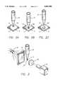
- FIG. 1illustrates a perspective view of a robotically manipulated micropipette used to spot biological probes onto an array
- FIGS. 2A through 2Cillustrate a tip of the micropipette brought into proximity with a selected location of an array for dispensing of a probe droplet
- FIG. 3illustrates a vision-assisted apparatus for estimating a volume measurement of the probe droplet
- FIG. 4is a block diagram of the vision-assisted apparatus of FIG. 3;
- FIG. 5is an enlarged view of a probe droplet affixed to a tip of the micropipette.
- FIGS. 6A through 6Cillustrates signals corresponding to diameter measurements of the droplet of FIG. 5.
- This inventionprovides a method and apparatus for accurately spotting minuscule volumes of biological probes in solution onto a test slide.
- the methodprovides for precise control over probe volume as well as placement. Further, the method is cost effective and readily adaptable for large scale production of test slides having high numbers of individual probes.
- a robotically manipulated micropipetteis used to spot biological probes onto an array 10.
- the array 10is partitioned into a plurality of individual cells 12, each of which receives a distinct type of probe. It is anticipated that the array 10 have a high number of cells 12, such as on the order of one thousand, though a smaller number of cells is illustrated in FIG. 1 for exemplary purposes.
- the array 10may be comprised of fused silica, or other such material common to microscope slides.
- the individual cells 12may be designated by use of a numerical coordinate system based on each cell's position relative to the x and y-axis directions.
- the individual cells 12are partitioned by indices 14 that further aid in designation of the cells for evaluation of probe reactions.
- the indices 14may be directly etched onto the surface of the array 10, or may be graphically applied to a separate structure onto which the array is disposed with the indices viewable through the transparent material of the array. Alternatively, the indices 14 may not be used on an actual array, but may be virtually superimposed by use of computer graphics.
- a micropipette 20is provided to convey a supply of the probe in solution to a particular cell 12 of the array 10.
- the micropipette 20comprises a tube 22 which is open at both ends.
- a first end 21 of the tube 22is drawn to a very small diameter providing a tip 24.
- the second end 23 of the tube 22is open for filling with a reservoir of the probe in solution and for connection to a gas pressure source, as will be further described below.
- a gas pressure sourceas will be further described below.
- the supply of the probe in solutionmay not be maintained within the micropipette 20, but may instead be held in an external vial (not shown) connected to the micropipette.
- an external vialnot shown
- a quantity of the probe in solutionmay be inhaled into the micropipette 20 for spotting onto the array 10. This way, the size of the micropipette 20 can be kept relatively small, and can be periodically refilled from the external vial.
- the second end 23 of the tube 22is coupled to a gas pressure line 28 that connects the micropipette 20 to a pressure source, such as a pump.
- a stopper 26provides a seal between the gas pressure line 28 and the tube 22.
- Application of gas pressure through the line 28forces a droplet 30 of the probe to be dispensed from the tip 24.
- the micropipette 20may alternatively be an initially sealed ampule containing a supply of the probe in solution, which is punctured by the gas line 28 for usage. In such an embodiment, the ampule would be disposed once a spotting operation is complete.
- the exterior of the micropipette 20may be provided with a hydrophobic coating so that the dispensed liquid will remain in the form of a globule at the tip 24, and not tend to wick up the outer surface of the micropipette which would aggravate dispensing.
- the surface area defined between the indices 14may be provided with a hydrophilic coating.
- the indices 14may also be provided with a hydrophobic coating to prevent undesired traveling of the probe to an adjacent cell 12.
- Manipulation of the micropipette 20is provided by a cartesian robot, shown symbolically in FIG. 1 as having an x-axis rail 32 and a y-axis rail 34.
- the x-axis rail 32couples to the y-axis rail 34 at a movable y-axis joint 36, and travels in the y-axis direction by motion of the y-axis joint along the y-axis rail.
- An arm 38extends from a movable x-axis joint 35 that is coupled to the x-axis rail 32, and travels in the x-axis direction by motion of the x-axis joint along the x-axis rail.
- the micropipette 20is mounted to an end of the arm 38 by use of a controllable clamp 37.
- the clamp 37should be capable of selectively grasping the micropipette 20, and permit the replacement of the micropipette with a different one as desired.
- the y-axis rail 34may be further movable in the z-axis direction by use of an additional rail and joint (not shown).
- the joints 36, 35are movable relative to the respective axes by use of motors, gears or other such frictional engagements.
- Cartesian robots of this natureare known in the art, and are capable of precise movement to position the micropipette 20 over a desired cell 12. Alternatively, the micropipette 20 may remain in a fixed position, and the array 10 shuttled into a precise position below the micropipette.
- a micropipette 20is illustrated dispensing a single droplet 30 onto an individual cell 12 of an array 10.
- the droplet 30Ais produced at the tip 24 by application of gas pressure from the pressure source.
- the droplet 30Aremains firmly affixed to the tip 24 by the surface tension of the liquid.
- the droplet 30Bis brought into contact with the cell 12 either by lowering the micropipette 20 or by raising the array 10.
- the droplet 30Bcontacts the hydrophilic surface of the cell 12, the droplet wicks completely onto the cell, as illustrated in FIG. 2C at 30C.
- the micropipette 20is withdrawn from proximity to the array 10, reversing the previous motion of either the micropipette 20 or array 10.
- the droplet 30may be gravity dispensed onto the cell 12 of the array 10.
- the micropipette 20After the droplet 30A is produced at the tip 24 of the micropipette 20 by application of gas pressure from the pressure source, as illustrated in FIG. 2A, the micropipette is rapidly withdrawn in a direction away from the cell 12. The rapid acceleration of the micropipette 20 causes the droplet 30 to become dislodged from the tip 24, enabling the droplet to fall to the cell 12 by force of gravity.
- This alternative approachavoids the risk of unintended contact between the tip 24 and the cell 12, which could potentially damage the tip and/or the array 10.
- FIG. 3illustrates the elements of a vision-assisted system used to estimate the droplet volume.
- the vision-assisted systemcomprises a light source 42, a diffusing screen 44, an objective lens 46, and video imager 48.
- the light source 42is preferably a light emitting diode (LED), but could also be a conventional incandescent light source. It is anticipated that the light source 42 would emit light having a blue or green color since the shorter wavelengths of light can yield higher resolution, although other visible and non-visible light spectra could also be advantageously utilized.
- LEDlight emitting diode
- the diffusing screen 44diffuses light emitted from the light source 42. Light scattered by the screen 44 transmits through the droplet 30, and is focused by the lens 46 onto the video imager 48.
- the video imager 48may comprise a conventional video camera or charge coupled device. Accordingly, a two-dimensional image of the droplet 30 is transmitted onto the video imager 48, which converts the two-dimensional image into a signal representative of the volume of the droplet.
- FIG. 4is a block diagram illustrating the operation of the vision-assisted system.
- a central control device 54directs the operation of a three-axis motor 56 and a pump 62.
- the control device 54receives information from a volume estimating device 58 and an algorithm 64.
- the micropipette 20is manipulated into position in the x and y-axis directions by use of a cartesian robot, or other such mechanism.
- the micropipette 20is further lowered and raised relative to the array in the z-axis direction to dispense the droplet 30.
- the three-axis motor 56receives control signals from the control device 54 to manipulate the micropipette 20 in the desired manner.
- the pump 62provides the pressure source described above that applies pressure through the gas line 28 to force the droplet 30 through the tip 24. Since gas pressure (P) is proportional to the product of volume (V) and temperature (T), the amount of pressure necessary to discharge a droplet of predetermined volume can be estimated.
- the algorithm 64defines the precise relationship between pressure, volume and temperature, and provides data to the control device 54 to direct the pump 62 to supply the required amount of pressure for a period of time to the micropipette 20.
- the data values determined by the algorithmcould comprise a table stored in a memory device, such as a computer memory. After providing the required pressure to the micropipette 20 to produce the droplet, it may be necessary to reverse the pressure slightly in order to prevent the droplet from growing in size beyond the desired volume, and to hold the droplet in place at the tip of the micropipette.
- vision-assistanceprovides feedback to the control device necessary to increase the droplet volume accuracy and repeatability.
- the video imager 48produces a two-dimensional image of the droplet during its formation and provides an associated signal to a volume estimator 58.
- the volume estimator 58provides a signal representative of the droplet volume to the control device 54.
- the volume estimatecould be used to precisely control the turn-off time for the pump 62, rather than relying entirely on the algorithm data to define the timing of the turn-off signal.
- the volume estimatecould be used to verify the accuracy of the algorithm data, which would be periodically revised by the volume estimate.
- FIG. 5illustrates an enlarged view of a tip 24 of the micropipette of FIG. 4.
- the tip 24has a droplet 30 held in place by the surface tension of the droplet.
- the dropletis presumed to form a generally spherical shape due to the surface tension of the liquid. In actuality, the droplet is not perfectly spherical; nevertheless, the spherical shape approximates the actual shape of the droplet sufficiently to make an accurate volume determination.
- an imaginary centerline 72is defined at a fixed distance below the bottom edge of the tip 24, which divides the droplet into presumably equal hemispheres.
- Light transmitted through the screen 44passes through the droplet 30 and is received by the video imager 48.
- the curvature of the dropletcauses light at the circumference of the droplet to refract, thus the transmittance of light is greater at the center 74 of the droplet than at the respective edges 76, 78.
- the refraction of lightprovides a convenient measure to determine the diameter of the sphere that approximates the droplet.
- the light refractionis converted to a diameter value.
- the measured light refractionis illustrated in FIG. 6A, with the greatest amount of refraction occurring at the edges 76, 78, and the least amount of refraction occurring at the center 74.
- the refraction curve of FIG. 6Ais differentiated to produce the curve of FIG. 6B to more accurately define the edge points.
- the absolute value of the differentiated curveis illustrated in FIG. 6C.
- the absolute value curveprovides clear indication of the edge points for the droplet, and can be subtracted to provide a diameter measurement (D).
- the video imager 48could make a plurality of raster scans adjacent to the imaginary centerline 72. Refraction values from each of the raster scans could be averaged or differentiated to determine a mean diameter measurement (D). This technique would allow for slight variations in droplet shape due to external factors, such as temperature and vibration.
- the diameter measurement (D)can then be readily converted to a volume (V) value by the relation 4 ⁇ r 3 /3, where r is radius (D/2).
- the video imager 48could simply convert the two-dimensional image of the droplet into a signal representative of the area of the image ( ⁇ r 2 ). By calculating the integral of the signal and multiplying by four, a volume value (V) can also be determined.
- the video imager 48can further be used to provide precise positional control information to the control device 54.
- the video imager 48can accurately detect the precise position of the tip 24 of the micropipette 20 relative to the indices 14 that partition the array 10. Differences between actual tip position and desired tip position can be converted to control signals provided by the control device 54 to the three-axis motor 56.
- the vision-assistancecan be used in a real-time manner to control the tip position. In this configuration, the video imager 48 would identify the precise moment that the tip 24 has reached the proper position in order to command the three-axis motor 56 to stop moving.
- the vision-assistancecan be used merely as a spot check verification of position accuracy, and for periodic correction of the algorithm values that control the command signals provided by the control device 54.
Landscapes
- Chemical & Material Sciences (AREA)
- Health & Medical Sciences (AREA)
- Analytical Chemistry (AREA)
- Chemical Kinetics & Catalysis (AREA)
- Physics & Mathematics (AREA)
- Life Sciences & Earth Sciences (AREA)
- Clinical Laboratory Science (AREA)
- Biochemistry (AREA)
- General Health & Medical Sciences (AREA)
- General Physics & Mathematics (AREA)
- Immunology (AREA)
- Pathology (AREA)
- Automatic Analysis And Handling Materials Therefor (AREA)
Abstract
Description
Claims (20)
Priority Applications (1)
| Application Number | Priority Date | Filing Date | Title |
|---|---|---|---|
| US08/311,374US5601980A (en) | 1994-09-23 | 1994-09-23 | Manufacturing method and apparatus for biological probe arrays using vision-assisted micropipetting |
Applications Claiming Priority (1)
| Application Number | Priority Date | Filing Date | Title |
|---|---|---|---|
| US08/311,374US5601980A (en) | 1994-09-23 | 1994-09-23 | Manufacturing method and apparatus for biological probe arrays using vision-assisted micropipetting |
Publications (1)
| Publication Number | Publication Date |
|---|---|
| US5601980Atrue US5601980A (en) | 1997-02-11 |
Family
ID=23206610
Family Applications (1)
| Application Number | Title | Priority Date | Filing Date |
|---|---|---|---|
| US08/311,374Expired - Fee RelatedUS5601980A (en) | 1994-09-23 | 1994-09-23 | Manufacturing method and apparatus for biological probe arrays using vision-assisted micropipetting |
Country Status (1)
| Country | Link |
|---|---|
| US (1) | US5601980A (en) |
Cited By (122)
| Publication number | Priority date | Publication date | Assignee | Title |
|---|---|---|---|---|
| WO1997044134A1 (en)* | 1996-05-17 | 1997-11-27 | Incyte Pharmaceuticals, Inc. | Jet droplet device and method |
| US5910288A (en)* | 1997-07-10 | 1999-06-08 | Hewlett-Packard Company | Method and apparatus for mixing a thin film of fluid |
| WO1999030169A1 (en)* | 1997-12-08 | 1999-06-17 | MAX-PLANCK-Gesellschaft zur Förderung der Wissenschaften e.V. | Method and device for recording an image on drop-producing dispensing heads |
| US5961767A (en)* | 1997-05-15 | 1999-10-05 | Lucent Technologies, Inc. | Method for forming micron-sized and smaller liquid droplets |
| WO2000013796A1 (en) | 1998-09-09 | 2000-03-16 | Incyte Pharmaceuticals, Inc. | Capillary printing systems |
| WO2000063705A1 (en)* | 1999-04-16 | 2000-10-26 | Pe Corporation (Ny) | Apparatus and method for transferring small volumes of substances |
| WO2001023092A1 (en)* | 1999-09-28 | 2001-04-05 | Giesing, Michael | Device and method for absorbing and releasing minute amounts of liquid |
| US6220075B1 (en)* | 1996-05-31 | 2001-04-24 | Packard Instrument Company | Method for determining and verifying a microvolume of a sample liquid dispersed in droplets |
| US6228659B1 (en) | 1997-10-31 | 2001-05-08 | PE Corporation (“NY”) | Method and apparatus for making arrays |
| US6255119B1 (en)* | 1997-11-10 | 2001-07-03 | Hyseq, Inc. | Reagent transfer device |
| US6296702B1 (en) | 1999-03-15 | 2001-10-02 | Pe Corporation (Ny) | Apparatus and method for spotting a substrate |
| US20010031500A1 (en)* | 2000-04-13 | 2001-10-18 | Matsushita Electric Industrial Co., Ltd. | Method for verifying amount of sample solution, method for controlling measurement system and method for measuring concentration of solution in apparatus for measuring optical characteristic |
| WO2001076746A1 (en)* | 2000-04-10 | 2001-10-18 | Basf Aktiengesellschaft | Method for producing biopolymer fields by means of real-time control |
| EP1158281A1 (en)* | 2001-01-11 | 2001-11-28 | Elite Engineering Corporation | System and method for dispensing fluid droplets of known volume and generating very low fluid flow rates |
| WO2002004123A1 (en)* | 2000-07-06 | 2002-01-17 | Robodesign International, Inc. | Microarray dispensing with real-time verification and inspection |
| KR100320752B1 (en)* | 1999-08-06 | 2002-01-17 | 박한오 | automated microarray of samples |
| US6376619B1 (en) | 1998-04-13 | 2002-04-23 | 3M Innovative Properties Company | High density, miniaturized arrays and methods of manufacturing same |
| WO2002050552A1 (en)* | 2000-12-18 | 2002-06-27 | Ngk Insulators, Ltd. | Method of forming detection points in chip for detecting subject |
| US20020160370A1 (en)* | 2001-04-30 | 2002-10-31 | Bass Jay K. | Error detection in chemical array fabrication |
| US6476215B1 (en) | 1997-08-01 | 2002-11-05 | Canon Kabushiki Kaisha | Ink jet method of spotting a probe and manufacturing a probe array |
| WO2002102515A1 (en)* | 2001-06-18 | 2002-12-27 | Fraunhofer-Gesellschaft zur Förderung der angewandten Forschung e.V. | Method and device for dosing fluid media |
| US20030036090A1 (en)* | 1999-12-09 | 2003-02-20 | 3M Innovative Properties Company | Heat-relaxable substrates and arrays |
| WO2003013718A1 (en)* | 2001-08-10 | 2003-02-20 | Oxford Glycosciences (Uk) Ltd | Liquid delivery apparatus and method |
| US6537505B1 (en) | 1998-02-20 | 2003-03-25 | Bio Dot, Inc. | Reagent dispensing valve |
| US6551557B1 (en) | 1998-07-07 | 2003-04-22 | Cartesian Technologies, Inc. | Tip design and random access array for microfluidic transfer |
| US20030087328A1 (en)* | 1999-05-05 | 2003-05-08 | Pollok Brian A. | Optical probes and assays |
| EP1096250A3 (en)* | 1999-10-29 | 2003-06-11 | Agilent Technologies, Inc. (a Delaware corporation) | Apparatus and method for deposition and inspection of chemical and biological fluids |
| US6587579B1 (en) | 2000-01-26 | 2003-07-01 | Agilent Technologies Inc. | Feature quality in array fabrication |
| US6589791B1 (en) | 1999-05-20 | 2003-07-08 | Cartesian Technologies, Inc. | State-variable control system |
| US20030190612A1 (en)* | 2000-08-31 | 2003-10-09 | Nobuko Yamamoto | Detecting method and detection substrate for use therein |
| US20030202907A1 (en)* | 2001-09-13 | 2003-10-30 | Woodward Roger P. | Dispensing method and apparatus for dispensing very small quantities of fluid |
| US20030203494A1 (en)* | 2002-04-29 | 2003-10-30 | Hyde David D. | Dynamic metered fluid volume determination method and related apparatus |
| US20030207464A1 (en)* | 1999-02-19 | 2003-11-06 | Tony Lemmo | Methods for microfluidic aspirating and dispensing |
| GB2388601A (en)* | 1999-04-30 | 2003-11-19 | Agilent Technologies Inc | Fabrication of an addressable array of biopolymers |
| WO2003028868A3 (en)* | 2001-10-03 | 2004-01-08 | Kin Chiu Ng | Apparatus and method for fabricating high density microarrays and applications thereof |
| US20040018613A1 (en)* | 2002-07-16 | 2004-01-29 | Tomoaki Shoji | Method for producing a microarray |
| US20040023391A1 (en)* | 2002-07-30 | 2004-02-05 | Ye Fang | Method and device for protein delivery into cells |
| US6689323B2 (en) | 1998-10-30 | 2004-02-10 | Agilent Technologies | Method and apparatus for liquid transfer |
| WO2002080822A3 (en)* | 2001-04-04 | 2004-02-26 | Arradial Inc | System and method for dispensing liquids |
| US20040048241A1 (en)* | 2001-06-11 | 2004-03-11 | Freeman Beverly Annette | Methods for attaching molecules |
| US20040062686A1 (en)* | 2000-07-06 | 2004-04-01 | Ganz Brian L. | Microarray dispensing with real-time verification and inspection |
| US20040072364A1 (en)* | 1998-01-09 | 2004-04-15 | Tisone Thomas C. | Method for high-speed dot array dispensing |
| US6723569B1 (en)* | 1998-11-04 | 2004-04-20 | Genomic Solutions Acquisitions Limited | Liquid transfer system |
| US20040115722A1 (en)* | 2002-10-25 | 2004-06-17 | Mel Kronick | Biopolymeric arrays and methods of producing the same |
| GB2355716B (en)* | 1999-04-30 | 2004-06-23 | Agilent Technologies Inc | Polynucleotide array fabrication |
| US20040137491A1 (en)* | 2002-06-28 | 2004-07-15 | Tadashi Okamoto | Method of analyzing probe carrier using time-of-flight secondary ion mass spectrometry |
| US20040146944A1 (en)* | 2003-01-29 | 2004-07-29 | Ye Fang | Reverse protein delivery into cells on coded microparticles |
| US20040146863A1 (en)* | 2001-06-11 | 2004-07-29 | Pisharody Sobha M. | Electronic detection of biological molecules using thin layers |
| US20040152113A1 (en)* | 2002-06-28 | 2004-08-05 | Hiromitsu Takase | Probe carrier and method for analyzing the probe carrier |
| US6773927B2 (en)* | 2000-02-18 | 2004-08-10 | Hitachi Koki Co., Ltd. | Pipetting apparatus and a method of pipetting a liquid |
| US20040171043A1 (en)* | 2002-06-28 | 2004-09-02 | Canon Kabushiki Kaisha | Probe carrier, method of producing the probe carrier, method of evaluating the probe carrier and method of detecting a target nucleic acid using the same |
| US20040197817A1 (en)* | 1999-04-30 | 2004-10-07 | Caren Michael P. | Polynucleotide array fabrication |
| US20040219688A1 (en)* | 1998-01-09 | 2004-11-04 | Carl Churchill | Method and apparatus for high-speed microfluidic dispensing using text file control |
| US20040259088A1 (en)* | 2002-06-28 | 2004-12-23 | Canon Kabushiki Kaisha | Method for analyzing RNA using time of flight secondary ion mass spectrometry |
| US20050019223A1 (en)* | 2001-08-10 | 2005-01-27 | Platt Albert Edward | Liquid delivery apparatus and method |
| WO2005012329A2 (en) | 2003-07-29 | 2005-02-10 | Invitrogen Corporation | Kinase and phosphatase assays |
| US20050056713A1 (en)* | 2003-07-31 | 2005-03-17 | Tisone Thomas C. | Methods and systems for dispensing sub-microfluidic drops |
| US20050064485A1 (en)* | 2003-09-12 | 2005-03-24 | Kurt Vogel | Multiplex binding and activity assays |
| US20050065230A1 (en)* | 2000-02-18 | 2005-03-24 | Chin-Shiou Huang | Compositions and methods for surface imprinting |
| US6878341B2 (en) | 1999-05-27 | 2005-04-12 | Applera Corporation | Apparatus for the precise location of reaction plates |
| US20050170442A1 (en)* | 2003-07-29 | 2005-08-04 | Kupcho Kevin R. | Bimolecular optical probes |
| US20050208530A1 (en)* | 2003-12-01 | 2005-09-22 | Invitrogen Corporation | Nucleic acid molecules containing recombination sites and methods of using the same |
| US20050221271A1 (en)* | 2002-05-22 | 2005-10-06 | Platypus Technologies, Llc | Substrates, devices, and methods for cellular assays |
| US20060057736A1 (en)* | 2004-09-13 | 2006-03-16 | Peck Bill J | Methods and devices for fabricating chemical arrays |
| US20060160688A1 (en)* | 2005-01-17 | 2006-07-20 | Kak Namkoong | Handheld centrifuge |
| US20060188922A1 (en)* | 2002-10-25 | 2006-08-24 | Corson John F | Biopolymeric arrays having replicate elements |
| US20070042496A1 (en)* | 2002-06-28 | 2007-02-22 | Canon Kabushiki Kaisha | Method for acquiring information of a biochip using time of flight secondary ion mass spectrometry and an apparatus for acquiring information for the application thereof |
| US20070048191A1 (en)* | 2005-08-30 | 2007-03-01 | Seiko Epson Corporation | Biochip manufacturing method and biochip manufacturing device |
| US20070059787A1 (en)* | 2003-07-29 | 2007-03-15 | Invitrogen Corporation | Kinase and phosphatase assays |
| JP2007136568A (en)* | 2005-11-15 | 2007-06-07 | National Institute Of Advanced Industrial & Technology | Cutting method and cutting apparatus for minimal linear flexible object |
| US20070148763A1 (en)* | 2005-12-22 | 2007-06-28 | Nam Huh | Quantitative cell dispensing apparatus using liquid drop manipulation |
| US20070178534A1 (en)* | 2002-05-22 | 2007-08-02 | Christopher Murphy | Substrates, devices, and methods for cellular assays |
| US20070184436A1 (en)* | 2001-06-07 | 2007-08-09 | Joel Myerson | Generic capture probe arrays |
| US20070248498A1 (en)* | 2006-04-19 | 2007-10-25 | Archivex Llc | Micro-drop detection and detachment |
| WO2008021071A2 (en) | 2006-08-07 | 2008-02-21 | Platypus Technologies, Llc | Substrates, devices, and methods for cellular assays |
| US20080071074A1 (en)* | 2006-05-22 | 2008-03-20 | Third Wave Technologies, Inc. | Compositions, probes, and conjugates and uses thereof |
| US20080080593A1 (en)* | 2006-09-28 | 2008-04-03 | Welch Allyn, Inc. | Safety probe for thermometry apparatus |
| US20080160539A1 (en)* | 2006-08-07 | 2008-07-03 | Platypus Technologies, Llc | Substrates, devices, and methods for cellular assays |
| US20090054262A1 (en)* | 2007-08-20 | 2009-02-26 | Platypus Technologies, Llc | Devices for cell assays |
| US20090131263A1 (en)* | 2007-11-19 | 2009-05-21 | Longying Dong | Normalization methods for G-protein coupled receptor membrane array |
| US7662572B2 (en) | 2005-08-25 | 2010-02-16 | Platypus Technologies, Llc. | Compositions and liquid crystals |
| US20100093096A1 (en)* | 2008-09-15 | 2010-04-15 | Platypus Technologies, Llc | Detection of vapor phase compounds by changes in physical properties of a liquid crystal |
| US20100267120A1 (en)* | 1995-06-07 | 2010-10-21 | Hartley James L | Recombinational cloning using nucleic acids having recombination sites |
| US8030066B2 (en) | 2000-12-11 | 2011-10-04 | Life Technologies Corporation | Methods and compositions for synthesis of nucleic acid molecules using multiple recognition sites |
| US20110317004A1 (en)* | 2010-06-29 | 2011-12-29 | Kai Tao | IV Monitoring by Digital Image Processing |
| US20120013735A1 (en)* | 2010-07-15 | 2012-01-19 | Kai Tao | IV monitoring by video and image processing |
| WO2012108499A1 (en) | 2011-02-10 | 2012-08-16 | 三菱レイヨン株式会社 | Method for detecting nucleic acid |
| US8338091B2 (en) | 2003-08-08 | 2012-12-25 | Life Technologies Corporation | Methods and compositions for seamless cloning of nucleic acid molecules |
| EP2631652A1 (en) | 2005-04-05 | 2013-08-28 | Corning Incorporated | Label free biosensor |
| WO2013158740A1 (en)* | 2012-04-18 | 2013-10-24 | Biofire Diagnostics, Inc. | Microspotting device |
| WO2014205344A2 (en) | 2013-06-21 | 2014-12-24 | The Johns Hopkins University | Virion display array for profiling functions and interactions of human membrane proteins |
| US8920752B2 (en) | 2007-01-19 | 2014-12-30 | Biodot, Inc. | Systems and methods for high speed array printing and hybridization |
| EP2848306A1 (en) | 2013-09-13 | 2015-03-18 | Bruker Daltonik GmbH | Dispenser system for mass spectrometric sample preparations |
| US8988620B2 (en) | 2003-07-25 | 2015-03-24 | Platypus Technologies, Llc | Liquid crystal based analyte detection |
| US9068566B2 (en) | 2011-01-21 | 2015-06-30 | Biodot, Inc. | Piezoelectric dispenser with a longitudinal transducer and replaceable capillary tube |
| US9103794B2 (en) | 2001-08-27 | 2015-08-11 | Platypus Technologies Llc | Substrates, devices, and methods for quantitative liquid crystal assays |
| US9304141B2 (en) | 2007-04-18 | 2016-04-05 | Becton, Dickinson And Company | Method and apparatus for determing dispense volume |
| US9372486B2 (en) | 2011-12-21 | 2016-06-21 | Deka Products Limited Partnership | System, method, and apparatus for monitoring, regulating, or controlling fluid flow |
| US9435455B2 (en) | 2011-12-21 | 2016-09-06 | Deka Products Limited Partnership | System, method, and apparatus for monitoring, regulating, or controlling fluid flow |
| US9724466B2 (en) | 2011-12-21 | 2017-08-08 | Deka Products Limited Partnership | Flow meter |
| US9746093B2 (en) | 2011-12-21 | 2017-08-29 | Deka Products Limited Partnership | Flow meter and related system and apparatus |
| US9746094B2 (en) | 2011-12-21 | 2017-08-29 | Deka Products Limited Partnership | Flow meter having a background pattern with first and second portions |
| US9759343B2 (en) | 2012-12-21 | 2017-09-12 | Deka Products Limited Partnership | Flow meter using a dynamic background image |
| USD799025S1 (en) | 2013-11-06 | 2017-10-03 | Deka Products Limited Partnership | Apparatus to control fluid flow through a tube |
| USD802118S1 (en) | 2013-11-06 | 2017-11-07 | Deka Products Limited Partnership | Apparatus to control fluid flow through a tube |
| USD813376S1 (en) | 2013-11-06 | 2018-03-20 | Deka Products Limited Partnership | Apparatus to control fluid flow through a tube |
| USD815730S1 (en) | 2013-11-06 | 2018-04-17 | Deka Products Limited Partnership | Apparatus to control fluid flow through a tube |
| USD816829S1 (en) | 2013-11-06 | 2018-05-01 | Deka Products Limited Partnership | Apparatus to control fluid flow through a tube |
| US10088346B2 (en) | 2011-12-21 | 2018-10-02 | Deka Products Limited Partnership | System, method, and apparatus for monitoring, regulating, or controlling fluid flow |
| US10093967B2 (en) | 2014-08-12 | 2018-10-09 | The Regents Of The University Of Michigan | Detection of nucleic acids |
| US20180348247A1 (en)* | 2017-06-01 | 2018-12-06 | Hitachi, Ltd. | Dispensing apparatus |
| US10228683B2 (en) | 2011-12-21 | 2019-03-12 | Deka Products Limited Partnership | System, method, and apparatus for monitoring, regulating, or controlling fluid flow |
| USD854145S1 (en) | 2016-05-25 | 2019-07-16 | Deka Products Limited Partnership | Apparatus to control fluid flow through a tube |
| US10488848B2 (en) | 2011-12-21 | 2019-11-26 | Deka Products Limited Partnership | System, method, and apparatus for monitoring, regulating, or controlling fluid flow |
| CN111665111A (en)* | 2020-07-03 | 2020-09-15 | 上海百傲科技股份有限公司 | Sample applicator |
| EP3711856A1 (en)* | 2019-03-22 | 2020-09-23 | Sysmex Corporation | Specimen measurement device, specimen measurement method, and nozzle |
| EP3751289A1 (en)* | 2019-06-13 | 2020-12-16 | Siemens Healthcare Diagnostics Products GmbH | Device for optically monitoring a dosing of a liquid to be pipetted |
| USD905848S1 (en) | 2016-01-28 | 2020-12-22 | Deka Products Limited Partnership | Apparatus to control fluid flow through a tube |
| CN114958583A (en)* | 2022-06-30 | 2022-08-30 | 南京普济生物有限公司 | System and method for detecting blood free DNA by using ultra-multiplex PCR |
| USD964563S1 (en) | 2019-07-26 | 2022-09-20 | Deka Products Limited Partnership | Medical flow clamp |
| US11744935B2 (en) | 2016-01-28 | 2023-09-05 | Deka Products Limited Partnership | Apparatus for monitoring, regulating, or controlling fluid flow |
| US11839741B2 (en) | 2019-07-26 | 2023-12-12 | Deka Products Limited Partneship | Apparatus for monitoring, regulating, or controlling fluid flow |
Citations (9)
| Publication number | Priority date | Publication date | Assignee | Title |
|---|---|---|---|---|
| US3783696A (en)* | 1971-12-09 | 1974-01-08 | C Coleman | Automatic volume control pipet |
| US4452899A (en)* | 1982-06-10 | 1984-06-05 | Eastman Kodak Company | Method for metering biological fluids |
| US4919899A (en)* | 1988-02-29 | 1990-04-24 | Herrmann Frederick T | Crystal growth apparatus |
| US5143849A (en)* | 1991-03-21 | 1992-09-01 | Eastman Kodak Company | Tip to surface spacing for optimum dispensing controlled by a detected pressure change in the tip |
| US5275951A (en)* | 1991-06-13 | 1994-01-04 | Abbott Laboratories | Liquid level sensing method and device |
| US5334353A (en)* | 1993-02-03 | 1994-08-02 | Blattner Frederick R | Micropipette device |
| US5336467A (en)* | 1989-11-22 | 1994-08-09 | Vettest S.A. | Chemical analyzer |
| US5338688A (en)* | 1990-08-02 | 1994-08-16 | Boehringer Mannheim Gmbh | Method for the metered application of a biochemical analytical liquid to a target |
| US5443791A (en)* | 1990-04-06 | 1995-08-22 | Perkin Elmer - Applied Biosystems Division | Automated molecular biology laboratory |
- 1994
- 1994-09-23USUS08/311,374patent/US5601980A/ennot_activeExpired - Fee Related
Patent Citations (9)
| Publication number | Priority date | Publication date | Assignee | Title |
|---|---|---|---|---|
| US3783696A (en)* | 1971-12-09 | 1974-01-08 | C Coleman | Automatic volume control pipet |
| US4452899A (en)* | 1982-06-10 | 1984-06-05 | Eastman Kodak Company | Method for metering biological fluids |
| US4919899A (en)* | 1988-02-29 | 1990-04-24 | Herrmann Frederick T | Crystal growth apparatus |
| US5336467A (en)* | 1989-11-22 | 1994-08-09 | Vettest S.A. | Chemical analyzer |
| US5443791A (en)* | 1990-04-06 | 1995-08-22 | Perkin Elmer - Applied Biosystems Division | Automated molecular biology laboratory |
| US5338688A (en)* | 1990-08-02 | 1994-08-16 | Boehringer Mannheim Gmbh | Method for the metered application of a biochemical analytical liquid to a target |
| US5143849A (en)* | 1991-03-21 | 1992-09-01 | Eastman Kodak Company | Tip to surface spacing for optimum dispensing controlled by a detected pressure change in the tip |
| US5275951A (en)* | 1991-06-13 | 1994-01-04 | Abbott Laboratories | Liquid level sensing method and device |
| US5334353A (en)* | 1993-02-03 | 1994-08-02 | Blattner Frederick R | Micropipette device |
Cited By (250)
| Publication number | Priority date | Publication date | Assignee | Title |
|---|---|---|---|---|
| US20100267120A1 (en)* | 1995-06-07 | 2010-10-21 | Hartley James L | Recombinational cloning using nucleic acids having recombination sites |
| US6001309A (en)* | 1996-05-17 | 1999-12-14 | Incyte Pharmaceuticals, Inc. | Jet droplet device |
| WO1997044134A1 (en)* | 1996-05-17 | 1997-11-27 | Incyte Pharmaceuticals, Inc. | Jet droplet device and method |
| US6220075B1 (en)* | 1996-05-31 | 2001-04-24 | Packard Instrument Company | Method for determining and verifying a microvolume of a sample liquid dispersed in droplets |
| US5961767A (en)* | 1997-05-15 | 1999-10-05 | Lucent Technologies, Inc. | Method for forming micron-sized and smaller liquid droplets |
| US5910288A (en)* | 1997-07-10 | 1999-06-08 | Hewlett-Packard Company | Method and apparatus for mixing a thin film of fluid |
| US7994297B2 (en) | 1997-08-01 | 2011-08-09 | Canon Kabushiki Kaisha | Method of spotting probes on a solid support |
| US20030059817A1 (en)* | 1997-08-01 | 2003-03-27 | Tadashi Okamoto | Inkjet method of spotting probe and manufacturing a probe array |
| US20060177868A1 (en)* | 1997-08-01 | 2006-08-10 | Canon Kabushiki Kaisha | Method of spotting probe on solid support, probe array and method of manufacturing thereof, and method of detecting target substance and method of identifying structure of target substance using probe array |
| EP1352976A3 (en)* | 1997-08-01 | 2003-10-29 | Canon Kabushiki Kaisha | Method of spotting probe on solid support, probe array and method of manufacturing thereof, and method of detecting target substance and method of identifying structure of target substance using probe array |
| US6476215B1 (en) | 1997-08-01 | 2002-11-05 | Canon Kabushiki Kaisha | Ink jet method of spotting a probe and manufacturing a probe array |
| EP0895082B1 (en)* | 1997-08-01 | 2003-09-24 | Canon Kabushiki Kaisha | Bubble jet method for spotting probes on a solid support and method of manufacturing probe arrays. |
| US6228659B1 (en) | 1997-10-31 | 2001-05-08 | PE Corporation (“NY”) | Method and apparatus for making arrays |
| US6255119B1 (en)* | 1997-11-10 | 2001-07-03 | Hyseq, Inc. | Reagent transfer device |
| US6851784B1 (en) | 1997-12-08 | 2005-02-08 | Max-Planck-Gesellschaft Zur Forderung Der Wissenschaften E.V. | Method and device for recording an image on drop-producing dispensing heads |
| WO1999030169A1 (en)* | 1997-12-08 | 1999-06-17 | MAX-PLANCK-Gesellschaft zur Förderung der Wissenschaften e.V. | Method and device for recording an image on drop-producing dispensing heads |
| US20040072364A1 (en)* | 1998-01-09 | 2004-04-15 | Tisone Thomas C. | Method for high-speed dot array dispensing |
| US20040219688A1 (en)* | 1998-01-09 | 2004-11-04 | Carl Churchill | Method and apparatus for high-speed microfluidic dispensing using text file control |
| US6537505B1 (en) | 1998-02-20 | 2003-03-25 | Bio Dot, Inc. | Reagent dispensing valve |
| US6376619B1 (en) | 1998-04-13 | 2002-04-23 | 3M Innovative Properties Company | High density, miniaturized arrays and methods of manufacturing same |
| US7189842B2 (en) | 1998-04-13 | 2007-03-13 | 3M Innovative Properties Company | High density, miniaturized arrays and methods of manufacturing same |
| US6548607B2 (en) | 1998-04-13 | 2003-04-15 | 3M Innovative Properties Company | High density, miniaturized arrays and methods of manufacturing same |
| US6573338B2 (en) | 1998-04-13 | 2003-06-03 | 3M Innovative Properties Company | High density, miniaturized arrays and methods of manufacturing same |
| US20020122917A1 (en)* | 1998-04-13 | 2002-09-05 | 3M Innovative Properties Company | High density, miniaturized arrays and methods of manufacturing same |
| US6841258B2 (en) | 1998-04-13 | 2005-01-11 | 3M Innovative Properties Company | High density, miniaturized arrays and methods of manufacturing same |
| US20070021602A1 (en)* | 1998-04-13 | 2007-01-25 | 3M Innovative Properties Company | High density, miniaturized arrays and methods of manufacturing same |
| US20040072365A1 (en)* | 1998-07-07 | 2004-04-15 | Don Rose | Method and apparatus for liquid dispensing |
| US6551557B1 (en) | 1998-07-07 | 2003-04-22 | Cartesian Technologies, Inc. | Tip design and random access array for microfluidic transfer |
| US7736591B2 (en) | 1998-07-07 | 2010-06-15 | Biodot, Inc. | Method and apparatus for liquid dispensing |
| WO2000013796A1 (en) | 1998-09-09 | 2000-03-16 | Incyte Pharmaceuticals, Inc. | Capillary printing systems |
| DE19950809B4 (en)* | 1998-10-30 | 2007-11-15 | Agilent Technologies, Inc. (n.d.Ges.d. Staates Delaware), Santa Clara | Method and apparatus for fluid transfer |
| US6689323B2 (en) | 1998-10-30 | 2004-02-10 | Agilent Technologies | Method and apparatus for liquid transfer |
| US6723569B1 (en)* | 1998-11-04 | 2004-04-20 | Genomic Solutions Acquisitions Limited | Liquid transfer system |
| US20030207464A1 (en)* | 1999-02-19 | 2003-11-06 | Tony Lemmo | Methods for microfluidic aspirating and dispensing |
| US6467700B2 (en) | 1999-03-15 | 2002-10-22 | Pe Corporation (Ny) | Apparatus and method for spotting a substrate |
| US6849127B2 (en) | 1999-03-15 | 2005-02-01 | Applera Corporation | Apparatus and method for spotting a substrate |
| US6296702B1 (en) | 1999-03-15 | 2001-10-02 | Pe Corporation (Ny) | Apparatus and method for spotting a substrate |
| US6440217B2 (en) | 1999-03-15 | 2002-08-27 | Pe Corporation (Ny) | Apparatus and method for spotting a substrate |
| US6413586B2 (en) | 1999-03-15 | 2002-07-02 | Pe Corporation (Ny) | Apparatus and method for spotting a substrate |
| US7211148B2 (en) | 1999-03-15 | 2007-05-01 | Applera Corporation | Apparatus and method for spotting a substrate |
| US6579367B2 (en) | 1999-03-15 | 2003-06-17 | Applera Corporation | Apparatus and method for spotting a substrate |
| US20050120949A1 (en)* | 1999-03-15 | 2005-06-09 | Applera Corporation | Apparatus and method for spotting a substrate |
| US20070148050A1 (en)* | 1999-03-15 | 2007-06-28 | Applera Corporation | Apparatus and method for spotting a substrate |
| US20020041829A1 (en)* | 1999-04-16 | 2002-04-11 | Pe Corporation (Ny) | Apparatus and method for transferring small volumes of substances |
| US6916447B2 (en) | 1999-04-16 | 2005-07-12 | Applera Corporation | Apparatus and method for transferring small volumes of substances |
| WO2000063705A1 (en)* | 1999-04-16 | 2000-10-26 | Pe Corporation (Ny) | Apparatus and method for transferring small volumes of substances |
| US6245297B1 (en) | 1999-04-16 | 2001-06-12 | Pe Corporation (Ny) | Apparatus and method for transferring small volumes of substances |
| US6355487B2 (en) | 1999-04-16 | 2002-03-12 | Pe Corporation (Ny) | Apparatus and method for transferring small volumes of substances |
| US20040203138A1 (en)* | 1999-04-30 | 2004-10-14 | Caren Michael P. | Polynucleotide array fabrication |
| US20040203047A1 (en)* | 1999-04-30 | 2004-10-14 | Caren Michael P. | Polynucleotide array fabrication |
| GB2355716B (en)* | 1999-04-30 | 2004-06-23 | Agilent Technologies Inc | Polynucleotide array fabrication |
| GB2388601A (en)* | 1999-04-30 | 2003-11-19 | Agilent Technologies Inc | Fabrication of an addressable array of biopolymers |
| GB2388601B (en)* | 1999-04-30 | 2004-04-28 | Agilent Technologies Inc | Polynucleotide array fabrication |
| US20040197817A1 (en)* | 1999-04-30 | 2004-10-07 | Caren Michael P. | Polynucleotide array fabrication |
| US20030087328A1 (en)* | 1999-05-05 | 2003-05-08 | Pollok Brian A. | Optical probes and assays |
| US20080146460A1 (en)* | 1999-05-05 | 2008-06-19 | Invitrogen Corporation | Optical probes and assays |
| US20030211620A1 (en)* | 1999-05-20 | 2003-11-13 | Labudde Edward V. | State-variable control system |
| US6589791B1 (en) | 1999-05-20 | 2003-07-08 | Cartesian Technologies, Inc. | State-variable control system |
| US6878341B2 (en) | 1999-05-27 | 2005-04-12 | Applera Corporation | Apparatus for the precise location of reaction plates |
| US20050152810A1 (en)* | 1999-05-27 | 2005-07-14 | Applera Corporation | Apparatus and method for the precise location of reaction plates |
| KR100320752B1 (en)* | 1999-08-06 | 2002-01-17 | 박한오 | automated microarray of samples |
| WO2001023092A1 (en)* | 1999-09-28 | 2001-04-05 | Giesing, Michael | Device and method for absorbing and releasing minute amounts of liquid |
| US6689319B1 (en) | 1999-10-29 | 2004-02-10 | Agilent Technologies, Ind. | Apparatus for deposition and inspection of chemical and biological fluids |
| EP1096250A3 (en)* | 1999-10-29 | 2003-06-11 | Agilent Technologies, Inc. (a Delaware corporation) | Apparatus and method for deposition and inspection of chemical and biological fluids |
| US20030036090A1 (en)* | 1999-12-09 | 2003-02-20 | 3M Innovative Properties Company | Heat-relaxable substrates and arrays |
| US20070218484A1 (en)* | 2000-01-26 | 2007-09-20 | Agilent Technologies Inc. | Feature Quality in Array Fabrication |
| US6587579B1 (en) | 2000-01-26 | 2003-07-01 | Agilent Technologies Inc. | Feature quality in array fabrication |
| US20050065230A1 (en)* | 2000-02-18 | 2005-03-24 | Chin-Shiou Huang | Compositions and methods for surface imprinting |
| US6773927B2 (en)* | 2000-02-18 | 2004-08-10 | Hitachi Koki Co., Ltd. | Pipetting apparatus and a method of pipetting a liquid |
| RU2302294C2 (en)* | 2000-04-10 | 2007-07-10 | Басф Акциенгезельшафт | Method for detecting application of sample substance and apparatus for performing the same |
| WO2001076746A1 (en)* | 2000-04-10 | 2001-10-18 | Basf Aktiengesellschaft | Method for producing biopolymer fields by means of real-time control |
| US20070059217A1 (en)* | 2000-04-10 | 2007-03-15 | Heinz Eipel | Process for the production of biopolymer fields with real-time control |
| US20030109052A1 (en)* | 2000-04-10 | 2003-06-12 | Heinz Eipel | Method for producing biopolymer fields by means of real-time control |
| US7282367B2 (en)* | 2000-04-13 | 2007-10-16 | Matsushita Electric Industrial Co., Ltd. | Method for verifying amount of sample solution, method for controlling measurement system and method for measuring concentration of solution in apparatus for measuring optical characteristic |
| US20010031500A1 (en)* | 2000-04-13 | 2001-10-18 | Matsushita Electric Industrial Co., Ltd. | Method for verifying amount of sample solution, method for controlling measurement system and method for measuring concentration of solution in apparatus for measuring optical characteristic |
| US6558623B1 (en)* | 2000-07-06 | 2003-05-06 | Robodesign International, Inc. | Microarray dispensing with real-time verification and inspection |
| US20040062686A1 (en)* | 2000-07-06 | 2004-04-01 | Ganz Brian L. | Microarray dispensing with real-time verification and inspection |
| WO2002004123A1 (en)* | 2000-07-06 | 2002-01-17 | Robodesign International, Inc. | Microarray dispensing with real-time verification and inspection |
| US7025933B2 (en)* | 2000-07-06 | 2006-04-11 | Robodesign International, Inc. | Microarray dispensing with real-time verification and inspection |
| US9309520B2 (en) | 2000-08-21 | 2016-04-12 | Life Technologies Corporation | Methods and compositions for synthesis of nucleic acid molecules using multiple recognition sites |
| US20030190612A1 (en)* | 2000-08-31 | 2003-10-09 | Nobuko Yamamoto | Detecting method and detection substrate for use therein |
| US8030066B2 (en) | 2000-12-11 | 2011-10-04 | Life Technologies Corporation | Methods and compositions for synthesis of nucleic acid molecules using multiple recognition sites |
| US8945884B2 (en) | 2000-12-11 | 2015-02-03 | Life Technologies Corporation | Methods and compositions for synthesis of nucleic acid molecules using multiplerecognition sites |
| WO2002050552A1 (en)* | 2000-12-18 | 2002-06-27 | Ngk Insulators, Ltd. | Method of forming detection points in chip for detecting subject |
| EP1158281A1 (en)* | 2001-01-11 | 2001-11-28 | Elite Engineering Corporation | System and method for dispensing fluid droplets of known volume and generating very low fluid flow rates |
| WO2002080822A3 (en)* | 2001-04-04 | 2004-02-26 | Arradial Inc | System and method for dispensing liquids |
| EP1414585A4 (en)* | 2001-04-04 | 2007-03-07 | Arradial Inc | System and method for dispensing liquids |
| US20020160370A1 (en)* | 2001-04-30 | 2002-10-31 | Bass Jay K. | Error detection in chemical array fabrication |
| US20050287586A1 (en)* | 2001-04-30 | 2005-12-29 | Bass Jay K | Error detection in chemical array fabrication |
| US6943036B2 (en) | 2001-04-30 | 2005-09-13 | Agilent Technologies, Inc. | Error detection in chemical array fabrication |
| US20070184436A1 (en)* | 2001-06-07 | 2007-08-09 | Joel Myerson | Generic capture probe arrays |
| US20040048241A1 (en)* | 2001-06-11 | 2004-03-11 | Freeman Beverly Annette | Methods for attaching molecules |
| US20040146863A1 (en)* | 2001-06-11 | 2004-07-29 | Pisharody Sobha M. | Electronic detection of biological molecules using thin layers |
| US6824974B2 (en) | 2001-06-11 | 2004-11-30 | Genorx, Inc. | Electronic detection of biological molecules using thin layers |
| WO2002102515A1 (en)* | 2001-06-18 | 2002-12-27 | Fraunhofer-Gesellschaft zur Förderung der angewandten Forschung e.V. | Method and device for dosing fluid media |
| US20040185167A1 (en)* | 2001-06-18 | 2004-09-23 | Heiko Zimmermann | Method and device for dosing fluid media |
| US20090124519A1 (en)* | 2001-08-10 | 2009-05-14 | Oxford Genome Sciences (Uk) Ltd. | Liquid delivery apparatus and method |
| US20050019223A1 (en)* | 2001-08-10 | 2005-01-27 | Platt Albert Edward | Liquid delivery apparatus and method |
| WO2003013718A1 (en)* | 2001-08-10 | 2003-02-20 | Oxford Glycosciences (Uk) Ltd | Liquid delivery apparatus and method |
| US9797843B2 (en) | 2001-08-27 | 2017-10-24 | Platypus Technologies, Llc | Substrates, devices, and methods for quantitative liquid crystal assays |
| US9103794B2 (en) | 2001-08-27 | 2015-08-11 | Platypus Technologies Llc | Substrates, devices, and methods for quantitative liquid crystal assays |
| US20030202907A1 (en)* | 2001-09-13 | 2003-10-30 | Woodward Roger P. | Dispensing method and apparatus for dispensing very small quantities of fluid |
| WO2003028868A3 (en)* | 2001-10-03 | 2004-01-08 | Kin Chiu Ng | Apparatus and method for fabricating high density microarrays and applications thereof |
| US7361509B2 (en)* | 2002-04-29 | 2008-04-22 | Ortho-Clinical Diagnostics | Dynamic metered fluid volume determination method and related apparatus |
| US20030203494A1 (en)* | 2002-04-29 | 2003-10-30 | Hyde David D. | Dynamic metered fluid volume determination method and related apparatus |
| US7018838B2 (en) | 2002-05-22 | 2006-03-28 | Platypus Technologies, Llc | Substrates, devices, and methods for cellular assays |
| EP2093565A2 (en) | 2002-05-22 | 2009-08-26 | Platypus Technologies, Inc. | Substrates, devices, and methods for cellular assays |
| US20070178534A1 (en)* | 2002-05-22 | 2007-08-02 | Christopher Murphy | Substrates, devices, and methods for cellular assays |
| US8268614B2 (en) | 2002-05-22 | 2012-09-18 | Platypus Technologies, Llc | Method for assaying cell movement |
| US20050221271A1 (en)* | 2002-05-22 | 2005-10-06 | Platypus Technologies, Llc | Substrates, devices, and methods for cellular assays |
| US20070042496A1 (en)* | 2002-06-28 | 2007-02-22 | Canon Kabushiki Kaisha | Method for acquiring information of a biochip using time of flight secondary ion mass spectrometry and an apparatus for acquiring information for the application thereof |
| US7188031B1 (en) | 2002-06-28 | 2007-03-06 | Canon Kabushiki Kaisha | Method for acquiring information of a biochip using time of flight secondary ion mass spectrometry and an apparatus for acquiring information for the application thereof |
| US7208276B2 (en) | 2002-06-28 | 2007-04-24 | Canon Kabushiki Kaisha | Probe carrier and method for analyzing the probe carrier |
| US20040259088A1 (en)* | 2002-06-28 | 2004-12-23 | Canon Kabushiki Kaisha | Method for analyzing RNA using time of flight secondary ion mass spectrometry |
| US20070111250A1 (en)* | 2002-06-28 | 2007-05-17 | Canon Kabushiki Kaisha | Probe carrier, method of producing the probe carrier, method of evaluating the probe carrier and method of detecting a target nucleic acid using the same |
| US20040152113A1 (en)* | 2002-06-28 | 2004-08-05 | Hiromitsu Takase | Probe carrier and method for analyzing the probe carrier |
| US20040171043A1 (en)* | 2002-06-28 | 2004-09-02 | Canon Kabushiki Kaisha | Probe carrier, method of producing the probe carrier, method of evaluating the probe carrier and method of detecting a target nucleic acid using the same |
| US20040137491A1 (en)* | 2002-06-28 | 2004-07-15 | Tadashi Okamoto | Method of analyzing probe carrier using time-of-flight secondary ion mass spectrometry |
| US20040018613A1 (en)* | 2002-07-16 | 2004-01-29 | Tomoaki Shoji | Method for producing a microarray |
| EP1402953A1 (en)* | 2002-07-16 | 2004-03-31 | Nisshinbo Industries, Inc. | Method for producing a microarray |
| US20040023391A1 (en)* | 2002-07-30 | 2004-02-05 | Ye Fang | Method and device for protein delivery into cells |
| US8293484B2 (en) | 2002-07-30 | 2012-10-23 | Corning Incorporated | Method and device for protein delivery into cells |
| US20060105371A1 (en)* | 2002-07-30 | 2006-05-18 | Ye Fang | Method and device for protein delivery into cells |
| US8288113B2 (en) | 2002-07-30 | 2012-10-16 | Corning Incorporated | Method and device for protein delivery into cells |
| US7105347B2 (en) | 2002-07-30 | 2006-09-12 | Corning Incorporated | Method and device for protein delivery into cells |
| US7829290B2 (en) | 2002-07-30 | 2010-11-09 | Corning Incorporated | Method and device for protein delivery into cells |
| US20060188922A1 (en)* | 2002-10-25 | 2006-08-24 | Corson John F | Biopolymeric arrays having replicate elements |
| US8093186B2 (en) | 2002-10-25 | 2012-01-10 | Agilent Technologies, Inc. | Biopolymeric arrays having replicate elements |
| US20100120633A1 (en)* | 2002-10-25 | 2010-05-13 | Agilent Technologies, Inc. | Biopolymeric Arrays Having Replicate Elements |
| US7635564B2 (en) | 2002-10-25 | 2009-12-22 | Agilent Technologies, Inc. | Biopolymeric arrays having replicate elements |
| US20040115722A1 (en)* | 2002-10-25 | 2004-06-17 | Mel Kronick | Biopolymeric arrays and methods of producing the same |
| US20040146944A1 (en)* | 2003-01-29 | 2004-07-29 | Ye Fang | Reverse protein delivery into cells on coded microparticles |
| US7691580B2 (en) | 2003-01-29 | 2010-04-06 | Corning Incorporated | Reverse protein delivery into cells on coded microparticles |
| US8988620B2 (en) | 2003-07-25 | 2015-03-24 | Platypus Technologies, Llc | Liquid crystal based analyte detection |
| US9816147B2 (en) | 2003-07-25 | 2017-11-14 | Platypus Technologies, Llc | Liquid crystal based analyte detection |
| US8067536B2 (en) | 2003-07-29 | 2011-11-29 | Life Technologies Corporation | Kinase and phosphatase assays |
| US20070059787A1 (en)* | 2003-07-29 | 2007-03-15 | Invitrogen Corporation | Kinase and phosphatase assays |
| US7727752B2 (en) | 2003-07-29 | 2010-06-01 | Life Technologies Corporation | Kinase and phosphatase assays |
| WO2005012329A2 (en) | 2003-07-29 | 2005-02-10 | Invitrogen Corporation | Kinase and phosphatase assays |
| US7582461B2 (en) | 2003-07-29 | 2009-09-01 | Life Technologies Corporation | Kinase and phosphatase assays |
| US7619059B2 (en) | 2003-07-29 | 2009-11-17 | Life Technologies Corporation | Bimolecular optical probes |
| US20050054573A1 (en)* | 2003-07-29 | 2005-03-10 | Werner Elizabeth A. | Kinase and phosphatase assays |
| US20100240080A1 (en)* | 2003-07-29 | 2010-09-23 | Life Technologies Corporation | Kinase and phosphatase assays |
| US20050170442A1 (en)* | 2003-07-29 | 2005-08-04 | Kupcho Kevin R. | Bimolecular optical probes |
| US7470547B2 (en) | 2003-07-31 | 2008-12-30 | Biodot, Inc. | Methods and systems for dispensing sub-microfluidic drops |
| US20050056713A1 (en)* | 2003-07-31 | 2005-03-17 | Tisone Thomas C. | Methods and systems for dispensing sub-microfluidic drops |
| US8338091B2 (en) | 2003-08-08 | 2012-12-25 | Life Technologies Corporation | Methods and compositions for seamless cloning of nucleic acid molecules |
| US20050064485A1 (en)* | 2003-09-12 | 2005-03-24 | Kurt Vogel | Multiplex binding and activity assays |
| US8512974B2 (en) | 2003-11-10 | 2013-08-20 | Platypus Technologies, Llc | Method for assaying cell movement |
| US9534252B2 (en) | 2003-12-01 | 2017-01-03 | Life Technologies Corporation | Nucleic acid molecules containing recombination sites and methods of using the same |
| US20050208530A1 (en)* | 2003-12-01 | 2005-09-22 | Invitrogen Corporation | Nucleic acid molecules containing recombination sites and methods of using the same |
| US8304189B2 (en) | 2003-12-01 | 2012-11-06 | Life Technologies Corporation | Nucleic acid molecules containing recombination sites and methods of using the same |
| US20060057736A1 (en)* | 2004-09-13 | 2006-03-16 | Peck Bill J | Methods and devices for fabricating chemical arrays |
| US20060160688A1 (en)* | 2005-01-17 | 2006-07-20 | Kak Namkoong | Handheld centrifuge |
| EP2631652A1 (en) | 2005-04-05 | 2013-08-28 | Corning Incorporated | Label free biosensor |
| US7662572B2 (en) | 2005-08-25 | 2010-02-16 | Platypus Technologies, Llc. | Compositions and liquid crystals |
| US20100086444A1 (en)* | 2005-08-30 | 2010-04-08 | Seiko Epson Corporation | Biochip manufacturing method and biochip manufacturing device |
| US7960183B2 (en)* | 2005-08-30 | 2011-06-14 | Seiko Epson Corporation | Biochip manufacturing method and biochip manufacturing device |
| CN1924576B (en)* | 2005-08-30 | 2012-06-06 | 精工爱普生株式会社 | Biochip manufacturing method and biochip manufacturing device |
| US20070048191A1 (en)* | 2005-08-30 | 2007-03-01 | Seiko Epson Corporation | Biochip manufacturing method and biochip manufacturing device |
| JP2007136568A (en)* | 2005-11-15 | 2007-06-07 | National Institute Of Advanced Industrial & Technology | Cutting method and cutting apparatus for minimal linear flexible object |
| US7901633B2 (en)* | 2005-12-22 | 2011-03-08 | Samsung Electronics Co., Ltd. | Quantitative cell dispensing apparatus using liquid drop manipulation |
| US20070148763A1 (en)* | 2005-12-22 | 2007-06-28 | Nam Huh | Quantitative cell dispensing apparatus using liquid drop manipulation |
| US20070248498A1 (en)* | 2006-04-19 | 2007-10-25 | Archivex Llc | Micro-drop detection and detachment |
| US7892494B2 (en)* | 2006-04-19 | 2011-02-22 | Archivex Llc | Micro-drop detection and detachment |
| US20100152431A1 (en)* | 2006-05-22 | 2010-06-17 | Third Wave Technologies, Inc. | Compositions, probes and conjugates and uses thereof |
| US8003771B2 (en) | 2006-05-22 | 2011-08-23 | Third Wave Technologies, Inc. | Compositions, probes and conjugates and uses thereof |
| US7674924B2 (en) | 2006-05-22 | 2010-03-09 | Third Wave Technologies, Inc. | Compositions, probes, and conjugates and uses thereof |
| US8552173B2 (en) | 2006-05-22 | 2013-10-08 | Third Wave Technologies, Inc. | Compositions, probes, and conjugates and uses thereof |
| US20080071074A1 (en)* | 2006-05-22 | 2008-03-20 | Third Wave Technologies, Inc. | Compositions, probes, and conjugates and uses thereof |
| US7842499B2 (en) | 2006-08-07 | 2010-11-30 | Platypus Technologies, Llc | Substrates, devices, and methods for cellular assays |
| US20080160539A1 (en)* | 2006-08-07 | 2008-07-03 | Platypus Technologies, Llc | Substrates, devices, and methods for cellular assays |
| WO2008021071A2 (en) | 2006-08-07 | 2008-02-21 | Platypus Technologies, Llc | Substrates, devices, and methods for cellular assays |
| US7484884B2 (en) | 2006-09-28 | 2009-02-03 | Welch Allyn, Inc. | Probe for thermometry apparatus having light passage features to enable safe insertion |
| US20080080593A1 (en)* | 2006-09-28 | 2008-04-03 | Welch Allyn, Inc. | Safety probe for thermometry apparatus |
| WO2008039915A3 (en)* | 2006-09-28 | 2008-08-14 | Welch Allyn Inc | Safety probe for thermometry apparatus |
| US8920752B2 (en) | 2007-01-19 | 2014-12-30 | Biodot, Inc. | Systems and methods for high speed array printing and hybridization |
| US9304141B2 (en) | 2007-04-18 | 2016-04-05 | Becton, Dickinson And Company | Method and apparatus for determing dispense volume |
| US9968935B2 (en) | 2007-08-20 | 2018-05-15 | Platypus Technologies, Llc | Devices for cell assays |
| US20090054262A1 (en)* | 2007-08-20 | 2009-02-26 | Platypus Technologies, Llc | Devices for cell assays |
| US20090131263A1 (en)* | 2007-11-19 | 2009-05-21 | Longying Dong | Normalization methods for G-protein coupled receptor membrane array |
| US20100093096A1 (en)* | 2008-09-15 | 2010-04-15 | Platypus Technologies, Llc | Detection of vapor phase compounds by changes in physical properties of a liquid crystal |
| US8178355B2 (en) | 2008-09-15 | 2012-05-15 | Platypus Technologies, Llc. | Detection of vapor phase compounds by changes in physical properties of a liquid crystal |
| US9341576B2 (en) | 2008-09-15 | 2016-05-17 | Platypus Technologies, Llc | Detection of vapor phase compounds by changes in physical properties of a liquid crystal |
| US20110317004A1 (en)* | 2010-06-29 | 2011-12-29 | Kai Tao | IV Monitoring by Digital Image Processing |
| US8531517B2 (en)* | 2010-07-15 | 2013-09-10 | Kai Tao | IV monitoring by video and image processing |
| US20120013735A1 (en)* | 2010-07-15 | 2012-01-19 | Kai Tao | IV monitoring by video and image processing |
| US9068566B2 (en) | 2011-01-21 | 2015-06-30 | Biodot, Inc. | Piezoelectric dispenser with a longitudinal transducer and replaceable capillary tube |
| WO2012108499A1 (en) | 2011-02-10 | 2012-08-16 | 三菱レイヨン株式会社 | Method for detecting nucleic acid |
| US9724467B2 (en) | 2011-12-21 | 2017-08-08 | Deka Products Limited Partnership | Flow meter |
| US9856990B2 (en) | 2011-12-21 | 2018-01-02 | Deka Products Limited Partnership | Flow metering using a difference image for liquid parameter estimation |
| US9724466B2 (en) | 2011-12-21 | 2017-08-08 | Deka Products Limited Partnership | Flow meter |
| US9372486B2 (en) | 2011-12-21 | 2016-06-21 | Deka Products Limited Partnership | System, method, and apparatus for monitoring, regulating, or controlling fluid flow |
| US9724465B2 (en) | 2011-12-21 | 2017-08-08 | Deka Products Limited Partnership | Flow meter |
| US9746093B2 (en) | 2011-12-21 | 2017-08-29 | Deka Products Limited Partnership | Flow meter and related system and apparatus |
| US9746094B2 (en) | 2011-12-21 | 2017-08-29 | Deka Products Limited Partnership | Flow meter having a background pattern with first and second portions |
| US10739759B2 (en) | 2011-12-21 | 2020-08-11 | Deka Products Limited Partnership | System, method, and apparatus for monitoring, regulating, or controlling fluid flow |
| US9772044B2 (en) | 2011-12-21 | 2017-09-26 | Deka Products Limited Partnership | Flow metering using a difference image for liquid parameter estimation |
| US12100507B2 (en) | 2011-12-21 | 2024-09-24 | Deka Products Limited Partnership | System, method, and apparatus for monitoring, regulating, or controlling fluid flow |
| US10718445B2 (en) | 2011-12-21 | 2020-07-21 | Deka Products Limited Partnership | Flow meter having a valve |
| US11793928B2 (en) | 2011-12-21 | 2023-10-24 | Deka Products Limited Partnership | Flow meter and related method |
| US10876868B2 (en) | 2011-12-21 | 2020-12-29 | Deka Products Limited Partnership | System, method, and apparatus for monitoring, regulating, or controlling fluid flow |
| US10228683B2 (en) | 2011-12-21 | 2019-03-12 | Deka Products Limited Partnership | System, method, and apparatus for monitoring, regulating, or controlling fluid flow |
| US11738143B2 (en) | 2011-12-21 | 2023-08-29 | Deka Products Limited Partnership | Flow meier having a valve |
| US9435455B2 (en) | 2011-12-21 | 2016-09-06 | Deka Products Limited Partnership | System, method, and apparatus for monitoring, regulating, or controlling fluid flow |
| US11574407B2 (en) | 2011-12-21 | 2023-02-07 | Deka Products Limited Partnership | System, method, and apparatus for monitoring, regulating, or controlling fluid flow |
| US10844970B2 (en) | 2011-12-21 | 2020-11-24 | Deka Products Limited Partnership | Flow meter |
| US9976665B2 (en) | 2011-12-21 | 2018-05-22 | Deka Products Limited Partnership | Flow meter |
| US10088346B2 (en) | 2011-12-21 | 2018-10-02 | Deka Products Limited Partnership | System, method, and apparatus for monitoring, regulating, or controlling fluid flow |
| US11449037B2 (en) | 2011-12-21 | 2022-09-20 | Deka Products Limited Partnership | System, method, and apparatus for monitoring, regulating, or controlling fluid flow |
| US10488848B2 (en) | 2011-12-21 | 2019-11-26 | Deka Products Limited Partnership | System, method, and apparatus for monitoring, regulating, or controlling fluid flow |
| US10113660B2 (en) | 2011-12-21 | 2018-10-30 | Deka Products Limited Partnership | Flow meter |
| US11339887B2 (en) | 2011-12-21 | 2022-05-24 | Deka Products Limited Partnership | Flow meter and related method |
| US10436342B2 (en) | 2011-12-21 | 2019-10-08 | Deka Products Limited Partnership | Flow meter and related method |
| US10894638B2 (en) | 2011-12-21 | 2021-01-19 | Deka Products Limited Partnership | System, method, and apparatus for monitoring, regulating, or controlling fluid flow |
| WO2013158740A1 (en)* | 2012-04-18 | 2013-10-24 | Biofire Diagnostics, Inc. | Microspotting device |
| US11207655B2 (en) | 2012-04-18 | 2021-12-28 | Biofire Diagnostics, Llc | Microspotting device |
| US10471408B2 (en) | 2012-04-18 | 2019-11-12 | Biofire Diagnostics, Llc | Microspotting device |
| US9759343B2 (en) | 2012-12-21 | 2017-09-12 | Deka Products Limited Partnership | Flow meter using a dynamic background image |
| US10208290B2 (en) | 2013-06-21 | 2019-02-19 | The Johns Hopkins University | Virion display array for profiling functions and interactions of human membrane proteins |
| WO2014205344A2 (en) | 2013-06-21 | 2014-12-24 | The Johns Hopkins University | Virion display array for profiling functions and interactions of human membrane proteins |
| US20180374692A1 (en)* | 2013-09-13 | 2018-12-27 | Bruker Daltonik Gmbh | Dispenser system for mass spectrometric sample preparations |
| US10103017B2 (en) | 2013-09-13 | 2018-10-16 | Bruker Daltonik Gmbh | Dispenser system for mass spectrometric sample preparations |
| US10546739B2 (en)* | 2013-09-13 | 2020-01-28 | Bruker Daltonik Gmbh | Dispenser system for mass spectrometric sample preparations |
| EP2848306A1 (en) | 2013-09-13 | 2015-03-18 | Bruker Daltonik GmbH | Dispenser system for mass spectrometric sample preparations |
| USD815730S1 (en) | 2013-11-06 | 2018-04-17 | Deka Products Limited Partnership | Apparatus to control fluid flow through a tube |
| USD816829S1 (en) | 2013-11-06 | 2018-05-01 | Deka Products Limited Partnership | Apparatus to control fluid flow through a tube |
| USD799025S1 (en) | 2013-11-06 | 2017-10-03 | Deka Products Limited Partnership | Apparatus to control fluid flow through a tube |
| USD802118S1 (en) | 2013-11-06 | 2017-11-07 | Deka Products Limited Partnership | Apparatus to control fluid flow through a tube |
| USD813376S1 (en) | 2013-11-06 | 2018-03-20 | Deka Products Limited Partnership | Apparatus to control fluid flow through a tube |
| US10093967B2 (en) | 2014-08-12 | 2018-10-09 | The Regents Of The University Of Michigan | Detection of nucleic acids |
| EP4105338A1 (en) | 2014-08-12 | 2022-12-21 | The Regents of The University of Michigan | Detection of nucleic acids |
| EP3705609A1 (en) | 2014-08-12 | 2020-09-09 | The Regents of The University of Michigan | Detection of nucleic acids |
| US11744935B2 (en) | 2016-01-28 | 2023-09-05 | Deka Products Limited Partnership | Apparatus for monitoring, regulating, or controlling fluid flow |
| USD943736S1 (en) | 2016-01-28 | 2022-02-15 | Deka Products Limited Partnership | Apparatus to control fluid flow through a tube |
| USD905848S1 (en) | 2016-01-28 | 2020-12-22 | Deka Products Limited Partnership | Apparatus to control fluid flow through a tube |
| USD972125S1 (en) | 2016-05-25 | 2022-12-06 | Deka Products Limited Partnership | Apparatus to control fluid flow through a tube |
| USD860437S1 (en) | 2016-05-25 | 2019-09-17 | Deka Products Limited Partnership | Apparatus to control fluid flow through a tube |
| USD1060608S1 (en) | 2016-05-25 | 2025-02-04 | Deka Products Limited Partnership | Device to control fluid flow through a tube |
| USD854145S1 (en) | 2016-05-25 | 2019-07-16 | Deka Products Limited Partnership | Apparatus to control fluid flow through a tube |
| USD972718S1 (en) | 2016-05-25 | 2022-12-13 | Deka Products Limited Partnership | Apparatus to control fluid flow through a tube |
| US20180348247A1 (en)* | 2017-06-01 | 2018-12-06 | Hitachi, Ltd. | Dispensing apparatus |
| US10613109B2 (en)* | 2017-06-01 | 2020-04-07 | Hitachi, Ltd. | Dispensing apparatus |
| EP3711856A1 (en)* | 2019-03-22 | 2020-09-23 | Sysmex Corporation | Specimen measurement device, specimen measurement method, and nozzle |
| US11567097B2 (en) | 2019-06-13 | 2023-01-31 | Siemens Healthcare Diagnostics Products Gmbh | Apparatus for optically monitoring a dosing of a liquid to be pipetted |
| EP3751289A1 (en)* | 2019-06-13 | 2020-12-16 | Siemens Healthcare Diagnostics Products GmbH | Device for optically monitoring a dosing of a liquid to be pipetted |
| USD964563S1 (en) | 2019-07-26 | 2022-09-20 | Deka Products Limited Partnership | Medical flow clamp |
| US11839741B2 (en) | 2019-07-26 | 2023-12-12 | Deka Products Limited Partneship | Apparatus for monitoring, regulating, or controlling fluid flow |
| CN111665111A (en)* | 2020-07-03 | 2020-09-15 | 上海百傲科技股份有限公司 | Sample applicator |
| CN114958583A (en)* | 2022-06-30 | 2022-08-30 | 南京普济生物有限公司 | System and method for detecting blood free DNA by using ultra-multiplex PCR |
Similar Documents
| Publication | Publication Date | Title |
|---|---|---|
| US5601980A (en) | Manufacturing method and apparatus for biological probe arrays using vision-assisted micropipetting | |
| CA2391758C (en) | Apparatus for liquid sample handling | |
| EP0207532B1 (en) | Recording apparatus | |
| EP0488761B1 (en) | Quantitative liquid sampling instrument | |
| US20050244302A1 (en) | Depositing fluid specimens on substrates, resulting ordered arrays, techniques for analysis of deposited arrays | |
| US20080305012A1 (en) | Method and Device For Checking Whether a Liquid Transfer Has Been Successful | |
| KR20020097181A (en) | Microarray fabrication techniques and apparatus | |
| EP1055108A1 (en) | Depositing fluid specimens on substrates, resulting ordered arrays, techniques for analysis of deposited arrays | |
| JP2005148080A (en) | Analytic biochemical apparatus due to bioarray supported by robot | |
| JP2004535273A (en) | System and method for dispensing liquid | |
| EP1761747B1 (en) | Confocal liquid level measurement | |
| US20050118060A1 (en) | Multi-well container positioning devices and related systems and methods | |
| EP1068513B1 (en) | Improvements in and relating to biomedical assays | |
| US11131632B2 (en) | Measuring device with injector and spray deflector | |
| JP2024516261A (en) | Microscope system and method using immersion liquid | |
| JPS61254833A (en) | Device for taking out fixed quantity of liquid | |
| EP2218505B1 (en) | Small size gene analysis apparatus | |
| US20140300727A1 (en) | Method and device for inspecting the volume and the composition of at least one sample | |
| JP2010019859A (en) | Method and apparatus for arranging liquid reaction components on substrate surface for detecting target substance by reaction among plural reaction components on substrate and article utilized in method | |
| JP2007108146A (en) | Method for detecting direction of arrangement of well and optical analyzing apparatus | |
| GB2335978A (en) | Detecting the Background Noise of a Biomedical Assay | |
| GB2368640A (en) | Liquid sample handling or transferring device | |
| JPS6095362A (en) | Reaction tube exchange device in automatic biochemical analyzer |
Legal Events
| Date | Code | Title | Description |
|---|---|---|---|
| AS | Assignment | Owner name:HEWLETT-PACKARD COMPANY, CALIFORNIA Free format text:ASSIGNMENT OF ASSIGNORS INTEREST;ASSIGNORS:GORDON, GARY B.;CONRADSON, SCOTT;LICHTENWALTER, KAY;REEL/FRAME:007222/0667;SIGNING DATES FROM 19940912 TO 19941013 | |
| FEPP | Fee payment procedure | Free format text:PAYOR NUMBER ASSIGNED (ORIGINAL EVENT CODE: ASPN); ENTITY STATUS OF PATENT OWNER: LARGE ENTITY | |
| AS | Assignment | Owner name:HEWLETT-PACKARD COMPANY, A DELAWARE CORPORATION, C Free format text:MERGER;ASSIGNOR:HEWLETT-PACKARD COMPANY, A CALIFORNIA CORPORATION;REEL/FRAME:010841/0649 Effective date:19980520 | |
| AS | Assignment | Owner name:AGILENT TECHNOLOGIES INC, CALIFORNIA Free format text:ASSIGNMENT OF ASSIGNORS INTEREST;ASSIGNOR:HEWLETT-PACKARD COMPANY;REEL/FRAME:010977/0540 Effective date:19991101 | |
| FPAY | Fee payment | Year of fee payment:4 | |
| FPAY | Fee payment | Year of fee payment:8 | |
| REMI | Maintenance fee reminder mailed | ||
| LAPS | Lapse for failure to pay maintenance fees | ||
| STCH | Information on status: patent discontinuation | Free format text:PATENT EXPIRED DUE TO NONPAYMENT OF MAINTENANCE FEES UNDER 37 CFR 1.362 | |
| FP | Lapsed due to failure to pay maintenance fee | Effective date:20090211 |