US5601079A - Non-invasive quantification of glucose control, aging, and advanced maillard products by stimulated fluorescence - Google Patents
Non-invasive quantification of glucose control, aging, and advanced maillard products by stimulated fluorescenceDownload PDFInfo
- Publication number
- US5601079A US5601079AUS08/307,125US30712594AUS5601079AUS 5601079 AUS5601079 AUS 5601079AUS 30712594 AUS30712594 AUS 30712594AUS 5601079 AUS5601079 AUS 5601079A
- Authority
- US
- United States
- Prior art keywords
- light
- blood
- hemoglobin
- detecting
- concentration
- Prior art date
- Legal status (The legal status is an assumption and is not a legal conclusion. Google has not performed a legal analysis and makes no representation as to the accuracy of the status listed.)
- Expired - Fee Related
Links
- 108010005094Advanced Glycation End ProductsProteins0.000titleclaimsabstractdescription66
- WQZGKKKJIJFFOK-GASJEMHNSA-NGlucoseNatural productsOC[C@H]1OC(O)[C@H](O)[C@@H](O)[C@@H]1OWQZGKKKJIJFFOK-GASJEMHNSA-N0.000titleabstractdescription22
- 239000008103glucoseSubstances0.000titleabstractdescription22
- 230000032683agingEffects0.000titledescription4
- 238000011002quantificationMethods0.000titledescription4
- 210000004369bloodAnatomy0.000claimsabstractdescription89
- 239000008280bloodSubstances0.000claimsabstractdescription89
- 230000000694effectsEffects0.000claimsabstractdescription8
- 239000012503blood componentSubstances0.000claimsabstract6
- 239000000306componentSubstances0.000claimsabstract3
- 102000001554HemoglobinsHuman genes0.000claimsdescription53
- 108010054147HemoglobinsProteins0.000claimsdescription53
- 238000005259measurementMethods0.000claimsdescription36
- 230000003287optical effectEffects0.000claimsdescription28
- 210000002615epidermisAnatomy0.000claimsdescription19
- 239000013307optical fiberSubstances0.000claimsdescription11
- 230000005457Black-body radiationEffects0.000claimsdescription9
- 238000010521absorption reactionMethods0.000claimsdescription6
- 241001465754MetazoaSpecies0.000claimsdescription4
- 230000032258transportEffects0.000claimsdescription3
- 239000000835fiberSubstances0.000claims2
- 238000009534blood testMethods0.000claims1
- 239000000356contaminantSubstances0.000claims1
- 230000007775lateEffects0.000description36
- 230000005855radiationEffects0.000description35
- 230000005284excitationEffects0.000description33
- 210000003811fingerAnatomy0.000description23
- 210000001519tissueAnatomy0.000description19
- 238000000034methodMethods0.000description18
- 210000004204blood vesselAnatomy0.000description12
- 206010012601diabetes mellitusDiseases0.000description11
- 239000000126substanceSubstances0.000description9
- INGWEZCOABYORO-UHFFFAOYSA-N2-(furan-2-yl)-7-methyl-1h-1,8-naphthyridin-4-oneChemical compoundN=1C2=NC(C)=CC=C2C(O)=CC=1C1=CC=CO1INGWEZCOABYORO-UHFFFAOYSA-N0.000description8
- 102000007513Hemoglobin AHuman genes0.000description7
- 108010085682Hemoglobin AProteins0.000description7
- 238000003556assayMethods0.000description7
- 238000012360testing methodMethods0.000description7
- 210000003743erythrocyteAnatomy0.000description6
- 102000017011Glycated Hemoglobin AHuman genes0.000description5
- 210000004027cellAnatomy0.000description5
- 230000013595glycosylationEffects0.000description5
- 238000006206glycosylation reactionMethods0.000description5
- 238000012545processingMethods0.000description5
- 108090000623proteins and genesProteins0.000description5
- 239000004065semiconductorSubstances0.000description5
- 235000000346sugarNutrition0.000description5
- 230000008901benefitEffects0.000description4
- 230000006835compressionEffects0.000description4
- 238000007906compressionMethods0.000description4
- 230000003247decreasing effectEffects0.000description4
- 230000005281excited stateEffects0.000description4
- 108091005996glycated proteinsProteins0.000description4
- 239000000463materialSubstances0.000description4
- 102000004169proteins and genesHuman genes0.000description4
- 230000003595spectral effectEffects0.000description4
- 150000008163sugarsChemical class0.000description4
- 125000002987valine groupChemical group[H]N([H])C([H])(C(*)=O)C([H])(C([H])([H])[H])C([H])([H])[H]0.000description4
- XUIMIQQOPSSXEZ-UHFFFAOYSA-NSiliconChemical compound[Si]XUIMIQQOPSSXEZ-UHFFFAOYSA-N0.000description3
- 230000015572biosynthetic processEffects0.000description3
- 230000008859changeEffects0.000description3
- 238000001514detection methodMethods0.000description3
- 238000001962electrophoresisMethods0.000description3
- 210000005224forefingerAnatomy0.000description3
- 108091005995glycated hemoglobinProteins0.000description3
- 230000036252glycationEffects0.000description3
- 230000007170pathologyEffects0.000description3
- 230000004481post-translational protein modificationEffects0.000description3
- 239000000047productSubstances0.000description3
- 230000001105regulatory effectEffects0.000description3
- SBIBMFFZSBJNJF-UHFFFAOYSA-Nselenium;zincChemical compound[Se]=[Zn]SBIBMFFZSBJNJF-UHFFFAOYSA-N0.000description3
- 229910052710siliconInorganic materials0.000description3
- 239000010703siliconSubstances0.000description3
- 241000894007speciesSpecies0.000description3
- 238000001228spectrumMethods0.000description3
- IKHGUXGNUITLKF-UHFFFAOYSA-NAcetaldehydeChemical compoundCC=OIKHGUXGNUITLKF-UHFFFAOYSA-N0.000description2
- 102000009027AlbuminsHuman genes0.000description2
- 108010088751AlbuminsProteins0.000description2
- IJGRMHOSHXDMSA-UHFFFAOYSA-NAtomic nitrogenChemical compoundN#NIJGRMHOSHXDMSA-UHFFFAOYSA-N0.000description2
- 102000004506Blood ProteinsHuman genes0.000description2
- 108010017384Blood ProteinsProteins0.000description2
- 108010014663Glycated Hemoglobin AProteins0.000description2
- QIVBCDIJIAJPQS-VIFPVBQESA-NL-tryptophaneChemical compoundC1=CC=C2C(C[C@H](N)C(O)=O)=CNC2=C1QIVBCDIJIAJPQS-VIFPVBQESA-N0.000description2
- 208000024556Mendelian diseaseDiseases0.000description2
- QIVBCDIJIAJPQS-UHFFFAOYSA-NTryptophanNatural productsC1=CC=C2C(CC(N)C(O)=O)=CNC2=C1QIVBCDIJIAJPQS-UHFFFAOYSA-N0.000description2
- 230000004075alterationEffects0.000description2
- 238000004458analytical methodMethods0.000description2
- 238000013459approachMethods0.000description2
- 230000002238attenuated effectEffects0.000description2
- WQZGKKKJIJFFOK-VFUOTHLCSA-Nbeta-D-glucoseChemical compoundOC[C@H]1O[C@@H](O)[C@H](O)[C@@H](O)[C@@H]1OWQZGKKKJIJFFOK-VFUOTHLCSA-N0.000description2
- 230000005540biological transmissionEffects0.000description2
- 230000004397blinkingEffects0.000description2
- 230000017531blood circulationEffects0.000description2
- 238000005277cation exchange chromatographyMethods0.000description2
- 230000009850completed effectEffects0.000description2
- 238000012937correctionMethods0.000description2
- 201000010099diseaseDiseases0.000description2
- 208000037265diseases, disorders, signs and symptomsDiseases0.000description2
- 239000003814drugSubstances0.000description2
- 238000002189fluorescence spectrumMethods0.000description2
- IXZISFNWUWKBOM-ARQDHWQXSA-NfructosamineChemical compoundNC[C@@]1(O)OC[C@@H](O)[C@@H](O)[C@@H]1OIXZISFNWUWKBOM-ARQDHWQXSA-N0.000description2
- 108091005608glycosylated proteinsProteins0.000description2
- 230000005283ground stateEffects0.000description2
- 230000005847immunogenicityEffects0.000description2
- 239000000543intermediateSubstances0.000description2
- 238000001155isoelectric focusingMethods0.000description2
- 238000011068loading methodMethods0.000description2
- 125000003588lysine groupChemical group[H]N([H])C([H])([H])C([H])([H])C([H])([H])C([H])([H])C([H])(N([H])[H])C(*)=O0.000description2
- 238000007726management methodMethods0.000description2
- 239000011159matrix materialSubstances0.000description2
- 230000002503metabolic effectEffects0.000description2
- 230000004060metabolic processEffects0.000description2
- 238000012544monitoring processMethods0.000description2
- 238000012987post-synthetic modificationMethods0.000description2
- 230000008569processEffects0.000description2
- 239000011347resinSubstances0.000description2
- 229920005989resinPolymers0.000description2
- 208000007056sickle cell anemiaDiseases0.000description2
- 210000003491skinAnatomy0.000description2
- 230000004936stimulating effectEffects0.000description2
- 238000003786synthesis reactionMethods0.000description2
- 230000007306turnoverEffects0.000description2
- RVBUGGBMJDPOST-UHFFFAOYSA-N2-thiobarbituric acidChemical compoundO=C1CC(=O)NC(=S)N1RVBUGGBMJDPOST-UHFFFAOYSA-N0.000description1
- NOEGNKMFWQHSLB-UHFFFAOYSA-N5-hydroxymethylfurfuralChemical compoundOCC1=CC=C(C=O)O1NOEGNKMFWQHSLB-UHFFFAOYSA-N0.000description1
- 102000007698Alcohol dehydrogenaseHuman genes0.000description1
- 108010021809Alcohol dehydrogenaseProteins0.000description1
- 208000007848AlcoholismDiseases0.000description1
- 108090000935Antithrombin IIIProteins0.000description1
- 102100022977Antithrombin-IIIHuman genes0.000description1
- 208000037157AzotemiaDiseases0.000description1
- 201000004569BlindnessDiseases0.000description1
- 102000005701Calcium-Binding ProteinsHuman genes0.000description1
- 108010045403Calcium-Binding ProteinsProteins0.000description1
- 102000000584CalmodulinHuman genes0.000description1
- 108010041952CalmodulinProteins0.000description1
- 102000014914Carrier ProteinsHuman genes0.000description1
- 108010078791Carrier ProteinsProteins0.000description1
- 208000002177CataractDiseases0.000description1
- 102000004225Cathepsin BHuman genes0.000description1
- 108090000712Cathepsin BProteins0.000description1
- 102000008186CollagenHuman genes0.000description1
- 108010035532CollagenProteins0.000description1
- 206010010356Congenital anomalyDiseases0.000description1
- 102000014824CrystallinsHuman genes0.000description1
- 108010064003CrystallinsProteins0.000description1
- 102000004190EnzymesHuman genes0.000description1
- 108090000790EnzymesProteins0.000description1
- 102000010834Extracellular Matrix ProteinsHuman genes0.000description1
- 108010037362Extracellular Matrix ProteinsProteins0.000description1
- 108010044495Fetal HemoglobinProteins0.000description1
- 102000009123FibrinHuman genes0.000description1
- 108010073385FibrinProteins0.000description1
- BWGVNKXGVNDBDI-UHFFFAOYSA-NFibrin monomerChemical compoundCNC(=O)CNC(=O)CNBWGVNKXGVNDBDI-UHFFFAOYSA-N0.000description1
- 102000008946FibrinogenHuman genes0.000description1
- 108010049003FibrinogenProteins0.000description1
- 229930091371FructoseNatural products0.000description1
- RFSUNEUAIZKAJO-ARQDHWQXSA-NFructoseChemical compoundOC[C@H]1O[C@](O)(CO)[C@@H](O)[C@@H]1ORFSUNEUAIZKAJO-ARQDHWQXSA-N0.000description1
- 239000005715FructoseSubstances0.000description1
- 208000013016HypoglycemiaDiseases0.000description1
- 108060003951ImmunoglobulinProteins0.000description1
- YQEZLKZALYSWHR-UHFFFAOYSA-NKetamineChemical compoundC=1C=CC=C(Cl)C=1C1(NC)CCCCC1=OYQEZLKZALYSWHR-UHFFFAOYSA-N0.000description1
- KZSNJWFQEVHDMF-BYPYZUCNSA-NL-valineChemical compoundCC(C)[C@H](N)C(O)=OKZSNJWFQEVHDMF-BYPYZUCNSA-N0.000description1
- 102000004895LipoproteinsHuman genes0.000description1
- 108090001030LipoproteinsProteins0.000description1
- 102000006386Myelin ProteinsHuman genes0.000description1
- 108010083674Myelin ProteinsProteins0.000description1
- 108020002230Pancreatic RibonucleaseProteins0.000description1
- 102000005891Pancreatic ribonucleaseHuman genes0.000description1
- 101710101148Probable 6-oxopurine nucleoside phosphorylaseProteins0.000description1
- 102000030764Purine-nucleoside phosphorylaseHuman genes0.000description1
- 208000001647Renal InsufficiencyDiseases0.000description1
- 108010016797Sickle HemoglobinProteins0.000description1
- 229920002472StarchPolymers0.000description1
- 101710172711Structural proteinProteins0.000description1
- OUUQCZGPVNCOIJ-UHFFFAOYSA-MSuperoxideChemical compound[O-][O]OUUQCZGPVNCOIJ-UHFFFAOYSA-M0.000description1
- 208000002903ThalassemiaDiseases0.000description1
- 102000004243TubulinHuman genes0.000description1
- 108090000704TubulinProteins0.000description1
- KZSNJWFQEVHDMF-UHFFFAOYSA-NValineNatural productsCC(C)C(N)C(O)=OKZSNJWFQEVHDMF-UHFFFAOYSA-N0.000description1
- 238000000862absorption spectrumMethods0.000description1
- 238000001042affinity chromatographyMethods0.000description1
- 239000011543agarose gelSubstances0.000description1
- 230000004931aggregating effectEffects0.000description1
- 201000007930alcohol dependenceDiseases0.000description1
- 125000003277amino groupChemical group0.000description1
- 125000000129anionic groupChemical group0.000description1
- 229960005348antithrombin iiiDrugs0.000description1
- 239000007864aqueous solutionSubstances0.000description1
- QVGXLLKOCUKJST-UHFFFAOYSA-Natomic oxygenChemical compound[O]QVGXLLKOCUKJST-UHFFFAOYSA-N0.000description1
- 210000002469basement membraneAnatomy0.000description1
- 238000001574biopsyMethods0.000description1
- 230000036760body temperatureEffects0.000description1
- 230000021235carbamoylationEffects0.000description1
- 150000001720carbohydratesChemical class0.000description1
- 235000014633carbohydratesNutrition0.000description1
- 239000000969carrierSubstances0.000description1
- 230000015556catabolic processEffects0.000description1
- 230000001364causal effectEffects0.000description1
- 210000000170cell membraneAnatomy0.000description1
- 238000001311chemical methods and processMethods0.000description1
- 238000006243chemical reactionMethods0.000description1
- 239000007795chemical reaction productSubstances0.000description1
- 239000003638chemical reducing agentSubstances0.000description1
- 238000004587chromatography analysisMethods0.000description1
- 230000004087circulationEffects0.000description1
- 238000005345coagulationMethods0.000description1
- 230000015271coagulationEffects0.000description1
- 229920001436collagenPolymers0.000description1
- 238000007398colorimetric assayMethods0.000description1
- 238000004440column chromatographyMethods0.000description1
- 150000001875compoundsChemical class0.000description1
- 238000004132cross linkingMethods0.000description1
- 230000003413degradative effectEffects0.000description1
- 230000001419dependent effectEffects0.000description1
- 210000004207dermisAnatomy0.000description1
- 238000013461designMethods0.000description1
- 238000010586diagramMethods0.000description1
- 235000005911dietNutrition0.000description1
- 230000037213dietEffects0.000description1
- 229940079593drugDrugs0.000description1
- 230000009977dual effectEffects0.000description1
- 230000002500effect on skinEffects0.000description1
- 210000002889endothelial cellAnatomy0.000description1
- 230000007613environmental effectEffects0.000description1
- 230000007515enzymatic degradationEffects0.000description1
- 230000002255enzymatic effectEffects0.000description1
- 210000000981epitheliumAnatomy0.000description1
- 238000011156evaluationMethods0.000description1
- 238000000695excitation spectrumMethods0.000description1
- 238000002474experimental methodMethods0.000description1
- 210000002744extracellular matrixAnatomy0.000description1
- 229950003499fibrinDrugs0.000description1
- 229940012952fibrinogenDrugs0.000description1
- 238000001506fluorescence spectroscopyMethods0.000description1
- 210000002196fr. bAnatomy0.000description1
- 238000001502gel electrophoresisMethods0.000description1
- 238000007429general methodMethods0.000description1
- 102000018146globinHuman genes0.000description1
- 108060003196globinProteins0.000description1
- 230000002414glycolytic effectEffects0.000description1
- 125000003147glycosyl groupChemical group0.000description1
- 102000035122glycosylated proteinsHuman genes0.000description1
- 150000003278haemChemical class0.000description1
- 230000036541healthEffects0.000description1
- 238000004128high performance liquid chromatographyMethods0.000description1
- RJGBSYZFOCAGQY-UHFFFAOYSA-NhydroxymethylfurfuralNatural productsCOC1=CC=C(C=O)O1RJGBSYZFOCAGQY-UHFFFAOYSA-N0.000description1
- 201000001421hyperglycemiaDiseases0.000description1
- 230000002218hypoglycaemic effectEffects0.000description1
- 230000001900immune effectEffects0.000description1
- 238000003018immunoassayMethods0.000description1
- 102000018358immunoglobulinHuman genes0.000description1
- 229940072221immunoglobulinsDrugs0.000description1
- 238000001727in vivoMethods0.000description1
- 230000000977initiatory effectEffects0.000description1
- 230000002427irreversible effectEffects0.000description1
- 229960003299ketamineDrugs0.000description1
- 210000003734kidneyAnatomy0.000description1
- 201000006370kidney failureDiseases0.000description1
- 238000001307laser spectroscopyMethods0.000description1
- 150000002632lipidsChemical class0.000description1
- 239000004973liquid crystal related substanceSubstances0.000description1
- 210000002540macrophageAnatomy0.000description1
- 230000036244malformationEffects0.000description1
- 238000000691measurement methodMethods0.000description1
- 230000007246mechanismEffects0.000description1
- 210000004379membraneAnatomy0.000description1
- 239000012528membraneSubstances0.000description1
- 238000002156mixingMethods0.000description1
- 230000004048modificationEffects0.000description1
- 238000012986modificationMethods0.000description1
- 210000005012myelinAnatomy0.000description1
- JPXMTWWFLBLUCD-UHFFFAOYSA-Nnitro blue tetrazolium(2+)Chemical compoundCOC1=CC(C=2C=C(OC)C(=CC=2)[N+]=2N(N=C(N=2)C=2C=CC=CC=2)C=2C=CC(=CC=2)[N+]([O-])=O)=CC=C1[N+]1=NC(C=2C=CC=CC=2)=NN1C1=CC=C([N+]([O-])=O)C=C1JPXMTWWFLBLUCD-UHFFFAOYSA-N0.000description1
- 229910052757nitrogenInorganic materials0.000description1
- 238000010606normalizationMethods0.000description1
- 108020004707nucleic acidsProteins0.000description1
- 102000039446nucleic acidsHuman genes0.000description1
- 150000007523nucleic acidsChemical class0.000description1
- 230000008520organizationEffects0.000description1
- 239000007800oxidant agentSubstances0.000description1
- 230000036542oxidative stressEffects0.000description1
- 239000001301oxygenSubstances0.000description1
- 229910052760oxygenInorganic materials0.000description1
- 238000012856packingMethods0.000description1
- 230000001575pathological effectEffects0.000description1
- 230000037361pathwayEffects0.000description1
- 230000035515penetrationEffects0.000description1
- 230000035699permeabilityEffects0.000description1
- 238000000053physical methodMethods0.000description1
- 230000004962physiological conditionEffects0.000description1
- 239000000049pigmentSubstances0.000description1
- 230000001323posttranslational effectEffects0.000description1
- 230000009145protein modificationEffects0.000description1
- 238000001243protein synthesisMethods0.000description1
- 230000000541pulsatile effectEffects0.000description1
- 238000010791quenchingMethods0.000description1
- 230000000171quenching effectEffects0.000description1
- 238000003127radioimmunoassayMethods0.000description1
- 230000009467reductionEffects0.000description1
- 238000000926separation methodMethods0.000description1
- 210000002966serumAnatomy0.000description1
- 239000007787solidSubstances0.000description1
- 239000000243solutionSubstances0.000description1
- 238000004611spectroscopical analysisMethods0.000description1
- 238000010561standard procedureMethods0.000description1
- 235000019698starchNutrition0.000description1
- 239000008107starchSubstances0.000description1
- 150000003431steroidsChemical class0.000description1
- 239000000758substrateSubstances0.000description1
- 201000005665thrombophiliaDiseases0.000description1
- 230000036962time dependentEffects0.000description1
- 230000014616translationEffects0.000description1
- UFTFJSFQGQCHQW-UHFFFAOYSA-NtriforminChemical compoundO=COCC(OC=O)COC=OUFTFJSFQGQCHQW-UHFFFAOYSA-N0.000description1
- 208000009852uremiaDiseases0.000description1
- 239000004474valineSubstances0.000description1
Images


Classifications
- A—HUMAN NECESSITIES
- A61—MEDICAL OR VETERINARY SCIENCE; HYGIENE
- A61B—DIAGNOSIS; SURGERY; IDENTIFICATION
- A61B5/00—Measuring for diagnostic purposes; Identification of persons
- A61B5/145—Measuring characteristics of blood in vivo, e.g. gas concentration or pH-value ; Measuring characteristics of body fluids or tissues, e.g. interstitial fluid or cerebral tissue
- A61B5/14532—Measuring characteristics of blood in vivo, e.g. gas concentration or pH-value ; Measuring characteristics of body fluids or tissues, e.g. interstitial fluid or cerebral tissue for measuring glucose, e.g. by tissue impedance measurement
- A—HUMAN NECESSITIES
- A61—MEDICAL OR VETERINARY SCIENCE; HYGIENE
- A61B—DIAGNOSIS; SURGERY; IDENTIFICATION
- A61B5/00—Measuring for diagnostic purposes; Identification of persons
- A61B5/145—Measuring characteristics of blood in vivo, e.g. gas concentration or pH-value ; Measuring characteristics of body fluids or tissues, e.g. interstitial fluid or cerebral tissue
- A61B5/1455—Measuring characteristics of blood in vivo, e.g. gas concentration or pH-value ; Measuring characteristics of body fluids or tissues, e.g. interstitial fluid or cerebral tissue using optical sensors, e.g. spectral photometrical oximeters
- A—HUMAN NECESSITIES
- A61—MEDICAL OR VETERINARY SCIENCE; HYGIENE
- A61B—DIAGNOSIS; SURGERY; IDENTIFICATION
- A61B5/00—Measuring for diagnostic purposes; Identification of persons
- A61B5/68—Arrangements of detecting, measuring or recording means, e.g. sensors, in relation to patient
- A61B5/6801—Arrangements of detecting, measuring or recording means, e.g. sensors, in relation to patient specially adapted to be attached to or worn on the body surface
- A61B5/6813—Specially adapted to be attached to a specific body part
- A61B5/6825—Hand
- A61B5/6826—Finger
Definitions
- the present inventionis in the field of medical instrumentation. More specifically it relates to a small and portable sensor capable of making non-invasive measurements of average glucose levels and the aging process over time by quantifying fluorescent chemical molecular species in blood and tissue.
- hemoglobin AIn normal human erythrocytes, hemoglobin A (Hb A or A 0 ) comprises about 90 percent of the total hemoglobin. Besides Hb A, human red cells contain other hemoglobin components that are of considerable interest. Some of these, such as Hbs A 2 and F, like sickle hemoglobin (Hb S) are products of alternate globin chain genes, and others such as HbA 1c are post-translational modifications of Hb A.
- Kunkel and Walleniusanalyzed human hemolysates by starch gel electrophoresis and found a minor component that had less negative charge than HbA and comprised about 2.5% of the total. This component was designated Hb A2 with two alpha and two a chains and was found to be elevated in individuals with 3 thalassemia.
- Kunkel HG and Wallensius GNew hemoglobins in normal adult blood. Science 1955; 122:228-9.
- Kunkel HG, Ceppellini R, Muller-Eberhard U, Wolf JObservations on the minor basic hemoglobin components in the blood of normal individuals and patients with thalassemia. J Clin Invest 1961:36:1615-21.
- hemoglobin specieswhich had more negative charge, one of which was undoubtably Hb A 1c .
- Allen and coworkersnoted that human hemoglobin could be separated into at least 3 minor components using column chromatography. Allen DW, Schroeder WA, Balog J: Observations on the chromatographic heterogeneity of normal adult and fetal human hemoglobin. J Am Chem Soc 1958:80:1628-34. The minor components were labelled in order of their elution from the column; hence, hemoglobins A 1a , A 1b , A 1c .
- Huisman THJ, Dozy AMStudies on the heterogeneity of hemoglobin. V. Binding of hemoglobin with oxidized glutathione. J. Lab Clin Med 1962;60: 302-19. However, this finding remained unheeded until Rahbar, also using gel electrophoresis, re-discovered the existence of an elevation in these minor fractions in two patients with diabetes mellitus and later confirmed the observation in 140 diabetic patients.
- Rahbar SAn abnormal hemoglobin in red cells of diabetics. Clin Chem Acta 1968; 22:296-8.
- Rahbar S, Blumenfeld O, Ranney HMStudies of an unusual hemoglobin in patients with diabetes mellitus. Biochem Biophys Res Commun 1969; 36:838-43.
- Trivelli et al(using the column cation exchange chromatographic method which became standard) found that in persons with diabetes mellitus, the concentration of Hb A 1c was 2-3 times higher than in non-diabetic individuals.
- Trivelli LA, Ranney HM, Lai H-THemoglobin components in patients with diabetes mellitus. N Eng J Med 1971; 248:353-7.
- Tattersall et alstudied a series of monozygotic twins of whom only one had diabetes and found elevated Hb A 1c levels in the diabetic twins only. Because of a low correlation with fasting plasma glucose levels, they concluded that the measurement did not correlate with glycemia per se.
- Tatersall RB, Pyke DA, Ranney HM, Bruckheimer SMHemoglobin components in diabetes mellitus: Studies in identical twins. N Engl J Med 1975; 293: 117103.
- Hb A 1cresulted from a post translational modification of hemoglobin A by glucose and that there was a clinical relationship between Hb A 1c and fasting plasma glucose, peak on the glucose tolerance test, area under curve of the glucose tolerance test, and mean glucose levels over the preceding weeks.
- Bunn HFHaney Dn, Kamin S, Gabbay KH, Gallop PM.
- Maillardsuggested that the chemical reactions that now bear his name might play a role in the pathology associated with diabetes mellitus.
- Maillard LCReaction generale des acides amines sur leses; ses consequences matters. C.R. Acad Sci 1912; 154:66-68.
- the ability of reducing sugars to react with the amino groups of proteinsis now widely recognized as is the natural occurrence of many nonenzymatically glycosylated proteins. Important details as to the nature of such reactions are, however, still unclear.
- the initial stepinvolves the condensation of an amino moiety with the aldehyde form of a particular sugar. Only a very small fraction of most common sugars is normally present in the aldehyde form (Table 1).
- Angyal SJThe composition of reducing sugars in solution, in Harmon RE (ed): Asyummetry in Carbohydrates. New York, Marcel Dekker, 1979, pp 15-30.
- Benkovic SJAnomeric specificity of carbohydrate utilizing enzymes. Methods Enzymol 1979; 63:370-9. A number of transformations are possible following the addition of an amine to a sugar carbonyl group.
- Kaneshige HNonenzymatic glycosylation of serum IgG and its effect on antibody activity in patients with diabetes mellitus. Diabetes 1987; 36:822-8. In addition to the numerous studies of Early Maillard Reactions, the Late Maillard Reactions have been shown to increase concomitantly with diabetes related pathologies. Brownlee M, Cerami A, Vlassara H: Advanced products of nonenzymatic glycosylation and the pathogenesis of diabetic vascular disease. Diabetes Metab Rev. 1988; 4: 437-51. Monnier VM, Sell DR, Abdul Karim FW, Emancipator SN: Collagen browning and cross-linking are increased in chronic experimental hyperglycemia. Relevance to diabetes and aging. Diabetes 37:86772.
- Table 3summarizes terminology used for hemoglobin which has been reacted with sugars.
- HbA 1cis one of several minor hemoglobins, but because of its relatively high concentration in normal persons (3% to 6% of normal hemoglobin), it is the one most extensively studied. Because circulating red blood cells or erythrocytes are incapable of initiating protein synthesis, HbA 1c is produced as a post-synthetic modification of hemoglobin A 0 (or adult hemoglobin). The rate of modification depends on the mean circulating sugar (glucose) levels to which the erythrocyte is exposed. The post-synthetic modification of hemoglobin A to form HbA 1c is nearly irreversible, and its rate of synthesis reflects the glucose environment in which the erythrocyte circulates.
- Hemoglobin A 1is a descriptive term which describes all the fast hemoglobins which include HbA 1c as well as HbA 1a and HbA 1b . Because many of these hemoglobins have glucose or glucose breakdown products (phosphorylated glycolytic intermediates) attached, they are referred to as glycosylated hemoglobins and also reflect average glucose over time; however, the value for glycosylated hemoglobins is about 50% higher than the measurement of HbA 1c (or glucosylated hemoglobin) alone.
- Table 4summarizes the available clinical methods of measurement for circulating glycated proteins and the relative benefits and disadvantages of each method.
- Today a number of HbA 1c column methodsare available in addition to new radioimmunoassay methods, isoelectric focusing methods, and calorimetric methods. This later measurement can be performed on a spot of capillary blood placed on a filter paper shipped to a central laboratory.
- Fructosamine assays of serum proteinshave become popular because they are amenable to automation; however, they have the major disadvantage of being influenced by ingested oxidants and reductants thus leading to considerable intraindividual variation not related to glycemia.
- Dyer DGDunn JA, Thorpe SR, Bailie KE, Lyons TJ, McCance DR, Baynes JW. Accumulation of Maillard reaction products in skin collagen in diabetes and aging. Journal of Clinical Investigation, 1993 Jun. 91(6) :2463-9. 3-deoxyglucosone, an intermediate product of the Maillard reaction with an excitation wavelength of 370nm and an emission of 440 nm has also been characterized. Kato H, Hayase F, Shin DB, Oimomi M, Baba S. 3-Deoxyglucosone, an intermediate product of the Maillard Reaction. In Baynes JW and Monnier VM (1989)The Maillard Reaction in Aging, Diabetes, and Nutrition. Alan R.
- the present inventionrecognizes the fundamental problem of requiring a tissue biopsy or blood specimen to measure these compounds.
- the present inventiontakes advantage of the fluorescent molecular structure(s) of late or advanced Maillard products or melanoidins that occur in vivo. These molecules as noted above are stable to excitation with specific wavelengths and in return are capable of emission at specific wavelengths.
- the present inventionuses a suitable optical source to stimulate the molecules of interest into their excited state and then synchronously detects the subsequent characteristic emission by the molecules on their way back to their ground states.
- the present inventionseeks out, upon a suitable stimulated excitation of blood and/or tissue, a particular wavelength of the relaxation emission that is characteristic and unique and spectrally distinct from the relaxation emissions of other molecular species that are also found in tissue (eg tryptophan) or blood (eg heme or hemoglobin with absorption between 400 and 600 nm).
- the present inventionproposes to separate the readings in tissue from those in blood through the use of readings obtained with and without digital, ear or tissue compression to eliminate capillary blood from the excitation path.
- the readingswill be taken when the target for analysis is deplete and replete with blood allowing for both measurements of tissue and blood late Maillard products.
- the prototype sitewill include inter alia the backside of the index finger (opposite to the nail side) into the capillary bed to excite the molecules of interest in blood vessels and/or tissue following tissue compression to eliminate blood flow.
- the stimulating radiationis carefully focussed just inside the dermal layer and on the top part of the capillary bed that is closest to the epidermis.
- the stimulated relaxation emission radiation from the excited molecules in tissue and/or bloodhas only to traverse a very short distance before being detected by a sensitive and calibrated detector assembly.
- the configurationwill allow the detection of tissue fluorescence during compression to eliminate capillary blood and the sum of tissue and blood fluorescence.
- blood fluorescencecan be determined using the formula: blood value blood plus tissue reading (decompressed)--tissue reading (compressed). Further refinements include the normalization of readings to the characteristic spectra of tryptophan for tissue and hemoglobin for blood.
- doppler quantification of blood flowmay be used to provide and independent and pulsatile assessment of blood content in the field of interest.
- the present disclosed inventiondiffers from the popular florescence spectroscopy method used for example by Bromberg in U.S. Pat No 4,055,768 in the design of an apparatus for measuring the concentration of fluorescent material in a specimen.
- the first oneis the specificity of the spectral domain for the detection of advanced Maillard products.
- the fluorescence spectratake the form of emission bands rather than well-defined spectral peaks as in the presently disclosed case. Since the visible absorption spectrum of the substance or substances involved is in general very complicated, it is difficult, if not impossible, to single out a spectral region of the florescence spectrum for detection that is characteristic of the substance in question without mixing up with others the substance's specific identity.
- the method of fluorescence spectroscopyworks best when the quenching of a characteristic fluorescence from a matrix is linked to the presence or absence of a particular substance to be identified or quantitatively determined as noted above with the use of compression to change the nature of the matrix.
- the currently disclosed technique of stimulated emissionbears no resemblance whatsoever.
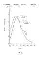
- FIG. 1a characteristic excitation/fluorescence spectra for a typical Maillard product bound to albumin.
- FIG. 2is a pictorial illustration of the epidermis of the skin of the finger showing sections of the dermis, capillary bed, and blood vessels.
- the distance between the blood vessels and the top surface of the epidermisis typically 0.3 mm.
- FIG. 3is a transmission versus wavelength plot showing the absorption characteristic of hemoglobin and pigment epithelium as a function of spectral wavelength.
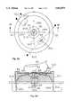
- FIG. 4A and 4Bare top plan and side cross-sectional views, respectively, of a first embodiment of a non-invasive low-cost, blood late Maillard products concentration detector.
- FIG. 5is an electronic block diagram of the signal processing components of the preferred embodiment.
- FIGS. 6A,and 6Billustrate an alternate embodiment of the non-invasive, low-cost blood late Maillard products concentration detector of FIGS. 4A and 4B, having four excitation light sources.
- FIGS. 7A and 7Billustrate an alternate embodiment of the low-cost, non-invasive blood late Maillard products detector of FIGS. 4A and 4B, having three detectors spaced around a central source.
- FIGS. 8A and 8Billustrate an alternate embodiment of the low-cost, non-invasive blood late Maillard products concentration detector of FIGS. 4A and 4B that uses optical fibers for light sources.
- FIGS. 9A and 9Billustrate an alternate embodiment of the low-cost, non-invasive blood late Maillard products concentration detector of FIGS. 7A and 7B in which a single excitation light source is replaced by a single optical fiber.
- FIGS. 10A and 10Billustrate an alternate embodiment of the low-cost, non-invasive blood late Maillard products concentration detector of FIGS. 9A and 9B in which the optical fiber penetrates the fiat optical plate.
- the inventionwill be illustrated for the case of a noninvasive, blood late Maillard products concentration detector, but, as indicated above, this invention is also applicable to testing solute concentrations in many different types of solutions and in other environments, such as a sample contained within a test tube or smeared onto a slide.
- this process of measuring the concentration of a component of a blood samplelight of wavelength in the range from 270-370 nm is directed at a portion of a person or animal's epidermis to pass into a blood-rich region of that person or animal to excite the blood late Maillard products in the user's blood. As is illustrated in FIG.
- this radiationis focussed, through the epidermis 41 of the front side of the index finger (i.e., the side that is opposite to the nail side of the finger) and a derm layer 44, to a point on the top part of the capillary bed 42 that is closest to the epidermis onto the capillary bed to excite the Maillard products in blood vessels (see FIG. 1).
- This regionis selected for exposure because it is a blood-rich region, having many blood vessels 43, closely spaced (about 0.3 mm) from the epidermis.
- any fingercan be used, it is preferred that the least calloused finger be used to achieve improved penetration of the excitation radiation into that finger.
- the 270-370 nm rangeis selected for the exposing light because it can pass, without significant attenuation, through the epidermis 41 (see FIG. 2).
- the exposing lightis within the range from 200-600 nm because this range not only passes through the epidermis without undue attenuation, as illustrated in FIG. 3, it is also effective in exciting hemoglobin molecules, so that pressure-related changes in the amount of blood in capillary bed 42 can be compensated for from a knowledge of the amount of absorption by the hemoglobin molecules.
- This exposing lightnot only excites blood Maillard products and hemoglobin molecules into an excited state, it also stimulates emission of light from such excited molecules. Because the incident light need only travel through about 0.3 mm of tissue, the exposing beam will not be significantly attenuated prior to exposing the blood in the capillary bed. Because the emitted light likewise need travel only through about 0.3 mm of tissue before incidence on a detector, it will likewise not be significantly attenuated. Because of this, the detected signal will be much larger than in prior techniques. The detected intensity of light is utilized to calculate the concentration of blood late Maillard products.
- optical sourcessuch as semiconductor LEDs and semiconductor laser diodes
- the probability of a Maillard product's excitation in a beam of intensity on the order of 5 Watts/cm 2is expected to be very small (typically around 10 -4 or smaller)
- this exposing lightplays the dual role of exciting blood late Maillard products molecules into excited states and stimulating emission from such excited molecules, the rate of emission is proportional to the square of the intensity of this light. Therefore, the rate of emission is proportional to the square of the power of the excitation radiation.
- this lightonto a small area of the capillary bed 42.
- this areahas a diameter on the order of a few tens of microns, but diameters of up to 100 microns are also adequate.
- the depth of focusshould also be on the order of the thickness, 100 microns, of the capillary bed.
- Semiconductor LEDs and semiconductor laser diodestypically have output powers in excess of 100 mW and higher and with proper delivery optics these output power levels are more than adequate for the currently disclosed blood late Maillard products measurement technique.
- Super-radiant diodesare advantageous because of their particularly large beam intensity.
- An ultraviolet flashlampcould also be utilized as the light source.
- the optical sourceis substantially monochromatic, because monochromatic or substantially monochromatic light can be focussed onto a very tiny region with inexpensive optical components that do not correct for chromatic aberration. Such accurate focussing is needed to be able to focus this light accurately onto the top part of the capillary bed that is closest to the epidermis and to obtain the desired spot size.
- the rate of emissionis proportional to the square of the intensity of this light, such concentrated focussing of the light will greatly increase the rate of stimulated emission.
- Light emitting diodes (LEDs), emitting in the super-radiant mode, and semiconductor laser diodesmeet all of these criteria for the optical source. Diode lasers of wavelength 0.67 microns are readily available with a bandwidth of 0. 1 A, FWHM. At a modest increase in cost, laser diodes having a bandwidth of 0.01A are also available.
- An additional advantage of such sourcesis that they are relatively inexpensive so that low cost, non-invasive blood late Maillard products monitors can be produced.
- FIGS. 4A and 4Bare top plan view and side cross-sectional views, respectively, of a first embodiment of a non-invasive, low-cost, blood late Maillard products concentration detector.
- a pair of excitation light sources 61 and 62such as LEDs or laser diodes, focus excitation radiation of wavenumber 1040 cm -1 into a spatially small focal region 63 approximately 0.3 mm beyond an optically flat plate 64 that is transparent to both the excitation radiation and the subsequent relaxation emission radiation.
- Many choices of material, such as ZnS and ZnSe,are possible for this flat plate.
- This plateis "transparent" to light of a given wavelength if its absorptivity is less than a few percent for such light.
- a userpresses a front side (i.e., the side opposite to that person's nail) of his or her finger 65 against this plate, so that the excitation radiation beam can be accurately focussed onto the top of the capillary bed 42.
- This point of focusis chosen because the numerous small blood vessels 43 within the capillary bed provide one of the closest locations of blood to a patient's epidermis and because a finger is conveniently pressed against plate 64 during testing.
- the Maillard products and hemoglobin moleculesare excited by the excitation radiation and, upon returning to their ground states, emit radiation 66 that is characteristic of the Maillard product molecules.
- other wavelengths of relaxation radiationare also emitted by the Maillard product molecules, they are not uniquely emitted by Maillard product molecules and therefore are not as easily utilized to measure the concentration of Maillard product molecules.
- Detector assembly 67includes three infrared detectors 68, 69 and 610, each of which occupies about one-third of the total field of view subtended by the detector assembly 67 at the small focal region 63 of excitation radiation.
- Detectors 68, 69 and 610are each housed in a separate compartment to prevent cross-talk or light leakage between them.
- Detectors 68, 69 and 610each includes its own unique narrow band-pass interference filter passing only radiation at 378 nm, 9.02 microns and 3.80 microns respectively. As will be explained in greater detail below, these choices of filters enable a measured concentration of Maillard product molecules to be produced that is unaffected by the temperature and touching pressure of the patient's finger.
- the excitation light sources 61 and 62 and the detector assembly 67are mounted inside a circular sensor housing 611 in such a way that a set of electrical leads 612 from the sources and detector assembly all come out from one side of detector assembly 67 opposite to that of the optical flat plate 64.
- Leads 612can be conveniently soldered to a printed circuit board (PCB) 613 that contains processing electronic circuits 614 and also supports the overall sensor housing 611.
- the sensor housing 611is temperature regulated at a temperature T o of approximately 37° C. (i.e., normal human body temperature) by means of a heater resistor 615 and a thermistor 616 imbedded therein.
- the electrical leads 612 of heater resistor 615 and thermistor 616are also routed to the PCB 613 that also contains a temperature regulating circuit.
- the narrow band-pass, interference filter included within detector 610passes a narrow range of light centered at wavenumber 2,632 cm -1 (wavelength 3.8 microns). This light is primarily blackbody radiation from the optical flat plate 64 and from those portions of detector assembly 67 immediate adjacent to this plate.
- the output of this detectorprovides information about any temperature changes caused by internal or external environmental changes, such as by the fore finger of the patient touching the optical flat plate during the blood late Maillard products measurement itself.
- the relationship between the instantaneously measured signal from detector 610 and the instantaneous temperature T(t) measured in degree Celsiusis given by
- I s (t)is the instantaneous output signal of detector 610
- I ois the output at T 0 ° C.
- T(t)° C.is the instantaneous spatially averaged temperature of the cavity surrounding the detector assembly 67.
- the narrow band-pass, interference filter included within detector 69passes a narrow range of light centered at 9.02 microns.
- Detector 69receives radiation from three separate sources: (i) blackbody radiation from the cavity surrounding detector assembly 67 (which includes optical flat plate 64); (ii) relaxation radiation from hemoglobin inside the blood vessels; and (iii) relaxation radiation from the Maillard product molecules inside the blood vessels.
- the output signal of detector 69is needed to eliminate the effect of the variable touching pressure of the fore finger on the optical flat plate 64 during measurement, because the volume of blood and the quantity of hemoglobin in the blood vessels within the region exposed by the exposing light, is dependent of how hard the finger is pressed against optical flat plate 64. This blood volume is a function of this pressure because the touching pressure forces blood out of the capillary bed 42 in the region of the fore finger that is in contact with optical flat plate 64.
- the narrow band-pass, interference filter included within detector 68passes a narrow range of light centered at 378 nm.
- Detector 68receives radiation from two different sources: (i) relaxation radiation from the Maillard product molecules in the blood vessels; and (ii) blackbody radiation from the cavity surrounding detector assembly 67.
- the output signal of detector 68therefore contains information relating to the amount of blood late Maillard products in the blood vessels of the patient under test.
- a net output signalis produced that indicates the concentration of Maillard product molecules in the blood and is not affected by the temperature and touching pressure of the patient's finger on the optical flat plate.
- This processingis well known from basic algebra. Let I s (t), J s (t) and K s (t) be the outputs, as a function of time t, of detectors 610, 69 and 68, respectively during a particular blood late Maillard products measurement routine. I s (t) is a function of the instantaneous temperature T(t) of the cavity surrounding the detector assembly 67 including the optical flatplate 64.
- J s (t)is the sum of blackbody radiation at temperature T(t) and the relaxation radiation from hemoglobin and the Maillard product molecules in the blood vessels when these vessels are exposed by light from excitation light sources 61 and 62.
- K s (t)is the sum of the blackbody radiation at temperature T(t) and the relaxation radiation from the Maillard product molecules when the latter is stimulated by the excitation light sources 61 and 62.
- the temperature of the cavity immediately surrounding the detector assembly 67is regulated by thermistor 612 and heater resistor 615 to a temperature T 0 of 37° C. This temperature is selected because it should most closely match the actual temperature of a patient's finger.
- the outputs from detectors 610, 69 and 68 under this conditionare represented as I 0 , J 0 and K 0 , respectively, and they represent only the blackbody radiation received at the wavelengths defined by the narrow band-pass filters of each of the respective detectors.
- the cavity and plate temperatureschange to a slightly different temperature T(t) and the outputs from the detectors 68, 69 and 610 are, respectively:
- H(T)is the component of the output currents produced by relaxation radiation from hemoglobin molecules
- G[H(T)]is the component of the output currents produced by relaxation radiation from Maillard product molecules.
- I 0 , J 0 and K 0are known constants determined from measurements made when no patients finger is present in the test apparatus. Measurement of the three parameters I s (t), J s (t) and K s (t) enables the three unknowns H(t), G[H(t)] and T(t) to be determined by standard methods from basic algebra.
- the amount of relaxation radiation from Maillard product moleculesdepends on the number of Maillard product molecules, which in turn is proportional to the blood volume being excited.
- the function H(t)measures the relaxation radiation coming only from the hemoglobin molecules and is therefore a function of the blood volume being excited, which is why G is written as a function of H(t).
- ⁇is a constant determined during a calibration procedure and where P(t) is measured separately by a pressure sensor 617 before and during the blood late Maillard products measurement.
- the patientis guided to press his or her finger onto flat plate 64 with a pressure that is within a preselected pressure range.
- a green lightis illuminated to indicate that this pressure is within the desired range.
- This rangeis selected to ensure that the volume of illuminated blood is within a range such that H(t) can be accurately represented by equation (1) above.
- a low-cost, low-power processor 71such as the model 68HCll single-chip, 16-bit microprocessor manufactured by Motorola, is used to control operation and to perform all calculations.
- microprocessor 71receives an ON signal from a manual ON/OFF switch 72, it activates temperature regulation circuitry 73 which controls the temperature of sensor housing 611 by means of heater resistor 615 and thermistor 612.
- a temperature ready light 74is turned on and microprocessor starts pulsing excitation light sources 61 and 62 at a frequency of F hertz (e.g., 60 Hz).
- F hertze.g. 60 Hz.
- Signals from detectors I s (t), J s (t) and K s (t) from detectors 68, 69 and 610, respectively,are amplified by preamplifiers 75-77 and A/D converters 78, 79 and 710, respectively. Because no finger is pressed against flat plate 64 at this point in the measurement process, the radiation detected by detector assembly 67 (which contains detectors 68, 69 and 610) is just blackbody radiation emanating from the sensor housing cavity, including the optical flat plate 64.
- microprocessor 71When microprocessor 71 receives an initialization signal from a manual switch 711, the outputs of A/D converters 78, 79 and 710 are stored and represent the values of I 0 , J 0 and K 0 , respectively.
- a measurement ready light 712begins to blink, indicating that the blood late Maillard products concentration detector is ready. The patient is then to press his or her finger against flat plate 64.
- pressure sensor 617detects a pressure against flat plate 64 in the preselected pressure range, measurement ready light 712 converts from a blinking mode to a steady mode, thereby indicating that the actual blood late Maillard products measurement has commenced.
- a liquid crystal display (LCD) 713displays the blood late Maillard products concentration.
- microprocessor 71acquires the values I s (t), J s (t) and K s (t) and solves the three equations (Ia), (Ib), and (Ic) above to produce the value of the blood late Maillard products concentration of the patient.
- measurement ready light 712again begins to blink, thereby indicating that it is ready to perform another blood late Maillard products concentration measurement. If desired, the patient can initiate another measurement simply by removing his or her finger from the optical flat plate, waiting for a couple of minutes and then repeating the measurement procedure. If at any time during a measurement, the pressure on flat plate 64 is outside of the preselected range, ready light 712 turns off and the measurement is voided.
- Calibration of this low-cost, non-invasive blood late Maillard products concentration detectoris achieved by determining the value of ⁇ in equation (1) above. This can be done by a patient by measuring a sample of blood with the present blood late Maillard products concentration detector and by concurrently drawing a sample of blood in which the ratio of blood late Maillard products concentration to hemoglobin concentration is determined by another blood late Maillard products concentration detector that is known to be accurate. The ratio of these two values can be used by the patient to multiply the output of LCD 713 to produce an accurate concentration value.
- an input mechanismcan be included that allows the user to input this ratio into microprocessor 71 so that this correction factor can be applied automatically by microprocessor 71. Alternatively, the user would have to take the value calculated by the instrument and scale it according to the results of the calibration measurement.
- FIGS. 6A and 6Billustrate an alternate embodiment of sensor housing 611 that includes four excitation light sources 81-84 instead of two excitation light sources 61 and 62 as in the embodiment of FIGS. 4A and 4B.
- the use of four or more excitation light sourcesenhances the signal level of the relaxation radiation from both hemoglobin and Maillard product molecules, because of the resulting increase in excitation light energy density. This increases the signal-to-noise ratio of the emitted light received by detector assembly 67.
- FIGS. 7A and 7Billustrate an alternate embodiment of the low-cost, non-invasive blood late Maillard products concentration detector of FIGS. 4A and 4B, having three detectors spaced around a central optical source.
- a single excitation light source 91is centered laterally within sensor housing 611. Excitation radiation is focussed by a relatively long focal length, achromatic doublet or triplet lens system onto a small focal region 63 just beyond optical flat plate 64.
- Three detectors 92-94are located symmetrically about a central axis of sensor housing 611 to collect stimulated relaxation radiation from a patient's finger 65.
- Detectors 92-94are each equipped with a different narrow bandpass, interference filter that passes radiation only at 378 nm, 9.02 microns (1,109 cm -1 ) and 3.80 microns (2,632 cm -1 ), respectively.
- FIGS. 8A and 8Billustrate an alternate embodiment of the low-cost, non-invasive blood late Maillard products concentration detector of FIGS. 4A and 4B.
- the two excitation light sources 61 and 62 of the embodiment in FIGS. 4A and 4Bare at least two optical fibers 1001-1004 arranged symmetrically about a longitudinal axis of sensor housing 611. This particular embodiment utilizes four such optical fibers. Excitation light is piped through the optical fibers and focussed onto a small focal region 63 just beyond the optical flat plate 64 as before.
- optical fibersas the carriers of the excitation radiation enables the detector assembly 67, containing detectors 68, 69 and 610, to be mounted very close to focal region 63, thereby significantly increasing the solid angle within which these detectors receive light from focal region 63. This provides a concomitant increase in the signal to noise ratio of this concentration detector.
- FIGS. 9A and 9Billustrate an alternate embodiment of the low-cost, non-invasive blood late Maillard products concentration detector.
- the only difference between this embodiment and the embodiment of FIGS. 7A and 7Bis that single excitation light source 91 is replaced by a single optical fiber 1101 that transports light from a remote location.
- FIGS. 10A and 10Billustrate an alternate embodiment of the low-cost, non-invasive blood late Maillard products concentration detector.
- optical fiber 1201penetrates through optical flat plate 64. Because the excitation light does not now pass through optical flat plate 64, this plate can be made out of a different and less expensive material, such as silicon. Silicon blocks all radiation of wavelength less than about one micron, but has good transmission characteristics in the medium to far infrared.
- flat plate 64be made of a material, such as ZnS or ZnSe, that transmits radiation from the visible all the way to the medium and far infrared. In general, ZnS and ZnSe plates are significantly more expensive than silicon plates.
Landscapes
- Health & Medical Sciences (AREA)
- Life Sciences & Earth Sciences (AREA)
- Physics & Mathematics (AREA)
- Medical Informatics (AREA)
- Surgery (AREA)
- Biophysics (AREA)
- Pathology (AREA)
- Engineering & Computer Science (AREA)
- Biomedical Technology (AREA)
- Heart & Thoracic Surgery (AREA)
- Veterinary Medicine (AREA)
- Molecular Biology (AREA)
- Optics & Photonics (AREA)
- Animal Behavior & Ethology (AREA)
- General Health & Medical Sciences (AREA)
- Public Health (AREA)
- Spectroscopy & Molecular Physics (AREA)
- Emergency Medicine (AREA)
- Measurement Of The Respiration, Hearing Ability, Form, And Blood Characteristics Of Living Organisms (AREA)
- Investigating Or Analysing Biological Materials (AREA)
- Investigating Or Analysing Materials By Optical Means (AREA)
Abstract
Description
TABLE 1 ______________________________________ % OF SUGARS IN ALDEHYDE FORM ______________________________________ Glucose 0.001 Ribose 0.017 Fructose 0.25 Glucose 6-P 0.4 Fructose 6-P 4-5 ______________________________________
TABLE 2 ______________________________________ Hypotheses regarding the potential role of non-enzymatic glucosylation and browning in the pathology associated with diabetes mellitus ______________________________________ I Structural proteins A Collagen: Decreased turnover, flexibility, solubility; increased aggregating potential for platelets, binding of immunoglobulins, crosslinking, and immunogenicity B Lens crystallins and membrane: opacification, increased vulnerability to oxidative stress C Basement membrane: increased permeability, decreased turnover, increased thickness D Extracellular matrix: changes in binding to other proteins E Hemoglobin: change in oxygen binding F Fibrin: decreased enzymatic degradation G Red cell membrane: increased rigidity H Tubulin: cell structure and transport I Myelin: altered structure and immunologic recognition II Carrier proteins A Lipoproteins: alternate degradative pathways and metabolism by macrophages and endothelial cells, increased immunogenicity B Albumin: alteration in binding properties for drugs and in handling by the kidney C Ig G: Altered binding III Enzyme systems A Cu--Zn Superoxide Dismutace B Fibrinogen: altered coagulation C Antithrombin III: hypercoagulable state D Purine nucleoside phosphorylase: aging of erythrocytes E Alcohol dehydrogenase: substrate metabolism F Ribonuclease A: lose of activity G Cathepsin B: loss of activity H N-acetyl-D-glucosaminidase: loss of activity I Calmodulin: decreased calcium binding IV Nucleic acids Age-related changes, congenital malformations V Potentiation of other diseases of post-synthetic protein modification A Carbamylation-associated pathology in uremia B Steroid cataract formation C Acetaldehyde-induced changes in alcoholism ______________________________________
TABLE 3 ______________________________________ Hemoglobin (Hb) Terminology ______________________________________ "Fast" Hemoglobin. The total HbA.sub.1 fractions (HbA.sub.1a, HbA.sub.1a2, HbA.sub.1b, HbA.sub.1c) which, because of more negative charge, migrates toward the anode on electrophoresis and elutes earlier on cation exchange chromatography than HbA.sub.c. Fetal Hemoglobin (HbF). The major hemoglobin component of newborn blood. HbF co-elutes with HbA.sub.1c by column chroma- tography. Glucosylated Hemoglobin. Hemoglobin modified by glucose at beta chain valine residues and epsilon amino groups of lysine residues. Glycated Hemoglobin. A term favored by biochemists to indicate adducts of sugars and hemoglobin which are formed non- enzymatically. Glycosylated Hemoglobin (glyco-hemoglobin). A generic term for hemoglobin containing glucose and/or other carbohydrate at either valine or lysine residues thus the sum of glycosyl adducts. Hemoglobin A. The major adult form of hemoglobin. A tetramer consisting of two alpha and two beta chains (alpha.sub.2, beta.sub.2). Hemoglobin A.sub.c. The major component of HbA identified by its chromatographic and electrophoretic properties. Post-translational modifications, including glycosylation do exist, but do not significantly affect the charged properties of the protein. Hemoglobin A.sub.1. Post-translationally modified, more negatively charged forms of HbA.sub.c (primarily glycosylation at the beta chain terminal valine residue) separable from HbA.sub.c by chromatographic and electrophoretic methods. Hemoglobin A.sub.1a1, HbA.sub.1a2, HbA.sub.1b, HbA.sub.1c. Chromatographically distinct stable components of HbA.sub.1. Hemoglobin A1.sub.1a1, HbA.sub.1a2, HbA.sub.1b : "Fastest" most anionic forms of HbA consisting primarily of adducts of phosphorylated glycoyl- tic intermediates with HbA.sub.c. Hemoglobin A1.sub.c. Component of HbA.sub.1 which consists of 50 to 90% hemoglobin (depending on the quality of resolution of the chromatographic system) glucosylated by a ketamine linkage at the beta chain terminal valine residue. Pre-Hemglobin A.sub.1c. A labile form of glycosylated Hb containing glucose bound in aldimin linkage to the beta chain terminal valine residue. Hemoglobin-AGE. Advanced Maillard or glycosylation end pro- ducts bound to hemoglobin. Circulate in the red cell and corre- lates with the amount of hemoglobin A.sub.1c. ______________________________________
TABLE 4 ______________________________________ Classification of Current Clinical Assays ______________________________________ I. Physical methods based on changes in p1 A. Cation exchange chromatography PRO: Inexpensive and rapid CON: Sensitive to small changes in resin packing, ionic strength, pH, temperature, column loading, and affected by the labile fraction. Variant hemoglobins may interfere. B. High performance liquid chromatography (HPLC) PRO: Dedicated instruments avoid many problems in 1 CON. Relatively expensive C. Agarose gel eletrophoresis PRO: Inexpensive, minimal technician time, standardized plates and conditions in kits less sensitive to pH, triglyceride concentrations, and temperature CON: Precision problems induced by scanner and loading variation; sensitive to liabile fraction and variant hemoglobin. D. Isoelectric focusing PRO: Separates most minor hemoglobin variants CON: Precision over time dependent on use of same batch of ampholines on standardized plates; scanning effects precision II. Methods based on chemical principles A. Thiobarbituric acid/colorimetric assay PRO: Minimally effected by storage condition, fructose or 5-hydroxy-methyl furfural standards may be incorporated, filter paper assays available CON: Difficult to establish, large amount of technical time required, and affected by labile fraction B. Affinity chromatography with immobilized m-phenyl- boronate PRO: Rapid, inexpensive, minimally effected by chromatographic conditions, eliminates labile adduct CON: Resins vary within and between manufacturers C. Fructosamine determination by nitroblue tetrazolium reduction PRO: Inexpensive, standards incorporated, may be automated, not effected by labile adduct CON: Only for serum, lipids may interfere, reducing substances (in diet) may interfere III. Immunoassay PRO: Inexpensive, rapid, sensitive, specific, not effected by labile adduct CON: Antibodies difficult to raise ______________________________________
I.sub.s (t)=I.sub.0 ×[(T(t)+273)/T.sub.0 +273)].sup.4
I.sub.s (t)=I.sub.0 ×[T(t)/T.sub.0 ].sup.4 (Ia)
I.sub.s (t)=I.sub.0 ×[T(t)/T.sub.0 ].sup.4 ÷G[H(t)]÷H(t)(Ib)
K.sub.s (t)=K.sub.0 ×[T(t)/T.sub.0 ].sup.4 ÷G[H(t)](IC)
H(t)=ΩP(t)
Claims (18)
Priority Applications (1)
| Application Number | Priority Date | Filing Date | Title |
|---|---|---|---|
| US08/307,125US5601079A (en) | 1992-03-12 | 1994-09-16 | Non-invasive quantification of glucose control, aging, and advanced maillard products by stimulated fluorescence |
Applications Claiming Priority (2)
| Application Number | Priority Date | Filing Date | Title |
|---|---|---|---|
| US07/852,085US5370114A (en) | 1992-03-12 | 1992-03-12 | Non-invasive blood chemistry measurement by stimulated infrared relaxation emission |
| US08/307,125US5601079A (en) | 1992-03-12 | 1994-09-16 | Non-invasive quantification of glucose control, aging, and advanced maillard products by stimulated fluorescence |
Related Parent Applications (1)
| Application Number | Title | Priority Date | Filing Date |
|---|---|---|---|
| US07/852,085Continuation-In-PartUS5370114A (en) | 1992-03-12 | 1992-03-12 | Non-invasive blood chemistry measurement by stimulated infrared relaxation emission |
Publications (1)
| Publication Number | Publication Date |
|---|---|
| US5601079Atrue US5601079A (en) | 1997-02-11 |
Family
ID=25312466
Family Applications (2)
| Application Number | Title | Priority Date | Filing Date |
|---|---|---|---|
| US07/852,085Expired - Fee RelatedUS5370114A (en) | 1992-03-12 | 1992-03-12 | Non-invasive blood chemistry measurement by stimulated infrared relaxation emission |
| US08/307,125Expired - Fee RelatedUS5601079A (en) | 1992-03-12 | 1994-09-16 | Non-invasive quantification of glucose control, aging, and advanced maillard products by stimulated fluorescence |
Family Applications Before (1)
| Application Number | Title | Priority Date | Filing Date |
|---|---|---|---|
| US07/852,085Expired - Fee RelatedUS5370114A (en) | 1992-03-12 | 1992-03-12 | Non-invasive blood chemistry measurement by stimulated infrared relaxation emission |
Country Status (5)
| Country | Link |
|---|---|
| US (2) | US5370114A (en) |
| EP (1) | EP0631490A4 (en) |
| JP (1) | JPH07506987A (en) |
| CA (1) | CA2131715A1 (en) |
| WO (1) | WO1993017621A1 (en) |
Cited By (62)
| Publication number | Priority date | Publication date | Assignee | Title |
|---|---|---|---|---|
| US5771891A (en)* | 1995-05-10 | 1998-06-30 | Massachusetts Inst Technology | Apparatus and method for non-invasive blood analyte measurement |
| US5961451A (en)* | 1997-04-07 | 1999-10-05 | Motorola, Inc. | Noninvasive apparatus having a retaining member to retain a removable biosensor |
| US6044285A (en)* | 1997-11-12 | 2000-03-28 | Lightouch Medical, Inc. | Method for non-invasive measurement of an analyte |
| US6055451A (en) | 1997-12-12 | 2000-04-25 | Spectrx, Inc. | Apparatus and method for determining tissue characteristics |
| US6097975A (en)* | 1998-05-13 | 2000-08-01 | Biosensor, Inc. | Apparatus and method for noninvasive glucose measurement |
| US6147749A (en)* | 1995-08-07 | 2000-11-14 | Kyoto Daiichi Kagaku Co., Ltd | Method and apparatus for measuring concentration by light projection |
| WO2001022869A1 (en)* | 1999-09-30 | 2001-04-05 | Jipjap B.V. | Method and apparatus for determining autofluorescence of skin tissue |
| WO2001033278A1 (en) | 1999-11-03 | 2001-05-10 | Argose, Inc. | Interface medium for tissue surface probe |
| US6289230B1 (en) | 1998-07-07 | 2001-09-11 | Lightouch Medical, Inc. | Tissue modulation process for quantitative noninvasive in vivo spectroscopic analysis of tissues |
| WO2001073406A1 (en)* | 2000-03-29 | 2001-10-04 | M.U.T Gmbh | Device for carrying out optical in vivo measurements on the surface of living beings |
| WO2002026123A1 (en)* | 2000-09-26 | 2002-04-04 | Sensys Medical, Inc. | Led light source-based instrument for non-invasive blood analyte determination |
| US6413779B1 (en)* | 1996-08-30 | 2002-07-02 | Institut National Agronomique Paris-Grignon | Method for evaluating the heat treatment to which a proteinic nutrient such as milk is subjected |
| US20020091324A1 (en)* | 1998-04-06 | 2002-07-11 | Nikiforos Kollias | Non-invasive tissue glucose level monitoring |
| US6430422B1 (en)* | 1999-07-23 | 2002-08-06 | Kurabo Industries Ltd. | Intraoral jig for optical measurement |
| US6505059B1 (en) | 1998-04-06 | 2003-01-07 | The General Hospital Corporation | Non-invasive tissue glucose level monitoring |
| US20030108976A1 (en)* | 2001-10-09 | 2003-06-12 | Braig James R. | Method and apparatus for improving clinical accuracy of analyte measurements |
| US20030135122A1 (en)* | 1997-12-12 | 2003-07-17 | Spectrx, Inc. | Multi-modal optical tissue diagnostic system |
| US6597932B2 (en) | 2000-02-18 | 2003-07-22 | Argose, Inc. | Generation of spatially-averaged excitation-emission map in heterogeneous tissue |
| US20030175806A1 (en)* | 2001-11-21 | 2003-09-18 | Peter Rule | Method and apparatus for improving the accuracy of alternative site analyte concentration measurements |
| US20030176805A1 (en)* | 2000-06-09 | 2003-09-18 | Wolfgang Kleibohmer | Method for detecting alpha-oxoaldehydes in the whole blood, blood plasma and/or serum of a patient |
| US20030208169A1 (en)* | 2000-07-11 | 2003-11-06 | Joseph Chaiken | Method of tissue modulation for noninvasive measurement of an analyte |
| US20030226769A1 (en)* | 2000-05-23 | 2003-12-11 | Koji Sode | Kit for assaying saccharified protein |
| US20040010197A1 (en)* | 1998-09-11 | 2004-01-15 | Spectrx, Inc | Multi-modal optical tissue diagnostic system |
| US6721582B2 (en) | 1999-04-06 | 2004-04-13 | Argose, Inc. | Non-invasive tissue glucose level monitoring |
| US6728560B2 (en) | 1998-04-06 | 2004-04-27 | The General Hospital Corporation | Non-invasive tissue glucose level monitoring |
| US20040138537A1 (en)* | 1999-03-10 | 2004-07-15 | Braig James R. | Solid-state non-invasive thermal cycling spectrometer |
| US20040147843A1 (en)* | 1999-11-05 | 2004-07-29 | Shabbir Bambot | System and method for determining tissue characteristics |
| US6770883B2 (en) | 2002-01-30 | 2004-08-03 | Beckman Coulter, Inc. | Sample level detection system |
| US20040235477A1 (en)* | 2003-05-22 | 2004-11-25 | Lucent Technologies Inc. | Wireless handover using anchor termination |
| US20040247162A1 (en)* | 2003-06-05 | 2004-12-09 | Kim Sang Min | Apparatus for controlling temperature of fingerprint sensor for vehicle and method thereof |
| US6831746B2 (en)* | 2001-05-30 | 2004-12-14 | Sciperio, Inc. | System, method, and apparatus for non-intrusively determining concentration of a solute in a solution |
| US20050043603A1 (en)* | 2003-08-21 | 2005-02-24 | Ishler Larry W. | Non-invasive blood glucose monitoring system |
| US20050065415A1 (en)* | 2003-09-24 | 2005-03-24 | Ok-Kyung Cho | Optical measurement apparatus and blood sugar level measuring apparatus using the same |
| US20050148834A1 (en)* | 2002-04-04 | 2005-07-07 | Hull Edward L. | Determination of a measure of a glycation end-product or disease state using tissue fluorescence |
| US6952695B1 (en)* | 2001-05-15 | 2005-10-04 | Global Safety Surveillance, Inc. | Spontaneous adverse events reporting |
| US20060195022A1 (en)* | 1998-04-06 | 2006-08-31 | Pierre Trepagnier | Non-invasive tissue glucose level monitoring |
| US20070197880A1 (en)* | 2002-04-04 | 2007-08-23 | Maynard John D | Determination of a Measure of a Glycation End-Product or Disease State Using Tissue Fluorescence of Various Sites |
| US20070265532A1 (en)* | 2002-04-04 | 2007-11-15 | Maynard John D | Determination of a Measure of a Glycation End-Product or Disease State Using a Flexible Probe to Determine Tissue Fluorescence of Various Sites |
| US20070276199A1 (en)* | 2002-04-04 | 2007-11-29 | Ediger Marwood N | Determination of a Measure of a Glycation End-Product or Disease State Using Tissue Fluorescence |
| WO2009126422A2 (en) | 2008-04-11 | 2009-10-15 | Glucovista Llc | Apparatus and methods for non-invasive measurement of a substance within a body |
| WO2009108024A3 (en)* | 2008-02-29 | 2009-12-17 | 주식회사 이노제스트 | Device and method for measuring fluorescence of skin tissues |
| US20110004080A1 (en)* | 2008-04-11 | 2011-01-06 | Glucovista, Llc | Method for non-invasive analysis of a substance concentration within a body |
| US20110098542A1 (en)* | 2009-10-28 | 2011-04-28 | Yonatan Gerlitz | Apparatus and method for non-invasive measurement of a substance within a body |
| US20120078075A1 (en)* | 2002-04-04 | 2012-03-29 | Maynard John D | Determination of a measure of a glycation end-product or disease state using tissue fluorescence in combination with one or more other tests |
| US20130109936A1 (en)* | 2007-12-21 | 2013-05-02 | Covidien Lp | Medical sensor and technique for using the same |
| JP2014062740A (en)* | 2012-09-19 | 2014-04-10 | Sharp Corp | Aging evaluation method and aging evaluation device |
| US8903466B2 (en) | 2009-10-28 | 2014-12-02 | Glucovista Inc. | Apparatus and method for non-invasive measurement of a substance within a body |
| CN105848569A (en)* | 2014-07-30 | 2016-08-10 | 皇家飞利浦有限公司 | Hemoglobin detection and photoplethysmography using spectral modulation |
| US9442065B2 (en) | 2014-09-29 | 2016-09-13 | Zyomed Corp. | Systems and methods for synthesis of zyotons for use in collision computing for noninvasive blood glucose and other measurements |
| US9554738B1 (en) | 2016-03-30 | 2017-01-31 | Zyomed Corp. | Spectroscopic tomography systems and methods for noninvasive detection and measurement of analytes using collision computing |
| US20170071514A1 (en)* | 2014-03-26 | 2017-03-16 | Kyocera Corporation | Measurement apparatus, measurement system, measurement method, and electronic device provided with measurement apparatus |
| US20170119293A1 (en)* | 2014-05-20 | 2017-05-04 | Pioneer Corporation | Pulse oximeter |
| CN107063978A (en)* | 2015-09-10 | 2017-08-18 | 阿自倍尔株式会社 | In liquid in the detection means and liquid of biologic grain biologic grain detection method |
| US10258265B1 (en)* | 2008-07-03 | 2019-04-16 | Masimo Corporation | Multi-stream data collection system for noninvasive measurement of blood constituents |
| US10448871B2 (en) | 2015-07-02 | 2019-10-22 | Masimo Corporation | Advanced pulse oximetry sensor |
| US11490863B2 (en)* | 2018-10-19 | 2022-11-08 | Samsung Electronics Co., Ltd. | Apparatus and method for estimating bio-information, and apparatus for supporting estimation of bio-information |
| US11638532B2 (en) | 2008-07-03 | 2023-05-02 | Masimo Corporation | User-worn device for noninvasively measuring a physiological parameter of a user |
| US11864909B2 (en) | 2018-07-16 | 2024-01-09 | Bbi Medical Innovations, Llc | Perfusion and oxygenation measurement |
| US12114974B2 (en) | 2020-01-13 | 2024-10-15 | Masimo Corporation | Wearable device with physiological parameters monitoring |
| USD1051738S1 (en) | 2023-01-06 | 2024-11-19 | Medibeacon Inc. | Sensor ring |
| USD1052412S1 (en) | 2023-01-06 | 2024-11-26 | Medibeacon Inc. | Sensor |
| US12336796B2 (en) | 2021-07-13 | 2025-06-24 | Masimo Corporation | Wearable device with physiological parameters monitoring |
Families Citing this family (109)
| Publication number | Priority date | Publication date | Assignee | Title |
|---|---|---|---|---|
| US5370114A (en)* | 1992-03-12 | 1994-12-06 | Wong; Jacob Y. | Non-invasive blood chemistry measurement by stimulated infrared relaxation emission |
| CA2174719C (en)* | 1993-08-24 | 2005-07-26 | Mark R. Robinson | A robust accurate non-invasive analyte monitor |
| US5529755A (en)* | 1994-02-22 | 1996-06-25 | Minolta Co., Ltd. | Apparatus for measuring a glucose concentration |
| EP0670492B1 (en)* | 1994-03-04 | 2001-05-09 | Kyoto Dai-ichi Kagaku Co., Ltd. | Method of and apparatus for measuring uric components |
| US5743262A (en)* | 1995-06-07 | 1998-04-28 | Masimo Corporation | Blood glucose monitoring system |
| SG38866A1 (en)* | 1995-07-31 | 1997-04-17 | Instrumentation Metrics Inc | Liquid correlation spectrometry |
| US6025597A (en)* | 1995-10-17 | 2000-02-15 | Optiscan Biomedical Corporation | Non-invasive infrared absorption spectrometer for measuring glucose or other constituents in a human or other body |
| US5983120A (en)* | 1995-10-23 | 1999-11-09 | Cytometrics, Inc. | Method and apparatus for reflected imaging analysis |
| US6882873B2 (en) | 1996-01-17 | 2005-04-19 | Respironics, Inc. | Method and system for determining bilirubin concentration |
| US5747806A (en)* | 1996-02-02 | 1998-05-05 | Instrumentation Metrics, Inc | Method and apparatus for multi-spectral analysis in noninvasive nir spectroscopy |
| US5742392A (en)* | 1996-04-16 | 1998-04-21 | Seymour Light, Inc. | Polarized material inspection apparatus |
| US5666956A (en)* | 1996-05-20 | 1997-09-16 | Buchert; Janusz Michal | Instrument and method for non-invasive monitoring of human tissue analyte by measuring the body's infrared radiation |
| KR0165522B1 (en)* | 1996-05-23 | 1999-03-20 | 김광호 | Optimal Spot Searching Device for Blood-Free Diagnosis and Blood-Free Diagnose Using the Same |
| US5871442A (en) | 1996-09-10 | 1999-02-16 | International Diagnostics Technologies, Inc. | Photonic molecular probe |
| US6594510B2 (en) | 1996-09-10 | 2003-07-15 | Xoetronics Llc | Photonic molecular probe |
| JP4212007B2 (en)* | 1996-11-26 | 2009-01-21 | パナソニック電工株式会社 | Blood component concentration analyzer |
| EP1011426A1 (en)* | 1997-02-26 | 2000-06-28 | Diasense, Inc. | Individual calibration of blood glucose for supporting noninvasive self-monitoring blood glucose |
| US6161028A (en) | 1999-03-10 | 2000-12-12 | Optiscan Biomedical Corporation | Method for determining analyte concentration using periodic temperature modulation and phase detection |
| US5900632A (en)* | 1997-03-12 | 1999-05-04 | Optiscan Biomedical Corporation | Subsurface thermal gradient spectrometry |
| US5823966A (en)* | 1997-05-20 | 1998-10-20 | Buchert; Janusz Michal | Non-invasive continuous blood glucose monitoring |
| US6246893B1 (en)* | 1997-06-12 | 2001-06-12 | Tecmed Incorporated | Method and device for glucose concentration measurement with special attention to blood glucose determinations |
| SE523374C2 (en)* | 1997-07-31 | 2004-04-13 | Ericsson Telefon Ab L M | Communication using spectrum spreading methods over optical fibers |
| GB2328279B (en)* | 1997-08-12 | 2001-10-10 | Abbott Lab | Optical glucose detector |
| JPH11123195A (en)* | 1997-08-22 | 1999-05-11 | Kdk Corp | Biological measurement method and device |
| ATE230239T1 (en)* | 1998-03-06 | 2003-01-15 | Rudolf H Dittel | MEASURING DEVICE FOR DETERMINING THE CONCENTRATION CONTENT OF SUBSTANCES CONTAINED IN LIQUID USING AN ATR ELEMENT. |
| US6078833A (en)* | 1998-03-25 | 2000-06-20 | I.S.S. (Usa) Inc. | Self referencing photosensor |
| US6090050A (en)* | 1998-07-16 | 2000-07-18 | Salix Medical, Inc. | Thermometric apparatus and method |
| US6424851B1 (en)* | 1998-10-13 | 2002-07-23 | Medoptix, Inc. | Infrared ATR glucose measurement system (II) |
| CA2352295C (en)* | 1998-11-30 | 2008-07-15 | Novo Nordisk A/S | A method and a system for assisting a user in a medical self treatment, said self treatment comprising a plurality of actions |
| US6959211B2 (en) | 1999-03-10 | 2005-10-25 | Optiscan Biomedical Corp. | Device for capturing thermal spectra from tissue |
| US6360114B1 (en) | 1999-03-25 | 2002-03-19 | Masimo Corporation | Pulse oximeter probe-off detector |
| DE59900583D1 (en)* | 1999-03-29 | 2002-01-31 | Hoffmann La Roche | Device for the quantitative determination of the local distribution of a measured variable |
| US6526300B1 (en) | 1999-06-18 | 2003-02-25 | Masimo Corporation | Pulse oximeter probe-off detection system |
| US6370407B1 (en) | 1999-07-27 | 2002-04-09 | Tecmed, Incorporated | System for improving the sensitivity and stability of optical polarimetric measurements |
| DE19937699C1 (en)* | 1999-08-10 | 2001-11-22 | Ges Foerderung Spektrochemie | Method and device for non-invasive measurement of blood components and clinical parameters |
| WO2001037722A1 (en)* | 1999-11-23 | 2001-05-31 | Forskningscenter Risø | A non-invasive method for the measurement of body fluid analytes |
| US6577884B1 (en) | 2000-06-19 | 2003-06-10 | The General Hospital Corporation | Detection of stroke events using diffuse optical tomagraphy |
| GB2361765A (en)* | 2000-04-28 | 2001-10-31 | Ncr Int Inc | Media validation by diffusely reflected light |
| US6549861B1 (en) | 2000-08-10 | 2003-04-15 | Euro-Celtique, S.A. | Automated system and method for spectroscopic analysis |
| WO2002015782A1 (en)* | 2000-08-18 | 2002-02-28 | Masimo Corporation | Side applied optical finger hematometer |
| US6675030B2 (en) | 2000-08-21 | 2004-01-06 | Euro-Celtique, S.A. | Near infrared blood glucose monitoring system |
| US6522903B1 (en)* | 2000-10-19 | 2003-02-18 | Medoptix, Inc. | Glucose measurement utilizing non-invasive assessment methods |
| DE60106555T2 (en) | 2000-12-25 | 2005-02-24 | Fuji Photo Film Co., Ltd., Minami-Ashigara | Sensor using attenuated total reflection |
| US6609015B2 (en)* | 2001-01-18 | 2003-08-19 | Koninklijke Philips Electronics N.V. | Analysis of a composition |
| JP2002360530A (en)* | 2001-06-11 | 2002-12-17 | Waatekkusu:Kk | Pulse wave sensor and pulse rate detector |
| US6731967B1 (en)* | 2001-07-16 | 2004-05-04 | Pacesetter, Inc. | Methods and devices for vascular plethysmography via modulation of source intensity |
| US7009180B2 (en)* | 2001-12-14 | 2006-03-07 | Optiscan Biomedical Corp. | Pathlength-independent methods for optically determining material composition |
| US6862534B2 (en)* | 2001-12-14 | 2005-03-01 | Optiscan Biomedical Corporation | Method of determining an analyte concentration in a sample from an absorption spectrum |
| EP1335199A1 (en)* | 2002-02-11 | 2003-08-13 | Bayer Corporation | Non-invasive system for the determination of analytes in body fluids |
| WO2003082634A2 (en)* | 2002-03-26 | 2003-10-09 | Automotive Systems Laboratory, Inc. | Multiple chamber dual stage inflator |
| US20040115754A1 (en)* | 2002-12-11 | 2004-06-17 | Umax Data Systems Inc. | Method for establishing a long-term profile of blood sugar level aiding self-control of the same |
| TW200411178A (en)* | 2002-12-31 | 2004-07-01 | Veutron Corp | Method for determining the resolution of blood glucose by using rising time curve |
| TW592667B (en)* | 2003-04-04 | 2004-06-21 | Veutron Corp | Method for determining the resolution of blood glucose |
| US7283242B2 (en)* | 2003-04-11 | 2007-10-16 | Thornton Robert L | Optical spectroscopy apparatus and method for measurement of analyte concentrations or other such species in a specimen employing a semiconductor laser-pumped, small-cavity fiber laser |
| US7633621B2 (en)* | 2003-04-11 | 2009-12-15 | Thornton Robert L | Method for measurement of analyte concentrations and semiconductor laser-pumped, small-cavity fiber lasers for such measurements and other applications |
| US7271912B2 (en) | 2003-04-15 | 2007-09-18 | Optiscan Biomedical Corporation | Method of determining analyte concentration in a sample using infrared transmission data |
| US6975892B2 (en) | 2003-10-21 | 2005-12-13 | Oculir, Inc. | Methods for non-invasive analyte measurement from the conjunctiva |
| US6968222B2 (en) | 2003-05-02 | 2005-11-22 | Oculir, Inc. | Methods and device for non-invasive analyte measurement |
| US6958039B2 (en) | 2003-05-02 | 2005-10-25 | Oculir, Inc. | Method and instruments for non-invasive analyte measurement |
| US20040225206A1 (en)* | 2003-05-09 | 2004-11-11 | Kouchnir Mikhail A. | Non-invasive analyte measurement device having increased signal to noise ratios |
| US7356365B2 (en)* | 2003-07-09 | 2008-04-08 | Glucolight Corporation | Method and apparatus for tissue oximetry |
| US7266401B2 (en)* | 2003-08-22 | 2007-09-04 | C8 Medisensors Inc. | Measuring analytes from an electromagnetic spectrum using a wavelength router |
| WO2005041103A2 (en)* | 2003-10-29 | 2005-05-06 | Novo Nordisk A/S | Medical advisory system |
| US20050137469A1 (en)* | 2003-12-17 | 2005-06-23 | Berman Herbert L. | Single detector infrared ATR glucose measurement system |
| US7510849B2 (en)* | 2004-01-29 | 2009-03-31 | Glucolight Corporation | OCT based method for diagnosis and therapy |
| EP1576921A1 (en)* | 2004-03-17 | 2005-09-21 | Matsushita Electric Industrial Co., Ltd. | Method and device for measuring biological information |
| US8036727B2 (en)* | 2004-08-11 | 2011-10-11 | Glt Acquisition Corp. | Methods for noninvasively measuring analyte levels in a subject |
| US7254429B2 (en) | 2004-08-11 | 2007-08-07 | Glucolight Corporation | Method and apparatus for monitoring glucose levels in a biological tissue |
| US7236814B2 (en) | 2004-08-20 | 2007-06-26 | Matsushita Electric Industrial Co., Ltd. | Optical member for biological information measurement, biological information calculation apparatus, biological information calculation method, computer-executable program, and recording medium |
| US7647083B2 (en) | 2005-03-01 | 2010-01-12 | Masimo Laboratories, Inc. | Multiple wavelength sensor equalization |
| WO2006110859A2 (en) | 2005-04-13 | 2006-10-19 | Glucolight Corporation | Method for data reduction and calibration of an oct-based blood glucose monitor |
| US8180422B2 (en)* | 2005-04-15 | 2012-05-15 | Bayer Healthcare Llc | Non-invasive system and method for measuring an analyte in the body |
| US8452365B2 (en)* | 2005-05-25 | 2013-05-28 | Bayer Healthcare Llc | Methods of using Raman spectral information in determining analyte concentrations |
| GB0523832D0 (en)* | 2005-11-23 | 2006-01-04 | Univ City | Non-invasive optical monitoring of glucose using an adaptive modelling scheme |
| IL185609A0 (en) | 2007-08-30 | 2008-01-06 | Dan Furman | Multi function senssor |
| CN100560161C (en)* | 2006-02-17 | 2009-11-18 | 杭州大力神医疗器械有限公司 | Photoelectric finger pulse sensing infrared ray generator |
| EP2026058A4 (en)* | 2006-06-08 | 2009-12-30 | Omron Healthcare Co Ltd | Biological component measurement device capable of accurately noninvasively measuring biological component |
| US8265723B1 (en) | 2006-10-12 | 2012-09-11 | Cercacor Laboratories, Inc. | Oximeter probe off indicator defining probe off space |
| ITMI20070464A1 (en)* | 2007-03-09 | 2008-09-10 | Vittorio Londoni | DIODIC LASER DEVICE FOR INCRUABLE DETERMINATION OF GLYCEMIA |
| US8374665B2 (en) | 2007-04-21 | 2013-02-12 | Cercacor Laboratories, Inc. | Tissue profile wellness monitor |
| JP2009106373A (en)* | 2007-10-26 | 2009-05-21 | Panasonic Electric Works Co Ltd | Sensing device for biological surface tissue |
| US7802883B2 (en) | 2007-12-20 | 2010-09-28 | Johnson & Johnson Vision Care, Inc. | Cosmetic contact lenses having a sparkle effect |
| US8571617B2 (en) | 2008-03-04 | 2013-10-29 | Glt Acquisition Corp. | Flowometry in optical coherence tomography for analyte level estimation |
| CA2722659A1 (en) | 2008-05-12 | 2009-11-19 | Cardio Art Technologies, Ltd. | Doppler motion sensor apparatus and method of using same |
| DE102008002520A1 (en)* | 2008-06-19 | 2009-12-24 | Robert Bosch Gmbh | Device for determining and / or monitoring the fluid content of the skin |
| US10475529B2 (en) | 2011-07-19 | 2019-11-12 | Optiscan Biomedical Corporation | Method and apparatus for analyte measurements using calibration sets |
| US20110082711A1 (en) | 2009-10-06 | 2011-04-07 | Masimo Laboratories, Inc. | Personal digital assistant or organizer for monitoring glucose levels |
| US9839381B1 (en) | 2009-11-24 | 2017-12-12 | Cercacor Laboratories, Inc. | Physiological measurement system with automatic wavelength adjustment |
| WO2011069122A1 (en) | 2009-12-04 | 2011-06-09 | Masimo Corporation | Calibration for multi-stage physiological monitors |
| US9464983B2 (en) | 2010-07-12 | 2016-10-11 | Seiko Epson Corporation | Concentration determination apparatus, probe, concentration determination method, and program |
| AU2012207287B2 (en)* | 2011-01-19 | 2015-12-17 | The Regents Of The University Of California | Apparatus, systems, and methods for tissue oximetry and perfusion imaging |
| GB2494622A (en) | 2011-08-30 | 2013-03-20 | Oxitone Medical Ltd | Wearable pulse oximetry device |
| JP5866910B2 (en)* | 2011-09-16 | 2016-02-24 | セイコーエプソン株式会社 | Concentration measuring method and concentration measuring apparatus |
| DE102014108424B3 (en) | 2014-06-16 | 2015-06-11 | Johann Wolfgang Goethe-Universität | Non-invasive substance analysis |
| CN104161526A (en)* | 2014-08-25 | 2014-11-26 | 康泰医学系统(秦皇岛)股份有限公司 | Lateral jetting type blood oxygen detection device |
| KR102323207B1 (en)* | 2014-09-29 | 2021-11-08 | 삼성전자주식회사 | Device for correcting light absorption spectrum and methods of manufacturing the same and correcting light absorption spectrum |
| JP6555503B2 (en)* | 2015-01-13 | 2019-08-07 | 株式会社リコー | Optical sensor, optical inspection apparatus, and optical property detection method |
| WO2016117520A1 (en) | 2015-01-21 | 2016-07-28 | 国立研究開発法人日本原子力研究開発機構 | Device for measuring concentration of substance in blood, and method for measuring concentration of substance in blood |
| EP3244793A1 (en)* | 2015-02-11 | 2017-11-22 | Siemens Aktiengesellschaft | System and non-invasive method for examining at least parts of the blood fractions, and use of said system |
| US10492861B2 (en) | 2015-04-02 | 2019-12-03 | Mark John McDonald Rogers | Method and apparatus for treating soft tissue injury |
| JP6156804B2 (en)* | 2015-06-09 | 2017-07-05 | シャープライフサイエンス株式会社 | Aging evaluation equipment |
| EP3359948B1 (en) | 2015-12-09 | 2019-02-27 | Diamontech GmbH | Apparatus and method for analyzing a material |
| WO2017097824A1 (en) | 2015-12-09 | 2017-06-15 | Diamontech Gmbh | Device and method for analysing a material |
| JP2019037752A (en)* | 2017-08-23 | 2019-03-14 | 株式会社リコー | Measuring device and measuring method |
| JP2019093078A (en)* | 2017-11-28 | 2019-06-20 | 京セラ株式会社 | Optical sensor device |
| CN111436947B (en)* | 2019-01-16 | 2022-12-23 | 麦迪贝肯有限公司 | Two-piece sensor assembly and method of using same |
| US11701035B2 (en) | 2019-10-28 | 2023-07-18 | Bruce M. Landy | Noninvasive blood glucose detector and method using IR |
| JP7516773B2 (en)* | 2020-02-20 | 2024-07-17 | 株式会社リコー | Measuring device and measuring method |
| KR20220053934A (en) | 2020-10-23 | 2022-05-02 | 삼성전자주식회사 | Optical apparatus and apparatus for estimating bio-information using the same |
Citations (7)
| Publication number | Priority date | Publication date | Assignee | Title |
|---|---|---|---|---|
| US4556057A (en)* | 1982-08-31 | 1985-12-03 | Hamamatsu Tv Co., Ltd. | Cancer diagnosis device utilizing laser beam pulses |
| US4894547A (en)* | 1987-09-28 | 1990-01-16 | Yale University | Optical method and apparatus for detecting and measuring aging, photoaging, dermal disease and pigmentation in skin |
| US4957114A (en)* | 1985-04-01 | 1990-09-18 | Kun Zeng | Diagnostic apparatus for intrinsic fluorescence of malignant tumor |
| US4981138A (en)* | 1988-06-30 | 1991-01-01 | Yale University | Endoscopic fiberoptic fluorescence spectrometer |
| US5341805A (en)* | 1993-04-06 | 1994-08-30 | Cedars-Sinai Medical Center | Glucose fluorescence monitor and method |
| US5348018A (en)* | 1991-11-25 | 1994-09-20 | Alfano Robert R | Method for determining if tissue is malignant as opposed to non-malignant using time-resolved fluorescence spectroscopy |
| US5370114A (en)* | 1992-03-12 | 1994-12-06 | Wong; Jacob Y. | Non-invasive blood chemistry measurement by stimulated infrared relaxation emission |
Family Cites Families (23)
| Publication number | Priority date | Publication date | Assignee | Title |
|---|---|---|---|---|
| US3412729A (en)* | 1965-08-30 | 1968-11-26 | Nasa Usa | Method and apparatus for continuously monitoring blood oxygenation, blood pressure, pulse rate and the pressure pulse curve utilizing an ear oximeter as transducer |
| US3830222A (en)* | 1972-07-07 | 1974-08-20 | Johnson Res Foundation | Method and apparatus for observing rates of reaction of oxygen in living tissues |
| US3963019A (en)* | 1974-11-25 | 1976-06-15 | Quandt Robert S | Ocular testing method and apparatus |
| US4014321A (en)* | 1974-11-25 | 1977-03-29 | March Wayne F | Non-invasive glucose sensor system |
| US3958560A (en)* | 1974-11-25 | 1976-05-25 | Wayne Front March | Non-invasive automatic glucose sensor system |
| JPS5251785A (en)* | 1975-10-23 | 1977-04-25 | Minolta Camera Kk | Oximeter |
| US4055768A (en)* | 1976-09-07 | 1977-10-25 | Bromberg Nathan S | Light measuring apparatus |
| DE2934190A1 (en)* | 1979-08-23 | 1981-03-19 | Müller, Gerhard, Prof. Dr.-Ing., 7080 Aalen | METHOD AND DEVICE FOR MOLECULAR SPECTROSCOPY, ESPECIALLY FOR DETERMINING METABOLISM PRODUCTS |
| AU564398B2 (en)* | 1983-02-24 | 1987-08-13 | Abbott Laboratories | Improved fluorometric assay |
| US4704029A (en)* | 1985-12-26 | 1987-11-03 | Research Corporation | Blood glucose monitor |
| JPH07113602B2 (en)* | 1986-06-10 | 1995-12-06 | 株式会社日立製作所 | Concentration analyzer |
| US4981779A (en)* | 1986-06-26 | 1991-01-01 | Becton, Dickinson And Company | Apparatus for monitoring glucose |
| DE3801158A1 (en)* | 1988-01-16 | 1989-07-27 | Struck Marina | Measurement of blood sugar level - based on emission of photons from excited glucose molecules |
| US4882492A (en)* | 1988-01-19 | 1989-11-21 | Biotronics Associates, Inc. | Non-invasive near infrared measurement of blood analyte concentrations |
| US5009230A (en)* | 1988-05-31 | 1991-04-23 | Eol, Inc. | Personal glucose monitor |
| US4901728A (en)* | 1988-05-31 | 1990-02-20 | Eol, Inc. | Personal glucose monitor |
| US5028787A (en)* | 1989-01-19 | 1991-07-02 | Futrex, Inc. | Non-invasive measurement of blood glucose |
| US5112124A (en)* | 1990-04-19 | 1992-05-12 | Worcester Polytechnic Institute | Method and apparatus for measuring the concentration of absorbing substances |
| US5183042A (en)* | 1989-05-23 | 1993-02-02 | Vivascan Corporation | Electromagnetic method and apparatus to measure constituents of human or animal tissue |
| US4968887A (en)* | 1989-07-14 | 1990-11-06 | Evionics, Inc. | Gaseous nitrogen detection using excited-state laser spectroscopy |
| US5054487A (en)* | 1990-02-02 | 1991-10-08 | Boston Advanced Technologies, Inc. | Laser systems for material analysis based on reflectance ratio detection |
| US5115133A (en)* | 1990-04-19 | 1992-05-19 | Inomet, Inc. | Testing of body fluid constituents through measuring light reflected from tympanic membrane |
| WO1993000855A1 (en)* | 1991-07-03 | 1993-01-21 | Vivascan Corporation | Electromagnetic method and apparatus to measure constituents of human or animal tissue |
- 1992
- 1992-03-12USUS07/852,085patent/US5370114A/ennot_activeExpired - Fee Related
- 1993
- 1993-03-11WOPCT/US1993/002473patent/WO1993017621A1/ennot_activeApplication Discontinuation
- 1993-03-11EPEP93908391Apatent/EP0631490A4/ennot_activeWithdrawn
- 1993-03-11CACA002131715Apatent/CA2131715A1/ennot_activeAbandoned
- 1993-03-11JPJP5516060Apatent/JPH07506987A/enactivePending
- 1994
- 1994-09-16USUS08/307,125patent/US5601079A/ennot_activeExpired - Fee Related
Patent Citations (7)
| Publication number | Priority date | Publication date | Assignee | Title |
|---|---|---|---|---|
| US4556057A (en)* | 1982-08-31 | 1985-12-03 | Hamamatsu Tv Co., Ltd. | Cancer diagnosis device utilizing laser beam pulses |
| US4957114A (en)* | 1985-04-01 | 1990-09-18 | Kun Zeng | Diagnostic apparatus for intrinsic fluorescence of malignant tumor |
| US4894547A (en)* | 1987-09-28 | 1990-01-16 | Yale University | Optical method and apparatus for detecting and measuring aging, photoaging, dermal disease and pigmentation in skin |
| US4981138A (en)* | 1988-06-30 | 1991-01-01 | Yale University | Endoscopic fiberoptic fluorescence spectrometer |
| US5348018A (en)* | 1991-11-25 | 1994-09-20 | Alfano Robert R | Method for determining if tissue is malignant as opposed to non-malignant using time-resolved fluorescence spectroscopy |
| US5370114A (en)* | 1992-03-12 | 1994-12-06 | Wong; Jacob Y. | Non-invasive blood chemistry measurement by stimulated infrared relaxation emission |
| US5341805A (en)* | 1993-04-06 | 1994-08-30 | Cedars-Sinai Medical Center | Glucose fluorescence monitor and method |
Cited By (134)
| Publication number | Priority date | Publication date | Assignee | Title |
|---|---|---|---|---|
| US5771891A (en)* | 1995-05-10 | 1998-06-30 | Massachusetts Inst Technology | Apparatus and method for non-invasive blood analyte measurement |
| US6147749A (en)* | 1995-08-07 | 2000-11-14 | Kyoto Daiichi Kagaku Co., Ltd | Method and apparatus for measuring concentration by light projection |
| US6413779B1 (en)* | 1996-08-30 | 2002-07-02 | Institut National Agronomique Paris-Grignon | Method for evaluating the heat treatment to which a proteinic nutrient such as milk is subjected |
| US5961451A (en)* | 1997-04-07 | 1999-10-05 | Motorola, Inc. | Noninvasive apparatus having a retaining member to retain a removable biosensor |
| US6377828B1 (en) | 1997-11-12 | 2002-04-23 | Lightouch Medical, Inc. | Method for non-invasive measurement of an analyte |
| US6044285A (en)* | 1997-11-12 | 2000-03-28 | Lightouch Medical, Inc. | Method for non-invasive measurement of an analyte |
| US6055451A (en) | 1997-12-12 | 2000-04-25 | Spectrx, Inc. | Apparatus and method for determining tissue characteristics |
| US20030135122A1 (en)* | 1997-12-12 | 2003-07-17 | Spectrx, Inc. | Multi-modal optical tissue diagnostic system |
| US7899518B2 (en) | 1998-04-06 | 2011-03-01 | Masimo Laboratories, Inc. | Non-invasive tissue glucose level monitoring |
| US20060195022A1 (en)* | 1998-04-06 | 2006-08-31 | Pierre Trepagnier | Non-invasive tissue glucose level monitoring |
| US6728560B2 (en) | 1998-04-06 | 2004-04-27 | The General Hospital Corporation | Non-invasive tissue glucose level monitoring |
| US20020091324A1 (en)* | 1998-04-06 | 2002-07-11 | Nikiforos Kollias | Non-invasive tissue glucose level monitoring |
| US6505059B1 (en) | 1998-04-06 | 2003-01-07 | The General Hospital Corporation | Non-invasive tissue glucose level monitoring |
| US6097975A (en)* | 1998-05-13 | 2000-08-01 | Biosensor, Inc. | Apparatus and method for noninvasive glucose measurement |
| US6289230B1 (en) | 1998-07-07 | 2001-09-11 | Lightouch Medical, Inc. | Tissue modulation process for quantitative noninvasive in vivo spectroscopic analysis of tissues |
| US6975899B2 (en) | 1998-09-11 | 2005-12-13 | Spectrx, Inc. | Multi-modal optical tissue diagnostic system |
| US20060089556A1 (en)* | 1998-09-11 | 2006-04-27 | Spectrx, Inc. | Multi-modal optical tissue diagnostic system |
| US20040010197A1 (en)* | 1998-09-11 | 2004-01-15 | Spectrx, Inc | Multi-modal optical tissue diagnostic system |
| US20040138537A1 (en)* | 1999-03-10 | 2004-07-15 | Braig James R. | Solid-state non-invasive thermal cycling spectrometer |
| US6721582B2 (en) | 1999-04-06 | 2004-04-13 | Argose, Inc. | Non-invasive tissue glucose level monitoring |
| US6430422B1 (en)* | 1999-07-23 | 2002-08-06 | Kurabo Industries Ltd. | Intraoral jig for optical measurement |
| US7966060B2 (en) | 1999-09-30 | 2011-06-21 | Diagnoptics Holding B.V. | Method and apparatus for determining autofluorescence of skin tissue |
| WO2001022869A1 (en)* | 1999-09-30 | 2001-04-05 | Jipjap B.V. | Method and apparatus for determining autofluorescence of skin tissue |
| US20040186363A1 (en)* | 1999-09-30 | 2004-09-23 | Smit Andries Jan | Method and apparatus for determining autofluorescence of skin tissue |
| WO2001033278A1 (en) | 1999-11-03 | 2001-05-10 | Argose, Inc. | Interface medium for tissue surface probe |
| US20040147843A1 (en)* | 1999-11-05 | 2004-07-29 | Shabbir Bambot | System and method for determining tissue characteristics |
| US6597932B2 (en) | 2000-02-18 | 2003-07-22 | Argose, Inc. | Generation of spatially-averaged excitation-emission map in heterogeneous tissue |
| WO2001073406A1 (en)* | 2000-03-29 | 2001-10-04 | M.U.T Gmbh | Device for carrying out optical in vivo measurements on the surface of living beings |
| US20030226769A1 (en)* | 2000-05-23 | 2003-12-11 | Koji Sode | Kit for assaying saccharified protein |
| US20030176805A1 (en)* | 2000-06-09 | 2003-09-18 | Wolfgang Kleibohmer | Method for detecting alpha-oxoaldehydes in the whole blood, blood plasma and/or serum of a patient |
| US6884223B2 (en)* | 2000-06-09 | 2005-04-26 | Institut Fur Chemo-Und Biosensorik Munster E.V. | Method for detecting α-oxoaldehydes in the whole blood, blood plasma and/or serum of a patient |
| US7050842B2 (en) | 2000-07-11 | 2006-05-23 | Lightouch Medical, Inc. | Method of tissue modulation for noninvasive measurement of an analyte |
| US20030208169A1 (en)* | 2000-07-11 | 2003-11-06 | Joseph Chaiken | Method of tissue modulation for noninvasive measurement of an analyte |
| WO2002026123A1 (en)* | 2000-09-26 | 2002-04-04 | Sensys Medical, Inc. | Led light source-based instrument for non-invasive blood analyte determination |
| US20020041166A1 (en)* | 2000-09-26 | 2002-04-11 | Dragan Grubisic | Led light source-based instrument for non-invasive blood analyte determination |
| US6816241B2 (en)* | 2000-09-26 | 2004-11-09 | Sensys Medical, Inc. | LED light source-based instrument for non-invasive blood analyte determination |
| US6952695B1 (en)* | 2001-05-15 | 2005-10-04 | Global Safety Surveillance, Inc. | Spontaneous adverse events reporting |
| US6831746B2 (en)* | 2001-05-30 | 2004-12-14 | Sciperio, Inc. | System, method, and apparatus for non-intrusively determining concentration of a solute in a solution |
| US20030108976A1 (en)* | 2001-10-09 | 2003-06-12 | Braig James R. | Method and apparatus for improving clinical accuracy of analyte measurements |
| US20030175806A1 (en)* | 2001-11-21 | 2003-09-18 | Peter Rule | Method and apparatus for improving the accuracy of alternative site analyte concentration measurements |
| US6770883B2 (en) | 2002-01-30 | 2004-08-03 | Beckman Coulter, Inc. | Sample level detection system |
| US20070276199A1 (en)* | 2002-04-04 | 2007-11-29 | Ediger Marwood N | Determination of a Measure of a Glycation End-Product or Disease State Using Tissue Fluorescence |
| US8078243B2 (en) | 2002-04-04 | 2011-12-13 | Veralight, Inc. | Determination of a measure of a glycation end-product or disease state using tissue fluorescence |
| US20050148834A1 (en)* | 2002-04-04 | 2005-07-07 | Hull Edward L. | Determination of a measure of a glycation end-product or disease state using tissue fluorescence |
| US8121671B2 (en) | 2002-04-04 | 2012-02-21 | Veralight, Inc. | Determination of a measure of a glycation end-product or disease state using tissue fluorescence preferentially from the dermis |
| US20060211928A1 (en)* | 2002-04-04 | 2006-09-21 | Hull Edward L | Determination of a measure of a glycation end-product or disease state using tissue fluorescence preferentially from the dermis |
| US7139598B2 (en) | 2002-04-04 | 2006-11-21 | Veralight, Inc. | Determination of a measure of a glycation end-product or disease state using tissue fluorescence |
| US8131332B2 (en) | 2002-04-04 | 2012-03-06 | Veralight, Inc. | Determination of a measure of a glycation end-product or disease state using tissue fluorescence of various sites |
| US20070088205A1 (en)* | 2002-04-04 | 2007-04-19 | Hull Edward L | Determination of a Measure of a Glycation End-Product or Disease State Using Tissue Fluorescence |
| US8140147B2 (en) | 2002-04-04 | 2012-03-20 | Veralight, Inc. | Determination of a measure of a glycation end-product or disease state using a flexible probe to determine tissue fluorescence of various sites |
| US20120078075A1 (en)* | 2002-04-04 | 2012-03-29 | Maynard John D | Determination of a measure of a glycation end-product or disease state using tissue fluorescence in combination with one or more other tests |
| US20070197880A1 (en)* | 2002-04-04 | 2007-08-23 | Maynard John D | Determination of a Measure of a Glycation End-Product or Disease State Using Tissue Fluorescence of Various Sites |
| US20070265532A1 (en)* | 2002-04-04 | 2007-11-15 | Maynard John D | Determination of a Measure of a Glycation End-Product or Disease State Using a Flexible Probe to Determine Tissue Fluorescence of Various Sites |
| US20040235477A1 (en)* | 2003-05-22 | 2004-11-25 | Lucent Technologies Inc. | Wireless handover using anchor termination |
| US7181219B2 (en) | 2003-05-22 | 2007-02-20 | Lucent Technologies Inc. | Wireless handover using anchor termination |
| US7257242B2 (en) | 2003-06-05 | 2007-08-14 | Hyundai Motor Company | Apparatus for controlling temperature of fingerprint sensor for vehicle and method thereof |
| US20040247162A1 (en)* | 2003-06-05 | 2004-12-09 | Kim Sang Min | Apparatus for controlling temperature of fingerprint sensor for vehicle and method thereof |
| US6949070B2 (en)* | 2003-08-21 | 2005-09-27 | Ishler Larry W | Non-invasive blood glucose monitoring system |
| US20050043603A1 (en)* | 2003-08-21 | 2005-02-24 | Ishler Larry W. | Non-invasive blood glucose monitoring system |
| US7254427B2 (en) | 2003-09-24 | 2007-08-07 | Hitachi, Ltd. | Optical measurements apparatus and blood sugar level measuring apparatus using the same |
| US20050065415A1 (en)* | 2003-09-24 | 2005-03-24 | Ok-Kyung Cho | Optical measurement apparatus and blood sugar level measuring apparatus using the same |
| US8855734B2 (en)* | 2007-12-21 | 2014-10-07 | Covidien Lp | Medical sensor and technique for using the same |
| US20130109936A1 (en)* | 2007-12-21 | 2013-05-02 | Covidien Lp | Medical sensor and technique for using the same |
| WO2009108024A3 (en)* | 2008-02-29 | 2009-12-17 | 주식회사 이노제스트 | Device and method for measuring fluorescence of skin tissues |
| US8401604B2 (en) | 2008-04-11 | 2013-03-19 | Glucovista, Llc | Apparatus and methods for non-invasive measurement of a substance within a body |
| WO2009126422A2 (en) | 2008-04-11 | 2009-10-15 | Glucovista Llc | Apparatus and methods for non-invasive measurement of a substance within a body |
| US20110004080A1 (en)* | 2008-04-11 | 2011-01-06 | Glucovista, Llc | Method for non-invasive analysis of a substance concentration within a body |
| US11426103B2 (en) | 2008-07-03 | 2022-08-30 | Masimo Corporation | Multi-stream data collection system for noninvasive measurement of blood constituents |
| US11484230B2 (en) | 2008-07-03 | 2022-11-01 | Masimo Corporation | User-worn device for noninvasively measuring a physiological parameter of a user |
| US12036009B1 (en) | 2008-07-03 | 2024-07-16 | Masimo Corporation | User-worn device for noninvasively measuring a physiological parameter of a user |
| US12023139B1 (en) | 2008-07-03 | 2024-07-02 | Masimo Corporation | User-worn device for noninvasively measuring a physiological parameter of a user |
| US11751773B2 (en) | 2008-07-03 | 2023-09-12 | Masimo Corporation | Emitter arrangement for physiological measurements |
| US11647914B2 (en) | 2008-07-03 | 2023-05-16 | Masimo Corporation | User-worn device for noninvasively measuring a physiological parameter of a user |
| US11642037B2 (en) | 2008-07-03 | 2023-05-09 | Masimo Corporation | User-worn device for noninvasively measuring a physiological parameter of a user |
| US11642036B2 (en) | 2008-07-03 | 2023-05-09 | Masimo Corporation | User-worn device for noninvasively measuring a physiological parameter of a user |
| US11638532B2 (en) | 2008-07-03 | 2023-05-02 | Masimo Corporation | User-worn device for noninvasively measuring a physiological parameter of a user |
| US11484229B2 (en) | 2008-07-03 | 2022-11-01 | Masimo Corporation | User-worn device for noninvasively measuring a physiological parameter of a user |
| US10624564B1 (en) | 2008-07-03 | 2020-04-21 | Masimo Corporation | Multi-stream data collection system for noninvasive measurement of blood constituents |
| US10945648B2 (en) | 2008-07-03 | 2021-03-16 | Masimo Corporation | User-worn device for noninvasively measuring a physiological parameter of a user |
| US10912501B2 (en) | 2008-07-03 | 2021-02-09 | Masimo Corporation | User-worn device for noninvasively measuring a physiological parameter of a user |
| US10912502B2 (en) | 2008-07-03 | 2021-02-09 | Masimo Corporation | User-worn device for noninvasively measuring a physiological parameter of a user |
| US10912500B2 (en) | 2008-07-03 | 2021-02-09 | Masimo Corporation | Multi-stream data collection system for noninvasive measurement of blood constituents |
| US10758166B2 (en) | 2008-07-03 | 2020-09-01 | Masimo Corporation | Multi-stream data collection system for noninvasive measurement of blood constituents |
| US10743803B2 (en) | 2008-07-03 | 2020-08-18 | Masimo Corporation | Multi-stream data collection system for noninvasive measurement of blood constituents |
| US10631765B1 (en) | 2008-07-03 | 2020-04-28 | Masimo Corporation | Multi-stream data collection system for noninvasive measurement of blood constituents |
| US10258265B1 (en)* | 2008-07-03 | 2019-04-16 | Masimo Corporation | Multi-stream data collection system for noninvasive measurement of blood constituents |
| US10292628B1 (en)* | 2008-07-03 | 2019-05-21 | Masimo Corporation | Multi-stream data collection system for noninvasive measurement of blood constituents |
| US10299708B1 (en)* | 2008-07-03 | 2019-05-28 | Masimo Corporation | Multi-stream data collection system for noninvasive measurement of blood constituents |
| US10376191B1 (en) | 2008-07-03 | 2019-08-13 | Masimo Corporation | Multi-stream data collection system for noninvasive measurement of blood constituents |
| US10376190B1 (en) | 2008-07-03 | 2019-08-13 | Masimo Corporation | Multi-stream data collection system for noninvasive measurement of blood constituents |
| US10709366B1 (en) | 2008-07-03 | 2020-07-14 | Masimo Corporation | Multi-stream data collection system for noninvasive measurement of blood constituents |
| US10702194B1 (en) | 2008-07-03 | 2020-07-07 | Masimo Corporation | Multi-stream data collection system for noninvasive measurement of blood constituents |
| US10582886B2 (en) | 2008-07-03 | 2020-03-10 | Masimo Corporation | Multi-stream data collection system for noninvasive measurement of blood constituents |
| US10588554B2 (en) | 2008-07-03 | 2020-03-17 | Masimo Corporation | Multi-stream data collection system for noninvasive measurement of blood constituents |
| US10588553B2 (en) | 2008-07-03 | 2020-03-17 | Masimo Corporation | Multi-stream data collection system for noninvasive measurement of blood constituents |
| US10702195B1 (en) | 2008-07-03 | 2020-07-07 | Masimo Corporation | Multi-stream data collection system for noninvasive measurement of blood constituents |
| US10610138B2 (en) | 2008-07-03 | 2020-04-07 | Masimo Corporation | Multi-stream data collection system for noninvasive measurement of blood constituents |
| US10617338B2 (en) | 2008-07-03 | 2020-04-14 | Masimo Corporation | Multi-stream data collection system for noninvasive measurement of blood constituents |
| US10624563B2 (en) | 2008-07-03 | 2020-04-21 | Masimo Corporation | Multi-stream data collection system for noninvasive measurement of blood constituents |
| US8611975B2 (en) | 2009-10-28 | 2013-12-17 | Gluco Vista, Inc. | Apparatus and method for non-invasive measurement of a substance within a body |
| US20110098542A1 (en)* | 2009-10-28 | 2011-04-28 | Yonatan Gerlitz | Apparatus and method for non-invasive measurement of a substance within a body |
| US8903466B2 (en) | 2009-10-28 | 2014-12-02 | Glucovista Inc. | Apparatus and method for non-invasive measurement of a substance within a body |
| US20150223696A1 (en)* | 2012-09-19 | 2015-08-13 | Sharp Kabushiki Kaisha | Method for evaluating aging, and device for evaluating aging |
| JP2014062740A (en)* | 2012-09-19 | 2014-04-10 | Sharp Corp | Aging evaluation method and aging evaluation device |
| US20170071514A1 (en)* | 2014-03-26 | 2017-03-16 | Kyocera Corporation | Measurement apparatus, measurement system, measurement method, and electronic device provided with measurement apparatus |
| US20170119293A1 (en)* | 2014-05-20 | 2017-05-04 | Pioneer Corporation | Pulse oximeter |
| US10588555B2 (en)* | 2014-05-20 | 2020-03-17 | Pioneer Corporation | Pulse oximeter |
| CN105848569A (en)* | 2014-07-30 | 2016-08-10 | 皇家飞利浦有限公司 | Hemoglobin detection and photoplethysmography using spectral modulation |
| US9448165B2 (en) | 2014-09-29 | 2016-09-20 | Zyomed Corp. | Systems and methods for control of illumination or radiation collection for blood glucose and other analyte detection and measurement using collision computing |
| US9459203B2 (en) | 2014-09-29 | 2016-10-04 | Zyomed, Corp. | Systems and methods for generating and using projector curve sets for universal calibration for noninvasive blood glucose and other measurements |
| US9448164B2 (en) | 2014-09-29 | 2016-09-20 | Zyomed Corp. | Systems and methods for noninvasive blood glucose and other analyte detection and measurement using collision computing |
| US9610018B2 (en) | 2014-09-29 | 2017-04-04 | Zyomed Corp. | Systems and methods for measurement of heart rate and other heart-related characteristics from photoplethysmographic (PPG) signals using collision computing |
| US9442065B2 (en) | 2014-09-29 | 2016-09-13 | Zyomed Corp. | Systems and methods for synthesis of zyotons for use in collision computing for noninvasive blood glucose and other measurements |
| US9459202B2 (en) | 2014-09-29 | 2016-10-04 | Zyomed Corp. | Systems and methods for collision computing for detection and noninvasive measurement of blood glucose and other substances and events |
| US9459201B2 (en) | 2014-09-29 | 2016-10-04 | Zyomed Corp. | Systems and methods for noninvasive blood glucose and other analyte detection and measurement using collision computing |
| US9453794B2 (en) | 2014-09-29 | 2016-09-27 | Zyomed Corp. | Systems and methods for blood glucose and other analyte detection and measurement using collision computing |
| US10638961B2 (en) | 2015-07-02 | 2020-05-05 | Masimo Corporation | Physiological measurement devices, systems, and methods |
| US10687744B1 (en) | 2015-07-02 | 2020-06-23 | Masimo Corporation | Physiological measurement devices, systems, and methods |
| US10687743B1 (en) | 2015-07-02 | 2020-06-23 | Masimo Corporation | Physiological measurement devices, systems, and methods |
| US10470695B2 (en) | 2015-07-02 | 2019-11-12 | Masimo Corporation | Advanced pulse oximetry sensor |
| US10722159B2 (en) | 2015-07-02 | 2020-07-28 | Masimo Corporation | Physiological monitoring devices, systems, and methods |
| US10687745B1 (en) | 2015-07-02 | 2020-06-23 | Masimo Corporation | Physiological monitoring devices, systems, and methods |
| US10448871B2 (en) | 2015-07-02 | 2019-10-22 | Masimo Corporation | Advanced pulse oximetry sensor |
| US10646146B2 (en) | 2015-07-02 | 2020-05-12 | Masimo Corporation | Physiological monitoring devices, systems, and methods |
| CN107063978A (en)* | 2015-09-10 | 2017-08-18 | 阿自倍尔株式会社 | In liquid in the detection means and liquid of biologic grain biologic grain detection method |
| US9554738B1 (en) | 2016-03-30 | 2017-01-31 | Zyomed Corp. | Spectroscopic tomography systems and methods for noninvasive detection and measurement of analytes using collision computing |
| US11864909B2 (en) | 2018-07-16 | 2024-01-09 | Bbi Medical Innovations, Llc | Perfusion and oxygenation measurement |
| US12419572B2 (en) | 2018-07-16 | 2025-09-23 | Bbi Medical Innovations, Llc | Perfusion and oxygenation measurement |
| US12193831B2 (en) | 2018-07-16 | 2025-01-14 | Bbi Medical Innovations, Llc | Perfusion and oxygenation measurement |
| US11490863B2 (en)* | 2018-10-19 | 2022-11-08 | Samsung Electronics Co., Ltd. | Apparatus and method for estimating bio-information, and apparatus for supporting estimation of bio-information |
| US12114974B2 (en) | 2020-01-13 | 2024-10-15 | Masimo Corporation | Wearable device with physiological parameters monitoring |
| US12336796B2 (en) | 2021-07-13 | 2025-06-24 | Masimo Corporation | Wearable device with physiological parameters monitoring |
| USD1052412S1 (en) | 2023-01-06 | 2024-11-26 | Medibeacon Inc. | Sensor |
| USD1051738S1 (en) | 2023-01-06 | 2024-11-19 | Medibeacon Inc. | Sensor ring |
Also Published As
| Publication number | Publication date |
|---|---|
| JPH07506987A (en) | 1995-08-03 |
| EP0631490A1 (en) | 1995-01-04 |
| EP0631490A4 (en) | 1996-09-11 |
| CA2131715A1 (en) | 1993-09-16 |
| WO1993017621A1 (en) | 1993-09-16 |
| US5370114A (en) | 1994-12-06 |
Similar Documents
| Publication | Publication Date | Title |
|---|---|---|
| US5601079A (en) | Non-invasive quantification of glucose control, aging, and advanced maillard products by stimulated fluorescence | |
| US7774037B2 (en) | Non-invasive measurement of blood analytes | |
| US6898451B2 (en) | Non-invasive blood analyte measuring system and method utilizing optical absorption | |
| US4704029A (en) | Blood glucose monitor | |
| US7016713B2 (en) | Non-invasive determination of direction and rate of change of an analyte | |
| US5379764A (en) | Non-invasive determination of analyte concentration in body of mammals | |
| US6741876B1 (en) | Method for determination of analytes using NIR, adjacent visible spectrum and discrete NIR wavelenths | |
| US5885839A (en) | Methods of determining initiation and variable end points for measuring a chemical reaction | |
| US6741875B1 (en) | Method for determination of analytes using near infrared, adjacent visible spectrum and an array of longer near infrared wavelengths | |
| US20070259436A1 (en) | Blood Hemolysis Analyzer | |
| EP0631137A2 (en) | Glucose related measurement method and apparatus | |
| CA2383725A1 (en) | Method for determination of analytes using nir, adjacent visible spectrum and discrete nir wavelengths | |
| WO2001016578A1 (en) | Method for determination of analytes using near infrared, adjacent visible spectrum and an array of longer near infrared wavelengths | |
| EP3861318B1 (en) | Disposable hemolysis sensor | |
| JPH05507866A (en) | Device for measuring blood sugar levels in vivo | |
| JP4472794B2 (en) | Glucose concentration determination device | |
| Heise et al. | Ex-vivo determination of blood glucose by microdialysis in combination with infrared attenuated total reflection spectroscopy | |
| EP0756168B1 (en) | Method of measuring amadori compound by light scattering | |
| RU2438130C2 (en) | Method for blood cell analysis for glucose content | |
| EP2319394A1 (en) | Non-invasive device and method for monitoring analytes in biological samples | |
| Cherkasova et al. | Investigation of glycation products by THz time-domain spectroscopy | |
| Khalil et al. | Response of near-infrared localized reflectance signals of intact diabetic human skin to thermal stimuli | |
| Skogerboe | Contributions of analytical chemistry to the clinical laboratory | |
| Alam et al. | Near-infrared spectroscopy of lysed blood: pH effects | |
| Kuenen | Measures of glycemic control:: do we need more than HbA1c? |
Legal Events
| Date | Code | Title | Description |
|---|---|---|---|
| AS | Assignment | Owner name:ENGELHARD SENSOR TECHNOLOGIES, INC., CALIFORNIA Free format text:ASSIGNMENT OF ASSIGNORS INTEREST;ASSIGNOR:TALAIRE SYSTEMS, INC.;REEL/FRAME:008210/0328 Effective date:19960920 Owner name:TELAIRE SYSTEMS, INC., CALIFORNIA Free format text:ASSIGNMENT OF ASSIGNORS INTEREST;ASSIGNOR:WONG, JACOB Y.;REEL/FRAME:008206/0422 Effective date:19960920 | |
| AS | Assignment | Owner name:ENGELHARD SENSOR TECHNOLOGIES, INC., NEW JERSEY Free format text:INEVOCABLE RIGHT TO LICENSE;ASSIGNORS:PETERSON, CHARLES M.;FORMBY, BENT;REEL/FRAME:008512/0040 Effective date:19970206 | |
| AS | Assignment | Owner name:WONG, JACOB Y. PH.D., CALIFORNIA Free format text:NEGOTIATION OF TERMINATION OF AGREEMENT (WONG RE MEMORANDUM OF UNDERSTANDING);ASSIGNORS:WONG, JACOB Y., PH.D.;PETERSON, CHARLES M., M.D.;FORMBY, BENT PH.D., D.SC.;REEL/FRAME:008723/0006 Effective date:19970628 Owner name:PETERSON, CHARLES M. M.D., CALIFORNIA Free format text:NEGOTIATION OF TERMINATION OF AGREEMENT (WONG RE MEMORANDUM OF UNDERSTANDING);ASSIGNORS:WONG, JACOB Y., PH.D.;PETERSON, CHARLES M., M.D.;FORMBY, BENT PH.D., D.SC.;REEL/FRAME:008723/0006 Effective date:19970628 Owner name:FORMBY, BENT PH.D., D.SC., CALIFORNIA Free format text:NEGOTIATION OF TERMINATION OF AGREEMENT (WONG RE MEMORANDUM OF UNDERSTANDING);ASSIGNORS:WONG, JACOB Y., PH.D.;PETERSON, CHARLES M., M.D.;FORMBY, BENT PH.D., D.SC.;REEL/FRAME:008723/0006 Effective date:19970628 | |
| AS | Assignment | Owner name:BIO/DIAGNOSTIC, INC., PENNSYLVANIA Free format text:LICENSE TO PATENTS;ASSIGNORS:FORMBY, BENT;PETERSON, CHARLES M.;REEL/FRAME:009064/0604;SIGNING DATES FROM 19980224 TO 19980305 Owner name:LIGHTOUCH MEDICAL, INC., PENNSYLVANIA Free format text:LICENSE TO PATENTS;ASSIGNORS:FORMBY, BENT;PETERSON, CHARLES M.;REEL/FRAME:009064/0604;SIGNING DATES FROM 19980224 TO 19980305 | |
| FEPP | Fee payment procedure | Free format text:PAYER NUMBER DE-ASSIGNED (ORIGINAL EVENT CODE: RMPN); ENTITY STATUS OF PATENT OWNER: LARGE ENTITY Free format text:PAYOR NUMBER ASSIGNED (ORIGINAL EVENT CODE: ASPN); ENTITY STATUS OF PATENT OWNER: LARGE ENTITY | |
| AS | Assignment | Owner name:EDWARDS SYSTEMS TECHNOLOGY, INC., CONNECTICUT Free format text:ASSIGNMENT OF ASSIGNORS INTEREST;ASSIGNORS:ENGELHARD CORPORATION;ENGELHARD SENSOR TECHNOLOGIES;REEL/FRAME:010742/0069 Effective date:20000208 | |
| FPAY | Fee payment | Year of fee payment:4 | |
| AS | Assignment | Owner name:EDWARDS SYTEMS TECHNOLOGY, INC., CONNECTICUT Free format text:ASSIGNMENT OF ASSIGNORS INTEREST;ASSIGNORS:ENGELHARD SENSOR TECHNOLOGIES, INC.;ENGELHARD CORPORATION;REEL/FRAME:011369/0402 Effective date:20000208 | |
| AS | Assignment | Owner name:GSBS DEVELOPMENT CORPORATION, MICHIGAN Free format text:ASSIGNMENT OF ASSIGNORS INTEREST;ASSIGNOR:EDWARDS SYSTEMS TECHNOLOGY, INC.;REEL/FRAME:011770/0577 Effective date:20010430 | |
| AS | Assignment | Owner name:EDWARDS SYSTEMS TECHNOLOGY, INC., MICHIGAN Free format text:CORRECTIVE ASSIGNMENT TO CORRECT THE ASSIGNEE'S NAME, PREVIOUSLY RECORDED AT REEL 011770 FRAME 0577;ASSIGNOR:EDWARDS SYSTEMS TECHNOLOGY, INC.;REEL/FRAME:012119/0093 Effective date:20010430 | |
| FPAY | Fee payment | Year of fee payment:8 | |
| SULP | Surcharge for late payment | Year of fee payment:7 | |
| REMI | Maintenance fee reminder mailed | ||
| LAPS | Lapse for failure to pay maintenance fees | ||
| STCH | Information on status: patent discontinuation | Free format text:PATENT EXPIRED DUE TO NONPAYMENT OF MAINTENANCE FEES UNDER 37 CFR 1.362 | |
| FP | Lapsed due to failure to pay maintenance fee | Effective date:20090211 |