US5562077A - Method and apparatus for ventilation and aspiration - Google Patents
Method and apparatus for ventilation and aspirationDownload PDFInfo
- Publication number
- US5562077A US5562077AUS08/416,304US41630495AUS5562077AUS 5562077 AUS5562077 AUS 5562077AUS 41630495 AUS41630495 AUS 41630495AUS 5562077 AUS5562077 AUS 5562077A
- Authority
- US
- United States
- Prior art keywords
- port
- wand
- hollow tube
- tube
- open end
- Prior art date
- Legal status (The legal status is an assumption and is not a legal conclusion. Google has not performed a legal analysis and makes no representation as to the accuracy of the status listed.)
- Expired - Lifetime
Links
- 238000000034methodMethods0.000title1
- 238000009423ventilationMethods0.000title1
Images

Classifications
- A—HUMAN NECESSITIES
- A61—MEDICAL OR VETERINARY SCIENCE; HYGIENE
- A61M—DEVICES FOR INTRODUCING MEDIA INTO, OR ONTO, THE BODY; DEVICES FOR TRANSDUCING BODY MEDIA OR FOR TAKING MEDIA FROM THE BODY; DEVICES FOR PRODUCING OR ENDING SLEEP OR STUPOR
- A61M16/00—Devices for influencing the respiratory system of patients by gas treatment, e.g. ventilators; Tracheal tubes
- A61M16/04—Tracheal tubes
- A61M16/0463—Tracheal tubes combined with suction tubes, catheters or the like; Outside connections
- A—HUMAN NECESSITIES
- A61—MEDICAL OR VETERINARY SCIENCE; HYGIENE
- A61M—DEVICES FOR INTRODUCING MEDIA INTO, OR ONTO, THE BODY; DEVICES FOR TRANSDUCING BODY MEDIA OR FOR TAKING MEDIA FROM THE BODY; DEVICES FOR PRODUCING OR ENDING SLEEP OR STUPOR
- A61M1/00—Suction or pumping devices for medical purposes; Devices for carrying-off, for treatment of, or for carrying-over, body-liquids; Drainage systems
- A61M1/71—Suction drainage systems
- A61M1/74—Suction control
- A61M1/741—Suction control with means for varying suction manually
- A61M1/7411—Suction control with means for varying suction manually by changing the size of a vent
Definitions
- the present inventionrelates generally to the field of aspiration, ventilation and instillation of fluids, in patients.
- Suctioncan be applied to maintain a patient's airway, and it can also be used to remove fluids from a surgical site.
- the area to be suctionedmay be easily accessible through a large orifice or it may be in a hard to reach deep recess.
- Various suction equipmentis available to meet the particular demands of the different situations requiring patient aspiration.
- a long thin flexible catheteris often used for suctioning newborns because of the ability to go both short distances into the nares and mouth as well as going beyond the nasopharynx and oropharynx, into the esophagus and stomach, to remove swallowed secretions.
- the ability to suction the esophagus and stomachis important because secretions located therein may be regurgitated and subsequently inhaled into the trachea and lungs.
- a free handis often required to perform other functions-- such as monitoring umbilical pulse rate, stabilizing and positioning the head, stabilizing the body, opening the jaw, handling equipment, drying the baby, and/or administering oxygen-- the use of a flexible catheter can impair the patient care effort.
- the flexible catheteris often replaced with a rigid catheter, which provides more control.
- a rigid catheteris the Yankauer.
- a principle objective of the present inventionto provide a single device which functions as both a flexible and a rigid catheter.
- a flexible catheter and a rigid catheterwhose distal and proximal ends are open, are connected side by side.
- the flexible catheteris attachable to a source of negative pressure and can be used as an ordinary flexible suction device when so desired. If a rigid suction device is indicated, the flexible catheter is inserted through the proximal open end of the rigid catheter, thereby causing the flow of negative pressure to exit through the distill open end of the rigid catheter.
- aspirationis a routine part of the delivery of neonates.
- amniotic fluidthat has to be removed from the neonatal's oropharynx and nasopharynx approximately ten percent (10%) of all neonates present with meconium in their airway, as well.
- the presence of meconiumis considered an emergency and the meconium fluid is often removed from the oropharynx intrapartum, by the obstetrician, as the baby's head is delivered.
- the babyis immediately intubated and suctioned to retrieve any aspirated meconium prior to the first breath of the infant, which would draw the meconium down into the lungs.
- NALSNeonatal Advanced Life Support
- This single deviceincludes a first inlet port for suctioning particulate matter such as meconium and a second inlet port for suctioning through a flexible or rigid catheter.
- the first inlet porthas an inner diameter such that an ET tube adapter can be inserted therein and is situated relative to the second inlet port for the flexible or rigid catheter such that insertion of the ET tube adaptor through the first inlet port occludes the second inlet port. Accordingly, the flow of negative pressure can be alternately directed through the first inlet port and the second inlet port.
- the present inventionalso provides a novel port design that permits simultaneous attachment of a positive pressure source without removal of the endotracheal tube, thereby saving time and minimizing equipment.
- this devicewould allow both suctioning and ventilation through the same ET tube, or suctioning and instillation, as for example, when performing a lung lavage as has been tried experimentally with surfactant.
- This novel porthas dimensions to accommodate standard ventilation connectors as well as easy occlusion by a finger as would normally be necessary for flow control.
- objectives of the present inventioninclude a single resuscitation device with numerous finger grips and appendages for a more secure hold, especially desirable while wearing gloves; use of mechanical suction rather than poorly regulated mouth suction; and a single aspiration ventilation and instillation device attached to a non-interfering standard ET tube.
- Another objective of the present inventionrelates to the devices described above and suction devices in general.
- the difficulties in aspirating patients as described above,are complicated by the dangling hoses which connect ventilation and aspiration attachable devices to sources of oxygen and negative pressure, respectively. These hoses interfere with the operation and transfer of the attachable device by obstructing the line of sight and by projecting back toward the operator of the device, inhibiting free movement.
- An improved connecting appendageis described with an open proximal end extending from the attachable device toward the distal end and lateral side of the device forming an oblique angle with the attachable device.
- the connecting hoseis directed away from the device, minimizing interference with the operator.
- the oblique angle formed between the connecting appendage and the attachable devicefunctions as a hook so that the device can conveniently hang from a stretcher, tray or other nearby edge. This helps to prevent potential slippage onto the ground, caused by the weight of the connecting hose to the relative lightweight device and maintains the device within hygienic domains and the operators immediate field of use, providing easier access.
- the devicesince a single channel is used for suctioning, the device does not provide a means to differentiate between meconium removed from the trachea and meconium removed from the oropharynx. The origin of the meconium is an important consideration for deciding whether further suctioning is indicated prior to ventilation.
- One embodiment of the present inventionis a single suction device attachable to a source of negative pressure, comprising a flexible catheter and a rigid catheter, such that when operated as described below, the functions of both a flexible and a rigid suction catheter are available in one unit.
- the flexible cathetermay be detachable so that it can be used for instillation or removal of matter into or from the patient.
- a second embodiment of the present inventionis a single device capable of the dual suction functions described above, as well as having an inlet port into which a standard ET tube can be inserted and through which suctioning of particulate matter, such as meconium is possible. Insertion of the ET tube or an ET tube adapter directs the flow of negative pressure away from the flexible and rigid catheters and through the ET tube or adapter.
- This second embodimentcan also be outfitted with an ET tube grasper so that the location of the ET tube is always known and is conveniently available for immediate use.
- the finger flow control valve commonly found on most suction devicesis modified in this second embodiment to serve the added function of an inlet port for a ventilation device.
- the detached flexible cathetercan be used as a nasogastric tube ("NG") for the instillation and removal of fluids, gases and solids for gastric lavage, lung lavage, and emergency instillation of medications into the endotracheal tube such as epinephrine, naloxone or surfactant.
- NGnasogastric tube
- Both the first and second embodiments of the present inventionmay include a novel appendage serving as an inlet port for positive or negative pressure delivered through a tube connected to the device, from an external source.
- This appendagehas a distal open end suitable for attachment to a standard hose used for the delivery of positive or negative pressure.
- the appendageextends from the body of the device, away from and toward the distal end of the device such that an oblique angle is formed between the appendage and the device. Accordingly, a hose connected to the device is directed away from the operator of the device, minimizing entangling and other interference.
- the oblique angle formed by the appendage and the deviceis also capable of functioning as a hook so that the device can hang from a nearby edge so that it is at all times conveniently located near the operator.
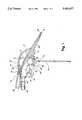
- FIG. 1depicts one embodiment of the present invention, comprising a flexible catheter, rigid holster, endotracheal tube gripper, catheter restraints, flow control valve, fluid trap, and negative pressure source attachment means connected to a wand, to form a single device.
- FIG. 1also depicts use of the present invention with an endotracheal tube, and its intersection with port 11 to form a novel valve for directing suction from the flexible catheter 10 to the endotracheal tube 50 or adaptor 51.
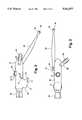
- FIG. 2depicts another embodiment of the present invention, modified to include an inlet source for delivery of negative pressure, projecting from the wand to form an oblique angle with the device.
- FIG. 3depicts an arial view of the same embodiment as shown in FIG. 2.
- FIG. 4depicts a frontal view of the embodiment as shown in FIG. 1.
- wand 10has four ports.
- Distal port 11opens to hollow appendage 21 which extends in a downward projection, forming an obtuse angle, preferably in the range of 120 to 150 degrees, with the wall of wand 10 immediately above appendage 21.
- the lower region between appendage 21 and wand 10provides a finger rest for the operator.
- Proximal port 12is the inlet port for a flow of negative pressure.
- Attachment means 22may be permanently secured to port 12 for readily connecting wand 10 to a source of negative pressure.
- a conventional attachment meansis depicted in FIG. 1, fitted with barbs 23, for a secured connection to a source of negative pressure.
- Upper port 13functions as a finger-operated flow control valve when wand 10 is connected to a source of negative pressure and as an inlet port for a ventilating device, when negative pressure is discontinued.
- a first cylindrical wall 32extends above wand 10 from the edge of upper port 13.
- a second cylindrical wall 31also extends above wand 10, surrounding the first cylindrical wall 32.
- Second cylindrical wall 31has a diameter such that a standard ventilating source can be securely coupled thereon.
- cylindrical wall 31can be constructed with an inner diameter suitable for occlusion with the operator's finger, and an outer diameter suitable for connecting to a standard ventilating source. This obviates the need for cylindrical wall 32.
- ventilating sourcesrefer to, for example, resuscitation bags and mechanical ventilators.
- Lower port 19is the outlet for either the negative or positive pressure connected to wand 10 and has a diameter such that a standard endotracheal (“ET”) tube 50 can be securely attached thereto by insertion of the ET adapter 51.
- ETendotracheal
- ET adapter 51One preferred means of securely attaching ET adapter 51 to wand 10 is for wand 10 to extend to form a tapered hollow cylindrical tube 18 ending at port 19.
- a flexible hollow tube 40such as a flexible catheter conventionally used for patient aspiration or instillation, is connected to appendage 21 by inserting the distal end of appendage 21, through the proximal opening of flexible hollow tube 40.
- the connection of appendage 21 to flexible hollow tube 40may be designed to enable easy and quick attachment and detachment.
- a rigid hollow tube 20such as a rigid catheter conventionally used for patient aspiration, is attached along the outer wall of one side of wand 10 such that rigid hollow tube 20 projects from wand 10.
- the inner diameter of first opening 24is greater than the outer diameter of flexible hollow tube 40 so that flexible hollow tube 40 can be easily inserted into first opening 24.
- Rigid tube 20has a tapered lumen so that the diameter decreases from opening 24 to opening 25.
- Distal end 26 of rigid tube 20forms a rounded edge to reduce the possibility of trauma when inserted into a patient's oropharynx.
- the lumen of rigid hollow tube 20preferably has a curvature in the range of 30-60 degrees such that flexible hollow tube 40, when inserted through first opening 24 will be directed in a downward fashion to second opening 25.
- the length of rigid hollow tube 20is consistent with the size of the area to be suctioned. For example, if the oropharynx of a neonate were to be suctioned, its length should be long enough to extend to the posterior aspect of the oropharynx (25) and extend slightly beyond the patient's lips.
- One embodiment of the present inventionis a device having a rigid catheter, such as rigid hollow tube 20, attached to a flexible catheter, such as flexible hollow tube 40, operating together as described above.
- a finger flow control valvesimilar to port 13 in FIGS. 1, 2 and 3 can also be included for better flow regulation.
- an attachment means to a source of negative pressureis included, however, there is no need to provide a means for attachment to a ventilating source.
- a conventional attachment meanscan be used, as depicted in FIG. I or an improved attachment means can be used, as depicted in FIGS. 2 and 3 and described in more detail below.
- a second embodiment of the present inventionincludes appendage 21 having a length suitable for operation as a rigid catheter.
- flexible hollow tube 40can be detached from appendage 21.
- Appendage 21can then be inserted directly into the patient's airway or other suction site. This obviates the need for rigid hollow tube 20.
- restraints 16are attached to the outer wall of wand 10, having a curved or circular configuration such that flexible tube 40 can be positioned therein, preventing flexible hollow tube 40 from interfering with vision or balance of the device when it is inserted in rigid hollow tube 20.
- One preferred embodimentincludes two such restraints.
- Wand 10also includes various gripping attachments to allow easier control in the operator's hands. This is especially necessary as the device will often be used in situations such as meconium deliveries in which a large amount of slippery secretions such as meconium, amniotic fluid and blood are present.
- the device of the present inventioncan be used with bare hands, universal precautions and barrier protection with gloves is strongly recommended. Since gloves, often latex, may make manipulations more difficult, the gripping attachments, the finger rest, formed by the appendage 21 and the cylindrical wall 18, the finger rest formed by fluid trap 27 and any other finger rests formed by variations in the shape of the device that are within the scope of the claimed invention, as well as the gripping ridges 15 on the outer upper surface of wand 10 are included in the device.
- the lower inner surface of wand 10may include an oblique ridge 17, which forms a wall extending within wand 10, angled toward the upper region of wand 10. This wall forms a division between the anterior section of wand 10, encompassing distal port 11 and the lower port 19, and the posterior section of wand 10.
- the posterior section of wand 10 below the upper edge of ridge 17forms a depression 27, providing a fluid trap for material suctioned from lower port 14.
- the fluid trapenables the operator of the device to determine the amount of material suctioned, and at what point in the procedure this had been done.
- the fluid trap 27 and oblique ridge 17prevent the backflow of suctioned material.
- Wand 10is preferably made of a transparent material to further assist the operator to ascertain that the device is functioning and to allow for visual analysis of the results of the procedure.
- a gripping attachment 30projects from alongside wand 10. As depicted in FIGS. 1, 2 and 3, gripping attachment 30 forms an incomplete cylinder, such that a slit runs along its entire length. The slit enables the insertion of an endotracheal tube adapter 51, without interference of the winged extensions 52 or a stylet which might be preferentially placed through adapter 51 and endotracheal tube 50. Other suitable devices such as clips or clamps, will be readily apparent to those skilled in the relevant art.
- FIGS. 2 and 3Another embodiment of the present invention is depicted in FIGS. 2 and 3.
- the devicehas a uniquely positioned inlet port for delivery of negative pressure.
- distal port 11is relocated alongside wand 10.
- Attachment means 22projects from wand 10 forming an oblique angle with wand 10.
- attachment means 22forms a 45 degree angle with wand 10.
- situating attachment means 22 as depicted in FIGS. 3 and 4minimizes interference between the hose connecting the device to an external source of negative pressure and the operator, by directing the connecting hose away from the device and operator.
- the oblique anglealso forms a hook for hanging the device from a nearby edge.
- the instant inventionhas been described as it relates to neonatal resuscitation as this is a most complex procedure requiring all of the innovations and specifications of the preferred embodiment of the instant invention. This should not be construed to exclude application of the present invention under other circumstances which employ the device to a more limited degree or with altered dimensions. It will be readily apparent to one of ordinary skill in the art that the present invention has many other applications consistent with its design.
Landscapes
- Health & Medical Sciences (AREA)
- Pulmonology (AREA)
- Emergency Medicine (AREA)
- Engineering & Computer Science (AREA)
- Anesthesiology (AREA)
- Biomedical Technology (AREA)
- Heart & Thoracic Surgery (AREA)
- Hematology (AREA)
- Life Sciences & Earth Sciences (AREA)
- Animal Behavior & Ethology (AREA)
- General Health & Medical Sciences (AREA)
- Public Health (AREA)
- Veterinary Medicine (AREA)
- External Artificial Organs (AREA)
Abstract
Description
Claims (27)
Priority Applications (2)
| Application Number | Priority Date | Filing Date | Title |
|---|---|---|---|
| US08416304US5562077B1 (en) | 1995-04-04 | 1995-04-04 | Method and apparatus for ventilation and aspiration |
| PCT/US1996/004508WO1996031248A1 (en) | 1995-04-04 | 1996-04-02 | Method and apparatus for ventilation and aspiration |
Applications Claiming Priority (1)
| Application Number | Priority Date | Filing Date | Title |
|---|---|---|---|
| US08416304US5562077B1 (en) | 1995-04-04 | 1995-04-04 | Method and apparatus for ventilation and aspiration |
Publications (2)
| Publication Number | Publication Date |
|---|---|
| US5562077Atrue US5562077A (en) | 1996-10-08 |
| US5562077B1 US5562077B1 (en) | 2000-09-05 |
Family
ID=23649426
Family Applications (1)
| Application Number | Title | Priority Date | Filing Date |
|---|---|---|---|
| US08416304Expired - LifetimeUS5562077B1 (en) | 1995-04-04 | 1995-04-04 | Method and apparatus for ventilation and aspiration |
Country Status (2)
| Country | Link |
|---|---|
| US (1) | US5562077B1 (en) |
| WO (1) | WO1996031248A1 (en) |
Cited By (25)
| Publication number | Priority date | Publication date | Assignee | Title |
|---|---|---|---|---|
| GB2359752A (en)* | 2000-03-01 | 2001-09-05 | Meddis Ltd | Catheter |
| US20020108614A1 (en)* | 2000-01-18 | 2002-08-15 | Schultz Joseph P. | Medical component system |
| US20030195482A1 (en)* | 2001-02-02 | 2003-10-16 | Schultz Joseph P. | Pneumatic medical system |
| US20030196667A1 (en)* | 2002-04-18 | 2003-10-23 | Croteau Kenneth S. | Meconium aspirator |
| US20040025873A1 (en)* | 2002-04-09 | 2004-02-12 | Edward Padgett | 3-In-1 in- line nebulizer, mediport dispenser and suction chamber for bag valve masks, endotracheal tubes and tracheotomy tubes |
| US20060000320A1 (en)* | 2004-06-30 | 2006-01-05 | Hutton William M | Ratchet wrench tool assembly for underground work and process of using |
| US20060002765A1 (en)* | 2004-06-30 | 2006-01-05 | Hutton William M | Tool assembly with universal coupling for various tools, for work on underground pipes |
| US20060002766A1 (en)* | 2004-06-30 | 2006-01-05 | Hutton William M | Apparatus and process for installing "t" couplings on underground pipe |
| US20070065433A1 (en)* | 2003-02-21 | 2007-03-22 | Mollnes Tom E | Methods and compositions for the treatment of meconium aspiration syndrome |
| US7300424B1 (en) | 2006-04-03 | 2007-11-27 | Mulford Thomas B | Aspirator and associated method |
| US20080045885A1 (en)* | 2006-08-21 | 2008-02-21 | Tycohealthcare Group Lp | Compliant guard for use with an aspiration instrument |
| US20090208900A1 (en)* | 2008-02-20 | 2009-08-20 | Leon Emmanuel Jew | Bifunctional dental apparatus |
| US7587796B1 (en) | 2001-03-07 | 2009-09-15 | Schultz Joseph P | Secure strap systems |
| US8371000B1 (en) | 2001-03-07 | 2013-02-12 | Joseph P. Schultz | Secure strap systems |
| USD678501S1 (en) | 2010-09-21 | 2013-03-19 | Peter Champe | Aspirator |
| US9386824B1 (en) | 2001-03-07 | 2016-07-12 | Joseph P. Schultz | Secure strap systems |
| US10099027B2 (en) | 2014-01-24 | 2018-10-16 | Cole Research & Design | Oral suction device |
| US10149956B2 (en) | 2015-02-28 | 2018-12-11 | John P. Ure | Bi-lateral endobronchial suctioning device and medical suctioning system for intubated patients |
| US10363356B2 (en) | 2000-01-18 | 2019-07-30 | Joseph P. Schultz | Abscess irrigation systems |
| WO2020198327A1 (en)* | 2019-03-26 | 2020-10-01 | Pocket Naloxone Corp. | Devices and methods for delivering pharmaceutical compositions |
| US11083835B2 (en) | 2009-08-05 | 2021-08-10 | Joseph P. Schultz | Abscess irrigation systems |
| US11191934B2 (en) | 2019-03-26 | 2021-12-07 | Pocket Naloxone Corp. | Devices and methods for delivery of pharmaceutical compositions |
| US11278709B1 (en) | 2021-03-12 | 2022-03-22 | Pocket Naloxone Corp. | Drug delivery device and methods for using same |
| US11654227B1 (en)* | 2019-06-20 | 2023-05-23 | Sang In Han | Wet seal suction device |
| USD1085409S1 (en) | 2000-01-18 | 2025-07-22 | Splash Medical Devices, LLC | Abscess irrigation device |
Families Citing this family (1)
| Publication number | Priority date | Publication date | Assignee | Title |
|---|---|---|---|---|
| EP1861154B1 (en) | 2005-03-25 | 2016-11-16 | Nalini Vadivelu | Medical apparatus with hypopharyngeal suctioning capability |
Citations (13)
| Publication number | Priority date | Publication date | Assignee | Title |
|---|---|---|---|---|
| US4193406A (en)* | 1978-09-18 | 1980-03-18 | Jinotti Walter J | Dual purpose catheter |
| US4275724A (en)* | 1979-04-02 | 1981-06-30 | Barry Behrstock | Endotracheal intubation device |
| US4487600A (en)* | 1981-11-09 | 1984-12-11 | Brownlie Alan W | Adjustable suction device for medical use |
| US4534542A (en)* | 1983-12-05 | 1985-08-13 | Superior Plastic Products Corp. | Suction control device for aspirator system |
| US4699138A (en)* | 1986-07-25 | 1987-10-13 | Barry Behrstock | Endotracheal intubation suction device |
| US4787894A (en)* | 1987-10-26 | 1988-11-29 | Turnbull Christopher J | Meconium aspiration device |
| US4805611A (en)* | 1988-02-10 | 1989-02-21 | Becton, Dickinson And Company | Aspirating device |
| US4915691A (en)* | 1987-05-07 | 1990-04-10 | Gesco International, Inc. | Aspirator |
| US5000175A (en)* | 1979-08-08 | 1991-03-19 | Pue Alexander F | Meconium aspiration device |
| US5065754A (en)* | 1990-06-06 | 1991-11-19 | Ballard Medical Products | Aspirating catheter tube inserter |
| US5101817A (en)* | 1989-08-04 | 1992-04-07 | Nellcor, Inc. | Airway adapter for use with closed suction catheter system |
| US5325851A (en)* | 1991-04-01 | 1994-07-05 | Sorenson Laboratories, Inc. | Apparatus and method for ventilating and aspirating |
| US5335655A (en)* | 1992-09-10 | 1994-08-09 | Sherwood Medical Company | Suction control valve |
- 1995
- 1995-04-04USUS08416304patent/US5562077B1/ennot_activeExpired - Lifetime
- 1996
- 1996-04-02WOPCT/US1996/004508patent/WO1996031248A1/enactiveApplication Filing
Patent Citations (13)
| Publication number | Priority date | Publication date | Assignee | Title |
|---|---|---|---|---|
| US4193406A (en)* | 1978-09-18 | 1980-03-18 | Jinotti Walter J | Dual purpose catheter |
| US4275724A (en)* | 1979-04-02 | 1981-06-30 | Barry Behrstock | Endotracheal intubation device |
| US5000175A (en)* | 1979-08-08 | 1991-03-19 | Pue Alexander F | Meconium aspiration device |
| US4487600A (en)* | 1981-11-09 | 1984-12-11 | Brownlie Alan W | Adjustable suction device for medical use |
| US4534542A (en)* | 1983-12-05 | 1985-08-13 | Superior Plastic Products Corp. | Suction control device for aspirator system |
| US4699138A (en)* | 1986-07-25 | 1987-10-13 | Barry Behrstock | Endotracheal intubation suction device |
| US4915691A (en)* | 1987-05-07 | 1990-04-10 | Gesco International, Inc. | Aspirator |
| US4787894A (en)* | 1987-10-26 | 1988-11-29 | Turnbull Christopher J | Meconium aspiration device |
| US4805611A (en)* | 1988-02-10 | 1989-02-21 | Becton, Dickinson And Company | Aspirating device |
| US5101817A (en)* | 1989-08-04 | 1992-04-07 | Nellcor, Inc. | Airway adapter for use with closed suction catheter system |
| US5065754A (en)* | 1990-06-06 | 1991-11-19 | Ballard Medical Products | Aspirating catheter tube inserter |
| US5325851A (en)* | 1991-04-01 | 1994-07-05 | Sorenson Laboratories, Inc. | Apparatus and method for ventilating and aspirating |
| US5335655A (en)* | 1992-09-10 | 1994-08-09 | Sherwood Medical Company | Suction control valve |
Cited By (34)
| Publication number | Priority date | Publication date | Assignee | Title |
|---|---|---|---|---|
| US10363356B2 (en) | 2000-01-18 | 2019-07-30 | Joseph P. Schultz | Abscess irrigation systems |
| USD1085409S1 (en) | 2000-01-18 | 2025-07-22 | Splash Medical Devices, LLC | Abscess irrigation device |
| US20020108614A1 (en)* | 2000-01-18 | 2002-08-15 | Schultz Joseph P. | Medical component system |
| US7802574B2 (en)* | 2000-01-18 | 2010-09-28 | Schultz Joseph P | Medical component system |
| US10576198B2 (en) | 2000-01-18 | 2020-03-03 | Joseph P. Schultz | Abscess irrigation systems |
| GB2359752A (en)* | 2000-03-01 | 2001-09-05 | Meddis Ltd | Catheter |
| GB2359752B (en)* | 2000-03-01 | 2004-08-25 | Meddis Ltd | Catheter |
| EP1129735A3 (en)* | 2000-03-01 | 2002-02-27 | Meddis Limited | Catheter |
| US20030195482A1 (en)* | 2001-02-02 | 2003-10-16 | Schultz Joseph P. | Pneumatic medical system |
| US8371000B1 (en) | 2001-03-07 | 2013-02-12 | Joseph P. Schultz | Secure strap systems |
| US9386824B1 (en) | 2001-03-07 | 2016-07-12 | Joseph P. Schultz | Secure strap systems |
| US7587796B1 (en) | 2001-03-07 | 2009-09-15 | Schultz Joseph P | Secure strap systems |
| US20040025873A1 (en)* | 2002-04-09 | 2004-02-12 | Edward Padgett | 3-In-1 in- line nebulizer, mediport dispenser and suction chamber for bag valve masks, endotracheal tubes and tracheotomy tubes |
| US20030196667A1 (en)* | 2002-04-18 | 2003-10-23 | Croteau Kenneth S. | Meconium aspirator |
| US20070065433A1 (en)* | 2003-02-21 | 2007-03-22 | Mollnes Tom E | Methods and compositions for the treatment of meconium aspiration syndrome |
| US20060002766A1 (en)* | 2004-06-30 | 2006-01-05 | Hutton William M | Apparatus and process for installing "t" couplings on underground pipe |
| US20060002765A1 (en)* | 2004-06-30 | 2006-01-05 | Hutton William M | Tool assembly with universal coupling for various tools, for work on underground pipes |
| US20060000320A1 (en)* | 2004-06-30 | 2006-01-05 | Hutton William M | Ratchet wrench tool assembly for underground work and process of using |
| US7300424B1 (en) | 2006-04-03 | 2007-11-27 | Mulford Thomas B | Aspirator and associated method |
| US7918835B2 (en) | 2006-08-21 | 2011-04-05 | Tyco Healthcare Group Lp | Compliant guard for use with an aspiration instrument |
| US20080045885A1 (en)* | 2006-08-21 | 2008-02-21 | Tycohealthcare Group Lp | Compliant guard for use with an aspiration instrument |
| US20090208900A1 (en)* | 2008-02-20 | 2009-08-20 | Leon Emmanuel Jew | Bifunctional dental apparatus |
| US12208226B2 (en) | 2009-08-05 | 2025-01-28 | Joseph P. Schultz | Abscess irrigation systems |
| US11083835B2 (en) | 2009-08-05 | 2021-08-10 | Joseph P. Schultz | Abscess irrigation systems |
| USD678501S1 (en) | 2010-09-21 | 2013-03-19 | Peter Champe | Aspirator |
| US10099027B2 (en) | 2014-01-24 | 2018-10-16 | Cole Research & Design | Oral suction device |
| US10149956B2 (en) | 2015-02-28 | 2018-12-11 | John P. Ure | Bi-lateral endobronchial suctioning device and medical suctioning system for intubated patients |
| US11191934B2 (en) | 2019-03-26 | 2021-12-07 | Pocket Naloxone Corp. | Devices and methods for delivery of pharmaceutical compositions |
| US12194263B2 (en) | 2019-03-26 | 2025-01-14 | Pocket Naloxone Corp. | Devices and methods for delivery of pharmaceutical compositions |
| WO2020198327A1 (en)* | 2019-03-26 | 2020-10-01 | Pocket Naloxone Corp. | Devices and methods for delivering pharmaceutical compositions |
| US11654227B1 (en)* | 2019-06-20 | 2023-05-23 | Sang In Han | Wet seal suction device |
| US12017026B2 (en) | 2021-03-12 | 2024-06-25 | Pocket Naloxone Corp. | Drug delivery device and methods for using same |
| US11278709B1 (en) | 2021-03-12 | 2022-03-22 | Pocket Naloxone Corp. | Drug delivery device and methods for using same |
| US12285581B2 (en) | 2021-03-12 | 2025-04-29 | Pocket Naloxone Corp. | Drug delivery device and methods for using same |
Also Published As
| Publication number | Publication date |
|---|---|
| WO1996031248A1 (en) | 1996-10-10 |
| US5562077B1 (en) | 2000-09-05 |
Similar Documents
| Publication | Publication Date | Title |
|---|---|---|
| US5562077A (en) | Method and apparatus for ventilation and aspiration | |
| US4699138A (en) | Endotracheal intubation suction device | |
| US4915691A (en) | Aspirator | |
| US5016614A (en) | Endotracheal intubation apparatus | |
| US5509408A (en) | Neonatal resuscitation device | |
| US4275724A (en) | Endotracheal intubation device | |
| US4787894A (en) | Meconium aspiration device | |
| JP5951013B2 (en) | Insertion aid | |
| US7802574B2 (en) | Medical component system | |
| US6776157B2 (en) | Medical pacifier and method for use thereof | |
| US8998806B2 (en) | Insertion aid for oral and nasal medical devices | |
| US5000175A (en) | Meconium aspiration device | |
| US4490138A (en) | Pharyngeal suction device | |
| US4848331A (en) | Apparatus and method for pulmonary ventilation of a patient concurrent with fiberoptic respiratory tract examination and tracheal intubation | |
| JP2935895B2 (en) | Oxygen supply port medical device | |
| US5890488A (en) | Coupling device and sound resonating membrane for a stethoscope and an endotracheal tube | |
| US4221220A (en) | Surgical suction apparatus | |
| US5711294A (en) | Ventilator manifold having cleaning ports and method of use thereof | |
| CN110496285A (en) | A soft head adjustable nasopharyngeal ventilation tube | |
| US5431157A (en) | Anesthesia conduit | |
| CA3030879A1 (en) | Rigid stylet apparatus with optional suction | |
| US10272217B2 (en) | Device for gripping and directing bougies for intubation | |
| JP2023057993A (en) | Intraoral liquid delivery/suction device | |
| CN209809123U (en) | Sputum suction tube capable of accurately sampling | |
| CN222033190U (en) | Automatic sputum suction external member of general artifical air flue |
Legal Events
| Date | Code | Title | Description |
|---|---|---|---|
| CC | Certificate of correction | ||
| RR | Request for reexamination filed | Effective date:19990122 | |
| FEPP | Fee payment procedure | Free format text:PAYOR NUMBER ASSIGNED (ORIGINAL EVENT CODE: ASPN); ENTITY STATUS OF PATENT OWNER: SMALL ENTITY | |
| REMI | Maintenance fee reminder mailed | ||
| B1 | Reexamination certificate first reexamination | Free format text:THE PATENTABILITY OF CLAIMS 4-27 IS CONFIRMED. CLAIM 1 IS DETERMINED TO BE PATENTABLE AS AMENDED. CLAIMS 2 AND 3, DEPENDENT ON AN AMENDED CLAIM, ARE DETERMINED TO BE PATENTABLE. | |
| REIN | Reinstatement after maintenance fee payment confirmed | ||
| FP | Lapsed due to failure to pay maintenance fee | Effective date:20001008 | |
| FEPP | Fee payment procedure | Free format text:PETITION RELATED TO MAINTENANCE FEES DENIED/DISMISSED (ORIGINAL EVENT CODE: PMFD); ENTITY STATUS OF PATENT OWNER: SMALL ENTITY | |
| FEPP | Fee payment procedure | Free format text:PETITION RELATED TO MAINTENANCE FEES FILED (ORIGINAL EVENT CODE: PMFP); ENTITY STATUS OF PATENT OWNER: SMALL ENTITY | |
| FEPP | Fee payment procedure | Free format text:PETITION RELATED TO MAINTENANCE FEES GRANTED (ORIGINAL EVENT CODE: PMFG); ENTITY STATUS OF PATENT OWNER: SMALL ENTITY | |
| FEPP | Fee payment procedure | Free format text:PETITION RELATED TO MAINTENANCE FEES FILED (ORIGINAL EVENT CODE: PMFP); ENTITY STATUS OF PATENT OWNER: SMALL ENTITY | |
| FPAY | Fee payment | Year of fee payment:4 | |
| SULP | Surcharge for late payment | ||
| STCF | Information on status: patent grant | Free format text:PATENTED CASE | |
| PRDP | Patent reinstated due to the acceptance of a late maintenance fee | Effective date:20021202 | |
| FPAY | Fee payment | Year of fee payment:8 | |
| FPAY | Fee payment | Year of fee payment:12 |