US5533997A - Apparatus and method for performing presbyopia corrective surgery - Google Patents
Apparatus and method for performing presbyopia corrective surgeryDownload PDFInfo
- Publication number
- US5533997A US5533997AUS08/268,182US26818294AUS5533997AUS 5533997 AUS5533997 AUS 5533997AUS 26818294 AUS26818294 AUS 26818294AUS 5533997 AUS5533997 AUS 5533997A
- Authority
- US
- United States
- Prior art keywords
- cornea
- corneal stroma
- corneal
- ablating
- laser
- Prior art date
- Legal status (The legal status is an assumption and is not a legal conclusion. Google has not performed a legal analysis and makes no representation as to the accuracy of the status listed.)
- Expired - Lifetime
Links
- 238000000034methodMethods0.000titleclaimsabstractdescription35
- 201000010041presbyopiaDiseases0.000titleclaimsabstractdescription29
- 238000001356surgical procedureMethods0.000titledescription14
- 210000004087corneaAnatomy0.000claimsabstractdescription54
- 238000002679ablationMethods0.000claimsabstractdescription42
- 210000003683corneal stromaAnatomy0.000claimsabstractdescription42
- 230000008569processEffects0.000claimsabstractdescription24
- 230000005855radiationEffects0.000claimsabstractdescription12
- 230000001680brushing effectEffects0.000claimsabstractdescription3
- 238000004140cleaningMethods0.000claimsdescription9
- 230000008859changeEffects0.000claimsdescription7
- 229920003229poly(methyl methacrylate)Polymers0.000claimsdescription7
- 239000004926polymethyl methacrylateSubstances0.000claimsdescription7
- 238000001035dryingMethods0.000claimsdescription4
- 238000007664blowingMethods0.000claimsdescription3
- 230000001678irradiating effectEffects0.000claimsdescription2
- 238000012937correctionMethods0.000abstractdescription11
- 239000003550markerSubstances0.000abstractdescription7
- 206010030113OedemaDiseases0.000abstractdescription3
- 239000012530fluidSubstances0.000abstractdescription2
- 239000011521glassSubstances0.000description5
- 210000001747pupilAnatomy0.000description5
- 230000004438eyesightEffects0.000description4
- 239000000463materialSubstances0.000description3
- 208000001491myopiaDiseases0.000description3
- 206010027646MiosisDiseases0.000description2
- FAPWRFPIFSIZLT-UHFFFAOYSA-MSodium chlorideChemical compound[Na+].[Cl-]FAPWRFPIFSIZLT-UHFFFAOYSA-M0.000description2
- 230000004308accommodationEffects0.000description2
- 230000009471actionEffects0.000description2
- 201000009310astigmatismDiseases0.000description2
- 210000002919epithelial cellAnatomy0.000description2
- 230000035876healingEffects0.000description2
- 230000006698inductionEffects0.000description2
- 230000001788irregularEffects0.000description2
- 230000004379myopiaEffects0.000description2
- 239000002245particleSubstances0.000description2
- 238000011084recoveryMethods0.000description2
- 229920003002synthetic resinPolymers0.000description2
- 239000000057synthetic resinSubstances0.000description2
- 238000002691topical anesthesiaMethods0.000description2
- QCHFTSOMWOSFHM-WPRPVWTQSA-N(+)-PilocarpineChemical compoundC1OC(=O)[C@@H](CC)[C@H]1CC1=CN=CN1CQCHFTSOMWOSFHM-WPRPVWTQSA-N0.000description1
- RBTBFTRPCNLSDE-UHFFFAOYSA-N3,7-bis(dimethylamino)phenothiazin-5-iumChemical compoundC1=CC(N(C)C)=CC2=[S+]C3=CC(N(C)C)=CC=C3N=C21RBTBFTRPCNLSDE-UHFFFAOYSA-N0.000description1
- 206010002091AnaesthesiaDiseases0.000description1
- 241001227713ChironSpecies0.000description1
- 206010020675HypermetropiaDiseases0.000description1
- 230000004075alterationEffects0.000description1
- 238000001949anaesthesiaMethods0.000description1
- 230000037005anaesthesiaEffects0.000description1
- 230000008901benefitEffects0.000description1
- 244000309464bullSpecies0.000description1
- 238000004040coloringMethods0.000description1
- 238000004891communicationMethods0.000description1
- ZXJXZNDDNMQXFV-UHFFFAOYSA-Mcrystal violetChemical compound[Cl-].C1=CC(N(C)C)=CC=C1[C+](C=1C=CC(=CC=1)N(C)C)C1=CC=C(N(C)C)C=C1ZXJXZNDDNMQXFV-UHFFFAOYSA-M0.000description1
- 230000007547defectEffects0.000description1
- 239000006196dropSubstances0.000description1
- 230000000694effectsEffects0.000description1
- 239000003889eye dropSubstances0.000description1
- 229940012356eye dropsDrugs0.000description1
- 230000004424eye movementEffects0.000description1
- 229960001235gentian violetDrugs0.000description1
- 230000004305hyperopiaEffects0.000description1
- 201000006318hyperopiaDiseases0.000description1
- 239000012535impuritySubstances0.000description1
- 229940039014isoptocarpineDrugs0.000description1
- 239000007788liquidSubstances0.000description1
- 229960000907methylthioninium chlorideDrugs0.000description1
- 238000012986modificationMethods0.000description1
- 230000004048modificationEffects0.000description1
- 230000000737periodic effectEffects0.000description1
- 230000002980postoperative effectEffects0.000description1
- 230000009467reductionEffects0.000description1
- 238000002271resectionMethods0.000description1
- 238000012360testing methodMethods0.000description1
- 238000012876topographyMethods0.000description1
- 230000001131transforming effectEffects0.000description1
- 230000000007visual effectEffects0.000description1
Images

Classifications
- A—HUMAN NECESSITIES
- A61—MEDICAL OR VETERINARY SCIENCE; HYGIENE
- A61F—FILTERS IMPLANTABLE INTO BLOOD VESSELS; PROSTHESES; DEVICES PROVIDING PATENCY TO, OR PREVENTING COLLAPSING OF, TUBULAR STRUCTURES OF THE BODY, e.g. STENTS; ORTHOPAEDIC, NURSING OR CONTRACEPTIVE DEVICES; FOMENTATION; TREATMENT OR PROTECTION OF EYES OR EARS; BANDAGES, DRESSINGS OR ABSORBENT PADS; FIRST-AID KITS
- A61F9/00—Methods or devices for treatment of the eyes; Devices for putting in contact-lenses; Devices to correct squinting; Apparatus to guide the blind; Protective devices for the eyes, carried on the body or in the hand
- A61F9/007—Methods or devices for eye surgery
- A61F9/013—Instruments for compensation of ocular refraction ; Instruments for use in cornea removal, for reshaping or performing incisions in the cornea
- A—HUMAN NECESSITIES
- A61—MEDICAL OR VETERINARY SCIENCE; HYGIENE
- A61F—FILTERS IMPLANTABLE INTO BLOOD VESSELS; PROSTHESES; DEVICES PROVIDING PATENCY TO, OR PREVENTING COLLAPSING OF, TUBULAR STRUCTURES OF THE BODY, e.g. STENTS; ORTHOPAEDIC, NURSING OR CONTRACEPTIVE DEVICES; FOMENTATION; TREATMENT OR PROTECTION OF EYES OR EARS; BANDAGES, DRESSINGS OR ABSORBENT PADS; FIRST-AID KITS
- A61F9/00—Methods or devices for treatment of the eyes; Devices for putting in contact-lenses; Devices to correct squinting; Apparatus to guide the blind; Protective devices for the eyes, carried on the body or in the hand
- A61F9/007—Methods or devices for eye surgery
- A61F9/008—Methods or devices for eye surgery using laser
- A61F9/00802—Methods or devices for eye surgery using laser for photoablation
- A61F9/00804—Refractive treatments
- A61F9/00808—Inducing higher orders, e.g. for correction of presbyopia
- A—HUMAN NECESSITIES
- A61—MEDICAL OR VETERINARY SCIENCE; HYGIENE
- A61F—FILTERS IMPLANTABLE INTO BLOOD VESSELS; PROSTHESES; DEVICES PROVIDING PATENCY TO, OR PREVENTING COLLAPSING OF, TUBULAR STRUCTURES OF THE BODY, e.g. STENTS; ORTHOPAEDIC, NURSING OR CONTRACEPTIVE DEVICES; FOMENTATION; TREATMENT OR PROTECTION OF EYES OR EARS; BANDAGES, DRESSINGS OR ABSORBENT PADS; FIRST-AID KITS
- A61F9/00—Methods or devices for treatment of the eyes; Devices for putting in contact-lenses; Devices to correct squinting; Apparatus to guide the blind; Protective devices for the eyes, carried on the body or in the hand
- A61F9/007—Methods or devices for eye surgery
- A61F9/008—Methods or devices for eye surgery using laser
- A61F9/00802—Methods or devices for eye surgery using laser for photoablation
- A61F9/00817—Beam shaping with masks
- A—HUMAN NECESSITIES
- A61—MEDICAL OR VETERINARY SCIENCE; HYGIENE
- A61F—FILTERS IMPLANTABLE INTO BLOOD VESSELS; PROSTHESES; DEVICES PROVIDING PATENCY TO, OR PREVENTING COLLAPSING OF, TUBULAR STRUCTURES OF THE BODY, e.g. STENTS; ORTHOPAEDIC, NURSING OR CONTRACEPTIVE DEVICES; FOMENTATION; TREATMENT OR PROTECTION OF EYES OR EARS; BANDAGES, DRESSINGS OR ABSORBENT PADS; FIRST-AID KITS
- A61F9/00—Methods or devices for treatment of the eyes; Devices for putting in contact-lenses; Devices to correct squinting; Apparatus to guide the blind; Protective devices for the eyes, carried on the body or in the hand
- A61F9/007—Methods or devices for eye surgery
- A61F9/008—Methods or devices for eye surgery using laser
- A61F2009/00861—Methods or devices for eye surgery using laser adapted for treatment at a particular location
- A61F2009/00872—Cornea
- A—HUMAN NECESSITIES
- A61—MEDICAL OR VETERINARY SCIENCE; HYGIENE
- A61F—FILTERS IMPLANTABLE INTO BLOOD VESSELS; PROSTHESES; DEVICES PROVIDING PATENCY TO, OR PREVENTING COLLAPSING OF, TUBULAR STRUCTURES OF THE BODY, e.g. STENTS; ORTHOPAEDIC, NURSING OR CONTRACEPTIVE DEVICES; FOMENTATION; TREATMENT OR PROTECTION OF EYES OR EARS; BANDAGES, DRESSINGS OR ABSORBENT PADS; FIRST-AID KITS
- A61F9/00—Methods or devices for treatment of the eyes; Devices for putting in contact-lenses; Devices to correct squinting; Apparatus to guide the blind; Protective devices for the eyes, carried on the body or in the hand
- A61F9/007—Methods or devices for eye surgery
- A61F9/008—Methods or devices for eye surgery using laser
- A61F2009/00885—Methods or devices for eye surgery using laser for treating a particular disease
- A61F2009/00895—Presbyopia
Definitions
- the inventionrelates to an apparatus and method for correcting presbyopia through surgery.
- the system used in accordance with the invention(called a "PRESBYSYSTEM”) includes several different elements that, when used together, modify the front corneal curvature, thereby creating a multifocal surface that permits the patient to see with normal vision with respect to objects at a distance, while also permitting the patient to see nearby objects with normal vision.
- This simultaneous correction of near and distant visionis accomplished through the use of just a part of the corneal surface (the middle). This means that the portion of the corneal area which is used for near vision is not the same as that used for long distance vision.
- Bifocal lensespresent lines where the two portions of the lens are joined together. Furthermore, people must become accustomed to reading through the one relatively small portion of the lens, while looking at distant objects through a different portion of the lens. Bifocal glasses also have the disadvantages present in regular glasses. Such disadvantages include the fact that glasses are breakable, they become fogged when coming in from the cold, they steam up in hot weather, and they require periodic cleaning.
- This inventionis directed to an apparatus and method for correcting the presbyopia problems directly on the eye of the patient, such that the use of glasses is avoided and the eye of the patient will adapt the focus automatically for nearsight and farsight.
- the inventionrelates to a process for surgically correcting presbyopia.
- the processincludes anesthetizing a patient and marking a portion of an eye of the patient which is to be ablated. At least a portion of the cornea is resected to expose the corneal stroma. An annular portion of the corneal stroma is then ablated using radiation from a laser beam. After ablation, the cornea is repositioned onto the eye.
- the corneamay be resected such that a portion of the cornea remains intact, and thereafter the cornea may be folded back to expose the corneal stroma.
- the corneamay be resected such that a complete disk of the cornea is removed from the eye, to thereby expose the corneal stroma. Thereafter, the cornea disk would have to be reattached onto the eye.
- the corneal stromashould be dried after it has been exposed by the resection and before the ablation process. Otherwise, uneven ablation may occur due to liquids present on the stroma.
- the laser beammay be directed in a circular fashion until an annular ablation of a predetermined width and depth is provided.
- a maskmay be provided over a central area of the corneal stroma to stop the radiation from the laser beam at the central portion.
- the laser beam diameteris controlled (and provided somewhat larger than the mask diameter) such that an annular ablation of a predetermined width and depth is provided.
- the maskmay be made from polymethyl methacrylate or other suitable synthetic resin.
- the lasermay be a conventional pulsed laser.
- the ablated portionshould be cleaned, in order to prevent edema. This may be accomplished by brushing and irrigating the portion which was ablated.
- the cornea portionmay be reattached to the eye by blowing air onto the cornea.
- the inventionalso relates to a system for surgically correcting presbyopia.
- This systemincludes a means for marking a portion of an eye of a patient which is to be ablated.
- a means for resecting at least a portion of a cornea of the eye of the patientis included for the purpose of exposing the corneal stroma.
- the systemfurther incudes a means for ablating an annular portion of the corneal stroma.
- This means for ablatingincludes a laser (such as a pulsed laser), wherein ablation takes place by irradiating the corneal stroma with radiation from the laser.
- a corneal shapermay be provided as the means for resecting, wherein the corneal shaper either partially resects the cornea such that a portion of the cornea remains intact, or fully removes a corneal disk, as mentioned above.
- the systemmay further include a means for drying the corneal stroma after it has been exposed by the means for resecting, such as by blowing air thereon.
- a maskmay be provided with the means for ablating for positioning over a central area of the corneal stroma to stop the radiation from the laser.
- This maskmay be made from a material which stops laser radiation, such as polymethyl methacrylate (PMMA) or other suitable synthetic resin.
- PMMApolymethyl methacrylate
- a means for providing a predetermined diameter to a laser beam from the laseris provided with this embodiment to limit the laser beam width so that an annular ablation of a predetermined width and depth is provided.
- the system in accordance with the inventionis preferably provided with a means for cleaning the portion of the stroma which was ablated.
- This means for cleaningmay include a delicate brush or a means for irrigating the portion which was ablated, or both.
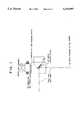
- FIG. 1is schematic view of a portion of the excimer laser where the setting of the axis of the laser ray is performed;
- FIG. 2is a schematic view of the path of the laser beam and the optics used in an embodiment of the apparatus according to the invention
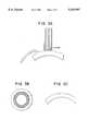
- FIG. 3Ashows the laser beam performing the ablation on the cornea, and the mask protecting the center area of the cornea
- FIG. 3Bshows the ring for the ablation zone
- FIG. 3Cshows the way the ablation of the cornea appears finally.
- the system in accordance with the inventionincludes an excimer laser, an automatic corneal shaper, a pneumatic fixation ring, a mask and an air source.
- a preferred automatic corneal shaper for use in the system in accordance with this inventionis the Automatic Corneal Shaper described in the inventor's U.S. Pat. No. 5,133,726, issued on Jul. 28, 1992. This patent is entirely incorporated herein by reference.
- the eyeis fixed by the fixation ring which also functions as a guide for the automatic corneal shaper.
- the fixation or retaining ringas illustrated in the above mentioned U.S. Pat. No. 5,133,726, permits total control of the eye movement.
- the pupil of the eyewill be a reference point for making a very central stromal ablation on the cornea.
- a keratectomyis performed using the automatic corneal shaper.
- the keratectomymay be partial, which means that a cornea flap technique is used. This means that an end portion of the corneal disc remains attached to the cornea base, which thereby permits its repositioning in an easier and surer way, once the ablation is performed.
- the flapis retracted, the corneal stroma becomes exposed, which is ideal tissue on which to practice the ablation.
- the superficial layers of the cornearemain untouched. In this way, undesirable healing is avoided, and inaccuracy in the post operative correction and regression is also avoided.
- annular ablationis made on the stroma having a diameter not exceeding 3.5 mm, with a central zone varying between 2 and 3 mm.
- the annular ablationproduces a central protrusion of the stroma such that when the corneal flap is repositioned at its initial position, this stromal curvature change is transmitted to the forward corneal surface, thereby transforming it into a multifocal surface, which is in fact myopic in its central part. This is what makes it possible for the patient to read without optic correction after the procedure, regardless of the age of the patient or the loss of accommodation.
- the annular ablationcan be made in isolated form, for presbyopia correction, or it can be made together with hyperopia, myopia and astigmatism surgery, either isolated or combined.
- the procedurecontinues with exhaustive cleaning of the interface using a balanced saline solution, a brush and aspiration, in order to assure that the interface is free from impurities, epithelial cells or foreign particles.
- the flapis replaced in the bed, adequately oriented in order to avoid altering its natural position.
- the edges of the flapare dried using air for several seconds to obtain adherence of the flap, such that the patient may be permitted to leave the operating room with no bandages and to obtain less than 24 hours recovery time.
- a conventional topical anesthesiai.e., in the form of eye drops
- This topical anesthesiais sufficient for the surgical technique in accordance with the invention to do a painless job.
- a pneumatic fixation ringis positioned over the eye.
- the surgical procedure in accordance with the inventionshould be carried out in a sterile area (i.e., a surgery room), because the cornea will be touched not in a superficial manner as would be required for the photo-ablation for the correction of myopia. Rather, in the presbyopia corrective surgical technique in accordance with the invention, a corneal flap is lifted in a laminar way in order to work directly on the stroma. Therefore surgical fields are located in order to isolate the working area and also a blepharostat is provided in order to maintain the eye sufficiently exposed so as to be able to practice the surgery.
- a markeris advantageously used to aid in the practice of the invention.
- the marker used in this new techniquehas the shape of a bullock eye having two concentric circles (thereby forming an inner ring and an outer ring) in which its external portion has a diameter of about 10.5 mm and its inner part has a diameter of about 3 to 5 mm.
- This markeris impregnated with a coloring product, such as gentian violet, methylene blue, or the like.
- the markeris placed on the patient's eye.
- the internal ringhas the function of centering the marker, having as a reference point, the pupil. In this manner, the external ring is automatically marked and in turn this will be used as a reference when positioning of the pneumatic fixation ring.
- the markeralso has a para-radial line joining both rings.
- the para-radial linesare useful for adequately orienting the corneal flap.
- the para-radial linesare used in order to assist in positioning the disk in the right place, that is, epithelial toward the exterior and stroma toward the inner part, and once located in this manner, it may now be oriented in adequate form.
- the pneumatic fixation ringcomprises two main components.
- the ringitself which will be adapted to the eye by means of a bottom vacuum chamber, allowing it in this manner to hold the eye in place and to increase the inter ocular pressure. This makes it easier to make the necessary cut in the cornea in a uniform manner.
- the fixation ringalso has a central orifice through which the cornea protrudes. In its top portion, the most important component of the fixation ring is a line of toothed protrusions which contact with the pinions of the automatic corneal shaper (see U.S. Pat. No. 5,133,726). This allows the corneal shaper to be displaced in a horizontal way for performing the laminar cut in the cornea.
- the second component of this ringis a handle which places the bottom vacuum chamber of the fixation ring in communication with a vacuum pump.
- the vacuum pumpis responsible for suction fitting the ring on the patient's eye.
- This handlealso is used to manipulate the eye once the ring is fixed to the eye.
- the next step of the surgical procedureis performed by the automatic corneal shaper, as noted above.
- the shaperis positioned over the fixation ring, and once the pinions of the shaper are in contact with the toothed protrusions of the ring, the shaper motor is started, and the shaper is moved horizontally and uniformly over the cornea.
- the cutter of the shaperwill make the laminar cut very accurately in its thickness, in the manner described in U.S. Pat. No. 5,133,726.
- the motor of the shaperis stopped at a predetermined position of the cut so as to have a thin portion of cornea still fixed to one side.
- the corneal stromawill appear.
- the corneal stromais the place where the object of the surgery will be practiced, because it has the great advantage that once the corneal flap is repositioned after the stromal ablation, all the natural structures of the eye will be preserved in their original place, but with a change in topography, thereby avoiding unwanted healings and other alterations that would be present if this system is not used.
- the exposed stromal surfaceOnce the exposed stromal surface is examined, it must be dried prior to the ablation action of an excimer laser, because any remaining fluid on the stroma will be considered by the laser ray as corneal tissue. This would result in an irregular ablation: that is, different depths of ablation would be produced on the stroma.
- the excimer laser systemis one that will perform the correction of this visual defect by providing a stromal ablation in the required manner, location and depth in order to create a multifocal surface in the cornea that allows good far sight, as well as good near sight. This sight usually is lost during a person's later years due to a physical lack of accommodation and loss of elasticity of the lens.
- the systemincludes the novel combination of the above elements in order to obtain an annular shaped ablation within a corneal area which is not used for far sight.
- Theseare the theoretical and real bases of the system in accordance with the invention for presbyopia correction. There can be different ways to obtain the results, as will be described below.
- the laseris directed toward a zone where the ablation must be done.
- the laseris directed with a circular movement of the laser beam so that the ablation is made with the required width and depth, to thereby obtain the desired change in curvature.
- the apparatus that sends the laser ray beamincludes an eye follower system in order to follow any movement of the eye, so that an irregular ablation ring does not result.
- the laser beam rayis sent toward the center of the chosen area, having as a reference point the small pupil.
- a maskis positioned over the central area so that it prevents the laser rays from touching the corneal stroma in the central area. In this manner, the ablation will be delimited at the outside by the selected diameter of the laser beam and at the inside by the border of the mask, thereby leaving a ring shaped area, as shown in FIGS. 3B and 3C.
- the cornea over the pupil areawill be totally preserved.
- the surgical method for presbyopia correctionproceeds in the following manner: once the stroma is totally dried, the area that is not to be touched by the laser ray is marked. That area will be called the optic zone taking into account that the fundamental factor for the success of the operation lays on the centering of such optic zone.
- the diameter of this optic zonemust be between about 2 and 3 mm.
- a maskis provided, made out of a material that stops the laser rays.
- a material that stops the laser raysgenerally a material called polymethyl methacrylate (PMMA) is used, and it should have the same dimension of the mark already located.
- PMMApolymethyl methacrylate
- the laser apparatusis then positioned so as to provide laser rays on the cornea.
- laser systemsare commercially available, such as provided by Chiron Technolas GmbH.
- the laser apparatusis previously set in order to obtain a laser ray having the desired diameter. It also may be set up so as to provide a predetermined number of pulses which will be required for performing an ablation having an adequate depth so that the necessary corneal curvature change is produced, in order to obtain the multifocal effect.
- the maskis withdrawn, and the treated zone inspected and cleaned up completely, making sure that no epithelial cells or foreign particles remain on the surface.
- the cleaning stepis normally accomplished with a very delicate brush, with continuous irrigating using a balanced saline solution having an osmolarity similar to that of the cornea. This helps to avoid the induction of an important edema therein, which would cause a longer patient recovery time.
- the treated surfaceis ready to receive the flap which has to be repositioned in its place, perfectly oriented and without folds that would cause induction of corneal astigmatism and reduction of the sight.
- the tissueis dried by means of filtered air directed mainly to the borders thereof, to thereby obtain a good bonding of the flap to the treated surface. This bonding may be verified or tested with tweezers.
- the Blepharostat and the surgical fieldsare withdrawn, and the patient is asked to blink their eyes several times and to close their eyes tightly, to further test the bonding of the tissues. If no complications are observed, the operation is now successfully ended.
Landscapes
- Health & Medical Sciences (AREA)
- Ophthalmology & Optometry (AREA)
- Heart & Thoracic Surgery (AREA)
- Animal Behavior & Ethology (AREA)
- Veterinary Medicine (AREA)
- Surgery (AREA)
- Engineering & Computer Science (AREA)
- Biomedical Technology (AREA)
- Public Health (AREA)
- Vascular Medicine (AREA)
- Life Sciences & Earth Sciences (AREA)
- Nuclear Medicine, Radiotherapy & Molecular Imaging (AREA)
- General Health & Medical Sciences (AREA)
- Physics & Mathematics (AREA)
- Optics & Photonics (AREA)
- Laser Surgery Devices (AREA)
- Prostheses (AREA)
- Medicines Containing Material From Animals Or Micro-Organisms (AREA)
- Medicines That Contain Protein Lipid Enzymes And Other Medicines (AREA)
Abstract
Description
The invention relates to an apparatus and method for correcting presbyopia through surgery. The system used in accordance with the invention (called a "PRESBYSYSTEM") includes several different elements that, when used together, modify the front corneal curvature, thereby creating a multifocal surface that permits the patient to see with normal vision with respect to objects at a distance, while also permitting the patient to see nearby objects with normal vision. This simultaneous correction of near and distant vision is accomplished through the use of just a part of the corneal surface (the middle). This means that the portion of the corneal area which is used for near vision is not the same as that used for long distance vision.
Most people, as they age, suffer from a presbyopia problem. The usual way to correct this problem is to use bifocal lenses. However, some people dislike wearing glasses, particularly bifocals, for many reasons. Bifocal lenses present lines where the two portions of the lens are joined together. Furthermore, people must become accustomed to reading through the one relatively small portion of the lens, while looking at distant objects through a different portion of the lens. Bifocal glasses also have the disadvantages present in regular glasses. Such disadvantages include the fact that glasses are breakable, they become fogged when coming in from the cold, they steam up in hot weather, and they require periodic cleaning.
This invention is directed to an apparatus and method for correcting the presbyopia problems directly on the eye of the patient, such that the use of glasses is avoided and the eye of the patient will adapt the focus automatically for nearsight and farsight.
The invention relates to a process for surgically correcting presbyopia. The process includes anesthetizing a patient and marking a portion of an eye of the patient which is to be ablated. At least a portion of the cornea is resected to expose the corneal stroma. An annular portion of the corneal stroma is then ablated using radiation from a laser beam. After ablation, the cornea is repositioned onto the eye.
In the process according to the invention, the cornea may be resected such that a portion of the cornea remains intact, and thereafter the cornea may be folded back to expose the corneal stroma. Alternatively, the cornea may be resected such that a complete disk of the cornea is removed from the eye, to thereby expose the corneal stroma. Thereafter, the cornea disk would have to be reattached onto the eye.
The corneal stroma should be dried after it has been exposed by the resection and before the ablation process. Otherwise, uneven ablation may occur due to liquids present on the stroma.
During ablation, the laser beam may be directed in a circular fashion until an annular ablation of a predetermined width and depth is provided. Alternatively, a mask may be provided over a central area of the corneal stroma to stop the radiation from the laser beam at the central portion. In this instance, the laser beam diameter is controlled (and provided somewhat larger than the mask diameter) such that an annular ablation of a predetermined width and depth is provided. The mask may be made from polymethyl methacrylate or other suitable synthetic resin. The laser may be a conventional pulsed laser.
After ablating, the ablated portion should be cleaned, in order to prevent edema. This may be accomplished by brushing and irrigating the portion which was ablated.
Once the cornea portion has been properly repositioned, it may be reattached to the eye by blowing air onto the cornea.
The invention also relates to a system for surgically correcting presbyopia. This system includes a means for marking a portion of an eye of a patient which is to be ablated. A means for resecting at least a portion of a cornea of the eye of the patient is included for the purpose of exposing the corneal stroma. The system further incudes a means for ablating an annular portion of the corneal stroma. This means for ablating includes a laser (such as a pulsed laser), wherein ablation takes place by irradiating the corneal stroma with radiation from the laser.
A corneal shaper may be provided as the means for resecting, wherein the corneal shaper either partially resects the cornea such that a portion of the cornea remains intact, or fully removes a corneal disk, as mentioned above. The system may further include a means for drying the corneal stroma after it has been exposed by the means for resecting, such as by blowing air thereon.
In order to ablate the corneal stroma in an annular fashion, a mask may be provided with the means for ablating for positioning over a central area of the corneal stroma to stop the radiation from the laser. This mask may be made from a material which stops laser radiation, such as polymethyl methacrylate (PMMA) or other suitable synthetic resin. A means for providing a predetermined diameter to a laser beam from the laser is provided with this embodiment to limit the laser beam width so that an annular ablation of a predetermined width and depth is provided.
The system in accordance with the invention is preferably provided with a means for cleaning the portion of the stroma which was ablated. This means for cleaning may include a delicate brush or a means for irrigating the portion which was ablated, or both.
The invention will be described in more detail with the aid of the attached drawings, wherein:
FIG. 1 is schematic view of a portion of the excimer laser where the setting of the axis of the laser ray is performed;
FIG. 2 is a schematic view of the path of the laser beam and the optics used in an embodiment of the apparatus according to the invention;
FIG. 3A shows the laser beam performing the ablation on the cornea, and the mask protecting the center area of the cornea;
FIG. 3B shows the ring for the ablation zone; and
FIG. 3C shows the way the ablation of the cornea appears finally.
The system in accordance with the invention includes an excimer laser, an automatic corneal shaper, a pneumatic fixation ring, a mask and an air source. A preferred automatic corneal shaper for use in the system in accordance with this invention is the Automatic Corneal Shaper described in the inventor's U.S. Pat. No. 5,133,726, issued on Jul. 28, 1992. This patent is entirely incorporated herein by reference.
Using only a local anaesthesia, the eye is fixed by the fixation ring which also functions as a guide for the automatic corneal shaper. The fixation or retaining ring, as illustrated in the above mentioned U.S. Pat. No. 5,133,726, permits total control of the eye movement. The pupil of the eye will be a reference point for making a very central stromal ablation on the cornea.
Once the eye is fixed, a keratectomy is performed using the automatic corneal shaper. The keratectomy may be partial, which means that a cornea flap technique is used. This means that an end portion of the corneal disc remains attached to the cornea base, which thereby permits its repositioning in an easier and surer way, once the ablation is performed. When the flap is retracted, the corneal stroma becomes exposed, which is ideal tissue on which to practice the ablation. The superficial layers of the cornea remain untouched. In this way, undesirable healing is avoided, and inaccuracy in the post operative correction and regression is also avoided.
Using an excimer laser, which accurately allows an ablation of 0.24 μm/pulse, an annular ablation is made on the stroma having a diameter not exceeding 3.5 mm, with a central zone varying between 2 and 3 mm. The annular ablation produces a central protrusion of the stroma such that when the corneal flap is repositioned at its initial position, this stromal curvature change is transmitted to the forward corneal surface, thereby transforming it into a multifocal surface, which is in fact myopic in its central part. This is what makes it possible for the patient to read without optic correction after the procedure, regardless of the age of the patient or the loss of accommodation.
The annular ablation can be made in isolated form, for presbyopia correction, or it can be made together with hyperopia, myopia and astigmatism surgery, either isolated or combined. After the ablation is made, the procedure continues with exhaustive cleaning of the interface using a balanced saline solution, a brush and aspiration, in order to assure that the interface is free from impurities, epithelial cells or foreign particles. Thereafter, the flap is replaced in the bed, adequately oriented in order to avoid altering its natural position. The edges of the flap are dried using air for several seconds to obtain adherence of the flap, such that the patient may be permitted to leave the operating room with no bandages and to obtain less than 24 hours recovery time.
The following is a more detailed description of an example of the apparatus and method for performing presbyopia correction surgery. A conventional topical anesthesia (i.e., in the form of eye drops) is applied onto the patient's eye. This topical anesthesia is sufficient for the surgical technique in accordance with the invention to do a painless job. Next, a pneumatic fixation ring is positioned over the eye.
In this type of surgery, centering of the device on the eye's pupil is vital. Furthermore, it is desirable to have the pupil as small as possible. For this purpose, it is convenient to apply a drop of Isopto-Carpine at a 2%, a half an hour prior to the surgery. From then on, this small pupil will be used as a reference point for making the ablation.
The surgical procedure in accordance with the invention should be carried out in a sterile area (i.e., a surgery room), because the cornea will be touched not in a superficial manner as would be required for the photo-ablation for the correction of myopia. Rather, in the presbyopia corrective surgical technique in accordance with the invention, a corneal flap is lifted in a laminar way in order to work directly on the stroma. Therefore surgical fields are located in order to isolate the working area and also a blepharostat is provided in order to maintain the eye sufficiently exposed so as to be able to practice the surgery.
A marker is advantageously used to aid in the practice of the invention. The marker used in this new technique has the shape of a bullock eye having two concentric circles (thereby forming an inner ring and an outer ring) in which its external portion has a diameter of about 10.5 mm and its inner part has a diameter of about 3 to 5 mm. This marker is impregnated with a coloring product, such as gentian violet, methylene blue, or the like. The marker is placed on the patient's eye. The internal ring has the function of centering the marker, having as a reference point, the pupil. In this manner, the external ring is automatically marked and in turn this will be used as a reference when positioning of the pneumatic fixation ring. In addition to these two rings, the marker also has a para-radial line joining both rings. The para-radial lines are useful for adequately orienting the corneal flap. Alternatively, in the case where a completely separated corneal disk is removed for the surgical procedure instead of using a corneal flap, the para-radial lines are used in order to assist in positioning the disk in the right place, that is, epithelial toward the exterior and stroma toward the inner part, and once located in this manner, it may now be oriented in adequate form.
The pneumatic fixation ring comprises two main components. The ring itself which will be adapted to the eye by means of a bottom vacuum chamber, allowing it in this manner to hold the eye in place and to increase the inter ocular pressure. This makes it easier to make the necessary cut in the cornea in a uniform manner. The fixation ring also has a central orifice through which the cornea protrudes. In its top portion, the most important component of the fixation ring is a line of toothed protrusions which contact with the pinions of the automatic corneal shaper (see U.S. Pat. No. 5,133,726). This allows the corneal shaper to be displaced in a horizontal way for performing the laminar cut in the cornea. The second component of this ring is a handle which places the bottom vacuum chamber of the fixation ring in communication with a vacuum pump. The vacuum pump is responsible for suction fitting the ring on the patient's eye. This handle also is used to manipulate the eye once the ring is fixed to the eye.
The next step of the surgical procedure is performed by the automatic corneal shaper, as noted above. The shaper is positioned over the fixation ring, and once the pinions of the shaper are in contact with the toothed protrusions of the ring, the shaper motor is started, and the shaper is moved horizontally and uniformly over the cornea. The cutter of the shaper will make the laminar cut very accurately in its thickness, in the manner described in U.S. Pat. No. 5,133,726.
Preferably, the motor of the shaper is stopped at a predetermined position of the cut so as to have a thin portion of cornea still fixed to one side. When this thin portion is lifted, the corneal stroma will appear. The corneal stroma is the place where the object of the surgery will be practiced, because it has the great advantage that once the corneal flap is repositioned after the stromal ablation, all the natural structures of the eye will be preserved in their original place, but with a change in topography, thereby avoiding unwanted healings and other alterations that would be present if this system is not used.
Once the exposed stromal surface is examined, it must be dried prior to the ablation action of an excimer laser, because any remaining fluid on the stroma will be considered by the laser ray as corneal tissue. This would result in an irregular ablation: that is, different depths of ablation would be produced on the stroma.
One main element of this new system for the correction of presbyopia is the excimer laser. The excimer laser system, as illustrated in FIGS. 1 and 2, is one that will perform the correction of this visual defect by providing a stromal ablation in the required manner, location and depth in order to create a multifocal surface in the cornea that allows good far sight, as well as good near sight. This sight usually is lost during a person's later years due to a physical lack of accommodation and loss of elasticity of the lens.
The system includes the novel combination of the above elements in order to obtain an annular shaped ablation within a corneal area which is not used for far sight. These are the theoretical and real bases of the system in accordance with the invention for presbyopia correction. There can be different ways to obtain the results, as will be described below.
In one embodiment, the laser is directed toward a zone where the ablation must be done. The laser is directed with a circular movement of the laser beam so that the ablation is made with the required width and depth, to thereby obtain the desired change in curvature. For this, the apparatus that sends the laser ray beam includes an eye follower system in order to follow any movement of the eye, so that an irregular ablation ring does not result.
In another embodiment, as shown in FIG. 3A, the laser beam ray is sent toward the center of the chosen area, having as a reference point the small pupil. A mask is positioned over the central area so that it prevents the laser rays from touching the corneal stroma in the central area. In this manner, the ablation will be delimited at the outside by the selected diameter of the laser beam and at the inside by the border of the mask, thereby leaving a ring shaped area, as shown in FIGS. 3B and 3C. Using the mask, the cornea over the pupil area will be totally preserved.
With this in mind, the surgical method for presbyopia correction proceeds in the following manner: once the stroma is totally dried, the area that is not to be touched by the laser ray is marked. That area will be called the optic zone taking into account that the fundamental factor for the success of the operation lays on the centering of such optic zone. The diameter of this optic zone must be between about 2 and 3 mm.
Now over the marked area, a mask is provided, made out of a material that stops the laser rays. For the mask, generally a material called polymethyl methacrylate (PMMA) is used, and it should have the same dimension of the mark already located.
The laser apparatus is then positioned so as to provide laser rays on the cornea. Such laser systems are commercially available, such as provided by Chiron Technolas GmbH. The laser apparatus is previously set in order to obtain a laser ray having the desired diameter. It also may be set up so as to provide a predetermined number of pulses which will be required for performing an ablation having an adequate depth so that the necessary corneal curvature change is produced, in order to obtain the multifocal effect. During the time of action of the laser ray over the cornea, and mainly when the laser equipment is not provided with an eye follower system, it is convenient to hold the eye with a pneumatic fixing ring in view of the fact that this permits a greater uniformity of the ablation ring produced.
Once the ablation step is completed, the mask is withdrawn, and the treated zone inspected and cleaned up completely, making sure that no epithelial cells or foreign particles remain on the surface. The cleaning step is normally accomplished with a very delicate brush, with continuous irrigating using a balanced saline solution having an osmolarity similar to that of the cornea. This helps to avoid the induction of an important edema therein, which would cause a longer patient recovery time.
Now the treated surface is ready to receive the flap which has to be repositioned in its place, perfectly oriented and without folds that would cause induction of corneal astigmatism and reduction of the sight. Once the flap is repositioned, the tissue is dried by means of filtered air directed mainly to the borders thereof, to thereby obtain a good bonding of the flap to the treated surface. This bonding may be verified or tested with tweezers.
Once the tissues are bonded, the Blepharostat and the surgical fields are withdrawn, and the patient is asked to blink their eyes several times and to close their eyes tightly, to further test the bonding of the tissues. If no complications are observed, the operation is now successfully ended.
While the invention has been described in terms of various preferred embodiments and methods for performing the procedure, those skilled in the art will recognize that various changes and modifications may be made without departing from the spirit and scope of the invention, as defined in the appended claims.
Claims (30)
1. A process for surgically correcting presbyopia, comprising:
anesthetizing a patient;
resecting at least a portion of a cornea of an eye of the patient to expose a corneal stroma;
ablating an annular portion of the corneal stroma using radiation from a laser beam, wherein during the ablating step, a central optic zone of the corneal stroma is left unablated; and
repositioning the portion of the cornea onto the eye, wherein a central corneal curvature change is induced to thereby correct presbyopia in the patient.
2. A process according to claim 1, wherein in the resecting step, the cornea is resected such that a portion of the cornea remains intact, and the cornea is folded back to expose the corneal stroma.
3. A process according to claim 1, wherein in the resecting step, the cornea is resected such that a disk of the cornea is removed from the eye, to thereby expose the corneal stroma.
4. A process according to claim 1, further including drying the corneal stroma after it has been exposed in the resecting step and before the ablating step.
5. A process according to claim 1, wherein the ablating step is accomplished by directing the laser beam in a circular fashion until an annular ablation of a predetermined width and depth is provided.
6. A process according to claim 1, wherein the ablating step is accomplished by providing a mask over a central area of the corneal stroma to stop the radiation from the laser beam, and providing the laser beam with a predetermined diameter, such that an annular ablation of a predetermined width and depth is provided.
7. A process according to claim 1, wherein the ablating step is accomplished by providing a mask made from polymethyl methacrylate over a central area of the corneal stroma to stop the radiation from the laser beam, and providing the laser beam with a predetermined diameter, such that an annular ablation of a predetermined width and depth is provided.
8. A process according to claim 1, wherein the laser is a pulsed laser.
9. A process according to claim 1, further including cleaning the portion which was ablated after the ablating step.
10. A process according to claim 9, wherein the cleaning step includes brushing and irrigating the portion which was ablated.
11. A process according to claim 1, wherein after the cornea is repositioned, drying the cornea by blowing air onto the cornea.
12. A process for surgically correcting presbyopia according to claim 1, wherein the central optic zone has a diameter in the range of about 2-3 millimeters.
13. A process for surgically correcting presbyopia according to claim 12, wherein the annular portion which is ablated has an outer diameter not exceeding 3.5 millimeters.
14. A process for surgically correcting presbyopia according to claim 1, wherein the annular portion which is ablated has an outer diameter not exceeding 3.5 millimeters.
15. A system for surgically correcting presbyopia, comprising:
means for resecting at least a portion of a cornea of an eye of a patient to expose a corneal stroma; and
means for ablating an annular portion of the corneal stroma including a laser, wherein ablation takes place by irradiating the corneal stroma with radiation from the laser, wherein the means for ablating ablates the annular portion of the corneal stroma while leaving a central optic zone of the corneal stroma unablated, to thereby induce a central corneal curvature change to correct presbyopia in the patient.
16. A system according to claim 15, wherein the means for resecting includes a corneal shaper which partially resects the cornea such that an end portion of the cornea remains attached to a cornea base, to thereby produce a cornea flap.
17. A system according to claim 15, wherein the means for resecting includes a corneal shaper which completely removes a disk of the cornea to thereby expose the corneal stroma.
18. A system according to claim 15, further including means for drying the corneal stroma after it has been exposed by the means for resecting.
19. A system according to claim 15, wherein the means for ablating includes a mask which is positioned over a central area of the corneal stroma to stop the radiation from the laser, and means for providing a predetermined diameter to a laser beam from the laser, such that an annular ablation of a predetermined width and depth is provided.
20. A system according to claim 13, wherein the mask is made from polymethyl methacrylate.
21. A system according to claim 15, wherein the laser is a pulsed laser.
22. A system according to claim 15, further including means for cleaning the portion which was ablated.
23. A system according to claim 22, wherein the means for cleaning includes a brush and means for irrigating the portion which was ablated.
24. A system for surgically correcting presbyopia according to claim 15, wherein the means for ablating leaves the unablated central optic zone having a diameter in the range of about 2-3 millimeters.
25. A system for surgically correcting presbyopia according to claim 24, wherein the means for ablating ablates the annular portion to a size having an outer diameter not exceeding 3.5 millimeters.
26. A system for surgically correcting presbyopia according to claim 15, wherein the means for ablating ablates the annular portion to a size having an outer diameter not exceeding 3.5 millimeters.
27. A process for surgically correcting presbyopia, comprising:
resecting at least a portion of a cornea of an eye of a patient to expose a corneal stroma;
ablating an annular portion of the corneal stroma using radiation from a laser beam, wherein during the ablating step, a central optic zone of the corneal stroma is left unablated; and
repositioning the portion of the cornea onto the eye, wherein a central corneal curvature change is induced to thereby correct presbyopia in the patient.
28. A process for surgically correcting presbyopia according to claim 27, wherein the central optic zone has a diameter in the range of about 2-3 millimeters.
29. A process for surgically correcting presbyopia according to claim 28, wherein the annular portion which is ablated has an outer diameter not exceeding 3.5 millimeters.
30. A process for surgically correcting presbyopia according to claim 27, wherein the annular portion which is ablated has an outer diameter not exceeding 3.5 millimeters.
Priority Applications (18)
| Application Number | Priority Date | Filing Date | Title |
|---|---|---|---|
| US08/268,182US5533997A (en) | 1994-06-29 | 1994-06-29 | Apparatus and method for performing presbyopia corrective surgery |
| IL11435895AIL114358A (en) | 1994-06-29 | 1995-06-27 | Apparatus for performing presbyopia corrective surgery |
| ZA955310AZA955310B (en) | 1994-06-29 | 1995-06-27 | System for presbyopia treatment |
| RU95110777/14ARU2157679C2 (en) | 1994-06-29 | 1995-06-27 | Surgical method and device for correcting presbyopia |
| AT95201763TATE251875T1 (en) | 1994-06-29 | 1995-06-28 | DEVICE FOR THE SURGICAL TREATMENT OF PRESBYOPIA |
| DE69531781TDE69531781T2 (en) | 1994-06-29 | 1995-06-28 | System for the surgical treatment of presbyopia |
| DE69531912TDE69531912T2 (en) | 1994-06-29 | 1995-06-28 | Device for the surgical treatment of presbyopia |
| EP95201763AEP0689811B1 (en) | 1994-06-29 | 1995-06-28 | Apparatus for performing presbyopia corrective surgery |
| EP96203386AEP0764432B1 (en) | 1994-06-29 | 1995-06-28 | System for surgically correcting presbyopia |
| CA002152856ACA2152856C (en) | 1994-06-29 | 1995-06-28 | System for presbyopia treatment |
| AT96203386TATE249801T1 (en) | 1994-06-29 | 1995-06-28 | SYSTEM FOR SURGICAL TREATMENT OF PRESBYOPIA |
| CA 2341795CA2341795C (en) | 1994-06-29 | 1995-06-28 | Apparatus and method for performing presbyopia corrective treatment |
| CNB95107623XACN100473366C (en) | 1994-06-29 | 1995-06-29 | Apparatus for presbyopia corrective surgery |
| SA95160195ASA95160195B1 (en) | 1994-06-29 | 1995-08-23 | Apparatus and method for corrective surgery for presbyopia |
| US08/660,376US5928129A (en) | 1994-06-29 | 1996-06-07 | Apparatus and method for performing presbyopia corrective surgery |
| US09/186,884US6302877B1 (en) | 1994-06-29 | 1998-11-06 | Apparatus and method for performing presbyopia corrective surgery |
| HK98115156.1AHK1014476B (en) | 1994-06-29 | 1998-12-23 | Apparatus for performing presbyopia corrective surgery |
| US09/944,102US6843787B2 (en) | 1994-06-29 | 2001-09-04 | Apparatus and method for performing presbyopia corrective surgery |
Applications Claiming Priority (1)
| Application Number | Priority Date | Filing Date | Title |
|---|---|---|---|
| US08/268,182US5533997A (en) | 1994-06-29 | 1994-06-29 | Apparatus and method for performing presbyopia corrective surgery |
Related Child Applications (1)
| Application Number | Title | Priority Date | Filing Date |
|---|---|---|---|
| US08/660,376Continuation-In-PartUS5928129A (en) | 1994-06-29 | 1996-06-07 | Apparatus and method for performing presbyopia corrective surgery |
Publications (1)
| Publication Number | Publication Date |
|---|---|
| US5533997Atrue US5533997A (en) | 1996-07-09 |
Family
ID=23021827
Family Applications (1)
| Application Number | Title | Priority Date | Filing Date |
|---|---|---|---|
| US08/268,182Expired - LifetimeUS5533997A (en) | 1994-06-29 | 1994-06-29 | Apparatus and method for performing presbyopia corrective surgery |
Country Status (10)
| Country | Link |
|---|---|
| US (1) | US5533997A (en) |
| EP (2) | EP0764432B1 (en) |
| CN (1) | CN100473366C (en) |
| AT (2) | ATE251875T1 (en) |
| CA (1) | CA2152856C (en) |
| DE (2) | DE69531781T2 (en) |
| IL (1) | IL114358A (en) |
| RU (1) | RU2157679C2 (en) |
| SA (1) | SA95160195B1 (en) |
| ZA (1) | ZA955310B (en) |
Cited By (88)
| Publication number | Priority date | Publication date | Assignee | Title |
|---|---|---|---|---|
| WO1997016126A1 (en)* | 1995-11-03 | 1997-05-09 | Luxar Corporation | Laser scanning method and apparatus |
| US5743902A (en)* | 1995-01-23 | 1998-04-28 | Coherent, Inc. | Hand-held laser scanner |
| US5928129A (en)* | 1994-06-29 | 1999-07-27 | Ruiz; Luis Antonio | Apparatus and method for performing presbyopia corrective surgery |
| US5935140A (en)* | 1997-07-31 | 1999-08-10 | Buratto; Lucio | Method for modifying the curvature of the cornea |
| WO1999044492A1 (en) | 1998-03-04 | 1999-09-10 | Visx, Incorporated | Method and systems for laser treatment of presbyopia using offset imaging |
| US5971978A (en)* | 1997-08-29 | 1999-10-26 | Nidek Co., Ltd. | Laser treatment apparatus |
| US6010497A (en)* | 1998-01-07 | 2000-01-04 | Lasersight Technologies, Inc. | Method and apparatus for controlling scanning of an ablating laser beam |
| US6019754A (en)* | 1998-10-29 | 2000-02-01 | Kawesch; Glenn | Method and apparatus for improving lasik flap adherence |
| WO2000027324A1 (en) | 1998-11-06 | 2000-05-18 | Bearer, George, F. | Apparatus and method for performing presbyopia corrective surgery |
| EP1034756A2 (en) | 1999-03-10 | 2000-09-13 | Luis Antonio Ruiz, M.D. | Interactive corrective laser eye surgery system |
| US6263879B1 (en)* | 1998-11-10 | 2001-07-24 | J. T. Lin | Treatment of presbyopia and other eye disorders using a scanning laser system |
| US6312403B1 (en) | 1999-08-23 | 2001-11-06 | Luis Antonio Ruiz | Eye dryer, eye dryer system and method of using the same |
| US6312424B1 (en)* | 1995-07-25 | 2001-11-06 | Allergan | Method of vision correction |
| WO2001097724A2 (en) | 2000-06-21 | 2001-12-27 | Ruiz Luis A | Liquid crystal mask for ophthamological laser surgery |
| US6350272B1 (en) | 2000-03-20 | 2002-02-26 | Glenn Kawesch | Method and apparatus for cutting an oblong corneal flap |
| WO2002062245A3 (en)* | 2001-02-02 | 2002-10-10 | Mark Whitten | Method for correcting presbyopia |
| US6551306B1 (en)* | 1999-04-13 | 2003-04-22 | Cesar C. Carriazo | Refractive laser ablation through topography |
| US6582445B1 (en) | 1998-03-11 | 2003-06-24 | Visx, Incorporated | Trephine for lamellar keratectomy |
| US6607527B1 (en) | 2000-10-17 | 2003-08-19 | Luis Antonio Ruiz | Method and apparatus for precision laser surgery |
| US20040059320A1 (en)* | 2002-09-06 | 2004-03-25 | Alain Telandro | Corneal-ablation-data calculation apparatus and a corneal surgery apparatus |
| US20040097909A1 (en)* | 2002-11-19 | 2004-05-20 | Visx, Incorporated | Ablation shape for the correction of presbyopia |
| US6740078B2 (en) | 2001-04-24 | 2004-05-25 | Gustavo E. Tamayo | Method and apparatus for treating presbyopia |
| US6745775B2 (en) | 1998-11-10 | 2004-06-08 | Surgilight, Inc. | Methods and apparatus for presbyopia treatment using a scanning laser system |
| US20040169820A1 (en)* | 2002-12-06 | 2004-09-02 | Visx, Incorporated | Presbyopia correction using patient data |
| EP1462074A1 (en) | 2003-03-28 | 2004-09-29 | Cesar C. Dr. Carriazo | Ablation depth control system for corneal surgery |
| US6824540B1 (en) | 2000-11-06 | 2004-11-30 | Surgilight, Inc. | Apparatus and methods for the treatment of presbyopia using fiber-coupled-lasers |
| US20050107774A1 (en)* | 1999-05-03 | 2005-05-19 | Lin J. T. | Methods and apparatus for presbyopia correction using ultraviolet and infrared lasers |
| US20050213040A1 (en)* | 2004-02-20 | 2005-09-29 | Visx, Incorporated | Volumetric point spread function for eye diagnosis and treatment |
| US20050254006A1 (en)* | 2002-12-06 | 2005-11-17 | Visx, Incorporated | Compound modulation transfer function for laser surgery and other optical applications |
| US20050270491A1 (en)* | 2002-12-06 | 2005-12-08 | Visx, Incorporated | Residual accommodation threshold for correction of presbyopia and other presbyopia correction using patient data |
| US20060224146A1 (en)* | 2005-03-30 | 2006-10-05 | Lin J T | Method and system for non-invasive treatment of hyperopia, presbyopia and glaucoma |
| US20060235493A1 (en)* | 2005-04-14 | 2006-10-19 | Dotson Robert S | Ophthalmic phototherapy device and associated treatment method |
| US20060264916A1 (en)* | 2005-05-19 | 2006-11-23 | Visx, Incorporated. | Training enhanced pseudo accommodation methods, systems and devices for mitigation of presbyopia |
| US20070055220A1 (en)* | 2003-11-14 | 2007-03-08 | Jui-Teng Lin | Methods and systems for treating presbyopia via laser ablation |
| US20070280994A1 (en)* | 2006-06-01 | 2007-12-06 | Cunanan Crystal M | Ocular Tissue Separation Areas With Barrier Regions For Inlays Or Other Refractive Procedures |
| US20080009839A1 (en)* | 2005-04-14 | 2008-01-10 | Dotson Robert S | Ophthalmic Phototherapy Device and Associated Treatment Method |
| USRE40002E1 (en)* | 1998-11-10 | 2008-01-15 | Surgilight, Inc. | Treatment of presbyopia and other eye disorders using a scanning laser system |
| USRE40184E1 (en) | 1999-05-03 | 2008-03-25 | Surgilight, Inc. | Refractive surgery and presbyopia correction using infrared and ultraviolet lasers |
| US20080183288A1 (en)* | 2000-05-04 | 2008-07-31 | Tyler Thomas D | Device and Method for Incremental Correction of Sight Disorders and Ocular Diseases |
| US20080269730A1 (en)* | 2005-04-14 | 2008-10-30 | Dotson Robert S | Ophthalmic Phototherapy Device and Associated Treatment Method |
| EP1997523A2 (en) | 2007-05-31 | 2008-12-03 | Life Spring Biotech Co., Ltd. | Structure for modulating intraocular pressure |
| US20090157063A1 (en)* | 2007-12-17 | 2009-06-18 | Luis Antonio Ruiz | Method patterns for intrastromal refractive surgery |
| US20090157061A1 (en)* | 2007-12-17 | 2009-06-18 | Luis Antonio Ruiz | Method for intrastromal refractive surgery |
| US20090299346A1 (en)* | 2008-05-30 | 2009-12-03 | Bille Josef F | System and method for stray light compensation of corneal cuts |
| US7655002B2 (en) | 1996-03-21 | 2010-02-02 | Second Sight Laser Technologies, Inc. | Lenticular refractive surgery of presbyopia, other refractive errors, and cataract retardation |
| WO2010091419A1 (en) | 2009-02-09 | 2010-08-12 | Amo Development Llc. | System and method for intrastromal refractive correction |
| US20100249762A1 (en)* | 2007-12-17 | 2010-09-30 | Bille Josef F | System for Performing Intrastromal Refractive Surgery |
| US20100249761A1 (en)* | 2007-12-17 | 2010-09-30 | Luis Antonio Ruiz | System and method for altering the optical properties of a material |
| US7914149B2 (en) | 2005-08-19 | 2011-03-29 | Wavelight Ag | Presbyopia correction program |
| US20110106066A1 (en)* | 2009-11-05 | 2011-05-05 | Amo Development, Llc. | Methods and Systems for Treating Presbyopia |
| EP2347740A1 (en) | 2010-01-25 | 2011-07-27 | Technolas Perfect Vision GmbH | System and method for performing a presbyopic correction |
| US20110224659A1 (en)* | 2007-12-17 | 2011-09-15 | Luis Antonio Ruiz | Intrastromal Hyperplanes for Vision Correction |
| US8057541B2 (en)* | 2006-02-24 | 2011-11-15 | Revision Optics, Inc. | Method of using small diameter intracorneal inlays to treat visual impairment |
| US8162953B2 (en) | 2007-03-28 | 2012-04-24 | Revision Optics, Inc. | Insertion system for corneal implants |
| US8262646B2 (en) | 2006-01-20 | 2012-09-11 | Lensar, Inc. | System and method for providing the shaped structural weakening of the human lens with a laser |
| US8342686B2 (en) | 2002-12-06 | 2013-01-01 | Amo Manufacturing Usa, Llc. | Compound modulation transfer function for laser surgery and other optical applications |
| US8382745B2 (en) | 2009-07-24 | 2013-02-26 | Lensar, Inc. | Laser system and method for astigmatic corrections in association with cataract treatment |
| US8465478B2 (en) | 2009-07-24 | 2013-06-18 | Lensar, Inc. | System and method for performing LADAR assisted procedures on the lens of an eye |
| US8469948B2 (en) | 2010-08-23 | 2013-06-25 | Revision Optics, Inc. | Methods and devices for forming corneal channels |
| US8480659B2 (en) | 2008-07-25 | 2013-07-09 | Lensar, Inc. | Method and system for removal and replacement of lens material from the lens of an eye |
| US8500723B2 (en) | 2008-07-25 | 2013-08-06 | Lensar, Inc. | Liquid filled index matching device for ophthalmic laser procedures |
| US8556425B2 (en) | 2010-02-01 | 2013-10-15 | Lensar, Inc. | Purkinjie image-based alignment of suction ring in ophthalmic applications |
| USD694890S1 (en) | 2010-10-15 | 2013-12-03 | Lensar, Inc. | Laser system for treatment of the eye |
| USD695408S1 (en) | 2010-10-15 | 2013-12-10 | Lensar, Inc. | Laser system for treatment of the eye |
| US8617146B2 (en) | 2009-07-24 | 2013-12-31 | Lensar, Inc. | Laser system and method for correction of induced astigmatism |
| US8668735B2 (en) | 2000-09-12 | 2014-03-11 | Revision Optics, Inc. | Corneal implant storage and delivery devices |
| US8758332B2 (en) | 2009-07-24 | 2014-06-24 | Lensar, Inc. | Laser system and method for performing and sealing corneal incisions in the eye |
| US8801186B2 (en) | 2010-10-15 | 2014-08-12 | Lensar, Inc. | System and method of scan controlled illumination of structures within an eye |
| US8900296B2 (en) | 2007-04-20 | 2014-12-02 | Revision Optics, Inc. | Corneal inlay design and methods of correcting vision |
| US8911086B2 (en) | 2002-12-06 | 2014-12-16 | Amo Manufacturing Usa, Llc | Compound modulation transfer function for laser surgery and other optical applications |
| US9005280B2 (en) | 2000-09-12 | 2015-04-14 | Revision Optics, Inc. | System for packaging and handling an implant and method of use |
| US9155659B2 (en) | 2010-10-19 | 2015-10-13 | Wake Forest University Health Sciences | One-card presbyopia treatment laser systems and related methods |
| US9180051B2 (en) | 2006-01-20 | 2015-11-10 | Lensar Inc. | System and apparatus for treating the lens of an eye |
| US9271828B2 (en) | 2007-03-28 | 2016-03-01 | Revision Optics, Inc. | Corneal implant retaining devices and methods of use |
| US9345569B2 (en) | 2011-10-21 | 2016-05-24 | Revision Optics, Inc. | Corneal implant storage and delivery devices |
| US9375349B2 (en) | 2006-01-20 | 2016-06-28 | Lensar, Llc | System and method for providing laser shot patterns to the lens of an eye |
| US9393154B2 (en) | 2011-10-28 | 2016-07-19 | Raymond I Myers | Laser methods for creating an antioxidant sink in the crystalline lens for the maintenance of eye health and physiology and slowing presbyopia development |
| US9539143B2 (en) | 2008-04-04 | 2017-01-10 | Revision Optics, Inc. | Methods of correcting vision |
| US9545338B2 (en) | 2006-01-20 | 2017-01-17 | Lensar, Llc. | System and method for improving the accommodative amplitude and increasing the refractive power of the human lens with a laser |
| US9549848B2 (en) | 2007-03-28 | 2017-01-24 | Revision Optics, Inc. | Corneal implant inserters and methods of use |
| US9592405B2 (en) | 2005-04-14 | 2017-03-14 | Photospectra Health Sciences, Inc. | Ophthalmic phototherapy device and associated treatment method |
| US9889043B2 (en) | 2006-01-20 | 2018-02-13 | Lensar, Inc. | System and apparatus for delivering a laser beam to the lens of an eye |
| US10219944B2 (en) | 2014-09-09 | 2019-03-05 | LumiThera, Inc. | Devices and methods for non-invasive multi-wavelength photobiomodulation for ocular treatments |
| US10463541B2 (en) | 2011-03-25 | 2019-11-05 | Lensar, Inc. | System and method for correcting astigmatism using multiple paired arcuate laser generated corneal incisions |
| US10555805B2 (en) | 2006-02-24 | 2020-02-11 | Rvo 2.0, Inc. | Anterior corneal shapes and methods of providing the shapes |
| US10583041B2 (en) | 2015-03-12 | 2020-03-10 | RVO 2.0 Inc. | Methods of correcting vision |
| US10835371B2 (en) | 2004-04-30 | 2020-11-17 | Rvo 2.0, Inc. | Small diameter corneal inlay methods |
| WO2022130169A1 (en)* | 2020-12-19 | 2022-06-23 | Alcon Inc. | Ablation systems and methods for treating presbyopia |
Families Citing this family (14)
| Publication number | Priority date | Publication date | Assignee | Title |
|---|---|---|---|---|
| US6051009A (en)* | 1996-02-07 | 2000-04-18 | Hellenkamp; Johann F. | Automatic surgical device for cutting a cornea and a cutting blade assembly and control assembly |
| US5984914A (en)* | 1998-07-03 | 1999-11-16 | Cumming; J. Stuart | Apparatus and method for corneal keratotomy |
| RU2207828C2 (en)* | 2001-03-22 | 2003-07-10 | Государственное учреждение Межотраслевой научно-технический комплекс "Микрохирургия глаза" | Method for producing biological coating transplant for performing superficial medical keratoplastic operation |
| US7130835B2 (en)* | 2002-03-28 | 2006-10-31 | Bausch & Lomb Incorporated | System and method for predictive ophthalmic correction |
| CN1306920C (en)* | 2002-06-27 | 2007-03-28 | 博士伦公司 | Devices for laser vision correction |
| RU2235549C1 (en)* | 2003-02-10 | 2004-09-10 | Новосибирский научно-исследовательский институт травматологии и ортопедии | Method for protecting anatomic formations in the brain when removing extracerebral tumor with laser |
| CN100421966C (en)* | 2003-04-25 | 2008-10-01 | 爵荣有限公司 | Method for processing laser mark on reflecting material |
| JP2005334232A (en)* | 2004-05-26 | 2005-12-08 | Topcon Corp | Ophthalmic equipment |
| CN103025282B (en) | 2010-05-10 | 2015-03-11 | 特拉维夫大学拉玛特有限公司 | System for treating glaucoma by directing electromagnetic energy to the limbal area of an eye |
| US11771596B2 (en) | 2010-05-10 | 2023-10-03 | Ramot At Tel-Aviv University Ltd. | System and method for treating an eye |
| ES2799154T3 (en) | 2013-02-26 | 2020-12-15 | Belkin Laser Ltd | Glaucoma treatment system |
| IL308110A (en) | 2018-07-02 | 2023-12-01 | Belkin Vision Ltd | Direct selective laser trabeculoplasty |
| JP7448973B2 (en) | 2018-10-28 | 2024-03-13 | ベルキン ヴィジョン リミテッド | Direct selective laser trabeculoplasty protection |
| WO2022018525A1 (en) | 2020-07-19 | 2022-01-27 | Belkin Vision Ltd. | Automated capsulotomy |
Citations (25)
| Publication number | Priority date | Publication date | Assignee | Title |
|---|---|---|---|---|
| US4173980A (en)* | 1977-02-25 | 1979-11-13 | Curtin Brian J | Corneal resurfacing apparatus and method |
| US4660556A (en)* | 1985-02-14 | 1987-04-28 | Techno Opthalmics International, Inc. | Method and apparatus for modifying corneal buttons |
| US4662370A (en)* | 1984-09-13 | 1987-05-05 | Carl-Zeiss-Stiftung | Apparatus for performing lamellar refractive corneal surgery |
| US4665914A (en)* | 1985-12-27 | 1987-05-19 | Emanuel Tanne | Automatic corneal surgery system |
| US4674503A (en)* | 1981-03-05 | 1987-06-23 | Peyman Gholam A | Controlled depth penetrant apparatus and method |
| US4688570A (en)* | 1981-03-09 | 1987-08-25 | The Regents Of The University Of California | Ophthalmologic surgical instrument |
| US4705035A (en)* | 1986-09-02 | 1987-11-10 | Visitec Company | Parallax-free optical zone marker |
| US4718418A (en)* | 1983-11-17 | 1988-01-12 | Lri L.P. | Apparatus for ophthalmological surgery |
| US4729372A (en)* | 1983-11-17 | 1988-03-08 | Lri L.P. | Apparatus for performing ophthalmic laser surgery |
| US4770172A (en)* | 1983-11-17 | 1988-09-13 | Lri L.P. | Method of laser-sculpture of the optically used portion of the cornea |
| US4773414A (en)* | 1983-11-17 | 1988-09-27 | Lri L.P. | Method of laser-sculpture of the optically used portion of the cornea |
| US4798204A (en)* | 1987-05-13 | 1989-01-17 | Lri L.P. | Method of laser-sculpture of the optically used portion of the cornea |
| US4840175A (en)* | 1986-12-24 | 1989-06-20 | Peyman Gholam A | Method for modifying corneal curvature |
| US4903695A (en)* | 1988-11-30 | 1990-02-27 | Lri L.P. | Method and apparatus for performing a keratomileusis or the like operation |
| US4907586A (en)* | 1988-03-31 | 1990-03-13 | Intelligent Surgical Lasers | Method for reshaping the eye |
| US4988348A (en)* | 1989-05-26 | 1991-01-29 | Intelligent Surgical Lasers, Inc. | Method for reshaping the cornea |
| US5009660A (en)* | 1989-09-15 | 1991-04-23 | Visx, Incorporated | Gas purging, eye fixation hand piece |
| US5133726A (en)* | 1990-02-14 | 1992-07-28 | Ruiz Luis A | Automatic corneal shaper |
| US5152759A (en)* | 1989-06-07 | 1992-10-06 | University Of Miami, School Of Medicine, Dept. Of Ophthalmology | Noncontact laser microsurgical apparatus |
| US5163934A (en)* | 1987-08-05 | 1992-11-17 | Visx, Incorporated | Photorefractive keratectomy |
| US5246435A (en)* | 1992-02-25 | 1993-09-21 | Intelligent Surgical Lasers | Method for removing cataractous material |
| US5263951A (en)* | 1989-04-21 | 1993-11-23 | Kerus Medical Systems | Correction of the optical focusing system of the eye using laser thermal keratoplasty |
| US5314422A (en)* | 1990-11-14 | 1994-05-24 | Nibatec S.A. | Equipment for the correction of presbyopia by remodelling the corneal surface by means of photo-ablation |
| US5324281A (en)* | 1987-03-09 | 1994-06-28 | Summit Technology, Inc. | Laser reprofiling system employing a photodecomposable mask |
| US5439462A (en)* | 1992-02-25 | 1995-08-08 | Intelligent Surgical Lasers | Apparatus for removing cataractous material |
Family Cites Families (7)
| Publication number | Priority date | Publication date | Assignee | Title |
|---|---|---|---|---|
| US4461294A (en)* | 1982-01-20 | 1984-07-24 | Baron Neville A | Apparatus and process for recurving the cornea of an eye |
| US4856513A (en)* | 1987-03-09 | 1989-08-15 | Summit Technology, Inc. | Laser reprofiling systems and methods |
| US5019074A (en)* | 1987-03-09 | 1991-05-28 | Summit Technology, Inc. | Laser reprofiling system employing an erodable mask |
| US4880017A (en)* | 1988-04-05 | 1989-11-14 | Soll David B | Method of marking the sclera and/or limbus in intraocular surgery |
| JPH02167164A (en)* | 1988-06-09 | 1990-06-27 | Lri Lp | Carving instrument for |
| US5269795A (en)* | 1991-07-03 | 1993-12-14 | Arnott Eric J | Trephine device for removing anterior epithelial cells from corneal surfaces |
| RU2020908C1 (en)* | 1992-06-23 | 1994-10-15 | Межотраслевой научно-технический комплекс "Микрохирургия глаза" | Method of correcting myopia |
- 1994
- 1994-06-29USUS08/268,182patent/US5533997A/ennot_activeExpired - Lifetime
- 1995
- 1995-06-27ZAZA955310Apatent/ZA955310B/enunknown
- 1995-06-27ILIL11435895Apatent/IL114358A/ennot_activeIP Right Cessation
- 1995-06-27RURU95110777/14Apatent/RU2157679C2/ennot_activeIP Right Cessation
- 1995-06-28EPEP96203386Apatent/EP0764432B1/ennot_activeExpired - Lifetime
- 1995-06-28EPEP95201763Apatent/EP0689811B1/ennot_activeExpired - Lifetime
- 1995-06-28DEDE69531781Tpatent/DE69531781T2/ennot_activeExpired - Lifetime
- 1995-06-28ATAT95201763Tpatent/ATE251875T1/ennot_activeIP Right Cessation
- 1995-06-28DEDE69531912Tpatent/DE69531912T2/ennot_activeExpired - Lifetime
- 1995-06-28CACA002152856Apatent/CA2152856C/ennot_activeExpired - Fee Related
- 1995-06-28ATAT96203386Tpatent/ATE249801T1/ennot_activeIP Right Cessation
- 1995-06-29CNCNB95107623XApatent/CN100473366C/ennot_activeExpired - Fee Related
- 1995-08-23SASA95160195Apatent/SA95160195B1/enunknown
Patent Citations (26)
| Publication number | Priority date | Publication date | Assignee | Title |
|---|---|---|---|---|
| US4173980A (en)* | 1977-02-25 | 1979-11-13 | Curtin Brian J | Corneal resurfacing apparatus and method |
| US4674503A (en)* | 1981-03-05 | 1987-06-23 | Peyman Gholam A | Controlled depth penetrant apparatus and method |
| US4688570A (en)* | 1981-03-09 | 1987-08-25 | The Regents Of The University Of California | Ophthalmologic surgical instrument |
| US4718418A (en)* | 1983-11-17 | 1988-01-12 | Lri L.P. | Apparatus for ophthalmological surgery |
| US4729372A (en)* | 1983-11-17 | 1988-03-08 | Lri L.P. | Apparatus for performing ophthalmic laser surgery |
| US4770172A (en)* | 1983-11-17 | 1988-09-13 | Lri L.P. | Method of laser-sculpture of the optically used portion of the cornea |
| US4773414A (en)* | 1983-11-17 | 1988-09-27 | Lri L.P. | Method of laser-sculpture of the optically used portion of the cornea |
| US4662370A (en)* | 1984-09-13 | 1987-05-05 | Carl-Zeiss-Stiftung | Apparatus for performing lamellar refractive corneal surgery |
| US4660556A (en)* | 1985-02-14 | 1987-04-28 | Techno Opthalmics International, Inc. | Method and apparatus for modifying corneal buttons |
| US4665914A (en)* | 1985-12-27 | 1987-05-19 | Emanuel Tanne | Automatic corneal surgery system |
| US4705035A (en)* | 1986-09-02 | 1987-11-10 | Visitec Company | Parallax-free optical zone marker |
| US4840175A (en)* | 1986-12-24 | 1989-06-20 | Peyman Gholam A | Method for modifying corneal curvature |
| US5324281A (en)* | 1987-03-09 | 1994-06-28 | Summit Technology, Inc. | Laser reprofiling system employing a photodecomposable mask |
| US4798204A (en)* | 1987-05-13 | 1989-01-17 | Lri L.P. | Method of laser-sculpture of the optically used portion of the cornea |
| US5163934A (en)* | 1987-08-05 | 1992-11-17 | Visx, Incorporated | Photorefractive keratectomy |
| US4907586A (en)* | 1988-03-31 | 1990-03-13 | Intelligent Surgical Lasers | Method for reshaping the eye |
| US4903695A (en)* | 1988-11-30 | 1990-02-27 | Lri L.P. | Method and apparatus for performing a keratomileusis or the like operation |
| US4903695C1 (en)* | 1988-11-30 | 2001-09-11 | Lri L P | Method and apparatus for performing a keratomileusis or the like operation |
| US5263951A (en)* | 1989-04-21 | 1993-11-23 | Kerus Medical Systems | Correction of the optical focusing system of the eye using laser thermal keratoplasty |
| US4988348A (en)* | 1989-05-26 | 1991-01-29 | Intelligent Surgical Lasers, Inc. | Method for reshaping the cornea |
| US5152759A (en)* | 1989-06-07 | 1992-10-06 | University Of Miami, School Of Medicine, Dept. Of Ophthalmology | Noncontact laser microsurgical apparatus |
| US5009660A (en)* | 1989-09-15 | 1991-04-23 | Visx, Incorporated | Gas purging, eye fixation hand piece |
| US5133726A (en)* | 1990-02-14 | 1992-07-28 | Ruiz Luis A | Automatic corneal shaper |
| US5314422A (en)* | 1990-11-14 | 1994-05-24 | Nibatec S.A. | Equipment for the correction of presbyopia by remodelling the corneal surface by means of photo-ablation |
| US5246435A (en)* | 1992-02-25 | 1993-09-21 | Intelligent Surgical Lasers | Method for removing cataractous material |
| US5439462A (en)* | 1992-02-25 | 1995-08-08 | Intelligent Surgical Lasers | Apparatus for removing cataractous material |
Non-Patent Citations (2)
| Title |
|---|
| Steinway Instrument Co., Inc., The Steinway/Barraquer In Situ Microkeratome Set, from the Steinway Instrument Co. of San Diego, California.* |
| Steinway Instrument Co., Inc., The Steinway/Barraquer In-Situ Microkeratome Set, from the Steinway Instrument Co. of San Diego, California. |
Cited By (169)
| Publication number | Priority date | Publication date | Assignee | Title |
|---|---|---|---|---|
| US6843787B2 (en) | 1994-06-29 | 2005-01-18 | Luis Antonio Ruiz | Apparatus and method for performing presbyopia corrective surgery |
| US5928129A (en)* | 1994-06-29 | 1999-07-27 | Ruiz; Luis Antonio | Apparatus and method for performing presbyopia corrective surgery |
| US6302877B1 (en) | 1994-06-29 | 2001-10-16 | Luis Antonio Ruiz | Apparatus and method for performing presbyopia corrective surgery |
| US6328733B1 (en) | 1995-01-23 | 2001-12-11 | Lumenis Inc. | Hand-held laser scanner |
| US5957915A (en)* | 1995-01-23 | 1999-09-28 | Coherent, Inc. | Hand-held laser scanner |
| US5743902A (en)* | 1995-01-23 | 1998-04-28 | Coherent, Inc. | Hand-held laser scanner |
| US6312424B1 (en)* | 1995-07-25 | 2001-11-06 | Allergan | Method of vision correction |
| WO1997016126A1 (en)* | 1995-11-03 | 1997-05-09 | Luxar Corporation | Laser scanning method and apparatus |
| GB2322560B (en)* | 1995-11-03 | 1999-08-18 | Luxar Corp | Laser scanning method and apparatus |
| GB2322560A (en)* | 1995-11-03 | 1998-09-02 | Luxar Corp | Laser scanning method and apparatus |
| US7655002B2 (en) | 1996-03-21 | 2010-02-02 | Second Sight Laser Technologies, Inc. | Lenticular refractive surgery of presbyopia, other refractive errors, and cataract retardation |
| US5935140A (en)* | 1997-07-31 | 1999-08-10 | Buratto; Lucio | Method for modifying the curvature of the cornea |
| US5971978A (en)* | 1997-08-29 | 1999-10-26 | Nidek Co., Ltd. | Laser treatment apparatus |
| US6010497A (en)* | 1998-01-07 | 2000-01-04 | Lasersight Technologies, Inc. | Method and apparatus for controlling scanning of an ablating laser beam |
| US6663619B2 (en)* | 1998-03-04 | 2003-12-16 | Visx Incorporated | Method and systems for laser treatment of presbyopia using offset imaging |
| US6280435B1 (en) | 1998-03-04 | 2001-08-28 | Visx, Incorporated | Method and systems for laser treatment of presbyopia using offset imaging |
| US20040054356A1 (en)* | 1998-03-04 | 2004-03-18 | Visx, Incorporated | Method and systems for laser treatment of presbyopia using offset imaging |
| EP1645222A1 (en) | 1998-03-04 | 2006-04-12 | Visx, Incorporated | System for laser treatment of presbyopia |
| WO1999044492A1 (en) | 1998-03-04 | 1999-09-10 | Visx, Incorporated | Method and systems for laser treatment of presbyopia using offset imaging |
| US20090216217A1 (en)* | 1998-03-04 | 2009-08-27 | Amo Manufacturing Usa, Llc. | Method and systems for laser treatment of presbyopia using offset imaging |
| US6582445B1 (en) | 1998-03-11 | 2003-06-24 | Visx, Incorporated | Trephine for lamellar keratectomy |
| US6019754A (en)* | 1998-10-29 | 2000-02-01 | Kawesch; Glenn | Method and apparatus for improving lasik flap adherence |
| EP0997122A1 (en)* | 1998-10-29 | 2000-05-03 | Glenn Kawesch | Apparatus for Improving LASIK flap adherence |
| CN100400018C (en)* | 1998-11-06 | 2008-07-09 | 路易斯·安东尼奥·鲁伊兹 | Device and method for performing presbyopia corrective surgery |
| WO2000027324A1 (en) | 1998-11-06 | 2000-05-18 | Bearer, George, F. | Apparatus and method for performing presbyopia corrective surgery |
| US6745775B2 (en) | 1998-11-10 | 2004-06-08 | Surgilight, Inc. | Methods and apparatus for presbyopia treatment using a scanning laser system |
| US6263879B1 (en)* | 1998-11-10 | 2001-07-24 | J. T. Lin | Treatment of presbyopia and other eye disorders using a scanning laser system |
| WO2002069828A1 (en)* | 1998-11-10 | 2002-09-12 | Lin J T | Apparatus for treatment of presbyopia and other eye disorders using a dual-laser scanning system |
| USRE40002E1 (en)* | 1998-11-10 | 2008-01-15 | Surgilight, Inc. | Treatment of presbyopia and other eye disorders using a scanning laser system |
| US6547393B2 (en) | 1999-03-10 | 2003-04-15 | Luis Antonio Ruiz | Interactive corrective eye surgery system with topography and laser system interface |
| EP1034756A2 (en) | 1999-03-10 | 2000-09-13 | Luis Antonio Ruiz, M.D. | Interactive corrective laser eye surgery system |
| US6129722A (en)* | 1999-03-10 | 2000-10-10 | Ruiz; Luis Antonio | Interactive corrective eye surgery system with topography and laser system interface |
| US6299309B1 (en) | 1999-03-10 | 2001-10-09 | Luis Antonio Ruiz | Interactive corrective eye surgery system with topography and laser system interface |
| US6551306B1 (en)* | 1999-04-13 | 2003-04-22 | Cesar C. Carriazo | Refractive laser ablation through topography |
| USRE40184E1 (en) | 1999-05-03 | 2008-03-25 | Surgilight, Inc. | Refractive surgery and presbyopia correction using infrared and ultraviolet lasers |
| US7275545B2 (en) | 1999-05-03 | 2007-10-02 | Surgilight, Inc. | Methods and apparatus for presbyopia correction using ultraviolet and infrared lasers |
| US20070005046A1 (en)* | 1999-05-03 | 2007-01-04 | Lin J T | Methods and apparatus for presbyopia correction using ultraviolet and infrared lasers |
| US20050107774A1 (en)* | 1999-05-03 | 2005-05-19 | Lin J. T. | Methods and apparatus for presbyopia correction using ultraviolet and infrared lasers |
| US6312403B1 (en) | 1999-08-23 | 2001-11-06 | Luis Antonio Ruiz | Eye dryer, eye dryer system and method of using the same |
| US6350272B1 (en) | 2000-03-20 | 2002-02-26 | Glenn Kawesch | Method and apparatus for cutting an oblong corneal flap |
| US7635388B1 (en) | 2000-05-04 | 2009-12-22 | Tyler Thomas D | Device and method for incremental correction of sight disorders and occular diseases |
| US20080183288A1 (en)* | 2000-05-04 | 2008-07-31 | Tyler Thomas D | Device and Method for Incremental Correction of Sight Disorders and Ocular Diseases |
| US8236052B2 (en) | 2000-05-04 | 2012-08-07 | Tyler Thomas D | Device and method for incremental correction of sight disorders and ocular diseases |
| US20100049176A1 (en)* | 2000-05-04 | 2010-02-25 | Tyler Thomas D | Device and Method for Incremental Correction of Sight Disorders and Ocular Diseases |
| US7628809B2 (en) | 2000-05-04 | 2009-12-08 | Tyler Thomas D | Device and method for incremental correction of sight disorders and ocular diseases |
| US7470286B2 (en) | 2000-05-04 | 2008-12-30 | Tyler Thomas D | Device and method for incremental correction of sight disorders and ocular diseases |
| US20080294253A1 (en)* | 2000-05-04 | 2008-11-27 | Tyler Thomas D | Device and Method for Incremental Correction of Sight Disorders and Ocular Diseases |
| WO2001097724A2 (en) | 2000-06-21 | 2001-12-27 | Ruiz Luis A | Liquid crystal mask for ophthamological laser surgery |
| US8668735B2 (en) | 2000-09-12 | 2014-03-11 | Revision Optics, Inc. | Corneal implant storage and delivery devices |
| US9005280B2 (en) | 2000-09-12 | 2015-04-14 | Revision Optics, Inc. | System for packaging and handling an implant and method of use |
| US9889000B2 (en) | 2000-09-12 | 2018-02-13 | Revision Optics, Inc. | Corneal implant applicators |
| US6607527B1 (en) | 2000-10-17 | 2003-08-19 | Luis Antonio Ruiz | Method and apparatus for precision laser surgery |
| US20040054359A1 (en)* | 2000-10-17 | 2004-03-18 | Ruiz Luis Antonio | Method and apparatus for precision laser surgery |
| US6824540B1 (en) | 2000-11-06 | 2004-11-30 | Surgilight, Inc. | Apparatus and methods for the treatment of presbyopia using fiber-coupled-lasers |
| WO2002062245A3 (en)* | 2001-02-02 | 2002-10-10 | Mark Whitten | Method for correcting presbyopia |
| US6740078B2 (en) | 2001-04-24 | 2004-05-25 | Gustavo E. Tamayo | Method and apparatus for treating presbyopia |
| EP1396244A3 (en)* | 2002-09-06 | 2004-11-24 | Nidek Co., Ltd. | Corneal-ablation-data calculation apparatus and corneal surgery apparatus |
| US20040059320A1 (en)* | 2002-09-06 | 2004-03-25 | Alain Telandro | Corneal-ablation-data calculation apparatus and a corneal surgery apparatus |
| US6932808B2 (en) | 2002-11-19 | 2005-08-23 | Visx, Incorporated | Ablation shape for the correction of presbyopia |
| US20040097909A1 (en)* | 2002-11-19 | 2004-05-20 | Visx, Incorporated | Ablation shape for the correction of presbyopia |
| WO2004045388A2 (en) | 2002-11-19 | 2004-06-03 | Visx, Incorporated | Ablation shape for the correction of presbyopia |
| US20090066913A1 (en)* | 2002-12-06 | 2009-03-12 | Visx, Incorporated | Presbyopia correction using patient data |
| US7320517B2 (en) | 2002-12-06 | 2008-01-22 | Visx, Incorporated | Compound modulation transfer function for laser surgery and other optical applications |
| US20080129962A1 (en)* | 2002-12-06 | 2008-06-05 | Visx, Incorporated | Compound modulation transfer function for laser surgery and other optical applications |
| US20080106698A1 (en)* | 2002-12-06 | 2008-05-08 | Visx, Incorporated | Presbyopia correction using patient data |
| US7762668B2 (en) | 2002-12-06 | 2010-07-27 | Amo Manufacturing Usa, Llc. | Residual accommodation threshold for correction of presbyopia and other presbyopia correction using patient data |
| US20050254006A1 (en)* | 2002-12-06 | 2005-11-17 | Visx, Incorporated | Compound modulation transfer function for laser surgery and other optical applications |
| US7434936B2 (en) | 2002-12-06 | 2008-10-14 | Amo Manufacturing Usa, Llc | Residual accommodation threshold for correction of presbyopia and other presbyopia correction using patient data |
| US8220925B2 (en) | 2002-12-06 | 2012-07-17 | Amo Manufacturing Usa, Llc. | Compound modulation transfer function for laser surgery and other optical applications |
| US7931372B2 (en) | 2002-12-06 | 2011-04-26 | Amo Manufacturing Usa, Llc. | Presbyopia correction using patient data |
| US7648238B2 (en) | 2002-12-06 | 2010-01-19 | AMO Manufacturing | Presbyopia correction using patient data |
| US20080309871A1 (en)* | 2002-12-06 | 2008-12-18 | Amo Manufacturing Usa, Llc | Residual accommodation threshold for correction of presbyopia and other presbyopia correction using patient data |
| US20040169820A1 (en)* | 2002-12-06 | 2004-09-02 | Visx, Incorporated | Presbyopia correction using patient data |
| US7475986B2 (en) | 2002-12-06 | 2009-01-13 | Amo Manufacturing Usa, Llc | Compound modulation transfer function for laser surgery and other optical applications |
| US7293873B2 (en) | 2002-12-06 | 2007-11-13 | Visx, Incorporated | Presbyopia correction using patient data |
| US7862170B2 (en) | 2002-12-06 | 2011-01-04 | Amo Manufacturing Usa, Llc. | Compound modulation transfer function for laser surgery and other optical applications |
| US8911086B2 (en) | 2002-12-06 | 2014-12-16 | Amo Manufacturing Usa, Llc | Compound modulation transfer function for laser surgery and other optical applications |
| US20090079940A1 (en)* | 2002-12-06 | 2009-03-26 | Amo Manufacturing Usa, Llc, Delaware Corporation | Presbyopia correction using patient data |
| US20090086163A1 (en)* | 2002-12-06 | 2009-04-02 | Amo Manufacturing Usa, Llc. | Compound modulation transfer function for laser surgery and other optical applications |
| US7520612B2 (en) | 2002-12-06 | 2009-04-21 | Amo Manufacturing Usa, Llc | Presbyopia correction using patient data |
| US20050270491A1 (en)* | 2002-12-06 | 2005-12-08 | Visx, Incorporated | Residual accommodation threshold for correction of presbyopia and other presbyopia correction using patient data |
| US8342686B2 (en) | 2002-12-06 | 2013-01-01 | Amo Manufacturing Usa, Llc. | Compound modulation transfer function for laser surgery and other optical applications |
| US8029137B2 (en) | 2002-12-06 | 2011-10-04 | Amo Manufacturing Usa, Llc. | Compound modulation transfer function for laser surgery and other optical applications |
| US8348425B2 (en) | 2002-12-06 | 2013-01-08 | Amo Manufacturing Usa, Llc. | Compound modulation transfer function for laser surgery and other optical applications |
| EP1462074A1 (en) | 2003-03-28 | 2004-09-29 | Cesar C. Dr. Carriazo | Ablation depth control system for corneal surgery |
| US20070055220A1 (en)* | 2003-11-14 | 2007-03-08 | Jui-Teng Lin | Methods and systems for treating presbyopia via laser ablation |
| US7377648B2 (en) | 2004-02-20 | 2008-05-27 | Amo Manufacturing Usa, Llc | Volumetric point spread function for eye diagnosis and treatment |
| US20050213040A1 (en)* | 2004-02-20 | 2005-09-29 | Visx, Incorporated | Volumetric point spread function for eye diagnosis and treatment |
| US10835371B2 (en) | 2004-04-30 | 2020-11-17 | Rvo 2.0, Inc. | Small diameter corneal inlay methods |
| US20060224146A1 (en)* | 2005-03-30 | 2006-10-05 | Lin J T | Method and system for non-invasive treatment of hyperopia, presbyopia and glaucoma |
| US9782604B2 (en) | 2005-04-14 | 2017-10-10 | Photospectra Health Sciences, Inc. | Ophthalmic phototherapy device and associated treatment method |
| US10252078B2 (en) | 2005-04-14 | 2019-04-09 | Photospectra Health Sciences, Inc. | Ophthalmic phototherapy method |
| US20080009839A1 (en)* | 2005-04-14 | 2008-01-10 | Dotson Robert S | Ophthalmic Phototherapy Device and Associated Treatment Method |
| US9592405B2 (en) | 2005-04-14 | 2017-03-14 | Photospectra Health Sciences, Inc. | Ophthalmic phototherapy device and associated treatment method |
| US9592404B2 (en) | 2005-04-14 | 2017-03-14 | Photospectra Health Sciences, Inc. | Ophthalmic phototherapy device and associated treatment method |
| US9814903B2 (en) | 2005-04-14 | 2017-11-14 | Photospectra Health Services, Inc. | Ophthalmic phototherapy system and associated method |
| US7479136B2 (en)* | 2005-04-14 | 2009-01-20 | Dotson Robert S | Ophthalmic phototherapy treatment method |
| US20060235493A1 (en)* | 2005-04-14 | 2006-10-19 | Dotson Robert S | Ophthalmic phototherapy device and associated treatment method |
| US20080269730A1 (en)* | 2005-04-14 | 2008-10-30 | Dotson Robert S | Ophthalmic Phototherapy Device and Associated Treatment Method |
| US9974971B2 (en) | 2005-04-14 | 2018-05-22 | Photospectra Health Sciences, Inc | Ophthalmic phototherapy method |
| US8079367B2 (en) | 2005-05-19 | 2011-12-20 | Amo Manufacturing Usa, Llc. | Training enhanced pseudo accommodation methods, systems and devices for mitigation of presbyopia |
| AU2006247862B2 (en)* | 2005-05-19 | 2011-07-07 | Visx, Incorporated | Training enhanced pseudo accommodation methods, systems and devices for mitigation of presbyopia |
| US20090036981A1 (en)* | 2005-05-19 | 2009-02-05 | Amo Manufacturing Usa, Llc | Training enhanced pseudo accommodation methods, systems and devices for mitigation of presbyopia |
| US7413566B2 (en)* | 2005-05-19 | 2008-08-19 | Amo Manufacturing Usa, Llc | Training enhanced pseudo accommodation methods, systems and devices for mitigation of presbyopia |
| US20060264916A1 (en)* | 2005-05-19 | 2006-11-23 | Visx, Incorporated. | Training enhanced pseudo accommodation methods, systems and devices for mitigation of presbyopia |
| US8454167B2 (en) | 2005-08-19 | 2013-06-04 | Wavelight Ag | Presbyopia correction program |
| US7914149B2 (en) | 2005-08-19 | 2011-03-29 | Wavelight Ag | Presbyopia correction program |
| US20110190743A1 (en)* | 2005-08-19 | 2011-08-04 | Wavelight Ag | Presbyopia Correction Program |
| US9545338B2 (en) | 2006-01-20 | 2017-01-17 | Lensar, Llc. | System and method for improving the accommodative amplitude and increasing the refractive power of the human lens with a laser |
| US9180051B2 (en) | 2006-01-20 | 2015-11-10 | Lensar Inc. | System and apparatus for treating the lens of an eye |
| US8262646B2 (en) | 2006-01-20 | 2012-09-11 | Lensar, Inc. | System and method for providing the shaped structural weakening of the human lens with a laser |
| US9375349B2 (en) | 2006-01-20 | 2016-06-28 | Lensar, Llc | System and method for providing laser shot patterns to the lens of an eye |
| US9889043B2 (en) | 2006-01-20 | 2018-02-13 | Lensar, Inc. | System and apparatus for delivering a laser beam to the lens of an eye |
| US10842675B2 (en) | 2006-01-20 | 2020-11-24 | Lensar, Inc. | System and method for treating the structure of the human lens with a laser |
| US10555805B2 (en) | 2006-02-24 | 2020-02-11 | Rvo 2.0, Inc. | Anterior corneal shapes and methods of providing the shapes |
| US8057541B2 (en)* | 2006-02-24 | 2011-11-15 | Revision Optics, Inc. | Method of using small diameter intracorneal inlays to treat visual impairment |
| US20070280994A1 (en)* | 2006-06-01 | 2007-12-06 | Cunanan Crystal M | Ocular Tissue Separation Areas With Barrier Regions For Inlays Or Other Refractive Procedures |
| US9271828B2 (en) | 2007-03-28 | 2016-03-01 | Revision Optics, Inc. | Corneal implant retaining devices and methods of use |
| US8162953B2 (en) | 2007-03-28 | 2012-04-24 | Revision Optics, Inc. | Insertion system for corneal implants |
| US9877823B2 (en) | 2007-03-28 | 2018-01-30 | Revision Optics, Inc. | Corneal implant retaining devices and methods of use |
| US8540727B2 (en) | 2007-03-28 | 2013-09-24 | Revision Optics, Inc. | Insertion system for corneal implants |
| US9549848B2 (en) | 2007-03-28 | 2017-01-24 | Revision Optics, Inc. | Corneal implant inserters and methods of use |
| US8900296B2 (en) | 2007-04-20 | 2014-12-02 | Revision Optics, Inc. | Corneal inlay design and methods of correcting vision |
| EP1997523A2 (en) | 2007-05-31 | 2008-12-03 | Life Spring Biotech Co., Ltd. | Structure for modulating intraocular pressure |
| US20090157061A1 (en)* | 2007-12-17 | 2009-06-18 | Luis Antonio Ruiz | Method for intrastromal refractive surgery |
| US8409179B2 (en) | 2007-12-17 | 2013-04-02 | Technolas Perfect Vision Gmbh | System for performing intrastromal refractive surgery |
| US20110224659A1 (en)* | 2007-12-17 | 2011-09-15 | Luis Antonio Ruiz | Intrastromal Hyperplanes for Vision Correction |
| US20100249761A1 (en)* | 2007-12-17 | 2010-09-30 | Luis Antonio Ruiz | System and method for altering the optical properties of a material |
| US20100249762A1 (en)* | 2007-12-17 | 2010-09-30 | Bille Josef F | System for Performing Intrastromal Refractive Surgery |
| US7717907B2 (en) | 2007-12-17 | 2010-05-18 | Technolas Perfect Vision Gmbh | Method for intrastromal refractive surgery |
| US20090157063A1 (en)* | 2007-12-17 | 2009-06-18 | Luis Antonio Ruiz | Method patterns for intrastromal refractive surgery |
| US7717908B2 (en) | 2007-12-17 | 2010-05-18 | Technolas Perfect Vision Gmbh | Method patterns for intrastromal refractive surgery |
| US9539143B2 (en) | 2008-04-04 | 2017-01-10 | Revision Optics, Inc. | Methods of correcting vision |
| US20090299346A1 (en)* | 2008-05-30 | 2009-12-03 | Bille Josef F | System and method for stray light compensation of corneal cuts |
| US8529557B2 (en) | 2008-05-30 | 2013-09-10 | Technolas Perfect Vision Gmbh | System and method for stray light compensation of corneal cuts |
| US8480659B2 (en) | 2008-07-25 | 2013-07-09 | Lensar, Inc. | Method and system for removal and replacement of lens material from the lens of an eye |
| US8708491B2 (en) | 2008-07-25 | 2014-04-29 | Lensar, Inc. | Method and system for measuring an eye |
| US8500723B2 (en) | 2008-07-25 | 2013-08-06 | Lensar, Inc. | Liquid filled index matching device for ophthalmic laser procedures |
| EP3335679A1 (en) | 2009-02-09 | 2018-06-20 | AMO Development, LLC | System and method for intrastromal refractive correction |
| WO2010091419A1 (en) | 2009-02-09 | 2010-08-12 | Amo Development Llc. | System and method for intrastromal refractive correction |
| US8663208B2 (en) | 2009-02-09 | 2014-03-04 | Amo Development, Llc | System and method for intrastromal refractive correction |
| US8465478B2 (en) | 2009-07-24 | 2013-06-18 | Lensar, Inc. | System and method for performing LADAR assisted procedures on the lens of an eye |
| US8382745B2 (en) | 2009-07-24 | 2013-02-26 | Lensar, Inc. | Laser system and method for astigmatic corrections in association with cataract treatment |
| US8758332B2 (en) | 2009-07-24 | 2014-06-24 | Lensar, Inc. | Laser system and method for performing and sealing corneal incisions in the eye |
| US8617146B2 (en) | 2009-07-24 | 2013-12-31 | Lensar, Inc. | Laser system and method for correction of induced astigmatism |
| US20110106066A1 (en)* | 2009-11-05 | 2011-05-05 | Amo Development, Llc. | Methods and Systems for Treating Presbyopia |
| US8409181B2 (en) | 2009-11-05 | 2013-04-02 | Amo Development, Llc. | Methods and systems for treating presbyopia |
| EP2347740A1 (en) | 2010-01-25 | 2011-07-27 | Technolas Perfect Vision GmbH | System and method for performing a presbyopic correction |
| WO2011089052A1 (en) | 2010-01-25 | 2011-07-28 | Technolas Perfect Vision Gmbh | System and method for performing a presbyopic correction |
| US8556425B2 (en) | 2010-02-01 | 2013-10-15 | Lensar, Inc. | Purkinjie image-based alignment of suction ring in ophthalmic applications |
| US8469948B2 (en) | 2010-08-23 | 2013-06-25 | Revision Optics, Inc. | Methods and devices for forming corneal channels |
| USD695408S1 (en) | 2010-10-15 | 2013-12-10 | Lensar, Inc. | Laser system for treatment of the eye |
| USD694890S1 (en) | 2010-10-15 | 2013-12-03 | Lensar, Inc. | Laser system for treatment of the eye |
| US8801186B2 (en) | 2010-10-15 | 2014-08-12 | Lensar, Inc. | System and method of scan controlled illumination of structures within an eye |
| US9155659B2 (en) | 2010-10-19 | 2015-10-13 | Wake Forest University Health Sciences | One-card presbyopia treatment laser systems and related methods |
| US10058452B2 (en) | 2010-10-19 | 2018-08-28 | Wake Forest University Health Sciences | One-card presbyopia systems and related methods |
| US11033430B2 (en) | 2010-10-19 | 2021-06-15 | Wake Forest University Health Sciences | One-card presbyopia systems and related methods |
| US10463541B2 (en) | 2011-03-25 | 2019-11-05 | Lensar, Inc. | System and method for correcting astigmatism using multiple paired arcuate laser generated corneal incisions |
| US9987124B2 (en) | 2011-10-21 | 2018-06-05 | Revision Optics, Inc. | Corneal implant storage and delivery devices |
| US9345569B2 (en) | 2011-10-21 | 2016-05-24 | Revision Optics, Inc. | Corneal implant storage and delivery devices |
| US9393154B2 (en) | 2011-10-28 | 2016-07-19 | Raymond I Myers | Laser methods for creating an antioxidant sink in the crystalline lens for the maintenance of eye health and physiology and slowing presbyopia development |
| US9937078B2 (en) | 2011-10-28 | 2018-04-10 | Raymond I Myers | Laser methods for creating an antioxidant sink in the crystalline lens for the maintenance of eye health and physiology and slowing presbyopia development |
| US10881550B2 (en) | 2014-09-09 | 2021-01-05 | LumiThera, Inc. | Multi-wavelength phototherapy systems and methods for the treatment of damaged or diseased tissue |
| US10596037B2 (en) | 2014-09-09 | 2020-03-24 | LumiThera, Inc. | Devices and methods for non-invasive multi-wavelength photobiomodulation for ocular treatments |
| US10219944B2 (en) | 2014-09-09 | 2019-03-05 | LumiThera, Inc. | Devices and methods for non-invasive multi-wavelength photobiomodulation for ocular treatments |
| US12336937B2 (en) | 2014-09-09 | 2025-06-24 | LumiThera, Inc. | Multi-wavelength phototherapy devices, systems, and methods for the non-invasive treatment of damaged or diseased tissue |
| US10583041B2 (en) | 2015-03-12 | 2020-03-10 | RVO 2.0 Inc. | Methods of correcting vision |
| WO2022130169A1 (en)* | 2020-12-19 | 2022-06-23 | Alcon Inc. | Ablation systems and methods for treating presbyopia |
| US12336938B2 (en) | 2020-12-19 | 2025-06-24 | Alcon Inc. | Ablation systems and methods for treating presbyopia |
Also Published As
| Publication number | Publication date |
|---|---|
| DE69531781T2 (en) | 2004-07-15 |
| ATE249801T1 (en) | 2003-10-15 |
| SA95160195B1 (en) | 2006-02-11 |
| HK1014476A1 (en) | 1999-09-30 |
| CN1118684A (en) | 1996-03-20 |
| ZA955310B (en) | 1996-02-19 |
| EP0764432A2 (en) | 1997-03-26 |
| CA2152856C (en) | 2001-08-21 |
| DE69531781D1 (en) | 2003-10-23 |
| EP0689811B1 (en) | 2003-10-15 |
| CA2152856A1 (en) | 1995-12-30 |
| DE69531912T2 (en) | 2004-08-19 |
| IL114358A (en) | 2001-03-19 |
| EP0689811A1 (en) | 1996-01-03 |
| IL114358A0 (en) | 1995-10-31 |
| EP0764432A3 (en) | 1997-04-23 |
| CN100473366C (en) | 2009-04-01 |
| EP0764432B1 (en) | 2003-09-17 |
| ATE251875T1 (en) | 2003-11-15 |
| RU2157679C2 (en) | 2000-10-20 |
| DE69531912D1 (en) | 2003-11-20 |
Similar Documents
| Publication | Publication Date | Title |
|---|---|---|
| US5533997A (en) | Apparatus and method for performing presbyopia corrective surgery | |
| US5928129A (en) | Apparatus and method for performing presbyopia corrective surgery | |
| EP0997122B1 (en) | Apparatus for Improving LASIK flap adherence | |
| US5755700A (en) | Corneal irrigation cannula and method of using | |
| US6843787B2 (en) | Apparatus and method for performing presbyopia corrective surgery | |
| US5984913A (en) | Corneal aspiration cannula and method of using | |
| US5395356A (en) | Correction of presbyopia by photorefractive keratectomy | |
| US6063072A (en) | Methods and systems for correction of hyperopia and/or astigmatism using ablative radiation | |
| US6951556B2 (en) | Method and apparatus for correcting off-center laser ablations in refractive surgery | |
| WO1993025166A1 (en) | Correction of presbyopia by photorefractive keratectomy | |
| US6551306B1 (en) | Refractive laser ablation through topography | |
| US5800406A (en) | Corneal irrigation cannula | |
| EP1462074A1 (en) | Ablation depth control system for corneal surgery | |
| RU2147854C1 (en) | Method for performing repeated special laser keratomyleusis operation | |
| CA2341795C (en) | Apparatus and method for performing presbyopia corrective treatment | |
| JP3020049B2 (en) | Presbyopia treatment system | |
| RU2145828C1 (en) | Method for making laser keratomylesis operation | |
| HK1014476B (en) | Apparatus for performing presbyopia corrective surgery | |
| RU2146118C1 (en) | Method for making laser keratomylesis operations after performing photorefraction keratectomy | |
| RU2147828C1 (en) | Method for determining corneal flap border in repeated special laser keratomyleusis operation | |
| MXPA99009953A (en) | Method and apparatus for improving the adherence of the flap of the | |
| WO1996029115A1 (en) | Method for corneal laser surgery |
Legal Events
| Date | Code | Title | Description |
|---|---|---|---|
| STCF | Information on status: patent grant | Free format text:PATENTED CASE | |
| FEPP | Fee payment procedure | Free format text:PAT HLDR NO LONGER CLAIMS SMALL ENT STAT AS INDIV INVENTOR (ORIGINAL EVENT CODE: LSM1); ENTITY STATUS OF PATENT OWNER: LARGE ENTITY Free format text:PAYOR NUMBER ASSIGNED (ORIGINAL EVENT CODE: ASPN); ENTITY STATUS OF PATENT OWNER: LARGE ENTITY | |
| FPAY | Fee payment | Year of fee payment:4 | |
| FPAY | Fee payment | Year of fee payment:8 | |
| FPAY | Fee payment | Year of fee payment:12 | |
| REMI | Maintenance fee reminder mailed |