US5519208A - Infrared aided method and apparatus for venous examination - Google Patents
Infrared aided method and apparatus for venous examinationDownload PDFInfo
- Publication number
- US5519208A US5519208AUS08/315,128US31512894AUS5519208AUS 5519208 AUS5519208 AUS 5519208AUS 31512894 AUS31512894 AUS 31512894AUS 5519208 AUS5519208 AUS 5519208A
- Authority
- US
- United States
- Prior art keywords
- area
- radiation
- user
- selected wavelength
- veins
- Prior art date
- Legal status (The legal status is an assumption and is not a legal conclusion. Google has not performed a legal analysis and makes no representation as to the accuracy of the status listed.)
- Expired - Lifetime
Links
- 238000000034methodMethods0.000titleclaimsabstractdescription23
- 230000005855radiationEffects0.000claimsabstractdescription37
- 210000003462veinAnatomy0.000claimsabstractdescription30
- 238000001990intravenous administrationMethods0.000claimsabstractdescription16
- 230000001678irradiating effectEffects0.000claimsabstractdescription12
- 239000008280bloodSubstances0.000claimsabstractdescription8
- 210000004369bloodAnatomy0.000claimsabstractdescription8
- 238000002834transmittanceMethods0.000claimsdescription13
- 238000003384imaging methodMethods0.000claimsdescription4
- XKRFYHLGVUSROY-UHFFFAOYSA-NargonSubstances[Ar]XKRFYHLGVUSROY-UHFFFAOYSA-N0.000claims3
- 229910052786argonInorganic materials0.000claims3
- 238000001914filtrationMethods0.000claims2
- 210000000245forearmAnatomy0.000description9
- 239000004973liquid crystal related substanceSubstances0.000description4
- 238000013459approachMethods0.000description3
- 239000011521glassSubstances0.000description3
- 239000000758substrateSubstances0.000description3
- 210000003414extremityAnatomy0.000description2
- 238000005286illuminationMethods0.000description2
- 208000014674injuryDiseases0.000description2
- 238000003780insertionMethods0.000description2
- 230000037431insertionEffects0.000description2
- 230000004297night visionEffects0.000description2
- 238000001228spectrumMethods0.000description2
- 230000008733traumaEffects0.000description2
- 230000000007visual effectEffects0.000description2
- 230000006978adaptationEffects0.000description1
- 238000010276constructionMethods0.000description1
- 230000003247decreasing effectEffects0.000description1
- 230000001934delayEffects0.000description1
- 230000000694effectsEffects0.000description1
- 239000000839emulsionSubstances0.000description1
- 238000005516engineering processMethods0.000description1
- 230000002708enhancing effectEffects0.000description1
- 230000004927fusionEffects0.000description1
- 230000003760hair shineEffects0.000description1
- 238000004020luminiscence typeMethods0.000description1
- 230000003287optical effectEffects0.000description1
- 229920000642polymerPolymers0.000description1
- 230000035945sensitivityEffects0.000description1
- 230000002792vascularEffects0.000description1
- 238000001429visible spectrumMethods0.000description1
Images
Classifications
- A—HUMAN NECESSITIES
- A61—MEDICAL OR VETERINARY SCIENCE; HYGIENE
- A61B—DIAGNOSIS; SURGERY; IDENTIFICATION
- A61B5/00—Measuring for diagnostic purposes; Identification of persons
- A61B5/0059—Measuring for diagnostic purposes; Identification of persons using light, e.g. diagnosis by transillumination, diascopy, fluorescence
- A—HUMAN NECESSITIES
- A61—MEDICAL OR VETERINARY SCIENCE; HYGIENE
- A61B—DIAGNOSIS; SURGERY; IDENTIFICATION
- A61B5/00—Measuring for diagnostic purposes; Identification of persons
- A61B5/02—Detecting, measuring or recording for evaluating the cardiovascular system, e.g. pulse, heart rate, blood pressure or blood flow
- A61B5/026—Measuring blood flow
- A61B5/0261—Measuring blood flow using optical means, e.g. infrared light
- A—HUMAN NECESSITIES
- A61—MEDICAL OR VETERINARY SCIENCE; HYGIENE
- A61B—DIAGNOSIS; SURGERY; IDENTIFICATION
- A61B5/00—Measuring for diagnostic purposes; Identification of persons
- A61B5/15—Devices for taking samples of blood
- A61B5/150007—Details
- A61B5/150015—Source of blood
- A61B5/15003—Source of blood for venous or arterial blood
- A—HUMAN NECESSITIES
- A61—MEDICAL OR VETERINARY SCIENCE; HYGIENE
- A61B—DIAGNOSIS; SURGERY; IDENTIFICATION
- A61B5/00—Measuring for diagnostic purposes; Identification of persons
- A61B5/15—Devices for taking samples of blood
- A61B5/150007—Details
- A61B5/150748—Having means for aiding positioning of the piercing device at a location where the body is to be pierced
- A—HUMAN NECESSITIES
- A61—MEDICAL OR VETERINARY SCIENCE; HYGIENE
- A61B—DIAGNOSIS; SURGERY; IDENTIFICATION
- A61B5/00—Measuring for diagnostic purposes; Identification of persons
- A61B5/48—Other medical applications
- A61B5/4887—Locating particular structures in or on the body
- A61B5/489—Blood vessels
- A—HUMAN NECESSITIES
- A61—MEDICAL OR VETERINARY SCIENCE; HYGIENE
- A61B—DIAGNOSIS; SURGERY; IDENTIFICATION
- A61B17/00—Surgical instruments, devices or methods
- A61B17/34—Trocars; Puncturing needles
- A61B17/3403—Needle locating or guiding means
- A—HUMAN NECESSITIES
- A61—MEDICAL OR VETERINARY SCIENCE; HYGIENE
- A61B—DIAGNOSIS; SURGERY; IDENTIFICATION
- A61B90/00—Instruments, implements or accessories specially adapted for surgery or diagnosis and not covered by any of the groups A61B1/00 - A61B50/00, e.g. for luxation treatment or for protecting wound edges
- A61B90/50—Supports for surgical instruments, e.g. articulated arms
- A61B2090/502—Headgear, e.g. helmet, spectacles
- A—HUMAN NECESSITIES
- A61—MEDICAL OR VETERINARY SCIENCE; HYGIENE
- A61B—DIAGNOSIS; SURGERY; IDENTIFICATION
- A61B5/00—Measuring for diagnostic purposes; Identification of persons
- A61B5/0002—Remote monitoring of patients using telemetry, e.g. transmission of vital signals via a communication network
- A—HUMAN NECESSITIES
- A61—MEDICAL OR VETERINARY SCIENCE; HYGIENE
- A61B—DIAGNOSIS; SURGERY; IDENTIFICATION
- A61B5/00—Measuring for diagnostic purposes; Identification of persons
- A61B5/44—Detecting, measuring or recording for evaluating the integumentary system, e.g. skin, hair or nails
- A61B5/441—Skin evaluation, e.g. for skin disorder diagnosis
- A—HUMAN NECESSITIES
- A61—MEDICAL OR VETERINARY SCIENCE; HYGIENE
- A61M—DEVICES FOR INTRODUCING MEDIA INTO, OR ONTO, THE BODY; DEVICES FOR TRANSDUCING BODY MEDIA OR FOR TAKING MEDIA FROM THE BODY; DEVICES FOR PRODUCING OR ENDING SLEEP OR STUPOR
- A61M5/00—Devices for bringing media into the body in a subcutaneous, intra-vascular or intramuscular way; Accessories therefor, e.g. filling or cleaning devices, arm-rests
- A61M5/42—Devices for bringing media into the body in a subcutaneous, intra-vascular or intramuscular way; Accessories therefor, e.g. filling or cleaning devices, arm-rests having means for desensitising skin, for protruding skin to facilitate piercing, or for locating point where body is to be pierced
- A61M5/427—Locating point where body is to be pierced, e.g. vein location means using ultrasonic waves, injection site templates
Definitions
- This inventionrelates to visual examination of the features of the human body such as the venous system and particularly to improvements in the method of gaining intravenous access by enhancing the view of the venous system by infrared illumination.
- hypodermic needledwill be understood to mean any access device such as a syringe with needle for drawing blood, intravenous cathater, etc.
- the veinsare small, deepset, and scarcely visible so that gaining intravenous access is very unpleasant for both the practitioner and the patient.
- the complexion of the patientcan be another troublesome factor.
- the veins of Afro-Americansare not nearly as visible as the veins of many other patients which hinders the process of finding a vein and drawing blood therefrom.
- Infantshave immature vacular development.
- Obese patientshave venous structure that is difficult to penetrate. At the very least, these complications can greatly increase the stress experienced by the patient. At worst, delays in gaining intravenous access can result in death.
- the subject producing the imagereflects varying amounts of infra red radiation falling on it.
- the subjectcan emit luminescence in the infrared range when illuminated with visible light.
- the actinic band of infraredhas been the most useful for medical infrared photography.
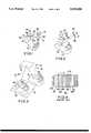
- the actinic bandlies in the range from 700 to 900 nanometers. This is the range of the near infra red. It has been found in studies using black and white infrared photography, that skin and superficial tissues reflect most of the radiation in this range of the spectrum while the blood absorbs much of this radiation. This effect results in photographs of the body in which the veins stand out as dark prominent lines against a light background as illustrated by comparing the arm shown in FIG. 1A irradiated with normal light to the same arm shown in FIG. 1A irradiated with infrared.
- U.S. Pat. No. 5,233,465 to Wheatley et aldiscloses a polymeric multilayered film which reflects wavelengths of light in the infrared while being substantially transparent to wavelengths in the visible spectrum.
- the wavelengthis selected by appropriate selection of the "optical thickness" of the multilayers defined as the physical thickness multiplied by the index of refraction.
- U.S. Pat. No. 3,514,174 to Gansdiscloses a multilayer interference transmittance filter for use in the infrared region of the spectrum.
- Filters and mirrors operating at selective wavelengthscan be custom made and purchased from the Rolyn Optics Co., Covina, Calif.
- U.S. Pat. No. 5,248,874 to Raverdydiscloses an image intensifier tube having a connected brightness curve.
- U.S. Pat. No. 5,204,774 to Owen et aldiscloses a night vision goggle with an achromatic lens assembly.
- FIG. 4is a sectional view of an image intensifier of the prior an showing the incident image on cover 52, transparent conductive electrodes 54, photoconductor 56, light block 58, dielectric rain or 60, liquid crystal alignment films 62, liquid crystal 64, cover 52.
- This inventionis directed toward a method for gaining intravenous access in which an apparatus presents a view of an area of the patient (e.g., arm or leg) wherein veins are clearly delineated. An operator is thereby provided with a visual guide to locate the tip of a hypodermic needle proximal to the site of the vein.
- an apparatuspresents a view of an area of the patient (e.g., arm or leg) wherein veins are clearly delineated.
- An operatoris thereby provided with a visual guide to locate the tip of a hypodermic needle proximal to the site of the vein.
- the apparatusincludes a lamp arranged to illuminate a surface area of the patient with radiation having a selected wavelength that is reflected by all of the surface area except where veins are located.
- the apparatusalso includes a viewing screen through which the user views the illuminated area.
- the viewing screenhas a transmittance filter which transmits radiation having the selected wavelength and effectively blocks all other radiation.
- the venous system viewed through the screenappears as dark lines that clearly stand out enabling the operator to locate the hypodermic needle at the appropriate point of entry.
- the apparatusis attached to a headband worn by the user.
- the apparatusis detachably mounted on a floorstand.
- the optimum wavelengthwill be in the range, between about 400 to 700 nm which is a visible range and further enhancement of the image is not necessary.
- venous systemsrequire the optimum wavelength for differentiating between venous and non venous areas to be in the range between 700 and 900 nm so that images formed therefrom are not visible.
- the apparatusincludes an image intensifier to more clearly delineate the venous structure.
- the apparatusincludes a pair of mirrors positioned such that the operator views an image of the patient's limb with delineated veins along side the actual limb as an aid to positioning the hypodermic needle in the appropriate location.
- An LED located on the needle about 1/4 inch from the tip and emitting light having wavelength readily discernible through the projection system of the apparatusis a further aid to maintaining registration between the tip and point of entry.
- FIG. 1shows the apparatus of this invention arranged to aid in locating an insertion point.
- FIG. 2shows the apparatus of FIG. 1 with the addition of an image intensifier.
- FIG. 3shows an apparatus for presenting a reflected image simultaneously with the real object.
- FIG. 4shows an image intensifier unit of the prior art.
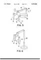
- FIG. 5shows the apparatus arranged for mounting on the head of a user.
- FIG. 6shows the invention mounted on a floor stand.
- FIG. 1shows a lamp 10 with a transmittance filter 12 irradiating a forearm 14 of a patient. Veins 16 in the arm are delineated by selection of the passband wavelength of the transmittance filter 12 which is between 400 and 900 nm.
- the illuminated forearm 14is viewed by an observer 20 through an imaging means 18 including a second transmittance filter 36 mounted on the support surface 32 of a transparent substrate 36.
- Second transmittance filter 38has a passband identical to filter 12.
- the second filter 36eliminates any extraneous light from sources other than the lamp 10. The observer is thereby guided to position the tip of a hypodermic needle 22 against the delineated vein 16.
- FIG. 5shows the lamp 10 with filters 12 and 18 mounted on a headband 60 by cantilever arms 62.
- FIG. 6shows the lamp 10 with filters 12 and 18 mounted on a floor stand 66.
- the lamp 10 with filters 12 and 18are supported on a handle which is hand held.
- Filter 12is a multilayer transmittance filter supported on glass substrate 26 whose support surface 30 is contoured to focus radiation from lamp 10 onto forearm 14.
- Imaging means 18is a multilayer transmittance filter 36 supported on glass substrate 38 whose support surface 32 is contoured to magnify the image of the forearm 14 presented to the observer 20.
- FIG. 2shows the apparatus of FIG. 1 with an image intensifier 34 positioned between the eye of the observer 20 and the imaging means.
- An image intensifier suitable for this applicationis referenced in the specification, "Liquid Crystals, Applications and Uses.”
- a preferred embodiment of this inventionis an image intensifier utilizing a CdS photosensor with large sensitivity between 500 to 530 nm. (close to near infrared) matched with an Argon-ion light source having an output between 500 to 530 nm. (See FIG. 4 for location of photosensor).
- FIG. 3shows yet another embodiment of the invention.
- a light beam from lamp 10passes through narrow bandpass filter 12 and shines on the forearm 14.
- the beam 15 reflected from forearm 14is then reflected by overhead reflectance filter 17 and forms an image in reflectance filter 19
- the operatorcan view side by side and simultaneously the forearm and the reflected image of the forearm 40 and thus be guided to insert the needle in the most appropriate location in the forearm.
- the combination lamp 10 and transmittance filter 12emits radiation in the near infrared.
- the reflectance filters 17 and 19are each a multilayer film on glass which absorbs all radiation other than the wavelength of beam 15.
- a small LED emitting in the near infrared wavelength rangeis located near the tip of the hypodermic needle and can thereby seen by the viewer as he positions the needle at the insertion point.
Landscapes
- Health & Medical Sciences (AREA)
- Life Sciences & Earth Sciences (AREA)
- Surgery (AREA)
- Animal Behavior & Ethology (AREA)
- Pathology (AREA)
- Engineering & Computer Science (AREA)
- Biomedical Technology (AREA)
- Heart & Thoracic Surgery (AREA)
- Medical Informatics (AREA)
- Molecular Biology (AREA)
- Physics & Mathematics (AREA)
- Biophysics (AREA)
- General Health & Medical Sciences (AREA)
- Public Health (AREA)
- Veterinary Medicine (AREA)
- Hematology (AREA)
- Vascular Medicine (AREA)
- Cardiology (AREA)
- Physiology (AREA)
- Infusion, Injection, And Reservoir Apparatuses (AREA)
Abstract
Description
Claims (24)
Priority Applications (2)
| Application Number | Priority Date | Filing Date | Title |
|---|---|---|---|
| US08/315,128US5519208A (en) | 1994-09-29 | 1994-09-29 | Infrared aided method and apparatus for venous examination |
| US08/618,744US5608210A (en) | 1994-09-29 | 1996-03-20 | Infrared aided method and apparatus for venous examination |
Applications Claiming Priority (1)
| Application Number | Priority Date | Filing Date | Title |
|---|---|---|---|
| US08/315,128US5519208A (en) | 1994-09-29 | 1994-09-29 | Infrared aided method and apparatus for venous examination |
Related Child Applications (1)
| Application Number | Title | Priority Date | Filing Date |
|---|---|---|---|
| US08/618,744Continuation-In-PartUS5608210A (en) | 1994-09-29 | 1996-03-20 | Infrared aided method and apparatus for venous examination |
Publications (1)
| Publication Number | Publication Date |
|---|---|
| US5519208Atrue US5519208A (en) | 1996-05-21 |
Family
ID=23223021
Family Applications (1)
| Application Number | Title | Priority Date | Filing Date |
|---|---|---|---|
| US08/315,128Expired - LifetimeUS5519208A (en) | 1994-09-29 | 1994-09-29 | Infrared aided method and apparatus for venous examination |
Country Status (1)
| Country | Link |
|---|---|
| US (1) | US5519208A (en) |
Cited By (80)
| Publication number | Priority date | Publication date | Assignee | Title |
|---|---|---|---|---|
| WO1996039925A1 (en)* | 1995-06-07 | 1996-12-19 | University Of Arkansas | Method and apparatus for detecting electro-magnetic reflection from biological tissue |
| WO1996036273A3 (en)* | 1995-05-16 | 1996-12-19 | Us Air Force | System and method for enhanced visualization of subcutaneous structures |
| US5725480A (en)* | 1996-03-06 | 1998-03-10 | Abbott Laboratories | Non-invasive calibration and categorization of individuals for subsequent non-invasive detection of biological compounds |
| US5871442A (en)* | 1996-09-10 | 1999-02-16 | International Diagnostics Technologies, Inc. | Photonic molecular probe |
| US5947906A (en)* | 1997-11-14 | 1999-09-07 | Dawson, Jr.; Fredric O. | Apparatus for enhanced visual venous examination |
| US5949073A (en)* | 1996-05-07 | 1999-09-07 | Matsushita Electric Industrial Co., Ltd. | Photo detector |
| WO1999048420A1 (en) | 1998-03-23 | 1999-09-30 | Veino-Med Ltd. | Instrument and method for locating and marking a 'hot spot' in a person's body tissue |
| US6178340B1 (en)* | 1998-08-24 | 2001-01-23 | Eduardo Svetliza | Three-dimensional infrared imager for subcutaneous puncture and study of vascular network |
| US6353753B1 (en) | 1998-05-05 | 2002-03-05 | Stephen Thomas Flock | Optical imaging of deep anatomic structures |
| WO2002035994A1 (en)* | 2000-11-02 | 2002-05-10 | Universitat Autonoma De Barcelona | Lighting device for better visualizing superficial blood vessels and skin alterations |
| US6463309B1 (en) | 2000-05-11 | 2002-10-08 | Hanna Ilia | Apparatus and method for locating vessels in a living body |
| US6529617B1 (en)* | 1996-07-29 | 2003-03-04 | Francine J. Prokoski | Method and apparatus for positioning an instrument relative to a patients body during a medical procedure |
| US6556858B1 (en) | 2000-01-19 | 2003-04-29 | Herbert D. Zeman | Diffuse infrared light imaging system |
| US6567682B1 (en)* | 1999-11-16 | 2003-05-20 | Carecord Technologies, Inc. | Apparatus and method for lesion feature identification and characterization |
| US6594510B2 (en) | 1996-09-10 | 2003-07-15 | Xoetronics Llc | Photonic molecular probe |
| US20030210810A1 (en)* | 2002-05-08 | 2003-11-13 | Gee, James W. | Method and apparatus for detecting structures of interest |
| US20040015158A1 (en)* | 2002-07-19 | 2004-01-22 | To-Mu Chen | Transilluminator device |
| EP1421899A1 (en)* | 2001-10-10 | 2004-05-26 | Lifescan, Inc. | Devices for physiological fluid sampling and methods of using the same |
| US20040111030A1 (en)* | 2000-01-19 | 2004-06-10 | Zeman Herbert D. | Imaging system using diffuse infrared light |
| US20040171923A1 (en)* | 2002-12-06 | 2004-09-02 | Kalafut John F. | Devices, systems and methods for improving vessel access |
| US20040215081A1 (en)* | 2003-04-23 | 2004-10-28 | Crane Robert L. | Method for detection and display of extravasation and infiltration of fluids and substances in subdermal or intradermal tissue |
| US20050168980A1 (en)* | 2004-01-30 | 2005-08-04 | Dryden Paul E. | Vein locator |
| US20060020212A1 (en)* | 2004-07-26 | 2006-01-26 | Tianning Xu | Portable vein locating device |
| US20060100523A1 (en)* | 2004-11-08 | 2006-05-11 | Ogle John S | Noninvasive blood vessel location device and method |
| US20060122515A1 (en)* | 2000-01-19 | 2006-06-08 | Luminetx Corporation | Projection of subsurface structure onto an object's surface |
| WO2006069066A2 (en) | 2004-12-22 | 2006-06-29 | Syris Scientific, Llc | System and method for locating and accessing a blood vessel |
| US20060173351A1 (en)* | 2005-01-03 | 2006-08-03 | Ronald Marcotte | System and method for inserting a needle into a blood vessel |
| US20060173360A1 (en)* | 2005-01-07 | 2006-08-03 | Kalafut John F | Method for detection and display of extravasation and infiltration of fluids and substances in subdermal or intradermal tissue |
| US20060241494A1 (en)* | 2004-09-28 | 2006-10-26 | Zila Pharmaceuticals, Inc. | Methods for detecting abnormal epithelial tissue |
| US20070032721A1 (en)* | 2005-05-13 | 2007-02-08 | Crane Robert L | Disposable Light Source Patch for Enhanced Visualization of Subcutaneous Structures |
| US20070038118A1 (en)* | 2005-08-10 | 2007-02-15 | Depue Marshall Thomas | Subcutaneous tissue imager |
| US20070161909A1 (en)* | 2006-01-10 | 2007-07-12 | Ron Goldman | Micro Vein Enhancer |
| US20070158569A1 (en)* | 2000-01-19 | 2007-07-12 | Luminetx Technologies Corporation | Method and Apparatus for Projection of Subsurface Structure onto an Object's Surface |
| US20070161906A1 (en)* | 2000-01-19 | 2007-07-12 | Luminetx Technologies Corporation | Method To Facilitate A Dermatological Procedure |
| US20070276258A1 (en)* | 2006-03-28 | 2007-11-29 | Crane Robert L | Synchronization of Illumination Source and Sensor for Improved Visualization of Subcutaneous Structures |
| CN100352402C (en)* | 2004-06-23 | 2007-12-05 | 游秀珍 | Method and device for positioning body superficial vein or specific tissue using LED light source |
| US20070299425A1 (en)* | 2001-02-12 | 2007-12-27 | Milton Waner | Infrared assisted monitoring of a catheter |
| US20080004525A1 (en)* | 2006-01-10 | 2008-01-03 | Ron Goldman | Three dimensional imaging of veins |
| US20080027317A1 (en)* | 2006-06-29 | 2008-01-31 | Fred Wood | Scanned laser vein contrast enhancer |
| US20080045818A1 (en)* | 2006-06-29 | 2008-02-21 | Fred Wood | Laser vein contrast enhancer |
| US20080162217A1 (en)* | 2004-06-14 | 2008-07-03 | Symphonyrpm, Inc. | Decision object for associating a plurality of business plans |
| US20080177174A1 (en)* | 2006-10-11 | 2008-07-24 | Crane Robert L | Determining Inserted Catheter End Location and Orientation |
| US20080177184A1 (en)* | 2006-06-29 | 2008-07-24 | Ron Goldman | Micro vein enhancer |
| US20080194930A1 (en)* | 2007-02-09 | 2008-08-14 | Harris Melvyn L | Infrared-visible needle |
| US20080203307A1 (en)* | 2007-02-23 | 2008-08-28 | Determan Gary E | Encoded binary liveness detector |
| US20080308753A1 (en)* | 2007-06-15 | 2008-12-18 | Stuba Robert M | Flexible infrared delivery apparatus and method |
| US20090002488A1 (en)* | 2007-06-28 | 2009-01-01 | Vincent Luciano | Automatic alignment of a contrast enhancement system |
| US20090018414A1 (en)* | 2007-03-23 | 2009-01-15 | Mehrdad Toofan | Subcutanous Blood Vessels Imaging System |
| US20100177182A1 (en)* | 2006-04-07 | 2010-07-15 | Novarix Ltd | Vein navigation device |
| US20100274202A1 (en)* | 2009-04-28 | 2010-10-28 | Searete Llc | Systems and methods for automatically inserting a needle into a living subject |
| US20110009751A1 (en)* | 2009-07-13 | 2011-01-13 | Mcguire Jr James E | Subcutaneous access device and related methods |
| US20110021925A1 (en)* | 2006-06-29 | 2011-01-27 | Fred Wood | Mounted vein contrast enchancer |
| US20110112407A1 (en)* | 2006-06-29 | 2011-05-12 | Fred Wood | Multispectral detection and presentation of an object's characteristics |
| US20110118611A1 (en)* | 2006-06-29 | 2011-05-19 | Vincent Luciano | Module mounting mirror endoscopy |
| US20110125028A1 (en)* | 2009-07-22 | 2011-05-26 | Fred Wood | Vein scanner |
| EP2540214A1 (en) | 2011-06-28 | 2013-01-02 | Christie Digital Systems USA, Inc. | Apparatus for detection of catheter location for intravenous access |
| US9061109B2 (en) | 2009-07-22 | 2015-06-23 | Accuvein, Inc. | Vein scanner with user interface |
| US9492117B2 (en) | 2006-01-10 | 2016-11-15 | Accuvein, Inc. | Practitioner-mounted micro vein enhancer |
| US9572530B2 (en) | 2010-03-19 | 2017-02-21 | Quickvein, Inc. | Apparatus and methods for imaging blood vessels |
| US9782079B2 (en) | 2012-08-02 | 2017-10-10 | Accuvein, Inc. | Device for detecting and illuminating the vasculature using an FPGA |
| US9854977B2 (en) | 2006-01-10 | 2018-01-02 | Accuvein, Inc. | Scanned laser vein contrast enhancer using a single laser, and modulation circuitry |
| US9955910B2 (en) | 2005-10-14 | 2018-05-01 | Aranz Healthcare Limited | Method of monitoring a surface feature and apparatus therefor |
| US10013527B2 (en) | 2016-05-02 | 2018-07-03 | Aranz Healthcare Limited | Automatically assessing an anatomical surface feature and securely managing information related to the same |
| US10238294B2 (en) | 2006-06-29 | 2019-03-26 | Accuvein, Inc. | Scanned laser vein contrast enhancer using one laser |
| US10376148B2 (en) | 2012-12-05 | 2019-08-13 | Accuvein, Inc. | System and method for laser imaging and ablation of cancer cells using fluorescence |
| CN111513824A (en)* | 2020-05-01 | 2020-08-11 | 哈尔滨医科大学 | Disposable arterial blood gas puncture patch |
| US10813588B2 (en) | 2006-01-10 | 2020-10-27 | Accuvein, Inc. | Micro vein enhancer |
| US10874302B2 (en) | 2011-11-28 | 2020-12-29 | Aranz Healthcare Limited | Handheld skin measuring or monitoring device |
| CN112534274A (en)* | 2018-07-25 | 2021-03-19 | 株式会社大赛璐 | Measurement system, measurement method, injector, and method for injecting solution containing biomolecule into injection target cell using the injector |
| TWI729813B (en)* | 2020-05-18 | 2021-06-01 | 友達光電股份有限公司 | Blood vessel image forming system |
| US11116407B2 (en) | 2016-11-17 | 2021-09-14 | Aranz Healthcare Limited | Anatomical surface assessment methods, devices and systems |
| US11207024B2 (en)* | 2017-07-12 | 2021-12-28 | Boe Technology Group Co., Ltd. | Vascular imaging apparatus and vascular imaging method |
| US11253198B2 (en) | 2006-01-10 | 2022-02-22 | Accuvein, Inc. | Stand-mounted scanned laser vein contrast enhancer |
| US11278240B2 (en) | 2006-01-10 | 2022-03-22 | Accuvein, Inc. | Trigger-actuated laser vein contrast enhancer |
| US20230190191A1 (en)* | 2020-08-26 | 2023-06-22 | Terumo Kabushiki Kaisha | Blood vessel visualization apparatus, blood vessel puncture system, and observation window member |
| US11903723B2 (en) | 2017-04-04 | 2024-02-20 | Aranz Healthcare Limited | Anatomical surface assessment methods, devices and systems |
| US12039726B2 (en) | 2019-05-20 | 2024-07-16 | Aranz Healthcare Limited | Automated or partially automated anatomical surface assessment methods, devices and systems |
| US12048560B2 (en) | 2006-01-10 | 2024-07-30 | Accuvein, Inc. | Vein scanner configured for single-handed lifting and use |
| US12295744B2 (en) | 2006-01-10 | 2025-05-13 | Accuvein, Inc. | Micro vein enhancer with two lasers and two optical detectors configured for removing surface topology |
| US12408865B2 (en) | 2006-01-10 | 2025-09-09 | Accuvein Inc. | Vein imaging device with differential image resolution at the center and the extremities of the vein image |
Citations (6)
| Publication number | Priority date | Publication date | Assignee | Title |
|---|---|---|---|---|
| US3514174A (en)* | 1965-05-11 | 1970-05-26 | Centre Nat Rech Scient | Infrared interference filters |
| US4817622A (en)* | 1986-07-22 | 1989-04-04 | Carl Pennypacker | Infrared imager for viewing subcutaneous location of vascular structures and method of use |
| US4975581A (en)* | 1989-06-21 | 1990-12-04 | University Of New Mexico | Method of and apparatus for determining the similarity of a biological analyte from a model constructed from known biological fluids |
| US5204774A (en)* | 1991-12-06 | 1993-04-20 | Varo Inc. | Night vision goggle with improved optical system |
| US5233465A (en)* | 1992-05-27 | 1993-08-03 | The Dow Chemical Company | Visibly transparent infrared reflecting film with color masking |
| US5248874A (en)* | 1991-09-20 | 1993-09-28 | Thomson Tubes Electroniques | Image intensifier tube with correction of brightness at the output window |
- 1994
- 1994-09-29USUS08/315,128patent/US5519208A/ennot_activeExpired - Lifetime
Patent Citations (6)
| Publication number | Priority date | Publication date | Assignee | Title |
|---|---|---|---|---|
| US3514174A (en)* | 1965-05-11 | 1970-05-26 | Centre Nat Rech Scient | Infrared interference filters |
| US4817622A (en)* | 1986-07-22 | 1989-04-04 | Carl Pennypacker | Infrared imager for viewing subcutaneous location of vascular structures and method of use |
| US4975581A (en)* | 1989-06-21 | 1990-12-04 | University Of New Mexico | Method of and apparatus for determining the similarity of a biological analyte from a model constructed from known biological fluids |
| US5248874A (en)* | 1991-09-20 | 1993-09-28 | Thomson Tubes Electroniques | Image intensifier tube with correction of brightness at the output window |
| US5204774A (en)* | 1991-12-06 | 1993-04-20 | Varo Inc. | Night vision goggle with improved optical system |
| US5233465A (en)* | 1992-05-27 | 1993-08-03 | The Dow Chemical Company | Visibly transparent infrared reflecting film with color masking |
Cited By (192)
| Publication number | Priority date | Publication date | Assignee | Title |
|---|---|---|---|---|
| WO1996036273A3 (en)* | 1995-05-16 | 1996-12-19 | Us Air Force | System and method for enhanced visualization of subcutaneous structures |
| US6230046B1 (en)* | 1995-05-16 | 2001-05-08 | The United States Of America As Represented By The Secretary Of The Air Force | System and method for enhanced visualization of subcutaneous structures |
| US6032070A (en)* | 1995-06-07 | 2000-02-29 | University Of Arkansas | Method and apparatus for detecting electro-magnetic reflection from biological tissue |
| WO1996039925A1 (en)* | 1995-06-07 | 1996-12-19 | University Of Arkansas | Method and apparatus for detecting electro-magnetic reflection from biological tissue |
| US5725480A (en)* | 1996-03-06 | 1998-03-10 | Abbott Laboratories | Non-invasive calibration and categorization of individuals for subsequent non-invasive detection of biological compounds |
| US5949073A (en)* | 1996-05-07 | 1999-09-07 | Matsushita Electric Industrial Co., Ltd. | Photo detector |
| US6529617B1 (en)* | 1996-07-29 | 2003-03-04 | Francine J. Prokoski | Method and apparatus for positioning an instrument relative to a patients body during a medical procedure |
| US5871442A (en)* | 1996-09-10 | 1999-02-16 | International Diagnostics Technologies, Inc. | Photonic molecular probe |
| US6236870B1 (en) | 1996-09-10 | 2001-05-22 | International Diagnostic Technologies, Inc. | Photonic molecular probe |
| US6594510B2 (en) | 1996-09-10 | 2003-07-15 | Xoetronics Llc | Photonic molecular probe |
| US5947906A (en)* | 1997-11-14 | 1999-09-07 | Dawson, Jr.; Fredric O. | Apparatus for enhanced visual venous examination |
| WO1999048420A1 (en) | 1998-03-23 | 1999-09-30 | Veino-Med Ltd. | Instrument and method for locating and marking a 'hot spot' in a person's body tissue |
| US6464646B1 (en) | 1998-03-23 | 2002-10-15 | Veino-Med Ltd. | Instrument and method for locating and marking a hot spot in a person's body tissue |
| US6353753B1 (en) | 1998-05-05 | 2002-03-05 | Stephen Thomas Flock | Optical imaging of deep anatomic structures |
| US6178340B1 (en)* | 1998-08-24 | 2001-01-23 | Eduardo Svetliza | Three-dimensional infrared imager for subcutaneous puncture and study of vascular network |
| US6567682B1 (en)* | 1999-11-16 | 2003-05-20 | Carecord Technologies, Inc. | Apparatus and method for lesion feature identification and characterization |
| EP1906832A4 (en)* | 2000-01-19 | 2008-04-09 | Luminetx Corp | IMAGING SYSTEM USING DIFFUSED INFRARED LIGHT |
| US6556858B1 (en) | 2000-01-19 | 2003-04-29 | Herbert D. Zeman | Diffuse infrared light imaging system |
| US7239909B2 (en)* | 2000-01-19 | 2007-07-03 | Luminetx Technologies Corp. | Imaging system using diffuse infrared light |
| US8494616B2 (en)* | 2000-01-19 | 2013-07-23 | Christie Medical Holdings, Inc. | Method and apparatus for projection of subsurface structure onto an object's surface |
| US20070158569A1 (en)* | 2000-01-19 | 2007-07-12 | Luminetx Technologies Corporation | Method and Apparatus for Projection of Subsurface Structure onto an Object's Surface |
| US20070161906A1 (en)* | 2000-01-19 | 2007-07-12 | Luminetx Technologies Corporation | Method To Facilitate A Dermatological Procedure |
| US20040111030A1 (en)* | 2000-01-19 | 2004-06-10 | Zeman Herbert D. | Imaging system using diffuse infrared light |
| US20060122515A1 (en)* | 2000-01-19 | 2006-06-08 | Luminetx Corporation | Projection of subsurface structure onto an object's surface |
| US8078263B2 (en)* | 2000-01-19 | 2011-12-13 | Christie Medical Holdings, Inc. | Projection of subsurface structure onto an object's surface |
| US6463309B1 (en) | 2000-05-11 | 2002-10-08 | Hanna Ilia | Apparatus and method for locating vessels in a living body |
| ES2170019A1 (en)* | 2000-11-02 | 2002-07-16 | Univ Barcelona Autonoma | LIGHTING DEVICE FOR BETTER VISUALIZATION OF SURFACE BLOOD VESSELS AND CUTANEOUS ALTERATIONS. |
| WO2002035994A1 (en)* | 2000-11-02 | 2002-05-10 | Universitat Autonoma De Barcelona | Lighting device for better visualizing superficial blood vessels and skin alterations |
| US20070299425A1 (en)* | 2001-02-12 | 2007-12-27 | Milton Waner | Infrared assisted monitoring of a catheter |
| US20050113717A1 (en)* | 2001-10-10 | 2005-05-26 | David Matzinger | Devices for physiological fluid sampling and methds of using the same |
| US6939310B2 (en) | 2001-10-10 | 2005-09-06 | Lifescan, Inc. | Devices for physiological fluid sampling and methods of using the same |
| EP1421899A1 (en)* | 2001-10-10 | 2004-05-26 | Lifescan, Inc. | Devices for physiological fluid sampling and methods of using the same |
| US7070564B2 (en) | 2001-10-10 | 2006-07-04 | Lifescan, Inc. | Devices for physiological fluid sampling and methods of using the same |
| US20030210810A1 (en)* | 2002-05-08 | 2003-11-13 | Gee, James W. | Method and apparatus for detecting structures of interest |
| US7158660B2 (en) | 2002-05-08 | 2007-01-02 | Gee Jr James W | Method and apparatus for detecting structures of interest |
| US20040015158A1 (en)* | 2002-07-19 | 2004-01-22 | To-Mu Chen | Transilluminator device |
| US20040171923A1 (en)* | 2002-12-06 | 2004-09-02 | Kalafut John F. | Devices, systems and methods for improving vessel access |
| US20040215081A1 (en)* | 2003-04-23 | 2004-10-28 | Crane Robert L. | Method for detection and display of extravasation and infiltration of fluids and substances in subdermal or intradermal tissue |
| US20050168980A1 (en)* | 2004-01-30 | 2005-08-04 | Dryden Paul E. | Vein locator |
| US20080162217A1 (en)* | 2004-06-14 | 2008-07-03 | Symphonyrpm, Inc. | Decision object for associating a plurality of business plans |
| CN100352402C (en)* | 2004-06-23 | 2007-12-05 | 游秀珍 | Method and device for positioning body superficial vein or specific tissue using LED light source |
| US20060020212A1 (en)* | 2004-07-26 | 2006-01-26 | Tianning Xu | Portable vein locating device |
| US20060241494A1 (en)* | 2004-09-28 | 2006-10-26 | Zila Pharmaceuticals, Inc. | Methods for detecting abnormal epithelial tissue |
| US20060100523A1 (en)* | 2004-11-08 | 2006-05-11 | Ogle John S | Noninvasive blood vessel location device and method |
| WO2006069066A2 (en) | 2004-12-22 | 2006-06-29 | Syris Scientific, Llc | System and method for locating and accessing a blood vessel |
| US20060173351A1 (en)* | 2005-01-03 | 2006-08-03 | Ronald Marcotte | System and method for inserting a needle into a blood vessel |
| WO2006073869A3 (en)* | 2005-01-03 | 2007-11-15 | Syris Scient Llc | System and method for inserting a needle into a blood vessel |
| EP2412313A1 (en) | 2005-01-03 | 2012-02-01 | Syris Scientific LLC | System and method for inserting a needle into a blood vessel |
| US20060173360A1 (en)* | 2005-01-07 | 2006-08-03 | Kalafut John F | Method for detection and display of extravasation and infiltration of fluids and substances in subdermal or intradermal tissue |
| US20070032721A1 (en)* | 2005-05-13 | 2007-02-08 | Crane Robert L | Disposable Light Source Patch for Enhanced Visualization of Subcutaneous Structures |
| US7925332B2 (en) | 2005-05-13 | 2011-04-12 | Infrared Imaging Systems, Inc. | Disposable light source patch for enhanced visualization of subcutaneous structures |
| US20070038118A1 (en)* | 2005-08-10 | 2007-02-15 | Depue Marshall Thomas | Subcutaneous tissue imager |
| US9955910B2 (en) | 2005-10-14 | 2018-05-01 | Aranz Healthcare Limited | Method of monitoring a surface feature and apparatus therefor |
| US10827970B2 (en) | 2005-10-14 | 2020-11-10 | Aranz Healthcare Limited | Method of monitoring a surface feature and apparatus therefor |
| US8478386B2 (en) | 2006-01-10 | 2013-07-02 | Accuvein Inc. | Practitioner-mounted micro vein enhancer |
| US11191482B2 (en) | 2006-01-10 | 2021-12-07 | Accuvein, Inc. | Scanned laser vein contrast enhancer imaging in an alternating frame mode |
| US12408865B2 (en) | 2006-01-10 | 2025-09-09 | Accuvein Inc. | Vein imaging device with differential image resolution at the center and the extremities of the vein image |
| US12295744B2 (en) | 2006-01-10 | 2025-05-13 | Accuvein, Inc. | Micro vein enhancer with two lasers and two optical detectors configured for removing surface topology |
| US12089951B2 (en) | 2006-01-10 | 2024-09-17 | AccuVeiw, Inc. | Scanned laser vein contrast enhancer with scanning correlated to target distance |
| US12048560B2 (en) | 2006-01-10 | 2024-07-30 | Accuvein, Inc. | Vein scanner configured for single-handed lifting and use |
| US11642080B2 (en) | 2006-01-10 | 2023-05-09 | Accuvein, Inc. | Portable hand-held vein-image-enhancing device |
| US11638558B2 (en) | 2006-01-10 | 2023-05-02 | Accuvein, Inc. | Micro vein enhancer |
| US11484260B2 (en) | 2006-01-10 | 2022-11-01 | Accuvein, Inc. | Patient-mounted micro vein enhancer |
| US11399768B2 (en) | 2006-01-10 | 2022-08-02 | Accuvein, Inc. | Scanned laser vein contrast enhancer utilizing surface topology |
| US11357449B2 (en) | 2006-01-10 | 2022-06-14 | Accuvein, Inc. | Micro vein enhancer for hands-free imaging for a venipuncture procedure |
| US11278240B2 (en) | 2006-01-10 | 2022-03-22 | Accuvein, Inc. | Trigger-actuated laser vein contrast enhancer |
| US11253198B2 (en) | 2006-01-10 | 2022-02-22 | Accuvein, Inc. | Stand-mounted scanned laser vein contrast enhancer |
| US11172880B2 (en) | 2006-01-10 | 2021-11-16 | Accuvein, Inc. | Vein imager with a dual buffer mode of operation |
| US11109806B2 (en) | 2006-01-10 | 2021-09-07 | Accuvein, Inc. | Three dimensional imaging of veins |
| US20070161909A1 (en)* | 2006-01-10 | 2007-07-12 | Ron Goldman | Micro Vein Enhancer |
| US10813588B2 (en) | 2006-01-10 | 2020-10-27 | Accuvein, Inc. | Micro vein enhancer |
| US10617352B2 (en) | 2006-01-10 | 2020-04-14 | Accuvein, Inc. | Patient-mounted micro vein enhancer |
| US10500350B2 (en) | 2006-01-10 | 2019-12-10 | Accuvein, Inc. | Combination vein contrast enhancer and bar code scanning device |
| US10470706B2 (en) | 2006-01-10 | 2019-11-12 | Accuvein, Inc. | Micro vein enhancer for hands-free imaging for a venipuncture procedure |
| US10258748B2 (en) | 2006-01-10 | 2019-04-16 | Accuvein, Inc. | Vein scanner with user interface for controlling imaging parameters |
| US20070161907A1 (en)* | 2006-01-10 | 2007-07-12 | Ron Goldman | Micro vein enhancer |
| US9949688B2 (en) | 2006-01-10 | 2018-04-24 | Accuvein, Inc. | Micro vein enhancer with a dual buffer mode of operation |
| US7983738B2 (en) | 2006-01-10 | 2011-07-19 | Accuvein, Llc | Three dimensional imaging of veins |
| US20110208121A1 (en)* | 2006-01-10 | 2011-08-25 | Ron Goldman | Micro vein enhancer |
| US9854977B2 (en) | 2006-01-10 | 2018-01-02 | Accuvein, Inc. | Scanned laser vein contrast enhancer using a single laser, and modulation circuitry |
| US8073531B2 (en) | 2006-01-10 | 2011-12-06 | Accuvein, Llc | Micro vein enhancer |
| US20080004525A1 (en)* | 2006-01-10 | 2008-01-03 | Ron Goldman | Three dimensional imaging of veins |
| US9788788B2 (en) | 2006-01-10 | 2017-10-17 | AccuVein, Inc | Three dimensional imaging of veins |
| US8150500B2 (en) | 2006-01-10 | 2012-04-03 | Accuvein Inc. | Micro vein enhancer |
| US20120130221A1 (en)* | 2006-01-10 | 2012-05-24 | Ron Goldman | Micro Vein Enhancer |
| US9788787B2 (en) | 2006-01-10 | 2017-10-17 | Accuvein, Inc. | Patient-mounted micro vein enhancer |
| US9492117B2 (en) | 2006-01-10 | 2016-11-15 | Accuvein, Inc. | Practitioner-mounted micro vein enhancer |
| US9125629B2 (en) | 2006-01-10 | 2015-09-08 | Accuvein, Inc. | Vial-mounted micro vein enhancer |
| US8295904B2 (en)* | 2006-01-10 | 2012-10-23 | Accuvein, Llc | Micro vein enhancer |
| US9044207B2 (en) | 2006-01-10 | 2015-06-02 | Accuvein, Inc. | Micro vein enhancer for use with a vial holder |
| US9042966B2 (en) | 2006-01-10 | 2015-05-26 | Accuvein, Inc. | Three dimensional imaging of veins |
| US8818493B2 (en) | 2006-01-10 | 2014-08-26 | Accuvein, Inc. | Three-dimensional imaging of veins |
| US8750970B2 (en) | 2006-01-10 | 2014-06-10 | Accu Vein, Inc. | Micro vein enhancer |
| US8712498B2 (en) | 2006-01-10 | 2014-04-29 | Accuvein Inc. | Micro vein enhancer |
| US20070161908A1 (en)* | 2006-01-10 | 2007-07-12 | Ron Goldman | Micro vein enhancer |
| US20070162094A1 (en)* | 2006-01-10 | 2007-07-12 | Ron Goldman | Micro vein enhancer |
| US8649848B2 (en) | 2006-03-28 | 2014-02-11 | The United States Of America, As Represented By The Secretary Of The Air Force | Synchronization of illumination source and sensor for improved visualization of subcutaneous structures |
| US20070276258A1 (en)* | 2006-03-28 | 2007-11-29 | Crane Robert L | Synchronization of Illumination Source and Sensor for Improved Visualization of Subcutaneous Structures |
| US8199189B2 (en) | 2006-04-07 | 2012-06-12 | Novarix Ltd. | Vein navigation device |
| US20100177182A1 (en)* | 2006-04-07 | 2010-07-15 | Novarix Ltd | Vein navigation device |
| US9345427B2 (en) | 2006-06-29 | 2016-05-24 | Accuvein, Inc. | Method of using a combination vein contrast enhancer and bar code scanning device |
| US8489178B2 (en) | 2006-06-29 | 2013-07-16 | Accuvein Inc. | Enhanced laser vein contrast enhancer with projection of analyzed vein data |
| US8665507B2 (en) | 2006-06-29 | 2014-03-04 | Accuvein, Inc. | Module mounting mirror endoscopy |
| US8706200B2 (en)* | 2006-06-29 | 2014-04-22 | Accuvein, Inc. | Scanned laser vein contrast enhancer |
| US20130123640A1 (en)* | 2006-06-29 | 2013-05-16 | Ron Goldman | Scanned Laser Vein Contrast Enhancer |
| US20080021329A1 (en)* | 2006-06-29 | 2008-01-24 | Fred Wood | Scanned laser vein contrast enhancer |
| US8391960B2 (en)* | 2006-06-29 | 2013-03-05 | Accuvein Inc. | Scanned laser vein contrast enhancer |
| US8380291B2 (en) | 2006-06-29 | 2013-02-19 | Accuvein Inc. | Scanned laser vein contrast enhancer |
| US8838210B2 (en) | 2006-06-29 | 2014-09-16 | AccuView, Inc. | Scanned laser vein contrast enhancer using a single laser |
| US20110112407A1 (en)* | 2006-06-29 | 2011-05-12 | Fred Wood | Multispectral detection and presentation of an object's characteristics |
| US20080287806A1 (en)* | 2006-06-29 | 2008-11-20 | Fred Wood | Scanned laser vein contrast enhancer |
| US11051755B2 (en) | 2006-06-29 | 2021-07-06 | Accuvein, Inc. | Scanned laser vein contrast enhancer using a retro collective mirror |
| US8255040B2 (en) | 2006-06-29 | 2012-08-28 | Accuvein, Llc | Micro vein enhancer |
| US9186063B2 (en) | 2006-06-29 | 2015-11-17 | Accu Vein, Inc. | Scanned laser vein contrast enhancer using one laser for a detection mode and a display mode |
| US9226664B2 (en) | 2006-06-29 | 2016-01-05 | Accuvein, Inc. | Scanned laser vein contrast enhancer using a single laser |
| US11051697B2 (en) | 2006-06-29 | 2021-07-06 | Accuvein, Inc. | Multispectral detection and presentation of an object's characteristics |
| US20080177184A1 (en)* | 2006-06-29 | 2008-07-24 | Ron Goldman | Micro vein enhancer |
| US11523739B2 (en) | 2006-06-29 | 2022-12-13 | Accuvein, Inc. | Multispectral detection and presentation of an object's characteristics |
| US8594770B2 (en) | 2006-06-29 | 2013-11-26 | Accuvein, Inc. | Multispectral detection and presentation of an object's characteristics |
| US8244333B2 (en) | 2006-06-29 | 2012-08-14 | Accuvein, Llc | Scanned laser vein contrast enhancer |
| US12193838B2 (en) | 2006-06-29 | 2025-01-14 | Accuvein, Inc. | Scanned laser vein contrast enhancer with reduced laser intensity during scan line reversals |
| US20080045818A1 (en)* | 2006-06-29 | 2008-02-21 | Fred Wood | Laser vein contrast enhancer |
| US20080045841A1 (en)* | 2006-06-29 | 2008-02-21 | Fred Wood | Scanned laser vein contrast enhancer |
| US20080027317A1 (en)* | 2006-06-29 | 2008-01-31 | Fred Wood | Scanned laser vein contrast enhancer |
| US10357200B2 (en)* | 2006-06-29 | 2019-07-23 | Accuvein, Inc. | Scanning laser vein contrast enhancer having releasable handle and scan head |
| US20110118611A1 (en)* | 2006-06-29 | 2011-05-19 | Vincent Luciano | Module mounting mirror endoscopy |
| US10238294B2 (en) | 2006-06-29 | 2019-03-26 | Accuvein, Inc. | Scanned laser vein contrast enhancer using one laser |
| US20110021925A1 (en)* | 2006-06-29 | 2011-01-27 | Fred Wood | Mounted vein contrast enchancer |
| US7917193B2 (en) | 2006-10-11 | 2011-03-29 | The United States Of America As Represented By The Secretary Of The Air Force | Determining inserted catheter end location and orientation |
| US20110270080A1 (en)* | 2006-10-11 | 2011-11-03 | Crane Robert L | Determining inserted catheter end location and orientation |
| US8548572B2 (en)* | 2006-10-11 | 2013-10-01 | The United States Of America, As Represented By The Secretary Of The Air Force | Determining inserted catheter end location and orientation |
| US20080177174A1 (en)* | 2006-10-11 | 2008-07-24 | Crane Robert L | Determining Inserted Catheter End Location and Orientation |
| US20080194930A1 (en)* | 2007-02-09 | 2008-08-14 | Harris Melvyn L | Infrared-visible needle |
| US20080203307A1 (en)* | 2007-02-23 | 2008-08-28 | Determan Gary E | Encoded binary liveness detector |
| US20090018414A1 (en)* | 2007-03-23 | 2009-01-15 | Mehrdad Toofan | Subcutanous Blood Vessels Imaging System |
| US7977658B2 (en) | 2007-06-15 | 2011-07-12 | Precision Endoscopic Technologies | Flexible infrared delivery apparatus and method |
| WO2008156623A1 (en)* | 2007-06-15 | 2008-12-24 | Max Endoscopy, Inc. | Flexible infrared delivery apparatus and method |
| US20080308753A1 (en)* | 2007-06-15 | 2008-12-18 | Stuba Robert M | Flexible infrared delivery apparatus and method |
| US11847768B2 (en) | 2007-06-28 | 2023-12-19 | Accuvein Inc. | Automatic alignment of a contrast enhancement system |
| US9760982B2 (en) | 2007-06-28 | 2017-09-12 | Accuvein, Inc. | Automatic alignment of a contrast enhancement system |
| US10580119B2 (en) | 2007-06-28 | 2020-03-03 | Accuvein, Inc. | Automatic alignment of a contrast enhancement system |
| US10096096B2 (en) | 2007-06-28 | 2018-10-09 | Accuvein, Inc. | Automatic alignment of a contrast enhancement system |
| US10713766B2 (en) | 2007-06-28 | 2020-07-14 | Accuvein, Inc. | Automatic alignment of a contrast enhancement system |
| US20090002488A1 (en)* | 2007-06-28 | 2009-01-01 | Vincent Luciano | Automatic alignment of a contrast enhancement system |
| US8730321B2 (en) | 2007-06-28 | 2014-05-20 | Accuvein, Inc. | Automatic alignment of a contrast enhancement system |
| US11132774B2 (en) | 2007-06-28 | 2021-09-28 | Accuvein, Inc. | Automatic alignment of a contrast enhancement system |
| US9430819B2 (en) | 2007-06-28 | 2016-08-30 | Accuvein, Inc. | Automatic alignment of a contrast enhancement system |
| US8308741B2 (en) | 2009-04-28 | 2012-11-13 | The Invention Science Fund I, Llc | Systems and methods for automatically inserting a needle into a living subject |
| US20100274202A1 (en)* | 2009-04-28 | 2010-10-28 | Searete Llc | Systems and methods for automatically inserting a needle into a living subject |
| US8498694B2 (en) | 2009-07-13 | 2013-07-30 | Entrotech, Inc. | Subcutaneous access device and related methods |
| US20110009751A1 (en)* | 2009-07-13 | 2011-01-13 | Mcguire Jr James E | Subcutaneous access device and related methods |
| US10518046B2 (en) | 2009-07-22 | 2019-12-31 | Accuvein, Inc. | Vein scanner with user interface |
| US9061109B2 (en) | 2009-07-22 | 2015-06-23 | Accuvein, Inc. | Vein scanner with user interface |
| US12426835B2 (en) | 2009-07-22 | 2025-09-30 | Accuvein, Inc. | Vein scanner with housing configured for single-handed lifting and use |
| US9789267B2 (en) | 2009-07-22 | 2017-10-17 | Accuvein, Inc. | Vein scanner with user interface |
| US20110125028A1 (en)* | 2009-07-22 | 2011-05-26 | Fred Wood | Vein scanner |
| US12433535B2 (en) | 2009-07-22 | 2025-10-07 | Accuvein, Inc. | Method and system for optimizing a projected vein image and identifying vein locations using vein scanner |
| US11826166B2 (en) | 2009-07-22 | 2023-11-28 | Accuvein, Inc. | Vein scanner with housing configured for single-handed lifting and use |
| USD999380S1 (en) | 2009-07-22 | 2023-09-19 | Accuvein, Inc. | Vein imager and cradle in combination |
| US8463364B2 (en) | 2009-07-22 | 2013-06-11 | Accuvein Inc. | Vein scanner |
| US12318219B2 (en) | 2009-07-22 | 2025-06-03 | Accuvein Inc. | Vein scanner with limited photodiode FOV moving synchronously with scanned laser light |
| US9572530B2 (en) | 2010-03-19 | 2017-02-21 | Quickvein, Inc. | Apparatus and methods for imaging blood vessels |
| US11191481B2 (en) | 2010-03-19 | 2021-12-07 | Quickvein, Inc. | Apparatus and methods for imaging blood vessels |
| USD998152S1 (en) | 2010-07-22 | 2023-09-05 | Accuvein, Inc. | Vein imager cradle |
| USD999379S1 (en) | 2010-07-22 | 2023-09-19 | Accuvein, Inc. | Vein imager and cradle in combination |
| EP2540214A1 (en) | 2011-06-28 | 2013-01-02 | Christie Digital Systems USA, Inc. | Apparatus for detection of catheter location for intravenous access |
| US9247906B2 (en) | 2011-06-28 | 2016-02-02 | Christie Digital Systems Usa, Inc. | Method and apparatus for detection of catheter location for intravenous access |
| US11850025B2 (en) | 2011-11-28 | 2023-12-26 | Aranz Healthcare Limited | Handheld skin measuring or monitoring device |
| US10874302B2 (en) | 2011-11-28 | 2020-12-29 | Aranz Healthcare Limited | Handheld skin measuring or monitoring device |
| US11510617B2 (en) | 2012-08-02 | 2022-11-29 | Accuvein, Inc. | Device for detecting and illuminating the vasculature using an FPGA |
| US9782079B2 (en) | 2012-08-02 | 2017-10-10 | Accuvein, Inc. | Device for detecting and illuminating the vasculature using an FPGA |
| US10568518B2 (en) | 2012-08-02 | 2020-02-25 | Accuvein, Inc. | Device for detecting and illuminating the vasculature using an FPGA |
| US11439307B2 (en) | 2012-12-05 | 2022-09-13 | Accuvein, Inc. | Method for detecting fluorescence and ablating cancer cells of a target surgical area |
| US10517483B2 (en) | 2012-12-05 | 2019-12-31 | Accuvein, Inc. | System for detecting fluorescence and projecting a representative image |
| US10376147B2 (en) | 2012-12-05 | 2019-08-13 | AccuVeiw, Inc. | System and method for multi-color laser imaging and ablation of cancer cells using fluorescence |
| US10376148B2 (en) | 2012-12-05 | 2019-08-13 | Accuvein, Inc. | System and method for laser imaging and ablation of cancer cells using fluorescence |
| US10777317B2 (en) | 2016-05-02 | 2020-09-15 | Aranz Healthcare Limited | Automatically assessing an anatomical surface feature and securely managing information related to the same |
| US11250945B2 (en) | 2016-05-02 | 2022-02-15 | Aranz Healthcare Limited | Automatically assessing an anatomical surface feature and securely managing information related to the same |
| US11923073B2 (en) | 2016-05-02 | 2024-03-05 | Aranz Healthcare Limited | Automatically assessing an anatomical surface feature and securely managing information related to the same |
| US10013527B2 (en) | 2016-05-02 | 2018-07-03 | Aranz Healthcare Limited | Automatically assessing an anatomical surface feature and securely managing information related to the same |
| US12268472B2 (en) | 2016-11-17 | 2025-04-08 | ARANZ Medical Limited | Anatomical surface assessment methods, devices and systems |
| US11116407B2 (en) | 2016-11-17 | 2021-09-14 | Aranz Healthcare Limited | Anatomical surface assessment methods, devices and systems |
| US12279883B2 (en) | 2017-04-04 | 2025-04-22 | ARANZ Medical Limited | Anatomical surface assessment methods, devices and systems |
| US11903723B2 (en) | 2017-04-04 | 2024-02-20 | Aranz Healthcare Limited | Anatomical surface assessment methods, devices and systems |
| US11207024B2 (en)* | 2017-07-12 | 2021-12-28 | Boe Technology Group Co., Ltd. | Vascular imaging apparatus and vascular imaging method |
| CN112534274A (en)* | 2018-07-25 | 2021-03-19 | 株式会社大赛璐 | Measurement system, measurement method, injector, and method for injecting solution containing biomolecule into injection target cell using the injector |
| CN112534274B (en)* | 2018-07-25 | 2023-10-13 | 株式会社大赛璐 | Measurement system, measurement method, injector, and injection method for injecting solution containing biological molecule into cell to be injected using the injector |
| US12039726B2 (en) | 2019-05-20 | 2024-07-16 | Aranz Healthcare Limited | Automated or partially automated anatomical surface assessment methods, devices and systems |
| CN111513824A (en)* | 2020-05-01 | 2020-08-11 | 哈尔滨医科大学 | Disposable arterial blood gas puncture patch |
| CN111513824B (en)* | 2020-05-01 | 2021-03-19 | 哈尔滨医科大学 | Disposable arterial blood gas puncture patch |
| TWI729813B (en)* | 2020-05-18 | 2021-06-01 | 友達光電股份有限公司 | Blood vessel image forming system |
| US20230190191A1 (en)* | 2020-08-26 | 2023-06-22 | Terumo Kabushiki Kaisha | Blood vessel visualization apparatus, blood vessel puncture system, and observation window member |
Similar Documents
| Publication | Publication Date | Title |
|---|---|---|
| US5519208A (en) | Infrared aided method and apparatus for venous examination | |
| US11609425B2 (en) | Augmented reality glasses with auto coregistration of invisible field on visible reality | |
| US5947906A (en) | Apparatus for enhanced visual venous examination | |
| US5608210A (en) | Infrared aided method and apparatus for venous examination | |
| US7286287B1 (en) | Visual aid in the form of telescopic spectacles with an automated focusing device | |
| US6178340B1 (en) | Three-dimensional infrared imager for subcutaneous puncture and study of vascular network | |
| US6089716A (en) | Electro-optic binocular indirect ophthalmoscope for stereoscopic observation of retina | |
| US5070883A (en) | Eye movement analyzing device utilizing pupil center-of-gravity data | |
| US7922327B2 (en) | Apparatus and method for illuminating and viewing the anterior segment of an eye of a patient | |
| US6305804B1 (en) | Non-invasive measurement of blood component using retinal imaging | |
| US4533222A (en) | Eye examining instrument | |
| US5841509A (en) | Electro-optic binocular indirect ophthalmoscope | |
| US20050281445A1 (en) | System and method for locating and accessing a blood vessel | |
| WO1996036273A3 (en) | System and method for enhanced visualization of subcutaneous structures | |
| EP1278452A1 (en) | Non-invasive measurement of blood components using retinal imaging | |
| US4737104A (en) | Method of eye protection using hand-held light filter | |
| Zeman et al. | Design of a clinical vein contrast enhancing projector | |
| Zeman et al. | Optimization of subcutaneous vein contrast enhancement | |
| WO2020014999A1 (en) | Invisible light display device and optical guidance system for operations | |
| US3424518A (en) | Device for projecting and observing images of external objects upon the retina of the eye | |
| JPH0638929A (en) | Ophthalmic equipment | |
| JPS6290133A (en) | Fundus camera with fixation target | |
| JP2002017680A (en) | Eyeball protection cap and eyeball microscope provided with the same | |
| JP2941847B2 (en) | Eye gaze detection device | |
| JPH041620B2 (en) |
Legal Events
| Date | Code | Title | Description |
|---|---|---|---|
| AS | Assignment | Owner name:ESPARZA, JOEL Free format text:ASSIGNMENT OF ASSIGNORS INTEREST;ASSIGNOR:SMITH, ROBERT SAMUEL;REEL/FRAME:007242/0740 Effective date:19940929 | |
| STCF | Information on status: patent grant | Free format text:PATENTED CASE | |
| FEPP | Fee payment procedure | Free format text:PAT HLDR NO LONGER CLAIMS SMALL ENT STAT AS INDIV INVENTOR (ORIGINAL EVENT CODE: LSM1); ENTITY STATUS OF PATENT OWNER: SMALL ENTITY | |
| REFU | Refund | Free format text:REFUND - PAYMENT OF MAINTENANCE FEE, 4TH YEAR, LARGE ENTITY (ORIGINAL EVENT CODE: R183); ENTITY STATUS OF PATENT OWNER: SMALL ENTITY | |
| FEPP | Fee payment procedure | Free format text:PAT HOLDER CLAIMS SMALL ENTITY STATUS - SMALL BUSINESS (ORIGINAL EVENT CODE: SM02); ENTITY STATUS OF PATENT OWNER: SMALL ENTITY | |
| REMI | Maintenance fee reminder mailed | ||
| FPAY | Fee payment | Year of fee payment:4 | |
| SULP | Surcharge for late payment | ||
| FPAY | Fee payment | Year of fee payment:8 | |
| AS | Assignment | Owner name:INFRAMED IMAGING, LLC, MONTANA Free format text:ASSIGNMENT OF ASSIGNORS INTEREST;ASSIGNOR:ESPARZA, JOEL;REEL/FRAME:015942/0323 Effective date:19940929 | |
| CC | Certificate of correction | ||
| REMI | Maintenance fee reminder mailed | ||
| FPAY | Fee payment | Year of fee payment:12 | |
| SULP | Surcharge for late payment | Year of fee payment:11 |