US5486182A - Polyp retrieval assembly with separable web member - Google Patents
Polyp retrieval assembly with separable web memberDownload PDFInfo
- Publication number
- US5486182A US5486182AUS08/213,196US21319694AUS5486182AUS 5486182 AUS5486182 AUS 5486182AUS 21319694 AUS21319694 AUS 21319694AUS 5486182 AUS5486182 AUS 5486182A
- Authority
- US
- United States
- Prior art keywords
- loop
- web member
- cauterization
- sheath
- loop means
- Prior art date
- Legal status (The legal status is an assumption and is not a legal conclusion. Google has not performed a legal analysis and makes no representation as to the accuracy of the status listed.)
- Expired - Lifetime
Links
Images


Classifications
- A—HUMAN NECESSITIES
- A61—MEDICAL OR VETERINARY SCIENCE; HYGIENE
- A61B—DIAGNOSIS; SURGERY; IDENTIFICATION
- A61B18/00—Surgical instruments, devices or methods for transferring non-mechanical forms of energy to or from the body
- A61B18/04—Surgical instruments, devices or methods for transferring non-mechanical forms of energy to or from the body by heating
- A61B18/12—Surgical instruments, devices or methods for transferring non-mechanical forms of energy to or from the body by heating by passing a current through the tissue to be heated, e.g. high-frequency current
- A61B18/14—Probes or electrodes therefor
- A—HUMAN NECESSITIES
- A61—MEDICAL OR VETERINARY SCIENCE; HYGIENE
- A61B—DIAGNOSIS; SURGERY; IDENTIFICATION
- A61B17/00—Surgical instruments, devices or methods
- A61B17/32—Surgical cutting instruments
- A61B17/3205—Excision instruments
- A61B17/32056—Surgical snare instruments
- A—HUMAN NECESSITIES
- A61—MEDICAL OR VETERINARY SCIENCE; HYGIENE
- A61B—DIAGNOSIS; SURGERY; IDENTIFICATION
- A61B10/00—Instruments for taking body samples for diagnostic purposes; Other methods or instruments for diagnosis, e.g. for vaccination diagnosis, sex determination or ovulation-period determination; Throat striking implements
- A61B10/02—Instruments for taking cell samples or for biopsy
- A—HUMAN NECESSITIES
- A61—MEDICAL OR VETERINARY SCIENCE; HYGIENE
- A61B—DIAGNOSIS; SURGERY; IDENTIFICATION
- A61B17/00—Surgical instruments, devices or methods
- A61B17/22—Implements for squeezing-off ulcers or the like on inner organs of the body; Implements for scraping-out cavities of body organs, e.g. bones; for invasive removal or destruction of calculus using mechanical vibrations; for removing obstructions in blood vessels, not otherwise provided for
- A61B17/22031—Gripping instruments, e.g. forceps, for removing or smashing calculi
- A—HUMAN NECESSITIES
- A61—MEDICAL OR VETERINARY SCIENCE; HYGIENE
- A61B—DIAGNOSIS; SURGERY; IDENTIFICATION
- A61B18/00—Surgical instruments, devices or methods for transferring non-mechanical forms of energy to or from the body
- A61B18/04—Surgical instruments, devices or methods for transferring non-mechanical forms of energy to or from the body by heating
- A61B18/12—Surgical instruments, devices or methods for transferring non-mechanical forms of energy to or from the body by heating by passing a current through the tissue to be heated, e.g. high-frequency current
- A61B18/14—Probes or electrodes therefor
- A61B18/1492—Probes or electrodes therefor having a flexible, catheter-like structure, e.g. for heart ablation
- A—HUMAN NECESSITIES
- A61—MEDICAL OR VETERINARY SCIENCE; HYGIENE
- A61B—DIAGNOSIS; SURGERY; IDENTIFICATION
- A61B17/00—Surgical instruments, devices or methods
- A61B17/00234—Surgical instruments, devices or methods for minimally invasive surgery
- A61B2017/00287—Bags for minimally invasive surgery
- A—HUMAN NECESSITIES
- A61—MEDICAL OR VETERINARY SCIENCE; HYGIENE
- A61B—DIAGNOSIS; SURGERY; IDENTIFICATION
- A61B18/00—Surgical instruments, devices or methods for transferring non-mechanical forms of energy to or from the body
- A61B18/04—Surgical instruments, devices or methods for transferring non-mechanical forms of energy to or from the body by heating
- A61B18/12—Surgical instruments, devices or methods for transferring non-mechanical forms of energy to or from the body by heating by passing a current through the tissue to be heated, e.g. high-frequency current
- A61B18/14—Probes or electrodes therefor
- A61B2018/1405—Electrodes having a specific shape
- A61B2018/1407—Loop
- A61B2018/141—Snare
Definitions
- This inventionrelates to a surgical instrument assembly for use in retrieving objects from internal body cavities.
- This inventionalso relates, more specifically, to a surgical instrument assembly for use in snare cauterization operations.
- This inventionalso relates to a related method for retrieving objects from internal body cavities and more particularly to a method for capturing and/or retrieving polyps and other clumps of organic tissue which have been severed from a patient's internal organs via a snare cauterization technique.
- an endoscopeis inserted into an internal cavity of a patient, e.g., into the colon, and is used to locate abnormal tissue growths such as polyps in the internal cavity.
- a wire extending through a tube in the biopsy channel of the endoscopeis slid in the distal direction so that a cauterization loop connected to the wire is ejected from the distal end of the tube and the endoscope.
- the loop and the endoscopeare manipulated from outside of the patient to pass the loop over the polyp or growth.
- the wireis then withdrawn in the proximal direction to tighten the loop around a base region or neck of the polyp.
- an electrical currentis conducted through the loop via the wire.
- electrical currentis transmittted through the narrowed organic tissues and thereby generates therein heat sufficiently great to cut and cauterize.
- a polypis severed by such a snare cauterization technique, it frequently becomes difficult to capture the polyp and retrieve it from the patient.
- the cauterization loopis used in an effort to ensnare the polyp.
- Other capture techniquesinvolve the use of forceps or the application of suction.
- forcepsthe snare cauterization tube is removed from the biopsy channel of the endoscope and replaced with the forceps.
- suctiona vacuum is applied via a suction channel of the endoscope.
- the polypfrequently escapes from the capturing instrumentality and falls away into the colon (or other cavity). Especially in cases where the polyp is large, the effort and time expended in retrieving the severed polyp may rival or even exceed the effort and time required to locate and sever the polyp. In extreme cases, the endoscope must be removed without the polyp and the patient given an enema in an attempt to flush out the polyp from the colon.
- An object of the present inventionis to provide an improved method for the removal of portions of internal body organs or other objects from patients.
- a more specific object of the present inventionis to provide an improved method for the performance of snare cauterization.
- Another object of the present inventionis to provide a snare cauterization technique wherein the capture and retrieval of severed polyps is facilitated.
- Another, more particular, object of the present inventionis to provide a snare cauterization technique wherein trauma to the patient and time in surgery are reduced.
- a further object of the present inventionis to provide an instrument assembly for use in removing portions of body organs or other objects from patients.
- object of the present inventionis to provide such an instrument assembly which facilitates the capture and retrieval of severed polyps and other clumps of severed body tissues from the internal cavities of patients.
- Another particular object of the present inventionis to provide such an instrument assembly which is simple to manufacture and therefore inexpensive.
- a further particular object of the present inventionis to provide such an instrument assembly which is easy to use.
- An additional particular object of the present inventionis to provide such an instrument assembly which is disposable.
- Such an instrument assemblyrequires no lengthy sterilization procedure and reduces the spread of infectitous diseases such as AIDS.
- An endoscopic surgical instrument for use in snare cauterization operationscomprises, in accordance with the present invention, a tubular sheath member, an alternately expandable and contractible cauterization loop, an electrically conductive wire operatively connected to the loop, the wire being slidable longitudinally through the sheath, and a flexible web member connected to the loop essentially around a circumference thereof to form a capture pocket.
- the loopdefines a mouth opening of the pocket and is attached to the loop in a manner so as to expose the loop to enable effective cauterization of organic tissues by the loop.
- the web memberis removably attached to the loop along a major portion thereof to enable at least a substantial separation of the web member from the loop upon a proximally directed stroke of the wire at the termination of a cauterization operation.
- the surgical instrumentfurther comprises a purse string attached to the web member along a ring shaped locus proximately to the mouth opening.
- the purse stringis preferably attached at a proximal end to the wire. More preferably, the purse string is attached to the wire proximate to the distal end thereof.
- the purse stringit is also possible for the purse string to extend in a proximal direction entirely through the sheath member to the proximal end thereof.
- the web membermay be attached by adhesive to the loop, either at a plurality of discrete points or along a continuous length of the loop.
- the web memberis removably attached to the loop along that radially outwardly facing surface area.
- the web memberis preferably in the form of a net, but may alternatively take the form of a continuous film of polymeric material.
- the web memberis permanently attached to the loop only at the distal tip thereof.
- This permanent attachmentserves in part to facilitate an assembly procedure wherein the snare with the substantially (but not completely) separable pocket is inserted into the sheath from the proximal end thereof.
- a method for removing a selected portion of internal body tissues of a patientutilizes, in accordance with the present invention, a conductive cauterization loop to which a flexible web member is removably connected to define an expandable pocket.
- the loopdefines a mouth opening of the pocket, while the web member is attached to the loop in a manner so as to expose the loop to enable effective cauterization of organic tissues by the loop.
- the methodcomprises the step of (a) ejecting the loop from a distal end of a tubular sheath member, (b) upon ejection of the loop, expanding the loop and the web member from a collapsed configuration to an at least partially opened configuration, and (c) pain, sing the expanded loop over the selected internal body tissues to be removed, so that the web member substantially surrounds the selected internal body tissues.
- the loopis drawn back into the distal end of the tubular sheath member, thereby closing the loop around a base region of the selected internal body tissues, while the web member is maintained surrounding the selected internal body tissues.
- the web memberis detached by being peeled away from the loop at a distal edge of the sheath member.
- the drawing of the loop back into the distal end of the sheath memberdraws the capture pocket into contact with a distal edge of the sheath member. That contact forces the capture pocket or web member from the loop.
- the loopis pulled completely into the sheath member upon the termination of a cauterization operation.
- the web member and the captured internal body tissuesremain outside of the sheath member.
- the cauterization snare assembly in accordance with the present invention and the associated method of removing a piece of organic tissues (such as a polyp)facilitates use of a cauterization snare assembly as described and claimed in U.S. Pat. No. 5,201,740 and U.S. Pat. No. 5,190,542.
- a cut by a snare in accordance with the present inventionis cleaner and more effective than in those cases where a capture pocket is permanently attached to the cauterization loop and must be partially pulled into the distal end of a sheath to complete a cauterization and capture operation, as described in U.S. Pat. Nos. 5,201,740 and 5,190,542.
- the present inventionprovides an improved method for the removal of portions of internal body organs from patients via snare cauterization.
- An instrument assembly in accordance with the present inventionis simple to use. Accordingly, trauma to the patient and time in surgery are reduced. More specifically, time under anaesthesia with the accompanying side effects is reduced. Concomitantly, the expense of hospitalization is decreased.
- a method for assembling a cauterization snare assemblycomprises, in accordance with the present invention, the steps of providing a cauterization loop having a capture pocket attached thereto so as to be at least substantially removable from the loop, providing a sheath having a flared distal end, inserting the loop with the capture pocket into the sheath through the flared distal end, and upon disposition of the loop with the capture pocket in a narrowed section of the sheath, severing the flared distal end from the narrowed section.
- a filmis wrapped around the loop and the capture pocket. Subsequently, the loop and the capture pocket, with the film wrapped therearound, are inserted into a distal end of a sheath.
- the filmmay be bonded to an inner surface of the sheath.
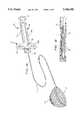
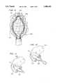
- FIG. 1Ais a schematic perspective view of a snare cauterization instrument assembly, showing a cauterization loop in an ejected, use configuration.
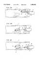
- FIG. 1Bis a schematic longitudinal cross-sectional view of a distal end of the cauterization instrument assembly of FIG. 1A, showing the cauterization loop in a withdrawn or retracted storage configuration inside the distal end of a tubular member of the instrument assembly.
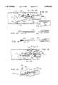
- FIG. 2Ais a schematic partial cross sectional view of a patient's colon with a polyp, showing the snare cauterization instrument assembly of FIG. 1A inserted in the biopsy channel of an endoscope which is itself inserted into the patient's colon, and further showing the instrument assembly in an initial stage of a snare cauterization procedure.
- FIG. 2Bis a schematic partial cross sectional view similar to FIG. 2A, showing a loop of the snare cauterization instrument assembly of FIG. 1A being passed around the polyp of FIG. 2A.
- FIG. 2Cis a schematic partial cross sectional view similar to FIGS. 2A-2B; showing the loop of the snare cauterization instrument assembly of FIG. 1A completely passed around the polyp of FIG. 2A.
- FIG. 2Dis a schematic partial cross sectional view similar to FIGS. 2A-2C, showing the loop of the snare cauterization instrument assembly of FIG. 1A being tightened around a base or neck of the polyp.
- FIG. 2Eis a schematic partial cross sectional view similar to FIGS. 2A-2D, showing the loop of the snare cauterization instrument assembly of FIG. 1A in an electrically energized state for burning through the base or neck of the polyp.
- FIG. 2Fis a schematic partial cross sectional view similar to FIGS. 2A-2E, showing the polyp severed from the colon wall and captured with the snare cauterization instrument assembly of FIG. 1A.
- FIG. 2Gis a schematic partial cross sectional view similar to FIGS. 2A-2G, showing the snare cauterization instrument assembly of FIG. 1A together with the captured polyp drawn towards the distal end of the endoscope.
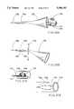
- FIGS. 3-6are schematic partial side perspective views, showing different specific embodiments of a snare cauterization instrument assembly.
- FIG. 7is a schematic side elevational view, on an enlarged scale, of another embodiment of a snare cauterization instrument assembly, showing a pocket-defining web member on an auxiliary loop.
- FIG. 8is a schematic perspective view, also on an enlarged scale, of a modified snare cauterization instrument assembly, showing an auxiliary loop attached at three points to a cauterization loop.
- FIG. 9is a schematic top view of another modified snare cauterization instrument assembly, showing an auxiliary loop attached at one point to a cauterization loop.
- FIG. 10is a schematic partial perspective view, on an enlarged scale, of an additional snare cauterization instrument assembly.
- FIG. 11is a schematic partial perspective view, on an enlarged scale, of yet a further snare cauterization instrument assembly.
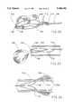
- FIG. 12is a schematic partial cross sectional view of a patient's colon with a polyp, showing a snare cauterization instrument assembly inserted through the biopsy channel of an endoscope which is itself inserted into the patient's colon, and further showing an instrument for depositing color markers on organic tissues.
- FIG. 13is partially a schematic partial side elevational view and partially a block diagram of another color deposition instrument alternatively utilizable with the endoscopic snare cauterization instrument assembly of FIG. 12.
- FIG. 14is partially a schematic partial side elevational view and partially a block diagram of yet another color deposition instrument alternatively utilizable with the endoscopic snare cauterization instrument assembly of FIG. 12.
- FIG. 15is a schematic partial cross sectional view of a patient's colon with a polyp, showing a snare cauterization instrument assembly inserted through an alternately collapsible and expandable biopsy channel of an endoscope assembly which is itself inserted into the patient's colon, and further showing an instrument for depositing color markers on organic tissues.
- FIG. 16is a schematic perspective view of a distal end portion of an endoscopic cauterization instrument assembly, showing a cauterization loop of the assembly in use to cauterize and sever a polyp in a patient's colon.
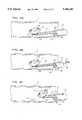
- FIG. 17is a schematic side elevational view, on an enlarged scale, of a cauterization snare assembly in accordance with the present invention.
- FIG. 18is a schematic cross-sectional view, on an enlarged scale, taken along line XVIII--XVIII in FIG. 17.
- FIGS. 19A-19Care schematic side elevational views of the cauterization snare assembly of FIGS. 17 and 18, showing successive steps in the use of the assembly of FIGS. 17 and 18, in accordance with the present invention.
- FIGS. 20A and 20Bare schematic side elevational views of a snare assembly, showing successive steps in a manufacturing process in accordance with the present invention.
- FIGS. 21A and 21Bare schematic perspective views of a snare assembly, showing successive steps in another manufacturing process in accordance with the present invention.
- FIG. 22is a schematic side elevational view, similar to FIG. 17, showing a modification of the snare assembly of that drawing Figure.
- FIG. 23is partially a cross-sectional view of a sheath and partially a side elevational view of the modified snare assembly of FIG. 22, at the termination of a polypectomy procedure in accordance with the present invention.
- FIG. 24is partially a cross-sectional view of a modified sheath and partially a side elevational view of the snare assembly of FIGS. 17, 18, and 19A-19C, showing an early stage in a retraction of loop 704 and pocket 708 into the sheath.
- FIGS. 25A-25Care schematic cross-sectional views of a cauterization loop holder, showing successive steps in a manufacturing process.
- FIG. 26is a schematic side elevational view of a modified cauterization snare with capture pocket, in accordance with the present invention, showing a cauterization loop and a pocket completely extended from a tubular sheath.
- FIG. 27is a schematic side elevation view of the modified cauterization snare and capture pocket of FIG. 26, showing partial retraction of the loop into the sheath and a concomitant dissociation of the capture pocket from the loop.
- FIG. 28is a schematic perspective view of another cauterization loop with a capture pocket, in accordance with the present invention.
- FIGS. 29A-29Care schematic side perspective views, partially in cross-section, of another modified cauterization snare and capture pocket in accordance with the present invention, showing three steps in the use of the device.
- FIG. 30is a schematic side elevational view, partially in cross-section, showing the cauterization snare and capture pocket of FIGS. 29A-29C in a retracted pre-firing insertion configuration.
- FIG. 31is a schematic side elevation view showing a stage in the assembly of the cauterization snare and capture pocket of FIGS. 29A-29C and 30.
- a snare cauterization instrument assemblycomprises a hand held control module 20, a flexible tubular member 22 connected to a distal end of the control module, and an alternately expandable and closable cauterization loop 24 at the distal tip of the flexible tubular member 22.
- a flexible sheet or web 26 specifically in the form of a netis attached to cauterization loop 24 for defining a capture pocket. Loop 24 defines the mouth of the capture pocket.
- Control module 20comprises a body member or frame 28 which includes a pair of parallel rails 30a and 30b to which a slider member 32 is reciprocatably secured.
- Frame 28has a thumb hole 31 at a proximal end
- slider member 32has a pair of finger holes 34a and 34b and is fastened to the proximal end of a wire 36 which passes through tubular member 22 and is in turn connected to cauterization loop 24 at the distal end of tubular member 22.
- Wire 36is sufficently flexible to bend with tubular member 22 during the negotiation thereby of curves or bends in a colon during surgery.
- Slider member 32is also provided with an electrical connector 38 which is couplable to a source of electrical energy. During a severing step of a cauterization operation, described in detail hereinafter with reference to FIG. 2E, electrical energy is fed to loop 24 via connector 38 and wire 36.
- Capture web 26is thin and flexible and preferably made of biologically inert flexible transparent synthetic resin or polymeric material such as polyethylene or nylon. Prior to the beginning of a snare cauterization operation, web 26 is disposed in a closed, folded or contracted state, together with loop 24, in the distal end of tubular member 22, as illustrated in FIG. 1B. Concomitantly, slider member 32 is retracted to the proximal end of rails 30a and 30b (towards the right side of frame 28 in FIG. 1A). Tubular member 22 is inserted in a biopsy channel 40 of an endoscope 42, as shown in FIG. 2A, and the endoscope is inserted into a body cavity of a patient, such as a colon C.
- endoscope 42is conventionally provided at its distal end with a pair of apertures 44 and 46 for respectively delivering light to and receiving light from a surgical site.
- the snare cauterization instrument assemblyis shifted in a distal direction so that tubular member 22 protrudes from the distal end of biopsy channel 40. Then, slider member 32 is shifted in a distal direction to eject loop 24 and capture web 26 from tubular member 22. Upon ejection, loop 24 and capture web 26 expand from a contracted or closed configuration into an at least partially opened configuration, as shown in FIG. 2A.
- FIG. 2Bdepicts; a later stage in the cauterization procedure.
- the snare cauterization instrument assembly of FIG. 1Ais manipulated to pass loop 24 around polyp P, with capture web 26 following.
- loop 24encircles a base region or neck N of polyp P and the polyp is surrounded by capture web 26, as shown in FIG. 2C.
- slider member 32is pulled back in the proximal direction, whereby wire 36 pulls loop 24 partially back into the distal end of tubular member 22, thereby causing loop 24 to tighten about neck N of polyp P, as illustrated in FIG. 2D.
- Every polyp severed by a snare cauterization instrument as described and illustrated hereinis captured immediately. Thus, the time for the capture and retrieval of severed polyps is reduced to a minimum. Trauma to patient is likewise reduced, as are hospitalization expenses.
- FIGS. 3-6like structural components bear the same reference designations.
- FIG. 3shows a capture web 126 in the form of a net fastened directly to loop 24, while FIG. 4 shows a capture web 226 in the form of a continuous or solid transparent film fastened directly to loop 24.
- FIG. 5illustrates a capture web 326 in the form of a net attached to loop 24 via a multiplicity of spaced ringlets 328. Loop 24 passes through ringlets 328, which are connected to a ring-shaped rim element 330 of web 326. Ringlets 328 are preferably made of a metallic material to facilitate the transmission of electrical current from cauterization loop 24 to the tie, sues of a polyp.
- FIG. 3shows a capture web 126 in the form of a net fastened directly to loop 24
- FIG. 4shows a capture web 226 in the form of a continuous or solid transparent film fastened directly to loop 24.
- FIG. 5illustrates a capture web 326 in the form of a net attached to loop
- FIG. 6shows a capture web 426 in the form of a continuous or solid film of transparent polymeric material attached to loop 24 via a multiplicity of spaced ringlets 428. Loop 24 passes through ringlets 428, which are connected to a ring-shaped rim element 430 of web 326.
- a snare cauterization instrument assemblycomprises a flexible cauterization loop 502, an electrical conductor 504 operatively connected to the cauterization loop for feeding an electrical current thereto, and a flexible auxiliary loop 506 connected via a fastening element 508 to the cauterization loop only at a distal end thereof.
- An actuator 510is operatively connected to cauterization loop 502 and auxiliary loop 506 for alternately expanding and contracting the two loops in tandem with one another.
- a flexible web member 512 in the form of a net (or a continuous transparent membrane)is connected to auxiliary Loop 506 essentially around the circumference thereof to form a capture pocket, auxiliary loop 506 defining a mouth opening of the pocket.
- net 512is fixed to auxiliary loop 506 only at a distal end and a proximal end (inside a tubular sheath member 514) thereof, the remaining connections 515 being slidable.
- Actuator 510is connected to cauterization loop 502 via conductor 504, which functions in response to manipulations of actuator 510 to eject cauterization loop 502 from a collapsed storage position inside the distal end of tubular sheath member 514 and subsequently to pull cauterization loop back into the sheath member.
- Actuator 510is coupled to auxiliary loop 506 via a flexible wire or rod member 516 which like conductor 504 extends longitudinally through sheath member 514.
- auxiliary loop 506are disposed in parallel planes P1 and P2, respectively.
- auxiliary loop 506may be connected at a proximal end to cauterization loop 502 at two points 518 and 519, as well as to the distal end of the cauterization loop. In that event, wire or rod member 516 may be omitted.
- auxiliary loop 506is slightly larger than cauterization loop 502. The loops 502 and 506 are close, almost touching one another.
- web member 512is fixedly connected to auxiliary loop 506 at a distal end and a proximal end thereof and slidably connected to the auxiliary loop between those ends.
- FIG. 9shows a cauterization loop 520 and an auxiliary loop 522 connected to one another at a distal end via a fastener 524.
- Cauterization loop 520 and auxiliary loop 522are essentially coplanar in the expanded configuration illustrated in FIG. 9.
- cauterization loop 520 and auxiliary loop 522expand and contract in unison in essentially a common plane.
- FIGS. 7-9are less expensive to manufacture than the ringlet embodiments of FIG. 5 and 6 and enable use of a wider range of materials for the pocket or web member (512 in FIG. 7) than the embodiments of FIGS. 3 and 4.
- a primary advantage of the particular dual loop embodiments of FIGS. 7-9is that auxiliary loops 506 and 522 are not connected to the cauterization loops 502 and 520 along operative portions thererof, thereby eliminating any possible interference that the auxiliary loops or capture nets 512 and 523 (FIG. 9) might otherwise exhibit with respect to the cutting and cauterization operations.
- cauterization loops 502 and 520at their distal ends with respective tongue-like extensions 526 and 528 to which auxiliary loops 506 and 522 are connected.
- Extensions 526 and 528may be coated with an insulating material (not illustrated) and serve to separate fasteners 508 and 524 from the site of the cauterization procedure.
- Auxiliary loops 506 and 522are made of electrically nonconductive material preferably in the form of a synthetic resin or polymeric material such as polythylene or nylon.
- cauterization loop 502 or 520 and auxiliary loop 506 or 522are expanded from a collapsed configuration inside the distal end of sheath member 514 to an expanded configuration.
- auxiliary loop 506 or 522is preferably larger than cauterization loop 502 or 520 and essentially parallel thereto.
- a special case of parallelismis found where the cauterization loop and the auxiliary loop are coplanar.
- pocket or web member 512is opened during the expansion of cauterization loop 502 or 520 and auxiliary loop 506 or 522 and the expanded loops are passed over a selected polyp or other internal tissue agglomeration to be removed, so that web member 512 substantially surrounds the polyp.
- Cauterization loop 502 or 520is then closed by pulling it into the distal end of sheath member 514 or 528 (FIG. 9).
- the closure of cauterization loop 502 or 506 around a base region of the polyp while the cauterization loop is energized with electrical currentserves to severe the polyp at its base. Maintaining web member 512 surrounding the polyp during the cauterization procedure serves to capture the severed polyp at the instant of its severance.
- a modified snare cauterization assemblyincludes a cauterization loop 530 surrounded along a substantial portion of its length by a tubular jacket or sleeve 532 to which a flexible pocket-defining web member 534 is connected.
- Jacket or sleeve 532is made of a heat-conductive and electricity-conductive material enabling cauterization to proceed through the medium of the sleeve.
- sleeve 532is provided with a coating or layer 535 of a biocompatible dye or ink material of a predetermined color. Color from coating 535 is transferred from the cauterization loop and particularly from sleeve 532 during the conduction of current through the loop.
- Coating 535may be a liquifiable solid or a powder. Such a color-transferable coating or layer may be provided directly on any of the cauterization loops described herein.
- the deposition of a common color on a severed polyp and an unsevered neck or base areaserves to facilitate a locating of the polyp's original situs upon a subsequent identification of the polyp as being malignant or a carcinoma. This is especially advantageous where several polyps are caught in the same procedure (see FIG. 15).
- another modified snare cauterization assemblycomprises a cauterization loop 536 enclosed along essentially its entire length by a tubular jacket or sleeve 538 to which a flexible pocket-defining web member 540 is coupled.
- Sleeve 538is provided along an inner side with a plurality of longitudinally extending windows 542 for facilitating or enabling the conduction of heat and/or electrical current from cauterization loop 536 to organic tissues of a polyp or other cell mass to be removed from a patient's body.
- a surgical instrument assembly for use in a snare cauterization operationscomprises an endoscope assembly 550 including a biopsy channel 552 and a light outlet 554 at a distal end of an endoscope insertion member 556 for delivering light to a surgical site inside a patient.
- the distal end of the endoscope insertion member 556is further provided with a light inlet 558 for receiving light reflected from a surgical site.
- Light outlet 554 and light inlet 558are located at the distal ends of a fiber optic illumination guide (not shown) and a fiber optic image guide (not shown), respectively, which extend longitudinally through endoscope insertion member 556.
- a tubular sheath member 560is inserted through biopsy channel 552, while a metal wire 562 passes longitudinally through the sheath 560 and is operatively connected at a distal end to an alternately expandable and collapsible metallic cauterization loop 564.
- An electrical supply(not shown in FIG. 12) is operatively connected to wire 562 for feeding an electrical current to loop 564 via the wire.
- a manually actuatable shifter(not illustrated in FIG. 12) is operatively connected to wire 562 at a proximal end thereof for longitudinally sliding the wire along sheath 560 in alternately opposite directions.
- a flexible web member 566is connected to loop 564 to form a capture pocket, the loop defining a mouth opening of the pocket. Web member 566 is attached to loop 564 in a manner so as to expose the loop to enable effective cauterization of organic tissues by the loop.
- tubular member 568Also extending through biopsy channel 552 is a tubular member 568 connected at a proximal end to a pressurizable dye or color source 570 such as a hypodermic syringe filled with a biocompatible liquid of a predetermined hue.
- a pressurizable dye or color source 570such as a hypodermic syringe filled with a biocompatible liquid of a predetermined hue.
- a distal end portion of tubular member 568is ejected from biopsy channel 552 upon arrival of the distal end of endoscope assembly 550 at an internal surgical site where a polyp PO is detected via light outlet 554 and light inlet 558 of endoscope assembly 550.
- Colored fluidis squirted from tubular member 568 to place recognizable markers M1 and M2 on polyp PO and a lower portion of a polyp neck PN by which polyp PO is connected to a colon wall CW of a patient.
- Markers M1 aand M2enable subsequent identification of the original location of polyp PO upon a medical analysis of the polyp after it has been severed and removed from the patient in accordance with procedures described herein and other steps known to those skilled in the art.
- endoscope assembly 550Upon an insertion of endoscope insertion member 556 into a patient's colon, endoscope assembly 550 is used to visually monitor internal body tissues of the patient, including the internal surface of colon wall CW. Upon detecting selected internal body tissues (e.g., polyp PO) to be removed from the patient, loop 564 and web member 566 are ejected from a distal end of biopsy channel 552. Loop 564 and web member 566 are at least partially expanded from a collapsed configuration upon their ejection from biopsy channel 552. Loop 564 is manipulated from outside of the patient, e.g., via endoscope assembly 550 and more particularly via wire 562 or sheath 560, to pass the expanded loop over the polyp PO so that web member 566 substantially surrounds the polyp.
- selected internal body tissuese.g., polyp PO
- loop 564is closed to engage the polyp PO around a base region thereof. Closure is effectuated by sliding sheath 560 in a distal direction so that a proximal part of loop 564 is retracted into the sheath. An electrical current is conducted through the closed or partially closed loop 564 to burn through the base region of polyp PO, thereby severing the polyp PO at the base region. Loop 564 is closed further upon a completed burning of the loop through the base of the polyp PO, thereby capturing the severed polyp in web member or pocket 566.
- Polyp PO and neck PNmay be marked with a biocompatible dye or ink by tubular member 568 prior to the cauterization procedure.
- at least the neck portion PNmay be marked after polyp PO has been severed by loop 564 and captured in web member 566.
- Tubular member 568operates to spray a dterminable quantity of liquid dye or ink onto the surfaces of polyp PO and neck or base PN.
- another instrument 572 utilizable with endoscope assembly 550 to mark organic tissues inside a patientcomprises a tubular member 574 operatively connected at a proximal end to a pressurized or pressurizable supply 576 of a biocompatible fluidic dye material.
- tubular member 574is provided with a needle 578 for use in injecting the dye material below the surface of polyp PO and neck PN.
- another instrument 580 utilizable with endoscope assembly 550 to mark organic tissues inside a patientcomprises a tubular member 582 operatively connected at a proximal end to a pressurized or pressurizable supply 584 of a biocompatible fluidic dye material.
- tubular member 582is provided with a brush 585 for use in applying or painting the dye material on the surface of polyp PO and neck PN.
- Instrument 572 of FIG. 13 or instrument 580 of FIG. 14may be inserted through biopsy channel 552 of endoscope assembly 550.
- tubular member 568 or marking instrument 572 or 580may be inserted through an alternately expandable and collapsible biopsy channel 586 provided on a sheath 588 surrounding an endoscope insertion member 590, as illustrated in FIG. 15.
- Such an endoscope sheath 588may take the form described and illustrated in U.S. Pat. Nos. 4,646,722 and 5,025,778, the disclosures of which are hereby incorporated by reference.
- Sheath 588is provided with other alternately expandable and collapsible biopsy channels 592 and 594, one of which receives a sheath 596 of a cauterization instrument assembly 598.
- an expanded web member 600 at a distal end of instrument assembly 598carries a pair of polyps P1 and P2 which have already been marked with respective colors and severed.
- FIG. 15shows a third polyp P3 being marked by instrument 572 (FIG. 13) prior to cauterization and severing by a loop 602 to which web member 600 is attached in a manner to enable cauterization by the loop.
- Arc or curvature 606, inherent in the prestressed or spring-biased construction of loop 604facilitates the capture of polyps by facilitating the encirclement thereof, as indicated in FIG. 16.
- the curved design of FIG. 16may be used in any of the snare embodiments described herein, as well as in prior art cauterization loops without an attached capture pocket or web.
- Loop 604is provided with a capture pocket 608 and is operatively connected to an eletrical energy source (not shown) via an elongate wire 610 extending longitudinally through a sheath 612 in turn extending through a biopsy channel 614 of an endoscope isnertion member 616.
- colored staplesmay be used to mark a polyp and/or its base, the staples being applied via an endoscopic stapling instrument as disclosed in U.S. Pat. Nos. 5,015,249 and 5,049,153 and 5,156,609, the disclosures of which are hereby incorporated by reference.
- the staplesmay be applied to the base or neck of a severed polyp either before or after a cauterization procedure.
- an endoscopic cauterization snare surgical instrument 700comprises a tubular sheath member 702, an alternately expandable and contractible cauterization loop 704, and an electrically conductive wire 706 operatively connected to loop 704.
- Wire 706is slidable longitudinally through sheath member 702.
- a flexible web member 708 in the form of a net or filmis connected to loop 704 essentially around a circumference thereof to form a capture pocket.
- Loop 704defines a mouth opening of the pocket which is attached to loop 704 in a manner so as to expose the loop to enable effective cauterization of organic tissues by the loop.
- Web member 708is removably attached to loop 704 to enable a separation of web member 708 from loop 704 upon a proximally directed stroke of wire 706 at the termination of a cauterization operation.
- a purse string 710is attached to web member 708 along a ring shaped locus proximately to the mouth opening of the capture pocket, i.e., proximately to loop 704.
- a proximal end strand or strands 712 of purse string 710are attached at 714 to wire 706, proximately to the distal end thereof.
- purse string end strands 712may extend in a proximal direction entirely through sheath member 702 to the proximal end thereof.
- Web member 708is attached by adhesive 716 (FIG. 18) to loop 704, either at a plurality of discrete points 718 (FIG. 17) or along a continuous length of loop 704. It is contemplated that web member 708 is removably attached to loop 704 along a radially outwardly facing surface area 720 of loop 704 (FIG. 18).
- web member 708may be the form of a net, or alternatively in the form of a continuous film of polymeric material.
- loop 704is ejected from a distal end of sheath member 702 which in turn is elected from a biopsy channel 724 of a flexible endoscope 726.
- loop 704 and web member 708are expanded from a collapsed configuration to an at least partially opened configuration, as shown in FIG. 19A.
- expanded loop 704is passed over the selected internal body tissues ST to be removed, so that web member 708 substantially surrounds the selected internal body tissues ST. Subsequently, as illustrated in FIG.
- loop 704is drawn back into the distal end of sheath member 702, thereby closing loop 704 around a base region BR of the selected internal body tissues, while web member 708 is maintained surrounding the selected internal body tissues ST (e.g., polyp).
- selected internal body tissues STe.g., polyp
- an electrical currentis conducted through loop 704 to sever the selected internal body tissues at base region BR.
- web member 708is detached from loop 704 during the drawing of loop 704 back into sheath member 702 so that web member 708 remains outside sheath member 702, as depicted in FIG. 19C.
- the mouth opening of web member 708is closed during the severing operation to thereby capture the severed internal body tissues ST in web member 708.
- Web member 708is detached by being peeled away from loop 704 at a distal edge 728 of sheath member 702.
- the drawing of loop 704 back into the distal end of sheath member 702draws the capture pocket into contact with distal edge 728. That contact forces the capture pocket or web member 708 from loop 704.
- Loop 704is pulled completely into sheath member 702 (FIG. 19C) upon the termination of a cauterization operation.
- Web member 708 and the captured internal body tissuesremain outside of sheath member 702.
- cauterization loop 704 with its attached web member or capture pocket 708is inserted into the distal end of sheath member 702 by initially providing the sheath member with a flared distal end portion 730.
- capture pocket or web member 708is gradually compressed into a collapsed configuration.
- flared end portion 730is severed by a blade 732 and discarded.
- the material of a capture pocket as described hereinmust be biocompatible and should be heat resistant as well.
- the material of the capture pockethas a memory as well, so that when the cauterization loop and the capture pocket are ejected from the distal end of a sheath, the capture pocket springs open, ready for use.
- FIGS. 21A and 21Bdepict another method for disposing cauterization loop 704 with its attached web member or capture pocket 708 in the distal end of sheath member 702.
- Loop 704 and web member or capture pocket 708are wrapped in a thin film 734, as shown in FIG. 21A.
- the entire assembly, including loop 704, pocket 708 and film 734is then slid into the distal end of sheath member 702, as shown in FIG. 21B.
- Film 734may be bonded to the inner surface of sheath 702, for example, by heat, adhesive, or ultrasonic welding.
- web member or capture pocket 708may be additionally provided, at a distal end only, with a permanent attachment 736 to cauterization loop 704, particularly at a distal finger-like extension 738 thereof.
- This permanent attachmentmay be in the form of a ringlet, a series of wound threads, a spot of adhesive, etc.
- Attachment 736serves to facilitate insertion of loop 704 with pocket 708 into sheath 702 from the proximal end thereof. Attachment 736 prevents separation of capture pocket 708 from loop 704 during the insertion procedure and additional provided extra assurance that the capture pocket will not become detached from loop 704 while inside the patient.
- FIG. 23shows a step at the termination of a polypectomy procedure, where capture pocket 708 is substantially separated from loop 704 but is retained thereon by virtue of attachment 736.
- sheath 702may be provided at a distal end with a sharp edge 740, formed by beveling the sheath.
- Edge 740serves to facilitate separation of capture pocket 708 from loop 704 by cutting into adhesive 716 along radially outwardly facing surface area 720 of loop 704 (see FIG. 18).
- loop 704is placed around a cylindrical container or holder 742 so that a radially inward facing surface portion of the loop is in contact with the holder, as shown in FIG. 25A.
- Holder 742has a circular shoulder 744 along a cylindrical outer surface. Shoulder 744 serves to support 704 loop in a predetermined position.
- a knitted net or web 748is then pushed into holder 742, as shown in FIG. 25B, to provide extra material to form a pocket.
- a cap 750is placed over the container, as indicated in FIGS. 25B and 25C. Net 748 is folded back from the edge of cap 750 (FIG.
- An adhesive or polymeric material such as PARYLENE from a reservoir or source 751is applied, e.g., sprayed, via a nozzle 752 or other applicator into a gap 754 between loop 704 and the folded back flap of net material 748.
- the adhesive or polymeric material such as PARYLENEsticks to the loop 704 and the net material, but not to the container or holder. After application of the adhesive or polymeric material, excess net material is cut off along a circular arc and the loop with the attached pocket is removed from the container or holder.
- a capture pocket 756is connected along a proximal side 758 of a cauterization loop 760 via a polymeric adhesive such as PARYLENE (not designated).
- Capture pocket 756is connected along a distal side 762 of loop 760 via a plurality of filaments 764.
- pocket 756is separated from the proximal side 758 of loop 760 owing to a peeling away of the polymeric adhesive layer during a retraction of loop 760 into the distal end of a sheath 765.
- pocket 756may be connected to loop 760 solely by filaments 764 which are burned off or otherwise severed during a cauterization operation, thereby freeing the capture pocket from loop 760.
- FIG. 28illustrates an embodiment of the invention which provides the possibility of repeated ejections and retractions of a cauterization loop 768 relative to a sheath (not illustrated) prior to a cauterization and severing of a polyp. This provides the practitioner with the capability of adjusting the location of the snare on a target polyp prior to completing the surgical severing operation.
- two threads 770 and 772are connected at their respective proximal ends to a slider member 775 which is disposed on a handle (not illustrated) of the snare.
- threads 770 and 772are connected to respective ringlets 774 and 776 which are slidably coupled to loop 768 proximally of other ringlets 778.
- loop 768Upon a retraction of loop 768 and a consequent sliding of ringlets 774, 776, 778 along the loop to a distal side thereof after a surrounding of a polyp (not shown) with a capture pocket 780 on loop 768, the practitioner may decide that loop 768 is not optimally positioned on the neck of the polyp. Loop 768 is then ejected again from its sheath. In order to open pocket 780 and properly position the pocket along loop 768, the practitioner shifts slider member 775 in the proximal direction and thereby pulls ringlets 774 and 776 back towards the proximal end of loop 768.
- FIGS. 29A-29Cillustrate steps in using a modified snare including a cauterization loop 782 with a capture pocket 784 attached by burnable ringlets 786 to the loop.
- Two most proximal ringlets 788are connected via respective threads 790 to an inner surface or side 794 of a deployment sheath 792.
- Points of connection 796 of threads 790 to sheath surface 794are located at a distance d1 from the distal tip 798 of sheath 792 approximately equal to half of the length L1 of loop 782.
- threads 790pull ringlets 788 in a proximal direction to the proximal side of loop 782, thereby stretching capture pocket 784 out to an optimally opened configuration.
- threads 790also limit the extent to which loop 782 may be distanced from the distal end of sheath 792.
- FIG. 29Bshows the sliding of ringlets 786 and 788 in a distal direction relative to loop 782 upon a retraction of the loop into sheath 792, after loop 782 and pocket 784 have been placed about a polyp (not shown).
- loop 782is pushed in a distal direction relative to sheath 792. This movement may be accomplished, of course, by pulling sheath 792 in a proximal direction relative to loop 782.
- threads 790again pull ringlets 788 in a proximal direction to the proximal side of loop 782 to thereby open capture pocket 784.
- loop 782is pulled further into sheath 792, as illustrated in FIG. 29C. Ringlets 786 and 788 are severed from loop 782 via a burning process, thereby freeing capture pocket 784 from loop 782.
- the polyp cauterization assembly of FIGS. 29A-29Cmay be provided with a purse string (net illustrated) as described above with reference to FIGS. 17-19C, for ensuring closure of capture pocket 784 upon completion of a polyp severing operation.
- pocket 784may be disposed distally of loop 782, thereby facilitating the packaging process.
- FIG. 31illustrates a step in a manufacturing operation.
- Sheath 792includes a distal segment 800 which is attached to a body portion 802 of the sheath via ultrasonic welding, adhesive, heating, or other process. Threads 790 extend through segment 800 and are sandwiched between segment 800 and body portion 802 upon connection of those sheath elements to one another. Threads 790 may be provided additionally with knots 804 which are located outside of the sheath 792 upon completion of manufacturing. Knots 804 serve as anchors, preventing dislodgement of threads 790 during use of the cauterization snare assembly.
Landscapes
- Health & Medical Sciences (AREA)
- Surgery (AREA)
- Life Sciences & Earth Sciences (AREA)
- Engineering & Computer Science (AREA)
- Biomedical Technology (AREA)
- Nuclear Medicine, Radiotherapy & Molecular Imaging (AREA)
- Heart & Thoracic Surgery (AREA)
- Medical Informatics (AREA)
- Molecular Biology (AREA)
- Animal Behavior & Ethology (AREA)
- General Health & Medical Sciences (AREA)
- Public Health (AREA)
- Veterinary Medicine (AREA)
- Otolaryngology (AREA)
- Plasma & Fusion (AREA)
- Physics & Mathematics (AREA)
- Surgical Instruments (AREA)
Abstract
Description
Claims (15)
Priority Applications (4)
| Application Number | Priority Date | Filing Date | Title |
|---|---|---|---|
| US08/213,196US5486182A (en) | 1991-11-05 | 1994-03-14 | Polyp retrieval assembly with separable web member |
| US08/333,363US5759187A (en) | 1991-11-05 | 1994-11-02 | Surgical retrieval assembly and associated method |
| US08/700,562US5741271A (en) | 1991-11-05 | 1996-08-08 | Surgical retrieval assembly and associated method |
| US09/062,185US5997547A (en) | 1991-11-05 | 1998-04-17 | Surgical retrieval assembly and associated method |
Applications Claiming Priority (4)
| Application Number | Priority Date | Filing Date | Title |
|---|---|---|---|
| US07/788,035US5201740A (en) | 1991-11-05 | 1991-11-05 | Surgical retrieval assembly and related method |
| US07/892,214US5190542A (en) | 1991-11-05 | 1992-06-02 | Surgical retrieval assembly and related method |
| US08/012,657US5336227A (en) | 1991-11-05 | 1993-02-01 | Surgical cauterization snare with polyp capturing web net |
| US08/213,196US5486182A (en) | 1991-11-05 | 1994-03-14 | Polyp retrieval assembly with separable web member |
Related Parent Applications (1)
| Application Number | Title | Priority Date | Filing Date |
|---|---|---|---|
| US08/012,657Continuation-In-PartUS5336227A (en) | 1991-11-05 | 1993-02-01 | Surgical cauterization snare with polyp capturing web net |
Related Child Applications (1)
| Application Number | Title | Priority Date | Filing Date |
|---|---|---|---|
| US08/333,363Continuation-In-PartUS5759187A (en) | 1991-11-05 | 1994-11-02 | Surgical retrieval assembly and associated method |
Publications (1)
| Publication Number | Publication Date |
|---|---|
| US5486182Atrue US5486182A (en) | 1996-01-23 |
Family
ID=46248978
Family Applications (1)
| Application Number | Title | Priority Date | Filing Date |
|---|---|---|---|
| US08/213,196Expired - LifetimeUS5486182A (en) | 1991-11-05 | 1994-03-14 | Polyp retrieval assembly with separable web member |
Country Status (1)
| Country | Link |
|---|---|
| US (1) | US5486182A (en) |
Cited By (132)
| Publication number | Priority date | Publication date | Assignee | Title |
|---|---|---|---|---|
| US5769794A (en)* | 1996-09-04 | 1998-06-23 | Smith & Nephew Endoscopy, Inc | Tissue retrieval bag and method for removing cancerous tissue |
| US5779716A (en)* | 1995-10-06 | 1998-07-14 | Metamorphic Surgical Devices, Inc. | Device for removing solid objects from body canals, cavities and organs |
| US5785677A (en)* | 1993-06-22 | 1998-07-28 | Auweiler; Udo | Laparoscopy bag |
| US5800444A (en)* | 1995-04-05 | 1998-09-01 | Duke University | Devices for removing fibrin sheaths from catheters |
| US5906621A (en)* | 1996-05-14 | 1999-05-25 | United States Endoscopy Group, Inc. | Endoscopic surgical device |
| US5971995A (en)* | 1998-03-30 | 1999-10-26 | Ethicon, Inc. | Surgical pouch instrument |
| US6007546A (en)* | 1998-10-26 | 1999-12-28 | Boston Scientific Ltd. | Injection snare |
| EP0973452A4 (en)* | 1997-02-14 | 2000-07-19 | Wilk & Nakao Medical Technolog | Improved snare cauterization surgical instrument assembly and method of manufacture |
| US6168604B1 (en) | 1995-10-06 | 2001-01-02 | Metamorphic Surgical Devices, Llc | Guide wire device for removing solid objects from body canals |
| US6221039B1 (en) | 1998-10-26 | 2001-04-24 | Scimed Life Systems, Inc. | Multi-function surgical instrument |
| US6231578B1 (en) | 1998-08-05 | 2001-05-15 | United States Surgical Corporation | Ultrasonic snare for excising tissue |
| US6264663B1 (en) | 1995-10-06 | 2001-07-24 | Metamorphic Surgical Devices, Llc | Device for removing solid objects from body canals, cavities and organs including an invertable basket |
| US6277083B1 (en) | 1999-12-27 | 2001-08-21 | Neothermia Corporation | Minimally invasive intact recovery of tissue |
| US6287304B1 (en) | 1999-10-15 | 2001-09-11 | Neothermia Corporation | Interstitial cauterization of tissue volumes with electrosurgically deployed electrodes |
| US6383198B1 (en)* | 1999-12-07 | 2002-05-07 | Scimed Life System, Inc. | Flexible vacuum grabber for holding lesions |
| US6454702B1 (en) | 1999-10-14 | 2002-09-24 | Scimed Life Systems, Inc. | Endoscope and endoscopic instrument system having reduced backlash when moving the endoscopic instrument within a working channel of the endoscope |
| US6471659B2 (en) | 1999-12-27 | 2002-10-29 | Neothermia Corporation | Minimally invasive intact recovery of tissue |
| US20020165580A1 (en)* | 2001-05-03 | 2002-11-07 | Aaron Zwiefel | Biopsy forceps device with transparent outer sheath |
| US6514248B1 (en) | 1999-10-15 | 2003-02-04 | Neothermia Corporation | Accurate cutting about and into tissue volumes with electrosurgically deployed electrodes |
| US6517539B1 (en) | 1999-08-06 | 2003-02-11 | Scimed Life Systems, Inc. | Polypectomy snare having ability to actuate through tortuous path |
| US6537205B1 (en) | 1999-10-14 | 2003-03-25 | Scimed Life Systems, Inc. | Endoscopic instrument system having reduced backlash control wire action |
| US20030125731A1 (en)* | 1999-08-06 | 2003-07-03 | Scimed Life Systems, Inc. | Polypectomy snare having ability to actuate through tortuous path |
| US20030216611A1 (en)* | 2002-05-15 | 2003-11-20 | Dinh Q. Vu | Endoscopic balloon for spill-proof laparoscopic ovarian cystectomy |
| US20030229260A1 (en)* | 2002-06-05 | 2003-12-11 | Acorn Cardiovascular, Inc. | Cardiac support device with tension indicator |
| US6679836B2 (en)* | 2002-06-21 | 2004-01-20 | Scimed Life Systems, Inc. | Universal programmable guide catheter |
| US20040059345A1 (en)* | 2001-01-12 | 2004-03-25 | Nakao Naomi L. | Medical cauterization snare assembly and associated methodology |
| US6761717B2 (en) | 1999-10-15 | 2004-07-13 | Scimed Life Systems, Inc. | Multifilar flexible rotary shaft and medical instruments incorporating the same |
| US20040158261A1 (en)* | 2002-05-15 | 2004-08-12 | Vu Dinh Q. | Endoscopic device for spill-proof laparoscopic ovarian cystectomy |
| US20040236345A1 (en)* | 2000-11-03 | 2004-11-25 | Greenberg Roy K. | Medical grasping device |
| US20040243174A1 (en)* | 2000-11-03 | 2004-12-02 | Ackerman Andrew J. | Medical grasping device having embolic protection |
| US20050033172A1 (en)* | 1998-02-10 | 2005-02-10 | Artemis Medical, Inc. | Tissue separation method |
| WO2005025427A1 (en)* | 2003-09-16 | 2005-03-24 | Xing Zhou | Improved tissue recovery bag |
| US20050085808A1 (en)* | 2003-10-16 | 2005-04-21 | Nakao Naomi L. | Medical instrument with indented loop and associated method |
| US20050101938A1 (en)* | 2003-11-06 | 2005-05-12 | Leiboff Arnold R. | Guidewire for use in colonic irrigation |
| US20050113845A1 (en)* | 2003-11-20 | 2005-05-26 | Scimed Life Systems, Inc. | Self-orienting polypectomy snare device |
| US20050154258A1 (en)* | 2000-04-03 | 2005-07-14 | Tartaglia Joseph M. | Endoscope with adjacently positioned guiding apparatus |
| US20050159648A1 (en)* | 2004-01-21 | 2005-07-21 | Scimed Life Systems, Inc. | Endoscopic device having spray mechanism and related methods of use |
| WO2004112571A3 (en)* | 2003-06-13 | 2005-07-21 | Lapsurgical Systems Llc | Laparoscopic stone safety device and method |
| US20050165280A1 (en)* | 2002-05-09 | 2005-07-28 | Russell Heinrich | Organ retractor and method of using the same |
| US20050203344A1 (en)* | 2002-03-02 | 2005-09-15 | Tyco Healthcare Group Lp | Endoscopic organ retraction system and method of using the same |
| US20050267490A1 (en)* | 2004-05-25 | 2005-12-01 | Secrest Dean J | Snare injection device |
| US20060052664A1 (en)* | 2000-04-03 | 2006-03-09 | Julian Christopher A | Connector device for a controllable instrument |
| US20060229639A1 (en)* | 2005-03-29 | 2006-10-12 | Whitfield Kenneth H | Specimen retrieval apparatus |
| US20060229640A1 (en)* | 2005-03-29 | 2006-10-12 | Whitfield Kenneth H | Specimen retrieval apparatus |
| US20060258912A1 (en)* | 2000-04-03 | 2006-11-16 | Amir Belson | Activated polymer articulated instruments and methods of insertion |
| US20070088379A1 (en)* | 2005-10-17 | 2007-04-19 | Jacob Schneiderman | Minimally invasive a AAPT extirpation |
| US20070088370A1 (en)* | 2005-10-14 | 2007-04-19 | Applied Medical Resources Corporation | Tissue retrieval system |
| US20070135781A1 (en)* | 2005-10-14 | 2007-06-14 | Applied Medical Resources Corporation | Device for isolating and removing tissue from a body cavity |
| US20070225554A1 (en)* | 2006-03-22 | 2007-09-27 | Boston Scientific Scimed, Inc. | Endoscope working channel with multiple functionality |
| US20070299387A1 (en)* | 2006-04-24 | 2007-12-27 | Williams Michael S | System and method for multi-instrument surgical access using a single access port |
| EP1180349A4 (en)* | 1999-05-19 | 2008-05-14 | Sumitomo Bakelite Co | Snare with recovering implement |
| EP1339339A4 (en)* | 2000-12-07 | 2008-05-28 | Rubicor Medical Inc | Methods and devices for radiofrequency electrosurgery |
| CN100409819C (en)* | 2003-06-13 | 2008-08-13 | 雷普医疗系统有限公司 | Laparoscopic stone safety device and method |
| US20090043317A1 (en)* | 2007-08-08 | 2009-02-12 | Cavanaugh Brian J | Method and apparatus for delivery of a ligating suture |
| US7530983B1 (en) | 2003-09-22 | 2009-05-12 | Jenkins Alma F | Surgical device for removing polyps |
| EP2082777A1 (en)* | 2008-01-27 | 2009-07-29 | Oncotherm Kft. | Flexible and porous large-area electrode for heating |
| US20090222035A1 (en)* | 2006-03-27 | 2009-09-03 | Tel Hashomer Medical Research Infrastructure And S | Intraluminal Mass Collector |
| US20090227843A1 (en)* | 2007-09-12 | 2009-09-10 | Smith Jeffrey A | Multi-instrument access devices and systems |
| US20100152746A1 (en)* | 2008-10-23 | 2010-06-17 | Ceniccola Anthony L | Surgical retrieval apparatus |
| US7753917B2 (en) | 2000-11-03 | 2010-07-13 | Cook Incorporated | Medical grasping device |
| US7776052B2 (en) | 2000-11-03 | 2010-08-17 | Cook Incorporated | Medical grasping device |
| US20100256579A1 (en)* | 2002-06-18 | 2010-10-07 | Tyco Healthcare Group Lp | Tissue removal device |
| US20100256523A1 (en)* | 2009-03-04 | 2010-10-07 | Margaret Uznanski | Specimen retrieval apparatus |
| US7833156B2 (en) | 2006-04-24 | 2010-11-16 | Transenterix, Inc. | Procedural cannula and support system for surgical procedures |
| US20110060183A1 (en)* | 2007-09-12 | 2011-03-10 | Salvatore Castro | Multi-instrument access devices and systems |
| US20110087235A1 (en)* | 2009-10-09 | 2011-04-14 | Applied Medical Resources Corporation | Single incision laparoscopic tissue retrieval system |
| US20110184231A1 (en)* | 2009-07-28 | 2011-07-28 | Page Brett M | Deflectable instrument ports |
| US20110190782A1 (en)* | 2010-02-03 | 2011-08-04 | Alistair Ian Fleming | Surgical retrieval apparatus |
| US20110190781A1 (en)* | 2010-02-03 | 2011-08-04 | Nicholas John Collier | Surgical retrieval apparatus |
| US20110230723A1 (en)* | 2008-12-29 | 2011-09-22 | Salvatore Castro | Active Instrument Port System for Minimally-Invasive Surgical Procedures |
| US8435237B2 (en) | 2008-01-29 | 2013-05-07 | Covidien Lp | Polyp encapsulation system and method |
| US8486087B2 (en) | 2010-05-03 | 2013-07-16 | Covidien Lp | System and method for removing excised tissue |
| US8579914B2 (en) | 2010-12-17 | 2013-11-12 | Covidien Lp | Specimen retrieval device |
| US20140025083A1 (en)* | 2005-02-28 | 2014-01-23 | Boston Scientific Scimed, Inc. | Distal release retrieval assembly and related methods of use |
| US8734464B2 (en) | 2011-01-06 | 2014-05-27 | Covidien Lp | Surgical retrieval apparatus for thoracic procedures |
| US20140194698A1 (en)* | 2013-01-09 | 2014-07-10 | Cook Medical Technologies Llc | Abdominal retractor |
| US8777961B2 (en) | 2010-10-04 | 2014-07-15 | Covidien Lp | Surgical retrieval apparatus |
| US8795291B2 (en) | 2011-04-29 | 2014-08-05 | Covidien Lp | Specimen retrieval device |
| US8827894B2 (en) | 2000-04-03 | 2014-09-09 | Intuitive Surgical Operations, Inc. | Steerable endoscope and improved method of insertion |
| US8906036B2 (en) | 2011-11-21 | 2014-12-09 | Covidien Lp | Surgical retrieval apparatus |
| US8956370B2 (en) | 2010-10-01 | 2015-02-17 | Applied Medical Resources Corporation | Laparoscopic tissue retrieval system |
| US8968329B2 (en) | 2011-10-19 | 2015-03-03 | Covidien Lp | Surgical retrieval apparatus for thoracic procedures |
| US9005215B2 (en) | 2010-10-04 | 2015-04-14 | Covidien Lp | Specimen retrieval apparatus |
| EP2734128A4 (en)* | 2011-07-22 | 2015-07-01 | Rafic Saleh | SURGICAL EXTRACTION DEVICE AND METHOD WITH A HALF-FIXED AND DISAPPEARABLE AND COLLAPSIBLE BASKET |
| US20150374392A1 (en)* | 2013-05-14 | 2015-12-31 | Mubashir H. Khan | Endoscopic snare combined with a clip applier |
| WO2016044729A1 (en) | 2014-09-19 | 2016-03-24 | Endochoice, Inc. | Method of attaching a mesh to a coated loop member of a surgical snare device |
| US9308008B2 (en) | 2012-12-21 | 2016-04-12 | Cook Medical Technologies Llc | Surgical bag device and remote operating mechanism |
| US9427282B2 (en) | 2000-04-03 | 2016-08-30 | Intuitive Surgical Operations, Inc. | Apparatus and methods for facilitating treatment of tissue via improved delivery of energy based and non-energy based modalities |
| US9549747B2 (en) | 2012-01-23 | 2017-01-24 | Covidien Lp | Reusable surgical retrieval apparatus with disposable cartridge assembly |
| US9592067B2 (en) | 2013-06-14 | 2017-03-14 | Covidien Lp | Specimen retrieval device including a reusable shaft with interchangeable pouch |
| WO2017070548A1 (en)* | 2015-10-23 | 2017-04-27 | Endochoice, Inc. | Method of attaching a mesh to a coated loop member of a surgical snare device |
| US9707012B2 (en) | 2015-07-31 | 2017-07-18 | Polygon Medical, Inc. | Polypectomy systems, devices, and methods |
| US9808140B2 (en) | 2000-04-03 | 2017-11-07 | Intuitive Surgical Operations, Inc. | Steerable segmented endoscope and method of insertion |
| US9987031B2 (en) | 2013-06-14 | 2018-06-05 | Covidien Lp | Specimen retrieval device including an integrated sliding grasper |
| US9993229B2 (en) | 2011-11-08 | 2018-06-12 | Covidien Lp | Specimen retrieval device |
| WO2018122136A1 (en)* | 2016-12-29 | 2018-07-05 | Epflex Feinwerktechnik Gmbh | Medical net-and-loop-type retrieval instrument |
| US10034661B2 (en) | 2013-08-23 | 2018-07-31 | Covidien Lp | Specimen retrieval device |
| US10154833B2 (en) | 2013-03-01 | 2018-12-18 | Covidien Lp | Specimen retrieval device with pouch stop |
| USD847992S1 (en) | 2017-06-27 | 2019-05-07 | Polygon Medical, Inc. | Medical device handle |
| US10285731B2 (en) | 2017-06-14 | 2019-05-14 | Polygon Medical, Inc. | Polypectomy systems, devices, and methods |
| KR20190112396A (en) | 2018-03-26 | 2019-10-07 | 박종현 | Image registration apparatus and method using multiple candidate points |
| EP2588010B1 (en)* | 2010-07-02 | 2019-10-09 | The University of Utah Research Foundation | Steerable surgical snare |
| US10512392B2 (en) | 2008-02-06 | 2019-12-24 | Intuitive Surgical Operations, Inc. | Segmented instrument having braking capabilities |
| US10524786B2 (en) | 2013-05-14 | 2020-01-07 | Mubashir H. Khan | Spring-closing endoscopic clip where the spring action can also reverse the clip prior anytime before full ejection |
| KR20200026851A (en) | 2020-02-26 | 2020-03-11 | (주)레벨소프트 | Image registration apparatus and method using multiple candidate points |
| US10603067B2 (en) | 2016-07-28 | 2020-03-31 | Boston Scientific Scimed, Inc. | Polypectomy snare devices |
| US10653400B2 (en) | 2017-08-07 | 2020-05-19 | Covidien Lp | Specimen retrieval device |
| US10874386B2 (en) | 2018-01-24 | 2020-12-29 | Covidien Lp | Specimen retrieval device |
| US10881384B2 (en) | 2014-09-19 | 2021-01-05 | Endochoice, Inc. | Method of attaching a mesh to a coated loop member of a surgical snare device |
| US10945756B2 (en) | 2013-03-01 | 2021-03-16 | Catch Medical, Llc | Device of inserting and controlling a snare |
| US10966748B2 (en) | 2017-11-27 | 2021-04-06 | Rafic Saleh | Endoscopic snare |
| US10973543B2 (en) | 2018-01-10 | 2021-04-13 | Covidien Lp | Dual wall tissue extraction bag |
| US11045176B2 (en) | 2018-05-18 | 2021-06-29 | Covidien Lp | Specimen retrieval device |
| US11064984B2 (en) | 2019-05-07 | 2021-07-20 | Covidien Lp | Specimen containment device |
| US11065051B2 (en) | 2017-11-03 | 2021-07-20 | Covidien Lp | Specimen retrieval device |
| US11083443B2 (en) | 2018-04-24 | 2021-08-10 | Covidien Lp | Specimen retrieval device |
| US11134932B2 (en) | 2018-08-13 | 2021-10-05 | Covidien Lp | Specimen retrieval device |
| DE202020104255U1 (en) | 2020-07-23 | 2021-10-26 | Hms Medical Gmbh | Device for removing raised cell tissue, in particular tumors |
| US11172915B2 (en) | 2019-04-24 | 2021-11-16 | Covidien Lp | Specimen retrieval devices with selective bag release |
| US11191559B2 (en) | 2018-09-19 | 2021-12-07 | Covidien Lp | Specimen retrieval device |
| DE102020119537A1 (en) | 2020-07-23 | 2022-01-27 | Hms Medical Gmbh | Apparatus and method for removing intracirculatory lesions from raised cellular tissue |
| US11246578B2 (en) | 2019-05-15 | 2022-02-15 | Covidien Lp | Tissue collection bags with inner surface pouches |
| US11278268B2 (en)* | 2019-09-16 | 2022-03-22 | Inventio Lcc | Endoscopy tools and methods of use |
| US11344300B2 (en) | 2019-03-26 | 2022-05-31 | Covidien Lp | Specimen capture stapler |
| US11426176B2 (en) | 2013-05-14 | 2022-08-30 | Mubashir H. Khan | Cartridge with multi-clip dispensing provisions |
| US11426151B2 (en) | 2019-06-04 | 2022-08-30 | Covidien Lp | Bag closure for specimen retrieval device |
| US11439375B2 (en) | 2017-07-12 | 2022-09-13 | Endo-Therapeutics, Inc. | Endoscopic snare net |
| US11446015B2 (en) | 2019-10-30 | 2022-09-20 | Covidien Lp | Specimen retrieval bag |
| US11547428B2 (en) | 2019-11-15 | 2023-01-10 | Applied Medical Resources Corporation | Redeploy able tissue retrieval system |
| US11707263B2 (en) | 2018-11-16 | 2023-07-25 | Applied Medical Resources Corporation | Tissue retrieval system with retention features |
| US11730480B2 (en) | 2018-09-14 | 2023-08-22 | Covidien Lp | Method and apparatus for accessing matter disposed within an internal body vessel |
| US11730459B2 (en) | 2018-02-22 | 2023-08-22 | Covidien Lp | Specimen retrieval devices and methods |
Citations (29)
| Publication number | Priority date | Publication date | Assignee | Title |
|---|---|---|---|---|
| DE46856C (en)* | G. hirschmann in Berlin S., Kommandantenstrafse 54 | Galvano-caustic cutting loop | ||
| US460940A (en)* | 1891-10-13 | Surgical instrument | ||
| US1609014A (en)* | 1925-09-25 | 1926-11-30 | Dowd James Edward | Tonsil remover |
| US3472230A (en)* | 1966-12-19 | 1969-10-14 | Fogarty T J | Umbrella catheter |
| US3715829A (en)* | 1970-12-10 | 1973-02-13 | C Hamilton | Collapsible fishing net |
| US4202338A (en)* | 1977-11-18 | 1980-05-13 | Richard Wolf Gmbh | Device for removing excrescences and polyps |
| US4326530A (en)* | 1980-03-05 | 1982-04-27 | Fleury Jr George J | Surgical snare |
| US4345599A (en)* | 1980-03-20 | 1982-08-24 | Mccarrell Stuart G | Tonsil snare |
| US4493320A (en)* | 1982-04-02 | 1985-01-15 | Treat Michael R | Bipolar electrocautery surgical snare |
| US4503855A (en)* | 1981-12-31 | 1985-03-12 | Harald Maslanka | High frequency surgical snare electrode |
| US4516347A (en)* | 1983-09-15 | 1985-05-14 | Dickie Investments Incorporated | Fishing net |
| US4557255A (en)* | 1983-08-22 | 1985-12-10 | Goodman Tobias M | Ureteroscope |
| US4638802A (en)* | 1984-09-21 | 1987-01-27 | Olympus Optical Co., Ltd. | High frequency instrument for incision and excision |
| US4643187A (en)* | 1983-05-30 | 1987-02-17 | Olympus Optical Co., Ltd. | High-frequency incising and excising instrument |
| US4718419A (en)* | 1985-08-05 | 1988-01-12 | Olympus Optical Co., Ltd. | Snare assembly for endoscope |
| US4997435A (en)* | 1989-09-25 | 1991-03-05 | Methodist Hospital Of Indiana Inc. | Percutaneous catheter with encapsulating receptacle |
| US5037379A (en)* | 1990-06-22 | 1991-08-06 | Vance Products Incorporated | Surgical tissue bag and method for percutaneously debulking tissue |
| US5084054A (en)* | 1990-03-05 | 1992-01-28 | C.R. Bard, Inc. | Surgical gripping instrument |
| US5122147A (en)* | 1991-04-05 | 1992-06-16 | Sewell Jr Frank K | Polyp marking device and method |
| US5143082A (en)* | 1991-04-03 | 1992-09-01 | Ethicon, Inc. | Surgical device for enclosing an internal organ |
| US5147371A (en)* | 1991-06-28 | 1992-09-15 | Washington Charles N | Apparatus for removing gallstones and tissue during surgery |
| US5158561A (en)* | 1992-03-23 | 1992-10-27 | Everest Medical Corporation | Monopolar polypectomy snare with coagulation electrode |
| US5190542A (en)* | 1991-11-05 | 1993-03-02 | Nakao Naomi L | Surgical retrieval assembly and related method |
| US5201740A (en)* | 1991-11-05 | 1993-04-13 | Nakao Naomi L | Surgical retrieval assembly and related method |
| US5279539A (en)* | 1992-08-17 | 1994-01-18 | Ethicon, Inc. | Drawstring surgical pouch and method of use for preventing ovarian adhesions |
| US5312416A (en)* | 1991-10-18 | 1994-05-17 | Endomedix Corporation | Method and system for enclosing, manipulating, debulking and removing tissue through minimal access incisions |
| US5341815A (en)* | 1993-03-25 | 1994-08-30 | Ethicon, Inc. | Endoscopic surgical pouch |
| US5354303A (en)* | 1991-01-09 | 1994-10-11 | Endomedix Corporation | Devices for enclosing, manipulating, debulking and removing tissue through minimal incisions |
| US5368597A (en)* | 1993-05-24 | 1994-11-29 | Pagedas; Anthony | Reclosable pouch for laparoscopic use |
- 1994
- 1994-03-14USUS08/213,196patent/US5486182A/ennot_activeExpired - Lifetime
Patent Citations (29)
| Publication number | Priority date | Publication date | Assignee | Title |
|---|---|---|---|---|
| DE46856C (en)* | G. hirschmann in Berlin S., Kommandantenstrafse 54 | Galvano-caustic cutting loop | ||
| US460940A (en)* | 1891-10-13 | Surgical instrument | ||
| US1609014A (en)* | 1925-09-25 | 1926-11-30 | Dowd James Edward | Tonsil remover |
| US3472230A (en)* | 1966-12-19 | 1969-10-14 | Fogarty T J | Umbrella catheter |
| US3715829A (en)* | 1970-12-10 | 1973-02-13 | C Hamilton | Collapsible fishing net |
| US4202338A (en)* | 1977-11-18 | 1980-05-13 | Richard Wolf Gmbh | Device for removing excrescences and polyps |
| US4326530A (en)* | 1980-03-05 | 1982-04-27 | Fleury Jr George J | Surgical snare |
| US4345599A (en)* | 1980-03-20 | 1982-08-24 | Mccarrell Stuart G | Tonsil snare |
| US4503855A (en)* | 1981-12-31 | 1985-03-12 | Harald Maslanka | High frequency surgical snare electrode |
| US4493320A (en)* | 1982-04-02 | 1985-01-15 | Treat Michael R | Bipolar electrocautery surgical snare |
| US4643187A (en)* | 1983-05-30 | 1987-02-17 | Olympus Optical Co., Ltd. | High-frequency incising and excising instrument |
| US4557255A (en)* | 1983-08-22 | 1985-12-10 | Goodman Tobias M | Ureteroscope |
| US4516347A (en)* | 1983-09-15 | 1985-05-14 | Dickie Investments Incorporated | Fishing net |
| US4638802A (en)* | 1984-09-21 | 1987-01-27 | Olympus Optical Co., Ltd. | High frequency instrument for incision and excision |
| US4718419A (en)* | 1985-08-05 | 1988-01-12 | Olympus Optical Co., Ltd. | Snare assembly for endoscope |
| US4997435A (en)* | 1989-09-25 | 1991-03-05 | Methodist Hospital Of Indiana Inc. | Percutaneous catheter with encapsulating receptacle |
| US5084054A (en)* | 1990-03-05 | 1992-01-28 | C.R. Bard, Inc. | Surgical gripping instrument |
| US5037379A (en)* | 1990-06-22 | 1991-08-06 | Vance Products Incorporated | Surgical tissue bag and method for percutaneously debulking tissue |
| US5354303A (en)* | 1991-01-09 | 1994-10-11 | Endomedix Corporation | Devices for enclosing, manipulating, debulking and removing tissue through minimal incisions |
| US5143082A (en)* | 1991-04-03 | 1992-09-01 | Ethicon, Inc. | Surgical device for enclosing an internal organ |
| US5122147A (en)* | 1991-04-05 | 1992-06-16 | Sewell Jr Frank K | Polyp marking device and method |
| US5147371A (en)* | 1991-06-28 | 1992-09-15 | Washington Charles N | Apparatus for removing gallstones and tissue during surgery |
| US5312416A (en)* | 1991-10-18 | 1994-05-17 | Endomedix Corporation | Method and system for enclosing, manipulating, debulking and removing tissue through minimal access incisions |
| US5190542A (en)* | 1991-11-05 | 1993-03-02 | Nakao Naomi L | Surgical retrieval assembly and related method |
| US5201740A (en)* | 1991-11-05 | 1993-04-13 | Nakao Naomi L | Surgical retrieval assembly and related method |
| US5158561A (en)* | 1992-03-23 | 1992-10-27 | Everest Medical Corporation | Monopolar polypectomy snare with coagulation electrode |
| US5279539A (en)* | 1992-08-17 | 1994-01-18 | Ethicon, Inc. | Drawstring surgical pouch and method of use for preventing ovarian adhesions |
| US5341815A (en)* | 1993-03-25 | 1994-08-30 | Ethicon, Inc. | Endoscopic surgical pouch |
| US5368597A (en)* | 1993-05-24 | 1994-11-29 | Pagedas; Anthony | Reclosable pouch for laparoscopic use |
Non-Patent Citations (12)
| Title |
|---|
| Abrams, J. S. "A Hard Look at Colonoscopy" The American Journal of Surgery (Jan. 1977) 133:111-115. |
| Abrams, J. S. A Hard Look at Colonoscopy The American Journal of Surgery (Jan. 1977) 133:111 115.* |
| Kobayashi, S. "Colonoscopic Polypectomy With Special Reference To Management of Multiple Polyps" (Kitano H. et al.) Gastro Endosc (1983) 29, 4:335-6. |
| Kobayashi, S. Colonoscopic Polypectomy With Special Reference To Management of Multiple Polyps (Kitano H. et al.) Gastro Endosc (1983) 29, 4:335 6.* |
| Maas, L. C. et al. Polyp Retrieval Impossible Without Colonoscopy Tip and Ward, W. J. Reply Gastrointestinal Endoscopy (1984) 30 6:378.* |
| Ricca, J. J. Retrieval of Polyps Severed at Colonoscopy Gastrointestinal Endoscopy (1977) 24, 1:44.* |
| Schwesinger, W. H. "Complications in Colonoscopy" Surgery, Gynecology & Obstetrics (Feb. 1979) 148:270-281. |
| Schwesinger, W. H. Complications in Colonoscopy Surgery, Gynecology & Obstetrics (Feb. 1979) 148:270 281.* |
| Sugarbaker, P. H. "Colonoscopy in the Management of Diseases of the Colon and Rectum" Surgery, Gynecology & Obstetrics (Sep. 1974) 139:341-349. |
| Sugarbaker, P. H. Colonoscopy in the Management of Diseases of the Colon and Rectum Surgery, Gynecology & Obstetrics (Sep. 1974) 139:341 349.* |
| Waye, J. D. et al. "The Lost Polyp: a Guide to Retrieval during Colonoscopy" Int. J Colorect Dis (1988) 3:229-231. |
| Waye, J. D. et al. The Lost Polyp: a Guide to Retrieval during Colonoscopy Int. J Colorect Dis (1988) 3:229 231.* |
Cited By (237)
| Publication number | Priority date | Publication date | Assignee | Title |
|---|---|---|---|---|
| US5785677A (en)* | 1993-06-22 | 1998-07-28 | Auweiler; Udo | Laparoscopy bag |
| US5800444A (en)* | 1995-04-05 | 1998-09-01 | Duke University | Devices for removing fibrin sheaths from catheters |
| US5779716A (en)* | 1995-10-06 | 1998-07-14 | Metamorphic Surgical Devices, Inc. | Device for removing solid objects from body canals, cavities and organs |
| US6168604B1 (en) | 1995-10-06 | 2001-01-02 | Metamorphic Surgical Devices, Llc | Guide wire device for removing solid objects from body canals |
| US6264663B1 (en) | 1995-10-06 | 2001-07-24 | Metamorphic Surgical Devices, Llc | Device for removing solid objects from body canals, cavities and organs including an invertable basket |
| US5906621A (en)* | 1996-05-14 | 1999-05-25 | United States Endoscopy Group, Inc. | Endoscopic surgical device |
| US5769794A (en)* | 1996-09-04 | 1998-06-23 | Smith & Nephew Endoscopy, Inc | Tissue retrieval bag and method for removing cancerous tissue |
| EP0973452A4 (en)* | 1997-02-14 | 2000-07-19 | Wilk & Nakao Medical Technolog | Improved snare cauterization surgical instrument assembly and method of manufacture |
| US20050033172A1 (en)* | 1998-02-10 | 2005-02-10 | Artemis Medical, Inc. | Tissue separation method |
| US5971995A (en)* | 1998-03-30 | 1999-10-26 | Ethicon, Inc. | Surgical pouch instrument |
| US6231578B1 (en) | 1998-08-05 | 2001-05-15 | United States Surgical Corporation | Ultrasonic snare for excising tissue |
| US20060036234A1 (en)* | 1998-10-26 | 2006-02-16 | Boston Scientific Scimed, Inc. | Multi-function surgical instrument |
| US6221039B1 (en) | 1998-10-26 | 2001-04-24 | Scimed Life Systems, Inc. | Multi-function surgical instrument |
| US6007546A (en)* | 1998-10-26 | 1999-12-28 | Boston Scientific Ltd. | Injection snare |
| US20030212389A1 (en)* | 1998-10-26 | 2003-11-13 | Durgin Russell F. | Multi-function surgical instrument |
| US6986767B2 (en) | 1998-10-26 | 2006-01-17 | Boston Scientific Scimed, Inc. | Multi-function surgical instrument |
| EP1180349A4 (en)* | 1999-05-19 | 2008-05-14 | Sumitomo Bakelite Co | Snare with recovering implement |
| US8506578B2 (en) | 1999-08-06 | 2013-08-13 | Boston Scientific Scimed, Inc. | Polypectomy snare instrument |
| US20060189978A1 (en)* | 1999-08-06 | 2006-08-24 | Scimed Life Systems, Inc. | Polypectomy snare instrument |
| US6517539B1 (en) | 1999-08-06 | 2003-02-11 | Scimed Life Systems, Inc. | Polypectomy snare having ability to actuate through tortuous path |
| US20050154254A1 (en)* | 1999-08-06 | 2005-07-14 | Smith Kevin W. | Polypectomy snare instrument |
| US8162938B2 (en) | 1999-08-06 | 2012-04-24 | Boston Scientific Scimed, Inc. | Polypectomy snare having ability to actuate through tortuous path |
| US20030125731A1 (en)* | 1999-08-06 | 2003-07-03 | Scimed Life Systems, Inc. | Polypectomy snare having ability to actuate through tortuous path |
| US6972017B2 (en) | 1999-08-06 | 2005-12-06 | Scimed Life Systems, Inc. | Polypectomy snare having ability to actuate through tortuous path |
| US20060047279A1 (en)* | 1999-08-06 | 2006-03-02 | Scimed Life Systems, Inc. | Polypectomy snare having ability to actuate through tortuous path |
| US7052495B2 (en) | 1999-08-06 | 2006-05-30 | Scimed Life Systems, Inc. | Polypectomy snare instrument |
| US6537205B1 (en) | 1999-10-14 | 2003-03-25 | Scimed Life Systems, Inc. | Endoscopic instrument system having reduced backlash control wire action |
| US6881186B2 (en) | 1999-10-14 | 2005-04-19 | Scimed Life Systems, Inc. | Endoscope and endoscopic instrument system having reduced backlash when moving the endoscopic instrument within a working channel of the endoscope |
| US8652028B2 (en) | 1999-10-14 | 2014-02-18 | Boston Scientific Scimed, Inc. | Endoscopic instrument system having reduced backlash control wire action |
| US20050107662A1 (en)* | 1999-10-14 | 2005-05-19 | Scimed Life Systems, Inc. | Endoscope and endoscopic instrument system having reduced backlash when moving the endoscopic instrument within a working channel of the endoscope |
| US20050107668A1 (en)* | 1999-10-14 | 2005-05-19 | Scimed Life Systems, Inc. | Endoscopic instrument system having reduced backlash control wire action |
| US20030083545A1 (en)* | 1999-10-14 | 2003-05-01 | Scimed Life Systems, Inc. | Endoscopic instrument system having reduced backlash control wire action |
| US6454702B1 (en) | 1999-10-14 | 2002-09-24 | Scimed Life Systems, Inc. | Endoscope and endoscopic instrument system having reduced backlash when moving the endoscopic instrument within a working channel of the endoscope |
| US6840900B2 (en) | 1999-10-14 | 2005-01-11 | Scimed Life Systems, Inc. | Endoscopic instrument system having reduced backlash control wire action |
| US8678998B2 (en) | 1999-10-14 | 2014-03-25 | Boston Scientific Scimed, Inc. | Endoscope and endoscopic instrument system having reduced backlash when moving the endoscopic instrument within a working channel of the endoscope |
| US7033315B2 (en) | 1999-10-14 | 2006-04-25 | Scimed Life Systems, Inc. | Endoscope and endoscopic instrument system having reduced backlash when moving the endoscopic instrument within a working channel of the endoscope |
| US7276067B2 (en) | 1999-10-15 | 2007-10-02 | Boston Scientific Scimed, Inc. | Multifilar flexible rotary shaft and medical instruments incorporating the same |
| US20070299428A1 (en)* | 1999-10-15 | 2007-12-27 | Boston Scientific Scimed, Inc. | Multifilar Flexible Rotary Shaft and Medical Instruments Incorporating the Same |
| US6514248B1 (en) | 1999-10-15 | 2003-02-04 | Neothermia Corporation | Accurate cutting about and into tissue volumes with electrosurgically deployed electrodes |
| US8241280B2 (en) | 1999-10-15 | 2012-08-14 | Boston Scientific Scimed, Inc. | Multifilar flexible rotary shaft and medical instruments incorporating the same |
| US6761717B2 (en) | 1999-10-15 | 2004-07-13 | Scimed Life Systems, Inc. | Multifilar flexible rotary shaft and medical instruments incorporating the same |
| US6287304B1 (en) | 1999-10-15 | 2001-09-11 | Neothermia Corporation | Interstitial cauterization of tissue volumes with electrosurgically deployed electrodes |
| US6383198B1 (en)* | 1999-12-07 | 2002-05-07 | Scimed Life System, Inc. | Flexible vacuum grabber for holding lesions |
| US6471659B2 (en) | 1999-12-27 | 2002-10-29 | Neothermia Corporation | Minimally invasive intact recovery of tissue |
| US6277083B1 (en) | 1999-12-27 | 2001-08-21 | Neothermia Corporation | Minimally invasive intact recovery of tissue |
| US9808140B2 (en) | 2000-04-03 | 2017-11-07 | Intuitive Surgical Operations, Inc. | Steerable segmented endoscope and method of insertion |
| US9427282B2 (en) | 2000-04-03 | 2016-08-30 | Intuitive Surgical Operations, Inc. | Apparatus and methods for facilitating treatment of tissue via improved delivery of energy based and non-energy based modalities |
| US11026564B2 (en) | 2000-04-03 | 2021-06-08 | Intuitive Surgical Operations, Inc. | Apparatus and methods for facilitating treatment of tissue via improved delivery of energy based and non-energy based modalities |
| US12076102B2 (en) | 2000-04-03 | 2024-09-03 | Intuitive Surgical Operations, Inc. | Connector device for a controllable instrument |
| US10327625B2 (en) | 2000-04-03 | 2019-06-25 | Intuitive Surgical Operations, Inc. | Apparatus and methods for facilitating treatment of tissue via improved delivery of energy based and non-energy based modalities |
| US10105036B2 (en) | 2000-04-03 | 2018-10-23 | Intuitive Surgical Operations, Inc. | Connector device for a controllable instrument |
| US20050154258A1 (en)* | 2000-04-03 | 2005-07-14 | Tartaglia Joseph M. | Endoscope with adjacently positioned guiding apparatus |
| US10893794B2 (en) | 2000-04-03 | 2021-01-19 | Intuitive Surgical Operations, Inc. | Steerable endoscope and improved method of insertion |
| US9138132B2 (en) | 2000-04-03 | 2015-09-22 | Intuitive Surgical Operations, Inc. | Steerable endoscope and improved method of insertion |
| US8888688B2 (en) | 2000-04-03 | 2014-11-18 | Intuitive Surgical Operations, Inc. | Connector device for a controllable instrument |
| US20060052664A1 (en)* | 2000-04-03 | 2006-03-09 | Julian Christopher A | Connector device for a controllable instrument |
| US8834354B2 (en) | 2000-04-03 | 2014-09-16 | Intuitive Surgical Operations, Inc. | Steerable endoscope and improved method of insertion |
| US10736490B2 (en) | 2000-04-03 | 2020-08-11 | Intuitive Surgical Operations, Inc. | Connector device for a controllable instrument |
| US20060258912A1 (en)* | 2000-04-03 | 2006-11-16 | Amir Belson | Activated polymer articulated instruments and methods of insertion |
| US8827894B2 (en) | 2000-04-03 | 2014-09-09 | Intuitive Surgical Operations, Inc. | Steerable endoscope and improved method of insertion |
| US7776052B2 (en) | 2000-11-03 | 2010-08-17 | Cook Incorporated | Medical grasping device |
| US7753917B2 (en) | 2000-11-03 | 2010-07-13 | Cook Incorporated | Medical grasping device |
| US7713275B2 (en) | 2000-11-03 | 2010-05-11 | Cook Incorporated | Medical grasping device |
| US7727253B2 (en) | 2000-11-03 | 2010-06-01 | Cook Incorporated | Medical grasping device having embolic protection |
| US20040236345A1 (en)* | 2000-11-03 | 2004-11-25 | Greenberg Roy K. | Medical grasping device |
| US20040243174A1 (en)* | 2000-11-03 | 2004-12-02 | Ackerman Andrew J. | Medical grasping device having embolic protection |
| EP1339339A4 (en)* | 2000-12-07 | 2008-05-28 | Rubicor Medical Inc | Methods and devices for radiofrequency electrosurgery |
| US20040059345A1 (en)* | 2001-01-12 | 2004-03-25 | Nakao Naomi L. | Medical cauterization snare assembly and associated methodology |
| US7115125B2 (en)* | 2001-01-12 | 2006-10-03 | Granit Medical Innovations, Llc | Medical cauterization snare assembly and associated methodology |
| US20020165580A1 (en)* | 2001-05-03 | 2002-11-07 | Aaron Zwiefel | Biopsy forceps device with transparent outer sheath |
| US7341564B2 (en) | 2001-05-03 | 2008-03-11 | Boston Scientific Scimed, Inc. | Biopsy forceps device with transparent outer sheath |
| US20050203344A1 (en)* | 2002-03-02 | 2005-09-15 | Tyco Healthcare Group Lp | Endoscopic organ retraction system and method of using the same |
| US7311661B2 (en)* | 2002-05-09 | 2007-12-25 | Tyco Healthcare Group Lp | Organ retractor and method of using the same |
| US20050165280A1 (en)* | 2002-05-09 | 2005-07-28 | Russell Heinrich | Organ retractor and method of using the same |
| US20040158261A1 (en)* | 2002-05-15 | 2004-08-12 | Vu Dinh Q. | Endoscopic device for spill-proof laparoscopic ovarian cystectomy |
| US6685628B2 (en)* | 2002-05-15 | 2004-02-03 | Dinh Q. Vu | Endoscopic balloon for spill-proof laparoscopic ovarian cystectomy |
| US20030216611A1 (en)* | 2002-05-15 | 2003-11-20 | Dinh Q. Vu | Endoscopic balloon for spill-proof laparoscopic ovarian cystectomy |
| US7112172B2 (en)* | 2002-05-17 | 2006-09-26 | Tyco Healthcare Group Lp | Endoscopic organ retraction system and method of using the same |
| US20030229260A1 (en)* | 2002-06-05 | 2003-12-11 | Acorn Cardiovascular, Inc. | Cardiac support device with tension indicator |
| US20100256579A1 (en)* | 2002-06-18 | 2010-10-07 | Tyco Healthcare Group Lp | Tissue removal device |
| US8282572B2 (en)* | 2002-06-18 | 2012-10-09 | Tyco Healthcare Group Lp | Tissue removal device |
| US6997870B2 (en) | 2002-06-21 | 2006-02-14 | Boston Scientific Scimed, Inc. | Universal, programmable guide catheter |
| US7261686B2 (en) | 2002-06-21 | 2007-08-28 | Boston Scientific Scimed, Inc. | Universal, programmable guide catheter |
| US6679836B2 (en)* | 2002-06-21 | 2004-01-20 | Scimed Life Systems, Inc. | Universal programmable guide catheter |
| GB2418866B (en)* | 2003-06-13 | 2007-09-19 | Lapsurgical Systems Llc | Laparoscopic stone safety device and method |
| WO2004112571A3 (en)* | 2003-06-13 | 2005-07-21 | Lapsurgical Systems Llc | Laparoscopic stone safety device and method |
| GB2418866A (en)* | 2003-06-13 | 2006-04-12 | Lapsurgical Systems Llc | Laparoscopic stone safety device and method |
| CN100409819C (en)* | 2003-06-13 | 2008-08-13 | 雷普医疗系统有限公司 | Laparoscopic stone safety device and method |
| WO2005025427A1 (en)* | 2003-09-16 | 2005-03-24 | Xing Zhou | Improved tissue recovery bag |
| US7530983B1 (en) | 2003-09-22 | 2009-05-12 | Jenkins Alma F | Surgical device for removing polyps |
| US7270663B2 (en) | 2003-10-16 | 2007-09-18 | Granit Medical Innovations, Llc | Medical snare loop with indentations for changing effective size of loop and associated method |
| US20050085808A1 (en)* | 2003-10-16 | 2005-04-21 | Nakao Naomi L. | Medical instrument with indented loop and associated method |
| US20050101938A1 (en)* | 2003-11-06 | 2005-05-12 | Leiboff Arnold R. | Guidewire for use in colonic irrigation |
| US8672949B2 (en) | 2003-11-20 | 2014-03-18 | Boston Scientific Scimed, Inc. | Self-orienting polypectomy snare device |
| US8142347B2 (en) | 2003-11-20 | 2012-03-27 | Boston Scientific Scimed, Inc. | Self-orienting polypectomy snare device |
| US20050113845A1 (en)* | 2003-11-20 | 2005-05-26 | Scimed Life Systems, Inc. | Self-orienting polypectomy snare device |
| US7951073B2 (en) | 2004-01-21 | 2011-05-31 | Boston Scientific Limited | Endoscopic device having spray mechanism and related methods of use |
| WO2005072635A1 (en)* | 2004-01-21 | 2005-08-11 | Boston Scientific Limited | Endoscopic device having spray mechanism and related methods of use |
| US20110230720A1 (en)* | 2004-01-21 | 2011-09-22 | Boston Scientific Limited | Endoscopic device having spray mechanism and related methods of use |
| US20050159648A1 (en)* | 2004-01-21 | 2005-07-21 | Scimed Life Systems, Inc. | Endoscopic device having spray mechanism and related methods of use |
| US7691110B2 (en)* | 2004-05-25 | 2010-04-06 | U.S. Endoscopy Group, Inc. | Snare injection device |
| US20050267490A1 (en)* | 2004-05-25 | 2005-12-01 | Secrest Dean J | Snare injection device |
| US20140025083A1 (en)* | 2005-02-28 | 2014-01-23 | Boston Scientific Scimed, Inc. | Distal release retrieval assembly and related methods of use |
| US8979870B2 (en)* | 2005-02-28 | 2015-03-17 | Boston Scientific Scimed, Inc. | Distal release retrieval assembly and related methods of use |
| US7547310B2 (en) | 2005-03-29 | 2009-06-16 | Tyco Healthcare Group Lp | Specimen retrieval apparatus |
| US20060229640A1 (en)* | 2005-03-29 | 2006-10-12 | Whitfield Kenneth H | Specimen retrieval apparatus |
| US7670346B2 (en) | 2005-03-29 | 2010-03-02 | Tyco Healthcare Group Lp | Specimen retrieval apparatus |
| US20060229639A1 (en)* | 2005-03-29 | 2006-10-12 | Whitfield Kenneth H | Specimen retrieval apparatus |
| US8652147B2 (en) | 2005-10-14 | 2014-02-18 | Applied Medical Resources Corporation | Device for isolating and removing tissue from a body cavity |
| US9579115B2 (en) | 2005-10-14 | 2017-02-28 | Applied Medical Resources Corporation | Tissue retrieval system |
| US20070088370A1 (en)* | 2005-10-14 | 2007-04-19 | Applied Medical Resources Corporation | Tissue retrieval system |
| US10537345B2 (en) | 2005-10-14 | 2020-01-21 | Applied Medical Resources Corporation | Tissue retrieval system |
| US8721658B2 (en) | 2005-10-14 | 2014-05-13 | Applied Medical Resources Corporation | Tissue retrieval system |
| US11357524B2 (en) | 2005-10-14 | 2022-06-14 | Applied Medical Resources Corporation | Tissue retrieval system |
| US20070135781A1 (en)* | 2005-10-14 | 2007-06-14 | Applied Medical Resources Corporation | Device for isolating and removing tissue from a body cavity |
| US20070088379A1 (en)* | 2005-10-17 | 2007-04-19 | Jacob Schneiderman | Minimally invasive a AAPT extirpation |
| US8834352B2 (en) | 2006-03-22 | 2014-09-16 | Boston Scientific Scimed, Inc. | Endoscope working channel with multiple functionality |
| US20110213202A1 (en)* | 2006-03-22 | 2011-09-01 | Boston Scientific Scimed, Inc. | Endoscope working channel with multiple functionality |
| US20070225554A1 (en)* | 2006-03-22 | 2007-09-27 | Boston Scientific Scimed, Inc. | Endoscope working channel with multiple functionality |
| US7918783B2 (en) | 2006-03-22 | 2011-04-05 | Boston Scientific Scimed, Inc. | Endoscope working channel with multiple functionality |
| US20090222035A1 (en)* | 2006-03-27 | 2009-09-03 | Tel Hashomer Medical Research Infrastructure And S | Intraluminal Mass Collector |
| US8919348B2 (en) | 2006-04-24 | 2014-12-30 | Transenterix Surgical, Inc. | System and method for multi-instrument surgical access |
| US8518024B2 (en) | 2006-04-24 | 2013-08-27 | Transenterix, Inc. | System and method for multi-instrument surgical access using a single access port |
| US7833156B2 (en) | 2006-04-24 | 2010-11-16 | Transenterix, Inc. | Procedural cannula and support system for surgical procedures |
| US20070299387A1 (en)* | 2006-04-24 | 2007-12-27 | Williams Michael S | System and method for multi-instrument surgical access using a single access port |
| US20110118545A1 (en)* | 2006-04-24 | 2011-05-19 | Williams Michael S | System and method for multi-instrument surgical access |
| US8540738B2 (en) | 2007-08-08 | 2013-09-24 | Cavanaugh Medical Devices, Llc | Method and apparatus for delivery of a ligating suture |
| US20090043317A1 (en)* | 2007-08-08 | 2009-02-12 | Cavanaugh Brian J | Method and apparatus for delivery of a ligating suture |
| US20110060183A1 (en)* | 2007-09-12 | 2011-03-10 | Salvatore Castro | Multi-instrument access devices and systems |
| US20090227843A1 (en)* | 2007-09-12 | 2009-09-10 | Smith Jeffrey A | Multi-instrument access devices and systems |
| US20110106226A1 (en)* | 2008-01-27 | 2011-05-05 | Andras Szasz | Flexible and porous large-area electrode for heating |
| EP2082777A1 (en)* | 2008-01-27 | 2009-07-29 | Oncotherm Kft. | Flexible and porous large-area electrode for heating |
| US9636495B2 (en) | 2008-01-27 | 2017-05-02 | Oncother Kft. | Flexible and porous large-area electrode for heating |
| WO2009092619A3 (en)* | 2008-01-27 | 2009-11-05 | Oncotherm Kft. | Flexible and porous large-area electrode for heating |
| US9017328B2 (en) | 2008-01-29 | 2015-04-28 | Covidien Lp | Polyp encapsulation system and method |
| US8435237B2 (en) | 2008-01-29 | 2013-05-07 | Covidien Lp | Polyp encapsulation system and method |
| US10512392B2 (en) | 2008-02-06 | 2019-12-24 | Intuitive Surgical Operations, Inc. | Segmented instrument having braking capabilities |
| US10952594B2 (en) | 2008-02-06 | 2021-03-23 | Intuitive Surgical Operations, Inc. | Segmented instrument having braking capabilities |
| US10335130B2 (en) | 2008-10-23 | 2019-07-02 | Covidien Lp | Surgical retrieval apparatus |
| US20100152746A1 (en)* | 2008-10-23 | 2010-06-17 | Ceniccola Anthony L | Surgical retrieval apparatus |
| US9370341B2 (en) | 2008-10-23 | 2016-06-21 | Covidien Lp | Surgical retrieval apparatus |
| US20110230723A1 (en)* | 2008-12-29 | 2011-09-22 | Salvatore Castro | Active Instrument Port System for Minimally-Invasive Surgical Procedures |
| US9247947B2 (en) | 2009-03-04 | 2016-02-02 | Covidien Lp | Specimen retrieval apparatus |
| US20100256523A1 (en)* | 2009-03-04 | 2010-10-07 | Margaret Uznanski | Specimen retrieval apparatus |
| US8430826B2 (en) | 2009-03-04 | 2013-04-30 | Covidien Lp | Specimen retrieval apparatus |
| US20110184231A1 (en)* | 2009-07-28 | 2011-07-28 | Page Brett M | Deflectable instrument ports |
| US10245053B2 (en) | 2009-10-09 | 2019-04-02 | Applied Medical Resources Corporation | Single incision laparoscopic tissue retrieval system |
| US9033995B2 (en) | 2009-10-09 | 2015-05-19 | Applied Medical Resources Corporation | Single incision laparoscopic tissue retrieval system |
| US11191557B2 (en) | 2009-10-09 | 2021-12-07 | Applied Medical Resources Corporation | Single incision laparoscopic tissue retrieval system |
| US20110087235A1 (en)* | 2009-10-09 | 2011-04-14 | Applied Medical Resources Corporation | Single incision laparoscopic tissue retrieval system |
| US11969179B2 (en) | 2009-10-09 | 2024-04-30 | Applied Medical Resources Corporation | Single incision laparoscopic tissue retrieval system |
| US20110190782A1 (en)* | 2010-02-03 | 2011-08-04 | Alistair Ian Fleming | Surgical retrieval apparatus |
| US20110190781A1 (en)* | 2010-02-03 | 2011-08-04 | Nicholas John Collier | Surgical retrieval apparatus |
| US9113848B2 (en) | 2010-02-03 | 2015-08-25 | Covidien Lp | Surgical retrieval apparatus |
| US8486087B2 (en) | 2010-05-03 | 2013-07-16 | Covidien Lp | System and method for removing excised tissue |
| EP2588010B1 (en)* | 2010-07-02 | 2019-10-09 | The University of Utah Research Foundation | Steerable surgical snare |
| US9949748B2 (en) | 2010-10-01 | 2018-04-24 | Applied Medical Resources Corporation | Laparoscopic tissue retrieval system |
| US8956370B2 (en) | 2010-10-01 | 2015-02-17 | Applied Medical Resources Corporation | Laparoscopic tissue retrieval system |
| US9005215B2 (en) | 2010-10-04 | 2015-04-14 | Covidien Lp | Specimen retrieval apparatus |
| US8777961B2 (en) | 2010-10-04 | 2014-07-15 | Covidien Lp | Surgical retrieval apparatus |
| US9468452B2 (en) | 2010-12-17 | 2016-10-18 | Covidien Lp | Specimen retrieval device |
| US10194894B2 (en) | 2010-12-17 | 2019-02-05 | Covidien Lp | Specimen retrieval device |
| US8579914B2 (en) | 2010-12-17 | 2013-11-12 | Covidien Lp | Specimen retrieval device |
| US8734464B2 (en) | 2011-01-06 | 2014-05-27 | Covidien Lp | Surgical retrieval apparatus for thoracic procedures |
| US8795291B2 (en) | 2011-04-29 | 2014-08-05 | Covidien Lp | Specimen retrieval device |
| US9629618B2 (en) | 2011-04-29 | 2017-04-25 | Covidien Lp | Specimen retrieval device |
| EP2734128A4 (en)* | 2011-07-22 | 2015-07-01 | Rafic Saleh | SURGICAL EXTRACTION DEVICE AND METHOD WITH A HALF-FIXED AND DISAPPEARABLE AND COLLAPSIBLE BASKET |
| US9101342B2 (en) | 2011-07-22 | 2015-08-11 | Rafic Saleh | Surgical retrieval apparatus and method with semi-rigidly extendable and collapsible basket |
| US8968329B2 (en) | 2011-10-19 | 2015-03-03 | Covidien Lp | Surgical retrieval apparatus for thoracic procedures |
| US10327745B2 (en) | 2011-10-19 | 2019-06-25 | Covidien Lp | Surgical retrieval apparatus for thoracic procedures |
| US9993229B2 (en) | 2011-11-08 | 2018-06-12 | Covidien Lp | Specimen retrieval device |
| US9084588B2 (en) | 2011-11-21 | 2015-07-21 | Covidien Lp | Surgical retrieval apparatus |
| US9622730B2 (en) | 2011-11-21 | 2017-04-18 | Covidien Lp | Surgical retrieval apparatus |
| US8906036B2 (en) | 2011-11-21 | 2014-12-09 | Covidien Lp | Surgical retrieval apparatus |
| US9549747B2 (en) | 2012-01-23 | 2017-01-24 | Covidien Lp | Reusable surgical retrieval apparatus with disposable cartridge assembly |
| US10258317B2 (en) | 2012-01-23 | 2019-04-16 | Covidien Lp | Reusable surgical retrieval apparatus with disposable cartridge assembly |
| US9308008B2 (en) | 2012-12-21 | 2016-04-12 | Cook Medical Technologies Llc | Surgical bag device and remote operating mechanism |
| US9492154B2 (en)* | 2013-01-09 | 2016-11-15 | Cook Medical Technologies Llc | Abdominal retractor |
| US20150305734A1 (en)* | 2013-01-09 | 2015-10-29 | Cook Medical Technologies Llc | Abdominal retractor |
| US9486200B2 (en)* | 2013-01-09 | 2016-11-08 | Cook Medical Technologies Llc | Abdominal retractor |
| US20140194698A1 (en)* | 2013-01-09 | 2014-07-10 | Cook Medical Technologies Llc | Abdominal retractor |
| US10945756B2 (en) | 2013-03-01 | 2021-03-16 | Catch Medical, Llc | Device of inserting and controlling a snare |
| US10154833B2 (en) | 2013-03-01 | 2018-12-18 | Covidien Lp | Specimen retrieval device with pouch stop |
| US11426176B2 (en) | 2013-05-14 | 2022-08-30 | Mubashir H. Khan | Cartridge with multi-clip dispensing provisions |
| US20150374392A1 (en)* | 2013-05-14 | 2015-12-31 | Mubashir H. Khan | Endoscopic snare combined with a clip applier |
| US10524786B2 (en) | 2013-05-14 | 2020-01-07 | Mubashir H. Khan | Spring-closing endoscopic clip where the spring action can also reverse the clip prior anytime before full ejection |
| US10653428B2 (en) | 2013-05-14 | 2020-05-19 | Mubashir H. Khan | Endoscopic snare combined with a clip applier |
| US9987031B2 (en) | 2013-06-14 | 2018-06-05 | Covidien Lp | Specimen retrieval device including an integrated sliding grasper |
| US11026711B2 (en) | 2013-06-14 | 2021-06-08 | Covidien Lp | Specimen retrieval device including an integrated sliding grasper |
| US9592067B2 (en) | 2013-06-14 | 2017-03-14 | Covidien Lp | Specimen retrieval device including a reusable shaft with interchangeable pouch |
| US10034661B2 (en) | 2013-08-23 | 2018-07-31 | Covidien Lp | Specimen retrieval device |
| US10772614B2 (en) | 2013-08-23 | 2020-09-15 | Covidien Lp | Specimen retrieval device |
| US11083474B2 (en) | 2014-09-19 | 2021-08-10 | Endochoice, Inc. | Method of attaching a mesh to a coated loop member of a surgical snare device |
| JP2024020387A (en)* | 2014-09-19 | 2024-02-14 | エンドチョイス インコーポレイテッド | Collection device |
| US10881384B2 (en) | 2014-09-19 | 2021-01-05 | Endochoice, Inc. | Method of attaching a mesh to a coated loop member of a surgical snare device |
| WO2016044729A1 (en) | 2014-09-19 | 2016-03-24 | Endochoice, Inc. | Method of attaching a mesh to a coated loop member of a surgical snare device |
| EP4427687A3 (en)* | 2014-09-19 | 2024-11-20 | Boston Scientific Scimed Inc. | Method of attaching a mesh to a coated loop member of a surgical snare device |
| EP3193747A4 (en)* | 2014-09-19 | 2018-05-30 | EndoChoice, Inc. | Method of attaching a mesh to a coated loop member of a surgical snare device |
| US9707012B2 (en) | 2015-07-31 | 2017-07-18 | Polygon Medical, Inc. | Polypectomy systems, devices, and methods |
| WO2017070548A1 (en)* | 2015-10-23 | 2017-04-27 | Endochoice, Inc. | Method of attaching a mesh to a coated loop member of a surgical snare device |
| JP2018534051A (en)* | 2015-10-23 | 2018-11-22 | エンドチョイス インコーポレイテッドEndochoice, Inc. | Method for attaching a mesh to a coated loop member of a surgical snare device |
| US10603067B2 (en) | 2016-07-28 | 2020-03-31 | Boston Scientific Scimed, Inc. | Polypectomy snare devices |
| US11779367B2 (en) | 2016-07-28 | 2023-10-10 | Boston Scientific Scimed, Inc. | Polypectomy snare devices |
| WO2018122136A1 (en)* | 2016-12-29 | 2018-07-05 | Epflex Feinwerktechnik Gmbh | Medical net-and-loop-type retrieval instrument |
| US11185316B2 (en) | 2016-12-29 | 2021-11-30 | Epflex Feinwerktechnik Gmbh | Medical net-and-loop type retrieval instrument |
| US10285731B2 (en) | 2017-06-14 | 2019-05-14 | Polygon Medical, Inc. | Polypectomy systems, devices, and methods |
| US11278320B2 (en) | 2017-06-14 | 2022-03-22 | Polygon Medical, Inc. | Polypectomy systems, devices, and methods |
| USD947378S1 (en) | 2017-06-27 | 2022-03-29 | Polygon Medical, Inc. | Medical device handle |
| USD847992S1 (en) | 2017-06-27 | 2019-05-07 | Polygon Medical, Inc. | Medical device handle |
| US11439375B2 (en) | 2017-07-12 | 2022-09-13 | Endo-Therapeutics, Inc. | Endoscopic snare net |
| US10653400B2 (en) | 2017-08-07 | 2020-05-19 | Covidien Lp | Specimen retrieval device |
| US11065051B2 (en) | 2017-11-03 | 2021-07-20 | Covidien Lp | Specimen retrieval device |
| US10966748B2 (en) | 2017-11-27 | 2021-04-06 | Rafic Saleh | Endoscopic snare |
| US10973543B2 (en) | 2018-01-10 | 2021-04-13 | Covidien Lp | Dual wall tissue extraction bag |
| US10874386B2 (en) | 2018-01-24 | 2020-12-29 | Covidien Lp | Specimen retrieval device |
| US11730459B2 (en) | 2018-02-22 | 2023-08-22 | Covidien Lp | Specimen retrieval devices and methods |
| KR20190112396A (en) | 2018-03-26 | 2019-10-07 | 박종현 | Image registration apparatus and method using multiple candidate points |
| US11083443B2 (en) | 2018-04-24 | 2021-08-10 | Covidien Lp | Specimen retrieval device |
| US11045176B2 (en) | 2018-05-18 | 2021-06-29 | Covidien Lp | Specimen retrieval device |
| US11134932B2 (en) | 2018-08-13 | 2021-10-05 | Covidien Lp | Specimen retrieval device |
| US11805999B2 (en) | 2018-08-13 | 2023-11-07 | Covidien Lp | Specimen retrieval device |
| US11730480B2 (en) | 2018-09-14 | 2023-08-22 | Covidien Lp | Method and apparatus for accessing matter disposed within an internal body vessel |
| US11191559B2 (en) | 2018-09-19 | 2021-12-07 | Covidien Lp | Specimen retrieval device |
| US12268376B2 (en) | 2018-11-16 | 2025-04-08 | Applied Medical Resources Corporation | Tissue retrieval system with retention features |
| US11707263B2 (en) | 2018-11-16 | 2023-07-25 | Applied Medical Resources Corporation | Tissue retrieval system with retention features |
| US11344300B2 (en) | 2019-03-26 | 2022-05-31 | Covidien Lp | Specimen capture stapler |
| US11172915B2 (en) | 2019-04-24 | 2021-11-16 | Covidien Lp | Specimen retrieval devices with selective bag release |
| US11064984B2 (en) | 2019-05-07 | 2021-07-20 | Covidien Lp | Specimen containment device |
| US11246578B2 (en) | 2019-05-15 | 2022-02-15 | Covidien Lp | Tissue collection bags with inner surface pouches |
| US11426151B2 (en) | 2019-06-04 | 2022-08-30 | Covidien Lp | Bag closure for specimen retrieval device |
| US11278268B2 (en)* | 2019-09-16 | 2022-03-22 | Inventio Lcc | Endoscopy tools and methods of use |
| US11446015B2 (en) | 2019-10-30 | 2022-09-20 | Covidien Lp | Specimen retrieval bag |
| US11547428B2 (en) | 2019-11-15 | 2023-01-10 | Applied Medical Resources Corporation | Redeploy able tissue retrieval system |
| US12193693B2 (en) | 2019-11-15 | 2025-01-14 | Applied Medical Resources Corporation | Redeployable tissue retrieval system |
| KR20200026851A (en) | 2020-02-26 | 2020-03-11 | (주)레벨소프트 | Image registration apparatus and method using multiple candidate points |
| DE102020119537A1 (en) | 2020-07-23 | 2022-01-27 | Hms Medical Gmbh | Apparatus and method for removing intracirculatory lesions from raised cellular tissue |
| DE202020104255U1 (en) | 2020-07-23 | 2021-10-26 | Hms Medical Gmbh | Device for removing raised cell tissue, in particular tumors |
Similar Documents
| Publication | Publication Date | Title |
|---|---|---|
| US5486182A (en) | Polyp retrieval assembly with separable web member | |
| US5741271A (en) | Surgical retrieval assembly and associated method | |
| US5336227A (en) | Surgical cauterization snare with polyp capturing web net | |
| US5997547A (en) | Surgical retrieval assembly and associated method | |
| US5759187A (en) | Surgical retrieval assembly and associated method | |
| US5201740A (en) | Surgical retrieval assembly and related method | |
| US5190542A (en) | Surgical retrieval assembly and related method | |
| WO1999053851A1 (en) | Surgical retrieval assembly and associated method | |
| US10624664B2 (en) | Apparatuses and methods for cutting a tissue bridge and/or removing a heart valve clip or suture | |
| US7115125B2 (en) | Medical cauterization snare assembly and associated methodology | |
| US5423830A (en) | Polyp retrieval method and associated instrument assembly | |
| EP1518507B1 (en) | Endoscopic mucosal resection device with overtube | |
| US7186252B2 (en) | Endoscopic mucosal resection device and method of use | |
| US6994705B2 (en) | Endoscopic mucosal resection device with conductive tissue stop | |
| EP0973452B1 (en) | Snare cauterization surgical instrument assembly and method of manufacture | |
| US7077803B2 (en) | Living tissue harvesting apparatus | |
| EP1906847B1 (en) | Endoscope retrieval instrument assembly | |
| US8206401B2 (en) | Endoscope cutting and retrieving snare instrument | |
| US9872600B2 (en) | Tissue resection bander and related methods of use | |
| WO2019058178A1 (en) | Apparatuses and methods for cutting a tissue bridge and/or removing a heart valve clip or suture | |
| US20070149993A1 (en) | Treatment sheath for endoscopic blood vessel harvesting | |
| EP0930047B1 (en) | Specimen retrieval pouch | |
| JPH09168546A (en) | High frequency surgical incision and excision instrument |
Legal Events
| Date | Code | Title | Description |
|---|---|---|---|
| AS | Assignment | Owner name:WILK & NAKAO MEDICAL TECHNOLOGY INCORPORATED, NEW Free format text:ASSIGNMENT OF ASSIGNORS INTEREST;ASSIGNORS:NAKAO, NAOMI L.;WILK, PETER J.;REEL/FRAME:006926/0191;SIGNING DATES FROM 19940222 TO 19940307 | |
| STCF | Information on status: patent grant | Free format text:PATENTED CASE | |
| REMI | Maintenance fee reminder mailed | ||
| FEPP | Fee payment procedure | Free format text:PAT HLDR NO LONGER CLAIMS SMALL ENT STAT AS SMALL BUSINESS (ORIGINAL EVENT CODE: LSM2); ENTITY STATUS OF PATENT OWNER: LARGE ENTITY | |
| FPAY | Fee payment | Year of fee payment:4 | |
| SULP | Surcharge for late payment | ||
| AS | Assignment | Owner name:GRANIT MEDICAL INNOVATIONS INCORPORATED, NEW YORK Free format text:ASSIGNMENT OF ASSIGNORS INTEREST;ASSIGNORS:NAKAO, NAOMI L.;WILK, PETER J.;WILK-NAKAO MEDICAL TECHNOLOGY INCORPORATED;REEL/FRAME:012676/0007 Effective date:20020123 | |
| FEPP | Fee payment procedure | Free format text:PAT HOLDER NO LONGER CLAIMS SMALL ENTITY STATUS, ENTITY STATUS SET TO UNDISCOUNTED (ORIGINAL EVENT CODE: STOL); ENTITY STATUS OF PATENT OWNER: LARGE ENTITY Free format text:PAT HOLDER CLAIMS SMALL ENTITY STATUS, ENTITY STATUS SET TO SMALL (ORIGINAL EVENT CODE: LTOS); ENTITY STATUS OF PATENT OWNER: LARGE ENTITY | |
| REMI | Maintenance fee reminder mailed | ||
| FPAY | Fee payment | Year of fee payment:8 | |
| SULP | Surcharge for late payment | Year of fee payment:7 | |
| AS | Assignment | Owner name:GRANIT MEDICAL INNOVATIONS LLC, NEW YORK Free format text:MERGER;ASSIGNOR:GRANIT MEDICAL INNOVATIONS INCORPORATED;REEL/FRAME:016641/0959 Effective date:20021126 | |
| FEPP | Fee payment procedure | Free format text:PAT HOLDER NO LONGER CLAIMS SMALL ENTITY STATUS, ENTITY STATUS SET TO UNDISCOUNTED (ORIGINAL EVENT CODE: STOL); ENTITY STATUS OF PATENT OWNER: LARGE ENTITY | |
| FPAY | Fee payment | Year of fee payment:12 |