US5471994A - Method and apparatus for obtaining a cytology monolayer - Google Patents
Method and apparatus for obtaining a cytology monolayerDownload PDFInfo
- Publication number
- US5471994A US5471994AUS08/172,232US17223293AUS5471994AUS 5471994 AUS5471994 AUS 5471994AUS 17223293 AUS17223293 AUS 17223293AUS 5471994 AUS5471994 AUS 5471994A
- Authority
- US
- United States
- Prior art keywords
- collection apparatus
- cytology
- cytology collection
- cells
- zone
- Prior art date
- Legal status (The legal status is an assumption and is not a legal conclusion. Google has not performed a legal analysis and makes no representation as to the accuracy of the status listed.)
- Expired - Lifetime
Links
Images


Classifications
- C—CHEMISTRY; METALLURGY
- C12—BIOCHEMISTRY; BEER; SPIRITS; WINE; VINEGAR; MICROBIOLOGY; ENZYMOLOGY; MUTATION OR GENETIC ENGINEERING
- C12M—APPARATUS FOR ENZYMOLOGY OR MICROBIOLOGY; APPARATUS FOR CULTURING MICROORGANISMS FOR PRODUCING BIOMASS, FOR GROWING CELLS OR FOR OBTAINING FERMENTATION OR METABOLIC PRODUCTS, i.e. BIOREACTORS OR FERMENTERS
- C12M29/00—Means for introduction, extraction or recirculation of materials, e.g. pumps
- A—HUMAN NECESSITIES
- A61—MEDICAL OR VETERINARY SCIENCE; HYGIENE
- A61B—DIAGNOSIS; SURGERY; IDENTIFICATION
- A61B10/00—Instruments for taking body samples for diagnostic purposes; Other methods or instruments for diagnosis, e.g. for vaccination diagnosis, sex determination or ovulation-period determination; Throat striking implements
- A61B10/0045—Devices for taking samples of body liquids
- A61B10/007—Devices for taking samples of body liquids for taking urine samples
- B—PERFORMING OPERATIONS; TRANSPORTING
- B01—PHYSICAL OR CHEMICAL PROCESSES OR APPARATUS IN GENERAL
- B01L—CHEMICAL OR PHYSICAL LABORATORY APPARATUS FOR GENERAL USE
- B01L3/00—Containers or dishes for laboratory use, e.g. laboratory glassware; Droppers
- B01L3/50—Containers for the purpose of retaining a material to be analysed, e.g. test tubes
- B01L3/502—Containers for the purpose of retaining a material to be analysed, e.g. test tubes with fluid transport, e.g. in multi-compartment structures
- B—PERFORMING OPERATIONS; TRANSPORTING
- B01—PHYSICAL OR CHEMICAL PROCESSES OR APPARATUS IN GENERAL
- B01L—CHEMICAL OR PHYSICAL LABORATORY APPARATUS FOR GENERAL USE
- B01L3/00—Containers or dishes for laboratory use, e.g. laboratory glassware; Droppers
- B01L3/50—Containers for the purpose of retaining a material to be analysed, e.g. test tubes
- B01L3/508—Containers for the purpose of retaining a material to be analysed, e.g. test tubes rigid containers not provided for above
- B01L3/5082—Test tubes per se
- C—CHEMISTRY; METALLURGY
- C12—BIOCHEMISTRY; BEER; SPIRITS; WINE; VINEGAR; MICROBIOLOGY; ENZYMOLOGY; MUTATION OR GENETIC ENGINEERING
- C12M—APPARATUS FOR ENZYMOLOGY OR MICROBIOLOGY; APPARATUS FOR CULTURING MICROORGANISMS FOR PRODUCING BIOMASS, FOR GROWING CELLS OR FOR OBTAINING FERMENTATION OR METABOLIC PRODUCTS, i.e. BIOREACTORS OR FERMENTERS
- C12M23/00—Constructional details, e.g. recesses, hinges
- C12M23/02—Form or structure of the vessel
- C12M23/04—Flat or tray type, drawers
- C—CHEMISTRY; METALLURGY
- C12—BIOCHEMISTRY; BEER; SPIRITS; WINE; VINEGAR; MICROBIOLOGY; ENZYMOLOGY; MUTATION OR GENETIC ENGINEERING
- C12M—APPARATUS FOR ENZYMOLOGY OR MICROBIOLOGY; APPARATUS FOR CULTURING MICROORGANISMS FOR PRODUCING BIOMASS, FOR GROWING CELLS OR FOR OBTAINING FERMENTATION OR METABOLIC PRODUCTS, i.e. BIOREACTORS OR FERMENTERS
- C12M33/00—Means for introduction, transport, positioning, extraction, harvesting, peeling or sampling of biological material in or from the apparatus
- C12M33/14—Means for introduction, transport, positioning, extraction, harvesting, peeling or sampling of biological material in or from the apparatus with filters, sieves or membranes
- C—CHEMISTRY; METALLURGY
- C12—BIOCHEMISTRY; BEER; SPIRITS; WINE; VINEGAR; MICROBIOLOGY; ENZYMOLOGY; MUTATION OR GENETIC ENGINEERING
- C12Q—MEASURING OR TESTING PROCESSES INVOLVING ENZYMES, NUCLEIC ACIDS OR MICROORGANISMS; COMPOSITIONS OR TEST PAPERS THEREFOR; PROCESSES OF PREPARING SUCH COMPOSITIONS; CONDITION-RESPONSIVE CONTROL IN MICROBIOLOGICAL OR ENZYMOLOGICAL PROCESSES
- C12Q1/00—Measuring or testing processes involving enzymes, nucleic acids or microorganisms; Compositions therefor; Processes of preparing such compositions
- C12Q1/02—Measuring or testing processes involving enzymes, nucleic acids or microorganisms; Compositions therefor; Processes of preparing such compositions involving viable microorganisms
- C12Q1/24—Methods of sampling, or inoculating or spreading a sample; Methods of physically isolating an intact microorganisms
- G—PHYSICS
- G01—MEASURING; TESTING
- G01N—INVESTIGATING OR ANALYSING MATERIALS BY DETERMINING THEIR CHEMICAL OR PHYSICAL PROPERTIES
- G01N1/00—Sampling; Preparing specimens for investigation
- G01N1/28—Preparing specimens for investigation including physical details of (bio-)chemical methods covered elsewhere, e.g. G01N33/50, C12Q
- G01N1/2813—Producing thin layers of samples on a substrate, e.g. smearing, spinning-on
- G—PHYSICS
- G01—MEASURING; TESTING
- G01N—INVESTIGATING OR ANALYSING MATERIALS BY DETERMINING THEIR CHEMICAL OR PHYSICAL PROPERTIES
- G01N1/00—Sampling; Preparing specimens for investigation
- G01N1/28—Preparing specimens for investigation including physical details of (bio-)chemical methods covered elsewhere, e.g. G01N33/50, C12Q
- G01N1/40—Concentrating samples
- G01N1/4077—Concentrating samples by other techniques involving separation of suspended solids
- G—PHYSICS
- G01—MEASURING; TESTING
- G01N—INVESTIGATING OR ANALYSING MATERIALS BY DETERMINING THEIR CHEMICAL OR PHYSICAL PROPERTIES
- G01N33/00—Investigating or analysing materials by specific methods not covered by groups G01N1/00 - G01N31/00
- G01N33/48—Biological material, e.g. blood, urine; Haemocytometers
- G01N33/50—Chemical analysis of biological material, e.g. blood, urine; Testing involving biospecific ligand binding methods; Immunological testing
- G01N33/53—Immunoassay; Biospecific binding assay; Materials therefor
- G01N33/543—Immunoassay; Biospecific binding assay; Materials therefor with an insoluble carrier for immobilising immunochemicals
- G01N33/54366—Apparatus specially adapted for solid-phase testing
- A—HUMAN NECESSITIES
- A61—MEDICAL OR VETERINARY SCIENCE; HYGIENE
- A61B—DIAGNOSIS; SURGERY; IDENTIFICATION
- A61B10/00—Instruments for taking body samples for diagnostic purposes; Other methods or instruments for diagnosis, e.g. for vaccination diagnosis, sex determination or ovulation-period determination; Throat striking implements
- A61B10/0045—Devices for taking samples of body liquids
- A61B10/0051—Devices for taking samples of body liquids for taking saliva or sputum samples
- A—HUMAN NECESSITIES
- A61—MEDICAL OR VETERINARY SCIENCE; HYGIENE
- A61B—DIAGNOSIS; SURGERY; IDENTIFICATION
- A61B10/00—Instruments for taking body samples for diagnostic purposes; Other methods or instruments for diagnosis, e.g. for vaccination diagnosis, sex determination or ovulation-period determination; Throat striking implements
- A61B10/0045—Devices for taking samples of body liquids
- A61B2010/0077—Cerebrospinal fluid
- G—PHYSICS
- G01—MEASURING; TESTING
- G01N—INVESTIGATING OR ANALYSING MATERIALS BY DETERMINING THEIR CHEMICAL OR PHYSICAL PROPERTIES
- G01N1/00—Sampling; Preparing specimens for investigation
- G01N1/28—Preparing specimens for investigation including physical details of (bio-)chemical methods covered elsewhere, e.g. G01N33/50, C12Q
- G01N1/40—Concentrating samples
- G01N1/4022—Concentrating samples by thermal techniques; Phase changes
- G01N2001/4027—Concentrating samples by thermal techniques; Phase changes evaporation leaving a concentrated sample
Definitions
- the present inventionis directed to an apparatus and method for collecting a uniform layer of cells from body fluids suitable for use in cytological protocols.
- Diagnostic cytologyparticularly in the area of clinical pathology, bases diagnoses on a microscopic examination of cells and other biological samples.
- the accuracy of a diagnosis and the preparation of optimally interpretable specimen slidesmay depend both upon adequate patient sampling and on culture or slide preparation procedures.
- a number of urine or other biological fluid specimen containershave been developed to allow liquid biological specimens to be tested without removing the lid of the urine or biological fluid container.
- U.S. Pat. No. 2,953,132discloses a parenteral solution bottle with an inwardly projecting tube and a rubber stopper and an associated dispenser bottle which is adapted to introduce medication into the parenteral solution bottle.
- U.S. Pat. No. 3,066,671discloses a disposable additive container provided with a cover formed with a shaft guiding sleeve.
- the shaft guiding sleevereceives an infusion holder and an additive container.
- U.S. Pat. No. 3,608,550discloses a transfer needle assembly for transferring fluid from a fluid source to a fluid collection container.
- the needle assemblyincludes a first cannula mounted on a support means which engages the collection container and is adapted to be connected at its forward end to the fluid source and at its rear end to the collection container.
- a second cannulais mounted on the support means and is adapted to be connected at its forward end to the fluid source and at its rear end to the atmosphere allowing fluid to be transferred from a fluid source to a collection container by atmospheric pressure when the volume within the collection container is sufficiently increased.
- U.S. Pat. No. 3,904,482discloses an apparatus and method for the collection, cultivation and identification of microorganisms obtained from bodily fluids.
- the apparatusincludes an evacuated tube containing a culture medium, an inert gaseous atmosphere and a vent-cap assembly.
- the tube containing the culture mediumis fitted with a stopper for introduction of bodily fluid by means of a cannula and, after growth of the organisms, transfer of the cultured medium is completed for subculturing or identification procedures.
- U.S. Pat. No. 4,024,857discloses a micro device for collecting blood from an individual or other blood source into a sampler cup.
- the cuphas a removable vented truncated cone-shaped top with a capillary tube passing through a well formed in the top, proximate to the inside wall of the cup to deliver blood directly from the blood source to the cup.
- U.S. Pat. No. 4,116,066discloses a device for the collection of a liquid, such as urine, comprising an open ended urine collection container provided with a hollow cannula attached to its bottom.
- the cannulais slotted near its base, and serves as the conduit through which liquid may be transferred from the container to an evacuated tube.
- the stopper of the evacuated tubeis punctured by the cannula, the pressure differential causes liquid deposited in the container to be drawn through the slot into the hollow cannula and into the tube.
- U.S. Pat. No. 4,300,404describes a container which has a snap tight lid.
- the lidis provided with a cannula which extends into the lower end of the container and projects through the lid at its upper end so as to be able to pierce the stopper of an air-evacuated tubular container.
- the containeris also provided with a depressed bottom to assure the maximum collection of fluids.
- the lidis provided with a recess to accommodate the air-evacuated tube.
- the process of transferring or collecting cells onto a slide or membraneis generally carried out by preserving or fixing the cytology specimen in the body fluid, secretions or smears.
- cytology specimens collected for cytological examinationsusually contain a preservative solution for preserving the cytology specimen during shipment from the collection site to the cytology laboratory.
- cytology specimens collected from the body cavities using a swab, smear, flush or brushare also preserved in special containers with fixatives prior to transferring cells onto the slide or membrane for staining or examination.
- Desirable fixativesare alcohol and acetone fixatives.
- the recovery (yield) as well as the quality of the cytology preparations from fresh body fluid specimensis superior as compared to routine cytology preparations requiring the use of preservatives. This is likely due to the fact that fresh cells stick better to glass and/or membranes than those preserved in alcohol or other preservatives.
- the present inventionrelates to an apparatus and method for collecting a uniform layer of cells from urine or other biological fluid specimen in a cytology collection apparatus or assay module, which can be removably detached from a collection container for application to a slide.
- the collection containerallows for the secondary collection of the biological fluid specimen along with the primary collection of cells.
- the lid of the collection containermay be replaced with a lid that seals the container and the biological fluid from which the cells were collected may be placed in a separate container (e.g., for storage or further assay).
- the separate containermay be sealed by the patient or medical person handling the collection.
- the collection of the cells in the cytology collection apparatusallows a uniform cell slide to be obtained without contamination of the cells by preservatives, workers or outside materials.
- the transfer from collection container to the cytology collection apparatusmay be carried out without pouring or pipetting the collected specimen.
- the present inventionis directed to a cell collection and distribution apparatus which can be disassembled to allow face to face transfer of cells from the device to a slide for microscope examination.
- This inventionis particularly useful for collecting cells for a pap smear.
- the present inventionis also directed to a cytology collection kit containing the cytology collection apparatus described above removably mounted to a fluid collection cup.
- the cytology collection kitmay also include a means for inducing fluid flow through the cytology collection apparatus, preferably a syringe, removably mounted to the cytology collection apparatus.
- the cytology collection kitmay further comprise a debris filtering apparatus which is removably mounted to a cytology collection apparatus.
- the debris filtering apparatusremoves debris and retains cells, which may then be expelled through the cytology collection apparatus to collect a monolayer of cells.
- This method of the present inventionhas many advantages for conventional cytology.
- the cellsare in a predetermined area allowing for significant timesaving when screening the slide. Such problems as cells lying outside the coverslip or on the frosted end are eliminated. Because cells are lying in a single layer, they are almost always in a one focal plane when using a 10 ⁇ objective--the objective most often used for the lower power screening of a slide. Even with a 40 ⁇ objective, most cells are in focus. This eliminates frequent refocusing and saves time.
- the minimal cell overlap achieved in this processensures that all cells and cell groups can easily be examined with little chance for diagnostic cells to be obscured by clumps of overlapping cells or debris. Moreover, because the process takes place in the cytology laboratory, slide preparation and fixation are controlled by laboratory personnel and quality assurance is easily implemented.
- FIG. 1is a perspective view of a cytology collection apparatus according to the present invention.
- FIG. 2is an exploded perspective view of a cytology collection apparatus according to the present invention.
- FIG. 3is a perspective view of the porous arrangement according to the present invention, including first and second porous media.
- FIG. 4is a cross sectional view of the second detachable portion of the cytology collection apparatus of FIGS. 1 and 2 as cells are collecting on the laminated filter mounted within the second detachable portion.
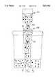
- FIG. 5is a cross sectional view of a debris removing device, detachably mounted on a collection cup, as fluid is aspirated through the device to remove debris and collect cells on a cell filter within the debris removing device.
- FIG. 6is a cross sectional view of the debris removing device of FIG. 5, mounted on the cytology collection apparatus of FIG. 1, showing the expulsion of fluid through the debris removing device into the cytology collection apparatus where a cell monolayer is collected on the laminated membrane.
- FIG. 7is an exploded cross sectional schematic view of a cytology collection apparatus according to the present invention.
- FIG. 8is a cross sectional schematic view of a syringe and cytology collection apparatus mounted on a collection cup.
- FIG. 9is a schematic exploded cross sectional view of a needle member for aspiration biopsy and a syringe mounted on the cytology collection apparatus of FIGS. 1 and 2.
- FIG. 10is a schematic cross sectional view of a second detachable portion, after removal from the cytology collection apparatus, showing the transfer of a monolayer of cells from the porous membrane onto a microscope slide.
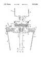
- FIG. 11is a schematic cross sectional view of a cytology collection apparatus, with the fluid collection cup removed, eluting a bacteriological sample into a microbiological culture tray.
- FIG. 12is a schematic cross sectional view of the second detachable portion of the cytology collection apparatus of FIG. 8 showing fluid flowing through the side of the second porous medium and bypassing the first porous medium.
- the present inventionincludes an assay module or cytology collection apparatus 10, comprising first and second detachable portions 44, 42, having first and second ports 54, 41, respectively.
- the first and second detachable portions 44, 42define a chamber 50 and the first and second ports 54, 41 define a fluid flow path through the chamber 50.
- a porous arrangement 46 having a collection site 45 adapted to collect cellsmay be positioned across the fluid flow path, the collection site 45 communicating with the first port 54.
- the porous arrangement 46 within the cytology collection apparatus 10is preferably adapted to define a flow path having first and second branches, the first branch 60 extending through the collection site 45 and the second branch 61 bypassing the collection site 45.
- the inventionincludes a porous arrangement 46 having a first porous medium 46b, suitable for preventing the passage of cells therethrough, and a second porous medium 46a, suitable for removing particulate matter from the fluid.
- the porous arrangement 46includes a first porous medium 46a and a second porous medium 46b, more preferably, a porous membrane 46b and a depth filter 46a wherein the depth filter communicates with the first port 54 through the porous membrane.
- the depth filter 46acommunicates with the first port 54 through a first branch of the flow path extending through the porous membrane 46b and communicates directly with the first port 54 through a second branch of the flow path.
- the first port 54may be configured as a connector and may be adapted to connect to a container, or may be configured as a needle or cannula 74 or the like.
- Second port 41may be configured as a connector and may be adapted to connect to a syringe, or the like.
- the porous arrangement 46may include a unitary structure having a first zone of density and pore size suitable to prevent the passage of cells therethrough and a second zone of density and pore size suitable for passing the fluid therethrough. The second zone may also remove particulate matter from the fluid. While the cytology collection apparatus 40 can be used for any biological fluid, it is particularly useful for preparing testing samples from urine and its associated cells for Pap smears.
- porous membranecan be used interchangeably with that of the present embodiment. While a polycarbonate membrane is especially suitable for use in the cytology collection apparatus of the present invention, other porous membranes are also suitable.
- One membrane that can be used for fluid screeningis LEUCOSORBTM, a leucocyte retention medium manufactured by Pall BioSupport Division of Pall Corporation.
- BIODYNE ATMan unmodified nylon with surface chemistry 50% amine and 50% carboxyl group which has an isoelectric point of pH 6.5
- BIODYNE BTMa surface-modified nylon with surface chemistry characterized by a high density of strong cationic quaternary groups (the zeta potential is positive to pH>10)
- BIODYNE CTMa surface-modified nylon with surface chemistry characterized by a high density of anionic carboxyl groups (the zeta potential is negative to pH>3
- LOPRODYNETMa low protein binding nylon 66 membrane with a tightly controlled microporous structure having high voids volume for rapid, efficient throughput of liquids and absolute retention of microparticles designed for cell separation and bacterial cell immunoassays.
- the porous arrangementincludes a porous polycarbonate membrane 46b, suitable for preventing the passage of cells therethrough.
- the porous arrangementmay further include a depth filter 46a laminated to the porous polycarbonate membrane 46b.
- the depth filter 46amay be made of polypropylene or high density polyethylene POREX® porous plastics.
- a preferred embodiment of the inventionincludes an assay module or cytology collection apparatus 10 which may be mounted on a collection cup 11 in which urine or other biological fluids, such as blood, cerebrospinal fluid (CSF), bronchial lavage, sputum or fine needle aspirates may be collected.
- the collection cup 11may be any container suitable for collection of body fluids.
- the patient or supervising medical personnelplaces a lid 14 on the cup housing 16.
- the cup housing 16is preferably provided with an external threaded surface 12.
- the lid 14may include a vent hole 13a with an optional removable vent cap 13b.
- the vent hole 13amay also be used to introduce a brush or spatula into the cup containing physiological saline solution or preservative after brushing or scraping the body site to obtain the cytology specimen.
- the lidhas a body 20 which is molded with a downwardly directed cylindrical extended skirt or flange 22 which is threaded 24 on its inner surface 23 for screwing onto the external threaded surface 12 of the cup housing 16.
- the lid body 20also defines a well 26 in which a threaded nipple 28 may be integrally molded.
- the nipple 28is provided with a channel 29 or the like leading to a hollow tube 30 which is preferably separately secured to the other side of the lid body in a circular planar seat 33 with its lumen 31 being axially aligned with the channel 29 of the nipple 28.
- the tube 30may have a series of perforations 32 and an open end 34 near the bottom of the collection cup 11 which allow different fluid layers as well as urinary sediments to be simultaneously tested when the urine or biological fluid is withdrawn from the cup.
- the cytology collection apparatus 10is preferably a two piece housing 40 with a first detachable portion 44 and a second detachable portion 42, although any housing providing access to the porous arrangement 46 is suitable.
- a first porous medium 46bis mounted on a second porous medium 46a to form the porous arrangement 46.
- a porous polycarbonate membrane 46bis laminated on filter member 46a to form the porous arrangement 46.
- the porous arrangement 46may be mounted on an annular step or seat 43 formed in the interior cavity 50 of the second detachable portion 42.
- the porous polycarbonate membrane 46bpreferably has a pore size from about 0.22 microns to about 8 microns, more preferably from about 1 micron to about 6 microns, most preferably about 2 microns, which allows it to trap cells which are more than 3 microns in size.
- the polycarbonate membrane 46bwhich may be mounted on the second porous medium 46a, is suitable to allow fluid flow to pass therethrough while preventing the passage of cells 60.
- the second porous medium 46ais suitable for passing fluid therethrough and may also be capable of removing particulate matter from the fluid.
- the pore size of the second porous medium 46amay range from about 5 microns to about 60 microns, preferably from about 15 microns to about 45 microns, most preferably about 35 microns.
- the second port 42may be adapted to connect to a syringe 64, or the like.
- Exemplary connectionsinclude, but are not limited to a luer lock, a threaded luer lock, a friction connection, a tapered hose connection and a threaded connection.
- any means suitable for inducing the flow of fluid from a source container through the cytology collection apparatusmay be used as part of the present invention.
- Exemplary fluid flow inducing meansinclude, but are not limited to a syringe or pump type device.
- Syringe 64has a barrel 66 and a piston (not shown) with assault piston head.
- any suitable pump type devicesuch as an autovial spunglass filter manufactured by Genex Corporation, could be used.
- a flexible, collapsible containersuch as a specimen container, which may be squeezed to force fluid through the cytology collection apparatus and into the syringe.
- the cytology collection apparatus 10may be mounted to syringe luer lock 62 and the nipple 28 of collection cup 11.
- the cytology collection apparatus 10preferably includes an easily openable housing and may comprise a simple two-piece construction including a first detachable portion 44 and a second detachable portion 42.
- the cytology collection apparatus 10comprises a female detachable portion 44 screwed onto a male detachable portion 42.
- a skirt member 48extends outward from base 47 and defines a cavity 50 and a flange 51 which holds O-ring 53.
- the cavity 50communicates with the bore 52 of the port 41.
- the skirt 48includes an annular step 43, which forms a seat for a porous arrangement 46.
- the inner surface 80 of the skirt 48may be threaded.
- the porous arrangement 46may comprise a polycarbonate membrane 46b laminated onto a disk shaped second porous medium 46a, which is preferably a depth filter.
- the second porous medium 46amay be provided with an outer cylindrical wall 81 having a threaded external surface, if such is desired, to screw into the step channel cut into skirt member 48 of the second detachable portion 42.
- the outer cylindrical wall 81 of the porous arrangement 46may extend past the end wall 49 of skirt member 48.
- the area of the porous arrangement 46 which extends past skirt end wall 49may act as a vent (low resistance to flow) to prevent piling up of cells on the surface 45 of the porous membrane 46b.
- the second detachable portion 42may be provided with a threaded nipple 41 having a throughgoing bore 52.
- the body of the second detachable portion 42(planar base 47 and skirt 48) defines a frustro conical chamber or cavity 50 in which a step 43 is formed which serves as a seat for the porous arrangement 46.
- port 41 of the cytology collection apparatus 10may be a threaded projection which is adapted to fit onto the luer lock 62 of a syringe 64, such as one manufactured by Becton Dickinson & Co.
- the first detachable portion 44may be provided with a threaded luer lock 54 having a throughgoing bore 55 communicating with the chamber 50.
- the threaded luer lock 54may be screwed onto nipple 28 of a collection cup 11 to remove liquid from the collection cup or alternatively attached to a needle assembly 70 as shown in FIG. 9.
- the needle assembly 70is constructed with a support member 72 defining a throughgoing aperture 73 in which is mounted a fine aspiration needle 74 with a lumen 75.
- a threaded nipple member 76is secured to the wall of the support member 72 thereby providing a means for the needle assembly 70 to be attached to the port 54 of the first detachable portion 44.
- the needle assembly 70can be used to aspirate biological fluid which is contained in the syringe or pump 66.
- the present inventionalso includes a the method for transferring cells to a microscope slide.
- the use of membrane filtrationprovides a method of depositing cells evenly over a slide with minimal overlap. This allows for clear observation and optimal diagnostic accuracy.
- cells 60 from the collection site 45 on the surface of the polycarbonate membrane 46bmay be placed on a glass slide 120 to transfer the cells, which then may be stained for cytologic determination. It is intended that the present invention should not be limited by the type of stain or detection protocol used.
- the area of the second porous medium 46a extending beyond the end wall 49 of skirt 48 of the second detachable portion 42acts as a vent (with low resistance to flow) which prevents the piling up of cells.
- the cytology collection apparatus 10may then be disconnected from collection cup 11 and, optionally, from syringe 64. It may then be unscrewed into two parts and the second detachable portion 42 and accompanying cell coated membrane 46a may be placed on a slide 120, as shown in FIG. 10, so that a transfer of the membrane 46b with the monolayer on surface 45 occurs.
- the membrane 46bis then pressed on the slide using a tissue wipe allowing cells 60 to form a monolayer on the slide 120.
- the membrane 46bcan be removed from the slide leaving the cells 60 on the slide. This allows a cytological examination to be performed on the cells by the practitioner without the interference of the pores in the membrane or delay due to processing requirements.
- FIGS. 5 and 6illustrate the use of a cytology collection apparatus 10 in combination with a debris filtering device 100.
- Any suitable debris filtration device 100such as a debris Shuttle, may be used.
- the debris filtration device 100preferably contains a cell filter 101, has an inlet 102 and outlet 103, and may be detachably connected to the cytology collection apparatus 10.
- a body fluidis first passed, preferably aspirated with a syringe, through the debris filtration device 100.
- cells in the body fluidaccumulate on the cell filter 101 mounted within the debris filtration device.
- the cell filter 101should have pores large enough to permit debris to flow through while retaining the desired cells in the surface of the cell filter.
- the pore size of the cell filter 101is preferably from about 3 microns to about 35 microns, more preferably 5 microns. Fluid is continually aspirated through the cell filter 101 until the flow is stopped by the accumulation of a cell mass on the filter.
- the debris filtering device 100may be connected to the first port 54 of the cytology collection apparatus 10. Fluid may be expelled through the debris filtering device 100 and the cytology collection apparatus 10, in a direction opposite that employed above. This results in the transfer of the cell mass, in the form of a monolayer, from the cell filter 101 of the debris filtering device 100 to collection site 45 of the porous arrangement 46 within the cell collection apparatus 10.
- a treatment devicemay also be used in combination with the cytology collection apparatus 10. Any suitable diagnostic or detection assembly may be used in conjunction with the cytology collection apparatus 10.
- a preferable deviceis an apparatus for testing for the presence of cancer utilizing a sandwich assay.
- the apparatusmay comprise a housing including inlet and outlet ports defining a flow path between the inlet and the outlet; a filter positioned across the flow path; and substrate beads having a primary antibody bound to the surface thereof, the beads being contained within the outlet, as disclosed in U.S. Pat. No. 4,953,561.
- the cytology collection apparatus 10 of the present inventionalso allows for isolation and collection of fresh cells and/or microorganisms from biological fluids to perform DNA probe and chromosomal analysis once the cells are hemolyzed by the proper buffer.
- the most widely used stain for visualization of cellular changes in cytologyis the Papanicolaou staining procedure.
- This stainwhich is used for both gynecologic and non-gynecologic applications, is basically composed of blue nuclear and orange, red and green cytoplasmic counterstains.
- the nuclear staindemonstrates the chromatic patterns associated with normal and abnormal cells, while the cytoplasmic stains help to indicate cell origin.
- the success of this procedurecan be attributed to the ability to observe a number of factors, including definition of nuclear detail and cell differentiation.
- This staining procedurealso results in a multicolor preparation that is very pleasing to the eye, possibly reducing eye strain.
- the monolayer of cellsmay be fixed directly on the collection site. This may be carried out by first depositing a monolayer of cells on the collection site of the cytology collection apparatus as described above and subsequently passing a solution containing a fixative, such as alcohol or acetone, through the cytology collection apparatus.
- a fixativesuch as alcohol or acetone
- captured microorganismscan be cultured, as shown in FIG. 11, in culture medium such as a standard petri dish 90. After a monolayer of cells has been collected in the cytology collection apparatus 10, fluid may be passed through the collection site 45 towards first port 54, thereby transferring the microorganisms to the petri dish 90.
- culture mediumsuch as a standard petri dish 90.
- the membrane 45can be used for culturing with a Qualture device (not shown) to determine the presence of specific bacteria colonies.
- the Qualture deviceis a plastic capsule containing a filter membrane and four nutrient pads of dehydrated, selective media.
- the Qualture techniqueis more sensitive than the agar plate method and more rapid in determining a presumptive diagnosis.
- the devicescreens, isolates and presumptively diagnoses bacterial isolates in one step most often in 4-6 hours. Tests have demonstrated that recovery from fifty milliliters of fluid is excellent and sensitive.
Landscapes
- Health & Medical Sciences (AREA)
- Life Sciences & Earth Sciences (AREA)
- Chemical & Material Sciences (AREA)
- Engineering & Computer Science (AREA)
- General Health & Medical Sciences (AREA)
- Organic Chemistry (AREA)
- Zoology (AREA)
- Wood Science & Technology (AREA)
- Immunology (AREA)
- Biomedical Technology (AREA)
- Biochemistry (AREA)
- Bioinformatics & Cheminformatics (AREA)
- Analytical Chemistry (AREA)
- Pathology (AREA)
- Hematology (AREA)
- Molecular Biology (AREA)
- Biotechnology (AREA)
- Microbiology (AREA)
- Physics & Mathematics (AREA)
- Genetics & Genomics (AREA)
- General Engineering & Computer Science (AREA)
- General Physics & Mathematics (AREA)
- Clinical Laboratory Science (AREA)
- Sustainable Development (AREA)
- Animal Behavior & Ethology (AREA)
- Veterinary Medicine (AREA)
- Public Health (AREA)
- Chemical Kinetics & Catalysis (AREA)
- Surgery (AREA)
- Urology & Nephrology (AREA)
- Proteomics, Peptides & Aminoacids (AREA)
- Medical Informatics (AREA)
- Heart & Thoracic Surgery (AREA)
- Cell Biology (AREA)
- Biophysics (AREA)
- Food Science & Technology (AREA)
- Medicinal Chemistry (AREA)
- Pulmonology (AREA)
- Apparatus Associated With Microorganisms And Enzymes (AREA)
- Sampling And Sample Adjustment (AREA)
Abstract
Description
Claims (29)
Priority Applications (3)
| Application Number | Priority Date | Filing Date | Title |
|---|---|---|---|
| US08/172,232US5471994A (en) | 1989-01-10 | 1993-12-23 | Method and apparatus for obtaining a cytology monolayer |
| US08/963,873US6423237B1 (en) | 1992-07-28 | 1997-11-04 | Method and apparatus for manually separating particulate matter from a liquid specimen |
| US09/053,010US6309362B1 (en) | 1992-07-28 | 1998-04-01 | Method and apparatus for automatically separating particulate matter from a fluid |
Applications Claiming Priority (8)
| Application Number | Priority Date | Filing Date | Title |
|---|---|---|---|
| US07/308,763US4961432A (en) | 1989-01-10 | 1989-01-10 | Modular fluid sample preparation assembly |
| US07/408,547US5024238A (en) | 1989-01-10 | 1989-09-18 | Blood withdrawing apparatus and antigen testing method |
| US07/411,041US4953561A (en) | 1989-09-18 | 1989-09-22 | Urine testing module and method of collecting urine antigen |
| US07/553,585US5042502A (en) | 1989-09-18 | 1990-07-18 | Urine testing module with cytology cup |
| US07/680,896US5139031A (en) | 1989-09-18 | 1991-04-08 | Method and device for cytology and microbiological testing |
| US07/686,934US5429803A (en) | 1991-04-18 | 1991-04-18 | Liquid specimen container and attachable testing modules |
| US07/920,662US5301685A (en) | 1989-01-10 | 1992-07-28 | Method and apparatus for obtaining a cytology monolayer |
| US08/172,232US5471994A (en) | 1989-01-10 | 1993-12-23 | Method and apparatus for obtaining a cytology monolayer |
Related Parent Applications (3)
| Application Number | Title | Priority Date | Filing Date |
|---|---|---|---|
| US07/408,547Continuation-In-PartUS5024238A (en) | 1989-01-10 | 1989-09-18 | Blood withdrawing apparatus and antigen testing method |
| US07/686,934Continuation-In-PartUS5429803A (en) | 1989-01-10 | 1991-04-18 | Liquid specimen container and attachable testing modules |
| US07/920,662DivisionUS5301685A (en) | 1989-01-10 | 1992-07-28 | Method and apparatus for obtaining a cytology monolayer |
Related Child Applications (1)
| Application Number | Title | Priority Date | Filing Date |
|---|---|---|---|
| US47489495ADivision | 1989-01-10 | 1995-06-07 |
Publications (1)
| Publication Number | Publication Date |
|---|---|
| US5471994Atrue US5471994A (en) | 1995-12-05 |
Family
ID=25444160
Family Applications (2)
| Application Number | Title | Priority Date | Filing Date |
|---|---|---|---|
| US07/920,662Expired - LifetimeUS5301685A (en) | 1989-01-10 | 1992-07-28 | Method and apparatus for obtaining a cytology monolayer |
| US08/172,232Expired - LifetimeUS5471994A (en) | 1989-01-10 | 1993-12-23 | Method and apparatus for obtaining a cytology monolayer |
Family Applications Before (1)
| Application Number | Title | Priority Date | Filing Date |
|---|---|---|---|
| US07/920,662Expired - LifetimeUS5301685A (en) | 1989-01-10 | 1992-07-28 | Method and apparatus for obtaining a cytology monolayer |
Country Status (9)
| Country | Link |
|---|---|
| US (2) | US5301685A (en) |
| EP (1) | EP0654972B8 (en) |
| JP (1) | JP3479300B2 (en) |
| KR (1) | KR100324810B1 (en) |
| AU (1) | AU4777693A (en) |
| BR (1) | BR9306818A (en) |
| CH (1) | CH686324A5 (en) |
| DE (1) | DE69331878T2 (en) |
| WO (1) | WO1994003103A1 (en) |
Cited By (88)
| Publication number | Priority date | Publication date | Assignee | Title |
|---|---|---|---|---|
| WO1999010723A1 (en)* | 1997-08-25 | 1999-03-04 | Monogen, Inc. | Method and apparatus for automatically forming monolayers from particulate matter separated from fluid samples |
| WO2000002031A1 (en)* | 1998-07-07 | 2000-01-13 | Lamina, Inc. | Improved method for mixing and processing specimen samples |
| US6296764B1 (en) | 1997-11-04 | 2001-10-02 | Lamina, Inc. | Apparatus for mixing and separating particulate matter from a fluid |
| US6328709B1 (en) | 1998-11-13 | 2001-12-11 | Pro Duct Health, Inc. | Devices and methods to identify ductal orifices during nipple aspiration |
| US20020039796A1 (en)* | 2000-04-04 | 2002-04-04 | Dores Gerson Botacini Das | Device and method for cytology slide preparation |
| US6394966B1 (en) | 2000-09-08 | 2002-05-28 | Diagnostic Cytology Laboratories, Inc. | Apparatus and method for removing cells from an endocervical brush in a liquid-based pap test system |
| US6423237B1 (en)* | 1992-07-28 | 2002-07-23 | Lamina, Inc. | Method and apparatus for manually separating particulate matter from a liquid specimen |
| US6506167B1 (en) | 1997-12-24 | 2003-01-14 | I-Design Co., Ltd. | Blood-collecting tubes |
| US20030077838A1 (en)* | 2001-10-19 | 2003-04-24 | Monogen, Inc. | Vial system and method for processing liquid-based specimens |
| US20030092170A1 (en)* | 2001-10-19 | 2003-05-15 | Monogen, Inc. | Filtration system and method for obtaining a cytology layer |
| US20040065622A1 (en)* | 2002-10-02 | 2004-04-08 | Ferguson Gary William | Filter device to capture a desired amount of material and methods of use |
| US20040069714A1 (en)* | 2002-10-11 | 2004-04-15 | Ferguson Gary William | Filter apparatus and methods to capture a desired amount of material from a sample suspension for monolayer deposition, analysis or other uses |
| US6830935B1 (en) | 1998-07-07 | 2004-12-14 | Lamina, Inc. | Method for mixing and processing specimen samples |
| US20050169809A1 (en)* | 2002-04-24 | 2005-08-04 | Jean-Luc Centeleghe | Device for producing a cytological preparation on an object-carrying slide |
| US20070287193A1 (en)* | 2004-11-09 | 2007-12-13 | Monogen, Inc. | Vial Assembly, Sampling Apparatus And Method For Processing Liquid-Based Specimens |
| US20080287826A1 (en)* | 2004-07-09 | 2008-11-20 | Bard Peripheral Vasular, Inc. | Transport System for Biopsy Device |
| WO2008150779A1 (en)* | 2007-05-31 | 2008-12-11 | 3M Innovative Properties Company | Devices and processes for collecting and concentrating samples for microbiological analysis |
| WO2011044033A1 (en)* | 2009-10-05 | 2011-04-14 | Battelle Memorial Institute | Sample preparation disposable devices and sample collection and preparation methods using same |
| US20110097250A1 (en)* | 2005-07-28 | 2011-04-28 | Infinite Medical Technology Corp. | Safety, biodegradable biological sample collection system |
| US20110105946A1 (en)* | 2009-10-31 | 2011-05-05 | Sorensen Peter L | Biopsy system with infrared communications |
| US20110159536A1 (en)* | 2009-12-25 | 2011-06-30 | Hitachi Plant Technologies, Ltd. | Device for capturing object and method for using the same |
| US20110208085A1 (en)* | 2005-01-31 | 2011-08-25 | C.R. Bard, Inc. | Quick cycle biopsy system |
| WO2011057707A3 (en)* | 2009-11-12 | 2011-10-27 | Sartorius Stedim Biotech Gmbh | Device and method for treating a filtration medium |
| US20110301496A1 (en)* | 2010-06-08 | 2011-12-08 | Merit Medical Systems, Inc. | Biopsy collection device and related methods |
| US20120065541A1 (en)* | 2009-04-15 | 2012-03-15 | Videbaek Karsten | Biopsy apparatus having integrated fluid management |
| WO2012176065A2 (en) | 2011-06-24 | 2012-12-27 | Biotechnology Developers, S.A. | Method compositions and device for preparing cytological specimens |
| US20130040338A1 (en)* | 2010-03-23 | 2013-02-14 | Noe Miyashita | Device for capturing object and method for using the same |
| WO2014025490A1 (en) | 2012-08-10 | 2014-02-13 | Montagu Jean I | Filtering blood |
| US8690793B2 (en) | 2009-03-16 | 2014-04-08 | C. R. Bard, Inc. | Biopsy device having rotational cutting |
| US8721563B2 (en) | 2005-08-10 | 2014-05-13 | C. R. Bard, Inc. | Single-insertion, multiple sample biopsy device with integrated markers |
| US8728004B2 (en) | 2003-03-29 | 2014-05-20 | C.R. Bard, Inc. | Biopsy needle system having a pressure generating unit |
| US8752598B2 (en) | 2011-04-17 | 2014-06-17 | Medimop Medical Projects Ltd. | Liquid drug transfer assembly |
| US8771200B2 (en) | 2005-08-10 | 2014-07-08 | C.R. Bard, Inc. | Single insertion, multiple sampling biopsy device with linear drive |
| US8808197B2 (en) | 2009-10-29 | 2014-08-19 | Bard Peripheral Vascular, Inc. | Biopsy driver assembly having a control circuit for conserving battery power |
| US8852145B2 (en) | 2010-11-14 | 2014-10-07 | Medimop Medical Projects, Ltd. | Inline liquid drug medical device having rotary flow control member |
| US8858463B2 (en) | 2007-12-20 | 2014-10-14 | C. R. Bard, Inc. | Biopsy device |
| US8905994B1 (en) | 2011-10-11 | 2014-12-09 | Medimop Medical Projects, Ltd. | Valve assembly for use with liquid container and drug vial |
| USD720451S1 (en) | 2012-02-13 | 2014-12-30 | Medimop Medical Projects Ltd. | Liquid drug transfer assembly |
| US8951209B2 (en) | 2002-03-19 | 2015-02-10 | C. R. Bard, Inc. | Biopsy device and insertable biopsy needle module |
| US8951208B2 (en) | 2006-08-21 | 2015-02-10 | C. R. Bard, Inc. | Self-contained handheld biopsy needle |
| US8961430B2 (en) | 2005-08-10 | 2015-02-24 | C.R. Bard, Inc. | Single-insertion, multiple sampling biopsy device usable with various transport systems and integrated markers |
| US8979792B2 (en) | 2009-11-12 | 2015-03-17 | Medimop Medical Projects Ltd. | Inline liquid drug medical devices with linear displaceable sliding flow control member |
| US8998875B2 (en) | 2009-10-01 | 2015-04-07 | Medimop Medical Projects Ltd. | Vial assemblage with vial and pre-attached fluid transfer device |
| US9072502B2 (en) | 2002-03-19 | 2015-07-07 | C. R. Bard, Inc. | Disposable biopsy unit |
| USD734868S1 (en) | 2012-11-27 | 2015-07-21 | Medimop Medical Projects Ltd. | Drug vial adapter with downwardly depending stopper |
| USD737436S1 (en) | 2012-02-13 | 2015-08-25 | Medimop Medical Projects Ltd. | Liquid drug reconstitution assembly |
| US9173641B2 (en) | 2009-08-12 | 2015-11-03 | C. R. Bard, Inc. | Biopsy apparatus having integrated thumbwheel mechanism for manual rotation of biopsy cannula |
| US9283324B2 (en) | 2012-04-05 | 2016-03-15 | Medimop Medical Projects, Ltd | Fluid transfer devices having cartridge port with cartridge ejection arrangement |
| US9282949B2 (en) | 2009-09-01 | 2016-03-15 | Bard Peripheral Vascular, Inc. | Charging station for battery powered biopsy apparatus |
| US9339438B2 (en) | 2012-09-13 | 2016-05-17 | Medimop Medical Projects Ltd. | Telescopic female drug vial adapter |
| USD757933S1 (en) | 2014-09-11 | 2016-05-31 | Medimop Medical Projects Ltd. | Dual vial adapter assemblage |
| USD765837S1 (en) | 2013-08-07 | 2016-09-06 | Medimop Medical Projects Ltd. | Liquid transfer device with integral vial adapter |
| USD767124S1 (en) | 2013-08-07 | 2016-09-20 | Medimop Medical Projects Ltd. | Liquid transfer device with integral vial adapter |
| US9566045B2 (en) | 2006-10-06 | 2017-02-14 | Bard Peripheral Vascular, Inc. | Tissue handling system with reduced operator exposure |
| US9795536B2 (en) | 2012-08-26 | 2017-10-24 | Medimop Medical Projects, Ltd. | Liquid drug transfer devices employing manual rotation for dual flow communication step actuations |
| USD801522S1 (en) | 2015-11-09 | 2017-10-31 | Medimop Medical Projects Ltd. | Fluid transfer assembly |
| US9801786B2 (en) | 2013-04-14 | 2017-10-31 | Medimop Medical Projects Ltd. | Drug container closure for mounting on open-topped drug container to form drug reconstitution assemblage for use with needleless syringe |
| US9839580B2 (en) | 2012-08-26 | 2017-12-12 | Medimop Medical Projects, Ltd. | Liquid drug transfer devices |
| US9943463B2 (en) | 2013-05-10 | 2018-04-17 | West Pharma. Services IL, Ltd. | Medical devices including vial adapter with inline dry drug module |
| USD832430S1 (en) | 2016-11-15 | 2018-10-30 | West Pharma. Services IL, Ltd. | Dual vial adapter assemblage |
| US10149664B2 (en) | 2006-10-24 | 2018-12-11 | C. R. Bard, Inc. | Large sample low aspect ratio biopsy needle |
| US10278897B2 (en) | 2015-11-25 | 2019-05-07 | West Pharma. Services IL, Ltd. | Dual vial adapter assemblage including drug vial adapter with self-sealing access valve |
| US10285673B2 (en) | 2013-03-20 | 2019-05-14 | Bard Peripheral Vascular, Inc. | Biopsy device |
| US10285907B2 (en) | 2015-01-05 | 2019-05-14 | West Pharma. Services IL, Ltd. | Dual vial adapter assemblages with quick release drug vial adapter for ensuring correct usage |
| US10357429B2 (en) | 2015-07-16 | 2019-07-23 | West Pharma. Services IL, Ltd. | Liquid drug transfer devices for secure telescopic snap fit on injection vials |
| US10456120B2 (en) | 2013-11-05 | 2019-10-29 | C. R. Bard, Inc. | Biopsy device having integrated vacuum |
| US10463350B2 (en) | 2015-05-01 | 2019-11-05 | C. R. Bard, Inc. | Biopsy device |
| US10533932B2 (en) | 2013-09-13 | 2020-01-14 | Cancer Research Technology Limited | Apparatus and methods for liquid separation and capture of biologics |
| US10646404B2 (en) | 2016-05-24 | 2020-05-12 | West Pharma. Services IL, Ltd. | Dual vial adapter assemblages including identical twin vial adapters |
| US10688295B2 (en) | 2013-08-07 | 2020-06-23 | West Pharma. Services IL, Ltd. | Liquid transfer devices for use with infusion liquid containers |
| US10765604B2 (en) | 2016-05-24 | 2020-09-08 | West Pharma. Services IL, Ltd. | Drug vial adapter assemblages including vented drug vial adapter and vented liquid vial adapter |
| US10772797B2 (en) | 2016-12-06 | 2020-09-15 | West Pharma. Services IL, Ltd. | Liquid drug transfer devices for use with intact discrete injection vial release tool |
| US10806667B2 (en) | 2016-06-06 | 2020-10-20 | West Pharma. Services IL, Ltd. | Fluid transfer devices for filling drug pump cartridges with liquid drug contents |
| US10806671B2 (en) | 2016-08-21 | 2020-10-20 | West Pharma. Services IL, Ltd. | Syringe assembly |
| USD903864S1 (en) | 2018-06-20 | 2020-12-01 | West Pharma. Services IL, Ltd. | Medication mixing apparatus |
| US10945921B2 (en) | 2017-03-29 | 2021-03-16 | West Pharma. Services IL, Ltd. | User actuated liquid drug transfer devices for use in ready-to-use (RTU) liquid drug transfer assemblages |
| USD917693S1 (en) | 2018-07-06 | 2021-04-27 | West Pharma. Services IL, Ltd. | Medication mixing apparatus |
| USD923782S1 (en) | 2019-01-17 | 2021-06-29 | West Pharma. Services IL, Ltd. | Medication mixing apparatus |
| USD923812S1 (en) | 2019-01-16 | 2021-06-29 | West Pharma. Services IL, Ltd. | Medication mixing apparatus |
| USD954253S1 (en) | 2019-04-30 | 2022-06-07 | West Pharma. Services IL, Ltd. | Liquid transfer device |
| US11364020B2 (en) | 2016-12-09 | 2022-06-21 | Techmed Ventures, Llc | Brush biopsy device, kit and method |
| USD956958S1 (en) | 2020-07-13 | 2022-07-05 | West Pharma. Services IL, Ltd. | Liquid transfer device |
| US20220323050A1 (en)* | 2012-11-20 | 2022-10-13 | The Trustees Of Columbia University In The City Of New York | Medical apparatus and method for collecting biological samples |
| US11642285B2 (en) | 2017-09-29 | 2023-05-09 | West Pharma. Services IL, Ltd. | Dual vial adapter assemblages including twin vented female vial adapters |
| RU2804168C1 (en)* | 2023-05-05 | 2023-09-26 | федеральное государственное бюджетное образовательное учреждение высшего образования "Омский государственный медицинский университет" Министерства здравоохранения Российской Федерации (ФГБОУ ВО ОмГМУ Минздрава России) | Gel for monolayer distribution of cells in the manufacture of cytological preparations |
| US11918542B2 (en) | 2019-01-31 | 2024-03-05 | West Pharma. Services IL, Ltd. | Liquid transfer device |
| US12274670B2 (en) | 2019-04-09 | 2025-04-15 | West Pharma. Services IL, Ltd. | Liquid transfer device with integrated syringe |
| US12427091B2 (en) | 2019-01-18 | 2025-09-30 | West Pharma. Services IL, Ltd. | Liquid transfer devices for use with intravenous (IV) bottles |
Families Citing this family (55)
| Publication number | Priority date | Publication date | Assignee | Title |
|---|---|---|---|---|
| US5797130A (en)* | 1993-11-16 | 1998-08-18 | Neopath, Inc. | Method for testing proficiency in screening images of biological slides |
| US5514119A (en)* | 1994-08-19 | 1996-05-07 | Curtis; John L. | Embryo collection device |
| PL319978A1 (en)* | 1994-11-04 | 1997-09-01 | Lamina Inc | Method of and apparatus for preparing substances for optical analysis |
| US5888409A (en)* | 1995-06-07 | 1999-03-30 | Cedars-Sinai Medical Center | Methods for cell isolation and collection |
| US6091483A (en)* | 1995-11-03 | 2000-07-18 | Lamina, Inc. | Method and apparatus for preparing substances for optical analysis |
| JP2000508780A (en)* | 1996-12-20 | 2000-07-11 | フイルメニツヒ ソシエテ アノニム | Apparatus for sampling volatile products |
| US5980481A (en)* | 1997-05-08 | 1999-11-09 | Transvivo, Inc. | Method and apparatus for continuous peritoneal cascade dialysis and hemofiltration (CPCD/H) |
| FR2777903B1 (en)* | 1998-04-24 | 2000-12-29 | Millipore Sa | METHOD FOR DETECTION OF MICROORGANISMS AND CASSETTE SUITABLE FOR IMPLEMENTING IT |
| WO2000034775A1 (en)* | 1998-12-08 | 2000-06-15 | Cohen Andre | Disposable device for sampling a biological or mineral liquid detecting and/or assaying a specific parameter |
| NO994228D0 (en)* | 1999-09-01 | 1999-09-01 | Oddbjoern Gjelsnes | Method and apparatus for counting cells in urine |
| WO2002048681A2 (en)* | 2000-12-04 | 2002-06-20 | Molecular Diagnostics, Inc. | Cell transfer device |
| IL161062A0 (en)* | 2001-10-19 | 2004-08-31 | Monogen Inc | Vial system and method for processing liquid-based specimens |
| FR2843011B1 (en)* | 2002-07-31 | 2006-02-17 | Draeger Safety Ag & Co Kgaa | DEVICE AND METHOD FOR PREDICTING AND DEPOSITING SALIVA |
| US7211225B2 (en)* | 2002-08-26 | 2007-05-01 | Perceptronix Medical Inc. | Filter devices for depositing material and density gradients of material from sample suspension |
| CN101072992A (en)* | 2004-11-12 | 2007-11-14 | 卢米尼克斯股份有限公司 | Methods and systems for positioning microspheres for imaging |
| US7996188B2 (en) | 2005-08-22 | 2011-08-09 | Accuri Cytometers, Inc. | User interface for a flow cytometer system |
| US8017402B2 (en) | 2006-03-08 | 2011-09-13 | Accuri Cytometers, Inc. | Fluidic system for a flow cytometer |
| US8303894B2 (en) | 2005-10-13 | 2012-11-06 | Accuri Cytometers, Inc. | Detection and fluidic system of a flow cytometer |
| US7780916B2 (en)* | 2006-03-08 | 2010-08-24 | Accuri Cytometers, Inc. | Flow cytometer system with unclogging feature |
| US8283177B2 (en)* | 2006-03-08 | 2012-10-09 | Accuri Cytometers, Inc. | Fluidic system with washing capabilities for a flow cytometer |
| JP4950277B2 (en) | 2006-03-20 | 2012-06-13 | ロンシャン リ | Cell block production system and method of use thereof |
| US8715573B2 (en) | 2006-10-13 | 2014-05-06 | Accuri Cytometers, Inc. | Fluidic system for a flow cytometer with temporal processing |
| US8445286B2 (en)* | 2006-11-07 | 2013-05-21 | Accuri Cytometers, Inc. | Flow cell for a flow cytometer system |
| US7739060B2 (en)* | 2006-12-22 | 2010-06-15 | Accuri Cytometers, Inc. | Detection system and user interface for a flow cytometer system |
| US8432541B2 (en)* | 2007-12-17 | 2013-04-30 | Accuri Cytometers, Inc. | Optical system for a flow cytometer with an interrogation zone |
| US7790117B2 (en)* | 2008-03-21 | 2010-09-07 | Scientific Plastic Products, Inc. | Filter vial |
| FR2934681B1 (en) | 2008-07-29 | 2011-09-30 | Bastien Karkouche | DEVICE FOR CAPTURING BIOLOGICAL PARTICLES AND USE. |
| US8507279B2 (en)* | 2009-06-02 | 2013-08-13 | Accuri Cytometers, Inc. | System and method of verification of a prepared sample for a flow cytometer |
| US20110061471A1 (en)* | 2009-06-02 | 2011-03-17 | Rich Collin A | System and method of verification of a sample for a flow cytometer |
| WO2011106402A1 (en)* | 2010-02-23 | 2011-09-01 | Accuri Cytometers, Inc. | Method and system for detecting fluorochromes in a flow cytometer |
| US9551600B2 (en) | 2010-06-14 | 2017-01-24 | Accuri Cytometers, Inc. | System and method for creating a flow cytometer network |
| US9280635B2 (en) | 2010-10-25 | 2016-03-08 | Accuri Cytometers, Inc. | Systems and user interface for collecting a data set in a flow cytometer |
| WO2012075508A2 (en)* | 2010-12-03 | 2012-06-07 | Blood Cell Storage, Inc. | Processes for isolating microorganisms |
| SG194067A1 (en) | 2011-04-11 | 2013-11-29 | Rongshan Li | Multifunction aspiration biopsy device and methods of use |
| JP5659960B2 (en)* | 2011-06-09 | 2015-01-28 | コニカミノルタ株式会社 | Cell plane development method |
| GB2496597B (en)* | 2011-11-14 | 2017-10-04 | Bio-Check(Uk) Ltd | Cartridge for containing a sample |
| KR101404507B1 (en)* | 2012-04-12 | 2014-06-10 | 한국과학기술원 | Particle processing device using combination of multiple membrane structures |
| BR112014029655A2 (en) | 2012-05-30 | 2017-06-27 | Lifecell Corp | tissue preparation device, and method for preparing tissue |
| EP2867647A4 (en) | 2012-06-27 | 2016-04-06 | Univ Wake Forest Health Sciences | CYTOPATHOLOGICAL ANALYSIS TUBES AND METHODS OF TREATING CELLS THEREFOR |
| US20140045253A1 (en)* | 2012-08-08 | 2014-02-13 | Qian Zou | Multi-compartment device for cell cloning and method of performing the same |
| BR112015004003B1 (en) | 2012-09-06 | 2020-05-19 | The Gid Group Inc | apparatus for processing human biological material containing fibrous tissue and method for processing adipose tissue |
| ES2716114T3 (en)* | 2013-05-24 | 2019-06-10 | Occam Biolabs Inc | System and procedure to collect a nucleic acid sample |
| US10302535B2 (en) | 2013-09-12 | 2019-05-28 | CellectGen, Inc. | Biofluid collection and filtration device |
| US10092711B2 (en) | 2014-05-02 | 2018-10-09 | Lifecell Corporation | Injection sensor with feedback mechanism |
| KR101623876B1 (en) | 2014-07-10 | 2016-05-24 | 신진메딕스(주) | Cellular tissue inspection device |
| KR101776245B1 (en)* | 2014-11-20 | 2017-09-11 | 울산과학기술원 | Particle filtration device and method of particle filtration |
| RU2020124757A (en) | 2014-11-21 | 2021-06-30 | Оккам Байолэбс, Инк. | SYSTEM AND METHOD FOR COLLECTING NUCLEIC ACID SAMPLE |
| ES2775597T3 (en) | 2015-10-21 | 2020-07-27 | Lifecell Corp | Systems and methods for tube management |
| AU2016342012A1 (en) | 2015-10-21 | 2018-05-10 | Lifecell Corporation | Systems and methods for medical device control |
| AU2016378566A1 (en) | 2015-12-22 | 2018-06-28 | Lifecell Corporation | Syringe filling device for fat transfer |
| WO2018044791A1 (en) | 2016-08-30 | 2018-03-08 | Lifecell Corporation | Systems and methods for medical device control |
| USD852375S1 (en) | 2017-10-25 | 2019-06-25 | Wake Forest University Health Sciences | Cell collection and centrifuge tube assembly |
| KR101985008B1 (en)* | 2018-09-08 | 2019-05-31 | (주)바이오다인 | exfoliative cell processing device |
| CN114126741B (en)* | 2019-09-09 | 2024-02-06 | 株式会社村田制作所 | Concentrating device and concentrating method |
| US20240189811A1 (en)* | 2022-12-13 | 2024-06-13 | Rapid Micro Biosystems, Inc. | Tube set with dual pressure regulating valve |
Citations (20)
| Publication number | Priority date | Publication date | Assignee | Title |
|---|---|---|---|---|
| US536552A (en)* | 1895-03-26 | James powell swift | ||
| GB503128A (en)* | 1937-10-04 | 1939-03-31 | Claudius Blanchard | Improvements in brakes for cycles and the like |
| US3722502A (en)* | 1971-10-18 | 1973-03-27 | S Besuner | Multiple liquid sample collection apparatus |
| US3774455A (en)* | 1971-12-22 | 1973-11-27 | D Seidler | Urine testing apparatus |
| US3851972A (en)* | 1973-10-18 | 1974-12-03 | Coulter Electronics | Automatic method and system for analysis and review of a plurality of stored slides |
| US4040791A (en)* | 1976-06-22 | 1977-08-09 | David H Kuntz | Specimen collecting device |
| US4170056A (en)* | 1976-03-25 | 1979-10-09 | Baxter Travenol Laboratories, Inc. | Blood filter |
| US4395493A (en)* | 1981-05-14 | 1983-07-26 | Coulter Electronics, Inc. | Monolayer device using filter techniques |
| US4435507A (en)* | 1980-07-08 | 1984-03-06 | Stenkvist Bjoern G | Process and device for preparation of cell samples for cytological tests |
| US4473530A (en)* | 1980-09-24 | 1984-09-25 | Villa Real Antony Euclid C | Compact sanitary urinalysis unit |
| US4557274A (en)* | 1982-06-07 | 1985-12-10 | Cawood Charles David | Midstream urine collector |
| US4573983A (en)* | 1984-07-27 | 1986-03-04 | The Kendall Company | Liquid collection system having an anti-septic member on the discharge section |
| US4609264A (en)* | 1984-01-23 | 1986-09-02 | The Micromanipulator Microscope Company, Inc. | Apparatus for positioning flat objects for microscopic examination |
| US4685472A (en)* | 1984-01-23 | 1987-08-11 | Rudolph Muto | Specimen collector |
| US4827944A (en)* | 1987-07-22 | 1989-05-09 | Becton, Dickinson And Company | Body fluid sample collection tube composite |
| US4960130A (en)* | 1989-01-10 | 1990-10-02 | Cancer Diagnostics, Inc. | Modular fluid sample preparation assembly |
| US5022411A (en)* | 1989-09-18 | 1991-06-11 | La Mina Ltd. | Modular fluid testing device |
| US5038793A (en)* | 1989-01-10 | 1991-08-13 | La Mina Ltd. | Urine testing membrane module and method of conducting same |
| US5077012A (en)* | 1989-01-10 | 1991-12-31 | La Mina Ltd. | Device for detecting disease markers |
| US5143627A (en)* | 1990-07-09 | 1992-09-01 | Cytyc Corporation | Method and apparatus for preparing cells for examination |
Family Cites Families (10)
| Publication number | Priority date | Publication date | Assignee | Title |
|---|---|---|---|---|
| US3867924A (en)* | 1973-02-05 | 1975-02-25 | Microbyx Corp | Internal blood collection |
| US4334538A (en)* | 1979-12-12 | 1982-06-15 | Juhn Steven K | Aspirator for collecting liquid samples |
| DE3223589A1 (en)* | 1982-06-24 | 1983-12-29 | Bernhard Dr.med. 3004 Isernhagen Aeikens | Process and apparatus for the preparation of liquid samples such as urine or other body fluids for transport to an analysis point |
| US5026653A (en)* | 1985-04-02 | 1991-06-25 | Leeco Diagnostic, Inc. | Scavenger antibody mixture and its use for immunometric assay |
| CA1291948C (en)* | 1986-04-24 | 1991-11-12 | Albert E. Chu | Variable volume assay device and method |
| US4874691A (en)* | 1987-10-16 | 1989-10-17 | Quadra Logic Technologies Inc. | Membrane-supported immunoassays |
| WO1990000251A1 (en)* | 1988-06-30 | 1990-01-11 | Bio-Metric Systems, Inc. | Pressure-assisted analytical apparatus and method |
| US5139031A (en)* | 1989-09-18 | 1992-08-18 | La Mina Ltd. | Method and device for cytology and microbiological testing |
| US5042502A (en)* | 1989-09-18 | 1991-08-27 | La Mina Ltd. | Urine testing module with cytology cup |
| US5137031A (en)* | 1989-09-18 | 1992-08-11 | La Mina Ltd. | Urine testing apparatus with urinary sediment device |
- 1992
- 1992-07-28USUS07/920,662patent/US5301685A/ennot_activeExpired - Lifetime
- 1993
- 1993-07-21CHCH101694Apatent/CH686324A5/ennot_activeIP Right Cessation
- 1993-07-21EPEP93918266Apatent/EP0654972B8/ennot_activeExpired - Lifetime
- 1993-07-21JPJP50534394Apatent/JP3479300B2/ennot_activeExpired - Lifetime
- 1993-07-21KRKR1019950700397Apatent/KR100324810B1/ennot_activeExpired - Fee Related
- 1993-07-21BRBR9306818Apatent/BR9306818A/ennot_activeIP Right Cessation
- 1993-07-21DEDE69331878Tpatent/DE69331878T2/ennot_activeExpired - Lifetime
- 1993-07-21WOPCT/US1993/006810patent/WO1994003103A1/enactiveIP Right Grant
- 1993-07-21AUAU47776/93Apatent/AU4777693A/ennot_activeAbandoned
- 1993-12-23USUS08/172,232patent/US5471994A/ennot_activeExpired - Lifetime
Patent Citations (20)
| Publication number | Priority date | Publication date | Assignee | Title |
|---|---|---|---|---|
| US536552A (en)* | 1895-03-26 | James powell swift | ||
| GB503128A (en)* | 1937-10-04 | 1939-03-31 | Claudius Blanchard | Improvements in brakes for cycles and the like |
| US3722502A (en)* | 1971-10-18 | 1973-03-27 | S Besuner | Multiple liquid sample collection apparatus |
| US3774455A (en)* | 1971-12-22 | 1973-11-27 | D Seidler | Urine testing apparatus |
| US3851972A (en)* | 1973-10-18 | 1974-12-03 | Coulter Electronics | Automatic method and system for analysis and review of a plurality of stored slides |
| US4170056A (en)* | 1976-03-25 | 1979-10-09 | Baxter Travenol Laboratories, Inc. | Blood filter |
| US4040791A (en)* | 1976-06-22 | 1977-08-09 | David H Kuntz | Specimen collecting device |
| US4435507A (en)* | 1980-07-08 | 1984-03-06 | Stenkvist Bjoern G | Process and device for preparation of cell samples for cytological tests |
| US4473530A (en)* | 1980-09-24 | 1984-09-25 | Villa Real Antony Euclid C | Compact sanitary urinalysis unit |
| US4395493A (en)* | 1981-05-14 | 1983-07-26 | Coulter Electronics, Inc. | Monolayer device using filter techniques |
| US4557274A (en)* | 1982-06-07 | 1985-12-10 | Cawood Charles David | Midstream urine collector |
| US4609264A (en)* | 1984-01-23 | 1986-09-02 | The Micromanipulator Microscope Company, Inc. | Apparatus for positioning flat objects for microscopic examination |
| US4685472A (en)* | 1984-01-23 | 1987-08-11 | Rudolph Muto | Specimen collector |
| US4573983A (en)* | 1984-07-27 | 1986-03-04 | The Kendall Company | Liquid collection system having an anti-septic member on the discharge section |
| US4827944A (en)* | 1987-07-22 | 1989-05-09 | Becton, Dickinson And Company | Body fluid sample collection tube composite |
| US4960130A (en)* | 1989-01-10 | 1990-10-02 | Cancer Diagnostics, Inc. | Modular fluid sample preparation assembly |
| US5038793A (en)* | 1989-01-10 | 1991-08-13 | La Mina Ltd. | Urine testing membrane module and method of conducting same |
| US5077012A (en)* | 1989-01-10 | 1991-12-31 | La Mina Ltd. | Device for detecting disease markers |
| US5022411A (en)* | 1989-09-18 | 1991-06-11 | La Mina Ltd. | Modular fluid testing device |
| US5143627A (en)* | 1990-07-09 | 1992-09-01 | Cytyc Corporation | Method and apparatus for preparing cells for examination |
Non-Patent Citations (6)
| Title |
|---|
| "Nuclepore Membrane Filter Techniques For Diagnostic Cytology Of Urine And Other Body Fluids", G. H. Green et al., Medical Laboratory Technology, 30:265-271 (1973). |
| "Optically Eliminating The Visible Outlines Of Pores In Intact Polycarbonate(Nuclepore) Filters", Goran Ocklind, ACTA Cytological 31:946-949 (1987). |
| Nuclepore Membrane Filter Techniques For Diagnostic Cytology Of Urine And Other Body Fluids , G. H. Green et al., Medical Laboratory Technology, 30:265 271 (1973).* |
| Optically Eliminating The Visible Outlines Of Pores In Intact Polycarbonate(Nuclepore) Filters , Goran Ocklind, ACTA Cytological 31:946 949 (1987).* |
| Wartio Vaara et al., Nature, 238:407 408 (1972).* |
| Wartio Vaara et al., Nature, 238:407-408 (1972). |
Cited By (164)
| Publication number | Priority date | Publication date | Assignee | Title |
|---|---|---|---|---|
| US6309362B1 (en) | 1992-07-28 | 2001-10-30 | Lamina, Inc. | Method and apparatus for automatically separating particulate matter from a fluid |
| US6423237B1 (en)* | 1992-07-28 | 2002-07-23 | Lamina, Inc. | Method and apparatus for manually separating particulate matter from a liquid specimen |
| WO1999010723A1 (en)* | 1997-08-25 | 1999-03-04 | Monogen, Inc. | Method and apparatus for automatically forming monolayers from particulate matter separated from fluid samples |
| AU755416B2 (en)* | 1997-08-25 | 2002-12-12 | La Mina Ltd | Method and apparatus for automatically forming monolayers from particulate matter separated from fluid samples |
| US6296764B1 (en) | 1997-11-04 | 2001-10-02 | Lamina, Inc. | Apparatus for mixing and separating particulate matter from a fluid |
| US6379565B1 (en) | 1997-11-04 | 2002-04-30 | Lamina, Inc. | Method for simultaneously processing plural samples containing particulate matter in a fluid |
| US6506167B1 (en) | 1997-12-24 | 2003-01-14 | I-Design Co., Ltd. | Blood-collecting tubes |
| AU758047B2 (en)* | 1998-07-07 | 2003-03-13 | La Mina Ltd | Improved method for mixing and processing specimen samples |
| WO2000002031A1 (en)* | 1998-07-07 | 2000-01-13 | Lamina, Inc. | Improved method for mixing and processing specimen samples |
| KR100886264B1 (en)* | 1998-07-07 | 2009-02-27 | 라미나 인코포레이티드 | Improved method of mixing and processing sample samples |
| US6830935B1 (en) | 1998-07-07 | 2004-12-14 | Lamina, Inc. | Method for mixing and processing specimen samples |
| US6328709B1 (en) | 1998-11-13 | 2001-12-11 | Pro Duct Health, Inc. | Devices and methods to identify ductal orifices during nipple aspiration |
| US6890729B2 (en)* | 2000-04-04 | 2005-05-10 | Digene Corporation | Device and method for cytology slide preparation |
| US6436662B1 (en)* | 2000-04-04 | 2002-08-20 | Digene Corporation | Device and method for cytology slide preparation |
| US20020039796A1 (en)* | 2000-04-04 | 2002-04-04 | Dores Gerson Botacini Das | Device and method for cytology slide preparation |
| US7001776B2 (en) | 2000-04-04 | 2006-02-21 | Digene Corporation | Device and method for cytology slide preparation |
| US6394966B1 (en) | 2000-09-08 | 2002-05-28 | Diagnostic Cytology Laboratories, Inc. | Apparatus and method for removing cells from an endocervical brush in a liquid-based pap test system |
| US20030077838A1 (en)* | 2001-10-19 | 2003-04-24 | Monogen, Inc. | Vial system and method for processing liquid-based specimens |
| US20030092170A1 (en)* | 2001-10-19 | 2003-05-15 | Monogen, Inc. | Filtration system and method for obtaining a cytology layer |
| US7807476B2 (en) | 2001-10-19 | 2010-10-05 | Hologic, Inc. | Vial system and method for processing liquid-based specimens |
| US7771662B2 (en) | 2001-10-19 | 2010-08-10 | Hologic, Inc | Vial system and method for processing liquid-based specimens |
| US20100178711A1 (en)* | 2001-10-19 | 2010-07-15 | Hologic, Inc. | Vial system and method for processing liquid-based specimens |
| US20080164200A1 (en)* | 2001-10-19 | 2008-07-10 | Monogen, Inc. | Filtration system and method for obtaining a cytology layer |
| US20080166801A1 (en)* | 2001-10-19 | 2008-07-10 | Monogen, Inc. | Filtration system and method for obtaining a cytology layer |
| US7316779B2 (en) | 2001-10-19 | 2008-01-08 | Monogen, Inc. | Filtration system and method for obtaining a cytology layer |
| US9421002B2 (en) | 2002-03-19 | 2016-08-23 | C. R. Bard, Inc. | Disposable biopsy unit |
| US9439631B2 (en) | 2002-03-19 | 2016-09-13 | C. R. Bard, Inc. | Biopsy device and insertable biopsy needle module |
| US11382608B2 (en) | 2002-03-19 | 2022-07-12 | C. R. Bard, Inc. | Disposable biopsy unit |
| US10335128B2 (en) | 2002-03-19 | 2019-07-02 | C. R. Bard, Inc. | Biopsy device and insertable biopsy needle module |
| US9072502B2 (en) | 2002-03-19 | 2015-07-07 | C. R. Bard, Inc. | Disposable biopsy unit |
| US8951209B2 (en) | 2002-03-19 | 2015-02-10 | C. R. Bard, Inc. | Biopsy device and insertable biopsy needle module |
| US10271827B2 (en) | 2002-03-19 | 2019-04-30 | C. R. Bard, Inc. | Disposable biopsy unit |
| US20050169809A1 (en)* | 2002-04-24 | 2005-08-04 | Jean-Luc Centeleghe | Device for producing a cytological preparation on an object-carrying slide |
| US20040065622A1 (en)* | 2002-10-02 | 2004-04-08 | Ferguson Gary William | Filter device to capture a desired amount of material and methods of use |
| US20050189286A1 (en)* | 2002-10-02 | 2005-09-01 | Ferguson Gary W. | Filter device to capture a desired amount of material and methods of use |
| US6884341B2 (en) | 2002-10-02 | 2005-04-26 | G6 Science Corp. | Filter device to capture a desired amount of material |
| WO2004034036A3 (en)* | 2002-10-11 | 2004-11-18 | G6 Science Corp | Filter apparatus and methods to capture a desired amount of material from a sample suspension for monolayer deposition |
| US20040069714A1 (en)* | 2002-10-11 | 2004-04-15 | Ferguson Gary William | Filter apparatus and methods to capture a desired amount of material from a sample suspension for monolayer deposition, analysis or other uses |
| US6905594B2 (en) | 2002-10-11 | 2005-06-14 | G6 Science Corp. | Filter apparatus and methods to capture a desired amount of material from a sample suspension for monolayer deposition, analysis or other uses |
| US8728004B2 (en) | 2003-03-29 | 2014-05-20 | C.R. Bard, Inc. | Biopsy needle system having a pressure generating unit |
| US8926527B2 (en) | 2004-07-09 | 2015-01-06 | Bard Peripheral Vascular, Inc. | Tissue sample flushing system for biopsy device |
| US10499888B2 (en) | 2004-07-09 | 2019-12-10 | Bard Peripheral Vascular, Inc. | Tissue sample flushing system for biopsy device |
| US9456809B2 (en) | 2004-07-09 | 2016-10-04 | Bard Peripheral Vascular, Inc. | Tissue sample flushing system for biopsy device |
| US9345458B2 (en) | 2004-07-09 | 2016-05-24 | Bard Peripheral Vascular, Inc. | Transport system for biopsy device |
| US9872672B2 (en) | 2004-07-09 | 2018-01-23 | Bard Peripheral Vascular, Inc. | Length detection system for biopsy device |
| US20080287826A1 (en)* | 2004-07-09 | 2008-11-20 | Bard Peripheral Vasular, Inc. | Transport System for Biopsy Device |
| US8992440B2 (en) | 2004-07-09 | 2015-03-31 | Bard Peripheral Vascular, Inc. | Length detection system for biopsy device |
| US10166011B2 (en) | 2004-07-09 | 2019-01-01 | Bard Peripheral Vascular, Inc. | Transport system for biopsy device |
| US8864680B2 (en) | 2004-07-09 | 2014-10-21 | Bard Peripheral Vascular, Inc. | Transport system for biopsy device |
| US20070287193A1 (en)* | 2004-11-09 | 2007-12-13 | Monogen, Inc. | Vial Assembly, Sampling Apparatus And Method For Processing Liquid-Based Specimens |
| US9161743B2 (en) | 2005-01-31 | 2015-10-20 | C. R. Bard, Inc. | Quick cycle biopsy system |
| US20110208085A1 (en)* | 2005-01-31 | 2011-08-25 | C.R. Bard, Inc. | Quick cycle biopsy system |
| US8702621B2 (en) | 2005-01-31 | 2014-04-22 | C.R. Bard, Inc. | Quick cycle biopsy system |
| US8702622B2 (en) | 2005-01-31 | 2014-04-22 | C.R. Bard, Inc. | Quick cycle biopsy system |
| US10058308B2 (en) | 2005-01-31 | 2018-08-28 | C. R. Bard, Inc. | Method for operating a biopsy apparatus |
| US11166702B2 (en) | 2005-01-31 | 2021-11-09 | C.R. Bard, Inc. | Quick cycle biopsy system |
| US8491855B2 (en) | 2005-07-28 | 2013-07-23 | Peter A. K. Yong | Safety, biodegradable biological sample collection system |
| US20110097250A1 (en)* | 2005-07-28 | 2011-04-28 | Infinite Medical Technology Corp. | Safety, biodegradable biological sample collection system |
| US8728003B2 (en) | 2005-08-10 | 2014-05-20 | C.R. Bard Inc. | Single insertion, multiple sample biopsy device with integrated markers |
| US10010307B2 (en) | 2005-08-10 | 2018-07-03 | C. R. Bard, Inc. | Single-insertion, multiple sampling biopsy device with linear drive |
| US11849928B2 (en) | 2005-08-10 | 2023-12-26 | C. R. Bard, Inc. | Single-insertion, multiple sampling biopsy device usable with various transport systems and integrated markers |
| US11219431B2 (en) | 2005-08-10 | 2022-01-11 | C.R. Bard, Inc. | Single-insertion, multiple sampling biopsy device with linear drive |
| US10368849B2 (en) | 2005-08-10 | 2019-08-06 | C. R. Bard, Inc. | Single-insertion, multiple sampling biopsy device usable with various transport systems and integrated markers |
| US8771200B2 (en) | 2005-08-10 | 2014-07-08 | C.R. Bard, Inc. | Single insertion, multiple sampling biopsy device with linear drive |
| US8961430B2 (en) | 2005-08-10 | 2015-02-24 | C.R. Bard, Inc. | Single-insertion, multiple sampling biopsy device usable with various transport systems and integrated markers |
| US8721563B2 (en) | 2005-08-10 | 2014-05-13 | C. R. Bard, Inc. | Single-insertion, multiple sample biopsy device with integrated markers |
| US10617399B2 (en) | 2006-08-21 | 2020-04-14 | C.R. Bard, Inc. | Self-contained handheld biopsy needle |
| US8951208B2 (en) | 2006-08-21 | 2015-02-10 | C. R. Bard, Inc. | Self-contained handheld biopsy needle |
| US9439632B2 (en) | 2006-08-21 | 2016-09-13 | C. R. Bard, Inc. | Self-contained handheld biopsy needle |
| US11559289B2 (en) | 2006-10-06 | 2023-01-24 | Bard Peripheral Vascular, Inc. | Tissue handling system with reduced operator exposure |
| US10172594B2 (en) | 2006-10-06 | 2019-01-08 | Bard Peripheral Vascular, Inc. | Tissue handling system with reduced operator exposure |
| US9566045B2 (en) | 2006-10-06 | 2017-02-14 | Bard Peripheral Vascular, Inc. | Tissue handling system with reduced operator exposure |
| US11583261B2 (en) | 2006-10-24 | 2023-02-21 | C. R. Bard, Inc. | Large sample low aspect ratio biopsy needle |
| US10149664B2 (en) | 2006-10-24 | 2018-12-11 | C. R. Bard, Inc. | Large sample low aspect ratio biopsy needle |
| WO2008150779A1 (en)* | 2007-05-31 | 2008-12-11 | 3M Innovative Properties Company | Devices and processes for collecting and concentrating samples for microbiological analysis |
| US10687791B2 (en) | 2007-12-20 | 2020-06-23 | C. R. Bard, Inc. | Biopsy device |
| US8858463B2 (en) | 2007-12-20 | 2014-10-14 | C. R. Bard, Inc. | Biopsy device |
| US9775588B2 (en) | 2007-12-20 | 2017-10-03 | C. R. Bard, Inc. | Biopsy device |
| US8690793B2 (en) | 2009-03-16 | 2014-04-08 | C. R. Bard, Inc. | Biopsy device having rotational cutting |
| US8708930B2 (en) | 2009-04-15 | 2014-04-29 | Bard Peripheral Vascular, Inc. | Biopsy apparatus having integrated fluid management |
| US20120065541A1 (en)* | 2009-04-15 | 2012-03-15 | Videbaek Karsten | Biopsy apparatus having integrated fluid management |
| US8708929B2 (en) | 2009-04-15 | 2014-04-29 | Bard Peripheral Vascular, Inc. | Biopsy apparatus having integrated fluid management |
| US8708928B2 (en)* | 2009-04-15 | 2014-04-29 | Bard Peripheral Vascular, Inc. | Biopsy apparatus having integrated fluid management |
| US9655599B2 (en) | 2009-08-12 | 2017-05-23 | C. R. Bard, Inc. | Biopsy apparatus having integrated thumbwheel mechanism for manual rotation of biopsy cannula |
| US9173641B2 (en) | 2009-08-12 | 2015-11-03 | C. R. Bard, Inc. | Biopsy apparatus having integrated thumbwheel mechanism for manual rotation of biopsy cannula |
| US10575833B2 (en) | 2009-08-12 | 2020-03-03 | C. R. Bard, Inc. | Biopsy apparatus having integrated thumbwheel mechanism for manual rotation of biopsy cannula |
| US9949726B2 (en) | 2009-09-01 | 2018-04-24 | Bard Peripheral Vscular, Inc. | Biopsy driver assembly having a control circuit for conserving battery power |
| US9282949B2 (en) | 2009-09-01 | 2016-03-15 | Bard Peripheral Vascular, Inc. | Charging station for battery powered biopsy apparatus |
| US8998875B2 (en) | 2009-10-01 | 2015-04-07 | Medimop Medical Projects Ltd. | Vial assemblage with vial and pre-attached fluid transfer device |
| US20130071944A1 (en)* | 2009-10-05 | 2013-03-21 | Battelle Memorial Institute | Sample preparation disposable devices and sample collection and preparation methods using same |
| WO2011044033A1 (en)* | 2009-10-05 | 2011-04-14 | Battelle Memorial Institute | Sample preparation disposable devices and sample collection and preparation methods using same |
| US8808197B2 (en) | 2009-10-29 | 2014-08-19 | Bard Peripheral Vascular, Inc. | Biopsy driver assembly having a control circuit for conserving battery power |
| US20110105946A1 (en)* | 2009-10-31 | 2011-05-05 | Sorensen Peter L | Biopsy system with infrared communications |
| WO2011057707A3 (en)* | 2009-11-12 | 2011-10-27 | Sartorius Stedim Biotech Gmbh | Device and method for treating a filtration medium |
| US9404075B2 (en) | 2009-11-12 | 2016-08-02 | Sartorius Stedim Biotech Gmbh | Device and method for treating a filtration medium |
| US9132063B2 (en) | 2009-11-12 | 2015-09-15 | Medimop Medical Projects Ltd. | Inline liquid drug medical devices with linear displaceable sliding flow control member |
| US8979792B2 (en) | 2009-11-12 | 2015-03-17 | Medimop Medical Projects Ltd. | Inline liquid drug medical devices with linear displaceable sliding flow control member |
| EP2685232A1 (en)* | 2009-12-25 | 2014-01-15 | Hitachi Plant Technologies, Ltd. | Method for using object-capturing device |
| US20110159536A1 (en)* | 2009-12-25 | 2011-06-30 | Hitachi Plant Technologies, Ltd. | Device for capturing object and method for using the same |
| US9476808B2 (en) | 2009-12-25 | 2016-10-25 | Hitachi Plant Services Co., Ltd. | Device for capturing object and method for using the same |
| CN102154096A (en)* | 2009-12-25 | 2011-08-17 | 株式会社日立工业设备技术 | Device for capturing object and method for using the same |
| EP2339321A3 (en)* | 2009-12-25 | 2012-03-14 | Hitachi Plant Technologies, Ltd. | Device for capturing object and method for using the same |
| CN102154096B (en)* | 2009-12-25 | 2014-04-30 | 株式会社日立制作所 | Device for capturing object and method for using the same |
| US20130040338A1 (en)* | 2010-03-23 | 2013-02-14 | Noe Miyashita | Device for capturing object and method for using the same |
| US8485986B2 (en)* | 2010-06-08 | 2013-07-16 | Merit Medical Systems, Inc. | Biopsy collection device and related methods |
| US20110301496A1 (en)* | 2010-06-08 | 2011-12-08 | Merit Medical Systems, Inc. | Biopsy collection device and related methods |
| US8852145B2 (en) | 2010-11-14 | 2014-10-07 | Medimop Medical Projects, Ltd. | Inline liquid drug medical device having rotary flow control member |
| US8752598B2 (en) | 2011-04-17 | 2014-06-17 | Medimop Medical Projects Ltd. | Liquid drug transfer assembly |
| WO2012176065A2 (en) | 2011-06-24 | 2012-12-27 | Biotechnology Developers, S.A. | Method compositions and device for preparing cytological specimens |
| US8905994B1 (en) | 2011-10-11 | 2014-12-09 | Medimop Medical Projects, Ltd. | Valve assembly for use with liquid container and drug vial |
| USD720451S1 (en) | 2012-02-13 | 2014-12-30 | Medimop Medical Projects Ltd. | Liquid drug transfer assembly |
| USD737436S1 (en) | 2012-02-13 | 2015-08-25 | Medimop Medical Projects Ltd. | Liquid drug reconstitution assembly |
| US9283324B2 (en) | 2012-04-05 | 2016-03-15 | Medimop Medical Projects, Ltd | Fluid transfer devices having cartridge port with cartridge ejection arrangement |
| US10531822B2 (en) | 2012-08-10 | 2020-01-14 | Jean I. Montagu | Filtering in pre-evacuated containers |
| US9427707B2 (en) | 2012-08-10 | 2016-08-30 | Jean I. Montagu | Filtering blood |
| WO2014025490A1 (en) | 2012-08-10 | 2014-02-13 | Montagu Jean I | Filtering blood |
| US9795536B2 (en) | 2012-08-26 | 2017-10-24 | Medimop Medical Projects, Ltd. | Liquid drug transfer devices employing manual rotation for dual flow communication step actuations |
| US9839580B2 (en) | 2012-08-26 | 2017-12-12 | Medimop Medical Projects, Ltd. | Liquid drug transfer devices |
| US10299990B2 (en) | 2012-08-26 | 2019-05-28 | West Pharma. Services IL, Ltd. | Liquid drug transfer devices |
| US9339438B2 (en) | 2012-09-13 | 2016-05-17 | Medimop Medical Projects Ltd. | Telescopic female drug vial adapter |
| US11918192B2 (en)* | 2012-11-20 | 2024-03-05 | The Trustees Of Columbia University In The City Of New York | Medical apparatus and method for collecting biological samples |
| US20220323050A1 (en)* | 2012-11-20 | 2022-10-13 | The Trustees Of Columbia University In The City Of New York | Medical apparatus and method for collecting biological samples |
| USD734868S1 (en) | 2012-11-27 | 2015-07-21 | Medimop Medical Projects Ltd. | Drug vial adapter with downwardly depending stopper |
| US11779316B2 (en) | 2013-03-20 | 2023-10-10 | Bard Peripheral Vascular, Inc. | Biopsy device |
| US10285673B2 (en) | 2013-03-20 | 2019-05-14 | Bard Peripheral Vascular, Inc. | Biopsy device |
| US9801786B2 (en) | 2013-04-14 | 2017-10-31 | Medimop Medical Projects Ltd. | Drug container closure for mounting on open-topped drug container to form drug reconstitution assemblage for use with needleless syringe |
| US9943463B2 (en) | 2013-05-10 | 2018-04-17 | West Pharma. Services IL, Ltd. | Medical devices including vial adapter with inline dry drug module |
| US10688295B2 (en) | 2013-08-07 | 2020-06-23 | West Pharma. Services IL, Ltd. | Liquid transfer devices for use with infusion liquid containers |
| USD765837S1 (en) | 2013-08-07 | 2016-09-06 | Medimop Medical Projects Ltd. | Liquid transfer device with integral vial adapter |
| USD767124S1 (en) | 2013-08-07 | 2016-09-20 | Medimop Medical Projects Ltd. | Liquid transfer device with integral vial adapter |
| US10533932B2 (en) | 2013-09-13 | 2020-01-14 | Cancer Research Technology Limited | Apparatus and methods for liquid separation and capture of biologics |
| US10456120B2 (en) | 2013-11-05 | 2019-10-29 | C. R. Bard, Inc. | Biopsy device having integrated vacuum |
| US11534148B2 (en) | 2013-11-05 | 2022-12-27 | C. R. Bard, Inc. | Biopsy device having integrated vacuum |
| USD757933S1 (en) | 2014-09-11 | 2016-05-31 | Medimop Medical Projects Ltd. | Dual vial adapter assemblage |
| US10285907B2 (en) | 2015-01-05 | 2019-05-14 | West Pharma. Services IL, Ltd. | Dual vial adapter assemblages with quick release drug vial adapter for ensuring correct usage |
| US11179142B2 (en) | 2015-05-01 | 2021-11-23 | C.R. Bard, Inc. | Biopsy device |
| US10463350B2 (en) | 2015-05-01 | 2019-11-05 | C. R. Bard, Inc. | Biopsy device |
| US10357429B2 (en) | 2015-07-16 | 2019-07-23 | West Pharma. Services IL, Ltd. | Liquid drug transfer devices for secure telescopic snap fit on injection vials |
| USD801522S1 (en) | 2015-11-09 | 2017-10-31 | Medimop Medical Projects Ltd. | Fluid transfer assembly |
| US10278897B2 (en) | 2015-11-25 | 2019-05-07 | West Pharma. Services IL, Ltd. | Dual vial adapter assemblage including drug vial adapter with self-sealing access valve |
| US10765604B2 (en) | 2016-05-24 | 2020-09-08 | West Pharma. Services IL, Ltd. | Drug vial adapter assemblages including vented drug vial adapter and vented liquid vial adapter |
| US10646404B2 (en) | 2016-05-24 | 2020-05-12 | West Pharma. Services IL, Ltd. | Dual vial adapter assemblages including identical twin vial adapters |
| US10806667B2 (en) | 2016-06-06 | 2020-10-20 | West Pharma. Services IL, Ltd. | Fluid transfer devices for filling drug pump cartridges with liquid drug contents |
| US10806671B2 (en) | 2016-08-21 | 2020-10-20 | West Pharma. Services IL, Ltd. | Syringe assembly |
| USD832430S1 (en) | 2016-11-15 | 2018-10-30 | West Pharma. Services IL, Ltd. | Dual vial adapter assemblage |
| US11786443B2 (en) | 2016-12-06 | 2023-10-17 | West Pharma. Services IL, Ltd. | Liquid transfer device with integral telescopic vial adapter for use with infusion liquid container and discrete injection vial |
| US10772798B2 (en) | 2016-12-06 | 2020-09-15 | West Pharma Services Il, Ltd. | Liquid transfer device with integral telescopic vial adapter for use with infusion liquid container and discrete injection vial |
| US10772797B2 (en) | 2016-12-06 | 2020-09-15 | West Pharma. Services IL, Ltd. | Liquid drug transfer devices for use with intact discrete injection vial release tool |
| US11364020B2 (en) | 2016-12-09 | 2022-06-21 | Techmed Ventures, Llc | Brush biopsy device, kit and method |
| US10945921B2 (en) | 2017-03-29 | 2021-03-16 | West Pharma. Services IL, Ltd. | User actuated liquid drug transfer devices for use in ready-to-use (RTU) liquid drug transfer assemblages |
| US11642285B2 (en) | 2017-09-29 | 2023-05-09 | West Pharma. Services IL, Ltd. | Dual vial adapter assemblages including twin vented female vial adapters |
| USD903864S1 (en) | 2018-06-20 | 2020-12-01 | West Pharma. Services IL, Ltd. | Medication mixing apparatus |
| USD917693S1 (en) | 2018-07-06 | 2021-04-27 | West Pharma. Services IL, Ltd. | Medication mixing apparatus |
| USD923812S1 (en) | 2019-01-16 | 2021-06-29 | West Pharma. Services IL, Ltd. | Medication mixing apparatus |
| USD923782S1 (en) | 2019-01-17 | 2021-06-29 | West Pharma. Services IL, Ltd. | Medication mixing apparatus |
| US12427091B2 (en) | 2019-01-18 | 2025-09-30 | West Pharma. Services IL, Ltd. | Liquid transfer devices for use with intravenous (IV) bottles |
| US11918542B2 (en) | 2019-01-31 | 2024-03-05 | West Pharma. Services IL, Ltd. | Liquid transfer device |
| US12274670B2 (en) | 2019-04-09 | 2025-04-15 | West Pharma. Services IL, Ltd. | Liquid transfer device with integrated syringe |
| US11786442B2 (en) | 2019-04-30 | 2023-10-17 | West Pharma. Services IL, Ltd. | Liquid transfer device with dual lumen IV spike |
| US11484470B2 (en) | 2019-04-30 | 2022-11-01 | West Pharma. Services IL, Ltd. | Liquid transfer device with dual lumen IV spike |
| USD1043974S1 (en) | 2019-04-30 | 2024-09-24 | West Pharma. Services IL, Ltd. | Liquid transfer device |
| USD954253S1 (en) | 2019-04-30 | 2022-06-07 | West Pharma. Services IL, Ltd. | Liquid transfer device |
| USD956958S1 (en) | 2020-07-13 | 2022-07-05 | West Pharma. Services IL, Ltd. | Liquid transfer device |
| RU2804168C1 (en)* | 2023-05-05 | 2023-09-26 | федеральное государственное бюджетное образовательное учреждение высшего образования "Омский государственный медицинский университет" Министерства здравоохранения Российской Федерации (ФГБОУ ВО ОмГМУ Минздрава России) | Gel for monolayer distribution of cells in the manufacture of cytological preparations |
Also Published As
| Publication number | Publication date |
|---|---|
| EP0654972B8 (en) | 2002-09-18 |
| JPH08508395A (en) | 1996-09-10 |
| KR950702396A (en) | 1995-07-29 |
| BR9306818A (en) | 1998-12-08 |
| EP0654972B1 (en) | 2002-05-02 |
| DE69331878D1 (en) | 2002-06-06 |
| AU4777693A (en) | 1994-03-03 |
| JP3479300B2 (en) | 2003-12-15 |
| CH686324A5 (en) | 1996-02-29 |
| KR100324810B1 (en) | 2002-07-02 |
| WO1994003103A1 (en) | 1994-02-17 |
| EP0654972A4 (en) | 1996-05-29 |
| DE69331878T2 (en) | 2002-12-12 |
| US5301685A (en) | 1994-04-12 |
| EP0654972A1 (en) | 1995-05-31 |
Similar Documents
| Publication | Publication Date | Title |
|---|---|---|
| US5471994A (en) | Method and apparatus for obtaining a cytology monolayer | |
| EP0535212B1 (en) | Liquid specimen container and attachable testing modules | |
| US5139031A (en) | Method and device for cytology and microbiological testing | |
| EP1029228B1 (en) | Method and apparatus for mixing and separating particulate matter from a liquid specimen | |
| CA2299629C (en) | Method and apparatus for separating particulate matter from a liquid specimen | |
| US6106483A (en) | Apparatus for obtaining a cytology monolayer | |
| US7687032B2 (en) | Filter assembly for molecular testing | |
| USRE39457E1 (en) | Liquid specimen container and attachable testing modules | |
| EP2052231A1 (en) | Device and method for the preparation of biological samples for cytological and histological analyses |
Legal Events
| Date | Code | Title | Description |
|---|---|---|---|
| STCF | Information on status: patent grant | Free format text:PATENTED CASE | |
| FEPP | Fee payment procedure | Free format text:PAYOR NUMBER ASSIGNED (ORIGINAL EVENT CODE: ASPN); ENTITY STATUS OF PATENT OWNER: SMALL ENTITY | |
| FPAY | Fee payment | Year of fee payment:4 | |
| FEPP | Fee payment procedure | Free format text:PAYOR NUMBER ASSIGNED (ORIGINAL EVENT CODE: ASPN); ENTITY STATUS OF PATENT OWNER: SMALL ENTITY Free format text:PAYER NUMBER DE-ASSIGNED (ORIGINAL EVENT CODE: RMPN); ENTITY STATUS OF PATENT OWNER: SMALL ENTITY | |
| FPAY | Fee payment | Year of fee payment:8 | |
| REMI | Maintenance fee reminder mailed | ||
| FPAY | Fee payment | Year of fee payment:12 | |
| AS | Assignment | Owner name:SONABANK, VIRGINIA Free format text:ASSIGNMENT OF ASSIGNORS INTEREST;ASSIGNOR:GUIRGUIS, RAOUF A.;REEL/FRAME:022427/0598 Effective date:20090317 | |
| AS | Assignment | Owner name:HOLOGIC, INC., MASSACHUSETTS Free format text:ASSIGNMENT OF ASSIGNORS INTEREST;ASSIGNOR:MONOGEN, INC;REEL/FRAME:022813/0870 Effective date:20090529 | |
| AS | Assignment | Owner name:GOLDMAN SACHS BANK USA, NEW YORK Free format text:SECURITY AGREEMENT;ASSIGNORS:HOLOGIC, INC.;BIOLUCENT, LLC;CYTYC CORPORATION;AND OTHERS;REEL/FRAME:028810/0745 Effective date:20120801 | |
| AS | Assignment | Owner name:CYTYC SURGICAL PRODUCTS, LIMITED PARTNERSHIP, MASSACHUSETTS Free format text:SECURITY INTEREST RELEASE REEL/FRAME 028810/0745;ASSIGNOR:GOLDMAN SACHS BANK USA, AS COLLATERAL AGENT;REEL/FRAME:035820/0239 Effective date:20150529 Owner name:CYTYC CORPORATION, MASSACHUSETTS Free format text:SECURITY INTEREST RELEASE REEL/FRAME 028810/0745;ASSIGNOR:GOLDMAN SACHS BANK USA, AS COLLATERAL AGENT;REEL/FRAME:035820/0239 Effective date:20150529 Owner name:GEN-PROBE INCORPORATED, MASSACHUSETTS Free format text:SECURITY INTEREST RELEASE REEL/FRAME 028810/0745;ASSIGNOR:GOLDMAN SACHS BANK USA, AS COLLATERAL AGENT;REEL/FRAME:035820/0239 Effective date:20150529 Owner name:CYTYC SURGICAL PRODUCTS, LIMITED PARTNERSHIP, MASS Free format text:SECURITY INTEREST RELEASE REEL/FRAME 028810/0745;ASSIGNOR:GOLDMAN SACHS BANK USA, AS COLLATERAL AGENT;REEL/FRAME:035820/0239 Effective date:20150529 Owner name:HOLOGIC, INC., MASSACHUSETTS Free format text:SECURITY INTEREST RELEASE REEL/FRAME 028810/0745;ASSIGNOR:GOLDMAN SACHS BANK USA, AS COLLATERAL AGENT;REEL/FRAME:035820/0239 Effective date:20150529 Owner name:THIRD WAVE TECHNOLOGIES, INC., MASSACHUSETTS Free format text:SECURITY INTEREST RELEASE REEL/FRAME 028810/0745;ASSIGNOR:GOLDMAN SACHS BANK USA, AS COLLATERAL AGENT;REEL/FRAME:035820/0239 Effective date:20150529 Owner name:BIOLUCENT, LLC, MASSACHUSETTS Free format text:SECURITY INTEREST RELEASE REEL/FRAME 028810/0745;ASSIGNOR:GOLDMAN SACHS BANK USA, AS COLLATERAL AGENT;REEL/FRAME:035820/0239 Effective date:20150529 Owner name:SUROS SURGICAL SYSTEMS, INC., MASSACHUSETTS Free format text:SECURITY INTEREST RELEASE REEL/FRAME 028810/0745;ASSIGNOR:GOLDMAN SACHS BANK USA, AS COLLATERAL AGENT;REEL/FRAME:035820/0239 Effective date:20150529 | |
| AS | Assignment | Owner name:LUDWIG & ROBINSON PLLC, DISTRICT OF COLUMBIA Free format text:LIEN;ASSIGNORS:GUIRGUIS, RAOUF A.;BIOTECHPHARMA, LLC (AND AS SUCCESSOR IN INTEREST TO BIOTECHPHARMA CORP., LA MINA, LTD,, AND LA MINA, INC.);CONVERTING BIOPHILE LABORATORIES, INC.;REEL/FRAME:036701/0101 Effective date:20150714 | |
| AS | Assignment | Owner name:CYTYC SURGICAL PRODUCTS, LIMITED PARTNERSHIP, MASSACHUSETTS Free format text:CORRECTIVE ASSIGNMENT TO CORRECT THE INCORRECT PATENT NO. 8081301 PREVIOUSLY RECORDED AT REEL: 035820 FRAME: 0239. ASSIGNOR(S) HEREBY CONFIRMS THE SECURITY INTEREST RELEASE;ASSIGNOR:GOLDMAN SACHS BANK USA, AS COLLATERAL AGENT;REEL/FRAME:044727/0529 Effective date:20150529 Owner name:GOLDMAN SACHS BANK USA, NEW YORK Free format text:CORRECTIVE ASSIGNMENT TO CORRECT THE INCORRECT PATENT NO. 8081301 PREVIOUSLY RECORDED AT REEL: 028810 FRAME: 0745. ASSIGNOR(S) HEREBY CONFIRMS THE SECURITY AGREEMENT;ASSIGNORS:HOLOGIC, INC.;BIOLUCENT, LLC;CYTYC CORPORATION;AND OTHERS;REEL/FRAME:044432/0565 Effective date:20120801 Owner name:HOLOGIC, INC., MASSACHUSETTS Free format text:CORRECTIVE ASSIGNMENT TO CORRECT THE INCORRECT PATENT NO. 8081301 PREVIOUSLY RECORDED AT REEL: 035820 FRAME: 0239. ASSIGNOR(S) HEREBY CONFIRMS THE SECURITY INTEREST RELEASE;ASSIGNOR:GOLDMAN SACHS BANK USA, AS COLLATERAL AGENT;REEL/FRAME:044727/0529 Effective date:20150529 Owner name:BIOLUCENT, LLC, MASSACHUSETTS Free format text:CORRECTIVE ASSIGNMENT TO CORRECT THE INCORRECT PATENT NO. 8081301 PREVIOUSLY RECORDED AT REEL: 035820 FRAME: 0239. ASSIGNOR(S) HEREBY CONFIRMS THE SECURITY INTEREST RELEASE;ASSIGNOR:GOLDMAN SACHS BANK USA, AS COLLATERAL AGENT;REEL/FRAME:044727/0529 Effective date:20150529 Owner name:THIRD WAVE TECHNOLOGIES, INC., MASSACHUSETTS Free format text:CORRECTIVE ASSIGNMENT TO CORRECT THE INCORRECT PATENT NO. 8081301 PREVIOUSLY RECORDED AT REEL: 035820 FRAME: 0239. ASSIGNOR(S) HEREBY CONFIRMS THE SECURITY INTEREST RELEASE;ASSIGNOR:GOLDMAN SACHS BANK USA, AS COLLATERAL AGENT;REEL/FRAME:044727/0529 Effective date:20150529 Owner name:GEN-PROBE INCORPORATED, MASSACHUSETTS Free format text:CORRECTIVE ASSIGNMENT TO CORRECT THE INCORRECT PATENT NO. 8081301 PREVIOUSLY RECORDED AT REEL: 035820 FRAME: 0239. ASSIGNOR(S) HEREBY CONFIRMS THE SECURITY INTEREST RELEASE;ASSIGNOR:GOLDMAN SACHS BANK USA, AS COLLATERAL AGENT;REEL/FRAME:044727/0529 Effective date:20150529 Owner name:CYTYC CORPORATION, MASSACHUSETTS Free format text:CORRECTIVE ASSIGNMENT TO CORRECT THE INCORRECT PATENT NO. 8081301 PREVIOUSLY RECORDED AT REEL: 035820 FRAME: 0239. ASSIGNOR(S) HEREBY CONFIRMS THE SECURITY INTEREST RELEASE;ASSIGNOR:GOLDMAN SACHS BANK USA, AS COLLATERAL AGENT;REEL/FRAME:044727/0529 Effective date:20150529 Owner name:CYTYC SURGICAL PRODUCTS, LIMITED PARTNERSHIP, MASS Free format text:CORRECTIVE ASSIGNMENT TO CORRECT THE INCORRECT PATENT NO. 8081301 PREVIOUSLY RECORDED AT REEL: 035820 FRAME: 0239. ASSIGNOR(S) HEREBY CONFIRMS THE SECURITY INTEREST RELEASE;ASSIGNOR:GOLDMAN SACHS BANK USA, AS COLLATERAL AGENT;REEL/FRAME:044727/0529 Effective date:20150529 Owner name:SUROS SURGICAL SYSTEMS, INC., MASSACHUSETTS Free format text:CORRECTIVE ASSIGNMENT TO CORRECT THE INCORRECT PATENT NO. 8081301 PREVIOUSLY RECORDED AT REEL: 035820 FRAME: 0239. ASSIGNOR(S) HEREBY CONFIRMS THE SECURITY INTEREST RELEASE;ASSIGNOR:GOLDMAN SACHS BANK USA, AS COLLATERAL AGENT;REEL/FRAME:044727/0529 Effective date:20150529 |