US5458117A - Cerebral biopotential analysis system and method - Google Patents
Cerebral biopotential analysis system and methodDownload PDFInfo
- Publication number
- US5458117A US5458117AUS08/257,356US25735694AUS5458117AUS 5458117 AUS5458117 AUS 5458117AUS 25735694 AUS25735694 AUS 25735694AUS 5458117 AUS5458117 AUS 5458117A
- Authority
- US
- United States
- Prior art keywords
- sub
- bin
- generating
- diagnostic index
- spectral values
- Prior art date
- Legal status (The legal status is an assumption and is not a legal conclusion. Google has not performed a legal analysis and makes no representation as to the accuracy of the status listed.)
- Expired - Lifetime
Links
- 238000000034methodMethods0.000titleclaimsabstractdescription62
- 230000002490cerebral effectEffects0.000titleabstractdescription22
- 238000004458analytical methodMethods0.000titledescription30
- 238000001228spectrumMethods0.000claimsabstractdescription124
- 230000003595spectral effectEffects0.000claimsabstractdescription68
- 238000012545processingMethods0.000abstractdescription23
- 238000013459approachMethods0.000abstractdescription18
- 238000001914filtrationMethods0.000abstractdescription9
- 210000004556brainAnatomy0.000abstractdescription6
- 238000003491arrayMethods0.000description57
- 230000000763evoking effectEffects0.000description25
- 230000004044responseEffects0.000description21
- 206010002091AnaesthesiaDiseases0.000description17
- 230000037005anaesthesiaEffects0.000description17
- 230000000694effectsEffects0.000description13
- 238000012544monitoring processMethods0.000description12
- 238000010183spectrum analysisMethods0.000description12
- 230000008859changeEffects0.000description11
- 238000009826distributionMethods0.000description10
- 230000000147hypnotic effectEffects0.000description10
- 230000008569processEffects0.000description9
- 238000005070samplingMethods0.000description9
- 238000003860storageMethods0.000description9
- 201000006474Brain IschemiaDiseases0.000description8
- 206010008120Cerebral ischaemiaDiseases0.000description8
- 238000004364calculation methodMethods0.000description8
- 206010008118cerebral infarctionDiseases0.000description8
- 238000002627tracheal intubationMethods0.000description8
- 239000000872bufferSubstances0.000description7
- 230000019771cognitionEffects0.000description7
- 230000035987intoxicationEffects0.000description7
- 231100000566intoxicationToxicity0.000description7
- 208000002381Brain HypoxiaDiseases0.000description6
- 230000006870functionEffects0.000description6
- 206010065040AIDS dementia complexDiseases0.000description5
- 208000024827Alzheimer diseaseDiseases0.000description5
- 230000002159abnormal effectEffects0.000description5
- 230000003993interactionEffects0.000description5
- 238000012546transferMethods0.000description5
- 230000000007visual effectEffects0.000description5
- 238000013523data managementMethods0.000description4
- 238000002405diagnostic procedureMethods0.000description4
- 230000000926neurological effectEffects0.000description4
- 230000004043responsivenessEffects0.000description4
- 230000003444anaesthetic effectEffects0.000description3
- 238000004422calculation algorithmMethods0.000description3
- 230000008878couplingEffects0.000description3
- 238000010168coupling processMethods0.000description3
- 238000005859coupling reactionMethods0.000description3
- 230000001419dependent effectEffects0.000description3
- 238000009795derivationMethods0.000description3
- 238000005315distribution functionMethods0.000description3
- 238000000537electroencephalographyMethods0.000description3
- 238000011156evaluationMethods0.000description3
- 239000003193general anesthetic agentSubstances0.000description3
- 238000004519manufacturing processMethods0.000description3
- 230000003287optical effectEffects0.000description3
- 230000000638stimulationEffects0.000description3
- IDBPHNDTYPBSNI-UHFFFAOYSA-NN-(1-(2-(4-Ethyl-5-oxo-2-tetrazolin-1-yl)ethyl)-4-(methoxymethyl)-4-piperidyl)propionanilideChemical compoundC1CN(CCN2C(N(CC)N=N2)=O)CCC1(COC)N(C(=O)CC)C1=CC=CC=C1IDBPHNDTYPBSNI-UHFFFAOYSA-N0.000description2
- GQPLMRYTRLFLPF-UHFFFAOYSA-NNitrous OxideChemical compound[O-][N+]#NGQPLMRYTRLFLPF-UHFFFAOYSA-N0.000description2
- IUJDSEJGGMCXSG-UHFFFAOYSA-NThiopentalChemical compoundCCCC(C)C1(CC)C(=O)NC(=S)NC1=OIUJDSEJGGMCXSG-UHFFFAOYSA-N0.000description2
- 229960001391alfentanilDrugs0.000description2
- 230000004075alterationEffects0.000description2
- 230000036592analgesiaEffects0.000description2
- 230000004872arterial blood pressureEffects0.000description2
- QVGXLLKOCUKJST-UHFFFAOYSA-Natomic oxygenChemical compound[O]QVGXLLKOCUKJST-UHFFFAOYSA-N0.000description2
- 238000005311autocorrelation functionMethods0.000description2
- 230000006399behaviorEffects0.000description2
- 230000036772blood pressureEffects0.000description2
- 230000003788cerebral perfusionEffects0.000description2
- 239000003795chemical substances by applicationSubstances0.000description2
- 238000005314correlation functionMethods0.000description2
- 230000006698inductionEffects0.000description2
- 238000002576laryngoscopyMethods0.000description2
- 238000012417linear regressionMethods0.000description2
- 238000007477logistic regressionMethods0.000description2
- 230000007246mechanismEffects0.000description2
- 238000012986modificationMethods0.000description2
- 230000004048modificationEffects0.000description2
- 238000010606normalizationMethods0.000description2
- 229910052760oxygenInorganic materials0.000description2
- 239000001301oxygenSubstances0.000description2
- 230000036407painEffects0.000description2
- 238000007639printingMethods0.000description2
- 239000004065semiconductorSubstances0.000description2
- 230000035945sensitivityEffects0.000description2
- 230000001953sensory effectEffects0.000description2
- 229960004739sufentanilDrugs0.000description2
- GGCSSNBKKAUURC-UHFFFAOYSA-NsufentanilChemical compoundC1CN(CCC=2SC=CC=2)CCC1(COC)N(C(=O)CC)C1=CC=CC=C1GGCSSNBKKAUURC-UHFFFAOYSA-N0.000description2
- 230000001629suppressionEffects0.000description2
- 238000001356surgical procedureMethods0.000description2
- 229960003279thiopentalDrugs0.000description2
- 230000009466transformationEffects0.000description2
- 101150037378Atp6v1b2 geneProteins0.000description1
- 206010005746Blood pressure fluctuationDiseases0.000description1
- LFQSCWFLJHTTHZ-UHFFFAOYSA-NEthanolChemical compoundCCOLFQSCWFLJHTTHZ-UHFFFAOYSA-N0.000description1
- PIWKPBJCKXDKJR-UHFFFAOYSA-NIsofluraneChemical compoundFC(F)OC(Cl)C(F)(F)FPIWKPBJCKXDKJR-UHFFFAOYSA-N0.000description1
- 208000012902Nervous system diseaseDiseases0.000description1
- 208000025966Neurological diseaseDiseases0.000description1
- FAPWRFPIFSIZLT-UHFFFAOYSA-MSodium chlorideChemical compound[Na+].[Cl-]FAPWRFPIFSIZLT-UHFFFAOYSA-M0.000description1
- 230000005540biological transmissionEffects0.000description1
- 230000015572biosynthetic processEffects0.000description1
- 210000000133brain stemAnatomy0.000description1
- 239000003990capacitorSubstances0.000description1
- 210000003169central nervous systemAnatomy0.000description1
- 238000012512characterization methodMethods0.000description1
- 238000003759clinical diagnosisMethods0.000description1
- 238000004891communicationMethods0.000description1
- 238000000354decomposition reactionMethods0.000description1
- 230000007812deficiencyEffects0.000description1
- 238000001514detection methodMethods0.000description1
- 238000010586diagramMethods0.000description1
- AAOVKJBEBIDNHE-UHFFFAOYSA-NdiazepamChemical compoundN=1CC(=O)N(C)C2=CC=C(Cl)C=C2C=1C1=CC=CC=C1AAOVKJBEBIDNHE-UHFFFAOYSA-N0.000description1
- 229960003529diazepamDrugs0.000description1
- 201000010099diseaseDiseases0.000description1
- 208000037265diseases, disorders, signs and symptomsDiseases0.000description1
- 229940079593drugDrugs0.000description1
- 239000003814drugSubstances0.000description1
- 238000001647drug administrationMethods0.000description1
- 238000002283elective surgeryMethods0.000description1
- 238000005516engineering processMethods0.000description1
- 238000010304firingMethods0.000description1
- 230000000004hemodynamic effectEffects0.000description1
- 239000003326hypnotic agentSubstances0.000description1
- 230000007954hypoxiaEffects0.000description1
- 230000010354integrationEffects0.000description1
- 229960002725isofluraneDrugs0.000description1
- 238000002955isolationMethods0.000description1
- 238000007726management methodMethods0.000description1
- 238000013507mappingMethods0.000description1
- 239000000203mixtureSubstances0.000description1
- 210000003205muscleAnatomy0.000description1
- 239000004081narcotic agentSubstances0.000description1
- 229910052754neonInorganic materials0.000description1
- GKAOGPIIYCISHV-UHFFFAOYSA-Nneon atomChemical compound[Ne]GKAOGPIIYCISHV-UHFFFAOYSA-N0.000description1
- 230000001537neural effectEffects0.000description1
- 230000008062neuronal firingEffects0.000description1
- 239000001272nitrous oxideSubstances0.000description1
- 230000001936parietal effectEffects0.000description1
- 230000004962physiological conditionEffects0.000description1
- 230000002980postoperative effectEffects0.000description1
- 238000009101premedicationMethods0.000description1
- 238000002360preparation methodMethods0.000description1
- 230000008707rearrangementEffects0.000description1
- 230000009467reductionEffects0.000description1
- 238000000926separation methodMethods0.000description1
- 230000003238somatosensory effectEffects0.000description1
- 230000004936stimulating effectEffects0.000description1
- AXOIZCJOOAYSMI-UHFFFAOYSA-NsuccinylcholineChemical compoundC[N+](C)(C)CCOC(=O)CCC(=O)OCC[N+](C)(C)CAXOIZCJOOAYSMI-UHFFFAOYSA-N0.000description1
- 229940032712succinylcholineDrugs0.000description1
- 208000024891symptomDiseases0.000description1
- 238000012549trainingMethods0.000description1
- 230000007704transitionEffects0.000description1
- 230000001960triggered effectEffects0.000description1
- BGSZAXLLHYERSY-XQIGCQGXSA-NvecuroniumChemical compoundN1([C@@H]2[C@@H](OC(C)=O)C[C@@H]3CC[C@H]4[C@@H]5C[C@@H]([C@@H]([C@]5(CC[C@@H]4[C@@]3(C)C2)C)OC(=O)C)[N+]2(C)CCCCC2)CCCCC1BGSZAXLLHYERSY-XQIGCQGXSA-N0.000description1
- 229960003819vecuroniumDrugs0.000description1
- 238000012795verificationMethods0.000description1
- 210000000857visual cortexAnatomy0.000description1
- 238000011179visual inspectionMethods0.000description1
- 230000002618waking effectEffects0.000description1
Images


Classifications
- A—HUMAN NECESSITIES
- A61—MEDICAL OR VETERINARY SCIENCE; HYGIENE
- A61B—DIAGNOSIS; SURGERY; IDENTIFICATION
- A61B5/00—Measuring for diagnostic purposes; Identification of persons
- A61B5/16—Devices for psychotechnics; Testing reaction times ; Devices for evaluating the psychological state
- A—HUMAN NECESSITIES
- A61—MEDICAL OR VETERINARY SCIENCE; HYGIENE
- A61B—DIAGNOSIS; SURGERY; IDENTIFICATION
- A61B5/00—Measuring for diagnostic purposes; Identification of persons
- A61B5/24—Detecting, measuring or recording bioelectric or biomagnetic signals of the body or parts thereof
- A61B5/316—Modalities, i.e. specific diagnostic methods
- A61B5/369—Electroencephalography [EEG]
- A61B5/372—Analysis of electroencephalograms
- A61B5/374—Detecting the frequency distribution of signals, e.g. detecting delta, theta, alpha, beta or gamma waves
- A—HUMAN NECESSITIES
- A61—MEDICAL OR VETERINARY SCIENCE; HYGIENE
- A61B—DIAGNOSIS; SURGERY; IDENTIFICATION
- A61B5/00—Measuring for diagnostic purposes; Identification of persons
- A61B5/48—Other medical applications
- A61B5/4821—Determining level or depth of anaesthesia
- A—HUMAN NECESSITIES
- A61—MEDICAL OR VETERINARY SCIENCE; HYGIENE
- A61B—DIAGNOSIS; SURGERY; IDENTIFICATION
- A61B5/00—Measuring for diagnostic purposes; Identification of persons
- A61B5/72—Signal processing specially adapted for physiological signals or for diagnostic purposes
- A61B5/7235—Details of waveform analysis
- A61B5/725—Details of waveform analysis using specific filters therefor, e.g. Kalman or adaptive filters
- A—HUMAN NECESSITIES
- A61—MEDICAL OR VETERINARY SCIENCE; HYGIENE
- A61B—DIAGNOSIS; SURGERY; IDENTIFICATION
- A61B5/00—Measuring for diagnostic purposes; Identification of persons
- A61B5/72—Signal processing specially adapted for physiological signals or for diagnostic purposes
- A61B5/7235—Details of waveform analysis
- A61B5/7253—Details of waveform analysis characterised by using transforms
- G—PHYSICS
- G16—INFORMATION AND COMMUNICATION TECHNOLOGY [ICT] SPECIALLY ADAPTED FOR SPECIFIC APPLICATION FIELDS
- G16H—HEALTHCARE INFORMATICS, i.e. INFORMATION AND COMMUNICATION TECHNOLOGY [ICT] SPECIALLY ADAPTED FOR THE HANDLING OR PROCESSING OF MEDICAL OR HEALTHCARE DATA
- G16H15/00—ICT specially adapted for medical reports, e.g. generation or transmission thereof
- A—HUMAN NECESSITIES
- A61—MEDICAL OR VETERINARY SCIENCE; HYGIENE
- A61B—DIAGNOSIS; SURGERY; IDENTIFICATION
- A61B5/00—Measuring for diagnostic purposes; Identification of persons
- A61B5/72—Signal processing specially adapted for physiological signals or for diagnostic purposes
- A61B5/7235—Details of waveform analysis
- A61B5/7253—Details of waveform analysis characterised by using transforms
- A61B5/7257—Details of waveform analysis characterised by using transforms using Fourier transforms
Definitions
- the present inventionrelates to a real-time, high-resolution cerebral biopotential analysis system and method, and more particularly to a computer-based biopotential diagnostic system and method for quantitatively determining, in a noninvasive manner, cerebral phenomena that can be ascertained by analyzing the properties of cerebral electrical activity.
- EEGelectroencephalographic
- the Fourier transform of the third order autocorrelation function, or autobispectrumis an analytic process that quantifies deviation from normality, quadratic nonlinearities and inter-frequency phase relationships within a signal.
- the Fourier transform of the third order cross-correlation function, or cross bispectrumis an analytic process that provides similar information for two signals.
- EEG's frequency distributionmay dramatically change under relatively stable physiological conditions. Such changes will lead to changes in the power spectrum, bispectrum, and higher order spectra at the corresponding frequencies. For example, when hypnotic anesthetic agents are administered in low to medium concentrations, there is a substantial increase in the EEG activity in the 12-18 Hz frequency band. High doses of the same agents will lead to a sudden reduction in activity in the 12-18 Hz band and increase in activity in the 0.5-3.5 Hz band, followed by burst suppression at extremely high concentrations.
- a frequency-based analysis that uses the 12-18 Hz frequency band to track the patient's anesthetic depth during the administration of a hypnotic agentwill provide a misleading assessment of the patient's depth when the shift in activity from high to low frequency occurs. Such transitions are even more complicated when a mixture of anesthetic agents is used.
- a principal object of the present inventionis to provide a noninvasive, high resolution electroencephalographic system and method capable of recognizing and monitoring physical phenomena that are reflected in properties of cerebral electrical activity.
- Another object of the present inventionis to provide a noninvasive electroencephalographic system and method capable of determining and monitoring depth and adequacy of anesthesia, cerebral ischemia, cerebral hypoxia, levels of consciousness/hypnosis, degrees of intoxication, altered evoked potential responses, and normal or abnormal cognitive processes including but not limited to identifying patients with Alzheimer's disease and HIV-related dementias.
- the system and method of the present inventionuses a suitable electrode and amplifier system to obtain 19 unipolar EEG signals from regions of interest on both the left and right hemispheres of a subject's brain.
- the systemuses high-gain, low-noise amplifiers to maximize the dynamic range for low energy wave components of the signals.
- Band-pass filteringis used to reduce noise and to avoid aliasing.
- the systemapplies commonly used digital signal processing (DSP) techniques to digitize, to low-pass filter (100 Hz), and to decimate the signals. Power spectral, bispectral, and higher-order spectral processing is then performed.
- DSPdigital signal processing
- the systemdivides the most recent 63 seconds of digitized EEG data from each lead into 60 4-second intervals, each with 3 seconds of overlap with the previous interval.
- the systemproduces auto power spectrum, autobispectrum, and auto higher-order spectrum variables, by using either a Fast Fourier Transform (FFT) based approach or a parametric approach. Any pair of leads can be combined to compute cross power spectrum, cross bispectrum, and cross higher-order spectrum variables.
- FFTFast Fourier Transform
- the outcome of the auto power spectral processingis a one-dimensional array that represents the power at each frequency within an EEG waveform from a single lead.
- the cross power spectral processingwill yield a one dimensional array representing the product of the energy at each of the frequencies in two waveforms.
- the outcome of the autobispectral and auto higher-order spectral processingis a set of arrays representing the dynamic power and phase coupling between all the possible combinations of frequencies within a waveform.
- Cross bispectral and cross higher-order spectral processingyields a set of arrays representing the dynamic power and phase coupling between all the possible combinations of frequencies from two waveforms.
- auto/cross bispectrum analysisfour types can be generated: auto/cross bicoherence, auto/cross bispectral density, auto/cross real triple product, and auto/cross biphase.
- auto/cross bicoherencea type of arrays can be generated for auto/cross higher-order spectral processing.
- the values of auto/cross power spectrum, auto/cross bispectrum, and auto/cross higher-order spectrum arrayschange with different interventions or disease states. Therefore, these values are used to create a diagnostic criterion.
- the power spectrum, bispectrum, and higher-order spectrum arraysare used to create a clinically useful single-valued diagnostic index. This index is expected to accurately portray the particular diagnostic determination in question.
- the systemuses these indices as a diagnostic figure of merit for the assessment of depth and adequacy of anesthesia, cerebral ischemia, cerebral hypoxia, levels of consciousness/hypnosis, degree of intoxication, altered evoked potential responses, and normal or abnormal cognitive processes including but not limited to Alzheimer's disease and HIV-related dementias.
- the assessment/determination of depth and adequacy of anesthesiaincludes but is not limited to the assessment/determination of the level of analgesia (responsiveness to painful intraoperative stimulation) as well as the level of hypnosis/consciousness.
- index valuescan be continuously displayed on a video terminal, thereby enabling the operator to interactively evaluate regions of interest.
- index values and other pertinent variablescan be sent to a hard copy output device or stored on a storage device.
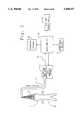
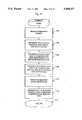
- FIG. 1is a schematic view of the system of the present invention for detecting cerebral phenomena in a non-invasive manner
- FIG. 2is a schematic view of a 19 channel EEG data acquisition and analysis system utilized in the system of FIG. 1;
- FIG. 3is a schematic view of the microcomputer used to display the EEG power spectrum and bispectrum higher-order spectrum in the system of FIG. 1;
- FIG. 4is a schematic view of the processing operations performed by the system of FIG. 1;
- FIG. 5is a flow chart of the operations of the monitor module shown in FIG. 4;
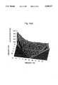
- FIGS. 6(a)-6(c)are views of sample display representations of diagnostic index generated by the system of FIG. 1;
- FIG. 7is a flow chart of the operations of the acquisition and EEG raw data management module of the system shown in FIG. 4;
- FIG. 8is a flow chart of the frequency-domain-based method for producing autobispectrum, cross bispectrum, auto power spectrum, or cross power spectrum used by the system of FIG. 1;
- FIG. 9is a flow chart of the parametric based method for producing autobispectrum, cross bispectrum, auto power spectrum, or cross power spectrum in the system of FIG. 1;
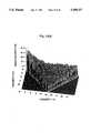
- FIG. 10(a)is a graph showing a bispectral density array generated by the system of FIG. 1;
- FIG. 10(b)is a graph showing a biphase array generated by the system of FIG. 1;
- FIG. 10(c)is a graph showing a bicoherence array generated by the system of FIG. 1;
- FIG. 10(d)is a graph showing an array of square root of real triple product generated by the system of FIG. 1;
- FIG. 11is a flow chart of the operations of the diagnostic index derivation module shown in FIG. 4;
- the apparatus of the present inventionincludes a 19 channel EEG data acquisition and analysis system 12 connected to a microcomputer 18.
- the EEG leadsare connected to a patient's head 14 by a set of surface electrodes 13.
- the international 10/20 electrode systemis preferred.
- the EEG signalsare detected by the electrodes and transmitted over a patient cable 16 to the EEG data acquisition and analysis system 12.
- the data acquisition and analysis system 12filters and amplifies the EEG waveforms.
- Commonly used digital signal processing (DSP) techniquesare applied to digitize, to low-pass filter (100 Hz), and to decimate the signals. Power spectral, bispectral, and higher-order spectral processing can then be performed.
- DSPdigital signal processing
- the system 12generates all power spectrum, bispectrum, and higher-order spectrum arrays. These arrays are then used in conjunction with clinically predetermined coefficient arrays to produce diagnostic indices. These indices are sent to the host computer 18 and are displayed on the graphics display 20. Printed output of the diagnostic index is also available on the hard copy output device 22 which is connected to the microcomputer 18. The operator interacts with the acquisition and analysis components of the system by means of a user input device 24 with feedback on the graphics display 20.
- the 19 channel data acquisition and analysis system 12is shown in greater detail in FIG. 2.
- the EEG surface potential, detected by surface electrodes 13 mounted on the patient's head 14,passes through an electrosurgery protection circuit 30, a defibrillator protection circuit 32, and an amplifier/filter circuit 36 before being passed on to the multi-channel analog to digital converter 38.
- the electrosurgery protection circuit 30includes a radio frequency (rf) filter, which limits the rf current through the patient leads 16 to less than 100 microamperes and thus protects the patient 15 from rf burns and protects the amplifiers 36 from damage resulting from exceeding the absolute maximum input voltage specified by the manufacturer.
- This circuitcan be an LC circuit consisting of a generic inductor connected in series to a generic capacitor which is then connected to ground.
- the defibrillator protection circuit 32limits the voltage to the amplifiers 36 to a safe level when a defibrillator is applied to the patient 15 and discharged.
- This circuitcan consist of a generic resistor connected, in series with the signal path, to a neon light bulb or other surge suppression device which is then connected to ground.
- the amplifier/filter circuitry 36is controlled by the microcomputer 18 for gain and filtering levels which may be adjusted by the operator. Preferred gain and filtering settings are discussed below.
- This circuit sectionconsists of three stages. The first is a pre-amplifier stage that can be assembled using a wide variety of high-impedance pre-amplifiers such as those sold by National Semiconductor, Sunnyvale Calif. The second is a stage composed of programmable filters which will allow an adjustable band pass cutoff to be selected anywhere in the range of 0.1 Hz to 4 KHz. The filters can be designed using components from Frequency Devices, Haverhill Mass. The third stage is composed of programmable amplifiers which can be assembled from operational amplifiers used in conjunction with a multiplying digital to analog (D/A) converter. Both components can be obtained from National Semiconductor. The multiplying D/A is used to set the gain to the appropriate levels requested by the microcomputer 18.
- D/Adigital to analog
- the high impedance pre-amplifier of each channelwill saturate to either the positive or negative supply voltage if the input of the pre-amplifier is not terminated. This will lead to large positive value or a large negative value at the output of amplifier/filter section 36. Such values will be used to identify lead failure.
- the output of all 19 channels of the amplifier/filter 36is fed to the multi-channel A/D converter 38 which is controlled by an input processor 44 for sampling rate settings.
- the analog signalsare converted to digital data format suitable for input to the input processor 44.
- A/D converters sold by Analog Devices, Norwood Mass.can be used for this purpose.
- the multi-channel A/D converter 38is optically coupled to the input processor 44 by optical isolator 40. All control lines to the A/D convertor 38 are also optically isolated by optical isolator 42. Any optical isolator can be used for this purpose.
- All DC power lines connected to the amplifiers 36 and A/D converter 38are also isolated from the AC power line with a DC/DC convertor 43 in order to provide complete patient isolation from ground.
- DC/DC converters available from Burr Browncan be used for this purpose.
- the basic instructions for controlling operation of the input processor 44are stored in a read only memory (ROM) 46.
- the random access memory (RAM) 48is used as a buffer memory for data and a portion of the RAM 48 can also be used as program memory when a control program is being downloaded from the microcomputer 18.
- the input processor 44has a bus 50 to communicate with its RAM 48 and ROM 46 and a separate bus 55 for communicating with the microcomputer 18.
- the memory architecture of the calculation processoris similar to that of the input processor.
- the basic instructions for controlling operation of the calculation processor 52are stored in a read only memory (ROM) 54.
- the random access memory (RAM) 56is used as a buffer memory for data and a portion of the RAM 56 can also be used as program memory when a control program is being downloaded from the microcomputer 18.
- the calculation processor 52has a bus 58 to communicate with its RAM 56 and ROM 54 and uses the bus 55 for communicating with the microcomputer 18.
- the A/D converter 38acquires the data at high speed and filtering is done by the input processor 44 to exclude frequencies outside the region of interest.
- the input processorsimultaneously decimates the sampling rate of the input data to a lower sampling rate.
- the input processor 44transfers the filtered and decimated data stream to the microcomputer 18 for display of the raw input signals via the data bus 55 and buffers 60 to the microcomputer data bus 40.
- the input processor 44also transfers the data to the calculation processor 52 for calculation of power spectrum and higher-order spectrum characteristics of the input signals via a serial communication interface 51.
- the calculation processor 52calculates power spectrum and higher-order spectrum characteristics of the input data and produces diagnostic indices from the calculated power spectrum and higher-order spectrum data.
- the input processorcan be any general purpose DSP processor such as the ADSP-2101 sold by Analog Devices, Norwood Mass.
- the calculation processoris a floating-point DSP processor in the preferred embodiment such as the TMS320C30 sold by Texas Instruments, Dallas, Tex.
- the host or microcomputer 18 of FIG. 1is shown in greater detail in FIG. 3.
- the entire microcomputer systemruns under control of a microprocessor 62 with the program memory stored in ROM 64.
- the RAM 66is used for storage of intermediate data.
- the storage device 84can be a Winchester disk or a large block of RAM or any other storage medium. It is used for storage of clinical information and can be used for archiving patient data.
- the microcomputer 18contains a math coprocessor 70 which is connected directly to microprocessor 62.
- the math coprocessor 70is used for scalar and graphic calculations.
- a graphics controller 72 operating under program control of the microprocessor 62drives a graphics display 20.
- An interface port 74provides the connection from the microcomputer bus 40 to the user interface device 24.
- the user interface device 24may be a keyboard, a pointing device or a keypad or any combination of these or similar devices.
- the interface port 74can also provide a connection between the microcomputer and an external evoked potential stimulating device. This connection will allow the microcomputer to trigger a stimulus or easily identify the onset of an independently triggered stimulus.
- the data bus 40can be used to send control data to the 19 channel data acquisition system 12 (e.g. filtering, gain, sampling rate, start/stop acquisition, perform self diagnostics) and to receive EEG data from the system, as well as to download program data to the system.
- a serial or parallel port 78is provided to drive a hard copy output device 22 for printing desired diagnostic indices.
- the system and method of the present inventioncomputes dynamic phase and density relations of EEG signals from a preselected number of leads.
- Single-valued diagnostic indicesare then generated from the data arrays by using clinically predetermined coefficient arrays.
- the resultsare quantitative indices useful for analyzing cerebral electrical activity as it relates to, for example, the assessment of depth and adequacy of anesthesia, cerebral ischemia, cerebral hypoxia, level of consciousness/hypnosis, degree of cerebral intoxication, altered evoked potential responses, and normal or abnormal cognitive processes that include but are not limited to Alzheimer's disease and HIV-related dementias.
- the assessment/determination of depth and adequacy of anesthesiaincludes but is not limited to the assessment/determination of the level of analgesia (responsiveness to painful intraoperative stimulation) as well as the level of hypnosis/consciousness.
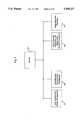
- the monitor module 402handles the overall operations of the system via integration of data and process information from the user interface module 404, acquisition and raw EEG data management module 406, power spectral, bispectral and higher-order spectral processing module 408, and the diagnostic index derivation module 410.
- a detailed illustration of module 402can be found in FIG. 5.
- the operatorcontrols and interacts with the system during the course of a procedure through the user interface and display management module 404.
- This interactionincludes, but is not limited to, entry of information regarding the patient and type of diagnostic procedure underway; lead and acquisition settings; continuous display of acquisition status, lead integrity, and diagnostic indices corresponding to regions probed by each electrode; and requests for printing and archiving results to the storage device.
- Module 404directly interacts with the monitor module 402.
- the operations handled by module 404can be achieved under a commercially available environment such as Microsoft Windows.
- the acquisition and raw EEG data management module 406handles all of the raw EEG data checking and processing prior to power spectrum, bispectrum, and higher-order spectrum analysis. This includes, but is not limited to, continuous acquisition of EEG data and the verification of its integrity; preparation of all unipolar EEG data for auto/cross power spectral, bispectral, and higher-order spectral processing. Module 406 directly interacts with the monitor module 402. A more detailed description of module 406 is provided below in connection with FIG. 7.
- the power spectral, bispectral, and higher-order spectral processing module 408controls the generation of all data arrays for power distribution, dynamic phase relations, and power coupling within the EEG. This information can be obtained by computing the auto/cross power spectrum, bispectrum, and higher-order spectra using either an FFT-based or parametric-based approach.
- the tasks performed by this moduleinclude, but are not limited to: Fourier transformation and the generation of power spectra; auto/cross bispectral density and higher order density generation; auto/cross bicoherence and higher order coherence generation; auto/cross bispectral real product and higher-order real product generation; and auto/cross biphase and higher-order phase generation.
- Module 408directly interacts with the monitor module 402. A more detailed description of module 408 is provided below in connection with FIGS. 8 and 9.
- the diagnostic index derivation module 410generates the data values used in the diagnostic process.
- the taskincludes, but is not limited to, sorting the values in the frequency band of interest for each of the required power spectrum, bispectrum, or higher-order spectrum arrays; dividing each of the sorted arrays into bins (that include one or more values) representing portions of the distribution histogram of the sorted data (i.e.
- the values in the frequency bands of interestcan also be reduced to a single number using common descriptive statistics methods such as computing the mean and standard deviation, or other preselected single values such as the minimum or maximum or any other procedure for combining or generating a single value from the values in the bin.
- Module 410directly interacts with the monitor module 402. A more detailed description of module 410 is provided below in connection with FIG. 11.
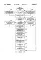
- FIG. 5A schematic of the operation of the monitor module 402 is shown in FIG. 5.
- the data arraysare filled with the most recent 63 seconds of raw digitized EEG signals, and the power spectrum, bispectrum, and higher-order spectrum data for each lead are initialized to zero.
- the data files required for storage and files containing data bases required for the computation of diagnostic indices,are also opened in the initializing step 502.
- step 504the system requests the information required to start the acquisition and diagnostic process from the user via the user interface module 404.
- This requested informationincludes patient descriptive statistics (sex, age, clinical symptoms, etc.), type of diagnostic procedure to be conducted, the leads used for auto power spectrum, bispectrum, and higher-order spectrum analysis as well as the leads to be used for cross power spectrum, bispectrum, and higher-order spectrum analysis.
- the systemIn its default mode of operation the system continuously monitors the depth and adequacy of anesthesia using a default autobispectrum database. Default band-pass filtering is performed, passing the range 0.5 to 100 Hz; the default sampling rate is set at 256 samples per second; and the default gain is set at 5000 for each lead.
- Default band-pass filteringis performed, passing the range 0.5 to 100 Hz; the default sampling rate is set at 256 samples per second; and the default gain is set at 5000 for each lead.
- the 19 EEG signals that can be acquired using the systemare: Fp1, Fp2, F7, F3, Fz, F4, FS, T3, C3, Cz, C4, T4, T5, P3, Pz, P4, T6, 01, and 02 (A1 or A2 for reference).
- one signalis required.
- This signalcan be measured directly from any of the above electrodes or it can be synthesized by linearly combining signals from two or more EEG leads. For example, two analog signals can be subtracted from each other using a differential amplifier to yield a third signal. The same operation can be performed on the two digitized signals using numerical subtraction.
- the auto power spectrum datawill provide information regarding the power distribution within the signal; the autobispectrum data will provide information regarding deviation from normality, quadratic nonlinearities and inter-frequency phase relationships within the signal; finally, auto higher-order spectrum data will provide information regarding deviation from normality, higher-order nonlinearities, and inter-frequency phase relationships within the signal.
- Such processingwill determine if the signal is made up of independent wave components or whether certain frequencies are simply harmonics of nonlinearly interacting fundamentals. Cerebral phenomena that alter the nonlinear frequency structure of the signal at the location probed by the electrode are best quantified by autobispectrum and higher-order spectrum type approaches.
- the two signalsare required.
- the two signalscan be measured directly from any of the above electrodes or either of the two signals can be synthesized by linearly combining two or more of the EEG leads as described earlier.
- the cross power spectrum datawill provide information regarding the power correlation between the two signals.
- the cross bispectrum datawill provide information regarding deviation from normality, quadratic nonlinearities, and inter-frequency phase relationships between the two signals.
- cross higher-order spectrum datawill provide information regarding deviation from normality, higher-order nonlinearities, and inter-frequency phase relationships between the two signals.
- Such processingwill determine if the frequencies in signal "X" are independent or whether they are harmonics of fundamentals present in signal "Y". This provides a better characterization of the relationship between two signals originating from separate regions of the cortex. Cerebral phenomena that alter nonlinear frequency relations between the various regions of the cortex are best quantified by cross bispectrum and cross higher-order spectrum approaches.
- the preferred embodimentwill use six signals to illustrate the operation of the system using autobispectrum analysis for the monitoring of the depth of anesthesia.
- the six signalsare derived from the following electrode placements: left and right frontal (FL/FR) signals are derived from (Fp1-Cz) and (Fp2-Cz) respectively; left and right parietal (PL/PR) signals are derived from (P3-Cz) and (P4-Cz) respectively; left and right fronto-parietal (FPL/FPR) signals are derived from (Fp1-P3) and (Fp2-P4) respectively.
- step 506a new one-second buffer of raw EEG data is acquired.
- the systemperforms artifact detection on the new one-second buffer and properly updates all data arrays. Any transmission of artifactual data is displayed to the operator in order to invoke the operator into correcting the problem.
- the systemin step 508, computes auto power spectrum and autobispectrum arrays for the signals FL, FR, PL, PR, FPL, FPR.
- Other signalsmay, of course, be used for auto/cross power spectral, bispectral, and higher-order spectral processing. Two different approaches for power spectrum, bispectrum, and higher-order spectrum computation will be discussed below with reference to FIGS. 8 and 9.
- step 510the single-valued diagnostic indices from all generated auto/cross power spectrum, bispectrum, and higher-order spectrum arrays are computed.
- the clinically predetermined coefficient arrays for the auto/cross power spectrum, bispectrum, and higher-order spectrum arraysare used for the diagnostic index computations. The generation of the coefficient arrays is discussed later.
- the systeminstantaneously displays, in step 512, all computed diagnostic indices for all signals being analyzed.
- step 514the system checks for an exit request, and if such a request has not been made, the system repeats steps 508 through 514.
- step 516requested printouts are produced, results are stored to a storage device for archival purposes and all files are closed.
- step 518the process is terminated.
- FIGS. 6(a)-6(c)A sample condensed display representation generated by the system is shown in FIGS. 6(a)-6(c). Representations of the patient's head are shown on the graphics display in FIG. 6(a) and FIG. 6(b).
- the first illustration FIG. 6(a)is divided into nineteen sections each representing the region probed by an electrode.
- the second illustration FIG. 6(b)is divided into three horizontal sections representing combined left and right hemisphere activity probed by a group of electrodes in that region.
- the virtual head displayed on the screenmay be partitioned as required for a particular diagnostic or monitoring application. For example, if a global effect like depth of anesthesia is being tracked, then one unified index along with its trend may occupy the whole display area.
- each sectioncontains the instantaneous value of the index 602 using EEG data acquired from the electrode in that region.
- each sectioncontains the instantaneous value of the computed index 604 using EEG data acquired from several electrodes in that region.
- a color-coded arrowis used to show the instantaneous change in the direction of the index. The arrow will be green if the index is within acceptable limits set as by the operator. The arrow will change to yellow if the index moves into a warning zone. A flashing red bar will replace the arrow if the index has a value that is outside the acceptable limits set for the patient.
- the instantaneous value of the index and its trend for any sectioncan be displayed as an enlarged view 606 for closer examination as shown in FIG. 6(c). This will facilitate the examination of the patient's status at a distance.
- Each sectionwill be covered by a large "X" 608 if a lead fails or artifact was detected, for any of the leads contributing to the data required to generate the diagnostic index for that region.
- step 702the system checks whether new data is being acquired for the first time. If it is, the acquisition system 12 in step 704 is supplied with requested filtering, gain, sampling rate, and lead selection information.
- the default settingsare band pass 0.5-100 Hz for filtering, 5000 for gain, 256 samples/sec for sampling rate and signals from the lead combinations FL, FR, PL, PR, FPL and FPR are acquired.
- the above settingsare quite different when the system is analyzing evoked EEG responses rather than continuous EEG signals. Common gain and filter settings to acquire signals for the various EEG evoked potentials are described below.
- EEG evoked potentialsare a means by which the sensory areas of the brain and of the central nervous system may be assayed by detecting responses in the EEG to sensory stimuli.
- PVEPPattern-shift visual evoked potentials
- BAEPBrainstem auditory evoked potentials
- SEPsomatosensory evoked potentials
- Electrodesare placed near the appropriate centers of the brain (i.e. over the visual cortexes in the case of visual evoked potentials) and EEGs are recorded for a certain period of time beginning with the administration of the stimulus.
- the stimulusis repeated many times and the resulting recordings are averaged (traditionally, in the time domain) so as to eliminate all parts of the EEG signal except that due to the stimulus.
- a series of power spectrum, bispectrum, or higher-order spectrum arrays, as produced from the EEG of the evoked responses,is averaged.
- a range of common gain settings for pattern-shift visual evoked potentialsis 20,000 to 100,000.
- a range of common filter settings for PSVEPsis 1 to 3 Hz for the low end of the band pass and 100 Hz to 300 Hz for the high end.
- step 706the acquisition system 12 acquires one second's worth of new data for all requested leads. Alternatively the signal from one complete evoked potential response is acquired if the system is analyzing evoked potentials. The system detects lead failures during the acquisition cycle in step 708 by checking for very large positive or negative values. Also in step 708, publicly available algorithms are used to check for artifacts in each lead. In step 710, leads that have failed and those producing artifactual data are marked for the monitor module 402.
- step 712the most recent 4-second record for each of the signals is assigned to X i (t), where X i (t) is the individual time series records provided for auto power spectral, autobispectral, and auto higher-order spectral processing (herein, the time series X i (t) (for all t, for one specific i) is referred to as a record).
- the most recent 4-second record from the second signalis assigned to Y i (t).
- Y i (t)is set to equal X i (t) in all cases, since only auto power spectrum, auto bispectrum, and auto higher-order spectrum computations are to be performed.
- the index idenotes the record number from 1 to 60. If evoked potentials are being analyzed, the most recent complete evoked potential response from each signal is assigned to the appropriate X i (t) and Y i (t) as described above. Using evoked potential responses as individual records will allow us to average a large number of them in the power spectrum, bispectrum, and higher-order spectrum domains.
- a circular buffer mechanismis used for storing the raw EEG for each lead, as well as the auto/cross power spectrum, bispectrum, and higher-order spectrum arrays for the sixty most recent 4-second X i (t) and Y i (t) records for each lead.
- the bufferis updated by storing the most recently acquired and processed data in the location of the oldest data. Operation of the system returns to the monitor module 402 in step 716.
- step 802the system checks whether the computation to be performed requires one signal or two signals. Typically, one time series is required to perform autospectrum analysis and two time series are required to perform cross spectrum analysis.
- step 804the system sets time records in the following manner in order to proceed with auto power spectral or autobispectral computation of the unipolar lead.
- the second set of records (Y i (t))is set to equal the first set (X i (t)).
- the corresponding Fourier transforms of X i (t) and Y i (t), respectively X i (f) and Y i (f)are also equal:
- idenotes the record number which, in this embodiment, ranges from 1 to 60.
- step 806time records are set for cross power spectral and cross bispectral analysis using two separate time series signals. As a consequence, the corresponding Fourier transforms are not equal:
- X i (t) and Y i (t)represent individually derived time series records from two separate regions probed by two or more electrodes.
- the fast Fourier transform (FFT) X i (f) and Y i (f) of each of the 60 selected records for that signalis computed using a standard IEEE library routine (or any other publicly available routine) in step 808.
- the series of transformed records, X i (f) and Y i (f)may be each normalized by dividing the value at each frequency by the constants C xi and C yi , respectively. These constants are derived separately for each record and each series (either X or Y).
- the constantcould be the total power, the largest peak in the spectrum of interest, or some other derivative of X i (f), X i (t), Y i (f), and Y i (t).
- step 810the system checks whether the computation to be performed is a power spectrum or bispectrum computation.
- the systemcomputes the auto/cross power spectral density values (PD(f)) in step 812 by using the following equations where PC(f) is the average complex product for a signal or signal pair: ##EQU1##
- Y * i (f)is the complex conjugate of Y i (f) (0 ⁇ f ⁇ f s /2) and M is the number of records (60 in the preferred embodiment).
- the systemthen returns the requested auto/cross power spectral density array to monitor module 402.
- step 814the system checks whether the computation to be performed is an autobispectrum or cross bispectrum computation.
- Autobispectrum analysisis a special case of crossbispectrum analysis and therefore different rules of symmetry apply.
- the systemuses the following equations to determine what ranges of f 1 and f 2 to use during autobispectral computation:
- f sequals the sampling rate (i.e. the number of samples per second) 256 samples per second in a preferred embodiment
- f 1 and f 2also referred to as F 1 and F 2 or Frequency 1 and Frequency 2 denote the frequency pairs over which bispectrum computation will be carried out.
- step 818the following equations are used to determine the range of f 1 and f 2 for cross bispectrum analysis:
- X i (f) and Y i (f)represent the Fourier transform of the individually derived time series records from two separate regions.
- Step 820the power spectra P xi (f) and P yi (f) of each of the 60 selected records for that signal are computed by squaring the magnitudes of each element of the Fourier transform X i (f) and Y i (f) respectively.
- the systemcomputes the average complex triple product in step 822 by using the following equations where bc i (f 1 ,f 2 ) is an individual complex triple product from one 4-second record and BC(f 1 ,f 2 ) is the average complex triple product for all 60 records:
- the average real triple productis computed in step 824 by using the following equations where br i (f 1 ,f 2 ) is an individual real triple product from one 4-second record and BR(f 1 ,f 2 ) is the average real triple product for all 60 records: ##EQU3##
- step 826the array of auto/cross bispectral density values (BD(f 1 ,f 2 )) are computed using the following equation:
- step 828the array of the square roots of the average real triple products (SBR(f 1 ,f 2 )) are computed using the following equation:
- step 830the system computes the array of auto/cross biphase values ( ⁇ (f 1 ,f 2 )) using the following equation:
- step 832the system computes the array of auto/cross bicoherence values (R(f 1 ,f 2 )) using the following equation:
- step 834the system returns the requested auto/cross power spectral density array or auto/cross bispectral density, squared-rooted average real triple product, bicoherence, biphase arrays to the monitor module 402.
- FIG. 9illustrates a parametric-based method for producing the auto power spectrum, autobispectrum, cross power spectrum, or cross bispectrum.
- steps 902, 904, and 906the system sets the time series records in the same manner as described above in steps 802, 804, and 806 respectively.
- the auto/cross power spectra of X i (t) and Y i (t)are estimated in steps 908, 910, and 912.
- This estimation methodincludes two major stages, the autoregressive (AR) model order selection and the auto/cross power spectrum computation for X i (t) and Y i (t).
- step 908the system computes two sequences of autocorrelations, [R 2x (m)]and [R 2y (m)] using the following equation. ##EQU7##
- Mis the number of records of each signal (60 in the described embodiment), and N is the number of samples per record (1024 in the described embodiment), and L is greater than the largest possible AR filter order (50 in the described embodiment).
- the order of the AR filterscan be determined by finding the location of the minimum of Final Prediction Errors: FPE x (m) and FPE y (m) respectively, i.e.,
- Qx and Qyare the locations of the minimum values for FPE x (m) and FPE y (m) (respectively) and, consequently, the orders of the AR filters of the power spectra X i (t) and Y i (t) (respectively).
- the autocorrelation sequences, [R 2x (m)] and [R 2y (m)],are entered into a Levinson recursion with order Qx and Qy, respectively, instead of L.
- step 912the transfer function of the AR filters for auto power spectra of X i (t) and Y i (t) are computed as the square root of the prediction error ( ⁇ z ) divided by the Fourier transform of the coefficients, i.e., ##EQU8##
- the auto/cross power spectral density values (PD(f))is the magnitude of the complex product of H PX (f) and the complex conjugate of H PY (f), i.e.,
- step 808the same normalization used in step 808 is may be used here (on H PZ (f).
- step 914the system checks whether the computation to be performed is a bispectrum computation, and if it is not, the system returns the requested auto/cross power spectral density array to monitor module 402.
- steps 916, 918, and 920the system sets the symmetries in the same manner as described above in steps 814, 816, and 818.
- the systemestimates the auto/cross bispectrum in steps 922, 924, and 926.
- the estimation processincludes two major stages: the order selection and bispectrum computation.
- step 924two matrices T X and T Y are formed as follows. ##EQU10##
- the auto/cross bispectrum of X i (t) and Y i (t)are computed in step 926 as the cubic root of the triple product of the skewnesses ( ⁇ X ⁇ Y ⁇ Y ) 1/3 , divided by the triple product of the Fourier transforms of the AR filter coefficients (H z (f)), i.e., ##EQU12##
- the systemAfter obtaining power spectrum and auto/cross bispectrum, the system computes the bispectral density array, the biphase, the bicoherence, and the square-rooted average real triple product (RTP) array in step 928 in the same way as in steps 826, 828, 830, and 832.
- the systemreturns to the monitor module 402 the requested auto/cross power spectral density array, bispectral density, square-rooted real triple product, biphase, and bicoherence arrays.
- the systemAfter obtaining the auto/cross Kth-order spectrum, the system computes the auto/cross Kth-order spectral density array, the auto/cross Kth-order phase, and the auto/cross Kth-order coherence the same way as in the frequency-domain-based method.
- FIGS. 10(a)-10(c)are graphs of sample autobispectral arrays showing frequency pairs 0 ⁇ f 1 ⁇ 30 Hz, and 0 ⁇ f 2 ⁇ 15 Hz.
- a bispectral density arrayis shown in FIG. 10(a) where the Z axis represents the magnitude in decibels (db) of the coupled interaction between all appropriate frequency pairs f 1 and f 2 .
- dbdecibels
- FIG. 10(c)A bicoherence array is shown in FIG. 10(c) where the Z axis represents the normalized magnitude in percent (%) of the coupled interaction between all appropriate frequency pairs f 1 and f 2 .
- a biphase arrayis shown in FIG. 10(b) where the Z axis represents the phase in radians of the coupled interaction between all appropriate frequency pairs f 1 and f 2 .
- An array of square root of real triple productis shown in FIG. 10(d) where the Z axis represents the magnitude in decibels (db) of the coupled interaction between all appropriate frequency pairs f 1 and f 2 .
- step 1102the system identifies the type of diagnostic assessment in progress.
- the 5 possible optionsare:
- the systemgets the auto/cross power spectrum, bispectrum, and/or higher-order spectrum arrays that are required for the computation of the requested diagnostic index using the sorting method described below.
- the various arrays that can be used in the generation of the diagnostic indexare: auto/cross power spectrum; auto/cross bispectral density; auto/cross bicoherence; auto/cross bispectral real product; auto/cross biphase; auto/cross Kth-order spectral density; auto/cross Kth-order coherence; auto/cross Kth-order spectral real product; and auto/cross Kth-order phase;
- the 120 data pointsare sorted in descending order, the first element in the sorted array will correspond to the largest power spectrum value, and the last element will correspond to the smallest power spectrum value.
- a distribution histogram of the powercan then be generated using the sorted array.
- the X axis on the histogramwill represent power in dBs and the Y axis will represent the number of points in the sorted array that correspond to a particular X axis power value. If all points in the sorted array are added together, the sum will represent the total power in the 0-30 Hz spectrum. If a number of adjacent points in the sorted array are added together, a portion of the histogram representing a percentage of total power is obtained. For example, in a particular EEG signal, the top 2 points in the sorted array represents the top 10% of the total power in the power distribution histogram. Similarly adding the bottom 70 points (for the same signal) in the sorted array will give the bottom 10% of the total power in the histogram.
- the top point in the sorted arraywill be equivalent to computing the peak or maximum power, while the middle element will be equivalent to the median power and the last element will be the minimum power.
- the peak valueis used.
- the minimum valueis used.
- both the peak value and the minimum valuemay be used.
- any portion of the power distribution histogramcan be obtained by adding one or more adjacent elements in the sorted array (top 25% of total power, middle 50% of total power, etc.) (given that one has empirically determined the transfer function from specific points to percentage of total power).
- sortingwe are able to track regions of high activity and low activity (peaks and valleys) in the 0-30 Hz power spectrum without having to analyze specific narrow frequency bands. This is equivalent to mapping the power spectrum to its power distribution function and operating on fixed bands within that distribution function. This transformation addresses some of the inconsistencies in the behavior of EEG power observed when hypnotic anesthetic agents are administered.
- the sorting scheme outlined abovewill transform any auto/cross power spectrum, bispectrum, and higher-order spectrum array of any dimension and any frequency band, into a one-dimensional distribution function of the values it contains.
- the one dimensional distributionis then divided into fixed bands that can be combined to produce a diagnostic index.
- the fixed bands or sequence of binscan be made up of one or more points to allow the evaluation of changes in specific peaks, valleys and other properties of the distribution of the data being analyzed.
- sortingis used in this preferred embodiment, it is intended to cover any rank ordering of any auto/cross power spectrum, bispectrum, and higher-order spectrum array of any dimension and any frequency band and the use of the rank ordering information to extract one or more points which is then used to generate one or more diagnostic indices for the assessment of cerebral phenomena in a manner consistent with this embodiment.
- the reference auto/cross power spectrum, bispectrum, and higher-order spectrum arraysare sorted.
- the corresponding dependent arraysare re-ordered according to the sorted sequence of the reference array.
- a reference arrayis an array whose values are used as the primary sort key for a group of corresponding arrays that have the same number of variables and are identical in size to the reference array. For example, if the reference array were to have four elements and they were given the indices 1, 2, 3, 4 before sorting, and after the sort the new order of the indices were 2, 1, 4, 3 then one could use the same rearrangement to reorder any other array of the same size (in this case by placing the second element first, the first element second, etc.).
- the sort of the reference arraycan rearrange the dependent arrays.
- the reference arrayis autobispectral density and the dependent arrays are autobicoherence and the square rooted average real triple product.
- Autobispectral densitywas selected as the reference array because it provides information about the residual power at each frequency pair after random phase cancellations.
- the sort of the autobispectral density arrayprovides a more stable means to select autobicoherence and real triple product values than would the sort of those arrays themselves.
- a different arraymay be selected to satisfy other requirements.
- the sum, mean, and standard deviation of the auto/cross power spectrum, bispectrum, and higher-order spectrum array of interestare also computed (before or after sorting will yield the same results since these descriptive statistics are rank order independent).
- These variablescan also be used in the generation of the diagnostic index. Additional variables can be derived directly from sorted and unsorted arrays by taking the simple ratio or product of any two sorted or descriptive variables. For example the ratio of the standard deviation to the mean will provide us with the coefficient of variation. The purpose is to break down the sorted or unsorted arrays into as many descriptors as possible.
- step 1108the sorted auto/cross power spectrum, bispectrum, and higher-order spectrum arrays are each divided into bins as described earlier. The sum of the points in each bin for each array is computed and stored in a temporary variable. Descriptive statistical variables that are generated in step 1108 may also be stored in temporary locations.
- step 1110the clinically predetermined coefficient array for the desired diagnostic index is retrieved from resident memory (or from the storage device). Each coefficient in the predetermined coefficient array corresponds to one of the temporary variables generated in step 1108.
- step 1112the diagnostic index is produced from the sum of all variables multiplied by their corresponding coefficients in the predetermined coefficient array.
- the variables used in producing the diagnostic indexmay be the sum of points in each bin, any descriptive statistical value (such as the mean, median, standard deviation, maximum value, minimum value, etc.) generated from the value of the points in each bin or any predetermined value from a sorted or unsorted array or from a bin in a sorted or unsorted array.
- the programreturns to the monitor module 402.
- the predetermined clinical coefficient arrays referred to aboveare essential to the device's ability to achieve clinically relevant diagnostic efficacy.
- the process adopted for generating these clinical reference arrayswill now be described. Since a large number of possible reference arrays must be generated to accommodate all the diagnostic modalities of the system, only one will be discussed in detail. All other reference arrays are generated in a similar fashion. For illustration purposes a method for generating the coefficients required to track the responsiveness to stressful stimulation component of depth of anesthesia using the derived signals FL and FR (of the preferred embodiment) is described below.
- a particular diagnostic indexcan be determined.
- the clinical coefficients which optimize the ability of this particular diagnostic index to predict the actual clinical diagnosisare calculated by the regression procedure.
- a series of potential diagnostic indicesmay be created and the predictive ability of each index may be determined.
- the subset of variables which results in the diagnostic index which most accurately predicts the actual outcomemay be determined.
- the purpose of the first studywas to determine whether autobispectrum variables provide information about anesthetic depth at incision. Forty adult patients were studied. Anesthesia was induced with thiopental (up to 5.0 mg/kg) and intubation performed after the administration of succinylcholine. Patients were randomly assigned to receive isoflurane 0.75 MAC (Mean Alveolar Concentration), 1.00 MAC, or 1.25 MAC in 100% oxygen. End-tidal agent concentration was monitored and after a period of steady-state had been achieved, purposeful movement in response to skin incision was assessed. Each patient was classified as either a "mover” or a "non-mover” based on the patient's response to incision.
- the purpose of the second studywas to determine whether autobispectrum variables provide information about predicting hemodynamic responses to laryngoscopy during induction with sufentanil or alfentanil.
- Forty adult patientswere studied. Patients received premedication with oral diazepam (0.05-0.15 mg/kg) and were induced with thiopental (4.0-6.0 mg/kg) and 60% nitrous oxide in oxygen, followed by vecuronium (0.1 mg/kg). Each patient was then randomly assigned to receive one of five dose regimens: normal saline; alfentanil 15 mcg/kg or 30 mcg/kg; sufentanil 0.5 mcg/kg or 1.5 mcg/kg. Laryngoscopy was performed 3 minutes after drug administration.
- Brachial blood pressurewas measured every minute with a cuff device. Patients who exhibited a change in mean arterial pressure of more than 20% in response to intubation were classified as “responders”; those who did not exhibit such a change at intubation were classified as “non-responders.”
- An autobispectral density, an auto bicoherence, and an auto square-rooted average real triple product arraywere generated for the derived signals FL and FR for each patient using a two minute period prior to the stimulus.
- the frequency band for which the bispectral arrays were computedwas 0.25-30 Hz.
- Each bispectral arraycontained 3600 data points.
- the resultant auto bispectral density, auto bicoherence, and auto square-rooted average real triple product arrayswere sorted using the auto bispectral density array as the sorting reference array.
- the sortingwas done using the algorithm described above.
- Var1Sum of the largest 15 points in sorted array
- Vat2Sum of points ranked 16th to 30th in sorted array
- Vat3Sum of points ranked 31st to 50th in sorted array
- Vat4Sum of points ranked 51th to 100th in sorted array
- Vat5Sum of points ranked 101th to 150th in sorted array
- Vat6Sum of points ranked 151th to 300th in sorted array
- Vat7Sum of points ranked 301th to 500th in sorted array
- Vat8Sum of points ranked 501st to 900th in sorted array
- Vat9Sum of points ranked 901st to 1500th in sorted array
- Var10Sum of points ranked 1501st to 2400th in sorted array
- Var11Sum of points ranked 2401st to 3600th in sorted array
- the 80 patientswere then classified into two groups.
- the first groupcontained all the patients from the first study that moved at incision and all the patients from the second study that had a change in blood pressure of greater than 20% in response to intubation.
- the secondhad all patients from the first study who did not move at incision and all the patients from the second study that had a blood pressure response of less than 20% for intubation.
- the diagnostic index (I(c 0 , c 1 , . . . , c 33 )) for a set of coefficients (c 0 , c 1 , . . . , c 33 )is given by:
- BIS A through BIS Kare the 11 sorted temporary variables from the bispectrum array; BIC A through BIC K are the variables from the bicoherence array; and PS A through PS K are the variables from the sorted square-rooted average real triple product array.
- the discriminant analysisgiven the values of the temporary variables mentioned above and the responder/non-responder classification for each patient, produces the set of coefficients which yield the best separation of responders and non-responders by the function I.
- Discriminant analysis algorithmsare publically available; in this case, the ones used are from the statistics library available from IMSL (Houston, Tex.). Below is a sample list of coefficients generated using a database of 170 patients:
- the example aboveshows one approach to obtaining a set of coefficients for a diagnostic application in a retrospective manner.
- approachescan be used to separate the clinical populations being studied using a diagnostic index.
- Such approachesinclude but are not limited to linear regression, stepwise linear regression, logistic regression, and stepwise logistic regression.
- performance of the final indexmust be confirmed in a prospective trial prior to using it in patient care.
- the analytic process described aboveis used to generate the reference databases for cerebral ischemia, cerebral hypoxia, consciousness/hypnosis, degrees of intoxication, altered evoked potential responses, and normal or abnormal cognitive processes including but not limited to identifying patients with Alzheimer's disease and HIV-related dementias.
- the system and method of the present inventionmay also be used to assess a myriad of cerebral phenomena that alter the nonlinear frequency structure of the EEG as quantified by bispectrum and higher-order spectrum approaches.
- cerebral phenomenainclude but are not limited to, cerebral ischemia, cerebral hypoxia, level of consciousness/hypnosis, degree of cerebral intoxication, altered evoked potential responses, and normal or abnormal cognitive processes caused by neurological disorders like Alzheimer's disease or HIV-related dementias.
- the system and method of the present inventionsort various auto/cross power spectra, bispectra, and higher-order spectral arrays, divides the sorted arrays into bins, and sums the variables in each bin to compute a variable, or computes a variable as a statistical value derived from the sorted or unsorted arrays or from a bin within the sorted or unsorted arrays to compute a variable, or selects a predetermined value from each sorted or unsorted array or from a bin within the sorted or unsorted array to compute a variable.
- the computed variablesare then each multiplied by a clinically derived coefficient and summed together to generate a diagnostic index.
- the different arrays that can be usedare: auto/cross power spectrum, auto/cross bispectral density, auto/cross bicoherence, auto/cross biphase, auto/cross average real triple product, auto/cross Kth-order spectral density, auto/cross Kth-order coherence, auto/cross phase, and auto/cross real product.
Landscapes
- Health & Medical Sciences (AREA)
- Life Sciences & Earth Sciences (AREA)
- Engineering & Computer Science (AREA)
- Public Health (AREA)
- General Health & Medical Sciences (AREA)
- Medical Informatics (AREA)
- Veterinary Medicine (AREA)
- Molecular Biology (AREA)
- Animal Behavior & Ethology (AREA)
- Physics & Mathematics (AREA)
- Biophysics (AREA)
- Pathology (AREA)
- Biomedical Technology (AREA)
- Heart & Thoracic Surgery (AREA)
- Surgery (AREA)
- Psychiatry (AREA)
- Physiology (AREA)
- Signal Processing (AREA)
- Computer Vision & Pattern Recognition (AREA)
- Artificial Intelligence (AREA)
- Psychology (AREA)
- Anesthesiology (AREA)
- Epidemiology (AREA)
- Primary Health Care (AREA)
- Developmental Disabilities (AREA)
- Child & Adolescent Psychology (AREA)
- Educational Technology (AREA)
- Social Psychology (AREA)
- Hospice & Palliative Care (AREA)
- Measurement And Recording Of Electrical Phenomena And Electrical Characteristics Of The Living Body (AREA)
- Investigating Or Analysing Biological Materials (AREA)
Abstract
Description
X.sub.i (t)=Y.sub.i (t)→X.sub.i (f)=Y.sub.i (f)
X.sub.i (t)≠Y.sub.i (t)→X.sub.i (f)≠Y.sub.i (f)
f.sub.1 +f.sub.2 ≦f.sub.2 /2
0≦f.sub.2 ≦f.sub.1
0≦f.sub.1 +f.sub.2 ≦f.sub.s /2
0≦f.sub.1 ≦f.sub.s /2
-f.sub.s /2≦f.sub.2 ≦f.sub.s /4
f.sub.2 ≦f.sub.1
bc.sub.i (f.sub.1,f.sub.2)=X.sub.i (f.sub.1)*X.sub.i (f.sub.2)*Y.sub.i.sup.* (f.sub.1 +f.sub.2)
BD(f.sub.1,f.sub.2)=|BC(f.sub.1,f.sub.2)|
SBR(f.sub.1,f.sub.2)=[BR(f.sub.1,f.sub.2)].sup.1/2
φ(f.sub.1,f.sub.2)=tan.sup.-8 [Im(BC(f.sub.1,f.sub.2) )/Re(BC(f.sub.1,f.sub.2))]0<φ<2π(radians)
R(f.sub.1,f.sub.2)=BD(f.sub.1,f.sub.2)/SBR(f.sub.1,f.sub.2) 0<R<1
KD(f.sub.1, f.sub.2, . . . , f.sub.K-1)=|KC(f.sub.1,f.sub.2, . . . , f.sub.K-1)|
R(f.sub.1,f.sub.2,. . . ,f.sub.K-1)=KD(f.sub.1,f.sub.2, . . . ,f.sub.K-1)/[KR(f.sub.1,f.sub.2, . . . , f.sub.K-1)].sup.1/20< R<1
FPE.sub.x (Qx)=min {FPE.sub.x (m)} and FPE.sub.y (Qy)=min {FPE.sub.y ()m)}Pc(f)=H.sub.PX (f)*H*.sub.PY (f)
PD(f)=|PC(f)|
BR(f.sub.1,f.sub.2)=P.sub.X (f.sub.1)*P.sub.X (f.sub.2)*P.sub.Y (f.sub.1 +f.sub.2)
KR(f.sub.1,f.sub.2, . . . ,f.sub.K-1)=P.sub.X (F.sub.1)*P.sub.X (f.sub.2)*. . . * P.sub.X (f.sub.K-1)* P.sub.Y (f.sub.1 +f.sub.2 +. . . +f.sub.K-1)
f.sub.1 +f.sub.2 ≦f.sub.2 /2
I(c.sub.0, c.sub.1, . . . , c.sub.33)=c.sub.0 +(BIS.sub.A *c.sub.1 +. . . +BIS.sub.K *c.sub.11) +(BIC.sub.A *C.sub.12 +. . . +BIC.sub.K *C.sub.22)+(PS.sub.A *c.sub.23 +. . . +PS.sub.K *c.sub.33)
______________________________________ for derived signals FL, FR ______________________________________ C.sub.0 -4.28 C.sub.1 -0.65 C.sub.2 +0.57 C.sub.3 +1.21 C.sub.4 -1.23 C.sub.5 +2.63 C.sub.6 -3.34 C.sub.7 +2.11 C.sub.8 +2.74 C.sub.9 -3.08 C.sub.10 0.0 C.sub.11 -0.66 C.sub.12 +0.04 C.sub.13 -1.86 C.sub.14 +0.50 C.sub.15 +0.14 C.sub.16 -0.30 C.sub.17 +0.15 C.sub.18 -0.08 C.sub.19 -0.11 C.sub.20 +0.05 C.sub.21 +0.05 C.sub.22 -0.02 C.sub.23 +0.67 C.sub.24 -1.02 C.sub.25 0.0 C.sub.26 -0.19 C.sub.27 -1.27 C.sub.28 +1.20 C.sub.29 +1.25 C.sub.30 -2.15 C.sub.31 -2.43 C.sub.32 +3.16 C.sub.33 +0.64 ______________________________________
______________________________________ Sensitivity; predicting movement at incision = 96% Specificity; predicting no movement at incision = 63% overall accuracy; predicting move/no move at = 83% incision Sensitivity; predicting >20% BP change at intubation = 100% Specificity; predicting <20% BP change at intubation = 50% Overall accuracy; predicting BP change at intubation = 85% ______________________________________
Claims (22)
Priority Applications (8)
| Application Number | Priority Date | Filing Date | Title |
|---|---|---|---|
| US08/257,356US5458117A (en) | 1991-10-25 | 1994-06-09 | Cerebral biopotential analysis system and method |
| AT95922265TATE240077T1 (en) | 1994-06-09 | 1995-06-08 | SYSTEM AND METHOD FOR ANALYZING CEREBRAL BIOPOTENTIAL |
| EP95922265AEP0764001B1 (en) | 1994-06-09 | 1995-06-08 | Cerebral biopotential analysis system and method |
| PCT/US1995/007310WO1995033404A1 (en) | 1994-06-09 | 1995-06-08 | Cerebral biopotential analysis system and method |
| CA002191594ACA2191594C (en) | 1994-06-09 | 1995-06-08 | Cerebral biopotential analysis system and method |
| ES95922265TES2199994T3 (en) | 1994-06-09 | 1995-06-08 | METHOD AND SYSTEM OF ANALYSIS OF THE BIOPOTENTIAL OF THE BRAIN. |
| DE69530768TDE69530768T2 (en) | 1994-06-09 | 1995-06-08 | SYSTEM AND METHOD FOR ANALYZING THE CEREBRAL BIOPOTENTIAL |
| JP8501313AJPH10501163A (en) | 1994-06-09 | 1995-06-08 | Brain biopotential analysis system and method |
Applications Claiming Priority (2)
| Application Number | Priority Date | Filing Date | Title |
|---|---|---|---|
| US07/782,636US5320109A (en) | 1991-10-25 | 1991-10-25 | Cerebral biopotential analysis system and method |
| US08/257,356US5458117A (en) | 1991-10-25 | 1994-06-09 | Cerebral biopotential analysis system and method |
Related Parent Applications (1)
| Application Number | Title | Priority Date | Filing Date |
|---|---|---|---|
| US07/782,636Continuation-In-PartUS5320109A (en) | 1991-10-25 | 1991-10-25 | Cerebral biopotential analysis system and method |
Publications (1)
| Publication Number | Publication Date |
|---|---|
| US5458117Atrue US5458117A (en) | 1995-10-17 |
Family
ID=22975980
Family Applications (1)
| Application Number | Title | Priority Date | Filing Date |
|---|---|---|---|
| US08/257,356Expired - LifetimeUS5458117A (en) | 1991-10-25 | 1994-06-09 | Cerebral biopotential analysis system and method |
Country Status (8)
| Country | Link |
|---|---|
| US (1) | US5458117A (en) |
| EP (1) | EP0764001B1 (en) |
| JP (1) | JPH10501163A (en) |
| AT (1) | ATE240077T1 (en) |
| CA (1) | CA2191594C (en) |
| DE (1) | DE69530768T2 (en) |
| ES (1) | ES2199994T3 (en) |
| WO (1) | WO1995033404A1 (en) |
Cited By (130)
| Publication number | Priority date | Publication date | Assignee | Title |
|---|---|---|---|---|
| WO1998027864A1 (en) | 1996-12-24 | 1998-07-02 | Aspect Medical Systems, Inc. | System for the extraction of cardiac artifacts from eeg signals |
| WO1998058325A1 (en) | 1997-06-15 | 1998-12-23 | Mario Siebler | Device for measuring and analyzing cerebral electrical activity in both cerebral hemispheres of a patient |
| US5885223A (en)* | 1995-05-31 | 1999-03-23 | Herrmann; Christoph | Process and device for assessing electroencephalograms |
| US5899867A (en)* | 1996-10-11 | 1999-05-04 | Collura; Thomas F. | System for self-administration of electroencephalographic (EEG) neurofeedback training |
| US5906208A (en)* | 1996-04-05 | 1999-05-25 | Nihon Kohden Corporation | Method and apparatus for judging a depth of anesthesia |
| US5964713A (en)* | 1995-06-02 | 1999-10-12 | Colin Corporation | Anesthetic depth measuring apparatus |
| WO2000011569A1 (en)* | 1998-08-18 | 2000-03-02 | Aspect Medical Systems, Inc. | System and method for facilitating clinical decision making |
| US6088615A (en)* | 1993-08-12 | 2000-07-11 | Masuo; Yoshihisa | Device to provide data as a guide to health management |
| WO2001060252A1 (en)* | 2000-02-17 | 2001-08-23 | The University Of Bristol | Monitoring electrical activity |
| US6298255B1 (en) | 1999-06-09 | 2001-10-02 | Aspect Medical Systems, Inc. | Smart electrophysiological sensor system with automatic authentication and validation and an interface for a smart electrophysiological sensor system |
| WO2001078586A1 (en)* | 2000-04-17 | 2001-10-25 | Stuart Graham | Method and apparatus for objective electrophysiological assessment of visual function |
| US6330470B1 (en) | 1999-12-17 | 2001-12-11 | Electrical Geodesics, Inc. | Method for localizing electrical activity in the body |
| WO2002032305A1 (en) | 2000-10-16 | 2002-04-25 | Instrumentarium Corporation | Method and apparatus for determining the cerebral state of a patient with fast response |
| DE10058230A1 (en)* | 2000-11-20 | 2002-05-29 | Gerald Ulrich | Electroencephalogram (EEG) derivation and method for converting temporal data to spectral data so that spectral data parameters can be calculated and stored in memory for quantitative comparison of patient past and future values |
| US20020085174A1 (en)* | 2000-11-22 | 2002-07-04 | Ciaran Bolger | Method and apparatus for monitoring eye tremor |
| WO2002094099A1 (en) | 2001-05-18 | 2002-11-28 | Instrumentarium Corporation | Closed loop drug administration method and apparatus using eeg complexity for control purposes |
| US6493577B1 (en) | 1997-09-23 | 2002-12-10 | Auckland Uniservices Ltd. | Method and system for detecting white matter neural injury and predicting neurological outcome particularly for preterm infants |
| US20030009111A1 (en)* | 2001-06-13 | 2003-01-09 | Cory Philip C. | Non-invasive method and apparatus for tissue detection |
| US6519486B1 (en) | 1998-10-15 | 2003-02-11 | Ntc Technology Inc. | Method, apparatus and system for removing motion artifacts from measurements of bodily parameters |
| US20030093005A1 (en)* | 2001-11-13 | 2003-05-15 | Tucker Don M. | Method for neural current imaging |
| US6594521B2 (en) | 1999-12-17 | 2003-07-15 | Electrical Geodesics, Inc. | Method for localizing electrical activity in the body |
| US20030181821A1 (en)* | 2002-01-04 | 2003-09-25 | Aspect Medical Systems, Inc. | System and method of assessment of neurological conditions using EEG |
| US20040010203A1 (en)* | 2002-07-12 | 2004-01-15 | Bionova Technologies Inc. | Method and apparatus for the estimation of anesthetic depth using wavelet analysis of the electroencephalogram |
| US6718199B2 (en)* | 2000-10-27 | 2004-04-06 | Massachusetts Eye & Ear Infirmary | Measurement of electrophysiologic response |
| US20040079372A1 (en)* | 2002-10-23 | 2004-04-29 | John Erwin R. | System and method for guidance of anesthesia, analgesia and amnesia |
| US6801803B2 (en) | 2000-10-16 | 2004-10-05 | Instrumentarium Corp. | Method and apparatus for determining the cerebral state of a patient with fast response |
| US20040210156A1 (en)* | 2002-02-19 | 2004-10-21 | Hogan Mark Bradford | Color-based neurofeedback |
| US20040243017A1 (en)* | 2003-05-06 | 2004-12-02 | Elvir Causevic | Anesthesia and sedation monitoring system and method |
| US20050043774A1 (en)* | 2003-05-06 | 2005-02-24 | Aspect Medical Systems, Inc | System and method of assessment of the efficacy of treatment of neurological disorders using the electroencephalogram |
| EP1395176A4 (en)* | 2001-06-13 | 2005-06-15 | Compumedics Ltd | Methods and apparatus for monitoring consciousness |
| US20050137494A1 (en)* | 2003-10-22 | 2005-06-23 | Viertio-Oja Hanna E. | Method and apparatus for determining the cerebral state of a patient using generalized spectral entropy of the EEG signal |
| WO2005060830A1 (en)* | 2003-12-23 | 2005-07-07 | Guangzhou Kefu Medical Technology Co., Ltd | A method of eeg fluctuation signais analysis and device thereof |
| EP1576923A1 (en)* | 2004-03-16 | 2005-09-21 | Omron Healthcare Co., Ltd. | Body composition measuring device with trend detection |
| US20050216071A1 (en)* | 2003-05-06 | 2005-09-29 | Aspect Medical Systems, Inc. | System and method of prediction of response to neurological treatment using the electroencephalogram |
| US20050280531A1 (en)* | 2004-06-18 | 2005-12-22 | Fadem Kalford C | Device and method for transmitting physiologic data |
| US20060058700A1 (en)* | 2004-08-26 | 2006-03-16 | Marro Dominic P | Patient sedation monitor |
| US20060085048A1 (en)* | 2004-10-20 | 2006-04-20 | Nervonix, Inc. | Algorithms for an active electrode, bioimpedance-based tissue discrimination system |
| US20060085049A1 (en)* | 2004-10-20 | 2006-04-20 | Nervonix, Inc. | Active electrode, bio-impedance based, tissue discrimination system and methods of use |
| US20060167370A1 (en)* | 2005-01-12 | 2006-07-27 | Aspect Medical Systems, Inc. | System and method for prediction of adverse events during treatment of psychological and neurological disorders |
| WO2006047449A3 (en)* | 2004-10-23 | 2006-07-27 | John O Bell | Passive monitoring of bioelectical signals and active electrical anesthesia stimulation |
| US20060258927A1 (en)* | 1998-10-15 | 2006-11-16 | Edgar Reuben W Jr | Method, apparatus, and system for removing motion artifacts from measurements of bodily parameters |
| US20070010723A1 (en)* | 2005-07-05 | 2007-01-11 | Kimmo Uutela | Determination of the clinical state of a subject |
| US7209787B2 (en) | 1998-08-05 | 2007-04-24 | Bioneuronics Corporation | Apparatus and method for closed-loop intracranial stimulation for optimal control of neurological disease |
| US7231254B2 (en) | 1998-08-05 | 2007-06-12 | Bioneuronics Corporation | Closed-loop feedback-driven neuromodulation |
| US20070135728A1 (en)* | 2005-12-01 | 2007-06-14 | Lexicor Medical Technology, Llc | Systems and Methods for Analyzing and Assessing Depression and Other Mood Disorders Using Electroencephalographic (EEG) Measurements |
| US7242984B2 (en) | 1998-08-05 | 2007-07-10 | Neurovista Corporation | Apparatus and method for closed-loop intracranial stimulation for optimal control of neurological disease |
| US7277758B2 (en) | 1998-08-05 | 2007-10-02 | Neurovista Corporation | Methods and systems for predicting future symptomatology in a patient suffering from a neurological or psychiatric disorder |
| US7324851B1 (en) | 1998-08-05 | 2008-01-29 | Neurovista Corporation | Closed-loop feedback-driven neuromodulation |
| US7403820B2 (en) | 1998-08-05 | 2008-07-22 | Neurovista Corporation | Closed-loop feedback-driven neuromodulation |
| US20080180278A1 (en)* | 2007-01-31 | 2008-07-31 | Medtronic, Inc. | Chopper-stabilized instrumentation amplifier for wireless telemetry |
| US7433732B1 (en)* | 2004-02-25 | 2008-10-07 | University Of Florida Research Foundation, Inc. | Real-time brain monitoring system |
| US20080269841A1 (en)* | 2007-04-30 | 2008-10-30 | Medtronic, Inc. | Chopper mixer telemetry circuit |
| US20080273084A1 (en)* | 2003-11-07 | 2008-11-06 | Neuro Kinetics, Inc. | Integrated video and electro-oculography system |
| US20090069653A1 (en)* | 2007-09-12 | 2009-03-12 | Canon Kabushiki Kaisha | Measurement apparatus |
| US20090082691A1 (en)* | 2007-09-26 | 2009-03-26 | Medtronic, Inc. | Frequency selective monitoring of physiological signals |
| US20090082829A1 (en)* | 2007-09-26 | 2009-03-26 | Medtronic, Inc. | Patient directed therapy control |
| US20090099622A1 (en)* | 2007-04-19 | 2009-04-16 | Northstar Neuroscience, Inc. | Methods and Systems for Establishing Neural Stimulation Parameters and Providing Neural Stimulation |
| EP2050388A1 (en) | 2007-10-18 | 2009-04-22 | Brain Functions Laboratory, Inc. | Apparatus for measuring brain local activity |
| US20090105785A1 (en)* | 2007-09-26 | 2009-04-23 | Medtronic, Inc. | Therapy program selection |
| US20090192556A1 (en)* | 2008-01-25 | 2009-07-30 | Medtronic, Inc. | Sleep stage detection |
| JP2009532130A (en)* | 2006-03-31 | 2009-09-10 | アスペクト メディカル システムズ,インク. | System and method for assessing the validity of analgesia using biopotential variability |
| US7623928B2 (en) | 1998-08-05 | 2009-11-24 | Neurovista Corporation | Controlling a subject's susceptibility to a seizure |
| US20100033240A1 (en)* | 2007-01-31 | 2010-02-11 | Medtronic, Inc. | Chopper-stabilized instrumentation amplifier for impedance measurement |
| US7676263B2 (en) | 2006-06-23 | 2010-03-09 | Neurovista Corporation | Minimally invasive system for selecting patient-specific therapy parameters |
| US20100094161A1 (en)* | 2008-10-09 | 2010-04-15 | Neuro Kinetics, Inc. | Quantitative, non-invasive, clinical diagnosis of traumatic brain injury using simulated distance visual stimulus device for neurologic testing |
| US20100092049A1 (en)* | 2008-04-08 | 2010-04-15 | Neuro Kinetics, Inc. | Method of Precision Eye-Tracking Through Use of Iris Edge Based Landmarks in Eye Geometry |
| US20100114223A1 (en)* | 2008-10-31 | 2010-05-06 | Wahlstrand John D | Determining intercardiac impedance |
| US7738935B1 (en)* | 2002-07-09 | 2010-06-15 | Pacesetter, Inc. | Methods and devices for reduction of motion-induced noise in pulse oximetry |
| US7747325B2 (en) | 1998-08-05 | 2010-06-29 | Neurovista Corporation | Systems and methods for monitoring a patient's neurological disease state |
| US20100327887A1 (en)* | 2007-01-31 | 2010-12-30 | Medtronic, Inc. | Chopper-stabilized instrumentation amplifier for impedance measurement |
| US20110066065A1 (en)* | 2009-08-28 | 2011-03-17 | Lexicor Medical Technology, Llc | Systems and methods to identify a subgroup of adhd at higher risk for complicating conditions |
| US20110068861A1 (en)* | 2007-01-31 | 2011-03-24 | Medtronic, Inc. | Chopper-stabilized instrumentation amplifier |
| US8036736B2 (en) | 2007-03-21 | 2011-10-11 | Neuro Vista Corporation | Implantable systems and methods for identifying a contra-ictal condition in a subject |
| WO2012027260A2 (en) | 2010-08-24 | 2012-03-01 | Nellcor Puritan Bennett Llc | SYSTEM AND METHOD TO DETERMINE SpO2 VARIABILITY AND ADDITIONAL PHYSIOLOGICAL PARAMETERS TO DETECT PATIENT STATUS |
| CN102397068A (en)* | 2010-09-15 | 2012-04-04 | 梅磊 | Measurement device for searching human brain neurotransmitter information and human brain complex network information |
| US8190249B1 (en)* | 2005-08-01 | 2012-05-29 | Infinite Biomedical Technologies, Llc | Multi-parametric quantitative analysis of bioelectrical signals |
| WO2012069887A1 (en) | 2010-11-24 | 2012-05-31 | Universidade De Trás-Os-Montes E Alto Douro | Method and device for electroencephalogram based assessment of anesthetic state during anesthesia or sedation |
| US8295934B2 (en) | 2006-11-14 | 2012-10-23 | Neurovista Corporation | Systems and methods of reducing artifact in neurological stimulation systems |
| US8554325B2 (en) | 2007-10-16 | 2013-10-08 | Medtronic, Inc. | Therapy control based on a patient movement state |
| US8588933B2 (en) | 2009-01-09 | 2013-11-19 | Cyberonics, Inc. | Medical lead termination sleeve for implantable medical devices |
| US8626264B1 (en)* | 2008-04-09 | 2014-01-07 | James T. Beran | Obtaining information about brain activity |
| WO2014008815A1 (en)* | 2012-07-11 | 2014-01-16 | 广西威利方舟科技有限公司 | Anesthesia depth monitoring device and method |
| US8725243B2 (en) | 2005-12-28 | 2014-05-13 | Cyberonics, Inc. | Methods and systems for recommending an appropriate pharmacological treatment to a patient for managing epilepsy and other neurological disorders |
| US20140155706A1 (en)* | 2011-06-17 | 2014-06-05 | Technische Universitaet Muenchen | Method and system for quantifying anaesthesia or a state of vigilance |
| US8762065B2 (en) | 1998-08-05 | 2014-06-24 | Cyberonics, Inc. | Closed-loop feedback-driven neuromodulation |
| US8786624B2 (en) | 2009-06-02 | 2014-07-22 | Cyberonics, Inc. | Processing for multi-channel signals |
| US8821397B2 (en) | 2010-09-28 | 2014-09-02 | Masimo Corporation | Depth of consciousness monitor including oximeter |
| US8849390B2 (en) | 2008-12-29 | 2014-09-30 | Cyberonics, Inc. | Processing for multi-channel signals |
| US8868172B2 (en) | 2005-12-28 | 2014-10-21 | Cyberonics, Inc. | Methods and systems for recommending an appropriate action to a patient for managing epilepsy and other neurological disorders |
| CN104605839A (en)* | 2015-02-05 | 2015-05-13 | 广州市润杰医疗器械有限公司 | Coma patient analepsia prediction method |
| US9039631B2 (en) | 2008-10-09 | 2015-05-26 | Neuro Kinetics | Quantitative, non-invasive, clinical diagnosis of traumatic brain injury using VOG device for neurologic testing |
| US9042988B2 (en) | 1998-08-05 | 2015-05-26 | Cyberonics, Inc. | Closed-loop vagus nerve stimulation |
| US20150208940A1 (en)* | 2014-01-28 | 2015-07-30 | Covidien Lp | Non-stationary feature relationship parameters for awareness monitoring |
| US9211411B2 (en) | 2010-08-26 | 2015-12-15 | Medtronic, Inc. | Therapy for rapid eye movement behavior disorder (RBD) |
| US9259591B2 (en) | 2007-12-28 | 2016-02-16 | Cyberonics, Inc. | Housing for an implantable medical device |
| US9375573B2 (en) | 1998-08-05 | 2016-06-28 | Cyberonics, Inc. | Systems and methods for monitoring a patient's neurological disease state |
| US9415222B2 (en) | 1998-08-05 | 2016-08-16 | Cyberonics, Inc. | Monitoring an epilepsy disease state with a supervisory module |
| US9439150B2 (en) | 2013-03-15 | 2016-09-06 | Medtronic, Inc. | Control of spectral agressors in a physiological signal montoring device |
| US20160310015A1 (en)* | 2011-07-25 | 2016-10-27 | Cheetah Medical, Inc. | Method and system for monitoring hemodynamics |
| US9521979B2 (en) | 2013-03-15 | 2016-12-20 | Medtronic, Inc. | Control of spectral agressors in a physiological signal monitoring device |
| US9622675B2 (en) | 2007-01-25 | 2017-04-18 | Cyberonics, Inc. | Communication error alerting in an epilepsy monitoring system |
| US9643019B2 (en) | 2010-02-12 | 2017-05-09 | Cyberonics, Inc. | Neurological monitoring and alerts |
| US9770204B2 (en) | 2009-11-11 | 2017-09-26 | Medtronic, Inc. | Deep brain stimulation for sleep and movement disorders |
| US9775545B2 (en) | 2010-09-28 | 2017-10-03 | Masimo Corporation | Magnetic electrical connector for patient monitors |
| US9788744B2 (en) | 2007-07-27 | 2017-10-17 | Cyberonics, Inc. | Systems for monitoring brain activity and patient advisory device |
| US9849241B2 (en) | 2013-04-24 | 2017-12-26 | Fresenius Kabi Deutschland Gmbh | Method of operating a control device for controlling an infusion device |
| US9898656B2 (en) | 2007-01-25 | 2018-02-20 | Cyberonics, Inc. | Systems and methods for identifying a contra-ictal condition in a subject |
| US9924904B2 (en) | 2014-09-02 | 2018-03-27 | Medtronic, Inc. | Power-efficient chopper amplifier |
| CN108618778A (en)* | 2018-05-07 | 2018-10-09 | 惠州善雅医疗器械有限公司 | A kind of deep index calculation method of fiber crops and anaesthesia depth monitor |
| IT201700039369A1 (en)* | 2017-04-10 | 2018-10-10 | Giuliano Ravasio | DEVICE FOR THE CALCULATION OF THE BETRETAL INDEX |
| US10111617B2 (en) | 2014-09-22 | 2018-10-30 | Covidien Lp | Systems and methods for EEG monitoring |
| US10154815B2 (en) | 2014-10-07 | 2018-12-18 | Masimo Corporation | Modular physiological sensors |
| US10398309B2 (en) | 2008-10-09 | 2019-09-03 | Neuro Kinetics, Inc. | Noninvasive rapid screening of mild traumatic brain injury using combination of subject's objective oculomotor, vestibular and reaction time analytic variables |
| US10743808B2 (en) | 2012-08-06 | 2020-08-18 | Neuro Kinetics | Method and associated apparatus for detecting minor traumatic brain injury |
| US10835174B2 (en) | 2016-01-12 | 2020-11-17 | Covidien Lp | System and method for monitoring cerebral activity |
| US10909177B1 (en)* | 2017-01-17 | 2021-02-02 | Workday, Inc. | Percentile determination system |
| WO2021019530A1 (en) | 2019-07-26 | 2021-02-04 | Oridion Medical 1987 Ltd. | Adaptive depth of anesthesia monitor |
| US11006841B2 (en) | 2017-06-07 | 2021-05-18 | Covidien Lp | Systems and methods for detecting strokes |
| US11273283B2 (en) | 2017-12-31 | 2022-03-15 | Neuroenhancement Lab, LLC | Method and apparatus for neuroenhancement to enhance emotional response |
| US11344745B2 (en)* | 2020-06-18 | 2022-05-31 | Diagnostyx, Inc. | Treatment of neurological abnormalities using dynamic electroencephalography |
| US11364361B2 (en) | 2018-04-20 | 2022-06-21 | Neuroenhancement Lab, LLC | System and method for inducing sleep by transplanting mental states |
| US11406317B2 (en) | 2007-12-28 | 2022-08-09 | Livanova Usa, Inc. | Method for detecting neurological and clinical manifestations of a seizure |
| US11452839B2 (en) | 2018-09-14 | 2022-09-27 | Neuroenhancement Lab, LLC | System and method of improving sleep |
| US11717686B2 (en) | 2017-12-04 | 2023-08-08 | Neuroenhancement Lab, LLC | Method and apparatus for neuroenhancement to facilitate learning and performance |
| US11723579B2 (en) | 2017-09-19 | 2023-08-15 | Neuroenhancement Lab, LLC | Method and apparatus for neuroenhancement |
| US11786694B2 (en) | 2019-05-24 | 2023-10-17 | NeuroLight, Inc. | Device, method, and app for facilitating sleep |
| US12239423B2 (en) | 2020-08-28 | 2025-03-04 | Covidien Lp | Detection of patient conditions using signals sensed on or near the head |
| US12263020B2 (en) | 2020-02-17 | 2025-04-01 | Covidien Lp | Systems and methods for detecting strokes |
| US12280219B2 (en) | 2017-12-31 | 2025-04-22 | NeuroLight, Inc. | Method and apparatus for neuroenhancement to enhance emotional response |
| US12364397B2 (en) | 2020-02-17 | 2025-07-22 | Covidien Lp | Systems and methods for detecting strokes |
Families Citing this family (9)
| Publication number | Priority date | Publication date | Assignee | Title |
|---|---|---|---|---|
| EP0828225A1 (en)* | 1996-09-04 | 1998-03-11 | Siemens Aktiengesellschaft | Process and means for analysing EEG data |
| KR100742296B1 (en)* | 2000-03-31 | 2007-07-24 | 단미터 에이/에스 | Device to extract a signal indicating the level of consciousness of the patient |
| US9259164B2 (en) | 2004-08-30 | 2016-02-16 | Neuronetrix, Inc. | Controller for neuromuscular testing |
| ATE464616T1 (en)* | 2004-08-30 | 2010-04-15 | Southern Illinois University C | BIOPOTENTIAL SIGNAL FORM DATA FUSION ANALYSIS AND CLASSIFICATION METHODS |
| DE102006017716A1 (en) | 2006-04-15 | 2007-10-18 | Forschungszentrum Jülich GmbH | Apparatus for measuring biomedical data of a subject and method for stimulating the subject with real-time processed data |
| JP6133443B2 (en)* | 2013-02-27 | 2017-05-24 | ティ・オ・ドォッブルビィ・エンジニアリング・アー/エス | Electrode and leakage current test in EEG monitor with implantable part |
| DE102013015427A1 (en)* | 2013-09-18 | 2015-03-19 | Universitätsklinikum Erlangen | Method and device for measuring cerebral perfusion |
| JP7083471B2 (en)* | 2018-09-26 | 2022-06-13 | 株式会社国際電気通信基礎技術研究所 | Estimator, estimation program and estimation method |
| CN109498004A (en)* | 2019-01-04 | 2019-03-22 | 深圳市舟洁信息咨询服务有限公司 | A kind of anesthesia data monitoring system and method |
Citations (11)
| Publication number | Priority date | Publication date | Assignee | Title |
|---|---|---|---|---|
| US3863625A (en)* | 1973-11-02 | 1975-02-04 | Us Health | Epileptic seizure warning system |
| US4407299A (en)* | 1981-05-15 | 1983-10-04 | The Children's Medical Center Corporation | Brain electrical activity mapping |
| US4557270A (en)* | 1983-08-23 | 1985-12-10 | New York University | Electroencephalographic system for intra-operative open-heart surgery |
| US4697598A (en)* | 1985-04-25 | 1987-10-06 | Westinghouse Electric Corp. | Evoked potential autorefractometry system |
| US4705049A (en)* | 1986-08-04 | 1987-11-10 | John Erwin R | Intraoperative monitoring or EP evaluation system utilizing an automatic adaptive self-optimizing digital comb filter |
| US4753246A (en)* | 1986-03-28 | 1988-06-28 | The Regents Of The University Of California | EEG spatial filter and method |
| US4846190A (en)* | 1983-08-23 | 1989-07-11 | John Erwin R | Electroencephalographic system data display |
| US4905285A (en)* | 1987-04-03 | 1990-02-27 | American Telephone And Telegraph Company, At&T Bell Laboratories | Analysis arrangement based on a model of human neural responses |
| US4907597A (en)* | 1987-10-09 | 1990-03-13 | Biometrak Corporation | Cerebral biopotential analysis system and method |
| US4974602A (en)* | 1988-08-16 | 1990-12-04 | Siemens Aktiengesellschaft | Arrangement for analyzing local bioelectric currents in biological tissue complexes |
| US5010891A (en)* | 1987-10-09 | 1991-04-30 | Biometrak Corporation | Cerebral biopotential analysis system and method |
Family Cites Families (1)
| Publication number | Priority date | Publication date | Assignee | Title |
|---|---|---|---|---|
| US5320109A (en)* | 1991-10-25 | 1994-06-14 | Aspect Medical Systems, Inc. | Cerebral biopotential analysis system and method |
- 1994
- 1994-06-09USUS08/257,356patent/US5458117A/ennot_activeExpired - Lifetime
- 1995
- 1995-06-08CACA002191594Apatent/CA2191594C/ennot_activeExpired - Lifetime
- 1995-06-08DEDE69530768Tpatent/DE69530768T2/ennot_activeExpired - Lifetime
- 1995-06-08ESES95922265Tpatent/ES2199994T3/ennot_activeExpired - Lifetime
- 1995-06-08WOPCT/US1995/007310patent/WO1995033404A1/enactiveIP Right Grant
- 1995-06-08ATAT95922265Tpatent/ATE240077T1/ennot_activeIP Right Cessation
- 1995-06-08EPEP95922265Apatent/EP0764001B1/ennot_activeExpired - Lifetime
- 1995-06-08JPJP8501313Apatent/JPH10501163A/enactivePending
Patent Citations (11)
| Publication number | Priority date | Publication date | Assignee | Title |
|---|---|---|---|---|
| US3863625A (en)* | 1973-11-02 | 1975-02-04 | Us Health | Epileptic seizure warning system |
| US4407299A (en)* | 1981-05-15 | 1983-10-04 | The Children's Medical Center Corporation | Brain electrical activity mapping |
| US4557270A (en)* | 1983-08-23 | 1985-12-10 | New York University | Electroencephalographic system for intra-operative open-heart surgery |
| US4846190A (en)* | 1983-08-23 | 1989-07-11 | John Erwin R | Electroencephalographic system data display |
| US4697598A (en)* | 1985-04-25 | 1987-10-06 | Westinghouse Electric Corp. | Evoked potential autorefractometry system |
| US4753246A (en)* | 1986-03-28 | 1988-06-28 | The Regents Of The University Of California | EEG spatial filter and method |
| US4705049A (en)* | 1986-08-04 | 1987-11-10 | John Erwin R | Intraoperative monitoring or EP evaluation system utilizing an automatic adaptive self-optimizing digital comb filter |
| US4905285A (en)* | 1987-04-03 | 1990-02-27 | American Telephone And Telegraph Company, At&T Bell Laboratories | Analysis arrangement based on a model of human neural responses |
| US4907597A (en)* | 1987-10-09 | 1990-03-13 | Biometrak Corporation | Cerebral biopotential analysis system and method |
| US5010891A (en)* | 1987-10-09 | 1991-04-30 | Biometrak Corporation | Cerebral biopotential analysis system and method |
| US4974602A (en)* | 1988-08-16 | 1990-12-04 | Siemens Aktiengesellschaft | Arrangement for analyzing local bioelectric currents in biological tissue complexes |
Non-Patent Citations (45)
| Title |
|---|
| Baker, A. B., and Roxburgh, A. J. Computerised EEG monitoring for carotid endarterectomy. Anaesth Intensive Care. 14(1): pp. 32 36 (1986).* |
| Baker, A. B., and Roxburgh, A. J. Computerised EEG monitoring for carotid endarterectomy. Anaesth-Intensive Care. 14(1): pp. 32-36 (1986). |
| Barnett, T. P., Johnson, L. C., Naitoh, P., Hicks, N. and Nute, C., Bispectrum analysis of electroencephalogram signals during waking and sleeping. Science 172:402 401 (1971).* |
| Barnett, T. P., Johnson, L. C., Naitoh, P., Hicks, N. and Nute, C., Bispectrum analysis of electroencephalogram signals during waking and sleeping. Science 172:402-401 (1971). |
| Brillinger, D. R. An introduction to polyspectra. Annals of Mathematical Statistics 36:1351 1374 (1965).* |
| Brillinger, D. R. An introduction to polyspectra. Annals of Mathematical Statistics 36:1351-1374 (1965). |
| Dumermuth, G., Huber, P. J., Kleiner, B., and Gasser, T. Analysis of the interrelations between frequency bands of the EEG by means of the bispectrum. A preliminary study. Electroencephalogr. Clin. Neurophysiol. 31(2):137 148 (1971).* |
| Dumermuth, G., Huber, P. J., Kleiner, B., and Gasser, T. Analysis of the interrelations between frequency bands of the EEG by means of the bispectrum. A preliminary study. Electroencephalogr. Clin. Neurophysiol. 31(2):137-148 (1971). |
| Eichhorn, J. H., Cooper, J. B., Cullen, D. J., Ward, M. R., Philip, J. H., and Seeman, R. G. Standards for patient monitoring during anesthesia at Harvard Medical School. JAMA. 256(8): pp. 1017 1020 (1986).* |
| Eichhorn, J. H., Cooper, J. B., Cullen, D. J., Ward, M. R., Philip, J. H., and Seeman, R. G. Standards for patient monitoring during anesthesia at Harvard Medical School. JAMA. 256(8): pp. 1017-1020 (1986). |
| Haykin, S. Adaptive filter theory. Prentice Hall, Englewood Cliffs, N.J. (1986).* |
| Haykin, S. Adaptive filter theory. Prentice-Hall, Englewood Cliffs, N.J. (1986). |
| Huber, P. J., B. Kleiner, T. Gasser and G. Dumermuth. Statistical method for investigating phase relations in stationary stochastic processes. IEEE Trans. Aud. & Electroacou. AU 19/1:78 86 (1971).* |
| Huber, P. J., B. Kleiner, T. Gasser and G. Dumermuth. Statistical method for investigating phase relations in stationary stochastic processes. IEEE Trans. Aud. & Electroacou. AU-19/1:78-86 (1971). |
| IEEE Engineering in Medicine & Biology Society 10th Annual Conference.* |
| IEEE Transactions on Biomedical Engineering, IEEE, New York, vol. 36, No. 4, Apr. 4, 1989, pp. 497 499, Ning et al, Bispectral Analysis of the Rat EEG During Various Vigilance States .* |
| IEEE Transactions on Biomedical Engineering, IEEE, New York, vol. 36, No. 4, Apr. 4, 1989, pp. 497-499, Ning et al, "Bispectral Analysis of the Rat EEG During Various Vigilance States". |
| IEEE, New York, vol. 3/4, Nov. 4, 1988, New Orleans, pp. 1218 1219, Ning et al, Bispectral Analysis of Rat EEG During Maturation .* |
| IEEE, New York, vol. 3/4, Nov. 4, 1988, New Orleans, pp. 1218-1219, Ning et al, "Bispectral Analysis of Rat EEG During Maturation". |
| Jasper, H. H. The ten twenty electrode system of the international federation in electroencephalography and clinical neurophysiology. EEG Journal. 10:371 375 (1985).* |
| Jasper, H. H. The ten-twenty electrode system of the international federation in electroencephalography and clinical neurophysiology. EEG Journal. 10:371-375 (1985). |
| Kleiner, B., Huber, P. J., and Dumermuth, G. Analysis of the interelations between frequency bands of the EEG by means of the bispectrum. Electroencephalogr. Neurophysiol. 27(7): 693 694 (1969).* |
| Kleiner, B., Huber, P. J., and Dumermuth, G. Analysis of the interelations between frequency bands of the EEG by means of the bispectrum. Electroencephalogr. Neurophysiol. 27(7): 693-694 (1969). |
| Levy, W. J. Intraoperative EEG patterns: implications for EEG monitoring. Anesthesiology. 60(5): pp. 430 434 (1984).* |
| Levy, W. J. Intraoperative EEG patterns: implications for EEG monitoring. Anesthesiology. 60(5): pp. 430-434 (1984). |
| Nikias, C. L., and Raghuveer, M. R. Bispectrum estimation: A digital signal processing framework. Proc. IEEE. 75,7:869 91 (1987).* |
| Nikias, C. L., and Raghuveer, M. R. Bispectrum estimation: A digital signal processing framework. Proc. IEEE. 75,7:869-91 (1987). |
| Pichlmayr, I., and Lips, U. EEG Monitoring in anesthesiology and intensive care. Neuropsychobiology. 10(4): pp. 239 248 (1983).* |
| Pichlmayr, I., and Lips, U. EEG Monitoring in anesthesiology and intensive care. Neuropsychobiology. 10(4): pp. 239-248 (1983). |
| Proceedings of the Fourteenth Annual Northeast Bioengineering Conference, IEEE, New York, US 10 Mar. 1988, Durham, N.H., pp. 198 203, XP000093434, Ning et al, Detecting Biphase Coupling of Sleep EEG Via Bispectra .* |
| Proceedings of the Fourteenth Annual Northeast Bioengineering Conference, IEEE, New York, US 10 Mar. 1988, Durham, N.H., pp. 198-203, XP000093434, Ning et al, "Detecting Biphase Coupling of Sleep EEG Via Bispectra". |
| Raghuveer, M. R. and Nikias, C. L. Bispectrum estimation: A parametric approach. IEEE Trans. on Acoustics, Speech & Signal Processing. 33:1213 1230 (1985).* |
| Raghuveer, M. R. and Nikias, C. L. Bispectrum estimation: A parametric approach. IEEE Trans. on Acoustics, Speech & Signal Processing. 33:1213-1230 (1985). |
| Rampil, I. J., Holzer, J. A., Quest, D. O., Rosenbaum, S. H., and Correll J. W., Prognostic value of computerized EEG analysis during carotic endarterectomy. Anesthesia Analgesia 62:186 92 (1983).* |
| Rampil, I. J., Holzer, J. A., Quest, D. O., Rosenbaum, S. H., and Correll J. W., Prognostic value of computerized EEG analysis during carotic endarterectomy. Anesthesia Analgesia 62:186-92 (1983). |
| Russ, W., Kling, D., Krumholz, W., Fraedrich, G., and Hempelmann, G., [Experience with a new EEG spectral analyzer in carotid surgery] Erfahrungen mit einem neuen EEG-Spektralanalysator in der Karotischirurgie. Anaesthetist 34(2): pp. 85-90 (1985). |
| Russ, W., Kling, D., Krumholz, W., Fraedrich, G., and Hempelmann, G., Experience with a new EEG spectral analyzer in carotid surgery Erfahrungen mit einem neuen EEG Spektralanalysator in der Karotischirurgie. Anaesthetist 34(2): pp. 85 90 (1985).* |
| Susumu, T. and Osamu, T. Analysis of wave shapes of alpha waves on EEG by means of the bispectrum. (1973).* |
| Tryon, P. V. The bispectrum and higher order spectra: A bibliography. US NBS (Tech Note 1036) (1981).* |
| Tryon, P. V. The bispectrum and higher-order spectra: A bibliography. US NBS (Tech Note 1036) (1981). |
| Volavka, J., Matousek, M., Feldstein, S. et al. The reliability of electroencephalography assesement. Electroencephalography and Electromyography. 4: 123 (1973).* |
| Whitton, Genetic Dependence of the Electroencephalogram Bispectrum, Electroenceophalogray and Clinical Neurophysiology, 1985, 60: 293 298, Elsevier Scientific Publishers Ireland, Ltd.* |
| Whitton, Genetic Dependence of the Electroencephalogram Bispectrum, Electroenceophalogray and Clinical Neurophysiology, 1985, 60: 293-298, Elsevier Scientific Publishers Ireland, Ltd. |
| Withington, PS., Morton, J., Arnold, R., Sebel, PS., and Moberg, R. Assessment of power spectral edge for monitoring depth of anesthesia using low methohexitone infusion. Int J Clin Monit Computing. 3(2): pp. 117 122 (1986).* |
| Withington, PS., Morton, J., Arnold, R., Sebel, PS., and Moberg, R. Assessment of power spectral edge for monitoring depth of anesthesia using low methohexitone infusion. Int-J-Clin-Monit-Computing. 3(2): pp. 117-122 (1986). |
Cited By (230)
| Publication number | Priority date | Publication date | Assignee | Title |
|---|---|---|---|---|
| US6321112B1 (en) | 1993-08-12 | 2001-11-20 | Omron Corporation | Device to provide data as a guide to health management |
| US6088615A (en)* | 1993-08-12 | 2000-07-11 | Masuo; Yoshihisa | Device to provide data as a guide to health management |
| US6308096B1 (en) | 1993-08-12 | 2001-10-23 | Omron, Corporation | Device to provide data as a guide to health management |
| US5885223A (en)* | 1995-05-31 | 1999-03-23 | Herrmann; Christoph | Process and device for assessing electroencephalograms |
| US5964713A (en)* | 1995-06-02 | 1999-10-12 | Colin Corporation | Anesthetic depth measuring apparatus |
| US5906208A (en)* | 1996-04-05 | 1999-05-25 | Nihon Kohden Corporation | Method and apparatus for judging a depth of anesthesia |
| US5899867A (en)* | 1996-10-11 | 1999-05-04 | Collura; Thomas F. | System for self-administration of electroencephalographic (EEG) neurofeedback training |
| US5792069A (en)* | 1996-12-24 | 1998-08-11 | Aspect Medical Systems, Inc. | Method and system for the extraction of cardiac artifacts from EEG signals |
| WO1998027864A1 (en) | 1996-12-24 | 1998-07-02 | Aspect Medical Systems, Inc. | System for the extraction of cardiac artifacts from eeg signals |
| WO1998058325A1 (en) | 1997-06-15 | 1998-12-23 | Mario Siebler | Device for measuring and analyzing cerebral electrical activity in both cerebral hemispheres of a patient |
| US6343229B1 (en) | 1997-06-15 | 2002-01-29 | Mario Siebler | Device for measurement and analysis of brain activity of both cerebral hemispheres in a patient |
| US6493577B1 (en) | 1997-09-23 | 2002-12-10 | Auckland Uniservices Ltd. | Method and system for detecting white matter neural injury and predicting neurological outcome particularly for preterm infants |
| US9375573B2 (en) | 1998-08-05 | 2016-06-28 | Cyberonics, Inc. | Systems and methods for monitoring a patient's neurological disease state |
| US7403820B2 (en) | 1998-08-05 | 2008-07-22 | Neurovista Corporation | Closed-loop feedback-driven neuromodulation |
| US7209787B2 (en) | 1998-08-05 | 2007-04-24 | Bioneuronics Corporation | Apparatus and method for closed-loop intracranial stimulation for optimal control of neurological disease |
| US8781597B2 (en) | 1998-08-05 | 2014-07-15 | Cyberonics, Inc. | Systems for monitoring a patient's neurological disease state |
| US7231254B2 (en) | 1998-08-05 | 2007-06-12 | Bioneuronics Corporation | Closed-loop feedback-driven neuromodulation |
| US7242984B2 (en) | 1998-08-05 | 2007-07-10 | Neurovista Corporation | Apparatus and method for closed-loop intracranial stimulation for optimal control of neurological disease |
| US7277758B2 (en) | 1998-08-05 | 2007-10-02 | Neurovista Corporation | Methods and systems for predicting future symptomatology in a patient suffering from a neurological or psychiatric disorder |
| US7930035B2 (en) | 1998-08-05 | 2011-04-19 | Neurovista Corporation | Providing output indicative of subject's disease state |
| US7324851B1 (en) | 1998-08-05 | 2008-01-29 | Neurovista Corporation | Closed-loop feedback-driven neuromodulation |
| US9042988B2 (en) | 1998-08-05 | 2015-05-26 | Cyberonics, Inc. | Closed-loop vagus nerve stimulation |
| US7623928B2 (en) | 1998-08-05 | 2009-11-24 | Neurovista Corporation | Controlling a subject's susceptibility to a seizure |
| US9421373B2 (en) | 1998-08-05 | 2016-08-23 | Cyberonics, Inc. | Apparatus and method for closed-loop intracranial stimulation for optimal control of neurological disease |
| US7853329B2 (en) | 1998-08-05 | 2010-12-14 | Neurovista Corporation | Monitoring efficacy of neural modulation therapy |
| US9415222B2 (en) | 1998-08-05 | 2016-08-16 | Cyberonics, Inc. | Monitoring an epilepsy disease state with a supervisory module |
| US9320900B2 (en) | 1998-08-05 | 2016-04-26 | Cyberonics, Inc. | Methods and systems for determining subject-specific parameters for a neuromodulation therapy |
| US7747325B2 (en) | 1998-08-05 | 2010-06-29 | Neurovista Corporation | Systems and methods for monitoring a patient's neurological disease state |
| US9113801B2 (en) | 1998-08-05 | 2015-08-25 | Cyberonics, Inc. | Methods and systems for continuous EEG monitoring |
| US8762065B2 (en) | 1998-08-05 | 2014-06-24 | Cyberonics, Inc. | Closed-loop feedback-driven neuromodulation |
| WO2000011569A1 (en)* | 1998-08-18 | 2000-03-02 | Aspect Medical Systems, Inc. | System and method for facilitating clinical decision making |
| US6338713B1 (en) | 1998-08-18 | 2002-01-15 | Aspect Medical Systems, Inc. | System and method for facilitating clinical decision making |
| US6810277B2 (en) | 1998-10-15 | 2004-10-26 | Ric Investments, Inc. | Method, apparatus and system for removing motion artifacts from measurements of bodily parameters |
| US6519486B1 (en) | 1998-10-15 | 2003-02-11 | Ntc Technology Inc. | Method, apparatus and system for removing motion artifacts from measurements of bodily parameters |
| US20060258927A1 (en)* | 1998-10-15 | 2006-11-16 | Edgar Reuben W Jr | Method, apparatus, and system for removing motion artifacts from measurements of bodily parameters |
| US7072702B2 (en) | 1998-10-15 | 2006-07-04 | Ric Investments, Llc | Method, apparatus and system for removing motion artifacts from measurements of bodily parameters |
| US7991448B2 (en) | 1998-10-15 | 2011-08-02 | Philips Electronics North America Corporation | Method, apparatus, and system for removing motion artifacts from measurements of bodily parameters |
| US6298255B1 (en) | 1999-06-09 | 2001-10-02 | Aspect Medical Systems, Inc. | Smart electrophysiological sensor system with automatic authentication and validation and an interface for a smart electrophysiological sensor system |
| US6594521B2 (en) | 1999-12-17 | 2003-07-15 | Electrical Geodesics, Inc. | Method for localizing electrical activity in the body |
| US6330470B1 (en) | 1999-12-17 | 2001-12-11 | Electrical Geodesics, Inc. | Method for localizing electrical activity in the body |
| WO2001060252A1 (en)* | 2000-02-17 | 2001-08-23 | The University Of Bristol | Monitoring electrical activity |
| US6748263B2 (en) | 2000-02-17 | 2004-06-08 | The University Of Bristol | Monitoring electrical activity |
| US7740592B2 (en) | 2000-04-17 | 2010-06-22 | The University Of Sydney | Method and apparatus for objective electrophysiological assessment of visual function |
| US20090076406A1 (en)* | 2000-04-17 | 2009-03-19 | Graham Stuart L | Method and apparatus for objective electrophysiological assessment of visual function |
| US20110218456A1 (en)* | 2000-04-17 | 2011-09-08 | The University Of Sydney | Method and apparatus for objective electrophysiological assessment of visual function |
| US7972278B2 (en) | 2000-04-17 | 2011-07-05 | The University Of Sydney | Method and apparatus for objective electrophysiological assessment of visual function |
| WO2001078586A1 (en)* | 2000-04-17 | 2001-10-25 | Stuart Graham | Method and apparatus for objective electrophysiological assessment of visual function |
| US20030158497A1 (en)* | 2000-04-17 | 2003-08-21 | Graham Stuart L. | Method and apparatus for objective electrophysiological assessment of visual function |
| US20030167019A1 (en)* | 2000-10-16 | 2003-09-04 | Viertio-Oja Hanna E. | Method and apparatus for determining the cerebral state of a patient with fast response |
| US7228169B2 (en) | 2000-10-16 | 2007-06-05 | Ge Healthcare Finland Oy | Method and apparatus for determining the cerebral state of a patient with fast response |
| WO2002032305A1 (en) | 2000-10-16 | 2002-04-25 | Instrumentarium Corporation | Method and apparatus for determining the cerebral state of a patient with fast response |
| US6801803B2 (en) | 2000-10-16 | 2004-10-05 | Instrumentarium Corp. | Method and apparatus for determining the cerebral state of a patient with fast response |
| US6731975B1 (en) | 2000-10-16 | 2004-05-04 | Instrumentarium Corp. | Method and apparatus for determining the cerebral state of a patient with fast response |
| US6718199B2 (en)* | 2000-10-27 | 2004-04-06 | Massachusetts Eye & Ear Infirmary | Measurement of electrophysiologic response |
| DE10058230A1 (en)* | 2000-11-20 | 2002-05-29 | Gerald Ulrich | Electroencephalogram (EEG) derivation and method for converting temporal data to spectral data so that spectral data parameters can be calculated and stored in memory for quantitative comparison of patient past and future values |
| US8025404B2 (en) | 2000-11-22 | 2011-09-27 | Eyetect, L.L.C. | Method and apparatus for monitoring eye tremor |
| US8500282B2 (en) | 2000-11-22 | 2013-08-06 | Boston Brainstem, Inc. | Method and apparatus for monitoring eye tremor |
| US20020085174A1 (en)* | 2000-11-22 | 2002-07-04 | Ciaran Bolger | Method and apparatus for monitoring eye tremor |
| US7011410B2 (en) | 2000-11-22 | 2006-03-14 | Eyetect, L.L.C. | Method and apparatus for monitoring eye tremor |
| US20100049075A1 (en)* | 2000-11-22 | 2010-02-25 | Ciaran Bolger | Method and apparatus for monitoring eye tremor |
| WO2002094099A1 (en) | 2001-05-18 | 2002-11-28 | Instrumentarium Corporation | Closed loop drug administration method and apparatus using eeg complexity for control purposes |
| USRE41291E1 (en) | 2001-05-18 | 2010-04-27 | Ge Healthcare Finland Oy | Closed loop drug administration method and apparatus using EEG complexity for control purposes |
| US6631291B2 (en) | 2001-05-18 | 2003-10-07 | Instrumentarium Corp. | Closed loop drug administration method and apparatus using EEG complexity for control purposes |
| US20030009111A1 (en)* | 2001-06-13 | 2003-01-09 | Cory Philip C. | Non-invasive method and apparatus for tissue detection |
| EP1395176A4 (en)* | 2001-06-13 | 2005-06-15 | Compumedics Ltd | Methods and apparatus for monitoring consciousness |
| CN100506147C (en)* | 2001-06-13 | 2009-07-01 | 康普麦迪克斯有限公司 | Methods and devices for monitoring consciousness |
| EP1989998A2 (en) | 2001-06-13 | 2008-11-12 | Compumedics Limited | Methods and apparatus for monitoring consciousness |
| US7840250B2 (en) | 2001-11-13 | 2010-11-23 | Electrical Geodesics, Inc. | Method for neural current imaging |
| US20030093005A1 (en)* | 2001-11-13 | 2003-05-15 | Tucker Don M. | Method for neural current imaging |
| US7231245B2 (en) | 2002-01-04 | 2007-06-12 | Aspect Medical Systems, Inc. | System and method of assessment of neurological conditions using EEG |
| US20030181821A1 (en)* | 2002-01-04 | 2003-09-25 | Aspect Medical Systems, Inc. | System and method of assessment of neurological conditions using EEG |
| EP1460933B1 (en)* | 2002-01-04 | 2008-12-17 | Aspect Medical Systems, Inc. | System and method of assessment of mood disorders using eeg |
| US20040210156A1 (en)* | 2002-02-19 | 2004-10-21 | Hogan Mark Bradford | Color-based neurofeedback |
| US7035686B2 (en) | 2002-02-19 | 2006-04-25 | Mark Bradford Hogan | Color-based neurofeedback |
| US7738935B1 (en)* | 2002-07-09 | 2010-06-15 | Pacesetter, Inc. | Methods and devices for reduction of motion-induced noise in pulse oximetry |
| US20040010203A1 (en)* | 2002-07-12 | 2004-01-15 | Bionova Technologies Inc. | Method and apparatus for the estimation of anesthetic depth using wavelet analysis of the electroencephalogram |
| US7373198B2 (en) | 2002-07-12 | 2008-05-13 | Bionova Technologies Inc. | Method and apparatus for the estimation of anesthetic depth using wavelet analysis of the electroencephalogram |
| US7089927B2 (en)* | 2002-10-23 | 2006-08-15 | New York University | System and method for guidance of anesthesia, analgesia and amnesia |
| US20040079372A1 (en)* | 2002-10-23 | 2004-04-29 | John Erwin R. | System and method for guidance of anesthesia, analgesia and amnesia |
| US7706871B2 (en) | 2003-05-06 | 2010-04-27 | Nellcor Puritan Bennett Llc | System and method of prediction of response to neurological treatment using the electroencephalogram |
| US20040243017A1 (en)* | 2003-05-06 | 2004-12-02 | Elvir Causevic | Anesthesia and sedation monitoring system and method |
| US20050043774A1 (en)* | 2003-05-06 | 2005-02-24 | Aspect Medical Systems, Inc | System and method of assessment of the efficacy of treatment of neurological disorders using the electroencephalogram |
| US20050216071A1 (en)* | 2003-05-06 | 2005-09-29 | Aspect Medical Systems, Inc. | System and method of prediction of response to neurological treatment using the electroencephalogram |
| US7418290B2 (en) | 2003-05-06 | 2008-08-26 | Aspect Medical Systems, Inc. | System and method of assessment of the efficacy of treatment of neurological disorders using the electroencephalogram |
| US7509161B2 (en) | 2003-10-22 | 2009-03-24 | Instrumentarium Corporation | Method and apparatus for determining the cerebral state of a patient using generalized spectral entropy of the EEG signal |
| US20050137494A1 (en)* | 2003-10-22 | 2005-06-23 | Viertio-Oja Hanna E. | Method and apparatus for determining the cerebral state of a patient using generalized spectral entropy of the EEG signal |
| US20080273084A1 (en)* | 2003-11-07 | 2008-11-06 | Neuro Kinetics, Inc. | Integrated video and electro-oculography system |
| US9101296B2 (en) | 2003-11-07 | 2015-08-11 | Neuro Kinetics | Integrated video and electro-oculography system |
| WO2005060830A1 (en)* | 2003-12-23 | 2005-07-07 | Guangzhou Kefu Medical Technology Co., Ltd | A method of eeg fluctuation signais analysis and device thereof |
| US7801597B2 (en)* | 2003-12-23 | 2010-09-21 | Guangzhou Kefu Medical Technology Co., Ltd | Method and apparatus for brain wave fluctuations analysis |
| US20070185407A1 (en)* | 2003-12-23 | 2007-08-09 | Guangzhou Kefu Medical Technology Co., Ltd. | Method and apparatus for brain wave fluctuations analysis |
| US7433732B1 (en)* | 2004-02-25 | 2008-10-07 | University Of Florida Research Foundation, Inc. | Real-time brain monitoring system |
| EP1576923A1 (en)* | 2004-03-16 | 2005-09-21 | Omron Healthcare Co., Ltd. | Body composition measuring device with trend detection |
| US20050209528A1 (en)* | 2004-03-16 | 2005-09-22 | Omron Healthcare Co., Ltd. | Body composition measuring device capable of comparing current measured value with past measured value |
| US20050280531A1 (en)* | 2004-06-18 | 2005-12-22 | Fadem Kalford C | Device and method for transmitting physiologic data |
| US20060058700A1 (en)* | 2004-08-26 | 2006-03-16 | Marro Dominic P | Patient sedation monitor |
| US20060085048A1 (en)* | 2004-10-20 | 2006-04-20 | Nervonix, Inc. | Algorithms for an active electrode, bioimpedance-based tissue discrimination system |
| US7865236B2 (en) | 2004-10-20 | 2011-01-04 | Nervonix, Inc. | Active electrode, bio-impedance based, tissue discrimination system and methods of use |
| US20060085049A1 (en)* | 2004-10-20 | 2006-04-20 | Nervonix, Inc. | Active electrode, bio-impedance based, tissue discrimination system and methods of use |
| WO2006047449A3 (en)* | 2004-10-23 | 2006-07-27 | John O Bell | Passive monitoring of bioelectical signals and active electrical anesthesia stimulation |
| US8005534B2 (en) | 2005-01-12 | 2011-08-23 | Nellcor Puritan Bennett Llc | System and method for prediction of adverse events during treatment of psychological and neurological disorders |
| US20060167370A1 (en)* | 2005-01-12 | 2006-07-27 | Aspect Medical Systems, Inc. | System and method for prediction of adverse events during treatment of psychological and neurological disorders |
| US20070010723A1 (en)* | 2005-07-05 | 2007-01-11 | Kimmo Uutela | Determination of the clinical state of a subject |
| US8574156B2 (en)* | 2005-07-05 | 2013-11-05 | General Electric Company | Determination of the clinical state of a subject |
| US8190249B1 (en)* | 2005-08-01 | 2012-05-29 | Infinite Biomedical Technologies, Llc | Multi-parametric quantitative analysis of bioelectrical signals |
| US8311622B2 (en) | 2005-12-01 | 2012-11-13 | Neba Health LLC | Systems and methods for analyzing and assessing depression and other mood disorders using electroencephalographic (EEG) measurements |
| US20070135728A1 (en)* | 2005-12-01 | 2007-06-14 | Lexicor Medical Technology, Llc | Systems and Methods for Analyzing and Assessing Depression and Other Mood Disorders Using Electroencephalographic (EEG) Measurements |
| US8725243B2 (en) | 2005-12-28 | 2014-05-13 | Cyberonics, Inc. | Methods and systems for recommending an appropriate pharmacological treatment to a patient for managing epilepsy and other neurological disorders |
| US8868172B2 (en) | 2005-12-28 | 2014-10-21 | Cyberonics, Inc. | Methods and systems for recommending an appropriate action to a patient for managing epilepsy and other neurological disorders |
| US9044188B2 (en) | 2005-12-28 | 2015-06-02 | Cyberonics, Inc. | Methods and systems for managing epilepsy and other neurological disorders |
| US9592004B2 (en) | 2005-12-28 | 2017-03-14 | Cyberonics, Inc. | Methods and systems for managing epilepsy and other neurological disorders |
| EP2007279A4 (en)* | 2006-03-31 | 2011-10-19 | Aspect Medical Systems Inc | SYSTEM AND METHOD FOR ASSESSING ANALGESIC SUITABILITY BY BIOPOTENTIAL VARIABILITY |
| JP2009532130A (en)* | 2006-03-31 | 2009-09-10 | アスペクト メディカル システムズ,インク. | System and method for assessing the validity of analgesia using biopotential variability |
| AU2007233071B2 (en)* | 2006-03-31 | 2013-09-26 | Covidien Lp | System and method of assessing analgesic adequacy using biopotental variability |
| US8538705B2 (en) | 2006-03-31 | 2013-09-17 | Covidien Lp | System and method of assessing analgesic adequacy using biopotential variability |
| US7676263B2 (en) | 2006-06-23 | 2010-03-09 | Neurovista Corporation | Minimally invasive system for selecting patient-specific therapy parameters |
| US9480845B2 (en) | 2006-06-23 | 2016-11-01 | Cyberonics, Inc. | Nerve stimulation device with a wearable loop antenna |
| US8295934B2 (en) | 2006-11-14 | 2012-10-23 | Neurovista Corporation | Systems and methods of reducing artifact in neurological stimulation systems |
| US8855775B2 (en) | 2006-11-14 | 2014-10-07 | Cyberonics, Inc. | Systems and methods of reducing artifact in neurological stimulation systems |
| US9622675B2 (en) | 2007-01-25 | 2017-04-18 | Cyberonics, Inc. | Communication error alerting in an epilepsy monitoring system |
| US9898656B2 (en) | 2007-01-25 | 2018-02-20 | Cyberonics, Inc. | Systems and methods for identifying a contra-ictal condition in a subject |
| US20110068861A1 (en)* | 2007-01-31 | 2011-03-24 | Medtronic, Inc. | Chopper-stabilized instrumentation amplifier |
| US8265769B2 (en) | 2007-01-31 | 2012-09-11 | Medtronic, Inc. | Chopper-stabilized instrumentation amplifier for wireless telemetry |
| US20080180278A1 (en)* | 2007-01-31 | 2008-07-31 | Medtronic, Inc. | Chopper-stabilized instrumentation amplifier for wireless telemetry |
| US8354881B2 (en) | 2007-01-31 | 2013-01-15 | Medtronic, Inc. | Chopper-stabilized instrumentation amplifier |
| US9197173B2 (en) | 2007-01-31 | 2015-11-24 | Medtronic, Inc. | Chopper-stabilized instrumentation amplifier for impedance measurement |
| US20100033240A1 (en)* | 2007-01-31 | 2010-02-11 | Medtronic, Inc. | Chopper-stabilized instrumentation amplifier for impedance measurement |
| US9615744B2 (en) | 2007-01-31 | 2017-04-11 | Medtronic, Inc. | Chopper-stabilized instrumentation amplifier for impedance measurement |
| US20100327887A1 (en)* | 2007-01-31 | 2010-12-30 | Medtronic, Inc. | Chopper-stabilized instrumentation amplifier for impedance measurement |
| US9445730B2 (en) | 2007-03-21 | 2016-09-20 | Cyberonics, Inc. | Implantable systems and methods for identifying a contra-ictal condition in a subject |
| US8036736B2 (en) | 2007-03-21 | 2011-10-11 | Neuro Vista Corporation | Implantable systems and methods for identifying a contra-ictal condition in a subject |
| US8543199B2 (en) | 2007-03-21 | 2013-09-24 | Cyberonics, Inc. | Implantable systems and methods for identifying a contra-ictal condition in a subject |
| US20090099622A1 (en)* | 2007-04-19 | 2009-04-16 | Northstar Neuroscience, Inc. | Methods and Systems for Establishing Neural Stimulation Parameters and Providing Neural Stimulation |
| US8090446B2 (en)* | 2007-04-19 | 2012-01-03 | Advanced Neuromodulation Systems, Inc. | Methods and systems for establishing neural stimulation parameters and providing neural stimulation |
| US20080269841A1 (en)* | 2007-04-30 | 2008-10-30 | Medtronic, Inc. | Chopper mixer telemetry circuit |
| US9449501B2 (en) | 2007-04-30 | 2016-09-20 | Medtronics, Inc. | Chopper mixer telemetry circuit |
| US8781595B2 (en) | 2007-04-30 | 2014-07-15 | Medtronic, Inc. | Chopper mixer telemetry circuit |
| US9788744B2 (en) | 2007-07-27 | 2017-10-17 | Cyberonics, Inc. | Systems for monitoring brain activity and patient advisory device |
| US20090069653A1 (en)* | 2007-09-12 | 2009-03-12 | Canon Kabushiki Kaisha | Measurement apparatus |
| US9248288B2 (en) | 2007-09-26 | 2016-02-02 | Medtronic, Inc. | Patient directed therapy control |
| US20090105785A1 (en)* | 2007-09-26 | 2009-04-23 | Medtronic, Inc. | Therapy program selection |
| US8380314B2 (en) | 2007-09-26 | 2013-02-19 | Medtronic, Inc. | Patient directed therapy control |
| US8290596B2 (en) | 2007-09-26 | 2012-10-16 | Medtronic, Inc. | Therapy program selection based on patient state |
| US10258798B2 (en) | 2007-09-26 | 2019-04-16 | Medtronic, Inc. | Patient directed therapy control |
| US20090082691A1 (en)* | 2007-09-26 | 2009-03-26 | Medtronic, Inc. | Frequency selective monitoring of physiological signals |
| US20090082829A1 (en)* | 2007-09-26 | 2009-03-26 | Medtronic, Inc. | Patient directed therapy control |
| US8554325B2 (en) | 2007-10-16 | 2013-10-08 | Medtronic, Inc. | Therapy control based on a patient movement state |
| US20090105603A1 (en)* | 2007-10-18 | 2009-04-23 | Brain Functions Laboratory, Inc. | Apparatus for measuring brain local activity |
| EP2050388A1 (en) | 2007-10-18 | 2009-04-22 | Brain Functions Laboratory, Inc. | Apparatus for measuring brain local activity |
| US7734337B2 (en) | 2007-10-18 | 2010-06-08 | Brain Functions Laboratory, Inc. | Apparatus for measuring brain local activity |
| US9259591B2 (en) | 2007-12-28 | 2016-02-16 | Cyberonics, Inc. | Housing for an implantable medical device |
| US11406317B2 (en) | 2007-12-28 | 2022-08-09 | Livanova Usa, Inc. | Method for detecting neurological and clinical manifestations of a seizure |
| US20090192556A1 (en)* | 2008-01-25 | 2009-07-30 | Medtronic, Inc. | Sleep stage detection |
| US9706957B2 (en) | 2008-01-25 | 2017-07-18 | Medtronic, Inc. | Sleep stage detection |
| US9072870B2 (en) | 2008-01-25 | 2015-07-07 | Medtronic, Inc. | Sleep stage detection |
| US10165977B2 (en) | 2008-01-25 | 2019-01-01 | Medtronic, Inc. | Sleep stage detection |
| US20100092049A1 (en)* | 2008-04-08 | 2010-04-15 | Neuro Kinetics, Inc. | Method of Precision Eye-Tracking Through Use of Iris Edge Based Landmarks in Eye Geometry |
| US9655515B2 (en) | 2008-04-08 | 2017-05-23 | Neuro Kinetics | Method of precision eye-tracking through use of iris edge based landmarks in eye geometry |
| US8626264B1 (en)* | 2008-04-09 | 2014-01-07 | James T. Beran | Obtaining information about brain activity |
| US9039632B2 (en) | 2008-10-09 | 2015-05-26 | Neuro Kinetics, Inc | Quantitative, non-invasive, clinical diagnosis of traumatic brain injury using VOG device for neurologic optokinetic testing |
| US9039631B2 (en) | 2008-10-09 | 2015-05-26 | Neuro Kinetics | Quantitative, non-invasive, clinical diagnosis of traumatic brain injury using VOG device for neurologic testing |
| US20100094161A1 (en)* | 2008-10-09 | 2010-04-15 | Neuro Kinetics, Inc. | Quantitative, non-invasive, clinical diagnosis of traumatic brain injury using simulated distance visual stimulus device for neurologic testing |
| US10398309B2 (en) | 2008-10-09 | 2019-09-03 | Neuro Kinetics, Inc. | Noninvasive rapid screening of mild traumatic brain injury using combination of subject's objective oculomotor, vestibular and reaction time analytic variables |
| US8585609B2 (en) | 2008-10-09 | 2013-11-19 | Neuro Kinetics, Inc. | Quantitative, non-invasive, clinical diagnosis of traumatic brain injury using simulated distance visual stimulus device for neurologic testing |
| US20100114223A1 (en)* | 2008-10-31 | 2010-05-06 | Wahlstrand John D | Determining intercardiac impedance |
| US8478402B2 (en) | 2008-10-31 | 2013-07-02 | Medtronic, Inc. | Determining intercardiac impedance |
| US8849390B2 (en) | 2008-12-29 | 2014-09-30 | Cyberonics, Inc. | Processing for multi-channel signals |
| US8588933B2 (en) | 2009-01-09 | 2013-11-19 | Cyberonics, Inc. | Medical lead termination sleeve for implantable medical devices |
| US9289595B2 (en) | 2009-01-09 | 2016-03-22 | Cyberonics, Inc. | Medical lead termination sleeve for implantable medical devices |
| US8786624B2 (en) | 2009-06-02 | 2014-07-22 | Cyberonics, Inc. | Processing for multi-channel signals |
| US8509884B2 (en) | 2009-08-28 | 2013-08-13 | Neba Health LLC | Systems and methods to identify a subgroup of ADHD at higher risk for complicating conditions |
| US20110066065A1 (en)* | 2009-08-28 | 2011-03-17 | Lexicor Medical Technology, Llc | Systems and methods to identify a subgroup of adhd at higher risk for complicating conditions |
| US9770204B2 (en) | 2009-11-11 | 2017-09-26 | Medtronic, Inc. | Deep brain stimulation for sleep and movement disorders |
| US9643019B2 (en) | 2010-02-12 | 2017-05-09 | Cyberonics, Inc. | Neurological monitoring and alerts |
| WO2012027260A2 (en) | 2010-08-24 | 2012-03-01 | Nellcor Puritan Bennett Llc | SYSTEM AND METHOD TO DETERMINE SpO2 VARIABILITY AND ADDITIONAL PHYSIOLOGICAL PARAMETERS TO DETECT PATIENT STATUS |
| US9211411B2 (en) | 2010-08-26 | 2015-12-15 | Medtronic, Inc. | Therapy for rapid eye movement behavior disorder (RBD) |
| CN102397068A (en)* | 2010-09-15 | 2012-04-04 | 梅磊 | Measurement device for searching human brain neurotransmitter information and human brain complex network information |
| US11717210B2 (en) | 2010-09-28 | 2023-08-08 | Masimo Corporation | Depth of consciousness monitor including oximeter |
| US9538949B2 (en) | 2010-09-28 | 2017-01-10 | Masimo Corporation | Depth of consciousness monitor including oximeter |
| US10531811B2 (en) | 2010-09-28 | 2020-01-14 | Masimo Corporation | Depth of consciousness monitor including oximeter |
| US8821397B2 (en) | 2010-09-28 | 2014-09-02 | Masimo Corporation | Depth of consciousness monitor including oximeter |
| US9775545B2 (en) | 2010-09-28 | 2017-10-03 | Masimo Corporation | Magnetic electrical connector for patient monitors |
| WO2012069887A1 (en) | 2010-11-24 | 2012-05-31 | Universidade De Trás-Os-Montes E Alto Douro | Method and device for electroencephalogram based assessment of anesthetic state during anesthesia or sedation |
| US9474452B2 (en)* | 2011-06-17 | 2016-10-25 | Technische Universitaet Muenchen | Method and system for quantifying anaesthesia or a state of vigilance |
| US20140155706A1 (en)* | 2011-06-17 | 2014-06-05 | Technische Universitaet Muenchen | Method and system for quantifying anaesthesia or a state of vigilance |
| US10433735B2 (en)* | 2011-07-25 | 2019-10-08 | Cheetah Medical, Inc. | Method and system for monitoring hemodynamics |
| US11389070B2 (en) | 2011-07-25 | 2022-07-19 | Baxter International Inc. | Method and system for monitoring hemodynamics |
| US10512405B2 (en) | 2011-07-25 | 2019-12-24 | Cheetah Medical, Inc. | Method and system for monitoring hemodynamics |
| US10456047B2 (en) | 2011-07-25 | 2019-10-29 | Cheetah Medical, Inc. | Method and system for monitoring hemodynamics |
| US20160310015A1 (en)* | 2011-07-25 | 2016-10-27 | Cheetah Medical, Inc. | Method and system for monitoring hemodynamics |
| US10448841B2 (en) | 2011-07-25 | 2019-10-22 | Cheetah Medical, Inc. | Method and system for monitoring hemodynamics |
| WO2014008815A1 (en)* | 2012-07-11 | 2014-01-16 | 广西威利方舟科技有限公司 | Anesthesia depth monitoring device and method |
| US10743808B2 (en) | 2012-08-06 | 2020-08-18 | Neuro Kinetics | Method and associated apparatus for detecting minor traumatic brain injury |
| US9521979B2 (en) | 2013-03-15 | 2016-12-20 | Medtronic, Inc. | Control of spectral agressors in a physiological signal monitoring device |
| US9439150B2 (en) | 2013-03-15 | 2016-09-06 | Medtronic, Inc. | Control of spectral agressors in a physiological signal montoring device |
| US9849241B2 (en) | 2013-04-24 | 2017-12-26 | Fresenius Kabi Deutschland Gmbh | Method of operating a control device for controlling an infusion device |
| US20150208940A1 (en)* | 2014-01-28 | 2015-07-30 | Covidien Lp | Non-stationary feature relationship parameters for awareness monitoring |
| US10786198B2 (en) | 2014-01-28 | 2020-09-29 | Covidien Lp | Non-stationary feature relationship parameters for awareness monitoring |
| US9955894B2 (en)* | 2014-01-28 | 2018-05-01 | Covidien Lp | Non-stationary feature relationship parameters for awareness monitoring |
| US9924904B2 (en) | 2014-09-02 | 2018-03-27 | Medtronic, Inc. | Power-efficient chopper amplifier |
| US11020050B2 (en) | 2014-09-22 | 2021-06-01 | Covidien Lp | Systems and methods for EEG monitoring |
| US10111617B2 (en) | 2014-09-22 | 2018-10-30 | Covidien Lp | Systems and methods for EEG monitoring |
| US11717218B2 (en) | 2014-10-07 | 2023-08-08 | Masimo Corporation | Modular physiological sensor |
| US10765367B2 (en) | 2014-10-07 | 2020-09-08 | Masimo Corporation | Modular physiological sensors |
| US10154815B2 (en) | 2014-10-07 | 2018-12-18 | Masimo Corporation | Modular physiological sensors |
| CN104605839A (en)* | 2015-02-05 | 2015-05-13 | 广州市润杰医疗器械有限公司 | Coma patient analepsia prediction method |
| US10835174B2 (en) | 2016-01-12 | 2020-11-17 | Covidien Lp | System and method for monitoring cerebral activity |
| US11844624B2 (en) | 2016-01-12 | 2023-12-19 | Covidien Lp | System and method for monitoring cerebral activity |
| US10909177B1 (en)* | 2017-01-17 | 2021-02-02 | Workday, Inc. | Percentile determination system |
| IT201700039369A1 (en)* | 2017-04-10 | 2018-10-10 | Giuliano Ravasio | DEVICE FOR THE CALCULATION OF THE BETRETAL INDEX |
| US11006841B2 (en) | 2017-06-07 | 2021-05-18 | Covidien Lp | Systems and methods for detecting strokes |
| US11723579B2 (en) | 2017-09-19 | 2023-08-15 | Neuroenhancement Lab, LLC | Method and apparatus for neuroenhancement |
| US11717686B2 (en) | 2017-12-04 | 2023-08-08 | Neuroenhancement Lab, LLC | Method and apparatus for neuroenhancement to facilitate learning and performance |
| US12383696B2 (en) | 2017-12-31 | 2025-08-12 | NeuroLight, Inc. | Method and apparatus for neuroenhancement to enhance emotional response |
| US12280219B2 (en) | 2017-12-31 | 2025-04-22 | NeuroLight, Inc. | Method and apparatus for neuroenhancement to enhance emotional response |
| US11318277B2 (en) | 2017-12-31 | 2022-05-03 | Neuroenhancement Lab, LLC | Method and apparatus for neuroenhancement to enhance emotional response |
| US11273283B2 (en) | 2017-12-31 | 2022-03-15 | Neuroenhancement Lab, LLC | Method and apparatus for neuroenhancement to enhance emotional response |
| US11478603B2 (en) | 2017-12-31 | 2022-10-25 | Neuroenhancement Lab, LLC | Method and apparatus for neuroenhancement to enhance emotional response |
| US12397128B2 (en) | 2017-12-31 | 2025-08-26 | NeuroLight, Inc. | Method and apparatus for neuroenhancement to enhance emotional response |
| US11364361B2 (en) | 2018-04-20 | 2022-06-21 | Neuroenhancement Lab, LLC | System and method for inducing sleep by transplanting mental states |
| CN108618778A (en)* | 2018-05-07 | 2018-10-09 | 惠州善雅医疗器械有限公司 | A kind of deep index calculation method of fiber crops and anaesthesia depth monitor |
| CN108618778B (en)* | 2018-05-07 | 2019-07-02 | 惠州善雅医疗器械有限公司 | A kind of deep index calculation method of fiber crops and anaesthesia depth monitor |
| US11452839B2 (en) | 2018-09-14 | 2022-09-27 | Neuroenhancement Lab, LLC | System and method of improving sleep |
| US11786694B2 (en) | 2019-05-24 | 2023-10-17 | NeuroLight, Inc. | Device, method, and app for facilitating sleep |
| WO2021019530A1 (en) | 2019-07-26 | 2021-02-04 | Oridion Medical 1987 Ltd. | Adaptive depth of anesthesia monitor |
| US12263020B2 (en) | 2020-02-17 | 2025-04-01 | Covidien Lp | Systems and methods for detecting strokes |
| US12364397B2 (en) | 2020-02-17 | 2025-07-22 | Covidien Lp | Systems and methods for detecting strokes |
| US11890489B2 (en) | 2020-06-18 | 2024-02-06 | Diagnostyx, Inc. | Treatment of neurological abnormalities using dynamic electroencephalography |
| US11344745B2 (en)* | 2020-06-18 | 2022-05-31 | Diagnostyx, Inc. | Treatment of neurological abnormalities using dynamic electroencephalography |
| US12239423B2 (en) | 2020-08-28 | 2025-03-04 | Covidien Lp | Detection of patient conditions using signals sensed on or near the head |
Also Published As
| Publication number | Publication date |
|---|---|
| EP0764001A1 (en) | 1997-03-26 |
| ATE240077T1 (en) | 2003-05-15 |
| DE69530768T2 (en) | 2004-03-18 |
| DE69530768D1 (en) | 2003-06-18 |
| ES2199994T3 (en) | 2004-03-01 |
| EP0764001A4 (en) | 1998-06-10 |
| JPH10501163A (en) | 1998-02-03 |
| CA2191594A1 (en) | 1995-12-14 |
| EP0764001B1 (en) | 2003-05-14 |
| CA2191594C (en) | 2005-09-20 |
| WO1995033404A1 (en) | 1995-12-14 |
Similar Documents
| Publication | Publication Date | Title |
|---|---|---|
| US5458117A (en) | Cerebral biopotential analysis system and method | |
| US5320109A (en) | Cerebral biopotential analysis system and method | |
| EP0468999B1 (en) | Cerebral biopotential analysis system and method | |
| AU653318B2 (en) | Cardiac biopotential analysis system and method | |
| AU2003206411B2 (en) | System and method of assessment of neurological conditions using EEG bispectrum | |
| US6195576B1 (en) | Quantitative magnetoencephalogram system and method | |
| CA2939790C (en) | Method for assessing brain function and portable automatic brain function assessment apparatus | |
| Abou-Loukh et al. | ECG classification using slantlet transform and artificial neural network | |
| Shukla | Designing of an ECG Signal Acquisition System and Analyzing Effect of Slow Music on Autonomic Nervous System |
Legal Events
| Date | Code | Title | Description |
|---|---|---|---|
| AS | Assignment | Owner name:ASPECT MEDICAL SYSTEMS, INC., MASSACHUSETTS Free format text:ASSIGNMENT OF ASSIGNORS INTEREST;ASSIGNORS:CHAMOUN, NASSIB G.;SIGL, JEFFREY C.;SMITH, CHARLES P.;REEL/FRAME:007035/0088 Effective date:19940602 | |
| STCF | Information on status: patent grant | Free format text:PATENTED CASE | |
| AS | Assignment | Owner name:ONE LIBERTY FUND III, L.P., MASSACHUSETTS Free format text:SECURITY INTEREST;ASSIGNOR:ASPECT MEDICAL SYSTEMS, INC.;REEL/FRAME:007677/0152 Effective date:19951003 Owner name:CATALYST VENTURES, LIMITED PARTNERSHIP, MARYLAND Free format text:SECURITY INTEREST;ASSIGNOR:ASPECT MEDICAL SYSTEMS, INC.;REEL/FRAME:007677/0152 Effective date:19951003 Owner name:NEW ENTERPRISE ASSOCIATES IV, LIMITED PARTNERSHIP, Free format text:SECURITY INTEREST;ASSIGNOR:ASPECT MEDICAL SYSTEMS, INC.;REEL/FRAME:007677/0152 Effective date:19951003 Owner name:CHARLES RIVER PARTNERSHIP VII, LIMITED PARTNERSHIP Free format text:SECURITY INTEREST;ASSIGNOR:ASPECT MEDICAL SYSTEMS, INC.;REEL/FRAME:007677/0152 Effective date:19951003 | |
| AS | Assignment | Owner name:ASPECT MEDICAL SYSTEMS, INC., MASSACHUSETTS Free format text:TERMINATION OF SECURITY INTEREST;ASSIGNORS:ONE LIBERTY FUND III, L.P.;CHARLES RIVER PARTNERSHIP VII, LIMITED;CATALYST VENTURES, LIMITED PARTNERSHIP;AND OTHERS;REEL/FRAME:008067/0888 Effective date:19951102 | |
| AS | Assignment | Owner name:IMPERIAL BANK, CALIFORNIA Free format text:PATENT COLLATERAL AND PLEDGE AGREEMENT;ASSIGNOR:ASPECT MEDICAL SYSTEMS, INC.;REEL/FRAME:009360/0801 Effective date:19980622 | |
| FPAY | Fee payment | Year of fee payment:4 | |
| AS | Assignment | Owner name:IMPERIAL BANK, CALIFORNIA Free format text:SECURITY INTEREST;ASSIGNOR:ASPECT MEDICAL SYSTEMS, INC.;REEL/FRAME:010526/0841 Effective date:19991210 | |
| FPAY | Fee payment | Year of fee payment:8 | |
| FPAY | Fee payment | Year of fee payment:12 | |
| AS | Assignment | Owner name:ASPECT MEDICAL SYSTEMS, INC,, MASSACHUSETTS Free format text:REASSIGNMENT AND RELEASE OF SECURITY INTEREST AT REEL 9360 AND FRAME 0801;ASSIGNOR:COMERICA BANK AS SUCCESSOR IN INTEREST TO IMPERIAL BANK;REEL/FRAME:023379/0663 Effective date:20091015 Owner name:ASPECT MEDICAL SYSTEMS, INC,, MASSACHUSETTS Free format text:REASSIGNMENT AND RELEASE OF SECURITY INTEREST AT REEL 10526 AND FRAME 0841;ASSIGNOR:COMERICA BANK AS SUCCESSOR IN INTEREST TO IMPERIAL BANK;REEL/FRAME:023379/0814 Effective date:20091015 | |
| AS | Assignment | Owner name:ASPECT MEDICAL SYSTEMS, LLC, MASSACHUSETTS Free format text:MERGER;ASSIGNOR:ASPECT MEDICAL SYSTEMS, INC.;REEL/FRAME:023758/0188 Effective date:20091223 Owner name:NELLCOR PURITAN BENNETT LLC, MISSOURI Free format text:ASSIGNMENT OF ASSIGNORS INTEREST;ASSIGNOR:ASPECT MEDICAL SYSTEMS, LLC;REEL/FRAME:023758/0222 Effective date:20091225 Owner name:ASPECT MEDICAL SYSTEMS, LLC,MASSACHUSETTS Free format text:MERGER;ASSIGNOR:ASPECT MEDICAL SYSTEMS, INC.;REEL/FRAME:023758/0188 Effective date:20091223 |