US5456258A - Catheter type ultrasound probe - Google Patents
Catheter type ultrasound probeDownload PDFInfo
- Publication number
- US5456258A US5456258AUS08/359,823US35982394AUS5456258AUS 5456258 AUS5456258 AUS 5456258AUS 35982394 AUS35982394 AUS 35982394AUS 5456258 AUS5456258 AUS 5456258A
- Authority
- US
- United States
- Prior art keywords
- section
- fore end
- end section
- joint
- rigid
- Prior art date
- Legal status (The legal status is an assumption and is not a legal conclusion. Google has not performed a legal analysis and makes no representation as to the accuracy of the status listed.)
- Expired - Lifetime
Links
- 238000002604ultrasonographyMethods0.000titleclaimsabstractdescription85
- 239000000523sampleSubstances0.000titleclaimsabstractdescription22
- 230000008054signal transmissionEffects0.000abstractdescription7
- 238000003780insertionMethods0.000description15
- 230000037431insertionEffects0.000description15
- 210000003238esophagusAnatomy0.000description5
- 238000009558endoscopic ultrasoundMethods0.000description4
- 230000002496gastric effectEffects0.000description4
- 239000003814drugSubstances0.000description3
- 238000005286illuminationMethods0.000description3
- 238000002347injectionMethods0.000description3
- 239000007924injectionSubstances0.000description3
- 210000000496pancreasAnatomy0.000description3
- 210000002784stomachAnatomy0.000description3
- 208000002193PainDiseases0.000description2
- 238000001574biopsyMethods0.000description2
- 239000002872contrast mediaSubstances0.000description2
- 230000003287optical effectEffects0.000description2
- 210000000056organAnatomy0.000description2
- 230000036407painEffects0.000description2
- 230000035515penetrationEffects0.000description2
- 230000000717retained effectEffects0.000description2
- 230000001154acute effectEffects0.000description1
- 238000005452bendingMethods0.000description1
- 230000005540biological transmissionEffects0.000description1
- 238000010276constructionMethods0.000description1
- 210000002249digestive systemAnatomy0.000description1
- 230000002708enhancing effectEffects0.000description1
- 210000001035gastrointestinal tractAnatomy0.000description1
- 239000000463materialSubstances0.000description1
- 238000012544monitoring processMethods0.000description1
- 230000000149penetrating effectEffects0.000description1
- 238000005070samplingMethods0.000description1
- XLYOFNOQVPJJNP-UHFFFAOYSA-NwaterSubstancesOXLYOFNOQVPJJNP-UHFFFAOYSA-N0.000description1
Images


Classifications
- A—HUMAN NECESSITIES
- A61—MEDICAL OR VETERINARY SCIENCE; HYGIENE
- A61B—DIAGNOSIS; SURGERY; IDENTIFICATION
- A61B1/00—Instruments for performing medical examinations of the interior of cavities or tubes of the body by visual or photographical inspection, e.g. endoscopes; Illuminating arrangements therefor
- A61B1/005—Flexible endoscopes
- A61B1/0051—Flexible endoscopes with controlled bending of insertion part
- A61B1/0055—Constructional details of insertion parts, e.g. vertebral elements
- A61B1/0056—Constructional details of insertion parts, e.g. vertebral elements the insertion parts being asymmetric, e.g. for unilateral bending mechanisms
- A—HUMAN NECESSITIES
- A61—MEDICAL OR VETERINARY SCIENCE; HYGIENE
- A61B—DIAGNOSIS; SURGERY; IDENTIFICATION
- A61B8/00—Diagnosis using ultrasonic, sonic or infrasonic waves
- A61B8/12—Diagnosis using ultrasonic, sonic or infrasonic waves in body cavities or body tracts, e.g. by using catheters
- A—HUMAN NECESSITIES
- A61—MEDICAL OR VETERINARY SCIENCE; HYGIENE
- A61B—DIAGNOSIS; SURGERY; IDENTIFICATION
- A61B8/00—Diagnosis using ultrasonic, sonic or infrasonic waves
- A61B8/44—Constructional features of the ultrasonic, sonic or infrasonic diagnostic device
- A61B8/4444—Constructional features of the ultrasonic, sonic or infrasonic diagnostic device related to the probe
- A61B8/445—Details of catheter construction
- A—HUMAN NECESSITIES
- A61—MEDICAL OR VETERINARY SCIENCE; HYGIENE
- A61B—DIAGNOSIS; SURGERY; IDENTIFICATION
- A61B8/00—Diagnosis using ultrasonic, sonic or infrasonic waves
- A61B8/44—Constructional features of the ultrasonic, sonic or infrasonic diagnostic device
- A61B8/4483—Constructional features of the ultrasonic, sonic or infrasonic diagnostic device characterised by features of the ultrasound transducer
- A61B8/4488—Constructional features of the ultrasonic, sonic or infrasonic diagnostic device characterised by features of the ultrasound transducer the transducer being a phased array
Definitions
- This inventionrelates to a catheter type ultrasound probe with an ultrasound observation means incorporated into a tip end section of a catheter member to be introduced into an intracavitary portion for examination or other purposes.
- Ultrasound examination systemsare generally arranged to transmit ultrasound pulses toward a target portion through an intracavitary wall by driving an ultrasound transducer while receiving and processing return echo signals to display tomographic ultrasound images on a monitor screen.
- catheter type ultrasound probeswhich have an ultrasound transducer at the tip end of a flexible catheter member to be introduced into an intracavitary portion of interest.
- Some of such catheter type ultrasound probesare tailored to secure a view field of ultrasound image by means of a B mode ultrasound scanning system adapted for a linear or convex scanning operation with an ultrasound transducer under either mechanical or electronic drive.
- a catheter memberwhich has a plural number of ultrasound transducer elements arrayed axially in a row on its rigid tip end portion, driving the ultrasound transducer elements sequentially under electronic control to produce an ultrasound image over a predetermined view field.
- the puncture needleswhich serve for these purposes generally has a hollow needle body with a sharp pointed end for penetration to an aimed depth of an intracorporeal portion. Such a puncturing operation is monitored through ultrasound images on a monitor screen to guide the needle point to a diseased portion or other locality which needs a treatment.
- the puncture needle of this sortis formed of a rigid pipe at least in its fore end portion to ensure smooth penetration into target intracorporeal portions.
- the catheter memberin case the ultrasound transducer consists of a plural number of transducer elements which are arrayed axially on a catheter member as mentioned hereinbefore, the catheter member necessarily needs to have a rigid fore end section which is axially elongated to support the transducer elements thereon.
- a problem with such a catheter memberis the difficulty which is invariably encountered in passing the lengthy rigid fore end section through an angularly bent portion in an intracavitary path of insertion of the catheter member, for example, in inserting same into the esophagus around the angularly bent throat portion.
- Difficultiesare also encountered in turning the rigid fore end section toward a particular direction within an intracavitary portion or in turning the active face of the ultrasound transducer closely toward a particular target portion of ultrasound examination. Further, for the purpose of monitoring the puncturing operation by way of ultrasound images under observation, it is necessary to launch the puncture needle from a position close to the ultrasound transducer and at a predetermined angle with the face of the ultrasound transducer, for example, at an angle of 30°, so that the needle point of a rigid pipe should fall within the view field of the ultrasound image under observation.
- a catheter type ultrasound probewith a needle guide passage for a puncture needle on a catheter member to be introduced into an intracavitary portion for examination or other purposes
- the ultrasound probehaving means for ensuring smooth insertion of the catheter member through a bend in an intracavitary path of insertion even if it has a relatively lengthy rigid section at its fore end, while facilitating to face the ultrasound transducer on the rigid fore end section properly and exactly toward a particular intracavitary portion of interest and facilitating passage of a puncture needle through a needle guide passage turning toward a needle exit opening on a lateral side of the rigid fore end section.
- a catheter type ultrasound probehaving an ultrasound transducer mounted on a rigid fore end section at the tip end of a soft and flexible body section of a catheter member to be introduced into an intracavitary portion
- the catheter memberincludes an articular joint section connected between the rigid fore end section and the flexible body section, the articular joint section having front and rear joint members pivotally connected with each other for angular flexing movements in directions forward and rearward of an active face (a signal transmission and reception surface) of the ultrasound transducer on the rigid fore end section of the catheter member, a biasing means mounted on the articular joint section and constantly urging the front joint member rearward into a bent position off the axis of the catheter member along with the rigid fore end section, and a joint operating wire means connected to the joint member to turn same back into a straight axial position against the biasing action of the biasing means.
- the rigid fore end sectionis retained in a bent position by the biasing means at a predetermined angle with the longitudinal axis of the catheter member. Accordingly, for example, on insertion into an intracavitary path which contains a bend of a relatively acute angle as at the throat, the passage of the rigid fore end section of the catheter member can be made easier by orienting the bent rigid fore end section to lie in the same direction as the bend in the path of insertion. In a straight path of insertion, the wire means is pulled to turn the rigid fore end section into the straight position axially in line with the remainder of the catheter member for smooth passage free of possibilities of being hooked on an obstacle portion which might exist in the path of insertion.
- the ultrasound transducer on the rigid fore end sectionis held in intimate contact with an intracavitary wall portion of interest, or faced properly toward an intracavitary wall portion of interest through a balloon which is inflated and filled with an ultrasound transmissive medium in intimate contact with the intracavitary wall portion.
- the ultrasound transduceris directly pressed into intimate contact with an intracavitary wall or faced toward an intracavitary wall through a balloon, it has to be disposed substantially in parallel relation with the intracavitary wall during an ultrasound examination.
- the active face (the signal transmission and reception surface) of the ultrasound transducer on the rigid fore end sectioncan be turned through a predetermined angle relative to the axis of the catheter member, so that the ultrasound transducer can be adjusted into an optimum direction or angle relative to a particular intracavitary wall portion by operating the above-mentioned wire means.
- the puncture needle guide passage which extends through the flexible section and through the rigid fore end section of the catheter memberis terminated with an exit opening, which is opened on the girder of the rigid fore end section on the near side of the ultrasound transducer to launch a puncture needle into the view field of the ultrasound transducer.
- the needle guide passage which runs axially through the flexible section of the catheter memberis turned toward the exit opening through a curved needle passage of a predetermined turn angle.
- the just-mentioned turn angle of the needle guide passagecan be moderated by bending the rigid fore end portion in a direction rearward of the active surface of the ultrasound transducer.
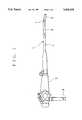
- FIG. 1is a schematic illustration showing the general arrangement of a catheter type endoscopic ultrasound probe embodying the present invention
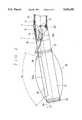
- FIG. 2is a partly sectioned outer view of a rigid fore end section of a catheter member of the probe
- FIG. 3is a fragmentary sectional view of an articular joint section of the catheter member
- FIG. 4is a schematic end view of the rigid fore end section of the catheter member.
- FIG. 5is a schematic illustration of a typical puncture needle construction.
- the catheter type ultrasound probe of the inventionis arranged as an endoscopic ultrasound probe, but it is to be understood that the ultrasound probe is not necessarily required to include endoscopic observation means.
- the endoscopic ultrasound probe according to the inventionincludes a catheter member 1, a main body 2 with manipulating control means, and a universal cable 3.
- the catheter member 1is constituted by a flexible body section 4 (hereinafter referred to as "a flexible section” for brevity) which can bend itself along an intracavitary path of insertion, except its proximal end which is connected to the operating unit, an angle section 5 which is connected to the fore end of the flexible section 4, and a rigid fore end section 6 which is connected to the fore end of the angle section 5.
- the angle section 5 and the rigid fore end section 6are connected with each other through an articular joint section 7. As shown particularly in FIG.
- the articular joint section 7is constituted by a front joint pipe 7a, which is fixedly connected to the rigid fore end section 6, and a rear joint pipe 7b, which is fixedly connected to the angle section 5, the front and rear joint pipes 7a and 7b being pivotally connected with each other at opposite lateral side portions by pins 8. Consequently, along with the front joint pipe 7a, the rigid fore end section 6 at the tip end of the catheter member 1 can be turned about the pins 8 of the articular Joint section 7 to take an angularly bent position within a predetermined range as indicated by arrow A in FIG. 2.
- the rigid fore end section 6is provided with endoscopic observation means on its distal end face 6a, including an illumination window 10 and an observation window 11.
- a light emitting end of an illumination light guideis disposed in the illumination window 10, while an objective lens is fitted in the observation window to form optical images of a subject on a predetermined plane where a solid-state image sensor element is located thereby to convert endoscopic optical images into electric signals.
- an biopsy channel 12is opened on the distal end face 6a to protrude forceps or other instrument therefrom.
- the arrangements of these endoscopic observation and biopsy channel 12are known, so that further description and illustration in this regard are omitted for the sake of simplicity of explanation.
- An ultrasound transducer 20is mounted on the rigid fore end section 6 at a position closer to its proximal end away from the distal end with the above-described endoscopic observation means.
- the ultrasound transducer 20is constituted by a large number of transer elements which are arrayed in a row in the axial direction of the rigid fore end section 6 for electronic convex or linear scanning operations with a view field as indicated by arrows B in FIG. 2 for ultrasound image observation.
- the rigid fore end section 6is provided with annular grooves 21 and 22 which serve to anchor fixedly therein the opposite ends of a tubular balloon 23 by the use of rubber rings or other suitable fixation means.
- a nozzle 24 which supplies an ultrasound transmissive medium like deaerated water to and from the balloon 23is opened on the girder of the rigid fore end section 6 at an intermediate position between the ultrasound transducer 20 and the annular groove 22. Namely, the balloon which is fitted on the rigid fore end section 6 is inflated with the ultrasound transmissive medium which is supplied through the nozzle 24.
- a puncture needlewhich has a hollow needle body 30a with a sharp-pointed end 30b as shown in FIG. 5.
- the puncture needle 30is introduced into a needle guide passage 31 which extends as far as the rigid fore end section 6 through the main body 2 and the flexible and angle sections 4 and 5 of the catheter member 1 to project the puncture needle 30 through an exit 32 which is opened on a lateral side of the rigid fore end section 6.
- the proximal or base end of the puncture needle 30is terminated with a coupler 30c of an increased diameter which is connectible to a injection medicine supply means or to a drainage means.
- the puncture needle guide passage 31is constituted by a flexible tube which runs axially through the catheter member 1.
- the exit opening 32 for the puncture needle 30is provided on a lateral side of the rigid fore end section 6, it is necessary to turn the axial needle passage 31 smoothly toward the exit opening 32 through the articular joint section 7 and part of the rigid fore end section 6.
- the fore end of the flexible tube of the axial needle passage 31is connected to the needle exit opening 32 through a curved needle passage 31a in the form of a hard rigid pipe with a predetermined turn angle toward the exit opening 32.
- the linear needle guide passage 31is connected to the curved needle passage 31a within the front joint pipe member 7a of the articular joint section 7 to turn the passage for the puncture needle 30 through a predetermined angle ⁇ 1 with the axis of the catheter member 1.
- the angle of the rigid fore end section 6 relative to the axis of the catheter member 1is adjustable through the articular joint section 7 which is pivotable to bend the rigid fore end section 6 in a direction rearward of the active surface of the ultrasound transducer 20 on a lateral side of the rigid fore end section 6.
- the rigid fore end section 6is urged into a bent position of a predetermined angle by a spring 40 which is tensioned between the front and rear pipe members 7a and 7b of the articular joint-section 7.
- a spring 40which is tensioned between the front and rear pipe members 7a and 7b of the articular joint-section 7.
- a joint operating wire 41which is extended from the operating section 2 through the catheter member 1 is fixedly connected at its fore end to an inner surface of the front pipe 7a of the articular joint section 7 at a radially opposite position or with a 180° phase difference relative to the biasing spring 40.
- the proximal end portion of the joint operating wire 40is wound on a spool 42 which is rotatably mounted within the main body 2 of the probe.
- the spool 42has its rotational shaft 42a extended out of the casing of the main body 2 and fitted with a knob 43 at its outer end, so that the joint operating wire 41 can be pulled back and forth by turning the knob 43.
- the rigid fore end section 6When the joint operating wire 41 is pulled backward or toward the main body 2, the rigid fore end section 6 is turned toward its straight axial position against the biasing force of the spring 40.
- the spool 42is releasably engaged with a lock member 44 which blocks rotational movements of the spool 42 to retain the joint operating wire 41 in a desired pulled state.
- the rigid fore end section 6can be set in a desired angular position relative to the axis of the catheter member 1 by applying the lock member 44.
- the catheter member 1can be introduced into the stomach through the throat and esophagus for intragastric examination by the endoscopic means or for ultrasound tomographic examination by the ultrasound transducer 20. If necessary, the puncture needle 30 can be driven out of the needle passage to penetrate into the pancreas or other organs through the gastric wall for injection of medicine or contrast medium or for drainage.
- the catheter member 1On insertion of the endoscopic ultrasound probe into the digestive tract, the catheter member 1 has to be passed through the throat where the path of insertion turns at a large angle to make the passage of the catheter member 1 extremely difficult especially when it has a lengthy rigid fore end section to accommodate endoscopic observation means in addition to the electronic scan type ultrasound transducer 20. Needless to say, if in a straight form, such a lengthy rigid fore end section will give considerable pains to the patient upon passage through the throat.
- the rigid fore end section 6is normally bent by a predetermined angle ⁇ off the axis of the catheter member 1 at the articular joint section 7, so that it can be smoothly passed around an angular bend in the path of insertion as at the throat by orienting the bent fore end section to lie in the same direction as the bend in the path of insertion, lessening the pains on the part of the patient.
- the catheter member 1is introduced into the esophagus which is substantially in the form of a straight tube.
- the front pipe member 7a of the articular joint section 7can be turned into a straight position relative to the rear pipe 7b by pulling the wire 41 toward the main body 2, permitting the catheter member 1 to advance smoothly through the esophagus and then into a gastric or other region to be examined.
- the esophagusis a canal of a flattened or elliptic shape in section, the rigid fore end section even in the bent position can be passed smoothly therethrough as long as the flat sides of the bent fore end section are kept in the direction of the longer side of the elliptic canal.
- the balloon 23is filled and inflated with an ultrasound transmissive medium, and the ultrasound transducer 20 is faced toward a gastric wall of interest.
- the catheter memberit is difficult for the catheter member to transmit and receive ultrasound signals exactly in a perpendicular direction due to limitations connected with the direction of insertion of the catheter member 1 and the direction of signal transmission and reception by the ultrasound transducer.
- the ultrasound transducer 20 on the rigid fore end section 6can be properly faced toward an aimed intracavitary wall portion when the rigid fore end section is bent off the axis of the catheter member 1 in a direction rearward of the active surface of the ultrasound transducer.
- the rigid fore end section 6 of the catheter member 1is normally retained in a bent position by the action of the spring 40 which is provided on the articular joint section 7.
- the ultrasound transducer 20 on the rigid fore end section 6is fixed in a particular direction, a difficulty may be experienced in turning the transducer into an optimum direction for signal transmission and reception by an angling operation of the angle section 5.
- the ultrasound transducer 20can be more easily turned into a desired direction simply by pulling the wire 41 forward or backward. This permits to adjust the ultrasound transducer into an optimum direction for signal transmission and reception without moving the catheter member 1 as a whole.
- the puncture needle 30When the puncture needle 30 is driven to penetrate the pancreas or other target organ through an intracavitary wall, the puncturing operation is monitored by way of the ultrasound images produced by the transducer 30. At this time, the puncture needle 30 is driven forward through the axial needle passage 31 and the curved passage 31a and projected through the exit opening 31a to penetrate the needle point 30b into a target portion through an intracavitary wall. In this regard, in order to let the needle point 30b reach a target depth smoothly, it is desirable to transmit the driving thrust force to the needle point 30a effectively to a sufficient degree.
- the hollow body 30a of the puncture needle 30should preferably be of high rigidity material although it has to be launched from the needle passage 31 turning toward the exit opening 32 on a lateral side of the rigid fore end section 6 through the curved needle passage 31a.
- this reduction of the turn angle of the curved needle passagemakes it possible to drive the puncture needle 30 toward the exit 32 of the needle passage 31 more easily, enhancing the needle drivability and operationability in puncturing operations.
- the turn angle of the curved needle passage 31acan be reduced to 20° by setting the rigid fore end section 2 in a bent position 10° off the axis of the catheter member by way of the articular joint section 7. It follows that the puncture needle 30 with a needle body 30a of higher rigidity can be smoothly passed through the curved passage 31a, and therefore a propelling force can be effectively applied to the needle body 30a to get the needle point 30b to a target.
- the present inventionhas been described by way of an ultrasound probe for examination of the upper digestive system including the stomach and a puncturing operation into the pancreas.
- the inventionis not restricted to the particular examples shown.
- the ultrasound probe according to the inventionhas the articular joint section connected to the proximal end of the rigid fore end section of the catheter member to hold the latter normally held in a bent position at a predetermined angle with the axis of the catheter member in a direction rearward of the active surface of the ultrasound transducer on the rigid fore end section.
- the angle of the rigid fore end section in the positioncan be varied within a predetermined range by pulling the wire back and forth. Therefore, even if the intracavitary path of insertion contains a bend or bends of a relatively large angle, the catheter member can be inserted smoothly around the bend by orienting the bent fore end section to lie in the same direction as the bend in the path of insertion.
- the active face of the ultrasound transducercan be easily turned into a desired direction at the time of fine adjustment of the direction of signal transmission and reception.
- the turn angle of the curved needle passagecan be minimized to such a degree as to make it possible to pass therethrough a hard puncture needle of higher rigidity to ensure higher needle operationability and drivability.
Landscapes
- Health & Medical Sciences (AREA)
- Life Sciences & Earth Sciences (AREA)
- Surgery (AREA)
- General Health & Medical Sciences (AREA)
- Medical Informatics (AREA)
- Veterinary Medicine (AREA)
- Pathology (AREA)
- Radiology & Medical Imaging (AREA)
- Biophysics (AREA)
- Engineering & Computer Science (AREA)
- Biomedical Technology (AREA)
- Heart & Thoracic Surgery (AREA)
- Nuclear Medicine, Radiotherapy & Molecular Imaging (AREA)
- Molecular Biology (AREA)
- Animal Behavior & Ethology (AREA)
- Physics & Mathematics (AREA)
- Public Health (AREA)
- Optics & Photonics (AREA)
- Gynecology & Obstetrics (AREA)
- Ultra Sonic Daignosis Equipment (AREA)
Abstract
Description
Claims (4)
Applications Claiming Priority (2)
| Application Number | Priority Date | Filing Date | Title |
|---|---|---|---|
| JP5-344575 | 1993-12-20 | ||
| JP5344575AJPH07171150A (en) | 1993-12-20 | 1993-12-20 | Ultrasound system inserted into the body |
Publications (1)
| Publication Number | Publication Date |
|---|---|
| US5456258Atrue US5456258A (en) | 1995-10-10 |
Family
ID=18370338
Family Applications (1)
| Application Number | Title | Priority Date | Filing Date |
|---|---|---|---|
| US08/359,823Expired - LifetimeUS5456258A (en) | 1993-12-20 | 1994-12-20 | Catheter type ultrasound probe |
Country Status (2)
| Country | Link |
|---|---|
| US (1) | US5456258A (en) |
| JP (1) | JPH07171150A (en) |
Cited By (82)
| Publication number | Priority date | Publication date | Assignee | Title |
|---|---|---|---|---|
| US5662116A (en)* | 1995-09-12 | 1997-09-02 | Fuji Photo Optical Co., Ltd. | Multi-plane electronic scan ultrasound probe |
| WO1998033429A3 (en)* | 1997-01-31 | 1998-10-29 | Acuson | Steering mechanism and steering line for a catheter-mounted ultrasonic transducer |
| US5954654A (en)* | 1997-01-31 | 1999-09-21 | Acuson Corporation | Steering mechanism and steering line for a catheter-mounted ultrasonic transducer |
| US20020002349A1 (en)* | 1996-10-11 | 2002-01-03 | Transvascular, Inc. | Systems and methods for delivering drugs to selected locations within the body |
| USD455210S1 (en) | 1997-01-31 | 2002-04-02 | Acuson Corporation | Ultrasonic transducer assembly controller |
| US6371919B1 (en)* | 1997-06-30 | 2002-04-16 | Eva Corporation | Method and apparatus for the surgical repair of aneurysms |
| US6390973B1 (en) | 1998-06-25 | 2002-05-21 | Asahi Kogaku Kogyo Kabushiki Kaisha | Endoscope for ultrasonic examination and surgical treatment associated thereto |
| US6464645B1 (en) | 1997-01-31 | 2002-10-15 | Acuson Corporation | Ultrasonic transducer assembly controller |
| USD465024S1 (en) | 1998-01-31 | 2002-10-29 | Acuson Corporation | Ultrasonic transducer assembly controller |
| US6494843B2 (en)* | 2000-12-19 | 2002-12-17 | Ge Medical Systems Global Technology Company, Llc | Transesophageal ultrasound probe with expandable scanhead |
| US6629951B2 (en) | 1999-08-05 | 2003-10-07 | Broncus Technologies, Inc. | Devices for creating collateral in the lungs |
| US6712812B2 (en) | 1999-08-05 | 2004-03-30 | Broncus Technologies, Inc. | Devices for creating collateral channels |
| US6749606B2 (en) | 1999-08-05 | 2004-06-15 | Thomas Keast | Devices for creating collateral channels |
| US20040133111A1 (en)* | 2003-01-03 | 2004-07-08 | Civco Medical Instruments Inc. | Shallow angle needle guide apparatus and method |
| US20050080336A1 (en)* | 2002-07-22 | 2005-04-14 | Ep Medsystems, Inc. | Method and apparatus for time gating of medical images |
| US20050124898A1 (en)* | 2002-01-16 | 2005-06-09 | Ep Medsystems, Inc. | Method and apparatus for isolating a catheter interface |
| US20050203410A1 (en)* | 2004-02-27 | 2005-09-15 | Ep Medsystems, Inc. | Methods and systems for ultrasound imaging of the heart from the pericardium |
| US20050228290A1 (en)* | 2004-04-07 | 2005-10-13 | Ep Medsystems, Inc. | Steerable ultrasound catheter |
| US20050240103A1 (en)* | 2004-04-20 | 2005-10-27 | Ep Medsystems, Inc. | Method and apparatus for ultrasound imaging with autofrequency selection |
| US7022088B2 (en) | 1999-08-05 | 2006-04-04 | Broncus Technologies, Inc. | Devices for applying energy to tissue |
| US20060122514A1 (en)* | 2004-11-23 | 2006-06-08 | Ep Medsystems, Inc. | Method and apparatus for localizing an ultrasound catheter |
| US7175644B2 (en) | 2001-02-14 | 2007-02-13 | Broncus Technologies, Inc. | Devices and methods for maintaining collateral channels in tissue |
| US20070167809A1 (en)* | 2002-07-22 | 2007-07-19 | Ep Medsystems, Inc. | Method and System For Estimating Cardiac Ejection Volume And Placing Pacemaker Electrodes Using Speckle Tracking |
| US20070167794A1 (en)* | 2005-12-14 | 2007-07-19 | Ep Medsystems, Inc. | Method and system for evaluating valvular function |
| US20070167793A1 (en)* | 2005-12-14 | 2007-07-19 | Ep Medsystems, Inc. | Method and system for enhancing spectral doppler presentation |
| US20070232949A1 (en)* | 2006-03-31 | 2007-10-04 | Ep Medsystems, Inc. | Method For Simultaneous Bi-Atrial Mapping Of Atrial Fibrillation |
| US20070293787A1 (en)* | 2003-08-13 | 2007-12-20 | Taylor James D | Targeted biopsy delivery system |
| US20070299479A1 (en)* | 2006-06-27 | 2007-12-27 | Ep Medsystems, Inc. | Method for Reversing Ventricular Dyssynchrony |
| US20080146928A1 (en)* | 2006-12-14 | 2008-06-19 | Ep Medsystems, Inc. | Method and System for Configuration of a Pacemaker and For Placement of Pacemaker Electrodes |
| US20080146942A1 (en)* | 2006-12-13 | 2008-06-19 | Ep Medsystems, Inc. | Catheter Position Tracking Methods Using Fluoroscopy and Rotational Sensors |
| US7422563B2 (en) | 1999-08-05 | 2008-09-09 | Broncus Technologies, Inc. | Multifunctional tip catheter for applying energy to tissue and detecting the presence of blood flow |
| US7462162B2 (en) | 2001-09-04 | 2008-12-09 | Broncus Technologies, Inc. | Antiproliferative devices for maintaining patency of surgically created channels in a body organ |
| US20080312536A1 (en)* | 2007-06-16 | 2008-12-18 | Ep Medsystems, Inc. | Oscillating Phased-Array Ultrasound Imaging Catheter System |
| US20090088631A1 (en)* | 2007-06-28 | 2009-04-02 | W.L. Gore & Associates - Englewood Group (Emd) | Catheter |
| US20090264759A1 (en)* | 2008-04-22 | 2009-10-22 | Ep Medsystems, Inc. | Ultrasound Imaging Catheter With Pivoting Head |
| US7648462B2 (en) | 2002-01-16 | 2010-01-19 | St. Jude Medical, Atrial Fibrillation Division, Inc. | Safety systems and methods for ensuring safe use of intra-cardiac ultrasound catheters |
| US20100023037A1 (en)* | 2003-01-14 | 2010-01-28 | Flowcardia, Inc. | Ultrasound catheter and methods for making and using same |
| US7708712B2 (en) | 2001-09-04 | 2010-05-04 | Broncus Technologies, Inc. | Methods and devices for maintaining patency of surgically created channels in a body organ |
| US7815590B2 (en) | 1999-08-05 | 2010-10-19 | Broncus Technologies, Inc. | Devices for maintaining patency of surgically created channels in tissue |
| US7914470B2 (en) | 2001-01-12 | 2011-03-29 | Celleration, Inc. | Ultrasonic method and device for wound treatment |
| US8002740B2 (en) | 2003-07-18 | 2011-08-23 | Broncus Technologies, Inc. | Devices for maintaining patency of surgically created channels in tissue |
| US20110237955A1 (en)* | 2008-05-30 | 2011-09-29 | Dietz Dennis R | Real Time Ultrasound Catheter Probe |
| US8057394B2 (en) | 2007-06-30 | 2011-11-15 | St. Jude Medical, Atrial Fibrillation Division, Inc. | Ultrasound image processing to render three-dimensional images from two-dimensional images |
| US8235919B2 (en) | 2001-01-12 | 2012-08-07 | Celleration, Inc. | Ultrasonic method and device for wound treatment |
| US8308682B2 (en) | 2003-07-18 | 2012-11-13 | Broncus Medical Inc. | Devices for maintaining patency of surgically created channels in tissue |
| US8409167B2 (en) | 2004-07-19 | 2013-04-02 | Broncus Medical Inc | Devices for delivering substances through an extra-anatomic opening created in an airway |
| US8491521B2 (en) | 2007-01-04 | 2013-07-23 | Celleration, Inc. | Removable multi-channel applicator nozzle |
| US8621946B1 (en) | 2011-06-08 | 2014-01-07 | Patrick Nefos | Support for ultrasonic probe |
| US8709034B2 (en) | 2011-05-13 | 2014-04-29 | Broncus Medical Inc. | Methods and devices for diagnosing, monitoring, or treating medical conditions through an opening through an airway wall |
| US8758256B2 (en) | 2010-07-12 | 2014-06-24 | Best Medical International, Inc. | Apparatus for brachytherapy that uses a scanning probe for treatment of malignant tissue |
| US8852112B2 (en) | 2007-06-28 | 2014-10-07 | W. L. Gore & Associates, Inc. | Catheter with deflectable imaging device and bendable electrical conductor |
| US8864675B2 (en) | 2007-06-28 | 2014-10-21 | W. L. Gore & Associates, Inc. | Catheter |
| US9044216B2 (en) | 2010-07-12 | 2015-06-02 | Best Medical International, Inc. | Biopsy needle assembly |
| US9345532B2 (en) | 2011-05-13 | 2016-05-24 | Broncus Medical Inc. | Methods and devices for ablation of tissue |
| US9522254B2 (en) | 2013-01-30 | 2016-12-20 | Vascular Pathways, Inc. | Systems and methods for venipuncture and catheter placement |
| US9616201B2 (en) | 2011-01-31 | 2017-04-11 | Vascular Pathways, Inc. | Intravenous catheter and insertion device with reduced blood spatter |
| US9675784B2 (en) | 2007-04-18 | 2017-06-13 | Vascular Pathways, Inc. | Intravenous catheter insertion and blood sample devices and method of use |
| US9861792B2 (en) | 2011-02-25 | 2018-01-09 | C. R. Bard, Inc. | Medical component insertion device including a retractable needle |
| US9872971B2 (en) | 2010-05-14 | 2018-01-23 | C. R. Bard, Inc. | Guidewire extension system for a catheter placement device |
| US9950139B2 (en) | 2010-05-14 | 2018-04-24 | C. R. Bard, Inc. | Catheter placement device including guidewire and catheter control elements |
| US10220191B2 (en) | 2005-07-06 | 2019-03-05 | Vascular Pathways, Inc. | Intravenous catheter insertion device and method of use |
| US10232146B2 (en) | 2014-09-05 | 2019-03-19 | C. R. Bard, Inc. | Catheter insertion device including retractable needle |
| US10272260B2 (en) | 2011-11-23 | 2019-04-30 | Broncus Medical Inc. | Methods and devices for diagnosing, monitoring, or treating medical conditions through an opening through an airway wall |
| US10384039B2 (en) | 2010-05-14 | 2019-08-20 | C. R. Bard, Inc. | Catheter insertion device including top-mounted advancement components |
| CN110384525A (en)* | 2019-06-17 | 2019-10-29 | 中国人民解放军联勤保障部队第九二〇医院 | It is a kind of through canalis spinalis mirror approach Micro-operation system |
| US10493262B2 (en) | 2016-09-12 | 2019-12-03 | C. R. Bard, Inc. | Blood control for a catheter insertion device |
| US20200100656A1 (en)* | 2017-04-12 | 2020-04-02 | Konstantin Bob | Endoscope head having a pivotable camera and working channel unit |
| USD903100S1 (en) | 2015-05-01 | 2020-11-24 | C. R. Bard, Inc. | Catheter placement device |
| USD903101S1 (en) | 2011-05-13 | 2020-11-24 | C. R. Bard, Inc. | Catheter |
| US11000678B2 (en) | 2010-05-14 | 2021-05-11 | C. R. Bard, Inc. | Catheter placement device and method |
| USD921884S1 (en) | 2018-07-27 | 2021-06-08 | Bard Access Systems, Inc. | Catheter insertion device |
| US11040176B2 (en) | 2015-05-15 | 2021-06-22 | C. R. Bard, Inc. | Catheter placement device including an extensible needle safety component |
| US11224767B2 (en) | 2013-11-26 | 2022-01-18 | Sanuwave Health, Inc. | Systems and methods for producing and delivering ultrasonic therapies for wound treatment and healing |
| CN114287360A (en)* | 2021-12-01 | 2022-04-08 | 中国科学院亚热带农业生态研究所 | A device for long-term monitoring of water intake of dairy cows |
| US11389626B2 (en) | 2018-03-07 | 2022-07-19 | Bard Access Systems, Inc. | Guidewire advancement and blood flashback systems for a medical device insertion system |
| US11400260B2 (en) | 2017-03-01 | 2022-08-02 | C. R. Bard, Inc. | Catheter insertion device |
| WO2022216862A1 (en)* | 2021-04-06 | 2022-10-13 | Boston Scientific Scimed, Inc. | Devices, systems, and methods for positioning medical devices within a body lumen |
| US11559665B2 (en) | 2019-08-19 | 2023-01-24 | Becton, Dickinson And Company | Midline catheter placement device |
| US11832877B2 (en) | 2017-04-03 | 2023-12-05 | Broncus Medical Inc. | Electrosurgical access sheath |
| US20240050146A1 (en)* | 2017-06-23 | 2024-02-15 | Oral Diagnostix, Llc | Transoral ultrasound probe |
| US11925779B2 (en) | 2010-05-14 | 2024-03-12 | C. R. Bard, Inc. | Catheter insertion device including top-mounted advancement components |
| US12440652B2 (en) | 2019-09-20 | 2025-10-14 | Bard Peripheral Vascular, Inc. | Intravenous catheter-placement device and method thereof |
Families Citing this family (3)
| Publication number | Priority date | Publication date | Assignee | Title |
|---|---|---|---|---|
| JP3508340B2 (en)* | 1995-10-19 | 2004-03-22 | 富士写真光機株式会社 | Ultrasound inspection device for insertion into body cavity |
| JPH11347037A (en)* | 1998-06-12 | 1999-12-21 | Asahi Optical Co Ltd | Ultrasound inspection device for insertion into body cavity |
| JP2006325780A (en)* | 2005-05-25 | 2006-12-07 | Pentax Corp | Ultrasound endoscope tip |
Citations (3)
| Publication number | Priority date | Publication date | Assignee | Title |
|---|---|---|---|---|
| US5191890A (en)* | 1991-04-22 | 1993-03-09 | Interspec, Inc. | Ultrasonic probe assembly |
| US5299578A (en)* | 1990-08-02 | 1994-04-05 | B.V. Optische Industrie "De Oude Delft" | Endoscopic probe |
| US5348017A (en)* | 1993-01-19 | 1994-09-20 | Cardiovascular Imaging Systems, Inc. | Drive shaft for an intravascular catheter system |
- 1993
- 1993-12-20JPJP5344575Apatent/JPH07171150A/enactivePending
- 1994
- 1994-12-20USUS08/359,823patent/US5456258A/ennot_activeExpired - Lifetime
Patent Citations (3)
| Publication number | Priority date | Publication date | Assignee | Title |
|---|---|---|---|---|
| US5299578A (en)* | 1990-08-02 | 1994-04-05 | B.V. Optische Industrie "De Oude Delft" | Endoscopic probe |
| US5191890A (en)* | 1991-04-22 | 1993-03-09 | Interspec, Inc. | Ultrasonic probe assembly |
| US5348017A (en)* | 1993-01-19 | 1994-09-20 | Cardiovascular Imaging Systems, Inc. | Drive shaft for an intravascular catheter system |
Cited By (154)
| Publication number | Priority date | Publication date | Assignee | Title |
|---|---|---|---|---|
| US5662116A (en)* | 1995-09-12 | 1997-09-02 | Fuji Photo Optical Co., Ltd. | Multi-plane electronic scan ultrasound probe |
| US20020002349A1 (en)* | 1996-10-11 | 2002-01-03 | Transvascular, Inc. | Systems and methods for delivering drugs to selected locations within the body |
| US6685648B2 (en)* | 1996-10-11 | 2004-02-03 | Transvascular, Inc. | Systems and methods for delivering drugs to selected locations within the body |
| USD455210S1 (en) | 1997-01-31 | 2002-04-02 | Acuson Corporation | Ultrasonic transducer assembly controller |
| US6228032B1 (en) | 1997-01-31 | 2001-05-08 | Acuson Corporation | Steering mechanism and steering line for a catheter-mounted ultrasonic transducer |
| US5954654A (en)* | 1997-01-31 | 1999-09-21 | Acuson Corporation | Steering mechanism and steering line for a catheter-mounted ultrasonic transducer |
| US5938616A (en)* | 1997-01-31 | 1999-08-17 | Acuson Corporation | Steering mechanism and steering line for a catheter-mounted ultrasonic transducer |
| US6464645B1 (en) | 1997-01-31 | 2002-10-15 | Acuson Corporation | Ultrasonic transducer assembly controller |
| DE19882093B4 (en)* | 1997-01-31 | 2007-02-22 | Acuson Corp., Mountain View | Ultraschallkatheteranordnug |
| WO1998033429A3 (en)* | 1997-01-31 | 1998-10-29 | Acuson | Steering mechanism and steering line for a catheter-mounted ultrasonic transducer |
| US6371919B1 (en)* | 1997-06-30 | 2002-04-16 | Eva Corporation | Method and apparatus for the surgical repair of aneurysms |
| USD465024S1 (en) | 1998-01-31 | 2002-10-29 | Acuson Corporation | Ultrasonic transducer assembly controller |
| US6390973B1 (en) | 1998-06-25 | 2002-05-21 | Asahi Kogaku Kogyo Kabushiki Kaisha | Endoscope for ultrasonic examination and surgical treatment associated thereto |
| US6749606B2 (en) | 1999-08-05 | 2004-06-15 | Thomas Keast | Devices for creating collateral channels |
| US7022088B2 (en) | 1999-08-05 | 2006-04-04 | Broncus Technologies, Inc. | Devices for applying energy to tissue |
| US6712812B2 (en) | 1999-08-05 | 2004-03-30 | Broncus Technologies, Inc. | Devices for creating collateral channels |
| US7393330B2 (en) | 1999-08-05 | 2008-07-01 | Broncus Technologies, Inc. | Electrosurgical device having hollow tissue cutting member and transducer assembly |
| US6629951B2 (en) | 1999-08-05 | 2003-10-07 | Broncus Technologies, Inc. | Devices for creating collateral in the lungs |
| US7422563B2 (en) | 1999-08-05 | 2008-09-09 | Broncus Technologies, Inc. | Multifunctional tip catheter for applying energy to tissue and detecting the presence of blood flow |
| US7815590B2 (en) | 1999-08-05 | 2010-10-19 | Broncus Technologies, Inc. | Devices for maintaining patency of surgically created channels in tissue |
| US6692494B1 (en) | 1999-08-05 | 2004-02-17 | Broncus Technologies, Inc. | Methods and devices for creating collateral channels in the lungs |
| US6494843B2 (en)* | 2000-12-19 | 2002-12-17 | Ge Medical Systems Global Technology Company, Llc | Transesophageal ultrasound probe with expandable scanhead |
| US8235919B2 (en) | 2001-01-12 | 2012-08-07 | Celleration, Inc. | Ultrasonic method and device for wound treatment |
| US7914470B2 (en) | 2001-01-12 | 2011-03-29 | Celleration, Inc. | Ultrasonic method and device for wound treatment |
| US7175644B2 (en) | 2001-02-14 | 2007-02-13 | Broncus Technologies, Inc. | Devices and methods for maintaining collateral channels in tissue |
| US7708712B2 (en) | 2001-09-04 | 2010-05-04 | Broncus Technologies, Inc. | Methods and devices for maintaining patency of surgically created channels in a body organ |
| US7462162B2 (en) | 2001-09-04 | 2008-12-09 | Broncus Technologies, Inc. | Antiproliferative devices for maintaining patency of surgically created channels in a body organ |
| US7648462B2 (en) | 2002-01-16 | 2010-01-19 | St. Jude Medical, Atrial Fibrillation Division, Inc. | Safety systems and methods for ensuring safe use of intra-cardiac ultrasound catheters |
| US20050124898A1 (en)* | 2002-01-16 | 2005-06-09 | Ep Medsystems, Inc. | Method and apparatus for isolating a catheter interface |
| US20070167809A1 (en)* | 2002-07-22 | 2007-07-19 | Ep Medsystems, Inc. | Method and System For Estimating Cardiac Ejection Volume And Placing Pacemaker Electrodes Using Speckle Tracking |
| US20050080336A1 (en)* | 2002-07-22 | 2005-04-14 | Ep Medsystems, Inc. | Method and apparatus for time gating of medical images |
| US7314446B2 (en) | 2002-07-22 | 2008-01-01 | Ep Medsystems, Inc. | Method and apparatus for time gating of medical images |
| WO2004062488A3 (en)* | 2003-01-03 | 2005-05-26 | Civco Medical Instr Inc | Shallow angle needle guide apparatus and method |
| US7351205B2 (en)* | 2003-01-03 | 2008-04-01 | Civco Medical Instruments Co., Inc. | Shallow angle needle guide apparatus and method |
| US20040133111A1 (en)* | 2003-01-03 | 2004-07-08 | Civco Medical Instruments Inc. | Shallow angle needle guide apparatus and method |
| US20100023037A1 (en)* | 2003-01-14 | 2010-01-28 | Flowcardia, Inc. | Ultrasound catheter and methods for making and using same |
| US8308682B2 (en) | 2003-07-18 | 2012-11-13 | Broncus Medical Inc. | Devices for maintaining patency of surgically created channels in tissue |
| US8002740B2 (en) | 2003-07-18 | 2011-08-23 | Broncus Technologies, Inc. | Devices for maintaining patency of surgically created channels in tissue |
| US9533128B2 (en) | 2003-07-18 | 2017-01-03 | Broncus Medical Inc. | Devices for maintaining patency of surgically created channels in tissue |
| US20070293787A1 (en)* | 2003-08-13 | 2007-12-20 | Taylor James D | Targeted biopsy delivery system |
| US20110144492A1 (en)* | 2003-08-13 | 2011-06-16 | Taylor James D | Targeted Treatment Delivery System |
| US7833168B2 (en) | 2003-08-13 | 2010-11-16 | Envisioneering Medical Technologies, Llc | Targeted biopsy delivery system |
| US20090054807A1 (en)* | 2003-08-13 | 2009-02-26 | Taylor James D | Targeted biopsy delivery system |
| US8317724B2 (en) | 2003-08-13 | 2012-11-27 | Envisioneering, Llc | Targeted treatment delivery system |
| US20050203410A1 (en)* | 2004-02-27 | 2005-09-15 | Ep Medsystems, Inc. | Methods and systems for ultrasound imaging of the heart from the pericardium |
| US20050228290A1 (en)* | 2004-04-07 | 2005-10-13 | Ep Medsystems, Inc. | Steerable ultrasound catheter |
| US7507205B2 (en) | 2004-04-07 | 2009-03-24 | St. Jude Medical, Atrial Fibrillation Division, Inc. | Steerable ultrasound catheter |
| US7654958B2 (en) | 2004-04-20 | 2010-02-02 | St. Jude Medical, Atrial Fibrillation Division, Inc. | Method and apparatus for ultrasound imaging with autofrequency selection |
| US20050240103A1 (en)* | 2004-04-20 | 2005-10-27 | Ep Medsystems, Inc. | Method and apparatus for ultrasound imaging with autofrequency selection |
| US11357960B2 (en) | 2004-07-19 | 2022-06-14 | Broncus Medical Inc. | Devices for delivering substances through an extra-anatomic opening created in an airway |
| US8409167B2 (en) | 2004-07-19 | 2013-04-02 | Broncus Medical Inc | Devices for delivering substances through an extra-anatomic opening created in an airway |
| US8608724B2 (en) | 2004-07-19 | 2013-12-17 | Broncus Medical Inc. | Devices for delivering substances through an extra-anatomic opening created in an airway |
| US10369339B2 (en) | 2004-07-19 | 2019-08-06 | Broncus Medical Inc. | Devices for delivering substances through an extra-anatomic opening created in an airway |
| US8784400B2 (en) | 2004-07-19 | 2014-07-22 | Broncus Medical Inc. | Devices for delivering substances through an extra-anatomic opening created in an airway |
| US20060122514A1 (en)* | 2004-11-23 | 2006-06-08 | Ep Medsystems, Inc. | Method and apparatus for localizing an ultrasound catheter |
| US7713210B2 (en) | 2004-11-23 | 2010-05-11 | St. Jude Medical, Atrial Fibrillation Division, Inc. | Method and apparatus for localizing an ultrasound catheter |
| US10639004B2 (en) | 2004-11-23 | 2020-05-05 | St. Jude Medical, Atrial Fibrillation Division, Inc. | Method and apparatus for localizing an ultrasound catheter |
| US11925778B2 (en) | 2005-07-06 | 2024-03-12 | Vascular Pathways, Inc. | Intravenous catheter insertion device |
| US10912930B2 (en) | 2005-07-06 | 2021-02-09 | Vascular Pathways, Inc. | Intravenous catheter insertion device and method of use |
| US10220191B2 (en) | 2005-07-06 | 2019-03-05 | Vascular Pathways, Inc. | Intravenous catheter insertion device and method of use |
| US11577054B2 (en) | 2005-07-06 | 2023-02-14 | Vascular Pathways, Inc. | Intravenous catheter insertion device and method of use |
| US10806906B2 (en) | 2005-07-06 | 2020-10-20 | Vascular Pathways, Inc. | Intravenous catheter insertion device and method of use |
| US11020571B2 (en) | 2005-07-06 | 2021-06-01 | Vascular Pathways, Inc. | Intravenous catheter insertion device and method of use |
| US12370349B2 (en) | 2005-07-06 | 2025-07-29 | Vascular Pathways, Inc. | Intravenous catheter insertion device and method of use |
| US20070167794A1 (en)* | 2005-12-14 | 2007-07-19 | Ep Medsystems, Inc. | Method and system for evaluating valvular function |
| US8070684B2 (en) | 2005-12-14 | 2011-12-06 | St. Jude Medical, Atrial Fibrillation Division, Inc. | Method and system for evaluating valvular function |
| US20070167793A1 (en)* | 2005-12-14 | 2007-07-19 | Ep Medsystems, Inc. | Method and system for enhancing spectral doppler presentation |
| US20070232949A1 (en)* | 2006-03-31 | 2007-10-04 | Ep Medsystems, Inc. | Method For Simultaneous Bi-Atrial Mapping Of Atrial Fibrillation |
| US20070299479A1 (en)* | 2006-06-27 | 2007-12-27 | Ep Medsystems, Inc. | Method for Reversing Ventricular Dyssynchrony |
| US9913969B2 (en) | 2006-10-05 | 2018-03-13 | Broncus Medical Inc. | Devices for delivering substances through an extra-anatomic opening created in an airway |
| US20080146942A1 (en)* | 2006-12-13 | 2008-06-19 | Ep Medsystems, Inc. | Catheter Position Tracking Methods Using Fluoroscopy and Rotational Sensors |
| US20080146928A1 (en)* | 2006-12-14 | 2008-06-19 | Ep Medsystems, Inc. | Method and System for Configuration of a Pacemaker and For Placement of Pacemaker Electrodes |
| US8187190B2 (en) | 2006-12-14 | 2012-05-29 | St. Jude Medical, Atrial Fibrillation Division, Inc. | Method and system for configuration of a pacemaker and for placement of pacemaker electrodes |
| US8491521B2 (en) | 2007-01-04 | 2013-07-23 | Celleration, Inc. | Removable multi-channel applicator nozzle |
| US9675784B2 (en) | 2007-04-18 | 2017-06-13 | Vascular Pathways, Inc. | Intravenous catheter insertion and blood sample devices and method of use |
| US9757540B2 (en) | 2007-04-18 | 2017-09-12 | Vascular Pathways, Inc. | Intravenous catheter insertion and blood sample devices and method of use |
| US10086171B2 (en) | 2007-05-07 | 2018-10-02 | Vascular Pathways, Inc. | Intravenous catheter insertion and blood sample devices and method of use |
| US10525236B2 (en) | 2007-05-07 | 2020-01-07 | Vascular Pathways, Inc. | Intravenous catheter insertion and blood sample devices and method of use |
| US10799680B2 (en) | 2007-05-07 | 2020-10-13 | Vascular Pathways, Inc. | Intravenous catheter insertion and blood sample devices and method of use |
| US8317711B2 (en) | 2007-06-16 | 2012-11-27 | St. Jude Medical, Atrial Fibrillation Division, Inc. | Oscillating phased-array ultrasound imaging catheter system |
| US20080312536A1 (en)* | 2007-06-16 | 2008-12-18 | Ep Medsystems, Inc. | Oscillating Phased-Array Ultrasound Imaging Catheter System |
| US8852112B2 (en) | 2007-06-28 | 2014-10-07 | W. L. Gore & Associates, Inc. | Catheter with deflectable imaging device and bendable electrical conductor |
| US8864675B2 (en) | 2007-06-28 | 2014-10-21 | W. L. Gore & Associates, Inc. | Catheter |
| US8285362B2 (en) | 2007-06-28 | 2012-10-09 | W. L. Gore & Associates, Inc. | Catheter with deflectable imaging device |
| US20090088631A1 (en)* | 2007-06-28 | 2009-04-02 | W.L. Gore & Associates - Englewood Group (Emd) | Catheter |
| US8057394B2 (en) | 2007-06-30 | 2011-11-15 | St. Jude Medical, Atrial Fibrillation Division, Inc. | Ultrasound image processing to render three-dimensional images from two-dimensional images |
| US11217000B2 (en) | 2007-06-30 | 2022-01-04 | St. Jude Medical, Atrial Fibrillation Division, Inc. | Ultrasound image processing to render three-dimensional images from two-dimensional images |
| US9697634B2 (en) | 2007-06-30 | 2017-07-04 | St. Jude Medical, Atrial Fibrillation Division, Inc. | Ultrasound image processing to render three-dimensional images from two-dimensional images |
| US8622915B2 (en) | 2007-06-30 | 2014-01-07 | St. Jude Medical, Atrial Fibrillation Division, Inc. | Ultrasound image processing to render three-dimensional images from two-dimensional images |
| US8052607B2 (en) | 2008-04-22 | 2011-11-08 | St. Jude Medical, Atrial Fibrillation Division, Inc. | Ultrasound imaging catheter with pivoting head |
| US20090264759A1 (en)* | 2008-04-22 | 2009-10-22 | Ep Medsystems, Inc. | Ultrasound Imaging Catheter With Pivoting Head |
| EP2190354A4 (en)* | 2008-04-22 | 2011-03-02 | St Jude Medical Atrial Fibrill | Ultrasound imaging catheter with pivoting head |
| US8535232B2 (en) | 2008-05-30 | 2013-09-17 | W. L. Gore & Associates, Inc. | Real time ultrasound catheter probe |
| US20110237955A1 (en)* | 2008-05-30 | 2011-09-29 | Dietz Dennis R | Real Time Ultrasound Catheter Probe |
| US9950139B2 (en) | 2010-05-14 | 2018-04-24 | C. R. Bard, Inc. | Catheter placement device including guidewire and catheter control elements |
| US12296115B2 (en) | 2010-05-14 | 2025-05-13 | C. R. Bard, Inc. | Insertion device |
| US11000678B2 (en) | 2010-05-14 | 2021-05-11 | C. R. Bard, Inc. | Catheter placement device and method |
| US10722685B2 (en) | 2010-05-14 | 2020-07-28 | C. R. Bard, Inc. | Catheter placement device including guidewire and catheter control elements |
| US10688281B2 (en) | 2010-05-14 | 2020-06-23 | C. R. Bard, Inc. | Catheter placement device including guidewire and catheter control elements |
| US10688280B2 (en) | 2010-05-14 | 2020-06-23 | C. R. Bard, Inc. | Catheter placement device including guidewire and catheter control elements |
| US11135406B2 (en) | 2010-05-14 | 2021-10-05 | C. R. Bard, Inc. | Catheter insertion device including top-mounted advancement components |
| US10384039B2 (en) | 2010-05-14 | 2019-08-20 | C. R. Bard, Inc. | Catheter insertion device including top-mounted advancement components |
| US11925779B2 (en) | 2010-05-14 | 2024-03-12 | C. R. Bard, Inc. | Catheter insertion device including top-mounted advancement components |
| US11278702B2 (en) | 2010-05-14 | 2022-03-22 | C. R. Bard, Inc. | Guidewire extension system for a catheter placement device |
| US9872971B2 (en) | 2010-05-14 | 2018-01-23 | C. R. Bard, Inc. | Guidewire extension system for a catheter placement device |
| US8758256B2 (en) | 2010-07-12 | 2014-06-24 | Best Medical International, Inc. | Apparatus for brachytherapy that uses a scanning probe for treatment of malignant tissue |
| US9044216B2 (en) | 2010-07-12 | 2015-06-02 | Best Medical International, Inc. | Biopsy needle assembly |
| US10328239B2 (en) | 2011-01-31 | 2019-06-25 | Vascular Pathways, Inc. | Intravenous catheter and insertion device with reduced blood spatter |
| US11202886B2 (en) | 2011-01-31 | 2021-12-21 | Vascular Pathways, Inc. | Intravenous catheter and insertion device with reduced blood spatter |
| US9616201B2 (en) | 2011-01-31 | 2017-04-11 | Vascular Pathways, Inc. | Intravenous catheter and insertion device with reduced blood spatter |
| US11123524B2 (en) | 2011-02-25 | 2021-09-21 | C. R. Bard, Inc. | Medical component insertion device including a retractable needle |
| US9861792B2 (en) | 2011-02-25 | 2018-01-09 | C. R. Bard, Inc. | Medical component insertion device including a retractable needle |
| US11931534B2 (en) | 2011-02-25 | 2024-03-19 | C. R. Bard, Inc. | Medical component insertion device including a retractable needle |
| US9421070B2 (en) | 2011-05-13 | 2016-08-23 | Broncus Medical Inc. | Methods and devices for diagnosing, monitoring, or treating medical conditions through an opening through an airway wall |
| US8709034B2 (en) | 2011-05-13 | 2014-04-29 | Broncus Medical Inc. | Methods and devices for diagnosing, monitoring, or treating medical conditions through an opening through an airway wall |
| US8932316B2 (en) | 2011-05-13 | 2015-01-13 | Broncus Medical Inc. | Methods and devices for diagnosing, monitoring, or treating medical conditions through an opening through an airway wall |
| US9993306B2 (en) | 2011-05-13 | 2018-06-12 | Broncus Medical Inc. | Methods and devices for diagnosing, monitoring, or treating medical conditions through an opening through an airway wall |
| USD903101S1 (en) | 2011-05-13 | 2020-11-24 | C. R. Bard, Inc. | Catheter |
| US9345532B2 (en) | 2011-05-13 | 2016-05-24 | Broncus Medical Inc. | Methods and devices for ablation of tissue |
| US9486229B2 (en) | 2011-05-13 | 2016-11-08 | Broncus Medical Inc. | Methods and devices for excision of tissue |
| US12016640B2 (en) | 2011-05-13 | 2024-06-25 | Broncus Medical Inc. | Methods and devices for diagnosing, monitoring, or treating medical conditions through an opening through an airway wall |
| US10631938B2 (en) | 2011-05-13 | 2020-04-28 | Broncus Medical Inc. | Methods and devices for diagnosing, monitoring, or treating medical conditions through an opening through an airway wall |
| US8621946B1 (en) | 2011-06-08 | 2014-01-07 | Patrick Nefos | Support for ultrasonic probe |
| US10272260B2 (en) | 2011-11-23 | 2019-04-30 | Broncus Medical Inc. | Methods and devices for diagnosing, monitoring, or treating medical conditions through an opening through an airway wall |
| US10265507B2 (en) | 2013-01-30 | 2019-04-23 | Vascular Pathways, Inc. | Systems and methods for venipuncture and catheter placement |
| US9522254B2 (en) | 2013-01-30 | 2016-12-20 | Vascular Pathways, Inc. | Systems and methods for venipuncture and catheter placement |
| US11224767B2 (en) | 2013-11-26 | 2022-01-18 | Sanuwave Health, Inc. | Systems and methods for producing and delivering ultrasonic therapies for wound treatment and healing |
| US11331520B2 (en) | 2013-11-26 | 2022-05-17 | Sanuwave Health, Inc. | Systems and methods for producing and delivering ultrasonic therapies for wound treatment and healing |
| US11033719B2 (en) | 2014-09-05 | 2021-06-15 | C. R. Bard, Inc. | Catheter insertion device including retractable needle |
| US11565089B2 (en) | 2014-09-05 | 2023-01-31 | C. R. Bard, Inc. | Catheter insertion device including retractable needle |
| US10232146B2 (en) | 2014-09-05 | 2019-03-19 | C. R. Bard, Inc. | Catheter insertion device including retractable needle |
| USD1069106S1 (en) | 2015-05-01 | 2025-04-01 | C. R. Bard, Inc. | Catheter placement device |
| USD903100S1 (en) | 2015-05-01 | 2020-11-24 | C. R. Bard, Inc. | Catheter placement device |
| US11040176B2 (en) | 2015-05-15 | 2021-06-22 | C. R. Bard, Inc. | Catheter placement device including an extensible needle safety component |
| US12161819B2 (en) | 2015-05-15 | 2024-12-10 | C. R. Bard, Inc. | Catheter placement device including an extensible needle safety component |
| US10493262B2 (en) | 2016-09-12 | 2019-12-03 | C. R. Bard, Inc. | Blood control for a catheter insertion device |
| US12403294B2 (en) | 2016-09-12 | 2025-09-02 | C. R. Bard, Inc. | Blood control for a catheter insertion device |
| US11759618B2 (en) | 2016-09-12 | 2023-09-19 | C. R. Bard, Inc. | Blood control for a catheter insertion device |
| US12357796B2 (en) | 2017-03-01 | 2025-07-15 | C. R. Bard, Inc. | Catheter insertion device |
| US11400260B2 (en) | 2017-03-01 | 2022-08-02 | C. R. Bard, Inc. | Catheter insertion device |
| US11832877B2 (en) | 2017-04-03 | 2023-12-05 | Broncus Medical Inc. | Electrosurgical access sheath |
| US12042122B2 (en)* | 2017-04-12 | 2024-07-23 | Konstantin Bob | Endoscope head having a pivotable camera and working channel unit |
| US20200100656A1 (en)* | 2017-04-12 | 2020-04-02 | Konstantin Bob | Endoscope head having a pivotable camera and working channel unit |
| US20240050146A1 (en)* | 2017-06-23 | 2024-02-15 | Oral Diagnostix, Llc | Transoral ultrasound probe |
| US12167887B2 (en)* | 2017-06-23 | 2024-12-17 | Oral Diagnostix, Llc | Transoral ultrasound probe |
| US12017020B2 (en) | 2018-03-07 | 2024-06-25 | Bard Access Systems, Inc. | Guidewire advancement and blood flashback systems for a medical device insertion system |
| US11389626B2 (en) | 2018-03-07 | 2022-07-19 | Bard Access Systems, Inc. | Guidewire advancement and blood flashback systems for a medical device insertion system |
| USD921884S1 (en) | 2018-07-27 | 2021-06-08 | Bard Access Systems, Inc. | Catheter insertion device |
| CN110384525A (en)* | 2019-06-17 | 2019-10-29 | 中国人民解放军联勤保障部队第九二〇医院 | It is a kind of through canalis spinalis mirror approach Micro-operation system |
| US11559665B2 (en) | 2019-08-19 | 2023-01-24 | Becton, Dickinson And Company | Midline catheter placement device |
| US11883615B2 (en) | 2019-08-19 | 2024-01-30 | Becton, Dickinson And Company | Midline catheter placement device |
| US12440652B2 (en) | 2019-09-20 | 2025-10-14 | Bard Peripheral Vascular, Inc. | Intravenous catheter-placement device and method thereof |
| WO2022216862A1 (en)* | 2021-04-06 | 2022-10-13 | Boston Scientific Scimed, Inc. | Devices, systems, and methods for positioning medical devices within a body lumen |
| CN114287360A (en)* | 2021-12-01 | 2022-04-08 | 中国科学院亚热带农业生态研究所 | A device for long-term monitoring of water intake of dairy cows |
Also Published As
| Publication number | Publication date |
|---|---|
| JPH07171150A (en) | 1995-07-11 |
Similar Documents
| Publication | Publication Date | Title |
|---|---|---|
| US5456258A (en) | Catheter type ultrasound probe | |
| US5499630A (en) | Catheter type ultrasound probe | |
| US6149598A (en) | Ultrasound endoscope | |
| EP0061332B1 (en) | Combined endoscope and ultrasonic diagnostic device | |
| US7488289B2 (en) | Imaging catheter and methods of use for ultrasound-guided ablation | |
| US6200313B1 (en) | Puncture instrument for punctured high frequency treatments | |
| EP1820437B1 (en) | Ultrasound endoscope | |
| WO2016136080A1 (en) | Endoscope puncture needle | |
| US6409666B1 (en) | Tip end of ultrasonic endoscope | |
| US5150715A (en) | Ultrasound-imaging diagnostic system | |
| CN113384299A (en) | Ultrasonic endoscope catheter | |
| JP3042339B2 (en) | Ultrasound endoscope | |
| JPH07194594A (en) | Instrument insertion passage for in-vivo inspection device | |
| JPH08126644A (en) | Ultrasonic endoscope | |
| JP3671764B2 (en) | Endoscope removable electronic scanning ultrasonic inspection system | |
| JPH09108224A (en) | Body cavity ultrasonic probe | |
| JP2004016505A (en) | Electronic scan type ultrasonic endoscope | |
| CN113384298A (en) | Double-cavity ultrasonic endoscope catheter | |
| JPH0221852A (en) | Ultrasonic endoscope | |
| JPH119601A (en) | Endoscope device and balloon device for ultrasonic observation | |
| JP3003961B2 (en) | Endoscope | |
| JPWO2020152791A1 (en) | Endoscopic ultrasound and endoscopy system | |
| JPS6258257B2 (en) | ||
| JPS61280849A (en) | Body cavity ultrasonic diagnostic apparatus | |
| JP2662546B2 (en) | Ultrasound endoscope |
Legal Events
| Date | Code | Title | Description |
|---|---|---|---|
| AS | Assignment | Owner name:KABUSHIKI KAISHA TOSHIBA, JAPAN Free format text:ASSIGNMENT OF ASSIGNORS INTEREST;ASSIGNORS:KONDO, MITUO;ABE, KENJI;HIKI, SUSUMU;REEL/FRAME:007345/0470 Effective date:19950203 Owner name:FUJI PHOTO OPTICAL CO., LTD., JAPAN Free format text:ASSIGNMENT OF ASSIGNORS INTEREST;ASSIGNORS:KONDO, MITUO;ABE, KENJI;HIKI, SUSUMU;REEL/FRAME:007345/0470 Effective date:19950203 | |
| STCF | Information on status: patent grant | Free format text:PATENTED CASE | |
| FEPP | Fee payment procedure | Free format text:PAYOR NUMBER ASSIGNED (ORIGINAL EVENT CODE: ASPN); ENTITY STATUS OF PATENT OWNER: LARGE ENTITY | |
| FPAY | Fee payment | Year of fee payment:4 | |
| FPAY | Fee payment | Year of fee payment:8 | |
| AS | Assignment | Owner name:FUJINON CORPORATION, JAPAN Free format text:CHANGE OF NAME;ASSIGNOR:FUJI PHOTO OPTICAL CO., LTD.;REEL/FRAME:018442/0701 Effective date:20041001 | |
| AS | Assignment | Owner name:KABUSHIKI KAISHA TOSHIBA, JAPAN Free format text:ASSIGNMENT OF ASSIGNORS INTEREST;ASSIGNOR:FUJINON CORPORATION;REEL/FRAME:018563/0022 Effective date:20061102 | |
| FPAY | Fee payment | Year of fee payment:12 |