US5439469A - Wound closure device - Google Patents
Wound closure deviceDownload PDFInfo
- Publication number
- US5439469A US5439469AUS08/148,555US14855593AUS5439469AUS 5439469 AUS5439469 AUS 5439469AUS 14855593 AUS14855593 AUS 14855593AUS 5439469 AUS5439469 AUS 5439469A
- Authority
- US
- United States
- Prior art keywords
- needle
- suture
- pointed end
- guide member
- suture device
- Prior art date
- Legal status (The legal status is an assumption and is not a legal conclusion. Google has not performed a legal analysis and makes no representation as to the accuracy of the status listed.)
- Expired - Lifetime
Links
Images


Classifications
- A—HUMAN NECESSITIES
- A61—MEDICAL OR VETERINARY SCIENCE; HYGIENE
- A61B—DIAGNOSIS; SURGERY; IDENTIFICATION
- A61B17/00—Surgical instruments, devices or methods
- A61B17/04—Surgical instruments, devices or methods for suturing wounds; Holders or packages for needles or suture materials
- A61B17/0469—Suturing instruments for use in minimally invasive surgery, e.g. endoscopic surgery
- A—HUMAN NECESSITIES
- A61—MEDICAL OR VETERINARY SCIENCE; HYGIENE
- A61B—DIAGNOSIS; SURGERY; IDENTIFICATION
- A61B17/00—Surgical instruments, devices or methods
- A61B17/04—Surgical instruments, devices or methods for suturing wounds; Holders or packages for needles or suture materials
- A61B17/0482—Needle or suture guides
- A—HUMAN NECESSITIES
- A61—MEDICAL OR VETERINARY SCIENCE; HYGIENE
- A61B—DIAGNOSIS; SURGERY; IDENTIFICATION
- A61B17/00—Surgical instruments, devices or methods
- A61B17/04—Surgical instruments, devices or methods for suturing wounds; Holders or packages for needles or suture materials
- A61B17/0401—Suture anchors, buttons or pledgets, i.e. means for attaching sutures to bone, cartilage or soft tissue; Instruments for applying or removing suture anchors
- A61B2017/0408—Rivets
- A—HUMAN NECESSITIES
- A61—MEDICAL OR VETERINARY SCIENCE; HYGIENE
- A61B—DIAGNOSIS; SURGERY; IDENTIFICATION
- A61B17/00—Surgical instruments, devices or methods
- A61B17/04—Surgical instruments, devices or methods for suturing wounds; Holders or packages for needles or suture materials
- A61B17/0401—Suture anchors, buttons or pledgets, i.e. means for attaching sutures to bone, cartilage or soft tissue; Instruments for applying or removing suture anchors
- A61B2017/0446—Means for attaching and blocking the suture in the suture anchor
- A61B2017/0458—Longitudinal through hole, e.g. suture blocked by a distal suture knot
- A—HUMAN NECESSITIES
- A61—MEDICAL OR VETERINARY SCIENCE; HYGIENE
- A61B—DIAGNOSIS; SURGERY; IDENTIFICATION
- A61B17/00—Surgical instruments, devices or methods
- A61B17/04—Surgical instruments, devices or methods for suturing wounds; Holders or packages for needles or suture materials
- A61B17/0401—Suture anchors, buttons or pledgets, i.e. means for attaching sutures to bone, cartilage or soft tissue; Instruments for applying or removing suture anchors
- A61B2017/0464—Suture anchors, buttons or pledgets, i.e. means for attaching sutures to bone, cartilage or soft tissue; Instruments for applying or removing suture anchors for soft tissue
- A—HUMAN NECESSITIES
- A61—MEDICAL OR VETERINARY SCIENCE; HYGIENE
- A61B—DIAGNOSIS; SURGERY; IDENTIFICATION
- A61B17/00—Surgical instruments, devices or methods
- A61B17/04—Surgical instruments, devices or methods for suturing wounds; Holders or packages for needles or suture materials
- A61B17/0469—Suturing instruments for use in minimally invasive surgery, e.g. endoscopic surgery
- A61B2017/047—Suturing instruments for use in minimally invasive surgery, e.g. endoscopic surgery having at least one proximally pointing needle located at the distal end of the instrument, e.g. for suturing trocar puncture wounds starting from inside the body
- A—HUMAN NECESSITIES
- A61—MEDICAL OR VETERINARY SCIENCE; HYGIENE
- A61B—DIAGNOSIS; SURGERY; IDENTIFICATION
- A61B17/00—Surgical instruments, devices or methods
- A61B17/04—Surgical instruments, devices or methods for suturing wounds; Holders or packages for needles or suture materials
- A61B17/06—Needles ; Sutures; Needle-suture combinations; Holders or packages for needles or suture materials
- A61B17/06004—Means for attaching suture to needle
- A61B2017/06019—Means for attaching suture to needle by means of a suture-receiving lateral eyelet machined in the needle
- A—HUMAN NECESSITIES
- A61—MEDICAL OR VETERINARY SCIENCE; HYGIENE
- A61B—DIAGNOSIS; SURGERY; IDENTIFICATION
- A61B17/00—Surgical instruments, devices or methods
- A61B17/04—Surgical instruments, devices or methods for suturing wounds; Holders or packages for needles or suture materials
- A61B17/06—Needles ; Sutures; Needle-suture combinations; Holders or packages for needles or suture materials
- A61B17/06004—Means for attaching suture to needle
- A61B2017/06042—Means for attaching suture to needle located close to needle tip
- A—HUMAN NECESSITIES
- A61—MEDICAL OR VETERINARY SCIENCE; HYGIENE
- A61B—DIAGNOSIS; SURGERY; IDENTIFICATION
- A61B17/00—Surgical instruments, devices or methods
- A61B17/04—Surgical instruments, devices or methods for suturing wounds; Holders or packages for needles or suture materials
- A61B17/06—Needles ; Sutures; Needle-suture combinations; Holders or packages for needles or suture materials
- A61B17/06066—Needles, e.g. needle tip configurations
- A61B2017/0608—J-shaped
Definitions
- This inventionrelates to a device to enable the closure of the puncture wounds made by trocars used in laparoendoscopic procedures. Specifically, the device allows the proximating of the peritoneal membrane and the posterior wall of the fascia (the inner layers of the abdominal wall) by passing a suture through these structures thus permitting placement of a subcutaneous surgical suture knot.
- the suture needleIn order to minimize the risk of injury to internal organs when a suture needle is inserted into the abdominal wall, the maintenance of abdominal expansion (pneumoperitoneum) is essential. Additionally, to ensure proper wound closure, the suture needle must be inserted at an angle which allows approximation of the innermost structures (i.e peritoneal wall and posterior fascia).
- the prior art wound closure devicesprovide inadequate means of closing the trocar wound.
- the prior art deviceshave several problems.
- some devicesdo not provide a method to maintain insufflation of the abdominal cavity (pneumoperitoneum), as they require the removal of the cannula prior to the introduction of the suture instrument into the wound. This allows the insufflating gas to escape from the abdomen, thus partially or fully deflating the abdomen and further increasing the risk of perforating an internal organ.
- the present inventionprovides solutions to the various problems noted above with respect to the prior art wound closure devices, and methods of manufacturing such devices.
- the inventionprovides a suture device comprising a hook needle and a guide member.
- the needlehas a distal portion extending between a longitudinally extending manipulating portion and a longitudinally extending pointed end of the needle, the pointed end of the needle including a suture holding member.
- the guide memberhas a guide channel receiving the manipulating portion of the needle and the manipulating portion of the needle is movable in the guide channel so as to advance and retract the pointed end of the needle toward and away from the guide member.
- the guide membercomprises a body portion pivotally connected to a handle portion.
- the body portionextends in a longitudinal direction and the handle portion is pivotally connected to the body portion so as to pivot about a pivot axis extending perpendicular to the longitudinal directions.
- the body portionincludes a fiat surface in sliding contact with a flat surface of the handle portion.
- a pivot pinpivotally connects the handle portion to the body portion such that a distal end of the handle portion is located between the pivot pin and a distal end of the body portion.
- the handle portionincludes a proximal end and the body portion includes a proximal end located between the pivot pin and the proximal end of the handle portion.
- the suture holding membercan comprise an eye opening in the pointed end of the needle for attaching a suture to the needle.
- the suture holding membercan also comprise a spring loaded entrapment mechanism or any other suitable means.
- the needleis preferably a non-tubular piece of metal such as wire.
- the devicecan further comprise a tubular member of a trocar surrounding the guide member such that the guide member is slidably received in the tubular member and provides a fluid-tight seal therebetween.
- the guide membercan be of a polymer or metal material and the handle portion and body portion can each be of a polymer or metal material.
- the body portionincludes a longitudinally extending slot which opens along one side of the body portion.
- the needleis movable in the slot such that the needle is completely within the slot when the manipulating portion is pulled away from the guide member and the pointed end of the needle is moved out of the slot when the manipulating portion is pushed toward the guide member.
- the slotcan include seal means such as one or more rubber gaskets which maintains a fluid tight seal with the needle when the needle moves in the slot and the pointed end of the needle passes through the seal means.
- the inventionalso provides a method of forming a subcutaneous tissue closure with a suture device including a hook needle and a guide member, the needle being movable toward and away from the guide member and being pivotal with respect to the guide member.
- the methodincludes steps of:
- the suture devicecan be rotated about an axis passing through the wound site during the orienting step (f).
- the needlecan slide through a guide channel in the guide member when the needle is moved during the needle moving steps (c, e, g and i).
- the pointed end of the needlecan be angled so as to pass only through the inner side of the tissue and into the wound side in the tissue without piercing the outer side of the tissue during the steps (c and g) of moving the needle in the first and third directions.
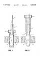
- FIG. 1is a side view of a suture device for closing a wound site in accordance with the invention (obturator, movable handle and needle), showing the device placed into a trocar located in an abdominal wall of a living body;
- FIG. 2is a side view showing the device remaining in place as the trocar/cannula is removed, with the central body section maintaining pneumoperitoneum;
- FIG. 3shows a side view as the hook needle is advanced inwardly into the abdominal cavity
- FIG. 4shows the handle pivoted to a desired angle to allow the hook needle to appropriately capture sufficient tissue to secure a suture
- FIG. 5shows the hook needle being advanced outwardly through the abdominal wall for attachment of a suture to a hook end of the needle at a location outside the abdominal cavity;
- FIG. 6shows the needle after it is retracted into the abdominal cavity with the suture attached
- FIG. 7shows the device after being rotated 180 degrees about the device axis
- FIG. 8shows the hook needle advanced outwardly for detachment of the suture from the needle through the abdominal wall and into the subcutaneous space for detachment of the suture from the needle;
- FIG. 9shows the needle after detachment of the suture from the needle and after being retracted into the abdominal cavity
- FIG. 10shows the handle pivoted into alignment with the central body, the needle withdrawn into the obturator and the device being withdrawn from the wound site.
- the two ends of the sutureremain outside the body in the subcutaneous space;
- FIG. 11shows the tying of a subcutaneous knot
- FIG. 12shows another embodiment of the suture device according to the invention.
- the inventionprovides a medical device usable within a body cavity.
- the deviceincludes a hooked needle and an obturator (guide member), which is sized to fit intimately within a cannula.
- the guide memberserves to maintain the pneumoperitoneum following exchange with the cannula.
- the guide membercan be of single piece or multipiece construction.
- a pivoting handleis attached to a body portion of the guide member by means of a rivet or rod which allows the handle to swivel to adjust the angle for insertion of the hooked needle.
- the swivel movement of the handlewill correspond to the angle of the needle inside the body providing the surgeon with a method to easily judge the angle of needle insertion into the peritoneal wall.
- the same effectcan also be achieved using a single piece guide member by simply tilting the guide member back and forth to the desired angle.
- the curved portion of the needleis housed at the proximal end of the body portion and can be withdrawn or advanced by manipulating the needle shaft which extends through the body portion and beyond a proximal end of the handle of the device.
- the hooked needleis pulled outward to pass the suture through the tissue layers, thus eliminating the risk of passing a sharp needle from outside the body toward the internal organs.
- the body memberis designed to fit snugly inside the cannula and allows the cannula to be removed over it, leaving the device body to fill the puncture wound.
- the body memberis preferably round in shape to allow maintenance of the pneumoperitoneum.
- a sutureis threaded into the needle or captured by a spring-loaded trap mechanism just behind the needle tip. This is done outside the body.
- the suture needleis then pushed back through the abdominal wall with the suture attached.
- the device bodyis rotated 180 degrees and the needle is again pulled through the abdominal wall towards the operator.
- the sutureis removed from the needle or capture mechanism. At this point, the two free ends of the suture remain outside the body.
- the needleis then pushed back through the abdominal wall. Once the needle is completely inside the abdominal cavity, the device handle is returned to its vertical position, the needle is withdrawn into the device body and the device is withdrawn from the wound.
- the suture knotcan then be tied, leaving a suture knot in the subcutaneous tissue.
- the obturatorcan comprise a polymeric material selected from the group consisting of ABS, polycarbonate, nylons, blends or other suitable polymers and mixtures thereof.
- the obturatorcould also be stainless steel or some other suitable material should a reusable, rather than a disposable device be required.
- FIG. 1is a side view showing the suture device 1 placed into a trocar 2 located in an abdominal wall 3 of a living body.
- the device 1includes a needle 4 and a guide member 5.
- the guide member 5includes a body portion 6 and a handle portion 7 pivoted to the body portion 6 by means of a pivot pin 8.
- FIG. 2is a side view showing the device 1 remaining in place as the trocar/cannula 2 is removed, with the central body portion 6 maintaining pneumoperitoneum.
- the needle 4includes a suture attaching mechanism for attaching a suture to the pointed end of the needle 4.
- FIG. 3shows a side view as the needle 4 is advanced inwardly into the abdominal cavity.
- the needle 4is slid in the direction indicated by arrow A in channel 9 until the pointed end of the needle is below the abdominal wall 3.
- FIG. 4shows the handle 7 pivoted in the direction indicated by arrow B to a desired angle ⁇ with respect to body axis to allow the needle 4 to appropriately capture sufficient tissue to secure a suture.
- the pointed end of the needle 4is below the abdominal wall 3 and positioned for passing through the wall in a direction away from internal organs.
- FIG. 5shows the needle 4 being advanced outwardly in the direction indicated by arrow C through the abdominal wall 3 for attachment of a suture 11 to mechanism 9 at a location outside the abdominal cavity.
- FIG. 6shows the needle 4 after it is retracted in the direction indicated by arrow D into the abdominal cavity with the suture 11 attached to the end of the needle.
- FIG. 7shows the device after being rotated 180 degrees in the direction indicated by arrow E about the body axis Z.
- FIG. 8shows the needle 4 advanced outwardly in the direction indicated by arrow F for detachment of the suture 11 from the needle 4. That is, the suture 11 can be removed from the attachment mechanism 9 after the needle passes outwardly through the abdominal wall 3 and into the subcutaneous space.
- FIG. 9shows the needle 4 after the suture 11 is removed and the needle has been retracted in the direction indicated by arrow G through the abdominal wall 3. At this point, the suture 11 now encircles the puncture wound.
- FIG. 10shows the handle 7 pivoted into alignment with the central body 6.
- FIG. 11shows the tying of a subcutaneous knot 12 after the device 1 has been withdrawn from the wound site.
- the knot 12can be located approximately half-way below the outer surface of the abdominal wall.
- the knot 12can be buried in the wound site and promote faster healing of the wound site.
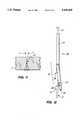
- FIG. 12shows another embodiment of a suture device 20 in accordance with the invention.
- the device 20includes a hook needle 21, a guide member 22 including a body portion 23 and a handle portion 24.
- the handle portion 24includes an axially extending hole which slidably receives the manipulating portion of the needle and the body portion 23 includes a slot 25 which slidably receives the pointed end of the needle.
- the slot 25extends through the distal end of the body portion 23 and seal means 26 is provided in the slot 25 for providing a fluid tight seal with the needle when the pointed end 27 of the needle passes through or out of the seal means 26.
- the handle portion 24shows the handle portion 24 pivoted with respect to the body portion 24 and the needle 21 in a nearly fully extended position with the pointed end 27 outside the slot 25 and ready to pierce through the inner side of tissue surrounding a wound site.
- the handle portion 24includes a tapered groove 28 at a distal end thereof. The groove 28 bends the pointed end 27 of the needle outwardly when the pointed end of the needle is advanced toward and slides along the groove 28 whereby the sharp tip of the needle is held against the handle portion and the suture attaching eyelet in the pointed end of the needle is easier to thread with a suture.
Landscapes
- Health & Medical Sciences (AREA)
- Life Sciences & Earth Sciences (AREA)
- Surgery (AREA)
- Heart & Thoracic Surgery (AREA)
- Engineering & Computer Science (AREA)
- Biomedical Technology (AREA)
- Nuclear Medicine, Radiotherapy & Molecular Imaging (AREA)
- Medical Informatics (AREA)
- Molecular Biology (AREA)
- Animal Behavior & Ethology (AREA)
- General Health & Medical Sciences (AREA)
- Public Health (AREA)
- Veterinary Medicine (AREA)
- Surgical Instruments (AREA)
Abstract
Description
Claims (20)
Priority Applications (1)
| Application Number | Priority Date | Filing Date | Title |
|---|---|---|---|
| US08/148,555US5439469A (en) | 1993-11-05 | 1993-11-05 | Wound closure device |
Applications Claiming Priority (1)
| Application Number | Priority Date | Filing Date | Title |
|---|---|---|---|
| US08/148,555US5439469A (en) | 1993-11-05 | 1993-11-05 | Wound closure device |
Related Parent Applications (1)
| Application Number | Title | Priority Date | Filing Date |
|---|---|---|---|
| US6686893AContinuation-In-Part | 1993-05-25 | 1993-05-25 |
Publications (1)
| Publication Number | Publication Date |
|---|---|
| US5439469Atrue US5439469A (en) | 1995-08-08 |
Family
ID=22526265
Family Applications (1)
| Application Number | Title | Priority Date | Filing Date |
|---|---|---|---|
| US08/148,555Expired - LifetimeUS5439469A (en) | 1993-11-05 | 1993-11-05 | Wound closure device |
Country Status (1)
| Country | Link |
|---|---|
| US (1) | US5439469A (en) |
Cited By (40)
| Publication number | Priority date | Publication date | Assignee | Title |
|---|---|---|---|---|
| WO1996003083A1 (en)* | 1994-07-22 | 1996-02-08 | Advanced Surgical, Inc. | Surgical suturing device |
| WO1996029012A1 (en)* | 1995-03-21 | 1996-09-26 | Scribner-Browne Medical Design Incorporated | Catheter introducer with suture capability |
| US5573540A (en)* | 1994-07-18 | 1996-11-12 | Yoon; Inbae | Apparatus and method for suturing an opening in anatomical tissue |
| WO1997007742A1 (en)* | 1995-08-28 | 1997-03-06 | Imagyn Medical Technologies, Inc. | Wound closure device |
| US5613975A (en)* | 1993-04-28 | 1997-03-25 | Christy; William J. | Endoscopic suturing device and method |
| US5766186A (en)* | 1996-12-03 | 1998-06-16 | Simon Fraser University | Suturing device |
| US5830220A (en)* | 1997-03-13 | 1998-11-03 | Wan; Shaw P. | Suturing instrument |
| US6203554B1 (en) | 1999-11-23 | 2001-03-20 | William Roberts | Apparatus, kit and methods for puncture site closure |
| US20040249412A1 (en)* | 2003-06-04 | 2004-12-09 | Snow Brent W. | Apparatus and methods for puncture site closure |
| US20050043746A1 (en)* | 2003-08-21 | 2005-02-24 | Pollak Stanley B. | Methods and instruments for closing laparoscopic trocar puncture wounds |
| US20050131505A1 (en)* | 2003-12-10 | 2005-06-16 | Japan General Medical Institute Co., Ltd. | Lead insertion support device |
| US6936054B2 (en) | 2002-07-22 | 2005-08-30 | Boston Scientific Scimed, Inc. | Placing sutures |
| US20060015006A1 (en)* | 2004-06-01 | 2006-01-19 | Laurence Bernard H | System and method for accessing a body cavity |
| US20090054895A1 (en)* | 2007-08-17 | 2009-02-26 | Wilson-Cook Medical Inc. | Device to open and close a bodily wall |
| US20090177031A1 (en)* | 2008-01-03 | 2009-07-09 | Wilson-Cook Medical Inc. | Medical systems, devices and methods for endoscopically suturing perforations |
| US7678133B2 (en) | 2004-07-10 | 2010-03-16 | Arstasis, Inc. | Biological tissue closure device and method |
| US20100185217A1 (en)* | 2007-06-08 | 2010-07-22 | Thomas Hsu | Devices and methods for closure of wounds |
| US20110125185A1 (en)* | 2009-11-24 | 2011-05-26 | Tyco Healthcare Group Lp, | Wound Plugs |
| US7998169B2 (en) | 2004-05-12 | 2011-08-16 | Arstasis, Inc. | Access and closure device and method |
| US8002794B2 (en) | 2005-05-12 | 2011-08-23 | Arstasis, Inc. | Access and closure device and method |
| RU2432914C1 (en)* | 2010-04-29 | 2011-11-10 | Сергей Геннадьевич Измайлов | Device for bringing together and fixation of laparotomic wound |
| USD652145S1 (en)* | 2009-10-14 | 2012-01-10 | Moris Topaz | Wound closure devices |
| US8551139B2 (en) | 2006-11-30 | 2013-10-08 | Cook Medical Technologies Llc | Visceral anchors for purse-string closure of perforations |
| US8647368B2 (en) | 2009-04-03 | 2014-02-11 | Cook Medical Technologies Llc | Tissue anchors and medical devices for rapid deployment of tissue anchors |
| US8740937B2 (en) | 2007-05-31 | 2014-06-03 | Cook Medical Technologies Llc | Suture lock |
| US8979882B2 (en) | 2008-07-21 | 2015-03-17 | Arstasis, Inc. | Devices, methods, and kits for forming tracts in tissue |
| US9393011B2 (en) | 2012-03-13 | 2016-07-19 | Suture Ease, Inc. | Needle and snare guide apparatus for passing suture |
| US9510823B2 (en) | 2013-08-02 | 2016-12-06 | Covidien Lp | Devices, systems, and methods for wound closure |
| US9655623B2 (en) | 2013-02-15 | 2017-05-23 | Alan E. Nash | System for closing a wound |
| US9662112B2 (en) | 2014-02-10 | 2017-05-30 | Alan E. Nash | System for closing a wound |
| RU2638764C2 (en)* | 2016-05-30 | 2017-12-15 | Государственное бюджетное учреждение Санкт-Петербургский научно-исследовательский институт скорой помощи им. И.И. Джанелидзе | Device for external fixation of abdominal wall for compartment syndrome treatment in case of tertiary peritonitis |
| US9861356B2 (en) | 2014-10-01 | 2018-01-09 | Brainchild Surgical Devices Llc | Suturing device and method |
| CN107789020A (en)* | 2017-11-13 | 2018-03-13 | 宁波胜杰康生物科技有限公司 | A kind of stitching unstrument |
| CN109157250A (en)* | 2018-07-23 | 2019-01-08 | 兰州大学 | Hysteroscope puncture path stitching unstrument |
| US10441753B2 (en) | 2012-05-25 | 2019-10-15 | Arstasis, Inc. | Vascular access configuration |
| WO2020016125A1 (en)* | 2018-07-18 | 2020-01-23 | Atropos Limited | Device and system for hernia repair |
| US10675447B2 (en) | 2012-05-25 | 2020-06-09 | Arstasis, Inc. | Vascular access configuration |
| US10966707B2 (en)* | 2018-01-04 | 2021-04-06 | Covidien Lp | Surgical port closure system |
| US10986982B2 (en) | 2007-06-08 | 2021-04-27 | Medeon Biodesign, Inc. | Lens cover modification |
| WO2023111890A1 (en)* | 2021-12-17 | 2023-06-22 | Cilag Gmbh International | Tissue suturing device |
Citations (24)
| Publication number | Priority date | Publication date | Assignee | Title |
|---|---|---|---|---|
| US385586A (en)* | 1888-07-03 | Surgeon s suture-carrier | ||
| US2738790A (en)* | 1954-08-12 | 1956-03-20 | George P Pilling & Son Company | Suturing instrument |
| SU1093329A1 (en)* | 1983-04-07 | 1984-05-23 | Калининский Государственный Медицинский Институт | Suture appliance for soft tissue |
| SU1115736A1 (en)* | 1983-05-13 | 1984-09-30 | Gashchenko Aleksandr | Apparatus for placing sutures on tissue |
| US4890614A (en)* | 1986-02-19 | 1990-01-02 | Yasuo Nakamura | Suture needle and its manufacturing process |
| WO1992012674A1 (en)* | 1991-01-17 | 1992-08-06 | Therkel Bisgaard | A set of tools for suturing in deep surgical apertures or body cavities |
| WO1992019313A1 (en)* | 1991-05-03 | 1992-11-12 | Medchem Products, Inc. | Method and device for wound closure |
| EP0529675A2 (en)* | 1991-08-29 | 1993-03-03 | Ethicon, Inc. | Shape memory effect surgical needles |
| US5192287A (en)* | 1991-04-05 | 1993-03-09 | American Cyanamid Company | Suture knot tying device |
| US5217471A (en)* | 1991-05-30 | 1993-06-08 | Burkhart Stephen S | Endoscopic suture knotting instrument |
| US5217470A (en)* | 1991-04-29 | 1993-06-08 | Weston Peter V | Apparatuses and methods for formation and use of a slipknot as a surgical suture knot |
| US5222508A (en)* | 1992-10-09 | 1993-06-29 | Osvaldo Contarini | Method for suturing punctures of the human body |
| WO1993013714A2 (en)* | 1992-01-07 | 1993-07-22 | Austin Leahy | A surgical device |
| US5234443A (en)* | 1991-07-26 | 1993-08-10 | The Regents Of The University Of California | Endoscopic knot tying apparatus and methods |
| US5236443A (en)* | 1992-05-21 | 1993-08-17 | Sidney Sontag | Suturing assembly and method |
| US5254105A (en)* | 1988-05-26 | 1993-10-19 | Haaga John R | Sheath for wound closure caused by a medical tubular device |
| US5258000A (en)* | 1991-11-25 | 1993-11-02 | Cook Incorporated | Tissue aperture repair device |
| WO1993021831A1 (en)* | 1992-05-01 | 1993-11-11 | Li Medical Technologies, Inc. | Intracorporeal knot tying apparatus and method |
| US5281237A (en)* | 1992-09-25 | 1994-01-25 | Gimpelson Richard J | Surgical stitching device and method of use |
| US5308353A (en)* | 1992-08-31 | 1994-05-03 | Merrimac Industries, Inc. | Surgical suturing device |
| US5318577A (en)* | 1990-06-26 | 1994-06-07 | Mitek Surgical Products, Inc. | Suture threading device |
| US5320632A (en)* | 1991-11-13 | 1994-06-14 | Harald Heidmueller | Surgical suturing apparatus |
| US5336239A (en)* | 1993-01-15 | 1994-08-09 | Gimpelson Richard J | Surgical needle |
| US5350385A (en)* | 1993-04-28 | 1994-09-27 | Christy William J | Surgical stab wound closure device and method |
- 1993
- 1993-11-05USUS08/148,555patent/US5439469A/ennot_activeExpired - Lifetime
Patent Citations (24)
| Publication number | Priority date | Publication date | Assignee | Title |
|---|---|---|---|---|
| US385586A (en)* | 1888-07-03 | Surgeon s suture-carrier | ||
| US2738790A (en)* | 1954-08-12 | 1956-03-20 | George P Pilling & Son Company | Suturing instrument |
| SU1093329A1 (en)* | 1983-04-07 | 1984-05-23 | Калининский Государственный Медицинский Институт | Suture appliance for soft tissue |
| SU1115736A1 (en)* | 1983-05-13 | 1984-09-30 | Gashchenko Aleksandr | Apparatus for placing sutures on tissue |
| US4890614A (en)* | 1986-02-19 | 1990-01-02 | Yasuo Nakamura | Suture needle and its manufacturing process |
| US5254105A (en)* | 1988-05-26 | 1993-10-19 | Haaga John R | Sheath for wound closure caused by a medical tubular device |
| US5318577A (en)* | 1990-06-26 | 1994-06-07 | Mitek Surgical Products, Inc. | Suture threading device |
| WO1992012674A1 (en)* | 1991-01-17 | 1992-08-06 | Therkel Bisgaard | A set of tools for suturing in deep surgical apertures or body cavities |
| US5192287A (en)* | 1991-04-05 | 1993-03-09 | American Cyanamid Company | Suture knot tying device |
| US5217470A (en)* | 1991-04-29 | 1993-06-08 | Weston Peter V | Apparatuses and methods for formation and use of a slipknot as a surgical suture knot |
| WO1992019313A1 (en)* | 1991-05-03 | 1992-11-12 | Medchem Products, Inc. | Method and device for wound closure |
| US5217471A (en)* | 1991-05-30 | 1993-06-08 | Burkhart Stephen S | Endoscopic suture knotting instrument |
| US5234443A (en)* | 1991-07-26 | 1993-08-10 | The Regents Of The University Of California | Endoscopic knot tying apparatus and methods |
| EP0529675A2 (en)* | 1991-08-29 | 1993-03-03 | Ethicon, Inc. | Shape memory effect surgical needles |
| US5320632A (en)* | 1991-11-13 | 1994-06-14 | Harald Heidmueller | Surgical suturing apparatus |
| US5258000A (en)* | 1991-11-25 | 1993-11-02 | Cook Incorporated | Tissue aperture repair device |
| WO1993013714A2 (en)* | 1992-01-07 | 1993-07-22 | Austin Leahy | A surgical device |
| WO1993021831A1 (en)* | 1992-05-01 | 1993-11-11 | Li Medical Technologies, Inc. | Intracorporeal knot tying apparatus and method |
| US5236443A (en)* | 1992-05-21 | 1993-08-17 | Sidney Sontag | Suturing assembly and method |
| US5308353A (en)* | 1992-08-31 | 1994-05-03 | Merrimac Industries, Inc. | Surgical suturing device |
| US5281237A (en)* | 1992-09-25 | 1994-01-25 | Gimpelson Richard J | Surgical stitching device and method of use |
| US5222508A (en)* | 1992-10-09 | 1993-06-29 | Osvaldo Contarini | Method for suturing punctures of the human body |
| US5336239A (en)* | 1993-01-15 | 1994-08-09 | Gimpelson Richard J | Surgical needle |
| US5350385A (en)* | 1993-04-28 | 1994-09-27 | Christy William J | Surgical stab wound closure device and method |
Cited By (71)
| Publication number | Priority date | Publication date | Assignee | Title |
|---|---|---|---|---|
| US5697941A (en)* | 1993-04-28 | 1997-12-16 | Christy; William J. | Endoscopic suturing device and method |
| US5613975A (en)* | 1993-04-28 | 1997-03-25 | Christy; William J. | Endoscopic suturing device and method |
| US5830125A (en)* | 1993-08-12 | 1998-11-03 | Scribner-Browne Medical Design Incorporated | Catheter introducer with suture capability |
| US5632752A (en)* | 1993-10-12 | 1997-05-27 | Urohealth Systems, Inc. | Surgical suturing device |
| US5573540A (en)* | 1994-07-18 | 1996-11-12 | Yoon; Inbae | Apparatus and method for suturing an opening in anatomical tissue |
| WO1996003083A1 (en)* | 1994-07-22 | 1996-02-08 | Advanced Surgical, Inc. | Surgical suturing device |
| WO1996029012A1 (en)* | 1995-03-21 | 1996-09-26 | Scribner-Browne Medical Design Incorporated | Catheter introducer with suture capability |
| US5653717A (en)* | 1995-08-28 | 1997-08-05 | Urohealth Systems, Inc. | Wound closure device |
| WO1997007742A1 (en)* | 1995-08-28 | 1997-03-06 | Imagyn Medical Technologies, Inc. | Wound closure device |
| EP0850021A4 (en)* | 1995-08-28 | 2000-03-08 | Imagyn Medical Technologies In | Wound closure device |
| US5766186A (en)* | 1996-12-03 | 1998-06-16 | Simon Fraser University | Suturing device |
| US5830220A (en)* | 1997-03-13 | 1998-11-03 | Wan; Shaw P. | Suturing instrument |
| US6203554B1 (en) | 1999-11-23 | 2001-03-20 | William Roberts | Apparatus, kit and methods for puncture site closure |
| US6936054B2 (en) | 2002-07-22 | 2005-08-30 | Boston Scientific Scimed, Inc. | Placing sutures |
| US8764771B2 (en) | 2002-07-22 | 2014-07-01 | Boston Scientific Scimed, Inc. | Placing sutures |
| US8361089B2 (en) | 2002-07-22 | 2013-01-29 | Boston Scientific Scimed, Inc. | Placing sutures |
| US20110060352A1 (en)* | 2002-07-22 | 2011-03-10 | Boston Scientific Scimed, Inc. | Placing sutures |
| US20050222589A1 (en)* | 2002-07-22 | 2005-10-06 | Boston Scientific Scimed, Inc. | Placing sutures |
| US7815654B2 (en) | 2002-07-22 | 2010-10-19 | Boston Scientific Scimed, Inc. | Placing sutures |
| US9504465B2 (en) | 2002-07-22 | 2016-11-29 | Boston Scientific Scimed, Inc. | Placing sutures |
| US8906041B2 (en) | 2002-07-22 | 2014-12-09 | Boston Scientific Scimed, Inc. | Placing sutures |
| US20040249412A1 (en)* | 2003-06-04 | 2004-12-09 | Snow Brent W. | Apparatus and methods for puncture site closure |
| US20050043746A1 (en)* | 2003-08-21 | 2005-02-24 | Pollak Stanley B. | Methods and instruments for closing laparoscopic trocar puncture wounds |
| US7320693B2 (en) | 2003-08-21 | 2008-01-22 | Pollak Stanley B | Methods and instruments for closing laparoscopic trocar puncture wounds |
| US20050131505A1 (en)* | 2003-12-10 | 2005-06-16 | Japan General Medical Institute Co., Ltd. | Lead insertion support device |
| US8002792B2 (en) | 2004-05-12 | 2011-08-23 | Arstasis, Inc. | Access and closure device and method |
| US8002791B2 (en) | 2004-05-12 | 2011-08-23 | Arstasis, Inc. | Access and closure device and method |
| US8012168B2 (en) | 2004-05-12 | 2011-09-06 | Arstasis, Inc. | Access and closure device and method |
| US7998169B2 (en) | 2004-05-12 | 2011-08-16 | Arstasis, Inc. | Access and closure device and method |
| US8002793B2 (en) | 2004-05-12 | 2011-08-23 | Arstasis, Inc. | Access and closure device and method |
| US20060015006A1 (en)* | 2004-06-01 | 2006-01-19 | Laurence Bernard H | System and method for accessing a body cavity |
| US8475476B2 (en) | 2004-06-01 | 2013-07-02 | Cook Medical Technologies Llc | System and method for accessing a body cavity |
| US7678133B2 (en) | 2004-07-10 | 2010-03-16 | Arstasis, Inc. | Biological tissue closure device and method |
| US8083767B2 (en) | 2005-05-12 | 2011-12-27 | Arstasis, Inc. | Access and closure device and method |
| US8241325B2 (en) | 2005-05-12 | 2012-08-14 | Arstasis, Inc. | Access and closure device and method |
| US8002794B2 (en) | 2005-05-12 | 2011-08-23 | Arstasis, Inc. | Access and closure device and method |
| US8551139B2 (en) | 2006-11-30 | 2013-10-08 | Cook Medical Technologies Llc | Visceral anchors for purse-string closure of perforations |
| US8740937B2 (en) | 2007-05-31 | 2014-06-03 | Cook Medical Technologies Llc | Suture lock |
| US10986982B2 (en) | 2007-06-08 | 2021-04-27 | Medeon Biodesign, Inc. | Lens cover modification |
| US20100185217A1 (en)* | 2007-06-08 | 2010-07-22 | Thomas Hsu | Devices and methods for closure of wounds |
| US10314566B2 (en) | 2007-06-08 | 2019-06-11 | Medeon Biodesign, Inc. | Devices and methods for closure of wounds |
| US9241613B2 (en) | 2007-06-08 | 2016-01-26 | Medeon Biodesign, Inc. | Devices and methods for closure of wounds |
| US7879049B2 (en) | 2007-08-17 | 2011-02-01 | Wilson-Cook Medical Inc. | Device to open and close a bodily wall |
| US20090054895A1 (en)* | 2007-08-17 | 2009-02-26 | Wilson-Cook Medical Inc. | Device to open and close a bodily wall |
| US8876701B2 (en) | 2008-01-03 | 2014-11-04 | Cook Medical Technologies Llc | Medical systems, devices and methods for endoscopically suturing perforations |
| US20090177031A1 (en)* | 2008-01-03 | 2009-07-09 | Wilson-Cook Medical Inc. | Medical systems, devices and methods for endoscopically suturing perforations |
| US8979882B2 (en) | 2008-07-21 | 2015-03-17 | Arstasis, Inc. | Devices, methods, and kits for forming tracts in tissue |
| US8647368B2 (en) | 2009-04-03 | 2014-02-11 | Cook Medical Technologies Llc | Tissue anchors and medical devices for rapid deployment of tissue anchors |
| USD652145S1 (en)* | 2009-10-14 | 2012-01-10 | Moris Topaz | Wound closure devices |
| US8858592B2 (en) | 2009-11-24 | 2014-10-14 | Covidien Lp | Wound plugs |
| US9439636B2 (en) | 2009-11-24 | 2016-09-13 | Covidien Lp | Wound plugs |
| US20110125185A1 (en)* | 2009-11-24 | 2011-05-26 | Tyco Healthcare Group Lp, | Wound Plugs |
| RU2432914C1 (en)* | 2010-04-29 | 2011-11-10 | Сергей Геннадьевич Измайлов | Device for bringing together and fixation of laparotomic wound |
| US9393011B2 (en) | 2012-03-13 | 2016-07-19 | Suture Ease, Inc. | Needle and snare guide apparatus for passing suture |
| US10441753B2 (en) | 2012-05-25 | 2019-10-15 | Arstasis, Inc. | Vascular access configuration |
| US10675447B2 (en) | 2012-05-25 | 2020-06-09 | Arstasis, Inc. | Vascular access configuration |
| US9655623B2 (en) | 2013-02-15 | 2017-05-23 | Alan E. Nash | System for closing a wound |
| US10143466B2 (en) | 2013-08-02 | 2018-12-04 | Covidien Lp | Devices, systems, and methods for wound closure |
| US9510823B2 (en) | 2013-08-02 | 2016-12-06 | Covidien Lp | Devices, systems, and methods for wound closure |
| US9662112B2 (en) | 2014-02-10 | 2017-05-30 | Alan E. Nash | System for closing a wound |
| US9861356B2 (en) | 2014-10-01 | 2018-01-09 | Brainchild Surgical Devices Llc | Suturing device and method |
| RU2638764C2 (en)* | 2016-05-30 | 2017-12-15 | Государственное бюджетное учреждение Санкт-Петербургский научно-исследовательский институт скорой помощи им. И.И. Джанелидзе | Device for external fixation of abdominal wall for compartment syndrome treatment in case of tertiary peritonitis |
| CN107789020A (en)* | 2017-11-13 | 2018-03-13 | 宁波胜杰康生物科技有限公司 | A kind of stitching unstrument |
| CN107789020B (en)* | 2017-11-13 | 2024-03-15 | 宁波胜杰康生物科技有限公司 | Stitching instrument |
| US10966707B2 (en)* | 2018-01-04 | 2021-04-06 | Covidien Lp | Surgical port closure system |
| US20210298743A1 (en)* | 2018-07-18 | 2021-09-30 | Atropolos Limited | Device and system for hernia repair |
| WO2020016125A1 (en)* | 2018-07-18 | 2020-01-23 | Atropos Limited | Device and system for hernia repair |
| US12089835B2 (en)* | 2018-07-18 | 2024-09-17 | Atropos Limited | Device and system for hernia repair |
| CN109157250A (en)* | 2018-07-23 | 2019-01-08 | 兰州大学 | Hysteroscope puncture path stitching unstrument |
| WO2020243143A1 (en)* | 2019-05-28 | 2020-12-03 | Atropos Limited | Device and system for hernia repair |
| WO2023111890A1 (en)* | 2021-12-17 | 2023-06-22 | Cilag Gmbh International | Tissue suturing device |
Similar Documents
| Publication | Publication Date | Title |
|---|---|---|
| US5439469A (en) | Wound closure device | |
| US12376839B2 (en) | Laparoscopic port site closure tool | |
| US8333774B2 (en) | Suturing instrument with needle dock | |
| US5626588A (en) | Trocar wound closure device | |
| US6224614B1 (en) | Suturing instrument with angled needle holder and method for use thereof | |
| US5337736A (en) | Method of using a laparoscopic retractor | |
| CA2569878C (en) | Surgical closure instrument and methods | |
| US5336239A (en) | Surgical needle | |
| US7320693B2 (en) | Methods and instruments for closing laparoscopic trocar puncture wounds | |
| US5374275A (en) | Surgical suturing device and method of use | |
| US5503634A (en) | Surgical stab wound closure device and method | |
| US8715251B2 (en) | Surgical access port and method of using same | |
| US5431666A (en) | Surgical suture instrument | |
| US5364408A (en) | Endoscopic suture system | |
| US6203554B1 (en) | Apparatus, kit and methods for puncture site closure | |
| US5507755A (en) | Apparatus and method for closing puncture wounds | |
| US5222977A (en) | Surgical needle with an adjustable eye | |
| JP4493258B2 (en) | Tissue puncture device | |
| US9364214B2 (en) | Cannulated instrument with curved shaft for passing suture through tissue | |
| US20040087978A1 (en) | Surgical fascia closure instrument, guide and method | |
| US9301748B2 (en) | Suture apparatus, system and method | |
| US20070135679A1 (en) | Colonoscopic device stabilizer | |
| CA2134662A1 (en) | Laparoscopic surgical ligation, repair and electrosurgical coagulation and cutting device | |
| US10660636B2 (en) | Suture apparatus, system and method | |
| US20150374359A1 (en) | Suture apparatus, system and method |
Legal Events
| Date | Code | Title | Description |
|---|---|---|---|
| AS | Assignment | Owner name:ADVANCED SURGICAL,INC., NEW JERSEY Free format text:ASSIGNMENT OF ASSIGNORS INTEREST;ASSIGNORS:HEAVEN, MALCOLM D.;SCHRAYER, HOWARD;REEL/FRAME:006860/0518;SIGNING DATES FROM 19931110 TO 19931116 | |
| FEPP | Fee payment procedure | Free format text:PAYOR NUMBER ASSIGNED (ORIGINAL EVENT CODE: ASPN); ENTITY STATUS OF PATENT OWNER: LARGE ENTITY | |
| STCF | Information on status: patent grant | Free format text:PATENTED CASE | |
| AS | Assignment | Owner name:UROHEALTH SYSTEMS, INC., CALIFORNIA Free format text:ASSIGNMENT OF ASSIGNORS INTEREST;ASSIGNOR:ADVANCED SURGICAL, INC.;REEL/FRAME:008126/0422 Effective date:19960903 | |
| AS | Assignment | Owner name:IMAGYN MEDICAL TECHNOLOGIES, INC., CALIFORNIA Free format text:CHANGE OF NAME;ASSIGNOR:UROHEALTH SYSTEMS, INC.;REEL/FRAME:008876/0078 Effective date:19970929 | |
| AS | Assignment | Owner name:BT COMMERCIAL CORPORATION, ILLINOIS Free format text:SECURITY AGREEMENT;ASSIGNOR:IMAGYN MEDICAL TECHNOLOGIES, INC.;REEL/FRAME:008886/0680 Effective date:19971230 | |
| AS | Assignment | Owner name:UROHEALTH, INC., CALIFORNIA Free format text:RELEASE OF SECURITY INTEREST;ASSIGNOR:CREDIT AGRICOLE INDOSUEZ;REEL/FRAME:008920/0448 Effective date:19971219 Owner name:ALLSTATE MEDICAL PRODUCTS, INC., MINNESOTA Free format text:RELEASE OF SECURITY INTEREST;ASSIGNOR:CREDIT AGRICOLE INDOSUEZ;REEL/FRAME:008920/0448 Effective date:19971219 Owner name:GATES PLASTICS, CALIFORNIA Free format text:RELEASE OF SECURITY INTEREST;ASSIGNOR:CREDIT AGRICOLE INDOSUEZ;REEL/FRAME:008920/0448 Effective date:19971219 Owner name:DACOMED CORPORATION, MINNESOTA Free format text:RELEASE OF SECURITY INTEREST;ASSIGNOR:CREDIT AGRICOLE INDOSUEZ;REEL/FRAME:008920/0448 Effective date:19971219 Owner name:UROHEALTH OF KENTUCKY, INC., KENTUCKY Free format text:RELEASE OF SECURITY INTEREST;ASSIGNOR:CREDIT AGRICOLE INDOSUEZ;REEL/FRAME:008920/0448 Effective date:19971219 Owner name:DACOMED INTERNATIONAL INC., MINNESOTA Free format text:RELEASE OF SECURITY INTEREST;ASSIGNOR:CREDIT AGRICOLE INDOSUEZ;REEL/FRAME:008920/0448 Effective date:19971219 | |
| FEPP | Fee payment procedure | Free format text:PAT HLDR NO LONGER CLAIMS SMALL ENT STAT AS SMALL BUSINESS (ORIGINAL EVENT CODE: LSM2); ENTITY STATUS OF PATENT OWNER: LARGE ENTITY | |
| FPAY | Fee payment | Year of fee payment:4 | |
| AS | Assignment | Owner name:BT COMMERCIAL CORPORATION, ILLINOIS Free format text:SECURITY AGREEMENT;ASSIGNOR:IMAGYN MEDICAL TECHNOLOGIES, INC.;REEL/FRAME:010444/0304 Effective date:19991029 | |
| AS | Assignment | Owner name:BT COMMERCIAL CORPORATION A DELAWARE CORPORATION, Free format text:SECURITY INTEREST;ASSIGNOR:IMAGYN MEDICAL TECHNOLOGIES, INC. A DELAWARE CORPORATION;REEL/FRAME:010437/0079 Effective date:19991029 | |
| FPAY | Fee payment | Year of fee payment:8 | |
| AS | Assignment | Owner name:JPMORGAN CHASE BANK, AS ADMINISTRATIVE AGENT, TEXA Free format text:SECURITY INTEREST;ASSIGNOR:CONMED CORPORATION;REEL/FRAME:014289/0859 Effective date:20020828 | |
| AS | Assignment | Owner name:IMAGYN MEDICAL TECHNOLOGIES, INC., CALIFORNIA Free format text:RELEASE OF SECURITY INTEREST;ASSIGNOR:BT COMMERCIAL CORPORATION;REEL/FRAME:014357/0540 Effective date:20010705 | |
| AS | Assignment | Owner name:CONMED CORPORATION, NEW YORK Free format text:ASSIGNMENT OF ASSIGNORS INTEREST;ASSIGNOR:IMAGYN MEDICAL TECHNOLOGIES, INC.;REEL/FRAME:014373/0092 Effective date:20010706 | |
| FPAY | Fee payment | Year of fee payment:12 |