US5434418A - Intra-oral sensor for computer aided radiography - Google Patents
Intra-oral sensor for computer aided radiographyDownload PDFInfo
- Publication number
- US5434418A US5434418AUS07/962,129US96212992AUS5434418AUS 5434418 AUS5434418 AUS 5434418AUS 96212992 AUS96212992 AUS 96212992AUS 5434418 AUS5434418 AUS 5434418A
- Authority
- US
- United States
- Prior art keywords
- sensor
- computer
- electrical signals
- array
- semiconductor image
- Prior art date
- Legal status (The legal status is an assumption and is not a legal conclusion. Google has not performed a legal analysis and makes no representation as to the accuracy of the status listed.)
- Expired - Lifetime
Links
- 238000002601radiographyMethods0.000titleclaimsdescription4
- 239000004065semiconductorSubstances0.000claimsabstractdescription32
- 239000000463materialSubstances0.000claimsabstractdescription7
- 238000003384imaging methodMethods0.000claimsdescription14
- 238000000034methodMethods0.000claimsdescription10
- 230000005540biological transmissionEffects0.000claimsdescription9
- 238000012545processingMethods0.000claimsdescription9
- 238000006243chemical reactionMethods0.000claimsdescription7
- 230000008569processEffects0.000claimsdescription5
- 230000000007visual effectEffects0.000claims3
- 229920001343polytetrafluoroethylenePolymers0.000abstractdescription9
- 239000004810polytetrafluoroethyleneSubstances0.000abstractdescription9
- 230000001681protective effectEffects0.000abstractdescription6
- 238000004458analytical methodMethods0.000abstractdescription5
- 230000005855radiationEffects0.000abstractdescription5
- 229910052716thalliumInorganic materials0.000abstractdescription5
- BKVIYDNLLOSFOA-UHFFFAOYSA-NthalliumChemical compound[Tl]BKVIYDNLLOSFOA-UHFFFAOYSA-N0.000abstractdescription5
- 239000000919ceramicSubstances0.000abstractdescription4
- 239000004033plasticSubstances0.000abstractdescription4
- XQPRBTXUXXVTKB-UHFFFAOYSA-Mcaesium iodideChemical compound[I-].[Cs+]XQPRBTXUXXVTKB-UHFFFAOYSA-M0.000abstractdescription3
- -1polytetrafluoroethylenePolymers0.000abstractdescription3
- 230000007480spreadingEffects0.000abstractdescription3
- 238000013519translationMethods0.000abstractdescription3
- 238000011065in-situ storageMethods0.000abstractdescription2
- 230000003287optical effectEffects0.000abstractdescription2
- 239000010410layerSubstances0.000description28
- XUIMIQQOPSSXEZ-UHFFFAOYSA-NSiliconChemical compound[Si]XUIMIQQOPSSXEZ-UHFFFAOYSA-N0.000description7
- 229910052710siliconInorganic materials0.000description7
- 239000010703siliconSubstances0.000description7
- 239000004593EpoxySubstances0.000description5
- 230000015556catabolic processEffects0.000description5
- 238000006731degradation reactionMethods0.000description5
- 229920001296polysiloxanePolymers0.000description4
- 230000035945sensitivityEffects0.000description3
- 238000003491arrayMethods0.000description2
- 230000008901benefitEffects0.000description2
- 238000001444catalytic combustion detectionMethods0.000description2
- 230000006835compressionEffects0.000description2
- 238000007906compressionMethods0.000description2
- 238000005516engineering processMethods0.000description2
- 230000006872improvementEffects0.000description2
- 230000007774longtermEffects0.000description2
- 239000013307optical fiberSubstances0.000description2
- 230000009467reductionEffects0.000description2
- WUPHOULIZUERAE-UHFFFAOYSA-N3-(oxolan-2-yl)propanoic acidChemical compoundOC(=O)CCC1CCCO1WUPHOULIZUERAE-UHFFFAOYSA-N0.000description1
- MARUHZGHZWCEQU-UHFFFAOYSA-N5-phenyl-2h-tetrazoleChemical compoundC1=CC=CC=C1C1=NNN=N1MARUHZGHZWCEQU-UHFFFAOYSA-N0.000description1
- 229910004829CaWO4Inorganic materials0.000description1
- OYPRJOBELJOOCE-UHFFFAOYSA-NCalciumChemical compound[Ca]OYPRJOBELJOOCE-UHFFFAOYSA-N0.000description1
- 208000035473Communicable diseaseDiseases0.000description1
- OAICVXFJPJFONN-UHFFFAOYSA-NPhosphorusChemical compound[P]OAICVXFJPJFONN-UHFFFAOYSA-N0.000description1
- 239000005083Zinc sulfideSubstances0.000description1
- 239000006096absorbing agentSubstances0.000description1
- 230000003321amplificationEffects0.000description1
- 210000000988bone and boneAnatomy0.000description1
- 229910052980cadmium sulfideInorganic materials0.000description1
- 229910052792caesiumInorganic materials0.000description1
- TVFDJXOCXUVLDH-UHFFFAOYSA-Ncaesium atomChemical compound[Cs]TVFDJXOCXUVLDH-UHFFFAOYSA-N0.000description1
- 229910052791calciumInorganic materials0.000description1
- 239000011575calciumSubstances0.000description1
- 239000011248coating agentSubstances0.000description1
- 238000000576coating methodMethods0.000description1
- 239000003086colorantSubstances0.000description1
- 230000002950deficientEffects0.000description1
- 230000032798delaminationEffects0.000description1
- 238000001514detection methodMethods0.000description1
- 238000011161developmentMethods0.000description1
- 230000008030eliminationEffects0.000description1
- 238000003379elimination reactionMethods0.000description1
- 230000002708enhancing effectEffects0.000description1
- 238000011156evaluationMethods0.000description1
- 238000010438heat treatmentMethods0.000description1
- 239000012212insulatorSubstances0.000description1
- 238000012544monitoring processMethods0.000description1
- 238000003199nucleic acid amplification methodMethods0.000description1
- 238000004091panningMethods0.000description1
- 230000035515penetrationEffects0.000description1
- 230000002093peripheral effectEffects0.000description1
- 239000011241protective layerSubstances0.000description1
- 230000000191radiation effectEffects0.000description1
- 239000000565sealantSubstances0.000description1
- 230000035939shockEffects0.000description1
- 210000004872soft tissueAnatomy0.000description1
- 230000001954sterilising effectEffects0.000description1
- 238000004659sterilization and disinfectionMethods0.000description1
- 238000003860storageMethods0.000description1
- 239000000126substanceSubstances0.000description1
- PBYZMCDFOULPGH-UHFFFAOYSA-NtungstateChemical compound[O-][W]([O-])(=O)=OPBYZMCDFOULPGH-UHFFFAOYSA-N0.000description1
- 238000007738vacuum evaporationMethods0.000description1
- 229910052984zinc sulfideInorganic materials0.000description1
- UQMZPFKLYHOJDL-UHFFFAOYSA-Nzinc;cadmium(2+);disulfideChemical compound[S-2].[S-2].[Zn+2].[Cd+2]UQMZPFKLYHOJDL-UHFFFAOYSA-N0.000description1
- DRDVZXDWVBGGMH-UHFFFAOYSA-Nzinc;sulfideChemical compound[S-2].[Zn+2]DRDVZXDWVBGGMH-UHFFFAOYSA-N0.000description1
Images
Classifications
- G—PHYSICS
- G01—MEASURING; TESTING
- G01T—MEASUREMENT OF NUCLEAR OR X-RADIATION
- G01T1/00—Measuring X-radiation, gamma radiation, corpuscular radiation, or cosmic radiation
- G01T1/16—Measuring radiation intensity
- G01T1/20—Measuring radiation intensity with scintillation detectors
- G01T1/2018—Scintillation-photodiode combinations
- G01T1/20188—Auxiliary details, e.g. casings or cooling
- G01T1/20189—Damping or insulation against damage, e.g. caused by heat or pressure
- A—HUMAN NECESSITIES
- A61—MEDICAL OR VETERINARY SCIENCE; HYGIENE
- A61B—DIAGNOSIS; SURGERY; IDENTIFICATION
- A61B6/00—Apparatus or devices for radiation diagnosis; Apparatus or devices for radiation diagnosis combined with radiation therapy equipment
- A61B6/50—Apparatus or devices for radiation diagnosis; Apparatus or devices for radiation diagnosis combined with radiation therapy equipment specially adapted for specific body parts; specially adapted for specific clinical applications
- A61B6/51—Apparatus or devices for radiation diagnosis; Apparatus or devices for radiation diagnosis combined with radiation therapy equipment specially adapted for specific body parts; specially adapted for specific clinical applications for dentistry
- A61B6/512—Intraoral means
- G—PHYSICS
- G01—MEASURING; TESTING
- G01T—MEASUREMENT OF NUCLEAR OR X-RADIATION
- G01T1/00—Measuring X-radiation, gamma radiation, corpuscular radiation, or cosmic radiation
- G01T1/16—Measuring radiation intensity
- G01T1/20—Measuring radiation intensity with scintillation detectors
- G01T1/2018—Scintillation-photodiode combinations
- G01T1/20183—Arrangements for preventing or correcting crosstalk, e.g. optical or electrical arrangements for correcting crosstalk
- G—PHYSICS
- G01—MEASURING; TESTING
- G01T—MEASUREMENT OF NUCLEAR OR X-RADIATION
- G01T1/00—Measuring X-radiation, gamma radiation, corpuscular radiation, or cosmic radiation
- G01T1/16—Measuring radiation intensity
- G01T1/20—Measuring radiation intensity with scintillation detectors
- G01T1/2018—Scintillation-photodiode combinations
- G01T1/20187—Position of the scintillator with respect to the photodiode, e.g. photodiode surrounding the crystal, the crystal surrounding the photodiode, shape or size of the scintillator
- A—HUMAN NECESSITIES
- A61—MEDICAL OR VETERINARY SCIENCE; HYGIENE
- A61B—DIAGNOSIS; SURGERY; IDENTIFICATION
- A61B6/00—Apparatus or devices for radiation diagnosis; Apparatus or devices for radiation diagnosis combined with radiation therapy equipment
- A61B6/44—Constructional features of apparatus for radiation diagnosis
- A61B6/4423—Constructional features of apparatus for radiation diagnosis related to hygiene or sterilisation
Definitions
- This inventionrelates to oral and dental radiology and particularly to computer aided radiology without use of x-ray film.
- Dentists and oral surgeonstypically utilize x-ray apparatus to examine patients prior to treatment. Film placed in the patient's mouth is exposed to the x-rays which pass through the soft tissue of skin and gums and are absorbed or refracted by the harder bone and teeth structures. The film is then chemically developed and dried to produce the image from which the dentist makes appropriate treatment; evaluations. Such technology, though with many refinements, has not basically changed over the past fifty years.
- the radiation dosagewhich optimally for conventional film exposure, is about 260 millirads. Since the high energy electrons from x-ray sources can cause damage to the nuclei of cells it is agreed that minimizing radiation exposure is highly desirable. In this regard, the average dose for dental x-rays has been reduced by 50% over the last thirty years, to the current levels, mostly as a result of improvement in film sensitivity. Further incremental reductions in requisite x-ray dosage for film exposure is unlikely to be of any great extent.
- Film processing itselfpresents other problems including the time, expense, inconvenience and uncertainty of processing x-ray films and many times the exposure is defective or blurred.
- the minimum time for developmentis four to six minutes.
- the additional componentsentail greater costs, introduce problems with component degradation and failure, and generally preclude direct sterilization by dental autoclaving.
- the sensors of the prior artare accordingly usually described as being used with disposable plastic sleeves.
- such sleeves while usefulmay be occasionally susceptible to perforation during use, a dangerous situation with prevalent communicable diseases.
- the systems describedprovided resolution of images substantially below that of x-ray dental film.
- x-ray dosageis reduced, it is at the cost of diagnostic accuracy.
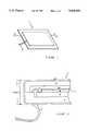
- FIG. 1is an isometric view of the sensor of the present invention
- FIG. 2is a magnified cross sectional view of the sensor of FIG. 1 taken along line 2--2;
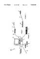
- FIG. 3shows the sensor of FIG. 1 and 2 connected to a small radio transmitter for image transmission to a remote computer
- FIG. 4is a schematic view showing the utilization of the sensor of the present invention in a computerized dental diagnostic system.
- the present inventioncomprises an intra-oral sensor for computer aided oral radiography, comprising a thin, large area semiconductor image array, such as a modified charge coupled device (CCD) or photodiode array, coated with a thin, epitaxial growth of a material, such as thallium doped cesium iodide CsI(Tl), which provides conversion of x-rays to light or visible photons; and said semiconductor image array converts the light or visible photons to electrical signals for transmission to computer means for imaging.
- the imagingcan be in the form of real time monitor viewing and/or as a hard copy printout.
- the coating materialalso provides protection to the semiconductor image array from x-ray degradation.
- the coated sensorcontains an integrated signal amplifier, and is bonded to and supported on a passivated ceramic chip, with the entire assembly being coated with a protective (particularly against moisture) inert plastic layer, e.g., polytetrafluoroethylene (PTFE), which is pervious to x-ray radiation.
- a protective (particularly against moisture) inert plastic layere.g., polytetrafluoroethylene (PTFE), which is pervious to x-ray radiation.
- PTFEpolytetrafluoroethylene
- the CsI(Tl)is sensitive to x-ray photons, efficiently converting them into visible photons in the 500-600 um range.
- Other x-ray-to-light converting materialsthough of lesser efficiency, include cadmium telluride and cadmium sulfide, calcium tungstate (CaWO 4 ), zinc sulfide and zinc cadmium sulfide.
- growth of the CsI(Tl) layeris preferably directed into narrow (10-20 um) Columns.
- CsI(Tl) thicknessesof 300 um or more it is further preferred that a laser be utilized to make channels between the columns in order to eliminate cross-talk in the computerized imaging.
- Light and/or visible photonsare detected by the large area semiconductor array and the output is monitored by a computer until polling of the CCD or photodiode array indicates that there is no further conducting.
- a signalthereafter causes a read out of the electrical charges, for translation from analog to digital signals of images, with computer display and analysis.
- the intra-oral sensoris exceedingly thin for proper mouth placement and in situ maneuvering, with a large active area substantially equivalent to corresponding dental film sizes.
- Such proper mouth placementdictates a maximum thickness of about 5 mm and more preferably a thickness of no more than about 3 mm.
- One common size of dental filmhas an active area of approximately 4 ⁇ 3 cm, corresponding to size 2 dental film which is commonly used for adult radiographs.
- Another dental film with an active area of approximately 3.2 ⁇ 2.4 cmis size 0 dental film, the standard pediatric film size.
- the sensorcontains limited electronics and no optical elements, is resistant to moisture and heat, and is readily autoclaved.
- the CsI layeris grown in a relatively thick layer of 200-300 um on the active portion of the semiconductor. This reduces the probability of x-ray photons from impinging on the silicon of the semi-conductor to less than 0.01%. To compensate for reduction in x-ray photons, spatial resolution is maintained by carefully growing high quality CsI layers.
- the CCDmay be formed on a thin (10 um) epitaxial silicon layer with p type doping, grown on a p + doped bulk silicon wafer. This process provides that only the x-rays which are absorbed by the epitaxial layer can contribute to the image. Since silicon is a very poor absorber of x-rays of average energy of 35 KeV, very few x-rays (less than 0.1%) will be absorbed in the top 10 um of the silicon.
- the CCDis preferably operated in Multi-Phase Pinned (MPP) mode.
- MPPMulti-Phase Pinned
- the primary long-term effect of radiation such as x-rays on CCDsis an increase in dark signal from thermally generated electron hole pairs. These hole pairs increase with the introduction of a trap level near the surface of the silicon by accumulated x-rays. The dark signal reduces the sensitivity of the CCDs and lowers the maximum exposure to which they can respond.
- the surface of the semiconductoris "pinned" by keeping the potential at the surface negative for a great portion of the time. As a result, thermally generated electron hole pairs at the surface are exterminated before they cause a signal.
- the senorcomprises a self-scanning array with a maximum pixel size of 48 um.
- the arraycontains (on-chip) all of the circuitry required to control the exposure and read-out of the image.
- the photodiode arrayis coated with a thin layer of thallium doped cesium iodide, CsI(TI), to convert the x-ray photons into visible photons in the 500-600 um range which are efficiently absorbed by the photodiodes.
- CsI(TI)cesium iodide
- Each of the CCD and photodiode arraysincorporates several discrete diodes, the outputs of which are monitored by the computer to determine the start and end of the exposure. This method allows for accurate exposures to be taken without the need to synchronize the x-ray source with the computer.
- the CsIis preferably applied to the surface of the CCD or photodiode array via a vacuum-evaporation process which is optimized to prevent light-spreading within the layer.
- the surface of the CCD or photodiode arrayis first preferably prepared by immersing it in a plasma-gas in order to promote adhesion.
- a thin, radio-opaque, light transmitting materialmay be deposited onto the array in order to prevent any transmitted high-energy photons from impinging on the CCD or photodiodes.
- the CsIis grown onto the array in such a way as to form the narrow columns which prevent the light from spreading within the CsI layer.
- the layeris covered with an oxide which is grown on top of the CsI to prevent moisture from being absorbed.
- the arrayis then bonded onto a thin ceramic which is attached to an insulator (e.g. PTFE) coated electrical cable and the chip area is passivated with an electrically insulating opaque sealant.
- insulatore.g. PTFE
- the assemblyis then coated with PTFE or some other plastic to protect against stress, shock and moisture.
- the electrical cablemay extend directly to the computer for the direct input of the electrical imaging signals.
- the cableis short and extends to a short range radio transmitter for transmission of the electrical imaging signals to the computer.
- the short cableis preferably detachable from the transmitter for autoclaving of the sensor.
- the intra-oral sensortranslates the x-rays to light which then generates an analog signal.
- the analog signalthen causes a read out of the electrical charges for translation from analog to digital signals of images with computer display and analysis.
- the sensoris attached via the thin, flexible PTFE cable to an interface box, which is connected to the computer.
- the interface boxdigitizes the analog video signal from the sensor and transmits it to the computer for the display and analysis.
- the computer, and associated peripherals, used to acquire the images from the sensorpreferably incorporates at least the following elements:
- DSPdigital signal processing
- a keypad and pointing device to act as an operator interface(e) A keypad and pointing device to act as an operator interface.
- Optional devices for additional enhancementsinclude:
- Software for operation of the systemincludes software which allows the dentist an easy and intuitive method of taking the x-rays, organizing and viewing them, and storing and recalling them. On a low-level, the software controls the sensor operation and other system functions.
- the softwarealso includes a set of algorithms for manipulating the images via:
- the normal exposure sequenceis conducted as follows:
- the computersenses current across the diodes.
- the arrayis placed in an exposure mode in which the pixels are allowed to integrate the accumulated charge.
- sensor 1is shown proportioned for adult use and the dotted lines indicate the proportioned size for pediatric use.
- Cable 2transmits the analog signal from the sensor 1 from within the patient's mouth and is adapted to retain integrity and inertness with such utilization.
- the x-ray active area of semiconductor 4is coated with a thallium doped cesium idodide layer 3 for conversion of x-rays to light or visible photons.
- Layers 6 and 6a of epoxy and siliconerespectively provide physical protection to the coated semiconductor.
- the various layers of the sensor 1provide an overall thickness of no more than about 3 mm, comparable to standard x-ray film with protective retaining elements.
- the entire sensoris enclosed by protective PTFE layer 8.
- Protective layer 8 and epoxy coat 6enable the sensor to be moisture resistant and autoclavable.
- Silicone layer 6aprovides a resilient match between the coated semiconductor and protective epoxy coat 6 which are of different coefficients of expansion.
- Silicone layer 6athereby prevents delamination of the epoxy coat from the coated semiconductor, during autoclave heating.
- the thickness of the PTFE layer 8is sufficient to resist tears by a patient's teeth.
- the x-ray-to-light converting layer 3 of thallium doped CsIis positioned to be directly exposed to the x-rays which readily pass through protective PTFE layer 8, epoxy coat 6 and silicone layer, 6a.
- the CsI layer 3is interposed between the x-ray source and active portion of the semiconductor layer (CCD or photodiode) 4 to both protect the semiconductor from degradation from x-ray exposure and to provide conversion of the x-rays to visible light for direct detection by the semiconductor.
- a very thin oxide layer 7, on the CsI layer 3further protects against moisture degradation.
- the semiconductor layer 4is supported on passivated ceramic chip 5 with semiconductor 4 further having integrated signal amplifier means therein for amplification of the analog signal from the semiconductor via conductive lead 4a, to the computer analysis and display system 100 (shown in FIG. 4), via cable 2.
- the analog signal from the sensor 1enters interface box 17 which digitizes the signal for computer processing by CPU unit 11.
- the digitized signalis thereafter directly carried by cable 2a to the CPU unit 11, or as shown in FIG. 3, the digitized signal is carried by short (14" or 36 cm) cable 2b to short range radio transmitter 20 (with internal analog to digital converter) for transmission to receiver 21 and then to CPU 11.
- the senor 10 and attached cable 2are autoclavable with cable 2 being detachable from interface box 17 and radio transmitter 20.
- the processingcan be made available on a network 16 or to a single output device such as monitor 12 and/or printer 14. Appropriate instructions and manipulation of image data is effected via keyboard or input control 13. X-ray images can thereafter be efficiently directly transmitted to remote insurance carrier computers via an internal modem (not shown) and standard telephone line 15.
Landscapes
- Health & Medical Sciences (AREA)
- Life Sciences & Earth Sciences (AREA)
- Physics & Mathematics (AREA)
- High Energy & Nuclear Physics (AREA)
- Molecular Biology (AREA)
- General Physics & Mathematics (AREA)
- Spectroscopy & Molecular Physics (AREA)
- Engineering & Computer Science (AREA)
- Medical Informatics (AREA)
- Biophysics (AREA)
- Biomedical Technology (AREA)
- Dentistry (AREA)
- Chemical & Material Sciences (AREA)
- Crystallography & Structural Chemistry (AREA)
- Nuclear Medicine, Radiotherapy & Molecular Imaging (AREA)
- Optics & Photonics (AREA)
- Pathology (AREA)
- Radiology & Medical Imaging (AREA)
- Oral & Maxillofacial Surgery (AREA)
- Heart & Thoracic Surgery (AREA)
- Surgery (AREA)
- Animal Behavior & Ethology (AREA)
- General Health & Medical Sciences (AREA)
- Public Health (AREA)
- Veterinary Medicine (AREA)
- Apparatus For Radiation Diagnosis (AREA)
- Measurement Of Radiation (AREA)
Abstract
Description
Claims (2)
Priority Applications (1)
| Application Number | Priority Date | Filing Date | Title |
|---|---|---|---|
| US07/962,129US5434418A (en) | 1992-10-16 | 1992-10-16 | Intra-oral sensor for computer aided radiography |
Applications Claiming Priority (1)
| Application Number | Priority Date | Filing Date | Title |
|---|---|---|---|
| US07/962,129US5434418A (en) | 1992-10-16 | 1992-10-16 | Intra-oral sensor for computer aided radiography |
Publications (1)
| Publication Number | Publication Date |
|---|---|
| US5434418Atrue US5434418A (en) | 1995-07-18 |
Family
ID=25505456
Family Applications (1)
| Application Number | Title | Priority Date | Filing Date |
|---|---|---|---|
| US07/962,129Expired - LifetimeUS5434418A (en) | 1992-10-16 | 1992-10-16 | Intra-oral sensor for computer aided radiography |
Country Status (1)
| Country | Link |
|---|---|
| US (1) | US5434418A (en) |
Cited By (100)
| Publication number | Priority date | Publication date | Assignee | Title |
|---|---|---|---|---|
| US5510623A (en)* | 1995-02-24 | 1996-04-23 | Loral Fairchild Corp. | Center readout intra-oral image sensor |
| US5519751A (en)* | 1992-10-15 | 1996-05-21 | Hamamatsu Photonics K.K. | Medical X-ray image processing apparatus |
| WO1996016510A1 (en)* | 1994-11-21 | 1996-05-30 | Philips Electronics N.V. | Image pick-up apparatus |
| WO1996034556A1 (en)* | 1995-05-04 | 1996-11-07 | Siemens Aktiengesellschaft | Diagnostic device with a mobile signal recording device and a stationary evaluation device |
| US5677537A (en)* | 1995-04-05 | 1997-10-14 | Pfeiffer; Manfred | Device for recording images in the oral cavity especially for dental diagnosis |
| US5691539A (en)* | 1995-04-22 | 1997-11-25 | Pfeiffer; Manfred | Intraoral sensing device to be placed into the mouth of a patient for producing tooth and jaw images |
| US5693948A (en)* | 1995-11-21 | 1997-12-02 | Loral Fairchild Corporation | Advanced CCD-based x-ray image sensor system |
| WO1998015227A1 (en)* | 1996-10-08 | 1998-04-16 | Afp Imaging Corporation | An operative network for digital dental imaging |
| WO1998020796A1 (en) | 1996-11-13 | 1998-05-22 | Schick Technologies, Inc. | Dental radiography using an intra-oral linear array sensor |
| WO1998032179A1 (en)* | 1997-01-21 | 1998-07-23 | Thomson Tubes Electroniques | Method for tight sealing of a radiation detector and detector obtained by this method |
| US5818900A (en)* | 1997-02-25 | 1998-10-06 | Infimed, Inc. | Image spot noise reduction employing rank order |
| US5828726A (en)* | 1995-06-23 | 1998-10-27 | Science Applications International Corp. | Portable, digital X-ray apparatus for producing, storing, and displayng electronic radioscopic images |
| FR2763399A1 (en)* | 1997-05-16 | 1998-11-20 | Commissariat Energie Atomique | DIGITAL RADIOGRAPHY DEVICE WITH PROTECTION AGAINST ELECTRICAL RISKS |
| US5844961A (en)* | 1995-07-26 | 1998-12-01 | Medfx Systems | Filmless digital x-ray system |
| US5864146A (en)* | 1996-11-13 | 1999-01-26 | University Of Massachusetts Medical Center | System for quantitative radiographic imaging |
| FR2768915A1 (en)* | 1997-09-30 | 1999-04-02 | Sirona Dental Sys Gmbh & Co Kg | Dental radiography intraoral sensor |
| US5898753A (en)* | 1997-06-06 | 1999-04-27 | Schick Technologies, Inc. | Apparatus for measuring bone density using active pixel sensors |
| US5912942A (en)* | 1997-06-06 | 1999-06-15 | Schick Technologies, Inc. | X-ray detection system using active pixel sensors |
| WO2000007499A1 (en) | 1998-08-07 | 2000-02-17 | Schick Technologies, Inc. | Filmless dental radiography system using universal serial bus port |
| EP0912048A3 (en)* | 1997-10-21 | 2000-07-12 | Eev Limited | Solid state imagers |
| WO2000042896A3 (en)* | 1999-01-19 | 2000-11-09 | Koninkl Philips Electronics Nv | X-ray detector |
| US6155713A (en)* | 1997-06-19 | 2000-12-05 | Kabushiki Kaisha Toshiba | X-ray diagnostic apparatus having an X-ray generating portion and an X-ray detecting portion independent of each other |
| US6178224B1 (en) | 1995-06-23 | 2001-01-23 | Science Applications International Corporation | Enhanced X-ray converter screen for X-ray radioscopic systems |
| FR2797760A1 (en)* | 1999-08-30 | 2001-03-02 | Trophy Radiologie | PROCESS FOR OBTAINING A RADIOGRAPHIC IMAGE OF A TOOTH AND ITS ENVIRONMENT, AND DEVICES FOR IMPLEMENTING THIS PROCESS |
| US6205199B1 (en) | 1995-06-23 | 2001-03-20 | Science Applications International Corporation | Pixel-correlated, digital X-ray imaging system |
| WO2001058148A1 (en)* | 2000-02-02 | 2001-08-09 | Dentsply International Inc. | Automatic x-ray detection for intra-oral dental x-ray imaging apparatus |
| US6282264B1 (en) | 1999-10-06 | 2001-08-28 | Hologic, Inc. | Digital flat panel x-ray detector positioning in diagnostic radiology |
| US6307915B1 (en) | 2000-06-26 | 2001-10-23 | Afp Imaging Corporation | Triggering of solid state X-ray imagers with non-destructive readout capability |
| USD450848S1 (en) | 2000-11-09 | 2001-11-20 | Canon Kabushiki Kaisha | Digital X-ray camera |
| USRE37614E1 (en) | 1994-07-28 | 2002-04-02 | Hologic, Inc. | Method of making x-ray photographs or exposures or other type of radiation sensoring, such as electronic image storage, and a patient table having a receptor unit for such photography, exposure or image storage |
| US6389105B1 (en) | 1995-06-23 | 2002-05-14 | Science Applications International Corporation | Design and manufacturing approach to the implementation of a microlens-array based scintillation conversion screen |
| US6404854B1 (en) | 2000-06-26 | 2002-06-11 | Afp Imaging Corporation | Dental x-ray imaging system |
| US20020070365A1 (en)* | 1989-12-05 | 2002-06-13 | University Of Massachusetts Medical Center | System for quantitative radiographic imaging |
| US20020096728A1 (en)* | 1999-07-30 | 2002-07-25 | Werner Kuhlmann | Photodetector for ultraviolet light radiation |
| US6527442B2 (en)* | 2000-06-26 | 2003-03-04 | Afp Imaging Corporation | Integrated sensor holder for dental imaging |
| US20030058989A1 (en)* | 2001-07-25 | 2003-03-27 | Giuseppe Rotondo | Real-time digital x-ray imaging apparatus |
| US6543936B2 (en) | 2001-04-24 | 2003-04-08 | Daniel Uzbelger Feldman | Apparatus for diagnosis and/or treatment in the field of dentistry using fluoroscopic and conventional radiography |
| US6553095B2 (en) | 1999-10-08 | 2003-04-22 | Dentsply Research & Development Corp | Automatic exposure control for dental panoramic and cephalographic x-ray equipment |
| US20030116716A1 (en)* | 2000-05-19 | 2003-06-26 | Takuya Homme | Radiation detector and method of manufacture thereof |
| EP1330982A2 (en) | 2002-01-24 | 2003-07-30 | Cygnus Technologies L.L.C | intraoral sensor |
| WO2003100460A1 (en)* | 2002-05-29 | 2003-12-04 | Koninklijke Philips Electronics N.V. | X-ray detector with csi: ti conversion layer |
| US20040038169A1 (en)* | 2002-08-22 | 2004-02-26 | Stan Mandelkern | Intra-oral camera coupled directly and independently to a computer |
| US20040065837A1 (en)* | 2002-10-03 | 2004-04-08 | Schick Technologies, Inc. | Intraoral sensor having power conservation features |
| US20040066898A1 (en)* | 2002-10-03 | 2004-04-08 | Schick Technologies, Inc. | Intraoral image sensor |
| US20040065836A1 (en)* | 2002-10-03 | 2004-04-08 | Schick Technologies, Inc. | Method of event detection for intraoral image sensor |
| US20040086079A1 (en)* | 2001-04-27 | 2004-05-06 | Canon Kabushiki Kaisha | Radiation imaging apparatus and radiation imaging system using the same |
| US6761561B2 (en) | 2002-06-07 | 2004-07-13 | Schick Technologies | Wireless dental camera |
| US6767208B2 (en) | 2002-01-10 | 2004-07-27 | Align Technology, Inc. | System and method for positioning teeth |
| US20040190678A1 (en)* | 2002-07-25 | 2004-09-30 | Giuseppe Rotondo | Real-time digital x-ray imaging apparatus |
| WO2004084730A2 (en) | 2003-03-24 | 2004-10-07 | Kaltenbach & Voigt Gmbh & Co. Kg | Intraoral x-ray sensor |
| US20040247123A1 (en)* | 2003-05-01 | 2004-12-09 | Goldstein Neil M. | Methods for transmitting digitized images |
| US6851851B2 (en) | 1999-10-06 | 2005-02-08 | Hologic, Inc. | Digital flat panel x-ray receptor positioning in diagnostic radiology |
| US6908307B2 (en) | 2003-02-03 | 2005-06-21 | Schick Technologies | Dental camera utilizing multiple lenses |
| US20050201518A1 (en)* | 1999-12-30 | 2005-09-15 | Thales Electron Devices S.A. | Radiological image detection system for a scanning X-ray generator |
| US20050220272A1 (en)* | 2004-04-05 | 2005-10-06 | Digital Dental X-Ray Systems | Wireless digital dental x-ray sensor with positioning apparatus |
| US20050254625A1 (en)* | 2004-05-11 | 2005-11-17 | Schick Technologies Inc. | Installation of an x-ray receiver |
| EP1623673A1 (en)* | 2004-08-06 | 2006-02-08 | Gendex Corporation | Image sensor for dental intraoral radiography |
| WO2006034978A1 (en)* | 2004-09-28 | 2006-04-06 | Siemens Aktiengesellschaft | Detector system in particular for intraoral x-ray images |
| US20060078090A1 (en)* | 1999-05-25 | 2006-04-13 | Arkady Kantor | Dental x-ray apparatus |
| FR2876572A1 (en)* | 2004-10-18 | 2006-04-21 | Visiodent Sa | Dental image capturing equipment for patient, has base controlling image sensor and retrieving electronic image from sensor, and radio circuits being disposed to transmit image data, representing retrieved image, to personal computer |
| EP1217388A3 (en)* | 2000-12-22 | 2006-07-05 | GE Medical Systems Global Technology Company LLC | Hermetically sealed digital detector |
| US7110496B1 (en) | 2004-07-21 | 2006-09-19 | Science Applications International Corporation | Portable system and method for non-intrusive radioscopic imaging |
| EP1707990A2 (en) | 2005-03-17 | 2006-10-04 | E2V Technologies (UK) Limited | X-ray sensor |
| WO2006103126A1 (en)* | 2005-04-01 | 2006-10-05 | E2V Semiconductors | Intraoral dental imaging sensor and radiological system comprising same |
| EP1131851A4 (en)* | 1998-09-11 | 2006-10-25 | Siemens Energy & Automat | Method of operating a charge coupled device in an accelerated mode, and in conjunction with an optical symbology imager |
| US7140769B2 (en) | 2002-04-12 | 2006-11-28 | Kay George W | Radiation sensitive recording plate with orientation identifying marker, method of making, and of using same |
| US20070025523A1 (en)* | 2005-07-28 | 2007-02-01 | Afp Imaging Corporation | Digital dental x-ray sensor protector |
| US20070081631A1 (en)* | 2002-04-12 | 2007-04-12 | Kay George W | Methods and Apparatus for Preserving Orientation Information in Radiography Images |
| US20070085016A1 (en)* | 2005-09-27 | 2007-04-19 | Schulz Reiner F | X-ray detector |
| US7279120B2 (en) | 2003-09-04 | 2007-10-09 | Intematix Corporation | Doped cadmium tungstate scintillator with improved radiation hardness |
| US7289602B1 (en) | 1995-06-23 | 2007-10-30 | Science Applications International Corporation | Portable, digital X-ray apparatus for producing, storing, and displaying electronic radioscopic images |
| WO2007129742A1 (en)* | 2006-05-09 | 2007-11-15 | Kabushiki Kaisha Toshiba | Radiation detector and method for manufacturing the same |
| US20070274439A1 (en)* | 2003-11-21 | 2007-11-29 | Alain Boucly | Dental Radiology Apparatus and Signal Processing Method Used Therewith |
| US20080019579A1 (en)* | 2006-07-24 | 2008-01-24 | Apteryx, Inc. | Method and system for automatic intra-oral sensor locating for image acquisition |
| US20080024635A1 (en)* | 2006-07-26 | 2008-01-31 | Xinqiao Liu | CMOS image sensors adapted for dental applications |
| WO2008058865A1 (en)* | 2006-11-17 | 2008-05-22 | E2V Semiconductors | Intra-oral dental-image sensor system with multiple leds |
| US20090080720A1 (en)* | 2007-09-21 | 2009-03-26 | Apteryx, Inc. | Kits for redundant image acquisition |
| US7615754B2 (en) | 2007-03-08 | 2009-11-10 | Fairchild Imaging, Inc. | Compact CMOS-based x-ray detector adapted for dental applications |
| US20100102241A1 (en)* | 2008-10-27 | 2010-04-29 | Uwe Zeller | System and method of x-ray detection with a sensor |
| US20100116987A1 (en)* | 2007-04-20 | 2010-05-13 | Jerome Guichard | Dental radiology image sensor with soft overmodling |
| USD624189S1 (en) | 2010-01-15 | 2010-09-21 | Imaging Sciences International Llc | Intraoral sensor |
| US20100246776A1 (en)* | 2007-11-16 | 2010-09-30 | Hamamatsu Photonics K.K. | X-ray image acquiring apparatus |
| US20100266187A1 (en)* | 2009-04-16 | 2010-10-21 | Apteryx, Inc. | Apparatus and method for virtual flaw removal from x-ray sensitive plates |
| USD629524S1 (en) | 2010-05-21 | 2010-12-21 | Imaging Sciences International Llc | Intraoral X-ray sensor |
| US20110013745A1 (en)* | 2009-07-17 | 2011-01-20 | Imaging Sciences International Llc | Intraoral x-ray sensor with embedded standard computer interface |
| US20110013746A1 (en)* | 2008-10-27 | 2011-01-20 | Imaging Sciences International Llc | Triggering of intraoral x-ray sensor using pixel array sub-sampling |
| US20110150185A1 (en)* | 2009-12-22 | 2011-06-23 | Daniel Uzbelger Feldman | Dental fluoroscopic imaging system |
| EP2434955A4 (en)* | 2008-12-16 | 2014-01-01 | Feldman Daniel Uzbelger | Dental fluoroscopic imaging system |
| US20140023177A1 (en)* | 2012-07-17 | 2014-01-23 | Cyber Medical Imaging, Inc. | Intraoral Radiographic Sensors with Cables Having Increased User Comfort and Methods of Using the Same |
| WO2015048873A1 (en)* | 2013-10-02 | 2015-04-09 | Teledyne Dalsa, Inc. | Moisture seal for a radiological image sensor |
| US20150335298A1 (en)* | 2014-05-22 | 2015-11-26 | Vatech Co., Ltd. | Sensor integrated protection pad for shielding radiation |
| US9917898B2 (en) | 2015-04-27 | 2018-03-13 | Dental Imaging Technologies Corporation | Hybrid dental imaging system with local area network and cloud |
| US20180114805A1 (en)* | 2015-05-27 | 2018-04-26 | Sony Corporation | Image pickup device |
| WO2019044701A1 (en)* | 2017-08-30 | 2019-03-07 | 浜松ホトニクス株式会社 | Intraoral sensor and method for producing intraoral sensor |
| WO2019044699A1 (en)* | 2017-08-30 | 2019-03-07 | 浜松ホトニクス株式会社 | Intraoral sensor and method for producing intraoral sensor |
| WO2019175474A1 (en)* | 2018-03-16 | 2019-09-19 | Athlos Oy | Wireless intraoral x-ray imaging sensor |
| US10849586B2 (en) | 2015-01-12 | 2020-12-01 | Real Time Imaging Technologies, Llc | Low-dose x-ray imaging system |
| US11191497B2 (en)* | 2018-10-16 | 2021-12-07 | Shayda Cullen | Digital dental x-ray sensor device having a rounded housing including a radio transceiver |
| US11559268B2 (en) | 2015-01-12 | 2023-01-24 | Real Time Imaging Technologies, Llc | Low-dose x-ray imaging system |
| US12029861B2 (en) | 2010-09-09 | 2024-07-09 | University Of Florida Research Foundation, Incorporated | Context-sensitive flow interrupter and drainage outflow optimization system |
Citations (8)
| Publication number | Priority date | Publication date | Assignee | Title |
|---|---|---|---|---|
| US4069438A (en)* | 1974-10-03 | 1978-01-17 | General Electric Company | Photoemissive cathode and method of using comprising either cadmiumtelluride or cesium iodide |
| US4160997A (en)* | 1974-05-14 | 1979-07-10 | Robert Schwartz | Intraoral fluoroscope |
| US4179100A (en)* | 1977-08-01 | 1979-12-18 | University Of Pittsburgh | Radiography apparatus |
| EP0058230A1 (en)* | 1981-02-05 | 1982-08-25 | Siemens Aktiengesellschaft | Integrated semiconductor detector of particles and/or X-rays |
| US4593400A (en)* | 1983-06-16 | 1986-06-03 | Francis Mouyen | Apparatus for providing a dental radiological image and intra-oral sensor used therewith |
| US4987307A (en)* | 1988-10-21 | 1991-01-22 | Fiad S.P.A. | Intrabuccal detector for X-ray Apparatus |
| US5187369A (en)* | 1990-10-01 | 1993-02-16 | General Electric Company | High sensitivity, high resolution, solid state x-ray imaging device with barrier layer |
| US5187380A (en)* | 1992-04-09 | 1993-02-16 | General Electric Company | Low capacitance X-ray radiation detector |
- 1992
- 1992-10-16USUS07/962,129patent/US5434418A/ennot_activeExpired - Lifetime
Patent Citations (8)
| Publication number | Priority date | Publication date | Assignee | Title |
|---|---|---|---|---|
| US4160997A (en)* | 1974-05-14 | 1979-07-10 | Robert Schwartz | Intraoral fluoroscope |
| US4069438A (en)* | 1974-10-03 | 1978-01-17 | General Electric Company | Photoemissive cathode and method of using comprising either cadmiumtelluride or cesium iodide |
| US4179100A (en)* | 1977-08-01 | 1979-12-18 | University Of Pittsburgh | Radiography apparatus |
| EP0058230A1 (en)* | 1981-02-05 | 1982-08-25 | Siemens Aktiengesellschaft | Integrated semiconductor detector of particles and/or X-rays |
| US4593400A (en)* | 1983-06-16 | 1986-06-03 | Francis Mouyen | Apparatus for providing a dental radiological image and intra-oral sensor used therewith |
| US4987307A (en)* | 1988-10-21 | 1991-01-22 | Fiad S.P.A. | Intrabuccal detector for X-ray Apparatus |
| US5187369A (en)* | 1990-10-01 | 1993-02-16 | General Electric Company | High sensitivity, high resolution, solid state x-ray imaging device with barrier layer |
| US5187380A (en)* | 1992-04-09 | 1993-02-16 | General Electric Company | Low capacitance X-ray radiation detector |
Cited By (204)
| Publication number | Priority date | Publication date | Assignee | Title |
|---|---|---|---|---|
| US6717174B2 (en) | 1989-12-05 | 2004-04-06 | University Of Massachusetts Medical Center | System for quantitative radiographic imaging |
| US20020070365A1 (en)* | 1989-12-05 | 2002-06-13 | University Of Massachusetts Medical Center | System for quantitative radiographic imaging |
| US5519751A (en)* | 1992-10-15 | 1996-05-21 | Hamamatsu Photonics K.K. | Medical X-ray image processing apparatus |
| USRE37614E1 (en) | 1994-07-28 | 2002-04-02 | Hologic, Inc. | Method of making x-ray photographs or exposures or other type of radiation sensoring, such as electronic image storage, and a patient table having a receptor unit for such photography, exposure or image storage |
| WO1996016510A1 (en)* | 1994-11-21 | 1996-05-30 | Philips Electronics N.V. | Image pick-up apparatus |
| US5510623A (en)* | 1995-02-24 | 1996-04-23 | Loral Fairchild Corp. | Center readout intra-oral image sensor |
| US5677537A (en)* | 1995-04-05 | 1997-10-14 | Pfeiffer; Manfred | Device for recording images in the oral cavity especially for dental diagnosis |
| US5691539A (en)* | 1995-04-22 | 1997-11-25 | Pfeiffer; Manfred | Intraoral sensing device to be placed into the mouth of a patient for producing tooth and jaw images |
| WO1996034556A1 (en)* | 1995-05-04 | 1996-11-07 | Siemens Aktiengesellschaft | Diagnostic device with a mobile signal recording device and a stationary evaluation device |
| US6091982A (en)* | 1995-05-04 | 2000-07-18 | Sirona Dental Systems Gmbh & Co. Kg | Diagnostic installation with a mobile signal pick-up unit and a stationary evaluation unit remote therefrom |
| US20050276379A1 (en)* | 1995-06-23 | 2005-12-15 | Science Applications International Corporation | Portable, digital X-ray apparatus for producing, storing, and displaying electronic radioscopic images |
| US6389105B1 (en) | 1995-06-23 | 2002-05-14 | Science Applications International Corporation | Design and manufacturing approach to the implementation of a microlens-array based scintillation conversion screen |
| US5909478A (en)* | 1995-06-23 | 1999-06-01 | Science Applications International Corporation | Portable, digital X-ray apparatus for producing, storing and displaying electronic radioscopic images |
| US7142638B2 (en) | 1995-06-23 | 2006-11-28 | Science Applications International Corporation | Portable, digital X-ray apparatus for producing, storing, and displaying electronic radioscopic images |
| US5828726A (en)* | 1995-06-23 | 1998-10-27 | Science Applications International Corp. | Portable, digital X-ray apparatus for producing, storing, and displayng electronic radioscopic images |
| US6205199B1 (en) | 1995-06-23 | 2001-03-20 | Science Applications International Corporation | Pixel-correlated, digital X-ray imaging system |
| US7289602B1 (en) | 1995-06-23 | 2007-10-30 | Science Applications International Corporation | Portable, digital X-ray apparatus for producing, storing, and displaying electronic radioscopic images |
| US6178224B1 (en) | 1995-06-23 | 2001-01-23 | Science Applications International Corporation | Enhanced X-ray converter screen for X-ray radioscopic systems |
| US6044131A (en)* | 1995-07-26 | 2000-03-28 | Medfx Systems | Secure digital x-ray image authentication method |
| US5844961A (en)* | 1995-07-26 | 1998-12-01 | Medfx Systems | Filmless digital x-ray system |
| US5693948A (en)* | 1995-11-21 | 1997-12-02 | Loral Fairchild Corporation | Advanced CCD-based x-ray image sensor system |
| US5773832A (en)* | 1995-11-21 | 1998-06-30 | Loral Fairchild Corporation | Advanced CCD-based x-ray image sensor system |
| WO1998015227A1 (en)* | 1996-10-08 | 1998-04-16 | Afp Imaging Corporation | An operative network for digital dental imaging |
| US5995583A (en)* | 1996-11-13 | 1999-11-30 | Schick Technologies, Inc. | Dental radiography using an intra-oral linear array sensor |
| WO1998020796A1 (en) | 1996-11-13 | 1998-05-22 | Schick Technologies, Inc. | Dental radiography using an intra-oral linear array sensor |
| US5864146A (en)* | 1996-11-13 | 1999-01-26 | University Of Massachusetts Medical Center | System for quantitative radiographic imaging |
| WO1998032179A1 (en)* | 1997-01-21 | 1998-07-23 | Thomson Tubes Electroniques | Method for tight sealing of a radiation detector and detector obtained by this method |
| FR2758630A1 (en)* | 1997-01-21 | 1998-07-24 | Thomson Tubes Electroniques | METHOD OF SEALING A SOLID STATE RADIATION DETECTOR AND DETECTOR OBTAINED BY THIS METHOD |
| US5818900A (en)* | 1997-02-25 | 1998-10-06 | Infimed, Inc. | Image spot noise reduction employing rank order |
| WO1998053340A1 (en)* | 1997-05-16 | 1998-11-26 | Commissariat A L'energie Atomique | Digital radiography device protected against risk of electrocution |
| FR2763399A1 (en)* | 1997-05-16 | 1998-11-20 | Commissariat Energie Atomique | DIGITAL RADIOGRAPHY DEVICE WITH PROTECTION AGAINST ELECTRICAL RISKS |
| US6295337B1 (en) | 1997-05-16 | 2001-09-25 | Commissariat A L'energie Atomique | Digital radiography device protected against risk of electrocution |
| EP1612581A3 (en)* | 1997-06-06 | 2009-03-25 | Schick Technologies, Inc. | X-ray detection system using active pixel sensors |
| US5898753A (en)* | 1997-06-06 | 1999-04-27 | Schick Technologies, Inc. | Apparatus for measuring bone density using active pixel sensors |
| US5912942A (en)* | 1997-06-06 | 1999-06-15 | Schick Technologies, Inc. | X-ray detection system using active pixel sensors |
| US6069935A (en)* | 1997-06-06 | 2000-05-30 | Schick Technologies, Inc. | Method for reading out data from an x-ray detector |
| EP1612581A2 (en) | 1997-06-06 | 2006-01-04 | Schick Technologies, Inc. | X-ray detection system using active pixel sensors |
| US6155713A (en)* | 1997-06-19 | 2000-12-05 | Kabushiki Kaisha Toshiba | X-ray diagnostic apparatus having an X-ray generating portion and an X-ray detecting portion independent of each other |
| FR2768915A1 (en)* | 1997-09-30 | 1999-04-02 | Sirona Dental Sys Gmbh & Co Kg | Dental radiography intraoral sensor |
| EP0912048A3 (en)* | 1997-10-21 | 2000-07-12 | Eev Limited | Solid state imagers |
| WO2000007499A1 (en) | 1998-08-07 | 2000-02-17 | Schick Technologies, Inc. | Filmless dental radiography system using universal serial bus port |
| US6134298A (en)* | 1998-08-07 | 2000-10-17 | Schick Technologies, Inc. | Filmless dental radiography system using universal serial bus port |
| EP1131851A4 (en)* | 1998-09-11 | 2006-10-25 | Siemens Energy & Automat | Method of operating a charge coupled device in an accelerated mode, and in conjunction with an optical symbology imager |
| WO2000042896A3 (en)* | 1999-01-19 | 2000-11-09 | Koninkl Philips Electronics Nv | X-ray detector |
| US20060078090A1 (en)* | 1999-05-25 | 2006-04-13 | Arkady Kantor | Dental x-ray apparatus |
| US7175345B2 (en) | 1999-05-25 | 2007-02-13 | Gendex Corporation | Dental x-ray apparatus |
| US20020096728A1 (en)* | 1999-07-30 | 2002-07-25 | Werner Kuhlmann | Photodetector for ultraviolet light radiation |
| US6713795B2 (en)* | 1999-07-30 | 2004-03-30 | Osram Opto Semiconductor Gmbh & Co. Ohg | Photodetector for ultraviolet light radiation |
| WO2001015603A1 (en)* | 1999-08-30 | 2001-03-08 | Trophy Radiologie | Method for obtaining a radiographic image of a tooth and its surrounding environment, and devices implementing said method |
| US6851852B1 (en) | 1999-08-30 | 2005-02-08 | Trophy Radiologie | Method for obtaining a radiographic image of a tooth and its surrounding environment, and devices implementing said method |
| FR2797760A1 (en)* | 1999-08-30 | 2001-03-02 | Trophy Radiologie | PROCESS FOR OBTAINING A RADIOGRAPHIC IMAGE OF A TOOTH AND ITS ENVIRONMENT, AND DEVICES FOR IMPLEMENTING THIS PROCESS |
| US6282264B1 (en) | 1999-10-06 | 2001-08-28 | Hologic, Inc. | Digital flat panel x-ray detector positioning in diagnostic radiology |
| US6851851B2 (en) | 1999-10-06 | 2005-02-08 | Hologic, Inc. | Digital flat panel x-ray receptor positioning in diagnostic radiology |
| US6553095B2 (en) | 1999-10-08 | 2003-04-22 | Dentsply Research & Development Corp | Automatic exposure control for dental panoramic and cephalographic x-ray equipment |
| US7082187B2 (en)* | 1999-12-30 | 2006-07-25 | Thales Electron Devices S.A. | Radiological image detection system for a scanning X-ray generator |
| US20050201518A1 (en)* | 1999-12-30 | 2005-09-15 | Thales Electron Devices S.A. | Radiological image detection system for a scanning X-ray generator |
| US6775351B2 (en) | 2000-02-02 | 2004-08-10 | Gerardo Rinaldi | Automatic X-ray detection for intra-oral dental x-ray imaging apparatus |
| WO2001058148A1 (en)* | 2000-02-02 | 2001-08-09 | Dentsply International Inc. | Automatic x-ray detection for intra-oral dental x-ray imaging apparatus |
| US20040228452A1 (en)* | 2000-02-02 | 2004-11-18 | Gerardo Rinaldi | Automatic x-ray detection for intra-oral dental x-ray imaging apparatus |
| US7016466B2 (en) | 2000-02-02 | 2006-03-21 | Gendex Corporation | Automatic x-ray detection for intra-oral dental x-ray imaging apparatus |
| US7151263B2 (en) | 2000-05-19 | 2006-12-19 | Hamamatsu Photonics K.K. | Radiation detector and method of manufacture thereof |
| EP1300694A4 (en)* | 2000-05-19 | 2003-07-02 | Hamamatsu Photonics Kk | Radiation detector and method of manufacture thereof |
| US20030116716A1 (en)* | 2000-05-19 | 2003-06-26 | Takuya Homme | Radiation detector and method of manufacture thereof |
| US6527442B2 (en)* | 2000-06-26 | 2003-03-04 | Afp Imaging Corporation | Integrated sensor holder for dental imaging |
| US6404854B1 (en) | 2000-06-26 | 2002-06-11 | Afp Imaging Corporation | Dental x-ray imaging system |
| US6307915B1 (en) | 2000-06-26 | 2001-10-23 | Afp Imaging Corporation | Triggering of solid state X-ray imagers with non-destructive readout capability |
| USD450848S1 (en) | 2000-11-09 | 2001-11-20 | Canon Kabushiki Kaisha | Digital X-ray camera |
| EP1217388A3 (en)* | 2000-12-22 | 2006-07-05 | GE Medical Systems Global Technology Company LLC | Hermetically sealed digital detector |
| US6543936B2 (en) | 2001-04-24 | 2003-04-08 | Daniel Uzbelger Feldman | Apparatus for diagnosis and/or treatment in the field of dentistry using fluoroscopic and conventional radiography |
| US7050538B2 (en)* | 2001-04-27 | 2006-05-23 | Canon Kabushiki Kaisha | Radiation imaging apparatus and radiation imaging system using the same |
| US20040086079A1 (en)* | 2001-04-27 | 2004-05-06 | Canon Kabushiki Kaisha | Radiation imaging apparatus and radiation imaging system using the same |
| US20060126780A1 (en)* | 2001-07-25 | 2006-06-15 | Gendex Corporation | Real-time digital x-ray imaging apparatus |
| US7016461B2 (en) | 2001-07-25 | 2006-03-21 | Gendex Corporation | Real-time digital x-ray imaging apparatus |
| US7319736B2 (en) | 2001-07-25 | 2008-01-15 | Gendex Corporation | Real-time digital x-ray imaging apparatus |
| US20030058989A1 (en)* | 2001-07-25 | 2003-03-27 | Giuseppe Rotondo | Real-time digital x-ray imaging apparatus |
| US20040191719A1 (en)* | 2002-01-10 | 2004-09-30 | Align Technology, Inc. | System and method for positioning teeth |
| US6767208B2 (en) | 2002-01-10 | 2004-07-27 | Align Technology, Inc. | System and method for positioning teeth |
| US7140877B2 (en) | 2002-01-10 | 2006-11-28 | Align Technology, Inc. | System and method for positioning teeth |
| EP1330982A3 (en)* | 2002-01-24 | 2004-03-03 | Cygnus Technologies L.L.C | intraoral sensor |
| US6652141B1 (en) | 2002-01-24 | 2003-11-25 | Cygnus Technologies, L.L.C. | Intraoral sensor |
| EP1330982A2 (en) | 2002-01-24 | 2003-07-30 | Cygnus Technologies L.L.C | intraoral sensor |
| US7563025B2 (en) | 2002-04-12 | 2009-07-21 | Kay George W | Methods and apparatus for preserving orientation information in radiography images |
| US7140769B2 (en) | 2002-04-12 | 2006-11-28 | Kay George W | Radiation sensitive recording plate with orientation identifying marker, method of making, and of using same |
| US20070081631A1 (en)* | 2002-04-12 | 2007-04-12 | Kay George W | Methods and Apparatus for Preserving Orientation Information in Radiography Images |
| US7608836B2 (en) | 2002-05-29 | 2009-10-27 | Koninklijke Philips Electronics N.V. | X-ray detector with CsI:T1 conversion layer |
| US20050199819A1 (en)* | 2002-05-29 | 2005-09-15 | Wieczorek Herfried K. | X-ray detector with csi:ti conversion layer |
| WO2003100460A1 (en)* | 2002-05-29 | 2003-12-04 | Koninklijke Philips Electronics N.V. | X-ray detector with csi: ti conversion layer |
| US6761561B2 (en) | 2002-06-07 | 2004-07-13 | Schick Technologies | Wireless dental camera |
| US7197109B2 (en) | 2002-07-25 | 2007-03-27 | Gendex Corporation | Real-time digital x-ray imaging apparatus |
| US20040190678A1 (en)* | 2002-07-25 | 2004-09-30 | Giuseppe Rotondo | Real-time digital x-ray imaging apparatus |
| US7672425B2 (en) | 2002-07-25 | 2010-03-02 | Gendex Corp. | Real-time digital X-ray imaging apparatus |
| US20040038169A1 (en)* | 2002-08-22 | 2004-02-26 | Stan Mandelkern | Intra-oral camera coupled directly and independently to a computer |
| US7072443B2 (en) | 2002-10-03 | 2006-07-04 | Schick Technologies, Inc. | Intraoral image sensor |
| US6972411B2 (en) | 2002-10-03 | 2005-12-06 | Schick Technologies, Inc. | Method of event detection for intraoral image sensor |
| US20040065836A1 (en)* | 2002-10-03 | 2004-04-08 | Schick Technologies, Inc. | Method of event detection for intraoral image sensor |
| US20040066898A1 (en)* | 2002-10-03 | 2004-04-08 | Schick Technologies, Inc. | Intraoral image sensor |
| EP2392260A3 (en)* | 2002-10-03 | 2012-06-06 | Schick Technologies, Inc. | Intraoral image sensor |
| US20060193436A1 (en)* | 2002-10-03 | 2006-08-31 | Schick Technologies, Inc. | Intraoral image sensor |
| US6924486B2 (en)* | 2002-10-03 | 2005-08-02 | Schick Technologies, Inc. | Intraoral sensor having power conservation features |
| EP2216980A1 (en) | 2002-10-03 | 2010-08-11 | Schick Technologies, Inc. | Intraoral image sensor with radiation detector |
| US7193219B2 (en) | 2002-10-03 | 2007-03-20 | Schick Technologies, Inc. | Intraoral image sensor |
| US20040065837A1 (en)* | 2002-10-03 | 2004-04-08 | Schick Technologies, Inc. | Intraoral sensor having power conservation features |
| EP2392260A2 (en) | 2002-10-03 | 2011-12-07 | Schick Technologies, Inc. | Intraoral image sensor |
| US6908307B2 (en) | 2003-02-03 | 2005-06-21 | Schick Technologies | Dental camera utilizing multiple lenses |
| WO2004084730A2 (en) | 2003-03-24 | 2004-10-07 | Kaltenbach & Voigt Gmbh & Co. Kg | Intraoral x-ray sensor |
| US7210847B2 (en) | 2003-03-24 | 2007-05-01 | Kaltenbach & Voigt Gmbh | Intraoral X-ray sensor |
| WO2004084730A3 (en)* | 2003-03-24 | 2005-03-31 | Kaltenbach & Voigt | Intraoral x-ray sensor |
| US20060067462A1 (en)* | 2003-03-24 | 2006-03-30 | Kaltenbach & Voigt Gmbh | Intraoral X-ray sensor |
| US20040247123A1 (en)* | 2003-05-01 | 2004-12-09 | Goldstein Neil M. | Methods for transmitting digitized images |
| US20070150751A1 (en)* | 2003-05-01 | 2007-06-28 | Neil Goldstein | Methods for transmitting digitized images |
| US7185206B2 (en)* | 2003-05-01 | 2007-02-27 | Goldstein Neil M | Methods for transmitting digitized images |
| US7279120B2 (en) | 2003-09-04 | 2007-10-09 | Intematix Corporation | Doped cadmium tungstate scintillator with improved radiation hardness |
| US7655157B2 (en) | 2003-09-04 | 2010-02-02 | Intematix Corporation | Doped cadmium tungstate scintillator with improved radiation hardness |
| US8481954B2 (en) | 2003-11-21 | 2013-07-09 | Carestream Health, Inc. | Dental radiology apparatus and signal processing method used therewith |
| US7608834B2 (en) | 2003-11-21 | 2009-10-27 | Carestream Health, Inc. | Dental radiology apparatus and signal processing method used therewith |
| US8481956B2 (en) | 2003-11-21 | 2013-07-09 | Carestream Health, Inc. | Dental radiology apparatus and signal processing method used therewith |
| US8008628B2 (en) | 2003-11-21 | 2011-08-30 | Carestream Health, Inc. | Dental radiology apparatus and signal processing method used therewith |
| EP1706035B2 (en)† | 2003-11-21 | 2015-04-15 | Carestream Health, Inc. | Dental radiology apparatus and signal processing method used therewith |
| US8319190B2 (en) | 2003-11-21 | 2012-11-27 | Carestream Health, Inc. | Dental radiology apparatus and signal processing method used therewith |
| US8481955B2 (en) | 2003-11-21 | 2013-07-09 | Carestream Health, Inc. | Dental radiology apparatus and signal processing method used therewith |
| US20090279661A1 (en)* | 2003-11-21 | 2009-11-12 | Alain Boucly | Dental radiology apparatus and signal processing method used therewith |
| US20070274439A1 (en)* | 2003-11-21 | 2007-11-29 | Alain Boucly | Dental Radiology Apparatus and Signal Processing Method Used Therewith |
| US7090395B2 (en) | 2004-04-05 | 2006-08-15 | Dov Glazer | Wireless digital dental x-ray sensor with positioning apparatus |
| US20050220272A1 (en)* | 2004-04-05 | 2005-10-06 | Digital Dental X-Ray Systems | Wireless digital dental x-ray sensor with positioning apparatus |
| US7551720B2 (en)* | 2004-05-11 | 2009-06-23 | Schick Technologies | Installation of a receiver as part of an x-ray tube housing |
| US20050254625A1 (en)* | 2004-05-11 | 2005-11-17 | Schick Technologies Inc. | Installation of an x-ray receiver |
| WO2005112524A3 (en)* | 2004-05-11 | 2006-04-27 | Schick Technologies Inc | Installation of a receiver that recevies and transmits x-ray receiver image data |
| US7110496B1 (en) | 2004-07-21 | 2006-09-19 | Science Applications International Corporation | Portable system and method for non-intrusive radioscopic imaging |
| US7336765B1 (en) | 2004-07-21 | 2008-02-26 | Science Applications International Corporation | Portable system and method for non-intrusive radioscopic imaging |
| US7281847B2 (en) | 2004-08-06 | 2007-10-16 | Gendex Corporation | Image sensor for dental intraoral radiography |
| US20060028546A1 (en)* | 2004-08-06 | 2006-02-09 | Gendex Corporation | Image sensor for dental intraoral radiography |
| EP1623673A1 (en)* | 2004-08-06 | 2006-02-08 | Gendex Corporation | Image sensor for dental intraoral radiography |
| WO2006034978A1 (en)* | 2004-09-28 | 2006-04-06 | Siemens Aktiengesellschaft | Detector system in particular for intraoral x-ray images |
| FR2876572A1 (en)* | 2004-10-18 | 2006-04-21 | Visiodent Sa | Dental image capturing equipment for patient, has base controlling image sensor and retrieving electronic image from sensor, and radio circuits being disposed to transmit image data, representing retrieved image, to personal computer |
| EP1707990A2 (en) | 2005-03-17 | 2006-10-04 | E2V Technologies (UK) Limited | X-ray sensor |
| US20060219963A1 (en)* | 2005-03-17 | 2006-10-05 | E2V Technologies (Uk) Limited | X-ray sensor |
| US7211817B2 (en) | 2005-03-17 | 2007-05-01 | E2V Technologies (Uk) Limited | X-ray sensor |
| WO2006103126A1 (en)* | 2005-04-01 | 2006-10-05 | E2V Semiconductors | Intraoral dental imaging sensor and radiological system comprising same |
| FR2883719A1 (en)* | 2005-04-01 | 2006-10-06 | Atmel Grenoble Soc Par Actions | INTRAORAL DENTAL IMAGE SENSOR AND RADIOLOGICAL SYSTEM USING THE SENSOR |
| US20090034687A1 (en)* | 2005-04-01 | 2009-02-05 | E2V Semiconductors | Intraoral dental image sensor and radiological system using this sensor |
| US20070025523A1 (en)* | 2005-07-28 | 2007-02-01 | Afp Imaging Corporation | Digital dental x-ray sensor protector |
| US7309158B2 (en)* | 2005-07-28 | 2007-12-18 | Afp Imaging Corporation | Digital dental x-ray sensor protector |
| US20070085016A1 (en)* | 2005-09-27 | 2007-04-19 | Schulz Reiner F | X-ray detector |
| WO2007129742A1 (en)* | 2006-05-09 | 2007-11-15 | Kabushiki Kaisha Toshiba | Radiation detector and method for manufacturing the same |
| US20090050817A1 (en)* | 2006-05-09 | 2009-02-26 | Kabushiki Kaisha Toshiba | Radial ray detector and method for manufacturing the same |
| US9219176B2 (en) | 2006-05-09 | 2015-12-22 | Kabushiki Kaisha Toshiba | Radial ray detector and method for manufacturing the same |
| US20100007725A1 (en)* | 2006-07-24 | 2010-01-14 | Apteryx, Inc. | Method and system for automatic intra-oral sensor locating for image acquisition |
| US20080019579A1 (en)* | 2006-07-24 | 2008-01-24 | Apteryx, Inc. | Method and system for automatic intra-oral sensor locating for image acquisition |
| US7599538B2 (en) | 2006-07-24 | 2009-10-06 | Apteryx, Inc. | Method and system for automatic intra-oral sensor locating for image acquisition |
| US7844092B2 (en) | 2006-07-24 | 2010-11-30 | Apteryx, Inc. | Method and system for automatic intra-oral sensor locating for image acquisition |
| US7655918B2 (en) | 2006-07-26 | 2010-02-02 | Fairchild Imaging, Inc | CMOS image sensors adapted for dental applications |
| US20080024635A1 (en)* | 2006-07-26 | 2008-01-31 | Xinqiao Liu | CMOS image sensors adapted for dental applications |
| US7891871B2 (en)* | 2006-11-17 | 2011-02-22 | E2V Semiconductors | Intra-oral dental-image sensor system with multiple LEDS |
| FR2908625A1 (en)* | 2006-11-17 | 2008-05-23 | E2V Semiconductors Soc Par Act | MULTI-LED INTRAORAL DENTAL IMAGE SENSOR SYSTEM |
| US20100040203A1 (en)* | 2006-11-17 | 2010-02-18 | E2V Semiconductors | Intra-oral dental-image sensor system with multiple leds |
| WO2008058865A1 (en)* | 2006-11-17 | 2008-05-22 | E2V Semiconductors | Intra-oral dental-image sensor system with multiple leds |
| US7615754B2 (en) | 2007-03-08 | 2009-11-10 | Fairchild Imaging, Inc. | Compact CMOS-based x-ray detector adapted for dental applications |
| US20100116987A1 (en)* | 2007-04-20 | 2010-05-13 | Jerome Guichard | Dental radiology image sensor with soft overmodling |
| US7972060B2 (en)* | 2007-04-20 | 2011-07-05 | E2V Semiconductors | Dental radiology image sensor with soft overmolding |
| US20090080720A1 (en)* | 2007-09-21 | 2009-03-26 | Apteryx, Inc. | Kits for redundant image acquisition |
| US8073228B2 (en)* | 2007-09-21 | 2011-12-06 | Apteryx, Inc. | Kits for redundant image acquisition |
| US20100246776A1 (en)* | 2007-11-16 | 2010-09-30 | Hamamatsu Photonics K.K. | X-ray image acquiring apparatus |
| US8265226B2 (en)* | 2007-11-16 | 2012-09-11 | Hamamatsu Photonics K.K. | X-ray image acquiring apparatus |
| US20100102241A1 (en)* | 2008-10-27 | 2010-04-29 | Uwe Zeller | System and method of x-ray detection with a sensor |
| US8119990B2 (en) | 2008-10-27 | 2012-02-21 | Imaging Sciences International Llc | System and method of X-ray detection with a sensor |
| US9259197B2 (en) | 2008-10-27 | 2016-02-16 | Dental Imaging Technologies Corporation | Intraoral x-ray sensor with embedded standard computer interface |
| US8324587B2 (en) | 2008-10-27 | 2012-12-04 | Imaging Sciences International Llc | Method and system of reducing false triggering of an X-ray sensor |
| US20110013746A1 (en)* | 2008-10-27 | 2011-01-20 | Imaging Sciences International Llc | Triggering of intraoral x-ray sensor using pixel array sub-sampling |
| US9492129B2 (en) | 2008-10-27 | 2016-11-15 | Dental Imaging Technologies Corporation | Triggering of intraoral X-ray sensor using pixel array sub-sampling |
| US9510796B2 (en) | 2008-10-27 | 2016-12-06 | Dental Imaging Technologies Corporation | Intraoral x-ray sensor with embedded standard computer interface |
| EP2434955A4 (en)* | 2008-12-16 | 2014-01-01 | Feldman Daniel Uzbelger | Dental fluoroscopic imaging system |
| US8265369B2 (en) | 2009-04-16 | 2012-09-11 | Apteryx, Inc. | Apparatus and method for virtual flaw removal from X-ray sensitive plates |
| US20100266187A1 (en)* | 2009-04-16 | 2010-10-21 | Apteryx, Inc. | Apparatus and method for virtual flaw removal from x-ray sensitive plates |
| US20110013745A1 (en)* | 2009-07-17 | 2011-01-20 | Imaging Sciences International Llc | Intraoral x-ray sensor with embedded standard computer interface |
| US8366318B2 (en) | 2009-07-17 | 2013-02-05 | Dental Imaging Technologies Corporation | Intraoral X-ray sensor with embedded standard computer interface |
| US8430563B2 (en) | 2009-12-22 | 2013-04-30 | Real Time Imaging Technologies, Llc | Dental fluoroscopic imaging system |
| US20110150185A1 (en)* | 2009-12-22 | 2011-06-23 | Daniel Uzbelger Feldman | Dental fluoroscopic imaging system |
| USD624189S1 (en) | 2010-01-15 | 2010-09-21 | Imaging Sciences International Llc | Intraoral sensor |
| USD629524S1 (en) | 2010-05-21 | 2010-12-21 | Imaging Sciences International Llc | Intraoral X-ray sensor |
| US12029861B2 (en) | 2010-09-09 | 2024-07-09 | University Of Florida Research Foundation, Incorporated | Context-sensitive flow interrupter and drainage outflow optimization system |
| US9538967B2 (en)* | 2011-11-18 | 2017-01-10 | Cyber Medical Imaging, Inc. | Intraoral radiographic sensors with cables having increased user comfort and methods of using the same |
| US9357972B2 (en)* | 2012-07-17 | 2016-06-07 | Cyber Medical Imaging, Inc. | Intraoral radiographic sensors with cables having increased user comfort and methods of using the same |
| US20140023177A1 (en)* | 2012-07-17 | 2014-01-23 | Cyber Medical Imaging, Inc. | Intraoral Radiographic Sensors with Cables Having Increased User Comfort and Methods of Using the Same |
| US9581702B2 (en) | 2013-10-02 | 2017-02-28 | Teledyne Dalsa, Inc. | Moisture seal for radiological image sensor |
| WO2015048873A1 (en)* | 2013-10-02 | 2015-04-09 | Teledyne Dalsa, Inc. | Moisture seal for a radiological image sensor |
| US20150335298A1 (en)* | 2014-05-22 | 2015-11-26 | Vatech Co., Ltd. | Sensor integrated protection pad for shielding radiation |
| US9554758B2 (en)* | 2014-05-22 | 2017-01-31 | Vatech Co., Ltd. | Sensor integrated protection pad for shielding radiation |
| US10849586B2 (en) | 2015-01-12 | 2020-12-01 | Real Time Imaging Technologies, Llc | Low-dose x-ray imaging system |
| US11559268B2 (en) | 2015-01-12 | 2023-01-24 | Real Time Imaging Technologies, Llc | Low-dose x-ray imaging system |
| US10530863B2 (en) | 2015-04-27 | 2020-01-07 | Dental Imaging Technologies Corporation | Compression of dental images and hybrid dental imaging system with local area and cloud networks |
| US9917898B2 (en) | 2015-04-27 | 2018-03-13 | Dental Imaging Technologies Corporation | Hybrid dental imaging system with local area network and cloud |
| US11099310B2 (en)* | 2015-05-27 | 2021-08-24 | Sony Corporation | Image pickup device |
| US20180114805A1 (en)* | 2015-05-27 | 2018-04-26 | Sony Corporation | Image pickup device |
| JP2019045174A (en)* | 2017-08-30 | 2019-03-22 | 浜松ホトニクス株式会社 | Intraoral sensor and method for manufacturing intraoral sensor |
| KR20200046029A (en)* | 2017-08-30 | 2020-05-06 | 하마마츠 포토닉스 가부시키가이샤 | Intraoral sensor and method for manufacturing intraoral sensor |
| WO2019044701A1 (en)* | 2017-08-30 | 2019-03-07 | 浜松ホトニクス株式会社 | Intraoral sensor and method for producing intraoral sensor |
| US11129580B2 (en) | 2017-08-30 | 2021-09-28 | Hamamatsu Photonics K.K. | Intraoral sensor and method for producing intraoral sensor |
| JP2019045175A (en)* | 2017-08-30 | 2019-03-22 | 浜松ホトニクス株式会社 | Intraoral sensor and method for producing intraoral sensor |
| US11576632B2 (en) | 2017-08-30 | 2023-02-14 | Hamamatsu Photonics K.K. | Intraoral sensor and method for producing intraoral sensor |
| WO2019044699A1 (en)* | 2017-08-30 | 2019-03-07 | 浜松ホトニクス株式会社 | Intraoral sensor and method for producing intraoral sensor |
| WO2019175474A1 (en)* | 2018-03-16 | 2019-09-19 | Athlos Oy | Wireless intraoral x-ray imaging sensor |
| US11432782B2 (en) | 2018-03-16 | 2022-09-06 | Athlos Oy | Wireless intraoral x-ray imaging sensor and method of imaging |
| US11730432B2 (en) | 2018-03-16 | 2023-08-22 | Athlos Oy | Wireless intraoral x-ray imaging sensor |
| US11191497B2 (en)* | 2018-10-16 | 2021-12-07 | Shayda Cullen | Digital dental x-ray sensor device having a rounded housing including a radio transceiver |
Similar Documents
| Publication | Publication Date | Title |
|---|---|---|
| US5434418A (en) | Intra-oral sensor for computer aided radiography | |
| EP0986938B1 (en) | X-ray detection system using active pixel sensors | |
| Nelvig et al. | Sens-A-Ray: A new system for direct digital intracral radiography | |
| Chaussat et al. | New CsI/a-Si 17" x 17" x-ray flat-panel detector provides superior detectivity and immediate direct digital output for general radiography systems | |
| Yaffe et al. | X-ray detectors for digital radiography | |
| US7563026B2 (en) | Flexible intra-oral x-ray imaging device | |
| US6952464B2 (en) | Radiation imaging apparatus, radiation imaging system, and radiation imaging method | |
| EP0922943B1 (en) | Radiation detecting device and radiation detecting method | |
| Hejazi et al. | System considerations in CCD‐based x‐ray imaging for digital chest radiography and digital mammography | |
| US20050109927A1 (en) | Radiation image pick-up device, radiation image pick-up method, and radiation image pick-up system | |
| US10130317B2 (en) | Intraoral dental radiological imaging sensor | |
| US9113827B2 (en) | Intraoral X-ray imaging device with optimized image data output | |
| KR987001092A (en) | Semiconductor Gamma-Ray Camera and Medical Imaging System | |
| JP2002523786A (en) | Semiconductor radiation detector with down-conversion element | |
| EP1948020B1 (en) | Method for reducing 3d ghost artefacts in an x-ray detector | |
| JP2985731B2 (en) | X-ray imaging device | |
| US20030020019A1 (en) | Flat dynamic radiation detector | |
| US5818900A (en) | Image spot noise reduction employing rank order | |
| EP1217388B1 (en) | Hermetically sealed digital detector | |
| JP3339285B2 (en) | X-ray imaging device | |
| US4868392A (en) | Method of and apparatus for modulating the counts of a PET camera | |
| Cox et al. | Direct x-ray sensing CCD array for intraoral dental x-ray imaging system | |
| Sakurai et al. | The development of a new direct digital extra-oral radiographic system prototype using a thin-film transistor panel | |
| US20010019600A1 (en) | Planar image detector for electromagnetic rays, particularly X-rays | |
| JPH05285128A (en) | X ray image pickup device |
Legal Events
| Date | Code | Title | Description |
|---|---|---|---|
| AS | Assignment | Owner name:SCHICK TECHNOLOGIES, INC., NEW YORK Free format text:ASSIGNMENT OF ASSIGNORS INTEREST;ASSIGNOR:SCHICK, DAVID B.;REEL/FRAME:008545/0027 Effective date:19970604 | |
| RF | Reissue application filed | Effective date:19970717 | |
| FEPP | Fee payment procedure | Free format text:PETITION RELATED TO MAINTENANCE FEES FILED (ORIGINAL EVENT CODE: PMFP); ENTITY STATUS OF PATENT OWNER: LARGE ENTITY | |
| REMI | Maintenance fee reminder mailed | ||
| AS | Assignment | Owner name:DVI FINANCIAL SERVICE INC., PENNSYLVANIA Free format text:COLLATERAL ASSIGNMENT;ASSIGNOR:SCHICK TECHNOLOGIES, INC.;REEL/FRAME:010247/0013 Effective date:19990731 | |
| FP | Lapsed due to failure to pay maintenance fee | Effective date:19990718 | |
| FPAY | Fee payment | Year of fee payment:4 | |
| SULP | Surcharge for late payment | ||
| FEPP | Fee payment procedure | Free format text:PETITION RELATED TO MAINTENANCE FEES GRANTED (ORIGINAL EVENT CODE: PMFG); ENTITY STATUS OF PATENT OWNER: LARGE ENTITY | |
| STCF | Information on status: patent grant | Free format text:PATENTED CASE | |
| PRDP | Patent reinstated due to the acceptance of a late maintenance fee | Effective date:20000211 | |
| AS | Assignment | Owner name:GREYSTONE SERVICING CORPORATION, INC., VIRGINIA Free format text:ASSIGNMENT OF ASSIGNORS INTEREST;ASSIGNOR:GREYSTONE FUNDING CORPORATION;REEL/FRAME:011467/0259 Effective date:20010116 | |
| FPAY | Fee payment | Year of fee payment:8 | |
| FEPP | Fee payment procedure | Free format text:PAT HOLDER NO LONGER CLAIMS SMALL ENTITY STATUS, ENTITY STATUS SET TO UNDISCOUNTED (ORIGINAL EVENT CODE: STOL); ENTITY STATUS OF PATENT OWNER: LARGE ENTITY | |
| REFU | Refund | Free format text:REFUND - SURCHARGE FOR LATE PAYMENT, SMALL ENTITY (ORIGINAL EVENT CODE: R2554); ENTITY STATUS OF PATENT OWNER: LARGE ENTITY Free format text:REFUND - SURCHARGE, PETITION TO ACCEPT PYMT AFTER EXP, UNINTENTIONAL (ORIGINAL EVENT CODE: R2551); ENTITY STATUS OF PATENT OWNER: LARGE ENTITY | |
| REMI | Maintenance fee reminder mailed | ||
| FPAY | Fee payment | Year of fee payment:12 | |
| AS | Assignment | Owner name:SIRONA DENTAL SYSTEMS, INC., NEW YORK Free format text:CHANGE OF NAME;ASSIGNOR:SCHICK TECHNOLOGIES, INC.;REEL/FRAME:024946/0741 Effective date:20060620 | |
| AS | Assignment | Owner name:SIRONA DENTAL SYSTEMS, INC., NEW YORK Free format text:RELEASE BY SECURED PARTY;ASSIGNOR:GREYSTONE SERVICING CORPORATION, INC.;REEL/FRAME:024953/0644 Effective date:20100902 | |
| AS | Assignment | Owner name:GREYSTONE FUNDING CORPORATION, NEW YORK Free format text:PURCHASE AND SALE AGREEMENT;ASSIGNOR:DVI FINANCIAL SERVICES INC.;REEL/FRAME:029564/0667 Effective date:20000828 | |
| AS | Assignment | Owner name:SIRONA DENTAL, INC., NEW YORK Free format text:ASSIGNMENT OF ASSIGNORS INTEREST;ASSIGNOR:SIRONA DENTAL SYSTEMS, INC.;REEL/FRAME:030169/0513 Effective date:20130325 |