US5419321A - Non-invasive medical sensor - Google Patents
Non-invasive medical sensorDownload PDFInfo
- Publication number
- US5419321A US5419321AUS08/316,065US31606594AUS5419321AUS 5419321 AUS5419321 AUS 5419321AUS 31606594 AUS31606594 AUS 31606594AUS 5419321 AUS5419321 AUS 5419321A
- Authority
- US
- United States
- Prior art keywords
- radiation
- patient
- emitter
- tissue
- detection means
- Prior art date
- Legal status (The legal status is an assumption and is not a legal conclusion. Google has not performed a legal analysis and makes no representation as to the accuracy of the status listed.)
- Expired - Fee Related
Links
- 230000005855radiationEffects0.000claimsabstractdescription73
- 239000000126substanceSubstances0.000claimsabstractdescription25
- 210000000056organAnatomy0.000claimsabstractdescription22
- 230000001419dependent effectEffects0.000claimsabstractdescription16
- 230000005670electromagnetic radiationEffects0.000claimsabstractdescription14
- 238000005259measurementMethods0.000claimsabstractdescription13
- 238000001514detection methodMethods0.000claimsdescription30
- QVGXLLKOCUKJST-UHFFFAOYSA-Natomic oxygenChemical compound[O]QVGXLLKOCUKJST-UHFFFAOYSA-N0.000claimsdescription28
- 239000001301oxygenSubstances0.000claimsdescription28
- 229910052760oxygenInorganic materials0.000claimsdescription28
- 238000012544monitoring processMethods0.000claimsdescription10
- 238000000034methodMethods0.000claimsdescription8
- 230000002238attenuated effectEffects0.000claims3
- 210000001519tissueAnatomy0.000description24
- 238000010521absorption reactionMethods0.000description8
- 102000004190EnzymesHuman genes0.000description7
- 108090000790EnzymesProteins0.000description7
- 206010021143HypoxiaDiseases0.000description5
- 230000003287optical effectEffects0.000description5
- 230000004783oxidative metabolismEffects0.000description5
- 238000006213oxygenation reactionMethods0.000description5
- 210000004556brainAnatomy0.000description4
- 238000012545processingMethods0.000description4
- 210000003491skinAnatomy0.000description4
- WQZGKKKJIJFFOK-GASJEMHNSA-NGlucoseNatural productsOC[C@H]1OC(O)[C@H](O)[C@@H](O)[C@@H]1OWQZGKKKJIJFFOK-GASJEMHNSA-N0.000description3
- 230000001413cellular effectEffects0.000description3
- 238000000605extractionMethods0.000description3
- 239000008103glucoseSubstances0.000description3
- 230000007954hypoxiaEffects0.000description3
- 238000011835investigationMethods0.000description3
- 230000008569processEffects0.000description3
- 238000004611spectroscopical analysisMethods0.000description3
- 238000012546transferMethods0.000description3
- 229910019142PO4Inorganic materials0.000description2
- 230000003321amplificationEffects0.000description2
- 230000008901benefitEffects0.000description2
- 210000004369bloodAnatomy0.000description2
- 239000008280bloodSubstances0.000description2
- 230000017531blood circulationEffects0.000description2
- 210000000988bone and boneAnatomy0.000description2
- 230000019522cellular metabolic processEffects0.000description2
- 230000002490cerebral effectEffects0.000description2
- DDRJAANPRJIHGJ-UHFFFAOYSA-NcreatinineChemical compoundCN1CC(=O)NC1=NDDRJAANPRJIHGJ-UHFFFAOYSA-N0.000description2
- 125000004435hydrogen atomChemical group[H]*0.000description2
- 230000003834intracellular effectEffects0.000description2
- 239000012528membraneSubstances0.000description2
- 230000002503metabolic effectEffects0.000description2
- 210000003470mitochondriaAnatomy0.000description2
- 238000003199nucleic acid amplification methodMethods0.000description2
- 230000003647oxidationEffects0.000description2
- 238000007254oxidation reactionMethods0.000description2
- 230000036284oxygen consumptionEffects0.000description2
- 239000010452phosphateSubstances0.000description2
- 230000009467reductionEffects0.000description2
- 230000035945sensitivityEffects0.000description2
- 241000894007speciesSpecies0.000description2
- 230000009885systemic effectEffects0.000description2
- ZKHQWZAMYRWXGA-KQYNXXCUSA-JATP(4-)Chemical compoundC1=NC=2C(N)=NC=NC=2N1[C@@H]1O[C@H](COP([O-])(=O)OP([O-])(=O)OP([O-])([O-])=O)[C@@H](O)[C@H]1OZKHQWZAMYRWXGA-KQYNXXCUSA-J0.000description1
- ZKHQWZAMYRWXGA-UHFFFAOYSA-NAdenosine triphosphateNatural productsC1=NC=2C(N)=NC=NC=2N1C1OC(COP(O)(=O)OP(O)(=O)OP(O)(O)=O)C(O)C1OZKHQWZAMYRWXGA-UHFFFAOYSA-N0.000description1
- 102000000634Cytochrome c oxidase subunit IVHuman genes0.000description1
- 108090000365Cytochrome-c oxidasesProteins0.000description1
- 206010014970EphelidesDiseases0.000description1
- 208000003351MelanosisDiseases0.000description1
- 241001465754MetazoaSpecies0.000description1
- 208000012641Pigmentation diseaseDiseases0.000description1
- 238000000862absorption spectrumMethods0.000description1
- 230000009056active transportEffects0.000description1
- TTWYZDPBDWHJOR-IDIVVRGQSA-Ladenosine triphosphate disodiumChemical compound[Na+].[Na+].C1=NC=2C(N)=NC=NC=2N1[C@@H]1O[C@H](COP(O)(=O)OP(O)(=O)OP([O-])([O-])=O)[C@@H](O)[C@H]1OTTWYZDPBDWHJOR-IDIVVRGQSA-L0.000description1
- 230000003444anaesthetic effectEffects0.000description1
- 230000008033biological extinctionEffects0.000description1
- 230000015572biosynthetic processEffects0.000description1
- 208000029028brain injuryDiseases0.000description1
- 210000004027cellAnatomy0.000description1
- 230000009087cell motilityEffects0.000description1
- 210000003169central nervous systemAnatomy0.000description1
- 210000003710cerebral cortexAnatomy0.000description1
- 238000006243chemical reactionMethods0.000description1
- 150000001875compoundsChemical class0.000description1
- 230000003750conditioning effectEffects0.000description1
- 238000010276constructionMethods0.000description1
- 238000011437continuous methodMethods0.000description1
- 230000008878couplingEffects0.000description1
- 238000010168coupling processMethods0.000description1
- 238000005859coupling reactionMethods0.000description1
- 229940109239creatinineDrugs0.000description1
- 230000006378damageEffects0.000description1
- 230000034994deathEffects0.000description1
- 231100000517deathToxicity0.000description1
- 230000007423decreaseEffects0.000description1
- 238000010586diagramMethods0.000description1
- 230000008034disappearanceEffects0.000description1
- 208000037265diseases, disorders, signs and symptomsDiseases0.000description1
- 230000000694effectsEffects0.000description1
- 238000005516engineering processMethods0.000description1
- 230000000763evoking effectEffects0.000description1
- 238000010304firingMethods0.000description1
- 230000005714functional activityEffects0.000description1
- 230000005802health problemEffects0.000description1
- 239000001257hydrogenSubstances0.000description1
- 229910052739hydrogenInorganic materials0.000description1
- 230000036044hypoxaemiaEffects0.000description1
- 230000001146hypoxic effectEffects0.000description1
- 230000008676importEffects0.000description1
- 230000006525intracellular processEffects0.000description1
- 150000002500ionsChemical class0.000description1
- 230000002427irreversible effectEffects0.000description1
- 230000031700light absorptionEffects0.000description1
- 230000033001locomotionEffects0.000description1
- 229920002521macromoleculePolymers0.000description1
- 238000012423maintenanceMethods0.000description1
- 239000000463materialSubstances0.000description1
- 230000007246mechanismEffects0.000description1
- 230000004060metabolic processEffects0.000description1
- 230000008811mitochondrial respiratory chainEffects0.000description1
- 230000004118muscle contractionEffects0.000description1
- 230000007658neurological functionEffects0.000description1
- 229930027945nicotinamide-adenine dinucleotideNatural products0.000description1
- BOPGDPNILDQYTO-NNYOXOHSSA-Nnicotinamide-adenine dinucleotideChemical compoundC1=CCC(C(=O)N)=CN1[C@H]1[C@H](O)[C@H](O)[C@@H](COP(O)(=O)OP(O)(=O)OC[C@@H]2[C@H]([C@@H](O)[C@@H](O2)N2C3=NC=NC(N)=C3N=C2)O)O1BOPGDPNILDQYTO-NNYOXOHSSA-N0.000description1
- 210000004789organ systemAnatomy0.000description1
- 230000001590oxidative effectEffects0.000description1
- 230000010627oxidative phosphorylationEffects0.000description1
- 230000001575pathological effectEffects0.000description1
- NBIIXXVUZAFLBC-UHFFFAOYSA-KphosphateChemical compound[O-]P([O-])([O-])=ONBIIXXVUZAFLBC-UHFFFAOYSA-K0.000description1
- 230000026731phosphorylationEffects0.000description1
- 238000006366phosphorylation reactionMethods0.000description1
- 230000006461physiological responseEffects0.000description1
- 238000002600positron emission tomographyMethods0.000description1
- 239000002243precursorSubstances0.000description1
- 230000002265preventionEffects0.000description1
- 230000035806respiratory chainEffects0.000description1
- 230000027756respiratory electron transport chainEffects0.000description1
- 230000029058respiratory gaseous exchangeEffects0.000description1
- 230000004044responseEffects0.000description1
- 210000004872soft tissueAnatomy0.000description1
- 230000003238somatosensory effectEffects0.000description1
- 238000001228spectrumMethods0.000description1
- 238000003786synthesis reactionMethods0.000description1
- 238000012360testing methodMethods0.000description1
- 230000000287tissue oxygenationEffects0.000description1
- 238000006276transfer reactionMethods0.000description1
- 230000032258transportEffects0.000description1
- XLYOFNOQVPJJNP-UHFFFAOYSA-NwaterSubstancesOXLYOFNOQVPJJNP-UHFFFAOYSA-N0.000description1
Images
Classifications
- A—HUMAN NECESSITIES
- A61—MEDICAL OR VETERINARY SCIENCE; HYGIENE
- A61B—DIAGNOSIS; SURGERY; IDENTIFICATION
- A61B5/00—Measuring for diagnostic purposes; Identification of persons
- A61B5/0059—Measuring for diagnostic purposes; Identification of persons using light, e.g. diagnosis by transillumination, diascopy, fluorescence
- A—HUMAN NECESSITIES
- A61—MEDICAL OR VETERINARY SCIENCE; HYGIENE
- A61B—DIAGNOSIS; SURGERY; IDENTIFICATION
- A61B5/00—Measuring for diagnostic purposes; Identification of persons
- A61B5/145—Measuring characteristics of blood in vivo, e.g. gas concentration or pH-value ; Measuring characteristics of body fluids or tissues, e.g. interstitial fluid or cerebral tissue
- A61B5/1455—Measuring characteristics of blood in vivo, e.g. gas concentration or pH-value ; Measuring characteristics of body fluids or tissues, e.g. interstitial fluid or cerebral tissue using optical sensors, e.g. spectral photometrical oximeters
- G—PHYSICS
- G01—MEASURING; TESTING
- G01N—INVESTIGATING OR ANALYSING MATERIALS BY DETERMINING THEIR CHEMICAL OR PHYSICAL PROPERTIES
- G01N21/00—Investigating or analysing materials by the use of optical means, i.e. using sub-millimetre waves, infrared, visible or ultraviolet light
- G01N21/17—Systems in which incident light is modified in accordance with the properties of the material investigated
- G01N21/47—Scattering, i.e. diffuse reflection
- G01N21/49—Scattering, i.e. diffuse reflection within a body or fluid
Definitions
- This inventionrelates to apparatus for the non-invasive quantitative measurement of a substance in a human or animal body, and in particular to apparatus for such measurement utilising electromagnetic radiation.
- apparatus according to the inventionmay be used in the quantitative measurement of a number of substances in the body it will primarily be described, by way of example, for use in determining a quantitative value for tissue oxygenation.
- Non-invasive techniques capable of monitoring oxidative metabolisminclude Magnetic Resonance Spectroscopy ( 31 P MRS); Positron emission tomography (PET); NADH fluorimetry; Somatosensory evoked potentials (CNS only); optical monitoring, e.g. visible spectroscopy, Near Infra Red (NIR) multi-wavelength spectroscopy.
- hypoxiawhich is the absence of sufficient oxygen in tissues and blood, is the major cause of anaesthetic-related deaths and is also symptomatic of a number of naturally occurring and technically induced health problems and disorders. Damage resulting from a hypoxic state can occur in a matter of seconds and is often irreversible. Intracellular hypoxia causes diverse physiological responses that depend on the sensitivity of different organ systems to oxygen deprivation.
- systemic measurement of oxygen delivery and uptakeare used to draw inferences about the availability of oxygen for intracellular processes. These systemic parameters can be helpful when the total supply of oxygen for the body becomes limited; however, they are unsuitable when various tissues respond and adapt differently to changes in regional oxygenation and metabolism.
- any instrument capable of providing continuous, real time, quantitative information on cerebral oxidative metabolism and haemoglobin oxygenationwould have significant advantages over current monitoring capabilities.
- Living organismsrequire a continual import of free energy for three major purposes; the performance of mechanical work in muscle contraction and other cellular movements, the active transport of molecules and ions, and the synthesis of macro-molecules and other biomolecules from simple precursors.
- cytochrome c oxidase(abbreviated cyt aa 3 ) is the terminal member of the mitochondrial respiratory chain and catalyses approximately 95% of all oxygen utilisation in the human body.
- British Patent Specification 2075668discloses apparatus for providing information regarding the oxygenation of specific tissue or organs (e.g. the brain), by monitoring the absorption by cyt aa 3 , of NIR radiation having wavelengths in the abovementioned region.
- Haemoglobinalso absorbs light in the near infra-red region of the spectrum. In addition, haemoglobin absorbs differently depending on whether it is present in its oxygenated form (HbO 2 ) or reduced form (Hb). Thus the optical signals are affected by the amounts of arterial and venous blood in the field of observation. To obtain the cyt aa 3 signal it is therefore necessary to determine, and remove, the Hb and HbO 2 contributions to light absorption in the NIR, and eliminate their interference with the cyt aa 3 signal. To do this multiple monochromatic light sources are required. Such light sources, together with suitable algorithms, enable simultaneous equations to be constructed and solved for the three unknowns (Hb, HbO 2 , cyt aa 3 ) giving qualitative information about these compounds.
- the path lengthis critical to the intensity of radiation detected by the detector. This relationship is given by the Beer-Lambert Law.
- apparatus for non-invasive quantitative measurement of a substance in living tissuewhich apparatus comprises:
- emitter meanscapable of emitting electromagnetic radiation, said emitter means being arrangeable in use in contact with the skin, tissue or organ of a patient,
- first radiation detection meansspaced from said emitter means, and arrangeable in use in contact with the skin, tissue or organ of said patient
- second radiation detection meansspaced from said emitter means by a distance greater than said spacing between said first radiation detection means and said emitter means, and arrangeable in use in contact with the skin, tissue or organ of said patient,
- the apparatusis adapted for continuous and/or discontinuous measurement of said substance such that discrete or continuous measurements can be made.
- the emitter meansis capable of emitting electromagnetic radiation of a wavelength which the substance under investigation in the body is known to absorb.
- the substance under investigationis cyt aa 3 and/or Hb, and or HbO 2 , (the concentrations of which are dependent on tissue oxidative metabolism)
- the emitter meansis capable of emitting electromagnetic radiation of a wavelength between 350 and 1600 nm, and more preferably in the range 700 to 1300 nm.
- the emitter meanscomprises a plurality of independently actuatable sub-emitters.
- at least three sub-emittersare provided, each being advantageously arranged to emit radiation of a discrete wavelength.
- four sub-emittersare utilised.
- the radiation sources for the sub-emittersmay, for example, be as described in the above-mentioned British Specification 2075668.
- At least one of the first and second detection meansare at least part annular detectors. It is particularly preferred that the first and second detection means should be annular detectors arranged concentrically encircling the emitter means.
- the radiation detection means and the means for producing the output signalmay be combined, for example, as in a photodiode.
- the means for producing the electrical signalfor example, a photomultiplier, may be remote from the detection means and connected thereto by a light guide such as an internally reflecting waveguide or the like.
- At least the emitter means and first and second radiation detector meansare preferably provided in a single housing unit, for ease and convenience of use.
- the means for processing the first and second output signalsinclude signal conditioning and amplification means.
- the apparatusmay incorporate means for administering oxygen to the patient, which means is advantageously operable when the processed value for the concentration of the substance (e.g. cyt aa 3 ) obtained falls below a predetermined minimum value.
- meansis provided for attachment of the apparatus to the skin, tissue or organ of the patient.
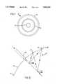
- FIG. 1is a schematic plan view of apparatus according to the invention.
- FIG. 2is a cross-section through the apparatus of FIG. 1 in use.
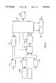
- FIG. 3is a block diagram of a control system used with apparatus according to the invention.
- a sensorgenerally designated 1, consists of a central emitter core 5 carrying light from four sub-emitter laser-diodes each capable of emitting infra-red radiation at a discrete wavelength within the range 700-1300 nm.
- a primary inner annular photodetector ring 3Arranged concentrically around the core 5 is a primary inner annular photodetector ring 3, and spaced therefrom a secondary outer annular photodetector ring 2.
- the photodetector rings 2, 3are coupled to respective photomultipliers (not shown), which are in turn connected to respective channels 7, 8 of suitable electronic amplification and signal processing circuitry.
- the output signals from the photodetector rings 2, 3are then passed via signal steering circuitry 9 and analogue to digital convertor 12 to a microprocessor control unit 13 programmed to calculate set algorithms dependent on the ratio of the two output signals enabling a value for the relationship between oxygen availability and oxygen consumption at the cellular level to be obtained, which is then recorded or displayed on a VDU and chart recorder 10 and 11. Data can also be stored on computer 16.
- FIG. 3also shows how the laser power supply drive and firing circuitry, 14 and respective signal processing channels 7, 8 are both controlled by a timing sequence controller 15 connected to the microprocessor control unit 13.
- the calibration of the apparatusenables a value for the relationship between oxygen availability and oxygen consumption at the cellular level to be found.
- the senorin use the sensor is placed on the tissue surface 6 (e.g. the head) with the annular photodetector rings 2, 3 and emitter core 5 in contact therewith, the radiation (in this case near infra-red radiation) enters the tissue and scatters (see arbitrary scattering points 18a, 18b) to follow multiple paths around inside the tissue before being detected by the annular photodetector rings 2, 3.
- the signal generated at the photodetector ringsis then amplified and processed as described above.
- the arrangement of the photodetectors 2, 3 concentrically around the emitter core 5allows very large area detectors, while still maintaining constant light path lengths to all parts of each detector. This increases the measured signal strength, without compromising path length considerations, and averages out local irregularities in skin pigmentation (e.g. freckles).
- the reduced sensitivity to the positioning of the sensorshould mean that the sensor is less prone to motion artefact, a factor of great importance in any optical system.
Landscapes
- Health & Medical Sciences (AREA)
- Life Sciences & Earth Sciences (AREA)
- Physics & Mathematics (AREA)
- Pathology (AREA)
- General Health & Medical Sciences (AREA)
- Medical Informatics (AREA)
- Public Health (AREA)
- Engineering & Computer Science (AREA)
- Biomedical Technology (AREA)
- Heart & Thoracic Surgery (AREA)
- Veterinary Medicine (AREA)
- Molecular Biology (AREA)
- Surgery (AREA)
- Animal Behavior & Ethology (AREA)
- Biophysics (AREA)
- Spectroscopy & Molecular Physics (AREA)
- Optics & Photonics (AREA)
- Chemical & Material Sciences (AREA)
- Analytical Chemistry (AREA)
- Biochemistry (AREA)
- General Physics & Mathematics (AREA)
- Immunology (AREA)
- Measurement Of The Respiration, Hearing Ability, Form, And Blood Characteristics Of Living Organisms (AREA)
Abstract
Description
ln(I.sub.o /I)=d×E×c
Claims (15)
Priority Applications (1)
| Application Number | Priority Date | Filing Date | Title |
|---|---|---|---|
| US08/316,065US5419321A (en) | 1990-05-17 | 1994-09-30 | Non-invasive medical sensor |
Applications Claiming Priority (4)
| Application Number | Priority Date | Filing Date | Title |
|---|---|---|---|
| GB9011131 | 1990-05-17 | ||
| GB9011131AGB2244128A (en) | 1990-05-17 | 1990-05-17 | Non-invasive medical sensor |
| US95285892A | 1992-11-16 | 1992-11-16 | |
| US08/316,065US5419321A (en) | 1990-05-17 | 1994-09-30 | Non-invasive medical sensor |
Related Parent Applications (1)
| Application Number | Title | Priority Date | Filing Date |
|---|---|---|---|
| US95285892AContinuation | 1990-05-17 | 1992-11-16 |
Publications (1)
| Publication Number | Publication Date |
|---|---|
| US5419321Atrue US5419321A (en) | 1995-05-30 |
Family
ID=26297092
Family Applications (1)
| Application Number | Title | Priority Date | Filing Date |
|---|---|---|---|
| US08/316,065Expired - Fee RelatedUS5419321A (en) | 1990-05-17 | 1994-09-30 | Non-invasive medical sensor |
Country Status (1)
| Country | Link |
|---|---|
| US (1) | US5419321A (en) |
Cited By (74)
| Publication number | Priority date | Publication date | Assignee | Title |
|---|---|---|---|---|
| US5513642A (en)* | 1994-10-12 | 1996-05-07 | Rensselaer Polytechnic Institute | Reflectance sensor system |
| WO1996041566A3 (en)* | 1995-06-09 | 1997-05-15 | Cybro Medical Ltd | Sensor, method and device for optical blood oximetry |
| WO1997027800A1 (en)* | 1996-02-05 | 1997-08-07 | Diasense, Inc. | Methods and apparatus for non-invasive glucose sensing: non-invasive probe |
| US5782237A (en)* | 1994-04-01 | 1998-07-21 | Nellcor Puritan Bennett Incorporated | Pulse oximeter and sensor optimized for low saturation |
| US5902235A (en)* | 1989-03-29 | 1999-05-11 | Somanetics Corporation | Optical cerebral oximeter |
| US5908384A (en)* | 1995-07-31 | 1999-06-01 | Critikon Company, L.L.C. | Apparatus for non-invasive measurement within a human or animal body |
| US6091984A (en)* | 1997-10-10 | 2000-07-18 | Massachusetts Institute Of Technology | Measuring tissue morphology |
| US6090050A (en)* | 1998-07-16 | 2000-07-18 | Salix Medical, Inc. | Thermometric apparatus and method |
| US6353226B1 (en) | 1998-11-23 | 2002-03-05 | Abbott Laboratories | Non-invasive sensor capable of determining optical parameters in a sample having multiple layers |
| US6404497B1 (en) | 1999-01-25 | 2002-06-11 | Massachusetts Institute Of Technology | Polarized light scattering spectroscopy of tissue |
| US20020171834A1 (en)* | 2001-04-11 | 2002-11-21 | Rowe Robert K. | Encoded variable filter spectrometer |
| US20020183624A1 (en)* | 2001-06-05 | 2002-12-05 | Rio Grande Medical Technologies, Inc. | Apparatus and method of biometric determination using specialized optical spectroscopy systems |
| US6574490B2 (en) | 2001-04-11 | 2003-06-03 | Rio Grande Medical Technologies, Inc. | System for non-invasive measurement of glucose in humans |
| US6577884B1 (en) | 2000-06-19 | 2003-06-10 | The General Hospital Corporation | Detection of stroke events using diffuse optical tomagraphy |
| US6615061B1 (en) | 1998-11-23 | 2003-09-02 | Abbott Laboratories | Optical sensor having a selectable sampling distance for determination of analytes |
| US6631282B2 (en) | 2001-08-09 | 2003-10-07 | Optiscan Biomedical Corporation | Device for isolating regions of living tissue |
| WO2002101668A3 (en)* | 2001-06-13 | 2003-10-30 | Tst Touchless Sensor Technolog | Method and device for recognition of natural skin |
| US6654125B2 (en) | 2002-04-04 | 2003-11-25 | Inlight Solutions, Inc | Method and apparatus for optical spectroscopy incorporating a vertical cavity surface emitting laser (VCSEL) as an interferometer reference |
| US6662033B2 (en) | 1994-04-01 | 2003-12-09 | Nellcor Incorporated | Pulse oximeter and sensor optimized for low saturation |
| US6718189B2 (en) | 1995-08-09 | 2004-04-06 | Rio Grande Medical Technologies, Inc. | Method and apparatus for non-invasive blood analyte measurement with fluid compartment equilibration |
| US20040119314A1 (en)* | 2002-11-01 | 2004-06-24 | Brian Haack | Tonneau cover apparatus |
| US6816605B2 (en) | 1999-10-08 | 2004-11-09 | Lumidigm, Inc. | Methods and systems for biometric identification of individuals using linear optical spectroscopy |
| US20040240712A1 (en)* | 2003-04-04 | 2004-12-02 | Lumidigm, Inc. | Multispectral biometric sensor |
| US20050007582A1 (en)* | 2003-07-07 | 2005-01-13 | Lumidigm, Inc. | Methods and apparatus for collection of optical reference measurements for monolithic sensors |
| US6862091B2 (en) | 2001-04-11 | 2005-03-01 | Inlight Solutions, Inc. | Illumination device and method for spectroscopic analysis |
| US6865408B1 (en) | 2001-04-11 | 2005-03-08 | Inlight Solutions, Inc. | System for non-invasive measurement of glucose in humans |
| US20050073690A1 (en)* | 2003-10-03 | 2005-04-07 | Abbink Russell E. | Optical spectroscopy incorporating a vertical cavity surface emitting laser (VCSEL) |
| DE19841217B4 (en)* | 1997-10-27 | 2005-06-16 | Applied Photonics Worldwide, Inc., Reno | Apparatus and method for the spectroscopic analysis of human or animal tissue or body fluids |
| US20050205667A1 (en)* | 2003-04-04 | 2005-09-22 | Lumidigm, Inc. | Combined total-internal-reflectance and tissue imaging systems and methods |
| US6959211B2 (en) | 1999-03-10 | 2005-10-25 | Optiscan Biomedical Corp. | Device for capturing thermal spectra from tissue |
| US20050265585A1 (en)* | 2004-06-01 | 2005-12-01 | Lumidigm, Inc. | Multispectral liveness determination |
| US20050271258A1 (en)* | 2004-06-01 | 2005-12-08 | Lumidigm, Inc. | Multispectral imaging biometrics |
| US6983176B2 (en) | 2001-04-11 | 2006-01-03 | Rio Grande Medical Technologies, Inc. | Optically similar reference samples and related methods for multivariate calibration models used in optical spectroscopy |
| US20060062438A1 (en)* | 2003-04-04 | 2006-03-23 | Lumidigm, Inc. | Comparative texture analysis of tissue for biometric spoof detection |
| US7027848B2 (en) | 2002-04-04 | 2006-04-11 | Inlight Solutions, Inc. | Apparatus and method for non-invasive spectroscopic measurement of analytes in tissue using a matched reference analyte |
| US7043288B2 (en) | 2002-04-04 | 2006-05-09 | Inlight Solutions, Inc. | Apparatus and method for spectroscopic analysis of tissue to detect diabetes in an individual |
| US20060110015A1 (en)* | 2003-04-04 | 2006-05-25 | Lumidigm, Inc. | Systems and methods for improved biometric feature definition |
| US20060122520A1 (en)* | 2004-12-07 | 2006-06-08 | Dr. Matthew Banet | Vital sign-monitoring system with multiple optical modules |
| US20070010721A1 (en)* | 2005-06-28 | 2007-01-11 | Chen Thomas C H | Apparatus and system of Internet-enabled wireless medical sensor scale |
| US7167734B2 (en) | 2001-04-13 | 2007-01-23 | Abbott Laboratories | Method for optical measurements of tissue to determine disease state or concentration of an analyte |
| US20070030475A1 (en)* | 2003-04-04 | 2007-02-08 | Lumidigm, Inc. | White-light spectral biometric sensors |
| US7203345B2 (en) | 1999-10-08 | 2007-04-10 | Lumidigm, Inc. | Apparatus and method for identification of individuals by near-infrared spectrum |
| US20070116331A1 (en)* | 2004-06-01 | 2007-05-24 | Lumidigm, Inc. | System and method for robust fingerprint acquisition |
| US20070197887A1 (en)* | 2006-02-17 | 2007-08-23 | Medwave, Inc. | Noninvasive vital signs sensor |
| US7263213B2 (en) | 2003-12-11 | 2007-08-28 | Lumidigm, Inc. | Methods and systems for estimation of personal characteristics from biometric measurements |
| US20080039729A1 (en)* | 2006-08-10 | 2008-02-14 | Samsung Electronics Co.; Ltd | Living body measurement apparatus |
| US7394919B2 (en) | 2004-06-01 | 2008-07-01 | Lumidigm, Inc. | Multispectral biometric imaging |
| US20080192988A1 (en)* | 2006-07-19 | 2008-08-14 | Lumidigm, Inc. | Multibiometric multispectral imager |
| US20090024041A1 (en)* | 2007-07-19 | 2009-01-22 | Samsung Electronics Co., Ltd. | Apparatus for measuring bio-information |
| US7545963B2 (en) | 2003-04-04 | 2009-06-09 | Lumidigm, Inc. | Texture-biometrics sensor |
| US20090245591A1 (en)* | 2006-07-19 | 2009-10-01 | Lumidigm, Inc. | Contactless Multispectral Biometric Capture |
| US7620212B1 (en) | 2002-08-13 | 2009-11-17 | Lumidigm, Inc. | Electro-optical sensor |
| US7801338B2 (en) | 2005-04-27 | 2010-09-21 | Lumidigm, Inc. | Multispectral biometric sensors |
| US7801339B2 (en) | 2006-07-31 | 2010-09-21 | Lumidigm, Inc. | Biometrics with spatiospectral spoof detection |
| US7804984B2 (en) | 2006-07-31 | 2010-09-28 | Lumidigm, Inc. | Spatial-spectral fingerprint spoof detection |
| US7884933B1 (en)* | 2010-05-05 | 2011-02-08 | Revolutionary Business Concepts, Inc. | Apparatus and method for determining analyte concentrations |
| US20110163163A1 (en)* | 2004-06-01 | 2011-07-07 | Lumidigm, Inc. | Multispectral barcode imaging |
| US8175346B2 (en) | 2006-07-19 | 2012-05-08 | Lumidigm, Inc. | Whole-hand multispectral biometric imaging |
| US8229185B2 (en) | 2004-06-01 | 2012-07-24 | Lumidigm, Inc. | Hygienic biometric sensors |
| US8285010B2 (en) | 2007-03-21 | 2012-10-09 | Lumidigm, Inc. | Biometrics based on locally consistent features |
| US8355545B2 (en) | 2007-04-10 | 2013-01-15 | Lumidigm, Inc. | Biometric detection using spatial, temporal, and/or spectral techniques |
| US8570149B2 (en) | 2010-03-16 | 2013-10-29 | Lumidigm, Inc. | Biometric imaging using an optical adaptive interface |
| US8731250B2 (en) | 2009-08-26 | 2014-05-20 | Lumidigm, Inc. | Multiplexed biometric imaging |
| US8787630B2 (en) | 2004-08-11 | 2014-07-22 | Lumidigm, Inc. | Multispectral barcode imaging |
| US9861317B2 (en) | 2014-02-20 | 2018-01-09 | Covidien Lp | Methods and systems for determining regional blood oxygen saturation |
| US9867561B2 (en) | 2014-01-27 | 2018-01-16 | Covidien Lp | Systems and methods for determining whether regional oximetry sensors are properly positioned |
| US20180177459A1 (en)* | 2012-10-07 | 2018-06-28 | Rhythm Diagnostic Systems, Inc. | Health monitoring systems and methods |
| US10213550B2 (en) | 2014-01-23 | 2019-02-26 | Covidien Lp | Systems and methods for monitoring clinical procedures using regional blood oxygen saturation |
| US10244949B2 (en) | 2012-10-07 | 2019-04-02 | Rhythm Diagnostic Systems, Inc. | Health monitoring systems and methods |
| USD850626S1 (en) | 2013-03-15 | 2019-06-04 | Rhythm Diagnostic Systems, Inc. | Health monitoring apparatuses |
| US10328202B2 (en) | 2015-02-04 | 2019-06-25 | Covidien Lp | Methods and systems for determining fluid administration |
| US10413251B2 (en) | 2012-10-07 | 2019-09-17 | Rhythm Diagnostic Systems, Inc. | Wearable cardiac monitor |
| US11903700B2 (en) | 2019-08-28 | 2024-02-20 | Rds | Vital signs monitoring systems and methods |
| US12109047B2 (en) | 2019-01-25 | 2024-10-08 | Rds | Health monitoring systems and methods |
Citations (13)
| Publication number | Priority date | Publication date | Assignee | Title |
|---|---|---|---|---|
| US3638640A (en)* | 1967-11-01 | 1972-02-01 | Robert F Shaw | Oximeter and method for in vivo determination of oxygen saturation in blood using three or more different wavelengths |
| US4223680A (en)* | 1977-06-28 | 1980-09-23 | Duke University, Inc. | Method and apparatus for monitoring metabolism in body organs in vivo |
| US4281931A (en)* | 1977-12-21 | 1981-08-04 | Machida Endoscope Co., Ltd. | Measuring apparatus comprising light optics utilizing cylindrical focusing glass fiber |
| US4485820A (en)* | 1982-05-10 | 1984-12-04 | The Johns Hopkins University | Method and apparatus for the continuous monitoring of hemoglobin saturation in the blood of premature infants |
| EP0140633A2 (en)* | 1983-10-14 | 1985-05-08 | Somanetics Corporation | Method and apparatus for spectral transmissibility examination and analysis |
| US4765340A (en)* | 1985-04-02 | 1988-08-23 | Minolta Camera Kabushiki Kaisha | Apnea detector |
| EP0290279A1 (en)* | 1987-05-08 | 1988-11-09 | Hamamatsu Photonics K.K. | Examination apparatus for measuring oxygenation |
| US4889116A (en)* | 1987-11-17 | 1989-12-26 | Phospho Energetics, Inc. | Adaptive control of neonatal fractional inspired oxygen |
| US4914512A (en)* | 1988-01-19 | 1990-04-03 | Kabushiki Kaisha Toshiba | Electronic endoscope apparatus capable of displaying hemoglobin concentration on color image |
| US5057695A (en)* | 1988-12-19 | 1991-10-15 | Otsuka Electronics Co., Ltd. | Method of and apparatus for measuring the inside information of substance with the use of light scattering |
| US5090415A (en)* | 1989-02-14 | 1992-02-25 | Hamamatsu Photonics Kabushiki Kaisha | Examination apparatus |
| US5139025A (en)* | 1983-10-14 | 1992-08-18 | Somanetics Corporation | Method and apparatus for in vivo optical spectroscopic examination |
| US5140989A (en)* | 1983-10-14 | 1992-08-25 | Somanetics Corporation | Examination instrument for optical-response diagnostic apparatus |
- 1994
- 1994-09-30USUS08/316,065patent/US5419321A/ennot_activeExpired - Fee Related
Patent Citations (15)
| Publication number | Priority date | Publication date | Assignee | Title |
|---|---|---|---|---|
| US3638640A (en)* | 1967-11-01 | 1972-02-01 | Robert F Shaw | Oximeter and method for in vivo determination of oxygen saturation in blood using three or more different wavelengths |
| US4223680A (en)* | 1977-06-28 | 1980-09-23 | Duke University, Inc. | Method and apparatus for monitoring metabolism in body organs in vivo |
| US4321930A (en)* | 1977-06-28 | 1982-03-30 | Duke University, Inc. | Apparatus for monitoring metabolism in body organs |
| US4281931A (en)* | 1977-12-21 | 1981-08-04 | Machida Endoscope Co., Ltd. | Measuring apparatus comprising light optics utilizing cylindrical focusing glass fiber |
| US4485820A (en)* | 1982-05-10 | 1984-12-04 | The Johns Hopkins University | Method and apparatus for the continuous monitoring of hemoglobin saturation in the blood of premature infants |
| US4570638A (en)* | 1983-10-14 | 1986-02-18 | Somanetics Corporation | Method and apparatus for spectral transmissibility examination and analysis |
| EP0140633A2 (en)* | 1983-10-14 | 1985-05-08 | Somanetics Corporation | Method and apparatus for spectral transmissibility examination and analysis |
| US5139025A (en)* | 1983-10-14 | 1992-08-18 | Somanetics Corporation | Method and apparatus for in vivo optical spectroscopic examination |
| US5140989A (en)* | 1983-10-14 | 1992-08-25 | Somanetics Corporation | Examination instrument for optical-response diagnostic apparatus |
| US4765340A (en)* | 1985-04-02 | 1988-08-23 | Minolta Camera Kabushiki Kaisha | Apnea detector |
| EP0290279A1 (en)* | 1987-05-08 | 1988-11-09 | Hamamatsu Photonics K.K. | Examination apparatus for measuring oxygenation |
| US4889116A (en)* | 1987-11-17 | 1989-12-26 | Phospho Energetics, Inc. | Adaptive control of neonatal fractional inspired oxygen |
| US4914512A (en)* | 1988-01-19 | 1990-04-03 | Kabushiki Kaisha Toshiba | Electronic endoscope apparatus capable of displaying hemoglobin concentration on color image |
| US5057695A (en)* | 1988-12-19 | 1991-10-15 | Otsuka Electronics Co., Ltd. | Method of and apparatus for measuring the inside information of substance with the use of light scattering |
| US5090415A (en)* | 1989-02-14 | 1992-02-25 | Hamamatsu Photonics Kabushiki Kaisha | Examination apparatus |
Non-Patent Citations (4)
| Title |
|---|
| "Fiber optic reflectance spectrophotometry system for in vivo tissue diagnosis" Ono, et al, Applied Optics/vol. 30, No. 1/ 1 Jan. 1991. |
| "System for long-term measurement of cerebral blood and tissue oxygenation on newborn infants by near infra-red transillumination" Cope et al, 2200 Medical & Biological Engineering & Computing 26 (1988) May, No. 3, Stevenage, Herts., Gr. Britain. |
| Fiber optic reflectance spectrophotometry system for in vivo tissue diagnosis Ono, et al, Applied Optics/vol. 30, No. 1/ 1 Jan. 1991.* |
| System for long term measurement of cerebral blood and tissue oxygenation on newborn infants by near infra red transillumination Cope et al, 2200 Medical & Biological Engineering & Computing 26 (1988) May, No. 3, Stevenage, Herts., Gr. Britain.* |
Cited By (141)
| Publication number | Priority date | Publication date | Assignee | Title |
|---|---|---|---|---|
| US5902235A (en)* | 1989-03-29 | 1999-05-11 | Somanetics Corporation | Optical cerebral oximeter |
| US20060195027A1 (en)* | 1994-04-01 | 2006-08-31 | Casciani James R | Pulse oximeter and sensor optimized for low saturation |
| US20060189862A1 (en)* | 1994-04-01 | 2006-08-24 | Casciani James R | Pulse oximeter and sensor optimized for low saturation |
| US5782237A (en)* | 1994-04-01 | 1998-07-21 | Nellcor Puritan Bennett Incorporated | Pulse oximeter and sensor optimized for low saturation |
| US20060211929A1 (en)* | 1994-04-01 | 2006-09-21 | Casciani James R | Pulse oximeter and sensor optimized for low saturation |
| US6662033B2 (en) | 1994-04-01 | 2003-12-09 | Nellcor Incorporated | Pulse oximeter and sensor optimized for low saturation |
| US7376454B2 (en) | 1994-04-01 | 2008-05-20 | Nellcor Puritan Bennett Inc. | Oximeter with selection between calculations based on patient type |
| US6272363B1 (en)* | 1994-04-01 | 2001-08-07 | Nellcor Incorporated | Pulse oximeter and sensor optimized for low saturation |
| US7349726B2 (en) | 1994-04-01 | 2008-03-25 | Nellcor Puritan Bennett Llc | Pulse oximeter and sensor optimized for low saturation |
| US7415298B2 (en) | 1994-04-01 | 2008-08-19 | Nellcor Puritan Bennett Inc. | Pulse oximeter and sensor optimized for low saturation |
| US5513642A (en)* | 1994-10-12 | 1996-05-07 | Rensselaer Polytechnic Institute | Reflectance sensor system |
| US6031603A (en)* | 1995-06-09 | 2000-02-29 | Cybro Medical, Ltd. | Sensor, method and device for optical blood oximetry |
| AU708051B2 (en)* | 1995-06-09 | 1999-07-29 | Conmed Israel Ltd | Sensor, method and device for optical blood oximetry |
| WO1996041566A3 (en)* | 1995-06-09 | 1997-05-15 | Cybro Medical Ltd | Sensor, method and device for optical blood oximetry |
| US5908384A (en)* | 1995-07-31 | 1999-06-01 | Critikon Company, L.L.C. | Apparatus for non-invasive measurement within a human or animal body |
| US6718189B2 (en) | 1995-08-09 | 2004-04-06 | Rio Grande Medical Technologies, Inc. | Method and apparatus for non-invasive blood analyte measurement with fluid compartment equilibration |
| EP0889703A4 (en)* | 1996-02-05 | 1999-10-13 | Diasense Inc | Methods and apparatus for non-invasive glucose sensing: non-invasive probe |
| US6219565B1 (en) | 1996-02-05 | 2001-04-17 | Diasense, Inc. | Methods and apparatus for non-invasive glucose sensing: non-invasive probe |
| WO1997027800A1 (en)* | 1996-02-05 | 1997-08-07 | Diasense, Inc. | Methods and apparatus for non-invasive glucose sensing: non-invasive probe |
| US9487398B2 (en) | 1997-06-09 | 2016-11-08 | Hid Global Corporation | Apparatus and method of biometric determination using specialized optical spectroscopy systems |
| US20080304712A1 (en)* | 1997-06-09 | 2008-12-11 | Lumidigm, Inc. | Apparatus and method of biometric determination using specialized optical spectroscopy systems |
| US6922583B1 (en) | 1997-10-10 | 2005-07-26 | Massachusetts Institute Of Technology | Method for measuring tissue morphology |
| US6091984A (en)* | 1997-10-10 | 2000-07-18 | Massachusetts Institute Of Technology | Measuring tissue morphology |
| DE19841217B4 (en)* | 1997-10-27 | 2005-06-16 | Applied Photonics Worldwide, Inc., Reno | Apparatus and method for the spectroscopic analysis of human or animal tissue or body fluids |
| US6090050A (en)* | 1998-07-16 | 2000-07-18 | Salix Medical, Inc. | Thermometric apparatus and method |
| US6630673B2 (en) | 1998-11-23 | 2003-10-07 | Abbott Laboratories | Non-invasive sensor capable of determining optical parameters in a sample having multiple layers |
| US6615061B1 (en) | 1998-11-23 | 2003-09-02 | Abbott Laboratories | Optical sensor having a selectable sampling distance for determination of analytes |
| US6353226B1 (en) | 1998-11-23 | 2002-03-05 | Abbott Laboratories | Non-invasive sensor capable of determining optical parameters in a sample having multiple layers |
| US6624890B2 (en) | 1999-01-25 | 2003-09-23 | Massachusetts Institute Of Technology | Polarized light scattering spectroscopy of tissue |
| US6404497B1 (en) | 1999-01-25 | 2002-06-11 | Massachusetts Institute Of Technology | Polarized light scattering spectroscopy of tissue |
| US6959211B2 (en) | 1999-03-10 | 2005-10-25 | Optiscan Biomedical Corp. | Device for capturing thermal spectra from tissue |
| US6816605B2 (en) | 1999-10-08 | 2004-11-09 | Lumidigm, Inc. | Methods and systems for biometric identification of individuals using linear optical spectroscopy |
| US7203345B2 (en) | 1999-10-08 | 2007-04-10 | Lumidigm, Inc. | Apparatus and method for identification of individuals by near-infrared spectrum |
| US6577884B1 (en) | 2000-06-19 | 2003-06-10 | The General Hospital Corporation | Detection of stroke events using diffuse optical tomagraphy |
| US7126682B2 (en) | 2001-04-11 | 2006-10-24 | Rio Grande Medical Technologies, Inc. | Encoded variable filter spectrometer |
| US20020171834A1 (en)* | 2001-04-11 | 2002-11-21 | Rowe Robert K. | Encoded variable filter spectrometer |
| US6862091B2 (en) | 2001-04-11 | 2005-03-01 | Inlight Solutions, Inc. | Illumination device and method for spectroscopic analysis |
| US6574490B2 (en) | 2001-04-11 | 2003-06-03 | Rio Grande Medical Technologies, Inc. | System for non-invasive measurement of glucose in humans |
| US6865408B1 (en) | 2001-04-11 | 2005-03-08 | Inlight Solutions, Inc. | System for non-invasive measurement of glucose in humans |
| US6983176B2 (en) | 2001-04-11 | 2006-01-03 | Rio Grande Medical Technologies, Inc. | Optically similar reference samples and related methods for multivariate calibration models used in optical spectroscopy |
| US7167734B2 (en) | 2001-04-13 | 2007-01-23 | Abbott Laboratories | Method for optical measurements of tissue to determine disease state or concentration of an analyte |
| US7613504B2 (en) | 2001-06-05 | 2009-11-03 | Lumidigm, Inc. | Spectroscopic cross-channel method and apparatus for improved optical measurements of tissue |
| US20020183624A1 (en)* | 2001-06-05 | 2002-12-05 | Rio Grande Medical Technologies, Inc. | Apparatus and method of biometric determination using specialized optical spectroscopy systems |
| US7890158B2 (en) | 2001-06-05 | 2011-02-15 | Lumidigm, Inc. | Apparatus and method of biometric determination using specialized optical spectroscopy systems |
| US7587071B2 (en) | 2001-06-13 | 2009-09-08 | Tst Biometrics Holding Ag | Method and device for recognition of natural skin during contact-free biometric identification of a person |
| CN100457031C (en)* | 2001-06-13 | 2009-02-04 | Tst生物测定学控股股份公司 | Method and device for recognition of natural skin |
| US20060056661A1 (en)* | 2001-06-13 | 2006-03-16 | Hans Einighammer | Method and device for recognition of natural skin |
| WO2002101668A3 (en)* | 2001-06-13 | 2003-10-30 | Tst Touchless Sensor Technolog | Method and device for recognition of natural skin |
| US20090310827A1 (en)* | 2001-06-13 | 2009-12-17 | Tst Biometrics Holding Ag | Method and device for recognition of natural skin |
| US8045768B2 (en) | 2001-06-13 | 2011-10-25 | Tst Biometrics Holding Ag | Method and device for recognition of natural skin |
| US6631282B2 (en) | 2001-08-09 | 2003-10-07 | Optiscan Biomedical Corporation | Device for isolating regions of living tissue |
| US7027848B2 (en) | 2002-04-04 | 2006-04-11 | Inlight Solutions, Inc. | Apparatus and method for non-invasive spectroscopic measurement of analytes in tissue using a matched reference analyte |
| US7043288B2 (en) | 2002-04-04 | 2006-05-09 | Inlight Solutions, Inc. | Apparatus and method for spectroscopic analysis of tissue to detect diabetes in an individual |
| US6654125B2 (en) | 2002-04-04 | 2003-11-25 | Inlight Solutions, Inc | Method and apparatus for optical spectroscopy incorporating a vertical cavity surface emitting laser (VCSEL) as an interferometer reference |
| US7620212B1 (en) | 2002-08-13 | 2009-11-17 | Lumidigm, Inc. | Electro-optical sensor |
| US20040119314A1 (en)* | 2002-11-01 | 2004-06-24 | Brian Haack | Tonneau cover apparatus |
| US7440597B2 (en) | 2003-04-04 | 2008-10-21 | Rowe Robert K | Liveness sensor |
| US7735729B2 (en) | 2003-04-04 | 2010-06-15 | Lumidigm, Inc. | Biometric sensor |
| US20070030475A1 (en)* | 2003-04-04 | 2007-02-08 | Lumidigm, Inc. | White-light spectral biometric sensors |
| US7147153B2 (en) | 2003-04-04 | 2006-12-12 | Lumidigm, Inc. | Multispectral biometric sensor |
| US20040240712A1 (en)* | 2003-04-04 | 2004-12-02 | Lumidigm, Inc. | Multispectral biometric sensor |
| US8184873B2 (en) | 2003-04-04 | 2012-05-22 | Lumidigm, Inc. | White-light spectral biometric sensors |
| US20060002597A1 (en)* | 2003-04-04 | 2006-01-05 | Lumidigm, Inc. | Liveness sensor |
| US7627151B2 (en) | 2003-04-04 | 2009-12-01 | Lumidigm, Inc. | Systems and methods for improved biometric feature definition |
| US7347365B2 (en) | 2003-04-04 | 2008-03-25 | Lumidigm, Inc. | Combined total-internal-reflectance and tissue imaging systems and methods |
| US20060210120A1 (en)* | 2003-04-04 | 2006-09-21 | Lumidigm, Inc. | Biometric sensor |
| US20060202028A1 (en)* | 2003-04-04 | 2006-09-14 | Lumidigm, Inc. | Multispectral biometric sensor |
| US7386152B2 (en) | 2003-04-04 | 2008-06-10 | Lumidigm, Inc. | Noninvasive alcohol sensor |
| US20050205667A1 (en)* | 2003-04-04 | 2005-09-22 | Lumidigm, Inc. | Combined total-internal-reflectance and tissue imaging systems and methods |
| US7819311B2 (en) | 2003-04-04 | 2010-10-26 | Lumidigm, Inc. | Multispectral biometric sensor |
| US7751594B2 (en) | 2003-04-04 | 2010-07-06 | Lumidigm, Inc. | White-light spectral biometric sensors |
| US20060110015A1 (en)* | 2003-04-04 | 2006-05-25 | Lumidigm, Inc. | Systems and methods for improved biometric feature definition |
| US7545963B2 (en) | 2003-04-04 | 2009-06-09 | Lumidigm, Inc. | Texture-biometrics sensor |
| US20060062438A1 (en)* | 2003-04-04 | 2006-03-23 | Lumidigm, Inc. | Comparative texture analysis of tissue for biometric spoof detection |
| US7668350B2 (en) | 2003-04-04 | 2010-02-23 | Lumidigm, Inc. | Comparative texture analysis of tissue for biometric spoof detection |
| US20060002598A1 (en)* | 2003-04-04 | 2006-01-05 | Lumidigm, Inc. | Noninvasive alcohol sensor |
| US20050007582A1 (en)* | 2003-07-07 | 2005-01-13 | Lumidigm, Inc. | Methods and apparatus for collection of optical reference measurements for monolithic sensors |
| US20050073690A1 (en)* | 2003-10-03 | 2005-04-07 | Abbink Russell E. | Optical spectroscopy incorporating a vertical cavity surface emitting laser (VCSEL) |
| US7263213B2 (en) | 2003-12-11 | 2007-08-28 | Lumidigm, Inc. | Methods and systems for estimation of personal characteristics from biometric measurements |
| US20110163163A1 (en)* | 2004-06-01 | 2011-07-07 | Lumidigm, Inc. | Multispectral barcode imaging |
| US7835554B2 (en) | 2004-06-01 | 2010-11-16 | Lumidigm, Inc. | Multispectral imaging biometrics |
| US7539330B2 (en) | 2004-06-01 | 2009-05-26 | Lumidigm, Inc. | Multispectral liveness determination |
| US20050271258A1 (en)* | 2004-06-01 | 2005-12-08 | Lumidigm, Inc. | Multispectral imaging biometrics |
| US20050265585A1 (en)* | 2004-06-01 | 2005-12-01 | Lumidigm, Inc. | Multispectral liveness determination |
| US20090092290A1 (en)* | 2004-06-01 | 2009-04-09 | Lumidigm, Inc. | Multispectral Imaging Biometrics |
| US7508965B2 (en) | 2004-06-01 | 2009-03-24 | Lumidigm, Inc. | System and method for robust fingerprint acquisition |
| US20070116331A1 (en)* | 2004-06-01 | 2007-05-24 | Lumidigm, Inc. | System and method for robust fingerprint acquisition |
| US7460696B2 (en) | 2004-06-01 | 2008-12-02 | Lumidigm, Inc. | Multispectral imaging biometrics |
| US8229185B2 (en) | 2004-06-01 | 2012-07-24 | Lumidigm, Inc. | Hygienic biometric sensors |
| US8165357B2 (en) | 2004-06-01 | 2012-04-24 | Lumidigm, Inc. | Two camera biometric imaging |
| US7394919B2 (en) | 2004-06-01 | 2008-07-01 | Lumidigm, Inc. | Multispectral biometric imaging |
| US8913800B2 (en) | 2004-06-01 | 2014-12-16 | Lumidigm, Inc. | Optical biometrics imaging with films |
| US7831072B2 (en) | 2004-06-01 | 2010-11-09 | Lumidigm, Inc. | Multispectral imaging biometrics |
| US8787630B2 (en) | 2004-08-11 | 2014-07-22 | Lumidigm, Inc. | Multispectral barcode imaging |
| US20060122520A1 (en)* | 2004-12-07 | 2006-06-08 | Dr. Matthew Banet | Vital sign-monitoring system with multiple optical modules |
| US7801338B2 (en) | 2005-04-27 | 2010-09-21 | Lumidigm, Inc. | Multispectral biometric sensors |
| US20070010721A1 (en)* | 2005-06-28 | 2007-01-11 | Chen Thomas C H | Apparatus and system of Internet-enabled wireless medical sensor scale |
| US20070197887A1 (en)* | 2006-02-17 | 2007-08-23 | Medwave, Inc. | Noninvasive vital signs sensor |
| US7899217B2 (en) | 2006-07-19 | 2011-03-01 | Lumidign, Inc. | Multibiometric multispectral imager |
| US7995808B2 (en) | 2006-07-19 | 2011-08-09 | Lumidigm, Inc. | Contactless multispectral biometric capture |
| US8831297B2 (en) | 2006-07-19 | 2014-09-09 | Lumidigm, Inc. | Contactless multispectral biometric capture |
| US20080192988A1 (en)* | 2006-07-19 | 2008-08-14 | Lumidigm, Inc. | Multibiometric multispectral imager |
| US8175346B2 (en) | 2006-07-19 | 2012-05-08 | Lumidigm, Inc. | Whole-hand multispectral biometric imaging |
| US8781181B2 (en) | 2006-07-19 | 2014-07-15 | Lumidigm, Inc. | Contactless multispectral biometric capture |
| US20090245591A1 (en)* | 2006-07-19 | 2009-10-01 | Lumidigm, Inc. | Contactless Multispectral Biometric Capture |
| US7801339B2 (en) | 2006-07-31 | 2010-09-21 | Lumidigm, Inc. | Biometrics with spatiospectral spoof detection |
| US7804984B2 (en) | 2006-07-31 | 2010-09-28 | Lumidigm, Inc. | Spatial-spectral fingerprint spoof detection |
| US20080039729A1 (en)* | 2006-08-10 | 2008-02-14 | Samsung Electronics Co.; Ltd | Living body measurement apparatus |
| US8116851B2 (en) | 2006-08-10 | 2012-02-14 | Samsung Electronics Co., Ltd. | Living body measurement apparatus with waveguide light source and light extracting pattern |
| EP1886624B1 (en)* | 2006-08-10 | 2012-05-23 | Samsung Electronics Co., Ltd. | Living body measurement apparatus |
| US8285010B2 (en) | 2007-03-21 | 2012-10-09 | Lumidigm, Inc. | Biometrics based on locally consistent features |
| US8355545B2 (en) | 2007-04-10 | 2013-01-15 | Lumidigm, Inc. | Biometric detection using spatial, temporal, and/or spectral techniques |
| US8131347B2 (en)* | 2007-07-19 | 2012-03-06 | Samsung Electronics Co., Ltd. | Optical apparatus for measuring bio-information |
| US20090024041A1 (en)* | 2007-07-19 | 2009-01-22 | Samsung Electronics Co., Ltd. | Apparatus for measuring bio-information |
| US8731250B2 (en) | 2009-08-26 | 2014-05-20 | Lumidigm, Inc. | Multiplexed biometric imaging |
| US8872908B2 (en) | 2009-08-26 | 2014-10-28 | Lumidigm, Inc | Dual-imager biometric sensor |
| US8570149B2 (en) | 2010-03-16 | 2013-10-29 | Lumidigm, Inc. | Biometric imaging using an optical adaptive interface |
| US8199322B2 (en)* | 2010-05-05 | 2012-06-12 | Revolutionary Business Concepts, Inc. | Apparatus and method for determining analyte concentrations |
| US7884933B1 (en)* | 2010-05-05 | 2011-02-08 | Revolutionary Business Concepts, Inc. | Apparatus and method for determining analyte concentrations |
| US10244949B2 (en) | 2012-10-07 | 2019-04-02 | Rhythm Diagnostic Systems, Inc. | Health monitoring systems and methods |
| US10610159B2 (en)* | 2012-10-07 | 2020-04-07 | Rhythm Diagnostic Systems, Inc. | Health monitoring systems and methods |
| US20180177459A1 (en)* | 2012-10-07 | 2018-06-28 | Rhythm Diagnostic Systems, Inc. | Health monitoring systems and methods |
| US11185291B2 (en) | 2012-10-07 | 2021-11-30 | Rds | Health monitoring systems and methods |
| US10993671B2 (en) | 2012-10-07 | 2021-05-04 | Rds | Health monitoring systems and methods |
| US11786182B2 (en) | 2012-10-07 | 2023-10-17 | Rds | Health monitoring systems and methods |
| US10980486B2 (en) | 2012-10-07 | 2021-04-20 | Rds | Health monitoring systems and methods |
| US11937946B2 (en) | 2012-10-07 | 2024-03-26 | Rds | Wearable cardiac monitor |
| US10413251B2 (en) | 2012-10-07 | 2019-09-17 | Rhythm Diagnostic Systems, Inc. | Wearable cardiac monitor |
| US20210100514A1 (en)* | 2012-10-07 | 2021-04-08 | Rds Sas | Health monitoring systems and methods |
| US10842391B2 (en) | 2012-10-07 | 2020-11-24 | Rds Sas | Health monitoring systems and methods |
| US10863947B2 (en) | 2012-10-07 | 2020-12-15 | Rds Sas | Health monitoring systems and methods |
| US10959678B2 (en) | 2012-10-07 | 2021-03-30 | Rds | Health monitoring systems and methods |
| USD850626S1 (en) | 2013-03-15 | 2019-06-04 | Rhythm Diagnostic Systems, Inc. | Health monitoring apparatuses |
| US10213550B2 (en) | 2014-01-23 | 2019-02-26 | Covidien Lp | Systems and methods for monitoring clinical procedures using regional blood oxygen saturation |
| US9867561B2 (en) | 2014-01-27 | 2018-01-16 | Covidien Lp | Systems and methods for determining whether regional oximetry sensors are properly positioned |
| US10201302B2 (en) | 2014-01-27 | 2019-02-12 | Covidien Lp | Systems and methods for determining whether regional oximetry sensors are properly positioned |
| US9861317B2 (en) | 2014-02-20 | 2018-01-09 | Covidien Lp | Methods and systems for determining regional blood oxygen saturation |
| US10328202B2 (en) | 2015-02-04 | 2019-06-25 | Covidien Lp | Methods and systems for determining fluid administration |
| US11975175B2 (en) | 2015-02-04 | 2024-05-07 | Covidien Lp | Methods and systems for determining fluid administration |
| US12109047B2 (en) | 2019-01-25 | 2024-10-08 | Rds | Health monitoring systems and methods |
| US11903700B2 (en) | 2019-08-28 | 2024-02-20 | Rds | Vital signs monitoring systems and methods |
Similar Documents
| Publication | Publication Date | Title |
|---|---|---|
| US5419321A (en) | Non-invasive medical sensor | |
| US8788004B2 (en) | Method for spectrophotometric blood oxygenation monitoring | |
| US4805623A (en) | Spectrophotometric method for quantitatively determining the concentration of a dilute component in a light- or other radiation-scattering environment | |
| US6456862B2 (en) | Method for non-invasive spectrophotometric blood oxygenation monitoring | |
| Germon et al. | Cerebral near infrared spectroscopy: emitter-detector separation must be increased | |
| KR100612827B1 (en) | Noninvasive hemoglobin concentration and oxygen saturation monitoring method and apparatus | |
| Yamashita et al. | Wavelength dependence of the precision of noninvasive optical measurement of oxy‐, deoxy‐, and total‐hemoglobin concentration | |
| Wahr et al. | Near-infrared spectroscopy: theory and applications | |
| US4223680A (en) | Method and apparatus for monitoring metabolism in body organs in vivo | |
| US5803908A (en) | System for noninvasive hematocrit monitoring | |
| US8180419B2 (en) | Tissue hydration estimation by spectral absorption bandwidth measurement | |
| US20080004513A1 (en) | VCSEL Tissue Spectrometer | |
| EP0352923A1 (en) | Spectrophotometric apparatus and method for monitoring oxygen saturation | |
| Kyriacou et al. | Optical techniques for blood and tissue oxygenation | |
| EP0528938B1 (en) | Non-invasive medical sensor | |
| Ferrari et al. | Determination of cerebral venous hemoglobin saturation by derivative near infrared spectrosocpy | |
| Piantadosi et al. | Application of NIR spectroscopy to problems of tissue oxygenation | |
| Skov et al. | Neonatal intensive care: an obvious, yet difficult area for cerebral near-infrared spectroscopy | |
| Takatani et al. | Design and evaluation of a reflectance oxygen sensor in critically ill patients | |
| Hartmann | Near Infrared Spectroscopy “In Vivo” | |
| Reich | Emerging Technologies |
Legal Events
| Date | Code | Title | Description |
|---|---|---|---|
| FEPP | Fee payment procedure | Free format text:PAYOR NUMBER ASSIGNED (ORIGINAL EVENT CODE: ASPN); ENTITY STATUS OF PATENT OWNER: LARGE ENTITY | |
| CC | Certificate of correction | ||
| FEPP | Fee payment procedure | Free format text:PAYER NUMBER DE-ASSIGNED (ORIGINAL EVENT CODE: RMPN); ENTITY STATUS OF PATENT OWNER: LARGE ENTITY | |
| FPAY | Fee payment | Year of fee payment:4 | |
| AS | Assignment | Owner name:STATE BOARD OF ADMINISTRATION OF FLORIDA, THE, NEW Free format text:SENIOR SECURITY AGREEMENT FOR PATENT;ASSIGNOR:CRITIKON COMPANY, L.L.C.;REEL/FRAME:009648/0508 Effective date:19981103 | |
| AS | Assignment | Owner name:STATE BOARD OF ADMINISTRATION OF FLORIDA, THE, NEW Free format text:SUBORDINATED SECURITY AGREEMENT FOR PATENTS;ASSIGNOR:CRITIKON COMPANY, L.L.C.;REEL/FRAME:009662/0013 Effective date:19981103 | |
| FPAY | Fee payment | Year of fee payment:8 | |
| AS | Assignment | Owner name:GE MEDICAL SYSTEMS INFORMATION TECHNOLOGIES, INC., Free format text:ASSIGNMENT OF ASSIGNORS INTEREST;ASSIGNOR:JOHNSON & JOHNSON;REEL/FRAME:014363/0532 Effective date:19981023 | |
| REMI | Maintenance fee reminder mailed | ||
| LAPS | Lapse for failure to pay maintenance fees | ||
| STCH | Information on status: patent discontinuation | Free format text:PATENT EXPIRED DUE TO NONPAYMENT OF MAINTENANCE FEES UNDER 37 CFR 1.362 | |
| FP | Lapsed due to failure to pay maintenance fee | Effective date:20070530 |