US5385572A - Trocar for endoscopic surgery - Google Patents
Trocar for endoscopic surgeryDownload PDFInfo
- Publication number
- US5385572A US5385572AUS07/974,956US97495692AUS5385572AUS 5385572 AUS5385572 AUS 5385572AUS 97495692 AUS97495692 AUS 97495692AUS 5385572 AUS5385572 AUS 5385572A
- Authority
- US
- United States
- Prior art keywords
- shaft
- trocar
- distal end
- puncturing head
- endoscope
- Prior art date
- Legal status (The legal status is an assumption and is not a legal conclusion. Google has not performed a legal analysis and makes no representation as to the accuracy of the status listed.)
- Expired - Lifetime
Links
- 238000002674endoscopic surgeryMethods0.000titleclaimsdescription7
- 238000004891communicationMethods0.000claimsabstractdescription3
- 230000037361pathwayEffects0.000abstractdescription24
- 229910052751metalInorganic materials0.000abstractdescription3
- 239000002184metalSubstances0.000abstractdescription3
- 238000001356surgical procedureMethods0.000description16
- 210000001015abdomenAnatomy0.000description14
- 238000000034methodMethods0.000description7
- 229920003023plasticPolymers0.000description6
- 239000000835fiberSubstances0.000description5
- 238000004519manufacturing processMethods0.000description3
- 239000000463materialSubstances0.000description3
- 229910001220stainless steelInorganic materials0.000description3
- 239000010935stainless steelSubstances0.000description3
- 238000012084abdominal surgeryMethods0.000description2
- 239000011521glassSubstances0.000description2
- 238000000465mouldingMethods0.000description2
- VVQNEPGJFQJSBK-UHFFFAOYSA-NMethyl methacrylateChemical compoundCOC(=O)C(C)=CVVQNEPGJFQJSBK-UHFFFAOYSA-N0.000description1
- 229920005372Plexiglas®Polymers0.000description1
- 229910000831SteelInorganic materials0.000description1
- 229910052782aluminiumInorganic materials0.000description1
- XAGFODPZIPBFFR-UHFFFAOYSA-NaluminiumChemical compound[Al]XAGFODPZIPBFFR-UHFFFAOYSA-N0.000description1
- 230000005540biological transmissionEffects0.000description1
- 239000002131composite materialSubstances0.000description1
- 238000010276constructionMethods0.000description1
- -1e.g.Substances0.000description1
- 210000000232gallbladderAnatomy0.000description1
- 210000000056organAnatomy0.000description1
- 238000011084recoveryMethods0.000description1
- 239000007787solidSubstances0.000description1
- 239000010959steelSubstances0.000description1
Images


Classifications
- A—HUMAN NECESSITIES
- A61—MEDICAL OR VETERINARY SCIENCE; HYGIENE
- A61B—DIAGNOSIS; SURGERY; IDENTIFICATION
- A61B17/00—Surgical instruments, devices or methods
- A61B17/34—Trocars; Puncturing needles
- A61B17/3417—Details of tips or shafts, e.g. grooves, expandable, bendable; Multiple coaxial sliding cannulas, e.g. for dilating
- A—HUMAN NECESSITIES
- A61—MEDICAL OR VETERINARY SCIENCE; HYGIENE
- A61B—DIAGNOSIS; SURGERY; IDENTIFICATION
- A61B1/00—Instruments for performing medical examinations of the interior of cavities or tubes of the body by visual or photographical inspection, e.g. endoscopes; Illuminating arrangements therefor
- A61B1/04—Instruments for performing medical examinations of the interior of cavities or tubes of the body by visual or photographical inspection, e.g. endoscopes; Illuminating arrangements therefor combined with photographic or television appliances
- A61B1/042—Instruments for performing medical examinations of the interior of cavities or tubes of the body by visual or photographical inspection, e.g. endoscopes; Illuminating arrangements therefor combined with photographic or television appliances characterised by a proximal camera, e.g. a CCD camera
- A—HUMAN NECESSITIES
- A61—MEDICAL OR VETERINARY SCIENCE; HYGIENE
- A61B—DIAGNOSIS; SURGERY; IDENTIFICATION
- A61B1/00—Instruments for performing medical examinations of the interior of cavities or tubes of the body by visual or photographical inspection, e.g. endoscopes; Illuminating arrangements therefor
- A61B1/313—Instruments for performing medical examinations of the interior of cavities or tubes of the body by visual or photographical inspection, e.g. endoscopes; Illuminating arrangements therefor for introducing through surgical openings, e.g. laparoscopes
- A—HUMAN NECESSITIES
- A61—MEDICAL OR VETERINARY SCIENCE; HYGIENE
- A61B—DIAGNOSIS; SURGERY; IDENTIFICATION
- A61B1/00—Instruments for performing medical examinations of the interior of cavities or tubes of the body by visual or photographical inspection, e.g. endoscopes; Illuminating arrangements therefor
- A61B1/04—Instruments for performing medical examinations of the interior of cavities or tubes of the body by visual or photographical inspection, e.g. endoscopes; Illuminating arrangements therefor combined with photographic or television appliances
- A61B1/05—Instruments for performing medical examinations of the interior of cavities or tubes of the body by visual or photographical inspection, e.g. endoscopes; Illuminating arrangements therefor combined with photographic or television appliances characterised by the image sensor, e.g. camera, being in the distal end portion
- A—HUMAN NECESSITIES
- A61—MEDICAL OR VETERINARY SCIENCE; HYGIENE
- A61B—DIAGNOSIS; SURGERY; IDENTIFICATION
- A61B90/00—Instruments, implements or accessories specially adapted for surgery or diagnosis and not covered by any of the groups A61B1/00 - A61B50/00, e.g. for luxation treatment or for protecting wound edges
- A61B90/30—Devices for illuminating a surgical field, the devices having an interrelation with other surgical devices or with a surgical procedure
- A—HUMAN NECESSITIES
- A61—MEDICAL OR VETERINARY SCIENCE; HYGIENE
- A61B—DIAGNOSIS; SURGERY; IDENTIFICATION
- A61B90/00—Instruments, implements or accessories specially adapted for surgery or diagnosis and not covered by any of the groups A61B1/00 - A61B50/00, e.g. for luxation treatment or for protecting wound edges
- A61B90/36—Image-producing devices or illumination devices not otherwise provided for
- A61B90/37—Surgical systems with images on a monitor during operation
Definitions
- Laparascopic surgeryis a genre of endoscopic surgery which is used to perform abdominal surgery, e.g., gall bladder removal. Laparascopic surgery, like many other types of endoscopic surgery, is often preferred over conventional surgery because laparascopic surgery is less invasive than conventional surgery. As a consequence, laparascopic surgery can be performed in less time and at less cost per procedure than conventional abdominal surgery, and it reduces patient recovery time from weeks to days.
- the present inventionrecognizes that when positioning a trocar during endoscopic surgery it would be advantageous to provide a viewing system which can display the path of advancement of the trocar to the surgeon as the trocar is being advanced into the patient.
- a trocarwhich can percutaneously be advanced into a patient by a surgeon who can view the path of advancement on a nearby video monitor.
- Another object of the present inventionis to provide a trocar which is easy to use and safely position within a patient.
- Yet another object of the present inventionis to provide a trocar which is cost-effective to manufacture.
- the distal portion of the puncturing headhas three generally longitudinal edges. Each edge is sharpened to thereby establish the cutting element.
- light from the pathway of intended advancement of the trocarcan propagate through the transparent puncturing head and shaft and enter the endoscope.
- the endoscopethen generates an electrical signal representing the image of the pathway of intended advancement and transmits the signal to a video display monitor for display of the pathway of intended advancement on the monitor, which is positioned nearby the surgeon. Accordingly, the surgeon can view the pathway of intended advancement of the trocar on the monitor as the surgeon positions the trocar in the patient, to thereby facilitate safe and proper positioning of the trocar.
- a trocar for endoscopic surgeryhas a shaft, and the shaft includes a distal end and a proximal segment having a cavity formed therein.
- the distal end of the shaftincludes a cutting element, and a video display system is operationally engaged with the cavity in the proximal segment of the shaft for displaying the image of an object located beyond the cutting element.
- FIG. 1is a perspective view of the transparent trocar of the present invention
- FIG. 2is a cross-sectional view of the trocar of the present invention, as seen along the line 2--2 in FIG. 1;
- FIG. 3is a cross-sectional view of the trocar of the present invention, as seen along the line 3--3 in FIG. 1;
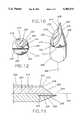
- FIG. 4is a cross-sectional view of an alternative embodiment of the trocar of the present invention, as would be seen along the line 2--2 in FIG. 1, showing cutting blades embedded in the puncturing head;
- FIG. 6is a cross-sectional view of the trocar of the present invention, as seen along the line 6--6 in FIG. 5;
- FIG. 8is a perspective view of the trocar of the present invention, shown in one intended environment
- FIG. 12is an end view of the distal end of the trocar shown in FIG. 9.
- the trocar 10has a transparent elongated cylindrical shaft 12.
- the shaft 12 of the trocar 10is made of a clear, biocompatible, axially rigid material, such as hard plastic, glass, or plexiglass.
- the outer surface of the shaft 12is polished.
- the shaft 12has a diameter D of about ten millimeters (10 mm), and a length L of about ten centimeters (10 cm). It is to be understood, however, that the trocar 10 can have other diameters, e.g., eight millimeters or twenty millimeters (8 mm or 20 mm).
- FIG. 1further shows that the shaft 12 has a proximal segment 14 and a distal end 16.
- a cavity 18is formed in the proximal segment 14 of the shaft 12, and a light gathering member, preferably a rigid endoscope 20, is positioned in the cavity 18.
- the rigid endoscope 20includes a plurality of rod lens 22.
- the rigid endoscope 20can be a rigid surgical endoscope made by Karl Storz Corp.
- FIGS. 9-12an alternate embodiment of the trocar of the present invention is shown, generally designated 200.
- the trocar 200has an elongated cylindrical shaft 202 defining an axis 204.
- FIG. 11also shows that a cutting blade 224 is glued to the distal segment 206 within a slot 226 which is formed in the distal segment 206.
- the blade 224is a metal or hard plastic blade that has opposed sharpened cutting edges 228, 230 (best shown in FIG. 10). As shown in FIG. 10, each cutting edge 228, 230 of the blade 224 protrudes outwardly from the curved surface 208 of the distal segment 206 to cut tissue and thereby aid advancing the trocar 200 through tissue. If desired, the edge 232 of the flat surface 210 can be sharpened, to further aid in advancing the trocar 200 through tissue.
- the trocar 10Upon positioning the trocar 10 with sleeve 112 as desired, the trocar 10 is retrieved from the patient 110, and the sleeve 112 remains in place inside the patient 110 to establish a working channel for an endoscopic instrument. It is to be understood that the operation of each of the trocars 50, 70, 200 is in all essential respects identical to the operation of the trocar 10.
Landscapes
- Health & Medical Sciences (AREA)
- Life Sciences & Earth Sciences (AREA)
- Surgery (AREA)
- General Health & Medical Sciences (AREA)
- Public Health (AREA)
- Veterinary Medicine (AREA)
- Pathology (AREA)
- Nuclear Medicine, Radiotherapy & Molecular Imaging (AREA)
- Animal Behavior & Ethology (AREA)
- Engineering & Computer Science (AREA)
- Biomedical Technology (AREA)
- Heart & Thoracic Surgery (AREA)
- Medical Informatics (AREA)
- Molecular Biology (AREA)
- Biophysics (AREA)
- Physics & Mathematics (AREA)
- Radiology & Medical Imaging (AREA)
- Optics & Photonics (AREA)
- Endoscopes (AREA)
- Surgical Instruments (AREA)
Abstract
Description
Claims (7)
Priority Applications (6)
| Application Number | Priority Date | Filing Date | Title |
|---|---|---|---|
| US07/974,956US5385572A (en) | 1992-11-12 | 1992-11-12 | Trocar for endoscopic surgery |
| PCT/US1993/011024WO1994010898A1 (en) | 1992-11-12 | 1993-11-12 | Trocar for endoscopic surgery |
| EP94902247AEP0674490A4 (en) | 1992-11-12 | 1993-11-12 | Trocar for endoscopic surgery. |
| CA002151926ACA2151926A1 (en) | 1992-11-12 | 1993-11-12 | Trocar for endoscopic surgery |
| US08/376,759US5562696A (en) | 1992-11-12 | 1995-01-20 | Visualization trocar |
| US08/928,680US5797944A (en) | 1992-11-12 | 1997-09-12 | Visualization trocar |
Applications Claiming Priority (1)
| Application Number | Priority Date | Filing Date | Title |
|---|---|---|---|
| US07/974,956US5385572A (en) | 1992-11-12 | 1992-11-12 | Trocar for endoscopic surgery |
Related Child Applications (1)
| Application Number | Title | Priority Date | Filing Date |
|---|---|---|---|
| US34326394AContinuation-In-Part | 1992-11-12 | 1994-11-21 |
Publications (1)
| Publication Number | Publication Date |
|---|---|
| US5385572Atrue US5385572A (en) | 1995-01-31 |
Family
ID=25522556
Family Applications (1)
| Application Number | Title | Priority Date | Filing Date |
|---|---|---|---|
| US07/974,956Expired - LifetimeUS5385572A (en) | 1992-11-12 | 1992-11-12 | Trocar for endoscopic surgery |
Country Status (4)
| Country | Link |
|---|---|
| US (1) | US5385572A (en) |
| EP (1) | EP0674490A4 (en) |
| CA (1) | CA2151926A1 (en) |
| WO (1) | WO1994010898A1 (en) |
Cited By (92)
| Publication number | Priority date | Publication date | Assignee | Title |
|---|---|---|---|---|
| US5441041A (en)* | 1993-09-13 | 1995-08-15 | United States Surgical Corporation | Optical trocar |
| US5467762A (en)* | 1993-09-13 | 1995-11-21 | United States Surgical Corporation | Optical trocar |
| US5551947A (en)* | 1992-11-17 | 1996-09-03 | Worldwide Optical Trocar Licensing Corporation | Visually directed trocar for laparoscopic surgical procedures and method of using same |
| US5554137A (en)* | 1993-10-08 | 1996-09-10 | United States Surgical Corporation | Tissue piercing members |
| US5569292A (en)* | 1995-02-01 | 1996-10-29 | Ethicon Endo-Surgery, Inc. | Surgical penetration instrument with transparent blades and tip cover |
| US5569290A (en)* | 1995-01-30 | 1996-10-29 | Paul C. McAfee | Method of and apparatus for laparoscopic or endoscopic spinal surgery using an unsealed anteriorly inserted transparent trochar |
| US5571133A (en)* | 1995-06-01 | 1996-11-05 | Yoon; Inbae | Penetrating instrument with sequential indication of entry into anatomical cavities |
| US5591183A (en)* | 1995-04-12 | 1997-01-07 | Origin Medsystems, Inc. | Dissection apparatus |
| US5609562A (en)* | 1993-11-16 | 1997-03-11 | Worldwide Optical Trocar Licensing Corporation | Visually directed trocar and method |
| DE19547246C1 (en)* | 1995-12-18 | 1997-03-20 | Riek Siegfried | Medicinal needle containing spring-loaded guard |
| US5624459A (en)* | 1995-01-26 | 1997-04-29 | Symbiosis Corporation | Trocar having an improved cutting tip configuration |
| US5632717A (en)* | 1994-10-07 | 1997-05-27 | Yoon; Inbae | Penetrating endoscope |
| WO1997026831A1 (en) | 1996-01-24 | 1997-07-31 | Origin Medsystems, Inc. | Tissue separation cannula with dissection probe and method |
| US5674184A (en)* | 1994-03-15 | 1997-10-07 | Ethicon Endo-Surgery, Inc. | Surgical trocars with cutting electrode and viewing rod |
| US5685820A (en)* | 1990-11-06 | 1997-11-11 | Partomed Medizintechnik Gmbh | Instrument for the penetration of body tissue |
| US5690664A (en)* | 1993-09-13 | 1997-11-25 | United States Surgical Corporation | Trocar having movable blade |
| US5720761A (en)* | 1993-11-16 | 1998-02-24 | Worldwide Optical Trocar Licensing Corp. | Visually directed trocar and method |
| US5738628A (en)* | 1995-03-24 | 1998-04-14 | Ethicon Endo-Surgery, Inc. | Surgical dissector and method for its use |
| US5759150A (en)* | 1995-07-07 | 1998-06-02 | Olympus Optical Co., Ltd. | System for evulsing subcutaneous tissue |
| US5797944A (en)* | 1992-11-12 | 1998-08-25 | Ethicon Endo-Surgery, Inc. | Visualization trocar |
| US5842971A (en)* | 1996-05-22 | 1998-12-01 | Yoon; Inbae | Optical endoscopic portals and methods of using the same to establish passages through cavity walls |
| US5860996A (en)* | 1994-05-26 | 1999-01-19 | United States Surgical Corporation | Optical trocar |
| US5957947A (en)* | 1997-07-18 | 1999-09-28 | Wattiez; Arnaud | Single use trocar assembly |
| US5980549A (en)* | 1995-07-13 | 1999-11-09 | Origin Medsystems, Inc. | Tissue separation cannula with dissection probe and method |
| US6077179A (en)* | 1998-05-21 | 2000-06-20 | Liechty, Ii; Victor Jay | Arrowhead with a tip having convex facets |
| US6206823B1 (en) | 1999-08-02 | 2001-03-27 | Ethicon Endo-Surgery, Inc. | Surgical instrument and method for endoscopic tissue dissection |
| US6258000B1 (en) | 1998-05-21 | 2001-07-10 | Liechty, Ii Victor Jay | Penetration enhancing aerodynamically favorable arrowhead |
| US6267732B1 (en) | 1997-09-12 | 2001-07-31 | Imagyn Medical Technologies, Inc. | Incisional breast biopsy device |
| US6283958B1 (en)* | 1996-04-04 | 2001-09-04 | Somatex Medizintechnische Instrumente Gmbh | Laser applicator set |
| US6383145B1 (en) | 1997-09-12 | 2002-05-07 | Imagyn Medical Technologies California, Inc. | Incisional breast biopsy device |
| US6447527B1 (en)* | 1998-04-23 | 2002-09-10 | Ronald J. Thompson | Apparatus and methods for the penetration of tissue |
| US6471638B1 (en) | 2000-04-28 | 2002-10-29 | Origin Medsystems, Inc. | Surgical apparatus |
| US6551253B2 (en) | 1997-09-12 | 2003-04-22 | Imagyn Medical Technologies | Incisional breast biopsy device |
| US20030136097A1 (en)* | 2002-01-22 | 2003-07-24 | Schoenherr Terrance L. | Inverter shield for a windrow merger |
| US20040082969A1 (en)* | 2002-10-23 | 2004-04-29 | Stephen Kerr | Laparoscopic direct vision dissecting port |
| US20040093000A1 (en)* | 2002-10-23 | 2004-05-13 | Stephen Kerr | Direct vision port site dissector |
| US20060052660A1 (en)* | 1999-08-10 | 2006-03-09 | Chin Albert K | Apparatus and methods for cardiac restraint |
| US20060116746A1 (en)* | 2003-01-17 | 2006-06-01 | Chin Albert K | Cardiac electrode attachment procedure |
| US20060173479A1 (en)* | 2005-01-28 | 2006-08-03 | Smith Robert C | Optical penetrating adapter for surgical portal |
| US20060224174A1 (en)* | 2005-03-31 | 2006-10-05 | Smith Robert C | Optical obturator |
| US20060226655A1 (en)* | 2005-04-12 | 2006-10-12 | Smith Robert C | Optical trocar with scope holding assembly |
| US20070260274A1 (en)* | 2006-05-02 | 2007-11-08 | Gimmi Gmbh | Instrument for producing a skin opening for minimally invasive surgery |
| US20080051770A1 (en)* | 2006-08-22 | 2008-02-28 | Synergetics, Inc. | Multiple Target Laser Probe |
| US20080086160A1 (en)* | 2006-10-06 | 2008-04-10 | Surgiquest, Incorporated | Visualization trocar |
| US20080300617A1 (en)* | 2007-06-01 | 2008-12-04 | Tyco Healthcare Group Lp | Obturator tips |
| US20080306333A1 (en)* | 1999-08-10 | 2008-12-11 | Chin Albert K | Apparatus and Method for Endoscopic Surgical Procedures |
| US20080309758A1 (en)* | 2007-06-14 | 2008-12-18 | Olympus Medical Systems Corp. | Endoscope system, camera set on body cavity inner wall and method of setting the camera set on body cavity inner wall on inner surface of body cavity wall |
| US20090023986A1 (en)* | 1998-08-12 | 2009-01-22 | Stewart Michael C | Vessel Harvesting |
| US20090024156A1 (en)* | 1995-07-13 | 2009-01-22 | Chin Albert K | Tissue Dissection Method |
| US20090093677A1 (en)* | 2007-10-05 | 2009-04-09 | Tyco Healthcare Group Lp | Visual obturator |
| US20090131907A1 (en)* | 1999-08-10 | 2009-05-21 | Maquet Cardiovascular Llc | Endoscopic Cardiac Surgery |
| US20090131747A1 (en)* | 1998-06-22 | 2009-05-21 | Maquet Cardiovascular Llc | Instrument And Method For Remotely Manipulating A Tissue Structure |
| EP2070484A1 (en) | 2002-10-23 | 2009-06-17 | Intellimed Surgical Solutions LLC. | Direct vision port site dissector |
| US20090270819A1 (en)* | 2008-04-29 | 2009-10-29 | Dario Vitali | Optical safety trocar and method of use thereof |
| US20100048994A1 (en)* | 2007-04-11 | 2010-02-25 | Okoniewski Gregory G | Visualized entry trocar with moving blade |
| US20100137895A1 (en)* | 2007-04-17 | 2010-06-03 | Smith Robert C | Visual obturator with handle |
| US20100273588A1 (en)* | 2006-08-18 | 2010-10-28 | Field Logic, Inc. | Expandable broadhead with rear deploying blades |
| US20110125084A1 (en)* | 2003-04-08 | 2011-05-26 | Surgiquest, Inc. | Trocar assembly with pneumatic sealing |
| US20110152910A1 (en)* | 2009-12-17 | 2011-06-23 | Tyco Healthcare Group Lp | Visual obturator with tip openings |
| US20110152753A1 (en)* | 2009-12-17 | 2011-06-23 | Tyco Healthcare Group Lp | Obturator tip |
| US7972265B1 (en) | 1998-06-22 | 2011-07-05 | Maquet Cardiovascular, Llc | Device and method for remote vessel ligation |
| USD654168S1 (en) | 2010-09-21 | 2012-02-14 | Tyco Healthcare Group Lp | Bladeless obturator member |
| USD655005S1 (en) | 2010-09-21 | 2012-02-28 | Tyco Healthcare Group Lp | Bladeless obturator member |
| USD656233S1 (en) | 2010-09-21 | 2012-03-20 | Tyco Healthcare Group Lp | Bladeless obturator member |
| USD663838S1 (en) | 2007-10-05 | 2012-07-17 | Surgiquest, Inc. | Visualization trocar |
| US8241210B2 (en) | 1998-06-22 | 2012-08-14 | Maquet Cardiovascular Llc | Vessel retractor |
| USD667954S1 (en) | 2007-10-05 | 2012-09-25 | Surgiquest, Inc. | Visualization trocar |
| US8277411B2 (en) | 2002-01-31 | 2012-10-02 | Boston Scientific Scimed, Inc. | Needle device |
| USRE44144E1 (en) | 2000-03-13 | 2013-04-09 | Out Rage, Llc | Expandable broadhead |
| US8430851B2 (en) | 2005-10-14 | 2013-04-30 | Applied Medical Resources Corporation | Surgical access port |
| US8795223B2 (en) | 2011-03-08 | 2014-08-05 | Surgiquest, Inc. | Trocar assembly with pneumatic sealing |
| US8821526B2 (en) | 2010-11-11 | 2014-09-02 | Specialtycare, Inc. | Trocar |
| US20140358070A1 (en) | 2007-04-13 | 2014-12-04 | Surgiquest, Inc. | System and method for improved gas recirculation in surgical trocars with pneumatic sealing |
| USD730471S1 (en) | 2013-12-18 | 2015-05-26 | Out Rage, Llc | Broadhead |
| CN104707234A (en)* | 2013-12-17 | 2015-06-17 | 标准科学技术株式会社 | Catheter for common hepatic duct |
| US9101315B2 (en) | 2010-11-11 | 2015-08-11 | Specialty Care, Inc. | Cannula system |
| WO2014145008A3 (en)* | 2013-03-15 | 2015-11-05 | Olive Medical Corporation | Viewing trocar for use with angled endoscope |
| US9186173B2 (en) | 2012-04-27 | 2015-11-17 | Specialty Care, Inc. | Optical obturator system |
| US9308020B2 (en) | 2013-03-14 | 2016-04-12 | Cook Medical Technologies Llc | Tri-fluted vascular access needle |
| WO2016100181A1 (en) | 2014-12-19 | 2016-06-23 | Ethicon Endo-Surgery, Llc | Adaptor for robotics cannula and seal assembly |
| EP3053507A1 (en)* | 2015-02-04 | 2016-08-10 | EDA Medical Devices Technology Inc. | Medical probe |
| US20160331207A1 (en)* | 2015-05-13 | 2016-11-17 | Tamer Ibrahim | Access visualization systems |
| USD776782S1 (en) | 2015-05-22 | 2017-01-17 | Feradyne Outdoors, Llc | Broadhead arrowhead having both expandable and fixed cutting blades |
| US20170100020A1 (en)* | 2015-05-08 | 2017-04-13 | Samark Technology Llc | Imaging needle apparatus |
| US9687273B2 (en) | 2013-09-11 | 2017-06-27 | Gimmi Gmbh | Endoscopic surgical instruments and related methods |
| US9700697B2 (en) | 2012-08-21 | 2017-07-11 | Optomeditech Oy | Intravascular catheter assembly |
| US20170245890A1 (en)* | 2016-02-26 | 2017-08-31 | Samark Technology Llc | Video needle syringe |
| CN107744401A (en)* | 2017-11-14 | 2018-03-02 | 北京水木天蓬医疗技术有限公司 | Ultrasonic osteotome head |
| US10299770B2 (en) | 2006-06-01 | 2019-05-28 | Maquet Cardiovascular Llc | Endoscopic vessel harvesting system components |
| US10463399B2 (en) | 2014-11-06 | 2019-11-05 | Asimion Inc. | Visually assisted entry of a Veress needle with a tapered videoscope for microlaparoscopy |
| US10507012B2 (en) | 2000-11-17 | 2019-12-17 | Maquet Cardiovascular Llc | Vein harvesting system and method |
| US20200138514A1 (en)* | 2018-11-02 | 2020-05-07 | Intuitive Surgical Operations, Inc. | Tissue penetrating device tips |
Families Citing this family (3)
| Publication number | Priority date | Publication date | Assignee | Title |
|---|---|---|---|---|
| US5552156A (en)* | 1992-10-23 | 1996-09-03 | Ohio State University | Liposomal and micellular stabilization of camptothecin drugs |
| US5736156A (en)* | 1995-03-22 | 1998-04-07 | The Ohio State University | Liposomal anf micellular stabilization of camptothecin drugs |
| CN110680480B (en)* | 2019-11-04 | 2020-12-01 | 商洛职业技术学院 | Dura mater puncture awl and matched handle thereof |
Citations (15)
| Publication number | Priority date | Publication date | Assignee | Title |
|---|---|---|---|---|
| US4254762A (en)* | 1979-10-23 | 1981-03-10 | Inbae Yoon | Safety endoscope system |
| US4269192A (en)* | 1977-12-02 | 1981-05-26 | Olympus Optical Co., Ltd. | Stabbing apparatus for diagnosis of living body |
| US4273127A (en)* | 1978-10-12 | 1981-06-16 | Research Corporation | Method for cutting and coagulating tissue |
| GB2102678A (en)* | 1981-08-01 | 1983-02-09 | Hpw Ltd | Illumination of precision instruments |
| US4535773A (en)* | 1982-03-26 | 1985-08-20 | Inbae Yoon | Safety puncturing instrument and method |
| US4566438A (en)* | 1984-10-05 | 1986-01-28 | Liese Grover J | Fiber-optic stylet for needle tip localization |
| US4740047A (en)* | 1985-03-26 | 1988-04-26 | Hatachi Cable, Ltd. | Fiber for lateral beaming of laser beam |
| US4832979A (en)* | 1986-11-21 | 1989-05-23 | Masahiko Hoshino | Process for preparing laser knife |
| US5151096A (en)* | 1991-03-28 | 1992-09-29 | Angiolaz, Incorporated | Laser catheter diffuser |
| US5196005A (en)* | 1991-11-26 | 1993-03-23 | Pdt Systems, Inc. | Continuous gradient cylindrical diffusion tip for optical fibers and method for making |
| US5215526A (en)* | 1988-07-06 | 1993-06-01 | Ethicon, Inc. | Safety trocar |
| US5234450A (en)* | 1989-07-28 | 1993-08-10 | Jacob Segalowitz | Valvulotome catheter |
| US5246436A (en)* | 1991-12-18 | 1993-09-21 | Alcon Surgical, Inc. | Midinfrared laser tissue ablater |
| US5250068A (en)* | 1990-11-30 | 1993-10-05 | Yakuouji Shinkiyu Chiryouin | Optical transmission type acupuncture needle |
| US5271380A (en)* | 1990-11-06 | 1993-12-21 | Siegfried Riek | Penetration instrument |
Family Cites Families (3)
| Publication number | Priority date | Publication date | Assignee | Title |
|---|---|---|---|---|
| US5057082A (en)* | 1988-11-04 | 1991-10-15 | Plastic Injectors, Inc. | Trocar assembly |
| DE4133073A1 (en)* | 1990-10-11 | 1992-04-16 | Effner Gmbh | Trocar for producing access for surgery under endoscopic control - has pointed tip at one end of shaft and optical system with objective in vicinity to tip |
| US5183471A (en)* | 1992-01-24 | 1993-02-02 | Wilk Peter J | Laparoscopic cannula |
- 1992
- 1992-11-12USUS07/974,956patent/US5385572A/ennot_activeExpired - Lifetime
- 1993
- 1993-11-12EPEP94902247Apatent/EP0674490A4/ennot_activeWithdrawn
- 1993-11-12WOPCT/US1993/011024patent/WO1994010898A1/ennot_activeApplication Discontinuation
- 1993-11-12CACA002151926Apatent/CA2151926A1/ennot_activeAbandoned
Patent Citations (15)
| Publication number | Priority date | Publication date | Assignee | Title |
|---|---|---|---|---|
| US4269192A (en)* | 1977-12-02 | 1981-05-26 | Olympus Optical Co., Ltd. | Stabbing apparatus for diagnosis of living body |
| US4273127A (en)* | 1978-10-12 | 1981-06-16 | Research Corporation | Method for cutting and coagulating tissue |
| US4254762A (en)* | 1979-10-23 | 1981-03-10 | Inbae Yoon | Safety endoscope system |
| GB2102678A (en)* | 1981-08-01 | 1983-02-09 | Hpw Ltd | Illumination of precision instruments |
| US4535773A (en)* | 1982-03-26 | 1985-08-20 | Inbae Yoon | Safety puncturing instrument and method |
| US4566438A (en)* | 1984-10-05 | 1986-01-28 | Liese Grover J | Fiber-optic stylet for needle tip localization |
| US4740047A (en)* | 1985-03-26 | 1988-04-26 | Hatachi Cable, Ltd. | Fiber for lateral beaming of laser beam |
| US4832979A (en)* | 1986-11-21 | 1989-05-23 | Masahiko Hoshino | Process for preparing laser knife |
| US5215526A (en)* | 1988-07-06 | 1993-06-01 | Ethicon, Inc. | Safety trocar |
| US5234450A (en)* | 1989-07-28 | 1993-08-10 | Jacob Segalowitz | Valvulotome catheter |
| US5271380A (en)* | 1990-11-06 | 1993-12-21 | Siegfried Riek | Penetration instrument |
| US5250068A (en)* | 1990-11-30 | 1993-10-05 | Yakuouji Shinkiyu Chiryouin | Optical transmission type acupuncture needle |
| US5151096A (en)* | 1991-03-28 | 1992-09-29 | Angiolaz, Incorporated | Laser catheter diffuser |
| US5196005A (en)* | 1991-11-26 | 1993-03-23 | Pdt Systems, Inc. | Continuous gradient cylindrical diffusion tip for optical fibers and method for making |
| US5246436A (en)* | 1991-12-18 | 1993-09-21 | Alcon Surgical, Inc. | Midinfrared laser tissue ablater |
Cited By (172)
| Publication number | Priority date | Publication date | Assignee | Title |
|---|---|---|---|---|
| US5685820A (en)* | 1990-11-06 | 1997-11-11 | Partomed Medizintechnik Gmbh | Instrument for the penetration of body tissue |
| US5797944A (en)* | 1992-11-12 | 1998-08-25 | Ethicon Endo-Surgery, Inc. | Visualization trocar |
| US5551947A (en)* | 1992-11-17 | 1996-09-03 | Worldwide Optical Trocar Licensing Corporation | Visually directed trocar for laparoscopic surgical procedures and method of using same |
| US20080154295A1 (en)* | 1993-09-13 | 2008-06-26 | Sauer Jude S | Optical trocar |
| US5658236A (en)* | 1993-09-13 | 1997-08-19 | United States Surgical Corporation | Optical trocar |
| US5569160A (en)* | 1993-09-13 | 1996-10-29 | United States Surgical Corporation | Optical trocar |
| US5467762A (en)* | 1993-09-13 | 1995-11-21 | United States Surgical Corporation | Optical trocar |
| US6685630B2 (en)* | 1993-09-13 | 2004-02-03 | United States Surgical Corporation | Optical trocar |
| US7322933B2 (en) | 1993-09-13 | 2008-01-29 | United States Surgical Corporation | Optical trocar |
| US7811225B2 (en) | 1993-09-13 | 2010-10-12 | Tyco Healthcare Group Lp | Optical trocar |
| US20040158126A1 (en)* | 1993-09-13 | 2004-08-12 | Sauer Jude S. | Optical trocar |
| US5690664A (en)* | 1993-09-13 | 1997-11-25 | United States Surgical Corporation | Trocar having movable blade |
| US5441041A (en)* | 1993-09-13 | 1995-08-15 | United States Surgical Corporation | Optical trocar |
| US20110040253A1 (en)* | 1993-09-13 | 2011-02-17 | Sauer Jude S | Optical trocar |
| US5554137A (en)* | 1993-10-08 | 1996-09-10 | United States Surgical Corporation | Tissue piercing members |
| US5720761A (en)* | 1993-11-16 | 1998-02-24 | Worldwide Optical Trocar Licensing Corp. | Visually directed trocar and method |
| US5609562A (en)* | 1993-11-16 | 1997-03-11 | Worldwide Optical Trocar Licensing Corporation | Visually directed trocar and method |
| US5674184A (en)* | 1994-03-15 | 1997-10-07 | Ethicon Endo-Surgery, Inc. | Surgical trocars with cutting electrode and viewing rod |
| US5860996A (en)* | 1994-05-26 | 1999-01-19 | United States Surgical Corporation | Optical trocar |
| US5632717A (en)* | 1994-10-07 | 1997-05-27 | Yoon; Inbae | Penetrating endoscope |
| US5624459A (en)* | 1995-01-26 | 1997-04-29 | Symbiosis Corporation | Trocar having an improved cutting tip configuration |
| US5569290A (en)* | 1995-01-30 | 1996-10-29 | Paul C. McAfee | Method of and apparatus for laparoscopic or endoscopic spinal surgery using an unsealed anteriorly inserted transparent trochar |
| AU696687B2 (en)* | 1995-02-01 | 1998-09-17 | Ethicon Endo-Surgery, Inc. | Surgical penetration instrument |
| US5569292A (en)* | 1995-02-01 | 1996-10-29 | Ethicon Endo-Surgery, Inc. | Surgical penetration instrument with transparent blades and tip cover |
| US5738628A (en)* | 1995-03-24 | 1998-04-14 | Ethicon Endo-Surgery, Inc. | Surgical dissector and method for its use |
| US5591183A (en)* | 1995-04-12 | 1997-01-07 | Origin Medsystems, Inc. | Dissection apparatus |
| US5571133A (en)* | 1995-06-01 | 1996-11-05 | Yoon; Inbae | Penetrating instrument with sequential indication of entry into anatomical cavities |
| US6019720A (en)* | 1995-07-07 | 2000-02-01 | Olympus Optical Co., Ltd. | System for evulsing subcutaneous tissue |
| US6080102A (en)* | 1995-07-07 | 2000-06-27 | Olympus Optical Co., Ltd. | System for evulsing subcutaneous tissue |
| EP1462060A1 (en)* | 1995-07-07 | 2004-09-29 | Olympus Optical Co., Ltd. | System for evulsing subcutaneous tissue |
| US5759150A (en)* | 1995-07-07 | 1998-06-02 | Olympus Optical Co., Ltd. | System for evulsing subcutaneous tissue |
| US5980549A (en)* | 1995-07-13 | 1999-11-09 | Origin Medsystems, Inc. | Tissue separation cannula with dissection probe and method |
| US20090024156A1 (en)* | 1995-07-13 | 2009-01-22 | Chin Albert K | Tissue Dissection Method |
| US7981133B2 (en) | 1995-07-13 | 2011-07-19 | Maquet Cardiovascular, Llc | Tissue dissection method |
| US6001084A (en)* | 1995-12-18 | 1999-12-14 | Riek; Siegfried | Medical needle for endoscopic surgery |
| DE19547246C1 (en)* | 1995-12-18 | 1997-03-20 | Riek Siegfried | Medicinal needle containing spring-loaded guard |
| WO1997026831A1 (en) | 1996-01-24 | 1997-07-31 | Origin Medsystems, Inc. | Tissue separation cannula with dissection probe and method |
| US6283958B1 (en)* | 1996-04-04 | 2001-09-04 | Somatex Medizintechnische Instrumente Gmbh | Laser applicator set |
| US5842971A (en)* | 1996-05-22 | 1998-12-01 | Yoon; Inbae | Optical endoscopic portals and methods of using the same to establish passages through cavity walls |
| US5957947A (en)* | 1997-07-18 | 1999-09-28 | Wattiez; Arnaud | Single use trocar assembly |
| US6383145B1 (en) | 1997-09-12 | 2002-05-07 | Imagyn Medical Technologies California, Inc. | Incisional breast biopsy device |
| US6551253B2 (en) | 1997-09-12 | 2003-04-22 | Imagyn Medical Technologies | Incisional breast biopsy device |
| US6267732B1 (en) | 1997-09-12 | 2001-07-31 | Imagyn Medical Technologies, Inc. | Incisional breast biopsy device |
| US6447527B1 (en)* | 1998-04-23 | 2002-09-10 | Ronald J. Thompson | Apparatus and methods for the penetration of tissue |
| US6258000B1 (en) | 1998-05-21 | 2001-07-10 | Liechty, Ii Victor Jay | Penetration enhancing aerodynamically favorable arrowhead |
| US6077179A (en)* | 1998-05-21 | 2000-06-20 | Liechty, Ii; Victor Jay | Arrowhead with a tip having convex facets |
| US6306053B1 (en) | 1998-05-21 | 2001-10-23 | Liechty, Ii Victor Jay | Razor-edged cutting tip |
| US8241210B2 (en) | 1998-06-22 | 2012-08-14 | Maquet Cardiovascular Llc | Vessel retractor |
| US20090131747A1 (en)* | 1998-06-22 | 2009-05-21 | Maquet Cardiovascular Llc | Instrument And Method For Remotely Manipulating A Tissue Structure |
| US7972265B1 (en) | 1998-06-22 | 2011-07-05 | Maquet Cardiovascular, Llc | Device and method for remote vessel ligation |
| US7867163B2 (en) | 1998-06-22 | 2011-01-11 | Maquet Cardiovascular Llc | Instrument and method for remotely manipulating a tissue structure |
| US9730782B2 (en) | 1998-08-12 | 2017-08-15 | Maquet Cardiovascular Llc | Vessel harvester |
| US20090023986A1 (en)* | 1998-08-12 | 2009-01-22 | Stewart Michael C | Vessel Harvesting |
| US8460331B2 (en) | 1998-08-12 | 2013-06-11 | Maquet Cardiovascular, Llc | Tissue dissector apparatus and method |
| US8986335B2 (en) | 1998-08-12 | 2015-03-24 | Maquet Cardiovascular Llc | Tissue dissector apparatus and method |
| US9700398B2 (en) | 1998-08-12 | 2017-07-11 | Maquet Cardiovascular Llc | Vessel harvester |
| US7938842B1 (en) | 1998-08-12 | 2011-05-10 | Maquet Cardiovascular Llc | Tissue dissector apparatus |
| US6206823B1 (en) | 1999-08-02 | 2001-03-27 | Ethicon Endo-Surgery, Inc. | Surgical instrument and method for endoscopic tissue dissection |
| US20090131907A1 (en)* | 1999-08-10 | 2009-05-21 | Maquet Cardiovascular Llc | Endoscopic Cardiac Surgery |
| US20080306333A1 (en)* | 1999-08-10 | 2008-12-11 | Chin Albert K | Apparatus and Method for Endoscopic Surgical Procedures |
| US20060052660A1 (en)* | 1999-08-10 | 2006-03-09 | Chin Albert K | Apparatus and methods for cardiac restraint |
| USRE44144E1 (en) | 2000-03-13 | 2013-04-09 | Out Rage, Llc | Expandable broadhead |
| US6471638B1 (en) | 2000-04-28 | 2002-10-29 | Origin Medsystems, Inc. | Surgical apparatus |
| US6814696B1 (en) | 2000-04-28 | 2004-11-09 | Origin Medsystems, Inc. | Surgical apparatus |
| US10507012B2 (en) | 2000-11-17 | 2019-12-17 | Maquet Cardiovascular Llc | Vein harvesting system and method |
| US20030136097A1 (en)* | 2002-01-22 | 2003-07-24 | Schoenherr Terrance L. | Inverter shield for a windrow merger |
| US8277411B2 (en) | 2002-01-31 | 2012-10-02 | Boston Scientific Scimed, Inc. | Needle device |
| US20040082969A1 (en)* | 2002-10-23 | 2004-04-29 | Stephen Kerr | Laparoscopic direct vision dissecting port |
| US20060079925A1 (en)* | 2002-10-23 | 2006-04-13 | Stephen Kerr | Direct vision port site dissector |
| US20040093000A1 (en)* | 2002-10-23 | 2004-05-13 | Stephen Kerr | Direct vision port site dissector |
| EP2070484A1 (en) | 2002-10-23 | 2009-06-17 | Intellimed Surgical Solutions LLC. | Direct vision port site dissector |
| US7604648B2 (en) | 2002-10-23 | 2009-10-20 | Intellimed Surgical Solutions, Llc | Direct vision port site dissector |
| US7056329B2 (en) | 2002-10-23 | 2006-06-06 | Intellimed Surgical Solutions, Llc | Laparoscopic direct vision dissecting port |
| US20070049963A1 (en)* | 2002-10-23 | 2007-03-01 | Stephen Kerr | Direct vision port site dissector |
| US20060116746A1 (en)* | 2003-01-17 | 2006-06-01 | Chin Albert K | Cardiac electrode attachment procedure |
| US10369303B2 (en) | 2003-04-08 | 2019-08-06 | Conmed Corporation | Trocar assembly with pneumatic sealing |
| US20110125084A1 (en)* | 2003-04-08 | 2011-05-26 | Surgiquest, Inc. | Trocar assembly with pneumatic sealing |
| US20060173479A1 (en)* | 2005-01-28 | 2006-08-03 | Smith Robert C | Optical penetrating adapter for surgical portal |
| US8070767B2 (en) | 2005-01-28 | 2011-12-06 | Tyco Healthcare Group Lp | Optical penetrating adapter for surgical portal |
| US8475486B2 (en) | 2005-01-28 | 2013-07-02 | Covidien Lp | Optical penetrating adapter for surgical portal |
| US10064648B2 (en) | 2005-01-28 | 2018-09-04 | Covidien Lp | Optical penetrating adapter for surgical portal |
| US9226770B2 (en) | 2005-01-28 | 2016-01-05 | Covidien Lp | Optical penetrating adapter for surgical portal |
| US10610258B2 (en) | 2005-03-31 | 2020-04-07 | Covidien Lp | Optical obturator |
| US20060224174A1 (en)* | 2005-03-31 | 2006-10-05 | Smith Robert C | Optical obturator |
| US7470230B2 (en) | 2005-03-31 | 2008-12-30 | Tyco Healthcare Group Lp | Optical obturator |
| US11559327B2 (en) | 2005-03-31 | 2023-01-24 | Covidien Lp | Optical obturator |
| US20090076323A1 (en)* | 2005-03-31 | 2009-03-19 | Tyco Healthcare Group Lp | Optical obturator |
| US20110004061A1 (en)* | 2005-04-12 | 2011-01-06 | Tyco Healthcare Group Lp | Optical trocar with scope holding assembly |
| US10159402B2 (en) | 2005-04-12 | 2018-12-25 | Covidien Lp | Optical trocar with scope holding assembly |
| US8517918B2 (en) | 2005-04-12 | 2013-08-27 | Covidien Lp | Optical trocar with scope holding assembly |
| US20060226655A1 (en)* | 2005-04-12 | 2006-10-12 | Smith Robert C | Optical trocar with scope holding assembly |
| US7824327B2 (en) | 2005-04-12 | 2010-11-02 | Tyco Healthcare Group Llp | Optical trocar with scope holding assembly |
| US8430851B2 (en) | 2005-10-14 | 2013-04-30 | Applied Medical Resources Corporation | Surgical access port |
| US10478219B2 (en) | 2005-10-14 | 2019-11-19 | Applied Medical Resources Corporation | Surgical access port |
| US8968250B2 (en) | 2005-10-14 | 2015-03-03 | Applied Medical Resources Corporation | Surgical access port |
| US11504157B2 (en) | 2005-10-14 | 2022-11-22 | Applied Medical Resources Corporation | Surgical access port |
| US9833259B2 (en) | 2005-10-14 | 2017-12-05 | Applied Medical Resources Corporation | Surgical access port |
| US8974481B2 (en)* | 2006-05-02 | 2015-03-10 | Gimmi Gmbh | Instrument for producing a skin opening for minimally invasive surgery |
| US20070260274A1 (en)* | 2006-05-02 | 2007-11-08 | Gimmi Gmbh | Instrument for producing a skin opening for minimally invasive surgery |
| US11141055B2 (en) | 2006-06-01 | 2021-10-12 | Maquet Cardiovascular Llc | Endoscopic vessel harvesting system components |
| US10299770B2 (en) | 2006-06-01 | 2019-05-28 | Maquet Cardiovascular Llc | Endoscopic vessel harvesting system components |
| US11134835B2 (en) | 2006-06-01 | 2021-10-05 | Maquet Cardiovascular Llc | Endoscopic vessel harvesting system components |
| US8512179B2 (en) | 2006-08-18 | 2013-08-20 | Out Rage, Llc | Expandable broadhead with rear deploying blades |
| US20100273588A1 (en)* | 2006-08-18 | 2010-10-28 | Field Logic, Inc. | Expandable broadhead with rear deploying blades |
| US20080051770A1 (en)* | 2006-08-22 | 2008-02-28 | Synergetics, Inc. | Multiple Target Laser Probe |
| US20080086160A1 (en)* | 2006-10-06 | 2008-04-10 | Surgiquest, Incorporated | Visualization trocar |
| US8317815B2 (en) | 2006-10-06 | 2012-11-27 | Surgiquest, Inc. | Visualization trocar |
| US9907569B2 (en) | 2006-10-06 | 2018-03-06 | Conmed Corporation | Trocar assembly with pneumatic sealing |
| US20100048994A1 (en)* | 2007-04-11 | 2010-02-25 | Okoniewski Gregory G | Visualized entry trocar with moving blade |
| US10092319B2 (en) | 2007-04-13 | 2018-10-09 | Surgiquest, Inc. | System and method for improved gas recirculation in surgical trocars with pneumatic sealing |
| US20140358070A1 (en) | 2007-04-13 | 2014-12-04 | Surgiquest, Inc. | System and method for improved gas recirculation in surgical trocars with pneumatic sealing |
| US8202290B2 (en) | 2007-04-17 | 2012-06-19 | Tyco Healthcare Group Lp | Visual obturator with handle |
| US20100137895A1 (en)* | 2007-04-17 | 2010-06-03 | Smith Robert C | Visual obturator with handle |
| US9028521B2 (en) | 2007-06-01 | 2015-05-12 | Covidien Lp | Obturator tips |
| US20080300617A1 (en)* | 2007-06-01 | 2008-12-04 | Tyco Healthcare Group Lp | Obturator tips |
| US20080309758A1 (en)* | 2007-06-14 | 2008-12-18 | Olympus Medical Systems Corp. | Endoscope system, camera set on body cavity inner wall and method of setting the camera set on body cavity inner wall on inner surface of body cavity wall |
| USD663838S1 (en) | 2007-10-05 | 2012-07-17 | Surgiquest, Inc. | Visualization trocar |
| US20090093677A1 (en)* | 2007-10-05 | 2009-04-09 | Tyco Healthcare Group Lp | Visual obturator |
| US8192353B2 (en) | 2007-10-05 | 2012-06-05 | Tyco Healthcare Group Lp | Visual obturator |
| USD667954S1 (en) | 2007-10-05 | 2012-09-25 | Surgiquest, Inc. | Visualization trocar |
| US20090270819A1 (en)* | 2008-04-29 | 2009-10-29 | Dario Vitali | Optical safety trocar and method of use thereof |
| US11202656B2 (en) | 2008-10-10 | 2021-12-21 | Conmed Corporation | System and method for improved gas recirculation in surgical trocars with pneumatic sealing |
| US12357346B2 (en) | 2008-10-10 | 2025-07-15 | Conmed Corporation | System and method for improved gas recirculation in surgical trocars with pneumatic sealing |
| US10639071B2 (en) | 2008-10-10 | 2020-05-05 | Conmed Corporation | System and method for improved gas recirculation in surgical trocars with pneumatic sealing |
| US20110152910A1 (en)* | 2009-12-17 | 2011-06-23 | Tyco Healthcare Group Lp | Visual obturator with tip openings |
| US20110152753A1 (en)* | 2009-12-17 | 2011-06-23 | Tyco Healthcare Group Lp | Obturator tip |
| US8979883B2 (en) | 2009-12-17 | 2015-03-17 | Covidien Lp | Obturator tip |
| US9301778B2 (en) | 2009-12-17 | 2016-04-05 | Covidien Lp | Obturator tip |
| US9226774B2 (en) | 2009-12-17 | 2016-01-05 | Covidien Lp | Visual obturator with tip openings |
| USD654168S1 (en) | 2010-09-21 | 2012-02-14 | Tyco Healthcare Group Lp | Bladeless obturator member |
| USD655005S1 (en) | 2010-09-21 | 2012-02-28 | Tyco Healthcare Group Lp | Bladeless obturator member |
| USD656233S1 (en) | 2010-09-21 | 2012-03-20 | Tyco Healthcare Group Lp | Bladeless obturator member |
| US9101315B2 (en) | 2010-11-11 | 2015-08-11 | Specialty Care, Inc. | Cannula system |
| US8821526B2 (en) | 2010-11-11 | 2014-09-02 | Specialtycare, Inc. | Trocar |
| US8795223B2 (en) | 2011-03-08 | 2014-08-05 | Surgiquest, Inc. | Trocar assembly with pneumatic sealing |
| US9186173B2 (en) | 2012-04-27 | 2015-11-17 | Specialty Care, Inc. | Optical obturator system |
| US9700697B2 (en) | 2012-08-21 | 2017-07-11 | Optomeditech Oy | Intravascular catheter assembly |
| US10349975B2 (en) | 2013-03-14 | 2019-07-16 | Cook Medical Technologies Llc | Tri-fluted vascular access needle |
| US9308020B2 (en) | 2013-03-14 | 2016-04-12 | Cook Medical Technologies Llc | Tri-fluted vascular access needle |
| AU2014233486B2 (en)* | 2013-03-15 | 2018-11-15 | DePuy Synthes Products, Inc. | Viewing trocar with integrated prism for use with angled endoscope |
| CN105263393A (en)* | 2013-03-15 | 2016-01-20 | 奥利弗医疗公司 | Viewing trocar for use with angled endoscope |
| WO2014145008A3 (en)* | 2013-03-15 | 2015-11-05 | Olive Medical Corporation | Viewing trocar for use with angled endoscope |
| US11690498B2 (en) | 2013-03-15 | 2023-07-04 | DePuy Synthes Products, Inc. | Viewing trocar with integrated prism for use with angled endoscope |
| CN105263393B (en)* | 2013-03-15 | 2018-10-26 | 德普伊新特斯产品公司 | With the observation trochar with integrated prism that angled endoscope is used together |
| JP2016521142A (en)* | 2013-03-15 | 2016-07-21 | オリーブ・メディカル・コーポレイションOlive Medical Corporation | Observation trocar with integrated prism for use with angled endoscopes |
| EP2967309A4 (en)* | 2013-03-15 | 2017-01-11 | Olive Medical Corporation | Viewing trocar for use with angled endoscope |
| US10561302B2 (en) | 2013-03-15 | 2020-02-18 | DePuy Synthes Products, Inc. | Viewing trocar with integrated prism for use with angled endoscope |
| US9687273B2 (en) | 2013-09-11 | 2017-06-27 | Gimmi Gmbh | Endoscopic surgical instruments and related methods |
| CN104707234B (en)* | 2013-12-17 | 2017-12-22 | 标准科学技术株式会社 | Catheters for the common hepatic duct |
| CN104707234A (en)* | 2013-12-17 | 2015-06-17 | 标准科学技术株式会社 | Catheter for common hepatic duct |
| US20150164668A1 (en)* | 2013-12-17 | 2015-06-18 | Standard Sci-Tech Inc. | Catheter for common hepatic duct |
| USD730471S1 (en) | 2013-12-18 | 2015-05-26 | Out Rage, Llc | Broadhead |
| US10463399B2 (en) | 2014-11-06 | 2019-11-05 | Asimion Inc. | Visually assisted entry of a Veress needle with a tapered videoscope for microlaparoscopy |
| US11602374B1 (en) | 2014-11-06 | 2023-03-14 | Enable, Inc. | Visually assisted entry of a fluid delivering needle with a tapered videoscope |
| WO2016100181A1 (en) | 2014-12-19 | 2016-06-23 | Ethicon Endo-Surgery, Llc | Adaptor for robotics cannula and seal assembly |
| EP3769705A1 (en) | 2014-12-19 | 2021-01-27 | Ethicon LLC | Adaptor for robotics cannula and seal assembly |
| EP4501269A2 (en) | 2014-12-19 | 2025-02-05 | Ethicon LLC | Adaptor for robotics cannula and seal assembly |
| US9888942B1 (en) | 2014-12-19 | 2018-02-13 | Ethicon Llc | Adaptor for robotics cannula and seal assembly |
| EP3053507A1 (en)* | 2015-02-04 | 2016-08-10 | EDA Medical Devices Technology Inc. | Medical probe |
| US10105040B2 (en)* | 2015-05-08 | 2018-10-23 | Nanosurgery Technology Corporation | Imaging needle apparatus |
| US20190038116A1 (en)* | 2015-05-08 | 2019-02-07 | Nanosurgery Technology Corporation | Imaging needle apparatus |
| US20170100020A1 (en)* | 2015-05-08 | 2017-04-13 | Samark Technology Llc | Imaging needle apparatus |
| US20160331207A1 (en)* | 2015-05-13 | 2016-11-17 | Tamer Ibrahim | Access visualization systems |
| US20230021205A1 (en)* | 2015-05-13 | 2023-01-19 | Atricure, Inc. | Access visualization systems |
| US12023010B2 (en)* | 2015-05-13 | 2024-07-02 | Atricure, Inc. | Access visualization systems |
| USD776782S1 (en) | 2015-05-22 | 2017-01-17 | Feradyne Outdoors, Llc | Broadhead arrowhead having both expandable and fixed cutting blades |
| US10548633B2 (en)* | 2016-02-26 | 2020-02-04 | Nanosurgery Technology Corporation | Video needle syringe |
| US20170245890A1 (en)* | 2016-02-26 | 2017-08-31 | Samark Technology Llc | Video needle syringe |
| US11779366B2 (en) | 2017-11-14 | 2023-10-10 | Beijing Smtp Technology Co., Ltd. | Ultrasonic osteotome bit |
| CN107744401B (en)* | 2017-11-14 | 2024-05-07 | 北京水木天蓬医疗技术有限公司 | Ultrasonic bone knife head |
| CN107744401A (en)* | 2017-11-14 | 2018-03-02 | 北京水木天蓬医疗技术有限公司 | Ultrasonic osteotome head |
| US20200138514A1 (en)* | 2018-11-02 | 2020-05-07 | Intuitive Surgical Operations, Inc. | Tissue penetrating device tips |
Also Published As
| Publication number | Publication date |
|---|---|
| EP0674490A1 (en) | 1995-10-04 |
| CA2151926A1 (en) | 1994-05-26 |
| EP0674490A4 (en) | 1996-01-10 |
| WO1994010898A1 (en) | 1994-05-26 |
Similar Documents
| Publication | Publication Date | Title |
|---|---|---|
| US5385572A (en) | Trocar for endoscopic surgery | |
| US5797944A (en) | Visualization trocar | |
| AU691900B2 (en) | Optical trocar | |
| US5674184A (en) | Surgical trocars with cutting electrode and viewing rod | |
| EP0642764B1 (en) | Optical Trocar | |
| US6048354A (en) | Sliding knife and needle assembly for making a portal for endoscopic or arthroscopic surgery | |
| US5899919A (en) | Miniature endoscopic surgical instrument assembly and method of use | |
| US5372588A (en) | Trocar having blunt tip | |
| AU653531B2 (en) | Surgical cutting instrument | |
| EP1685792B1 (en) | Optical penetrating adapter for surgical portal | |
| US5735865A (en) | Instrument for endoscopic therapy | |
| US8216185B2 (en) | Cannulated apertured grooved director | |
| US20130317298A1 (en) | Optical trocar with scope holding assembly | |
| EP2311394A1 (en) | Cannulated arthroscopic knife | |
| EP2150186B1 (en) | Visualized entry trocar with moving blade | |
| EP2380512B1 (en) | Obturator tips | |
| EP1972288A1 (en) | Shaver blade with depth markings | |
| WO1994010898B1 (en) | Trocar for endoscopic surgery | |
| US9955956B2 (en) | Needle tube | |
| AU2007338691A1 (en) | Surgical visual obturator | |
| KR20000010620A (en) | Graduated bone graft harvester | |
| US20080139961A1 (en) | Osteomedullar Biopsy Trocar | |
| EP0547772A1 (en) | Laparoscopic instrument | |
| WO2020235482A1 (en) | Endoscope puncture needle | |
| CN219207077U (en) | Working sleeve and sleeve assembly for minimally invasive visualization |
Legal Events
| Date | Code | Title | Description |
|---|---|---|---|
| AS | Assignment | Owner name:VISIONEERING, INCORPORATED, CALIFORNIA Free format text:ASSIGNMENT OF ASSIGNORS INTEREST.;ASSIGNORS:NOBLES, ANTHONY A.;LANCASTER, ARTHUR;FLOWER, ROBERT J.;AND OTHERS;REEL/FRAME:006389/0320 Effective date:19921106 | |
| AS | Assignment | Owner name:BEOWULF HOLDINGS, CALIFORNIA Free format text:ASSIGNMENT OF ASSIGNORS INTEREST;ASSIGNOR:VISIONEERING, INC.;REEL/FRAME:006565/0088 Effective date:19930511 | |
| STCF | Information on status: patent grant | Free format text:PATENTED CASE | |
| AS | Assignment | Owner name:VISIONEERING. INC.. ALSO KNOWN AS NOBLES-LAI ENGIN Free format text:ASSIGNMENT OF ASSIGNORS INTEREST;ASSIGNORS:NOBLES, ANTHONY A.;LAI, KWOK-YEUNG A/K/A ALEX LAI;REEL/FRAME:007639/0643 Effective date:19940622 | |
| AS | Assignment | Owner name:CORDIS INNOVASIVE SYSTEMS, INC., FLORIDA Free format text:ASSIGNMENT OF ASSIGNORS INTEREST;ASSIGNOR:VISIONEERING, INC.;REEL/FRAME:007662/0606 Effective date:19940622 | |
| AS | Assignment | Owner name:CORDIS INNOVASIVE SYSTEMS, INC., FLORIDA Free format text:ASSIGNMENT OF ASSIGNORS INTEREST;ASSIGNOR:BEOWULF HOLDINGS, ALSO KNOWN AS BEOWULF HOLDINGS CORPORATION, LTD.;REEL/FRAME:007541/0176 Effective date:19950502 | |
| AS | Assignment | Owner name:ETHICON ENDO-SURGERY, INC., OHIO Free format text:ASSIGNMENT OF ASSIGNORS INTEREST;ASSIGNOR:CORDIS INNOVASIVE SYSTEMS, INC.;REEL/FRAME:008126/0862 Effective date:19960801 Owner name:CORDIS INNOVASIVE SYSTEMS, INC., FLORIDA Free format text:ASSIGNMENT OF ASSIGNORS INTEREST;ASSIGNOR:BEOWULF HOLDINGS;REEL/FRAME:008126/0859 Effective date:19950502 | |
| FEPP | Fee payment procedure | Free format text:PAT HLDR NO LONGER CLAIMS SMALL ENT STAT AS SMALL BUSINESS (ORIGINAL EVENT CODE: LSM2); ENTITY STATUS OF PATENT OWNER: LARGE ENTITY | |
| FPAY | Fee payment | Year of fee payment:4 | |
| FPAY | Fee payment | Year of fee payment:8 | |
| FPAY | Fee payment | Year of fee payment:12 |