US5368707A - Convenient determination of trace lead in whole blood and other fluids - Google Patents
Convenient determination of trace lead in whole blood and other fluidsDownload PDFInfo
- Publication number
- US5368707A US5368707AUS08/073,806US7380693AUS5368707AUS 5368707 AUS5368707 AUS 5368707AUS 7380693 AUS7380693 AUS 7380693AUS 5368707 AUS5368707 AUS 5368707A
- Authority
- US
- United States
- Prior art keywords
- lead
- enzyme
- inhibition
- blood
- ion
- Prior art date
- Legal status (The legal status is an assumption and is not a legal conclusion. Google has not performed a legal analysis and makes no representation as to the accuracy of the status listed.)
- Expired - Lifetime
Links
- 239000008280bloodSubstances0.000titleclaimsabstractdescription47
- 210000004369bloodAnatomy0.000titleclaimsabstractdescription47
- 239000012530fluidSubstances0.000titleabstractdescription5
- 238000000034methodMethods0.000claimsabstractdescription64
- 102000012011Isocitrate DehydrogenaseHuman genes0.000claimsabstractdescription62
- 108010075869Isocitrate DehydrogenaseProteins0.000claimsabstractdescription62
- 150000002500ionsChemical class0.000claimsabstractdescription8
- 102000004190EnzymesHuman genes0.000claimsdescription90
- 108090000790EnzymesProteins0.000claimsdescription90
- 230000005764inhibitory processEffects0.000claimsdescription69
- KCXVZYZYPLLWCC-UHFFFAOYSA-NEDTAChemical compoundOC(=O)CN(CC(O)=O)CCN(CC(O)=O)CC(O)=OKCXVZYZYPLLWCC-UHFFFAOYSA-N0.000claimsdescription45
- XLJKHNWPARRRJB-UHFFFAOYSA-Ncobalt(2+)Chemical compound[Co+2]XLJKHNWPARRRJB-UHFFFAOYSA-N0.000claimsdescription21
- 229910021645metal ionInorganic materials0.000claimsdescription15
- 230000003647oxidationEffects0.000claimsdescription15
- 238000007254oxidation reactionMethods0.000claimsdescription15
- 229910052751metalInorganic materials0.000claimsdescription14
- 239000002184metalSubstances0.000claimsdescription14
- 239000003795chemical substances by applicationSubstances0.000claimsdescription10
- 230000002427irreversible effectEffects0.000claimsdescription9
- 108010093096Immobilized EnzymesProteins0.000claimsdescription8
- JAWGVVJVYSANRY-UHFFFAOYSA-Ncobalt(3+)Chemical compound[Co+3]JAWGVVJVYSANRY-UHFFFAOYSA-N0.000claimsdescription3
- 238000002848electrochemical methodMethods0.000claimsdescription3
- 238000006479redox reactionMethods0.000claimsdescription3
- 230000001590oxidative effectEffects0.000claimsdescription2
- ACTRVOBWPAIOHC-UHFFFAOYSA-NsuccimerChemical compoundOC(=O)C(S)C(S)C(O)=OACTRVOBWPAIOHC-UHFFFAOYSA-N0.000claimsdescription2
- BQYXHIMKDBRLDF-UHFFFAOYSA-N2-[2-[bis(carboxymethyl)amino]ethyl-(carboxymethyl)amino]propanoic acidChemical compoundOC(=O)C(C)N(CC(O)=O)CCN(CC(O)=O)CC(O)=OBQYXHIMKDBRLDF-UHFFFAOYSA-N0.000claims1
- CWYNVVGOOAEACU-UHFFFAOYSA-NFe2+Chemical compound[Fe+2]CWYNVVGOOAEACU-UHFFFAOYSA-N0.000claims1
- 229950007919egtazic acidDrugs0.000claims1
- DEFVIWRASFVYLL-UHFFFAOYSA-Nethylene glycol bis(2-aminoethyl)tetraacetic acidChemical compoundOC(=O)CN(CC(O)=O)CCOCCOCCN(CC(O)=O)CC(O)=ODEFVIWRASFVYLL-UHFFFAOYSA-N0.000claims1
- RVPVRDXYQKGNMQ-UHFFFAOYSA-Nlead(2+)Chemical compound[Pb+2]RVPVRDXYQKGNMQ-UHFFFAOYSA-N0.000abstractdescription71
- 238000001514detection methodMethods0.000abstractdescription9
- 229910001385heavy metalInorganic materials0.000abstractdescription6
- 239000002738chelating agentSubstances0.000abstractdescription2
- 238000000926separation methodMethods0.000abstract1
- 229940088598enzymeDrugs0.000description88
- 230000000694effectsEffects0.000description28
- 239000000243solutionSubstances0.000description26
- XJLXINKUBYWONI-DQQFMEOOSA-N[[(2r,3r,4r,5r)-5-(6-aminopurin-9-yl)-3-hydroxy-4-phosphonooxyoxolan-2-yl]methoxy-hydroxyphosphoryl] [(2s,3r,4s,5s)-5-(3-carbamoylpyridin-1-ium-1-yl)-3,4-dihydroxyoxolan-2-yl]methyl phosphateChemical compoundNC(=O)C1=CC=C[N+]([C@@H]2[C@H]([C@@H](O)[C@H](COP([O-])(=O)OP(O)(=O)OC[C@@H]3[C@H]([C@@H](OP(O)(O)=O)[C@@H](O3)N3C4=NC=NC(N)=C4N=C3)O)O2)O)=C1XJLXINKUBYWONI-DQQFMEOOSA-N0.000description19
- 239000000523sampleSubstances0.000description18
- 238000006243chemical reactionMethods0.000description15
- 238000005259measurementMethods0.000description15
- 230000035945sensitivityEffects0.000description15
- WAEMQWOKJMHJLA-UHFFFAOYSA-NManganese(2+)Chemical compound[Mn+2]WAEMQWOKJMHJLA-UHFFFAOYSA-N0.000description14
- ODBLHEXUDAPZAU-UHFFFAOYSA-Nisocitric acidChemical compoundOC(=O)C(O)C(C(O)=O)CC(O)=OODBLHEXUDAPZAU-UHFFFAOYSA-N0.000description14
- PCHJSUWPFVWCPO-UHFFFAOYSA-NgoldChemical compound[Au]PCHJSUWPFVWCPO-UHFFFAOYSA-N0.000description12
- 239000008279solSubstances0.000description12
- 239000000758substrateSubstances0.000description11
- SPTMROWPYQRZSX-UHFFFAOYSA-N2-methyl-N-(5-methyl-3-isoxazolyl)-1,1,4-trioxo-3H-1$l^{6},2-benzothiazine-3-carboxamideChemical compoundO=C1C2=CC=CC=C2S(=O)(=O)N(C)C1C(=O)NC=1C=C(C)ON=1SPTMROWPYQRZSX-UHFFFAOYSA-N0.000description10
- 150000001875compoundsChemical class0.000description10
- 239000000872bufferSubstances0.000description9
- 239000003112inhibitorSubstances0.000description8
- OKTJSMMVPCPJKN-UHFFFAOYSA-NCarbonChemical compound[C]OKTJSMMVPCPJKN-UHFFFAOYSA-N0.000description7
- 101710088105Isocitrate dehydrogenase [NAD] subunit 1, mitochondrialProteins0.000description7
- 101710086399Isocitrate dehydrogenase [NAD] subunit 2, mitochondrialProteins0.000description7
- 102100021332Isocitrate dehydrogenase [NAD] subunit alpha, mitochondrialHuman genes0.000description7
- 239000007983Tris bufferSubstances0.000description7
- 229910052799carbonInorganic materials0.000description7
- 239000003446ligandSubstances0.000description7
- 239000012528membraneSubstances0.000description7
- LENZDBCJOHFCAS-UHFFFAOYSA-NtrisChemical compoundOCC(N)(CO)COLENZDBCJOHFCAS-UHFFFAOYSA-N0.000description7
- BAWFJGJZGIEFAR-NNYOXOHSSA-ONAD(+)Chemical compoundNC(=O)C1=CC=C[N+]([C@H]2[C@@H]([C@H](O)[C@@H](COP(O)(=O)OP(O)(=O)OC[C@@H]3[C@H]([C@@H](O)[C@@H](O3)N3C4=NC=NC(N)=C4N=C3)O)O2)O)=C1BAWFJGJZGIEFAR-NNYOXOHSSA-O0.000description6
- 229910021607Silver chlorideInorganic materials0.000description6
- 238000003556assayMethods0.000description6
- 238000000502dialysisMethods0.000description6
- 229910021397glassy carbonInorganic materials0.000description6
- 239000010931goldSubstances0.000description6
- BQPIGGFYSBELGY-UHFFFAOYSA-Nmercury(2+)Chemical compound[Hg+2]BQPIGGFYSBELGY-UHFFFAOYSA-N0.000description6
- 229930027945nicotinamide-adenine dinucleotideNatural products0.000description6
- BASFCYQUMIYNBI-UHFFFAOYSA-NplatinumChemical compound[Pt]BASFCYQUMIYNBI-UHFFFAOYSA-N0.000description6
- 230000004044responseEffects0.000description6
- HKZLPVFGJNLROG-UHFFFAOYSA-Msilver monochlorideChemical compound[Cl-].[Ag+]HKZLPVFGJNLROG-UHFFFAOYSA-M0.000description6
- 108010017384Blood ProteinsProteins0.000description5
- 102000004506Blood ProteinsHuman genes0.000description5
- 230000002411adverseEffects0.000description5
- 238000006073displacement reactionMethods0.000description5
- 230000036541healthEffects0.000description5
- 239000000463materialSubstances0.000description5
- QSHDDOUJBYECFT-UHFFFAOYSA-NmercuryChemical compound[Hg]QSHDDOUJBYECFT-UHFFFAOYSA-N0.000description5
- 229910052753mercuryInorganic materials0.000description5
- 150000002739metalsChemical class0.000description5
- 239000003973paintSubstances0.000description5
- 230000002829reductive effectEffects0.000description5
- ACTRVOBWPAIOHC-XIXRPRMCSA-NsuccimerChemical compoundOC(=O)[C@@H](S)[C@@H](S)C(O)=OACTRVOBWPAIOHC-XIXRPRMCSA-N0.000description5
- 108010021809Alcohol dehydrogenaseProteins0.000description4
- MHAJPDPJQMAIIY-UHFFFAOYSA-NHydrogen peroxideChemical compoundOOMHAJPDPJQMAIIY-UHFFFAOYSA-N0.000description4
- 238000004458analytical methodMethods0.000description4
- 230000008901benefitEffects0.000description4
- 239000012503blood componentSubstances0.000description4
- 238000006555catalytic reactionMethods0.000description4
- 238000000576coating methodMethods0.000description4
- 238000000151depositionMethods0.000description4
- 230000008021depositionEffects0.000description4
- 238000009792diffusion processMethods0.000description4
- 238000004070electrodepositionMethods0.000description4
- 230000007274generation of a signal involved in cell-cell signalingEffects0.000description4
- 230000003993interactionEffects0.000description4
- 230000008569processEffects0.000description4
- 241000894007speciesSpecies0.000description4
- XLYOFNOQVPJJNP-UHFFFAOYSA-NwaterSubstancesOXLYOFNOQVPJJNP-UHFFFAOYSA-N0.000description4
- QLAJNZSPVITUCQ-UHFFFAOYSA-N1,3,2-dioxathietane 2,2-dioxideChemical compoundO=S1(=O)OCO1QLAJNZSPVITUCQ-UHFFFAOYSA-N0.000description3
- XZPNVGKRRGOOMS-UHFFFAOYSA-N10-methyl-5h-phenazineChemical compoundC1=CC=C2N(C)C3=CC=CC=C3NC2=C1XZPNVGKRRGOOMS-UHFFFAOYSA-N0.000description3
- 102000007698Alcohol dehydrogenaseHuman genes0.000description3
- KRKNYBCHXYNGOX-UHFFFAOYSA-KCitrateChemical compound[O-]C(=O)CC(O)(CC([O-])=O)C([O-])=OKRKNYBCHXYNGOX-UHFFFAOYSA-K0.000description3
- BQCADISMDOOEFD-UHFFFAOYSA-NSilverChemical compound[Ag]BQCADISMDOOEFD-UHFFFAOYSA-N0.000description3
- DGEZNRSVGBDHLK-UHFFFAOYSA-N[1,10]phenanthrolineChemical compoundC1=CN=C2C3=NC=CC=C3C=CC2=C1DGEZNRSVGBDHLK-UHFFFAOYSA-N0.000description3
- 239000012190activatorSubstances0.000description3
- 239000003146anticoagulant agentSubstances0.000description3
- 229940127219anticoagulant drugDrugs0.000description3
- 238000013459approachMethods0.000description3
- 230000015572biosynthetic processEffects0.000description3
- 239000003153chemical reaction reagentSubstances0.000description3
- 239000011248coating agentSubstances0.000description3
- KTVIXTQDYHMGHF-UHFFFAOYSA-Lcobalt(2+) sulfateChemical compound[Co+2].[O-]S([O-])(=O)=OKTVIXTQDYHMGHF-UHFFFAOYSA-L0.000description3
- 239000008139complexing agentSubstances0.000description3
- 238000005094computer simulationMethods0.000description3
- XUJNEKJLAYXESH-UHFFFAOYSA-NcysteineNatural productsSCC(N)C(O)=OXUJNEKJLAYXESH-UHFFFAOYSA-N0.000description3
- 235000018417cysteineNutrition0.000description3
- YAGKRVSRTSUGEY-UHFFFAOYSA-NferricyanideChemical compound[Fe+3].N#[C-].N#[C-].N#[C-].N#[C-].N#[C-].N#[C-]YAGKRVSRTSUGEY-UHFFFAOYSA-N0.000description3
- 238000011068loading methodMethods0.000description3
- 230000007774longtermEffects0.000description3
- 239000000203mixtureSubstances0.000description3
- BOPGDPNILDQYTO-NNYOXOHSSA-Nnicotinamide-adenine dinucleotideChemical compoundC1=CCC(C(=O)N)=CN1[C@H]1[C@H](O)[C@H](O)[C@@H](COP(O)(=O)OP(O)(=O)OC[C@@H]2[C@H]([C@@H](O)[C@@H](O2)N2C3=NC=NC(N)=C3N=C2)O)O1BOPGDPNILDQYTO-NNYOXOHSSA-N0.000description3
- 239000012074organic phaseSubstances0.000description3
- 239000003960organic solventSubstances0.000description3
- 239000000047productSubstances0.000description3
- 230000027756respiratory electron transport chainEffects0.000description3
- 239000000126substanceSubstances0.000description3
- LJRDOKAZOAKLDU-UDXJMMFXSA-N(2s,3s,4r,5r,6r)-5-amino-2-(aminomethyl)-6-[(2r,3s,4r,5s)-5-[(1r,2r,3s,5r,6s)-3,5-diamino-2-[(2s,3r,4r,5s,6r)-3-amino-4,5-dihydroxy-6-(hydroxymethyl)oxan-2-yl]oxy-6-hydroxycyclohexyl]oxy-4-hydroxy-2-(hydroxymethyl)oxolan-3-yl]oxyoxane-3,4-diol;sulfuric acChemical compoundOS(O)(=O)=O.N[C@@H]1[C@@H](O)[C@H](O)[C@H](CN)O[C@@H]1O[C@H]1[C@@H](O)[C@H](O[C@H]2[C@@H]([C@@H](N)C[C@@H](N)[C@@H]2O)O[C@@H]2[C@@H]([C@@H](O)[C@H](O)[C@@H](CO)O2)N)O[C@@H]1COLJRDOKAZOAKLDU-UDXJMMFXSA-N0.000description2
- HGPSVOAVAYJEIJ-XDHOZWIPSA-N2-[(e)-(3,4-dihydroxyphenyl)-(3-hydroxy-4-oxoniumylidenecyclohexa-2,5-dien-1-ylidene)methyl]benzenesulfonateChemical compoundC1=CC(=O)C(O)=C\C1=C(C=1C(=CC=CC=1)S(O)(=O)=O)/C1=CC=C(O)C(O)=C1HGPSVOAVAYJEIJ-XDHOZWIPSA-N0.000description2
- QTBSBXVTEAMEQO-UHFFFAOYSA-MAcetateChemical compoundCC([O-])=OQTBSBXVTEAMEQO-UHFFFAOYSA-M0.000description2
- 108010088751AlbuminsProteins0.000description2
- 102000009027AlbuminsHuman genes0.000description2
- ROFVEXUMMXZLPA-UHFFFAOYSA-NBipyridylChemical groupN1=CC=CC=C1C1=CC=CC=N1ROFVEXUMMXZLPA-UHFFFAOYSA-N0.000description2
- FYYHWMGAXLPEAU-UHFFFAOYSA-NMagnesiumChemical compound[Mg]FYYHWMGAXLPEAU-UHFFFAOYSA-N0.000description2
- 206010027439Metal poisoningDiseases0.000description2
- 102000004316OxidoreductasesHuman genes0.000description2
- 108090000854OxidoreductasesProteins0.000description2
- 239000004809TeflonSubstances0.000description2
- 229920006362Teflon®Polymers0.000description2
- 230000004913activationEffects0.000description2
- 238000004082amperometric methodMethods0.000description2
- 239000012736aqueous mediumSubstances0.000description2
- 239000007864aqueous solutionSubstances0.000description2
- 239000007853buffer solutionSubstances0.000description2
- 238000004364calculation methodMethods0.000description2
- 229920001525carrageenanPolymers0.000description2
- 239000010941cobaltSubstances0.000description2
- 229910017052cobaltInorganic materials0.000description2
- GUTLYIVDDKVIGB-UHFFFAOYSA-Ncobalt atomChemical compound[Co]GUTLYIVDDKVIGB-UHFFFAOYSA-N0.000description2
- 238000010790dilutionMethods0.000description2
- 239000012895dilutionSubstances0.000description2
- ZUOUZKKEUPVFJK-UHFFFAOYSA-NdiphenylChemical compoundC1=CC=CC=C1C1=CC=CC=C1ZUOUZKKEUPVFJK-UHFFFAOYSA-N0.000description2
- 238000005516engineering processMethods0.000description2
- 239000002532enzyme inhibitorSubstances0.000description2
- 229940125532enzyme inhibitorDrugs0.000description2
- 238000001704evaporationMethods0.000description2
- 230000008020evaporationEffects0.000description2
- 230000002349favourable effectEffects0.000description2
- 239000000499gelSubstances0.000description2
- 229910052737goldInorganic materials0.000description2
- 238000010438heat treatmentMethods0.000description2
- QRMZSPFSDQBLIX-UHFFFAOYSA-Nhomovanillic acidChemical compoundCOC1=CC(CC(O)=O)=CC=C1OQRMZSPFSDQBLIX-UHFFFAOYSA-N0.000description2
- WQYVRQLZKVEZGA-UHFFFAOYSA-NhypochloriteChemical compoundCl[O-]WQYVRQLZKVEZGA-UHFFFAOYSA-N0.000description2
- 238000011534incubationMethods0.000description2
- 230000002401inhibitory effectEffects0.000description2
- 230000002452interceptive effectEffects0.000description2
- -1lead ionsChemical class0.000description2
- 208000008127lead poisoningDiseases0.000description2
- 229910001416lithium ionInorganic materials0.000description2
- 239000011777magnesiumSubstances0.000description2
- 229910052749magnesiumInorganic materials0.000description2
- 229910001437manganese ionInorganic materials0.000description2
- WPBNNNQJVZRUHP-UHFFFAOYSA-Lmanganese(2+);methyl n-[[2-(methoxycarbonylcarbamothioylamino)phenyl]carbamothioyl]carbamate;n-[2-(sulfidocarbothioylamino)ethyl]carbamodithioateChemical compound[Mn+2].[S-]C(=S)NCCNC([S-])=S.COC(=O)NC(=S)NC1=CC=CC=C1NC(=S)NC(=O)OCWPBNNNQJVZRUHP-UHFFFAOYSA-L0.000description2
- 239000007800oxidant agentSubstances0.000description2
- 229960001639penicillamineDrugs0.000description2
- 239000008363phosphate bufferSubstances0.000description2
- 229910052697platinumInorganic materials0.000description2
- 230000009467reductionEffects0.000description2
- 229910052709silverInorganic materials0.000description2
- 239000004332silverSubstances0.000description2
- 239000001509sodium citrateSubstances0.000description2
- NLJMYIDDQXHKNR-UHFFFAOYSA-Ksodium citrateChemical compoundO.O.[Na+].[Na+].[Na+].[O-]C(=O)CC(O)(CC([O-])=O)C([O-])=ONLJMYIDDQXHKNR-UHFFFAOYSA-K0.000description2
- MRMOZBOQVYRSEM-UHFFFAOYSA-NtetraethylleadChemical compoundCC[Pb](CC)(CC)CCMRMOZBOQVYRSEM-UHFFFAOYSA-N0.000description2
- TZMSYXZUNZXBOL-UHFFFAOYSA-N10H-phenoxazineChemical compoundC1=CC=C2NC3=CC=CC=C3OC2=C1TZMSYXZUNZXBOL-UHFFFAOYSA-N0.000description1
- XNCSCQSQSGDGES-UHFFFAOYSA-N2-[2-[bis(carboxymethyl)amino]propyl-(carboxymethyl)amino]acetic acidChemical compoundOC(=O)CN(CC(O)=O)C(C)CN(CC(O)=O)CC(O)=OXNCSCQSQSGDGES-UHFFFAOYSA-N0.000description1
- KPGXRSRHYNQIFN-UHFFFAOYSA-N2-oxoglutaric acidChemical compoundOC(=O)CCC(=O)C(O)=OKPGXRSRHYNQIFN-UHFFFAOYSA-N0.000description1
- LFCGYFQNFRGNKL-UHFFFAOYSA-N5-ethylphenazin-5-iumChemical compoundC1=CC=C2[N+](CC)=C(C=CC=C3)C3=NC2=C1LFCGYFQNFRGNKL-UHFFFAOYSA-N0.000description1
- 229920001817AgarPolymers0.000description1
- 241000272517AnseriformesSpecies0.000description1
- 108091003079Bovine Serum AlbuminProteins0.000description1
- FERIUCNNQQJTOY-UHFFFAOYSA-MButyrateChemical compoundCCCC([O-])=OFERIUCNNQQJTOY-UHFFFAOYSA-M0.000description1
- FERIUCNNQQJTOY-UHFFFAOYSA-NButyric acidNatural productsCCCC(O)=OFERIUCNNQQJTOY-UHFFFAOYSA-N0.000description1
- OYPRJOBELJOOCE-UHFFFAOYSA-NCalciumChemical compound[Ca]OYPRJOBELJOOCE-UHFFFAOYSA-N0.000description1
- BVKZGUZCCUSVTD-UHFFFAOYSA-LCarbonateChemical compound[O-]C([O-])=OBVKZGUZCCUSVTD-UHFFFAOYSA-L0.000description1
- RYGMFSIKBFXOCR-UHFFFAOYSA-NCopperChemical compound[Cu]RYGMFSIKBFXOCR-UHFFFAOYSA-N0.000description1
- 230000010665Enzyme InteractionsEffects0.000description1
- 108090000371EsterasesProteins0.000description1
- VGGSQFUCUMXWEO-UHFFFAOYSA-NEtheneChemical compoundC=CVGGSQFUCUMXWEO-UHFFFAOYSA-N0.000description1
- 239000005977EthyleneSubstances0.000description1
- 206010072063Exposure to leadDiseases0.000description1
- VTLYFUHAOXGGBS-UHFFFAOYSA-NFe3+Chemical compound[Fe+3]VTLYFUHAOXGGBS-UHFFFAOYSA-N0.000description1
- KRHYYFGTRYWZRS-UHFFFAOYSA-MFluoride anionChemical compound[F-]KRHYYFGTRYWZRS-UHFFFAOYSA-M0.000description1
- 108010010803GelatinProteins0.000description1
- 108010015776Glucose oxidaseProteins0.000description1
- 229910003803Gold(III) chlorideInorganic materials0.000description1
- 229910004042HAuCl4Inorganic materials0.000description1
- HTTJABKRGRZYRN-UHFFFAOYSA-NHeparinChemical compoundOC1C(NC(=O)C)C(O)OC(COS(O)(=O)=O)C1OC1C(OS(O)(=O)=O)C(O)C(OC2C(C(OS(O)(=O)=O)C(OC3C(C(O)C(O)C(O3)C(O)=O)OS(O)(=O)=O)C(CO)O2)NS(O)(=O)=O)C(C(O)=O)O1HTTJABKRGRZYRN-UHFFFAOYSA-N0.000description1
- 108010001336Horseradish PeroxidaseProteins0.000description1
- 102100037845Isocitrate dehydrogenase [NADP], mitochondrialHuman genes0.000description1
- XUJNEKJLAYXESH-REOHCLBHSA-NL-CysteineChemical compoundSC[C@H](N)C(O)=OXUJNEKJLAYXESH-REOHCLBHSA-N0.000description1
- 108090000004LeadzymeProteins0.000description1
- HBBGRARXTFLTSG-UHFFFAOYSA-NLithium ionChemical compound[Li+]HBBGRARXTFLTSG-UHFFFAOYSA-N0.000description1
- 229920005479Lucite®Polymers0.000description1
- ACFIXJIJDZMPPO-NNYOXOHSSA-NNADPHChemical compoundC1=CCC(C(=O)N)=CN1[C@H]1[C@H](O)[C@H](O)[C@@H](COP(O)(=O)OP(O)(=O)OC[C@@H]2[C@H]([C@@H](OP(O)(O)=O)[C@@H](O2)N2C3=NC=NC(N)=C3N=C2)O)O1ACFIXJIJDZMPPO-NNYOXOHSSA-N0.000description1
- QAOWNCQODCNURD-UHFFFAOYSA-LSulfateChemical compound[O-]S([O-])(=O)=OQAOWNCQODCNURD-UHFFFAOYSA-L0.000description1
- HCHKCACWOHOZIP-UHFFFAOYSA-NZincChemical compound[Zn]HCHKCACWOHOZIP-UHFFFAOYSA-N0.000description1
- 230000005856abnormalityEffects0.000description1
- 238000010521absorption reactionMethods0.000description1
- 230000003213activating effectEffects0.000description1
- 230000001154acute effectEffects0.000description1
- 239000000443aerosolSubstances0.000description1
- 239000008272agarSubstances0.000description1
- 230000002429anti-coagulating effectEffects0.000description1
- 239000008346aqueous phaseSubstances0.000description1
- 239000011230binding agentSubstances0.000description1
- 239000013060biological fluidSubstances0.000description1
- 235000010290biphenylNutrition0.000description1
- 239000004305biphenylSubstances0.000description1
- 210000001124body fluidAnatomy0.000description1
- 239000010839body fluidSubstances0.000description1
- 238000009835boilingMethods0.000description1
- 229940098773bovine serum albuminDrugs0.000description1
- 210000005013brain tissueAnatomy0.000description1
- 238000011094buffer selectionMethods0.000description1
- 239000008366buffered solutionSubstances0.000description1
- 239000008364bulk solutionSubstances0.000description1
- 229910052793cadmiumInorganic materials0.000description1
- BDOSMKKIYDKNTQ-UHFFFAOYSA-Ncadmium atomChemical compound[Cd]BDOSMKKIYDKNTQ-UHFFFAOYSA-N0.000description1
- 229910052791calciumInorganic materials0.000description1
- 239000011575calciumSubstances0.000description1
- 238000011088calibration curveMethods0.000description1
- 239000000679carrageenanSubstances0.000description1
- 235000010418carrageenanNutrition0.000description1
- 229940113118carrageenanDrugs0.000description1
- 238000005266castingMethods0.000description1
- 230000003197catalytic effectEffects0.000description1
- 238000005119centrifugationMethods0.000description1
- 230000008859changeEffects0.000description1
- 239000007795chemical reaction productSubstances0.000description1
- 230000007665chronic toxicityEffects0.000description1
- 231100000160chronic toxicityToxicity0.000description1
- KRKNYBCHXYNGOX-UHFFFAOYSA-Ncitric acidChemical compoundOC(=O)CC(O)(C(O)=O)CC(O)=OKRKNYBCHXYNGOX-UHFFFAOYSA-N0.000description1
- 230000015271coagulationEffects0.000description1
- 238000005345coagulationMethods0.000description1
- 229910001429cobalt ionInorganic materials0.000description1
- 230000000536complexating effectEffects0.000description1
- 239000000306componentSubstances0.000description1
- 239000004020conductorSubstances0.000description1
- 238000011109contaminationMethods0.000description1
- 229910052802copperInorganic materials0.000description1
- 239000010949copperSubstances0.000description1
- 230000008878couplingEffects0.000description1
- 238000010168coupling processMethods0.000description1
- 238000005859coupling reactionMethods0.000description1
- 125000004122cyclic groupChemical group0.000description1
- 238000002484cyclic voltammetryMethods0.000description1
- 230000001419dependent effectEffects0.000description1
- 239000003599detergentSubstances0.000description1
- 230000001627detrimental effectEffects0.000description1
- 238000010586diagramMethods0.000description1
- 201000010099diseaseDiseases0.000description1
- 208000037265diseases, disorders, signs and symptomsDiseases0.000description1
- 238000009826distributionMethods0.000description1
- 239000003651drinking waterSubstances0.000description1
- 235000020188drinking waterNutrition0.000description1
- 238000001035dryingMethods0.000description1
- 230000009977dual effectEffects0.000description1
- 238000000835electrochemical detectionMethods0.000description1
- 239000003792electrolyteSubstances0.000description1
- 230000008030eliminationEffects0.000description1
- 238000003379elimination reactionMethods0.000description1
- 238000006911enzymatic reactionMethods0.000description1
- 238000001952enzyme assayMethods0.000description1
- 238000002474experimental methodMethods0.000description1
- 210000000416exudates and transudateAnatomy0.000description1
- KTWOOEGAPBSYNW-UHFFFAOYSA-NferroceneChemical compound[Fe+2].C=1C=C[CH-]C=1.C=1C=C[CH-]C=1KTWOOEGAPBSYNW-UHFFFAOYSA-N0.000description1
- 238000001914filtrationMethods0.000description1
- 239000011888foilSubstances0.000description1
- 239000008273gelatinSubstances0.000description1
- 229920000159gelatinPolymers0.000description1
- 235000019322gelatineNutrition0.000description1
- 235000011852gelatine dessertsNutrition0.000description1
- RJHLTVSLYWWTEF-UHFFFAOYSA-Kgold trichlorideChemical compoundCl[Au](Cl)ClRJHLTVSLYWWTEF-UHFFFAOYSA-K0.000description1
- 229940076131gold trichlorideDrugs0.000description1
- 235000015220hamburgersNutrition0.000description1
- 229960002897heparinDrugs0.000description1
- 229920000669heparinPolymers0.000description1
- 239000012456homogeneous solutionSubstances0.000description1
- 239000000017hydrogelSubstances0.000description1
- 229920001477hydrophilic polymerPolymers0.000description1
- 230000002209hydrophobic effectEffects0.000description1
- 150000004679hydroxidesChemical class0.000description1
- NBZBKCUXIYYUSX-UHFFFAOYSA-Niminodiacetic acidChemical compoundOC(=O)CNCC(O)=ONBZBKCUXIYYUSX-UHFFFAOYSA-N0.000description1
- 230000000415inactivating effectEffects0.000description1
- 230000002779inactivationEffects0.000description1
- 230000000670limiting effectEffects0.000description1
- 210000004185liverAnatomy0.000description1
- 238000004519manufacturing processMethods0.000description1
- 239000002609mediumSubstances0.000description1
- 230000003340mental effectEffects0.000description1
- 238000012544monitoring processMethods0.000description1
- 239000006225natural substrateSubstances0.000description1
- 230000007935neutral effectEffects0.000description1
- 231100000252nontoxicToxicity0.000description1
- 230000003000nontoxic effectEffects0.000description1
- TVMXDCGIABBOFY-UHFFFAOYSA-NoctaneChemical compoundCCCCCCCCTVMXDCGIABBOFY-UHFFFAOYSA-N0.000description1
- 230000036961partial effectEffects0.000description1
- 239000002245particleSubstances0.000description1
- 238000005192partitionMethods0.000description1
- 239000004417polycarbonateSubstances0.000description1
- 229920000515polycarbonatePolymers0.000description1
- 238000006116polymerization reactionMethods0.000description1
- 239000004926polymethyl methacrylateSubstances0.000description1
- 239000011148porous materialSubstances0.000description1
- 238000002360preparation methodMethods0.000description1
- 235000018102proteinsNutrition0.000description1
- 102000004169proteins and genesHuman genes0.000description1
- 108090000623proteins and genesProteins0.000description1
- 230000005588protonationEffects0.000description1
- 238000010992refluxMethods0.000description1
- 238000011160researchMethods0.000description1
- 230000002441reversible effectEffects0.000description1
- 210000003296salivaAnatomy0.000description1
- 150000003839saltsChemical class0.000description1
- 239000012488sample solutionSubstances0.000description1
- 238000007650screen-printingMethods0.000description1
- 239000011734sodiumSubstances0.000description1
- 239000002689soilSubstances0.000description1
- 229910000679solderInorganic materials0.000description1
- 239000002904solventSubstances0.000description1
- 238000000935solvent evaporationMethods0.000description1
- 238000000638solvent extractionMethods0.000description1
- 238000009718spray depositionMethods0.000description1
- 238000001538stripping potentiometryMethods0.000description1
- 229960005346succimerDrugs0.000description1
- 239000013589supplementSubstances0.000description1
- 210000004243sweatAnatomy0.000description1
- 229910052716thalliumInorganic materials0.000description1
- BKVIYDNLLOSFOA-UHFFFAOYSA-NthalliumChemical compound[Tl]BKVIYDNLLOSFOA-UHFFFAOYSA-N0.000description1
- 238000002207thermal evaporationMethods0.000description1
- 210000001519tissueAnatomy0.000description1
- 238000004448titrationMethods0.000description1
- 231100000331toxicToxicity0.000description1
- 230000002588toxic effectEffects0.000description1
- 230000001988toxicityEffects0.000description1
- 231100000419toxicityToxicity0.000description1
- 229910021655trace metal ionInorganic materials0.000description1
- 238000012546transferMethods0.000description1
- 238000000108ultra-filtrationMethods0.000description1
- 210000002700urineAnatomy0.000description1
- 230000035899viabilityEffects0.000description1
- 238000004832voltammetryMethods0.000description1
- 239000002351wastewaterSubstances0.000description1
- 239000010930yellow goldSubstances0.000description1
- 229910001097yellow goldInorganic materials0.000description1
- 229910052725zincInorganic materials0.000description1
- 239000011701zincSubstances0.000description1
Images


Classifications
- C—CHEMISTRY; METALLURGY
- C12—BIOCHEMISTRY; BEER; SPIRITS; WINE; VINEGAR; MICROBIOLOGY; ENZYMOLOGY; MUTATION OR GENETIC ENGINEERING
- C12Q—MEASURING OR TESTING PROCESSES INVOLVING ENZYMES, NUCLEIC ACIDS OR MICROORGANISMS; COMPOSITIONS OR TEST PAPERS THEREFOR; PROCESSES OF PREPARING SUCH COMPOSITIONS; CONDITION-RESPONSIVE CONTROL IN MICROBIOLOGICAL OR ENZYMOLOGICAL PROCESSES
- C12Q1/00—Measuring or testing processes involving enzymes, nucleic acids or microorganisms; Compositions therefor; Processes of preparing such compositions
- C12Q1/001—Enzyme electrodes
- C12Q1/002—Electrode membranes
- C12Q1/003—Functionalisation
- C—CHEMISTRY; METALLURGY
- C12—BIOCHEMISTRY; BEER; SPIRITS; WINE; VINEGAR; MICROBIOLOGY; ENZYMOLOGY; MUTATION OR GENETIC ENGINEERING
- C12Q—MEASURING OR TESTING PROCESSES INVOLVING ENZYMES, NUCLEIC ACIDS OR MICROORGANISMS; COMPOSITIONS OR TEST PAPERS THEREFOR; PROCESSES OF PREPARING SUCH COMPOSITIONS; CONDITION-RESPONSIVE CONTROL IN MICROBIOLOGICAL OR ENZYMOLOGICAL PROCESSES
- C12Q1/00—Measuring or testing processes involving enzymes, nucleic acids or microorganisms; Compositions therefor; Processes of preparing such compositions
- C12Q1/001—Enzyme electrodes
- C12Q1/004—Enzyme electrodes mediator-assisted
- C—CHEMISTRY; METALLURGY
- C12—BIOCHEMISTRY; BEER; SPIRITS; WINE; VINEGAR; MICROBIOLOGY; ENZYMOLOGY; MUTATION OR GENETIC ENGINEERING
- C12Q—MEASURING OR TESTING PROCESSES INVOLVING ENZYMES, NUCLEIC ACIDS OR MICROORGANISMS; COMPOSITIONS OR TEST PAPERS THEREFOR; PROCESSES OF PREPARING SUCH COMPOSITIONS; CONDITION-RESPONSIVE CONTROL IN MICROBIOLOGICAL OR ENZYMOLOGICAL PROCESSES
- C12Q1/00—Measuring or testing processes involving enzymes, nucleic acids or microorganisms; Compositions therefor; Processes of preparing such compositions
- C12Q1/001—Enzyme electrodes
- C12Q1/005—Enzyme electrodes involving specific analytes or enzymes
- Y—GENERAL TAGGING OF NEW TECHNOLOGICAL DEVELOPMENTS; GENERAL TAGGING OF CROSS-SECTIONAL TECHNOLOGIES SPANNING OVER SEVERAL SECTIONS OF THE IPC; TECHNICAL SUBJECTS COVERED BY FORMER USPC CROSS-REFERENCE ART COLLECTIONS [XRACs] AND DIGESTS
- Y10—TECHNICAL SUBJECTS COVERED BY FORMER USPC
- Y10S—TECHNICAL SUBJECTS COVERED BY FORMER USPC CROSS-REFERENCE ART COLLECTIONS [XRACs] AND DIGESTS
- Y10S435/00—Chemistry: molecular biology and microbiology
- Y10S435/817—Enzyme or microbe electrode
Definitions
- the inventionrelates to novel methods employing immobilized enzyme biosensors to determine micromolar levels of lead. Also disclosed are methods of releasing lead from complexes to make the lead available for detection, by conventional assay or by various electrochemical methods based on enzyme catalyzed reactions. The disclosed methods are adaptable to the rapid, efficient detection of lead ion in whole blood, employing a unique metal replacement of lead ion from a capture complex.
- Heavy metals such as leadhave received increasing recognition as serious threats to the environment and to human health.
- the effects of leadmay not be acute; indeed chronic toxicity is of particular concern because this metal accumulates in tissues over a period of long-term exposure. This may lead to mental and physical abnormalities, especially in the very young.
- Instruments currently available for monitoring trace metalsgenerally require highly trained personnel to perform relatively sophisticated techniques. Consequently, analyses are performed in centralized laboratories set up for routine multiple sample analysis. However, there is no instrumentation available for use in the field or in the physician's office allowing rapid metal determinations with simple portable instruments that do not require highly technically trained personnel.
- Trace heavy metal determination based on metal-enzyme interactionhas taken advantage of either activation or inhibition of an enzyme by a metal, usually specific for the enzyme. Fluoride has been measured by its inhibition of liver esterase catalysis of a butyrate substrate (Linde, 1959) and magnesium has been measured in plasma by isocitric dehydrogenase activation (Baum and Czok, 1959). Titration determinations or rates of TPNH formation measured spectrophotometrically have been reported to be useful for measuring levels of activating metals such as manganese, magnesium and cobalt. Inhibiting metals such as lead can also be measured (Kratochvil, et al., 1967).
- the present inventionaddresses one or more of the foregoing problems in providing simple, reliable methods of detecting lead in blood and other fluids.
- the methodstake advantage of the irreversible inhibition of isocitrate dehydrogenase or other enzymes selectively inhibited by lead ions to determine micromolar levels of lead in various fluids, including blood.
- the methodemploys novel bioelectrodes constructed from adsorbed enzymes.
- biosensorsincluding biosensors constructed from novel colloidal gold based bioelectrodes to measure changes in current generated in the presence of an oxidizable substrate and an enzyme cofactor.
- the methods of the present inventionconcern detection of very low levels of heavy metal ions, particularly lead ions, employing isocitrate dehydrogenase based biosensors.
- the biosensorstypically include a reference electrode, a counter electrode and a working electrode, or optionally, a counter electrode combined with the reference electrode.
- the appropriate enzymeis located on or near the surface of the working electrode. In the presence of a suitable substrate the enzyme catalyzes a redox reaction. Current generated by the redox reaction is adversely affected by metal ions that inhibit the enzyme. In particular, lead ion concentrations in the 1 ⁇ g/dL range are detectable when the disclosed methods are employed.
- the disclosed methodsmay be used to detect lead as well as a wide variety of nontoxic and toxic metal ions including mercury, cadmium, silver, zinc, copper, calcium, manganese, thallium, etc.
- sensitivity and selectivity of the biosensorwill depend on the enzyme selected. In order to be effectively inhibited by low metal ion concentrations, it is preferable that the enzyme, or enzymes, selected is strongly and irreversibly inhibited by the metal ion. In most situations, one will desire inhibition in the micromolar or lower ranges of metal ion concentration.
- Mercury ionfor example, is detected in the nanomolar range employing colloidal gold adsorbed alcohol dehydrogenase biosensors.
- Lead ionmay be quantitatively determined employing immobilized isocitrate dehydrogenase.
- a conditional stability or formation constantis an equilibrium constant characterizing the ability of a ligand to bind to a metal ion under particular solution conditions.
- the conditional constantsinclude (a) the effect of solution pH on the availability of free ligand for binding (protonation of the ligand), (b) the effect of solution pH on the availability of free metal ion for binding (formation of hydroxide complexes of the metal), and (c) the effect of other competing ligands present in the solution.
- K' PbXrepresents the conditional stability constant for PbX complex (where X is a component that binds with Pb +2 ) with inclusion of effect (a) only
- symbol K" PbXdenotes the conditional stability constant for PbX complex with inclusion of effects (a) and (b).
- conditional stability constantis well known to those of skill in the art and is found in conventional analytical chemistry textbooks.
- enzyme inhibition constants of enzymes selected for special use biosensorsmay differ depending the physical state of the enzyme. Constants may be quite different for immobilized species compared with the same species in solution. While solution inhibition constants in the nanomolar range may suggest suitability of an enzyme as a detection agent for low levels of inhibition, the constant may change after immobilization, thus requiring some degree of experimentation after potentially suitably enzymes have been selected on the basis of solution inhibition constants. The inventors have found, however, that solution inhibition of isocitrate dehydrogenase by lead is comparable to inhibition by the immobilized enzyme.
- the disclosed methods of determining low lead concentrationsthus provide the option of employing amperometric determination of current inhibition for reactions catalyzed by isocitrate dehydrogenase in solution or by immobilized forms such as the particular colloidal gold biosensors developed by the inventors.
- Enzymes useful in the practice of the present inventioninclude a wide variety of redox enzymes. Cofactors typically associated with such enzymes include NADP and NAD. During the oxidation process, NADP or NAD is reduced to NADPH or NADH respectively. While it is not necessary to employ mediators with the methods of the present invention as oxidation currents are detectable without added mediators, mediators may be optionally employed and may under some conditions enhance efficiency. Suitable mediators include ferrocene and its derivatives, ferricyanide, N-methylphenazine methosulfate and related compounds such as N-ethyl phenazinium, phenoxazine and the like. Generally, mediators will be selected based on the electrochemical properties of the bioelectrode which depend on the enzyme and substrates chosen. Mediators are typicalIy utilized in their oxidized forms initially to reduce background signal.
- the inventorsalso contemplate alternate embodiments which employ a working electrode surface-coated with a membranous or gelatinous film.
- the redox enzymemay be dispersed within the gelatin or membrane material and then applied to the surface of the working electrode. Alternatively, the enzyme applied to the electrode may be covered with a membranous film.
- Enzyme cofactorssuch as NADP or NAD may also be included in the gelatinous film with the enzyme. Alternatively, such cofactors may be present in the bulk solution where they may freely enter and exit the membrane material with access to the electrode surface and to the gelatinous film immobilized enzyme.
- Suitable film materialsinclude substances that are compatible with the enzyme selected. These include any of a variety of carrageenans, such as k-carrageenan, hydrophilic polymers or hydrophilic gels such as agar.
- the working electrode surface of the biosensor of the present inventionis typically a conducting material such as gold, platinum, or carbon.
- a preferred surfaceis carbon.
- Biosensors useful in practicing methods of the present inventionmay optionally include both a cofactor such as NADP or NAD and a mediator.
- Mediatorsmay be associated with the enzyme through hydrophobic association, ionic interactions or by covalent bonding.
- mediators or cofactorsmay also be included within a film used to immobilize selected enzymes near the working electrode surface. It is also possible to coat a mediator on the electrode surface, e.g., an insoluble compound on a surface that will slowly dissolve to provide a relatively low but constant amount of mediator or cofactor.
- the inventionprovides novel methods of detecting lead ion in fluids, particularly with regard to the measurement of lead ion in whole blood samples.
- a sample suspected of containing lead ionis contacted with the working electrode surface of a bioelectrode.
- An enzyme substratepreferably isocitrate, is then added to the sample. Alternatively, it is also possible to coat the substrate on the electrode surface.
- the presence of lead ioncauses the current to decrease compared with current generated in the absence of lead ion.
- the current decreaseis inversely related to lead ion concentrations, and may be used to quantitate lead levels well below 10 ⁇ g/dL.
- enzyme concentration, pH, temperature, etc.the inventors have shown that enzyme sensitivity may be adjusted to particular ranges.
- isocitrate dehydrogenase activityis completely inhibited between 0 and 5.5 ⁇ g/dL lead ion concentration in a linear fashion, while at pH 8.5 the enzyme is inhibited less than 50% at a lead ion concentration of 16.6 ⁇ g/dL.
- the disclosed methodsare suitable for determining trace metal ion concentrations in water, waste water, biological fluids such as urine, saliva, sweat, tissue exudate and the like.
- the methodis also adaptable for detecting trace lead ion concentrations in whole blood.
- a major problem in enzymatically determining blood lead levelsis interference by blood components.
- the inventorshave addressed this problem by devising methods of releasing lead from the Pb +2 EDTA complexes that are formed when blood samples are drawn into EDTA, as is the case in most clinical situations.
- the methoddepends on a displacement of lead ion from the EDTA complex by another ion, preferably cobalt ion.
- the released lead ionmay then be "captured" by another ligand to which it preferentially binds.
- the capture agentis selected so as not to interfere with the inhibitory effect on isocitrate dehydrogenase; for example, DMSA, o-phenanthroline and biphenyl will bind tightly to Pb 2+ but do not interfere with electrochemical detection of lead by ICDH inhibition.
- the reactionmay be further secured by oxidizing the displacing metal in the EDTA complex, i.e., oxidation of Co(II) to Co(III). This results in an irreversible complex of the cobalt with EDTA which should increase the efficiency of the reaction.
- lead ionnot only inhibits ICDH but also binds irreversibly to the enzyme. Irreversible binding indicates that one may select a fairly wide range of binding agents as capture agents for lead ion which would release Pb +2 to affect the enzyme.
- the disclosed methodsare adaptable for measurement of very low levels of lead in aqueous media and in blood.
- the methodstake advantage of the irreversible inhibition of isocitrate dehydrogenase at low lead concentrations but have been especially modified to overcome the interferences encountered in whole blood samples.
- FIG. 1is a diagram showing the general operating principle of a bioelectrode.
- FIG. 2illustrates a coplanar carbon electrode consisting of a planar three-electrode cell with a sample capacity of about 200 ⁇ l.
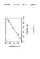
- FIG. 3shows the inhibition of isocitrate dehydrogenase by lead in various buffer solutions.
- FIG. 4illustrates the linear amperometric response to mercury ion using a colloidal gold/ADH bioelectrode prepared by deposition on glassy carbon.
- FIG. 5shows the effect of temperature on inhibition of isocitrate dehydrogenase.
- FIG. 6shows the effect of pH on inhibition of ICDH at various lead ion concentrations; at pH 9.0; at pH 8.5; at pH 8.0; and at pH 7.5.
- the present inventionconcerns approaches to and methods for determining lead ion levels in whole blood.
- Several enzymesare known to be specifically inhibited by heavy metal ions. Isocitrate dehydrogenase for example is inhibited by lead.
- whole blood samples containing leaddo not detectably inhibit isocitrate dehydrogenase activity as determined amperometrically. This is likely due to binding of lead by blood proteins such as albumin or other proteins, although it is also possible that the enzyme is stabilized by an unidentified blood component.
- Anticoagulantsare typically added to blood samples. Although there are numerous compounds with anticoagulant effect, including citrate, the most commonly used are heparin and EDTA. EDTA is generally the anticoagulant of choice in collection of blood samples for clinical analysis. Any lead ion in such treated blood samples will be complexed by EDTA. The challenge therefore was to develop a method to release lead from EDTA and convert it to a form readily detected by an enzyme sensor without inactivating the enzyme or significantly reducing its selectivity and sensitivity.
- the inventorshave explored two methods to generate release of lead ion from the Pb 2+ EDTA complex.
- the first methodis a displacement of lead by cobalt(II).
- Co 2+ EDTAis less stable than Pb 2+ EDTA.
- the conditional stability constant for Co 2+ EDTA at pH 8.5is 10 14 .65.
- the stability constant for Pb 2+ EDTAis 10 15 .11.
- Co 2+ concentrationmust exceed EDTA concentration by at least 10%
- EGTAethylene glycol-bis-( ⁇ -aminoethylether)-N,N,N,N'-tetraacetic acid
- MEDTA1-methylethylenediaminetetraacetic acid
- DMSAmeso 2,3-dimercaptosuccinic acid
- cysteineand penicillamine. While this list is only partial, it will be evident to those skilled in the art that other compounds, not necessarily structurally related, will also be suitable, provided the stated requirements are met.
- the percent conversion of lead from the EDTA complex to a complex with one or the other of the listed or other suitable compoundsis independent of the lead concentration in the sample. Additionally, the concentrations of Co 2+ , X and EDTA, or the ratios of these substances, do not have to be constant (i.e., from sample to sample) or strictly controlled in order for this approach to be successful, as long as the conditions discussed are fulfilled.
- Any agent used for displacing lead from the EDTA complexmust not interfere with the function or activity of the enzyme employed for detection.
- the inventorshave tested DMSA, o-phenanthroline, dipyridyl and acetate and found that for isocitrate dehydrogenase the activity, stability and lead inhibition are not adversely affected.
- Other auxiliary complexing agentsmay also be useful, including iminodiacetate, citrate and lithium ion. Li +1 is known to weaken lead ion binding to bovine serum albumin and is contemplated to be effective in displacing Pb 2+ from the complex.
- R edrepresents the reduced form of an oxidizing agent, Ox.
- OxExcess Ox is then decomposed, employing agents that do not interfere with the enzyme assay. Suitable agents include hydrogen peroxide whose excess can be decomposed by oxidase or catalytically by iron(III). Hypochlorite or ferricyanide may also be employed.
- EDTA complexesIn a practical sense, one need not be limited to EDTA complexes. Without EDTA added to samples, for example, an "auxiliary" complexing agent such as pyrocatechol violet may be added directly to a blood sample and the Pb 2+ complex measured directly. A freshly drawn blood sample, to prevent coagulation, may be treated with an anticoagulant that binds lead, citrate for example. It is possible that lead complexed with citrate will interact directly with isocitrate dehydrogenase to inhibit catalysis.
- auxiliary complexing agentsuch as pyrocatechol violet
- Electrodes prepared from enzymes sensitive to very low lead levelsgenerate detectable oxidation currents that show a linear decrease in current in the presence of increasing levels of metals to which they are sensitive.
- the general oxidation/reduction schemeis shown in FIG. 1.
- a typical mediatoris N-methylphenazine methosulfate which operates at 0 V relative to the Ag/AgCl couple.
- Pig heart NADP-linked isocitric dehydrogenase(ICD, EC 1.1.1.42), an oxidoreductase, is sensitive to trace levels of lead ion as low as 1 ⁇ g/dl.
- ICDis inactive or only slightly active unless Mg ++ or Mn ++ is present.
- activatore.g., ⁇ 200 ppb Mn ++
- the activity of ICDis proportional to Mn ++ and may be used to determine activator concentration (Guilbault, 1970). Inactivation by inhibitors is useful for determining inhibitor concentration.
- inhibitor concentrationmay be selectively determined.
- Silver and mercuryhave been determined in the presence of each other using isocitrate dehydrogenase (Mealor and Townshend, 1968). However, until now, there has been no satisfactory rapid amperometric method to determine lead ion based on ICD inhibition, much less a simple method to determine low lead ion concentrations in whole blood.
- a bioelectroderefers to a single electrode, the working electrode, at the surface of which an electron transfer takes place representing a reaction catalyzed by an enzyme located on or near the surface of the electrode.
- a bioelectrodewhen set up with an appropriate reference/counter electrode may constitute a biosensor.
- a biosensoris intended to indicate a system capable of producing a signal that may be related to a reaction catalyzed by an enzyme constituting the biosensor.
- Biosensors comprising bioelectrodeswill operate by producing a current related to the activity of an enzyme catalyzing electron transfer.
- Electrodeposition of a lead-detecting enzyme on or near an electrode surfacemay be accomplished in several ways, including electrodeposition, evaporation, screen printing, spray deposition (e.g., aerosol), or electrolyte deposition.
- Electrodepositionmay be accomplished by setting a working electrode at an appropriate potential, for example 1.6 v vs. a Ag/AgCl reference electrode with a platinum wire counter electrode. Using a two electrode system with a glassy carbon disk electrode held at a fixed position in a cavity in a lucite block, or other suitable material, a platinum plate at the bottom of the cavity serves as a reference/counter electrode. Electrodeposition may be performed at constant current or constant potential and optimized for the enzyme to be deposited.
- Solvent evaporationis preferred for ease and convenience. The method is simply performed by applying a fixed amount of enzyme to the electrode surface and then drying at room temperature or near 4° C.
- Isocitrate dehydrogenaseunder specific conditions, may be employed in an assay to quantitatively determine lead ion in the range 0-30 ⁇ g per deciliter of solution.
- Assay sensitivitycan be adjusted to a desired concentration range for lead by altering one or more of several parameters such as the concentration of isocitric dehydrogenase. Lower enzyme concentrations increase sensitivity to lead ion.
- isocitrate dehydrogenaseis affected by the pH of the medium. At pH 7.5, for example, very little if any inhibition is observed at lead concentrations up to about 28 mg per deciliter. Raising the pH to 8 results in about 60% enzyme inhibition in this concentration range. The inhibition at pH 8.5 is about 90%, while raising the pH to 9.0 results in complete inhibition between 0 and 5.5 mg per deciliter lead concentration.
- Enzyme sensitivityis also affected by the concentration of Mn +2 which is a cofactor for isocitrate dehydrogenase catalyzed reaction.
- Mn +2is a cofactor for isocitrate dehydrogenase catalyzed reaction.
- Lower cofactor concentrations to which the enzyme is exposed prior to incubation with the leadincrease isocitric dehydrogenase sensitivity.
- Higher sensitivity to lead ion concentrationalso results when Mn +2 concentration is lower during the incubation process.
- Mn +2 concentrationsare effective only in a relatively narrow range on the scale of several micromolar so that manipulation of concentration is limited.
- Temperaturealso affects the inhibition reaction. Higher temperature increases inhibition, but also increases sensitivity. Temperatures in the range of 18° C. to 37° C. typically increase inhibition rates from 25% to about 85% at the higher temperature.
- Mn 2+is generally employed as cofactor of isocitric dehydrogenase in assay reactions; however, Mg +2 is equally effective and can be used in millimolar concentrations as a cofactor, a distinct advantage over Mn 2+ which is effective only within relatively narrow micromolar concentration ranges.
- Mg +2is preferable to Mn 2+ in not binding to other species, a disadvantage frequently encountered with Mn 2+ .
- Mg 2+is effective over a relatively broad concentration range without adversely affecting enzyme properties.
- the inventorshave found that lead ion inhibition of isocitric dehydrogenase may be measured in the presence of some ligands that bind lead ion, including acetate, dipyridyl, o-phenanthroline and the like.
- some blood proteinssuch as albumins and strong chelating agents (e.g., EDTA) adversely affect lead ion inhibition of isocitric dehydrogenase depending on the amount of excess of these agents over the lead ion concentration.
- the inventorshave employed strong chelating reagents to sequester lead ion from interfering blood proteins subsequent to release of the lead from the selected chelating reagent.
- a Pine Instrument dual potentiostat interfaced to an IBM-386 computerwas for enzyme electrode measurement.
- the systemis controlled with an ASYST program (J. Zhao, Enzyme Technology Research Group, Inc., 710 West Main Street, Durham, N.C. 27701).
- Cyclic voltametry measurementswere used to determine amounts of immobilized mediator. Cyclic voltammograms were obtained in the quiescent state. In steady state amperometry experiments the potential was set at 0 V/Ag in stirred buffer with regular sized cell or in quiescent solution with a micro cell and the steady state current was measured. A fixed potential method or chronoamperometric method was used to determine enzyme inhibition.
- the working electrodewas held at a fixed potential while current versus time data were collected with the aid of a computer until steady state was reached. This was observed either from a real-time graphic display and/or the numeric display on the computer screen.
- the computerwas set to automatically provide a calibration curve of percentage inhibition vs. inhibitor concentration, heterogeneous binding constants for reversible inhibitors, and/or binding rate constants for irreversible inhibitors. Programs were modified as required.
- Enzymeswere purchased as indicated and used directly. Results were improved in some cases after the stock enzyme was purified by dialysis. Isocitrate dehydrogenase (Sigma, St. Louis, Mo., ICD Type VI) was dialyzed against buffer containing buffer and manganese ion. If extensive dialysis was performed, substantial enzyme activity was lost; therefore, manganese ion was added to the dialysis buffer.
- the following exampleillustrates determination of trace amounts of lead ion in aqueous medium through the inhibition of isocitrate dehydrogenase (ICDH) in homogeneous solution.
- ICDHisocitrate dehydrogenase
- ICDHCommercially available ICDH (Sigma Chemical Company, St. Louis, Mo.) typically contains considerable amounts of sulfate that interferes with lead ion inhibition of ICDH. Dialysis of ICDH against low ionic strength phosphate buffer caused loss of its activity, which was restored by addition of Mn 2+ . Dialysis against Tris buffer containing a low concentration of Mn 2+ did not affect ICDH activity.
- Buffer selectionwas important because of potential interactions of the buffer with lead ion. Tris buffer did not cause interference, while carbonate or phosphate buffers were unsatisfactory because of lead ion interactions.
- NADPHwas directly oxidized at high potentials (>0.7 V vs. Ag/AgCl) on carbon electrode. However, at this high potential the background current was high. Additionally, the electrode surface was fouled, presumably due to polymerization during the oxidation process. Direct oxidation of NADPH produced a background current of approximately 900 nA while the total current with isocitrate was only about 1600 nA.
- Ferricyanideproduced a relatively high background signal because of operation at potentials >0.2 V vs. Ag/AgCl.
- N-methylphenazine methosulfateproduced a background signal of about 20 Na without isocitrate while the total signal with isocitrate was more than 500 nA at 0 V vs. Ag/AgCl.
- a glassy carbon rod of 3 mm diameterwas wrapped in teflon tubing as working electrode and surrounded with a layer of Pt foil as the counter electrode with a silver wire placed in between as the reference electrode. At least one layer of teflon was inserted between two of the three electrodes. All three electrode surfaces were on the same plane. Tubing was fixed on the top of the coplanar electrode surface with an O-ring, forming a microcell of 100-200 ⁇ l in volume. Microcell configuration is shown in FIG. 2.
- microcellTo the microcell was added in sequence: 100 ⁇ l 50 mM pH 8.5 Tris buffer, ca. 0.3-0.4 units ICDH and, after 15 min, 5 ⁇ l 25 mM NMP-MS, 5 ⁇ l 60 mM NADP. The background current was measured at 0 V. 10 ⁇ l of 0.5M isocitrate was added and the current measured again. The difference in the two signals was taken as due to the oxidation of isocitrate catalyzed by ICDH.
- FIG. 3indicates the sensitivity of the electrode to lead ion concentrations in the submicromolar range.
- the following exampleillustrates a typical preparation of an active enzyme adsorbed to colloidal gold.
- Such enzymesmay be used to prepare bioelectrodes, generally by evaporative or electrodeposition of the enzyme/colloidal gold solution onto a suitable electrode surface.
- Colloidal gold solutionswere prepared by adding a solution of 1% aqueous sodium citrate to a boiling rapidly stirred solution of gold trichloride and refluxing for 30 min. Final concentrations (w/w) were 0.01% HAuCl 4 and 0.03% sodium citrate.
- the particle sizewas estimated by filtration of the sol through polycarbonate membranes (Nuclepore Corporation, Pleasanton, Calif.) of varying pore size using an Amicon micro ultrafiltration unit. Approximately 40% of the sol passed through a 500 A Nucleopore filter and was quantitatively collected on a 300 A Nucleopore filter.
- the gold solwas concentrated by centrifugation at room temperature.
- the concentrated solwas mixed with appropriate amounts of dialyzed isocitrate dehydrogenase solutions.
- a fixed amount of the Au-ICDH solwas evaporated on a coplanar carbon electrode surface and the activity measured.
- the ICDH concentration profile in the Au-ICDH solwas constructed vs. the measured immobilized activity to determine the optimum composition of the Au-ICDH sol.
- the electrode surfaceAfter evaporation of ICDH-Au sol onto a carbon electrode surface, the electrode surface exhibited a yellow-gold appearance which was not washed off. The electrode surface was briefly rinsed with water to remove any loosely bound material before measurements were made. Buffer solution with NMP-MS and NADP was added to the microcell. Background current was measured, then isocitrate added and current again measured. Typical background and sample signals were 25 and 550 nA respectively.
- FIG. 2The basic operational principle for ICDH is shown in FIG. 2.
- An electron transfer mediator for efficient charge coupling with the electrode surfaceis required.
- the generated oxidation current signalsare directly proportional to the total amount of enzyme immobilized on the electrode surface.
- a mediatorcarries electrons between the enzyme(cofactor) and the electrode surface. Substrate is consumed with the production of a catalytic current.
- the following exampleillustrates several methods contemplated for the detection of lead in whole blood using the bioelectrode of Example 2.
- a bare coplanar carbon electrodewas used to determine whether a current could be generated.
- a microcell containing 0.1 ml Tris buffer with appropriate amounts of ICDH, NMP-MS, NADP and isocitratethe electrode produced an oxidation current at 0 V relative to Ag/AgCl.
- the interferencearises from the low usable concentrations of ICD, NADP and NMP-MS. When diffusion processes are slow because of solution viscosity, the generated electric signal is reduced. If all key elements required for signal generation are immobilized on or near the electrode surface, long range diffusion or mass transfer is no longer necessary for signal generation and interference is eliminated.
- a mediator in the form of an insoluble conducting salt NMP-TCNQ, NADP and ICDare co-immobilized at the electrode surface. Only enzyme substrate, isocitrate, is then required for signal generation and this is added in excess to overcome diffusion limitation.
- Lead inhibition and signal measurementwill be separately performed.
- the bioelectrodeis first treated with a blood sample containing lead for a fixed amount of time during which lead ion will inactivate the enzyme.
- the bloodis rinsed off and signal measurement quickly determined. Rinsing will remove the blood and reduce blood interference without altering lead ion inhibition.
- the two-step methodwill eliminate any blood interference and will remove any potentially interfering species in the blood sample that are electrochemically active.
- the blood sampleis treated before measurement.
- Several appropriate methods of treatmentare contemplated.
- the blood samplewill be diluted with a buffer containing a detergent such as SDS to hemolyze the blood, or, treated with a lead complexing agent. Appropriate dilution of the sample will reduce blood interference to a tolerable level and may facilitate the inhibition process.
- Lead in bloodwill be extracted into an organic solvent, in a manner analogous to that routinely used in the atomic absorption method of lead determination.
- inhibitionis performed directly in the organic solvent, provided that the solvent selected is one in which the immobilized enzyme is stable.
- the enzyme electrodeis treated so that there is a stationary aqueous layer on the electrode surface allowing lead to partition from the organic phase to the thin aqueous layer where it will inhibit the immobilized enzyme.
- the electrode surfaceis coated with a thin hydrogel layer above the immobilized enzyme, the gel being wetted prior to application of the organic solvent into which the lead ion has been extracted.
- Yet another optionis to extract the organic phase containing the lead into the aqueous phase prior to inhibition measurements.
- This exampleillustrates the co-immobilization of ICDH and a mediator on the electrode surface.
- a glassy carbon electrodewas first coated with NMP-TCNQ-PVC paste and dried. Then ICDH-Au sol was evaporated onto the coating surface. Then Tris buffer with NADP was added and background current of 5 nA measured at 0 V. The signal increased to 231 nA on addition of isocitrate. This indicated that both ICDH and mediator could be immobilized on the electrode surface.
- NADPwas added to the NMP-TCNQ-PVC paste before coating onto a glassy carbon electrode surface.
- ICDH-Au solwas then evaporated onto the coating.
- the formed electrodecontained the key elements for signal generation, except the substrate, isocitrate.
- the following exampleillustrates a bioelectrode sensitive to low levels of mercury ion.
- a bioelectrodewas prepared from colloidal gold adsorbed alcohol dehydrogenase according the procedure of Example 2 for ICD. Measurements were made in the microcell as described in Example 1 using various concentrations of mercury. The linear portion of the curve is shown in FIG. 4, indicating a linear response in at nanomolar levels of mercury ion. The inhibition was irreversible and was specific for mercury in the presence of added lead ion.
- the inventorshave shown that less than 1 ⁇ g/dL of lead in solution may be measured employing the enzyme isocitrate dehydrogenase.
- the measurementsare sensitive to several factors, including pH, enzyme concentration, co-factor concentration and temperature.
- the following exampleillustrates the effect of pH on enzyme inhibition.
- Inhibition of the enzyme isocitrate dehydrogenasewas calculated based on the amperometric signal (I) which is the mediator oxidation current measured with a glassy carbon electrode at an applied potential of +100 mV. Inhibition was determined as (I uninhibited -I inhibited )/I uninhibited . 50 mM Tris buffer at various pH values as shown in Table 2 was used with 1.0 U/mL enzyme in the solution.
- lead chelated with EDTAis exchangeable with cobalt(II).
- lead Pb(II)is displaced from EDTA by cobalt(II) and detected by its inhibition of isocitrate dehydrogenase (ICDH).
- ICDHisocitrate dehydrogenase
Landscapes
- Chemical & Material Sciences (AREA)
- Organic Chemistry (AREA)
- Life Sciences & Earth Sciences (AREA)
- Zoology (AREA)
- Wood Science & Technology (AREA)
- Proteomics, Peptides & Aminoacids (AREA)
- Health & Medical Sciences (AREA)
- Engineering & Computer Science (AREA)
- Microbiology (AREA)
- Biochemistry (AREA)
- Physics & Mathematics (AREA)
- Molecular Biology (AREA)
- Biotechnology (AREA)
- Biophysics (AREA)
- Analytical Chemistry (AREA)
- Immunology (AREA)
- Bioinformatics & Cheminformatics (AREA)
- General Engineering & Computer Science (AREA)
- General Health & Medical Sciences (AREA)
- Genetics & Genomics (AREA)
- Measuring Or Testing Involving Enzymes Or Micro-Organisms (AREA)
- Apparatus Associated With Microorganisms And Enzymes (AREA)
Abstract
Description
Pb.sup.2+ EDTA+Co.sup.2+ ⃡Co.sup.2+ EDTA+Pb.sup.2+ (logK".sub.eq =-0.46)
Pb.sup.2+ EDTA+2CO.sup.2+ +2X⃡Pb.sup.2+ X+CO.sup.2+ EDTA+CO.sup.2+ X
Pb.sup.2+ EDTA+Co.sup.3+ ⃡Co.sup.3+ EDTA+Pb.sup.2+ (K".sub.eq =16.3 at pH 8.5)
Pb.sup.2+ EDTA+Co.sup.2+ +Ox⃡Co.sup.3+ +Co.sup.3+ (or Co.sup.2+ EDTA)+Pb.sup.2+ +Red
isocitrate+NAD+→α-ketoglutarate+NADH+H.sup.+
NADH+2 MED.sub.ox →NAD++2MED.sub.red +H.sup.+
2MED.sub.red -e.sup.- →2MED.sub.ox (oxidation current)
TABLE 1 ______________________________________ [Pb.sup.++ ]/μM 0 1.89 90.9 current/nA 1480 218 26% inhibition 0 85.3 98.2 ______________________________________ K.sub.i = 0.33 μM or 6.83 μg/dl lead
TABLE 2 ______________________________________ INHIBITION OF ISOCITRATE DEHYDROGENASE.sup.1 BY LEAD ION pH 7.5 pH 8.0 pH 8.5 pH 9.0 Lead Lead Lead Lead Pb.sup.+2 Cur- Inhi- Cur- Inhi- Cur- Inhi- Cur- Inhi- μg/ rent bition rent bition rent bition rent bition dL (nA) (%) (nA) (%) (nA) (%) (nA) (%) ______________________________________ 0 751 0 713 0 776 0 832 0 5.5 749 0 673 6 636 18 4 99 11.1 754 0 613 14 522 33 21 98 16.6 760 -1 532 25 406 48 22.1 750 0 439 38 241 69 27.6 761 -1 303 58 99 87 ______________________________________ .sup.1 1.0 U/mL in 50 mM tris buffer
TABLE 3 ______________________________________ Lead Ion Concentration (μM) Inhibition (%) ______________________________________ 0 0 2.4 24 4.8 46 12 71 ______________________________________ .sup.1 CoSO.sub.4 concentration was 1 mM
TABLE 4 ______________________________________ Lead Ion Concentration (μM) Inhibition(%) ______________________________________ 0 0 12 23 36 32 121 47 ______________________________________ .sup.1 Concentration of EDTA was 2 mM; concentration of CoSO.sub.4 was 2. mM
Pb.sup.2+ EDTA+Co.sup.2+ =Co.sup.2+ EDTA+PbX
TABLE 5 ______________________________________ X log"K.sub.PbX logK".sub.CoX Percent Pb released ______________________________________ EGTA 12.07 10.86 58 MEDTA 13.66 11.85 85 DMSA 12.48 10.48 38 Pyrocatechol violet 6.75 3.60 94 Cysteine 9.14 5.93 99 Penicillamine 8.95 6.72 94 ______________________________________
Claims (11)
Priority Applications (2)
| Application Number | Priority Date | Filing Date | Title |
|---|---|---|---|
| US08/073,806US5368707A (en) | 1992-01-15 | 1993-06-07 | Convenient determination of trace lead in whole blood and other fluids |
| US08/316,433US5468366A (en) | 1992-01-15 | 1994-09-30 | Colloidal-gold electrosensor measuring device |
Applications Claiming Priority (2)
| Application Number | Priority Date | Filing Date | Title |
|---|---|---|---|
| US07/821,732US5217594A (en) | 1992-01-15 | 1992-01-15 | Convenient determination of trace lead in whole blood and other fluids |
| US08/073,806US5368707A (en) | 1992-01-15 | 1993-06-07 | Convenient determination of trace lead in whole blood and other fluids |
Related Parent Applications (1)
| Application Number | Title | Priority Date | Filing Date |
|---|---|---|---|
| US07/821,732Continuation-In-PartUS5217594A (en) | 1992-01-15 | 1992-01-15 | Convenient determination of trace lead in whole blood and other fluids |
Related Child Applications (1)
| Application Number | Title | Priority Date | Filing Date |
|---|---|---|---|
| US08/316,433Continuation-In-PartUS5468366A (en) | 1992-01-15 | 1994-09-30 | Colloidal-gold electrosensor measuring device |
Publications (1)
| Publication Number | Publication Date |
|---|---|
| US5368707Atrue US5368707A (en) | 1994-11-29 |
Family
ID=25234164
Family Applications (2)
| Application Number | Title | Priority Date | Filing Date |
|---|---|---|---|
| US07/821,732Expired - LifetimeUS5217594A (en) | 1992-01-15 | 1992-01-15 | Convenient determination of trace lead in whole blood and other fluids |
| US08/073,806Expired - LifetimeUS5368707A (en) | 1992-01-15 | 1993-06-07 | Convenient determination of trace lead in whole blood and other fluids |
Family Applications Before (1)
| Application Number | Title | Priority Date | Filing Date |
|---|---|---|---|
| US07/821,732Expired - LifetimeUS5217594A (en) | 1992-01-15 | 1992-01-15 | Convenient determination of trace lead in whole blood and other fluids |
Country Status (3)
| Country | Link |
|---|---|
| US (2) | US5217594A (en) |
| AU (1) | AU3474193A (en) |
| WO (1) | WO1993014185A1 (en) |
Cited By (23)
| Publication number | Priority date | Publication date | Assignee | Title |
|---|---|---|---|---|
| US5468366A (en)* | 1992-01-15 | 1995-11-21 | Andcare, Inc. | Colloidal-gold electrosensor measuring device |
| US5595635A (en)* | 1995-09-08 | 1997-01-21 | The United States Of America As Represented By The Secretary Of The Navy | Apparatus for measuring lead content of water |
| US5814205A (en)* | 1994-02-11 | 1998-09-29 | Palintest Limited | Detection of lead in blood |
| US5821074A (en)* | 1993-12-21 | 1998-10-13 | Abbott Laboratories | Method and compositions for enhancing aminolevulinic acid dehydratase assay |
| US5925570A (en)* | 1991-12-25 | 1999-07-20 | Iatron Laboratories, Inc. | Method of measuring metals in samples of living body |
| US6391558B1 (en) | 1997-03-18 | 2002-05-21 | Andcare, Inc. | Electrochemical detection of nucleic acid sequences |
| US20020139059A1 (en)* | 2001-03-13 | 2002-10-03 | Zimmerman David L. | Device which secures external walls and components of a room and which has improved drainage capabilities and aesthetics |
| US20030132125A1 (en)* | 2001-11-26 | 2003-07-17 | Ischemia Technologies Inc. | Electrochemical detection of ischemia |
| US20070111202A1 (en)* | 2000-04-14 | 2007-05-17 | Andcare, Inc. | Electrochemical detection of nucleic acid sequences |
| US20090068754A1 (en)* | 2006-10-24 | 2009-03-12 | Bayer Healthcare Llc | Transient Decay Amperometry |
| US20090071846A1 (en)* | 2007-07-19 | 2009-03-19 | Arnulf Staib | Continuous monitor sensor with covalently bound enzyme |
| US20100188069A1 (en)* | 2007-09-18 | 2010-07-29 | Fan Ren | Sensors using high electron mobility transistors |
| US20110068372A1 (en)* | 2007-09-18 | 2011-03-24 | University Of Florida Research Foundation, Inc. | Sensors using high electron mobility transistors |
| US8232105B1 (en) | 2010-09-30 | 2012-07-31 | Magellan Biosciences Point-of-Care, Inc. | Reagents and methods and systems using them |
| US8404100B2 (en) | 2005-09-30 | 2013-03-26 | Bayer Healthcare Llc | Gated voltammetry |
| US8425757B2 (en) | 2005-07-20 | 2013-04-23 | Bayer Healthcare Llc | Gated amperometry |
| WO2014130809A1 (en)* | 2013-02-22 | 2014-08-28 | The Curators Of The University Of Missouri | Immobilized ligands for the removal of metal ions and methods thereof |
| US9080248B2 (en)* | 2007-12-10 | 2015-07-14 | Bayer Healthcare Llc | Method for forming a test sensor |
| US20160097740A1 (en)* | 2014-10-06 | 2016-04-07 | ALVEO Technologies Inc. | System and Method for Detection of Mercury |
| US9410917B2 (en) | 2004-02-06 | 2016-08-09 | Ascensia Diabetes Care Holdings Ag | Method of using a biosensor |
| US9933385B2 (en) | 2007-12-10 | 2018-04-03 | Ascensia Diabetes Care Holdings Ag | Method of using an electrochemical test sensor |
| US10473582B2 (en) | 2014-12-23 | 2019-11-12 | Magellan Diagnostics, Inc. | Combination optical hemoglobin and electrochemical lead assay |
| US11143595B2 (en)* | 2018-04-26 | 2021-10-12 | Arkray, Inc. | Plasma spectroscopy analysis method |
Families Citing this family (34)
| Publication number | Priority date | Publication date | Assignee | Title |
|---|---|---|---|---|
| GB9315183D0 (en)* | 1993-07-22 | 1993-09-08 | British Nuclear Fuels Plc | Biosensors |
| US5373734A (en)* | 1993-09-24 | 1994-12-20 | Fmc Corporation | Method and apparatus for determining the quality of a coating |
| US5427953A (en)* | 1993-11-08 | 1995-06-27 | The Detroit Medical Center | Blood testing method |
| DE4436910A1 (en)* | 1994-10-15 | 1996-04-18 | Behringwerke Ag | Regenerable solid phase for carrying out specific binding reactions |
| DE4442253A1 (en)* | 1994-11-28 | 1996-05-30 | Bayer Corp N D Ges D Staates I | Electrochemical enzyme biosensor |
| US20020098509A1 (en)* | 1995-04-13 | 2002-07-25 | Heike Jurgens | Method for the quantitative determination of reversible acting inhibitors of the oxidoreductases |
| AU6904496A (en)* | 1995-08-22 | 1997-03-19 | Andcare, Inc. | Handheld electromonitor device |
| US8527026B2 (en) | 1997-03-04 | 2013-09-03 | Dexcom, Inc. | Device and method for determining analyte levels |
| US6001067A (en) | 1997-03-04 | 1999-12-14 | Shults; Mark C. | Device and method for determining analyte levels |
| RU2218563C2 (en)* | 1998-02-16 | 2003-12-10 | Вашенков Евгений Георгиевич | Technique and indicator for detection of lead, process of manufacture of indicator |
| ATE287269T1 (en)* | 2001-05-25 | 2005-02-15 | Gorm Danscher | METHOD FOR IMPLANTING HEAVY METAL, SUCH AS A PRECIOUS METAL, E.G. GOLD, AND METAL FOR USE IN IMPLANTATION |
| US20030032874A1 (en) | 2001-07-27 | 2003-02-13 | Dexcom, Inc. | Sensor head for use with implantable devices |
| US7613491B2 (en) | 2002-05-22 | 2009-11-03 | Dexcom, Inc. | Silicone based membranes for use in implantable glucose sensors |
| US7379765B2 (en) | 2003-07-25 | 2008-05-27 | Dexcom, Inc. | Oxygen enhancing membrane systems for implantable devices |
| ATE419536T1 (en)* | 2002-03-05 | 2009-01-15 | Univ Texas | BIOSPECIFIC CONTRAST AGENTS |
| US6811997B2 (en)* | 2002-07-02 | 2004-11-02 | Bechtel Bwxt Idaho, Llc | Method for chromium analysis and speciation |
| US7886590B2 (en)* | 2002-08-08 | 2011-02-15 | Sun Chemical Corporation | Apparatus and method for quantitatively measuring liquid film drying rates on substrates |
| US20100126858A1 (en)* | 2003-01-30 | 2010-05-27 | Tanita Corporation | Chemical sensor type measuring apparatus |
| US7452457B2 (en) | 2003-06-20 | 2008-11-18 | Roche Diagnostics Operations, Inc. | System and method for analyte measurement using dose sufficiency electrodes |
| US7488601B2 (en) | 2003-06-20 | 2009-02-10 | Roche Diagnostic Operations, Inc. | System and method for determining an abused sensor during analyte measurement |
| US7920906B2 (en) | 2005-03-10 | 2011-04-05 | Dexcom, Inc. | System and methods for processing analyte sensor data for sensor calibration |
| US9247900B2 (en) | 2004-07-13 | 2016-02-02 | Dexcom, Inc. | Analyte sensor |
| US7654956B2 (en) | 2004-07-13 | 2010-02-02 | Dexcom, Inc. | Transcutaneous analyte sensor |
| US8744546B2 (en) | 2005-05-05 | 2014-06-03 | Dexcom, Inc. | Cellulosic-based resistance domain for an analyte sensor |
| US8682408B2 (en) | 2008-03-28 | 2014-03-25 | Dexcom, Inc. | Polymer membranes for continuous analyte sensors |
| US8583204B2 (en) | 2008-03-28 | 2013-11-12 | Dexcom, Inc. | Polymer membranes for continuous analyte sensors |
| US11730407B2 (en) | 2008-03-28 | 2023-08-22 | Dexcom, Inc. | Polymer membranes for continuous analyte sensors |
| EP4227675B1 (en) | 2008-09-19 | 2024-12-11 | DexCom, Inc. | Particle-containing membrane and particulate electrode for analyte sensors |
| CN102645479A (en)* | 2012-04-19 | 2012-08-22 | 湖南大学 | Lead ion specific detection sensor and preparation method and using method thereof |
| EP2840143A1 (en)* | 2013-08-20 | 2015-02-25 | Roche Diagniostics GmbH | A method for making a dry sensor element for an enzymatic determination of an analyte in a body fluid and a dry sensor element |
| ES2733706T3 (en)* | 2014-09-15 | 2019-12-02 | Qorvo Us Inc | Mass detection by redox coupling |
| EP3892317B1 (en) | 2014-12-22 | 2025-02-19 | Medicus Engineering ApS | Device for measuring pulse wave and breathing pressure |
| RU2682162C1 (en)* | 2018-08-16 | 2019-03-15 | Федеральное государственное бюджетное образовательное учреждение высшего образования "Санкт-Петербургский государственный университет" (СПбГУ) | Method for determining lead (ii) in water and biological samples |
| US12422396B2 (en)* | 2023-05-25 | 2025-09-23 | Enlisense Llc | Systems and methods for multi-modal and multiplexed electrochemical detection and reporting of environmental contaminants |
Citations (10)
| Publication number | Priority date | Publication date | Assignee | Title |
|---|---|---|---|---|
| US4090926A (en)* | 1974-09-11 | 1978-05-23 | Environmental Sciences, Inc. | Testing product |
| US4374041A (en)* | 1974-03-01 | 1983-02-15 | Environmental Sciences Associates, Inc. | Testing reagent |
| US4581336A (en)* | 1982-04-26 | 1986-04-08 | Uop Inc. | Surface-modified electrodes |
| WO1987007295A1 (en)* | 1986-05-27 | 1987-12-03 | Cambridge Life Sciences Plc | Immobilised enzyme electrodes |
| US4820399A (en)* | 1984-08-31 | 1989-04-11 | Shimadzu Corporation | Enzyme electrodes |
| US4950378A (en)* | 1987-07-17 | 1990-08-21 | Daikin Industries, Ltd. | Biosensor |
| WO1991017259A1 (en)* | 1990-05-01 | 1991-11-14 | The Secretary Of State For Defence In Her Britannic Majesty's Government Of The United Kingdom Of Great Britain And Northern Ireland | Substrate regenerating biosensor |
| US5160418A (en)* | 1988-07-28 | 1992-11-03 | Cambridge Life Sciences Plc | Enzyme electrodes and improvements in the manufacture thereof |
| US5227307A (en)* | 1991-07-26 | 1993-07-13 | Diagnostic Markers, Inc. | Test for the rapid evaluation of ischemic state |
| US5302531A (en)* | 1992-10-22 | 1994-04-12 | Miles Inc. | Composition for the semiquantitative determination of specific gravity of a test sample |
- 1992
- 1992-01-15USUS07/821,732patent/US5217594A/ennot_activeExpired - Lifetime
- 1993
- 1993-01-14AUAU34741/93Apatent/AU3474193A/ennot_activeAbandoned
- 1993-01-14WOPCT/US1993/000354patent/WO1993014185A1/enactiveApplication Filing
- 1993-06-07USUS08/073,806patent/US5368707A/ennot_activeExpired - Lifetime
Patent Citations (11)
| Publication number | Priority date | Publication date | Assignee | Title |
|---|---|---|---|---|
| US4374041A (en)* | 1974-03-01 | 1983-02-15 | Environmental Sciences Associates, Inc. | Testing reagent |
| US4090926A (en)* | 1974-09-11 | 1978-05-23 | Environmental Sciences, Inc. | Testing product |
| US4581336A (en)* | 1982-04-26 | 1986-04-08 | Uop Inc. | Surface-modified electrodes |
| US4820399A (en)* | 1984-08-31 | 1989-04-11 | Shimadzu Corporation | Enzyme electrodes |
| WO1987007295A1 (en)* | 1986-05-27 | 1987-12-03 | Cambridge Life Sciences Plc | Immobilised enzyme electrodes |
| US4970145A (en)* | 1986-05-27 | 1990-11-13 | Cambridge Life Sciences Plc | Immobilized enzyme electrodes |
| US4950378A (en)* | 1987-07-17 | 1990-08-21 | Daikin Industries, Ltd. | Biosensor |
| US5160418A (en)* | 1988-07-28 | 1992-11-03 | Cambridge Life Sciences Plc | Enzyme electrodes and improvements in the manufacture thereof |
| WO1991017259A1 (en)* | 1990-05-01 | 1991-11-14 | The Secretary Of State For Defence In Her Britannic Majesty's Government Of The United Kingdom Of Great Britain And Northern Ireland | Substrate regenerating biosensor |
| US5227307A (en)* | 1991-07-26 | 1993-07-13 | Diagnostic Markers, Inc. | Test for the rapid evaluation of ischemic state |
| US5302531A (en)* | 1992-10-22 | 1994-04-12 | Miles Inc. | Composition for the semiquantitative determination of specific gravity of a test sample |
Non-Patent Citations (58)
| Title |
|---|
| "U.S. CDC Releases Revised Guidelines on Childhood Lead Poisoning-Blood Lead Level of Concern Lowered to ≧10 μg/dl," ILZRO Environmental Update, 1(10):2, 1991. |
| Albery et al., "Inhibited Enzyme Electrodes. Part 3.," Biosensors & Bioelectronics, 5:397-413, 1990, published in Great Britain. |
| Albery et al., Inhibited Enzyme Electrodes. Part 3., Biosensors & Bioelectronics, 5:397 413, 1990, published in Great Britain.* |
| Almestrand et al., "Determination of Lead in Whole Blood with a Simple Flow-Injection System and Computerized Stripping Potentiometry," Analytica Chimica Acta, 209:339-343, 1988, published in The Netherlands. |
| Almestrand et al., Determination of Lead in Whole Blood with a Simple Flow Injection System and Computerized Stripping Potentiometry, Analytica Chimica Acta, 209:339 343, 1988, published in The Netherlands.* |
| Baum & Czok, "Enzymatische Bestimmung von, ionisiertem Magnesium im Plasma," Biochemische Zeitschrift, 332:121-130, 1959. |
| Baum & Czok, Enzymatische Bestimmung von, ionisiertem Magnesium im Plasma, Biochemische Zeitschrift, 332:121 130, 1959.* |
| Botr et al., Synthesis and Inhibitory Activity on Carbonic Anhydrase of Some New Sulpiride Analogues Studied by Means of a New Method, Journal of Medicinal Chemistry, 29:1814 1020, 1986.* |
| Botre et al., "Synthesis and Inhibitory Activity on Carbonic Anhydrase of Some New Sulpiride Analogues Studied by Means of a New Method," Journal of Medicinal Chemistry, 29:1814-1020, 1986. |
| Crumbliss, A. L., Henkens, R. W., Hunter, K., Kitchell, B. S. O Daly, J. P., Stonehuerner, J., and Tubergen, K. R., The Influence of Colloidal Gold Surfaces on Enzyme Activity, ACS North Carolina Divisional Meeting, Sep. 1988, published in USA.* |
| Crumbliss, A. L., Henkens, R. W., Hunter, K., Kitchell, B. S. O'Daly, J. P., Stonehuerner, J., and Tubergen, K. R., "The Influence of Colloidal Gold Surfaces on Enzyme Activity," ACS North Carolina Divisional Meeting, Sep. 1988, published in USA. |
| Crumbliss, A. L., Henkens, R. W., Kitchell, B. S., McLachlan, K. L., O Daly, J. P., Perine, S. C., Stonehuerner, J., Tubergen, K. R., and Zhao, J., The Use of Inorganic Materials to Control or Maintain Immobilized Enzyme Activity, Symposium on opportunities for inorganic chemistry in biotechnology, ACS National Meeting in Boston, Apr. 23, 1990, published in USA.* |
| Crumbliss, A. L., Henkens, R. W., Kitchell, B. S., McLachlan, K. L., O'Daly, J. P., Perine, S. C., Stonehuerner, J., Tubergen, K. R., and Zhao, J., "The Use of Inorganic Materials to Control or Maintain Immobilized Enzyme Activity," Symposium on opportunities for inorganic chemistry in biotechnology, ACS National Meeting in Boston, Apr. 23, 1990, published in USA. |
| Crumbliss, A. L., Henkens, R. W., Kitchell, B. S., Perine, S. C., Stonehuerner, J., and Tubergen, K. R., "Amperometric Glucose Sensor Fabricated from Glucose Oxidase and a Mediator Co-Immobilized on a Colloidal Gold Hydrogel Electrode," ACS North Carolina Divisional Meeting, University of North Carolina at Chapel Hill, Sep. 7-9, 1989, published in USA. |
| Crumbliss, A. L., Henkens, R. W., Kitchell, B. S., Perine, S. C., Stonehuerner, J., and Tubergen, K. R., "Amperometric Glucose Sensor Fabricated from Glucose Oxidase and a Mediator Co-Immobilized on a Colloidal Gold Hydrogel Electrode," Biosensors Technology: Fundamentals and Application, Chapter 13, 1990, published in USA. |
| Crumbliss, A. L., Henkens, R. W., Kitchell, B. S., Perine, S. C., Stonehuerner, J., and Tubergen, K. R., Amperometric Glucose Sensor Fabricated from Glucose Oxidase and a Mediator Co Immobilized on a Colloidal Gold Hydrogel Electrode, ACS North Carolina Divisional Meeting, University of North Carolina at Chapel Hill, Sep. 7 9, 1989, published in USA.* |
| Crumbliss, A. L., Henkens, R. W., Kitchell, B. S., Perine, S. C., Stonehuerner, J., and Tubergen, K. R., Amperometric Glucose Sensor Fabricated from Glucose Oxidase and a Mediator Co Immobilized on a Colloidal Gold Hydrogel Electrode, Biosensors Technology: Fundamentals and Application, Chapter 13, 1990, published in USA.* |
| Crumbliss, A. L., Kitchell, B. S., Perine, S. C., Stonehuerner, J., Tubergen, K. R., Zhao, J., and Henkens, R. W., Catalytic and Electroactivity of Irreversibly Adsorbed Enzymes at Gold Electrode Surfaces, Symposium on Protein Electrochemistry: ACS Southeast Regional Meeting (SERM), Oct. 1989, published in USA.* |
| Fair & Jamieson, "Studies of Protein Adsorption On Polystyrene Latex Surfaces," Journal of Colloid and Interface Science, 77(2):525-534, 1980. |
| Fair & Jamieson, Studies of Protein Adsorption On Polystyrene Latex Surfaces, Journal of Colloid and Interface Science, 77(2):525 534, 1980.* |
| Guilbault et al., "Homovanillic Acid as a Fluorometric Substrate for Oxidative Enzymes," Analytical Chemistry, 40(1):190-196, 1968. |
| Guilbault et al., Homovanillic Acid as a Fluorometric Substrate for Oxidative Enzymes, Analytical Chemistry, 40(1):190 196, 1968.* |
| Guilbault, "Determination of Inhibitors," Enzymatic Methods of Analysis, Pergamon Press, pp. 197-209, 1970, published in Great Britain. |
| Guilbault, Determination of Inhibitors, Enzymatic Methods of Analysis, Pergamon Press, pp. 197 209, 1970, published in Great Britain.* |
| Gunasingham et al., "Performance and Evaluation of a Handheld Electrochemical Monitor for Toxic Metals," Cole-Parmer Instrument Company, Chicago, Ill. |
| Gunasingham et al., Performance and Evaluation of a Handheld Electrochemical Monitor for Toxic Metals, Cole Parmer Instrument Company, Chicago, Ill.* |
| Henkens, R. W., Kitchell, B. S., O Daly, J. P., Perine, S. C., and Crumbliss, A. L., Bioactive Electrodes Using Metallo Proteins Attached to Colloidal Gold, Recl.: Trav. Chim. Pays Bas, 106:298, 1987.* |
| Henkens, R. W., Kitchell, B. S., O'Daly, J. P., Perine, S. C., and Crumbliss, A. L., "Bioactive Electrodes Using Metallo Proteins Attached to Colloidal Gold," Recl.: Trav. Chim. Pays Bas, 106:298, 1987. |
| Henkens, R. W., Zhao, J., and O Daly, J. P., Multi Analyte Enzyme Electrodes for Environmental Monitoring, Proceedings of 5th International Biotechnology Conference in Copenhagen, Jul. 8 13, 1990.* |
| Henkens, R. W., Zhao, J., and O'Daly, J. P., "Multi-Analyte Enzyme Electrodes for Environmental Monitoring," Proceedings of 5th International Biotechnology Conference in Copenhagen, Jul. 8-13, 1990. |
| Holleck, "The Reduction of Chlorine on Carbon in AlCl3 -KCl, NaCl Melts," Journal of the Electrochemical Society, 119(9):1158-1161. |
| Holleck, The Reduction of Chlorine on Carbon in AlCl 3 KCl, NaCl Melts, Journal of the Electrochemical Society, 119(9):1158 1161.* |
| Kamata & Onoyama, "Lead-Selective Membrane Electrode Using Methylene Bis(diisobutyldithiocarbamate) Neutral Carrier," Analytical Chemistry, 63:1295-1298, 1991. |
| Kamata & Onoyama, Lead Selective Membrane Electrode Using Methylene Bis(diisobutyldithiocarbamate) Neutral Carrier, Analytical Chemistry, 63:1295 1298, 1991.* |
| Kratochvil et al., "Effect of Metals on the Activation and Inhibition of Isocitric Dehydrogenase," Analytical Chemistry, 39(1):45-51, 1967. |
| Kratochvil et al., Effect of Metals on the Activation and Inhibition of Isocitric Dehydrogenase, Analytical Chemistry, 39(1):45 51, 1967.* |
| Linde, "Estimation of Small Amounts of Fluoride in Body Fluids," Analytical Chemistry, 31(12):2092-2094, 1959. |
| Linde, Estimation of Small Amounts of Fluoride in Body Fluids, Analytical Chemistry, 31(12):2092 2094, 1959.* |
| Morrissey & Han, "The Conformation of γ-Globulin Adsorbed on Polystyrene Latices Determined by Quasielastic Light Scattering," Journal of Colloid and Interface Science, 65(3):423-431, 1978. |
| Morrissey & Han, The Conformation of Globulin Adsorbed on Polystyrene Latices Determined by Quasielastic Light Scattering, Journal of Colloid and Interface Science, 65(3):423 431, 1978.* |
| New Rules Set for Blood Lead Levels, Chemical and Engineering News, p. 17, Oct. 14, 1991.* |
| PCT Search Report maield Apr. 5, 1993.* |
| Sakai et al., "Determination of Heavy Metal Ions by Urea Sensor Using ISFET," Sensors and Materials, 2(4):217-227, 1991. |
| Sakai et al., Determination of Heavy Metal Ions by Urea Sensor Using ISFET, Sensors and Materials, 2(4):217 227, 1991.* |
| Sheikh & Townshend, "Applications of Enzyme-Catalysed Reactions in Trace Analysis-VII," Talanta, 21:401-409, 1974 published in Great Britain. |
| Sheikh & Townshend, Applications of Enzyme Catalysed Reactions in Trace Analysis VII, Talanta, 21:401 409, 1974 published in Great Britain.* |
| Sheikh, R. A., "The Determination of Nanogram Amounts of Indium, Lead and Drugs of Forensic Importance by Enzymic Inhibition," Proc. Soc. Analyt. Chem., 10(11):263-286, 1973. |
| Sheikh, R. A., The Determination of Nanogram Amounts of Indium, Lead and Drugs of Forensic Importance by Enzymic Inhibition, Proc. Soc. Analyt. Chem., 10(11):263 286, 1973.* |
| Smit & Cass, "Cyanide Detection Using a Substrate-Regenerating, Peroxidase-Based Biosensor," Analytical Chemistry, 62:2429-2436, 1990. |
| Smit & Cass, Cyanide Detection Using a Substrate Regenerating, Peroxidase Based Biosensor, Analytical Chemistry, 62:2429 2436, 1990.* |
| Smith, "Air Pollution and Forest Damage," Chemical and Engineering News, pp. 30-42, Nov. 11, 1991. |
| Smith, Air Pollution and Forest Damage, Chemical and Engineering News, pp. 30 42, Nov. 11, 1991.* |
| Toren & Burger, "Trace Determination of Metal Ion Inhibitors of the Glucose-Glucose Oxidase System," Mikrochimica Acta (Wien), pp. 538-545, 1968. |
| Toren & Burger, Trace Determination of Metal Ion Inhibitors of the Glucose Glucose Oxidase System, Mikrochimica Acta ( Wien ), pp. 538 545, 1968.* |
| Trade Brochure: esa, Inc. Trace Metal Analyzer Brochure, Bedford, Mass., 1990.* |
| Tran Minh et al., Studies on Acetylcholine Sensor and its Analytical Application Based on the Inhibition of Cholinesterase, Biosensors & Bioelectronics, 5:461 471, 1990.* |
| Tran-Minh et al., "Studies on Acetylcholine Sensor and its Analytical Application Based on the Inhibition of Cholinesterase," Biosensors & Bioelectronics, 5:461-471, 1990. |
| U.S. CDC Releases Revised Guidelines on Childhood Lead Poisoning Blood Lead Level of Concern Lowered to 10 g/dl, ILZRO Environmental Update, 1(10):2, 1991.* |
Cited By (47)
| Publication number | Priority date | Publication date | Assignee | Title |
|---|---|---|---|---|
| US5925570A (en)* | 1991-12-25 | 1999-07-20 | Iatron Laboratories, Inc. | Method of measuring metals in samples of living body |
| US5468366A (en)* | 1992-01-15 | 1995-11-21 | Andcare, Inc. | Colloidal-gold electrosensor measuring device |
| US5821074A (en)* | 1993-12-21 | 1998-10-13 | Abbott Laboratories | Method and compositions for enhancing aminolevulinic acid dehydratase assay |
| US5814205A (en)* | 1994-02-11 | 1998-09-29 | Palintest Limited | Detection of lead in blood |
| US5595635A (en)* | 1995-09-08 | 1997-01-21 | The United States Of America As Represented By The Secretary Of The Navy | Apparatus for measuring lead content of water |
| US6391558B1 (en) | 1997-03-18 | 2002-05-21 | Andcare, Inc. | Electrochemical detection of nucleic acid sequences |
| US20070111202A1 (en)* | 2000-04-14 | 2007-05-17 | Andcare, Inc. | Electrochemical detection of nucleic acid sequences |
| US7455975B2 (en) | 2000-04-14 | 2008-11-25 | Esa Biosciences, Inc. | Electrochemical detection of nucleic acid sequences |
| US20020139059A1 (en)* | 2001-03-13 | 2002-10-03 | Zimmerman David L. | Device which secures external walls and components of a room and which has improved drainage capabilities and aesthetics |
| US20030132125A1 (en)* | 2001-11-26 | 2003-07-17 | Ischemia Technologies Inc. | Electrochemical detection of ischemia |
| US7063782B2 (en) | 2001-11-26 | 2006-06-20 | Ischemia Technologies, Inc. | Electrochemical detection of ischemia |
| US20060191788A1 (en)* | 2001-11-26 | 2006-08-31 | Ischemia Technologies, Inc. | Electrochemical detection of ischemia |
| US9410917B2 (en) | 2004-02-06 | 2016-08-09 | Ascensia Diabetes Care Holdings Ag | Method of using a biosensor |
| US10067082B2 (en) | 2004-02-06 | 2018-09-04 | Ascensia Diabetes Care Holdings Ag | Biosensor for determining an analyte concentration |
| US8425757B2 (en) | 2005-07-20 | 2013-04-23 | Bayer Healthcare Llc | Gated amperometry |
| US8877035B2 (en) | 2005-07-20 | 2014-11-04 | Bayer Healthcare Llc | Gated amperometry methods |
| US8647489B2 (en) | 2005-09-30 | 2014-02-11 | Bayer Healthcare Llc | Gated voltammetry devices |
| US11435312B2 (en) | 2005-09-30 | 2022-09-06 | Ascensia Diabetes Care Holdings Ag | Devices using gated voltammetry methods |
| US10670553B2 (en) | 2005-09-30 | 2020-06-02 | Ascensia Diabetes Care Holdings Ag | Devices using gated voltammetry methods |
| US9835582B2 (en) | 2005-09-30 | 2017-12-05 | Ascensia Diabetes Care Holdings Ag | Devices using gated voltammetry methods |
| US9110013B2 (en) | 2005-09-30 | 2015-08-18 | Bayer Healthcare Llc | Gated voltammetry methods |
| US8404100B2 (en) | 2005-09-30 | 2013-03-26 | Bayer Healthcare Llc | Gated voltammetry |
| US8470604B2 (en) | 2006-10-24 | 2013-06-25 | Bayer Healthcare Llc | Transient decay amperometry |
| US11091790B2 (en) | 2006-10-24 | 2021-08-17 | Ascensia Diabetes Care Holdings Ag | Determining analyte concentration from variant concentration distribution in measurable species |
| US10190150B2 (en) | 2006-10-24 | 2019-01-29 | Ascensia Diabetes Care Holdings Ag | Determining analyte concentration from variant concentration distribution in measurable species |
| US8026104B2 (en) | 2006-10-24 | 2011-09-27 | Bayer Healthcare Llc | Transient decay amperometry |
| US9005527B2 (en) | 2006-10-24 | 2015-04-14 | Bayer Healthcare Llc | Transient decay amperometry biosensors |
| US20090068754A1 (en)* | 2006-10-24 | 2009-03-12 | Bayer Healthcare Llc | Transient Decay Amperometry |
| US8871068B2 (en) | 2007-07-19 | 2014-10-28 | Roche Diagnostics Operations, Inc. | Continuous monitor sensor with covalently bound enzyme |
| US20090071846A1 (en)* | 2007-07-19 | 2009-03-19 | Arnulf Staib | Continuous monitor sensor with covalently bound enzyme |
| US20100188069A1 (en)* | 2007-09-18 | 2010-07-29 | Fan Ren | Sensors using high electron mobility transistors |
| US20110068372A1 (en)* | 2007-09-18 | 2011-03-24 | University Of Florida Research Foundation, Inc. | Sensors using high electron mobility transistors |
| US9316637B2 (en) | 2007-09-18 | 2016-04-19 | University Of Florida Research Foundation, Inc. | Sensors using high electron mobility transistors |
| US20110074381A1 (en)* | 2007-09-18 | 2011-03-31 | University Of Florida Research Foundation, Inc. | Sensors using high electron mobility transistors |
| US8835984B2 (en) | 2007-09-18 | 2014-09-16 | University Of Florida Research Foundation, Inc. | Sensors using high electron mobility transistors |
| US8828713B2 (en) | 2007-09-18 | 2014-09-09 | University Of Florida Research Foundation, Inc. | Sensors using high electron mobility transistors |
| US9080248B2 (en)* | 2007-12-10 | 2015-07-14 | Bayer Healthcare Llc | Method for forming a test sensor |
| US9933385B2 (en) | 2007-12-10 | 2018-04-03 | Ascensia Diabetes Care Holdings Ag | Method of using an electrochemical test sensor |
| US10329684B2 (en) | 2007-12-10 | 2019-06-25 | Ascensia Diabetes Care Holdings Ag | Method for forming an optical test sensor |
| US10690614B2 (en) | 2007-12-10 | 2020-06-23 | Ascensia Diabetes Care Holdings Ag | Method of using an electrochemical test sensor |
| US8232105B1 (en) | 2010-09-30 | 2012-07-31 | Magellan Biosciences Point-of-Care, Inc. | Reagents and methods and systems using them |
| WO2014130809A1 (en)* | 2013-02-22 | 2014-08-28 | The Curators Of The University Of Missouri | Immobilized ligands for the removal of metal ions and methods thereof |
| US9921182B2 (en)* | 2014-10-06 | 2018-03-20 | ALVEO Technologies Inc. | System and method for detection of mercury |
| US20160097740A1 (en)* | 2014-10-06 | 2016-04-07 | ALVEO Technologies Inc. | System and Method for Detection of Mercury |
| US10473582B2 (en) | 2014-12-23 | 2019-11-12 | Magellan Diagnostics, Inc. | Combination optical hemoglobin and electrochemical lead assay |
| US11035784B2 (en) | 2014-12-23 | 2021-06-15 | Magellan Diagnostics, Inc. | Methods and systems for optical hemoglobin measurement |
| US11143595B2 (en)* | 2018-04-26 | 2021-10-12 | Arkray, Inc. | Plasma spectroscopy analysis method |
Also Published As
| Publication number | Publication date |
|---|---|
| US5217594A (en) | 1993-06-08 |
| WO1993014185A1 (en) | 1993-07-22 |
| AU3474193A (en) | 1993-08-03 |
Similar Documents
| Publication | Publication Date | Title |
|---|---|---|
| US5368707A (en) | Convenient determination of trace lead in whole blood and other fluids | |
| CA2069946C (en) | Redox mediator reagent and biosensor | |
| Zhao et al. | Direct electron transfer at horseradish peroxidase—colloidal gold modified electrodes | |
| US5334296A (en) | Peroxidase colloidal gold oxidase biosensors for mediatorless glucose determination | |
| US5508171A (en) | Assay method with enzyme electrode system | |
| US5225064A (en) | Peroxidase colloidal gold oxidase biosensors for mediatorless glucose determination | |
| Vidal et al. | A chronoamperometric sensor for hydrogen peroxide based on electron transfer between immobilized horseradish peroxidase on a glassy carbon electrode and a diffusing ferrocene mediator | |
| US5356786A (en) | Interferant eliminating biosensor | |
| US4929545A (en) | Method and reagent for determination of an analyte via enzymatic means using a ferricyanide/ferric compound system | |
| Mizutani et al. | Amperometric determination of pyruvate, phosphate and urea using enzyme electrodes based on pyruvate oxidase-containing poly (vinyl alcohol)/polyion complex-bilayer membrane | |
| Yamamoto et al. | Highly-sensitive flow injection determination of hydrogen peroxide with a peroxidase-immobilized electrode and its application to clinical chemistry | |
| EP0705348A1 (en) | Diagnostic reagent stabilizer | |
| JPH0777511A (en) | Biosensor for gas measurement and preparation thereof | |
| Moore et al. | Enzymically amplified voltammetric sensor for microliter sample volumes of salicylate | |
| Ayenimo et al. | Rapid amperometric detection of trace metals by inhibition of an ultrathin polypyrrole-based glucose biosensor | |
| Boguslavsky et al. | Thin film bienzyme amperometric biosensors based on polymeric redox mediators with electrostatic bipolar protecting layer | |
| JP2009244013A (en) | Biosensor for measuring neutral fat | |
| US5434055A (en) | Compositions useful in anaerobic determination of analytes | |
| Bartlett et al. | Electrochemical immobilization of enzymes. Part VI. Microelectrodes for the detection of L-lactate based on flavocytochrome b 2 immobilized in a poly (phenol) film | |
| AU681848B2 (en) | Peroxidase colloidal gold oxidase biosensors for mediatorless glucose determination | |
| Wang et al. | In situ elimination of metal inhibitory effects using ligand-containing carbon paste enzyme electrodes | |
| JP4000708B2 (en) | Method for measuring substances using enzyme-immobilized electrode | |
| Shul'ga et al. | An alternative microbiosensor for hydrogen peroxide based on an enzyme field effect transistor with a fast response | |
| ERDEM et al. | Incorporation of EDTA for the elimination of metal inhibitory effects in an amperometric biosensor based on mushroom tissue polyphenol oxidase | |
| Markas | Voltammetric and amperometric behaviour of uric acid at bare and surface-modified screen-printed electrodes: studies towards a disposable uric acid sensor |
Legal Events
| Date | Code | Title | Description |
|---|---|---|---|
| STPP | Information on status: patent application and granting procedure in general | Free format text:APPLICATION UNDERGOING PREEXAM PROCESSING | |
| AS | Assignment | Owner name:ANDCARE, INC., NORTH CAROLINA Free format text:ASSIGNMENT OF ASSIGNORS INTEREST;ASSIGNORS:HENKENS, ROBERT O.;ZHAO, ZUNGUO;WOJCIECHOWSKI, MAREK;AND OTHERS;REEL/FRAME:006834/0627 Effective date:19940107 | |
| AS | Assignment | Owner name:ANDCARE, INC., NORTH CAROLINA Free format text:CORRECTED ASSIGNMENT TO CORRECT ASSIGNOR, PREVIOUSLY RECORDED ON REEL 6834, FRAME 0627;ASSIGNORS:HENKENS, ROBERT O.;ZHAO, JUNGUO;WOJCIECHOWSKI, MAREK;AND OTHERS;REEL/FRAME:006911/0640 Effective date:19940107 | |
| FPAY | Fee payment | Year of fee payment:4 | |
| FPAY | Fee payment | Year of fee payment:8 | |
| FPAY | Fee payment | Year of fee payment:12 | |
| AS | Assignment | Owner name:ALDERON BIOSCIENCES, INC.,NORTH CAROLINA Free format text:CHANGE OF NAME;ASSIGNOR:ANDCARE, INC.;REEL/FRAME:019280/0404 Effective date:20010827 Owner name:ALDERON BIOSCIENCES, INC., NORTH CAROLINA Free format text:CHANGE OF NAME;ASSIGNOR:ANDCARE, INC.;REEL/FRAME:019280/0404 Effective date:20010827 | |
| AS | Assignment | Owner name:ESA BIOSCIENCES, INC.,MASSACHUSETTS Free format text:ASSIGNMENT OF ASSIGNORS INTEREST;ASSIGNOR:ALDERON BIOSCIENCES, INC.;REEL/FRAME:019287/0602 Effective date:20070504 Owner name:ESA BIOSCIENCES, INC., MASSACHUSETTS Free format text:ASSIGNMENT OF ASSIGNORS INTEREST;ASSIGNOR:ALDERON BIOSCIENCES, INC.;REEL/FRAME:019287/0602 Effective date:20070504 |