US5329927A - Apparatus and method for locating an interventional medical device with a ultrasound color imaging system - Google Patents
Apparatus and method for locating an interventional medical device with a ultrasound color imaging systemDownload PDFInfo
- Publication number
- US5329927A US5329927AUS08/022,112US2211293AUS5329927AUS 5329927 AUS5329927 AUS 5329927AUS 2211293 AUS2211293 AUS 2211293AUS 5329927 AUS5329927 AUS 5329927A
- Authority
- US
- United States
- Prior art keywords
- frequency
- needle
- imaging system
- viber
- medical device
- Prior art date
- Legal status (The legal status is an assumption and is not a legal conclusion. Google has not performed a legal analysis and makes no representation as to the accuracy of the status listed.)
- Expired - Lifetime
Links
- 238000003384imaging methodMethods0.000titleclaimsdescription53
- 238000000034methodMethods0.000titleclaimsdescription23
- 238000002604ultrasonographyMethods0.000titledescription20
- 230000007246mechanismEffects0.000claimsabstractdescription91
- 230000033001locomotionEffects0.000claimsabstractdescription84
- 238000012285ultrasound imagingMethods0.000claimsabstractdescription20
- 230000010355oscillationEffects0.000claimsabstractdescription15
- 238000001514detection methodMethods0.000claimsabstractdescription10
- 239000000463materialSubstances0.000claimsdescription14
- 238000001574biopsyMethods0.000claimsdescription10
- 230000008859changeEffects0.000claimsdescription6
- 238000005070samplingMethods0.000claimsdescription6
- 238000006073displacement reactionMethods0.000claimsdescription5
- 230000002093peripheral effectEffects0.000claimsdescription4
- 210000000746body regionAnatomy0.000claimsdescription3
- 230000008878couplingEffects0.000claimsdescription3
- 238000010168coupling processMethods0.000claimsdescription3
- 238000005859coupling reactionMethods0.000claimsdescription3
- 230000004044responseEffects0.000claimsdescription3
- 238000011835investigationMethods0.000claims1
- 239000000696magnetic materialSubstances0.000claims1
- 230000010358mechanical oscillationEffects0.000claims1
- 210000004369bloodAnatomy0.000description12
- 239000008280bloodSubstances0.000description12
- 210000001519tissueAnatomy0.000description10
- 239000013598vectorSubstances0.000description10
- 210000000601blood cellAnatomy0.000description8
- 230000005284excitationEffects0.000description8
- 238000005259measurementMethods0.000description8
- 239000000919ceramicSubstances0.000description7
- 230000005540biological transmissionEffects0.000description6
- 239000002184metalSubstances0.000description6
- 229910052751metalInorganic materials0.000description6
- 230000003534oscillatory effectEffects0.000description6
- 230000008901benefitEffects0.000description5
- 230000000694effectsEffects0.000description5
- 230000000737periodic effectEffects0.000description5
- 210000004204blood vesselAnatomy0.000description4
- 238000010586diagramMethods0.000description4
- 238000012800visualizationMethods0.000description4
- 229910001369BrassInorganic materials0.000description3
- 238000013459approachMethods0.000description3
- 239000010951brassSubstances0.000description3
- 239000007787solidSubstances0.000description3
- 239000010935stainless steelSubstances0.000description3
- 229910001220stainless steelInorganic materials0.000description3
- 239000004593EpoxySubstances0.000description2
- PXHVJJICTQNCMI-UHFFFAOYSA-NNickelChemical compound[Ni]PXHVJJICTQNCMI-UHFFFAOYSA-N0.000description2
- 230000003213activating effectEffects0.000description2
- 230000004913activationEffects0.000description2
- 239000013078crystalSubstances0.000description2
- 239000003292glueSubstances0.000description2
- 238000003754machiningMethods0.000description2
- 239000000203mixtureSubstances0.000description2
- 230000001902propagating effectEffects0.000description2
- 230000035945sensitivityEffects0.000description2
- 230000001360synchronised effectEffects0.000description2
- 229910052727yttriumInorganic materials0.000description2
- 229910001017AlpermInorganic materials0.000description1
- 241001631457CannulaSpecies0.000description1
- 206010028980NeoplasmDiseases0.000description1
- HCHKCACWOHOZIP-UHFFFAOYSA-NZincChemical compound[Zn]HCHKCACWOHOZIP-UHFFFAOYSA-N0.000description1
- 229910045601alloyInorganic materials0.000description1
- 239000000956alloySubstances0.000description1
- 238000010420art techniqueMethods0.000description1
- 210000001367arteryAnatomy0.000description1
- JRPBQTZRNDNNOP-UHFFFAOYSA-Nbarium titanateChemical class[Ba+2].[Ba+2].[O-][Ti]([O-])([O-])[O-]JRPBQTZRNDNNOP-UHFFFAOYSA-N0.000description1
- 239000011324beadSubstances0.000description1
- 238000005452bendingMethods0.000description1
- 201000011510cancerDiseases0.000description1
- 229910010293ceramic materialInorganic materials0.000description1
- 239000002131composite materialSubstances0.000description1
- 238000007796conventional methodMethods0.000description1
- 238000005520cutting processMethods0.000description1
- 230000007812deficiencyEffects0.000description1
- 230000026058directional locomotionEffects0.000description1
- 238000009826distributionMethods0.000description1
- 229940079593drugDrugs0.000description1
- 239000003814drugSubstances0.000description1
- 238000005516engineering processMethods0.000description1
- 238000013152interventional procedureMethods0.000description1
- 229910052451lead zirconate titanateInorganic materials0.000description1
- HFGPZNIAWCZYJU-UHFFFAOYSA-Nlead zirconate titanateChemical class[O-2].[O-2].[O-2].[O-2].[O-2].[Ti+4].[Zr+4].[Pb+2]HFGPZNIAWCZYJU-UHFFFAOYSA-N0.000description1
- 239000007788liquidSubstances0.000description1
- 230000004807localizationEffects0.000description1
- 239000007769metal materialSubstances0.000description1
- 230000005012migrationEffects0.000description1
- 238000013508migrationMethods0.000description1
- 238000012544monitoring processMethods0.000description1
- 230000001338necrotic effectEffects0.000description1
- 230000007935neutral effectEffects0.000description1
- 229910052759nickelInorganic materials0.000description1
- ORQBXQOJMQIAOY-UHFFFAOYSA-NnobeliumChemical compound[No]ORQBXQOJMQIAOY-UHFFFAOYSA-N0.000description1
- 230000010363phase shiftEffects0.000description1
- 230000010287polarizationEffects0.000description1
- 238000002360preparation methodMethods0.000description1
- 230000000644propagated effectEffects0.000description1
- 230000009467reductionEffects0.000description1
- 210000004872soft tissueAnatomy0.000description1
- 238000001228spectrumMethods0.000description1
- 230000026683transductionEffects0.000description1
- 238000010361transductionMethods0.000description1
- 238000003466weldingMethods0.000description1
- 229910052725zincInorganic materials0.000description1
- 239000011701zincSubstances0.000description1
Images


Classifications
- A—HUMAN NECESSITIES
- A61—MEDICAL OR VETERINARY SCIENCE; HYGIENE
- A61B—DIAGNOSIS; SURGERY; IDENTIFICATION
- A61B8/00—Diagnosis using ultrasonic, sonic or infrasonic waves
- A61B8/08—Clinical applications
- A61B8/0833—Clinical applications involving detecting or locating foreign bodies or organic structures
- A61B8/0841—Clinical applications involving detecting or locating foreign bodies or organic structures for locating instruments
- A—HUMAN NECESSITIES
- A61—MEDICAL OR VETERINARY SCIENCE; HYGIENE
- A61B—DIAGNOSIS; SURGERY; IDENTIFICATION
- A61B8/00—Diagnosis using ultrasonic, sonic or infrasonic waves
- A61B8/08—Clinical applications
- A61B8/0833—Clinical applications involving detecting or locating foreign bodies or organic structures
Definitions
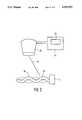
- FIG. 4there is shown three views, namely 4a, 4b and 4c of the VIBER mechanism 40 as shown in FIG. 3, depicting three different modes of oscillations.
- FIG. 4athere is shown a front plan view of the VIBER mechanism 40 including a small replica of an X, Y and Z set of axes in the center of the diagram.
- the left hand bottom of the VIBER mechanism 40is characterized by an extending circular arm 50 having a reduced neck portion 51 which extends to and terminates in the pendulum-like member 45 with the relatively large bottom mass.
- the pendulum analogyis made based on the thinner neck portion with the relatively larger end portion.
- the member 45is of a generally "L" shape cross-section used to reinforce oscillations caused by periodic distortion of the piezocrystal diaphragm 48, as described above.
- FIG. 8athere is shown various alternate devices where a separate sensing element is formed as part of the resonant circuit.
- a sensing piezoelectric elementis formed adjacent to the drive piezoelectric element.
- the sensing element depicted by reference numeral 105is formed by conventional techniques and is located adjacent the driveelement 106.
- the sensing elementis energized by means of these driving voltages and enables a separate output to be provided.
- a separate piezoelectric element or alternatively a separate electrode on the drivingpiezoelectric platewill produce a characteristic change in voltage whose amplitude is related to the amount of motion and is a maximum at resonance.
- the peak in such a signal which is present in the output of thesense piezoelectric element 105can easily be detected utilizing the feedback scheme as shown in FIG. 7. In these cases, the output of the sensing device is applied to the input of the A/D converter.
Landscapes
- Life Sciences & Earth Sciences (AREA)
- Health & Medical Sciences (AREA)
- Biomedical Technology (AREA)
- Biophysics (AREA)
- Nuclear Medicine, Radiotherapy & Molecular Imaging (AREA)
- Pathology (AREA)
- Radiology & Medical Imaging (AREA)
- Engineering & Computer Science (AREA)
- Physics & Mathematics (AREA)
- Heart & Thoracic Surgery (AREA)
- Medical Informatics (AREA)
- Molecular Biology (AREA)
- Surgery (AREA)
- Animal Behavior & Ethology (AREA)
- General Health & Medical Sciences (AREA)
- Public Health (AREA)
- Veterinary Medicine (AREA)
- Ultra Sonic Daignosis Equipment (AREA)
Abstract
Description
Claims (23)
Priority Applications (7)
| Application Number | Priority Date | Filing Date | Title |
|---|---|---|---|
| US08/022,112US5329927A (en) | 1993-02-25 | 1993-02-25 | Apparatus and method for locating an interventional medical device with a ultrasound color imaging system |
| US08/180,126US5343865A (en) | 1993-02-25 | 1994-01-11 | Apparatus and method for locating an interventional medical device with a ultrasound color imaging system |
| JP51915494AJP3537822B2 (en) | 1993-02-25 | 1994-02-22 | Positioning of an interventional medical device by ultrasound |
| DE69430734TDE69430734T2 (en) | 1993-02-25 | 1994-02-22 | DETERMINING THE SITUATION OF A SURGICAL INSTRUMENT USING ULTRASOUND |
| EP94910127AEP0686012B1 (en) | 1993-02-25 | 1994-02-22 | Locating an interventional medical device by ultrasound |
| PCT/US1994/001752WO1994018887A1 (en) | 1993-02-25 | 1994-02-22 | Locating an interventional medical device by ultrasound |
| CA002156831ACA2156831C (en) | 1993-02-25 | 1994-02-22 | Locating an interventional medical device by ultrasound |
Applications Claiming Priority (1)
| Application Number | Priority Date | Filing Date | Title |
|---|---|---|---|
| US08/022,112US5329927A (en) | 1993-02-25 | 1993-02-25 | Apparatus and method for locating an interventional medical device with a ultrasound color imaging system |
Related Child Applications (1)
| Application Number | Title | Priority Date | Filing Date |
|---|---|---|---|
| US08/180,126DivisionUS5343865A (en) | 1993-02-25 | 1994-01-11 | Apparatus and method for locating an interventional medical device with a ultrasound color imaging system |
Publications (1)
| Publication Number | Publication Date |
|---|---|
| US5329927Atrue US5329927A (en) | 1994-07-19 |
Family
ID=21807881
Family Applications (2)
| Application Number | Title | Priority Date | Filing Date |
|---|---|---|---|
| US08/022,112Expired - LifetimeUS5329927A (en) | 1993-02-25 | 1993-02-25 | Apparatus and method for locating an interventional medical device with a ultrasound color imaging system |
| US08/180,126Expired - LifetimeUS5343865A (en) | 1993-02-25 | 1994-01-11 | Apparatus and method for locating an interventional medical device with a ultrasound color imaging system |
Family Applications After (1)
| Application Number | Title | Priority Date | Filing Date |
|---|---|---|---|
| US08/180,126Expired - LifetimeUS5343865A (en) | 1993-02-25 | 1994-01-11 | Apparatus and method for locating an interventional medical device with a ultrasound color imaging system |
Country Status (6)
| Country | Link |
|---|---|
| US (2) | US5329927A (en) |
| EP (1) | EP0686012B1 (en) |
| JP (1) | JP3537822B2 (en) |
| CA (1) | CA2156831C (en) |
| DE (1) | DE69430734T2 (en) |
| WO (1) | WO1994018887A1 (en) |
Cited By (66)
| Publication number | Priority date | Publication date | Assignee | Title |
|---|---|---|---|---|
| US5421336A (en)* | 1994-04-04 | 1995-06-06 | Echo Cath, Inc. | Method for attaching an interventional medical device to a vibratory member associated with visualization by an ultrasound imaging system |
| US5425370A (en)* | 1994-03-23 | 1995-06-20 | Echocath, Inc. | Method and apparatus for locating vibrating devices |
| US5823956A (en)* | 1993-02-22 | 1998-10-20 | Heartport, Inc. | Method and apparatus for thoracoscopic intracardiac procedures |
| US5836882A (en)* | 1997-03-17 | 1998-11-17 | Frazin; Leon J. | Method and apparatus of localizing an insertion end of a probe within a biotic structure |
| US5967991A (en)* | 1996-12-03 | 1999-10-19 | Echocath, Inc. | Drive apparatus for an interventional medical device used in an ultrasonic imaging system |
| US6039693A (en)* | 1991-11-08 | 2000-03-21 | Mayo Foundation For Medical Education And Research | Volumetric image ultrasound transducer underfluid catheter system |
| US6059731A (en)* | 1998-08-19 | 2000-05-09 | Mayo Foundation For Medical Education And Research | Simultaneous side-and-end viewing underfluid catheter |
| EP1001705A4 (en)* | 1997-01-21 | 2000-05-24 | Cook William A Australia | CALIBRATED HOLLOW PROBE FOR ULTRASONIC IMAGING |
| US6171247B1 (en) | 1997-06-13 | 2001-01-09 | Mayo Foundation For Medical Education And Research | Underfluid catheter system and method having a rotatable multiplane transducer |
| US6306096B1 (en) | 1991-11-08 | 2001-10-23 | Mayo Foundation For Medical Education And Research | Volumetric image ultrasound transducer underfluid catheter system |
| US6398736B1 (en) | 1999-03-31 | 2002-06-04 | Mayo Foundation For Medical Education And Research | Parametric imaging ultrasound catheter |
| US6428491B1 (en)* | 1999-08-27 | 2002-08-06 | Dan Weiss | Delivery of ultrasound to percutaneous and intrabody devices |
| US20030004302A1 (en)* | 2001-05-15 | 2003-01-02 | Satoshi Okamoto | Process for producing purified polyether sulfones |
| US6517481B2 (en)* | 1998-12-23 | 2003-02-11 | Radi Medical Systems Ab | Method and sensor for wireless measurement of physiological variables |
| US6651672B2 (en) | 1993-02-22 | 2003-11-25 | Heartport, Inc. | Devices for less-invasive intracardiac interventions |
| US6702761B1 (en) | 2000-03-06 | 2004-03-09 | Fonar Corporation | Vibration assisted needle device |
| US20040230111A1 (en)* | 2003-02-12 | 2004-11-18 | Smith Stephen W. | Methods, devices, systems and computer program products for oscillating shafts using real time 3D ultrasound |
| WO2005046444A2 (en) | 2003-11-11 | 2005-05-26 | Soma Development Llc | Ultrasound guided probe device and method |
| WO2006059966A1 (en)* | 2004-11-30 | 2006-06-08 | Omnisonics Medical Technologies, Inc. | Ultrasonic medical device with variable frequency drive |
| US20080255444A1 (en)* | 2006-10-12 | 2008-10-16 | Geng Li | Novel needle driver for magnetic resonance elastography |
| US20090011032A1 (en)* | 2004-04-16 | 2009-01-08 | Lepivert Patrick | Methods for improved cryo-chemotherapy tissue ablation |
| US7494468B2 (en) | 1999-10-05 | 2009-02-24 | Omnisonics Medical Technologies, Inc. | Ultrasonic medical device operating in a transverse mode |
| WO2009027890A1 (en)* | 2007-08-28 | 2009-03-05 | Koninklijke Philips Electronics N.V. | Dual path color doppler imaging system and method for simultaneous invasive device visualization and vasculature imaging |
| US7503895B2 (en) | 1999-10-05 | 2009-03-17 | Omnisonics Medical Technologies, Inc. | Ultrasonic device for tissue ablation and sheath for use therewith |
| US20090221867A1 (en)* | 2005-11-11 | 2009-09-03 | Ams Research Corporation | Integral Sling Connection System and Method |
| US20100016726A1 (en)* | 2008-07-18 | 2010-01-21 | Meier Joseph H | Handheld Imaging Device And Method For Manufacture Thereof |
| US7794414B2 (en) | 2004-02-09 | 2010-09-14 | Emigrant Bank, N.A. | Apparatus and method for an ultrasonic medical device operating in torsional and transverse modes |
| EP1094752B1 (en)* | 1998-05-14 | 2010-11-17 | David N. Krag | System for bracketing tissue |
| US20100305432A1 (en)* | 2009-05-28 | 2010-12-02 | Edwards Lifesciences Corporation | System and Method for Locating Medical Devices in Vivo Using Ultrasound Doppler Mode |
| US20100312120A1 (en)* | 2008-07-18 | 2010-12-09 | Meier Joseph H | Handheld imaging devices and related methods |
| US20110087106A1 (en)* | 2009-10-09 | 2011-04-14 | Soma Development, Llc | Clamp for a Medical Probe Device |
| US20110112467A1 (en)* | 2004-04-16 | 2011-05-12 | Lepivert Patrick | Systems and methods for improving image-guided tissue ablation |
| WO2012024201A1 (en)* | 2010-08-19 | 2012-02-23 | Mayo Foundation For Medical Education And Research | Steerable catheter navigation with the use of interference ultrasonography |
| US20120130414A1 (en)* | 2010-11-19 | 2012-05-24 | Corlius Fourie Birkill | Vibrating needle adjustment device |
| US20130261436A1 (en)* | 2006-11-02 | 2013-10-03 | Cooltouch Incorporated | Sonic Endovenous Catheter |
| US8761862B2 (en) | 2009-10-09 | 2014-06-24 | Stephen F. Ridley | Ultrasound guided probe device and sterilizable shield for same |
| US20140200511A1 (en)* | 2009-10-30 | 2014-07-17 | Searete Llc | Systems, devices, and methods for making or administering frozen particles |
| US8790359B2 (en) | 1999-10-05 | 2014-07-29 | Cybersonics, Inc. | Medical systems and related methods |
| US20140221820A1 (en)* | 2013-02-05 | 2014-08-07 | Muffin Incorporated | Temporal echogenic markers |
| US20150190660A1 (en)* | 2012-08-02 | 2015-07-09 | Flowcardia, Inc. | Ultrasound catheter system |
| US9138286B2 (en) | 2008-10-20 | 2015-09-22 | Nuvue Therapeutics, Inc. | Ultrasound detectable interventional medical device |
| US9289185B2 (en) | 2012-07-23 | 2016-03-22 | ClariTrac, Inc. | Ultrasound device for needle procedures |
| US9359885B2 (en) | 2013-03-15 | 2016-06-07 | Baxter International Inc. | Acoustic line tracing system and method for fluid transfer system |
| US20160242811A1 (en)* | 2013-03-15 | 2016-08-25 | University Court Of The University Of Dundee | Medical Apparatus and Its Visualisation |
| US10111680B2 (en) | 2002-08-02 | 2018-10-30 | Flowcardia, Inc. | Therapeutic ultrasound system |
| US10130329B2 (en) | 2014-01-28 | 2018-11-20 | General Electric Company | Distinct needle display in ultrasonic image |
| US10130380B2 (en) | 2003-02-26 | 2018-11-20 | Flowcardia, Inc. | Ultrasound catheter apparatus |
| US10157266B2 (en) | 2012-09-06 | 2018-12-18 | Baxter International Inc. | Patient information software system including infusion map |
| GB2566532A (en)* | 2017-09-18 | 2019-03-20 | Active Needle Tech Ltd | Vibrating probe |
| US10285719B2 (en) | 2005-01-20 | 2019-05-14 | Flowcardia, Inc. | Vibrational catheter devices and methods for making same |
| US10285727B2 (en) | 2002-08-26 | 2019-05-14 | Flowcardia, Inc. | Steerable ultrasound catheter |
| US10349964B2 (en) | 2003-09-19 | 2019-07-16 | Flowcardia, Inc. | Connector for securing ultrasound catheter to transducer |
| US10357263B2 (en) | 2012-01-18 | 2019-07-23 | C. R. Bard, Inc. | Vascular re-entry device |
| US10537712B2 (en) | 2006-11-07 | 2020-01-21 | Flowcardia, Inc. | Ultrasound catheter having improved distal end |
| US10582983B2 (en) | 2017-02-06 | 2020-03-10 | C. R. Bard, Inc. | Ultrasonic endovascular catheter with a controllable sheath |
| US10682151B2 (en) | 2004-08-26 | 2020-06-16 | Flowcardia, Inc. | Ultrasound catheter devices and methods |
| US10758256B2 (en) | 2016-12-22 | 2020-09-01 | C. R. Bard, Inc. | Ultrasonic endovascular catheter |
| US10788154B2 (en) | 2012-11-13 | 2020-09-29 | Baxter International Inc. | Infusion line management system |
| US10835267B2 (en) | 2002-08-02 | 2020-11-17 | Flowcardia, Inc. | Ultrasound catheter having protective feature against breakage |
| CN112334076A (en)* | 2018-06-29 | 2021-02-05 | 皇家飞利浦有限公司 | Biopsy prediction and guidance using ultrasound imaging and associated devices, systems and methods |
| US10940292B2 (en) | 2015-07-08 | 2021-03-09 | Actuated Medical, Inc. | Reduced force device for intravascular access and guidewire placement |
| US11123141B2 (en) | 2010-08-19 | 2021-09-21 | Mayo Foundation For Medical Education And Research | Systems and methods for navigating a catheter and delivering a needle |
| US11596726B2 (en) | 2016-12-17 | 2023-03-07 | C.R. Bard, Inc. | Ultrasound devices for removing clots from catheters and related methods |
| US11633206B2 (en) | 2016-11-23 | 2023-04-25 | C.R. Bard, Inc. | Catheter with retractable sheath and methods thereof |
| US11642100B2 (en) | 2018-09-20 | 2023-05-09 | Mayo Foundation For Medical Education And Research | Systems and methods for localizing a medical device using symmetric Doppler frequency shifts measured with ultrasound imaging |
| EP4308023A4 (en)* | 2021-03-16 | 2024-12-11 | Francis Medical, Inc. | Vapor therapy systems and methods |
Families Citing this family (104)
| Publication number | Priority date | Publication date | Assignee | Title |
|---|---|---|---|---|
| US5676694A (en)* | 1996-06-07 | 1997-10-14 | Medtronic, Inc. | Medical electrical lead |
| US5837900A (en)* | 1996-06-21 | 1998-11-17 | Medtronic Inc | System and method for detecting metal ion oxidation in pacing lead |
| US5968085A (en)* | 1998-04-20 | 1999-10-19 | Medtronic, Inc. | Pacing lead with integral guidance using ultrasound |
| US6095981A (en)* | 1998-07-01 | 2000-08-01 | The Regents Of The University Of California | Apparatus for attachment of needle or catheter to endoluminal ultrasound probe |
| US7452331B1 (en) | 1999-04-08 | 2008-11-18 | Rick L Pruter | Vascular adjustable multi-gauge tilt-out method and apparatus for guiding needles |
| US6612990B1 (en) | 1999-04-08 | 2003-09-02 | Rick L. Pruter | Method and apparatus for guiding needles |
| US6515657B1 (en) | 2000-02-11 | 2003-02-04 | Claudio I. Zanelli | Ultrasonic imager |
| US6577904B1 (en) | 2000-03-30 | 2003-06-10 | Cardiac Pacemakers, Inc. | Ultrasound echogenic cardiac lead |
| US6520916B1 (en) | 2000-08-02 | 2003-02-18 | Medtronic, Inc. | Ultrasound imaging system and method for implantable and invasive devices |
| EP1337183B1 (en) | 2000-11-24 | 2005-05-25 | Innovacell Biotechnologie GmbH | Ultrasonic probe comprising a positioning device for examination devices and operation devices |
| DE10105592A1 (en) | 2001-02-06 | 2002-08-08 | Achim Goepferich | Placeholder for drug release in the frontal sinus |
| US8214015B2 (en)* | 2001-02-06 | 2012-07-03 | Medtronic Vascular, Inc. | In vivo localization and tracking of tissue penetrating catheters using magnetic resonance imaging |
| US7344507B2 (en)* | 2002-04-19 | 2008-03-18 | Pelikan Technologies, Inc. | Method and apparatus for lancet actuation |
| GB0129139D0 (en)* | 2001-12-05 | 2002-01-23 | Sra Dev Ltd | Ultrasonic generator system |
| US8353840B1 (en) | 2002-09-11 | 2013-01-15 | Pruter Rick L | Method and disposable apparatus for guiding needles with a double button unlocking and locking mechanism |
| US6758817B1 (en) | 2002-09-11 | 2004-07-06 | Protek Medical Products, Inc. | Method and disposable apparatus for guiding needles |
| US20040068168A1 (en)* | 2002-09-13 | 2004-04-08 | Lala Louis A. | Coordinative resonance detection by a coordinated feedback system |
| US8317816B2 (en) | 2002-09-30 | 2012-11-27 | Acclarent, Inc. | Balloon catheters and methods for treating paranasal sinuses |
| US6884219B1 (en) | 2002-10-17 | 2005-04-26 | Rick L. Pruter | Method and disposable apparatus for guiding needles with an endocavity medical imaging device |
| US7909815B2 (en)* | 2003-05-23 | 2011-03-22 | Civco Medical Instruments Co., Inc. | Instrument guide for use with needles and catheters |
| EP1543854A1 (en)* | 2003-12-16 | 2005-06-22 | Novo Nordisk A/S | Vibrating injection needle and method for detecting the presence of medicament therein |
| US20060081255A1 (en)* | 2004-04-02 | 2006-04-20 | Michael Miller | Ultrasonic placement and monitoring of an endotracheal tube |
| US9089258B2 (en) | 2004-04-21 | 2015-07-28 | Acclarent, Inc. | Endoscopic methods and devices for transnasal procedures |
| US9554691B2 (en) | 2004-04-21 | 2017-01-31 | Acclarent, Inc. | Endoscopic methods and devices for transnasal procedures |
| US8864787B2 (en) | 2004-04-21 | 2014-10-21 | Acclarent, Inc. | Ethmoidotomy system and implantable spacer devices having therapeutic substance delivery capability for treatment of paranasal sinusitis |
| US7361168B2 (en) | 2004-04-21 | 2008-04-22 | Acclarent, Inc. | Implantable device and methods for delivering drugs and other substances to treat sinusitis and other disorders |
| US8764729B2 (en) | 2004-04-21 | 2014-07-01 | Acclarent, Inc. | Frontal sinus spacer |
| US20060063973A1 (en) | 2004-04-21 | 2006-03-23 | Acclarent, Inc. | Methods and apparatus for treating disorders of the ear, nose and throat |
| US8747389B2 (en) | 2004-04-21 | 2014-06-10 | Acclarent, Inc. | Systems for treating disorders of the ear, nose and throat |
| US7654997B2 (en) | 2004-04-21 | 2010-02-02 | Acclarent, Inc. | Devices, systems and methods for diagnosing and treating sinusitus and other disorders of the ears, nose and/or throat |
| US7803150B2 (en) | 2004-04-21 | 2010-09-28 | Acclarent, Inc. | Devices, systems and methods useable for treating sinusitis |
| US10188413B1 (en) | 2004-04-21 | 2019-01-29 | Acclarent, Inc. | Deflectable guide catheters and related methods |
| US7419497B2 (en) | 2004-04-21 | 2008-09-02 | Acclarent, Inc. | Methods for treating ethmoid disease |
| US7559925B2 (en) | 2006-09-15 | 2009-07-14 | Acclarent Inc. | Methods and devices for facilitating visualization in a surgical environment |
| US7410480B2 (en)* | 2004-04-21 | 2008-08-12 | Acclarent, Inc. | Devices and methods for delivering therapeutic substances for the treatment of sinusitis and other disorders |
| US20060004323A1 (en) | 2004-04-21 | 2006-01-05 | Exploramed Nc1, Inc. | Apparatus and methods for dilating and modifying ostia of paranasal sinuses and other intranasal or paranasal structures |
| US9101384B2 (en) | 2004-04-21 | 2015-08-11 | Acclarent, Inc. | Devices, systems and methods for diagnosing and treating sinusitis and other disorders of the ears, Nose and/or throat |
| US20070167682A1 (en) | 2004-04-21 | 2007-07-19 | Acclarent, Inc. | Endoscopic methods and devices for transnasal procedures |
| US8932276B1 (en) | 2004-04-21 | 2015-01-13 | Acclarent, Inc. | Shapeable guide catheters and related methods |
| US8894614B2 (en) | 2004-04-21 | 2014-11-25 | Acclarent, Inc. | Devices, systems and methods useable for treating frontal sinusitis |
| US7720521B2 (en) | 2004-04-21 | 2010-05-18 | Acclarent, Inc. | Methods and devices for performing procedures within the ear, nose, throat and paranasal sinuses |
| US9351750B2 (en) | 2004-04-21 | 2016-05-31 | Acclarent, Inc. | Devices and methods for treating maxillary sinus disease |
| US20070208252A1 (en) | 2004-04-21 | 2007-09-06 | Acclarent, Inc. | Systems and methods for performing image guided procedures within the ear, nose, throat and paranasal sinuses |
| US9399121B2 (en) | 2004-04-21 | 2016-07-26 | Acclarent, Inc. | Systems and methods for transnasal dilation of passageways in the ear, nose or throat |
| US8146400B2 (en) | 2004-04-21 | 2012-04-03 | Acclarent, Inc. | Endoscopic methods and devices for transnasal procedures |
| US20190314620A1 (en) | 2004-04-21 | 2019-10-17 | Acclarent, Inc. | Apparatus and methods for dilating and modifying ostia of paranasal sinuses and other intranasal or paranasal structures |
| US7462175B2 (en) | 2004-04-21 | 2008-12-09 | Acclarent, Inc. | Devices, systems and methods for treating disorders of the ear, nose and throat |
| US8702626B1 (en) | 2004-04-21 | 2014-04-22 | Acclarent, Inc. | Guidewires for performing image guided procedures |
| US8951225B2 (en) | 2005-06-10 | 2015-02-10 | Acclarent, Inc. | Catheters with non-removable guide members useable for treatment of sinusitis |
| US8784336B2 (en) | 2005-08-24 | 2014-07-22 | C. R. Bard, Inc. | Stylet apparatuses and methods of manufacture |
| US8114113B2 (en) | 2005-09-23 | 2012-02-14 | Acclarent, Inc. | Multi-conduit balloon catheter |
| US8303505B2 (en) | 2005-12-02 | 2012-11-06 | Abbott Cardiovascular Systems Inc. | Methods and apparatuses for image guided medical procedures |
| US8190389B2 (en) | 2006-05-17 | 2012-05-29 | Acclarent, Inc. | Adapter for attaching electromagnetic image guidance components to a medical device |
| US9820688B2 (en) | 2006-09-15 | 2017-11-21 | Acclarent, Inc. | Sinus illumination lightwire device |
| US8388546B2 (en) | 2006-10-23 | 2013-03-05 | Bard Access Systems, Inc. | Method of locating the tip of a central venous catheter |
| US7794407B2 (en) | 2006-10-23 | 2010-09-14 | Bard Access Systems, Inc. | Method of locating the tip of a central venous catheter |
| US8439687B1 (en) | 2006-12-29 | 2013-05-14 | Acclarent, Inc. | Apparatus and method for simulated insertion and positioning of guidewares and other interventional devices |
| US8118757B2 (en) | 2007-04-30 | 2012-02-21 | Acclarent, Inc. | Methods and devices for ostium measurement |
| US8485199B2 (en) | 2007-05-08 | 2013-07-16 | Acclarent, Inc. | Methods and devices for protecting nasal turbinate during surgery |
| US9521961B2 (en) | 2007-11-26 | 2016-12-20 | C. R. Bard, Inc. | Systems and methods for guiding a medical instrument |
| US8781555B2 (en) | 2007-11-26 | 2014-07-15 | C. R. Bard, Inc. | System for placement of a catheter including a signal-generating stylet |
| ES2465915T3 (en) | 2007-11-26 | 2014-06-09 | C.R. Bard, Inc. | Integrated system for intravascular catheter placement |
| US10449330B2 (en) | 2007-11-26 | 2019-10-22 | C. R. Bard, Inc. | Magnetic element-equipped needle assemblies |
| US10751509B2 (en) | 2007-11-26 | 2020-08-25 | C. R. Bard, Inc. | Iconic representations for guidance of an indwelling medical device |
| US10524691B2 (en) | 2007-11-26 | 2020-01-07 | C. R. Bard, Inc. | Needle assembly including an aligned magnetic element |
| US9649048B2 (en) | 2007-11-26 | 2017-05-16 | C. R. Bard, Inc. | Systems and methods for breaching a sterile field for intravascular placement of a catheter |
| US9636031B2 (en) | 2007-11-26 | 2017-05-02 | C.R. Bard, Inc. | Stylets for use with apparatus for intravascular placement of a catheter |
| US8849382B2 (en) | 2007-11-26 | 2014-09-30 | C. R. Bard, Inc. | Apparatus and display methods relating to intravascular placement of a catheter |
| US10206821B2 (en) | 2007-12-20 | 2019-02-19 | Acclarent, Inc. | Eustachian tube dilation balloon with ventilation path |
| US8478382B2 (en) | 2008-02-11 | 2013-07-02 | C. R. Bard, Inc. | Systems and methods for positioning a catheter |
| US8182432B2 (en) | 2008-03-10 | 2012-05-22 | Acclarent, Inc. | Corewire design and construction for medical devices |
| RU2500337C2 (en) | 2008-07-30 | 2013-12-10 | Аккларент, Инк. | Device and methods of identifying orifice of paranasal sinus |
| US9901714B2 (en) | 2008-08-22 | 2018-02-27 | C. R. Bard, Inc. | Catheter assembly including ECG sensor and magnetic assemblies |
| BRPI0919195A2 (en) | 2008-09-18 | 2019-09-24 | Acclarent Inc | Methods and Apparatus for the Treatment of Ear, Nose, and Throat Disorders |
| US8437833B2 (en) | 2008-10-07 | 2013-05-07 | Bard Access Systems, Inc. | Percutaneous magnetic gastrostomy |
| US20100241155A1 (en) | 2009-03-20 | 2010-09-23 | Acclarent, Inc. | Guide system with suction |
| US8435290B2 (en) | 2009-03-31 | 2013-05-07 | Acclarent, Inc. | System and method for treatment of non-ventilating middle ear by providing a gas pathway through the nasopharynx |
| US7978742B1 (en) | 2010-03-24 | 2011-07-12 | Corning Incorporated | Methods for operating diode lasers |
| JP5795576B2 (en) | 2009-06-12 | 2015-10-14 | バード・アクセス・システムズ,インコーポレーテッド | Method of operating a computer-based medical device that uses an electrocardiogram (ECG) signal to position an intravascular device in or near the heart |
| US9532724B2 (en) | 2009-06-12 | 2017-01-03 | Bard Access Systems, Inc. | Apparatus and method for catheter navigation using endovascular energy mapping |
| EP2464407A4 (en) | 2009-08-10 | 2014-04-02 | Bard Access Systems Inc | Devices and methods for endovascular electrography |
| WO2011044421A1 (en) | 2009-10-08 | 2011-04-14 | C. R. Bard, Inc. | Spacers for use with an ultrasound probe |
| WO2011097312A1 (en) | 2010-02-02 | 2011-08-11 | C.R. Bard, Inc. | Apparatus and method for catheter navigation and tip location |
| EP4122385A1 (en) | 2010-05-28 | 2023-01-25 | C. R. Bard, Inc. | Insertion guidance system for needles and medical components |
| EP2912999B1 (en) | 2010-05-28 | 2022-06-29 | C. R. Bard, Inc. | Apparatus for use with needle insertion guidance system |
| CN103228219B (en) | 2010-08-09 | 2016-04-27 | C·R·巴德股份有限公司 | Support and Covering Structures for Ultrasound Probe Heads |
| BR112013002431B1 (en) | 2010-08-20 | 2021-06-29 | C.R. Bard, Inc | SYSTEM FOR RECONFIRMING THE POSITION OF A CATHETER INSIDE A PATIENT |
| US9155492B2 (en) | 2010-09-24 | 2015-10-13 | Acclarent, Inc. | Sinus illumination lightwire device |
| US8801693B2 (en) | 2010-10-29 | 2014-08-12 | C. R. Bard, Inc. | Bioimpedance-assisted placement of a medical device |
| RU2609203C2 (en) | 2011-07-06 | 2017-01-30 | Си.Ар. Бард, Инк. | Determination and calibration of needle length for needle guidance system |
| USD724745S1 (en) | 2011-08-09 | 2015-03-17 | C. R. Bard, Inc. | Cap for an ultrasound probe |
| USD699359S1 (en) | 2011-08-09 | 2014-02-11 | C. R. Bard, Inc. | Ultrasound probe head |
| US9211107B2 (en) | 2011-11-07 | 2015-12-15 | C. R. Bard, Inc. | Ruggedized ultrasound hydrogel insert |
| CN103169493A (en)* | 2011-12-20 | 2013-06-26 | 通用电气公司 | Device and method for guiding ultraphonic probe and ultraphonic system |
| EP2861153A4 (en) | 2012-06-15 | 2016-10-19 | Bard Inc C R | Apparatus and methods for detection of a removable cap on an ultrasound probe |
| US9622719B2 (en) | 2013-02-26 | 2017-04-18 | Allen Maizes | Color ultrasound needle |
| US9629684B2 (en) | 2013-03-15 | 2017-04-25 | Acclarent, Inc. | Apparatus and method for treatment of ethmoid sinusitis |
| US9433437B2 (en) | 2013-03-15 | 2016-09-06 | Acclarent, Inc. | Apparatus and method for treatment of ethmoid sinusitis |
| WO2015120256A2 (en) | 2014-02-06 | 2015-08-13 | C.R. Bard, Inc. | Systems and methods for guidance and placement of an intravascular device |
| US10973584B2 (en) | 2015-01-19 | 2021-04-13 | Bard Access Systems, Inc. | Device and method for vascular access |
| WO2016210325A1 (en) | 2015-06-26 | 2016-12-29 | C.R. Bard, Inc. | Connector interface for ecg-based catheter positioning system |
| US11000207B2 (en) | 2016-01-29 | 2021-05-11 | C. R. Bard, Inc. | Multiple coil system for tracking a medical device |
| US10992079B2 (en) | 2018-10-16 | 2021-04-27 | Bard Access Systems, Inc. | Safety-equipped connection systems and methods thereof for establishing electrical connections |
| WO2021236588A1 (en)* | 2020-05-18 | 2021-11-25 | Baylor University | Ultrasound locatable surgical guidewire system and method |
Citations (7)
| Publication number | Priority date | Publication date | Assignee | Title |
|---|---|---|---|---|
| US3805596A (en)* | 1972-02-24 | 1974-04-23 | C Klahr | High resolution ultrasonic imaging scanner |
| US3921622A (en)* | 1973-02-27 | 1975-11-25 | Edward Michael Cole | Method and apparatus for ultrasonic detection of inclusions in a flowing fluid |
| US4217516A (en)* | 1976-04-27 | 1980-08-12 | Tokyo Shibaura Electric Co., Ltd. | Probe for ultrasonic diagnostic apparatus |
| US4461178A (en)* | 1982-04-02 | 1984-07-24 | The Charles Stark Draper Laboratory, Inc. | Ultrasonic aircraft ice detector using flexural waves |
| US4819649A (en)* | 1986-11-03 | 1989-04-11 | Georgia Tech Research Corporation | Noninvasive vibration measurement system and method for measuring amplitude of vibration of tissue in an object being investigated |
| US5076278A (en)* | 1990-10-15 | 1991-12-31 | Catheter Technology Co. | Annular ultrasonic transducers employing curved surfaces useful in catheter localization |
| US5095910A (en)* | 1990-04-18 | 1992-03-17 | Advanced Technology Laboratories, Inc. | Ultrasonic imaging of biopsy needle |
Family Cites Families (5)
| Publication number | Priority date | Publication date | Assignee | Title |
|---|---|---|---|---|
| US3577981A (en)* | 1968-06-13 | 1971-05-11 | Ultrasonic Systems | Ultrasonic method for detecting the accumulation of cholesterol and other deposits in blood vessels and the like |
| JPS5224877B2 (en)* | 1971-09-14 | 1977-07-04 | ||
| DE3223985A1 (en)* | 1982-06-26 | 1983-12-29 | Hauke, Rudolf, Dr., 4300 Essen | Method and device for representing and localising foreign bodies in medical diagnostics by means of ultrasound |
| US4906840A (en)* | 1988-01-27 | 1990-03-06 | The Board Of Trustees Of Leland Stanford Jr., University | Integrated scanning tunneling microscope |
| DE4037586A1 (en)* | 1990-11-26 | 1992-05-27 | Siemens Ag | METHOD FOR REAL-TIME REPRESENTATION OF A MEDICAL PROBE AND PROBE FOR IMPLEMENTING THE PROCESS |
- 1993
- 1993-02-25USUS08/022,112patent/US5329927A/ennot_activeExpired - Lifetime
- 1994
- 1994-01-11USUS08/180,126patent/US5343865A/ennot_activeExpired - Lifetime
- 1994-02-22EPEP94910127Apatent/EP0686012B1/ennot_activeExpired - Lifetime
- 1994-02-22JPJP51915494Apatent/JP3537822B2/ennot_activeExpired - Fee Related
- 1994-02-22DEDE69430734Tpatent/DE69430734T2/ennot_activeExpired - Fee Related
- 1994-02-22CACA002156831Apatent/CA2156831C/ennot_activeExpired - Fee Related
- 1994-02-22WOPCT/US1994/001752patent/WO1994018887A1/enactiveIP Right Grant
Patent Citations (7)
| Publication number | Priority date | Publication date | Assignee | Title |
|---|---|---|---|---|
| US3805596A (en)* | 1972-02-24 | 1974-04-23 | C Klahr | High resolution ultrasonic imaging scanner |
| US3921622A (en)* | 1973-02-27 | 1975-11-25 | Edward Michael Cole | Method and apparatus for ultrasonic detection of inclusions in a flowing fluid |
| US4217516A (en)* | 1976-04-27 | 1980-08-12 | Tokyo Shibaura Electric Co., Ltd. | Probe for ultrasonic diagnostic apparatus |
| US4461178A (en)* | 1982-04-02 | 1984-07-24 | The Charles Stark Draper Laboratory, Inc. | Ultrasonic aircraft ice detector using flexural waves |
| US4819649A (en)* | 1986-11-03 | 1989-04-11 | Georgia Tech Research Corporation | Noninvasive vibration measurement system and method for measuring amplitude of vibration of tissue in an object being investigated |
| US5095910A (en)* | 1990-04-18 | 1992-03-17 | Advanced Technology Laboratories, Inc. | Ultrasonic imaging of biopsy needle |
| US5076278A (en)* | 1990-10-15 | 1991-12-31 | Catheter Technology Co. | Annular ultrasonic transducers employing curved surfaces useful in catheter localization |
Cited By (114)
| Publication number | Priority date | Publication date | Assignee | Title |
|---|---|---|---|---|
| US6039693A (en)* | 1991-11-08 | 2000-03-21 | Mayo Foundation For Medical Education And Research | Volumetric image ultrasound transducer underfluid catheter system |
| US6306096B1 (en) | 1991-11-08 | 2001-10-23 | Mayo Foundation For Medical Education And Research | Volumetric image ultrasound transducer underfluid catheter system |
| US6129672A (en)* | 1991-11-08 | 2000-10-10 | Mayo Foundation For Medical Education And Research | Volumetric image ultrasound transducer underfluid catheter system |
| US6099475A (en)* | 1991-11-08 | 2000-08-08 | Mayo Foundation For Medical Education And Research | Volumetric image ultrasound transducer underfluid catheter system |
| US6651672B2 (en) | 1993-02-22 | 2003-11-25 | Heartport, Inc. | Devices for less-invasive intracardiac interventions |
| US6401720B1 (en) | 1993-02-22 | 2002-06-11 | John H. Stevens | Method and apparatus for thoracoscopic intracardiac procedures |
| US5855614A (en)* | 1993-02-22 | 1999-01-05 | Heartport, Inc. | Method and apparatus for thoracoscopic intracardiac procedures |
| US6955175B2 (en) | 1993-02-22 | 2005-10-18 | Stevens John H | Method and apparatus for thoracoscopic intracardiac procedures |
| US5829447A (en)* | 1993-02-22 | 1998-11-03 | Heartport, Inc. | Method and apparatus for thoracoscopic intracardiac procedures |
| US20040117032A1 (en)* | 1993-02-22 | 2004-06-17 | Roth Alex T. | Devices for less-invasive intracardiac interventions |
| US6679268B2 (en) | 1993-02-22 | 2004-01-20 | Heartport, Inc. | Method and apparatus for thoracoscopic intracardiac procedures |
| US6079414A (en)* | 1993-02-22 | 2000-06-27 | Heartport, Inc. | Method for thoracoscopic intracardiac procedures including septal defect |
| US5823956A (en)* | 1993-02-22 | 1998-10-20 | Heartport, Inc. | Method and apparatus for thoracoscopic intracardiac procedures |
| US7100614B2 (en) | 1993-02-22 | 2006-09-05 | Heartport, Inc. | Method of Forming a Lesion in Heart Tissue |
| US20020100485A1 (en)* | 1993-02-22 | 2002-08-01 | Stevens John H. | Method and apparatus for thoracoscopic intracardiac procedures |
| US5425370A (en)* | 1994-03-23 | 1995-06-20 | Echocath, Inc. | Method and apparatus for locating vibrating devices |
| WO1995025464A1 (en)* | 1994-03-23 | 1995-09-28 | Echocath, Inc. | Method and apparatus for locating vibrating devices |
| US5421336A (en)* | 1994-04-04 | 1995-06-06 | Echo Cath, Inc. | Method for attaching an interventional medical device to a vibratory member associated with visualization by an ultrasound imaging system |
| US5967991A (en)* | 1996-12-03 | 1999-10-19 | Echocath, Inc. | Drive apparatus for an interventional medical device used in an ultrasonic imaging system |
| EP1001705A4 (en)* | 1997-01-21 | 2000-05-24 | Cook William A Australia | CALIBRATED HOLLOW PROBE FOR ULTRASONIC IMAGING |
| US5836882A (en)* | 1997-03-17 | 1998-11-17 | Frazin; Leon J. | Method and apparatus of localizing an insertion end of a probe within a biotic structure |
| US6171247B1 (en) | 1997-06-13 | 2001-01-09 | Mayo Foundation For Medical Education And Research | Underfluid catheter system and method having a rotatable multiplane transducer |
| EP1094752B1 (en)* | 1998-05-14 | 2010-11-17 | David N. Krag | System for bracketing tissue |
| US6059731A (en)* | 1998-08-19 | 2000-05-09 | Mayo Foundation For Medical Education And Research | Simultaneous side-and-end viewing underfluid catheter |
| US6517481B2 (en)* | 1998-12-23 | 2003-02-11 | Radi Medical Systems Ab | Method and sensor for wireless measurement of physiological variables |
| US6544187B2 (en) | 1999-03-31 | 2003-04-08 | Mayo Foundation For Medical Education And Research | Parametric imaging ultrasound catheter |
| US6398736B1 (en) | 1999-03-31 | 2002-06-04 | Mayo Foundation For Medical Education And Research | Parametric imaging ultrasound catheter |
| US20020123787A1 (en)* | 1999-08-27 | 2002-09-05 | Dan Weiss | Delivery of ultrasound to precutaneous and intrabody devices |
| US6428491B1 (en)* | 1999-08-27 | 2002-08-06 | Dan Weiss | Delivery of ultrasound to percutaneous and intrabody devices |
| US7503895B2 (en) | 1999-10-05 | 2009-03-17 | Omnisonics Medical Technologies, Inc. | Ultrasonic device for tissue ablation and sheath for use therewith |
| US7494468B2 (en) | 1999-10-05 | 2009-02-24 | Omnisonics Medical Technologies, Inc. | Ultrasonic medical device operating in a transverse mode |
| US8790359B2 (en) | 1999-10-05 | 2014-07-29 | Cybersonics, Inc. | Medical systems and related methods |
| US7008383B1 (en) | 2000-03-06 | 2006-03-07 | Fonar Corporation | Method of conducting a needle biopsy procedure |
| US6702761B1 (en) | 2000-03-06 | 2004-03-09 | Fonar Corporation | Vibration assisted needle device |
| US20030004302A1 (en)* | 2001-05-15 | 2003-01-02 | Satoshi Okamoto | Process for producing purified polyether sulfones |
| US10835267B2 (en) | 2002-08-02 | 2020-11-17 | Flowcardia, Inc. | Ultrasound catheter having protective feature against breakage |
| US10722262B2 (en) | 2002-08-02 | 2020-07-28 | Flowcardia, Inc. | Therapeutic ultrasound system |
| US10111680B2 (en) | 2002-08-02 | 2018-10-30 | Flowcardia, Inc. | Therapeutic ultrasound system |
| US10285727B2 (en) | 2002-08-26 | 2019-05-14 | Flowcardia, Inc. | Steerable ultrasound catheter |
| US7329225B2 (en) | 2003-02-12 | 2008-02-12 | Duke University | Methods, devices, systems and computer program products for oscillating shafts using real time 3D ultrasound |
| US20040230111A1 (en)* | 2003-02-12 | 2004-11-18 | Smith Stephen W. | Methods, devices, systems and computer program products for oscillating shafts using real time 3D ultrasound |
| US11103261B2 (en) | 2003-02-26 | 2021-08-31 | C.R. Bard, Inc. | Ultrasound catheter apparatus |
| US10130380B2 (en) | 2003-02-26 | 2018-11-20 | Flowcardia, Inc. | Ultrasound catheter apparatus |
| US10349964B2 (en) | 2003-09-19 | 2019-07-16 | Flowcardia, Inc. | Connector for securing ultrasound catheter to transducer |
| US11426189B2 (en) | 2003-09-19 | 2022-08-30 | Flowcardia, Inc. | Connector for securing ultrasound catheter to transducer |
| US8152724B2 (en) | 2003-11-11 | 2012-04-10 | Soma Access Systems, Llc | Ultrasound guided probe device and method of using same |
| WO2005046444A2 (en) | 2003-11-11 | 2005-05-26 | Soma Development Llc | Ultrasound guided probe device and method |
| US9433396B2 (en) | 2003-11-11 | 2016-09-06 | Soma Research, Llc | Ultrasound guided probe device and method of using same |
| US8900151B2 (en) | 2003-11-11 | 2014-12-02 | M. Dexter Hagy | Ultrasound guided probe device and method of using same |
| US20070208255A1 (en)* | 2003-11-11 | 2007-09-06 | Soma Development, Llc | Ultrasound guided probe device and method of using same |
| US11109884B2 (en) | 2003-11-24 | 2021-09-07 | Flowcardia, Inc. | Steerable ultrasound catheter |
| US7794414B2 (en) | 2004-02-09 | 2010-09-14 | Emigrant Bank, N.A. | Apparatus and method for an ultrasonic medical device operating in torsional and transverse modes |
| US20110112467A1 (en)* | 2004-04-16 | 2011-05-12 | Lepivert Patrick | Systems and methods for improving image-guided tissue ablation |
| US20090011032A1 (en)* | 2004-04-16 | 2009-01-08 | Lepivert Patrick | Methods for improved cryo-chemotherapy tissue ablation |
| US8088413B2 (en) | 2004-04-16 | 2012-01-03 | Nuvue Therapeutics, Inc. | Methods for improved cryo-chemotherapy tissue ablation |
| US8382698B2 (en) | 2004-04-16 | 2013-02-26 | Nuvue Therapeutics, Inc. | Systems and methods for improving image-guided tissue ablation |
| US10682151B2 (en) | 2004-08-26 | 2020-06-16 | Flowcardia, Inc. | Ultrasound catheter devices and methods |
| WO2006059966A1 (en)* | 2004-11-30 | 2006-06-08 | Omnisonics Medical Technologies, Inc. | Ultrasonic medical device with variable frequency drive |
| US10285719B2 (en) | 2005-01-20 | 2019-05-14 | Flowcardia, Inc. | Vibrational catheter devices and methods for making same |
| US11510690B2 (en) | 2005-01-20 | 2022-11-29 | Flowcardia, Inc. | Vibrational catheter devices and methods for making same |
| US20090221867A1 (en)* | 2005-11-11 | 2009-09-03 | Ams Research Corporation | Integral Sling Connection System and Method |
| US20080255444A1 (en)* | 2006-10-12 | 2008-10-16 | Geng Li | Novel needle driver for magnetic resonance elastography |
| US7979109B2 (en) | 2006-10-12 | 2011-07-12 | Lawrence Group Medical Device Trust | Needle driver for magnetic resonance elastography |
| US20130261436A1 (en)* | 2006-11-02 | 2013-10-03 | Cooltouch Incorporated | Sonic Endovenous Catheter |
| US11229772B2 (en) | 2006-11-07 | 2022-01-25 | Flowcardia, Inc. | Ultrasound catheter having improved distal end |
| US10537712B2 (en) | 2006-11-07 | 2020-01-21 | Flowcardia, Inc. | Ultrasound catheter having improved distal end |
| WO2009027890A1 (en)* | 2007-08-28 | 2009-03-05 | Koninklijke Philips Electronics N.V. | Dual path color doppler imaging system and method for simultaneous invasive device visualization and vasculature imaging |
| US9022940B2 (en) | 2008-07-18 | 2015-05-05 | Joseph H. Meier | Handheld imaging devices and related methods |
| US20100312120A1 (en)* | 2008-07-18 | 2010-12-09 | Meier Joseph H | Handheld imaging devices and related methods |
| US10206657B2 (en) | 2008-07-18 | 2019-02-19 | Glo-Tip, Llc | Handheld imaging devices and related methods |
| US20100016726A1 (en)* | 2008-07-18 | 2010-01-21 | Meier Joseph H | Handheld Imaging Device And Method For Manufacture Thereof |
| US9138286B2 (en) | 2008-10-20 | 2015-09-22 | Nuvue Therapeutics, Inc. | Ultrasound detectable interventional medical device |
| US20100305432A1 (en)* | 2009-05-28 | 2010-12-02 | Edwards Lifesciences Corporation | System and Method for Locating Medical Devices in Vivo Using Ultrasound Doppler Mode |
| WO2010138853A3 (en)* | 2009-05-28 | 2011-02-24 | Edwards Lifesciences Corporation | System and method for locating medical devices in vivo using ultrasound doppler mode |
| US8449466B2 (en) | 2009-05-28 | 2013-05-28 | Edwards Lifesciences Corporation | System and method for locating medical devices in vivo using ultrasound Doppler mode |
| CN102448377A (en)* | 2009-05-28 | 2012-05-09 | 爱德华兹生命科学公司 | System and method for locating medical devices in vivo using ultrasound doppler mode |
| US20110087106A1 (en)* | 2009-10-09 | 2011-04-14 | Soma Development, Llc | Clamp for a Medical Probe Device |
| US8761862B2 (en) | 2009-10-09 | 2014-06-24 | Stephen F. Ridley | Ultrasound guided probe device and sterilizable shield for same |
| US8496592B2 (en) | 2009-10-09 | 2013-07-30 | Stephen F. Ridley | Clamp for a medical probe device |
| US20140200511A1 (en)* | 2009-10-30 | 2014-07-17 | Searete Llc | Systems, devices, and methods for making or administering frozen particles |
| WO2012024201A1 (en)* | 2010-08-19 | 2012-02-23 | Mayo Foundation For Medical Education And Research | Steerable catheter navigation with the use of interference ultrasonography |
| US11123141B2 (en) | 2010-08-19 | 2021-09-21 | Mayo Foundation For Medical Education And Research | Systems and methods for navigating a catheter and delivering a needle |
| US20120130414A1 (en)* | 2010-11-19 | 2012-05-24 | Corlius Fourie Birkill | Vibrating needle adjustment device |
| WO2012066510A1 (en)* | 2010-11-19 | 2012-05-24 | Xavant Technology (Pty) Limited | Vibrating needle adjustment device |
| US11191554B2 (en) | 2012-01-18 | 2021-12-07 | C.R. Bard, Inc. | Vascular re-entry device |
| US10357263B2 (en) | 2012-01-18 | 2019-07-23 | C. R. Bard, Inc. | Vascular re-entry device |
| US9289185B2 (en) | 2012-07-23 | 2016-03-22 | ClariTrac, Inc. | Ultrasound device for needle procedures |
| US10238895B2 (en)* | 2012-08-02 | 2019-03-26 | Flowcardia, Inc. | Ultrasound catheter system |
| US20150190660A1 (en)* | 2012-08-02 | 2015-07-09 | Flowcardia, Inc. | Ultrasound catheter system |
| US11344750B2 (en) | 2012-08-02 | 2022-05-31 | Flowcardia, Inc. | Ultrasound catheter system |
| US10943686B2 (en) | 2012-09-06 | 2021-03-09 | Baxter International Inc. | Patient information software system including infusion map |
| US10157266B2 (en) | 2012-09-06 | 2018-12-18 | Baxter International Inc. | Patient information software system including infusion map |
| US10788154B2 (en) | 2012-11-13 | 2020-09-29 | Baxter International Inc. | Infusion line management system |
| CN105073012B (en)* | 2013-02-05 | 2018-04-20 | 玛芬股份有限公司 | Time echo marker |
| US20140221820A1 (en)* | 2013-02-05 | 2014-08-07 | Muffin Incorporated | Temporal echogenic markers |
| US10034655B2 (en)* | 2013-02-05 | 2018-07-31 | Muffin Incorporated | Temporal echogenic markers |
| CN105073012A (en)* | 2013-02-05 | 2015-11-18 | 玛芬股份有限公司 | Temporal echogenic markers |
| US9656052B2 (en) | 2013-03-15 | 2017-05-23 | Baxter International Inc. | Acoustic line tracing system and method for fluid transfer system |
| US20160242811A1 (en)* | 2013-03-15 | 2016-08-25 | University Court Of The University Of Dundee | Medical Apparatus and Its Visualisation |
| US10092696B2 (en) | 2013-03-15 | 2018-10-09 | Baxter International Inc. | Acoustic line tracing system and method for fluid transfer system |
| US9359885B2 (en) | 2013-03-15 | 2016-06-07 | Baxter International Inc. | Acoustic line tracing system and method for fluid transfer system |
| US11123100B2 (en)* | 2013-03-15 | 2021-09-21 | University Court Of The University Of Dundee | Medical apparatus and its visualisation |
| US10130329B2 (en) | 2014-01-28 | 2018-11-20 | General Electric Company | Distinct needle display in ultrasonic image |
| US10940292B2 (en) | 2015-07-08 | 2021-03-09 | Actuated Medical, Inc. | Reduced force device for intravascular access and guidewire placement |
| US11633206B2 (en) | 2016-11-23 | 2023-04-25 | C.R. Bard, Inc. | Catheter with retractable sheath and methods thereof |
| US11596726B2 (en) | 2016-12-17 | 2023-03-07 | C.R. Bard, Inc. | Ultrasound devices for removing clots from catheters and related methods |
| US10758256B2 (en) | 2016-12-22 | 2020-09-01 | C. R. Bard, Inc. | Ultrasonic endovascular catheter |
| US10582983B2 (en) | 2017-02-06 | 2020-03-10 | C. R. Bard, Inc. | Ultrasonic endovascular catheter with a controllable sheath |
| US11638624B2 (en) | 2017-02-06 | 2023-05-02 | C.R. Bard, Inc. | Ultrasonic endovascular catheter with a controllable sheath |
| GB2566532A (en)* | 2017-09-18 | 2019-03-20 | Active Needle Tech Ltd | Vibrating probe |
| CN112334076A (en)* | 2018-06-29 | 2021-02-05 | 皇家飞利浦有限公司 | Biopsy prediction and guidance using ultrasound imaging and associated devices, systems and methods |
| US11642100B2 (en) | 2018-09-20 | 2023-05-09 | Mayo Foundation For Medical Education And Research | Systems and methods for localizing a medical device using symmetric Doppler frequency shifts measured with ultrasound imaging |
| US12343204B2 (en) | 2018-09-20 | 2025-07-01 | Mayo Foundation For Medical Education And Research | Systems and methods for localizing a medical device using symmetric doppler frequency shifts measured with ultrasound imaging |
| EP4308023A4 (en)* | 2021-03-16 | 2024-12-11 | Francis Medical, Inc. | Vapor therapy systems and methods |
Also Published As
| Publication number | Publication date |
|---|---|
| DE69430734T2 (en) | 2003-02-06 |
| JP3537822B2 (en) | 2004-06-14 |
| EP0686012A1 (en) | 1995-12-13 |
| CA2156831C (en) | 1998-06-16 |
| EP0686012B1 (en) | 2002-06-05 |
| WO1994018887A1 (en) | 1994-09-01 |
| US5343865A (en) | 1994-09-06 |
| DE69430734D1 (en) | 2002-07-11 |
| EP0686012A4 (en) | 1998-10-07 |
| CA2156831A1 (en) | 1994-09-01 |
| JPH08506979A (en) | 1996-07-30 |
Similar Documents
| Publication | Publication Date | Title |
|---|---|---|
| US5329927A (en) | Apparatus and method for locating an interventional medical device with a ultrasound color imaging system | |
| US5967991A (en) | Drive apparatus for an interventional medical device used in an ultrasonic imaging system | |
| US5903516A (en) | Acoustic force generator for detection, imaging and information transmission using the beat signal of multiple intersecting sonic beams | |
| US5921928A (en) | Acoustic force generation by amplitude modulating a sonic beam | |
| JP4713339B2 (en) | High frequency high frame rate ultrasound imaging system | |
| CN100438833C (en) | Device and method for measuring elasticity of human or animal organs | |
| US7578789B2 (en) | Device and method for measuring the elasticity of a human or animal organ | |
| CN105748106B (en) | Ultrasonic probe and ultrasonic detection equipment with the ultrasonic probe | |
| US4819649A (en) | Noninvasive vibration measurement system and method for measuring amplitude of vibration of tissue in an object being investigated | |
| US6964640B2 (en) | System and method for detection of motion | |
| CN107438393B (en) | Magnetic resonance MR compatible transducer for magnetic resonance elastography | |
| US5421336A (en) | Method for attaching an interventional medical device to a vibratory member associated with visualization by an ultrasound imaging system | |
| US11439367B2 (en) | Hybrid elastography Method, probe and device for hybrid elastography | |
| US20110230766A1 (en) | Ultrasound imaging modality improvement | |
| CN112739271A (en) | Apparatus and system for increasing visibility of an object | |
| FI62950C (en) | UNDERSOEKNINGSMODUL TILL EN ULTRALJUDSAVBILDNINGSANORDNING | |
| GB2367895A (en) | Needle location system for use with ultrasound imager | |
| CN209899435U (en) | Probe for elastography | |
| JP2004283372A (en) | Magnetic resonance elasticity imaging apparatus and probe used in this apparatus | |
| CN210447159U (en) | Vibration puncture device and ultrasonic imaging system | |
| JPS5854940A (en) | Composite ultrasound diagnostic device | |
| CN109717905B (en) | Probe for elastography | |
| JP4672879B2 (en) | Vibration measuring method and ultrasonic microscope system | |
| JPH1189840A (en) | Stabbing needle, ultrasonic imaging and apparatus therefor | |
| JP2755964B2 (en) | Ultrasonic probe |
Legal Events
| Date | Code | Title | Description |
|---|---|---|---|
| AS | Assignment | Owner name:ECHO CATH, INC., NEW JERSEY Free format text:ASSIGNMENT OF ASSIGNORS INTEREST.;ASSIGNORS:GARDINEER, BAYARD;VILKOMERSON, DAVID;REEL/FRAME:006453/0603 Effective date:19930225 | |
| STCF | Information on status: patent grant | Free format text:PATENTED CASE | |
| FPAY | Fee payment | Year of fee payment:4 | |
| AS | Assignment | Owner name:PRISCHAK, JOSEPH J., PENNSYLVANIA Free format text:SECURITY INTEREST;ASSIGNOR:ECHOCATH, INC.;REEL/FRAME:011911/0816 Effective date:20010529 | |
| FEPP | Fee payment procedure | Free format text:PAYOR NUMBER ASSIGNED (ORIGINAL EVENT CODE: ASPN); ENTITY STATUS OF PATENT OWNER: SMALL ENTITY | |
| FPAY | Fee payment | Year of fee payment:8 | |
| AS | Assignment | Owner name:CRITICAL CARE INNOVATIONS, INC., VIRGINIA Free format text:RELEASE AND TERMINATION OF SECURITY INTEREST;ASSIGNOR:ECHOCATH, INC.;REEL/FRAME:013447/0140 Effective date:20020917 | |
| REMI | Maintenance fee reminder mailed | ||
| FPAY | Fee payment | Year of fee payment:12 | |
| SULP | Surcharge for late payment | Year of fee payment:11 | |
| AS | Assignment | Owner name:KELTIC FINANCIAL PARTNERS, LP, NEW YORK Free format text:SECURITY AGREEMENT;ASSIGNOR:EP MEDSYSTEMS, INC.;REEL/FRAME:020599/0373 Effective date:20080228 Owner name:KELTIC FINANCIAL PARTNERS, LP,NEW YORK Free format text:SECURITY AGREEMENT;ASSIGNOR:EP MEDSYSTEMS, INC.;REEL/FRAME:020599/0373 Effective date:20080228 | |
| AS | Assignment | Owner name:KELTIC FINANCIAL PARTNERS, LP, NEW YORK Free format text:RELEASE BY SECURED PARTY;ASSIGNOR:EP MEDSYSTEMS, INC.;REEL/FRAME:021243/0030 Effective date:20080710 Owner name:KELTIC FINANCIAL PARTNERS, LP,NEW YORK Free format text:RELEASE BY SECURED PARTY;ASSIGNOR:EP MEDSYSTEMS, INC.;REEL/FRAME:021243/0030 Effective date:20080710 |