US5316908A - Size markers for electrophoretic analysis of DNA - Google Patents
Size markers for electrophoretic analysis of DNADownload PDFInfo
- Publication number
- US5316908A US5316908AUS07/552,406US55240690AUS5316908AUS 5316908 AUS5316908 AUS 5316908AUS 55240690 AUS55240690 AUS 55240690AUS 5316908 AUS5316908 AUS 5316908A
- Authority
- US
- United States
- Prior art keywords
- dna
- ecor
- ava
- probe
- xho
- Prior art date
- Legal status (The legal status is an assumption and is not a legal conclusion. Google has not performed a legal analysis and makes no representation as to the accuracy of the status listed.)
- Expired - Lifetime
Links
Images
Classifications
- C—CHEMISTRY; METALLURGY
- C12—BIOCHEMISTRY; BEER; SPIRITS; WINE; VINEGAR; MICROBIOLOGY; ENZYMOLOGY; MUTATION OR GENETIC ENGINEERING
- C12Q—MEASURING OR TESTING PROCESSES INVOLVING ENZYMES, NUCLEIC ACIDS OR MICROORGANISMS; COMPOSITIONS OR TEST PAPERS THEREFOR; PROCESSES OF PREPARING SUCH COMPOSITIONS; CONDITION-RESPONSIVE CONTROL IN MICROBIOLOGICAL OR ENZYMOLOGICAL PROCESSES
- C12Q1/00—Measuring or testing processes involving enzymes, nucleic acids or microorganisms; Compositions therefor; Processes of preparing such compositions
- C12Q1/70—Measuring or testing processes involving enzymes, nucleic acids or microorganisms; Compositions therefor; Processes of preparing such compositions involving virus or bacteriophage
- C—CHEMISTRY; METALLURGY
- C12—BIOCHEMISTRY; BEER; SPIRITS; WINE; VINEGAR; MICROBIOLOGY; ENZYMOLOGY; MUTATION OR GENETIC ENGINEERING
- C12Q—MEASURING OR TESTING PROCESSES INVOLVING ENZYMES, NUCLEIC ACIDS OR MICROORGANISMS; COMPOSITIONS OR TEST PAPERS THEREFOR; PROCESSES OF PREPARING SUCH COMPOSITIONS; CONDITION-RESPONSIVE CONTROL IN MICROBIOLOGICAL OR ENZYMOLOGICAL PROCESSES
- C12Q1/00—Measuring or testing processes involving enzymes, nucleic acids or microorganisms; Compositions therefor; Processes of preparing such compositions
- C12Q1/68—Measuring or testing processes involving enzymes, nucleic acids or microorganisms; Compositions therefor; Processes of preparing such compositions involving nucleic acids
- C12Q1/6813—Hybridisation assays
- C12Q1/6827—Hybridisation assays for detection of mutation or polymorphism
- C12Q1/683—Hybridisation assays for detection of mutation or polymorphism involving restriction enzymes, e.g. restriction fragment length polymorphism [RFLP]
- Y—GENERAL TAGGING OF NEW TECHNOLOGICAL DEVELOPMENTS; GENERAL TAGGING OF CROSS-SECTIONAL TECHNOLOGIES SPANNING OVER SEVERAL SECTIONS OF THE IPC; TECHNICAL SUBJECTS COVERED BY FORMER USPC CROSS-REFERENCE ART COLLECTIONS [XRACs] AND DIGESTS
- Y10—TECHNICAL SUBJECTS COVERED BY FORMER USPC
- Y10S—TECHNICAL SUBJECTS COVERED BY FORMER USPC CROSS-REFERENCE ART COLLECTIONS [XRACs] AND DIGESTS
- Y10S435/00—Chemistry: molecular biology and microbiology
- Y10S435/967—Standards, controls, materials, e.g. validation studies, buffer systems
- Y—GENERAL TAGGING OF NEW TECHNOLOGICAL DEVELOPMENTS; GENERAL TAGGING OF CROSS-SECTIONAL TECHNOLOGIES SPANNING OVER SEVERAL SECTIONS OF THE IPC; TECHNICAL SUBJECTS COVERED BY FORMER USPC CROSS-REFERENCE ART COLLECTIONS [XRACs] AND DIGESTS
- Y10—TECHNICAL SUBJECTS COVERED BY FORMER USPC
- Y10T—TECHNICAL SUBJECTS COVERED BY FORMER US CLASSIFICATION
- Y10T436/00—Chemistry: analytical and immunological testing
- Y10T436/10—Composition for standardization, calibration, simulation, stabilization, preparation or preservation; processes of use in preparation for chemical testing
Definitions
- the present inventionis in the field of molecular biology and specifically relates to the technique of gel electrophoresis of nucleic acid fragments.
- a number of mixtures of nucleic acid fragmentsare commercially available that can be used as markers for determining the sizes of nucleic acid molecules of experimental interest.
- Collaborative Research, Inc.(Lexington, Mass.) has sold a marker ladder ("Quik-Kit Size Markers", cat. no. 30013) that is a mixture of 12 bacteriophage ⁇ (lambda) fragments. They are visualized by hybridization with two 32 P-labeled 12-nucleotide synthetic oligonucleotides, complementary to the left and right bacteriophage cos sites.
- DNA marker fragmentsare available from numerous suppliers. In every case, except the Collaborative markers, these marker fragments are restriction digests of several bacteriophage or plasmid DNAS. Every DNA fragment in the digests can then be visualized by hybridization to the same bacteriophage or plasmid DNAS.
- DNA marker laddersoften use collections of fragments that have a quasi-random size distribution.
- the quasi-random size distributionmay be made by a digest of a DNA, often ⁇ DNA, by a single restriction enzyme.
- the fragmentsmay vary linearly with molecular weight, i.e. adjacent bands may differ by about 1000 base pairs (e.g. "1 Kb DNA Ladder", cat. no. 5615SA, BRL, Gaithersburg, Md.). Bands in these linear ladders are not evenly spaced after electrophoresis, they are "compressed” in the "upper", higher molecular weight region of a gel. However some ladders have been constructed and sold that are logarithmically spaced ("GenePrintTM", cat. no. DG1911, Promega, Madison, Wis.).
- the inventionconsists of a "target DNA” and a "probe DNA”.
- Target DNAis constructed by pooling several restriction endonuclease digests of a single DNA of known sequence. Each restriction endonuclease digest generates a number of DNA fragments, one of which contains a specific sequence "S".
- the restriction endonucleases and the sequence "S"are chosen so that the set of DNA fragments containing the same sequence "S" would give approximately a logarithmic distribution of lengths. In other words, when electrophoresed through a gel where nucleic acid fragments migrate as a logarithmic function of molecular weight, the marker fragments will be approximately evenly spaced and will leave no molecular weight range without a marker.
- a ladder of fragmentsis generated containing sequence "S", with approximately equal spacing between them.
- the probe DNAis complementary to sequence "S”, and therefore can be bound specifically to sequence "S" by nucleic acid hybridization.
- probe DNAis labeled (for example, with radioactive phosphorus, biotin, or alkaline phosphatase) it allows visualization of the DNA fragments containing sequence "S".
- the present inventionpreferably utilizes internal labeling sites, thus allowing both ends of the DNA fragment to be altered by restriction endonuclease cleavage. Therefore, a greater variety of DNA fragment sizes can be generated.
- the present inventionis expected to be useful to research laboratories employing DNA or RNA analysis techniques and it is especially useful to laboratories and law enforcement agencies using DNA analysis to identify individuals.
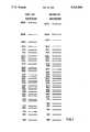
- FIG. 1is a schematic, scale drawing of the how the first and second molecular marker kits would migrate on an electrophoretic gel. The positions were calculated by assuming that relative mobilities are a linear function of the logarithm of the length of the fragment in base pairs (bp). The length of each band in bp is indicated to the left of the band.
- the present inventionis a DNA size marker system, preferably a DNA marker ladder, having pooled DNA restriction endonuclease digests.
- DNA marker ladderis meant DNA fragments of varying sizes containing the sequence "S" that when electrophoresed through a gel matrix migrate with approximately equal spacing between them. "Equal spacing” may refer either to the physical location on a gel after electrophoresis (e.g. bands about 0.5 cm. apart) or to the size being marked (e.g. bands differing in size by 1,000 bp).
- Each restriction digestcontains at least one DNA fragment having an "S" sequence complementary to a probe and one or more other DNA fragments not complementary to the probe. The same probe is thus used for all restriction digests.
- the region of complementarity between the probe and the first DNA fragment of each digestis a double-stranded segment of the first fragment.
- the number of restriction digests pooledis at least 5, preferably at least 10, more preferably at least 15, yet more preferably at least 20, and most preferably at least 25.
- the largest target fragmentis at least 10-fold, preferably 14-fold, and most preferably 17-fold, longer than the smallest target fragment.
- target fragments most similar in sizediffer in length by defined amounts.
- Table 1shows the relationship between M, U, and L, (U and L are in bp) with the latter being held constant at 1,000 bp. Note that if U and L are both changed by the same factor or multiple, M remains constant. For example, bands of 1,059 bp and 1,000 bp and bands of 530 bp and 500 bp both differ in size by measures of 0.025.
- target fragment pairs most similar in sizediffer in size by no more than a measure of about 0.1 (e.g. bands of 1,259 bp and 1,000 bp), and, most preferably, by no more than a measure of about 0.075 (e.g. bands of 1,188 bp and 1,000 bp).
- bands that after gel electrophoresis and Southern blottingwould be adjacent to each other differ in size by no more than a measure of about 0.1.
- the target fragment pairs most similar in sizediffer in size by at least a measure of about 0.025 (e.g. bands of 1,059 bp and 1,000 bp).
- the target fragmentsall anneal to a single probe sequence or its complement. More than one molecular species may be in the probe, provided that each digest contains at least one fragment that can anneal to a probe molecule and at least one fragment that cannot anneal to a probe molecule.
- the target fragmentsare derived from bacteriophage ⁇ .
- the target fragmentsmay be detected with a probe having sequence present in or a sequence complementary to a sequence present in nucleotides 33,783 to 34,212 of bacteriophage ⁇ .
- the present inventionmay further be included in a kit having, in addition to the target fragments, a probe nucleic acid complementary to target DNA fragments.
- a probe nucleic acid complementary to target DNA fragmentsAs exemplified herein, the sequence of the probe is present in or is complementary to a sequence present in nucleotides 33,783 to 34,212 of bacteriophage ⁇ .
- the kitmay further include an enzyme capable of radioactively labeling the probe, e.g. polynucleotide kinase or the Klenow fragment of E. coli DNA polymerase I.
- an enzyme capable of radioactively labeling the probee.g. polynucleotide kinase or the Klenow fragment of E. coli DNA polymerase I.
- the target DNAis constructed from a single bacteriophage or plasmid.
- the target DNApreferably consists of at least 10 restriction endonuclease digests of that target DNA. Each restriction digest of the target DNA creates one fragment complementary to the probe DNA, and the lengths of these fragments may be distributed in a logarithmic array.
- the probe DNAis supplied as a pair of synthetic oligonucleotides.
- Each of the probe oligonucleotidesis preferably at least 20 nucleotides in length and are complementary to each other for 15 to 30 base pairs at their 3'-ends.
- These oligonucleotidescan then be labeled by incorporation of labeled nucleotides in a chain extension reaction, with each oligonucleotide serving as a primer and using the other as a template in the chain extension reaction.
- the upper and lower case lettersare complements of each other: ##STR1##
- the oligonucleotideswill have the following structure: ##STR2## This structure can then be separated to form two probes labeled at their 3'-ends: 5'abc . . . lmnopqrst . . . xyz3' and 5'ZYX . . . TSRQPONML . . . CBA3'.
- the probemay be labeled with a radioisotope (e.g. 3 H, 32 P, 35 S, or 125 I), a ligand (e.g. biotin), a hapten (e.g. dinitrophenol, fluorescein), or an enzyme (e.g. alkaline phosphatase, ⁇ -galactosidase, horseradish peroxidase, microperoxidase), or any other suitable labeling method known to or discovered by the art.
- a radioisotopee.g. 3 H, 32 P, 35 S, or 125 I
- a ligande.g. biotin
- a haptene.g. dinitrophenol, fluorescein
- an enzymee.g. alkaline phosphatase, ⁇ -galactosidase, horseradish peroxidase, microperoxidase
- the choice of labeling methodwill generally depend on the chosen method for detecting the experimental sample for
- a DNA marker kit of the present inventionalso include a means for making a probe, instead of just a means for added labeled nucleotides, e.g. with DNA polymerase, or another labeled entity, e.g. 32 PO 4 and kinase.
- This meansmay be a means for making an RNA probe.
- the means for making a probemay include being probe sequences under control of a promoter (i.e. a means-DNA).
- the kitcould also include an RNA polymerase capable of initiating transcription from the promoter and transcribing probe sequences of the means-DNA. Examples of such means-DNAs and RNA polymerases are well known in the art. For instance, DNA sequences downstream from SP6 promoters are commonly transcribed in vitro by SP6 RNA polymerase and sequences downstream from T7 promoters are commonly transcribed in vitro by T7 RNA polymerase.
- the bandsmay not be spaced exactly as shown in FIG. 1 due to well known phenomena concerning mobility of very large and very small fragments, sample loading effects, and inhomogeneities in the gel. With the use of the present invention, these effects can be detected more readily. Indeed, due to the way that DNA fragments run in 1.0% agarose gels, the largest (e.g. above 10 kbp) target fragments of the exemplified kits will appear more evenly spaced than as illustrated in FIG. 1.
- the DNA marker fragmentsshould be hybridized with the probe, with the fragments which bind probe molecules being the fragments detected.
- the ladder DNAmay appear as a "smear" due to the multitude of fragments.
- E. coli bacteriophage ⁇ (lambda) DNA(cIind 1, ts857, Sam 7) was the source of all target DNAs.
- the probe DNA for either of the ladders exemplified hereinmay consist of any DNA from between nucleotides 33,783 and 34,212 of that ⁇ DNA. Oligonucleotides were synthesized using standard phosphoramidite chemistry well known to the art.
- ⁇ DNAwas digested with one or two restriction endonucleases.
- the enzymes used for individual digestsare indicated in Tables 2 and 3. Digestions were performed under standard conditions, generally according to the instructions of the enzyme's manufacturer. Restriction digests were pooled after digestion.
- the target DNAconsisted of pooled equal amounts of 31 different restriction digests of phage ⁇ DNA.
- the probe DNAwas a 26-base oligonucleotide having a sequence of
- the first kitwas improved in three ways.
- the first improvementwas to change the probe DNA such that (a) it could easily be labeled with DNA polymerase as well as polynucleotide kinase, and (b) it would remain hybridized to the Southern blot even when washed at high temperature (65° C.) and low salt concentration (0.015M NaCl).
- This was achievedby utilizing two 70-base, synthetic oligonucleotides that were complementary to opposite strands of ⁇ DNA, and also complementary to one another for 15 bases at their 3'-termini.
- the two oligonucleotideswere as follows: ##STR3##
- the underlined segmentsare complementary to each other.
- the first oligonucleotideis encoded by sequences from coordinates 34,078 (5'-end) to 34,147 (3'-end) and the second oligonucleotide is encoded by sequences from 34,133 (3'-end) to 34,202 (5'-end) on the standard ⁇ map.
- These oligonucleotideswere mixed together with each other and the Klenow fragment of E. coli DNA polymerase I and four deoxynucleotide triphosphates, one of which was ⁇ - 32 P-labelled.
- the polymeraseextended each oligonucleotide using the other as a template and produced two ⁇ - 32 P-labelled, complementary oligonucleotides. This new probe hybridizes to the same target fragments as the previous probe.
- a mixture of the new 70-merswas labeled with the large fragment of E. coli DNA polymerase I and hybridized to a Southern blot of the target DNA.
- the second improvementwas to change the target DNA to give a more linear spacing on the Southern blot.
- the third improvementwas to increase the amounts, i.e. relative copy number or the dosage, of the target DNA for the largest and smallest bands.
- Large DNA fragmentsblot inefficiently.
- small fragmentsare retained on membranes poorly during hybridization. Therefore, the signal from large DNA fragments and small DNA fragments tends to be less than the signal from bands in the middle range. This improvement compensated for that effect.
Landscapes
- Chemical & Material Sciences (AREA)
- Life Sciences & Earth Sciences (AREA)
- Organic Chemistry (AREA)
- Health & Medical Sciences (AREA)
- Zoology (AREA)
- Engineering & Computer Science (AREA)
- Wood Science & Technology (AREA)
- Proteomics, Peptides & Aminoacids (AREA)
- Immunology (AREA)
- Bioinformatics & Cheminformatics (AREA)
- Biotechnology (AREA)
- Microbiology (AREA)
- Molecular Biology (AREA)
- Biophysics (AREA)
- Analytical Chemistry (AREA)
- Biochemistry (AREA)
- Physics & Mathematics (AREA)
- General Engineering & Computer Science (AREA)
- General Health & Medical Sciences (AREA)
- Genetics & Genomics (AREA)
- Virology (AREA)
- Measuring Or Testing Involving Enzymes Or Micro-Organisms (AREA)
Abstract
Description
5'GCGACATTGCTCCGTGTATTCACTCG3'
TABLE 1 ______________________________________ Examples of Relationships between the Measure of the Difference in Size and Sizes of Fragments. M U L ______________________________________ 0.0 1,000 1,000 0.025 1,059 1,000 0.05 1,122 1,000 0.075 1,188 1,000 0.1 1,259 1,000 0.15 1,413 1,000 0.2 1,585 1,000 0.3 1,995 1,000 0.5 3,162 1,000 0.7 5,012 1,000 1.0 10,000 1,000 ______________________________________ M = log.sub.10 (U) log.sub.10 (L) = Measure of the difference in size. U = Size in bp of the upper band in a comparison. L = Size in bp of the lower band in a comparison, held constant at 1,000 bp.
TABLE 2 ______________________________________ DNA Analysis Marker Ladder Target DNA Fragments, First Kit Lambda Coordinates Enzyme(s) Size Diff. Left Right ______________________________________ Xba I* 23,994 0.204 24,508 48,502 Xho I 15,004 0.127 33,498 48,502 Xba I/Bgl II* 11,203 0.075 24,508 35,711 Hind III 9,416 0.056 27,479 36,895 Sma I 8,271 0.047 31,619 39,890 EcoR I 7,421 0.061 31,747 39,168 Ava II 6,442 0.041 32,562 39,004 Hae II 5,861 0.034 28,859 34,720 EcoR V/Ava II 5,415 0.060 33,589 39,004 Ava I 4,716 0.067 33,498 38,214 Bgl I/BstE II* 4,045 0.026 32,329 36,374 Ava II/BstE II 3,812 0.025 32,562 36,374 Dra I* 3,599 0.065 32,705 36,304 Sma I/Hae II 3,101 0.033 31,619 34,720 Xho I/BstE II 2,876 0.036 33,498 36,374 Nci I 2,650 0.037 33,158 35,808 Nde I 2,433 0.026 33,680 36,113 Msp I* 2,293 0.056 33,157 35,450 Hinc II 2,015 0.035 33,246 35,261 EcoR V/Msp I 1,861 0.023 33,589 35,450 Xho I/Hinc II* 1,763 0.051 33,498 35,261 Rsa I 1,568 0.040 32,868 34,436 Ssp I 1,431 0.028 33,572 35,003 Msp I/BamH I* 1,342 0.057 33,157 34,499 Sau3A I 1,176 0.024 33,323 34,499 Cla I* 1,112 0.087 33,585 34,697 EcoR V/BamH I 910 0.033 33,589 34,499 Hinf I* 844 0.064 33,783 34,627 EcoR V/Cvn I* 730 0.048 33,589 34,319 Hinf I/Rsa I 653 0.094 33,783 34,436 Nsi I 526 -- 33,686 34,212 ______________________________________ Diff. = The difference, M, in size between the band and the band immediately below, calculated by the formula, M = log.sub.10 (U) log.sub.10 (L), where U and L are the lengths in bp of the upper and lower, respectively, of the two bands being compared. *indicates enzyme combinations used in the first ladder but not used in the second ladder.
TABLE 3 ______________________________________ DNA Analysis Marker Ladder Target DNA Fragments, Second Kit Lambda Coordinates Enzyme(s) Size Diff. Left Right Dose ______________________________________ Sst I* 22,621 0.178 25,881 48,502 3 Xho I 15,004 0.100 33,498 48,502 3 Nco I/Bgl I* 11,919 0.102 32,329 44,248 3 Hind III 9,416 0.056 27,479 36,895 3 Sma I 8,271 0.047 31,619 39,890 3 EcoR I 7,421 0.061 31,747 39,168 3 Ava II 6,442 0.041 32,562 39,004 3 Hae II 5,861 0.034 28,859 34,720 1 EcoR V/Ava II 5,415 0.060 33,589 39,004 1 Ava I 4,716 0.037 33,498 38,214 1 Ava II/Hind III* 4,333 0.056 32,562 36,895 1 Ava II/BstE II 3,812 0.050 32,562 36,374 1 Xho I/Hind III* 3,397 0.040 33,498 36,895 1 Sma I/Hae II 3,101 0.033 31,619 34,720 1 Xho I/BstE II 2,876 0.036 33,498 36,374 1 Nci I 2,650 0.037 33,158 35,808 1 Nde I 2,433 0.041 33,680 36,113 1 Xho I/Bgl II* 2,213 0.041 33,498 35,711 1 Hinc II 2,015 0.035 33,246 35,261 1 EcoR V/Msp I 1,861 0.047 33,589 35,450 1 EcoR V/Hinc II* 1,672 0.028 33,589 35,261 1 Rsa I 1,568 0.040 32,868 34,436 1 Ssp I 1,431 0.046 33,572 35,003 1 Tha I/Rsa I* 1,287 0.039 33,149 34,436 1 Sau3A I 1,176 0.073 33,323 34,499 1 Cfo I* 993 0.038 33,726 34,719 1 EcoR V/BamH I 910 0.065 33,589 34,499 1 Dde I* 784 0.079 33,535 34,319 3 Hinf I/Rsa I 653 0.094 33,783 34,436 3 Nsi I 526 -- 33,686 34,212 3 ______________________________________ Diff. = The difference, M, in size between the band and the band immediately below, caculated by the formula M = log.sub.10 (U) log.sub.1 (L), where U and L are the lengths in bp of the upper and lower, respectively, of the two bands being compared. *indicates enzyme combinations used in the second ladder but not used in the first ladder. Dose refers to the relative amounts of each restriction digest.
Claims (32)
Priority Applications (5)
| Application Number | Priority Date | Filing Date | Title |
|---|---|---|---|
| US07/552,406US5316908A (en) | 1990-07-13 | 1990-07-13 | Size markers for electrophoretic analysis of DNA |
| CA002045794ACA2045794A1 (en) | 1990-07-13 | 1991-06-27 | Size markers for electrophoretic analysis of dna |
| DE69124297TDE69124297T2 (en) | 1990-07-13 | 1991-07-04 | Size marker for electrophoretic analysis of DNA |
| AT91306104TATE148174T1 (en) | 1990-07-13 | 1991-07-04 | SIZE MARKERS FOR ELECTROPHORETIC ANALYSIS OF DNA |
| EP91306104AEP0466404B1 (en) | 1990-07-13 | 1991-07-04 | Size markers for electrophoretic analysis of DNA |
Applications Claiming Priority (1)
| Application Number | Priority Date | Filing Date | Title |
|---|---|---|---|
| US07/552,406US5316908A (en) | 1990-07-13 | 1990-07-13 | Size markers for electrophoretic analysis of DNA |
Publications (1)
| Publication Number | Publication Date |
|---|---|
| US5316908Atrue US5316908A (en) | 1994-05-31 |
Family
ID=24205190
Family Applications (1)
| Application Number | Title | Priority Date | Filing Date |
|---|---|---|---|
| US07/552,406Expired - LifetimeUS5316908A (en) | 1990-07-13 | 1990-07-13 | Size markers for electrophoretic analysis of DNA |
Country Status (5)
| Country | Link |
|---|---|
| US (1) | US5316908A (en) |
| EP (1) | EP0466404B1 (en) |
| AT (1) | ATE148174T1 (en) |
| CA (1) | CA2045794A1 (en) |
| DE (1) | DE69124297T2 (en) |
Cited By (21)
| Publication number | Priority date | Publication date | Assignee | Title |
|---|---|---|---|---|
| US5714326A (en)* | 1991-01-24 | 1998-02-03 | Dawson; Elliott P. | Method for the multiplexed preparation of nucleic acid molecular weight markers and resultant products |
| WO1999003872A1 (en)* | 1997-07-15 | 1999-01-28 | Life Technologies, Inc. | Nucleic acid ladders |
| US6153389A (en)* | 1999-02-22 | 2000-11-28 | Haarer; Brian K. | DNA additives as a mechanism for unambiguously marking biological samples |
| US20020155455A1 (en)* | 2000-08-11 | 2002-10-24 | Invitrogen Corporation | Highly homogeneous molecular markers for electrophoresis |
| US6537785B1 (en) | 1999-09-14 | 2003-03-25 | Genzyme Glycobiology Research Institute, Inc. | Methods of treating lysosomal storage diseases |
| US6576106B1 (en)* | 1999-04-14 | 2003-06-10 | Helena Laboratories Co., Ltd. | Method for separating and assaying lipoprotein, an assembly for performing such a method, and a system including such an assembly |
| US20030157720A1 (en)* | 2002-02-06 | 2003-08-21 | Expression Technologies Inc. | Protein standard for estimating size and mass |
| US20030190662A1 (en)* | 1993-10-28 | 2003-10-09 | Invitrogen Corporation | Nucleic acid marker ladder for estimating mass |
| US20050064485A1 (en)* | 2003-09-12 | 2005-03-24 | Kurt Vogel | Multiplex binding and activity assays |
| US20050164287A1 (en)* | 1997-03-27 | 2005-07-28 | Invitrogen Corporation | Nucleic acid ladders |
| US20050170442A1 (en)* | 2003-07-29 | 2005-08-04 | Kupcho Kevin R. | Bimolecular optical probes |
| US20050277202A1 (en)* | 2000-02-09 | 2005-12-15 | A-Fem Medical Corporation | Collection device for lateral flow chromatography |
| US20070059787A1 (en)* | 2003-07-29 | 2007-03-15 | Invitrogen Corporation | Kinase and phosphatase assays |
| US20070184527A1 (en)* | 1997-01-08 | 2007-08-09 | Invitrogen Corporation | Methods for Production of Proteins |
| US20160084860A1 (en)* | 2014-09-19 | 2016-03-24 | Li-Cor,Inc. | Multifunctional protein molecular weight ladders |
| WO2017152149A1 (en)* | 2016-03-03 | 2017-09-08 | University Of Massachusetts | Closed-ended linear duplex dna for non-viral gene transfer |
| CN110923305A (en)* | 2019-11-25 | 2020-03-27 | 广州市达瑞生物技术股份有限公司 | DNA molecular weight standard suitable for fragile X syndrome southern blot hybridization detection |
| US11167247B2 (en) | 2017-02-15 | 2021-11-09 | Nanolc-12, Llc | Length-based separation of carbon nanotubes |
| US11353424B2 (en)* | 2018-04-12 | 2022-06-07 | Nano LC-12, LLC | Length-based carbon nanotube ladders |
| US11421038B2 (en) | 2007-01-09 | 2022-08-23 | Curevac Ag | RNA-coded antibody |
| WO2024059197A1 (en)* | 2022-09-14 | 2024-03-21 | Revvity Health Sciences, Inc. | Methods and compositions for single-stranded nucleic acid ladders by guided cleavage |
Families Citing this family (5)
| Publication number | Priority date | Publication date | Assignee | Title |
|---|---|---|---|---|
| WO1993014224A1 (en)* | 1992-01-14 | 1993-07-22 | Applied Biosystems, Inc. | Size-calibration dna fragment mixture and method |
| US5302510A (en)* | 1992-07-27 | 1994-04-12 | Life Technologies, Inc. | DNA sizing control standards for electrophoretic analyses |
| DK0725821T3 (en)* | 1993-10-28 | 1999-02-15 | Life Technologies Inc | Nucleic acid marker ladder for mass estimation |
| DE69713800T2 (en) | 1996-12-19 | 2003-01-02 | S.C. Johnson Commercial Markets, Inc. | DISPERSIONS OF MIXED POLYCARBOXYPOLYAMIDE RESINS AND ALKALI DISPERSIBLE RESINS; MANUFACTURE AND THEIR USE |
| CA2362771A1 (en)* | 1999-03-12 | 2000-09-21 | Amersham Pharmacia Biotech Uk Limited | Genetic analysis |
Citations (9)
| Publication number | Priority date | Publication date | Assignee | Title |
|---|---|---|---|---|
| US4415732A (en)* | 1981-03-27 | 1983-11-15 | University Patents, Inc. | Phosphoramidite compounds and processes |
| US4458066A (en)* | 1980-02-29 | 1984-07-03 | University Patents, Inc. | Process for preparing polynucleotides |
| US4500707A (en)* | 1980-02-29 | 1985-02-19 | University Patents, Inc. | Nucleosides useful in the preparation of polynucleotides |
| US4668777A (en)* | 1981-03-27 | 1987-05-26 | University Patents, Inc. | Phosphoramidite nucleoside compounds |
| JPS63113359A (en)* | 1986-06-25 | 1988-05-18 | Yakult Honsha Co Ltd | Standard marker for DNA molecular weight measurement |
| US4762779A (en)* | 1985-06-13 | 1988-08-09 | Amgen Inc. | Compositions and methods for functionalizing nucleic acids |
| US4771384A (en)* | 1986-07-24 | 1988-09-13 | Dnastar, Inc. | System and method for fragmentation mapping |
| WO1989009780A1 (en)* | 1988-04-15 | 1989-10-19 | Chemical Industry Institute Of Toxicology | Site-specific tritium-labeled oligodeoxynucleotides |
| US4965349A (en)* | 1987-12-24 | 1990-10-23 | Applied Biosystems, Inc. | Method of synthesizing oligonucleotides labeled with ammonia-labile groups on solid phase supports |
Family Cites Families (1)
| Publication number | Priority date | Publication date | Assignee | Title |
|---|---|---|---|---|
| DE3829535A1 (en)* | 1988-08-31 | 1990-03-15 | Biotest Ag | NUCLEIC ACID FRAGMENTS FROM EUKARYONTIC GENOMES, METHOD FOR THE PRODUCTION THEREOF AND THEIR USE FOR DETECTING RESTRICTION FRAGMENT LENGTH POLYMORPHISMS (RFLPS) |
- 1990
- 1990-07-13USUS07/552,406patent/US5316908A/ennot_activeExpired - Lifetime
- 1991
- 1991-06-27CACA002045794Apatent/CA2045794A1/ennot_activeAbandoned
- 1991-07-04EPEP91306104Apatent/EP0466404B1/ennot_activeExpired - Lifetime
- 1991-07-04DEDE69124297Tpatent/DE69124297T2/ennot_activeExpired - Fee Related
- 1991-07-04ATAT91306104Tpatent/ATE148174T1/enactive
Patent Citations (9)
| Publication number | Priority date | Publication date | Assignee | Title |
|---|---|---|---|---|
| US4458066A (en)* | 1980-02-29 | 1984-07-03 | University Patents, Inc. | Process for preparing polynucleotides |
| US4500707A (en)* | 1980-02-29 | 1985-02-19 | University Patents, Inc. | Nucleosides useful in the preparation of polynucleotides |
| US4415732A (en)* | 1981-03-27 | 1983-11-15 | University Patents, Inc. | Phosphoramidite compounds and processes |
| US4668777A (en)* | 1981-03-27 | 1987-05-26 | University Patents, Inc. | Phosphoramidite nucleoside compounds |
| US4762779A (en)* | 1985-06-13 | 1988-08-09 | Amgen Inc. | Compositions and methods for functionalizing nucleic acids |
| JPS63113359A (en)* | 1986-06-25 | 1988-05-18 | Yakult Honsha Co Ltd | Standard marker for DNA molecular weight measurement |
| US4771384A (en)* | 1986-07-24 | 1988-09-13 | Dnastar, Inc. | System and method for fragmentation mapping |
| US4965349A (en)* | 1987-12-24 | 1990-10-23 | Applied Biosystems, Inc. | Method of synthesizing oligonucleotides labeled with ammonia-labile groups on solid phase supports |
| WO1989009780A1 (en)* | 1988-04-15 | 1989-10-19 | Chemical Industry Institute Of Toxicology | Site-specific tritium-labeled oligodeoxynucleotides |
Non-Patent Citations (20)
| Title |
|---|
| 1989 1990 New England Biolabs, Inc. Catalog, pp. 9 11, 14 20, 23, 24, 26, 28, 29, 31 and 33.* |
| 1989 GIBCO/BRL Catalogue & Reference Guide, Life Technologies, Inc., Gaithersburg, Md., pp. 3,4, 8 18, 21, 22, 24, 26, 27, 29, 30 and 32.* |
| 1989 GIBCO/BRL Catalogue & Reference Guide, Life Technologies, Inc., Gaithersburg, Md., pp. 3,4, 8-18, 21, 22, 24, 26, 27, 29, 30 and 32. |
| 1989-1990 New England Biolabs, Inc. Catalog, pp. 9-11, 14-20, 23, 24, 26, 28, 29, 31 and 33. |
| Bernards et al., Chemical Abstracts, No. 92470r, 105(11):187 (1986).* |
| Budowle and Baechtel, Appl. Theor. Electrophoresis 1: 181 187 (1990).* |
| Budowle and Baechtel, Appl. Theor. Electrophoresis 1: 181-187 (1990). |
| Carman et al., J. Clin. Microb., Nov. 1989, vol. 27, No. 11, Detection of Enzymatically Amplified Human Immunodeficiency Virus DNA by Oligonucleotide Solution Hybridization and by Incorporation of Radiolabeled Deoxynucleotides, pp. 2570 2573.* |
| Carman et al., J. Clin. Microb., Nov. 1989, vol. 27, No. 11, Detection of Enzymatically Amplified Human Immunodeficiency Virus DNA by Oligonucleotide Solution Hybridization and by Incorporation of Radiolabeled Deoxynucleotides, pp. 2570-2573. |
| European Search Report for Applicants Co pending European Application EP O 466 404 A1.* |
| European Search Report for Applicants' Co-pending European Application EP O 466 404 A1. |
| Gralla, Proc. Natl. Acad. Sci. USA, vol. 82, May 1985, Rapid "footprinting" on supercoiled DNA, pp. 3078-3081. pp. 3078-3081. |
| Gralla, Proc. Natl. Acad. Sci. USA, vol. 82, May 1985, Rapid footprinting on supercoiled DNA, pp. 3078 3081. pp. 3078 3081.* |
| Huang J. et al., Chemical Abstracts No. 34093a 107(5):175 (1987).* |
| Jones C. P. et al., Chemical Abstracts No. 230760h 112(25):172 (1990).* |
| Package inserts supplied with Ava I, Ava II, Bam H I, Bgl I, Bgl II, Bst E II, Cfo I, Cla I, Cvn I, Dde I, Dra I, Eco R I, Eco R V, Hae II, Hin c II, Hin d III, Hin f I, Msp I, Nci I, Nco I, Nde I, Nsi I, Rsa I, Sau 3A I, Sma I, Ssp I, SstI, Tha I, Xba I, and Xho I which were available on Jul. 13, 1990 and supplied by BRL/Life Technologies, Inc.* |
| Pharmacia Catalogue, "Molecular Weight Markers", p. 23. |
| Pharmacia Catalogue, Molecular Weight Markers , p. 23.* |
| Rickwood D. et al., Practical Approaches in Biochemistry Series, pp. 227 232, Appendix I.* |
| Rickwood D. et al., Practical Approaches in Biochemistry Series, pp. 227-232, Appendix I. |
Cited By (43)
| Publication number | Priority date | Publication date | Assignee | Title |
|---|---|---|---|---|
| US5714326A (en)* | 1991-01-24 | 1998-02-03 | Dawson; Elliott P. | Method for the multiplexed preparation of nucleic acid molecular weight markers and resultant products |
| US7455974B2 (en) | 1993-10-28 | 2008-11-25 | Invitrogen Corporation | Nucleic acid marker ladder for estimating mass |
| US7846665B2 (en) | 1993-10-28 | 2010-12-07 | Life Technologies Corporation | Nucleic acid marker ladder for estimating mass |
| US20090111112A1 (en)* | 1993-10-28 | 2009-04-30 | Invitrogen Corporation | Nucleic Acid Marker Ladder for Estimating Mass |
| US20030190662A1 (en)* | 1993-10-28 | 2003-10-09 | Invitrogen Corporation | Nucleic acid marker ladder for estimating mass |
| US6680378B1 (en) | 1993-10-28 | 2004-01-20 | Invitrogen Corporation | Nucleic acid marker ladder for estimating mass |
| US7132520B2 (en) | 1993-10-28 | 2006-11-07 | Invitrogen Corporation | Nucleic acid marker ladder for estimating mass |
| US20070020680A1 (en)* | 1993-10-28 | 2007-01-25 | Invitrogen Corporation | Nucleic acid marker ladder for estimating mass |
| US8519099B2 (en) | 1997-01-08 | 2013-08-27 | Life Technologies Corporation | Methods for production of proteins |
| US8389681B2 (en) | 1997-01-08 | 2013-03-05 | Life Technologies Corporation | Methods for production of proteins |
| US8012715B2 (en) | 1997-01-08 | 2011-09-06 | Life Technologies Corporation | Methods for production of proteins |
| US9409942B2 (en) | 1997-01-08 | 2016-08-09 | Life Technologies Corporation | Methods for production of proteins |
| US20070190606A1 (en)* | 1997-01-08 | 2007-08-16 | Invitrogen Corporation | Methods for Production of Proteins |
| US20070184527A1 (en)* | 1997-01-08 | 2007-08-09 | Invitrogen Corporation | Methods for Production of Proteins |
| US20050164287A1 (en)* | 1997-03-27 | 2005-07-28 | Invitrogen Corporation | Nucleic acid ladders |
| US6924098B2 (en) | 1997-03-27 | 2005-08-02 | Invitrogen Corporation | Nucleic acid ladders |
| WO1999003872A1 (en)* | 1997-07-15 | 1999-01-28 | Life Technologies, Inc. | Nucleic acid ladders |
| US8273863B1 (en)* | 1997-07-15 | 2012-09-25 | Life Technologies Corporation | Nucleic acid ladders |
| US6153389A (en)* | 1999-02-22 | 2000-11-28 | Haarer; Brian K. | DNA additives as a mechanism for unambiguously marking biological samples |
| US6576106B1 (en)* | 1999-04-14 | 2003-06-10 | Helena Laboratories Co., Ltd. | Method for separating and assaying lipoprotein, an assembly for performing such a method, and a system including such an assembly |
| US6537785B1 (en) | 1999-09-14 | 2003-03-25 | Genzyme Glycobiology Research Institute, Inc. | Methods of treating lysosomal storage diseases |
| US20050277202A1 (en)* | 2000-02-09 | 2005-12-15 | A-Fem Medical Corporation | Collection device for lateral flow chromatography |
| US20020155455A1 (en)* | 2000-08-11 | 2002-10-24 | Invitrogen Corporation | Highly homogeneous molecular markers for electrophoresis |
| US20030157720A1 (en)* | 2002-02-06 | 2003-08-21 | Expression Technologies Inc. | Protein standard for estimating size and mass |
| US7727752B2 (en) | 2003-07-29 | 2010-06-01 | Life Technologies Corporation | Kinase and phosphatase assays |
| US8067536B2 (en) | 2003-07-29 | 2011-11-29 | Life Technologies Corporation | Kinase and phosphatase assays |
| US7619059B2 (en) | 2003-07-29 | 2009-11-17 | Life Technologies Corporation | Bimolecular optical probes |
| US20050170442A1 (en)* | 2003-07-29 | 2005-08-04 | Kupcho Kevin R. | Bimolecular optical probes |
| US20070059787A1 (en)* | 2003-07-29 | 2007-03-15 | Invitrogen Corporation | Kinase and phosphatase assays |
| US20100240080A1 (en)* | 2003-07-29 | 2010-09-23 | Life Technologies Corporation | Kinase and phosphatase assays |
| US20050064485A1 (en)* | 2003-09-12 | 2005-03-24 | Kurt Vogel | Multiplex binding and activity assays |
| US11421038B2 (en) | 2007-01-09 | 2022-08-23 | Curevac Ag | RNA-coded antibody |
| US20160084860A1 (en)* | 2014-09-19 | 2016-03-24 | Li-Cor,Inc. | Multifunctional protein molecular weight ladders |
| US9663554B2 (en)* | 2014-09-19 | 2017-05-30 | Li-Cor, Inc. | Multifunctional protein molecular weight ladders |
| US10365290B2 (en) | 2014-09-19 | 2019-07-30 | Li-Cor, Inc. | Multifunctional protein molecular weight ladders |
| WO2017152149A1 (en)* | 2016-03-03 | 2017-09-08 | University Of Massachusetts | Closed-ended linear duplex dna for non-viral gene transfer |
| US11066679B2 (en) | 2016-03-03 | 2021-07-20 | University Of Massachusetts | Closed-ended linear duplex DNA for non-viral gene transfer |
| US12252703B2 (en) | 2016-03-03 | 2025-03-18 | University Of Massachusetts | Closed-ended linear duplex DNA for non-viral gene transfer |
| US11167247B2 (en) | 2017-02-15 | 2021-11-09 | Nanolc-12, Llc | Length-based separation of carbon nanotubes |
| US11353424B2 (en)* | 2018-04-12 | 2022-06-07 | Nano LC-12, LLC | Length-based carbon nanotube ladders |
| CN110923305A (en)* | 2019-11-25 | 2020-03-27 | 广州市达瑞生物技术股份有限公司 | DNA molecular weight standard suitable for fragile X syndrome southern blot hybridization detection |
| CN110923305B (en)* | 2019-11-25 | 2023-12-29 | 广州市达瑞生物技术股份有限公司 | DNA molecular weight standard suitable for southern blot hybridization detection of fragile X syndrome |
| WO2024059197A1 (en)* | 2022-09-14 | 2024-03-21 | Revvity Health Sciences, Inc. | Methods and compositions for single-stranded nucleic acid ladders by guided cleavage |
Also Published As
| Publication number | Publication date |
|---|---|
| CA2045794A1 (en) | 1992-01-14 |
| EP0466404A1 (en) | 1992-01-15 |
| DE69124297D1 (en) | 1997-03-06 |
| EP0466404B1 (en) | 1997-01-22 |
| ATE148174T1 (en) | 1997-02-15 |
| DE69124297T2 (en) | 1997-05-28 |
Similar Documents
| Publication | Publication Date | Title |
|---|---|---|
| US5316908A (en) | Size markers for electrophoretic analysis of DNA | |
| Cawkwell et al. | Rapid detection of allele loss in colorectal tumours using microsatellites and fluorescent DNA technology | |
| EP0123513B1 (en) | Method of detecting mutations in dna | |
| US5068176A (en) | Method for the simultaneous determination of dna sequence variations at a large number of sites, and a kit suitable therefor | |
| US5869255A (en) | Probes labeled with energy transfer couples dyes exemplified with DNA fragment analysis | |
| US6028190A (en) | Probes labeled with energy transfer coupled dyes | |
| DE69122712T2 (en) | METHOD FOR NUCLEIC ACID HYBRIDIZATION AND AMPLIFICATION | |
| DE69233458T2 (en) | NUCLEIC ACIDIFYING BY POLYMERASE EXTENSION OF OLIGONUCLEOTIDES USING TERMINATOR MIXTURES | |
| EP0497272A1 (en) | Strand displacement amplification | |
| KR870009024A (en) | Production method of polynucleotide | |
| DE69028325D1 (en) | Nucleic acid amplification using a single primer | |
| Kohara et al. | The distribution and properties of RNA primed initiation sites of DNA synthesis at the replication origin of Escherichia coli chromosome | |
| Highsmith Jr et al. | Use of a DNA toolbox for the characterization of mutation scanning methods. I: construction of the toolbox and evaluation of heteroduplex analysis | |
| US6420109B1 (en) | Nucleic acid ligand interaction assays | |
| US5667971A (en) | Method for determining DNA sequences | |
| Stüber et al. | Electron microscopic analysis of in vitro transcriptional complexes: mapping of promoters of the coliphage T5 genome | |
| Elson et al. | Fractionation of oligodeoxynucleotides by polyacrylamide gel electrophoresis | |
| Holgersson et al. | Fluorescent‐based typing of the two short tandem repeat loci HUMTH01 and HUMACTBP2: Reproducibility of size measurements and genetic variation in the swedish population | |
| Wenz et al. | Identification of known p53 point mutations by capillary electrophoresis using unique mobility profiles in a blinded study | |
| EP1863934B1 (en) | Unique sequence hybridization probes (usp) | |
| Emerson et al. | Isolation and genomic arrangement of active and inactive forms of mammalian 5 S RNA genes. | |
| Kumar et al. | Separation of transforming amino acid-substituting mutations in codons 12, 13 and 61 of the N-ras gene by constant denaturant capillary electrophoresis (CDCE) | |
| Muth et al. | Fast capillary electrophoresis‐laser induced fluorescence analysis of ligase chain reaction products: Human mitochondrial DNA point mutations causing Leber's hereditary optic neuropathy | |
| US6872530B2 (en) | Method for determining the presence of DNA variants using peptide nucleic acid probes | |
| WO1993014224A1 (en) | Size-calibration dna fragment mixture and method |
Legal Events
| Date | Code | Title | Description |
|---|---|---|---|
| AS | Assignment | Owner name:LIFE TECHNOLOGIES, INC., Free format text:ASSIGNMENT OF ASSIGNORS INTEREST.;ASSIGNORS:CARLSON, DAVID P.;WATKINS, PAUL C.;KLEVAN, LEONARD;REEL/FRAME:005453/0551 Effective date:19900828 | |
| STCF | Information on status: patent grant | Free format text:PATENTED CASE | |
| FPAY | Fee payment | Year of fee payment:4 | |
| AS | Assignment | Owner name:WHATMAN, INC., MASSACHUSETTS Free format text:ASSIGNMENT OF ASSIGNORS INTEREST;ASSIGNOR:INVITROGEN CORPORATION;REEL/FRAME:012002/0047 Effective date:20010622 | |
| FPAY | Fee payment | Year of fee payment:8 | |
| AS | Assignment | Owner name:INVITROGEN CORPORATION, CALIFORNIA Free format text:MERGER;ASSIGNOR:LIFE TECHNOLOGIES, INC.;REEL/FRAME:013774/0861 Effective date:20000913 | |
| FPAY | Fee payment | Year of fee payment:12 | |
| AS | Assignment | Owner name:LIFE TECHNOLOGIES CORPORATION,CALIFORNIA Free format text:MERGER;ASSIGNOR:INVITROGEN CORPORATION;REEL/FRAME:023882/0551 Effective date:20081121 Owner name:LIFE TECHNOLOGIES CORPORATION, CALIFORNIA Free format text:MERGER;ASSIGNOR:INVITROGEN CORPORATION;REEL/FRAME:023882/0551 Effective date:20081121 | |
| AS | Assignment | Owner name:LIFE TECHNOLOGIES CORPORATION, CALIFORNIA Free format text:CORRECTIVE ASSIGNMENT TO CORRECT THE APPLICATION NO 09452626 PREVIOUSLY RECORDED ON REEL 023882 FRAME 0551. ASSIGNOR(S) HEREBY CONFIRMS THE MERGER SHOULD NOT HAVE BEEN RECORDED AGAINST THIS PATENT APPLICATION NUMBER;ASSIGNOR:INVITROGEN CORPORATION;REEL/FRAME:034217/0490 Effective date:20081121 |