US5308704A - Cell adhesive material and method for producing same - Google Patents
Cell adhesive material and method for producing sameDownload PDFInfo
- Publication number
- US5308704A US5308704AUS07/933,358US93335892AUS5308704AUS 5308704 AUS5308704 AUS 5308704AUS 93335892 AUS93335892 AUS 93335892AUS 5308704 AUS5308704 AUS 5308704A
- Authority
- US
- United States
- Prior art keywords
- adhesive material
- cell adhesive
- cell
- ion
- producing
- Prior art date
- Legal status (The legal status is an assumption and is not a legal conclusion. Google has not performed a legal analysis and makes no representation as to the accuracy of the status listed.)
- Expired - Lifetime
Links
Images


Classifications
- B—PERFORMING OPERATIONS; TRANSPORTING
- B29—WORKING OF PLASTICS; WORKING OF SUBSTANCES IN A PLASTIC STATE IN GENERAL
- B29C—SHAPING OR JOINING OF PLASTICS; SHAPING OF MATERIAL IN A PLASTIC STATE, NOT OTHERWISE PROVIDED FOR; AFTER-TREATMENT OF THE SHAPED PRODUCTS, e.g. REPAIRING
- B29C59/00—Surface shaping of articles, e.g. embossing; Apparatus therefor
- B29C59/16—Surface shaping of articles, e.g. embossing; Apparatus therefor by wave energy or particle radiation, e.g. infrared heating
- B—PERFORMING OPERATIONS; TRANSPORTING
- B29—WORKING OF PLASTICS; WORKING OF SUBSTANCES IN A PLASTIC STATE IN GENERAL
- B29C—SHAPING OR JOINING OF PLASTICS; SHAPING OF MATERIAL IN A PLASTIC STATE, NOT OTHERWISE PROVIDED FOR; AFTER-TREATMENT OF THE SHAPED PRODUCTS, e.g. REPAIRING
- B29C35/00—Heating, cooling or curing, e.g. crosslinking or vulcanising; Apparatus therefor
- B29C35/02—Heating or curing, e.g. crosslinking or vulcanizing during moulding, e.g. in a mould
- B29C35/08—Heating or curing, e.g. crosslinking or vulcanizing during moulding, e.g. in a mould by wave energy or particle radiation
- B29C35/0866—Heating or curing, e.g. crosslinking or vulcanizing during moulding, e.g. in a mould by wave energy or particle radiation using particle radiation
- B29C2035/0872—Heating or curing, e.g. crosslinking or vulcanizing during moulding, e.g. in a mould by wave energy or particle radiation using particle radiation using ion-radiation, e.g. alpha-rays
- B—PERFORMING OPERATIONS; TRANSPORTING
- B29—WORKING OF PLASTICS; WORKING OF SUBSTANCES IN A PLASTIC STATE IN GENERAL
- B29K—INDEXING SCHEME ASSOCIATED WITH SUBCLASSES B29B, B29C OR B29D, RELATING TO MOULDING MATERIALS OR TO MATERIALS FOR MOULDS, REINFORCEMENTS, FILLERS OR PREFORMED PARTS, e.g. INSERTS
- B29K2025/00—Use of polymers of vinyl-aromatic compounds or derivatives thereof as moulding material
- B—PERFORMING OPERATIONS; TRANSPORTING
- B29—WORKING OF PLASTICS; WORKING OF SUBSTANCES IN A PLASTIC STATE IN GENERAL
- B29K—INDEXING SCHEME ASSOCIATED WITH SUBCLASSES B29B, B29C OR B29D, RELATING TO MOULDING MATERIALS OR TO MATERIALS FOR MOULDS, REINFORCEMENTS, FILLERS OR PREFORMED PARTS, e.g. INSERTS
- B29K2075/00—Use of PU, i.e. polyureas or polyurethanes or derivatives thereof, as moulding material
- Y—GENERAL TAGGING OF NEW TECHNOLOGICAL DEVELOPMENTS; GENERAL TAGGING OF CROSS-SECTIONAL TECHNOLOGIES SPANNING OVER SEVERAL SECTIONS OF THE IPC; TECHNICAL SUBJECTS COVERED BY FORMER USPC CROSS-REFERENCE ART COLLECTIONS [XRACs] AND DIGESTS
- Y10—TECHNICAL SUBJECTS COVERED BY FORMER USPC
- Y10T—TECHNICAL SUBJECTS COVERED BY FORMER US CLASSIFICATION
- Y10T428/00—Stock material or miscellaneous articles
- Y10T428/31—Surface property or characteristic of web, sheet or block
- Y10T428/315—Surface modified glass [e.g., tempered, strengthened, etc.]
- Y—GENERAL TAGGING OF NEW TECHNOLOGICAL DEVELOPMENTS; GENERAL TAGGING OF CROSS-SECTIONAL TECHNOLOGIES SPANNING OVER SEVERAL SECTIONS OF THE IPC; TECHNICAL SUBJECTS COVERED BY FORMER USPC CROSS-REFERENCE ART COLLECTIONS [XRACs] AND DIGESTS
- Y10—TECHNICAL SUBJECTS COVERED BY FORMER USPC
- Y10T—TECHNICAL SUBJECTS COVERED BY FORMER US CLASSIFICATION
- Y10T428/00—Stock material or miscellaneous articles
- Y10T428/31504—Composite [nonstructural laminate]
- Y10T428/31551—Of polyamidoester [polyurethane, polyisocyanate, polycarbamate, etc.]
- Y—GENERAL TAGGING OF NEW TECHNOLOGICAL DEVELOPMENTS; GENERAL TAGGING OF CROSS-SECTIONAL TECHNOLOGIES SPANNING OVER SEVERAL SECTIONS OF THE IPC; TECHNICAL SUBJECTS COVERED BY FORMER USPC CROSS-REFERENCE ART COLLECTIONS [XRACs] AND DIGESTS
- Y10—TECHNICAL SUBJECTS COVERED BY FORMER USPC
- Y10T—TECHNICAL SUBJECTS COVERED BY FORMER US CLASSIFICATION
- Y10T428/00—Stock material or miscellaneous articles
- Y10T428/31504—Composite [nonstructural laminate]
- Y10T428/31855—Of addition polymer from unsaturated monomers
Definitions
- the present inventionrelates to a cell adhesive material applicable to cell culture containers or various medicinal materials and also relates to a method for producing the same. More specifically, the present invention relates particularly to a material with improved cell adhesion and a method for improving the adhesion.
- Matricesare generally constituted of polymer materials, and such polymer materials have conventionally been surface modified by a variety of processes.
- Surface modification processesare schematically grouped in dry process representatively illustrated by plasma process, arc process, etc. and wet process represented by coating, graft polymerization, etc.
- the plasma processincludes, for example, non-reactive plasma process based on the sputtering action of inactive ions such as argon, etc., reactive plasma process using reactive gases such as oxygen, water vapor, etc.
- the incident energy of ionsis approximately several keV, which can modify cell adhesion or the contact angle of cells to water by rendering the surface of a polymer material in a rough surface or can give hydrophilicity via the introduction of polarized structures such as >C ⁇ O group, C--O-- bond, and the like.
- the coatingis conducted by precoating an adhesive protein in solution from connective tissues such as collagen, fibronectin, etc. onto a matrix surface, thereby improving the adhesion of endothelial cells, fibroblasts, etc.
- the surface of polymer materialsgets rough once the plasma process is effected, so the resulting surface is likely to suppress cell proliferation although it generally propagates cell adhesion.
- the plasma statusvaries depending on the apparatus, so uniform conditions for such process are substantially impossible to realize, resulting in no chance of improving controllability and reproducibility.
- the coatinghas a problem in that the adhesive strength between a matrix and a cell is relatively weak because cells bond through the layer of an adhesive protein to the matrix.
- the present inventorshave made various investigations in order to achieve the above object. They have found that the adhesion and proliferation of cells can be improved remarkably by the surface modification of cell adhesive materials via ion bombardment and that ion implantation in a certain preset fluence range is effective so as to effect such modification.
- the surface modification of polymer materials via ion implantationis disclosed in Mat. Res. Soc. Symp., 110, 669 (1989) or Japanese Patent Laid-open No. 3-112560, such that an antithrombotic material is prepared by implanting ions such as H + , O 2 + , N 2 + , etc. into silicone resin. According to the present invention, such ion implantation technique is applied to the surface modification of cell adhesive materials.
- the cell adhesive material according to a first aspect of the present inventioncomprises a polymer material containing carbon as a constituting element, wherein at least a part of the surface is modified by ion bombardment.
- the method for producing a cell adhesive material according to a second aspect of the presentrange of 1 ⁇ 10 15 ⁇ 1 ⁇ 10 18 ions/cm 2 into at least a part of the surface of a polymer material containing carbon as a constituting element.
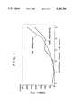
- FIG. 1is a graph depicting the time course of the change in the cell number of bovine vascular endothelial cells cultured on an Na + irradiated PS petri dish, compared with the cell number on a non-irradiated PS petri dish;
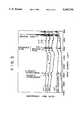
- FIG. 2is a schematic plane view depicting the cell adhesion state on a PS petri dish after N 2 + irradiation on the disk patterns, on the basis of microscopic observation; (a) represents an overall view of the PS petri dish while (b) represents an enlarged view of the part thereof;
- FIG. 3is infrared absorption spectra of the PS petri dishes implanted with individual ions
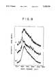
- FIG. 4is Raman scattering spectra of the PS petri dishes implanted with individual ions
- FIG. 5is a schematic plane view depicting the cell adhesion state on the PS petri dish after N 2 + irradiation on the mesh-like patterns, on the basis of microscopic observation;
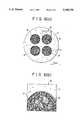
- FIG. 6is a schematic plane view depicting the cell adhesion state on the SPU petri dish after Na + irradiation on the disk patterns, on the basis of microscopic observation; (a) represents an overall plane view of the SPU petri dish while (b) represents an enlarged view of the part thereof;
- FIG. 7is infrared absorption spectra of the SPU petri dishes implanted with individual ions
- FIG. 8is Raman scattering spectra of the SPU petri dishes implanted with individual ions
- FIG. 9is a schematic plane view depicting the cell adhesion state on the SPU petri dish after Na + irradiation on the mesh-like patterns, on the basis of microscopic observation.
- the polymer material containing carbon as the constituting elementmay be a so-called organic polymer material, having a primary chain of carbons such as polystyrene, polyurethane, etc., or a silicon resin having a primary chain of siloxane (Si--O--bond) together with a side chain of hydrocarbon groups, etc.
- the ion species to be implantedis illustrated by He + , C + , N + , Ne + , Na + , N 2 + , O 2 + , Kr + , and the like, but is not limited to them. Any ion may be used as long as its solubilization does not suppresses cell growth.
- the fluence ⁇is selected in a range of 1 ⁇ 10 15 ⁇ 1 ⁇ 10 18 ions/cm 2 At the order of 1 ⁇ 10 14 ions/cm 2 , no distinctive effect can be observed on improving cell adhesion, while at the order of 1 ⁇ 10 18 ions/cm 2 , the effect on improving cell adhesion is saturated although the period required for such treatment is elongated much longer.
- the degree thereofmay induce the difference in energy transmission mechanism. Practically, the energy is satisfactorily set in a range of about several tens to several hundreds keV.
- the beam current densityis preset in a range not above 0.5 ⁇ A/cm 2 . This is because an extraordinarily high beam current density raises abnormally the temperature of a polymer material as a target, leading to the deterioration of the polymer material of itself and a possible reduction of cell adhesion.
- ion implantationis proposed as a means for providing the ion bombardment described above.
- Ion implantation of itselfis limited to the interaction between ion beam and an implanting material (target material).
- ionscan be implanted at an optional depth from the surface by selecting the incident energy of ions. Such reaction is thus under excellent control, which is a unique characteristic feature that cannot be observed in plasma process.
- the implanted ionsFor the implanted ions, a certain difference in mechanism is observed between an ion with a relatively small mass and an ion with a relatively large mass, such that electron stopping power functions on the ion with a relatively small mass at the initial discharge while nucleus stopping power initially functions on the ion with a relatively large mass.
- the implanted ionsput a polymer material in a heated state due to lattice vibration (thermal non-equilibrium state), resulting in melting, amorphous preparation, and the like.
- ion implantationwas conducted, through a predetermined mask pattern, into a polystyrene (PS) petri dish or a glass petri dish with the bottom surface laminated with a segmented polyurethane (SPU) film, using each ion beam of He + , N + , Ne + , Na + , or N 2 + .
- Vascular endothelial cells from a bovine descending thoracic aortawere cultured in these petri dishes, to compare cell proliferation in between beam irradiated and non-irradiated parts.
- Each ion of He + , N + , Ne + , Na + , N 2 + , O 2 + , and Kr +was implanted at room temperature under the conditions of an accelerating energy of 150 keV, a fluence of 1 ⁇ 10 15 to 3 ⁇ 10 17 ions/cm 2 and a beam current density of 0.5 ⁇ A/cm 2 or less.
- polystyrene (PS) petri dishwas FALCON 1008, a product manufactured by Becton Dichinson and Company.
- the chemical structure of polystyreneis in the following chemical formula 1. ##STR1##
- Segmented polyurethanegenerally has been put into practical use as the inner coating material of artificial cardiac system.
- the material presently employedis supplied from Kanegafuchi Chemical Industry, Co. Ltd., which soft segment comprises polytetramethylene oxide (MW; 2,000) and a dimethyl polysiloxane compound (MW; 2,400) and which hard segment comprises diphenylmethane-4,4-diisocyanate and ethylene glycol.
- the dimethyl polysiloxane compoundis prepared by bonding polyethylene oxide to both ends of the primary chain of dimethyl polysiloxane. ##STR2##
- dry type SPUwhich was produced by dissolving polyurethane in a mixed solution of dioxane and N,N-dimethylacetamide (7:3), coating the resulting solution onto an appropriate substrate followed by drying into a film form.
- Structural decomposition and radical formation through ion implantationwere subjected to analysis by Fourier transform-infrared spectroscopy-absorptiometry of total reflection (FT-IR-ATR).
- FT-IR-ATRFourier transform-infrared spectroscopy-absorptiometry of total reflection
- the stretching vibration of the multiple bonds between carbonswas detected by Raman spectroscopy, using a system Type Ramanor U-1000, manufactured by Jobin Yvon Co. Ltd.
- the scattering of argon laser (5145 Angstroms)was measured at room temperature.
- the system described aboveis of a double beam type, which can measure differential spectra.
- endothelial cellswere isolated from a bovine descending thoracic aorta.
- Vascular endothelial cellswere suspended in a culture broth (RPMI-1640, manufactured by Nissui Pharm. Co.) supplemented 10% fetal bovine serum at 2 ⁇ 10 4 to 2.5 ⁇ 10 4 cells/ml. The suspension was placed in a petri dish after ion implantation, and incubated in an incubator in an atmosphere of 5% CO 2 for 2 to 7 days.
- RPMI-1640manufactured by Nissui Pharm. Co.
- the suspension of vascular endothelial cells at a concentration of 2 ⁇ 10 14 cells/mlwas placed in the PS petri dish, to examine the relation between the culturing period and the change in cell number. For comparison, the same experiment was done in a non-irradiated PS petri dish without ion implantation.
- the ordinancerepresents the cell number and the abscissa represents the culturing period (in days); the manufactured graph in solid line corresponds to the case using the non-irradiated PS petri dish, while the graph in dotted line corresponds to the case of the Na + irradiated PS petri dish. From the results, the cell number on the Na + irradiated petri dish was far larger than the cell number on the non irradiated petri dish, which confirms the effectiveness of ion implantation.
- N 2 + at a fluence of 1 ⁇ 10 16 ions/cm 2was ion implanted, through an aluminium mask with four openings of a 7-mm diameter disk pattern, into the bottom of a PS petri dish.
- Vascular endothelial cells(simply referred to as "cells" hereinafter) were cultured on the petri dish.
- FIG. 2illustrates a sketched view of the state 3 days later on the basis of microscopic observation.
- FIG. 2ais an overall view of the PS petri dish while FIG. 2b illustrates an enlarged region A of FIG. 2a.
- the ion irradiated faces 2in 4 circles, cells 3 adhered to nearly the entire surfaces, but on the non-irradiated face 1, the adhering cell number is less, which clearly indicates that the cell adhesion is improved by ion implantation.
- the cells 3are localized on the circumference of the ion irradiated faces 2, which indicates that the cells having adhered onto the ion irradiated faces 2 promote the adhesion of other cells 3.
- the cells 3 having adhered onto the non-irradiated face 1are approximately in circular forms, while the cells having adhered onto the ion irradiated faces 2 elongate pseudopods, which indicates excellent adhesion state.
- ion implantationwas conducted individually via He + , Ne + , O 2 + and Kr + in a fluence range of 1 ⁇ 10 15 to 3 ⁇ 10 17 ions/cm 2 , for the same cell culture as described above. Almost the same results were obtained.
- FT-IR-ATR analysiswas done for the ion irradiated face of the PS petri dishes after the ion implantation of each ion of Na + , N 2 + , O 2 + , and Kr + at a fluence of 1 ⁇ 10 17 ions/cm 2 .
- the resultsare shown in FIG. 3.
- the specimens without ion implantationwere measured, and the results are also shown as non-irradiated PS.
- New structures developed by ion implantationare >C ⁇ O group (1700 cm -1 ), condensed ring (1400 to 1600 cm -1 ), C--C bond and C--O bond (1000 to 1200 cm -1 ), OH group (3400 cm -1 ) and the like.
- the absorption peak areas of themare normalized on the basis of the peak area of CH 2 (1460 cm -1 ) constantly appearing in any of the specimens, so that different effects are demonstrated by the individual ion species.
- Na + and N 2 +exhibit effects on the generation of condensed rings and --OH groups, respectively.
- FIG. 4further shows the Raman spectra of the specimens described above.
- an asymmetric peakwas observed with a shoulder on the shorter wave length side and a center around 1500 cm -1 .
- the one around 1330 cm -1corresponds to disordered graphitic sp 2 carbon.
- the one around 1480 cm -1corresponds to the amorphous state with sp 1 , sp 2 and sp 3 carbons being mixed together.
- N + at a fluence of 1 ⁇ 10 15 ions/cm 2was ion implanted, through an aluminium mask in a mesh form with a great number of openings of a 0.01 mm diameter disk pattern, into the bottom of a PS petri dish. Cells were cultured on the petri dish.
- FIG. 5illustrates a sketched view of the state 2 days later on the basis of microscopic observation.
- the parts common to those in FIG. 2are represented by the same symbols.
- cells 3adhered more intensely to the circular ion irradiated faces 2a than to the non-irradiated face 1 on the circumference thereof.
- Na + at a fluence of 3 ⁇ 10 17 ions/cm 2was ion implanted, through an aluminium mask with four openings of a 7-mm diameter disk pattern, into the SPU film-laminated bottom of a glass petri dish (referred to as SPU petri dish hereinafter). Cells were cultured on the SPU petri dish.
- FIG. 6illustrates a sketched view of the state 2 days later on the basis of microscopic observation.
- FIG. 6ais an overall view of the SPU petri dish while FIG. 6b illustrates an enlarged region B of FIG. 6a.
- SPUis a material with a low cell adhesion. Almost none of cells 3 adhered to the ion non-irradiated face 11. However, cells 3 adhered to nearly the entire surface of the 4 circular ion irradiated faces 12 on day 2 after the initiation of the culture.
- ion implantationwas conducted individually via He + , Ne + , O 2 + and Kr + in a fluence range of 1 ⁇ 10 15 to 3 ⁇ 10 17 ions/cm 2 , for the same cell culture as described above. Almost the same results were obtained.
- FT-IR-ATR analysiswas done for the ion irradiated face of the PS petri dishes after the ion implantation of each ion of Na + , O 2 + , and Kr + at a fluence of 1 ⁇ 10 17 ions/cm 2 .
- the resultsare shown in FIG. 7.
- the specimens without ion implantationwere measured, and the results are also shown as non-irradiated SPU.
- New structures developed by ion implantationare --OH group ⁇ 3400 cm -1 ), >C ⁇ O group (1700 cm -1 ), amorphous carbon (1600 cm -1 ), C--C bond and C--O bond (1000 to 1200 cm -1 ), Si--H bond (2120 cm -1 ) and the like.
- the absorption peak areas of themare normalized on the basis of the peak area of Si--O bond (1110 cm -1 ), so that it is indicated that Na + has a remarkable effect on the generation of amorphous carbon.
- FIG. 8further shows the Raman spectra of the specimens described above. The results are the same as the PS petri dish results shown in FIG. 4 above, which indicates that PS and SPU produce almost the same carbon structure via ion implantation.
- Na + at a fluence of 1 ⁇ 10 15 ions/cm 2was ion implanted, through an aluminium mask in a mesh form with a great number of openings of a 0.01-mm diameter disk pattern, into the bottom of an SPU petri dish. Cells were cultured on the petri dish.
- FIG. 9illustrates a sketched view of a part of the state 7 days later on the basis of microscopic observation.
- the parts common to those in FIG. 6are represented by the same symbols.
- cells 3also adhered to almost the entire surface of the ion irradiated faces 12a, although no adhesion of cells 3 was observed on the non-irradiated face 11, which indicates that selective ion implantation can control cell adhesion two-dimensionally if SPU is employed.
- the cell adhesive material according to the first aspect of the present inventionhas more improved cell adhesion than the conventional materials, or has such adhesion newly provided.
- the improvement or provision of cell adhesiondirectly reflects the change in chemical structure of the polymer material surface, no process for coating an adhesive protein such as collagen is required.
- petri dishes for cell cultureare premising. From the standpoint of providing a fundamental basis for developing further a variety of medicinal materials including hybrid-type artificial vessel via the adhesion of vascular endothelial cells and artificial skin via the adhesion of dermal cells, the present invention is of an industrially significant value.
- the above-mentioned surface modificationis effected through ion implantation with excellent controllability and reproducibility.
- This inventionis also extremely industrially as a practical means for supplying the cell adhesive material.
Landscapes
- Health & Medical Sciences (AREA)
- Toxicology (AREA)
- Materials For Medical Uses (AREA)
- Apparatus Associated With Microorganisms And Enzymes (AREA)
- Treatments Of Macromolecular Shaped Articles (AREA)
Abstract
Description
Claims (10)
Applications Claiming Priority (2)
| Application Number | Priority Date | Filing Date | Title |
|---|---|---|---|
| JP3259550AJPH0549689A (en) | 1991-08-20 | 1991-08-20 | Cell adhesive material and method for producing the same |
| JP3-259550 | 1991-08-20 |
Publications (1)
| Publication Number | Publication Date |
|---|---|
| US5308704Atrue US5308704A (en) | 1994-05-03 |
Family
ID=17335673
Family Applications (1)
| Application Number | Title | Priority Date | Filing Date |
|---|---|---|---|
| US07/933,358Expired - LifetimeUS5308704A (en) | 1991-08-20 | 1992-08-19 | Cell adhesive material and method for producing same |
Country Status (2)
| Country | Link |
|---|---|
| US (1) | US5308704A (en) |
| JP (1) | JPH0549689A (en) |
Cited By (20)
| Publication number | Priority date | Publication date | Assignee | Title |
|---|---|---|---|---|
| US5468562A (en)* | 1991-03-01 | 1995-11-21 | Spire Corporation | Metallized polymeric implant with ion embedded coating |
| US5474797A (en)* | 1991-10-18 | 1995-12-12 | Spire Corporation | Bactericidal coatings for implants |
| EP0688660A3 (en)* | 1994-06-24 | 1996-02-07 | Atomic Energy Authority Uk | Surface treatment of plastics films |
| US5492763A (en)* | 1992-06-08 | 1996-02-20 | Spire Corporation | Infection resistant medical devices and process |
| WO1996015223A1 (en)* | 1994-11-14 | 1996-05-23 | Universite Catholique De Louvain | Biomaterial and method for obtaining it |
| US5520664A (en)* | 1991-03-01 | 1996-05-28 | Spire Corporation | Catheter having a long-lasting antimicrobial surface treatment |
| WO1997010097A1 (en)* | 1995-09-12 | 1997-03-20 | Daimler-Benz Aktiengesellschaft | Plastic foil |
| WO1998052623A1 (en)* | 1997-05-22 | 1998-11-26 | The Regents Of The University Of California | Ion-implanted protein-coated intralumenal implants |
| WO1999030672A3 (en)* | 1997-12-15 | 1999-09-02 | Univ Eberhard Karls | Coating material for prostheses and prosthetic parts |
| US20020058897A1 (en)* | 1998-09-10 | 2002-05-16 | Percardia, Inc. | Designs for left ventricular conduit |
| US20020155295A1 (en)* | 2001-04-23 | 2002-10-24 | Riken | Artificial dura mater having cell adhesiveness and a process for producing the same |
| US6632470B2 (en) | 2001-01-31 | 2003-10-14 | Percardia | Methods for surface modification |
| WO2004026355A1 (en)* | 2002-08-30 | 2004-04-01 | Riken | Biological repair material compatible with biological tissue adhesive |
| US20060058835A1 (en)* | 1999-09-27 | 2006-03-16 | Yuichi Murayama | Bioabsorbable polymeric implants and a method of using the same to create occlusions |
| US20070185570A1 (en)* | 2003-08-19 | 2007-08-09 | The Chemo-Sero-Therapeutic Research Institute | Material for aneurysm curing |
| US20080064090A1 (en)* | 2006-09-07 | 2008-03-13 | Nalge Nunc International | Culture dish with lid |
| EP1854491A4 (en)* | 2005-02-24 | 2011-01-05 | Riken | CATHETER HAVING A DENATURED PART FOR CONTACT WITH THE BODY |
| US8785167B2 (en) | 2010-05-27 | 2014-07-22 | The Johns Hopkins University | Biocompatible article for the treatment of water and production of energy |
| US9303257B2 (en) | 2010-07-01 | 2016-04-05 | Empire Technology Development Llc | Method and system for cell and tissue cultivation |
| EP3970762A4 (en)* | 2019-05-15 | 2022-12-14 | Seoul National University R&DB Foundation | Method for manufacturing eptfe artificial blood vessels having improved hemocompatibility via selective plasma etching |
Families Citing this family (5)
| Publication number | Priority date | Publication date | Assignee | Title |
|---|---|---|---|---|
| JP4919557B2 (en)* | 2001-09-10 | 2012-04-18 | 独立行政法人理化学研究所 | Cell recovery membrane and method for producing the same |
| JP4660713B2 (en)* | 2003-07-15 | 2011-03-30 | 財団法人新産業創造研究機構 | Cell adhesion material |
| KR101597731B1 (en)* | 2008-02-21 | 2016-02-26 | 센토코 오르토 바이오테크 인코포레이티드 | Methods, surface modified plates and compositions for cell attachment, cultivation and detachment |
| JP2012066096A (en)* | 2011-11-02 | 2012-04-05 | Institute Of Physical & Chemical Research | Artificial dura mater having cell adhesion property and method of manufacturing the same |
| WO2014175432A1 (en)* | 2013-04-26 | 2014-10-30 | 株式会社長町サイエンスラボ | Dcl-layer-bearing structure and method for forming dlc layer |
Citations (7)
| Publication number | Priority date | Publication date | Assignee | Title |
|---|---|---|---|---|
| US4743493A (en)* | 1986-10-06 | 1988-05-10 | Spire Corporation | Ion implantation of plastics |
| US4927676A (en)* | 1988-07-01 | 1990-05-22 | Becton, Dickinson And Company | Method for rapid adherence of endothelial cells onto a surface and surfaces prepared thereby |
| US4957602A (en)* | 1989-06-12 | 1990-09-18 | The United States Of America As Represented By The Secretary Of The Army | Method of modifying the dielectric properties of an organic polymer film |
| JPH0368754A (en)* | 1989-08-03 | 1991-03-25 | Toppan Printing Co Ltd | Composite material manufacturing method |
| JPH03112560A (en)* | 1989-09-28 | 1991-05-14 | Sony Corp | antithrombotic material |
| US5130161A (en)* | 1990-04-12 | 1992-07-14 | Mansur Louis K | Process for hardening the surface of polymers |
| US5132108A (en)* | 1990-11-08 | 1992-07-21 | Cordis Corporation | Radiofrequency plasma treated polymeric surfaces having immobilized anti-thrombogenic agents |
- 1991
- 1991-08-20JPJP3259550Apatent/JPH0549689A/ennot_activeWithdrawn
- 1992
- 1992-08-19USUS07/933,358patent/US5308704A/ennot_activeExpired - Lifetime
Patent Citations (8)
| Publication number | Priority date | Publication date | Assignee | Title |
|---|---|---|---|---|
| US4743493A (en)* | 1986-10-06 | 1988-05-10 | Spire Corporation | Ion implantation of plastics |
| US4927676A (en)* | 1988-07-01 | 1990-05-22 | Becton, Dickinson And Company | Method for rapid adherence of endothelial cells onto a surface and surfaces prepared thereby |
| US4957602A (en)* | 1989-06-12 | 1990-09-18 | The United States Of America As Represented By The Secretary Of The Army | Method of modifying the dielectric properties of an organic polymer film |
| JPH0368754A (en)* | 1989-08-03 | 1991-03-25 | Toppan Printing Co Ltd | Composite material manufacturing method |
| JPH03112560A (en)* | 1989-09-28 | 1991-05-14 | Sony Corp | antithrombotic material |
| US5152783A (en)* | 1989-09-28 | 1992-10-06 | Sony Corporation | Antithrombogenic material |
| US5130161A (en)* | 1990-04-12 | 1992-07-14 | Mansur Louis K | Process for hardening the surface of polymers |
| US5132108A (en)* | 1990-11-08 | 1992-07-21 | Cordis Corporation | Radiofrequency plasma treated polymeric surfaces having immobilized anti-thrombogenic agents |
Non-Patent Citations (2)
| Title |
|---|
| Y. Suzuki, et al., "Effects of Ion Implantation on Protein Adsorption Onto Silicone Rubber," Mat. Res. Soc. Symp. Proc., vol. 110, 1989 Materials Research Society, pp. 669-679. |
| Y. Suzuki, et al., Effects of Ion Implantation on Protein Adsorption Onto Silicone Rubber, Mat. Res. Soc. Symp. Proc., vol. 110, 1989 Materials Research Society, pp. 669 679.* |
Cited By (35)
| Publication number | Priority date | Publication date | Assignee | Title |
|---|---|---|---|---|
| US5468562A (en)* | 1991-03-01 | 1995-11-21 | Spire Corporation | Metallized polymeric implant with ion embedded coating |
| US5520664A (en)* | 1991-03-01 | 1996-05-28 | Spire Corporation | Catheter having a long-lasting antimicrobial surface treatment |
| US5474797A (en)* | 1991-10-18 | 1995-12-12 | Spire Corporation | Bactericidal coatings for implants |
| US5492763A (en)* | 1992-06-08 | 1996-02-20 | Spire Corporation | Infection resistant medical devices and process |
| US6096439A (en)* | 1994-06-24 | 2000-08-01 | Aea Technology Plc | Surface treatment of plastics films |
| EP0688660A3 (en)* | 1994-06-24 | 1996-02-07 | Atomic Energy Authority Uk | Surface treatment of plastics films |
| WO1996015223A1 (en)* | 1994-11-14 | 1996-05-23 | Universite Catholique De Louvain | Biomaterial and method for obtaining it |
| US5962136A (en)* | 1994-11-14 | 1999-10-05 | Universite Catholique De Louvain | Biomaterial and method for obtaining it |
| BE1008955A3 (en)* | 1994-11-14 | 1996-10-01 | Univ Catholique Louvain | Process for obtaining and products obtained biomaterials. |
| WO1997010097A1 (en)* | 1995-09-12 | 1997-03-20 | Daimler-Benz Aktiengesellschaft | Plastic foil |
| WO1998052623A1 (en)* | 1997-05-22 | 1998-11-26 | The Regents Of The University Of California | Ion-implanted protein-coated intralumenal implants |
| US5891192A (en)* | 1997-05-22 | 1999-04-06 | The Regents Of The University Of California | Ion-implanted protein-coated intralumenal implants |
| WO1999030672A3 (en)* | 1997-12-15 | 1999-09-02 | Univ Eberhard Karls | Coating material for prostheses and prosthetic parts |
| US20020058897A1 (en)* | 1998-09-10 | 2002-05-16 | Percardia, Inc. | Designs for left ventricular conduit |
| US6926690B2 (en) | 1998-09-10 | 2005-08-09 | Percardia, Inc. | Transmyocardial shunt and its attachment mechanism, for left ventricular revascularization |
| US8388643B2 (en) | 1999-09-27 | 2013-03-05 | The Regents Of The University Of California | Bioabsorbable polymeric implants and a method of using the same to create occlusions |
| US20060058835A1 (en)* | 1999-09-27 | 2006-03-16 | Yuichi Murayama | Bioabsorbable polymeric implants and a method of using the same to create occlusions |
| US6632470B2 (en) | 2001-01-31 | 2003-10-14 | Percardia | Methods for surface modification |
| US20040037946A1 (en)* | 2001-01-31 | 2004-02-26 | Percardia | Methods for surface modification |
| US20020155295A1 (en)* | 2001-04-23 | 2002-10-24 | Riken | Artificial dura mater having cell adhesiveness and a process for producing the same |
| US6872759B2 (en) | 2001-04-23 | 2005-03-29 | Riken | Artificial dura mater having cell adhesiveness and a process for producing the same |
| EP1252902A1 (en)* | 2001-04-23 | 2002-10-30 | Riken | Artificial dura mater having cell adhesiveness and a process for producing the same |
| US20060155041A1 (en)* | 2002-08-30 | 2006-07-13 | Yoshiaki Suzuki | Biological repair material compatible with biological tissue adhesive |
| US7722934B2 (en) | 2002-08-30 | 2010-05-25 | Riken | Biological repair material compatible with biological tissue adhesive |
| WO2004026355A1 (en)* | 2002-08-30 | 2004-04-01 | Riken | Biological repair material compatible with biological tissue adhesive |
| US20070185570A1 (en)* | 2003-08-19 | 2007-08-09 | The Chemo-Sero-Therapeutic Research Institute | Material for aneurysm curing |
| US20110196415A1 (en)* | 2003-08-19 | 2011-08-11 | Riken | Material for aneurysm curing |
| US8268300B2 (en)* | 2003-08-19 | 2012-09-18 | Riken | Material for aneurysm curing |
| EP1854491A4 (en)* | 2005-02-24 | 2011-01-05 | Riken | CATHETER HAVING A DENATURED PART FOR CONTACT WITH THE BODY |
| US8858527B2 (en) | 2005-02-24 | 2014-10-14 | Riken | Catheter having denatured part for contact with body |
| US20080064090A1 (en)* | 2006-09-07 | 2008-03-13 | Nalge Nunc International | Culture dish with lid |
| US8053230B2 (en) | 2006-09-07 | 2011-11-08 | Nalge Nunc International Corporation | Culture dish with lid |
| US8785167B2 (en) | 2010-05-27 | 2014-07-22 | The Johns Hopkins University | Biocompatible article for the treatment of water and production of energy |
| US9303257B2 (en) | 2010-07-01 | 2016-04-05 | Empire Technology Development Llc | Method and system for cell and tissue cultivation |
| EP3970762A4 (en)* | 2019-05-15 | 2022-12-14 | Seoul National University R&DB Foundation | Method for manufacturing eptfe artificial blood vessels having improved hemocompatibility via selective plasma etching |
Also Published As
| Publication number | Publication date |
|---|---|
| JPH0549689A (en) | 1993-03-02 |
Similar Documents
| Publication | Publication Date | Title |
|---|---|---|
| US5308704A (en) | Cell adhesive material and method for producing same | |
| Lee et al. | Artificial cornea: surface modification of silicone rubber membrane by graft polymerization of pHEMA via glow discharge | |
| EP0800574B1 (en) | Biomaterial and method for obtaining it | |
| EP0881918B1 (en) | Medical implant based on tetracarbon | |
| Yamaguchi et al. | Surface modification of poly (L-lactic acid) affects initial cell attachment, cell morphology, and cell growth | |
| US4994298A (en) | Method of making a biocompatible prosthesis | |
| US4919659A (en) | Radio frequency plasma deposited polymers that enhance cell growth | |
| US5449383A (en) | Cell growth substrates | |
| US5122470A (en) | Floating cell culture device and method | |
| Khang et al. | Interaction of different types of cells on physicochemically treated poly (L‐lactide‐co‐glycolide) surfaces | |
| CA1191096A (en) | Neutral buoyancy glass-surface microcarrier for growth of cell cultures, and method of manufacture | |
| Bačáková et al. | Adhesion and proliferation of cultured human aortic smooth muscle cells on polystyrene implanted with N+, F+ and Ar+ ions: correlation with polymer surface polarity and carbonization | |
| DK157142B (en) | PROCEDURE FOR CULTIVATING A CELL CULTURE IN A GROWTH MEDIUM CONTACTING A SUBSTRATE SURFACE AND ITEMS TO USE THEREOF | |
| Schakenraad et al. | Kinetics of cell spreading on protein precoated substrata: a study of interfacial aspects | |
| EP0552380A1 (en) | Cell culture support, production thereof, and production of cell cluster using same | |
| Yamamoto et al. | Ultrastructure of the interface between cultured osteoblasts and surface‐modified polymer substrates | |
| US11193107B2 (en) | Substrate for supporting cells and method for producing same | |
| Zhu et al. | Engineering porous polyurethane scaffolds by photografting polymerization of methacrylic acid for improved endothelial cell compatibility | |
| Brož et al. | Surface modifications of a silicalite film designed for coating orthopaedic implants | |
| US5906824A (en) | Antithrombogenic material and method for producing the same | |
| Sipehia | X-ray photoelectron spectroscopy studies, surface tension measurements, immobilization of human serum albumin, human fibrinogen and human fibronectin onto ammonia plasma treated surfaces of biomaterials useful for cardiovascular implants and artificial cornea implants | |
| JP3406351B2 (en) | Cell culture material and method for producing the same | |
| JPH07108060A (en) | Cell adhesion control material and method for producing the same | |
| JP4919557B2 (en) | Cell recovery membrane and method for producing the same | |
| Oyama et al. | 3D cell sheets formed via cell-driven buckling-delamination of patterned thin films |
Legal Events
| Date | Code | Title | Description |
|---|---|---|---|
| AS | Assignment | Owner name:RIKAGAKU KENKYUSYO, JAPAN Free format text:ASSIGNMENT OF ASSIGNORS INTEREST;ASSIGNORS:SUZUKI, YOSHIAKI;LEE, JAE S.;IWAKI, MASAYA;AND OTHERS;REEL/FRAME:006772/0837 Effective date:19931112 Owner name:SONY CORPORATION, JAPAN Free format text:ASSIGNMENT OF ASSIGNORS INTEREST;ASSIGNORS:SUZUKI, YOSHIAKI;LEE, JAE S.;IWAKI, MASAYA;AND OTHERS;REEL/FRAME:006772/0837 Effective date:19931112 | |
| AS | Assignment | Owner name:COMPACTION TECHNOLOGY (SOIL) LIMITED, UNITED KINGD Free format text:ASSIGNMENT OF ASSIGNORS INTEREST;ASSIGNOR:BERRANGE, AUBREY RALPH;REEL/FRAME:007505/0455 Effective date:19950223 | |
| FPAY | Fee payment | Year of fee payment:4 | |
| FEPP | Fee payment procedure | Free format text:PAYOR NUMBER ASSIGNED (ORIGINAL EVENT CODE: ASPN); ENTITY STATUS OF PATENT OWNER: LARGE ENTITY | |
| FPAY | Fee payment | Year of fee payment:8 | |
| REMI | Maintenance fee reminder mailed | ||
| REMI | Maintenance fee reminder mailed | ||
| STCH | Information on status: patent discontinuation | Free format text:PATENT EXPIRED DUE TO NONPAYMENT OF MAINTENANCE FEES UNDER 37 CFR 1.362 | |
| FP | Lapsed due to failure to pay maintenance fee | Effective date:20060503 |