US5255683A - Methods of and systems for examining tissue perfusion using ultrasonic contrast agents - Google Patents
Methods of and systems for examining tissue perfusion using ultrasonic contrast agentsDownload PDFInfo
- Publication number
- US5255683A US5255683AUS07/816,640US81664091AUS5255683AUS 5255683 AUS5255683 AUS 5255683AUS 81664091 AUS81664091 AUS 81664091AUS 5255683 AUS5255683 AUS 5255683A
- Authority
- US
- United States
- Prior art keywords
- frequency
- tissue
- data
- amplitude
- ultrasonic energy
- Prior art date
- Legal status (The legal status is an assumption and is not a legal conclusion. Google has not performed a legal analysis and makes no representation as to the accuracy of the status listed.)
- Expired - Lifetime
Links
Images


Classifications
- A—HUMAN NECESSITIES
- A61—MEDICAL OR VETERINARY SCIENCE; HYGIENE
- A61B—DIAGNOSIS; SURGERY; IDENTIFICATION
- A61B8/00—Diagnosis using ultrasonic, sonic or infrasonic waves
- A61B8/48—Diagnostic techniques
- A61B8/481—Diagnostic techniques involving the use of contrast agents, e.g. microbubbles introduced into the bloodstream
- A—HUMAN NECESSITIES
- A61—MEDICAL OR VETERINARY SCIENCE; HYGIENE
- A61B—DIAGNOSIS; SURGERY; IDENTIFICATION
- A61B8/00—Diagnosis using ultrasonic, sonic or infrasonic waves
- A61B8/06—Measuring blood flow
- G—PHYSICS
- G01—MEASURING; TESTING
- G01S—RADIO DIRECTION-FINDING; RADIO NAVIGATION; DETERMINING DISTANCE OR VELOCITY BY USE OF RADIO WAVES; LOCATING OR PRESENCE-DETECTING BY USE OF THE REFLECTION OR RERADIATION OF RADIO WAVES; ANALOGOUS ARRANGEMENTS USING OTHER WAVES
- G01S15/00—Systems using the reflection or reradiation of acoustic waves, e.g. sonar systems
- G01S15/88—Sonar systems specially adapted for specific applications
- G01S15/89—Sonar systems specially adapted for specific applications for mapping or imaging
- G01S15/8906—Short-range imaging systems; Acoustic microscope systems using pulse-echo techniques
- G01S15/8977—Short-range imaging systems; Acoustic microscope systems using pulse-echo techniques using special techniques for image reconstruction, e.g. FFT, geometrical transformations, spatial deconvolution, time deconvolution
Definitions
- the present inventionrelates generally to medical examination methods and systems, and more particularly to methods and systems, and more particularly to methods and systems for examining tissue using ultrasonic energy and ultrasound-specific contrast agents.
- Ultrasonic techniquesare commonly used in medical imaging systems to study the anatomy and function of organs and other tissue structures within the body. Such systems typically energize a transducer to transmit short pulses of ultrasound into the body. The backscattered ultrasonic energy reflected by acoustic interfaces within the body is converted by the transducer into an electrical signal. The amplitude of the signal at various points in time is detected and this information is utilized to construct a moving image representing a tomographic slice through parts of the body. Such systems are also capable of obtaining information about the direction and velocity of blood flow within the body utilizing Doppler techniques. Referring to FIG. 1, a curve 8 represents the frequency spectrum transmitted by the transducer.
- the Doppler effect resulting from ultrasound striking moving red blood cellsmanifests itself as a shifting of the entire frequency spectrum upwardly or downwardly without a significant change in overall spectrum shape, as illustrated by the curves 9a and 9b of FIG. 1.
- This shiftis detected and, in Doppler color flow mapping systems, the imaging system causes different colors and intensities to be superimposed on the moving image based upon the detected shift so that an indication of direction and velocity of blood flow is obtained.
- a different type of system known as a spectral Doppler display systemcreates a graphical display of blood flow velocity and direction plotted against time.
- ultrasound contrast agentshave been developed to allow the study of perfusion or distribution of blood supply within body tissues.
- Such contrast agentsare commonly made of small microbubbles or gas filled spheres.
- Such contrast agentsare strong scatters of ultrasound.
- Semi-quantitative assessment of degrees of perfusionmay be obtained using additional equipment that can analyze the magnitude of increase in backscattered intensity.
- various temporal parameterscan be measured which relate to the number of blood cells and contrast microbubbles flowing through specific areas of tissues.
- Such systemshave the disadvantage, however, in that a large number of contrast microbubbles must be delivered into the tissue to provide a sufficient change in backscattered intensity that can be reliably detected.
- contrast agentshave led to the development of microbubbles of an acceptably consistent size on the order of the size of red blood cells or smaller. Such microbubbles can travel through the lungs into the arteries following an intravenous injection and hence these contrast agents can reach organ tissues without the need to perform arterial catheterization. While the use of such contrast agents involves less risk and is less expensive and more convenient to use than agents that must be delivered via catheterization into an artery, it appears that the number of contrast microbubbles reaching organ tissues following intravenous injection is insufficient to permit reliable evaluation of tissues using changes in backscattered intensity. In addition, variable attenuation of the ultrasound signals by body tissues and the contrast agent in the space between the transducer and the tissues to be studied limits the use of methods reliant upon changes in backscatter intensity for evaluation of relative perfusion.
- a method of and system for ultrasonically examining tissue using an ultrasound contrast enhancing agentdetects one or more changes in a frequency characteristic of the ultrasonic energy rather than changes in backscatter intensity.
- a method of ultrasonically examining tissue using an ultrasound contrast enhancing agentincludes the steps of insonating the tissue with ultrasonic energy during a first time period in the absence of a contrast agent in the tissue and during a second time period in the presence of a contrast agent in the tissue, detecting a frequency characteristic of ultrasonic energy during the first time period to obtain baseline frequency data and detecting the frequency characteristic of the ultrasonic energy reflected by the tissue during the second time period to obtain post-introduction frequency data.
- the baseline frequency data and the post-introduction frequency dataare used to obtain an indication of presence of the agent in the tissue.
- the step of detecting the frequency characteristic during the first and second time periodscomprises the step of detecting the amplitudes of first and second frequency components.
- the first and second frequency amplitudes detected during the first time periodare divided to derive a first ratio
- the first and second frequency amplitudes detected during the second time periodare divided to derive a second ratio
- the first and second ratiosare compared.
- a displayis preferably operated in accordance with the comparison of the first and second ratios to obtain the indication of presence of the agent.
- the displaymay be of the color type and image data representing an image of the tissue may be derived from the reflected ultrasonic energy.
- a color codermay develop color display data based on the comparison of the first and second ratios and the image data and the color display data may be combined and provided to the display.
- the step of detecting the frequency characteristic during the first and second time periodsincludes the step of providing a reflection signal developed by a transducer to first and second bandpass filters having center frequencies substantially equal to the first and second frequencies to obtain first and second filtered signals.
- the amplitudes of the first and second filtered signalsare divided to obtain the above mentioned first and second ratios.
- the first and second bandpass filtersmay be replaced by one or more frequency analyzers, which may process the reflection signal using a fast Fourier transformation algorithm or a Chirp-Z algorithm.
- the frequency analyzermay comprise a zero crossing detector or an autocorrelation frequency estimator.
- a transducermay be operated to sequentially insonate the tissue with ultrasonic energy at first and second frequencies.
- the reflection signal developed by the transduceris then applied to first and second amplitude detectors that detect the first and second frequency amplitudes.
- the ultrasonic energymay be directed sequentially along scan lines and the frequency dependent characteristic of the ultrasonic energy during the first and second time periods is determined a plurality of times for each scan line.
- a system for detecting tissue perfusion using a contrast agentincludes a transducer capable of directing ultrasonic energy at first and second different frequencies to tissue wherein the contrast agent has a resonant frequency in the tissue and the first frequency is substantially equal to the resonant frequency.
- the transduceris also capable of developing a baseline reflection signal and a post introduction reflection signal representative of ultrasonic energy reflected by the tissue prior to and after introduction of the contrast agent into the tissue, respectively.
- Meansare coupled to the transducer for detecting a frequency characteristic of the baseline reflection signal and the post-introduction reflection signal and means are also provided for developing an indication of tissue perfusion therefrom.
- a system for ultrasonically examining blood flow through at least a part of a living body using a contrast agent injectable into the flow of blood through the partincludes means for insonating the part with ultrasonic energy at first and second frequencies during a first time period when the contrast agent is present in the part and during a second time period when contrast agent is not in the part and means for detecting amplitudes of first and second frequency components of the ultrasonic energy reflected from the part during the first and second time periods. Means are coupled to the detecting means for comparing the amplitudes detected during the first time period with the amplitudes detected during the second time period. Means are coupled to the comparing means for operating a display in accordance with the comparison to obtain an indication of blood flow.
- FIG. 1comprises a series of curves illustrating transmitted and reflected frequency spectra in a conventional system utilizing Doppler techniques
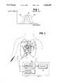
- FIG. 2comprises a block diagram of a system according to the present invention during imaging of human myocardium
- FIG. 3comprises a pair of curves illustrating exemplary reflected baseline and post-introduction frequency spectra detected by the system of the present invention.
- FIGS. 4-7are block diagrams of systems comprising alternative embodiments of the present invention.
- the system 10includes a transducer 14, which may comprise, for example, a piezoelectric element that may be driven over a band or spectrum of frequencies with a center frequency of, for example, 2.5 megahertz. This center frequency and the shape of the spectrum may be varied to obtain optimum imaging as needed.
- the transduceris in turn coupled to a frequency detection circuit 16 that detects one or more parameters of the ultrasonic energy reflected by the body tissues and imaging circuitry 17.
- the system 10is particularly useful in the examination of blood flow or perfusion through human tissues using a contrast enhancing agent.
- the contrast agentpreferably comprises ALBUNEX® (a registered trademark of Molecular Biosystems, Inc. of San Diego, California).
- This contrast agentincludes sonicated microbubbles or microspheres formed of denatured proteins or derivatives thereof obtained from an aqueous protein solution of human serum albumin.
- the contrast agentmay be introduced into the tissue via a vein or artery, inasmuch as the microbubbles have a size which permits their passage through the capillaries of the lungs and into the myocardium 12.
- contrast microbubbleshave a resonant in-vivo frequency.
- This resonant frequencydepends upon a number of factors including the size of the microbubble or microsphere and the surrounding medium, pressure and temperature.
- the present inventionutilizes the theory that because microbubbles have a resonant frequency, they should not be perfect, uniform scatterers of all ultrasound frequencies.
- the amount of ultrasound energy backscattereddepends upon the specific frequency of the ultrasound. This effect permits detection of perfusion by analyzing changes in the backscattered frequency spectrum from diagnostic ultrasound pulses when microbubbles are delivered into the tissue. This change in the frequency spectrum of backscattered ultrasound pulses is greater in statistical significance than changes in the amplitude (i.e. intensity) of the backscattered signal and provides the basis for more sensitive detection of ultrasonic contrast agents.
- microbubble invivo characteristicsare such that its resonant frequency is low when compared to the transducer center frequency, then attenuation of the lower frequency components of the backscattered signal will result a net upward shift in the mean frequency of the backscattered ultrasound signal.
- the resonant frequency of the microbubblesis higher than the transducer center frequency, then attenuation of the higher frequency components will result in a downward shift in the mean frequency. Therefore, this effect may be detected using known commercially available diagnostic ultrasound imaging systems together with additional components to provide an alternative method of evaluating contrast microbubbles within body tissue.
- the tissue undergoing examinationis insonated a first time in the absence of a contrast agent therein and reflected ultrasonic energy is detected and converted by the transducer 14 into a baseline reflection signal, which may comprise a voltage waveform.
- this baseline signalmay have a frequency spectrum represented by the curve 19a.
- a frequency characteristic of the baseline reflection signalis detected by the detection circuit 16 and is stored in a memory (described hereinafter).
- the tissue undergoing examinationis also insonated when a contrast agent is present therein (either before or after the baseline detection), and the reflected ultrasonic energy is received and converted by the transducer 14 into a post-introduction reflection signal. Again, this signal may be a voltage waveform. As seen in FIG.
- the presence of the contrast agentmay change the shape of the reflected frequency spectrum to the curve 19b, for example.
- a frequency characteristic of the post-introduction reflection signalis detected by the detection circuit 16, compared against the frequency characteristic stored in the memory and the display 18 is operated in accordance with the comparison to provide an indication of perfusion or blood flow.
- FIG. 4illustrates a first embodiment of the present invention in greater detail.
- the transducer 14is periodically excited by an oscillator 20 to sequentially provide ultrasonic energy over a first or incident frequency spectrum down a series of scan lines.
- the ultrasonic energy reflected by the tissues being examinedis converted by the transducer 14 into a reflection signal, which is amplified to a proper signal level by a transducer receiver circuit 22.
- the reflection signalhas a second or reflected frequency spectrum.
- the receiver circuit 22may receive a blanking signal during transmission of ultrasonic energy by the transducer 14 until transmission of the energy for that scan line is complete and once a suitable ring down period has expired. The blanking signal is then removed, allowing the receiver 22 to receive the reflected signal from the transducer 14.
- a conventional image processing circuit 24samples the reflection signal at spaced points thereof and integrates the resulting information with data obtained from other scan lines to obtain video display data which is provided to a combiner 26 and the display 18. This data typically results in a grey scale, real-time image of the tissue on the display 18.
- the reflection signal from the receiver circuit 22is also provided to a frequency detector in the form of a frequency analyzer 28 that detects one or more frequency characteristics of the reflection signal.
- the resonant frequency of the microbubbles in the tissue undergoing examinationis determined and the transducer 14 is excited to produce a band of frequencies that includes the microbubble in-vivo resonant frequency.
- the center frequency of the transduceris not coincident with the microbubble in-vivo resonant frequency, but is spaced therefrom in the frequency spectrum.
- the frequency component detector 28analyzes the backscattered frequency spectrum at multiple points over each scan line in the imaging plane in real time using a Fast Fourier transformation algorithm or a Chirp-Z algorithm. During analysis of the baseline reflection signal, the magnitudes or amplitudes of first and second frequency components in the voltage signal developed by the transducer are detected.
- the first frequencyis substantially equal to the microbubble in-vivo resonant frequency and the second frequency is at a selected frequency within the response band of the transducer but removed or spaced from the resonant frequency.
- the second frequencyis substantially coincident with the transducer center frequency.
- the color codermay simply be a lookup table that converts the comparator output into color display information that is combined by the combiner 26 with the video display data developed by the image processing circuit 24.
- the display 18thus displays a grey scale image of the tissue with color superimposed thereon representing the travel of contrast agent through blood vessels therein.
- the resulting display imagemay be static (or frozen) or real-time. Alternatively, a combination of real-time and post-processed images may be shown.
- backscattered ultrasound datashould be analyzed and compared at identical phases of the cardiac cycle.
- triggeringoccurs at diastole, when the heart is at rest.
- This triggeringis accomplished by a triggering circuit 36 that is responsive to an electrocardiogram (EKG) waveform developed by suitable monitoring apparatus (not shown).
- EKGelectrocardiogram
- the circuit 36allows a full frame of scan line data to pass to the frequency analyzer 28.
- triggeringor capture of data may occur at multiple points in the cardiac cycle, if desired.
- Such triggeringis accomplished in conventional ultrasound machines and will be readily apparent to one of ordinary skill in the art.
- the detected frequency characteristicsmay be analyzed in any of a number of different ways in order to obtain an indication of perfusion.
- the ratio A1/A2 of the first and second frequency component amplitudesmay be obtained from the baseline reflection signal and compared with the ratio A3/A4 of the first and second frequency component amplitudes in the post-introduction reflection signal. If the difference between these ratios exceeds a reference level (set by a signal-to-noise ratio), the color coder develops a display signal that causes the display 18 to display a particular color at a corresponding point in the image with an intensity that varies with the amplitude of the ratio difference. Alternatively, the hue of the displayed color may be varied with the magnitude of the ratio difference.
- the frequency analyzer 28may detect a characteristic of the overall frequency spectra of the reflected signals, rather than a characteristic of one or more frequency components.
- the width of the frequency spectrum of a reflected signal, the mean frequency, the skewness or kurtosis of the spectrum or the likemay be detected in the absence and presence of contrast agent in the tissue and these frequency parameters may be compared by the comparator 32 and the results of the comparison passed to the color coder 34 for development of color display data. In this way, a spatial representation of the distribution of the microbubbles within the tissues visualized in the imaging scan plane may be obtained.
- the frequency analyzer 28(FIG. 4) is illustrated as comprising a zero crossing detector or an autocorrelation frequency estimator 40.
- Backscattered frequency informationmay be derived from multiple points over the image plane by utilizing a zero crossing detector that counts how many times the reflected signal passes through zero within a specific time period. The number of times the reflected signal passes through zero is roughly proportional to the frequency of the waveform over that time and space. While this conventional method of obtaining frequency information is rather crude, such an analysis can be undertaken rapidly and is therefore suitable for real time applications.
- FIG. 6illustrates a preferred form of the present invention. Again, elements common to FIGS. 4 and 6 are assigned like reference numerals.
- the frequency analyzer 28 of FIG. 4is illustrated as comprising first and second narrow bandpass filters 42, 44 and a divider 46.
- One of the bandpass filters 42, 44has a center frequency coincident with the in-vivo resonant frequency of the contrast microbubbles.
- the other bandpass filterhas a center frequency removed from the center frequency of the first bandpass filter but within the transducer response frequency band.
- the amplitudes of the output signals of the bandpass filters 42, 44are divided by the divider 46 both during baseline measurement (during which the result of the division is stored in the baseline memory 30) and after the introduction of contrast microbubbles into the tissue undergoing study. As before, the ratios are compared by the comparator 32 and the color coder 34 develops display information in accordance with the comparison which is combined by the combiner 26 with the conventional image data and provided to the display 18.
- one or both of the filters 42 and 44may be a variable bandpass filter that may be tuned or set to select a predetermined frequency to provide greater flexibility.
- different contrast agentsmay have different resonant frequencies in tissue.
- one of the filters 42 and 44may be tuned or set to pass reflected energy having a frequency corresponding to the resonant frequency in tissue of the selected contrast agent, and the other filter, for example, may be set or tuned to the center frequency of the transducer 14.
- FIG. 7illustrates yet another alternative embodiment.
- the oscillator 20, transducer receiver circuit 22 and triggering circuitry 36are not shown for purposes of simplicity.
- the single transducer 14is replaced by two transducers 50, 52.
- the second transducer 52develops ultrasonic energy at a center frequency substantially coincident with the in-vivo resonant frequency of the contrast microbubbles.
- the first transducer 50develops ultrasonic energy at a reference frequency other than the resonant frequency.
- the two transducersmay be replaced by a single transducer alternately driven at the reference and resonant frequencies.
- the reflected signals developed by the transducers 50 and 52are provided to first and second amplitude detectors 54, 56 that replace the frequency analyzer 28 of FIG. 4 and that detect the amplitudes of the reference and resonant frequency components in the baseline and post-introduction reflection signals, respectively.
- the frequency detector 56may detect the amplitude of a harmonic of the resonant frequency.
- the reflection signal from the first transducer 50is utilized by the conventional image processing circuit 24 to obtain the tissue display data.
- the outputs of the amplitude detectors 54 and 56are provided to the divider 46, which in turn obtains baseline and post-introduction ratios that are compared by the comparator 32 as before. Also as previously noted, the output of the comparator 32 is provided by the color coder 34 and the combiner 26 to the display 18. Sequential ultrasound pulses at the resonant and reference frequencies are transmitted down each scan line and the ratios of backscattered amplitudes from both frequency pulses are examined at multiple positions down the image scan lines. Because of the frequency dependent effect of the backscatter, the ratios of signal amplitudes from the different frequency pulses are modified by the introduction of contrast microbubbles within the tissues. The baseline ratio is determined in the absence of microbubbles in the tissues and then the magnitude of a shift in the ratio caused by introduction of contrast agent is determined at multiple points over the image to obtain an indication of microbubble distribution within the tissues.
Landscapes
- Health & Medical Sciences (AREA)
- Life Sciences & Earth Sciences (AREA)
- Physics & Mathematics (AREA)
- Engineering & Computer Science (AREA)
- Radar, Positioning & Navigation (AREA)
- Remote Sensing (AREA)
- Surgery (AREA)
- Heart & Thoracic Surgery (AREA)
- Hematology (AREA)
- Veterinary Medicine (AREA)
- Biophysics (AREA)
- Nuclear Medicine, Radiotherapy & Molecular Imaging (AREA)
- Pathology (AREA)
- Radiology & Medical Imaging (AREA)
- Biomedical Technology (AREA)
- Public Health (AREA)
- Medical Informatics (AREA)
- Molecular Biology (AREA)
- Acoustics & Sound (AREA)
- Animal Behavior & Ethology (AREA)
- General Health & Medical Sciences (AREA)
- General Physics & Mathematics (AREA)
- Computer Networks & Wireless Communication (AREA)
- Ultra Sonic Daignosis Equipment (AREA)
- Medicines Containing Antibodies Or Antigens For Use As Internal Diagnostic Agents (AREA)
Abstract
Description
Claims (48)
Priority Applications (8)
| Application Number | Priority Date | Filing Date | Title |
|---|---|---|---|
| US07/816,640US5255683A (en) | 1991-12-30 | 1991-12-30 | Methods of and systems for examining tissue perfusion using ultrasonic contrast agents |
| EP93901396AEP0626822B1 (en) | 1991-12-30 | 1992-12-16 | System for examination of tissue perfusion with ultrasound contrast agent |
| CA002127127ACA2127127A1 (en) | 1991-12-30 | 1992-12-16 | Ultrasound contrast agent examination of tissue perfusion |
| JP5511697AJPH07506010A (en) | 1991-12-30 | 1992-12-16 | Examination of tissue perfusion using ultrasound contrast agents |
| AU32756/93AAU3275693A (en) | 1991-12-30 | 1992-12-16 | Ultrasound contrast agent examination of tissue perfusion |
| DE69232490TDE69232490T2 (en) | 1991-12-30 | 1992-12-16 | DEVICE FOR TISSUE EXAMINATION USING ULTRASONIC CONTRASTING AGENTS |
| PCT/US1992/010689WO1993012720A1 (en) | 1991-12-30 | 1992-12-16 | Ultrasound contrast agent examination of tissue perfusion |
| IL10415992AIL104159A (en) | 1991-12-30 | 1992-12-18 | Methods of and systems for examining tissue perfusion using ultrasonic contrast agents |
Applications Claiming Priority (1)
| Application Number | Priority Date | Filing Date | Title |
|---|---|---|---|
| US07/816,640US5255683A (en) | 1991-12-30 | 1991-12-30 | Methods of and systems for examining tissue perfusion using ultrasonic contrast agents |
Publications (1)
| Publication Number | Publication Date |
|---|---|
| US5255683Atrue US5255683A (en) | 1993-10-26 |
Family
ID=25221229
Family Applications (1)
| Application Number | Title | Priority Date | Filing Date |
|---|---|---|---|
| US07/816,640Expired - LifetimeUS5255683A (en) | 1991-12-30 | 1991-12-30 | Methods of and systems for examining tissue perfusion using ultrasonic contrast agents |
Country Status (8)
| Country | Link |
|---|---|
| US (1) | US5255683A (en) |
| EP (1) | EP0626822B1 (en) |
| JP (1) | JPH07506010A (en) |
| AU (1) | AU3275693A (en) |
| CA (1) | CA2127127A1 (en) |
| DE (1) | DE69232490T2 (en) |
| IL (1) | IL104159A (en) |
| WO (1) | WO1993012720A1 (en) |
Cited By (123)
| Publication number | Priority date | Publication date | Assignee | Title |
|---|---|---|---|---|
| US5410516A (en)* | 1988-09-01 | 1995-04-25 | Schering Aktiengesellschaft | Ultrasonic processes and circuits for performing them |
| US5431161A (en)* | 1993-04-15 | 1995-07-11 | Adac Laboratories | Method and apparatus for information acquistion, processing, and display within a medical camera system |
| US5433207A (en)* | 1993-11-15 | 1995-07-18 | Pretlow, Iii; Robert A. | Method and apparatus to characterize ultrasonically reflective contrast agents |
| US5456257A (en)* | 1994-11-23 | 1995-10-10 | Advanced Technology Laboratories, Inc. | Ultrasonic detection of contrast agents |
| US5526816A (en)* | 1994-09-22 | 1996-06-18 | Bracco Research S.A. | Ultrasonic spectral contrast imaging |
| DE19548988A1 (en)* | 1994-12-28 | 1996-07-11 | Toshiba Kawasaki Kk | Ultrasonic probe for contrast echography |
| US5560364A (en)* | 1995-05-12 | 1996-10-01 | The Board Of Regents Of The University Of Nebraska | Suspended ultra-sound induced microbubble cavitation imaging |
| DE19619808A1 (en)* | 1995-05-15 | 1996-11-21 | Toshiba Kawasaki Kk | Ultrasound imaging and diagnostic system |
| US5601086A (en)* | 1995-05-12 | 1997-02-11 | The United States Of America As Represented By The Administrator Of The National Aeronautics And Space Administration | Beat frequency ultrasonic microsphere contrast agent detection system |
| US5678553A (en)* | 1994-11-01 | 1997-10-21 | Schering Aktiengesellschaft | Ultrasonic processes and circuits for carrying out those processes |
| US5694937A (en)* | 1995-01-31 | 1997-12-09 | Kabushiki Kaisha Toshiba | Ultrasound diagnostic apparatus and method |
| US5701899A (en)* | 1993-05-12 | 1997-12-30 | The Board Of Regents Of The University Of Nebraska | Perfluorobutane ultrasound contrast agent and methods for its manufacture and use |
| US5706819A (en)* | 1995-10-10 | 1998-01-13 | Advanced Technology Laboratories, Inc. | Ultrasonic diagnostic imaging with harmonic contrast agents |
| US5733527A (en)* | 1994-09-28 | 1998-03-31 | Alliance Pharmaceutical Corp. | Methods for harmonic imaging with ultrasound |
| US5743266A (en)* | 1995-04-25 | 1998-04-28 | Molecular Biosystems, Inc. | Method for processing real-time contrast enhanced ultrasonic images |
| WO1998046139A1 (en)* | 1997-04-11 | 1998-10-22 | Acuson Corporation | Ultrasound imaging enhancement methods and systems |
| US5833614A (en)* | 1997-07-15 | 1998-11-10 | Acuson Corporation | Ultrasonic imaging method and apparatus for generating pulse width modulated waveforms with reduced harmonic response |
| US5833613A (en)* | 1996-09-27 | 1998-11-10 | Advanced Technology Laboratories, Inc. | Ultrasonic diagnostic imaging with contrast agents |
| US5833615A (en)* | 1997-05-09 | 1998-11-10 | Thomas Jefferson University | Excitation enhanced ultrasound system |
| US5846202A (en)* | 1996-07-30 | 1998-12-08 | Acuson Corporation | Ultrasound method and system for imaging |
| US5849727A (en)* | 1996-06-28 | 1998-12-15 | Board Of Regents Of The University Of Nebraska | Compositions and methods for altering the biodistribution of biological agents |
| US5860931A (en)* | 1997-10-10 | 1999-01-19 | Acuson Corporation | Ultrasound method and system for measuring perfusion |
| US5873830A (en)* | 1997-08-22 | 1999-02-23 | Acuson Corporation | Ultrasound imaging system and method for improving resolution and operation |
| US5873829A (en)* | 1996-01-29 | 1999-02-23 | Kabushiki Kaisha Toshiba | Diagnostic ultrasound system using harmonic echo imaging |
| US5879303A (en)* | 1996-09-27 | 1999-03-09 | Atl Ultrasound | Ultrasonic diagnostic imaging of response frequency differing from transmit frequency |
| US5882306A (en)* | 1997-04-11 | 1999-03-16 | Acuson Corporation | Ultrasound imaging methods and systems |
| WO1999017808A1 (en)* | 1997-10-03 | 1999-04-15 | University Of Virginia | Ultrasound bubble recognition imaging |
| US5897500A (en)* | 1997-12-18 | 1999-04-27 | Acuson Corporation | Ultrasonic imaging system and method for displaying composite fundamental and harmonic images |
| US5908389A (en)* | 1996-09-27 | 1999-06-01 | Atl Ultrasound, Inc. | Ultrasonic diagnostic imaging of harmonic frequencies with speckle reduction processing |
| US5913823A (en)* | 1997-07-15 | 1999-06-22 | Acuson Corporation | Ultrasound imaging method and system for transmit signal generation for an ultrasonic imaging system capable of harmonic imaging |
| US5924991A (en)* | 1997-08-22 | 1999-07-20 | Acuson Corporation | Ultrasonic system and method for harmonic imaging in three dimensions |
| US5933389A (en)* | 1995-03-02 | 1999-08-03 | Acuson Corporation | Ultrasonic imaging system and method |
| US5935069A (en)* | 1997-10-10 | 1999-08-10 | Acuson Corporation | Ultrasound system and method for variable transmission of ultrasonic signals |
| US5944666A (en)* | 1997-08-21 | 1999-08-31 | Acuson Corporation | Ultrasonic method for imaging blood flow including disruption or activation of contrast agent |
| US5957852A (en)* | 1998-06-02 | 1999-09-28 | Acuson Corporation | Ultrasonic harmonic imaging system and method |
| US5957845A (en)* | 1997-04-11 | 1999-09-28 | Acuson Corporation | Gated ultrasound imaging apparatus and method |
| US5961464A (en)* | 1998-09-16 | 1999-10-05 | Hewlett-Packard Company | Ultrasound contrast agent detection using spectral analysis from acoustic scan lines |
| US5971928A (en)* | 1998-11-02 | 1999-10-26 | Acuson Corporation | Diagnostic medical ultrasonic system and method for image subtraction |
| US5976088A (en)* | 1998-06-24 | 1999-11-02 | Ecton, Inc. | Ultrasound imaging systems and methods of increasing the effective acquisition frame rate |
| US6005827A (en)* | 1995-03-02 | 1999-12-21 | Acuson Corporation | Ultrasonic harmonic imaging system and method |
| US6004270A (en)* | 1998-06-24 | 1999-12-21 | Ecton, Inc. | Ultrasound system for contrast agent imaging and quantification in echocardiography using template image for image alignment |
| US6009046A (en)* | 1995-03-02 | 1999-12-28 | Acuson Corporation | Ultrasonic harmonic imaging system and method |
| US6014473A (en)* | 1996-02-29 | 2000-01-11 | Acuson Corporation | Multiple ultrasound image registration system, method and transducer |
| US6015384A (en)* | 1998-08-31 | 2000-01-18 | Acuson Corporation | Ultrasonic system and method for tissue viability imaging |
| US6023977A (en)* | 1997-08-01 | 2000-02-15 | Acuson Corporation | Ultrasonic imaging aberration correction system and method |
| US6027448A (en)* | 1995-03-02 | 2000-02-22 | Acuson Corporation | Ultrasonic transducer and method for harmonic imaging |
| US6030344A (en)* | 1996-12-04 | 2000-02-29 | Acuson Corporation | Methods and apparatus for ultrasound image quantification |
| US6034922A (en)* | 1988-09-01 | 2000-03-07 | Schering Aktiengesellschaft | Ultrasonic processes and circuits for performing them |
| US6039690A (en)* | 1997-06-17 | 2000-03-21 | Acuson Corporation | Method and apparatus for frequency control of an ultrasound system |
| US6048316A (en)* | 1998-10-16 | 2000-04-11 | Acuson Corporation | Medical diagnostic ultrasonic imaging system and method for displaying composite fundamental and harmonic images |
| US6080107A (en)* | 1999-01-26 | 2000-06-27 | Hewlett-Packard Company | Methods for the use of contrast agents in ultrasonic imaging |
| US6104670A (en)* | 1995-03-02 | 2000-08-15 | Acuson Corporation | Ultrasonic harmonic imaging system and method |
| US6106465A (en)* | 1997-08-22 | 2000-08-22 | Acuson Corporation | Ultrasonic method and system for boundary detection of an object of interest in an ultrasound image |
| US6132377A (en)* | 1999-03-31 | 2000-10-17 | Acuson Corporation | Medical diagnostic ultrasonic imaging system and method using differential sub-band detection techniques |
| US6132374A (en)* | 1997-08-01 | 2000-10-17 | Acuson Corporation | Ultrasonic imaging method and system |
| US6171246B1 (en) | 1999-04-29 | 2001-01-09 | Michalakis Averkiou | Realtime ultrasonic imaging of perfusion using ultrasonic contrast agents |
| US6171245B1 (en)* | 1998-03-12 | 2001-01-09 | Siemens Medical Systems, Inc. | Method of imaging scatterers based on acoustically stimulated changes of their acoustic properties |
| US6174287B1 (en) | 1999-06-11 | 2001-01-16 | Acuson Corporation | Medical diagnostic ultrasound system and method for continuous M-mode imaging and periodic imaging of contrast agents |
| US6193659B1 (en) | 1997-07-15 | 2001-02-27 | Acuson Corporation | Medical ultrasonic diagnostic imaging method and apparatus |
| US6206833B1 (en) | 1996-11-08 | 2001-03-27 | Research Corporation Technologiers, Inc. | Finite amplitude distortion-based inhomogeneous pulse echo ultrasonic imaging |
| US6213946B1 (en) | 1998-12-24 | 2001-04-10 | Agilent Technologies, Inc. | Methods and apparatus for speckle reduction by orthogonal pulse compounding in medical ultrasound imaging |
| US6245747B1 (en) | 1996-03-12 | 2001-06-12 | The Board Of Regents Of The University Of Nebraska | Targeted site specific antisense oligodeoxynucleotide delivery method |
| US6251074B1 (en) | 1996-11-26 | 2001-06-26 | Atl Ultrasound | Ultrasonic tissue harmonic imaging |
| US6254541B1 (en)* | 1997-09-23 | 2001-07-03 | Scimed Life Systems, Inc. | Methods and apparatus for blood speckle detection in an intravascular ultrasound imaging system |
| USD445189S1 (en) | 1999-09-14 | 2001-07-17 | Ecton, Inc. | Medical diagnostic ultrasound system |
| US6312381B1 (en) | 1999-09-14 | 2001-11-06 | Acuson Corporation | Medical diagnostic ultrasound system and method |
| US6312379B1 (en) | 1997-08-15 | 2001-11-06 | Acuson Corporation | Ultrasonic harmonic imaging system and method using waveform pre-distortion |
| US6322512B1 (en)* | 1997-01-22 | 2001-11-27 | Acuson Corporation | Ultrasound contrast imaging |
| US6340348B1 (en) | 1999-07-02 | 2002-01-22 | Acuson Corporation | Contrast agent imaging with destruction pulses in diagnostic medical ultrasound |
| US20020031476A1 (en)* | 1993-07-30 | 2002-03-14 | Trevino Leo A. | Stabilized gas emulsion containing phospholipid for ultrasound contrast enhancement |
| US6385474B1 (en) | 1999-03-19 | 2002-05-07 | Barbara Ann Karmanos Cancer Institute | Method and apparatus for high-resolution detection and characterization of medical pathologies |
| US20020054854A1 (en)* | 1993-07-30 | 2002-05-09 | Schutt Ernest G. | Stabilized microbubble compositions |
| US6409671B1 (en)* | 1997-12-18 | 2002-06-25 | Nycomed Imaging As | Ultrasonography |
| US6436039B1 (en) | 1999-09-14 | 2002-08-20 | Ecton, Inc. | Medicial diagnostic ultrasound system and method |
| US6458083B1 (en) | 1996-11-26 | 2002-10-01 | Koninklijke Philips Electronics N.V. | Ultrasonic harmonic imaging with adaptive image formation |
| US6468213B1 (en) | 1999-09-14 | 2002-10-22 | Ecton, Inc. | Medical diagnostic ultrasound system and method |
| US6488625B1 (en) | 1999-09-14 | 2002-12-03 | Ecton, Inc. | Medical diagnostic ultrasound system and method |
| US6497666B1 (en) | 2000-10-25 | 2002-12-24 | Acuson Corporation | Medical ultrasonic contrast agent imaging method and apparatus |
| US6497664B1 (en) | 1999-09-14 | 2002-12-24 | Ecton, Inc. | Medical diagnostic ultrasound system and method |
| US20030004414A1 (en)* | 2001-05-31 | 2003-01-02 | Mclaughlin Glen | System and method for phase inversion ultrasonic imaging |
| US6508763B1 (en) | 1999-09-14 | 2003-01-21 | Ecton, Inc. | Medical diagnostic ultrasound system and method |
| US6524244B1 (en) | 1999-09-14 | 2003-02-25 | Ecton Inc. | Medical diagnostic ultrasound system and method |
| US6527718B1 (en) | 1999-08-20 | 2003-03-04 | Brian G Connor | Ultrasound system for continuous imaging and delivery of an encapsulated agent |
| US6533728B1 (en) | 2001-11-20 | 2003-03-18 | Mayo Foundation For Medical Education And Research | Method and apparatus for recovery and parametric display of contrast agents in ultrasound imaging |
| US6537222B1 (en)* | 1997-08-26 | 2003-03-25 | Koninklijke Philips Electronics N.V. | Methods for the detection of contrast agents in ultrasonic imaging |
| US6544177B1 (en) | 1998-10-01 | 2003-04-08 | Atl Ultrasound, Inc. | Ultrasonic diagnostic imaging system and method with harmonic spatial compounding |
| US6547738B2 (en)* | 2001-05-03 | 2003-04-15 | Ge Medical Systems Global Technology Company, Llc | Methods and apparatus for using ultrasound with contrast agent |
| US6561979B1 (en) | 1999-09-14 | 2003-05-13 | Acuson Corporation | Medical diagnostic ultrasound system and method |
| US6582370B2 (en)* | 2001-02-01 | 2003-06-24 | Ge Medical Systems Global Technology Company, Llc | Blood flow imaging method, blood flow imaging apparatus and ultrasonic diagnostic apparatus |
| US20040030227A1 (en)* | 2002-05-16 | 2004-02-12 | Barbara Ann Karmanos Cancer Institute | Method and apparatus for combined diagnostic and therapeutic ultrasound system incorporating noninvasive thermometry, ablation control and automation |
| US6692438B2 (en)* | 2001-12-18 | 2004-02-17 | Koninklijke Philips Electronics Nv | Ultrasonic imaging system and method for displaying tissue perfusion and other parameters varying with time |
| US6726629B1 (en) | 1998-01-16 | 2004-04-27 | Acuson Corporation | Ultrasound contrast imaging |
| US20040097809A1 (en)* | 2002-11-11 | 2004-05-20 | Sei Kato | Ultrasonic diagnostic apparatus |
| US6740039B1 (en) | 1999-08-20 | 2004-05-25 | Koninklijke Philips Electronics N.V. | Methods and apparatus for displaying information relating to delivery and activation of a therapeutic agent using ultrasound energy |
| US20040122313A1 (en)* | 2002-12-18 | 2004-06-24 | Barbara Ann Karmanos Cancer Institute | Methods and systems for using reference images in acoustic image processing |
| US20040122325A1 (en)* | 2002-12-18 | 2004-06-24 | Barbara Ann Karmanos Cancer Institute | Diagnostic analysis of ultrasound data |
| US20040122322A1 (en)* | 2002-12-18 | 2004-06-24 | Barbara Ann Karmanos Cancer Institute | Electret acoustic transducer array for computerized ultrasound risk evaluation system |
| US20040225255A1 (en)* | 2003-04-28 | 2004-11-11 | Nemoto Kyorindo Co., Ltd. | Leak detector for detecting leak of liquid injected into blood vessel using pulse signal |
| US20050074406A1 (en)* | 2003-10-03 | 2005-04-07 | Scimed Life Systems, Inc. | Ultrasound coating for enhancing visualization of medical device in ultrasound images |
| US20050131294A1 (en)* | 2001-10-20 | 2005-06-16 | Zonare Medical Systems, Inc. | Ultrasound system for generating a single set of ultrasound pulse firings |
| USRE38971E1 (en)* | 1995-01-31 | 2006-02-07 | Kabushiki Kaisha Toshiba | Ultrasound diagnostic apparatus and method |
| US7047058B1 (en)* | 2001-02-06 | 2006-05-16 | Medrad, Inc. | Apparatuses, systems and methods for extravasation detection |
| WO2005096694A3 (en)* | 2004-04-08 | 2007-01-18 | Yeda Res & Dev | Three time point lung cancer detection, diagnosis and assessment of prognosis |
| US20070225637A1 (en)* | 2004-09-14 | 2007-09-27 | Seiichi Ono | Leak Detecting Apparatus |
| US20080275344A1 (en)* | 2007-05-04 | 2008-11-06 | Barbara Ann Karmanos Cancer Institute | Method and Apparatus for Categorizing Breast Density and Assessing Cancer Risk Utilizing Acoustic Parameters |
| US20090054775A1 (en)* | 2007-08-21 | 2009-02-26 | Sei Kato | Ultrasonic diagnostic apparatus and ultrasonic imaging method |
| US20090177087A1 (en)* | 2007-06-28 | 2009-07-09 | Sei Kato | Ultrasonic imaging apparatus |
| US7678048B1 (en) | 1999-09-14 | 2010-03-16 | Siemens Medical Solutions Usa, Inc. | Medical diagnostic ultrasound system and method |
| US20110201928A1 (en)* | 2010-02-12 | 2011-08-18 | Nebojsa Duric | Method of characterizing the pathological response of tissue to a treatment plan |
| US20110201932A1 (en)* | 2010-02-12 | 2011-08-18 | Nebojsa Duric | Method of characterizing tissue of a patient |
| US20120027282A1 (en)* | 2009-04-10 | 2012-02-02 | Hitachi Medical Corporation | Ultrasonic diagnosis apparatus and method for constructing distribution image of blood flow dynamic state |
| US8226561B2 (en) | 1999-08-20 | 2012-07-24 | Zonare Medical Systems, Inc. | Ultrasound imaging system |
| EP2752157A1 (en)* | 2013-01-08 | 2014-07-09 | Nihon Kohden Corporation | Biological signal averaging processing device |
| US20170124426A1 (en)* | 2015-11-03 | 2017-05-04 | Toshiba Medical Systems Corporation | Ultrasound diagnosis apparatus, image processing apparatus and image processing method |
| US20170168149A1 (en)* | 2015-12-09 | 2017-06-15 | University Of Rochester | Scattering and Reflection Identification System and Method |
| US9763641B2 (en) | 2012-08-30 | 2017-09-19 | Delphinus Medical Technologies, Inc. | Method and system for imaging a volume of tissue with tissue boundary detection |
| US10123770B2 (en) | 2013-03-13 | 2018-11-13 | Delphinus Medical Technologies, Inc. | Patient support system |
| US10143443B2 (en) | 2014-05-05 | 2018-12-04 | Delphinus Medical Technologies, Inc. | Method for representing tissue stiffness |
| US10201324B2 (en) | 2007-05-04 | 2019-02-12 | Delphinus Medical Technologies, Inc. | Patient interface system |
| US10285667B2 (en) | 2014-08-05 | 2019-05-14 | Delphinus Medical Technologies, Inc. | Method for generating an enhanced image of a volume of tissue |
| US10722136B2 (en) | 2011-09-02 | 2020-07-28 | Battelle Memorial Institute | Wireless and power-source-free extravasation and infiltration detection sensor |
| US10743837B2 (en) | 2014-08-04 | 2020-08-18 | Delphinus Medical Technologies, Inc. | Ultrasound waveform tomography method and system |
| US12153130B2 (en) | 2015-12-09 | 2024-11-26 | University Of Rochester | Fine-tuning the h-scan for visualizing types of tissue scatterers |
Families Citing this family (5)
| Publication number | Priority date | Publication date | Assignee | Title |
|---|---|---|---|---|
| JP3459304B2 (en) | 1995-01-31 | 2003-10-20 | 株式会社東芝 | Ultrasound diagnostic equipment |
| ATE285711T1 (en)* | 1995-10-10 | 2005-01-15 | Advanced Tech Lab | ULTRASONIC IMAGING FOR DIAGNOSTICS USING CONTRAST AGENTS |
| US6086540A (en)* | 1997-10-07 | 2000-07-11 | Molecular Biosystems, Inc. | Methods of ultrasound imaging using echogenically persistent contrast agents |
| GB9726664D0 (en) | 1997-12-17 | 1998-02-18 | Nycomed Imaging As | Improvements in or relating to ultrasonography |
| KR101239809B1 (en)* | 2010-05-07 | 2013-03-06 | 삼성메디슨 주식회사 | Ultrasound imaging device and method for clutter filtering |
Citations (14)
| Publication number | Priority date | Publication date | Assignee | Title |
|---|---|---|---|---|
| US3640271A (en)* | 1969-06-30 | 1972-02-08 | Ibm | Blood flow pressure measurement technique employing injected bubbled and ultrasonic frequency scanning |
| US4122713A (en)* | 1977-05-19 | 1978-10-31 | Medtronic, Inc. | Liquid velocity measuring system |
| US4140022A (en)* | 1977-12-20 | 1979-02-20 | Hewlett-Packard Company | Acoustic imaging apparatus |
| US4265251A (en)* | 1979-06-28 | 1981-05-05 | Rasor Associates, Inc. | Method of determining pressure within liquid containing vessel |
| US4276885A (en)* | 1979-05-04 | 1981-07-07 | Rasor Associates, Inc | Ultrasonic image enhancement |
| US4316391A (en)* | 1979-11-13 | 1982-02-23 | Ultra Med, Inc. | Flow rate measurement |
| US4466442A (en)* | 1981-10-16 | 1984-08-21 | Schering Aktiengesellschaft | Carrier liquid solutions for the production of gas microbubbles, preparation thereof, and use thereof as contrast medium for ultrasonic diagnostics |
| US4569353A (en)* | 1982-06-03 | 1986-02-11 | The Regents Of The University Of California | Sound attenuation measurement system |
| US4572203A (en)* | 1983-01-27 | 1986-02-25 | Feinstein Steven B | Contact agents for ultrasonic imaging |
| US4619267A (en)* | 1983-12-16 | 1986-10-28 | Cgr Ultrasonic | Method for characterizing the structure of a medium and device for carrying out the method |
| US4718433A (en)* | 1983-01-27 | 1988-01-12 | Feinstein Steven B | Contrast agents for ultrasonic imaging |
| US4803994A (en)* | 1987-08-12 | 1989-02-14 | General Electric Company | Backscatter data collection technique for ultrasound |
| US4827942A (en)* | 1985-06-25 | 1989-05-09 | Hewlett-Packard Company | Flow map apparatus |
| US4844882A (en)* | 1987-12-29 | 1989-07-04 | Molecular Biosystems, Inc. | Concentrated stabilized microbubble-type ultrasonic imaging agent |
Family Cites Families (5)
| Publication number | Priority date | Publication date | Assignee | Title |
|---|---|---|---|---|
| US4152928A (en)* | 1977-05-02 | 1979-05-08 | University Patents, Inc. | System for detecting fat particles in blood |
| JPS5826238A (en)* | 1981-08-08 | 1983-02-16 | Fujitsu Ltd | Ultrasonic pressure measurement method |
| US5040537A (en)* | 1987-11-24 | 1991-08-20 | Hitachi, Ltd. | Method and apparatus for the measurement and medical treatment using an ultrasonic wave |
| DE3829999A1 (en) | 1988-09-01 | 1990-03-15 | Schering Ag | ULTRASONIC METHOD AND CIRCUITS THEREOF |
| GB9009423D0 (en)* | 1990-04-26 | 1990-06-20 | Williams Alun R | Assessment of vascular perfusion by the display of harmonic echoes from ultrasonically excited gas bubbles |
- 1991
- 1991-12-30USUS07/816,640patent/US5255683A/ennot_activeExpired - Lifetime
- 1992
- 1992-12-16DEDE69232490Tpatent/DE69232490T2/ennot_activeExpired - Lifetime
- 1992-12-16JPJP5511697Apatent/JPH07506010A/enactivePending
- 1992-12-16EPEP93901396Apatent/EP0626822B1/ennot_activeExpired - Lifetime
- 1992-12-16CACA002127127Apatent/CA2127127A1/ennot_activeAbandoned
- 1992-12-16AUAU32756/93Apatent/AU3275693A/ennot_activeAbandoned
- 1992-12-16WOPCT/US1992/010689patent/WO1993012720A1/enactiveIP Right Grant
- 1992-12-18ILIL10415992Apatent/IL104159A/ennot_activeIP Right Cessation
Patent Citations (16)
| Publication number | Priority date | Publication date | Assignee | Title |
|---|---|---|---|---|
| US3640271A (en)* | 1969-06-30 | 1972-02-08 | Ibm | Blood flow pressure measurement technique employing injected bubbled and ultrasonic frequency scanning |
| US4122713A (en)* | 1977-05-19 | 1978-10-31 | Medtronic, Inc. | Liquid velocity measuring system |
| US4140022B1 (en)* | 1977-12-20 | 1995-05-16 | Hewlett Packard Co | Acoustic imaging apparatus |
| US4140022A (en)* | 1977-12-20 | 1979-02-20 | Hewlett-Packard Company | Acoustic imaging apparatus |
| US4276885A (en)* | 1979-05-04 | 1981-07-07 | Rasor Associates, Inc | Ultrasonic image enhancement |
| US4265251A (en)* | 1979-06-28 | 1981-05-05 | Rasor Associates, Inc. | Method of determining pressure within liquid containing vessel |
| US4316391A (en)* | 1979-11-13 | 1982-02-23 | Ultra Med, Inc. | Flow rate measurement |
| US4466442A (en)* | 1981-10-16 | 1984-08-21 | Schering Aktiengesellschaft | Carrier liquid solutions for the production of gas microbubbles, preparation thereof, and use thereof as contrast medium for ultrasonic diagnostics |
| US4569353A (en)* | 1982-06-03 | 1986-02-11 | The Regents Of The University Of California | Sound attenuation measurement system |
| US4572203A (en)* | 1983-01-27 | 1986-02-25 | Feinstein Steven B | Contact agents for ultrasonic imaging |
| US4718433A (en)* | 1983-01-27 | 1988-01-12 | Feinstein Steven B | Contrast agents for ultrasonic imaging |
| US4619267A (en)* | 1983-12-16 | 1986-10-28 | Cgr Ultrasonic | Method for characterizing the structure of a medium and device for carrying out the method |
| US4827942A (en)* | 1985-06-25 | 1989-05-09 | Hewlett-Packard Company | Flow map apparatus |
| US4774958A (en)* | 1985-12-05 | 1988-10-04 | Feinstein Steven B | Ultrasonic imaging agent and method of preparation |
| US4803994A (en)* | 1987-08-12 | 1989-02-14 | General Electric Company | Backscatter data collection technique for ultrasound |
| US4844882A (en)* | 1987-12-29 | 1989-07-04 | Molecular Biosystems, Inc. | Concentrated stabilized microbubble-type ultrasonic imaging agent |
Non-Patent Citations (45)
| Title |
|---|
| Abstract entitled Detection of Myocardial Contrast Using Backscatter Spectral Frequency Analysis by Monaghan, et al. appearing in the European Heart Journal, Aug., 1990, p. 113.* |
| Abstract entitled Detection of Myocardial Perfusion Using Intravenous Contrast Echocardiography, presented to the British Cardiac Society and British Heart Journal, May, 1990.* |
| Abstract entitled Digital Acquisition and Processing of Myocardial Contrast Echocardiographic Signals by Monaghan, et al. appearing in Echocardiography 1990 presented at the 7th International Congress on Echocardiography in Rome, Italy in Apr. 1990.* |
| Abstract entitled Digital Radiofrequency Ultrasound Analysis of Myocardial Contrast Following I.V. Albunex Injection by Monaghan, et al. appearing in the European Heart Journal, Aug. 1991.* |
| Article entitled "Digital Echocardiographic Techniques in Myocardial Contrast Echocardiography" by Monaghan, et al. appearing in Echocardiography 1990, A. Dagianti and H. Feigenbaum, eds., pp. 235-244. |
| Article entitled Digital Echocardiographic Techniques in Myocardial Contrast Echocardiography by Monaghan, et al. appearing in Echocardiography 1990, A. Dagianti and H. Feigenbaum, eds., pp. 235 244.* |
| Cardiac Imaging Principles and Practice, Marcus et al. eds., W. B. Sanders Company, Philadelphia, 1990. Chapter 27, Contrast Echocardiography , pp. 557 574 by Feinstein et al.* |
| Cardiac Imaging-Principles and Practice, Marcus et al. eds., W. B. Sanders Company, Philadelphia, 1990. Chapter 27, "Contrast Echocardiography", pp. 557-574 by Feinstein et al. |
| Color Atlas of Real Time Two Dimensional Echocardiography, (R. Omoto, ed.), Shindan To Chiro Co., Ltd., Tokyo (1984) pp. 7 36.* |
| Color Atlas of Real-Time Two-Dimensional Echocardiography, (R. Omoto, ed.), Shindan-To-Chiro Co., Ltd., Tokyo (1984) pp. 7-36. |
| Digital Techniques in Echocardiography, (Jay Roelandt, ed.), Martinus Najhoff Publishers, (1987), chapters 1 5.* |
| Digital Techniques in Echocardiography, (Jay Roelandt, ed.), Martinus Najhoff Publishers, (1987), chapters 1-5. |
| Excerpt from Science News, vol. 140, No. 13, pp. 207, Sep. 28, 1991.* |
| Feinstein, et al., Safety and Efficacy of a New Transpulmonary Ultrasound Contrast Agent: Initial Multicenter Clinical Results, Journal of the American College of Cardiology, vol. 16, No. 2, pp. 316 324, Aug., 1990.* |
| Feinstein, et al., Safety and Efficacy of a New Transpulmonary Ultrasound Contrast Agent: Initial Multicenter Clinical Results, Journal of the American College of Cardiology, vol. 16, No. 2, pp. 316-324, Aug., 1990. |
| Feinstein, Myocardial Perfusion Imaging: Contrast Echocardiography Today and Tomorrow, Journal of the American College of Cardiology, vol. 8, No. 1, Jul., 1986, pp. 251 253.* |
| Feinstein, Myocardial Perfusion Imaging: Contrast Echocardiography Today and Tomorrow, Journal of the American College of Cardiology, vol. 8, No. 1, Jul., 1986, pp. 251-253. |
| Feinstein, New Developments in Ultrasonic Contrast Techniques: Transpulmonary Passage of Contrast Agents and Diagnostic Implications, Echocardiography, vol. 6, No. 1, pp. 27 33, 1989, presented at CardioVision 88 Monaco, Jun. 16 18, 1988.* |
| Feinstein, New Developments in Ultrasonic Contrast Techniques: Transpulmonary Passage of Contrast Agents and Diagnostic Implications, Echocardiography, vol. 6, No. 1, pp. 27-33, 1989, presented at CardioVision '88 Monaco, Jun. 16-18, 1988. |
| Harvey Feigenbaum, Echocardiography, Lea and Febiger, (1981), chapter 1.* |
| Hayes and Griffith, Inc., Basic Report dated Apr. 29, 1987.* |
| Hayes and Griffith, Inc., Update dated Oct. 7, 1987.* |
| Kern, Microbubbles Show Promise for Enhancing Ultrasound Signal, Image, Other Applications, Journal of the American Medical Association (JAMA), vol. 261, No. 11, pp. 1542, Mar. 17, 1989.* |
| Kidder, Peabody Equity Research, Company Analysis dated Feb. 15, 1989.* |
| Kidder, Peabody Equity Research, Company Comment dated Sep. 28, 1989.* |
| Mark J. Monaghan, Practical Echocardiography and Doppler, John Wiley and Sons, (1990, reprinted Jul. 1991), chapters 1 and 11.* |
| Newspaper article entitled "New Technique Locates Hidden Breast Cancer", appearing at pp. 1 and 18 of The Chicago Sun Times dated Dec. 3, 1991. |
| Newspaper article entitled "Ultrasound Enters New Frontiers" appearing at The New York Times dated Nov. 28, 1990. |
| Newspaper article entitled New Technique Locates Hidden Breast Cancer , appearing at pp. 1 and 18 of The Chicago Sun Times dated Dec. 3, 1991.* |
| Newspaper article entitled Ultrasound Enters New Frontiers appearing at The New York Times dated Nov. 28, 1990.* |
| Pages 1 76 of the book by PNT Wells entitled Physical Principles of Ultrasonic Diagnosis , Academic Press, London (1969).* |
| Pages 1-76 of the book by PNT Wells entitled "Physical Principles of Ultrasonic Diagnosis", Academic Press, London (1969). |
| Paper entitled "Myocardial Contrast Echocardiography: Applications of Digital Ultrasound Data Acquisition and Processing" by Monaghan, et al. appearing in the American Journal of Cardiac Imaging, vol. 5, No. 3 (Sep., 1991: pp. 237-249). |
| Paper entitled Myocardial Contrast Echocardiography: Applications of Digital Ultrasound Data Acquisition and Processing by Monaghan, et al. appearing in the American Journal of Cardiac Imaging, vol. 5, No. 3 (Sep., 1991: pp. 237 249).* |
| Powsner, et al., High Speed Interface for Myocardial Sonicated Echocontrast Studies, appearing in SPIE, vol. 845, Visual Communications and Image Processing II, pp. 384 395 (1987).* |
| Powsner, et al., High Speed Interface for Myocardial Sonicated Echocontrast Studies, appearing in SPIE, vol. 845, Visual Communications and Image Processing II, pp. 384-395 (1987). |
| Press Release from Molecular Biosystems, Inc. entitled "Molecular Biosystem's Albunex™ Classified as a Device by FDA". |
| Press Release from Molecular Biosystems, Inc. entitled Molecular Biosystem s Albunex Classified as a Device by FDA .* |
| Product brochure entitled "Albunex Ultrasound Contrast Agent" by Molecular Biosystems, Inc. of San Diego, Ca. |
| Product brochure entitled Albunex Ultrasound Contrast Agent by Molecular Biosystems, Inc. of San Diego, Ca.* |
| Sahn, 3 rd Annual University of Chicago Symposium on Advances in Echocardiography, Sound Practice, vol. 1, pp. 1 8.* |
| Sahn, 3rd Annual University of Chicago Symposium on Advances in Echocardiography, Sound Practice, vol. 1, pp. 1-8. |
| Sutro and Co., Inc., Basic Report dated Aug. 3, 1988.* |
| Syllabus entitled "Advances in Echocardiography: Contrast Echocardiography, Perfusion Imaging, Transesophageal Echo", dated Oct. 4-5, 1991. |
| Syllabus entitled Advances in Echocardiography: Contrast Echocardiography, Perfusion Imaging, Transesophageal Echo , dated Oct. 4 5, 1991.* |
Cited By (230)
| Publication number | Priority date | Publication date | Assignee | Title |
|---|---|---|---|---|
| US6221017B1 (en) | 1988-09-01 | 2001-04-24 | Schering Aktiengesellschaft | Ultrasonic processes and circuits for performing them |
| US6443899B2 (en) | 1988-09-01 | 2002-09-03 | Schering Aktiengesellschaft | Ultrasonic processes and circuits for performing them |
| US5410516A (en)* | 1988-09-01 | 1995-04-25 | Schering Aktiengesellschaft | Ultrasonic processes and circuits for performing them |
| US6034922A (en)* | 1988-09-01 | 2000-03-07 | Schering Aktiengesellschaft | Ultrasonic processes and circuits for performing them |
| US6064628A (en)* | 1988-09-01 | 2000-05-16 | Schering Aktiengesellschaft | Ultrasonic processes and circuits for performing them |
| US5431161A (en)* | 1993-04-15 | 1995-07-11 | Adac Laboratories | Method and apparatus for information acquistion, processing, and display within a medical camera system |
| US5701899A (en)* | 1993-05-12 | 1997-12-30 | The Board Of Regents Of The University Of Nebraska | Perfluorobutane ultrasound contrast agent and methods for its manufacture and use |
| US20040228801A1 (en)* | 1993-07-30 | 2004-11-18 | Schutt Ernest G. | Osmotically stabilized microbubble preparations |
| US20050281747A1 (en)* | 1993-07-30 | 2005-12-22 | Schutt Ernest G | Mixed gas microbubble compositions |
| US20040033199A1 (en)* | 1993-07-30 | 2004-02-19 | Schutt Ernest G. | Ultrasonic imaging system utilizing a long-persistence contrast agent |
| US7005120B2 (en) | 1993-07-30 | 2006-02-28 | Imcor Pharmaceutical Company | Osmotically stabilized microbubble preparations |
| US6953569B2 (en) | 1993-07-30 | 2005-10-11 | Imcor Pharmaceutical Company | Mixed gas microbubble compositions |
| US6939531B2 (en) | 1993-07-30 | 2005-09-06 | Imcor Pharmaceutical Company | Ultrasonic imaging system utilizing a long-persistence contrast agent |
| US7141235B2 (en) | 1993-07-30 | 2006-11-28 | Imcor Pharmaceutical Co. | Stabilized gas emulsion containing phospholipid for ultrasound contrast enhancement |
| US20020098151A1 (en)* | 1993-07-30 | 2002-07-25 | Alliance Pharmaceutical Corp. | Mixed gas microbubble compositions |
| US20020054854A1 (en)* | 1993-07-30 | 2002-05-09 | Schutt Ernest G. | Stabilized microbubble compositions |
| US20020031476A1 (en)* | 1993-07-30 | 2002-03-14 | Trevino Leo A. | Stabilized gas emulsion containing phospholipid for ultrasound contrast enhancement |
| US5433207A (en)* | 1993-11-15 | 1995-07-18 | Pretlow, Iii; Robert A. | Method and apparatus to characterize ultrasonically reflective contrast agents |
| US5526816A (en)* | 1994-09-22 | 1996-06-18 | Bracco Research S.A. | Ultrasonic spectral contrast imaging |
| US5733527A (en)* | 1994-09-28 | 1998-03-31 | Alliance Pharmaceutical Corp. | Methods for harmonic imaging with ultrasound |
| US6019960A (en)* | 1994-09-28 | 2000-02-01 | Alliance Pharmaceutical Corp. | Systems for harmonic ultrasound imaging |
| US6036644A (en)* | 1994-09-28 | 2000-03-14 | Alliance Pharmaceutical Corp. | Enhanced methods of ultrasound imaging using multiple frequencies |
| US6056943A (en)* | 1994-09-28 | 2000-05-02 | Alliance Pharmaceutical Corp. | Methods of ultrasound imaging using phospholipid stabilized microbubbles |
| US20030044355A1 (en)* | 1994-09-28 | 2003-03-06 | Schutt Ernest G. | Harmonic ultrasound imaging with microbubbles |
| US7374744B2 (en) | 1994-09-28 | 2008-05-20 | Imcor Pharmaceutical Co. | Harmonic ultrasound imaging with microbubbles |
| US5678553A (en)* | 1994-11-01 | 1997-10-21 | Schering Aktiengesellschaft | Ultrasonic processes and circuits for carrying out those processes |
| EP0713680A3 (en)* | 1994-11-23 | 1998-08-12 | Advanced Technology Laboratories, Inc. | Ultrasonic detection of contrasting agents |
| US5456257A (en)* | 1994-11-23 | 1995-10-10 | Advanced Technology Laboratories, Inc. | Ultrasonic detection of contrast agents |
| DE19548988C2 (en)* | 1994-12-28 | 2001-02-22 | Toshiba Kawasaki Kk | Ultrasound diagnostic system |
| DE19548988A1 (en)* | 1994-12-28 | 1996-07-11 | Toshiba Kawasaki Kk | Ultrasonic probe for contrast echography |
| USRE38971E1 (en)* | 1995-01-31 | 2006-02-07 | Kabushiki Kaisha Toshiba | Ultrasound diagnostic apparatus and method |
| US5694937A (en)* | 1995-01-31 | 1997-12-09 | Kabushiki Kaisha Toshiba | Ultrasound diagnostic apparatus and method |
| US6027448A (en)* | 1995-03-02 | 2000-02-22 | Acuson Corporation | Ultrasonic transducer and method for harmonic imaging |
| US6104670A (en)* | 1995-03-02 | 2000-08-15 | Acuson Corporation | Ultrasonic harmonic imaging system and method |
| US6122222A (en)* | 1995-03-02 | 2000-09-19 | Acuson Corporation | Ultrasonic transmit and receive system |
| US5933389A (en)* | 1995-03-02 | 1999-08-03 | Acuson Corporation | Ultrasonic imaging system and method |
| US6009046A (en)* | 1995-03-02 | 1999-12-28 | Acuson Corporation | Ultrasonic harmonic imaging system and method |
| US6005827A (en)* | 1995-03-02 | 1999-12-21 | Acuson Corporation | Ultrasonic harmonic imaging system and method |
| US6222795B1 (en) | 1995-03-02 | 2001-04-24 | Acuson Corporation | Ultrasonic harmonic imaging system and method |
| US6226228B1 (en) | 1995-03-02 | 2001-05-01 | Acuson Corporation | Ultrasonic harmonic imaging system and method |
| US5743266A (en)* | 1995-04-25 | 1998-04-28 | Molecular Biosystems, Inc. | Method for processing real-time contrast enhanced ultrasonic images |
| US5740807A (en)* | 1995-05-12 | 1998-04-21 | The Board Of Regents Of The University Of Nebraska | Suspended ultra-sound induced microbubble cavitation imaging |
| US5685310A (en)* | 1995-05-12 | 1997-11-11 | The Board Of Regents Of The University Of Nebraska | Suspended ultra-sound microbubble imaging |
| US5601086A (en)* | 1995-05-12 | 1997-02-11 | The United States Of America As Represented By The Administrator Of The National Aeronautics And Space Administration | Beat frequency ultrasonic microsphere contrast agent detection system |
| US6080386A (en)* | 1995-05-12 | 2000-06-27 | The Board Of Regents Of The University Of Nebraska | Suspended ultra-sound induced microbubble cavitation imaging |
| US5560364A (en)* | 1995-05-12 | 1996-10-01 | The Board Of Regents Of The University Of Nebraska | Suspended ultra-sound induced microbubble cavitation imaging |
| DE19619808C2 (en)* | 1995-05-15 | 2002-01-24 | Toshiba Kawasaki Kk | Ultrasound imaging method and ultrasound diagnostic system |
| DE19619808A1 (en)* | 1995-05-15 | 1996-11-21 | Toshiba Kawasaki Kk | Ultrasound imaging and diagnostic system |
| US5706819A (en)* | 1995-10-10 | 1998-01-13 | Advanced Technology Laboratories, Inc. | Ultrasonic diagnostic imaging with harmonic contrast agents |
| US6540684B2 (en) | 1995-10-10 | 2003-04-01 | Koninklijke Philips Electronics N.V. | Ultrasonic perfusion measurement using contrast agents |
| USRE43048E1 (en) | 1995-10-10 | 2011-12-27 | Advanced Technology Laboratories, Inc. | Ultrasonic diagnostic imaging with harmonic contrast agents |
| US5951478A (en)* | 1995-10-10 | 1999-09-14 | Advanced Technology Laboratories, Inc. | Two pulse technique for ultrasonic harmonic imaging |
| US5873829A (en)* | 1996-01-29 | 1999-02-23 | Kabushiki Kaisha Toshiba | Diagnostic ultrasound system using harmonic echo imaging |
| US6014473A (en)* | 1996-02-29 | 2000-01-11 | Acuson Corporation | Multiple ultrasound image registration system, method and transducer |
| US6245747B1 (en) | 1996-03-12 | 2001-06-12 | The Board Of Regents Of The University Of Nebraska | Targeted site specific antisense oligodeoxynucleotide delivery method |
| US6537814B1 (en) | 1996-06-28 | 2003-03-25 | The Board Of Regents Of The University Of Nebraska | Compositions and methods for altering the biodistribution of biological agents |
| US7198949B2 (en) | 1996-06-28 | 2007-04-03 | Board Of Regents Of The University Of Nebraska | Compositions and methods for altering the biodistribution of biological agents |
| US20040057946A1 (en)* | 1996-06-28 | 2004-03-25 | The Board Of Regents Of The University Of Nebraska | Compositions and methods for altering the biodistribution of biological agents |
| US5849727A (en)* | 1996-06-28 | 1998-12-15 | Board Of Regents Of The University Of Nebraska | Compositions and methods for altering the biodistribution of biological agents |
| US6117858A (en)* | 1996-06-28 | 2000-09-12 | The Board Of Regents Of The University Of Nebraska | Compositions and methods for altering the biodistribution of biological agents |
| US5846202A (en)* | 1996-07-30 | 1998-12-08 | Acuson Corporation | Ultrasound method and system for imaging |
| USRE44708E1 (en)* | 1996-09-27 | 2014-01-14 | Philips Ultrasound, Inc. | Ultrasonic diagnostic imaging with blended tissue harmonic signals |
| US5833613A (en)* | 1996-09-27 | 1998-11-10 | Advanced Technology Laboratories, Inc. | Ultrasonic diagnostic imaging with contrast agents |
| US8454516B1 (en) | 1996-09-27 | 2013-06-04 | Philips Ultrasound, Inc. | System and method for three dimensional harmonic ultrasound imaging |
| US5879303A (en)* | 1996-09-27 | 1999-03-09 | Atl Ultrasound | Ultrasonic diagnostic imaging of response frequency differing from transmit frequency |
| US5908389A (en)* | 1996-09-27 | 1999-06-01 | Atl Ultrasound, Inc. | Ultrasonic diagnostic imaging of harmonic frequencies with speckle reduction processing |
| US7811233B2 (en) | 1996-11-08 | 2010-10-12 | Research Corporation Technologies, Inc. | Finite amplitude distortion-based inhomogeneous pulse echo ultrasonic imaging |
| US6206833B1 (en) | 1996-11-08 | 2001-03-27 | Research Corporation Technologiers, Inc. | Finite amplitude distortion-based inhomogeneous pulse echo ultrasonic imaging |
| US20090105591A1 (en)* | 1996-11-08 | 2009-04-23 | Research Corporation Technologies, Inc. | Finite amplitude distortion-based inhomogeneous pulse echo ultrasonic imaging |
| US20050033180A1 (en)* | 1996-11-08 | 2005-02-10 | Research Corporation Technologies, Inc. | Finite amplitude distortion-based inhomogeneous pulse echo ultrasonic imaging |
| US20060241433A1 (en)* | 1996-11-08 | 2006-10-26 | Research Technologies, Inc. | Finite amplitude distortion-based inhomogeneous pulse echo ultrasonic imaging |
| US7513870B2 (en)* | 1996-11-08 | 2009-04-07 | Research Corproation Technologies, Inc. | Finite amplitude distortion-based inhomogeneous pulse echo ultrasonic imaging |
| US7104956B1 (en)* | 1996-11-08 | 2006-09-12 | Research Corporation Technologies, Inc. | Finite amplitude distortion-based inhomogeneous pulse echo ultrasonic imaging |
| US20040249280A1 (en)* | 1996-11-08 | 2004-12-09 | Research Corporation Technologies, Inc. | Finite amplitude distortion-based inhomogeneous pulse echo ultrasonic imaging |
| US7004905B2 (en) | 1996-11-08 | 2006-02-28 | Research Corporation Technologies, Inc. | Finite amplitude distortion-based inhomogeneous pulse echo ultrasonic imaging |
| US6251074B1 (en) | 1996-11-26 | 2001-06-26 | Atl Ultrasound | Ultrasonic tissue harmonic imaging |
| US6458083B1 (en) | 1996-11-26 | 2002-10-01 | Koninklijke Philips Electronics N.V. | Ultrasonic harmonic imaging with adaptive image formation |
| US6283919B1 (en) | 1996-11-26 | 2001-09-04 | Atl Ultrasound | Ultrasonic diagnostic imaging with blended tissue harmonic signals |
| US6030344A (en)* | 1996-12-04 | 2000-02-29 | Acuson Corporation | Methods and apparatus for ultrasound image quantification |
| US6322512B1 (en)* | 1997-01-22 | 2001-11-27 | Acuson Corporation | Ultrasound contrast imaging |
| US5961460A (en)* | 1997-04-11 | 1999-10-05 | Acuson Corporation | Ultrasound imaging enhancement methods and systems |
| US6306095B1 (en) | 1997-04-11 | 2001-10-23 | Acuson Corporation | Gated ultrasound imaging apparatus and method |
| US5957845A (en)* | 1997-04-11 | 1999-09-28 | Acuson Corporation | Gated ultrasound imaging apparatus and method |
| US5882306A (en)* | 1997-04-11 | 1999-03-16 | Acuson Corporation | Ultrasound imaging methods and systems |
| US6110120A (en)* | 1997-04-11 | 2000-08-29 | Acuson Corporation | Gated ultrasound imaging apparatus and method |
| US6626831B2 (en) | 1997-04-11 | 2003-09-30 | Acuson Corporation | Gated ultrasound imaging apparatus and method |
| WO1998046139A1 (en)* | 1997-04-11 | 1998-10-22 | Acuson Corporation | Ultrasound imaging enhancement methods and systems |
| US5833615A (en)* | 1997-05-09 | 1998-11-10 | Thomas Jefferson University | Excitation enhanced ultrasound system |
| US6039690A (en)* | 1997-06-17 | 2000-03-21 | Acuson Corporation | Method and apparatus for frequency control of an ultrasound system |
| US6045505A (en)* | 1997-06-17 | 2000-04-04 | Acuson Corporation | Method and apparatus for frequency control of an ultrasound system |
| US6354997B1 (en) | 1997-06-17 | 2002-03-12 | Acuson Corporation | Method and apparatus for frequency control of an ultrasound system |
| US6050944A (en)* | 1997-06-17 | 2000-04-18 | Acuson Corporation | Method and apparatus for frequency control of an ultrasound system |
| US6221018B1 (en) | 1997-07-15 | 2001-04-24 | Acuson Corporation | Medical ultrasonic diagnostic imaging method and apparatus |
| US5833614A (en)* | 1997-07-15 | 1998-11-10 | Acuson Corporation | Ultrasonic imaging method and apparatus for generating pulse width modulated waveforms with reduced harmonic response |
| US6193659B1 (en) | 1997-07-15 | 2001-02-27 | Acuson Corporation | Medical ultrasonic diagnostic imaging method and apparatus |
| US5913823A (en)* | 1997-07-15 | 1999-06-22 | Acuson Corporation | Ultrasound imaging method and system for transmit signal generation for an ultrasonic imaging system capable of harmonic imaging |
| US6223599B1 (en) | 1997-08-01 | 2001-05-01 | Acuson Corporation | Ultrasonic imaging aberration correction system and method |
| US6023977A (en)* | 1997-08-01 | 2000-02-15 | Acuson Corporation | Ultrasonic imaging aberration correction system and method |
| US6132374A (en)* | 1997-08-01 | 2000-10-17 | Acuson Corporation | Ultrasonic imaging method and system |
| US6131458A (en)* | 1997-08-01 | 2000-10-17 | Acuson Corporation | Ultrasonic imaging aberration correction system and method |
| US6401539B1 (en) | 1997-08-01 | 2002-06-11 | Acuson Corporation | Ultrasonic imaging aberration correction system and method |
| US6905467B2 (en) | 1997-08-15 | 2005-06-14 | Acuson Corporation | Ultrasonic harmonic imaging system and method using waveform pre-distortion |
| US20010051771A1 (en)* | 1997-08-15 | 2001-12-13 | Acuson Corporation | Ultrasonic harmonic imaging system and method using waveform pre-distortion |
| US6312379B1 (en) | 1997-08-15 | 2001-11-06 | Acuson Corporation | Ultrasonic harmonic imaging system and method using waveform pre-distortion |
| US5944666A (en)* | 1997-08-21 | 1999-08-31 | Acuson Corporation | Ultrasonic method for imaging blood flow including disruption or activation of contrast agent |
| US5947904A (en)* | 1997-08-21 | 1999-09-07 | Acuson Corporation | Ultrasonic method and system for imaging blood flow including disruption or activation of a contrast agent |
| US5928151A (en)* | 1997-08-22 | 1999-07-27 | Acuson Corporation | Ultrasonic system and method for harmonic imaging in three dimensions |
| US6083168A (en)* | 1997-08-22 | 2000-07-04 | Acuson Corporation | Ultrasound imaging system and method for improving resolution and operation |
| US5924991A (en)* | 1997-08-22 | 1999-07-20 | Acuson Corporation | Ultrasonic system and method for harmonic imaging in three dimensions |
| US6106465A (en)* | 1997-08-22 | 2000-08-22 | Acuson Corporation | Ultrasonic method and system for boundary detection of an object of interest in an ultrasound image |
| US5873830A (en)* | 1997-08-22 | 1999-02-23 | Acuson Corporation | Ultrasound imaging system and method for improving resolution and operation |
| US6537222B1 (en)* | 1997-08-26 | 2003-03-25 | Koninklijke Philips Electronics N.V. | Methods for the detection of contrast agents in ultrasonic imaging |
| US6254541B1 (en)* | 1997-09-23 | 2001-07-03 | Scimed Life Systems, Inc. | Methods and apparatus for blood speckle detection in an intravascular ultrasound imaging system |
| WO1999017808A1 (en)* | 1997-10-03 | 1999-04-15 | University Of Virginia | Ultrasound bubble recognition imaging |
| US5860931A (en)* | 1997-10-10 | 1999-01-19 | Acuson Corporation | Ultrasound method and system for measuring perfusion |
| US5935069A (en)* | 1997-10-10 | 1999-08-10 | Acuson Corporation | Ultrasound system and method for variable transmission of ultrasonic signals |
| US6409671B1 (en)* | 1997-12-18 | 2002-06-25 | Nycomed Imaging As | Ultrasonography |
| US5897500A (en)* | 1997-12-18 | 1999-04-27 | Acuson Corporation | Ultrasonic imaging system and method for displaying composite fundamental and harmonic images |
| US6726629B1 (en) | 1998-01-16 | 2004-04-27 | Acuson Corporation | Ultrasound contrast imaging |
| US6171245B1 (en)* | 1998-03-12 | 2001-01-09 | Siemens Medical Systems, Inc. | Method of imaging scatterers based on acoustically stimulated changes of their acoustic properties |
| US6728567B2 (en) | 1998-03-20 | 2004-04-27 | Barbara Ann Karmanos Cancer Institute | Method and apparatus for high-resolution detection and characterization of medical pathologies |
| US5957852A (en)* | 1998-06-02 | 1999-09-28 | Acuson Corporation | Ultrasonic harmonic imaging system and method |
| US6086537A (en)* | 1998-06-24 | 2000-07-11 | Ecton, Inc. | System for reducing speckle in full motion ultrasound image data by filtering across physiologic cycles |
| US6056691A (en)* | 1998-06-24 | 2000-05-02 | Ecton, Inc. | System for collecting ultrasound imaging data at an adjustable collection image frame rate |
| US5976088A (en)* | 1998-06-24 | 1999-11-02 | Ecton, Inc. | Ultrasound imaging systems and methods of increasing the effective acquisition frame rate |
| US6004270A (en)* | 1998-06-24 | 1999-12-21 | Ecton, Inc. | Ultrasound system for contrast agent imaging and quantification in echocardiography using template image for image alignment |
| US6228030B1 (en) | 1998-06-24 | 2001-05-08 | Ecton, Inc. | Method of using ultrasound energy to locate the occurrence of predetermined event in the heart cycle or other physiologic cycle of the body |
| US6015384A (en)* | 1998-08-31 | 2000-01-18 | Acuson Corporation | Ultrasonic system and method for tissue viability imaging |
| US5961464A (en)* | 1998-09-16 | 1999-10-05 | Hewlett-Packard Company | Ultrasound contrast agent detection using spectral analysis from acoustic scan lines |
| US6544177B1 (en) | 1998-10-01 | 2003-04-08 | Atl Ultrasound, Inc. | Ultrasonic diagnostic imaging system and method with harmonic spatial compounding |
| US6048316A (en)* | 1998-10-16 | 2000-04-11 | Acuson Corporation | Medical diagnostic ultrasonic imaging system and method for displaying composite fundamental and harmonic images |
| US5971928A (en)* | 1998-11-02 | 1999-10-26 | Acuson Corporation | Diagnostic medical ultrasonic system and method for image subtraction |
| US6213946B1 (en) | 1998-12-24 | 2001-04-10 | Agilent Technologies, Inc. | Methods and apparatus for speckle reduction by orthogonal pulse compounding in medical ultrasound imaging |
| US6080107A (en)* | 1999-01-26 | 2000-06-27 | Hewlett-Packard Company | Methods for the use of contrast agents in ultrasonic imaging |
| US6385474B1 (en) | 1999-03-19 | 2002-05-07 | Barbara Ann Karmanos Cancer Institute | Method and apparatus for high-resolution detection and characterization of medical pathologies |
| US6132377A (en)* | 1999-03-31 | 2000-10-17 | Acuson Corporation | Medical diagnostic ultrasonic imaging system and method using differential sub-band detection techniques |
| US6171246B1 (en) | 1999-04-29 | 2001-01-09 | Michalakis Averkiou | Realtime ultrasonic imaging of perfusion using ultrasonic contrast agents |
| US6174287B1 (en) | 1999-06-11 | 2001-01-16 | Acuson Corporation | Medical diagnostic ultrasound system and method for continuous M-mode imaging and periodic imaging of contrast agents |
| US6340348B1 (en) | 1999-07-02 | 2002-01-22 | Acuson Corporation | Contrast agent imaging with destruction pulses in diagnostic medical ultrasound |
| US8679018B2 (en) | 1999-08-20 | 2014-03-25 | Zonare Medical Systems, Inc. | Broad-beam imaging |
| US6740039B1 (en) | 1999-08-20 | 2004-05-25 | Koninklijke Philips Electronics N.V. | Methods and apparatus for displaying information relating to delivery and activation of a therapeutic agent using ultrasound energy |
| US8764661B2 (en) | 1999-08-20 | 2014-07-01 | Zonare Medical Systems, Inc. | Echolocation data generation |
| US8226561B2 (en) | 1999-08-20 | 2012-07-24 | Zonare Medical Systems, Inc. | Ultrasound imaging system |
| US6527718B1 (en) | 1999-08-20 | 2003-03-04 | Brian G Connor | Ultrasound system for continuous imaging and delivery of an encapsulated agent |
| US6488625B1 (en) | 1999-09-14 | 2002-12-03 | Ecton, Inc. | Medical diagnostic ultrasound system and method |
| US7678048B1 (en) | 1999-09-14 | 2010-03-16 | Siemens Medical Solutions Usa, Inc. | Medical diagnostic ultrasound system and method |
| US6497664B1 (en) | 1999-09-14 | 2002-12-24 | Ecton, Inc. | Medical diagnostic ultrasound system and method |
| US6524244B1 (en) | 1999-09-14 | 2003-02-25 | Ecton Inc. | Medical diagnostic ultrasound system and method |
| US6508763B1 (en) | 1999-09-14 | 2003-01-21 | Ecton, Inc. | Medical diagnostic ultrasound system and method |
| US6312381B1 (en) | 1999-09-14 | 2001-11-06 | Acuson Corporation | Medical diagnostic ultrasound system and method |
| USD454954S1 (en) | 1999-09-14 | 2002-03-26 | Ecton, Inc. | Medical diagnostic ultrasound system |
| US6436039B1 (en) | 1999-09-14 | 2002-08-20 | Ecton, Inc. | Medicial diagnostic ultrasound system and method |
| US6561979B1 (en) | 1999-09-14 | 2003-05-13 | Acuson Corporation | Medical diagnostic ultrasound system and method |
| USD445189S1 (en) | 1999-09-14 | 2001-07-17 | Ecton, Inc. | Medical diagnostic ultrasound system |
| US6468213B1 (en) | 1999-09-14 | 2002-10-22 | Ecton, Inc. | Medical diagnostic ultrasound system and method |
| US6497666B1 (en) | 2000-10-25 | 2002-12-24 | Acuson Corporation | Medical ultrasonic contrast agent imaging method and apparatus |
| US6582370B2 (en)* | 2001-02-01 | 2003-06-24 | Ge Medical Systems Global Technology Company, Llc | Blood flow imaging method, blood flow imaging apparatus and ultrasonic diagnostic apparatus |
| US9289550B1 (en) | 2001-02-06 | 2016-03-22 | Bayer Healthcare Llc | Apparatus and method for detecting fluid extravasation |
| US7047058B1 (en)* | 2001-02-06 | 2006-05-16 | Medrad, Inc. | Apparatuses, systems and methods for extravasation detection |
| US6547738B2 (en)* | 2001-05-03 | 2003-04-15 | Ge Medical Systems Global Technology Company, Llc | Methods and apparatus for using ultrasound with contrast agent |
| US10222461B2 (en) | 2001-05-31 | 2019-03-05 | Shenzhen Mindray Bio-Medical Electronics Co., Ltd. | Phase inversion ultrasonic imaging |
| US20040158149A1 (en)* | 2001-05-31 | 2004-08-12 | Mclaughlin Glen | System for phase inversion ultrasonic imaging |
| US20030004414A1 (en)* | 2001-05-31 | 2003-01-02 | Mclaughlin Glen | System and method for phase inversion ultrasonic imaging |
| US20080103394A1 (en)* | 2001-05-31 | 2008-05-01 | Mclaughlin Glen | Phase Inversion Ultrasonic Imaging |
| US7699781B2 (en) | 2001-05-31 | 2010-04-20 | Zonare Medical Systems, Inc. | System for phase inversion ultrasonic imaging |
| US7682309B2 (en) | 2001-10-20 | 2010-03-23 | Zonare Medical Systems, Inc. | Ultrasound system for generating a single set of ultrasound pulse firings |
| US20050131294A1 (en)* | 2001-10-20 | 2005-06-16 | Zonare Medical Systems, Inc. | Ultrasound system for generating a single set of ultrasound pulse firings |
| US6533728B1 (en) | 2001-11-20 | 2003-03-18 | Mayo Foundation For Medical Education And Research | Method and apparatus for recovery and parametric display of contrast agents in ultrasound imaging |
| US6692438B2 (en)* | 2001-12-18 | 2004-02-17 | Koninklijke Philips Electronics Nv | Ultrasonic imaging system and method for displaying tissue perfusion and other parameters varying with time |
| US8376946B2 (en) | 2002-05-16 | 2013-02-19 | Barbara Ann Karamanos Cancer Institute | Method and apparatus for combined diagnostic and therapeutic ultrasound system incorporating noninvasive thermometry, ablation control and automation |
| US20040030227A1 (en)* | 2002-05-16 | 2004-02-12 | Barbara Ann Karmanos Cancer Institute | Method and apparatus for combined diagnostic and therapeutic ultrasound system incorporating noninvasive thermometry, ablation control and automation |
| US20040097809A1 (en)* | 2002-11-11 | 2004-05-20 | Sei Kato | Ultrasonic diagnostic apparatus |
| US7727154B2 (en) | 2002-11-11 | 2010-06-01 | Ge Medical Systems Global Technology Company, Llc | Ultrasonic diagnostic apparatus |
| US7285092B2 (en) | 2002-12-18 | 2007-10-23 | Barbara Ann Karmanos Cancer Institute | Computerized ultrasound risk evaluation system |
| US6926672B2 (en) | 2002-12-18 | 2005-08-09 | Barbara Ann Karmanos Cancer Institute | Electret acoustic transducer array for computerized ultrasound risk evaluation system |
| US6984210B2 (en) | 2002-12-18 | 2006-01-10 | Barbara Ann Karmanos Cancer Institute | Diagnostic analysis of ultrasound data |
| US20040122322A1 (en)* | 2002-12-18 | 2004-06-24 | Barbara Ann Karmanos Cancer Institute | Electret acoustic transducer array for computerized ultrasound risk evaluation system |
| US20040122313A1 (en)* | 2002-12-18 | 2004-06-24 | Barbara Ann Karmanos Cancer Institute | Methods and systems for using reference images in acoustic image processing |
| US20040122325A1 (en)* | 2002-12-18 | 2004-06-24 | Barbara Ann Karmanos Cancer Institute | Diagnostic analysis of ultrasound data |
| US6837854B2 (en) | 2002-12-18 | 2005-01-04 | Barbara Ann Karmanos Cancer Institute | Methods and systems for using reference images in acoustic image processing |
| US7546776B2 (en)* | 2003-04-28 | 2009-06-16 | Nemoto Kyorindo Co., Ltd. | Leak detector for detecting leak of liquid injected into blood vessel using pulse signal |
| US8320999B2 (en)* | 2003-04-28 | 2012-11-27 | Nemoto Kyorindo Co., Ltd. | Leak detector for detecting leak of liquid injected into blood vessel using pulse signal |
| US20100219959A1 (en)* | 2003-04-28 | 2010-09-02 | Nemoto Kyorindo Co., Ltd. | Leak detector for detecting leak of liquid injected into blood vessel using pulse signal |
| US20040225255A1 (en)* | 2003-04-28 | 2004-11-11 | Nemoto Kyorindo Co., Ltd. | Leak detector for detecting leak of liquid injected into blood vessel using pulse signal |
| US20050074406A1 (en)* | 2003-10-03 | 2005-04-07 | Scimed Life Systems, Inc. | Ultrasound coating for enhancing visualization of medical device in ultrasound images |
| US7693320B2 (en)* | 2004-04-08 | 2010-04-06 | Yeda Research And Development Co. Ltd. | Three time point lung cancer detection, diagnosis and assessment of prognosis |
| WO2005096694A3 (en)* | 2004-04-08 | 2007-01-18 | Yeda Res & Dev | Three time point lung cancer detection, diagnosis and assessment of prognosis |
| US20090028405A1 (en)* | 2004-04-08 | 2009-01-29 | Hadassa Degani | Three Time Point Lung Cancer Detection, Diagnosis and Assessment of Prognosis |
| US8874194B2 (en) | 2004-09-14 | 2014-10-28 | Nemoto Kyorindo Co., Ltd. | Leak detecting apparatus |
| US7970457B2 (en) | 2004-09-14 | 2011-06-28 | Nemoto Kyorindo Co., Ltd. | Leak detecting apparatus |
| US20110021909A1 (en)* | 2004-09-14 | 2011-01-27 | Nemoto Kyorindo Co., Ltd. | Leak detecting apparatus |
| US7809430B2 (en)* | 2004-09-14 | 2010-10-05 | Nemoto Kyorindo Co., Ltd. | Leak detecting apparatus |
| US20070225637A1 (en)* | 2004-09-14 | 2007-09-27 | Seiichi Ono | Leak Detecting Apparatus |
| US8870771B2 (en) | 2007-05-04 | 2014-10-28 | Barbara Ann Karmanos Cancer Institute | Method and apparatus for categorizing breast density and assessing cancer risk utilizing acoustic parameters |
| US12161505B2 (en) | 2007-05-04 | 2024-12-10 | Delphinus Medical Technologies, Inc. | Patient interface system |
| US20080275344A1 (en)* | 2007-05-04 | 2008-11-06 | Barbara Ann Karmanos Cancer Institute | Method and Apparatus for Categorizing Breast Density and Assessing Cancer Risk Utilizing Acoustic Parameters |
| US10201324B2 (en) | 2007-05-04 | 2019-02-12 | Delphinus Medical Technologies, Inc. | Patient interface system |
| US20090177087A1 (en)* | 2007-06-28 | 2009-07-09 | Sei Kato | Ultrasonic imaging apparatus |
| US20090054775A1 (en)* | 2007-08-21 | 2009-02-26 | Sei Kato | Ultrasonic diagnostic apparatus and ultrasonic imaging method |
| US20120027282A1 (en)* | 2009-04-10 | 2012-02-02 | Hitachi Medical Corporation | Ultrasonic diagnosis apparatus and method for constructing distribution image of blood flow dynamic state |
| US8971600B2 (en)* | 2009-04-10 | 2015-03-03 | Hitachi Medical Corporation | Ultrasonic diagnosis apparatus and method for constructing distribution image of blood flow dynamic state |
| US9814441B2 (en) | 2010-02-12 | 2017-11-14 | Delphinus Medical Technologies, Inc. | Method of characterizing tissue of a patient |
| US11399798B2 (en) | 2010-02-12 | 2022-08-02 | Delphinus Medical Technologies, Inc. | Method of characterizing tissue of a patient |
| US9101290B2 (en) | 2010-02-12 | 2015-08-11 | Delphinus Medical Technologies, Inc. | Method of characterizing breast tissue using multiple contrast enhanced ultrasound renderings |
| US20110201932A1 (en)* | 2010-02-12 | 2011-08-18 | Nebojsa Duric | Method of characterizing tissue of a patient |
| US9144403B2 (en) | 2010-02-12 | 2015-09-29 | Delphinus Medical Technologies, Inc. | Method of characterizing the pathological response of tissue to a treatment plan |
| US8876716B2 (en) | 2010-02-12 | 2014-11-04 | Delphinus Medical Technologies, Inc. | Method of characterizing breast tissue using muliple ultrasound renderings |
| US10278672B2 (en) | 2010-02-12 | 2019-05-07 | Delphinus Medical Technologies, Inc. | Method of characterizing the pathological response of tissue to a treatment plan |
| US10231696B2 (en) | 2010-02-12 | 2019-03-19 | Delphinus Medical Technologies, Inc. | Method of characterizing tissue of a patient |
| US20110201928A1 (en)* | 2010-02-12 | 2011-08-18 | Nebojsa Duric | Method of characterizing the pathological response of tissue to a treatment plan |
| US11986281B2 (en) | 2011-09-02 | 2024-05-21 | Battelle Memorial Institute | Distributed extravasation detection system for fluid change and to control the fluids levels in a body via wireless interface based on rate of activation |
| US11883143B2 (en) | 2011-09-02 | 2024-01-30 | Battelle Memorial Institute | Wireless and power-source-free extravasation and infiltration detection sensor circuitry provided on a substrate with signal splitter |
| US11234608B2 (en) | 2011-09-02 | 2022-02-01 | Battelle Memorial Institute | Extravasation and infiltration detection device with fluid guide provided on a substrate of the detection device to adjust fluid rate based on detection signal |
| US11883144B2 (en) | 2011-09-02 | 2024-01-30 | Battelle Memorial Institute | Integrated extravasation and infiltration detection device on a RF isolated flexible substrate with fluid guide to detect fluid changes via signal comparison |
| US10722136B2 (en) | 2011-09-02 | 2020-07-28 | Battelle Memorial Institute | Wireless and power-source-free extravasation and infiltration detection sensor |
| US10987017B2 (en) | 2011-09-02 | 2021-04-27 | Battelle Memorial Institute | Distributed extravasation detecton system |
| US9763641B2 (en) | 2012-08-30 | 2017-09-19 | Delphinus Medical Technologies, Inc. | Method and system for imaging a volume of tissue with tissue boundary detection |
| EP2752157A1 (en)* | 2013-01-08 | 2014-07-09 | Nihon Kohden Corporation | Biological signal averaging processing device |
| US10123770B2 (en) | 2013-03-13 | 2018-11-13 | Delphinus Medical Technologies, Inc. | Patient support system |
| US11064974B2 (en) | 2013-03-13 | 2021-07-20 | Delphinus Medical Technologies, Inc. | Patient interface system |
| US10143443B2 (en) | 2014-05-05 | 2018-12-04 | Delphinus Medical Technologies, Inc. | Method for representing tissue stiffness |
| US11147537B2 (en) | 2014-05-05 | 2021-10-19 | Delphinus Medical Technologies, Inc. | Method for representing tissue stiffness |
| US10743837B2 (en) | 2014-08-04 | 2020-08-18 | Delphinus Medical Technologies, Inc. | Ultrasound waveform tomography method and system |
| US11298111B2 (en) | 2014-08-05 | 2022-04-12 | Delphinus Medical Technologies, Inc. | Method for generating an enhanced image of a volume of tissue |
| US10285667B2 (en) | 2014-08-05 | 2019-05-14 | Delphinus Medical Technologies, Inc. | Method for generating an enhanced image of a volume of tissue |
| US10083372B2 (en)* | 2015-11-03 | 2018-09-25 | Toshiba Medical Systems Corporation | Ultrasound diagnosis apparatus, image processing apparatus and image processing method |
| US20170124426A1 (en)* | 2015-11-03 | 2017-05-04 | Toshiba Medical Systems Corporation | Ultrasound diagnosis apparatus, image processing apparatus and image processing method |
| US10890661B2 (en)* | 2015-12-09 | 2021-01-12 | University Of Rochester | Scattering and reflection identification system and method |
| US20170168149A1 (en)* | 2015-12-09 | 2017-06-15 | University Of Rochester | Scattering and Reflection Identification System and Method |
| US12153130B2 (en) | 2015-12-09 | 2024-11-26 | University Of Rochester | Fine-tuning the h-scan for visualizing types of tissue scatterers |
Also Published As
| Publication number | Publication date |
|---|---|
| JPH07506010A (en) | 1995-07-06 |
| WO1993012720A1 (en) | 1993-07-08 |
| AU3275693A (en) | 1993-07-28 |
| IL104159A0 (en) | 1993-05-13 |
| EP0626822A4 (en) | 1995-04-19 |
| IL104159A (en) | 1996-10-31 |
| EP0626822B1 (en) | 2002-03-13 |
| DE69232490T2 (en) | 2002-10-31 |
| CA2127127A1 (en) | 1993-07-08 |
| EP0626822A1 (en) | 1994-12-07 |
| DE69232490D1 (en) | 2002-04-18 |
Similar Documents
| Publication | Publication Date | Title |
|---|---|---|
| US5255683A (en) | Methods of and systems for examining tissue perfusion using ultrasonic contrast agents | |
| US5628322A (en) | Method of ultrasound imaging and diagnostic ultrasound system | |
| EP0747010B1 (en) | Continuous display of cardiac blood flow information | |
| JP3892538B2 (en) | Ultrasonic Doppler diagnostic device | |
| US7871379B2 (en) | Ultrasonic diagnostic apparatus and method of ultrasonic measurement | |
| EP0135170B2 (en) | Apparatus for observing blood flow patterns | |
| Phillips et al. | Detection of peripheral vascular disease using the duplex scanner III | |
| US6612989B1 (en) | System and method for synchronized persistence with contrast agent imaging | |
| Griffith et al. | An ultrasound system for combined cardiac imaging and Doppler blood flow measurement in man. | |
| JPS5849137A (en) | Ultrasonic blood flow measuring device | |
| JPH08182680A (en) | Ultrasonic diagnostic equipment | |
| WO1991015999A1 (en) | Ultrasound imaging technique using non linear scattering from bubbles | |
| JPH0924047A (en) | Ultrasound diagnostic equipment | |
| Merritt | Doppler US: the basics. | |
| Duck et al. | An esophageal Doppler probe for aortic flow velocity monitoring | |
| JP3488541B2 (en) | Ultrasound diagnostic equipment | |
| Tortoli et al. | A simplified approach for real-time detection of arterial wall velocity and distension | |
| JP3534667B2 (en) | Ultrasonic measuring device | |
| Greene et al. | Quantitative evaluation of atherosclerosis using Doppler ultrasound | |
| JP4590609B2 (en) | Ultrasonic inspection equipment | |
| Strandness Jr | History of ultrasonic duplex scanning | |
| SCHWARTZ et al. | Review and evaluation of range-gated, pulsed, echo-Doppler | |
| WO2006134595A2 (en) | Method and apparatus for use with doppler measurements in medical applications | |
| Reid | Processing and display techniques for Doppler flow signals | |
| Merritt | Spectral analysis |
Legal Events
| Date | Code | Title | Description |
|---|---|---|---|
| AS | Assignment | Owner name:SOUND SCIENCE LIMITED PARTNERSHIP Free format text:ASSIGNMENT OF ASSIGNORS INTEREST.;ASSIGNOR:MONAGHAN, MARK J.;REEL/FRAME:006329/0659 Effective date:19911230 | |
| STCF | Information on status: patent grant | Free format text:PATENTED CASE | |
| FPAY | Fee payment | Year of fee payment:4 | |
| AS | Assignment | Owner name:POINT BIOMEDICAL CORPORATION, A CORP. OF CA, CALIF Free format text:ASSIGNMENT OF ASSIGNORS INTEREST;ASSIGNOR:SOUND SCIENCE LIMITED PARTNERSHIP;REEL/FRAME:009614/0902 Effective date:19980722 | |
| AS | Assignment | Owner name:MEIER MITCHELL & COMPANY, CALIFORNIA Free format text:SECURITY AGREEMENT;ASSIGNOR:POINT BIOMEDICAL CORPORATION A CALIFORNIA COROPRATION;REEL/FRAME:011052/0435 Effective date:20000523 | |
| FPAY | Fee payment | Year of fee payment:8 | |
| AS | Assignment | Owner name:POINT BIOMEDICAL CORPORATION, CALIFORNIA Free format text:REMOVAL OF SECURITY INTEREST;ASSIGNOR:GATX VENTURES, INC. (F.K.A. MEIER MITCHELL & COMPANY);REEL/FRAME:012802/0064 Effective date:20020328 | |
| FEPP | Fee payment procedure | Free format text:PAT HOLDER CLAIMS SMALL ENTITY STATUS, ENTITY STATUS SET TO SMALL (ORIGINAL EVENT CODE: LTOS); ENTITY STATUS OF PATENT OWNER: SMALL ENTITY | |
| FPAY | Fee payment | Year of fee payment:12 | |
| AS | Assignment | Owner name:UNIVERSITY OF PITTSBURGH - OF THE COMMONWEALTH SYS Free format text:CHANGE OF NAME;ASSIGNOR:POINT BIOMEDICAL CORPORATION;REEL/FRAME:022572/0593 Effective date:20080709 |