US5251641A - Biopsy needle - Google Patents
Biopsy needleDownload PDFInfo
- Publication number
- US5251641A US5251641AUS07/921,338US92133892AUS5251641AUS 5251641 AUS5251641 AUS 5251641AUS 92133892 AUS92133892 AUS 92133892AUS 5251641 AUS5251641 AUS 5251641A
- Authority
- US
- United States
- Prior art keywords
- cannula
- tissue
- cutting
- specimen
- inner cannula
- Prior art date
- Legal status (The legal status is an assumption and is not a legal conclusion. Google has not performed a legal analysis and makes no representation as to the accuracy of the status listed.)
- Expired - Fee Related
Links
- 238000001574biopsyMethods0.000titleclaimsabstractdescription24
- 238000005520cutting processMethods0.000claimsabstractdescription71
- 241001631457CannulaSpecies0.000claimsabstractdescription20
- 238000000034methodMethods0.000claimsabstractdescription12
- 208000014674injuryDiseases0.000claimsabstractdescription7
- 238000003780insertionMethods0.000claimsabstractdescription7
- 230000037431insertionEffects0.000claimsabstractdescription7
- 230000008733traumaEffects0.000claimsabstractdescription7
- 230000000149penetrating effectEffects0.000claimsabstractdescription6
- 238000005070samplingMethods0.000claimsdescription12
- 210000003484anatomyAnatomy0.000claimsdescription3
- 230000002708enhancing effectEffects0.000claimsdescription3
- 230000002441reversible effectEffects0.000claimsdescription3
- 230000009977dual effectEffects0.000claims1
- 238000000605extractionMethods0.000claims1
- 230000002262irrigationEffects0.000abstractdescription4
- 238000003973irrigationMethods0.000abstractdescription4
- 230000035515penetrationEffects0.000description2
- 239000007787solidSubstances0.000description2
- 238000003384imaging methodMethods0.000description1
- 239000003589local anesthetic agentSubstances0.000description1
- 239000000463materialSubstances0.000description1
- 238000000926separation methodMethods0.000description1
Images

Classifications
- A—HUMAN NECESSITIES
- A61—MEDICAL OR VETERINARY SCIENCE; HYGIENE
- A61B—DIAGNOSIS; SURGERY; IDENTIFICATION
- A61B10/00—Instruments for taking body samples for diagnostic purposes; Other methods or instruments for diagnosis, e.g. for vaccination diagnosis, sex determination or ovulation-period determination; Throat striking implements
- A61B10/02—Instruments for taking cell samples or for biopsy
- A61B10/0233—Pointed or sharp biopsy instruments
- A61B10/0266—Pointed or sharp biopsy instruments means for severing sample
Definitions
- FIG. 5is a side elevational view of the outer cannula of FIG. 4 rotated 180°, with parts broken away for clarity;
- a biopsy needle 10 adapted for end cutting, side cutting and extracting of tissue specimensincludes a cylindrical hand held outer cannula 12 having an open, tubular longitudinal channel 14 and a wedge shaped semi-conical hollow piercing tip 16, the piercing tip having an open distal channel 18 in continuity with open longitudinal channel 14.
- Additional specimensmay then be obtained by simply reintroducing inner cannula 28 in the open position, into outer cannula 12. Open distal chamber 30 is then advanced slightly further into the donor tissue to push a new sample into the cylindrical chamber. As before, the tissue is then transversely severed by means of a 180° clockwise rotation of inner cannula 28 within the outer cannula 12. Once again, the specimen is removed by withdrawing the inner cannula, in the closed position, while the outer cannula remains stationary within the donor tissue.
- the lateral cutting surfaces of piercing tip 234have been elongated, (only one such surface 222 being shown), to accept larger tissue specimens, and a semi-circular circumferential cutting edge 242 has been inclined rearwardly toward the posterior of the needle at a sharper angle to improve its cutting action.
Landscapes
- Health & Medical Sciences (AREA)
- Life Sciences & Earth Sciences (AREA)
- Medical Informatics (AREA)
- Engineering & Computer Science (AREA)
- Biomedical Technology (AREA)
- Heart & Thoracic Surgery (AREA)
- Pathology (AREA)
- Molecular Biology (AREA)
- Surgery (AREA)
- Animal Behavior & Ethology (AREA)
- General Health & Medical Sciences (AREA)
- Public Health (AREA)
- Veterinary Medicine (AREA)
- Sampling And Sample Adjustment (AREA)
- Surgical Instruments (AREA)
Abstract
Description
1. Field of the Invention
The invention relates to biopsy needles for extracting human tissue specimens.
2. Description of the Prior Art
The prior art of which I am aware is listed on the enclosed form PTO-1449. None of the prior art is adapted for end cutting, side cutting and extracting of tissue specimens, the procedure being performed without the need for suction or irrigation and in such a manner that the structural integrity of the specimen is uniquely preserved.
The biopsy needle hereof includes a rotating and axially removable inner cannula housed within an outer cannula in the form of a penetrating needle, the configuration allowing for the removal of multiple tissue specimens with a single needle insertion, with a minimized risk of trauma to surrounding tissue, the distal ends of the outer and inner cannulas containing coextensive open channels, the outer cannula having an operative distal piercing end defined by converging lateral piercing edges interconnected inferiorly by a semiconical transverse base surface, and superiorly by a trailing semicircumferential cutting edge, angled forwardly with reference to the needle horizontal axis.
The inner cannula has an identical coaxial operative distal end which may be telescoped into the distal end of the outer cannula, the inner cannula also containing converging lateral cutting edges, connected inferiorly by a semiconical transverse base surface, the superior surface containing a semicircumferential edge which is angled rearwardly with reference to the needle horizontal axis.
The inner cannula has an operative 180° rotary motion around the common longitudinal axis of the inner and outer cannulas, said rotary action performing the transverse cutting of tissue entrapped within the open distal specimen chamber of the needle, the procedure being performed without the need for suction or irrigation, in such a manner that the structural integrity of the specimen is uniquely preserved.
Both inner and outer cannulas have proximal hand-held ends provided with engaging knobs for the control of the operation of the assembled device.
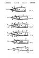
FIG. 1 is a top perspective view of a biopsy needle embodying a preferred form of the invention, with an inner cannula telescopically and rotatably fitted within an outer cannula and jointly defining a tissue sampling chamber;
FIG. 2 is a top perspective view of the inner cannula of the biopsy needle;
FIG. 3 is a side elevational view of the inner cannula of FIG. 2 rotated 180°, with parts broken away for clarity;
FIG. 4 is a top perspective view of the outer cannula of the biopsy needle;
FIG. 5 is a side elevational view of the outer cannula of FIG. 4 rotated 180°, with parts broken away for clarity;
FIG. 6 is a side elevational view of the assembled inner and outer cannulas of the biopsy needle, the inner cannula having been rotated 180° to fully enclose the tissue sampling chamber;
FIG. 7 is a greatly enlarged, fragmentary, top plan view of the outer cannula;
FIG. 8 is a greatly enlarged, fragmentary, top plan view of the inner cannula;
FIG. 9 is a greatly enlarged, fragmentary top plan view of the inner cannula telescopically and rotatably fitted within the outer cannula and jointly defining a tissue sampling chamber;
FIG. 10 is a view similar to FIG. 9, the inner cannula having been rotated 180° to fully enclose the tissue sampling chamber;
FIG. 11 is a greatly enlarged, fragmentary, top plan view of a first modified form of inner cannula; and
FIG. 12 is a greatly enlarged, fragmentary, top plan view of a second modified form of inner cannula.
Abiopsy needle 10 adapted for end cutting, side cutting and extracting of tissue specimens includes a cylindrical hand heldouter cannula 12 having an open, tubularlongitudinal channel 14 and a wedge shaped semi-conicalhollow piercing tip 16, the piercing tip having an opendistal channel 18 in continuity with openlongitudinal channel 14.
Wedge shapedhollow piercing tip 16 is defined by two converginglateral cutting surfaces semi-conical base surface 24 and interconnected superiorly by asemicircumferential cutting edge 26 angled forwardly with reference to the longitudinal axis of the cannula toward its distal end.
A hand heldinner cannula 28 is telescopically and rotatably fitted withinouter cannula 12 and has an opendistal channel 30 co-extensive withdistal channel 18 of the outer cannula, the opendistal channels tissue sampling chamber 32, as will appear.
Both inner and outer cannulas have proximal hand-held ends provided withengaging knobs
Positive stop means 48 is provided to signal complete 180° rotation ofinner cannula 28 relative toouter cannula 12 and includes apin 50 which extends longitudinally rearwardly fromengaging knob 46 ofouter cannula 12 and apin 52 which extends transversely outwardly fromengaging knob 44 ofinner cannula 28.
The closed-tip piercing operational mode is achieved through the rotation ofinner cannula 28 withinouter cannula 12 such that they are in an inverse relationship with regard to their geometric features, as shown in FIGS. 6 and 10, with such alignment of the cannulas producing an effective conical shape facilitating the piercing of tissue during needle insertion, while avoiding the entrapment of random samples.
The longitudinal cutting mode is achieved through the rotation ofinner cannula 28 withinouter cannula 12 such that the semiconicaldistal ends piercing tip 36 ofinner cannula 28 fitting precisely withinpiercing tip 16 ofouter cannula 12 thereby opening thelongitudinal channel 30 within the two cannulas with longitudinal cutting being accomplished by the two converginglateral surfaces superior edge 26 of a outer cannula as the needle is advanced in the aforementioned open position into the desired tissue by means of forward motion along the horizontal axis of the biopsy needle.
The transverse cutting mode is achieved sequentially following the longitudinal entrapment of the specimen tissue through the 180° rotation ofinner cannula 28 withinouter cannula 12, whereby upon completion of the transverse cutting process, a three dimensional specimen, not shown, is severed and entrapped within the interior diameter of the inner and outer cannulas, whose alignments are now inversely related, as shown in FIGS. 6 and 10, with minimized risk of structural damage to the specimen due to crushing or ripping.
1. The operational end ofneedle 10 is manually rotated to achieve the conical piercing mode of FIGS. 6 and 10.Conical piercing tip 16 is then inserted through the skin by applying gentle forward pressure to hand heldproximal knob 44. Due to the piercing characteristics of the conical tip, rotation of the needle is not necessary. The use of a local anesthetic will provide for painless skin penetration.
After penetration, the needle shaft is directed to the specimen site, as the opposite hand of the operator palpates the target point and functions as a guide.
2. The needle is advanced in its closed piercing mode to the periphery of the specimen site. The solid configuration of the conical tip during insertion prevents the inadvertent collection of unwanted materials as the needle is inserted through intermediate tissue.
3. As the needle tip reaches the periphery of the specimen, as perceived by the operator's hands or by imaging techniques,inner cannula 28 is rotated counterclockwise 180° into the longitudinal cutting mode. In this configuration, the operational distal end presents a semi-conical configuration, with hollowtissue entrapment chamber 30 ofinner cannula 28 in an open position.
4. The needle tip is next advanced into the desired specimen tissue through a forward motion along a longitudinal axis for a distance generally equivalent to the length ofspecimen chamber 32. This forward motion performs the longitudinal cutting of the desired tissue as it is advanced intospecimen chamber 32. This cutting action is performed by the inferior convergingsemi-conical tip 24 in conjunction with the forward advancing semi-cylindricalsuperior cutting edge 42.
Upon completion of this step, the specimen is housed withinchamber 32, but is still attached to the donor tissue by means of a cylindrical stem along its longitudinal axis, at the extreme distal end of the biopsy needle.
5. To completely sever and isolate the specimen from its donor source,inner cannula 28 is now rotated 180° clockwise. This rotary motion will transsect the remaining longitudinal tissue attachment in an inclined, transverse manner. This transverse cutting action completes the three dimensional separation of the specimen from its donor tissue. Further, the specimen is isolated within the cylindrical volume of the inner and outer cannulas, which are now configured in an oppositional alignment, thus returning the needle to its closed conical configuration as seen in FIGS. 6 and 10.
6. The isolated specimen is extracted from the assembly by the withdrawal of inner cannular 28 fromouter cannula 12. During this phase,inner cannula 28 remains in the closed position achieved during the final transection of the specimen, andouter cannula 12 remains stationary within the donor tissue.Inner cannula 28 is manually withdrawn by gently pulling onproximal handle 44 in a backward direction along a longitudinal axis.
7. Additional specimens may then be obtained by simply reintroducinginner cannula 28 in the open position, intoouter cannula 12. Opendistal chamber 30 is then advanced slightly further into the donor tissue to push a new sample into the cylindrical chamber. As before, the tissue is then transversely severed by means of a 180° clockwise rotation ofinner cannula 28 within theouter cannula 12. Once again, the specimen is removed by withdrawing the inner cannula, in the closed position, while the outer cannula remains stationary within the donor tissue.
8. This procedure may be repeated indefinitely at the discretion of the operator until a satisfactory volume of specimen is removed. Once a sufficient volume of tissue has been extracted, the entire assembly is removed.
In the modified form of FIG. 11, aninner cannula 128 is provided with anotch 129 located at the posterior end of a superiorsemi-circumferential cutting edge 142, which interconnects two converging lateral cutting surfaces 136 and 138 interconnected inferiorly by a semi-conicaltransverse base surface 140 and defining asemi-conical piercing tip 134.
Or, as shown in FIG. 12, aninner cannula 228 may be formed as asolid shaft 230 having anintegral piercing tip 234, or with the piercing tip welded thereto.
In the FIG. 12 embodiment, the lateral cutting surfaces of piercingtip 234 have been elongated, (only onesuch surface 222 being shown), to accept larger tissue specimens, and a semi-circularcircumferential cutting edge 242 has been inclined rearwardly toward the posterior of the needle at a sharper angle to improve its cutting action.
Claims (10)
1. A biopsy needle adapted for end cutting, side cutting and extracting of tissue specimens comprising:
a hand held outer cannula having an open longitudinal channel and a wedge shaped semi-conical hollow piercing tip,
said piercing tip having an open distal channel in continuity with the open longitudinal channel,
the wedge shaped hollow piercing tip being defined by two converging lateral cutting surfaces, interconnected inferiorly by a transverse semi-conical base surface,
and interconnected superiorly by a semicircumferential cutting edge, angled forwardly with reference to the longitudinal axis of the cannula toward the distal end of said cannula,
a hand held inner cannula telescopically and rotatably fitted within the outer cannula and having an open distal channel co-extensive with the distal channel of the outer cannula, the open distal channels jointly defining a tissue sampling chamber,
the inner cannula having a semi-conical hollow piercing tip defined by two converging lateral cutting surfaces, interconnected inferiorly by a semi-conical transverse base surface, and superiorly interconnected by a semi-circumferential cutting edge, angled rearwardly toward the posterior of the needle with reference to the longitudinal axis of the cannula, toward the proximal end of said cannula.
2. A method for extracting a tissue specimen from a donor source for diagnostic purposes with the biopsy needle recited in claim 1 comprising three sequential and reversible modes of operation namely,
a closed-tip piercing operational mode,
a longitudinal cutting mode and a transverse cutting mode,
wherein the closed-tip piercing operational mode is achieved through the rotation of the inner cannula within the outer cannula such that they are in an inverse relationship with regard to their geometric features, with such alignment of the cannulas producing an effective conical shape facilitating the piercing of tissue during needle insertion, while avoiding the entrapment of random samples,
wherein the longitudinal cutting mode is achieved through the rotation of the inner cannula within the outer cannula such that the semiconical distal ends of the cannulas are aligned in a contiguous manner,
with the tip of the inner cannula fitting precisely within the tip of the outer cannula thereby opening the longitudinal channel within the two cannulas, with longitudinal cutting being accomplished by the two converging lateral surfaces,
as well as by the forward advancing superior edge,
as the needle is advanced in the aforementioned open position into the desired tissue by means of forward motion along the horizontal axis of the biopsy needle,
and wherein the transverse cutting mode is achieved sequentially following the longitudinal entrapment of specimen tissue through the 180° rotation of the inner cannula within the outer cannula,
whereby upon completion of the transverse cutting process, the three dimensional specimen is severed and entrapped within the interior diameter of the inner and outer cannulas, whose alignments are now inversely related, with minimized risk of structural damage to the specimen due to crushing or ripping.
3. In the method of claim 2, with said inner cannula performing the transverse cutting of tissue as the outer cannula remains in a stationary position, thereby minimizing traumas to adjacent anatomical structures while enhancing the accuracy of the tissue sample.
4. A method for extracting a tissue specimen from a donor source for diagnostic purposes with a biopsy needle having an inner cannula slidably receivable in and rotational relative to an outer cannula for defining closed piercing/isolating positions and opened severing positions as to each other which comprises:
a closed conically shaped piercing mode consisting of advancing the closed-position needle to the specimen site,
an opening wedge-shaped longitudinal cutting mode consisting of rotating the inner cannula in opening rotational direction for opening a specimen chamber within the inner cannula and advancing the needle along the longitudinal needle axis in the longitudinal cutting of the donor source and the leading of the specimen into the specimen chamber,
a closing transverse cutting mode consisting of rotating the inner cannula in closing rotational direction for effecting the three-dimensional transection of the specimen while the entire needle assembly remains in a stationary position,
and an extracting mode consisting of withdrawing the inner cannula with the specimen from the outer cannula while said outer cannula remains stationarily fixed within the donor tissue,
such stationary positioning of the outer cannula securing the sampling accuracy of subsequent specimens by eliminating the possibility of inaccurate re-introduction of the needle assembly.
5. In the method of claim 4 including a repeating mode consisting of returning the inner cannula to its operational position within the outer cannula stationarily fixed within the donor source for a subsequent sampling procedure.
6. In a dual cannula biopsy needle adapted for the longitudinal and transverse cutting of a tissue specimen and separating same from a donor source, the combination of:
inner and outer cylindrical cannulae with each having a center through passage and a longitudinal axis,
the inner cannula being axially slidable within and rotational relative to the outer cannula between closed and opened positions,
the cannulae in closed position cooperatively defining a conical piercing tip,
each cannula having an open ended configuration for forming a cutting edge in the opened position,
the inner cannula defining a sealed specimen chamber within its passage in the closed position,
the distal ends of the cannulae being alignable in the opened position to define means effective upon the advancing of the cannulae into the donor source for axially incising a length of the tissue and effective upon the rotation of the inner cannula to closed position for transversely incising the incised length of tissue and severing the specimen from the donor source and isolating same.
7. In a biopsy needle for extracting a tissue specimen from a donor source for diagnostic purposes, the combination of:
an outer cannula having a hollow passage therethrough,
an inner cannula having a hollow passage therethrough and being slidably receivable in and rotational relative to the outer cannula,
the cannulae defining a closed position when the inner cannula is driven to a stop position in one rotational direction and an opened position when the inner cannula is driven to a stop position in the opposite rotational direction,
each cannula having a distal end defining a semi-conical half-section and an opposite opened half section with the opposite sides of the closed half section providing conveying lateral cutting edges toward the outboard termini and interconnected at their inboard termini by an angularized transverse cutting edge,
the outboard ends of the cannulae being alignable in the opened position to define means effective upon the advancing of the cannulae into the donor source for axially incising and entrapping a length of the tissue and effective upon the rotation of the inner cannula to closed position for transversely incising the incised length of tissue and severing the specimen from the donor source and isolating same.
8. In a biopsy needle adaptable for the extraction of multiple tissue specimens from a donor source with a single needle insertion and with a minimized risk of trauma to surrounding tissue, the combination of:
outer and inner cannulae each having an open longitudinal central through passage and an open semispherical channel in continuity with the central passage and a semi-conical penetrating tip at an outboard end,
the cannulae each having converging longitudinal cutting edges on opposite sides of the open channel and having a transverse semi-conical base surface interconnecting inferiorly between the longitudinal cutting edges,
the open channel of the outer cannula having a transverse semi-circumferential surface with a cutting edge interconnecting superiorly between the longitudinal cutting edges,
the open channel of the inner cannula having a transverse semi-circumferential surface with a cutting edge interconnecting superiorly between the longitudinal cutting edges,
the inner cannula being slidable within and rotational about an operative 180° relative to the outer cannula with the cannulae being extendable along a common longitudinal axis between closed and opened position,
the cannulae in the closed position jointly defining an exterior conical penetrating tip at the outboard extremity and an interior tissue sampling chamber,
the open channels and cutting edges of the cannulae being coextensive and aligned in the opened position,
the aligned cutting edges of the cannulae in the opened position defining means effective upon the penetrating of the cannulae into the donor source for axially incising a tissue length and effective upon the rotation of the inner cannula to closed position for transversely cutting the incised tissue length being entrapped within the closing tissue sampling chamber for isolating same with the withdrawal of the inner cannula from the outer cannula.
9. A biopsy needle adapted for end cutting, side cutting and extracting of tissue specimens comprising:
a hand held outer cannula having an open longitudinal channel and a wedge shaped semi-conical hollow piercing tip,
said piercing tip having an open distal channel in continuity with the open longitudinal channel,
the wedge shaped hollow piercing tip being defined by two converging lateral cutting surfaces, interconnected inferiorly by a transverse semi-conical base surface,
and interconnected superiorly by a semicircumferential cutting edge, angled forwardly with reference to the longitudinal axis of the cannula toward the distal end of said cannula,
a hand held inner cannula telescopically and rotatably fitted within the outer cannula and having an open distal channel co-extensive with the distal channel of the outer cannula, the open distal channels jointly defining a tissue sampling chamber,
the inner cannula having a semi-conical hollow piercing tip defined by two converging lateral cutting surfaces, interconnected inferiorly by a semi-conical transverse base surface, and superiorly interconnected by a semicircumferential cutting edge, angled rearwardly toward the posterior of the needle with reference to the longitudinal axis of the cannula, toward the proximal end of said cannula,
the biopsy needle having three sequential and reversible modes of operation namely,
a closed-tip piercing operational mode,
a longitudinal cutting mode and a transverse cutting mode defined by the procedure of rotating the inner cannula within the outer cannula,
wherein the transverse cutting mode is achieved sequentially following the longitudinal entrapment of specimen tissue through the 180° rotation of the inner cannula within the outer cannula, whereby upon completion of the transverse cutting process, the three dimensional specimen is severed and entrapped within the interior diameter of the inner and outer cannulas, whose alignments are now inversely related, with minimized risk of structural damage to the specimen due to crushing or ripping.
10. A biopsy needle as in claim 9, with said inner cannula performing the transverse cutting of tissue as the outer cannula remains in a stationary position, thereby minimizing traumas to adjacent anatomical structures while enhancing the accuracy of the tissue sample.
Priority Applications (5)
| Application Number | Priority Date | Filing Date | Title |
|---|---|---|---|
| US07/921,338US5251641A (en) | 1992-07-29 | 1992-07-29 | Biopsy needle |
| EP93917257AEP0652728A4 (en) | 1992-07-29 | 1993-07-19 | Biopsy needle. |
| JP6505336AJPH11500016A (en) | 1992-07-29 | 1993-07-19 | Biopsy needle |
| CA002140248ACA2140248A1 (en) | 1992-07-29 | 1993-07-19 | Biopsy needle |
| PCT/US1993/006745WO1994003099A2 (en) | 1992-07-29 | 1993-07-19 | Biopsy needle |
Applications Claiming Priority (1)
| Application Number | Priority Date | Filing Date | Title |
|---|---|---|---|
| US07/921,338US5251641A (en) | 1992-07-29 | 1992-07-29 | Biopsy needle |
Publications (1)
| Publication Number | Publication Date |
|---|---|
| US5251641Atrue US5251641A (en) | 1993-10-12 |
Family
ID=25445298
Family Applications (1)
| Application Number | Title | Priority Date | Filing Date |
|---|---|---|---|
| US07/921,338Expired - Fee RelatedUS5251641A (en) | 1992-07-29 | 1992-07-29 | Biopsy needle |
Country Status (5)
| Country | Link |
|---|---|
| US (1) | US5251641A (en) |
| EP (1) | EP0652728A4 (en) |
| JP (1) | JPH11500016A (en) |
| CA (1) | CA2140248A1 (en) |
| WO (1) | WO1994003099A2 (en) |
Cited By (71)
| Publication number | Priority date | Publication date | Assignee | Title |
|---|---|---|---|---|
| US5471992A (en)* | 1994-02-08 | 1995-12-05 | Boston Scientific Corporation | Multi-motion cutter multiple biopsy sampling device |
| US5542432A (en)* | 1992-02-18 | 1996-08-06 | Symbiosis Corporation | Endoscopic multiple sample bioptome |
| US5573008A (en)* | 1993-10-29 | 1996-11-12 | Boston Scientific Corporation | Multiple biopsy sampling coring device |
| US5601585A (en)* | 1994-02-08 | 1997-02-11 | Boston Scientific Corporation | Multi-motion side-cutting biopsy sampling device |
| WO1997020503A1 (en) | 1995-12-06 | 1997-06-12 | Interventional Concepts, Inc. | Biopsy specimen cutter |
| WO1997022299A1 (en)* | 1995-12-15 | 1997-06-26 | Swaim William R | Biopsy hand tool for capturing tissue sample |
| US5709697A (en)* | 1995-11-22 | 1998-01-20 | United States Surgical Corporation | Apparatus and method for removing tissue |
| EP0720440A4 (en)* | 1993-09-20 | 1998-04-08 | Boston Scient Corp | Multi-motion multiple biopsy sampling device |
| US5782775A (en)* | 1995-10-20 | 1998-07-21 | United States Surgical Corporation | Apparatus and method for localizing and removing tissue |
| US5810744A (en)* | 1993-05-17 | 1998-09-22 | Boston Scientific Corporation | Instrument for collecting multiple biopsy specimens |
| US5817034A (en)* | 1995-09-08 | 1998-10-06 | United States Surgical Corporation | Apparatus and method for removing tissue |
| US5857982A (en)* | 1995-09-08 | 1999-01-12 | United States Surgical Corporation | Apparatus and method for removing tissue |
| US5871453A (en)* | 1994-02-08 | 1999-02-16 | Boston Scientific Corporation | Moveable sample tube multiple biopsy sampling device |
| US5897507A (en)* | 1996-11-25 | 1999-04-27 | Symbiosis Corporation | Biopsy forceps instrument having irrigation and aspiration capabilities |
| US5947989A (en)* | 1996-12-12 | 1999-09-07 | United States Surgical Corporation | Method and apparatus for transmyocardial revascularization |
| US6048321A (en)* | 1997-10-10 | 2000-04-11 | William E. McPherson | Guide assembly for a biopsy device |
| US6139508A (en)* | 1998-08-04 | 2000-10-31 | Endonetics, Inc. | Articulated medical device |
| US6142956A (en)* | 1996-11-25 | 2000-11-07 | Symbiosis Corporation | Proximal actuation handle for a biopsy forceps instrument having irrigation and aspiration capabilities |
| US6142957A (en)* | 1993-09-20 | 2000-11-07 | Boston Scientific Corporation | Multiple biopsy sampling device |
| US6142955A (en) | 1997-09-19 | 2000-11-07 | United States Surgical Corporation | Biopsy apparatus and method |
| US6149607A (en)* | 1998-08-04 | 2000-11-21 | Endonetics, Inc. | Multiple sample biopsy device |
| US6162203A (en)* | 1999-01-11 | 2000-12-19 | Haaga; John R. | Cargo delivery needle |
| US6193671B1 (en) | 1994-02-01 | 2001-02-27 | Symbiosis Corporation | Endoscopic multiple sample bioptome with enhanced biting action |
| US6267732B1 (en) | 1997-09-12 | 2001-07-31 | Imagyn Medical Technologies, Inc. | Incisional breast biopsy device |
| US6274644B1 (en) | 1999-03-31 | 2001-08-14 | Joseph J. Pelerin | Dental composite |
| WO2001066177A1 (en)* | 2000-03-08 | 2001-09-13 | Dr. Japan Co., Ltd. | Diagnosis or therapy puncture needle |
| US6302853B1 (en)* | 2000-02-24 | 2001-10-16 | R & G Medical And Development Corp. | Method and apparatus for sampling cervical tissue |
| US20020029006A1 (en)* | 1996-11-25 | 2002-03-07 | Scimed Life Systems, Inc. | Biopsy instrument having irrigation and aspiration capabilities |
| US6383145B1 (en) | 1997-09-12 | 2002-05-07 | Imagyn Medical Technologies California, Inc. | Incisional breast biopsy device |
| US6395802B1 (en) | 1999-03-31 | 2002-05-28 | Advantage Dental Products, Inc. | Dental composite |
| US20020107457A1 (en)* | 1996-11-25 | 2002-08-08 | Francese Jose L. | Suction adapter for medical instrument |
| US6436054B1 (en) | 1998-11-25 | 2002-08-20 | United States Surgical Corporation | Biopsy system |
| US6551253B2 (en) | 1997-09-12 | 2003-04-22 | Imagyn Medical Technologies | Incisional breast biopsy device |
| US20030097144A1 (en)* | 2001-11-17 | 2003-05-22 | Lee Hoon Bum | Hair transplant device |
| US20030158603A1 (en)* | 2002-02-19 | 2003-08-21 | Maxilon Laboratories, Inc. | Apparatus and method for harvesting bone |
| US6626848B2 (en) | 2001-03-30 | 2003-09-30 | Eric M. Neuenfeldt | Method and device to reduce needle insertion force |
| US6860860B2 (en) | 2000-11-27 | 2005-03-01 | Tyco Healthcare Group, Lp | Tissue sampling and removal apparatus and method |
| US20050080441A1 (en)* | 2003-10-10 | 2005-04-14 | Duke University | Surgical instruments which are especially useful for ophthalmic surgical procedures, and methods of making the same |
| US20050101879A1 (en)* | 2003-11-06 | 2005-05-12 | Shidham Vinod B. | Needle aspiration biopsy device and method |
| US20060030785A1 (en)* | 2004-05-11 | 2006-02-09 | Inrad, Inc. | Core biopsy device |
| US7033357B2 (en)* | 2000-03-09 | 2006-04-25 | Origin Medsystems, Inc. | Apparatus and method for minimally invasive surgery using rotational cutting tool |
| US7087028B2 (en) | 2000-02-24 | 2006-08-08 | R&G Medical And Development Corp. | Method and apparatus for sampling cervical tissue |
| US20070016101A1 (en)* | 2005-07-13 | 2007-01-18 | Feldman Dennis D | Core Biopsy Device |
| US7189207B2 (en) | 2000-09-11 | 2007-03-13 | Tyco Healthcare Group Lp | Biopsy system having a single use loading unit operable with a trocar driver, a knife driver and firing module |
| US20070123935A1 (en)* | 2005-11-30 | 2007-05-31 | Myers Gene E | Method and apparatus for contemporaneous formation of a body structure opening and homologous pedicle |
| WO2008008622A1 (en)* | 2006-07-12 | 2008-01-17 | Boston Scientific Limited | Biopsy device |
| US20080097504A1 (en)* | 2004-05-21 | 2008-04-24 | Keshava Datta | Trocar obturator with cutting edges |
| US20080114265A1 (en)* | 2006-10-23 | 2008-05-15 | Tom Tarter | Double Core Biopsy Instrumentation Kit |
| US20080281226A1 (en)* | 2004-05-11 | 2008-11-13 | Inrad, Inc. | Core Biopsy Device with Specimen Length Adjustment |
| US20090012423A1 (en)* | 2004-05-11 | 2009-01-08 | Inrad, Inc. | Core Biopsy Device |
| US7588545B2 (en) | 2003-09-10 | 2009-09-15 | Boston Scientific Scimed, Inc. | Forceps and collection assembly with accompanying mechanisms and related methods of use |
| US7909850B2 (en) | 1999-10-25 | 2011-03-22 | Boston Scientific Scimed, Inc. | Forceps for medical use |
| US7942896B2 (en) | 2003-11-25 | 2011-05-17 | Scimed Life Systems, Inc. | Forceps and collection assembly and related methods of use and manufacture |
| US20130096459A1 (en)* | 2011-10-15 | 2013-04-18 | Transmed7, Llc | Soft tissue coring biopsy devices and methods |
| US9456807B2 (en) | 2013-08-22 | 2016-10-04 | Transmed7, Llc | Soft tissue coring biopsy devices and methods |
| US9463001B2 (en) | 2013-05-28 | 2016-10-11 | Transmed7, Llc | Soft tissue coring biopsy devices and methods |
| US20170188999A1 (en)* | 2014-02-03 | 2017-07-06 | The University Of Western Australia | Medical device for insertion into a material to obtain a material sample and a method thereof |
| CN106989976A (en)* | 2017-05-25 | 2017-07-28 | 郑州大学第附属医院 | Specimen sampling device |
| US9993231B2 (en) | 2013-11-20 | 2018-06-12 | Covidien Lp | Devices, systems, and methods for navigating a biopsy tool to a target location and obtaining a tissue sample using the same |
| US9999758B2 (en) | 2014-09-19 | 2018-06-19 | Transmed7, Llc | In-situ material delivery devices and methods |
| US20180193005A1 (en)* | 2011-11-09 | 2018-07-12 | Teesuvac Aps | Handheld tissue sample extraction device |
| US10070884B2 (en) | 2013-09-12 | 2018-09-11 | Transmed7, Llc | Soft tissue coring biopsy devices and methods |
| US10076315B2 (en) | 2014-09-18 | 2018-09-18 | Transmed7, Llc | Soft tissue biopsy or excisional devices and methods |
| US10231750B2 (en) | 2014-09-29 | 2019-03-19 | Transmed7, Llc | Excisional device distal working end actuation mechanism and method |
| US10278680B2 (en) | 2014-03-19 | 2019-05-07 | Covidien Lp | Devices, systems, and methods for navigating a biopsy tool to a target location and obtaining a tissue sample using the same |
| CN111358505A (en)* | 2020-03-24 | 2020-07-03 | 温州市中心医院 | Minimally invasive bone marrow biopsy sampling needle |
| US11596436B2 (en) | 2019-04-04 | 2023-03-07 | Transmed7, Llc | Excisional devices and methods |
| US20230181871A1 (en)* | 2019-04-30 | 2023-06-15 | Boston Scientific Scimed, Inc. | Endoscopic patch applicator |
| US11805998B2 (en) | 2020-04-20 | 2023-11-07 | Covidien Lp | Devices and methods for obtaining adenomyosis and other biopsy samples |
| US12150627B2 (en) | 2019-12-11 | 2024-11-26 | Merit Medical Systems, Inc. | Bone biopsy device and related methods |
| US12295556B2 (en) | 2019-09-27 | 2025-05-13 | Merit Medical Systems, Inc. | Rotation biopsy system and handle |
Families Citing this family (4)
| Publication number | Priority date | Publication date | Assignee | Title |
|---|---|---|---|---|
| US7156815B2 (en) | 2003-03-19 | 2007-01-02 | Biomedical Resources, Inc. | Soft tissue biopsy instrument |
| JP5839856B2 (en)* | 2011-06-29 | 2016-01-06 | オリンパス株式会社 | Observation surface cutter |
| JP6513099B2 (en)* | 2014-02-18 | 2019-05-15 | マサチューセッツ インスティテュート オブ テクノロジー | Tissue collection needle |
| JP7036313B2 (en)* | 2018-02-01 | 2022-03-15 | 高島産業株式会社 | Biometer and biometer |
Citations (16)
| Publication number | Priority date | Publication date | Assignee | Title |
|---|---|---|---|---|
| US1493240A (en)* | 1923-02-15 | 1924-05-06 | Frank J Bohn | Surgical bone cutter and extractor |
| US2516492A (en)* | 1950-02-09 | 1950-07-25 | Turkel Henry | Skin biopsy needle |
| US2749909A (en)* | 1956-06-12 | Biopsy knife | ||
| US2827039A (en)* | 1956-07-09 | 1958-03-18 | Seiger Harry Wright | Surgical instrument |
| US3330268A (en)* | 1963-12-18 | 1967-07-11 | Goldsmith Sidney | Biopsy needle |
| US3732858A (en)* | 1968-09-16 | 1973-05-15 | Surgical Design Corp | Apparatus for removing blood clots, cataracts and other objects from the eye |
| US3844272A (en)* | 1969-02-14 | 1974-10-29 | A Banko | Surgical instruments |
| US3929123A (en)* | 1973-02-07 | 1975-12-30 | Khosrow Jamshidi | Muscle biopsy needle |
| US4513754A (en)* | 1978-03-03 | 1985-04-30 | Southland Instruments, Inc. | Biopsy and aspiration unit with a replaceable cannula |
| US4543966A (en)* | 1981-06-10 | 1985-10-01 | Downs Surgical Plc | Biopsy needle |
| US4609370A (en)* | 1983-06-20 | 1986-09-02 | Morrison Peter C | Surgical needle assembly and apparatus for attachment on a surgical needle assembly |
| US4640296A (en)* | 1983-11-12 | 1987-02-03 | Schnepp Pesch Wolfram | Biopsy cannula |
| US4702260A (en)* | 1985-04-16 | 1987-10-27 | Ko Pen Wang | Flexible bronchoscopic needle assembly |
| US4708147A (en)* | 1985-02-25 | 1987-11-24 | Haaga John R | Universal biopsy needle |
| US4799494A (en)* | 1986-10-22 | 1989-01-24 | Wang Ko P | Percutaneous aspiration lung biopsy needle assembly |
| US4926877A (en)* | 1989-04-24 | 1990-05-22 | Bookwalter John R | Biopsy needle with completely closable cutting end bore |
Family Cites Families (3)
| Publication number | Priority date | Publication date | Assignee | Title |
|---|---|---|---|---|
| DE935625C (en)* | 1952-10-18 | 1955-11-24 | Guenther Bodendieck | Excision device |
| US3995619A (en)* | 1975-10-14 | 1976-12-07 | Glatzer Stephen G | Combination subcutaneous suture remover, biopsy sampler and syringe |
| DE3023955A1 (en)* | 1980-06-26 | 1982-01-14 | Josef 6902 Sandhausen Magasi | Hollow needle for taking tissue samples - has hollow cylinder with conical needle tip, and has peripheral wall aperture as tissue receptacle |
- 1992
- 1992-07-29USUS07/921,338patent/US5251641A/ennot_activeExpired - Fee Related
- 1993
- 1993-07-19WOPCT/US1993/006745patent/WO1994003099A2/ennot_activeApplication Discontinuation
- 1993-07-19EPEP93917257Apatent/EP0652728A4/ennot_activeWithdrawn
- 1993-07-19JPJP6505336Apatent/JPH11500016A/enactivePending
- 1993-07-19CACA002140248Apatent/CA2140248A1/ennot_activeAbandoned
Patent Citations (16)
| Publication number | Priority date | Publication date | Assignee | Title |
|---|---|---|---|---|
| US2749909A (en)* | 1956-06-12 | Biopsy knife | ||
| US1493240A (en)* | 1923-02-15 | 1924-05-06 | Frank J Bohn | Surgical bone cutter and extractor |
| US2516492A (en)* | 1950-02-09 | 1950-07-25 | Turkel Henry | Skin biopsy needle |
| US2827039A (en)* | 1956-07-09 | 1958-03-18 | Seiger Harry Wright | Surgical instrument |
| US3330268A (en)* | 1963-12-18 | 1967-07-11 | Goldsmith Sidney | Biopsy needle |
| US3732858A (en)* | 1968-09-16 | 1973-05-15 | Surgical Design Corp | Apparatus for removing blood clots, cataracts and other objects from the eye |
| US3844272A (en)* | 1969-02-14 | 1974-10-29 | A Banko | Surgical instruments |
| US3929123A (en)* | 1973-02-07 | 1975-12-30 | Khosrow Jamshidi | Muscle biopsy needle |
| US4513754A (en)* | 1978-03-03 | 1985-04-30 | Southland Instruments, Inc. | Biopsy and aspiration unit with a replaceable cannula |
| US4543966A (en)* | 1981-06-10 | 1985-10-01 | Downs Surgical Plc | Biopsy needle |
| US4609370A (en)* | 1983-06-20 | 1986-09-02 | Morrison Peter C | Surgical needle assembly and apparatus for attachment on a surgical needle assembly |
| US4640296A (en)* | 1983-11-12 | 1987-02-03 | Schnepp Pesch Wolfram | Biopsy cannula |
| US4708147A (en)* | 1985-02-25 | 1987-11-24 | Haaga John R | Universal biopsy needle |
| US4702260A (en)* | 1985-04-16 | 1987-10-27 | Ko Pen Wang | Flexible bronchoscopic needle assembly |
| US4799494A (en)* | 1986-10-22 | 1989-01-24 | Wang Ko P | Percutaneous aspiration lung biopsy needle assembly |
| US4926877A (en)* | 1989-04-24 | 1990-05-22 | Bookwalter John R | Biopsy needle with completely closable cutting end bore |
Non-Patent Citations (1)
| Title |
|---|
| Westcott, Biopsy Needle, Becton Dickinson advertisement, 1991.* |
Cited By (124)
| Publication number | Priority date | Publication date | Assignee | Title |
|---|---|---|---|---|
| US5951488A (en)* | 1992-02-18 | 1999-09-14 | Symbiosis Corporation | Endoscopic multiple sample bioptome |
| US5542432A (en)* | 1992-02-18 | 1996-08-06 | Symbiosis Corporation | Endoscopic multiple sample bioptome |
| US5810744A (en)* | 1993-05-17 | 1998-09-22 | Boston Scientific Corporation | Instrument for collecting multiple biopsy specimens |
| US6142957A (en)* | 1993-09-20 | 2000-11-07 | Boston Scientific Corporation | Multiple biopsy sampling device |
| EP0720440A4 (en)* | 1993-09-20 | 1998-04-08 | Boston Scient Corp | Multi-motion multiple biopsy sampling device |
| EP1348378A1 (en)* | 1993-09-20 | 2003-10-01 | Boston Scientific Corporation | Multi-motion multiple biopsy sampling device |
| US5573008A (en)* | 1993-10-29 | 1996-11-12 | Boston Scientific Corporation | Multiple biopsy sampling coring device |
| US5823971A (en)* | 1993-10-29 | 1998-10-20 | Boston Scientific Corporation | Multiple biopsy sampling coring device |
| US6193671B1 (en) | 1994-02-01 | 2001-02-27 | Symbiosis Corporation | Endoscopic multiple sample bioptome with enhanced biting action |
| US6561988B1 (en) | 1994-02-01 | 2003-05-13 | Symbiosis Corporation | Endoscopic multiple sample bioptome with enhanced biting action |
| US5871453A (en)* | 1994-02-08 | 1999-02-16 | Boston Scientific Corporation | Moveable sample tube multiple biopsy sampling device |
| US6053877A (en)* | 1994-02-08 | 2000-04-25 | Boston Scientific Corporation | Movable sample tube multiple biopsy sampling device |
| US5779648A (en)* | 1994-02-08 | 1998-07-14 | Boston Scientific Corporation | Multi-motion cutter multiple biopsy sampling device |
| US5961534A (en)* | 1994-02-08 | 1999-10-05 | Boston Scientific Corporation | Multi-motion side cutting biopsy sampling device |
| US5601585A (en)* | 1994-02-08 | 1997-02-11 | Boston Scientific Corporation | Multi-motion side-cutting biopsy sampling device |
| US5471992A (en)* | 1994-02-08 | 1995-12-05 | Boston Scientific Corporation | Multi-motion cutter multiple biopsy sampling device |
| US6036657A (en)* | 1995-09-08 | 2000-03-14 | United States Surgical Corporation | Apparatus for removing tissue |
| US5857982A (en)* | 1995-09-08 | 1999-01-12 | United States Surgical Corporation | Apparatus and method for removing tissue |
| US5817034A (en)* | 1995-09-08 | 1998-10-06 | United States Surgical Corporation | Apparatus and method for removing tissue |
| US6213957B1 (en) | 1995-09-08 | 2001-04-10 | United States Surgical Corporation | Apparatus and method for removing tissue |
| US5782775A (en)* | 1995-10-20 | 1998-07-21 | United States Surgical Corporation | Apparatus and method for localizing and removing tissue |
| US5709697A (en)* | 1995-11-22 | 1998-01-20 | United States Surgical Corporation | Apparatus and method for removing tissue |
| US5687739A (en)* | 1995-12-06 | 1997-11-18 | Interventional Concepts, Inc. | Biopsy specimen cutter |
| WO1997020503A1 (en) | 1995-12-06 | 1997-06-12 | Interventional Concepts, Inc. | Biopsy specimen cutter |
| US5807277A (en)* | 1995-12-15 | 1998-09-15 | Swaim; William R. | Biopsy hand tool for capturing tissue sample |
| WO1997022299A1 (en)* | 1995-12-15 | 1997-06-26 | Swaim William R | Biopsy hand tool for capturing tissue sample |
| US6165137A (en)* | 1996-06-14 | 2000-12-26 | United States Surgical Corporation | Apparatus and method for localizing and removing tissue |
| US6077231A (en)* | 1996-06-14 | 2000-06-20 | United States Surgical Corporation | Apparatus and method for localizing and removing tissue |
| US7297121B2 (en) | 1996-11-25 | 2007-11-20 | Boston Scientific Scimed, Inc. | Biopsy instrument having irrigation and aspiration capabilities |
| US5897507A (en)* | 1996-11-25 | 1999-04-27 | Symbiosis Corporation | Biopsy forceps instrument having irrigation and aspiration capabilities |
| US6926676B2 (en) | 1996-11-25 | 2005-08-09 | Boston Scientific Scimed, Inc. | Biopsy instrument having irrigation and aspiration capabilities |
| US7833167B2 (en) | 1996-11-25 | 2010-11-16 | Boston Scientific Miami Corporation | Proximal actuation handle for a biopsy forceps instrument having irrigation and aspiration capabilities |
| US6174292B1 (en) | 1996-11-25 | 2001-01-16 | Symbiosis Corporation | Biopsy forceps instrument having irrigation and aspiration capabilities |
| US6142956A (en)* | 1996-11-25 | 2000-11-07 | Symbiosis Corporation | Proximal actuation handle for a biopsy forceps instrument having irrigation and aspiration capabilities |
| US6544194B1 (en) | 1996-11-25 | 2003-04-08 | Symbiosis Corporation | Proximal actuation handle for a biopsy forceps instrument having irrigation and aspiration capabilities |
| US20020107457A1 (en)* | 1996-11-25 | 2002-08-08 | Francese Jose L. | Suction adapter for medical instrument |
| US7204811B2 (en) | 1996-11-25 | 2007-04-17 | Boston Scientific Miami Corporation | Proximal actuation handle for a biopsy forceps instrument having irrigation and aspiration capabilities |
| US7347828B2 (en) | 1996-11-25 | 2008-03-25 | Boston Scientific Miami Corporation | Suction adapter for medical instrument |
| US6832990B2 (en) | 1996-11-25 | 2004-12-21 | Symbiosis Corporation | Biopsy instrument having aspiration capabilities |
| US20020029006A1 (en)* | 1996-11-25 | 2002-03-07 | Scimed Life Systems, Inc. | Biopsy instrument having irrigation and aspiration capabilities |
| US5947989A (en)* | 1996-12-12 | 1999-09-07 | United States Surgical Corporation | Method and apparatus for transmyocardial revascularization |
| US6383145B1 (en) | 1997-09-12 | 2002-05-07 | Imagyn Medical Technologies California, Inc. | Incisional breast biopsy device |
| US6267732B1 (en) | 1997-09-12 | 2001-07-31 | Imagyn Medical Technologies, Inc. | Incisional breast biopsy device |
| US6551253B2 (en) | 1997-09-12 | 2003-04-22 | Imagyn Medical Technologies | Incisional breast biopsy device |
| US6142955A (en) | 1997-09-19 | 2000-11-07 | United States Surgical Corporation | Biopsy apparatus and method |
| US6048321A (en)* | 1997-10-10 | 2000-04-11 | William E. McPherson | Guide assembly for a biopsy device |
| US6149607A (en)* | 1998-08-04 | 2000-11-21 | Endonetics, Inc. | Multiple sample biopsy device |
| US6139508A (en)* | 1998-08-04 | 2000-10-31 | Endonetics, Inc. | Articulated medical device |
| US6436054B1 (en) | 1998-11-25 | 2002-08-20 | United States Surgical Corporation | Biopsy system |
| US6162203A (en)* | 1999-01-11 | 2000-12-19 | Haaga; John R. | Cargo delivery needle |
| US6395802B1 (en) | 1999-03-31 | 2002-05-28 | Advantage Dental Products, Inc. | Dental composite |
| US6274644B1 (en) | 1999-03-31 | 2001-08-14 | Joseph J. Pelerin | Dental composite |
| US7909850B2 (en) | 1999-10-25 | 2011-03-22 | Boston Scientific Scimed, Inc. | Forceps for medical use |
| US6302853B1 (en)* | 2000-02-24 | 2001-10-16 | R & G Medical And Development Corp. | Method and apparatus for sampling cervical tissue |
| US7087028B2 (en) | 2000-02-24 | 2006-08-08 | R&G Medical And Development Corp. | Method and apparatus for sampling cervical tissue |
| WO2001066177A1 (en)* | 2000-03-08 | 2001-09-13 | Dr. Japan Co., Ltd. | Diagnosis or therapy puncture needle |
| US7033357B2 (en)* | 2000-03-09 | 2006-04-25 | Origin Medsystems, Inc. | Apparatus and method for minimally invasive surgery using rotational cutting tool |
| US8128577B2 (en) | 2000-09-11 | 2012-03-06 | Tyco Healthcare Group Lp | Biopsy system |
| US7189207B2 (en) | 2000-09-11 | 2007-03-13 | Tyco Healthcare Group Lp | Biopsy system having a single use loading unit operable with a trocar driver, a knife driver and firing module |
| US7513877B2 (en) | 2000-11-27 | 2009-04-07 | Tyco Healthcare Group Lp | Tissue sampling and removal apparatus and method |
| US6860860B2 (en) | 2000-11-27 | 2005-03-01 | Tyco Healthcare Group, Lp | Tissue sampling and removal apparatus and method |
| US6626848B2 (en) | 2001-03-30 | 2003-09-30 | Eric M. Neuenfeldt | Method and device to reduce needle insertion force |
| US20030097144A1 (en)* | 2001-11-17 | 2003-05-22 | Lee Hoon Bum | Hair transplant device |
| US20030158603A1 (en)* | 2002-02-19 | 2003-08-21 | Maxilon Laboratories, Inc. | Apparatus and method for harvesting bone |
| US8460205B2 (en) | 2003-09-10 | 2013-06-11 | Boston Scientific Scimed, Inc. | Forceps and collection assembly with accompanying mechanisms and related methods of use |
| US8083686B2 (en) | 2003-09-10 | 2011-12-27 | Boston Scientific Scimed, Inc. | Forceps and collection assembly with accompanying mechanisms and related methods of use |
| US7588545B2 (en) | 2003-09-10 | 2009-09-15 | Boston Scientific Scimed, Inc. | Forceps and collection assembly with accompanying mechanisms and related methods of use |
| US20050080441A1 (en)* | 2003-10-10 | 2005-04-14 | Duke University | Surgical instruments which are especially useful for ophthalmic surgical procedures, and methods of making the same |
| US20050101879A1 (en)* | 2003-11-06 | 2005-05-12 | Shidham Vinod B. | Needle aspiration biopsy device and method |
| US7942896B2 (en) | 2003-11-25 | 2011-05-17 | Scimed Life Systems, Inc. | Forceps and collection assembly and related methods of use and manufacture |
| US8088081B2 (en) | 2004-05-11 | 2012-01-03 | Inrad, Inc. | Core biopsy device |
| US20080281226A1 (en)* | 2004-05-11 | 2008-11-13 | Inrad, Inc. | Core Biopsy Device with Specimen Length Adjustment |
| US20090012423A1 (en)* | 2004-05-11 | 2009-01-08 | Inrad, Inc. | Core Biopsy Device |
| US8568334B2 (en) | 2004-05-11 | 2013-10-29 | Inrad, Inc. | Core biopsy device |
| US20060030785A1 (en)* | 2004-05-11 | 2006-02-09 | Inrad, Inc. | Core biopsy device |
| US20080097504A1 (en)* | 2004-05-21 | 2008-04-24 | Keshava Datta | Trocar obturator with cutting edges |
| US20070016101A1 (en)* | 2005-07-13 | 2007-01-18 | Feldman Dennis D | Core Biopsy Device |
| US20070123935A1 (en)* | 2005-11-30 | 2007-05-31 | Myers Gene E | Method and apparatus for contemporaneous formation of a body structure opening and homologous pedicle |
| WO2008008622A1 (en)* | 2006-07-12 | 2008-01-17 | Boston Scientific Limited | Biopsy device |
| US20080027355A1 (en)* | 2006-07-12 | 2008-01-31 | Dicarlo Paul D | Biopsy device |
| US8394033B2 (en) | 2006-07-12 | 2013-03-12 | Boston Scientific Scimed, Inc. | Biopsy device |
| US20130190650A1 (en)* | 2006-07-12 | 2013-07-25 | Boston Scientific Scimed, Inc. | Biopsy device |
| US20150080759A1 (en)* | 2006-07-12 | 2015-03-19 | Boston Scientific Scimed, Inc. | Biopsy device |
| US8920338B2 (en)* | 2006-07-12 | 2014-12-30 | Boston Scientific Scimed, Inc. | Biopsy device |
| US20080114265A1 (en)* | 2006-10-23 | 2008-05-15 | Tom Tarter | Double Core Biopsy Instrumentation Kit |
| US7914463B2 (en) | 2006-10-23 | 2011-03-29 | Clipius Technologies, Inc. | Double core biopsy instrumentation kit |
| US20140128768A1 (en)* | 2011-10-15 | 2014-05-08 | Transmed7, Llc | Soft tissue coring and biopsy devices and methods |
| US9808226B2 (en)* | 2011-10-15 | 2017-11-07 | Transmed7, Llc | Soft tissue coring biopsy devices and methods |
| US20140128769A1 (en)* | 2011-10-15 | 2014-05-08 | Transmed7, Llc | Soft tissue coring biopsy devices and methods |
| US20130226031A1 (en)* | 2011-10-15 | 2013-08-29 | Transmed7, Llc | Soft tissue coring biopsy devices and methods |
| US20130190651A1 (en)* | 2011-10-15 | 2013-07-25 | Transmed7, Llc | Soft tissue coring biopsy devices and methods |
| US20130096459A1 (en)* | 2011-10-15 | 2013-04-18 | Transmed7, Llc | Soft tissue coring biopsy devices and methods |
| US11026665B2 (en)* | 2011-11-09 | 2021-06-08 | Teesuvac Aps | Handheld tissue sample extraction device |
| US20180193005A1 (en)* | 2011-11-09 | 2018-07-12 | Teesuvac Aps | Handheld tissue sample extraction device |
| US9463001B2 (en) | 2013-05-28 | 2016-10-11 | Transmed7, Llc | Soft tissue coring biopsy devices and methods |
| US10806434B2 (en) | 2013-08-22 | 2020-10-20 | Transmed7, Llc | Soft tissue coring biopsy devices and methods |
| US9592035B2 (en) | 2013-08-22 | 2017-03-14 | Transmed7, Llc | Stereotactic soft tissue coring biopsy devices and methods |
| US9456807B2 (en) | 2013-08-22 | 2016-10-04 | Transmed7, Llc | Soft tissue coring biopsy devices and methods |
| US10070884B2 (en) | 2013-09-12 | 2018-09-11 | Transmed7, Llc | Soft tissue coring biopsy devices and methods |
| US10070885B2 (en) | 2013-09-12 | 2018-09-11 | Transmed7, Llc | Soft tissue coring biospy devices and methods |
| US10555751B2 (en) | 2013-09-12 | 2020-02-11 | Transmed7, Llc | Soft tissue coring biopsy devices and methods |
| US9993231B2 (en) | 2013-11-20 | 2018-06-12 | Covidien Lp | Devices, systems, and methods for navigating a biopsy tool to a target location and obtaining a tissue sample using the same |
| US11160539B2 (en) | 2013-11-20 | 2021-11-02 | Covidien Lp | Devices, systems, and methods for navigating a biopsy tool to a target location and obtaining a tissue sample using the same |
| US20170188999A1 (en)* | 2014-02-03 | 2017-07-06 | The University Of Western Australia | Medical device for insertion into a material to obtain a material sample and a method thereof |
| US10052086B2 (en)* | 2014-02-03 | 2018-08-21 | The University Of Western Australia | Medical device for insertion into a material to obtain a material sample and a method thereof |
| US12059135B2 (en) | 2014-03-19 | 2024-08-13 | Covidien Lp | Devices, systems, and methods for navigating a biopsy tool to a target location and obtaining a tissue sample using the same |
| US11071531B2 (en) | 2014-03-19 | 2021-07-27 | Covidien Lp | Devices, systems, and methods for navigating a biopsy tool to a target location and obtaining a tissue sample using the same |
| US10278680B2 (en) | 2014-03-19 | 2019-05-07 | Covidien Lp | Devices, systems, and methods for navigating a biopsy tool to a target location and obtaining a tissue sample using the same |
| US10076315B2 (en) | 2014-09-18 | 2018-09-18 | Transmed7, Llc | Soft tissue biopsy or excisional devices and methods |
| US9999758B2 (en) | 2014-09-19 | 2018-06-19 | Transmed7, Llc | In-situ material delivery devices and methods |
| US10231750B2 (en) | 2014-09-29 | 2019-03-19 | Transmed7, Llc | Excisional device distal working end actuation mechanism and method |
| US10736651B2 (en) | 2014-09-29 | 2020-08-11 | Transmed7, Llc | Excisional devices and methods |
| CN106989976A (en)* | 2017-05-25 | 2017-07-28 | 郑州大学第附属医院 | Specimen sampling device |
| CN106989976B (en)* | 2017-05-25 | 2023-02-28 | 郑州大学第一附属医院 | Specimen sampler |
| US12178468B2 (en) | 2019-04-04 | 2024-12-31 | Transmed7, Llc | Excisional devices and methods |
| US11596436B2 (en) | 2019-04-04 | 2023-03-07 | Transmed7, Llc | Excisional devices and methods |
| US20230181871A1 (en)* | 2019-04-30 | 2023-06-15 | Boston Scientific Scimed, Inc. | Endoscopic patch applicator |
| US11890430B2 (en)* | 2019-04-30 | 2024-02-06 | Boston Scientific Scimed, Inc. | Endoscopic applicator with reagent ball cutter |
| US20240123190A1 (en)* | 2019-04-30 | 2024-04-18 | Boston Scientific Scimed, Inc. | Endoscopic applicator with reagent ball cutter |
| US12303654B2 (en)* | 2019-04-30 | 2025-05-20 | Boston Scientific Scimed, Inc. | Endoscopic applicator with reagent ball cutter |
| US12295556B2 (en) | 2019-09-27 | 2025-05-13 | Merit Medical Systems, Inc. | Rotation biopsy system and handle |
| US12150627B2 (en) | 2019-12-11 | 2024-11-26 | Merit Medical Systems, Inc. | Bone biopsy device and related methods |
| CN111358505A (en)* | 2020-03-24 | 2020-07-03 | 温州市中心医院 | Minimally invasive bone marrow biopsy sampling needle |
| US11805998B2 (en) | 2020-04-20 | 2023-11-07 | Covidien Lp | Devices and methods for obtaining adenomyosis and other biopsy samples |
Also Published As
| Publication number | Publication date |
|---|---|
| JPH11500016A (en) | 1999-01-06 |
| WO1994003099A3 (en) | 1994-03-31 |
| EP0652728A1 (en) | 1995-05-17 |
| WO1994003099A2 (en) | 1994-02-17 |
| CA2140248A1 (en) | 1994-02-17 |
| EP0652728A4 (en) | 1995-09-27 |
Similar Documents
| Publication | Publication Date | Title |
|---|---|---|
| US5251641A (en) | Biopsy needle | |
| US5823970A (en) | Biopsy needle set | |
| US11864744B2 (en) | Dual movable blade biopsy tool with stylet | |
| US5910121A (en) | Biopsy device | |
| US3606878A (en) | Needle instrument for extracting biopsy sections | |
| EP0808129B1 (en) | Device for automated biopsy and collection of soft tissue | |
| US4177797A (en) | Rotary biopsy device and method of using same | |
| US6083237A (en) | Biopsy instrument with tissue penetrating spiral | |
| US5271414A (en) | Biopsy cannula having non-cylindrical interior | |
| US4306570A (en) | Counter rotating biopsy needle | |
| JP4740510B2 (en) | Bone marrow biopsy needle | |
| EP3498176B1 (en) | Biopsy device | |
| US5224488A (en) | Biopsy needle with extendable cutting means | |
| US6626848B2 (en) | Method and device to reduce needle insertion force | |
| US5511556A (en) | Needle core biopsy instrument | |
| US6022324A (en) | Biopsy instrument | |
| US5425376A (en) | Method and apparatus for obtaining a biopsy sample | |
| US4708147A (en) | Universal biopsy needle | |
| CN104968282B (en) | device and method for biopsy | |
| JP2003530187A (en) | Bone marrow aspiration needle | |
| US9301736B2 (en) | Fine needle biopsy with adaptor | |
| US20080139961A1 (en) | Osteomedullar Biopsy Trocar | |
| US20140180164A1 (en) | Targetable biopsy needle set and method of using same | |
| KR20170065565A (en) | Biopsy kit and method of removal of a piece of target tissue | |
| JP7703348B2 (en) | Rotatable tissue sampling device |
Legal Events
| Date | Code | Title | Description |
|---|---|---|---|
| AS | Assignment | Owner name:HGG LASER FARE, INC. A CORP. OF RHODE ISLAND, R Free format text:ASSIGNMENT OF ASSIGNORS INTEREST.;ASSIGNOR:XAVIER, ALFREDO F.;REEL/FRAME:006200/0294 Effective date:19920722 | |
| FPAY | Fee payment | Year of fee payment:4 | |
| REMI | Maintenance fee reminder mailed | ||
| LAPS | Lapse for failure to pay maintenance fees | ||
| STCH | Information on status: patent discontinuation | Free format text:PATENT EXPIRED DUE TO NONPAYMENT OF MAINTENANCE FEES UNDER 37 CFR 1.362 | |
| FP | Lapsed due to failure to pay maintenance fee | Effective date:20011012 |