US5207670A - Photoreactive suturing of biological materials - Google Patents
Photoreactive suturing of biological materialsDownload PDFInfo
- Publication number
- US5207670A US5207670AUS07/804,791US80479191AUS5207670AUS 5207670 AUS5207670 AUS 5207670AUS 80479191 AUS80479191 AUS 80479191AUS 5207670 AUS5207670 AUS 5207670A
- Authority
- US
- United States
- Prior art keywords
- laser
- suture
- crosslinking agent
- biological
- crosslinking
- Prior art date
- Legal status (The legal status is an assumption and is not a legal conclusion. Google has not performed a legal analysis and makes no representation as to the accuracy of the status listed.)
- Expired - Fee Related
Links
- 239000012620biological materialSubstances0.000titleclaimsabstractdescription13
- 239000003356suture materialSubstances0.000claimsabstractdescription39
- 239000003431cross linking reagentSubstances0.000claimsabstractdescription34
- 238000004132cross linkingMethods0.000claimsdescription18
- 230000005855radiationEffects0.000claimsdescription11
- 239000003795chemical substances by applicationSubstances0.000claimsdescription6
- 230000008439repair processEffects0.000claimsdescription6
- 238000005259measurementMethods0.000claimsdescription5
- 102000008186CollagenHuman genes0.000claimsdescription4
- 108010035532CollagenProteins0.000claimsdescription4
- 229920001436collagenPolymers0.000claimsdescription4
- 102000009027AlbuminsHuman genes0.000claimsdescription3
- 108010088751AlbuminsProteins0.000claimsdescription3
- 102000016942ElastinHuman genes0.000claimsdescription3
- 108010014258ElastinProteins0.000claimsdescription3
- 102000009123FibrinHuman genes0.000claimsdescription3
- 108010073385FibrinProteins0.000claimsdescription3
- BWGVNKXGVNDBDI-UHFFFAOYSA-NFibrin monomerChemical compoundCNC(=O)CNC(=O)CNBWGVNKXGVNDBDI-UHFFFAOYSA-N0.000claimsdescription3
- 229920002549elastinPolymers0.000claimsdescription3
- 229950003499fibrinDrugs0.000claimsdescription3
- 238000001727in vivoMethods0.000claimsdescription3
- 238000000034methodMethods0.000abstractdescription18
- 239000000463materialSubstances0.000abstractdescription15
- 239000003292glueSubstances0.000abstractdescription2
- 239000013078crystalSubstances0.000description13
- 230000004927fusionEffects0.000description13
- 239000000835fiberSubstances0.000description12
- 239000007787solidSubstances0.000description12
- XKRFYHLGVUSROY-UHFFFAOYSA-NArgonChemical compound[Ar]XKRFYHLGVUSROY-UHFFFAOYSA-N0.000description10
- CURLTUGMZLYLDI-UHFFFAOYSA-NCarbon dioxideChemical compoundO=C=OCURLTUGMZLYLDI-UHFFFAOYSA-N0.000description8
- 238000005286illuminationMethods0.000description8
- JNDMLEXHDPKVFC-UHFFFAOYSA-Naluminum;oxygen(2-);yttrium(3+)Chemical compound[O-2].[O-2].[O-2].[Al+3].[Y+3]JNDMLEXHDPKVFC-UHFFFAOYSA-N0.000description6
- 230000003872anastomosisEffects0.000description6
- 230000003287optical effectEffects0.000description6
- 229910019901yttrium aluminum garnetInorganic materials0.000description6
- 229910052786argonInorganic materials0.000description5
- 238000010586diagramMethods0.000description5
- 230000000694effectsEffects0.000description5
- 239000013307optical fiberSubstances0.000description5
- VYPSYNLAJGMNEJ-UHFFFAOYSA-Nsilicon dioxideInorganic materialsO=[Si]=OVYPSYNLAJGMNEJ-UHFFFAOYSA-N0.000description5
- 239000001569carbon dioxideSubstances0.000description4
- 229910002092carbon dioxideInorganic materials0.000description4
- 238000003763carbonizationMethods0.000description4
- 230000035876healingEffects0.000description4
- 239000007788liquidSubstances0.000description4
- 230000035515penetrationEffects0.000description4
- 208000027418Wounds and injuryDiseases0.000description3
- 238000010521absorption reactionMethods0.000description3
- 238000013459approachMethods0.000description3
- 230000008901benefitEffects0.000description3
- 239000008280bloodSubstances0.000description3
- 210000004369bloodAnatomy0.000description3
- 238000006243chemical reactionMethods0.000description3
- 239000011248coating agentSubstances0.000description3
- 238000000576coating methodMethods0.000description3
- 239000007789gasSubstances0.000description3
- 230000000007visual effectEffects0.000description3
- IJGRMHOSHXDMSA-UHFFFAOYSA-NAtomic nitrogenChemical compoundN#NIJGRMHOSHXDMSA-UHFFFAOYSA-N0.000description2
- 229910052779NeodymiumInorganic materials0.000description2
- 206010052428WoundDiseases0.000description2
- 238000007792additionMethods0.000description2
- 239000003364biologic glueSubstances0.000description2
- 210000004204blood vesselAnatomy0.000description2
- 230000008859changeEffects0.000description2
- 238000003776cleavage reactionMethods0.000description2
- 230000015271coagulationEffects0.000description2
- 238000005345coagulationMethods0.000description2
- 239000002826coolantSubstances0.000description2
- 230000008878couplingEffects0.000description2
- 238000010168coupling processMethods0.000description2
- 238000005859coupling reactionMethods0.000description2
- 230000006378damageEffects0.000description2
- 230000002708enhancing effectEffects0.000description2
- 239000002223garnetSubstances0.000description2
- 239000000499gelSubstances0.000description2
- HIQSCMNRKRMPJT-UHFFFAOYSA-Jlithium;yttrium(3+);tetrafluorideChemical compound[Li+].[F-].[F-].[F-].[F-].[Y+3]HIQSCMNRKRMPJT-UHFFFAOYSA-J0.000description2
- 238000012544monitoring processMethods0.000description2
- QEFYFXOXNSNQGX-UHFFFAOYSA-Nneodymium atomChemical compound[Nd]QEFYFXOXNSNQGX-UHFFFAOYSA-N0.000description2
- 229920000728polyesterPolymers0.000description2
- 239000000843powderSubstances0.000description2
- 230000003014reinforcing effectEffects0.000description2
- 230000004044responseEffects0.000description2
- 230000007017scissionEffects0.000description2
- 238000001356surgical procedureMethods0.000description2
- XLYOFNOQVPJJNP-UHFFFAOYSA-NwaterSubstancesOXLYOFNOQVPJJNP-UHFFFAOYSA-N0.000description2
- UGFAIRIUMAVXCW-UHFFFAOYSA-NCarbon monoxideChemical compound[O+]#[C-]UGFAIRIUMAVXCW-UHFFFAOYSA-N0.000description1
- 229920000742CottonPolymers0.000description1
- VZPPHXVFMVZRTE-UHFFFAOYSA-N[Kr]FChemical compound[Kr]FVZPPHXVFMVZRTE-UHFFFAOYSA-N0.000description1
- 238000000137annealingMethods0.000description1
- 239000006117anti-reflective coatingSubstances0.000description1
- -1argon-flourideChemical class0.000description1
- 230000015572biosynthetic processEffects0.000description1
- 239000007767bonding agentSubstances0.000description1
- 229910002091carbon monoxideInorganic materials0.000description1
- 238000009833condensationMethods0.000description1
- 230000005494condensationEffects0.000description1
- 238000010276constructionMethods0.000description1
- 238000001816coolingMethods0.000description1
- 239000000498cooling waterSubstances0.000description1
- 230000001186cumulative effectEffects0.000description1
- 238000011161developmentMethods0.000description1
- 230000005611electricityEffects0.000description1
- 230000004438eyesightEffects0.000description1
- 239000005350fused silica glassSubstances0.000description1
- 229910052736halogenInorganic materials0.000description1
- 150000002367halogensChemical class0.000description1
- XLYOFNOQVPJJNP-UHFFFAOYSA-MhydroxideChemical compound[OH-]XLYOFNOQVPJJNP-UHFFFAOYSA-M0.000description1
- 208000014674injuryDiseases0.000description1
- 238000009413insulationMethods0.000description1
- 239000000203mixtureSubstances0.000description1
- 238000012986modificationMethods0.000description1
- 230000004048modificationEffects0.000description1
- 210000005036nerveAnatomy0.000description1
- 229910052757nitrogenInorganic materials0.000description1
- 229910052756noble gasInorganic materials0.000description1
- 150000002835noble gasesChemical class0.000description1
- 210000000056organAnatomy0.000description1
- 230000035790physiological processes and functionsEffects0.000description1
- 230000008569processEffects0.000description1
- 238000012545processingMethods0.000description1
- 230000000750progressive effectEffects0.000description1
- 230000001737promoting effectEffects0.000description1
- 230000001681protective effectEffects0.000description1
- 238000005086pumpingMethods0.000description1
- 239000010453quartzSubstances0.000description1
- 238000011160researchMethods0.000description1
- 239000010979rubySubstances0.000description1
- 229910001750rubyInorganic materials0.000description1
- 230000037390scarringEffects0.000description1
- 238000007789sealingMethods0.000description1
- 230000035945sensitivityEffects0.000description1
- 238000010008shearingMethods0.000description1
- 239000000377silicon dioxideSubstances0.000description1
- 238000010408sweepingMethods0.000description1
- 230000001360synchronised effectEffects0.000description1
- 230000001225therapeutic effectEffects0.000description1
- 230000003685thermal hair damageEffects0.000description1
- 238000002834transmittanceMethods0.000description1
- 230000003313weakening effectEffects0.000description1
- 230000029663wound healingEffects0.000description1
- IGELFKKMDLGCJO-UHFFFAOYSA-Nxenon difluorideChemical compoundF[Xe]FIGELFKKMDLGCJO-UHFFFAOYSA-N0.000description1
- HGCGQDMQKGRJNO-UHFFFAOYSA-Nxenon monochlorideChemical compound[Xe]ClHGCGQDMQKGRJNO-UHFFFAOYSA-N0.000description1
Images


Classifications
- A—HUMAN NECESSITIES
- A61—MEDICAL OR VETERINARY SCIENCE; HYGIENE
- A61B—DIAGNOSIS; SURGERY; IDENTIFICATION
- A61B18/00—Surgical instruments, devices or methods for transferring non-mechanical forms of energy to or from the body
- A61B18/18—Surgical instruments, devices or methods for transferring non-mechanical forms of energy to or from the body by applying electromagnetic radiation, e.g. microwaves
- A61B18/20—Surgical instruments, devices or methods for transferring non-mechanical forms of energy to or from the body by applying electromagnetic radiation, e.g. microwaves using laser
- A61B18/22—Surgical instruments, devices or methods for transferring non-mechanical forms of energy to or from the body by applying electromagnetic radiation, e.g. microwaves using laser the beam being directed along or through a flexible conduit, e.g. an optical fibre; Couplings or hand-pieces therefor
- A—HUMAN NECESSITIES
- A61—MEDICAL OR VETERINARY SCIENCE; HYGIENE
- A61B—DIAGNOSIS; SURGERY; IDENTIFICATION
- A61B17/00—Surgical instruments, devices or methods
- A61B17/00491—Surgical glue applicators
- B—PERFORMING OPERATIONS; TRANSPORTING
- B23—MACHINE TOOLS; METAL-WORKING NOT OTHERWISE PROVIDED FOR
- B23K—SOLDERING OR UNSOLDERING; WELDING; CLADDING OR PLATING BY SOLDERING OR WELDING; CUTTING BY APPLYING HEAT LOCALLY, e.g. FLAME CUTTING; WORKING BY LASER BEAM
- B23K26/00—Working by laser beam, e.g. welding, cutting or boring
- B23K26/02—Positioning or observing the workpiece, e.g. with respect to the point of impact; Aligning, aiming or focusing the laser beam
- B23K26/03—Observing, e.g. monitoring, the workpiece
- B23K26/032—Observing, e.g. monitoring, the workpiece using optical means
- B—PERFORMING OPERATIONS; TRANSPORTING
- B23—MACHINE TOOLS; METAL-WORKING NOT OTHERWISE PROVIDED FOR
- B23K—SOLDERING OR UNSOLDERING; WELDING; CLADDING OR PLATING BY SOLDERING OR WELDING; CUTTING BY APPLYING HEAT LOCALLY, e.g. FLAME CUTTING; WORKING BY LASER BEAM
- B23K26/00—Working by laser beam, e.g. welding, cutting or boring
- B23K26/02—Positioning or observing the workpiece, e.g. with respect to the point of impact; Aligning, aiming or focusing the laser beam
- B23K26/06—Shaping the laser beam, e.g. by masks or multi-focusing
- B23K26/064—Shaping the laser beam, e.g. by masks or multi-focusing by means of optical elements, e.g. lenses, mirrors or prisms
- B23K26/0648—Shaping the laser beam, e.g. by masks or multi-focusing by means of optical elements, e.g. lenses, mirrors or prisms comprising lenses
- B—PERFORMING OPERATIONS; TRANSPORTING
- B23—MACHINE TOOLS; METAL-WORKING NOT OTHERWISE PROVIDED FOR
- B23K—SOLDERING OR UNSOLDERING; WELDING; CLADDING OR PLATING BY SOLDERING OR WELDING; CUTTING BY APPLYING HEAT LOCALLY, e.g. FLAME CUTTING; WORKING BY LASER BEAM
- B23K26/00—Working by laser beam, e.g. welding, cutting or boring
- B23K26/02—Positioning or observing the workpiece, e.g. with respect to the point of impact; Aligning, aiming or focusing the laser beam
- B23K26/06—Shaping the laser beam, e.g. by masks or multi-focusing
- B23K26/0665—Shaping the laser beam, e.g. by masks or multi-focusing by beam condensation on the workpiece, e.g. for focusing
- A—HUMAN NECESSITIES
- A61—MEDICAL OR VETERINARY SCIENCE; HYGIENE
- A61B—DIAGNOSIS; SURGERY; IDENTIFICATION
- A61B18/00—Surgical instruments, devices or methods for transferring non-mechanical forms of energy to or from the body
- A61B18/18—Surgical instruments, devices or methods for transferring non-mechanical forms of energy to or from the body by applying electromagnetic radiation, e.g. microwaves
- A61B18/20—Surgical instruments, devices or methods for transferring non-mechanical forms of energy to or from the body by applying electromagnetic radiation, e.g. microwaves using laser
- A—HUMAN NECESSITIES
- A61—MEDICAL OR VETERINARY SCIENCE; HYGIENE
- A61B—DIAGNOSIS; SURGERY; IDENTIFICATION
- A61B17/00—Surgical instruments, devices or methods
- A61B2017/00017—Electrical control of surgical instruments
- A61B2017/00022—Sensing or detecting at the treatment site
- A61B2017/00057—Light
- A—HUMAN NECESSITIES
- A61—MEDICAL OR VETERINARY SCIENCE; HYGIENE
- A61B—DIAGNOSIS; SURGERY; IDENTIFICATION
- A61B18/00—Surgical instruments, devices or methods for transferring non-mechanical forms of energy to or from the body
- A61B2018/00636—Sensing and controlling the application of energy
- A—HUMAN NECESSITIES
- A61—MEDICAL OR VETERINARY SCIENCE; HYGIENE
- A61B—DIAGNOSIS; SURGERY; IDENTIFICATION
- A61B90/00—Instruments, implements or accessories specially adapted for surgery or diagnosis and not covered by any of the groups A61B1/00 - A61B50/00, e.g. for luxation treatment or for protecting wound edges
- A61B90/50—Supports for surgical instruments, e.g. articulated arms
- A61B2090/502—Headgear, e.g. helmet, spectacles
Definitions
- the technical field of this inventionis surgery and, in particular, method and materials for joining living tissues and promoting the healing of small biological structures.
- Argon and other visible light laseralso produced less than satisfactory effects.
- the output of argon lasers and the likewas found to be heavily absorbed by blood and subject to substantial scattering within the tissue. These effects combined to create a narrow therapeutic "window" between a proper amount of energy necessary for laser fusion and that which induces tissue carbonization, particularly in pigmented tissues and tissues that have a high degree of vascularization.
- argon lasershave been particularly cumbersome devices, requiring large amounts of electricity and cooling water.
- Such systemstypically employ rare earth-doped yttrium aluminum garnet (YAG) or yttrium lithium fluoride (YLF) or yttrium-scadium-golilinium-garnet (YSGG) lasers.
- YAGrare earth-doped yttrium aluminum garnet
- YLFyttrium lithium fluoride
- YSGGyttrium-scadium-golilinium-garnet
- the suture materialincludes a structure adapted for positioning at an anastomotic site and has at least a portion of the structure formed by a photoreactive crosslinking agent, such that upon irradiation of the structure the crosslinking agent adheres to the biological material.
- the suture materialcan also include a high tensile strength element which is coated with a laser activatable crosslinking agent or glue.
- the suture methodscan be practiced manually, or with various apparatus, such as endoscopes, catheters or hand-held instruments.
- the present inventioncan employ various "biological glue” materials as crosslinking agents in either solid, liquid, gel or powder form to form a bond to tissue segments and thereby hold them together while natural healing processes occur.
- the crosslinking agentsshould be biocompatible and are preferably biodegradable over time in vivo. Examples of such crosslinking agents include collagen, elastin, fibrin, albumin and various other photoreactive polymeric materials.
- Various strength enhancing agentscan also be incorporated into the suture structure to provide additional tensile support along and across the anastomosis.
- Such high tensile strength elementscan be formed from pre-crosslinked segments of the same material that forms the photoreactive crosslinking agent, or they can be formed from strips or fibers of other natural or synthetic biodegradable materials such as cotton or polyesters, to enhance the strength of the bond.
- the present inventionpermits the creation of anastomoses of biological structures with the optimal use of appropriate laser energy, minimizing the total energy delivered to the site while obtaining maximum bond strength and integrity.
- anastomosis and anastomotic siteare used herein to broadly encompass the joinder of biological structures, including, for example, incision and wound healing, repair of blood vessels and other tubular structures, sealing of fissures, nerve repairs, reconstructive procedures, and the like.
- the present inventionis preferably practiced in conjunction with a high energy light source, such as laser, for delivery of a beam of radiation to an anastomotic site, and can also employ a reflectance sensor for measuring light reflected from the site and a controller for monitoring changes in the reflectance of the light from the site and controlling the laser in response to the reflectance changes.
- a high energy light sourcesuch as laser
- the laser radiationis delivered through a hand-held instrument via an optical fiber.
- the instrumentcan also include one or more additional fibers for the delivery of illumination light or radiation from a laser diode (which can be broadband or white light or radiation from a laser diode) which is reflected and monitored by the reflectance sensor. Reflectance changes during the course of the suturing operation at one or more wavelengths can be monitored (or compared) to provide an indication of the degree of tissue crosslinking and determine when an optimal state of fusion has occurred.
- reflective feedbackis used to monitor the state of crosslinking of the suture material with the biological material, as well as the degree of fusion or coagulation of the biological structures so as to allow an optimal dose by either manipulation of the energy level or exposure time, or by controlling the sweep of energy across an exposure path. Reflectance changes can also be employed by a control means in the present invention to adjust or terminate laser operation.
- solid state laserscan be utilized instead of the more conventional (and cumbersome) gas lasers.
- solid state laserinclude optically-pumped (e.g., lamp or diode pumped) laser crystals, diode lasers, and diode pumped optical fibers.
- Tunable laser sourcescan also be used to practical advantage in the present invention.
- high intensity flash lampscan also be employed in lieu of a laser source.
- a tunable laser sourcecan be used to full advantage by matching the laser output wavelength with the absorptive and/or dimensional characteristics of the biological structures to be repaired or otherwise joined.
- the laser sourcecan be tuned over at least a portion of a wavelength range from about 1.4 micrometers to about 2.5 micrometers to match particular tissue profiles.
- a real-time display meanswhich can be incorporated into a surgical microscope or goggles worn by the clinician during the procedure to provide a visual display of the state of tissue coagulation simultaneously with the viewing of the surgical site.
- the displaycan reveal reflectance values at one or more specific wavelengths (preferably, chosen for their sensitivity to the onset and optimal state of tissue crosslinking), as well as display a warning of the onset of tissue carbonization.
- a technique for photoreactive suturing of biological structuresin which laser energy is applied to join together two or more tissue segments via a suture structure that includes a photoreactive crosslinking agent, while the reflectance of light from the irradiated site is monitored. Changes in scattering due to crosslinking of the tissue and crosslinking agent will cause reflectance changes.
- the reflectancecan be monitored in real-time to determine the optimal exposure duration or aid as visual feedback in the timing used in sweeping the energy across the anastomosis during the suturing procedure.
- the methodcan further be enhanced by employing a suturing material which incorporates high tensile strength elements, and/or by coating the entire anastomotic site with a biological glue.
- the reinforcing stripsprovide load support across and along the repair line, and preferably are also bonded by the crosslinking agent to the tissue, itself, providing superior bond strength.
- the depth of penetration of the laser energycan be controlled in one embodiment by tuning a mid-infrared laser along a range of wavelengths from about 1.4 micrometers to about 2.5 micrometers to adjust the penetration to match the desired weld depth. Tuning can be accomplished, for example, by mechanical or electro-optical variation in the orientation of a birefringent crystal disposed in the laser beam path.
- This feature of the inventioncan be particularly advantageous with delicate biological structures where accuracy is needed to crosslink only what is necessary for temporary strength, while avoiding thermal denaturing of critical structures that cannot function once scarred. In most instances, the patient's body will metabolize the suture material over time simultaneous with (or following) the natural healing of the repair site by physiological processes.
- FIG. 1is a schematic block diagram of a photoreactive suturing system according to the present invention
- FIG. 2is a perspective view of a clinical system embodying the principles of the invention
- FIG. 3is a schematic illustration of a suture material incorporating a high tensile strength element according to the invention
- FIG. 4is a schematic illustration of another suture material according to the invention.
- FIG. 5is a schematic illustration of a yet another suture material according to the invention.
- FIG. 6is a schematic illustration of a further suture material according to the invention.
- FIG. 7is a schematic illustration of a suture material and detachable carrier according to the invention.
- FIG. 8is a schematic illustration of a tubular suture material according to the invention.
- FIG. 9is a schematic illustration of a staple-like suture material according to the invention.
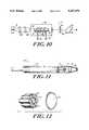
- FIG. 10is a more detailed schematic diagram of a laser source useful in the system of FIG. 1;
- FIG. 11is a partial, cross-sectional view of a laser beam delivery handpiece according to the invention.
- FIG. 12is a front view of the laser delivery handpiece of FIG. 11;
- FIG. 13is front view of a surgical instrument incorporating both a suture means and a laser means according to the invention.
- FIG. 14is a more detailed schematic diagram of reflectance monitor for use in the present invention.
- FIG. 15is a schematic illustration of a clinical eyepiece view showing a "heads-up" display of reflectance measurements according to the invention.
- FIG. 1a schematic block diagram of a photoreactive suturing system 10 is shown, including a laser 12, power supply 14, controller 16 and photoreactive suturing material 36.
- the systemcan further include a beamshaping/delivery assembly 20, illumination source 22, reflectance monitor 18, display 24 and tuner 26.
- the output of laser 12is delivered, preferably via beamshaping/delivery assembly 20, to an anastomotic site 30 to fuse the suture material 36 on opposite sides of a fissure or cleavage line 32 in a biological material.
- the laser beamirradiates exposure zone 34, a crosslinking reaction occurs to fuse the suture material and the biological tissue in the vicinity of the site 30.
- the degree of crosslinkingcan be determined by the reflectance monitor 18, which provides electrical signals to controller 16 in order to control the procedure.
- the reflectance monitor 18preferably receives light reflected by the site from a broadband or white light illumination source 22.
- the reflectance monitor 18 and/or controller 16can also provide signals to a display 24 to provide visual (and/or audio) feedback to the clinical user, thereby permitting manual control.
- Tuner 26can also be employed by the user (or automatically controlled by controller 16) to adjust the wavelength of the annealing radiation beam.
- FIG. 2provides further schematic illustration of the photoreactive suture system 10 in use.
- the electrical and optical components of the systemcan be housed in a system cabinet 60 suitable for use in an operating room or other clinical environment.
- the laser outputis delivered to the patient by an optical fiber cable 62 (which can include multiple optical fibers as detailed below) and a handpiece 64.
- the systemis preferably used in conjunction with a surgical microscope (or goggles) 66 which are adapted to provide a "heads-up" display to the user.
- Display signals from the system cabinet 60are transmitted to the microscope (or goggles) 66 by cable 68.
- the laser outputcan also be delivered to a remote site via an arthroscope, endoscope or catheter and the display features of such an instrument can be similarly adapted to provide the user with data on progress of the crosslinking reaction.
- the suture materials of the present inventioncan take various forms.
- the suture materialcomprises a strip or strand of a photoreactive crosslinking agent, such a collagen fibers, which can be sewn or draped upon a fissure or incision and then crosslinked to the tissue to provide closure. Once in place, the suture material is irradiated with laser or other high intensity light energy to fuse the suture to the anastomotic site.
- the suture material 36can include a high tensile strength core element 40 and an outer cross-linkable agent 38 which are likewise used to sew or drape the anastomotic site prior to irradiation and fusion.
- a suture material 36Acan be employed which is fabricated in a zig-zag strip form and applied directly upon the incision or fissure 32 to close the opening.
- suture material 36Acan include a high tensile strength core element 40 and an outer cross-linkable agent 38.
- the suture materialcan be fabricated as a patch with a high strength element 40 incorporated into the structure, and also including a crosslinking agent 38 to join the suture material to the underlying tissue and thereby effect closure of the anastomotic site 32.
- the high strength element 40can be fabricated, for example, from the same material as the bonding agent 38, but pre-crosslinked to provide the addition resistance to tearing or shearing forces as the wound heals.
- the present inventioncan employ various materials as crosslinking agents in either solid, liquid, gel or powder form to form a bond to tissue segments and thereby hold them together while natural healing processes occur.
- the crosslinking agentsshould be biocompatible and are preferably biodegradable over time in vivo. Examples of such crosslinking agents include collagen, elastin, fibrin, albumin and various other photoreactive polymeric materials.
- Various strength enhancing agentscan also be incorporated into the suture structure to provide additional tensile support along and across the anastomosis.
- Such high tensile strength elementscan be formed from pre-crosslinked segments of the same material that forms the photoreactive crosslinking agent, or they can be formed from strips or fibers of other natural or synthetic biodegradable materials such as polyesters, to enhance the strength of the bond.
- a detachable carrier 37is shown for use in applying a zig-zag type strip of crosslinking agent 36 to an anastomotic site 32.
- the detachable carrier 37is substantially transparent to photo-irradiation and can be detached from said crosslinking agent 36 following the bonding of the agent to the biological material.
- a tubular suture material 36is shown for repairing a torn blood vessel 31 or other body tube or lumen.
- the suture material 36preferably includes a crosslinking agent 38 and reinforcing elements which can be braided, woven or simply matted fibers 40.
- the suture materialis either fitted over the severed lumen (in the case of a tube-shaped suture material) or wrapped around the severed biological structure (e.g., with a strip-like suture material), and then irradiated to crosslink the materials together.
- the tubular suture material 36 of FIG. 8can be designed to shrink as the crosslinking reaction occurs and thereby more tightly wrap the anastomotic site. In such procedures, it may also be preferable to first dispose a stent 33 or similar support within the lumen to prevent collapse.
- a staple structure 37is shown incorporating a crosslinking agent 36 on each prong such that the staple can be applied to close a wound and then fused in place by application of laser radiation to the crosslinking agent 36.
- the entire staplecan be formed from a crosslinking agent and then irradiated (e.g., such that the exposed prongs are melted into tissue-bonding balls) to fuse the staple in place.
- the present inventioncan be practiced with a wide variety of laser sources, including both gas and solid state lasers, operating in either continuous wave ("c.w.") or pulsed modes. More specifically, the laser sources can be carbon monoxide, carbon dioxide, argon lasers or various excimer lasers utilizing mixtures of halogen and noble gases, such as argon-flouride, krypton-fluoride, xenon-chloride and xenon-fluoride.
- halogen and noble gasessuch as argon-flouride, krypton-fluoride, xenon-chloride and xenon-fluoride.
- the lasercan be a solid state laser employing a rare, earth-doped Yttrium Aluminum Garnet (YAG) or Yttrium Lithium Fluoride (YLF) or a Yttrium-Scandium-Gadolinium-Garnet (YSGG) laser.
- YAGYttrium Aluminum Garnet
- YLFYttrium Lithium Fluoride
- YSGGYttrium-Scandium-Gadolinium-Garnet
- the laser sourceis a rare, earth-doped, solid state laser, such as a holmium-doped, erbium-doped or thulium-doped solid state laser of the YAG, YLF or YSGG type which can be operated in a low wattage c.w. or pulsed mode with an output wavelength in the range of about 1.4 to about 2.5 micrometers and a power density of about 0.1 watt/mm 2 to about 1.0 watt/mm 2 .
- Such laser sourcesare disclosed in U.S. Pat. No. 4,917,084 issued on Apr. 17, 1990, to the present inventor and incorporated herein by reference.
- the absorption of laser energy from such solid state laser sources by biological tissuesis relatively high in relation to the absorption of such energy by water, thereby providing an absorption length in the subject's body of about 100 microns or more.
- itis possible to operate satisfactorily even with 10-20 micrometers of blood between the handpiece tip and the anastomotic site.
- FIG. 10is a schematic illustration of laser source 12, including a solid-state laser crystal 41, vacuum chamber 42 and diode pump source 44.
- the laser crystal 41is preferably surrounded by a cooling quartz or fused-silica jacket 46 having inlet pipe 48 and an outlet pipe 50 for circulation of liquid nitrogen or other cryogenic coolant.
- the laser cavitycan be formed by input crystal face coating 52 and partially-reflective output mirror 54.
- the laser crystal 41is excited by optical pumping, that being, irradiation of the crystal with light from the laser diode 44.
- the diode 44can be cooled by a pumped coolant or employ a heatsink).
- Both ends of the laser crystal 41are preferably polished flat.
- the input face of the crystal 41is preferably finished with a coating 52 for high transmittance at the pump wavelength and high reflectance of the output wavelength.
- the other end of the crystal 41preferably includes an antireflective coating 50 for high transmittal of the output wavelength.
- the entire cavity of the reflectorpreferably is evacuated to provide thermal insulation and avoid moisture condensation.
- cryogenic, solid-state laserssee, for example, an article by Barnes et al., Vol. 190, Society of the Photo-Optical Instrumentation Engineers, pp. 297-304 (1979), NASA/JPL Technical Brief No. NPO-17282/6780 by Hemmati (June, 1988) and above-referenced U.S. Pat. No. 4,917,084, all of which are herein incorporated by reference.
- a tuning element 26which can include, for example, a birefringent crystal 28 disposed along the beam path 58 at a slight offset from Brewster's angle.
- the crystal 28can be tuned electro-optically by application of a voltage, as shown schematically in the figure.
- the laser wavelengthcan be tuned mechanically by tilting or rotating the crystal 28 relative to the beam path using techniques well known in the art.
- a partial, cross-sectional side view of a handpiece 64is shown, including a casing 70 adapted for gripping by the clinical user and multiple lumens disposed therein.
- the handpieceserves to deliver laser irradiation suitable for biological tissue fusion via a central optical fiber 72 connected to laser source, as well as one or more additional illumination fibers 74 for the delivery of illumination light and the transmittal of reflected light.
- the surgical laser delivery fiber 72is preferable a low, hydroxyl ion content silica fiber.
- the handpiece 64can deliver illumination light via fibers 74. In one embodiment, these fibers 74 can also be used to collect reflective light and deliver it to a controller. Alternatively, some of the fibers 74 can be devoted entirely to collection of reflected light.
- the handpiece 64can further include one or more lens elements 76, as well as a transparent protective cover element or terminal lens 82.
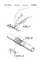
- FIG. 13shows an apparatus 81 for remote application of sutures according to the invention.
- the apparatus 81can be incorporated into a catheter, endoscope or arthroscope and disposed adjacent to a remote anastomotic site.
- apparatus 81includes a suture means 85 and a laser means 83.
- the suture port 85delivers a photoreactive suture material to the anastomotic site, the suture material comprising a structure with at least a portion of the structure formed by a crosslinking agent such that upon irradiation of said suture means the crosslinking agent adheres to the biological material and thereby provides closure at said anastomotic site.
- the laser means 83provides the necessary light energy in the form of laser radiation to effect crosslinking of the suture material at the anastomotic site.
- the apparatus 81can also include a viewing port 87, an illumination port 89 and a reflectance sensing port 91 to provide a display and monitoring of the crosslinking process, as described in more detail below.
- FIG. 14is a more detailed schematic diagram of a reflectance monitor 18, including a coupling port 90 for coupling with one or more fibers 76 to receive reflectance signals from the handpiece of FIG. 4 or the apparatus of FIG. 13.
- the reflectance monitor 18can further include a focusing lens 92 and first and second beam splitting elements 94 and 96, which serve to divide the reflected light into 3 (or more) different beams for processing.
- a first beamis transmitted to a first optical filter 98 to detector 102 (providing, for example, measurement of reflected light at wavelengths shorter than 0.7 micrometers).
- a second portion of the reflected light signalis transmitted by beam splitter 96 through a second optical filter 100 to detector 104 (e.g., providing measurement of light at wavelengths shorter than 1.1 micrometers).
- detector 104e.g., providing measurement of light at wavelengths shorter than 1.1 micrometers.
- a third portion of the reflected lightis transmitted to photodetector 106 (e.g., for measurement of reflected light at wavelengths greater than 1.6 micrometers).
- photodetector 106e.g., for measurement of reflected light at wavelengths greater than 1.6 micrometers.
- the detector elements 102, 104 and 106preferably include synchronous demodulation circuitry and are used in conjunction with a modulated illumination source to suppress any artifacts caused by stray light or the ambient environment. (It should be apparent that other optical arrangements can be employed to obtain multiple wavelength analysis, including the use, for example, of dichroic elements, either as beamsplitters or in conjunction with such beamsplitters, to effectively pass particular wavelengths to specific detector elements. It should also be apparent that more than three discreet wavelengths can be measured, depending upon the particular application.)
- the signals from the detector elementscan then be transmitted to a controller and/or a display element (as shown in FIG. 1).
- signals from the reflectance monitorare analyzed (as detailed below) to determine the degree of crosslinking which is occurring in the suture material and/or in the biological tissue exposed to the laser radiation.
- Such analysiscan generate control signals which will progressively reduce the laser output energy over time as a particular site experiences cumulative exposure.
- the control signalscan further provide for an automatic shut-off of the laser when the optimal state of crosslinking has been exceeded and/or the onset of carbonization is occurring.
- the data from the reflectance monitorcan also be provided directly to the clinician.
- a simulated view from an eyepiece 110is shown in which the field of view 112 includes a fissure or cleavage line 114 dividing separate bodies at an anastomotic site.
- the suture material 36Also shown within the field of view is the suture material 36, a fusion track 116 which has been formed by laser radiation, and a present exposure zone 118.
- the apparatus of the present inventioncan be employed to analyze the degree of crosslinking by comparing the reflectance ratios of a site at two or more wavelengths.
- intensity readings for three or more wavelength rangesare employed in order to accurately assess the degree of crosslinking and to ensure that the optimal state is not exceeded.
- the particular wavelengths to be monitoredwill, of course, vary with the particular tissue undergoing treatment. Although the tissue type, (e.g., blood-containing tissue or that which is relatively blood-free) will vary, the general principles of the invention, as disclosed herein, can be readily applied by those skilled in the art to diverse procedures in which the fusion of biological materials is desired.
- the analyzing circuitry of the controllercan be constructed to provide a warning (or automatically shut off the laser radiation) when darkening in the visible wavelengths occurs or when the ratio of visible to infrared values falls below a predefined level.
- the material to be joinede.g., aortic tissue
- infrared reflectance changese.g., above 1.1 micrometers
- the analyzing circuitrycan monitor infrared reflectance changes (e.g., greater than about 1.0 micrometers) as an indicator of proper crosslinking.
- the reflectance sensorcan also be used as a proximity monitor to ensue that the laser is in fact disposed at a proper distance from the anastomic site.
- the analyzing circuitrycan sense the changes in reflectance and generate a warning to the user (or automatically shut off the system) until proper placement is achieved.
Landscapes
- Physics & Mathematics (AREA)
- Optics & Photonics (AREA)
- Engineering & Computer Science (AREA)
- Health & Medical Sciences (AREA)
- Surgery (AREA)
- Life Sciences & Earth Sciences (AREA)
- Mechanical Engineering (AREA)
- Plasma & Fusion (AREA)
- Public Health (AREA)
- Biomedical Technology (AREA)
- Heart & Thoracic Surgery (AREA)
- Medical Informatics (AREA)
- Molecular Biology (AREA)
- Animal Behavior & Ethology (AREA)
- General Health & Medical Sciences (AREA)
- Veterinary Medicine (AREA)
- Nuclear Medicine, Radiotherapy & Molecular Imaging (AREA)
- Otolaryngology (AREA)
- Electromagnetism (AREA)
- Laser Surgery Devices (AREA)
Abstract
Description
Claims (8)
Priority Applications (4)
| Application Number | Priority Date | Filing Date | Title |
|---|---|---|---|
| US07/804,791US5207670A (en) | 1990-06-15 | 1991-12-09 | Photoreactive suturing of biological materials |
| US08/292,608US5569239A (en) | 1990-06-15 | 1994-08-18 | Photoreactive suturing of biological materials |
| US08/327,583US5540677A (en) | 1990-06-15 | 1994-10-24 | Endoscopic systems for photoreactive suturing of biological materials |
| US08/479,950US5725522A (en) | 1990-06-15 | 1995-06-07 | Laser suturing of biological materials |
Applications Claiming Priority (2)
| Application Number | Priority Date | Filing Date | Title |
|---|---|---|---|
| US07/538,977US5071417A (en) | 1990-06-15 | 1990-06-15 | Laser fusion of biological materials |
| US07/804,791US5207670A (en) | 1990-06-15 | 1991-12-09 | Photoreactive suturing of biological materials |
Related Parent Applications (1)
| Application Number | Title | Priority Date | Filing Date |
|---|---|---|---|
| US07/538,977Continuation-In-PartUS5071417A (en) | 1990-06-15 | 1990-06-15 | Laser fusion of biological materials |
Related Child Applications (2)
| Application Number | Title | Priority Date | Filing Date |
|---|---|---|---|
| US5700093AContinuation-In-Part | 1990-06-15 | 1993-05-03 | |
| US5619293AContinuation | 1990-06-15 | 1993-05-03 |
Publications (1)
| Publication Number | Publication Date |
|---|---|
| US5207670Atrue US5207670A (en) | 1993-05-04 |
Family
ID=24149235
Family Applications (3)
| Application Number | Title | Priority Date | Filing Date |
|---|---|---|---|
| US07/538,977Expired - Fee RelatedUS5071417A (en) | 1990-06-15 | 1990-06-15 | Laser fusion of biological materials |
| US07/804,791Expired - Fee RelatedUS5207670A (en) | 1990-06-15 | 1991-12-09 | Photoreactive suturing of biological materials |
| US08/292,608Expired - Fee RelatedUS5569239A (en) | 1990-06-15 | 1994-08-18 | Photoreactive suturing of biological materials |
Family Applications Before (1)
| Application Number | Title | Priority Date | Filing Date |
|---|---|---|---|
| US07/538,977Expired - Fee RelatedUS5071417A (en) | 1990-06-15 | 1990-06-15 | Laser fusion of biological materials |
Family Applications After (1)
| Application Number | Title | Priority Date | Filing Date |
|---|---|---|---|
| US08/292,608Expired - Fee RelatedUS5569239A (en) | 1990-06-15 | 1994-08-18 | Photoreactive suturing of biological materials |
Country Status (1)
| Country | Link |
|---|---|
| US (3) | US5071417A (en) |
Cited By (82)
| Publication number | Priority date | Publication date | Assignee | Title |
|---|---|---|---|---|
| US5300065A (en)* | 1992-11-06 | 1994-04-05 | Proclosure Inc. | Method and apparatus for simultaneously holding and sealing tissue |
| US5372585A (en)* | 1992-04-09 | 1994-12-13 | Tiefenbrun; Jonathan | Instrument and associated method for applying biologically effective composition during laparoscopic operation |
| US5470307A (en)* | 1994-03-16 | 1995-11-28 | Lindall; Arnold W. | Catheter system for controllably releasing a therapeutic agent at a remote tissue site |
| US5540677A (en)* | 1990-06-15 | 1996-07-30 | Rare Earth Medical, Inc. | Endoscopic systems for photoreactive suturing of biological materials |
| US5569239A (en)* | 1990-06-15 | 1996-10-29 | Rare Earth Medical, Inc. | Photoreactive suturing of biological materials |
| US5612050A (en)* | 1993-03-23 | 1997-03-18 | Focal, Inc. | Apparatus and method for local application of polymeric material to tissue |
| WO1997013461A1 (en)* | 1995-10-11 | 1997-04-17 | Fusion Medical Technologies, Inc. | Device and method for sealing tissue |
| US5662643A (en)* | 1994-09-28 | 1997-09-02 | Abiomed R & D, Inc. | Laser welding system |
| US5665063A (en)* | 1994-06-24 | 1997-09-09 | Focal, Inc. | Methods for application of intraluminal photopolymerized gels |
| US5669934A (en)* | 1991-02-13 | 1997-09-23 | Fusion Medical Technologies, Inc. | Methods for joining tissue by applying radiofrequency energy to performed collagen films and sheets |
| US5674231A (en)* | 1995-10-20 | 1997-10-07 | United States Surgical Corporation | Apparatus and method for vascular hole closure |
| US5709677A (en)* | 1995-07-12 | 1998-01-20 | Laser Industries, Ltd. | Apparatus and method as preparation for performing a myringotomy in a child's ear without the need for anaesthesia |
| US5725522A (en)* | 1990-06-15 | 1998-03-10 | Rare Earth Medical, Inc. | Laser suturing of biological materials |
| US5779673A (en)* | 1995-06-26 | 1998-07-14 | Focal, Inc. | Devices and methods for application of intraluminal photopolymerized gels |
| US5810846A (en)* | 1995-08-03 | 1998-09-22 | United States Surgical Corporation | Vascular hole closure |
| US5824015A (en)* | 1991-02-13 | 1998-10-20 | Fusion Medical Technologies, Inc. | Method for welding biological tissue |
| US5827265A (en)* | 1996-02-07 | 1998-10-27 | Regents Of The University Of California | Intraluminal tissue welding for anastomosis |
| US5860948A (en)* | 1993-10-12 | 1999-01-19 | Scimed Life Systems, Inc. | Apparatus and method for tissue defect repair by deposition |
| US5868731A (en)* | 1996-03-04 | 1999-02-09 | Innotech Usa, Inc. | Laser surgical device and method of its use |
| US5976127A (en)* | 1998-01-14 | 1999-11-02 | Lax; Ronald | Soft tissue fixation devices |
| WO2000012018A1 (en)* | 1998-08-26 | 2000-03-09 | Advanced Closure Systems, Inc. | Compositions, systems, and methods for creating in situ, chemically cross-linked, mechanical barriers or covering structures |
| US6245060B1 (en)* | 1997-03-25 | 2001-06-12 | Abbott Laboratories | Removal of stratum corneum by means of light |
| WO2001043646A1 (en)* | 1999-12-14 | 2001-06-21 | Shadduck John H | Electrical discharge surgical fastener for meniscal repairs |
| US20010051813A1 (en)* | 1998-11-06 | 2001-12-13 | Neomend, Inc. | Systems, methods, and compositions for achieving closure of vascular puncture sites |
| US6371975B2 (en) | 1998-11-06 | 2002-04-16 | Neomend, Inc. | Compositions, systems, and methods for creating in situ, chemically cross-linked, mechanical barriers |
| US20020065493A1 (en)* | 1999-10-22 | 2002-05-30 | Nyhart Eldon H. | Apparatus for the controllable modification of compound concentration in a tube |
| US6402739B1 (en)* | 1998-12-08 | 2002-06-11 | Y-Beam Technologies, Inc. | Energy application with cooling |
| US6451007B1 (en)* | 1999-07-29 | 2002-09-17 | Dale E. Koop | Thermal quenching of tissue |
| US6458147B1 (en) | 1998-11-06 | 2002-10-01 | Neomend, Inc. | Compositions, systems, and methods for arresting or controlling bleeding or fluid leakage in body tissue |
| US6468520B1 (en) | 1993-03-23 | 2002-10-22 | Focal, Inc. | Apparatus and method for local application of polymeric material to tissue |
| US6475138B1 (en) | 1995-07-12 | 2002-11-05 | Laser Industries Ltd. | Apparatus and method as preparation for performing a myringotomy in a child's ear without the need for anaesthesia |
| US6491715B1 (en)* | 1999-11-17 | 2002-12-10 | Pulsion Medical Systems Ag | Device for treating growing, dilated or malformed blood vessels and method for treating biological material |
| US20030100921A1 (en)* | 1998-11-06 | 2003-05-29 | Neomend, Inc. | Systems, methods, and compositions for achieving closure of vascular puncture sites |
| US6656496B1 (en) | 1999-03-01 | 2003-12-02 | The Uab Research Foundation | Porous tissue scaffolding materials and uses thereof |
| US20040063613A1 (en)* | 1998-06-23 | 2004-04-01 | James Rolke | Methods and compositions for sealing tissue leaks |
| US20040064167A1 (en)* | 2002-06-28 | 2004-04-01 | Berry Andrew J. | Dermatological Treatment Apparatus and Method |
| US20040073256A1 (en)* | 2002-08-09 | 2004-04-15 | Kevin Marchitto | Activated surgical fasteners, devices therefor and uses thereof |
| US6743248B2 (en) | 1996-12-18 | 2004-06-01 | Neomend, Inc. | Pretreatment method for enhancing tissue adhesion |
| US20040230185A1 (en)* | 2003-03-27 | 2004-11-18 | Cierra, Inc. | Energy based devices and methods for treatment of patent foramen ovale |
| US20040267191A1 (en)* | 2003-03-27 | 2004-12-30 | Cierra, Inc. | Methods and apparatus for treatment of patent foramen ovale |
| US20050021016A1 (en)* | 2003-03-27 | 2005-01-27 | Cierra, Inc. | Energy based devices and methods for treatment of anatomic tissue defects |
| US20050034735A1 (en)* | 2003-03-27 | 2005-02-17 | Cierra, Inc. | Methods and apparatus for treatment of patent foramen ovale |
| US20050080406A1 (en)* | 2003-03-27 | 2005-04-14 | Cierra, Inc. | Energy based devices and methods for treatment of patent foramen ovale |
| US6899889B1 (en) | 1998-11-06 | 2005-05-31 | Neomend, Inc. | Biocompatible material composition adaptable to diverse therapeutic indications |
| US6949114B2 (en) | 1998-11-06 | 2005-09-27 | Neomend, Inc. | Systems, methods, and compositions for achieving closure of vascular puncture sites |
| US20050228283A1 (en)* | 2003-06-10 | 2005-10-13 | Gifford Hanson S | Methods and apparatus for non-invasively treating atrial fibrillation using high intensity focused ultrasound |
| US6994686B2 (en) | 1998-08-26 | 2006-02-07 | Neomend, Inc. | Systems for applying cross-linked mechanical barriers |
| US20060074410A1 (en)* | 2004-06-21 | 2006-04-06 | Cierra, Inc. | Energy based devices and methods for treatment of anatomic tissue defects |
| US20060079870A1 (en)* | 2004-10-07 | 2006-04-13 | Barry Robert L | Systems and methods for shrinking and/or securing cardiovascular tissue |
| EP1650839A1 (en)* | 2004-10-20 | 2006-04-26 | Wavelight Laser Technologie AG | Fiber laser arrangement |
| US20060122680A1 (en)* | 2003-02-13 | 2006-06-08 | Auth David C | Systems and methods for securing cardiovascular tissue |
| US20060271089A1 (en)* | 2005-04-11 | 2006-11-30 | Cierra, Inc. | Methods and apparatus to achieve a closure of a layered tissue defect |
| US20070093805A1 (en)* | 2005-10-17 | 2007-04-26 | Coaptus Medical Corporation | Systems and methods for securing cardiovascular tissue, including via asymmetric electrodes |
| US20070123852A1 (en)* | 2003-03-27 | 2007-05-31 | Cierra, Inc. | Methods and apparatus for closing a layered tissue defect |
| US20070203479A1 (en)* | 2003-02-13 | 2007-08-30 | Coaptus Medical Corporation | Transseptal closure of a patent foramen ovale and other cardiac defects |
| US20080073163A1 (en)* | 2006-09-22 | 2008-03-27 | Weir Michael P | Micro-electromechanical device |
| US20080077179A1 (en)* | 1997-03-12 | 2008-03-27 | Neomend, Inc. | Pretreatment method for enhancing tissue adhesion |
| US20080091249A1 (en)* | 2006-10-11 | 2008-04-17 | Bwt Property, Inc. | Photobiomodulation Apparatus with Enhanced Performance and Safety Features |
| US20080140114A1 (en)* | 1997-03-12 | 2008-06-12 | Neomend, Inc. | Systems and methods for sealing a vascular puncture |
| US20080140069A1 (en)* | 2006-12-07 | 2008-06-12 | Cierra, Inc. | Multi-electrode apparatus for tissue welding and ablation |
| US20080167521A1 (en)* | 2007-01-09 | 2008-07-10 | Sheetz Jane A | Method of in vivo monitoring using an imaging system including scanned beam imaging unit |
| US20080226034A1 (en)* | 2007-03-12 | 2008-09-18 | Weir Michael P | Power modulation of a scanning beam for imaging, therapy, and/or diagnosis |
| US20080242967A1 (en)* | 2007-03-27 | 2008-10-02 | Ethicon Endo-Surgery, Inc. | Medical imaging and therapy utilizing a scanned beam system operating at multiple wavelengths |
| US20080255458A1 (en)* | 2007-04-13 | 2008-10-16 | Ethicon Endo-Surgery, Inc. | System and method using fluorescence to examine within a patient's anatomy |
| US20080252778A1 (en)* | 2007-04-13 | 2008-10-16 | Ethicon Endo-Surgery, Inc. | Combined SBI and conventional image processor |
| US20080275305A1 (en)* | 2007-05-01 | 2008-11-06 | Ethicon Endo-Surgery, Inc. | Medical scanned beam imager and components associated therewith |
| US20080312490A1 (en)* | 2007-06-18 | 2008-12-18 | Ethicon Endo-Surgery, Inc. | Methods and devices for repairing damaged or diseased tissue using a scanning beam assembly |
| US20090060381A1 (en)* | 2007-08-31 | 2009-03-05 | Ethicon Endo-Surgery, Inc. | Dynamic range and amplitude control for imaging |
| US20090062659A1 (en)* | 2007-08-28 | 2009-03-05 | Weir Michael P | Medical device including scanned beam unit with operational control features |
| US20090062658A1 (en)* | 2007-08-27 | 2009-03-05 | Dunki-Jacobs Robert J | Position tracking and control for a scanning assembly |
| US7558455B2 (en) | 2007-06-29 | 2009-07-07 | Ethicon Endo-Surgery, Inc | Receiver aperture broadening for scanned beam imaging |
| US7589316B2 (en) | 2007-01-18 | 2009-09-15 | Ethicon Endo-Surgery, Inc. | Scanning beam imaging with adjustable detector sensitivity or gain |
| US20090232784A1 (en)* | 2005-03-10 | 2009-09-17 | Dale Feldman | Endothelial predecessor cell seeded wound healing scaffold |
| US7713265B2 (en)* | 2006-12-22 | 2010-05-11 | Ethicon Endo-Surgery, Inc. | Apparatus and method for medically treating a tattoo |
| US7972330B2 (en) | 2003-03-27 | 2011-07-05 | Terumo Kabushiki Kaisha | Methods and apparatus for closing a layered tissue defect |
| US7982776B2 (en) | 2007-07-13 | 2011-07-19 | Ethicon Endo-Surgery, Inc. | SBI motion artifact removal apparatus and method |
| US8050520B2 (en) | 2008-03-27 | 2011-11-01 | Ethicon Endo-Surgery, Inc. | Method for creating a pixel image from sampled data of a scanned beam imager |
| US8273015B2 (en) | 2007-01-09 | 2012-09-25 | Ethicon Endo-Surgery, Inc. | Methods for imaging the anatomy with an anatomically secured scanner assembly |
| US8332014B2 (en) | 2008-04-25 | 2012-12-11 | Ethicon Endo-Surgery, Inc. | Scanned beam device and method using same which measures the reflectance of patient tissue |
| US9125552B2 (en) | 2007-07-31 | 2015-09-08 | Ethicon Endo-Surgery, Inc. | Optical scanning module and means for attaching the module to medical instruments for introducing the module into the anatomy |
| CN108451637A (en)* | 2018-03-01 | 2018-08-28 | 周玉梅 | A kind of puncture needle and stitching devices |
| US10448997B2 (en) | 2013-06-25 | 2019-10-22 | Koninklijke Philips N.V. | Measurement device for skin properties and non-invasive treatment device |
Families Citing this family (182)
| Publication number | Priority date | Publication date | Assignee | Title |
|---|---|---|---|---|
| US5464013A (en)* | 1984-05-25 | 1995-11-07 | Lemelson; Jerome H. | Medical scanning and treatment system and method |
| EP0437183B1 (en)* | 1990-01-09 | 1994-07-27 | Ciba-Geigy Ag | Light diffuser for a photodynamic therapy of tumours in the oesophagus of a patient |
| US5690675A (en)* | 1991-02-13 | 1997-11-25 | Fusion Medical Technologies, Inc. | Methods for sealing of staples and other fasteners in tissue |
| US5749895A (en)* | 1991-02-13 | 1998-05-12 | Fusion Medical Technologies, Inc. | Method for bonding or fusion of biological tissue and material |
| US5318023A (en)* | 1991-04-03 | 1994-06-07 | Cedars-Sinai Medical Center | Apparatus and method of use for a photosensitizer enhanced fluorescence based biopsy needle |
| US5169395A (en)* | 1991-04-26 | 1992-12-08 | Pdt Cardiovascular, Inc. | Laser delivery system |
| CA2042075C (en)* | 1991-05-08 | 2001-01-23 | Branko Palcic | Endoscopic imaging system |
| US5337741A (en)* | 1991-06-21 | 1994-08-16 | Diamond Donald A | Photo radiation treatment apparatus and method |
| US5769792A (en)* | 1991-07-03 | 1998-06-23 | Xillix Technologies Corp. | Endoscopic imaging system for diseased tissue |
| US5200604A (en)* | 1991-08-07 | 1993-04-06 | Laser Engineering, Inc. | Handpiece optical proximity detector for disabling surgical laser beam |
| AU2468092A (en)* | 1991-08-22 | 1993-03-16 | Roberto Enzo DI BIAGGIO | Medical light treatment apparatus |
| US5344418A (en)* | 1991-12-12 | 1994-09-06 | Shahriar Ghaffari | Optical system for treatment of vascular lesions |
| USRE36872E (en)* | 1992-01-15 | 2000-09-12 | Laser Industries Ltd. | System for causing ablation of irradiated material of living tissue while not causing damage below a predetermined depth |
| US5501680A (en)* | 1992-01-15 | 1996-03-26 | The University Of Pittsburgh | Boundary and proximity sensor apparatus for a laser |
| IL100664A0 (en)* | 1992-01-15 | 1992-09-06 | Laser Ind Ltd | Method and apparatus for controlling a laser beam |
| US5279611A (en)* | 1992-03-13 | 1994-01-18 | Mcdonnell Peter J | Laser shaping of ocular surfaces using ablation mask formed in situ |
| US5334191A (en)* | 1992-05-21 | 1994-08-02 | Dix Phillip Poppas | Laser tissue welding control system |
| AU4793793A (en)* | 1992-09-02 | 1994-03-29 | Robert L. Epstein | Instrument for ophthalmological surgery |
| US5552452A (en)* | 1993-03-15 | 1996-09-03 | Arch Development Corp. | Organic tissue glue for closure of wounds |
| US5350376A (en)* | 1993-04-16 | 1994-09-27 | Ceramoptec, Inc. | Optical controller device |
| US5759194A (en)* | 1993-09-28 | 1998-06-02 | Hemodynamics, Inc. | Vascular patch applicator |
| US5628744A (en)* | 1993-12-21 | 1997-05-13 | Laserscope | Treatment beam handpiece |
| US5411016A (en) | 1994-02-22 | 1995-05-02 | Scimed Life Systems, Inc. | Intravascular balloon catheter for use in combination with an angioscope |
| ES2178663T3 (en)* | 1994-03-25 | 2003-01-01 | Novartis Ag | LIGHT DIFFUSER AND PROCESS FOR THE MANUFACTURE OF A LIGHT DIFFUSER. |
| US5590660A (en)* | 1994-03-28 | 1997-01-07 | Xillix Technologies Corp. | Apparatus and method for imaging diseased tissue using integrated autofluorescence |
| EP1231496B1 (en)* | 1994-08-18 | 2004-12-29 | Carl Zeiss AG | Optical coherence tomography assisted surgical apparatus |
| US5931165A (en)* | 1994-09-06 | 1999-08-03 | Fusion Medical Technologies, Inc. | Films having improved characteristics and methods for their preparation and use |
| US6572609B1 (en) | 1999-07-14 | 2003-06-03 | Cardiofocus, Inc. | Phototherapeutic waveguide apparatus |
| US6676656B2 (en) | 1994-09-09 | 2004-01-13 | Cardiofocus, Inc. | Surgical ablation with radiant energy |
| US8025661B2 (en) | 1994-09-09 | 2011-09-27 | Cardiofocus, Inc. | Coaxial catheter instruments for ablation with radiant energy |
| US6579285B2 (en) | 1994-09-09 | 2003-06-17 | Cardiofocus, Inc. | Photoablation with infrared radiation |
| US6423055B1 (en)* | 1999-07-14 | 2002-07-23 | Cardiofocus, Inc. | Phototherapeutic wave guide apparatus |
| US6558375B1 (en) | 2000-07-14 | 2003-05-06 | Cardiofocus, Inc. | Cardiac ablation instrument |
| US5733278A (en)* | 1994-11-30 | 1998-03-31 | Laser Industries Limited | Method and apparatus for hair transplantation using a scanning continuous-working CO2 laser |
| AUPN066795A0 (en)* | 1995-01-20 | 1995-02-16 | Macquarie Research Limited | Method of repair |
| US5624434A (en)* | 1995-02-03 | 1997-04-29 | Laser Industries, Ltd. | Laser preparation of recipient holes for graft implantation in the treatment of icepick scars |
| US5817090A (en)* | 1995-02-03 | 1998-10-06 | Laser Industries, Ltd. | Laser dermal implants for the treatment of facial skin depressions |
| US5611795A (en)* | 1995-02-03 | 1997-03-18 | Laser Industries, Ltd. | Laser facial rejuvenation |
| US5634936A (en) | 1995-02-06 | 1997-06-03 | Scimed Life Systems, Inc. | Device for closing a septal defect |
| RU2096051C1 (en)* | 1995-02-24 | 1997-11-20 | Григорий Борисович Альтшулер | Apparatus for laser treatment of biological tissues (alternative embodiments) |
| US5718697A (en)* | 1995-12-14 | 1998-02-17 | Johnson & Johnson, Inc. | Liquid absorbent sphagnum moss article and method for manufacturing the absorbent article |
| WO1997017023A1 (en) | 1995-11-07 | 1997-05-15 | Fusion Medical Technologies, Inc. | Methods and articles for fusing polysaccharide-containing matrix layers to tissue |
| US5647368A (en)* | 1996-02-28 | 1997-07-15 | Xillix Technologies Corp. | Imaging system for detecting diseased tissue using native fluorsecence in the gastrointestinal and respiratory tract |
| US5728992A (en)* | 1996-02-29 | 1998-03-17 | Westinghouse Electric Corporation | Apparatus and method for real time evaluation of laser welds especially in confined spaces such as within heat exchanger tubing |
| WO1997037723A1 (en)* | 1996-04-10 | 1997-10-16 | New Star Lasers, Inc. | Improved method and device for laser induced shrinking of collagen |
| US5876426A (en)* | 1996-06-13 | 1999-03-02 | Scimed Life Systems, Inc. | System and method of providing a blood-free interface for intravascular light delivery |
| US5791352A (en)* | 1996-06-19 | 1998-08-11 | Fusion Medical Technologies, Inc. | Methods and compositions for inhibiting tissue adhesion |
| US5759200A (en)* | 1996-09-04 | 1998-06-02 | Azar; Zion | Method of selective photothermolysis |
| US6214034B1 (en) | 1996-09-04 | 2001-04-10 | Radiancy, Inc. | Method of selective photothermolysis |
| US5797901A (en)* | 1996-09-20 | 1998-08-25 | Cosmescu; Ioan | Automatic activation system for a medical diagnostic monitoring and surgical apparatus and method therefore |
| EP0959795A4 (en)* | 1996-10-16 | 2000-01-19 | Fusion Medical Technologies | Films having improved characteristics and methods for their preparation and use |
| US7204832B2 (en) | 1996-12-02 | 2007-04-17 | Pálomar Medical Technologies, Inc. | Cooling system for a photo cosmetic device |
| US6653618B2 (en) | 2000-04-28 | 2003-11-25 | Palomar Medical Technologies, Inc. | Contact detecting method and apparatus for an optical radiation handpiece |
| US8182473B2 (en) | 1999-01-08 | 2012-05-22 | Palomar Medical Technologies | Cooling system for a photocosmetic device |
| US6517532B1 (en) | 1997-05-15 | 2003-02-11 | Palomar Medical Technologies, Inc. | Light energy delivery head |
| US6015404A (en)* | 1996-12-02 | 2000-01-18 | Palomar Medical Technologies, Inc. | Laser dermatology with feedback control |
| US6036683A (en)* | 1997-01-02 | 2000-03-14 | G. Rodenstock Instruments Gmbh | Process and apparatus for changing the curvature of the cornea |
| ES2226133T3 (en) | 1997-05-15 | 2005-03-16 | Palomar Medical Technologies, Inc. | DERMATOLOGICAL TREATMENT DEVICE. |
| US6740497B2 (en)* | 1998-03-06 | 2004-05-25 | The Regents Of The University Of California | Method and apparatus for detecting cancerous cells using molecules that change electrophoretic mobility |
| US6156576A (en)* | 1998-03-06 | 2000-12-05 | The Regents Of The University Of California | Fast controllable laser lysis of cells for analysis |
| US6335201B1 (en)* | 1998-03-06 | 2002-01-01 | The Regents Of The University Of California | Method and apparatus for detecting enzymatic activity using molecules that change electrophoretic mobility |
| ES2245506T3 (en) | 1998-03-12 | 2006-01-01 | Palomar Medical Technologies, Inc. | ELECTROMAGNETIC RADIATION APPLICATION SYSTEM ON SKIN. |
| CA2326120C (en) | 1998-03-27 | 2015-01-13 | The General Hospital Corporation | Method and apparatus for the selective targeting of lipid-rich tissues |
| US6485414B1 (en)* | 1998-07-13 | 2002-11-26 | Ceramoptec Industries, Inc. | Color video diagnostic system for mini-endoscopes |
| FR2781358B1 (en)* | 1998-07-27 | 2000-10-13 | Cird Galderma | DEVICE FOR ASSEMBLING LIPS FROM A WOUND, HOLDING PIECE AND COSMETIC TREATMENT METHOD |
| US7137980B2 (en) | 1998-10-23 | 2006-11-21 | Sherwood Services Ag | Method and system for controlling output of RF medical generator |
| FR2785564B1 (en)* | 1998-11-10 | 2000-12-08 | Cebal | TREATMENT OF LASER MATERIALS, IN PARTICULAR CUTTING OR WELDING |
| US6514242B1 (en) | 1998-12-03 | 2003-02-04 | David Vasily | Method and apparatus for laser removal of hair |
| US8540704B2 (en) | 1999-07-14 | 2013-09-24 | Cardiofocus, Inc. | Guided cardiac ablation catheters |
| US7935108B2 (en) | 1999-07-14 | 2011-05-03 | Cardiofocus, Inc. | Deflectable sheath catheters |
| US8900219B2 (en) | 1999-07-14 | 2014-12-02 | Cardiofocus, Inc. | System and method for visualizing tissue during ablation procedures |
| US9033961B2 (en) | 1999-07-14 | 2015-05-19 | Cardiofocus, Inc. | Cardiac ablation catheters for forming overlapping lesions |
| US6413267B1 (en)* | 1999-08-09 | 2002-07-02 | Theralase, Inc. | Therapeutic laser device and method including noninvasive subsurface monitoring and controlling means |
| US20040167503A1 (en)* | 1999-08-25 | 2004-08-26 | Cardiofocus, Inc. | Malleable surgical ablation instruments |
| US20040147911A1 (en)* | 1999-08-25 | 2004-07-29 | Cardiofocus, Inc. | Surgical ablation instruments for forming an encircling lesion |
| US6419671B1 (en)* | 1999-12-23 | 2002-07-16 | Visx, Incorporated | Optical feedback system for vision correction |
| JP4495894B2 (en)* | 2000-01-25 | 2010-07-07 | パロマー・メディカル・テクノロジーズ・インコーポレーテッド | Device for medical treatment using long-term electromagnetic radiation |
| EP1700573A3 (en) | 2000-12-28 | 2010-12-01 | Palomar Medical Technologies, Inc. | Apparatus for therapeutic EMR treatment of the skin |
| US6699245B2 (en) | 2001-02-05 | 2004-03-02 | A-Med Systems, Inc. | Anastomosis system and related methods |
| US6888319B2 (en) | 2001-03-01 | 2005-05-03 | Palomar Medical Technologies, Inc. | Flashlamp drive circuit |
| DE10115426C2 (en)* | 2001-03-29 | 2003-03-13 | W & H Dentalwerk Buermoos Ges | Device and method for laser ablation of organic and inorganic material |
| DE10117347B4 (en)* | 2001-04-06 | 2006-04-13 | W&H Dentalwerk Bürmoos Gesellschaft m.b.H. | Laser treatment devices with lighting system |
| US7217266B2 (en)* | 2001-05-30 | 2007-05-15 | Anderson R Rox | Apparatus and method for laser treatment with spectroscopic feedback |
| US6723090B2 (en) | 2001-07-02 | 2004-04-20 | Palomar Medical Technologies, Inc. | Fiber laser device for medical/cosmetic procedures |
| FR2827151B1 (en)* | 2001-07-11 | 2004-04-02 | Europ Surgical Systems | METHOD FOR CONTROLLING TISSUE TEMPERATURE INDUCED BY A SURGICAL LASER |
| AU2002323492A1 (en)* | 2001-08-30 | 2003-03-18 | Pacgen Cellco Llc | The use of low-power laser irradiation for enhanced vascularization of tissue and tissue-engineered construct transplants |
| US6659966B2 (en)* | 2001-11-15 | 2003-12-09 | Roche Diagnostics Corporation | Fluid sampling apparatus |
| US7540869B2 (en) | 2001-12-27 | 2009-06-02 | Palomar Medical Technologies, Inc. | Method and apparatus for improved vascular related treatment |
| US6955542B2 (en)* | 2002-01-23 | 2005-10-18 | Aquatech Fitness Corp. | System for monitoring repetitive movement |
| CN1652729A (en) | 2002-03-12 | 2005-08-10 | 帕洛玛医疗技术公司 | Method and device for treating hair growth |
| US7166124B2 (en)* | 2002-03-21 | 2007-01-23 | Providence Health System - Oregon | Method for manufacturing sutureless bioprosthetic stent |
| CA2487987C (en) | 2002-06-19 | 2010-04-13 | Palomar Medical Technologies, Inc. | Method and apparatus for photothermal treatment of tissue at depth |
| WO2004000098A2 (en) | 2002-06-19 | 2003-12-31 | Palomar Medical Technologies, Inc. | Method and apparatus for treatment of cutaneous and subcutaneous conditions |
| EP2522294A2 (en) | 2002-10-23 | 2012-11-14 | Palomar Medical Technologies, Inc. | Phototreatment device for use with coolants and topical substances |
| EP2604216B1 (en) | 2003-02-25 | 2018-08-22 | Tria Beauty, Inc. | Self-contained, diode-laser-based dermatologic treatment apparatus |
| US7413567B2 (en)* | 2003-02-25 | 2008-08-19 | Spectragenics, Inc. | Optical sensor and method for identifying the presence of skin |
| US7309335B2 (en) | 2003-12-31 | 2007-12-18 | Palomar Medical Technologies, Inc. | Dermatological treatment with visualization |
| US20060009749A1 (en)* | 2004-02-19 | 2006-01-12 | Weckwerth Mark V | Efficient diffuse light source assembly and method |
| US8777935B2 (en) | 2004-02-25 | 2014-07-15 | Tria Beauty, Inc. | Optical sensor and method for identifying the presence of skin |
| EP2301471A1 (en) | 2004-04-01 | 2011-03-30 | The General Hospital Corporation | Method and apparatus for dermatological treatment and tissue reshaping |
| US7413572B2 (en)* | 2004-06-14 | 2008-08-19 | Reliant Technologies, Inc. | Adaptive control of optical pulses for laser medicine |
| US20060229515A1 (en)* | 2004-11-17 | 2006-10-12 | The Regents Of The University Of California | Fiber optic evaluation of tissue modification |
| US10413188B2 (en) | 2004-11-17 | 2019-09-17 | Lawrence Livermore National Security, Llc | Assessment of tissue or lesion depth using temporally resolved light scattering spectroscopy |
| US7731715B2 (en) | 2004-12-10 | 2010-06-08 | Edwards Lifesciences Corporation | Ablative treatment of atrial fibrillation via the coronary sinus |
| EP1681750A1 (en)* | 2005-01-17 | 2006-07-19 | Fanuc Ltd | Laser oscillator and method of estimating the lifetime of a pump light source |
| US8529560B2 (en) | 2005-03-04 | 2013-09-10 | The Invention Science Fund I, Llc | Hair treatment system |
| US8540701B2 (en) | 2005-03-04 | 2013-09-24 | The Invention Science Fund I, Llc | Hair treatment system |
| US8679101B2 (en)* | 2005-03-04 | 2014-03-25 | The Invention Science Fund I, Llc | Method and system for temporary hair removal |
| US8157807B2 (en) | 2005-06-02 | 2012-04-17 | The Invention Science Fund I, Llc | Skin treatment including patterned light |
| US7856985B2 (en) | 2005-04-22 | 2010-12-28 | Cynosure, Inc. | Method of treatment body tissue using a non-uniform laser beam |
| US9055958B2 (en) | 2005-06-29 | 2015-06-16 | The Invention Science Fund I, Llc | Hair modification using converging light |
| US20070122344A1 (en) | 2005-09-02 | 2007-05-31 | University Of Rochester Medical Center Office Of Technology Transfer | Intraoperative determination of nerve location |
| CN101309631A (en) | 2005-09-15 | 2008-11-19 | 帕洛玛医疗技术公司 | Skin optical characterization device |
| US20090216264A1 (en)* | 2005-09-19 | 2009-08-27 | Friedman Paul A | Implantable closure apparatus and methods |
| JP5680829B2 (en)* | 2006-02-01 | 2015-03-04 | ザ ジェネラル ホスピタル コーポレイション | A device that irradiates a sample with multiple electromagnetic radiations |
| US20070232871A1 (en)* | 2006-04-03 | 2007-10-04 | Edward Sinofsky | Method and system for determining tissue properties |
| US7586957B2 (en) | 2006-08-02 | 2009-09-08 | Cynosure, Inc | Picosecond laser apparatus and methods for its operation and use |
| US20080161744A1 (en) | 2006-09-07 | 2008-07-03 | University Of Rochester Medical Center | Pre-And Intra-Operative Localization of Penile Sentinel Nodes |
| US8202268B1 (en) | 2007-03-18 | 2012-06-19 | Lockheed Martin Corporation | Method and multiple-mode device for high-power short-pulse laser ablation and CW cauterization of bodily tissues |
| WO2008143955A2 (en)* | 2007-05-14 | 2008-11-27 | The Regents Of The University Of Colorado | Laser fusion of tissue layers |
| US20110125139A1 (en)* | 2007-10-04 | 2011-05-26 | Auld Jack R | Multi-fiber flexible surgical probe |
| DE102007047501A1 (en)* | 2007-10-04 | 2009-04-09 | Rolle + Rolle Gmbh + Co. Kg | Laserfokussierhandstück |
| WO2009088550A2 (en)* | 2007-10-19 | 2009-07-16 | Lockheed Martin Corporation | System and method for conditioning animal tissue using laser light |
| US8406860B2 (en) | 2008-01-25 | 2013-03-26 | Novadaq Technologies Inc. | Method for evaluating blush in myocardial tissue |
| US10219742B2 (en) | 2008-04-14 | 2019-03-05 | Novadaq Technologies ULC | Locating and analyzing perforator flaps for plastic and reconstructive surgery |
| WO2009132355A1 (en) | 2008-04-25 | 2009-10-29 | Tria Beauty, Inc. | Optical sensor and method for identifying the presence of skin and the pigmentation of skin |
| EP2687235A3 (en) | 2008-05-02 | 2014-11-05 | Novadaq Technologies Inc. | Methods for production and use of substance-loaded erythrocytes (S-LES) for observation and treatment of microvascular hemodynamics |
| WO2010007852A1 (en)* | 2008-07-16 | 2010-01-21 | 住友電気工業株式会社 | Laser processing apparatus and processing method employed therein |
| US10492671B2 (en) | 2009-05-08 | 2019-12-03 | Novadaq Technologies ULC | Near infra red fluorescence imaging for visualization of blood vessels during endoscopic harvest |
| US9919168B2 (en) | 2009-07-23 | 2018-03-20 | Palomar Medical Technologies, Inc. | Method for improvement of cellulite appearance |
| US8696653B2 (en) | 2009-10-02 | 2014-04-15 | Cardiofocus, Inc. | Cardiac ablation system with pulsed aiming light |
| US8702688B2 (en) | 2009-10-06 | 2014-04-22 | Cardiofocus, Inc. | Cardiac ablation image analysis system and process |
| BR112012020778A2 (en)* | 2010-02-17 | 2016-05-03 | Alcon Res Ltd | probe |
| JP5608871B2 (en)* | 2010-03-09 | 2014-10-15 | 学校法人慶應義塾 | System for preventing blood burn at the laser catheter emitting part |
| WO2011133660A2 (en)* | 2010-04-20 | 2011-10-27 | Alfano Robert R | Method for picosecond and femtosecond laser tissue welding |
| ES2453200T3 (en)* | 2010-08-24 | 2014-04-04 | Homag Holzbearbeitungssysteme Ag | Transmission device for radiation |
| US9039720B2 (en) | 2010-11-05 | 2015-05-26 | Ethicon Endo-Surgery, Inc. | Surgical instrument with ratcheting rotatable shaft |
| US9089338B2 (en) | 2010-11-05 | 2015-07-28 | Ethicon Endo-Surgery, Inc. | Medical device packaging with window for insertion of reusable component |
| US10085792B2 (en) | 2010-11-05 | 2018-10-02 | Ethicon Llc | Surgical instrument with motorized attachment feature |
| US9381058B2 (en) | 2010-11-05 | 2016-07-05 | Ethicon Endo-Surgery, Llc | Recharge system for medical devices |
| US10881448B2 (en) | 2010-11-05 | 2021-01-05 | Ethicon Llc | Cam driven coupling between ultrasonic transducer and waveguide in surgical instrument |
| US9375255B2 (en) | 2010-11-05 | 2016-06-28 | Ethicon Endo-Surgery, Llc | Surgical instrument handpiece with resiliently biased coupling to modular shaft and end effector |
| US9782214B2 (en) | 2010-11-05 | 2017-10-10 | Ethicon Llc | Surgical instrument with sensor and powered control |
| US9526921B2 (en) | 2010-11-05 | 2016-12-27 | Ethicon Endo-Surgery, Llc | User feedback through end effector of surgical instrument |
| US20120116381A1 (en) | 2010-11-05 | 2012-05-10 | Houser Kevin L | Surgical instrument with charging station and wireless communication |
| US9072523B2 (en) | 2010-11-05 | 2015-07-07 | Ethicon Endo-Surgery, Inc. | Medical device with feature for sterile acceptance of non-sterile reusable component |
| US9000720B2 (en) | 2010-11-05 | 2015-04-07 | Ethicon Endo-Surgery, Inc. | Medical device packaging with charging interface |
| US9597143B2 (en) | 2010-11-05 | 2017-03-21 | Ethicon Endo-Surgery, Llc | Sterile medical instrument charging device |
| US9510895B2 (en) | 2010-11-05 | 2016-12-06 | Ethicon Endo-Surgery, Llc | Surgical instrument with modular shaft and end effector |
| US9161803B2 (en) | 2010-11-05 | 2015-10-20 | Ethicon Endo-Surgery, Inc. | Motor driven electrosurgical device with mechanical and electrical feedback |
| US9017851B2 (en) | 2010-11-05 | 2015-04-28 | Ethicon Endo-Surgery, Inc. | Sterile housing for non-sterile medical device component |
| US9247986B2 (en) | 2010-11-05 | 2016-02-02 | Ethicon Endo-Surgery, Llc | Surgical instrument with ultrasonic transducer having integral switches |
| US9011471B2 (en) | 2010-11-05 | 2015-04-21 | Ethicon Endo-Surgery, Inc. | Surgical instrument with pivoting coupling to modular shaft and end effector |
| US9421062B2 (en) | 2010-11-05 | 2016-08-23 | Ethicon Endo-Surgery, Llc | Surgical instrument shaft with resiliently biased coupling to handpiece |
| US9017849B2 (en) | 2010-11-05 | 2015-04-28 | Ethicon Endo-Surgery, Inc. | Power source management for medical device |
| US10959769B2 (en) | 2010-11-05 | 2021-03-30 | Ethicon Llc | Surgical instrument with slip ring assembly to power ultrasonic transducer |
| US9649150B2 (en) | 2010-11-05 | 2017-05-16 | Ethicon Endo-Surgery, Llc | Selective activation of electronic components in medical device |
| US20120116265A1 (en) | 2010-11-05 | 2012-05-10 | Houser Kevin L | Surgical instrument with charging devices |
| US9782215B2 (en) | 2010-11-05 | 2017-10-10 | Ethicon Endo-Surgery, Llc | Surgical instrument with ultrasonic transducer having integral switches |
| US10660695B2 (en) | 2010-11-05 | 2020-05-26 | Ethicon Llc | Sterile medical instrument charging device |
| US9456870B2 (en) | 2011-05-16 | 2016-10-04 | Covidien Lp | Optical energy-based methods and apparatus for tissue sealing |
| US9113934B2 (en) | 2011-05-16 | 2015-08-25 | Covidien Lp | Optical energy-based methods and apparatus for tissue sealing |
| US9113933B2 (en) | 2011-05-16 | 2015-08-25 | Covidien Lp | Optical energy-based methods and apparatus for tissue sealing |
| EP2839552A4 (en) | 2012-04-18 | 2015-12-30 | Cynosure Inc | PICOSECOND LASER APPARATUS AND METHOD OF PROCESSING TARGET TISSUES USING THE SAME |
| WO2013190391A2 (en) | 2012-06-21 | 2013-12-27 | Novadaq Technologies Inc. | Quantification and analysis of angiography and perfusion |
| US9833285B2 (en) | 2012-07-17 | 2017-12-05 | Covidien Lp | Optical sealing device with cutting ability |
| US10285757B2 (en) | 2013-03-15 | 2019-05-14 | Cynosure, Llc | Picosecond optical radiation systems and methods of use |
| US9420967B2 (en)* | 2013-03-19 | 2016-08-23 | Surgisense Corporation | Apparatus, systems and methods for determining tissue oxygenation |
| CN107072505B (en) | 2014-04-05 | 2021-03-12 | 手术感应设备公司 | Devices, systems and methods for mapping of tissue oxygenation |
| EP3137007A4 (en) | 2014-04-28 | 2017-09-27 | Cardiofocus, Inc. | System and method for visualizing tissue with an icg dye composition during ablation procedures |
| HK1244873A1 (en) | 2014-09-29 | 2018-08-17 | Novadaq Technologies Inc. | Imaging a target fluorophore in a biological material in the presence of autofluorescence |
| CA2963450A1 (en) | 2014-10-09 | 2016-04-14 | Novadaq Technologies Inc. | Quantification of absolute blood flow in tissue using fluorescence-mediated photoplethysmography |
| US10136938B2 (en) | 2014-10-29 | 2018-11-27 | Ethicon Llc | Electrosurgical instrument with sensor |
| US10154888B2 (en) | 2014-12-03 | 2018-12-18 | Cardiofocus, Inc. | System and method for visual confirmation of pulmonary vein isolation during abalation procedures |
| WO2016130861A1 (en) | 2015-02-13 | 2016-08-18 | Medterials, Inc | Composition and method for use of energy activated wound dressing |
| US10569071B2 (en) | 2015-08-31 | 2020-02-25 | Ethicon Llc | Medicant eluting adjuncts and methods of using medicant eluting adjuncts |
| US10172973B2 (en)* | 2015-08-31 | 2019-01-08 | Ethicon Llc | Surgical adjuncts and medicants for promoting lung function |
| US20170232157A1 (en)* | 2016-02-11 | 2017-08-17 | Arizona Board Of Regents On Behalf Of Arizona State University | Tissue integrating materials for wound repair |
| CN106312312B (en)* | 2016-10-12 | 2018-02-06 | 长春工程学院 | One kind overlap joint Laser Welding penetration signal on-Line Monitor Device and method |
| EP3580609B1 (en) | 2017-02-10 | 2023-05-24 | Stryker European Operations Limited | Open-field handheld fluorescence imaging systems and methods |
| US10440830B2 (en)* | 2017-09-08 | 2019-10-08 | Robert Bosch Gmbh | Laser carbonization of polymer coatings in an open-air environment |
| WO2019165426A1 (en) | 2018-02-26 | 2019-08-29 | Cynosure, Inc. | Q-switched cavity dumped sub-nanosecond laser |
Citations (9)
| Publication number | Priority date | Publication date | Assignee | Title |
|---|---|---|---|---|
| US655190A (en)* | 1899-11-27 | 1900-08-07 | Louis Bramson | Suturating instrument. |
| US3272204A (en)* | 1965-09-22 | 1966-09-13 | Ethicon Inc | Absorbable collagen prosthetic implant with non-absorbable reinforcing strands |
| US3988782A (en)* | 1973-07-06 | 1976-11-02 | Dardik Irving I | Non-antigenic, non-thrombogenic infection-resistant grafts from umbilical cord vessels and process for preparing and using same |
| US4016877A (en)* | 1976-02-23 | 1977-04-12 | Avicon, Inc. | Fibrous collagen derived web having hemostatic and wound sealing properties |
| US4034750A (en)* | 1975-04-22 | 1977-07-12 | Maurice Seiderman | Electrochemically linking collagen fibrils to animal tissue |
| US4316467A (en)* | 1980-06-23 | 1982-02-23 | Lorenzo P. Maun | Control for laser hemangioma treatment system |
| US4747845A (en)* | 1983-10-17 | 1988-05-31 | Enquay Pharmaceutical Associates | Synthetic resin matrix system for the extended delivery of drugs |
| US4969912A (en)* | 1988-02-18 | 1990-11-13 | Kelman Charles D | Human collagen processing and autoimplant use |
| US5071417A (en)* | 1990-06-15 | 1991-12-10 | Rare Earth Medical Lasers, Inc. | Laser fusion of biological materials |
Family Cites Families (26)
| Publication number | Priority date | Publication date | Assignee | Title |
|---|---|---|---|---|
| US3276448A (en)* | 1962-12-14 | 1966-10-04 | Ethicon Inc | Collagen coated fabric prosthesis |
| GB1095552A (en)* | 1964-02-21 | |||
| US3593985A (en)* | 1969-06-03 | 1971-07-20 | Tanner James C Jun | Carrier for a skin graft patch |
| JPS494559B1 (en)* | 1970-08-06 | 1974-02-01 | ||
| SU618115A1 (en)* | 1974-11-20 | 1978-08-05 | Предприятие П/Я Г-4147 | Method of connecting biological tissues |
| SU618116A1 (en)* | 1975-01-24 | 1978-08-05 | Предприятие П/Я Г-4147 | Method of connecting the tissues of hollow organs |
| US4143660A (en)* | 1977-05-09 | 1979-03-13 | Malyshev Boris N | Method of surgery making use of laser emission and an apparatus for accomplishing same |
| US4428376A (en)* | 1980-05-02 | 1984-01-31 | Ethicon Inc. | Plastic surgical staple |
| DE3024169C2 (en)* | 1980-06-27 | 1983-09-15 | Reginald Dipl.-Phys. Dr. 8028 Taufkirchen Birngruber | Method and device for operating a photocoagulator for biological tissue |
| US4485816A (en)* | 1981-06-25 | 1984-12-04 | Alchemia | Shape-memory surgical staple apparatus and method for use in surgical suturing |
| CA1223644A (en)* | 1981-09-24 | 1987-06-30 | James R. Morris | Microsurgical laser |
| SU1091933A1 (en)* | 1981-10-14 | 1984-05-15 | Рижский Медицинский Институт | Method of connecting vessels |
| US4523591A (en)* | 1982-10-22 | 1985-06-18 | Kaplan Donald S | Polymers for injection molding of absorbable surgical devices |
| DE3306981C2 (en)* | 1983-02-28 | 1987-11-12 | Wolfram 8048 Haimhausen Weinberg | Device for the photocoagulation of biological tissue |
| DE3319203C2 (en)* | 1983-05-27 | 1986-03-27 | Fa. Carl Zeiss, 7920 Heidenheim | Device for dose measurement during photocoagulation |
| US4672969A (en)* | 1983-10-06 | 1987-06-16 | Sonomo Corporation | Laser healing method |
| US4854320A (en)* | 1983-10-06 | 1989-08-08 | Laser Surgery Software, Inc. | Laser healing method and apparatus |
| US4799479A (en)* | 1984-10-24 | 1989-01-24 | The Beth Israel Hospital Association | Method and apparatus for angioplasty |
| US4641650A (en)* | 1985-03-11 | 1987-02-10 | Mcm Laboratories, Inc. | Probe-and-fire lasers |
| EP0214712B1 (en)* | 1985-07-31 | 1992-09-02 | C.R. Bard, Inc. | Infrared laser catheter apparatus |
| US4917084A (en)* | 1985-07-31 | 1990-04-17 | C. R. Bard, Inc. | Infrared laser catheter system |
| US5100429A (en)* | 1989-04-28 | 1992-03-31 | C. R. Bard, Inc. | Endovascular stent and delivery system |
| US5066299A (en)* | 1990-06-29 | 1991-11-19 | Bellingham Medical, Inc. | Quick use suture package |
| US5209776A (en)* | 1990-07-27 | 1993-05-11 | The Trustees Of Columbia University In The City Of New York | Tissue bonding and sealing composition and method of using the same |
| US5234006A (en)* | 1991-01-18 | 1993-08-10 | Eaton Alexander M | Adjustable sutures and method of using the same |
| US5156613A (en)* | 1991-02-13 | 1992-10-20 | Interface Biomedical Laboratories Corp. | Collagen welding rod material for use in tissue welding |
- 1990
- 1990-06-15USUS07/538,977patent/US5071417A/ennot_activeExpired - Fee Related
- 1991
- 1991-12-09USUS07/804,791patent/US5207670A/ennot_activeExpired - Fee Related
- 1994
- 1994-08-18USUS08/292,608patent/US5569239A/ennot_activeExpired - Fee Related
Patent Citations (9)
| Publication number | Priority date | Publication date | Assignee | Title |
|---|---|---|---|---|
| US655190A (en)* | 1899-11-27 | 1900-08-07 | Louis Bramson | Suturating instrument. |
| US3272204A (en)* | 1965-09-22 | 1966-09-13 | Ethicon Inc | Absorbable collagen prosthetic implant with non-absorbable reinforcing strands |
| US3988782A (en)* | 1973-07-06 | 1976-11-02 | Dardik Irving I | Non-antigenic, non-thrombogenic infection-resistant grafts from umbilical cord vessels and process for preparing and using same |
| US4034750A (en)* | 1975-04-22 | 1977-07-12 | Maurice Seiderman | Electrochemically linking collagen fibrils to animal tissue |
| US4016877A (en)* | 1976-02-23 | 1977-04-12 | Avicon, Inc. | Fibrous collagen derived web having hemostatic and wound sealing properties |
| US4316467A (en)* | 1980-06-23 | 1982-02-23 | Lorenzo P. Maun | Control for laser hemangioma treatment system |
| US4747845A (en)* | 1983-10-17 | 1988-05-31 | Enquay Pharmaceutical Associates | Synthetic resin matrix system for the extended delivery of drugs |
| US4969912A (en)* | 1988-02-18 | 1990-11-13 | Kelman Charles D | Human collagen processing and autoimplant use |
| US5071417A (en)* | 1990-06-15 | 1991-12-10 | Rare Earth Medical Lasers, Inc. | Laser fusion of biological materials |
Non-Patent Citations (18)
| Title |
|---|
| Barnes et al, Society of the Photo Optical Instrument. Engineers, pp. 297 304, (1979), vol. 190.* |
| Barnes et al, Society of the Photo-Optical Instrument. Engineers, pp. 297-304, (1979), vol. 190. |
| Grubbs et al., J. of Surg. Res., pp. 112 119 (1988), vol. 45.* |
| Grubbs et al., J. of Surg. Res., pp. 112-119 (1988), vol. 45. |
| Hemmati, The NASA/JPL Technical Brief No. NPO 17282/6780, (1988).* |
| Hemmati, The NASA/JPL Technical Brief No. NPO-17282/6780, (1988). |
| Kamiji et al, Brit. J. of Plastic Surgery, pp. 54 58 (1988).* |
| Kamiji et al, Brit. J. of Plastic Surgery, pp. 54-58 (1988). |
| Oz et al, Lasers in Surgery and Medicine, pp. 248 252 (1989). vol. 9.* |
| Oz et al, Lasers in Surgery and Medicine, pp. 248-252 (1989). vol. 9. |
| Popp et al, Lasers in Surgery and Medicine, pp. 155 158 (1989), vol. 9.* |
| Popp et al, Lasers in Surgery and Medicine, pp. 155-158 (1989), vol. 9. |
| Poppas et al, J. of Urology, pp. 415 417 (1988), vol. 139.* |
| Poppas et al, J. of Urology, pp. 415-417 (1988), vol. 139. |
| Schober et al, Science, pp. 1421 1422 (1986), vol. 232.* |
| Schober et al, Science, pp. 1421-1422 (1986), vol. 232. |
| Yahr et al, Surgical Forum, pp. 224 226 (1964).* |
| Yahr et al, Surgical Forum, pp. 224-226 (1964). |
Cited By (165)
| Publication number | Priority date | Publication date | Assignee | Title |
|---|---|---|---|---|
| US5540677A (en)* | 1990-06-15 | 1996-07-30 | Rare Earth Medical, Inc. | Endoscopic systems for photoreactive suturing of biological materials |
| US5725522A (en)* | 1990-06-15 | 1998-03-10 | Rare Earth Medical, Inc. | Laser suturing of biological materials |
| US5569239A (en)* | 1990-06-15 | 1996-10-29 | Rare Earth Medical, Inc. | Photoreactive suturing of biological materials |
| US5669934A (en)* | 1991-02-13 | 1997-09-23 | Fusion Medical Technologies, Inc. | Methods for joining tissue by applying radiofrequency energy to performed collagen films and sheets |
| US5824015A (en)* | 1991-02-13 | 1998-10-20 | Fusion Medical Technologies, Inc. | Method for welding biological tissue |
| US5372585A (en)* | 1992-04-09 | 1994-12-13 | Tiefenbrun; Jonathan | Instrument and associated method for applying biologically effective composition during laparoscopic operation |
| US5300065A (en)* | 1992-11-06 | 1994-04-05 | Proclosure Inc. | Method and apparatus for simultaneously holding and sealing tissue |
| US6468520B1 (en) | 1993-03-23 | 2002-10-22 | Focal, Inc. | Apparatus and method for local application of polymeric material to tissue |
| US5612050A (en)* | 1993-03-23 | 1997-03-18 | Focal, Inc. | Apparatus and method for local application of polymeric material to tissue |
| US20040131554A1 (en)* | 1993-03-23 | 2004-07-08 | Focal, Inc. | Apparatus and method for local application of polymeric material to tissue |
| US7037490B2 (en) | 1993-03-23 | 2006-05-02 | Genzyme Corporation | Apparatus and method for local application of polymeric material to tissue |
| US6960340B2 (en) | 1993-03-23 | 2005-11-01 | Focal, Inc | Apparatus and method for local application of polymeric material to tissue |
| US5698189A (en)* | 1993-03-23 | 1997-12-16 | Focal, Inc. | Method for local application of polymeric material to tissue |
| US5860948A (en)* | 1993-10-12 | 1999-01-19 | Scimed Life Systems, Inc. | Apparatus and method for tissue defect repair by deposition |
| US5470307A (en)* | 1994-03-16 | 1995-11-28 | Lindall; Arnold W. | Catheter system for controllably releasing a therapeutic agent at a remote tissue site |
| US5665063A (en)* | 1994-06-24 | 1997-09-09 | Focal, Inc. | Methods for application of intraluminal photopolymerized gels |
| US5662643A (en)* | 1994-09-28 | 1997-09-02 | Abiomed R & D, Inc. | Laser welding system |
| US5779673A (en)* | 1995-06-26 | 1998-07-14 | Focal, Inc. | Devices and methods for application of intraluminal photopolymerized gels |
| US6293940B1 (en) | 1995-07-12 | 2001-09-25 | Michael Slatkine | Apparatus and method as preparation for performing a myringotomy in a child's ear without the need for anaesthesia |
| US5709677A (en)* | 1995-07-12 | 1998-01-20 | Laser Industries, Ltd. | Apparatus and method as preparation for performing a myringotomy in a child's ear without the need for anaesthesia |
| US6475138B1 (en) | 1995-07-12 | 2002-11-05 | Laser Industries Ltd. | Apparatus and method as preparation for performing a myringotomy in a child's ear without the need for anaesthesia |
| US5810846A (en)* | 1995-08-03 | 1998-09-22 | United States Surgical Corporation | Vascular hole closure |
| US5895412A (en)* | 1995-10-11 | 1999-04-20 | Fusion Medical Technologies, Inc. | Device and method for sealing tissue |
| WO1997013461A1 (en)* | 1995-10-11 | 1997-04-17 | Fusion Medical Technologies, Inc. | Device and method for sealing tissue |
| US5674231A (en)* | 1995-10-20 | 1997-10-07 | United States Surgical Corporation | Apparatus and method for vascular hole closure |
| US5827265A (en)* | 1996-02-07 | 1998-10-27 | Regents Of The University Of California | Intraluminal tissue welding for anastomosis |
| US5868731A (en)* | 1996-03-04 | 1999-02-09 | Innotech Usa, Inc. | Laser surgical device and method of its use |
| US6743248B2 (en) | 1996-12-18 | 2004-06-01 | Neomend, Inc. | Pretreatment method for enhancing tissue adhesion |
| US20080077179A1 (en)* | 1997-03-12 | 2008-03-27 | Neomend, Inc. | Pretreatment method for enhancing tissue adhesion |
| US20080140114A1 (en)* | 1997-03-12 | 2008-06-12 | Neomend, Inc. | Systems and methods for sealing a vascular puncture |
| US8221452B2 (en) | 1997-03-12 | 2012-07-17 | Neomend, Inc. | Systems and methods for sealing a vascular puncture |
| US6245060B1 (en)* | 1997-03-25 | 2001-06-12 | Abbott Laboratories | Removal of stratum corneum by means of light |
| US5976127A (en)* | 1998-01-14 | 1999-11-02 | Lax; Ronald | Soft tissue fixation devices |
| US20050037960A1 (en)* | 1998-06-23 | 2005-02-17 | James Rolke | Methods and compositions for sealing tissue leaks |
| US20050079999A1 (en)* | 1998-06-23 | 2005-04-14 | James Wilkie | Methods for controlling the viscosity of polymer-based tissue sealants and adhesives |
| US20040072756A1 (en)* | 1998-06-23 | 2004-04-15 | James Wilkie | Primers for use with tissue sealants and adhesives and methods for using the same |
| US20040063613A1 (en)* | 1998-06-23 | 2004-04-01 | James Rolke | Methods and compositions for sealing tissue leaks |
| US6994686B2 (en) | 1998-08-26 | 2006-02-07 | Neomend, Inc. | Systems for applying cross-linked mechanical barriers |
| WO2000012018A1 (en)* | 1998-08-26 | 2000-03-09 | Advanced Closure Systems, Inc. | Compositions, systems, and methods for creating in situ, chemically cross-linked, mechanical barriers or covering structures |
| US20080175817A1 (en)* | 1998-11-06 | 2008-07-24 | Neomend, Inc. | Biocompatible material composition adaptable to diverse therapeutic indications |
| US8034367B2 (en) | 1998-11-06 | 2011-10-11 | Neomend, Inc. | Tissue adhering compositions |
| US20010051813A1 (en)* | 1998-11-06 | 2001-12-13 | Neomend, Inc. | Systems, methods, and compositions for achieving closure of vascular puncture sites |
| US20030100921A1 (en)* | 1998-11-06 | 2003-05-29 | Neomend, Inc. | Systems, methods, and compositions for achieving closure of vascular puncture sites |
| US6458147B1 (en) | 1998-11-06 | 2002-10-01 | Neomend, Inc. | Compositions, systems, and methods for arresting or controlling bleeding or fluid leakage in body tissue |
| US20080038313A1 (en)* | 1998-11-06 | 2008-02-14 | Neomend, Inc. | Systems, methods, and compositions for mixing and applying lyophilized biomaterials |
| US6830756B2 (en) | 1998-11-06 | 2004-12-14 | Neomend, Inc. | Systems, methods, and compositions for achieving closure of vascular puncture sites |
| US7318933B2 (en) | 1998-11-06 | 2008-01-15 | Neomend, Inc. | Systems, methods, and compositions for achieving closure of vascular puncture sites |
| US7279001B2 (en) | 1998-11-06 | 2007-10-09 | Neomend, Inc. | Systems, methods, and compositions for achieving closure of vascular puncture sites |
| US20060024371A1 (en)* | 1998-11-06 | 2006-02-02 | Neomend, Inc. | Biocompatible material composition adaptable to diverse therapeutic indications |
| US7247314B2 (en) | 1998-11-06 | 2007-07-24 | Neomend, Inc | Biocompatible material composition adaptable to diverse therapeutic indications |
| US20080108577A1 (en)* | 1998-11-06 | 2008-05-08 | Neomend, Inc. | Tissue adhering compositions |
| US6371975B2 (en) | 1998-11-06 | 2002-04-16 | Neomend, Inc. | Compositions, systems, and methods for creating in situ, chemically cross-linked, mechanical barriers |
| US20050085858A1 (en)* | 1998-11-06 | 2005-04-21 | Neomend, Inc. | Systems, methods, and compositions for achieving closure of vascular puncture sites |
| US6899889B1 (en) | 1998-11-06 | 2005-05-31 | Neomend, Inc. | Biocompatible material composition adaptable to diverse therapeutic indications |
| US8383144B2 (en) | 1998-11-06 | 2013-02-26 | Neomend, Inc. | Tissue adhering compositions |
| US8409605B2 (en) | 1998-11-06 | 2013-04-02 | Neomend, Inc. | Biocompatible material composition adaptable to diverse therapeutic indications |
| US20100173843A1 (en)* | 1998-11-06 | 2010-07-08 | Neomend, Inc. | Tissue adhering compositions |
| US6949114B2 (en) | 1998-11-06 | 2005-09-27 | Neomend, Inc. | Systems, methods, and compositions for achieving closure of vascular puncture sites |
| US6402739B1 (en)* | 1998-12-08 | 2002-06-11 | Y-Beam Technologies, Inc. | Energy application with cooling |
| US6656496B1 (en) | 1999-03-01 | 2003-12-02 | The Uab Research Foundation | Porous tissue scaffolding materials and uses thereof |
| US6451007B1 (en)* | 1999-07-29 | 2002-09-17 | Dale E. Koop | Thermal quenching of tissue |
| US20060282067A1 (en)* | 1999-07-29 | 2006-12-14 | Koop Dale E | Thermal quenching of tissue |
| US7637906B2 (en) | 1999-07-29 | 2009-12-29 | Cooltouch, Incorporated | Thermal quenching of tissue |
| US20020115958A1 (en)* | 1999-10-22 | 2002-08-22 | Nyhart Eldon H. | Manufacturing methods for an apparatus for the controllable modification of compound concentration in a tube |
| US20020065493A1 (en)* | 1999-10-22 | 2002-05-30 | Nyhart Eldon H. | Apparatus for the controllable modification of compound concentration in a tube |
| US20080319373A1 (en)* | 1999-10-22 | 2008-12-25 | Nyhart Eldon H Jr | Compound Delivery Tube |
| US6985770B2 (en) | 1999-10-22 | 2006-01-10 | Biosynergetics, Inc. | Apparatus for the controllable modification of compound concentration in a tube |
| US8317760B2 (en) | 1999-10-22 | 2012-11-27 | Biosynergetics, Inc. | Apparatus and methods for the controllable modification of compound concentration in a tube |
| US7616989B2 (en) | 1999-10-22 | 2009-11-10 | Biosynergetics, Inc. | Manufacturing methods for an apparatus for the controllable modification of compound concentration in a tube |
| US20080108935A1 (en)* | 1999-10-22 | 2008-05-08 | Nyhart Eldon H Jr | Apparatus For The Controllable Modification Of Compound Concentration In A Tube |
| US6491715B1 (en)* | 1999-11-17 | 2002-12-10 | Pulsion Medical Systems Ag | Device for treating growing, dilated or malformed blood vessels and method for treating biological material |
| WO2001043646A1 (en)* | 1999-12-14 | 2001-06-21 | Shadduck John H | Electrical discharge surgical fastener for meniscal repairs |
| US20040064167A1 (en)* | 2002-06-28 | 2004-04-01 | Berry Andrew J. | Dermatological Treatment Apparatus and Method |
| US20040073256A1 (en)* | 2002-08-09 | 2004-04-15 | Kevin Marchitto | Activated surgical fasteners, devices therefor and uses thereof |
| US8021359B2 (en) | 2003-02-13 | 2011-09-20 | Coaptus Medical Corporation | Transseptal closure of a patent foramen ovale and other cardiac defects |
| US8052677B2 (en) | 2003-02-13 | 2011-11-08 | Coaptus Medical Corporation | Transseptal left atrial access and septal closure |
| US20060122680A1 (en)* | 2003-02-13 | 2006-06-08 | Auth David C | Systems and methods for securing cardiovascular tissue |
| US20070203479A1 (en)* | 2003-02-13 | 2007-08-30 | Coaptus Medical Corporation | Transseptal closure of a patent foramen ovale and other cardiac defects |
| US7257450B2 (en) | 2003-02-13 | 2007-08-14 | Coaptus Medical Corporation | Systems and methods for securing cardiovascular tissue |
| US20040267191A1 (en)* | 2003-03-27 | 2004-12-30 | Cierra, Inc. | Methods and apparatus for treatment of patent foramen ovale |
| US20060241581A1 (en)* | 2003-03-27 | 2006-10-26 | Cierra, Inc. | Energy based devices and methods for treatment of patent foramen ovale |
| US20050080406A1 (en)* | 2003-03-27 | 2005-04-14 | Cierra, Inc. | Energy based devices and methods for treatment of patent foramen ovale |
| US8852181B2 (en) | 2003-03-27 | 2014-10-07 | Terumo Kabushiki Kaisha | Energy based devices and methods for treatment of anatomic tissue defects |
| US20070123852A1 (en)* | 2003-03-27 | 2007-05-31 | Cierra, Inc. | Methods and apparatus for closing a layered tissue defect |
| US8075554B2 (en) | 2003-03-27 | 2011-12-13 | Terumo Kabushiki Kaisha | Energy based devices and methods for treatment of patent foramen ovale |
| US20050034735A1 (en)* | 2003-03-27 | 2005-02-17 | Cierra, Inc. | Methods and apparatus for treatment of patent foramen ovale |
| US7186251B2 (en) | 2003-03-27 | 2007-03-06 | Cierra, Inc. | Energy based devices and methods for treatment of patent foramen ovale |
| US20070044811A1 (en)* | 2003-03-27 | 2007-03-01 | Cierra, Inc. | Energy based devices and methods for treatment of patent foramen ovale |
| US20050021016A1 (en)* | 2003-03-27 | 2005-01-27 | Cierra, Inc. | Energy based devices and methods for treatment of anatomic tissue defects |
| US7293562B2 (en) | 2003-03-27 | 2007-11-13 | Cierra, Inc. | Energy based devices and methods for treatment of anatomic tissue defects |
| US7914527B2 (en) | 2003-03-27 | 2011-03-29 | Terumo Kabushiki Kaisha | Energy based devices and methods for treatment of patent foramen ovale |
| US20080004658A1 (en)* | 2003-03-27 | 2008-01-03 | Cierra, Inc. | Energy based devices and methods for treatment of anatomic tissue defects |
| US7165552B2 (en) | 2003-03-27 | 2007-01-23 | Cierra, Inc. | Methods and apparatus for treatment of patent foramen ovale |
| US20040230185A1 (en)* | 2003-03-27 | 2004-11-18 | Cierra, Inc. | Energy based devices and methods for treatment of patent foramen ovale |
| US7922716B2 (en) | 2003-03-27 | 2011-04-12 | Terumo Kabushiki Kaisha | Energy based devices and methods for treatment of anatomic tissue defects |
| US8070747B2 (en) | 2003-03-27 | 2011-12-06 | Terumo Kabushiki Kaisha | Energy based devices and methods for treatment of patent foramen ovale |
| US20060276779A1 (en)* | 2003-03-27 | 2006-12-07 | Cierra, Inc. | Energy based devices and methods for treatment of patent foramen ovale |
| US8066701B2 (en) | 2003-03-27 | 2011-11-29 | Terumo Kabushiki Kaisha | Energy based devices and methods for treatment of patent foramen ovale |
| US7972330B2 (en) | 2003-03-27 | 2011-07-05 | Terumo Kabushiki Kaisha | Methods and apparatus for closing a layered tissue defect |
| US20050131460A1 (en)* | 2003-03-27 | 2005-06-16 | Cierra, Inc. | Methods and apparatus for treatment of patent foramen ovale |
| US20050131401A1 (en)* | 2003-03-27 | 2005-06-16 | Cierra, Inc. | Energy based devices and methods for treatment of anatomic tissue defects |
| US20060241584A1 (en)* | 2003-03-27 | 2006-10-26 | Cierra, Inc. | Energy based devices and methods for treatment of patent foramen ovale |
| US8057469B2 (en) | 2003-03-27 | 2011-11-15 | Terumo Kabushiki Kaisha | Methods and apparatus for treatment of patent foramen ovale |
| US20060241582A1 (en)* | 2003-03-27 | 2006-10-26 | Cierra, Inc. | Energy based devices and methods for treatment of patent foramen ovale |
| US7637924B2 (en) | 2003-03-27 | 2009-12-29 | Terumo Kabushiki Kaisha | Methods and apparatus for treatment of patent foramen ovale |
| US8052678B2 (en) | 2003-03-27 | 2011-11-08 | Terumo Kabushiki Kaisha | Energy based devices and methods for treatment of patent foramen ovale |
| US8038671B2 (en) | 2003-03-27 | 2011-10-18 | Terumo Kabushiki Kaisha | Energy based devices and methods for treatment of patent foramen ovale |
| US8038673B2 (en) | 2003-03-27 | 2011-10-18 | Terumo Kabushiki Kaisha | Energy based devices and methods for treatment of patent foramen ovale |
| US8038672B2 (en) | 2003-03-27 | 2011-10-18 | Terumo Kabushiki Kaisha | Energy based devices and methods for treatment of patent foramen ovale |
| US8038669B2 (en) | 2003-03-27 | 2011-10-18 | Terumo Kabushiki Kaisha | Energy based devices and methods for treatment of patent foramen ovale |
| US20070078485A1 (en)* | 2003-03-27 | 2007-04-05 | Cierra, Inc. | Methods and apparatus for treatment of patent foramen ovale |
| US8465485B2 (en) | 2003-03-27 | 2013-06-18 | Terumo Kabushiki Kaisha | Energy based devices and methods for treatment of patent foramen ovale |
| US6939348B2 (en) | 2003-03-27 | 2005-09-06 | Cierra, Inc. | Energy based devices and methods for treatment of patent foramen ovale |
| US8021362B2 (en) | 2003-03-27 | 2011-09-20 | Terumo Kabushiki Kaisha | Methods and apparatus for closing a layered tissue defect |
| US20080058683A1 (en)* | 2003-06-10 | 2008-03-06 | Cierra, Inc. | Method and apparatus for non-invasively treating patent foramen ovale using high intensity focused ultrasound |
| US7311701B2 (en) | 2003-06-10 | 2007-12-25 | Cierra, Inc. | Methods and apparatus for non-invasively treating atrial fibrillation using high intensity focused ultrasound |
| US20050228283A1 (en)* | 2003-06-10 | 2005-10-13 | Gifford Hanson S | Methods and apparatus for non-invasively treating atrial fibrillation using high intensity focused ultrasound |
| US7367975B2 (en) | 2004-06-21 | 2008-05-06 | Cierra, Inc. | Energy based devices and methods for treatment of anatomic tissue defects |
| US8133221B2 (en) | 2004-06-21 | 2012-03-13 | Terumo Kabushiki Kaisha | Energy based devices and methods for treatment of anatomic tissue defects |
| US20060074410A1 (en)* | 2004-06-21 | 2006-04-06 | Cierra, Inc. | Energy based devices and methods for treatment of anatomic tissue defects |
| US7473252B2 (en) | 2004-10-07 | 2009-01-06 | Coaptus Medical Corporation | Systems and methods for shrinking and/or securing cardiovascular tissue |
| US20060079870A1 (en)* | 2004-10-07 | 2006-04-13 | Barry Robert L | Systems and methods for shrinking and/or securing cardiovascular tissue |
| US7751452B2 (en) | 2004-10-20 | 2010-07-06 | Quantel Derma Gmbh | Fiber laser arrangement |
| WO2006045437A1 (en)* | 2004-10-20 | 2006-05-04 | Wavelight Laser Technologie Ag | Fiber laser arrangement |
| US20080259970A1 (en)* | 2004-10-20 | 2008-10-23 | Wavelight Laser Technologie Ag | Fiber Laser Arrangement |
| EP1650839A1 (en)* | 2004-10-20 | 2006-04-26 | Wavelight Laser Technologie AG | Fiber laser arrangement |
| US20090232784A1 (en)* | 2005-03-10 | 2009-09-17 | Dale Feldman | Endothelial predecessor cell seeded wound healing scaffold |
| US20060271040A1 (en)* | 2005-04-11 | 2006-11-30 | Cierra, Inc. | Methods and electrode apparatus to achieve a closure of a layered tissue defect |
| US20060271089A1 (en)* | 2005-04-11 | 2006-11-30 | Cierra, Inc. | Methods and apparatus to achieve a closure of a layered tissue defect |
| US8109274B2 (en) | 2005-04-11 | 2012-02-07 | Terumo Kabushiki Kaisha | Methods and electrode apparatus to achieve a closure of a layered tissue defect |
| US20070093805A1 (en)* | 2005-10-17 | 2007-04-26 | Coaptus Medical Corporation | Systems and methods for securing cardiovascular tissue, including via asymmetric electrodes |
| US20070093804A1 (en)* | 2005-10-17 | 2007-04-26 | Coaptus Medical Corporation | Control systems for patient devices, including devices for securing cardiovascular tissue, and associated methods |
| US20070106214A1 (en)* | 2005-10-17 | 2007-05-10 | Coaptus Medical Corporation | Systems and methods for securing cardiovascular tissue, including via asymmetric inflatable members |
| US20070123824A1 (en)* | 2005-10-17 | 2007-05-31 | Coaptus Medical Corporation | Systems and methods for directing valves that control a vacuum applied to a patient |
| US20080073163A1 (en)* | 2006-09-22 | 2008-03-27 | Weir Michael P | Micro-electromechanical device |
| US9079762B2 (en) | 2006-09-22 | 2015-07-14 | Ethicon Endo-Surgery, Inc. | Micro-electromechanical device |
| US20080091249A1 (en)* | 2006-10-11 | 2008-04-17 | Bwt Property, Inc. | Photobiomodulation Apparatus with Enhanced Performance and Safety Features |
| US20080140069A1 (en)* | 2006-12-07 | 2008-06-12 | Cierra, Inc. | Multi-electrode apparatus for tissue welding and ablation |
| US7713265B2 (en)* | 2006-12-22 | 2010-05-11 | Ethicon Endo-Surgery, Inc. | Apparatus and method for medically treating a tattoo |
| US20080167521A1 (en)* | 2007-01-09 | 2008-07-10 | Sheetz Jane A | Method of in vivo monitoring using an imaging system including scanned beam imaging unit |
| US8801606B2 (en) | 2007-01-09 | 2014-08-12 | Ethicon Endo-Surgery, Inc. | Method of in vivo monitoring using an imaging system including scanned beam imaging unit |
| US8273015B2 (en) | 2007-01-09 | 2012-09-25 | Ethicon Endo-Surgery, Inc. | Methods for imaging the anatomy with an anatomically secured scanner assembly |
| US7589316B2 (en) | 2007-01-18 | 2009-09-15 | Ethicon Endo-Surgery, Inc. | Scanning beam imaging with adjustable detector sensitivity or gain |
| US8216214B2 (en) | 2007-03-12 | 2012-07-10 | Ethicon Endo-Surgery, Inc. | Power modulation of a scanning beam for imaging, therapy, and/or diagnosis |
| US20080226034A1 (en)* | 2007-03-12 | 2008-09-18 | Weir Michael P | Power modulation of a scanning beam for imaging, therapy, and/or diagnosis |
| US20080242967A1 (en)* | 2007-03-27 | 2008-10-02 | Ethicon Endo-Surgery, Inc. | Medical imaging and therapy utilizing a scanned beam system operating at multiple wavelengths |
| US20080252778A1 (en)* | 2007-04-13 | 2008-10-16 | Ethicon Endo-Surgery, Inc. | Combined SBI and conventional image processor |
| US8626271B2 (en) | 2007-04-13 | 2014-01-07 | Ethicon Endo-Surgery, Inc. | System and method using fluorescence to examine within a patient's anatomy |
| US7995045B2 (en) | 2007-04-13 | 2011-08-09 | Ethicon Endo-Surgery, Inc. | Combined SBI and conventional image processor |
| US20080255458A1 (en)* | 2007-04-13 | 2008-10-16 | Ethicon Endo-Surgery, Inc. | System and method using fluorescence to examine within a patient's anatomy |
| US20080275305A1 (en)* | 2007-05-01 | 2008-11-06 | Ethicon Endo-Surgery, Inc. | Medical scanned beam imager and components associated therewith |
| US8160678B2 (en) | 2007-06-18 | 2012-04-17 | Ethicon Endo-Surgery, Inc. | Methods and devices for repairing damaged or diseased tissue using a scanning beam assembly |
| US20080312490A1 (en)* | 2007-06-18 | 2008-12-18 | Ethicon Endo-Surgery, Inc. | Methods and devices for repairing damaged or diseased tissue using a scanning beam assembly |
| US7558455B2 (en) | 2007-06-29 | 2009-07-07 | Ethicon Endo-Surgery, Inc | Receiver aperture broadening for scanned beam imaging |
| US7982776B2 (en) | 2007-07-13 | 2011-07-19 | Ethicon Endo-Surgery, Inc. | SBI motion artifact removal apparatus and method |
| US9125552B2 (en) | 2007-07-31 | 2015-09-08 | Ethicon Endo-Surgery, Inc. | Optical scanning module and means for attaching the module to medical instruments for introducing the module into the anatomy |
| US20090062658A1 (en)* | 2007-08-27 | 2009-03-05 | Dunki-Jacobs Robert J | Position tracking and control for a scanning assembly |
| US7983739B2 (en) | 2007-08-27 | 2011-07-19 | Ethicon Endo-Surgery, Inc. | Position tracking and control for a scanning assembly |
| US20090062659A1 (en)* | 2007-08-28 | 2009-03-05 | Weir Michael P | Medical device including scanned beam unit with operational control features |
| US7925333B2 (en) | 2007-08-28 | 2011-04-12 | Ethicon Endo-Surgery, Inc. | Medical device including scanned beam unit with operational control features |
| US20090060381A1 (en)* | 2007-08-31 | 2009-03-05 | Ethicon Endo-Surgery, Inc. | Dynamic range and amplitude control for imaging |
| US8050520B2 (en) | 2008-03-27 | 2011-11-01 | Ethicon Endo-Surgery, Inc. | Method for creating a pixel image from sampled data of a scanned beam imager |
| US8332014B2 (en) | 2008-04-25 | 2012-12-11 | Ethicon Endo-Surgery, Inc. | Scanned beam device and method using same which measures the reflectance of patient tissue |
| US10448997B2 (en) | 2013-06-25 | 2019-10-22 | Koninklijke Philips N.V. | Measurement device for skin properties and non-invasive treatment device |
| CN108451637A (en)* | 2018-03-01 | 2018-08-28 | 周玉梅 | A kind of puncture needle and stitching devices |
Also Published As
| Publication number | Publication date |
|---|---|
| US5071417A (en) | 1991-12-10 |
| US5569239A (en) | 1996-10-29 |
Similar Documents
| Publication | Publication Date | Title |
|---|---|---|
| US5207670A (en) | Photoreactive suturing of biological materials | |
| US5725522A (en) | Laser suturing of biological materials | |
| US5540677A (en) | Endoscopic systems for photoreactive suturing of biological materials | |
| US5140984A (en) | Laser healing method and apparatus | |
| US4854320A (en) | Laser healing method and apparatus | |
| JP2895436B2 (en) | Laser healing equipment | |
| US5002051A (en) | Method for closing tissue wounds using radiative energy beams | |
| Poppas et al. | Temperature‐controlled laser photocoagulation of soft tissue: In vivo evaluation using a tissue welding model | |
| US5334191A (en) | Laser tissue welding control system | |
| US5409479A (en) | Method for closing tissue wounds using radiative energy beams | |
| Stewart et al. | Laser assisted vascular welding with real time temperature control | |
| Bass et al. | Sutureless microvascular anastomosis using the THC: YAG laser: a preliminary report | |
| WO2024088122A1 (en) | Microwave ablation system and method | |
| Spector et al. | In vitro large diameter bowel anastomosis using a temperature controlled laser tissue soldering system and albumin stent | |
| Kopchok et al. | Thermal studies of in-vivo vascular tissue fusion by argon laser | |
| Saeed et al. | A Real-Time Fluorescence Feedback System for Infrared Laser Sealing of Blood Vessels | |
| CA1324639C (en) | Laser operating device for intravascular surgery | |
| Strassman et al. | Laser soldering of the cornea in a rabbit model using a controlled-temperature CO2 laser system | |
| Perito et al. | Laser-assisted enterocystoplasty in rats | |
| Zelt et al. | Arterial laser welding with a 1.9 micrometer Raman-shifted laser | |
| McNally et al. | Laser-activated solid protein solder for nerve repair: In vitro studies of tensile strength and Solder/Tissue temperature | |
| Strassmann et al. | Temperature controlled CO2 laser soldering of pig cornea | |
| McNally-Heintzelman et al. | Laser solder repair technique for nerve anastomosis: temperatures required for optimal tensile strength | |
| Thomsen et al. | Pathology of laser tissue welding | |
| JP3739038B2 (en) | Apparatus for laser thermal welding of thoracic aortic blood vessels |
Legal Events
| Date | Code | Title | Description |
|---|---|---|---|
| AS | Assignment | Owner name:RARE EARTH MEDICAL LASERS, INC. A CORPORATION OF Free format text:ASSIGNMENT OF ASSIGNORS INTEREST.;ASSIGNOR:SINOFSKY, EDWARD L.;REEL/FRAME:006011/0806 Effective date:19920204 | |
| FEPP | Fee payment procedure | Free format text:PAYOR NUMBER ASSIGNED (ORIGINAL EVENT CODE: ASPN); ENTITY STATUS OF PATENT OWNER: SMALL ENTITY | |
| FPAY | Fee payment | Year of fee payment:4 | |
| FEPP | Fee payment procedure | Free format text:PAYOR NUMBER ASSIGNED (ORIGINAL EVENT CODE: ASPN); ENTITY STATUS OF PATENT OWNER: SMALL ENTITY Free format text:PAYER NUMBER DE-ASSIGNED (ORIGINAL EVENT CODE: RMPN); ENTITY STATUS OF PATENT OWNER: SMALL ENTITY | |
| FEPP | Fee payment procedure | Free format text:PAYOR NUMBER ASSIGNED (ORIGINAL EVENT CODE: ASPN); ENTITY STATUS OF PATENT OWNER: SMALL ENTITY Free format text:PAYER NUMBER DE-ASSIGNED (ORIGINAL EVENT CODE: RMPN); ENTITY STATUS OF PATENT OWNER: SMALL ENTITY | |
| AS | Assignment | Owner name:CARDIOFOCUS, INC., MASSACHUSETTS Free format text:CHANGE OF NAME;ASSIGNOR:RARE EARTH MEDICAL, INC.;REEL/FRAME:010822/0136 Effective date:19990305 Owner name:CARDIOFOCUS, INC., MASSACHUSETTS Free format text:ASSIGNMENT OF ASSIGNORS INTEREST;ASSIGNOR:CARDIOFOCUS, INC.;REEL/FRAME:010822/0143 Effective date:19990618 | |
| FPAY | Fee payment | Year of fee payment:8 | |
| AS | Assignment | Owner name:SILICON VALLEY BANK DBA SILICON VALLEY EAST, CALIF Free format text:SECURITY AGREEMENT;ASSIGNOR:CARDIOFOCUS,INC.;REEL/FRAME:013211/0256 Effective date:20021015 | |
| REMI | Maintenance fee reminder mailed | ||
| LAPS | Lapse for failure to pay maintenance fees | ||
| AS | Assignment | Owner name:VENTURE LENDING & LEASING IV, INC., CALIFORNIA Free format text:SECURITY INTEREST;ASSIGNOR:CARDIOFOCUS, INC.;REEL/FRAME:016547/0536 Effective date:20050415 | |
| AS | Assignment | Owner name:CARDIOFOCUS, INC., MASSACHUSETTS Free format text:RELEASE;ASSIGNOR:SILICON VALLEY BANK;REEL/FRAME:016610/0594 Effective date:20050406 | |
| STCH | Information on status: patent discontinuation | Free format text:PATENT EXPIRED DUE TO NONPAYMENT OF MAINTENANCE FEES UNDER 37 CFR 1.362 | |
| FP | Lapsed due to failure to pay maintenance fee | Effective date:20050504 | |
| AS | Assignment | Owner name:VENTURE LENDING & LEASING IV, INC., CALIFORNIA Free format text:RELEASE;ASSIGNOR:CARDIOFOCUS, INC.;REEL/FRAME:018720/0784 Effective date:20061109 |