US5203328A - Apparatus and methods for quantitatively measuring molecular changes in the ocular lens - Google Patents
Apparatus and methods for quantitatively measuring molecular changes in the ocular lensDownload PDFInfo
- Publication number
- US5203328A US5203328AUS07/731,533US73153391AUS5203328AUS 5203328 AUS5203328 AUS 5203328AUS 73153391 AUS73153391 AUS 73153391AUS 5203328 AUS5203328 AUS 5203328A
- Authority
- US
- United States
- Prior art keywords
- light
- radiation
- lens
- approximately
- fluorescent
- Prior art date
- Legal status (The legal status is an assumption and is not a legal conclusion. Google has not performed a legal analysis and makes no representation as to the accuracy of the status listed.)
- Expired - Lifetime
Links
Images


Classifications
- A—HUMAN NECESSITIES
- A61—MEDICAL OR VETERINARY SCIENCE; HYGIENE
- A61B—DIAGNOSIS; SURGERY; IDENTIFICATION
- A61B5/00—Measuring for diagnostic purposes; Identification of persons
- A61B5/145—Measuring characteristics of blood in vivo, e.g. gas concentration or pH-value ; Measuring characteristics of body fluids or tissues, e.g. interstitial fluid or cerebral tissue
- A61B5/1455—Measuring characteristics of blood in vivo, e.g. gas concentration or pH-value ; Measuring characteristics of body fluids or tissues, e.g. interstitial fluid or cerebral tissue using optical sensors, e.g. spectral photometrical oximeters
- A—HUMAN NECESSITIES
- A61—MEDICAL OR VETERINARY SCIENCE; HYGIENE
- A61B—DIAGNOSIS; SURGERY; IDENTIFICATION
- A61B3/00—Apparatus for testing the eyes; Instruments for examining the eyes
- A61B3/10—Objective types, i.e. instruments for examining the eyes independent of the patients' perceptions or reactions
- A61B3/117—Objective types, i.e. instruments for examining the eyes independent of the patients' perceptions or reactions for examining the anterior chamber or the anterior chamber angle, e.g. gonioscopes
- A61B3/1173—Objective types, i.e. instruments for examining the eyes independent of the patients' perceptions or reactions for examining the anterior chamber or the anterior chamber angle, e.g. gonioscopes for examining the eye lens
- A—HUMAN NECESSITIES
- A61—MEDICAL OR VETERINARY SCIENCE; HYGIENE
- A61B—DIAGNOSIS; SURGERY; IDENTIFICATION
- A61B5/00—Measuring for diagnostic purposes; Identification of persons
- A61B5/145—Measuring characteristics of blood in vivo, e.g. gas concentration or pH-value ; Measuring characteristics of body fluids or tissues, e.g. interstitial fluid or cerebral tissue
- A61B5/14532—Measuring characteristics of blood in vivo, e.g. gas concentration or pH-value ; Measuring characteristics of body fluids or tissues, e.g. interstitial fluid or cerebral tissue for measuring glucose, e.g. by tissue impedance measurement
Definitions
- This inventionrelates to evaluating changes in biological tissues and more specifically to apparatus and methods for quantitatively measuring molecular changes in the lens of the eye.
- a second diagnostic methodmay be used to predict those patients at risk for type I diabetes and can predate the onset of debilitating clinical symptoms by as much as five years.
- the ICA testis not typically utilized, however, because of its complexity, expense, and lack of specificity and because of a lack of standardization among evaluating laboratories. Furthermore, the test is useful only for detecting type I diabetes, which strikes only approximately ten percent of the entire diabetic patient population. By contrast, patients suspected of having the prediabetic condition for type II diabetes currently have no confirming diagnostic procedure.
- the lens of the eyecan be made to fluoresce intensely when illuminated with radiation having a wavelength between approximately 350 nm and 550 nm. Utilizing radiation of a wavelength less than approximately 400 nm typically is avoided (unless power levels and exposure times are restricted), however, since this higher frequency radiation is known to cause damage to ocular tissue.
- the presence of certain diseases in the human bodycause chemical changes in the lens of the eye, altering the amount of the fluorescent response to an illumination of the lens.
- the lenses of cataract patientsfor example, become opaque due to lipid peroxidation, protein glycosylation, and the conversion of sulfhydryl (--SH) bonds to disulfide bonds (--SS).
- --SHsulfhydryl
- -SSdisulfide bonds
- glucose and galactoseare converted to sorbitol and dulcitol, respectively. Accumulation of these compounds results in a high osmotic gradient within the lenticular cells.
- Prolonged therapy with drugssuch as corticosteroids and chlorpromazine also causes opacities of the human lens.
- the excitation wavelengthsare selected from the ranges 320-340 nm, 380-390 nm, and 430-450 nm, while the intensity of fluorescence peaks is measured within wavelength ranges of 410-440 nm, 450-460 nm, and 500-520 nm.
- the Lohmann patentmeasures the magnitude of fluorescence intensity at a single wavelength created by light of one excitation wavelength and compares this intensity to known intensities at the given wavelengths in order to determine the degree of eye lens cloudiness. Neither of these patents, however, teaches or suggests detection of diabetes or the prediabetic condition.
- the present inventionprovides apparatus and methods for noninvasively diagnosing selected diseases, including diabetes and the prediabetic condition, in tissues of humans or other animals.
- a narrow-band light source of wavelength between 400-430 nm (and, preferably, approximately 406.7 nm) from a laser or similar device and a confocal lens systemthe present invention illuminates the ocular lens tissue and determines the intensity of the backscattered radiation at both the peak of the fluorescent response (typically at approximately 490 nm within the range 460-500 nm) and the peak of the Rayleigh component (at the excitation wavelength).
- the detected radiationsubsequently is transmitted to a spectrometer to be divided into its various components (e.g. fluorescence and Rayleigh).
- the intensity of the fluorescent componentis then normalized to the intensity of the Rayleigh component by forming the ratio of the fluorescent intensity to the Rayleigh intensity.
- the relative amounts of the backscattered fluorescent and Rayleigh radiationprovide a reliable indicator of the onset and progression of diseases such as diabetes mellitus, the prediabetic condition, and cataracts in the human or other body.
- the present inventionessentially eliminates the age-dependent measurement variations previously shown to be present.
- the precise amount of illumination energy delivered to the subject lens tissue area relative to the amount of fluorescence signal generated by the tissuecan be determined.
- This approachreduces complications associated with variances in lens opacity which can alter, in an unknown fashion, the level of illumination delivered to the subject area.
- the techniquepermits establishment of a clear threshold--independent of age--separating the diabetic and prediabetic patients from those without the disease.
- the inventionalso neither requires use of a coherent light source nor suffers from the lack of specificity (existing in, e.g., the Weiss techniques) in discriminating the ultimate cause of the effect being measured.
- FIG. 1is a schematic representation of an apparatus of the present invention.
- FIG. 2is a schematic representation of an alternate embodiment of the apparatus of FIG. 1.
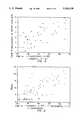
- FIG. 3is a graphical representation of the fluorescent signal intensity as a function of age of both diabetic and nondiabetic patients obtained using the apparatus of FIG. 1 as described in the EXAMPLE herein.
- FIG. 4is a graphical representation of the ratio of the fluorescent to Rayleigh signal intensities as a function of age of both diabetic and nondiabetic patients obtained using the apparatus of FIG. 1 as described in the EXAMPLE herein.
- FIG. 5is a graphical representation of the fluorescent signal intensity as a function of age of both diabetic and nondiabetic patients obtained using the apparatus of FIG. I for an illumination radiation wavelength outside the range of that used in connection with the present invention.
- FIG. 6is a graphical representation of the ratio of the fluorescent to Rayleigh signal intensities as a function of age of both diabetic and nondiabetic patients obtained using the apparatus of FIG. 1 for an illumination radiation wavelength outside the range of that used in connection with the present invention.
- FIG. 1illustrates an optical system 5 of the present invention.
- Optical system 5includes a light source 15, lens 25, a confocal lens system 35, collector 45, and a spectrometer 55.
- Source 15, which provides narrow-band illuminationtypically may be a low power krypton laser tuned to produce radiation having a wavelength between approximately 400-430 nm. In one embodiment of optical system 5, source 15 provides radiation at a wavelength of 406.7 nm.
- Attenuator 65used to reduce the power level of the transmitted radiation, receives radiation from source 15 and forwards it to lens 25.
- Lens 25,which may be a 40 ⁇ microscope objective or other similar device, then focuses the (attenuated) radiation onto the end of waveguide 95, which in turn transmits the radiation to confocal lens system 35.
- Lens system 35subsequently delivers the radiation to a selected volume of ocular lens tissue L (typically approximately 200 cubic micrometers).
- a modified slit lamp basemay be used to house and position lens system 35 for easy access to lens tissue L, while lens system 35 itself is designed to permit the same volume of lens tissue L to be held in the focal point of collector 45.
- the aperture 115 of lens system 35 at its focusis greater than approximately fifteen micrometers, ensuring that the excitation radiation diverges rapidly after passing through the focal point of lens system 35 and thereby reducing the spot intensity of the radiation should it encounter any other portions of the ocular tissue.
- Collector 4receives the radiation backscattered from lens tissue L as a result of it being illuminated by radiation from source 15. From collector 45, the backscattered radiation is directed into waveguide 105 and transmitted to the entrance slit 125 of the monochromator 135 forming spectrometer 55. If desired, collector 45 also may direct a portion of the backscattered radiation to eyepiece 75, permitting an operator to view the exact location of the selected volume of lens tissue L.
- spectrometer 55Division and processing of the backscattered radiation occurs in spectrometer 55 and detection and processing assembly 85. Radiation transmitted to spectrometer 55 initially is separated into its Rayleigh and florescence components. The two components subsequently are directed, respectively and as necessary, to amplifiers forming part of assembly 85, for determination of the intensities of each. Assembly 85 also may include a digital computer or similar computing device for forming the ratio of the fluorescent and Rayleigh components of the backscattered radiation, thereby normalizing the peak intensity of the fluorescent component.
- light source 20which may be a laser diode, produces radiation of wavelength approximately 813.4 nm (within the range of approximately 800-860 nm) and is coupled to a nonlinear frequency doubling device 30 to produce the desired wavelength output of 406.7 nm (within the range 400-430 nm).
- Light source 20alternatively may be a laser, light emitting diode, or other narrow-band light source (including broadband sources coupled to optical filters).
- the radiationsubsequently is directed through an optical delivery system 40 into the eye 50 of a patient.
- alternate embodiment 10includes an optical collector 60 confocal to the delivery system 40 to collect the backscattered radiation from the lens of eye 50.
- the backscattered radiation collectedincludes both a fluorescence signal (typically approximately 490-500 nm within the range 460-500 nm, or within the range 520-600 nm) and an intense Rayleigh component at the illumination wavelength.
- a fluorescence signaltypically approximately 490-500 nm within the range 460-500 nm, or within the range 520-600 nm
- an intense Rayleigh component at the illumination wavelengthtypically approximately 490-500 nm within the range 460-500 nm, or within the range 520-600 nm
- FIG. 2additionally discloses means for separating the components of interest of the backscattered radiation, including dichroic beam splitters 70 and 90, and for detecting the intensity of the components simultaneously using single chip silicon detectors 100 and 120 or similar devices.
- component separationmay be accomplished using beam splitters in conjunction with optical bandpass filters or dispersive elements such as diffraction gratings.
- Hybrid detector/filter assembliesalso may be used.
- Electronic circuitry 130such as but neither limited to nor necessarily requiring analog amplifiers, analog to digital (A/D) converters, and a digital computer, processes the data detected by detectors 100 and 120, calculates the normalized fluorescent/Rayleigh component ratio, and, if desired, makes the result available to an operator through a digital display or other suitable means.
- Eyepiece 80may be used by the operator to view the location of the excitation focal point in eye 50.
- FIGS. 3-6illustrate data obtained from clinical trials conducted using sixty-nine (69) human patients aged twelve (12) to sixty-five (65). Forty-eight (48) of the patients had previously been diagnosed as having diabetes, while the remaining twenty-one (21) had not.
- FIGS. 3shows the total fluorescence signal obtained for each patient (expressed in "Counts ⁇ 10 5 ,” where the number of Counts is a function of the number of emitted photons per unit time) using an illumination wavelength of 406.7 nm.
- FIG. 4details the results when those same fluorescence signals ar normalized by the Rayleigh component of the backscattered radiation in accordance with the present invention. As illustrated in FIG.
- FIGS. 5-6which correspond, respectively, to FIGS. 3-4, show (in FIG. 6) much less of a distinction between the normalized signals for the diabetic as opposed to nondiabetic patients. Furthermore, those patients who tested ICA positive are shown to have fluorescent/Rayleigh ratios within the range of nondiabetic patient values. As a result, no clearly established threshold is available for diagnostic purposes.
Landscapes
- Health & Medical Sciences (AREA)
- Life Sciences & Earth Sciences (AREA)
- Physics & Mathematics (AREA)
- General Health & Medical Sciences (AREA)
- Veterinary Medicine (AREA)
- Engineering & Computer Science (AREA)
- Biomedical Technology (AREA)
- Heart & Thoracic Surgery (AREA)
- Medical Informatics (AREA)
- Molecular Biology (AREA)
- Surgery (AREA)
- Animal Behavior & Ethology (AREA)
- Biophysics (AREA)
- Public Health (AREA)
- Optics & Photonics (AREA)
- Pathology (AREA)
- Ophthalmology & Optometry (AREA)
- Spectroscopy & Molecular Physics (AREA)
- Emergency Medicine (AREA)
- Investigating, Analyzing Materials By Fluorescence Or Luminescence (AREA)
- Eye Examination Apparatus (AREA)
- Optical Filters (AREA)
- Prostheses (AREA)
- Polymers With Sulfur, Phosphorus Or Metals In The Main Chain (AREA)
- Glass Compositions (AREA)
- Investigating Or Analysing Materials By Optical Means (AREA)
Abstract
Description
Claims (16)
Priority Applications (10)
| Application Number | Priority Date | Filing Date | Title |
|---|---|---|---|
| US07/731,533US5203328A (en) | 1991-07-17 | 1991-07-17 | Apparatus and methods for quantitatively measuring molecular changes in the ocular lens |
| AU23733/92AAU661026B2 (en) | 1991-07-17 | 1992-07-16 | Measuring molecular change in the ocular lens |
| AT92916422TATE158704T1 (en) | 1991-07-17 | 1992-07-16 | DETECTION OF MOLECULAR CHANGES IN THE EYE LENS |
| CA002113268ACA2113268C (en) | 1991-07-17 | 1992-07-16 | Measuring molecular change in the ocular lens |
| PCT/US1992/005941WO1993001745A1 (en) | 1991-07-17 | 1992-07-16 | Measuring molecular change in the ocular lens |
| EP92916422AEP0597932B1 (en) | 1991-07-17 | 1992-07-16 | Measuring molecular change in the ocular lens |
| DE69222535TDE69222535T2 (en) | 1991-07-17 | 1992-07-16 | DETECTION OF MOLECULAR CHANGES IN THE EYE LENS |
| JP5502944AJPH07500030A (en) | 1991-07-17 | 1992-07-16 | Measuring molecular changes in the eye lens |
| ES92916422TES2110007T3 (en) | 1991-07-17 | 1992-07-16 | MEASUREMENT OF THE CRYSTALLINE MOLECULAR MODIFICATION. |
| US08/007,584US5582168A (en) | 1991-07-17 | 1993-01-22 | Apparatus and methods for measuring characteristics of biological tissues and similar materials |
Applications Claiming Priority (1)
| Application Number | Priority Date | Filing Date | Title |
|---|---|---|---|
| US07/731,533US5203328A (en) | 1991-07-17 | 1991-07-17 | Apparatus and methods for quantitatively measuring molecular changes in the ocular lens |
Related Child Applications (1)
| Application Number | Title | Priority Date | Filing Date |
|---|---|---|---|
| US08/007,584Continuation-In-PartUS5582168A (en) | 1991-07-17 | 1993-01-22 | Apparatus and methods for measuring characteristics of biological tissues and similar materials |
Publications (1)
| Publication Number | Publication Date |
|---|---|
| US5203328Atrue US5203328A (en) | 1993-04-20 |
Family
ID=24939928
Family Applications (2)
| Application Number | Title | Priority Date | Filing Date |
|---|---|---|---|
| US07/731,533Expired - LifetimeUS5203328A (en) | 1991-07-17 | 1991-07-17 | Apparatus and methods for quantitatively measuring molecular changes in the ocular lens |
| US08/007,584Expired - LifetimeUS5582168A (en) | 1991-07-17 | 1993-01-22 | Apparatus and methods for measuring characteristics of biological tissues and similar materials |
Family Applications After (1)
| Application Number | Title | Priority Date | Filing Date |
|---|---|---|---|
| US08/007,584Expired - LifetimeUS5582168A (en) | 1991-07-17 | 1993-01-22 | Apparatus and methods for measuring characteristics of biological tissues and similar materials |
Country Status (9)
| Country | Link |
|---|---|
| US (2) | US5203328A (en) |
| EP (1) | EP0597932B1 (en) |
| JP (1) | JPH07500030A (en) |
| AT (1) | ATE158704T1 (en) |
| AU (1) | AU661026B2 (en) |
| CA (1) | CA2113268C (en) |
| DE (1) | DE69222535T2 (en) |
| ES (1) | ES2110007T3 (en) |
| WO (1) | WO1993001745A1 (en) |
Cited By (54)
| Publication number | Priority date | Publication date | Assignee | Title |
|---|---|---|---|---|
| US5427095A (en)* | 1993-11-09 | 1995-06-27 | Massachusetts Institute of Technology Oculon Corporation | Method and apparatus for detecting cataractogenesis |
| US5427094A (en)* | 1993-11-08 | 1995-06-27 | Oculon Corporation | Method and apparatus for detecting cataractogenesis |
| US5535743A (en)* | 1992-12-19 | 1996-07-16 | Boehringer Mannheim Gmbh | Device for the in vivo determination of an optical property of the aqueous humour of the eye |
| US5582168A (en)* | 1991-07-17 | 1996-12-10 | Georgia Tech Research Corp. | Apparatus and methods for measuring characteristics of biological tissues and similar materials |
| EP0781526A1 (en) | 1995-12-13 | 1997-07-02 | Akitoshi Yoshida | Measuring apparatus for intraocular substance employing light from eyeball |
| WO1997027469A1 (en)* | 1996-01-26 | 1997-07-31 | Boehringer Mannheim Gmbh | Process and device for determining an analyte contained in a scattering matrix |
| US5880812A (en)* | 1997-03-13 | 1999-03-09 | Ramot-University Authority For Applied Research And Industrial Development, Ltd. | Method and apparatus for evaluating and mapping visual field |
| DE19808779C1 (en)* | 1998-03-03 | 1999-10-28 | Paul Dobrinski | Cataract measuring device for eye of patient |
| US6002954A (en)* | 1995-11-22 | 1999-12-14 | The Regents Of The University Of California | Detection of biological molecules using boronate-based chemical amplification and optical sensors |
| US6011984A (en)* | 1995-11-22 | 2000-01-04 | Minimed Inc. | Detection of biological molecules using chemical amplification and optical sensors |
| US6088606A (en)* | 1999-03-22 | 2000-07-11 | Spectrx, Inc. | Method and apparatus for determining a duration of a medical condition |
| WO2001003571A1 (en)* | 1999-07-13 | 2001-01-18 | Photonics Research Ontario | Method of measuring concentration of luminescent materials in turbid media |
| US20020107668A1 (en)* | 2000-12-15 | 2002-08-08 | Costa Peter J. | System for normalizing spectra |
| US20020133073A1 (en)* | 1998-12-23 | 2002-09-19 | Nordstrom Robert J. | Spectroscopic system employing a plurality of data types |
| US20020177777A1 (en)* | 1998-12-23 | 2002-11-28 | Medispectra, Inc. | Optical methods and systems for rapid screening of the cervix |
| US20020197724A1 (en)* | 2001-02-15 | 2002-12-26 | Glenn Noronha | Polymers functionalized with fluorescent boronate motifs and methods for making them |
| US6567678B1 (en) | 1997-12-02 | 2003-05-20 | Abbott Laboratories | Multiplex sensor and method of use |
| US20030095721A1 (en)* | 1999-12-15 | 2003-05-22 | Thomas Clune | Methods and systems for correcting image misalignment |
| US20030144585A1 (en)* | 1999-12-15 | 2003-07-31 | Howard Kaufman | Image processing using measures of similarity |
| US20030207250A1 (en)* | 1999-12-15 | 2003-11-06 | Medispectra, Inc. | Methods of diagnosing disease |
| US6673625B2 (en) | 1999-09-15 | 2004-01-06 | The Regents Of The University Of California | Saccharide sensing molecules having enhanced fluorescent properties |
| US20040010375A1 (en)* | 2002-07-09 | 2004-01-15 | Medispectra, Inc. | Methods and apparatus for processing spectral data for use in tissue characterization |
| US20040007674A1 (en)* | 2002-07-09 | 2004-01-15 | Schomacker Kevin T. | Method and apparatus for identifying spectral artifacts |
| US6682938B1 (en) | 1999-09-15 | 2004-01-27 | The Regents Of The University Of California | Glucose sensing molecules having selected fluorescent properties |
| US20040023406A1 (en)* | 2002-07-09 | 2004-02-05 | Schomacker Kevin T. | Optimal windows for obtaining optical data for characterization of tissue samples |
| US6750311B1 (en) | 1996-11-21 | 2004-06-15 | Minimed Inc. | Detection of biological molecules using boronate-based chemical amplification and optical sensors |
| US20040138539A1 (en)* | 2003-01-07 | 2004-07-15 | Jay Paul R. | Non-invasive blood monitor |
| US6766183B2 (en) | 1995-11-22 | 2004-07-20 | Medtronic Minimed, Inc. | Long wave fluorophore sensor compounds and other fluorescent sensor compounds in polymers |
| US6768918B2 (en) | 2002-07-10 | 2004-07-27 | Medispectra, Inc. | Fluorescent fiberoptic probe for tissue health discrimination and method of use thereof |
| US20040186382A1 (en)* | 1997-01-13 | 2004-09-23 | Medispectra, Inc. | Spectral volume microprobe arrays |
| US20040208390A1 (en)* | 2003-04-18 | 2004-10-21 | Medispectra, Inc. | Methods and apparatus for processing image data for use in tissue characterization |
| US20040208385A1 (en)* | 2003-04-18 | 2004-10-21 | Medispectra, Inc. | Methods and apparatus for visually enhancing images |
| US20040206882A1 (en)* | 2003-04-18 | 2004-10-21 | Medispectra, Inc. | Methods and apparatus for evaluating image focus |
| US20040209237A1 (en)* | 2003-04-18 | 2004-10-21 | Medispectra, Inc. | Methods and apparatus for characterization of tissue samples |
| US20040207625A1 (en)* | 2003-04-18 | 2004-10-21 | Medispectra, Inc. | Methods and apparatus for displaying diagnostic data |
| US6847490B1 (en) | 1997-01-13 | 2005-01-25 | Medispectra, Inc. | Optical probe accessory device for use in vivo diagnostic procedures |
| US7045361B2 (en) | 2001-09-12 | 2006-05-16 | Medtronic Minimed, Inc. | Analyte sensing via acridine-based boronate biosensors |
| US7103401B2 (en) | 2002-07-10 | 2006-09-05 | Medispectra, Inc. | Colonic polyp discrimination by tissue fluorescence and fiberoptic probe |
| US20070019199A1 (en)* | 2005-07-25 | 2007-01-25 | The Wisconsin Alumni Research Foundation | Methods, systems, and computer program products for optimization of probes for spectroscopic measurement in turbid media |
| US20070232932A1 (en)* | 2006-03-17 | 2007-10-04 | Duke University | Monte Carlo based model of fluorescence in turbid media and methods and systems for using same to determine intrinsic fluorescence of turbid media |
| US7309867B2 (en) | 2003-04-18 | 2007-12-18 | Medispectra, Inc. | Methods and apparatus for characterization of tissue samples |
| US20080065158A1 (en)* | 2006-09-07 | 2008-03-13 | Omry Ben-Ezra | Techniques for reducing pain associated with nerve stimulation |
| US20080270091A1 (en)* | 2007-02-23 | 2008-10-30 | Nirmala Ramanujam | Scaling method for fast monte carlo simulation of diffuse reflectance spectra from multi-layered turbid media and methods and systems for using same to determine optical properties of multi-layered turbid medium from measured diffuse reflectance |
| US7459696B2 (en) | 2003-04-18 | 2008-12-02 | Schomacker Kevin T | Methods and apparatus for calibrating spectral data |
| US20090015826A1 (en)* | 2006-03-30 | 2009-01-15 | Duke University | Optical assay system for intraoperative assessment of tumor margins |
| WO2010097582A1 (en) | 2009-02-26 | 2010-09-02 | Edinburgh Instruments Limited | Fluorescence method and system |
| US20100245764A1 (en)* | 2009-03-30 | 2010-09-30 | Ottawa Health Research Institute | Apparatus and method for optical measurements |
| US20110059016A1 (en)* | 2007-09-27 | 2011-03-10 | Nirmala Ramanujam | Optical assay system with a multi-probe imaging array |
| US20110105865A1 (en)* | 2008-04-24 | 2011-05-05 | Duke University | Diffuse reflectance spectroscopy device for quantifying tissue absorption and scattering |
| US20110112435A1 (en)* | 2007-09-28 | 2011-05-12 | Nirmala Ramanujam | Systems and methods for spectral analysis of a tissue mass using an instrument, an optical probe, and a monte carlo or a diffusion algorithm |
| WO2012061836A2 (en) | 2010-11-05 | 2012-05-10 | Freedom Meditech, Inc. | Improved algorithm for detection of diabetes |
| US20120179010A1 (en)* | 2002-04-04 | 2012-07-12 | Maynard John D | Determination of a Measure of a Glycation End-Product or Disease State Using Tissue Fluorescence of Various Sites |
| AU2015202762B2 (en)* | 2010-11-05 | 2017-04-20 | Sinocare Meditech, Inc. | Improved algorithm for detection of diabetes |
| WO2025114307A1 (en) | 2023-11-27 | 2025-06-05 | Occuity Limited | Confocal optical measurement apparatus and method of optically measuring a biomarker |
Families Citing this family (109)
| Publication number | Priority date | Publication date | Assignee | Title |
|---|---|---|---|---|
| US5341805A (en)* | 1993-04-06 | 1994-08-30 | Cedars-Sinai Medical Center | Glucose fluorescence monitor and method |
| US5560356A (en)* | 1994-02-23 | 1996-10-01 | Vitrophage, Inc. | Diagnostic system and method using an implanted reflective device |
| US5685313A (en)* | 1994-05-31 | 1997-11-11 | Brain Monitor Ltd. | Tissue monitor |
| US5701902A (en)* | 1994-09-14 | 1997-12-30 | Cedars-Sinai Medical Center | Spectroscopic burn injury evaluation apparatus and method |
| JPH10501346A (en)* | 1995-03-23 | 1998-02-03 | フィリップス エレクトロニクス エヌ ベー | Device for making optical measurements in turbid media |
| US7328059B2 (en) | 1996-08-23 | 2008-02-05 | The Texas A & M University System | Imaging of light scattering tissues with fluorescent contrast agents |
| DE19538372A1 (en)* | 1995-10-14 | 1997-04-17 | Laser & Med Tech Gmbh | Non-invasive glucose measurement |
| US6232609B1 (en) | 1995-12-01 | 2001-05-15 | Cedars-Sinai Medical Center | Glucose monitoring apparatus and method using laser-induced emission spectroscopy |
| US6544193B2 (en) | 1996-09-04 | 2003-04-08 | Marcio Marc Abreu | Noninvasive measurement of chemical substances |
| CA2192036A1 (en)* | 1996-12-04 | 1998-06-04 | Harvey Lui | Fluorescence scope system for dermatologic diagnosis |
| US7865230B1 (en) | 1997-02-07 | 2011-01-04 | Texas A&M University System | Method and system for detecting sentinel lymph nodes |
| ATE409005T1 (en)* | 1997-03-19 | 2008-10-15 | Lucid Inc | CELL SURGERY USING CONFOCAL MICROSCOPY |
| US6091984A (en)* | 1997-10-10 | 2000-07-18 | Massachusetts Institute Of Technology | Measuring tissue morphology |
| EP1037553B1 (en)* | 1997-11-12 | 2007-01-24 | Lightouch Medical, Inc. | Method for non-invasive measurement of an analyte |
| US6055451A (en) | 1997-12-12 | 2000-04-25 | Spectrx, Inc. | Apparatus and method for determining tissue characteristics |
| US6174291B1 (en) | 1998-03-09 | 2001-01-16 | Spectrascience, Inc. | Optical biopsy system and methods for tissue diagnosis |
| US6560478B1 (en)* | 1998-03-16 | 2003-05-06 | The Research Foundation Of City University Of New York | Method and system for examining biological materials using low power CW excitation Raman spectroscopy |
| US6149589A (en)* | 1998-03-26 | 2000-11-21 | Universite De Montreal | On-line and real-time spectroreflectometry measurement of oxygenation in a patient's eye |
| US5919132A (en)* | 1998-03-26 | 1999-07-06 | Universite De Montreal | On-line and real-time spectroreflectometry measurement of oxygenation in a patient's eye |
| DE69903306T2 (en) | 1998-07-07 | 2003-05-22 | Lightouch Medical, Inc. | METHOD FOR TISSUE MODULATION FOR QUANTITATIVE NON-INVASIVE IN VIVO SPECTROSCOPIC ANALYSIS OF TISSUE |
| IL141864A0 (en) | 1998-09-11 | 2002-03-10 | Spectrx Inc | Multi-modal optical tissue diagnostic system |
| US6708060B1 (en) | 1998-11-09 | 2004-03-16 | Transpharma Ltd. | Handheld apparatus and method for transdermal drug delivery and analyte extraction |
| US6148232A (en) | 1998-11-09 | 2000-11-14 | Elecsys Ltd. | Transdermal drug delivery and analyte extraction |
| US6597946B2 (en) | 1998-11-09 | 2003-07-22 | Transpharma Ltd. | Electronic card for transdermal drug delivery and analyte extraction |
| US6611706B2 (en) | 1998-11-09 | 2003-08-26 | Transpharma Ltd. | Monopolar and bipolar current application for transdermal drug delivery and analyte extraction |
| US6721583B1 (en)* | 1998-11-19 | 2004-04-13 | The United States Of America | Method for non-invasive identification of individuals at risk for diabetes |
| US6404497B1 (en) | 1999-01-25 | 2002-06-11 | Massachusetts Institute Of Technology | Polarized light scattering spectroscopy of tissue |
| US6665556B1 (en)* | 1999-01-29 | 2003-12-16 | Robert R. Alfano | Method and apparatus for examining a tissue using the spectral wing emission therefrom induced by visible to infrared photoexcitation |
| DE60017755T2 (en) | 1999-08-26 | 2005-06-30 | Novartis Ag | AUGENANALYTFÜHLER |
| US6494576B1 (en)* | 1999-09-30 | 2002-12-17 | L'esperance, Jr. Francis A. | Method and apparatus for spectrophotometry of the eye |
| US7054002B1 (en) | 1999-10-08 | 2006-05-30 | The Texas A&M University System | Characterization of luminescence in a scattering medium |
| CA2389177C (en)* | 1999-11-05 | 2012-05-29 | Shabbir B. Bambot | Multi-modal optical tissue diagnostic system |
| US6377841B1 (en)* | 2000-03-31 | 2002-04-23 | Vanderbilt University | Tumor demarcation using optical spectroscopy |
| US20040044287A1 (en)* | 2000-03-31 | 2004-03-04 | Wei-Chiang Lin | Identification of human tissue using optical spectroscopy |
| WO2002007585A2 (en)* | 2000-07-13 | 2002-01-31 | Virginia Commonwealth University | Tissue interrogation spectroscopy |
| US6549861B1 (en) | 2000-08-10 | 2003-04-15 | Euro-Celtique, S.A. | Automated system and method for spectroscopic analysis |
| US6675030B2 (en) | 2000-08-21 | 2004-01-06 | Euro-Celtique, S.A. | Near infrared blood glucose monitoring system |
| EA200300574A1 (en) | 2000-11-16 | 2004-06-24 | Кэмилийен Медикал Инновейшн Лтд. | SYSTEM FOR DIAGNOSTIC EARS |
| US6697652B2 (en)* | 2001-01-19 | 2004-02-24 | Massachusetts Institute Of Technology | Fluorescence, reflectance and light scattering spectroscopy for measuring tissue |
| US20070276199A1 (en)* | 2002-04-04 | 2007-11-29 | Ediger Marwood N | Determination of a Measure of a Glycation End-Product or Disease State Using Tissue Fluorescence |
| US7139598B2 (en)* | 2002-04-04 | 2006-11-21 | Veralight, Inc. | Determination of a measure of a glycation end-product or disease state using tissue fluorescence |
| US7043288B2 (en)* | 2002-04-04 | 2006-05-09 | Inlight Solutions, Inc. | Apparatus and method for spectroscopic analysis of tissue to detect diabetes in an individual |
| US20040152963A1 (en)* | 2001-04-27 | 2004-08-05 | March Wayne Front | Apparatus for measuring blood glucose concentrations |
| US6650915B2 (en) | 2001-09-13 | 2003-11-18 | Fovioptics, Inc. | Non-invasive measurement of blood analytes using photodynamics |
| US8308797B2 (en)* | 2002-01-04 | 2012-11-13 | Colibri Heart Valve, LLC | Percutaneously implantable replacement heart valve device and method of making same |
| US7725144B2 (en)* | 2002-04-04 | 2010-05-25 | Veralight, Inc. | Determination of disease state using raman spectroscopy of tissue |
| US20120078075A1 (en)* | 2002-04-04 | 2012-03-29 | Maynard John D | Determination of a measure of a glycation end-product or disease state using tissue fluorescence in combination with one or more other tests |
| US8140147B2 (en)* | 2002-04-04 | 2012-03-20 | Veralight, Inc. | Determination of a measure of a glycation end-product or disease state using a flexible probe to determine tissue fluorescence of various sites |
| AU2003226605A1 (en)* | 2002-04-19 | 2003-11-03 | Transpharma Medical Ltd. | Handheld transdermal drug delivery and analyte extraction |
| US8328420B2 (en) | 2003-04-22 | 2012-12-11 | Marcio Marc Abreu | Apparatus and method for measuring biologic parameters |
| US9848815B2 (en) | 2002-04-22 | 2017-12-26 | Geelux Holdings, Ltd. | Apparatus and method for measuring biologic parameters |
| JP4347216B2 (en) | 2002-04-22 | 2009-10-21 | マルシオ マルク アブリュー | Biological parameter measuring device |
| US8849379B2 (en) | 2002-04-22 | 2014-09-30 | Geelux Holdings, Ltd. | Apparatus and method for measuring biologic parameters |
| US6895264B2 (en) | 2002-08-26 | 2005-05-17 | Fovioptics Inc. | Non-invasive psychophysical measurement of glucose using photodynamics |
| US7599732B2 (en)* | 2003-06-20 | 2009-10-06 | The Texas A&M University System | Method and system for near-infrared fluorescence contrast-enhanced imaging with area illumination and area detection |
| US7356365B2 (en)* | 2003-07-09 | 2008-04-08 | Glucolight Corporation | Method and apparatus for tissue oximetry |
| US20050090723A1 (en)* | 2003-10-23 | 2005-04-28 | Nassar Saeed | Method and apparatus for non-invasive measuring of physiological glucose concentration in bodies of humans or animals |
| GB2407378B (en)* | 2003-10-24 | 2006-09-06 | Lein Applied Diagnostics Ltd | Ocular property measuring apparatus and method therefor |
| CN1882278A (en)* | 2003-10-28 | 2006-12-20 | 薇拉莱特公司 | Use tissue fluorescence to identify a glycation end product or disease state |
| WO2005050156A2 (en)* | 2003-11-18 | 2005-06-02 | Chameleon Medical Innovation Ltd. | Measurement system and method for use in determining the patient's condition |
| GB2409033C (en)* | 2003-12-12 | 2006-05-24 | Lein Applied Diagnostics Ltd | Extended focal region measuring apparatus and method |
| US7510849B2 (en)* | 2004-01-29 | 2009-03-31 | Glucolight Corporation | OCT based method for diagnosis and therapy |
| US10227063B2 (en) | 2004-02-26 | 2019-03-12 | Geelux Holdings, Ltd. | Method and apparatus for biological evaluation |
| US7254429B2 (en) | 2004-08-11 | 2007-08-07 | Glucolight Corporation | Method and apparatus for monitoring glucose levels in a biological tissue |
| US8036727B2 (en)* | 2004-08-11 | 2011-10-11 | Glt Acquisition Corp. | Methods for noninvasively measuring analyte levels in a subject |
| WO2006110859A2 (en) | 2005-04-13 | 2006-10-19 | Glucolight Corporation | Method for data reduction and calibration of an oct-based blood glucose monitor |
| US7330747B2 (en)* | 2005-06-07 | 2008-02-12 | Chemimage Corporation | Invasive chemometry |
| US7330746B2 (en)* | 2005-06-07 | 2008-02-12 | Chem Image Corporation | Non-invasive biochemical analysis |
| WO2007014213A2 (en)* | 2005-07-25 | 2007-02-01 | Massachusetts Institute Of Technology | Tri modal spectroscopic imaging |
| US20070173736A1 (en)* | 2005-10-07 | 2007-07-26 | Femspec Llc | Apparatus and methods for endometrial biopsies |
| ES2399872T3 (en) | 2005-10-24 | 2013-04-04 | Marcio Marc Aurelio Martins Abreu | Apparatus for measuring biological parameters |
| EP1965853B1 (en)* | 2005-12-22 | 2011-10-12 | Koninklijke Philips Electronics N.V. | Device for controlled release of chemical molecules |
| US20080117416A1 (en)* | 2006-10-27 | 2008-05-22 | Hunter Ian W | Use of coherent raman techniques for medical diagnostic and therapeutic purposes, and calibration techniques for same |
| US8498695B2 (en) | 2006-12-22 | 2013-07-30 | Novadaq Technologies Inc. | Imaging system with a single color image sensor for simultaneous fluorescence and color video endoscopy |
| JP2008197088A (en)* | 2007-01-19 | 2008-08-28 | Shimadzu Corp | Fluorescence detector |
| GB2451443B (en) | 2007-07-30 | 2012-12-26 | Lein Applied Diagnostics Ltd | Optical measurement apparatus and method therefor |
| GB2451442B (en)* | 2007-07-30 | 2013-03-06 | Lein Applied Diagnostics Ltd | Optical measurement apparatus and method therefor |
| EP2057941A1 (en)* | 2007-11-07 | 2009-05-13 | Koninklijke Philips Electronics N.V. | Method and device for imaging an interior of an optically turbid medium |
| DE102007061987A1 (en)* | 2007-12-21 | 2009-06-25 | Carl Zeiss Meditec Ag | Apparatus and method for detecting molecules in the eye |
| US8986253B2 (en) | 2008-01-25 | 2015-03-24 | Tandem Diabetes Care, Inc. | Two chamber pumps and related methods |
| GB2457302B (en) | 2008-02-11 | 2013-04-10 | Lein Applied Diagnostics Ltd | Measurement apparatus and method therefor |
| US8571617B2 (en) | 2008-03-04 | 2013-10-29 | Glt Acquisition Corp. | Flowometry in optical coherence tomography for analyte level estimation |
| JP5094484B2 (en)* | 2008-03-11 | 2012-12-12 | 富士フイルム株式会社 | Fluorescence detection method and fluorescence detection apparatus |
| JP5097590B2 (en)* | 2008-03-26 | 2012-12-12 | 富士フイルム株式会社 | Raman signal measuring method and Raman signal measuring apparatus |
| US8408421B2 (en) | 2008-09-16 | 2013-04-02 | Tandem Diabetes Care, Inc. | Flow regulating stopcocks and related methods |
| CA2737461A1 (en) | 2008-09-19 | 2010-03-25 | Tandem Diabetes Care, Inc. | Solute concentration measurement device and related methods |
| US20100249607A1 (en)* | 2008-09-26 | 2010-09-30 | Massachusetts Institute Of Technology | Quantitative spectroscopic imaging |
| US8606366B2 (en) | 2009-02-18 | 2013-12-10 | Syneron Medical Ltd. | Skin treatment apparatus for personal use and method for using same |
| EP2724739B1 (en) | 2009-07-30 | 2015-07-01 | Tandem Diabetes Care, Inc. | Portable infusion pump system |
| US8634591B2 (en) | 2009-08-20 | 2014-01-21 | Koninklijke Philips N.V. | Method and system for image analysis |
| WO2011042851A1 (en)* | 2009-10-06 | 2011-04-14 | Koninklijke Philips Electronics N.V. | Method and system for carrying out photoplethysmography |
| US9091637B2 (en) | 2009-12-04 | 2015-07-28 | Duke University | Smart fiber optic sensors systems and methods for quantitative optical spectroscopy |
| EP2542184B1 (en) | 2010-03-01 | 2016-05-25 | Colibri Heart Valve LLC | Percutaneously deliverable heart valve and methods associated therewith |
| JP5936610B2 (en) | 2010-06-28 | 2016-06-22 | コリブリ ハート バルブ エルエルシーColibri Heart Valve Llc | Device for intracavity delivery of an intravascular injection device |
| AU2011343755A1 (en) | 2010-12-14 | 2013-06-06 | Colibri Heart Valve Llc | Percutaneously deliverable heart valve including folded membrane cusps with integral leaflets |
| US9180242B2 (en) | 2012-05-17 | 2015-11-10 | Tandem Diabetes Care, Inc. | Methods and devices for multiple fluid transfer |
| US9173998B2 (en) | 2013-03-14 | 2015-11-03 | Tandem Diabetes Care, Inc. | System and method for detecting occlusions in an infusion pump |
| US20150105687A1 (en) | 2013-10-11 | 2015-04-16 | Geelux Holding, Ltd. | Method and apparatus for biological evaluation |
| JP2017501844A (en) | 2014-01-10 | 2017-01-19 | マーシオ マーク アブリュー | Device for measuring the infrared output of an Abreu brain thermal tunnel |
| US10251776B2 (en) | 2014-01-10 | 2019-04-09 | Geelux Holding, Ltd. | Devices configured to monitor biological parameters, and to provide treatment, at an Abreu brain thermal tunnel |
| CN106163463A (en) | 2014-01-22 | 2016-11-23 | 马尔西奥·马克·阿布雷乌 | Device configured to provide treatment in an ABREU brain thermal tunnel |
| WO2016054079A1 (en) | 2014-09-29 | 2016-04-07 | Zyomed Corp. | Systems and methods for blood glucose and other analyte detection and measurement using collision computing |
| US11872018B2 (en) | 2015-03-10 | 2024-01-16 | Brain Tunnelgenix Technologies Corp. | Devices, apparatuses, systems, and methods for measuring temperature of an ABTT terminus |
| AU2016351730B2 (en)* | 2015-11-13 | 2019-07-11 | Novadaq Technologies Inc. | Systems and methods for illumination and imaging of a target |
| US9554738B1 (en) | 2016-03-30 | 2017-01-31 | Zyomed Corp. | Spectroscopic tomography systems and methods for noninvasive detection and measurement of analytes using collision computing |
| EP3469420A4 (en) | 2016-06-14 | 2020-02-12 | Novadaq Technologies ULC | METHODS AND SYSTEMS FOR ADAPTIVE IMAGING FOR THE AMPLIFICATION OF A WEAK LIGHT SIGNAL IN A MEDICAL VISUALIZATION |
| WO2018003906A1 (en)* | 2016-06-30 | 2018-01-04 | 興和株式会社 | Ophthalmic measurement device |
| EP3580609B1 (en) | 2017-02-10 | 2023-05-24 | Stryker European Operations Limited | Open-field handheld fluorescence imaging systems and methods |
| WO2019051476A1 (en) | 2017-09-11 | 2019-03-14 | Incubar, LLC | Conduit vascular implant sealing device for reducing endoleak |
Citations (19)
| Publication number | Priority date | Publication date | Assignee | Title |
|---|---|---|---|---|
| US4548498A (en)* | 1981-05-04 | 1985-10-22 | Folestad Sven S | Laser induced fluorescence detection in modern liquid chromatography with conventional and micro columns |
| US4582405A (en)* | 1983-09-01 | 1986-04-15 | Carl-Zeiss-Stiftung | Ophthalmological combination instrument for diagnosis and treatment |
| US4592361A (en)* | 1982-06-28 | 1986-06-03 | The Johns Hopkins University | Electro-optical device and method for monitoring instantaneous singlet oxygen concentration produced during photoradiation using pulsed excitation and time domain signal processing |
| US4675300A (en)* | 1985-09-18 | 1987-06-23 | The Board Of Trustees Of The Leland Stanford Junior University | Laser-excitation fluorescence detection electrokinetic separation |
| US4702576A (en)* | 1985-09-27 | 1987-10-27 | Cambridge Instruments Inc. | Ocular scattering analyzer |
| US4711540A (en)* | 1984-01-04 | 1987-12-08 | Tokyo Kogaku Kikai Kabushiki Kaisha | Eye disease inspecting instrument |
| US4711541A (en)* | 1984-02-02 | 1987-12-08 | Tokyo Kogaku Kikai Kabushiki Kaisha | Slit lamp and accessory device thereof |
| US4711542A (en)* | 1985-05-22 | 1987-12-08 | Kowa Company Ltd. | Ophthalmic disease detection apparatus |
| US4758081A (en)* | 1985-07-18 | 1988-07-19 | Bausch & Lomb Incorporated | Control of laser photocoagulation using Raman radiation |
| US4776687A (en)* | 1984-01-12 | 1988-10-11 | Kowa Company, Ltd. | Apparatus for detecting ophthalmic disease |
| US4781453A (en)* | 1986-05-12 | 1988-11-01 | Kowa Company Ltd. | Ophthalmic examination apparatus |
| US4836207A (en)* | 1987-09-09 | 1989-06-06 | The Beth Israel Hospital Association | Method and apparatus to monitor cholesterol levels with photon correlation spectroscopy |
| US4838683A (en)* | 1986-05-19 | 1989-06-13 | Kowa Company Ltd. | Ophthalmic measuring method and apparatus |
| US4848897A (en)* | 1987-03-31 | 1989-07-18 | Kowa Company Ltd. | Ophthalmological diagnosis apparatus |
| US4852987A (en)* | 1985-11-29 | 1989-08-01 | Wolfgang Lohmann | Device for measuring eye lens opacity |
| US4854693A (en)* | 1985-05-22 | 1989-08-08 | Kowa Company Ltd. | Ophthalmic disease detection apparatus |
| US4883351A (en)* | 1982-09-10 | 1989-11-28 | Weiss Jeffrey N | Apparatus for the detection of diabetes and other abnormalities affecting the lens of the eye |
| US4895159A (en)* | 1982-09-10 | 1990-01-23 | Weiss Jeffrey N | Diabetes detection method |
| US4950068A (en)* | 1987-07-14 | 1990-08-21 | Kowa Company Ltd. | Ophthalmic disease detection apparatus |
Family Cites Families (42)
| Publication number | Priority date | Publication date | Assignee | Title |
|---|---|---|---|---|
| FR1531145A (en)* | 1966-07-13 | 1968-06-28 | Ici Ltd | Polyamides |
| US3948248A (en)* | 1974-09-05 | 1976-04-06 | Zuckerman Joel L | Method of measuring ocular pulse |
| US3958560A (en)* | 1974-11-25 | 1976-05-25 | Wayne Front March | Non-invasive automatic glucose sensor system |
| US4014321A (en)* | 1974-11-25 | 1977-03-29 | March Wayne F | Non-invasive glucose sensor system |
| DE2619571A1 (en)* | 1976-05-04 | 1977-11-17 | Dynamit Nobel Ag | Acylase enzyme immobilisation - on lamellar aluminium hydrosilicates modified with amino-silanes |
| US4350163A (en)* | 1980-05-29 | 1982-09-21 | Ford Jr Norman C | Method and apparatus for analyzing contaminants in aqueous humor |
| US4412543A (en)* | 1981-04-09 | 1983-11-01 | Xanar, Inc. | Apparatus for determining the concentration of a fluorescent material in an eye |
| US5025785A (en)* | 1982-09-10 | 1991-06-25 | Weiss Jeffrey N | Diabetes detection method |
| US4573778A (en)* | 1983-03-16 | 1986-03-04 | Boston University | Aqueous fluorophotometer |
| DE3346338A1 (en)* | 1983-12-22 | 1985-07-11 | Pka Pyrolyse Kraftanlagen Gmbh, 7080 Aalen | ROTATING SUSPENSION DRUM FOR SUSPENSIONING WASTE |
| JPS61243827A (en)* | 1985-04-22 | 1986-10-30 | Toray Ind Inc | Production of nylon 66 polymer for high-speed spinning |
| US5042494A (en)* | 1985-11-13 | 1991-08-27 | Alfano Robert R | Method and apparatus for detecting cancerous tissue using luminescence excitation spectra |
| CH671329A5 (en)* | 1986-01-21 | 1989-08-31 | Interzeag Ag | |
| JPS63135128A (en)* | 1986-11-27 | 1988-06-07 | 興和株式会社 | Ophthalmic measuring apparatus |
| JP2520418B2 (en)* | 1987-04-09 | 1996-07-31 | 興和株式会社 | Ophthalmic measuring device |
| US4877322A (en)* | 1987-04-30 | 1989-10-31 | Eyedentify, Inc. | Method and apparatus for measuring blood oxygen levels in selected areas of the eye fundus |
| EP0292216B1 (en)* | 1987-05-20 | 1993-11-03 | Kowa Co. Ltd. | Ophthalmic disease detection apparatus |
| US4842401A (en)* | 1987-06-15 | 1989-06-27 | The Board Of Trustees Of The Leland Stanford Jr. University | Eye diagnosis process |
| JP2516631B2 (en)* | 1987-06-18 | 1996-07-24 | 興和株式会社 | Ophthalmic measuring device |
| DE3851183D1 (en)* | 1987-06-25 | 1994-09-29 | Kowa Co | Method and device for the diagnosis of eye disorders. |
| DD261957A1 (en)* | 1987-07-13 | 1988-11-16 | Friedrich Schiller Uni Jena Bf | ARRANGEMENT FOR CATARAFRUIT DIAGNOSTICS |
| JP2520426B2 (en)* | 1987-07-15 | 1996-07-31 | 興和株式会社 | Ophthalmic measuring device |
| US4957113A (en)* | 1987-09-01 | 1990-09-18 | Massachusetts Institute Of Technology | Method for detecting cataractogenesis using quasi-elastic light scattering |
| US4993827A (en)* | 1987-09-01 | 1991-02-19 | Massachusetts Institute Of Technology | Method for detecting cataractogenesis |
| US5072731A (en)* | 1987-09-01 | 1991-12-17 | Massachusetts Institute Of Technology | Apparatus for detecting cataractogenesis using quasielastic light scattering |
| US4832483A (en)* | 1987-09-03 | 1989-05-23 | New England Medical Center Hospitals, Inc. | Method of using resonance raman spectroscopy for detection of malignancy disease |
| JPS6469925A (en)* | 1987-09-11 | 1989-03-15 | Hitachi Cable | Optical fiber type temperature distribution measuring apparatus |
| US4882492A (en)* | 1988-01-19 | 1989-11-21 | Biotronics Associates, Inc. | Non-invasive near infrared measurement of blood analyte concentrations |
| US5137355A (en)* | 1988-06-08 | 1992-08-11 | The Research Foundation Of State University Of New York | Method of imaging a random medium |
| JPH02119837A (en)* | 1988-10-28 | 1990-05-07 | Kowa Co | Ophthalmological measurement method and device |
| DD298677A5 (en)* | 1989-11-16 | 1992-03-05 | ��������`������������@�������@�������@M�������]k�� | METHOD FOR DETERMINING THE VOLUME FLOW |
| JP2994703B2 (en)* | 1990-07-23 | 1999-12-27 | 興和株式会社 | Ophthalmic measurement device |
| US5186173A (en)* | 1990-08-14 | 1993-02-16 | Drexel University | Method for in vivo measurement of oxygen concentration levels |
| US5139022A (en)* | 1990-10-26 | 1992-08-18 | Philip Lempert | Method and apparatus for imaging and analysis of ocular tissue |
| US5243983A (en)* | 1990-12-14 | 1993-09-14 | Georgia Tech Research Corporation | Non-invasive blood glucose measurement system and method using stimulated raman spectroscopy |
| US5279296A (en)* | 1991-01-04 | 1994-01-18 | Oculon Corporation | Method and apparatus for detecting cataractogenesis |
| US5219400A (en)* | 1991-06-11 | 1993-06-15 | The United States Of America As Represented By The Secretary Of The Army | Noninvasive method for quantitation of oxyhemoglobin saturation by near-infrared reflectance spectrophotometry |
| US5203328A (en)* | 1991-07-17 | 1993-04-20 | Georgia Tech Research Corporation | Apparatus and methods for quantitatively measuring molecular changes in the ocular lens |
| JPH07508426A (en)* | 1991-10-17 | 1995-09-21 | サイエンティフィック ジェネリクス リミテッド | Blood sample measuring device and method |
| US5348018A (en)* | 1991-11-25 | 1994-09-20 | Alfano Robert R | Method for determining if tissue is malignant as opposed to non-malignant using time-resolved fluorescence spectroscopy |
| US5284149A (en)* | 1992-01-23 | 1994-02-08 | Dhadwal Harbans S | Method and apparatus for determining the physical characteristics of ocular tissue |
| US5340991A (en)* | 1993-05-21 | 1994-08-23 | The Board Of Regents Of The University Of Oklahoma | Fluorokinetic analysis of diffusion from a vessel |
- 1991
- 1991-07-17USUS07/731,533patent/US5203328A/ennot_activeExpired - Lifetime
- 1992
- 1992-07-16EPEP92916422Apatent/EP0597932B1/ennot_activeExpired - Lifetime
- 1992-07-16CACA002113268Apatent/CA2113268C/ennot_activeExpired - Lifetime
- 1992-07-16ESES92916422Tpatent/ES2110007T3/ennot_activeExpired - Lifetime
- 1992-07-16WOPCT/US1992/005941patent/WO1993001745A1/enactiveIP Right Grant
- 1992-07-16DEDE69222535Tpatent/DE69222535T2/ennot_activeExpired - Lifetime
- 1992-07-16AUAU23733/92Apatent/AU661026B2/ennot_activeCeased
- 1992-07-16ATAT92916422Tpatent/ATE158704T1/ennot_activeIP Right Cessation
- 1992-07-16JPJP5502944Apatent/JPH07500030A/ennot_activeExpired - Lifetime
- 1993
- 1993-01-22USUS08/007,584patent/US5582168A/ennot_activeExpired - Lifetime
Patent Citations (19)
| Publication number | Priority date | Publication date | Assignee | Title |
|---|---|---|---|---|
| US4548498A (en)* | 1981-05-04 | 1985-10-22 | Folestad Sven S | Laser induced fluorescence detection in modern liquid chromatography with conventional and micro columns |
| US4592361A (en)* | 1982-06-28 | 1986-06-03 | The Johns Hopkins University | Electro-optical device and method for monitoring instantaneous singlet oxygen concentration produced during photoradiation using pulsed excitation and time domain signal processing |
| US4895159A (en)* | 1982-09-10 | 1990-01-23 | Weiss Jeffrey N | Diabetes detection method |
| US4883351A (en)* | 1982-09-10 | 1989-11-28 | Weiss Jeffrey N | Apparatus for the detection of diabetes and other abnormalities affecting the lens of the eye |
| US4582405A (en)* | 1983-09-01 | 1986-04-15 | Carl-Zeiss-Stiftung | Ophthalmological combination instrument for diagnosis and treatment |
| US4711540A (en)* | 1984-01-04 | 1987-12-08 | Tokyo Kogaku Kikai Kabushiki Kaisha | Eye disease inspecting instrument |
| US4776687A (en)* | 1984-01-12 | 1988-10-11 | Kowa Company, Ltd. | Apparatus for detecting ophthalmic disease |
| US4711541A (en)* | 1984-02-02 | 1987-12-08 | Tokyo Kogaku Kikai Kabushiki Kaisha | Slit lamp and accessory device thereof |
| US4854693A (en)* | 1985-05-22 | 1989-08-08 | Kowa Company Ltd. | Ophthalmic disease detection apparatus |
| US4711542A (en)* | 1985-05-22 | 1987-12-08 | Kowa Company Ltd. | Ophthalmic disease detection apparatus |
| US4758081A (en)* | 1985-07-18 | 1988-07-19 | Bausch & Lomb Incorporated | Control of laser photocoagulation using Raman radiation |
| US4675300A (en)* | 1985-09-18 | 1987-06-23 | The Board Of Trustees Of The Leland Stanford Junior University | Laser-excitation fluorescence detection electrokinetic separation |
| US4702576A (en)* | 1985-09-27 | 1987-10-27 | Cambridge Instruments Inc. | Ocular scattering analyzer |
| US4852987A (en)* | 1985-11-29 | 1989-08-01 | Wolfgang Lohmann | Device for measuring eye lens opacity |
| US4781453A (en)* | 1986-05-12 | 1988-11-01 | Kowa Company Ltd. | Ophthalmic examination apparatus |
| US4838683A (en)* | 1986-05-19 | 1989-06-13 | Kowa Company Ltd. | Ophthalmic measuring method and apparatus |
| US4848897A (en)* | 1987-03-31 | 1989-07-18 | Kowa Company Ltd. | Ophthalmological diagnosis apparatus |
| US4950068A (en)* | 1987-07-14 | 1990-08-21 | Kowa Company Ltd. | Ophthalmic disease detection apparatus |
| US4836207A (en)* | 1987-09-09 | 1989-06-06 | The Beth Israel Hospital Association | Method and apparatus to monitor cholesterol levels with photon correlation spectroscopy |
Non-Patent Citations (31)
| Title |
|---|
| G. Bessems, et al., "Non-Tryptophan Fluorescence of Crystallins From Normal and Cataractous Human Lenses," Investigative ophthalmology & Visual Science, Jul. 1987, vol. 28, pp. 1157-1163. |
| G. Bessems, et al., Non Tryptophan Fluorescence of Crystallins From Normal and Cataractous Human Lenses, Investigative ophthalmology & Visual Science , Jul. 1987, vol. 28, pp. 1157 1163.* |
| Hockwin, et al., "Investigations on Lens Transparency and its Disturbances by Microdensitometric analysis of Scheimpflug Photographs," Current Eye Research, 1984, vol. 3, No. 1, pp. 15-22. |
| Hockwin, et al., Investigations on Lens Transparency and its Disturbances by Microdensitometric analysis of Scheimpflug Photographs, Current Eye Research , 1984, vol. 3, No. 1, pp. 15 22.* |
| J. Bleeker, et al., "Autofluorescence of the Lens in Diabetic and Healthy Subjects by Fluorophotometry," Investigative Ophthalmology & Visual Science, May 1986, vol. 27, No. 5, pp. 791-794. |
| J. Bleeker, et al., Autofluorescence of the Lens in Diabetic and Healthy Subjects by Fluorophotometry, Investigative Ophthalmology & Visual Science , May 1986, vol. 27, No. 5, pp. 791 794.* |
| J. Helve, et al., "Autofluorescence of the Human Diabetic Lens in Vivo," American Journal of Ophthalmology, Apr. 1976, vol. 81, No. 4, pp. 491-494. |
| J. Helve, et al., Autofluorescence of the Human Diabetic Lens in Vivo, American Journal of Ophthalmology , Apr. 1976, vol. 81, No. 4, pp. 491 494.* |
| J. van Best, et al., "Autofluorescence and Light Scatter in the Human Lens as Measured by a Fluorophotometer," Exp. Eye Research, 1989, vol. 49, pp. 511-513. |
| J. van Best, et al., "In vivo Assessment of Lens Transmission for Blue-Green Light by Autofluorescence Measurement," Ophthalmic Research, 1985, vol. 17, pp. 90-95. |
| J. van Best, et al., Autofluorescence and Light Scatter in the Human Lens as Measured by a Fluorophotometer, Exp. Eye Research , 1989, vol. 49, pp. 511 513.* |
| J. van Best, et al., In vivo Assessment of Lens Transmission for Blue Green Light by Autofluorescence Measurement, Ophthalmic Research, 1985, vol. 17, pp. 90 95.* |
| M. Mosier, et al., "Autofluorescence of the Crystalline Lens in Diabetes," Arch. Ophthalmology, Sep. 1986, vol. 104, pp. 1340-1343. |
| M. Mosier, et al., Autofluorescence of the Crystalline Lens in Diabetes, Arch. Ophthalmology , Sep. 1986, vol. 104, pp. 1340 1343.* |
| N. Yu, "Clinical Monitor of Diabetic Lenses by Fluorescence," Continuation grant no. EY07006-02, Jan. 1988. |
| N. Yu, "Clinical Monitor of Diabetic Lenses by Fluorescence," Progress report summary-grant No. EY07006-04, Jan. 1990. |
| N. Yu, "Clinical Monitor of Diabetic Lenses by Fluorescence/Raman," Grant No. EY07006-01, May 1986. |
| N. Yu, Clinical Monitor of Diabetic Lenses by Fluorescence, Continuation grant no. EY07006 02, Jan. 1988.* |
| N. Yu, Clinical Monitor of Diabetic Lenses by Fluorescence, Progress report summary grant No. EY07006 04, Jan. 1990.* |
| N. Yu, Clinical Monitor of Diabetic Lenses by Fluorescence/Raman, Grant No. EY07006 01, May 1986.* |
| N. Yu, et al., Progress Report, Apr. 1991.* |
| R. Jacobs, et al., "Fluorescence Intensity Profile of Human Lens Sections," Investigative Ophthalmology & Visual Science, Jan. 1981, vol. 20, No. 1, pp. 117-120. |
| R. Jacobs, et al., Fluorescence Intensity Profile of Human Lens Sections, Investigative Ophthalmology & Visual Science , Jan. 1981, vol. 20, No. 1, pp. 117 120.* |
| R. Lendrum, et al., "Islet-Cell Antibodies in Diabetes Mellitus," The Lancet, Dec. 11, 1976, pp.1273-1275. |
| R. Lendrum, et al., Islet Cell Antibodies in Diabetes Mellitus, The Lancet , Dec. 11, 1976, pp.1273 1275.* |
| S. Lerman, et al., "Ultraviolet-Visible Slit Lamp Densitography of the Human Eye," Exp. Eye Research, 1981, vol. 33, pp. 587-596. |
| S. Lerman, et al., Ultraviolet Visible Slit Lamp Densitography of the Human Eye, Exp. Eye Research , 1981, vol. 33, pp. 587 596.* |
| W. Lohmann, et al., "Device for Measuring Native Fluorescence of Lenses," Journal of Biochemical and Biophysical Methods, 1988, vol. 17, pp. 155-158. |
| W. Lohmann, et al., "Distribution Pattern of Native Fluorophores in Cataractous Lenses," Exp. Eye Research, 1990, vol. 50, pp. 227-230. |
| W. Lohmann, et al., Device for Measuring Native Fluorescence of Lenses, Journal of Biochemical and Biophysical Methods , 1988, vol. 17, pp. 155 158.* |
| W. Lohmann, et al., Distribution Pattern of Native Fluorophores in Cataractous Lenses, Exp. Eye Research , 1990, vol. 50, pp. 227 230.* |
Cited By (99)
| Publication number | Priority date | Publication date | Assignee | Title |
|---|---|---|---|---|
| US5582168A (en)* | 1991-07-17 | 1996-12-10 | Georgia Tech Research Corp. | Apparatus and methods for measuring characteristics of biological tissues and similar materials |
| US5535743A (en)* | 1992-12-19 | 1996-07-16 | Boehringer Mannheim Gmbh | Device for the in vivo determination of an optical property of the aqueous humour of the eye |
| US5427094A (en)* | 1993-11-08 | 1995-06-27 | Oculon Corporation | Method and apparatus for detecting cataractogenesis |
| US5427095A (en)* | 1993-11-09 | 1995-06-27 | Massachusetts Institute of Technology Oculon Corporation | Method and apparatus for detecting cataractogenesis |
| US6319540B1 (en) | 1995-11-22 | 2001-11-20 | Minimed Inc. | Detection of biological molecules using chemical amplification and optical sensors |
| US6002954A (en)* | 1995-11-22 | 1999-12-14 | The Regents Of The University Of California | Detection of biological molecules using boronate-based chemical amplification and optical sensors |
| US6766183B2 (en) | 1995-11-22 | 2004-07-20 | Medtronic Minimed, Inc. | Long wave fluorophore sensor compounds and other fluorescent sensor compounds in polymers |
| US6804544B2 (en) | 1995-11-22 | 2004-10-12 | Minimed, Inc. | Detection of biological molecules using chemical amplification and optical sensors |
| US6011984A (en)* | 1995-11-22 | 2000-01-04 | Minimed Inc. | Detection of biological molecules using chemical amplification and optical sensors |
| EP0781526A1 (en) | 1995-12-13 | 1997-07-02 | Akitoshi Yoshida | Measuring apparatus for intraocular substance employing light from eyeball |
| AU711422B2 (en)* | 1996-01-26 | 1999-10-14 | Roche Diagnostics Gmbh | Process and device for determining an analyte contained in a scattering matrix |
| US5962852A (en)* | 1996-01-26 | 1999-10-05 | Roche Diagnostics Gmbh | Process and device for determining an analyte contained in a scattering matrix |
| WO1997027469A1 (en)* | 1996-01-26 | 1997-07-31 | Boehringer Mannheim Gmbh | Process and device for determining an analyte contained in a scattering matrix |
| US6750311B1 (en) | 1996-11-21 | 2004-06-15 | Minimed Inc. | Detection of biological molecules using boronate-based chemical amplification and optical sensors |
| US6847490B1 (en) | 1997-01-13 | 2005-01-25 | Medispectra, Inc. | Optical probe accessory device for use in vivo diagnostic procedures |
| US20040186382A1 (en)* | 1997-01-13 | 2004-09-23 | Medispectra, Inc. | Spectral volume microprobe arrays |
| US20050159646A1 (en)* | 1997-01-13 | 2005-07-21 | Medispectra, Inc. | Optical probe accessory device for use in in vivo diagnostic procedures |
| US6826422B1 (en) | 1997-01-13 | 2004-11-30 | Medispectra, Inc. | Spectral volume microprobe arrays |
| US5880812A (en)* | 1997-03-13 | 1999-03-09 | Ramot-University Authority For Applied Research And Industrial Development, Ltd. | Method and apparatus for evaluating and mapping visual field |
| US6567678B1 (en) | 1997-12-02 | 2003-05-20 | Abbott Laboratories | Multiplex sensor and method of use |
| DE19808779C1 (en)* | 1998-03-03 | 1999-10-28 | Paul Dobrinski | Cataract measuring device for eye of patient |
| US20020177777A1 (en)* | 1998-12-23 | 2002-11-28 | Medispectra, Inc. | Optical methods and systems for rapid screening of the cervix |
| US20020133073A1 (en)* | 1998-12-23 | 2002-09-19 | Nordstrom Robert J. | Spectroscopic system employing a plurality of data types |
| US20050033186A1 (en)* | 1998-12-23 | 2005-02-10 | Medispectra, Inc. | Substantially monostatic, substantially confocal optical systems for examination of samples |
| US6760613B2 (en) | 1998-12-23 | 2004-07-06 | Medispectra, Inc. | Substantially monostatic, substantially confocal optical systems for examination of samples |
| US7127282B2 (en) | 1998-12-23 | 2006-10-24 | Medispectra, Inc. | Optical methods and systems for rapid screening of the cervix |
| US6088606A (en)* | 1999-03-22 | 2000-07-11 | Spectrx, Inc. | Method and apparatus for determining a duration of a medical condition |
| US6219566B1 (en) | 1999-07-13 | 2001-04-17 | Photonics Research Ontario | Method of measuring concentration of luminescent materials in turbid media |
| WO2001003571A1 (en)* | 1999-07-13 | 2001-01-18 | Photonics Research Ontario | Method of measuring concentration of luminescent materials in turbid media |
| US6682938B1 (en) | 1999-09-15 | 2004-01-27 | The Regents Of The University Of California | Glucose sensing molecules having selected fluorescent properties |
| US6673625B2 (en) | 1999-09-15 | 2004-01-06 | The Regents Of The University Of California | Saccharide sensing molecules having enhanced fluorescent properties |
| US20030095721A1 (en)* | 1999-12-15 | 2003-05-22 | Thomas Clune | Methods and systems for correcting image misalignment |
| US20050064602A1 (en)* | 1999-12-15 | 2005-03-24 | Medispectra, Inc. | Methods of monitoring effects of chemical agents on a sample |
| US7187810B2 (en) | 1999-12-15 | 2007-03-06 | Medispectra, Inc. | Methods and systems for correcting image misalignment |
| US20030144585A1 (en)* | 1999-12-15 | 2003-07-31 | Howard Kaufman | Image processing using measures of similarity |
| US7260248B2 (en) | 1999-12-15 | 2007-08-21 | Medispectra, Inc. | Image processing using measures of similarity |
| US20030207250A1 (en)* | 1999-12-15 | 2003-11-06 | Medispectra, Inc. | Methods of diagnosing disease |
| US20050043929A1 (en)* | 2000-12-15 | 2005-02-24 | Medispectra, Inc. | System for normalizing spectra |
| US20020107668A1 (en)* | 2000-12-15 | 2002-08-08 | Costa Peter J. | System for normalizing spectra |
| US6839661B2 (en) | 2000-12-15 | 2005-01-04 | Medispectra, Inc. | System for normalizing spectra |
| US20020197724A1 (en)* | 2001-02-15 | 2002-12-26 | Glenn Noronha | Polymers functionalized with fluorescent boronate motifs and methods for making them |
| US6927246B2 (en) | 2001-02-15 | 2005-08-09 | Medtronic Minimed, Inc. | Polymers functionalized with fluorescent boronate motifs and methods for making them |
| US7045361B2 (en) | 2001-09-12 | 2006-05-16 | Medtronic Minimed, Inc. | Analyte sensing via acridine-based boronate biosensors |
| US20120179010A1 (en)* | 2002-04-04 | 2012-07-12 | Maynard John D | Determination of a Measure of a Glycation End-Product or Disease State Using Tissue Fluorescence of Various Sites |
| US6818903B2 (en) | 2002-07-09 | 2004-11-16 | Medispectra, Inc. | Method and apparatus for identifying spectral artifacts |
| US20040214156A1 (en)* | 2002-07-09 | 2004-10-28 | Medispectra, Inc. | Method and apparatus for identifying spectral artifacts |
| US7282723B2 (en) | 2002-07-09 | 2007-10-16 | Medispectra, Inc. | Methods and apparatus for processing spectral data for use in tissue characterization |
| US20040010375A1 (en)* | 2002-07-09 | 2004-01-15 | Medispectra, Inc. | Methods and apparatus for processing spectral data for use in tissue characterization |
| US6933154B2 (en) | 2002-07-09 | 2005-08-23 | Medispectra, Inc. | Optimal windows for obtaining optical data for characterization of tissue samples |
| US20040023406A1 (en)* | 2002-07-09 | 2004-02-05 | Schomacker Kevin T. | Optimal windows for obtaining optical data for characterization of tissue samples |
| US20040007674A1 (en)* | 2002-07-09 | 2004-01-15 | Schomacker Kevin T. | Method and apparatus for identifying spectral artifacts |
| US20080091110A1 (en)* | 2002-07-10 | 2008-04-17 | Zelenchuk Alex R | Fluorescent Fiberoptic Probe for Tissue Health Discrimination and Method of Use Thereof |
| US20050043635A1 (en)* | 2002-07-10 | 2005-02-24 | Medispectra, Inc. | Fluorescent fiberoptic probe for tissue health discrimination and method of use thereof |
| US7103401B2 (en) | 2002-07-10 | 2006-09-05 | Medispectra, Inc. | Colonic polyp discrimination by tissue fluorescence and fiberoptic probe |
| US6768918B2 (en) | 2002-07-10 | 2004-07-27 | Medispectra, Inc. | Fluorescent fiberoptic probe for tissue health discrimination and method of use thereof |
| US7310547B2 (en) | 2002-07-10 | 2007-12-18 | Medispectra, Inc. | Fluorescent fiberoptic probe for tissue health discrimination |
| US8005527B2 (en) | 2002-07-10 | 2011-08-23 | Luma Imaging Corporation | Method of determining a condition of a tissue |
| US20040138539A1 (en)* | 2003-01-07 | 2004-07-15 | Jay Paul R. | Non-invasive blood monitor |
| US7469160B2 (en) | 2003-04-18 | 2008-12-23 | Banks Perry S | Methods and apparatus for evaluating image focus |
| US20040208385A1 (en)* | 2003-04-18 | 2004-10-21 | Medispectra, Inc. | Methods and apparatus for visually enhancing images |
| US20040209237A1 (en)* | 2003-04-18 | 2004-10-21 | Medispectra, Inc. | Methods and apparatus for characterization of tissue samples |
| US7136518B2 (en) | 2003-04-18 | 2006-11-14 | Medispectra, Inc. | Methods and apparatus for displaying diagnostic data |
| US20040208390A1 (en)* | 2003-04-18 | 2004-10-21 | Medispectra, Inc. | Methods and apparatus for processing image data for use in tissue characterization |
| US7309867B2 (en) | 2003-04-18 | 2007-12-18 | Medispectra, Inc. | Methods and apparatus for characterization of tissue samples |
| US20110110567A1 (en)* | 2003-04-18 | 2011-05-12 | Chunsheng Jiang | Methods and Apparatus for Visually Enhancing Images |
| US20040207625A1 (en)* | 2003-04-18 | 2004-10-21 | Medispectra, Inc. | Methods and apparatus for displaying diagnostic data |
| US20040206882A1 (en)* | 2003-04-18 | 2004-10-21 | Medispectra, Inc. | Methods and apparatus for evaluating image focus |
| US7459696B2 (en) | 2003-04-18 | 2008-12-02 | Schomacker Kevin T | Methods and apparatus for calibrating spectral data |
| US7835786B2 (en) | 2005-07-25 | 2010-11-16 | Wisconsin Alumni Research Foundation | Methods, systems, and computer program products for optimization of probes for spectroscopic measurement in turbid media |
| US20070019199A1 (en)* | 2005-07-25 | 2007-01-25 | The Wisconsin Alumni Research Foundation | Methods, systems, and computer program products for optimization of probes for spectroscopic measurement in turbid media |
| US7818154B2 (en) | 2006-03-17 | 2010-10-19 | Duke University | Monte Carlo based model of fluorescence in turbid media and methods and systems for using same to determine intrinsic fluorescence of turbid media |
| US20070232932A1 (en)* | 2006-03-17 | 2007-10-04 | Duke University | Monte Carlo based model of fluorescence in turbid media and methods and systems for using same to determine intrinsic fluorescence of turbid media |
| US20090015826A1 (en)* | 2006-03-30 | 2009-01-15 | Duke University | Optical assay system for intraoperative assessment of tumor margins |
| US7751039B2 (en) | 2006-03-30 | 2010-07-06 | Duke University | Optical assay system for intraoperative assessment of tumor margins |
| US20100301229A1 (en)* | 2006-03-30 | 2010-12-02 | Nirmala Ramanujam | Optical assay system for intraoperative assessment of tumor margins |
| US7952704B2 (en) | 2006-03-30 | 2011-05-31 | Duke University | Optical assay system for intraoperative assessment of tumor margins |
| US20080065158A1 (en)* | 2006-09-07 | 2008-03-13 | Omry Ben-Ezra | Techniques for reducing pain associated with nerve stimulation |
| US20080270091A1 (en)* | 2007-02-23 | 2008-10-30 | Nirmala Ramanujam | Scaling method for fast monte carlo simulation of diffuse reflectance spectra from multi-layered turbid media and methods and systems for using same to determine optical properties of multi-layered turbid medium from measured diffuse reflectance |
| US20110059016A1 (en)* | 2007-09-27 | 2011-03-10 | Nirmala Ramanujam | Optical assay system with a multi-probe imaging array |
| US20110112435A1 (en)* | 2007-09-28 | 2011-05-12 | Nirmala Ramanujam | Systems and methods for spectral analysis of a tissue mass using an instrument, an optical probe, and a monte carlo or a diffusion algorithm |
| US9820655B2 (en) | 2007-09-28 | 2017-11-21 | Duke University | Systems and methods for spectral analysis of a tissue mass using an instrument, an optical probe, and a Monte Carlo or a diffusion algorithm |
| US20110105865A1 (en)* | 2008-04-24 | 2011-05-05 | Duke University | Diffuse reflectance spectroscopy device for quantifying tissue absorption and scattering |
| WO2010097582A1 (en) | 2009-02-26 | 2010-09-02 | Edinburgh Instruments Limited | Fluorescence method and system |
| US20110313295A1 (en)* | 2009-02-26 | 2011-12-22 | Desmond Smith | Fluorescence Method and System |
| US7896498B2 (en) | 2009-03-30 | 2011-03-01 | Ottawa Hospital Research Institute | Apparatus and method for optical measurements |
| US20100245764A1 (en)* | 2009-03-30 | 2010-09-30 | Ottawa Health Research Institute | Apparatus and method for optical measurements |
| WO2012061835A3 (en)* | 2010-11-05 | 2012-08-16 | Freedom Meditech, Inc. | Apparatus and method for non-invasively detecting diseases that affect structural properties in biological tissues |
| WO2012061836A3 (en)* | 2010-11-05 | 2012-08-16 | Freedom Meditech, Inc. | Improved algorithm for detection of diabetes |
| WO2012061835A2 (en) | 2010-11-05 | 2012-05-10 | Freedom Meditech, Inc. | Apparatus and method for non-invasively detecting diseases that affect structural properties in biological tissues |
| CN103188993A (en)* | 2010-11-05 | 2013-07-03 | 自由医疗科技公司 | Apparatus and method for non-invasively detecting diseases that affect structural properties in biological tissues |
| KR20140008311A (en)* | 2010-11-05 | 2014-01-21 | 프리덤 메디테크 인크 | Improved algorithm for detection of diabetes |
| EP2635186A4 (en)* | 2010-11-05 | 2014-10-15 | Freedom Meditech Inc | APPARATUS AND METHOD FOR NON-INVASIVE DETECTION OF DISEASES AFFECTING STRUCTURAL CHARACTERISTICS IN BIOLOGICAL TISSUES |
| US8989848B2 (en) | 2010-11-05 | 2015-03-24 | Freedom Meditech, Inc. | Apparatus and method for non-invasively detecting diseases that affect structural properties in biological tissues |
| AU2011323111B2 (en)* | 2010-11-05 | 2015-06-11 | Sinocare Meditech, Inc. | Improved algorithm for detection of diabetes |
| AU2011323110B2 (en)* | 2010-11-05 | 2015-07-30 | Sinocare Meditech, Inc. | Apparatus and method for non-invasively detecting diseases that affect structural properties in biological tissues |
| AU2015202762B2 (en)* | 2010-11-05 | 2017-04-20 | Sinocare Meditech, Inc. | Improved algorithm for detection of diabetes |
| WO2012061836A2 (en) | 2010-11-05 | 2012-05-10 | Freedom Meditech, Inc. | Improved algorithm for detection of diabetes |
| US9877647B2 (en) | 2010-11-05 | 2018-01-30 | Sinocare Meditech, Inc. | Device for detection of diabetes |
| WO2025114307A1 (en) | 2023-11-27 | 2025-06-05 | Occuity Limited | Confocal optical measurement apparatus and method of optically measuring a biomarker |
Also Published As
| Publication number | Publication date |
|---|---|
| CA2113268C (en) | 1999-01-12 |
| EP0597932A1 (en) | 1994-05-25 |
| DE69222535T2 (en) | 1998-03-26 |
| EP0597932B1 (en) | 1997-10-01 |
| DE69222535D1 (en) | 1997-11-06 |
| AU661026B2 (en) | 1995-07-13 |
| CA2113268A1 (en) | 1993-02-04 |
| EP0597932A4 (en) | 1994-12-07 |
| ES2110007T3 (en) | 1998-02-01 |
| US5582168A (en) | 1996-12-10 |
| WO1993001745A1 (en) | 1993-02-04 |
| ATE158704T1 (en) | 1997-10-15 |
| AU2373392A (en) | 1993-02-23 |
| JPH07500030A (en) | 1995-01-05 |
Similar Documents
| Publication | Publication Date | Title |
|---|---|---|
| US5203328A (en) | Apparatus and methods for quantitatively measuring molecular changes in the ocular lens | |
| JP6188225B2 (en) | A method for displaying predicted and measured values so that the predicted values and measured values are compared to determine a disease state | |
| JP4621496B2 (en) | Line scan ophthalmoscope | |
| US4998533A (en) | Apparatus and method for in vivo analysis of red and white blood cell indices | |
| US4883351A (en) | Apparatus for the detection of diabetes and other abnormalities affecting the lens of the eye | |
| US5042494A (en) | Method and apparatus for detecting cancerous tissue using luminescence excitation spectra | |
| JP3234249B2 (en) | Near-infrared detector and surgical device | |
| US5072731A (en) | Apparatus for detecting cataractogenesis using quasielastic light scattering | |
| US7896498B2 (en) | Apparatus and method for optical measurements | |
| US5540226A (en) | Apparatus for detecting cataractogenesis | |
| US5025785A (en) | Diabetes detection method | |
| US4993827A (en) | Method for detecting cataractogenesis | |
| US4922919A (en) | Method for measuring ocular oxidative metabolism | |
| Rovati et al. | Autofluorescence methods in ophthalmology | |
| US4895159A (en) | Diabetes detection method | |
| US4957113A (en) | Method for detecting cataractogenesis using quasi-elastic light scattering | |
| Jongsma et al. | Confocal Raman spectroscopy system for noncontact scanning of ocular tissues: an in vitro study | |
| Weiss et al. | Laser light scattering spectroscopy of in vivo human lenses. | |
| Yu et al. | Development of a noninvasive diabetes screening device using the ratio of fluorescence to Rayleigh scattered light | |
| JPH10506019A (en) | Method and apparatus for detecting cataract occurrence | |
| Eppstein et al. | Noninvasive detection of diabetes mellitus | |
| JP3318379B2 (en) | Ophthalmic measurement device | |
| Weiss | Patents: Diabetes Detection Method# 1 | |
| Weiss | Patents: Apparatus for the Detection of Diabetes and Other Abnormalities Affecting the Lens of the Eye | |
| CA1326062C (en) | Apparatus and method for detecting cataractogenesis |
Legal Events
| Date | Code | Title | Description |
|---|---|---|---|
| AS | Assignment | Owner name:GEORGIA TECH RESEARCH CORPORATION, A NON-PROIFT CO Free format text:ASSIGNMENT OF ASSIGNORS INTEREST.;ASSIGNOR:YU, NAI T.;REEL/FRAME:005854/0940 Effective date:19910910 Owner name:GOERGIA TECH RESEARCH CORPORATION, A NON-PROIFT CO Free format text:ASSIGNMENT OF ASSIGNORS INTEREST.;ASSIGNORS:SAMUELS, MARK A.;PATTERSON, SCOTT W.;EPPSTEIN, JONATHAN A.;AND OTHERS;REEL/FRAME:005854/0933;SIGNING DATES FROM 19910819 TO 19910821 | |
| STCF | Information on status: patent grant | Free format text:PATENTED CASE | |
| CC | Certificate of correction | ||
| FEPP | Fee payment procedure | Free format text:PAYOR NUMBER ASSIGNED (ORIGINAL EVENT CODE: ASPN); ENTITY STATUS OF PATENT OWNER: SMALL ENTITY Free format text:PAYER NUMBER DE-ASSIGNED (ORIGINAL EVENT CODE: RMPN); ENTITY STATUS OF PATENT OWNER: SMALL ENTITY | |
| FEPP | Fee payment procedure | Free format text:PAYOR NUMBER ASSIGNED (ORIGINAL EVENT CODE: ASPN); ENTITY STATUS OF PATENT OWNER: SMALL ENTITY | |
| REFU | Refund | Free format text:REFUND - PAYMENT OF MAINTENANCE FEE, 4TH YR, SMALL ENTITY (ORIGINAL EVENT CODE: R283); ENTITY STATUS OF PATENT OWNER: SMALL ENTITY | |
| FEPP | Fee payment procedure | Free format text:PAT HLDR NO LONGER CLAIMS SMALL ENT STAT AS NONPROFIT ORG (ORIGINAL EVENT CODE: LSM3); ENTITY STATUS OF PATENT OWNER: SMALL ENTITY | |
| FPAY | Fee payment | Year of fee payment:4 | |
| SULP | Surcharge for late payment | ||
| FPAY | Fee payment | Year of fee payment:8 | |
| FEPP | Fee payment procedure | Free format text:PAT HOLDER CLAIMS SMALL ENTITY STATUS, ENTITY STATUS SET TO SMALL (ORIGINAL EVENT CODE: LTOS); ENTITY STATUS OF PATENT OWNER: SMALL ENTITY | |
| FPAY | Fee payment | Year of fee payment:12 | |
| AS | Assignment | Owner name:EASTON HUNT CAPITAL PARTNERS, L.P., NEW YORK Free format text:SECURITY AGREEMENT;ASSIGNOR:SPECTRX, INC.;REEL/FRAME:018099/0750 Effective date:20060628 |