US5184619A - Intrauterine pressure and fetal heart rate sensor - Google Patents
Intrauterine pressure and fetal heart rate sensorDownload PDFInfo
- Publication number
- US5184619A US5184619AUS07/103,651US10365187AUS5184619AUS 5184619 AUS5184619 AUS 5184619AUS 10365187 AUS10365187 AUS 10365187AUS 5184619 AUS5184619 AUS 5184619A
- Authority
- US
- United States
- Prior art keywords
- heart rate
- monitoring
- fetal heart
- pressure
- electrode
- Prior art date
- Legal status (The legal status is an assumption and is not a legal conclusion. Google has not performed a legal analysis and makes no representation as to the accuracy of the status listed.)
- Expired - Fee Related
Links
Images


Classifications
- A—HUMAN NECESSITIES
- A61—MEDICAL OR VETERINARY SCIENCE; HYGIENE
- A61B—DIAGNOSIS; SURGERY; IDENTIFICATION
- A61B5/00—Measuring for diagnostic purposes; Identification of persons
- A61B5/43—Detecting, measuring or recording for evaluating the reproductive systems
- A61B5/4306—Detecting, measuring or recording for evaluating the reproductive systems for evaluating the female reproductive systems, e.g. gynaecological evaluations
- A61B5/4343—Pregnancy and labour monitoring, e.g. for labour onset detection
- A61B5/4362—Assessing foetal parameters
- A—HUMAN NECESSITIES
- A61—MEDICAL OR VETERINARY SCIENCE; HYGIENE
- A61B—DIAGNOSIS; SURGERY; IDENTIFICATION
- A61B5/00—Measuring for diagnostic purposes; Identification of persons
- A61B5/03—Measuring fluid pressure within the body other than blood pressure, e.g. cerebral pressure ; Measuring pressure in body tissues or organs
- A61B5/033—Uterine pressure
- A61B5/035—Intra-uterine probes therefor
- A—HUMAN NECESSITIES
- A61—MEDICAL OR VETERINARY SCIENCE; HYGIENE
- A61B—DIAGNOSIS; SURGERY; IDENTIFICATION
- A61B5/00—Measuring for diagnostic purposes; Identification of persons
- A61B5/24—Detecting, measuring or recording bioelectric or biomagnetic signals of the body or parts thereof
- A61B5/316—Modalities, i.e. specific diagnostic methods
- A61B5/318—Heart-related electrical modalities, e.g. electrocardiography [ECG]
- A61B5/344—Foetal cardiography
Definitions
- the present inventionis directed to intrauterine pressure and fetal heart rate sensors and pressure transducers, and, more particularly to intrauterine pressure and fetal heart rate sensors and pressure transducers which are nonintrusive to the fetus during labor.
- intrauterine pressure and fetal heart rateare monitored with two separate sensors.
- the fetal ECGis used in some devices, including the present invention, to determine fetal heart rate.
- the intrauterine pressureis transmitted from the uterus to the external transducer diaphragm via a liquid column contained in various sizes of tubings. Such a liquid column is in direct contact with the amniotic fluid at its distal receiving end or through side holes in the tubing.
- the setup for such a systemrequires careful debubbling of the pressure catheter and transducer dome. Further, such a system requires frequent manual flushings for short-term applications and continuous flushing for long-term applications.
- Flushingis needed in such systems in order to avoid fluid coagulation in the monitoring line since the frequent contraction pressures present during labor will force the uterine fluid into the pressure tubing.
- the uterine fluidcontains materials which may completely clot the tubing unless the tubing is flushed frequently.
- the prior art pressure sensorworks on a hydraulic head differential principle, it requires frequent repositioning of the external transducer each time the mother changes position.
- Disadvantages of known pressure and fetal heart rate monitoring and sensing devicesinclude, among other factors, considerable time to set up continual monitoring and flushing of the devices, possible excessive amount of liquid administered to the patient, increased contamination possibilities, additional cost for flushing, trauma and possible injury to the fetus and/or the uterus, additional procedure time for electrode removal and spontaneous electrode release from the fetus requiring reinsertion.
- the apparatuscomprises a generally flat, elongated, tubular sheath housing suitable for insertion into the uterus and having a distal end and a proximal end.
- a surface electrodeis mounted near the distal end of the housing and another electrode may be mounted proximal to the distal electrode and suitably distanced from it such that ECG signals are sensed in cooperation with the distal electrode.
- Meansare provided for connecting the distal or both the distal and proximal electrodes to a means for monitoring the ECG signal.
- electronic pressure transducing means for sensing intrauterine pressure and producing a signal proportional to the pressureis also mounted to the housing, either separate from the surface electrodes or as a component part of one of the surface electrodes.
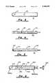
- FIG. 1shows a schematic block diagram of the electrical apparatus and wiring of one embodiment of a fetal heart rate and intrauterine pressure sensing and monitoring system.
- FIG. 2shows a schematic drawing of one embodiment of a fetal heart rate and intrauterine pressure sensing apparatus.
- FIG. 3shows a schematic drawing of one embodiment of a fetal heart rate sensing apparatus.
- FIG. 4shows a schematic drawing of another embodiment of a fetal heart rate and intrauterine pressure sensing apparatus having a tubular housing.
- FIG. 5shows a schematic drawing of another embodiment of a fetal heart rate and intrauterine pressure sensing apparatus having a tubular housing and the pressure transducer mounted at the distal tip of the sensor transverse to the axis of the tubular housing.
- FIG. 6shows a schematic drawing of one embodiment of a fetal heart rate and intrauterine pressure sensing and monitoring system including a pressure cavity and embedded, prefilled and sealed pressure tubing terminating in a Luer fitting.
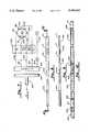
- FIG. 7is an electrical block diagram of an alternative embodiment of a fetal heart rate and intrauterine pressure sensing and monitoring system.
- FIG. 8is a plan view of the alternative embodiment.
- FIG. 9is a side elevation of the alternative embodiment.
- FIG. 10is an enlarged cross-sectional view taken along line 10--10 in FIG. 8.
- FIG. 11is a greatly enlarged cross-sectional view of the pressure transducer assembly.
- FIG. 12is an enlarged view of the pressure transducer assembly.
- FIG. 13illustrates the manner in which a tip electrode can be modified to contain the pressure transducer assembly.
- FIG. 1shows a schematic block diagram of the electrical apparatus and wiring of one embodiment of a fetal heart rate and intrauterine pressure sensing and monitoring system.
- the systemcomprises a sensor means 40 and monitoring means 60 connected together by standard connecting means (not shown).
- the sensor 40includes a first electrode 10, pressure transducer means 30, a second electrode 20 and means for connecting the sensor to the monitoring means 60.
- the monitoring means 60includes ECG and heart rate monitoring (ECG/HR) means 70 and pressure monitoring means 80.
- ECG/HRECG and heart rate monitoring
- the electrodes 10 and 20may advantageously be comprised of platinum, silver chloride, gold, stainless steel or other similar conductive metals. It will be recognized by one skilled in the art that other well-known materials may be employed as materials for the ECG pick-up electrodes 10 and 20.
- the electrodes 10 and 20transmit ECG signals to the ECG/HR monitoring means 70 through conductors 54 and 56, respectively.
- a signal ground or reference electrode 72is typically provided by a metal plate in contact with the mother's body, e.g., a leg plate. Such a plate is typically an integral part of the connecting means conventionally used with commercially available ECG/HR monitoring devices. Rather than utilizing a leg plate, the reference electrode may be a conventional ECG electrode.
- the ECG/HR monitoring meansmay be any commercially available unit suitable for use in perinatal care.
- pressure monitoring means 80is connected to pressure transducer electronics 52 via cable 32.
- Cable 32may advantageously be a flex cable which interconnects the transducer 30, the transducer electronics 52 and the monitor 80.
- Cable 32is preferably a multiconduit electrical cable suitable for transmitting electrical signals indicative of uterine pressure and providing electrical excitation to the pressure transducer.
- Pressure monitoring means 80may also suitably be any commercially available monitoring device suitable for use in perinatal care. Many such devices are typically integrated into one unit for monitoring both fetal ECG and heart rate and intrauterine pressure.
- a typical monitoring device suitable for use with the sensor of the present inventionis the Model 112 Fetal Monitor manufactured and sold by Corometrics Medical Systems, Inc. of Wallingford, Connecticut. However, other such monitors may also be used in combination with this invention.
- the pressure transducer means 30may advantageously be a miniature solid state differential pressure transducer.
- the transducer meansincludes a body, means for converting pressure signals to electrical signals (e.g. a piezoelectric crystal), means for interfacing with external pressure monitors and means for providing temperature and pressure compensation.
- One such transduceris readily available as an off-the-shelf item for use by other equipment manufacturers.
- Such miniature transducershave a pressure range of from about 0 to about 300 mmHg, a sensitivity of about 5 uV/V/mmHg within about 1% accuracy and an operating temperature range of from about 15° C. to about 40° C.
- Such commercially available pressure transducersfurther include electronics 52 for interfacing with means for monitoring pressure 80.
- the electronics 52typically comprise a sensor chip, a temperature compensating resistor chip and a flex circuit (not shown).
- the electronics 52may suitably be integrated into or mounted onto connecting means (shown in FIG. 2) by well known mounting or hybrid circuit manufacturing techniques.
- FIG. 2shows a schematic drawing of one embodiment of a fetal heart rate and intrauterine pressure sensing apparatus.
- the apparatuscomprises flexible tubular housing 100, two electrodes 10 and 20 for ECG pick-up mounted on the housing 100 distal and proximal to a pressure sensing transducer 30.
- the electrodesare slightly protruding from the surface of the housing, having a smooth surface and an appropriate radius to assure contact.
- the electrodes 10 and 20are connected with signal conductors 54 and 56 embedded within the housing to the connector 50 located on the housing's proximal end. Since the transducer 30 operates by differential means, it must be vented to the atmosphere for calibration of a reference or zero pressure reading. This venting is accomplished by means for venting 33.
- the venting means 33may be a tube or lumen running from inside of the housing 100 to the atmosphere.
- the venting tubemay run through or around the connector 50.
- the venting means 33may advantageously be small-bore polyethylene tubing or a similar biocompatible material.
- the housing 100may advantageously be comprised of a flat, semi-pliable plastic or medical grade silastic which may be in the shape of a flat tube, similar in shape to a multi-conductor, flat ribbon cable commonly used for interconnecting computer components. Contained within the housing 100 are the electrode lines 54 and 56 which terminate at the connector means 50, cable 32 and venting means 33.
- the pressure transducer means 30must be hermetically sealed within the housing by well-known sealing techniques.
- the transducer body utilized, and commercially available,may advantageously include a polycarbonate body filled with a dielectric gel (not shown).
- the senor 40is inserted between the fetus and the internal uterine wall following rupture of the membranes and using standard insertion techniques similar to well-known techniques used for inserting transcervical pressure tubing.
- FIG. 3a sensor is shown for fetal heart rate measurements only.
- the sensor in FIG. 3is identical to the sensor 40 described with respect to FIG. 1, except that the pressure transducer means 30 is deleted. Such a sensor would be used in situations where monitoring intrauterine pressure is not required.
- FIG. 4yet another embodiment of a sensor for sensing fetal heart rate and intrauterine pressure is shown.
- This embodimentcomprises a housing 142, band or ring electrodes 110 and 120 and pressure transducer 130.
- the housing 142is a tubular housing having a circular cross-section and similar to a catheter in shape.
- the electrodes 110 and 120conform to the shape of the housing 142 by banding around the housing.
- the transducer 130may be similar to those described hereinabove.
- the electrodes and transducermay be disposed as described above and comprise similar materials and components. It is not necessary for the transducer to be located between the electrodes in any of the embodiments shown.
- the transducermay be located distal to the first electrode, slightly recessed from the distal tip of the housing.
- the catheter diametermay be in the range of about 8 F to 12 F (French Catheter Scale)
- the catheter lengthmay be in the range of 80 cm to 100 cm
- the width of the ECG electrodesmay be in the range of 1 mm to 5 mm.
- the elementsare disposed as follows: the first electrode is about 1 cm from the distal tip of the housing, the pressure transducer is about 5 cm from the distal tip of the housing and the second electrode is about 9 cm from the distal tip of the housing.
- FIG. 5yet another embodiment of a sensor for sensing fetal heart rate and intrauterine pressure is shown.
- This embodimentis similar to the embodiment of FIG. 4 except that the transducer 130 is disposed transversely to the electrodes and the transducer is located at the distal tip of the sensor housing.
- FIG. 6yet another embodiment of a device for monitoring fetal ECG and intrauterine pressure is illustrated in a conceptual drawing.
- the device of FIG. 6features ECG electrodes 210 and 220, pre-filled pressure tubing 230 and housing 240 having a pre-filled and sealed sensing cavity 242.
- the ECG electrodes 210 and 220are similar to those electrodes described hereinabove with respect to the embodiments of FIGS. 1 and 2. Electrodes 210 and 220 are connected to an exterior ECG monitoring means (not shown) by means of conductors 212 and 222.
- the pressure cavity 242is recessed from the distal tip 214 of the house 240 and is sealed with a highly compliant . membrane such as silicon rubber.
- the cavity 242is connected through tubing 230 to the proximal (outside) end of the housing 240.
- the tubing 230is embedded in the housing 240 and terminates in a sealed Luer fitting 232.
- the pressure cavity 242 and tubing 230are advantageously prefilled with bubble-free, sterile, biocompatible liquid and completely sealed.
- the biocompatible liquidmay be glycerin or a similar high viscosity liquid.
- the approximate external dimensions of the sensor unitsmay advantageously be about 300 mm to 1000 mm in length by about 15 mm to 10 mm in width by about 3 mm maximum thickness.
- the preferable minimum distance between the first and second electrodesshould be in the range from about 4 to 8 cm.
- FIG. 7is an electrical schematic diagram of the intrauterine pressure and fetal heart rate sensor and is seen to include first and second surface electrodes E 1 and E 2 which are connected to one another by a conductor 300 and to a proximal connector plug 302 by a conductor 304.
- ECG signals attributable to both the mother and the fetusare picked up by the electrodes and conveyed over the conductors 300 and 304 to a connector terminal in plug 302 and then through a corresponding terminal of a mating plug part 306 and thence through a multi-conductor cable 308 to a monitor (not shown).
- the circuitry in the monitorthen is able to discriminate and isolate the fetal heart rate signal from that of the mother.
- the sensor deviceincludes a plurality of piezoresistive elements 310 through 313 connected as a Wheatstone bridge 314.
- the bridge 314is adapted to be energized by signals provided over conductors 316 and 318 to oppose terminals 320 and 322 of the ridge 314.
- the output from the bridgeis obtained across terminals 324 and 326 and is fed back over conductors 328 and 330 to the plug member 302 and thence through mating plug member 306 and the multi-conductor cable 308 to the monitor device (not shown).
- the monitoris capable of providing an indication of pressure variations corresponding to the voltage output obtained across terminals 324 and 326 of the bridge 314.
- FIG. 7there is also illustrated a compensation board 332 to which the bridge energization lines and bridge output lines connect.
- the resistive components on the compensation board 332are appropriately trimmed so as to accommodate variations in the piezo-resistive elements 310 through 313 so that there will be uniformity between sensor products.
- the constructional features of the compensation board 332will be explained in greater detail hereinbelow when FIGS. 11 and 12 are discussed.
- FIGS. 8 and 9are, respectively, top and side views of the intrauterine ribbon sensor of the instant embodiment. It is seen to include a flattened molded strip of a suitable medical grade plastic, preferably polyurethane or silicon rubber, on which is mounted a distal tip surface electrode E 1 , a pressure sensor P and a proximal surface electrode E 2 .
- the bridge 314 and the compensation board 332 as well as the conductors joining one to the other form part of the pressure sensor P and the conductors 304, 316, 318, 328 and 330extend internally Within the molded silicon or urethane rubber strip 334 to the proximal connector 302.
- the conductorsmay be shielded by a braided sheath.
- the thickness of the molded silicon rubbermay vary along the length of the intrauterine ribbon sensor to provide a desired balance in the flexibility along the length thereof to facilitate insertion through the vaginal channel and cervix into the uterine cavity and recognizing that just prior to delivery, the head of the fetus will be generally disposed in a position partially blocking access to the uterine cavity. It is, of course, also possible to tailor the stiffness properties of the intrauterine ribbon sensor by providing plastic of differing hardness (durometer) in the segments 336 and 338 than is used in the segment 340 which lies between the proximal surface electrode E 2 and the male connector 302.
- FIG. 10illustrates the possibility of fabricating the device out of a plurality of discrete segments including a distal tip electrode E 1 , a pressure sensing module P and a connector 302 respectively coupled in end-to-end relation by means of tubing segments 342, 344 and 346. While not shown in FIG. 10, it is to be recognized that the conductors 300, 304, 316, 318, 328 and 330 are routed through the lumen of the tubular segments 342, 344 and 346 to the proximal connector 302.
- the tubing segments 342 and 344will preferably be in the form of a flattened oval similar in cross-section to the device depicted in FIGS. 8 and 9 and, moreover, the segments 342, 344 and 346 may then be filled with a suitable plastic such as silicon rubber, with due consideration being paid to the durometer of the material filling the respective tubular segments to provide desired stiffness properties along the length thereof.
- a suitable plasticsuch as silicon rubber
- the distal tip electrode E 1may be formed from stainless steel or other suitable metal and includes an integrally formed stub 348 of oval cross-section dimensioned to fit within the lumen of the tube segment 342 and it would be bonded in place by an appropriate adhesive so that the exterior dimension of the electrode surface is continuous with the exterior dimensions of the tubular plastic segment 342.
- the electrode E 2is tubular in form and includes integrally formed stub segments 350 and 352 allowing it to be inserted between the plastic tubular segments 344 and 346 without creating a size discontinuity along the length of the ribbon sensor.
- the pressure sensor segment P of the intrauterine ribbon sensorIt is seen to include a carrier member 354 also in the form of a flattened oval and having an ovalular stub 356 and 358 extending longitudinally from opposite ends thereof for mating with the tubular segments 342 and 344, respectively (FIG. 10).
- the carrier 354may also be fabricated from stainless steel. However, because the operation of the sensor does not depend on the fact that the carrier 354 is conductive, it may just as well be fabricated in a molding operation from a suitable relatively non-deformable plastic, such as polysulfone resin.
- a sensor substrate 360comprising a phenolic printed circuit board having a conductive pattern formed on the top surface thereof as indicated by the cross-hatching in FIG. 12.
- the etched pathswhich are free of cross-hatching, effectively divide the conductive area into four segments. It is the internal resistance of these segments that create the compensating resistors shown o the compensation board 332 in FIG. 7.
- a hole 362is formed through the thickness dimension of the printed circuit substrate board 360 and on the upper surface of the board 360 is disposed a chip 364 on the surface of which is located the piezo-resistive elements 310-313.
- a wire bonding technique well known in the artmay be used to establish an electrical connection between the conductive pattern on the substrate 360 and the piezo-resistive elements on the chip 364. These wire bonds can be seen in FIG. 12 and are identified by numerals 365-370.
- the cavity 372 surrounding the chip 364 and lying above the substrate 360 in FIG. 11is filled with a silastic adhesive while the space 374 located beneath the substrate 360 remains empty.
- a vent tube 376extends the length of the ribbon sensor and is opened to the atmosphere such that the pressure within the cavity 374 remains at atmospheric pressure.
- the chip array 364is also subjected to atmospheric pressure on the underside thereof. As such, deflection of the chip 364, as by fluid pressures applied to it via the silastic filled window 372, unbalances the bridge 314 to produce an output signal proportional to such fluid pressure.
- FIG. 13is intended to illustrate the manner in which the pressure transducer card of FIG. 12 can be disposed in the distal tip electrode E 1 rather than in a separate carrier such as is shown by numeral 354 in FIG. 11.
- electrode E 1comprises a hollow conductive body 380 having a stub 382 for receiving a silastic or urethane tube segment 342 thereon. Milled through the side wall of the body 380 is a rectangular opening 384 leading to flange surfaces 386 and 388. The opening 384 is dimensioned to receive the printed circuit card 360 of FIG. 12 on which the piezoresistive chip 364 is mounted.
- the card 360rests upon the flanges 386 and 388 and the portion of the cavity above it is filled with a highly compliant medium which acts as a deformable membrane.
- the body 380is conductive, it can serve as an ECG electrode.
- the card 360being an insulating substrate, the circuit pattern thereon remains isolated from the conductive body. This alternative construction obviates the need for a separate pressure transducing carrier and reduces the overall cost of the intrauterine ribbon sensor.
- the overall length of the intrauterine ribbon sensor from its distal tip to its proximal connector 302may typically be 160 mm while the distance from the tip electrode E 1 to the proximal electrode E 2 may be 60 mm with the pressure sensor P disposed midway therebetween.
- the overall width dimension of the intrauterine ribbon sensor at its distal endmay be typically 8 mm and have a thickness of 3 mm.
- the pressure transducer utilized in the present inventionis extremely sensitive and has been found to produce a high frequency component due to the sound waves produced by the heart beat of the fetus and mother. It is, therefore, contemplated that by providing suitable signal processing circuitry in the monitor for discriminating between the two, it is possible to derive not only intrauterine pressure readings but heart rate information as well from the pressure transducer, thus potentially obviating the need for separate ECG surface electrodes on the ribbon sensor.
Landscapes
- Health & Medical Sciences (AREA)
- Life Sciences & Earth Sciences (AREA)
- Biomedical Technology (AREA)
- Molecular Biology (AREA)
- Veterinary Medicine (AREA)
- Physics & Mathematics (AREA)
- Public Health (AREA)
- Biophysics (AREA)
- Pathology (AREA)
- Engineering & Computer Science (AREA)
- General Health & Medical Sciences (AREA)
- Heart & Thoracic Surgery (AREA)
- Medical Informatics (AREA)
- Animal Behavior & Ethology (AREA)
- Surgery (AREA)
- Cardiology (AREA)
- Gynecology & Obstetrics (AREA)
- Pediatric Medicine (AREA)
- Pregnancy & Childbirth (AREA)
- Reproductive Health (AREA)
- Hematology (AREA)
- Measurement And Recording Of Electrical Phenomena And Electrical Characteristics Of The Living Body (AREA)
- Measuring And Recording Apparatus For Diagnosis (AREA)
Abstract
Description
Claims (32)
Priority Applications (1)
| Application Number | Priority Date | Filing Date | Title |
|---|---|---|---|
| US07/103,651US5184619A (en) | 1986-11-10 | 1987-10-02 | Intrauterine pressure and fetal heart rate sensor |
Applications Claiming Priority (2)
| Application Number | Priority Date | Filing Date | Title |
|---|---|---|---|
| US92963986A | 1986-11-10 | 1986-11-10 | |
| US07/103,651US5184619A (en) | 1986-11-10 | 1987-10-02 | Intrauterine pressure and fetal heart rate sensor |
Related Parent Applications (1)
| Application Number | Title | Priority Date | Filing Date |
|---|---|---|---|
| US92963986AContinuation-In-Part | 1986-11-10 | 1986-11-10 |
Publications (1)
| Publication Number | Publication Date |
|---|---|
| US5184619Atrue US5184619A (en) | 1993-02-09 |
Family
ID=26800699
Family Applications (1)
| Application Number | Title | Priority Date | Filing Date |
|---|---|---|---|
| US07/103,651Expired - Fee RelatedUS5184619A (en) | 1986-11-10 | 1987-10-02 | Intrauterine pressure and fetal heart rate sensor |
Country Status (1)
| Country | Link |
|---|---|
| US (1) | US5184619A (en) |
Cited By (87)
| Publication number | Priority date | Publication date | Assignee | Title |
|---|---|---|---|---|
| US5373852A (en)* | 1993-06-25 | 1994-12-20 | The Regents Of The University Of California | Monitoring uterine contractions by radiotelemetric transmission |
| US5377673A (en)* | 1993-03-22 | 1995-01-03 | Van Dell; Peter | Intrauterine monitoring device |
| US5397344A (en)* | 1992-12-22 | 1995-03-14 | Schering Aktiengesellschaft | Methods of and apparatus for measuring uterine electrical and mechanical activity |
| US5398687A (en)* | 1992-05-18 | 1995-03-21 | Wilson-Cook Medical Inc. | Methods for measuring motility within the biliary tract and instrumentation useful therefor |
| US5425362A (en)* | 1993-07-30 | 1995-06-20 | Criticare | Fetal sensor device |
| US5431171A (en)* | 1993-06-25 | 1995-07-11 | The Regents Of The University Of California | Monitoring fetal characteristics by radiotelemetric transmission |
| US5438985A (en)* | 1993-01-25 | 1995-08-08 | Synectics Medical, Incorporated | Ambulatory recording of the presence and activity of substances in gastro-intestinal compartments |
| US5450857A (en)* | 1994-05-19 | 1995-09-19 | Board Of Regents, The University Of Texas System | Method for the diagnosis of cervical changes |
| US5474065A (en)* | 1994-04-04 | 1995-12-12 | Graphic Controls Corporation | Non-invasive fetal probe |
| US5477860A (en)* | 1992-11-05 | 1995-12-26 | Synectics Medical, Inc. | Catheter for measuring respiration and respiratory effort |
| US5477854A (en)* | 1993-09-16 | 1995-12-26 | Synectics Medical, Inc. | System and method to monitor gastrointestinal Helicobacter pylori infection |
| US5479935A (en)* | 1993-10-21 | 1996-01-02 | Synectics Medical, Inc. | Ambulatory reflux monitoring system |
| US5507289A (en)* | 1993-09-16 | 1996-04-16 | Synectics Medical, Inc. | System and method to diagnose bacterial growth |
| US5551425A (en)* | 1993-05-13 | 1996-09-03 | Synectics Medical, Inc. | Potential difference and perfusion pressure catheter |
| US5566680A (en)* | 1995-09-22 | 1996-10-22 | Graphic Controls Corporation | Transducer-tipped intrauterine pressure catheter system |
| US5649548A (en)* | 1994-03-12 | 1997-07-22 | Hewlett-Packard Company | Transducer for monitoring labor pains |
| US5657759A (en)* | 1993-05-13 | 1997-08-19 | Synectics Medical, Incorporated | Measurement of gastric emptying and gastrointestinal output |
| EP0830841A1 (en)* | 1996-09-20 | 1998-03-25 | SICAN F&E GmbH ( SIBET) | Measuring device for medical applications having an intracorporeally-insertable sensor-element, and manufacturing method of such a measuring device |
| US5746212A (en)* | 1992-03-20 | 1998-05-05 | Rall; Gerhard | Process and device for measuring vital fetal parameters during labor and delivery |
| US5810741A (en)* | 1992-11-05 | 1998-09-22 | Synectics Medical Ab | Method of measuring respiration and respiratory effort using plural catheters |
| US5833622A (en)* | 1994-04-04 | 1998-11-10 | Graphic Controls Corporation | Non-invasive fetal probe having improved mechanical and electrical properties |
| US5833625A (en)* | 1993-10-21 | 1998-11-10 | Synectics Medical Ab | Ambulatory reflux monitoring system |
| WO1999005963A1 (en)* | 1997-07-30 | 1999-02-11 | Genesis Technologies, Inc. | Multiparameter fetal monitoring device |
| US5911690A (en)* | 1994-12-01 | 1999-06-15 | Reinhold Kintza | Use of a pulse oxymetry sensor device |
| USD417005S (en)* | 1999-04-01 | 1999-11-23 | Francisco Jijon | Fetus stimulation system |
| US5991649A (en)* | 1992-12-22 | 1999-11-23 | University Of Texas | Methods for activating the muscle cells or nerves of the uterus or cervix |
| US6083179A (en)* | 1996-05-20 | 2000-07-04 | Formo Medical Ab (Publ.) | Sensor to detect changes in the cross section of an elongated body cavity |
| US6086549A (en)* | 1997-07-03 | 2000-07-11 | Utah Medical Products, Inc. | Apparatus and method for treating female urinary incontinence |
| US6193670B1 (en)* | 1997-02-14 | 2001-02-27 | Tricardia, Llc | Hemostatic agent delivery device having built-in pressure sensor |
| US6356777B1 (en) | 1992-12-22 | 2002-03-12 | Schering Aktiengesellschaft | Methods of and apparatus for activating the muscle cells or nerves of the uterus or cervix |
| US6594515B2 (en)* | 2000-01-10 | 2003-07-15 | Richard L. Watson | Noninvasive, intrauterine fetal ECG strip electrode |
| US6625495B1 (en)* | 2000-08-02 | 2003-09-23 | Medisox Israel Ltd. | Body-cavity probe with body conformable member |
| WO2003094693A2 (en) | 2002-02-15 | 2003-11-20 | Biocontrol Medical Ltd. | Low power consumption implantable pressure sensor |
| US20030216658A1 (en)* | 2002-05-15 | 2003-11-20 | Colin Corporation | Fetal-pulse-wave-velocity-related-information obtaining apparatus and childbirth monitoring apparatus |
| US6712772B2 (en) | 2001-11-29 | 2004-03-30 | Biocontrol Medical Ltd. | Low power consumption implantable pressure sensor |
| US6751498B1 (en) | 1999-03-15 | 2004-06-15 | The Johns Hopkins University | Apparatus and method for non-invasive, passive fetal heart monitoring |
| US20050267377A1 (en)* | 2004-05-28 | 2005-12-01 | Dorothee Marossero | Maternal-fetal monitoring system |
| US20050287620A1 (en)* | 1991-03-04 | 2005-12-29 | Therasense, Inc. | Method of determining analyte level using subcutaneous electrode |
| US20060036188A1 (en)* | 2005-11-08 | 2006-02-16 | Anatosol, L.L.C. | Perineometer with wireless biofeedback |
| US20070142750A1 (en)* | 2005-12-20 | 2007-06-21 | Robert Kotmel | Method and apparatus for uterine cavity length measurement |
| US20070142752A1 (en)* | 2005-12-20 | 2007-06-21 | Robert Kotmel | Uterine cavity length measurement |
| US20070225584A1 (en)* | 2004-04-19 | 2007-09-27 | University Of Florida Researouchfoundation, Inc. | Novel Catheter Sensor |
| US20090036754A1 (en)* | 2007-07-31 | 2009-02-05 | Captomed Eurl | Self-calibrating pressure sensor |
| US7658196B2 (en) | 2005-02-24 | 2010-02-09 | Ethicon Endo-Surgery, Inc. | System and method for determining implanted device orientation |
| US20100076254A1 (en)* | 2006-06-05 | 2010-03-25 | Ams Research Corporation | Electrical muscle stimulation to treat fecal incontinence and/or pelvic prolapse |
| US7775215B2 (en) | 2005-02-24 | 2010-08-17 | Ethicon Endo-Surgery, Inc. | System and method for determining implanted device positioning and obtaining pressure data |
| US7775966B2 (en) | 2005-02-24 | 2010-08-17 | Ethicon Endo-Surgery, Inc. | Non-invasive pressure measurement in a fluid adjustable restrictive device |
| US20100217340A1 (en)* | 2009-02-23 | 2010-08-26 | Ams Research Corporation | Implantable Medical Device Connector System |
| ITFI20090115A1 (en)* | 2009-05-22 | 2010-11-23 | Pierfrancesco Belli | ELECTROMEDICAL EQUIPMENT FOR ASSISTANCE TO TRAVEL AND SPORTS. |
| US7844342B2 (en) | 2008-02-07 | 2010-11-30 | Ethicon Endo-Surgery, Inc. | Powering implantable restriction systems using light |
| US20100305422A1 (en)* | 1998-03-04 | 2010-12-02 | Abbott Diabetes Care Inc. | Electrochemical Analyte Sensor |
| US7927270B2 (en) | 2005-02-24 | 2011-04-19 | Ethicon Endo-Surgery, Inc. | External mechanical pressure sensor for gastric band pressure measurements |
| US20110112432A1 (en)* | 2009-11-11 | 2011-05-12 | Minerva Surgical, Inc. | Systems and devices for evaluating the integrity of a uterine cavity |
| US8016745B2 (en) | 2005-02-24 | 2011-09-13 | Ethicon Endo-Surgery, Inc. | Monitoring of a food intake restriction device |
| US8016744B2 (en) | 2005-02-24 | 2011-09-13 | Ethicon Endo-Surgery, Inc. | External pressure-based gastric band adjustment system and method |
| US8034065B2 (en) | 2008-02-26 | 2011-10-11 | Ethicon Endo-Surgery, Inc. | Controlling pressure in adjustable restriction devices |
| US8057492B2 (en) | 2008-02-12 | 2011-11-15 | Ethicon Endo-Surgery, Inc. | Automatically adjusting band system with MEMS pump |
| US8066629B2 (en) | 2005-02-24 | 2011-11-29 | Ethicon Endo-Surgery, Inc. | Apparatus for adjustment and sensing of gastric band pressure |
| US8100870B2 (en) | 2007-12-14 | 2012-01-24 | Ethicon Endo-Surgery, Inc. | Adjustable height gastric restriction devices and methods |
| US8114345B2 (en) | 2008-02-08 | 2012-02-14 | Ethicon Endo-Surgery, Inc. | System and method of sterilizing an implantable medical device |
| US8142452B2 (en) | 2007-12-27 | 2012-03-27 | Ethicon Endo-Surgery, Inc. | Controlling pressure in adjustable restriction devices |
| US8152710B2 (en) | 2006-04-06 | 2012-04-10 | Ethicon Endo-Surgery, Inc. | Physiological parameter analysis for an implantable restriction device and a data logger |
| US8187162B2 (en) | 2008-03-06 | 2012-05-29 | Ethicon Endo-Surgery, Inc. | Reorientation port |
| US8187163B2 (en) | 2007-12-10 | 2012-05-29 | Ethicon Endo-Surgery, Inc. | Methods for implanting a gastric restriction device |
| US8192350B2 (en) | 2008-01-28 | 2012-06-05 | Ethicon Endo-Surgery, Inc. | Methods and devices for measuring impedance in a gastric restriction system |
| US8221439B2 (en) | 2008-02-07 | 2012-07-17 | Ethicon Endo-Surgery, Inc. | Powering implantable restriction systems using kinetic motion |
| US8233995B2 (en) | 2008-03-06 | 2012-07-31 | Ethicon Endo-Surgery, Inc. | System and method of aligning an implantable antenna |
| US8337389B2 (en) | 2008-01-28 | 2012-12-25 | Ethicon Endo-Surgery, Inc. | Methods and devices for diagnosing performance of a gastric restriction system |
| CN102860831A (en)* | 2012-09-03 | 2013-01-09 | 杭州电子科技大学 | Uterine contraction pressure monitoring system and dynamic pressure calibrating method thereof |
| US8377079B2 (en) | 2007-12-27 | 2013-02-19 | Ethicon Endo-Surgery, Inc. | Constant force mechanisms for regulating restriction devices |
| US8380312B2 (en) | 2009-12-31 | 2013-02-19 | Ams Research Corporation | Multi-zone stimulation implant system and method |
| US8529562B2 (en) | 2009-11-13 | 2013-09-10 | Minerva Surgical, Inc | Systems and methods for endometrial ablation |
| US20130245463A1 (en)* | 2011-09-12 | 2013-09-19 | Susan Stuebe | System and method for displaying physiological information |
| US8591532B2 (en) | 2008-02-12 | 2013-11-26 | Ethicon Endo-Sugery, Inc. | Automatically adjusting band system |
| US8591395B2 (en) | 2008-01-28 | 2013-11-26 | Ethicon Endo-Surgery, Inc. | Gastric restriction device data handling devices and methods |
| US8870742B2 (en) | 2006-04-06 | 2014-10-28 | Ethicon Endo-Surgery, Inc. | GUI for an implantable restriction device and a data logger |
| US9421059B2 (en) | 2010-04-27 | 2016-08-23 | Minerva Surgical, Inc. | Device for endometrial ablation having an expandable seal for a cervical canal |
| US9731112B2 (en) | 2011-09-08 | 2017-08-15 | Paul J. Gindele | Implantable electrode assembly |
| US20180068591A1 (en)* | 2016-09-06 | 2018-03-08 | Dynasthetics, Llc | System and method for simulating fetal heart rate for noninvasive intra-partum fetal monitoring |
| WO2018093784A1 (en)* | 2016-11-15 | 2018-05-24 | Pretel Inc. | Vaginal electrode |
| US10210774B2 (en) | 2016-11-17 | 2019-02-19 | Dynasthetics, Llc | System and method for simulating arterial pressure pulses |
| US10269265B2 (en)* | 2016-09-06 | 2019-04-23 | Dynasthetics, Llc | System and method for simulating intrauterine contractions for noninvasive intra-partum fetal monitoring |
| US11224375B2 (en) | 2017-02-28 | 2022-01-18 | Mayo Foundation For Medical Education And Research | Systems and methods for fetal monitoring |
| US11234650B2 (en)* | 2006-11-20 | 2022-02-01 | St. Jude Medical Coordination Center Bvba | Measurement system |
| US20220110552A1 (en)* | 2019-10-16 | 2022-04-14 | James Robert Balman | Apparatus and method for determining physiological parameters of an infant in-utero |
| US11504054B2 (en) | 2017-03-07 | 2022-11-22 | University Of Southampton | Intra-uterine monitoring system |
| US11771953B1 (en) | 2023-03-06 | 2023-10-03 | Craig A. Hoffman | Perineometer and method for use of same |
Citations (8)
| Publication number | Priority date | Publication date | Assignee | Title |
|---|---|---|---|---|
| US3326207A (en)* | 1964-07-10 | 1967-06-20 | James J Egan | Electrocardiac instrument for testing unborn infants |
| US3724467A (en)* | 1971-04-23 | 1973-04-03 | Avery Labor Inc | Electrode implant for the neuro-stimulation of the spinal cord |
| US3769984A (en)* | 1971-03-11 | 1973-11-06 | Sherwood Medical Ind Inc | Pacing catheter with frictional fit lead attachment |
| US4114288A (en)* | 1976-10-14 | 1978-09-19 | Fowler Joe W | Bulk cure tobacco barn with improvements in construction for optimizing heating efficiency |
| US4172451A (en)* | 1978-04-06 | 1979-10-30 | Medical Evaluation Devices And Instruments Corp. | Intracardial electrode and a method of manufacture thereof |
| US4476871A (en)* | 1982-04-23 | 1984-10-16 | American Home Products Corporation | Monitoring of cervical dilatation during labor |
| US4510944A (en)* | 1982-12-30 | 1985-04-16 | Porges Stephen W | Method and apparatus for evaluating rhythmic oscillations in aperiodic physiological response systems |
| US4554927A (en)* | 1983-08-30 | 1985-11-26 | Thermometrics Inc. | Pressure and temperature sensor |
- 1987
- 1987-10-02USUS07/103,651patent/US5184619A/ennot_activeExpired - Fee Related
Patent Citations (8)
| Publication number | Priority date | Publication date | Assignee | Title |
|---|---|---|---|---|
| US3326207A (en)* | 1964-07-10 | 1967-06-20 | James J Egan | Electrocardiac instrument for testing unborn infants |
| US3769984A (en)* | 1971-03-11 | 1973-11-06 | Sherwood Medical Ind Inc | Pacing catheter with frictional fit lead attachment |
| US3724467A (en)* | 1971-04-23 | 1973-04-03 | Avery Labor Inc | Electrode implant for the neuro-stimulation of the spinal cord |
| US4114288A (en)* | 1976-10-14 | 1978-09-19 | Fowler Joe W | Bulk cure tobacco barn with improvements in construction for optimizing heating efficiency |
| US4172451A (en)* | 1978-04-06 | 1979-10-30 | Medical Evaluation Devices And Instruments Corp. | Intracardial electrode and a method of manufacture thereof |
| US4476871A (en)* | 1982-04-23 | 1984-10-16 | American Home Products Corporation | Monitoring of cervical dilatation during labor |
| US4510944A (en)* | 1982-12-30 | 1985-04-16 | Porges Stephen W | Method and apparatus for evaluating rhythmic oscillations in aperiodic physiological response systems |
| US4554927A (en)* | 1983-08-30 | 1985-11-26 | Thermometrics Inc. | Pressure and temperature sensor |
Non-Patent Citations (2)
| Title |
|---|
| "Tripolar HIS-Bundle Electrode" U.S. Catheter litterature Oct. 1971. |
| Tripolar HIS Bundle Electrode U.S. Catheter litterature Oct. 1971.* |
Cited By (131)
| Publication number | Priority date | Publication date | Assignee | Title |
|---|---|---|---|---|
| US20100030044A1 (en)* | 1991-03-04 | 2010-02-04 | Abbott Diabetes Care Inc. | Subcutaneous Glucose Electrode |
| US20050287620A1 (en)* | 1991-03-04 | 2005-12-29 | Therasense, Inc. | Method of determining analyte level using subcutaneous electrode |
| US8741590B2 (en) | 1991-03-04 | 2014-06-03 | Abbott Diabetes Care Inc. | Subcutaneous glucose electrode |
| US20100030047A1 (en)* | 1991-03-04 | 2010-02-04 | Abbott Diabetes Care Inc. | Subcutaneous Glucose Electrode |
| US8588881B2 (en) | 1991-03-04 | 2013-11-19 | Abbott Diabetes Care Inc. | Subcutaneous glucose electrode |
| US20070151869A1 (en)* | 1991-03-04 | 2007-07-05 | Abbott Diabetes Care, Inc. | Subcutaneous Glucose Electrode |
| US20070215491A1 (en)* | 1991-03-04 | 2007-09-20 | Abbott Diabetes Care, Inc. | Subcutaneous Glucose Electrode |
| US20100121166A1 (en)* | 1991-03-04 | 2010-05-13 | Abbott Diabetes Care Inc. | Subcutaneous Glucose Electrode |
| US20100030048A1 (en)* | 1991-03-04 | 2010-02-04 | Abbott Diabetes Care Inc. | Subcutaneous Glucose Electrode |
| US20100051479A1 (en)* | 1991-03-04 | 2010-03-04 | Adam Heller | Subcutaneous Glucose Electrode |
| US20100030046A1 (en)* | 1991-03-04 | 2010-02-04 | Abbott Diabetes Care Inc. | Subcutaneous Glucose Electrode |
| US5746212A (en)* | 1992-03-20 | 1998-05-05 | Rall; Gerhard | Process and device for measuring vital fetal parameters during labor and delivery |
| US5398687A (en)* | 1992-05-18 | 1995-03-21 | Wilson-Cook Medical Inc. | Methods for measuring motility within the biliary tract and instrumentation useful therefor |
| US5810741A (en)* | 1992-11-05 | 1998-09-22 | Synectics Medical Ab | Method of measuring respiration and respiratory effort using plural catheters |
| US5477860A (en)* | 1992-11-05 | 1995-12-26 | Synectics Medical, Inc. | Catheter for measuring respiration and respiratory effort |
| US5522877A (en)* | 1992-12-22 | 1996-06-04 | Schering Aktiengesellschaft | Methods for measuring uterine electrical and mechanical activity |
| US6356777B1 (en) | 1992-12-22 | 2002-03-12 | Schering Aktiengesellschaft | Methods of and apparatus for activating the muscle cells or nerves of the uterus or cervix |
| US5397344A (en)* | 1992-12-22 | 1995-03-14 | Schering Aktiengesellschaft | Methods of and apparatus for measuring uterine electrical and mechanical activity |
| US5991649A (en)* | 1992-12-22 | 1999-11-23 | University Of Texas | Methods for activating the muscle cells or nerves of the uterus or cervix |
| US5438985A (en)* | 1993-01-25 | 1995-08-08 | Synectics Medical, Incorporated | Ambulatory recording of the presence and activity of substances in gastro-intestinal compartments |
| US5377673A (en)* | 1993-03-22 | 1995-01-03 | Van Dell; Peter | Intrauterine monitoring device |
| US5551425A (en)* | 1993-05-13 | 1996-09-03 | Synectics Medical, Inc. | Potential difference and perfusion pressure catheter |
| US5657759A (en)* | 1993-05-13 | 1997-08-19 | Synectics Medical, Incorporated | Measurement of gastric emptying and gastrointestinal output |
| US6132372A (en)* | 1993-05-13 | 2000-10-17 | Synectics Medical, Incorporated | Measurement of gastric emptying and gastrointestinal output |
| US5373852A (en)* | 1993-06-25 | 1994-12-20 | The Regents Of The University Of California | Monitoring uterine contractions by radiotelemetric transmission |
| US5431171A (en)* | 1993-06-25 | 1995-07-11 | The Regents Of The University Of California | Monitoring fetal characteristics by radiotelemetric transmission |
| US5425362A (en)* | 1993-07-30 | 1995-06-20 | Criticare | Fetal sensor device |
| US5477854A (en)* | 1993-09-16 | 1995-12-26 | Synectics Medical, Inc. | System and method to monitor gastrointestinal Helicobacter pylori infection |
| US5507289A (en)* | 1993-09-16 | 1996-04-16 | Synectics Medical, Inc. | System and method to diagnose bacterial growth |
| US5833625A (en)* | 1993-10-21 | 1998-11-10 | Synectics Medical Ab | Ambulatory reflux monitoring system |
| US5479935A (en)* | 1993-10-21 | 1996-01-02 | Synectics Medical, Inc. | Ambulatory reflux monitoring system |
| US5649548A (en)* | 1994-03-12 | 1997-07-22 | Hewlett-Packard Company | Transducer for monitoring labor pains |
| US5665477A (en)* | 1994-04-04 | 1997-09-09 | Graphic Controls Corporation | Hydrogel adhesive for attaching medical device to patient |
| US5474065A (en)* | 1994-04-04 | 1995-12-12 | Graphic Controls Corporation | Non-invasive fetal probe |
| US5833622A (en)* | 1994-04-04 | 1998-11-10 | Graphic Controls Corporation | Non-invasive fetal probe having improved mechanical and electrical properties |
| US5450857A (en)* | 1994-05-19 | 1995-09-19 | Board Of Regents, The University Of Texas System | Method for the diagnosis of cervical changes |
| US5911690A (en)* | 1994-12-01 | 1999-06-15 | Reinhold Kintza | Use of a pulse oxymetry sensor device |
| US5566680A (en)* | 1995-09-22 | 1996-10-22 | Graphic Controls Corporation | Transducer-tipped intrauterine pressure catheter system |
| US6083179A (en)* | 1996-05-20 | 2000-07-04 | Formo Medical Ab (Publ.) | Sensor to detect changes in the cross section of an elongated body cavity |
| EP0830841A1 (en)* | 1996-09-20 | 1998-03-25 | SICAN F&E GmbH ( SIBET) | Measuring device for medical applications having an intracorporeally-insertable sensor-element, and manufacturing method of such a measuring device |
| US6193670B1 (en)* | 1997-02-14 | 2001-02-27 | Tricardia, Llc | Hemostatic agent delivery device having built-in pressure sensor |
| US6086549A (en)* | 1997-07-03 | 2000-07-11 | Utah Medical Products, Inc. | Apparatus and method for treating female urinary incontinence |
| US6115624A (en)* | 1997-07-30 | 2000-09-05 | Genesis Technologies, Inc. | Multiparameter fetal monitoring device |
| WO1999005963A1 (en)* | 1997-07-30 | 1999-02-11 | Genesis Technologies, Inc. | Multiparameter fetal monitoring device |
| US8463351B2 (en)* | 1998-03-04 | 2013-06-11 | Abbott Diabetes Care Inc. | Electrochemical analyte sensor |
| US20100305422A1 (en)* | 1998-03-04 | 2010-12-02 | Abbott Diabetes Care Inc. | Electrochemical Analyte Sensor |
| US8706180B2 (en) | 1998-03-04 | 2014-04-22 | Abbott Diabetes Care Inc. | Electrochemical analyte sensor |
| US6751498B1 (en) | 1999-03-15 | 2004-06-15 | The Johns Hopkins University | Apparatus and method for non-invasive, passive fetal heart monitoring |
| USD417005S (en)* | 1999-04-01 | 1999-11-23 | Francisco Jijon | Fetus stimulation system |
| US6973341B2 (en) | 2000-01-10 | 2005-12-06 | Watson Richard L | Noninvasive, intrauterine fetal ECG strip electrode |
| US6594515B2 (en)* | 2000-01-10 | 2003-07-15 | Richard L. Watson | Noninvasive, intrauterine fetal ECG strip electrode |
| US20040015067A1 (en)* | 2000-01-10 | 2004-01-22 | Watson Richard L. | Noninvasive, intrauterine fetal egg strip electrode |
| US6625495B1 (en)* | 2000-08-02 | 2003-09-23 | Medisox Israel Ltd. | Body-cavity probe with body conformable member |
| US20040152999A1 (en)* | 2001-11-29 | 2004-08-05 | Biocontrol Medical Ltd | Low power consumption implantable pressure sensor |
| US6712772B2 (en) | 2001-11-29 | 2004-03-30 | Biocontrol Medical Ltd. | Low power consumption implantable pressure sensor |
| WO2003094693A2 (en) | 2002-02-15 | 2003-11-20 | Biocontrol Medical Ltd. | Low power consumption implantable pressure sensor |
| EP2997890A1 (en) | 2002-05-07 | 2016-03-23 | AMS Research Corporation | Low power consumption implantable pressure sensor |
| EP1366709A1 (en)* | 2002-05-15 | 2003-12-03 | Colin Corporation | Fetal-pulse-wave-velocity-related-information obtaining apparatus |
| US20030216658A1 (en)* | 2002-05-15 | 2003-11-20 | Colin Corporation | Fetal-pulse-wave-velocity-related-information obtaining apparatus and childbirth monitoring apparatus |
| US20070225584A1 (en)* | 2004-04-19 | 2007-09-27 | University Of Florida Researouchfoundation, Inc. | Novel Catheter Sensor |
| US20050267377A1 (en)* | 2004-05-28 | 2005-12-01 | Dorothee Marossero | Maternal-fetal monitoring system |
| US7333850B2 (en) | 2004-05-28 | 2008-02-19 | University Of Florida Research Foundation, Inc. | Maternal-fetal monitoring system |
| US8275451B2 (en) | 2004-05-28 | 2012-09-25 | University Of Florida Research Foundation, Inc. | Maternal-fetal monitoring system |
| US20050267376A1 (en)* | 2004-05-28 | 2005-12-01 | Dorothee Marossero | Maternal-fetal monitoring system |
| US7658196B2 (en) | 2005-02-24 | 2010-02-09 | Ethicon Endo-Surgery, Inc. | System and method for determining implanted device orientation |
| US8066629B2 (en) | 2005-02-24 | 2011-11-29 | Ethicon Endo-Surgery, Inc. | Apparatus for adjustment and sensing of gastric band pressure |
| US8016744B2 (en) | 2005-02-24 | 2011-09-13 | Ethicon Endo-Surgery, Inc. | External pressure-based gastric band adjustment system and method |
| US7775215B2 (en) | 2005-02-24 | 2010-08-17 | Ethicon Endo-Surgery, Inc. | System and method for determining implanted device positioning and obtaining pressure data |
| US7775966B2 (en) | 2005-02-24 | 2010-08-17 | Ethicon Endo-Surgery, Inc. | Non-invasive pressure measurement in a fluid adjustable restrictive device |
| US8016745B2 (en) | 2005-02-24 | 2011-09-13 | Ethicon Endo-Surgery, Inc. | Monitoring of a food intake restriction device |
| US7927270B2 (en) | 2005-02-24 | 2011-04-19 | Ethicon Endo-Surgery, Inc. | External mechanical pressure sensor for gastric band pressure measurements |
| US20100087757A1 (en)* | 2005-11-08 | 2010-04-08 | Hoffman Craig A | Multi-Mode Pelvic Exercise Probe |
| US7645220B2 (en) | 2005-11-08 | 2010-01-12 | Anatasol, Llc | Perineometer with wireless biofeedback |
| US20070112284A1 (en)* | 2005-11-08 | 2007-05-17 | Anatosol, L.L.C. | Multi-mode pelvic exercise probe |
| US20060036188A1 (en)* | 2005-11-08 | 2006-02-16 | Anatosol, L.L.C. | Perineometer with wireless biofeedback |
| US7955241B2 (en) | 2005-11-08 | 2011-06-07 | Anatasol, Llc | Multi-mode pelvic exercise probe |
| US7628744B2 (en) | 2005-11-08 | 2009-12-08 | Anatasol, Llc | Multi-mode pelvic exercise probe |
| US8348864B2 (en)* | 2005-12-20 | 2013-01-08 | Cytyc Corporation | Uterine cavity length measurement |
| US8007449B2 (en)* | 2005-12-20 | 2011-08-30 | Cytyc Corporation | Method and apparatus for uterine cavity length measurement |
| US20070142752A1 (en)* | 2005-12-20 | 2007-06-21 | Robert Kotmel | Uterine cavity length measurement |
| US20070142750A1 (en)* | 2005-12-20 | 2007-06-21 | Robert Kotmel | Method and apparatus for uterine cavity length measurement |
| US20120109015A1 (en)* | 2005-12-20 | 2012-05-03 | Cytyc Corporation | Uterine Cavity Length Measurement |
| US8152710B2 (en) | 2006-04-06 | 2012-04-10 | Ethicon Endo-Surgery, Inc. | Physiological parameter analysis for an implantable restriction device and a data logger |
| US8870742B2 (en) | 2006-04-06 | 2014-10-28 | Ethicon Endo-Surgery, Inc. | GUI for an implantable restriction device and a data logger |
| US20100076254A1 (en)* | 2006-06-05 | 2010-03-25 | Ams Research Corporation | Electrical muscle stimulation to treat fecal incontinence and/or pelvic prolapse |
| US11813086B2 (en) | 2006-11-20 | 2023-11-14 | St. Jude Medical Coordination Center Bvba | Measurement system |
| US11234650B2 (en)* | 2006-11-20 | 2022-02-01 | St. Jude Medical Coordination Center Bvba | Measurement system |
| US20090036754A1 (en)* | 2007-07-31 | 2009-02-05 | Captomed Eurl | Self-calibrating pressure sensor |
| US8622923B2 (en)* | 2007-07-31 | 2014-01-07 | Captomed Eurl | Self-calibrating pressure sensor |
| US8187163B2 (en) | 2007-12-10 | 2012-05-29 | Ethicon Endo-Surgery, Inc. | Methods for implanting a gastric restriction device |
| US8100870B2 (en) | 2007-12-14 | 2012-01-24 | Ethicon Endo-Surgery, Inc. | Adjustable height gastric restriction devices and methods |
| US8142452B2 (en) | 2007-12-27 | 2012-03-27 | Ethicon Endo-Surgery, Inc. | Controlling pressure in adjustable restriction devices |
| US8377079B2 (en) | 2007-12-27 | 2013-02-19 | Ethicon Endo-Surgery, Inc. | Constant force mechanisms for regulating restriction devices |
| US8192350B2 (en) | 2008-01-28 | 2012-06-05 | Ethicon Endo-Surgery, Inc. | Methods and devices for measuring impedance in a gastric restriction system |
| US8591395B2 (en) | 2008-01-28 | 2013-11-26 | Ethicon Endo-Surgery, Inc. | Gastric restriction device data handling devices and methods |
| US8337389B2 (en) | 2008-01-28 | 2012-12-25 | Ethicon Endo-Surgery, Inc. | Methods and devices for diagnosing performance of a gastric restriction system |
| US8221439B2 (en) | 2008-02-07 | 2012-07-17 | Ethicon Endo-Surgery, Inc. | Powering implantable restriction systems using kinetic motion |
| US7844342B2 (en) | 2008-02-07 | 2010-11-30 | Ethicon Endo-Surgery, Inc. | Powering implantable restriction systems using light |
| US8114345B2 (en) | 2008-02-08 | 2012-02-14 | Ethicon Endo-Surgery, Inc. | System and method of sterilizing an implantable medical device |
| US8057492B2 (en) | 2008-02-12 | 2011-11-15 | Ethicon Endo-Surgery, Inc. | Automatically adjusting band system with MEMS pump |
| US8591532B2 (en) | 2008-02-12 | 2013-11-26 | Ethicon Endo-Sugery, Inc. | Automatically adjusting band system |
| US8034065B2 (en) | 2008-02-26 | 2011-10-11 | Ethicon Endo-Surgery, Inc. | Controlling pressure in adjustable restriction devices |
| US8233995B2 (en) | 2008-03-06 | 2012-07-31 | Ethicon Endo-Surgery, Inc. | System and method of aligning an implantable antenna |
| US8187162B2 (en) | 2008-03-06 | 2012-05-29 | Ethicon Endo-Surgery, Inc. | Reorientation port |
| US20100217340A1 (en)* | 2009-02-23 | 2010-08-26 | Ams Research Corporation | Implantable Medical Device Connector System |
| ITFI20090115A1 (en)* | 2009-05-22 | 2010-11-23 | Pierfrancesco Belli | ELECTROMEDICAL EQUIPMENT FOR ASSISTANCE TO TRAVEL AND SPORTS. |
| US8394037B2 (en) | 2009-11-11 | 2013-03-12 | Minerva Surgical, Inc. | Systems and devices for evaluating the integrity of a uterine cavity |
| US20110112432A1 (en)* | 2009-11-11 | 2011-05-12 | Minerva Surgical, Inc. | Systems and devices for evaluating the integrity of a uterine cavity |
| US20110112433A1 (en)* | 2009-11-11 | 2011-05-12 | Minerva Surgical, Inc. | Methods for evaluating the integrity of a uterine cavity |
| US8343078B2 (en) | 2009-11-11 | 2013-01-01 | Minerva Surgical, Inc. | Methods for evaluating the integrity of a uterine cavity |
| US9775542B2 (en) | 2009-11-11 | 2017-10-03 | Minerva Surgical, Inc. | Apparatus for evaluating the integrity of a uterine cavity |
| US8529562B2 (en) | 2009-11-13 | 2013-09-10 | Minerva Surgical, Inc | Systems and methods for endometrial ablation |
| US8380312B2 (en) | 2009-12-31 | 2013-02-19 | Ams Research Corporation | Multi-zone stimulation implant system and method |
| US10052150B2 (en) | 2010-04-27 | 2018-08-21 | Minerva Surgical, Inc. | Device for endometrial ablation having an expandable seal for a cervical canal |
| US9421059B2 (en) | 2010-04-27 | 2016-08-23 | Minerva Surgical, Inc. | Device for endometrial ablation having an expandable seal for a cervical canal |
| US9731112B2 (en) | 2011-09-08 | 2017-08-15 | Paul J. Gindele | Implantable electrode assembly |
| US20130245463A1 (en)* | 2011-09-12 | 2013-09-19 | Susan Stuebe | System and method for displaying physiological information |
| CN102860831A (en)* | 2012-09-03 | 2013-01-09 | 杭州电子科技大学 | Uterine contraction pressure monitoring system and dynamic pressure calibrating method thereof |
| CN102860831B (en)* | 2012-09-03 | 2014-04-02 | 杭州电子科技大学 | Uterine contraction pressure monitoring system and dynamic pressure calibrating method thereof |
| US20180068591A1 (en)* | 2016-09-06 | 2018-03-08 | Dynasthetics, Llc | System and method for simulating fetal heart rate for noninvasive intra-partum fetal monitoring |
| US10269265B2 (en)* | 2016-09-06 | 2019-04-23 | Dynasthetics, Llc | System and method for simulating intrauterine contractions for noninvasive intra-partum fetal monitoring |
| US10360816B2 (en)* | 2016-09-06 | 2019-07-23 | Dynasthetics, Llc | System and method for simulating fetal heart rate for noninvasive intra-partum fetal monitoring |
| WO2018093784A1 (en)* | 2016-11-15 | 2018-05-24 | Pretel Inc. | Vaginal electrode |
| US10210774B2 (en) | 2016-11-17 | 2019-02-19 | Dynasthetics, Llc | System and method for simulating arterial pressure pulses |
| US11224375B2 (en) | 2017-02-28 | 2022-01-18 | Mayo Foundation For Medical Education And Research | Systems and methods for fetal monitoring |
| US12251228B2 (en) | 2017-02-28 | 2025-03-18 | Mayo Foundation For Medical Education And Research | Systems and methods for fetal monitoring |
| US11504054B2 (en) | 2017-03-07 | 2022-11-22 | University Of Southampton | Intra-uterine monitoring system |
| US20220110552A1 (en)* | 2019-10-16 | 2022-04-14 | James Robert Balman | Apparatus and method for determining physiological parameters of an infant in-utero |
| US11622705B2 (en)* | 2019-10-16 | 2023-04-11 | James Robert Balman | Apparatus and method for determining physiological parameters of an infant in-utero |
| US20240000351A1 (en)* | 2019-10-16 | 2024-01-04 | James Robert Balman | Apparatus and method for determining physiological parameters of an infant in-utero |
| US11771953B1 (en) | 2023-03-06 | 2023-10-03 | Craig A. Hoffman | Perineometer and method for use of same |
Similar Documents
| Publication | Publication Date | Title |
|---|---|---|
| US5184619A (en) | Intrauterine pressure and fetal heart rate sensor | |
| US4873986A (en) | Disposable apparatus for monitoring intrauterine pressure and fetal heart rate | |
| US4785822A (en) | Disposable intracompartmental pressure transducer | |
| EP0419294B1 (en) | Apparatus for a catheter sensor support and method for using the support | |
| US4576181A (en) | Disposable pressure transducer apparatus for medical use | |
| US8764668B2 (en) | Disposable blood pressure transducer and monitor interface | |
| US5566680A (en) | Transducer-tipped intrauterine pressure catheter system | |
| US6115624A (en) | Multiparameter fetal monitoring device | |
| EP0232142B1 (en) | Pressure transducer | |
| EP0124308B1 (en) | Pressure transducer assembly | |
| US4966161A (en) | Apparatus for continuously measuring intracompartmental pressure within a body cavity | |
| US6264612B1 (en) | Catheter with mechano-responsive element for sensing physiological conditions | |
| EP0360286A2 (en) | Disposable pressure transducer | |
| CA2115712A1 (en) | Intrauterine pressure catheter system | |
| JP3157834B2 (en) | Transducer housing with calibration port | |
| JPH0623058A (en) | Sensor support apparatus in connector for catheter adaptor and use thereof | |
| EP2073696A2 (en) | Reusable invasive fluid pressure monitoring apparatus and method | |
| US9763622B2 (en) | Sensor element with an insulation layer | |
| US3662743A (en) | Pressure transducer for catheter pressure measurement | |
| EP0377943A1 (en) | Intrauterine catheter | |
| NL8302952A (en) | Semiconductor blood pressure sensor - is insertable into blood vessels inside catheter or hypodermic needle |
Legal Events
| Date | Code | Title | Description |
|---|---|---|---|
| AS | Assignment | Owner name:INNOVATIVE SCIENCES, LTD., 5724 MELODY LANE, EDINA Free format text:ASSIGNMENT OF ASSIGNORS INTEREST.;ASSIGNOR:AUSTIN, SANDOR;REEL/FRAME:004776/0687 Effective date:19870928 Owner name:INNOVATIVE SCIENCES, LTD.,MINNESOTA Free format text:ASSIGNMENT OF ASSIGNORS INTEREST;ASSIGNOR:AUSTIN, SANDOR;REEL/FRAME:004776/0687 Effective date:19870928 | |
| AS | Assignment | Owner name:LAUREL MEDICAL CORPORATION Free format text:CHANGE OF NAME;ASSIGNOR:INNOVATIVE SCIENCES, LTD., CHANGED TO;REEL/FRAME:004840/0427 Effective date:19871111 Owner name:LAUREL MEDICAL CORPORATION,STATELESS Free format text:CHANGE OF NAME;ASSIGNOR:INNOVATIVE SCIENCES, LTD., CHANGED TO;REEL/FRAME:004840/0427 Effective date:19871111 | |
| AS | Assignment | Owner name:PERITRONICS MEDICAL INC., CALIFORNIA Free format text:ASSIGNMENT OF ASSIGNORS INTEREST.;ASSIGNOR:LAUREL MEDICAL CORPORATION;REEL/FRAME:006285/0611 Effective date:19921015 | |
| AS | Assignment | Owner name:PARK, GEORGE D., CALIFORNIA Free format text:CORRECTED NOTICE OF PATENT ASSIGNMENT PURSUANT TO DEFAULT;ASSIGNOR:PERITRONICS MEDICAL, INC.;REEL/FRAME:007779/0598 Effective date:19951215 | |
| FPAY | Fee payment | Year of fee payment:4 | |
| AS | Assignment | Owner name:GENESIS TECHNOLOGES, LLC, CALIFORNIA Free format text:ASSIGNMENT OF ASSIGNORS INTEREST;ASSIGNORS:PERITRONICS MEDICAL, LTD.;CONSOLIDATED PERITRONICS MEDICAL, INC., NOW KNOWN AS PERITRONICS MEDICAL, LTD;REEL/FRAME:008296/0383 Effective date:19960731 | |
| AS | Assignment | Owner name:GENESIS TECHNOLOGIES, LLC, CALIFORNIA Free format text:ASSIGNMENT OF ASSIGNORS INTEREST;ASSIGNORS:PAUL, RICHARD H.;PARK, GEORGE D.;REEL/FRAME:008838/0114 Effective date:19971029 | |
| REMI | Maintenance fee reminder mailed | ||
| FPAY | Fee payment | Year of fee payment:8 | |
| SULP | Surcharge for late payment | Year of fee payment:7 | |
| REMI | Maintenance fee reminder mailed | ||
| LAPS | Lapse for failure to pay maintenance fees | ||
| STCH | Information on status: patent discontinuation | Free format text:PATENT EXPIRED DUE TO NONPAYMENT OF MAINTENANCE FEES UNDER 37 CFR 1.362 | |
| FP | Lapsed due to failure to pay maintenance fee | Effective date:20050209 |