US5179938A - Apparatus for endoscopic examination of body cavity using chemiluminescent light source - Google Patents
Apparatus for endoscopic examination of body cavity using chemiluminescent light sourceDownload PDFInfo
- Publication number
- US5179938A US5179938AUS07/660,674US66067491AUS5179938AUS 5179938 AUS5179938 AUS 5179938AUS 66067491 AUS66067491 AUS 66067491AUS 5179938 AUS5179938 AUS 5179938A
- Authority
- US
- United States
- Prior art keywords
- light
- chemiluminescent
- light source
- body cavity
- cavity
- Prior art date
- Legal status (The legal status is an assumption and is not a legal conclusion. Google has not performed a legal analysis and makes no representation as to the accuracy of the status listed.)
- Expired - Lifetime
Links
- 239000000463materialSubstances0.000claimsdescription25
- 238000005286illuminationMethods0.000claimsdescription11
- 238000001514detection methodMethods0.000claimsdescription8
- 238000012800visualizationMethods0.000claimsdescription7
- 230000000717retained effectEffects0.000claimsdescription4
- 239000000853adhesiveSubstances0.000claimsdescription3
- 230000001070adhesive effectEffects0.000claimsdescription3
- 238000003780insertionMethods0.000claimsdescription3
- 230000037431insertionEffects0.000claimsdescription3
- 230000003595spectral effectEffects0.000claims6
- 238000001228spectrumMethods0.000claims4
- 239000007788liquidSubstances0.000claims2
- 238000000034methodMethods0.000abstractdescription26
- 230000005856abnormalityEffects0.000abstractdescription10
- 230000003902lesionEffects0.000abstractdescription7
- 230000002159abnormal effectEffects0.000abstractdescription5
- 238000012216screeningMethods0.000abstractdescription5
- 206010058314DysplasiaDiseases0.000abstractdescription2
- 210000000214mouthAnatomy0.000abstractdescription2
- QTBSBXVTEAMEQO-UHFFFAOYSA-NAcetic acidChemical compoundCC(O)=OQTBSBXVTEAMEQO-UHFFFAOYSA-N0.000description30
- 210000003679cervix uteriAnatomy0.000description9
- 238000009595pap smearMethods0.000description9
- 210000001519tissueAnatomy0.000description8
- 238000002405diagnostic procedureMethods0.000description5
- 238000002573colposcopyMethods0.000description4
- 239000012528membraneSubstances0.000description4
- 238000012360testing methodMethods0.000description4
- 230000000007visual effectEffects0.000description4
- 239000003708ampulSubstances0.000description3
- 208000037265diseases, disorders, signs and symptomsDiseases0.000description3
- 239000000203mixtureSubstances0.000description3
- 206010059313Anogenital wartsDiseases0.000description2
- 244000261422Lysimachia clethroidesSpecies0.000description2
- 206010028980NeoplasmDiseases0.000description2
- 208000025865UlcerDiseases0.000description2
- 239000012190activatorSubstances0.000description2
- 201000011510cancerDiseases0.000description2
- 239000011248coating agentSubstances0.000description2
- 238000000576coating methodMethods0.000description2
- 238000013461designMethods0.000description2
- 208000035475disorderDiseases0.000description2
- 230000012010growthEffects0.000description2
- 210000004877mucosaAnatomy0.000description2
- 239000013307optical fiberSubstances0.000description2
- 239000007921spraySubstances0.000description2
- 239000000126substanceSubstances0.000description2
- 230000036269ulcerationEffects0.000description2
- 201000009030CarcinomaDiseases0.000description1
- 206010008342Cervix carcinomaDiseases0.000description1
- 241000606161ChlamydiaSpecies0.000description1
- 208000000907Condylomata AcuminataDiseases0.000description1
- XZMCDFZZKTWFGF-UHFFFAOYSA-NCyanamideChemical compoundNC#NXZMCDFZZKTWFGF-UHFFFAOYSA-N0.000description1
- 241000701806Human papillomavirusSpecies0.000description1
- 206010068052MosaicismDiseases0.000description1
- 241000700584SimplexvirusSpecies0.000description1
- 208000006105Uterine Cervical NeoplasmsDiseases0.000description1
- 208000000260WartsDiseases0.000description1
- 230000002411adverseEffects0.000description1
- 208000025009anogenital human papillomavirus infectionDiseases0.000description1
- 201000004201anogenital venereal wartDiseases0.000description1
- 230000003466anti-cipated effectEffects0.000description1
- 238000001574biopsyMethods0.000description1
- 210000004204blood vesselAnatomy0.000description1
- 230000001413cellular effectEffects0.000description1
- 201000010881cervical cancerDiseases0.000description1
- 238000012790confirmationMethods0.000description1
- 230000002380cytological effectEffects0.000description1
- 230000003247decreasing effectEffects0.000description1
- 230000001419dependent effectEffects0.000description1
- 238000003745diagnosisMethods0.000description1
- 230000003292diminished effectEffects0.000description1
- 201000010099diseaseDiseases0.000description1
- 230000000694effectsEffects0.000description1
- 230000005611electricityEffects0.000description1
- 238000001839endoscopyMethods0.000description1
- 210000000981epitheliumAnatomy0.000description1
- 210000000416exudates and transudateAnatomy0.000description1
- 239000000835fiberSubstances0.000description1
- 230000004313glareEffects0.000description1
- 239000011521glassSubstances0.000description1
- 238000010438heat treatmentMethods0.000description1
- 208000015181infectious diseaseDiseases0.000description1
- 230000002458infectious effectEffects0.000description1
- 238000007689inspectionMethods0.000description1
- 208000020082intraepithelial neoplasiaDiseases0.000description1
- 230000001788irregularEffects0.000description1
- 208000002741leukoplakiaDiseases0.000description1
- 239000002184metalSubstances0.000description1
- 244000005700microbiomeSpecies0.000description1
- 238000002406microsurgeryMethods0.000description1
- 230000009854mucosal lesionEffects0.000description1
- 230000003287optical effectEffects0.000description1
- 239000003973paintSubstances0.000description1
- 238000002559palpationMethods0.000description1
- 210000004197pelvisAnatomy0.000description1
- 239000002243precursorSubstances0.000description1
- 230000009257reactivityEffects0.000description1
- 238000005070samplingMethods0.000description1
- 201000010153skin papillomaDiseases0.000description1
- 239000007787solidSubstances0.000description1
- 238000001356surgical procedureMethods0.000description1
- 238000012549trainingMethods0.000description1
Images

Classifications
- A—HUMAN NECESSITIES
- A61—MEDICAL OR VETERINARY SCIENCE; HYGIENE
- A61B—DIAGNOSIS; SURGERY; IDENTIFICATION
- A61B1/00—Instruments for performing medical examinations of the interior of cavities or tubes of the body by visual or photographical inspection, e.g. endoscopes; Illuminating arrangements therefor
- A61B1/31—Instruments for performing medical examinations of the interior of cavities or tubes of the body by visual or photographical inspection, e.g. endoscopes; Illuminating arrangements therefor for the rectum, e.g. proctoscopes, sigmoidoscopes, colonoscopes
- A—HUMAN NECESSITIES
- A61—MEDICAL OR VETERINARY SCIENCE; HYGIENE
- A61B—DIAGNOSIS; SURGERY; IDENTIFICATION
- A61B1/00—Instruments for performing medical examinations of the interior of cavities or tubes of the body by visual or photographical inspection, e.g. endoscopes; Illuminating arrangements therefor
- A61B1/06—Instruments for performing medical examinations of the interior of cavities or tubes of the body by visual or photographical inspection, e.g. endoscopes; Illuminating arrangements therefor with illuminating arrangements
- A61B1/0661—Endoscope light sources
- A61B1/0676—Endoscope light sources at distal tip of an endoscope
- A—HUMAN NECESSITIES
- A61—MEDICAL OR VETERINARY SCIENCE; HYGIENE
- A61B—DIAGNOSIS; SURGERY; IDENTIFICATION
- A61B1/00—Instruments for performing medical examinations of the interior of cavities or tubes of the body by visual or photographical inspection, e.g. endoscopes; Illuminating arrangements therefor
- A61B1/32—Devices for opening or enlarging the visual field, e.g. of a tube of the body
Definitions
- This inventionis in the field of medical devices and methods, and more particularly, directed to the field of endoscopes, such as speculae, anoscopes and sigmoidoscopes, and the like, and their light sources, and the use of speculae and other endoscopic instruments with particular light sources for diagnosis of various abnormalities, as well as for regular examinations.
- This inventionalso relates to a new method for performing vaginal exams to greatly improve the accuracy of detecting various disorders, and new devices for performing the improved exams.
- speculoscopyrefers to an endoscopic procedure involving a visually magnified examination of a body cavity or cavities employing a diffuse, internal, chemiluminescent light source. It is a special type of endoscopy, generally suited for vaginal exams, although it may be employed in the examination of any body cavity.
- This patent applicationdiscloses an endoscopic instrument, such as a speculum, comprising a housing and a chemiluminescent light source attached thereto.
- the light sourceis comprised of an elongated sealed tube made of transparent or translucent material and having disposed therewithin a chemiluminescent material.
- the chemiluminescent light sourcetransmits a diffused source of light throughout the cavity being observed.
- the chemiluminescent light sourceis attached to the instrument body by an attachment means disposed along the longitudinal axis of the instrument body.
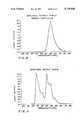
- This chemiluminescent light sourcewhile not specified in the application, has been an orange/yellow, also called amber, light having a wavelength profile as shown in FIG. 10. This device has been on sale for several years.
- the chemiluminescent light sourcein addition to providing a portable source of illumination of the body cavity, does so without producing any heat which could damage, or at least be uncomfortable to, the tissue in the body cavity which is being observed with the endoscopic instrument or by use of the speculum. Moreover, there is no requirement for any electrical source such as a power cord or batteries. This device also disburses light throughout the cavity being observed, rather than focuses light at a specified location.
- the entire instrumentmay be disposed of after use.
- those instrumentsmay be sterilized, and the chemiluminescent light source disposed of and then replaced, to obtain a completely sterile device.
- the endoscopedoes not have to be connected to or powered by any electrical source and can be stored for substantial periods of time without any loss of function because until the chemiluminescent light source is activated by the combination of the two chemiluminescent components, there is no loss of function.
- batteries which are used in the operation of standard electrical lights which are used in many prior art devicescan deteriorate in function even when not in use over a period of time, particularly under adverse conditions such as high heat and/or humidity.
- the present inventionis particularly useful in primitive locations where relatively high temperature and heat may be prevalent and long periods of storage may be required before the invention is used as an endoscope or speculum.
- the present inventiondoes have some anticipated shelf life and may, at times be somewhat temperature sensitive.
- a standard gynecological exam or gyn examcomprises the use of a speculum, a visual examination of the interior cavity and related structures, palpation of the pelvic region and a pap smear.
- the visual examinationis typically performed using a gooseneck lamp or even a flashlight without any use of magnification, although magnification is certainly available in the medical field in other areas, such as microsurgery.
- magnificationis certainly available in the medical field in other areas, such as microsurgery.
- the ability to visualize abnormalities or areas of concernis diminished since there is not evenly disbursed lighting and there may be shadows or glare which distort the appearance of the area.
- an external light sourceis used, the same problems occur.
- the light sourceis incandescent, it gets hot and cannot be placed too close to the patient without burning or uncomfortably heating the area being examined, or the area adjacent thereto.
- the various peaks of the wavelengths of the lightgenerally white light is not the most advantageous for viewing the various abnormalities to be detected. If the abnormalities are not detected visually, they may be detected by the Pap smear.
- a colposcopeThis device is a binocular microscope which is placed near the patient. A bright light, (blue/green filtered white incandescent light) is supplied. The operator looks through the eyepieces of the colposcope much like looking through field glasses. This procedure is performed with a vaginal speculum or similar device in place. Some of the colposcopes have camera attachments for still picture photography.
- the physicianwashes the area with 3-5% acetic acid and then exams the tissue for whitened areas after treatment.
- the acetic acidwhitens tissue which is thickened, such as cancer cells.
- the physicianalso looks for clusters of blood vessels which may indicate new growth such as cancer.
- colposcopyis a specialized procedure, requiring advanced and comprehensive training on very complicated and expensive apparatus, colposcopy is typically only performed on patients who have had an abnormal screening procedure (i.e. Pap smears or other indications). Such systems have been shown to be useful in the confirmation of Pap test results, as well as in other diagnostic procedures.
- Various forms and variations of colposcopesare disclosed in U.S. Pat. Nos. 3,994,288, 4,134,637, 4,232,933, 4,652,103 and 4,905,670.
- FIG. 1One prior art colposcope is described in Adair, U.S. Pat. No. 4,905,670, which discloses an apparatus which includes a vaginal speculum having a first fixed blade, a second blade mounted for pivotal movement toward and away from the fixed blade and spring means normally urging the second blade toward the fixed blade.
- the apparatusalso includes a video camera mounted on one of the blades for viewing the cervix, means providing light to the cervix, means for focusing the camera on a selected site on the cervix and means for providing a signal from the camera to a video screen for viewing the cervix and identifying lesions thereon.
- the light providing meanscan include a light carrier on the track for providing light to the cervix.
- meansis provided for selecting light for illumination of the cervix at any one of a range of light frequencies. This can be broad frequency light, monochromatic light or possibly even laser light for illumination. A particularly useful light frequency has been found to be from 200 nm through 1100 nm.
- a suitable means for stepping sequentially through the frequenciesis a monochromator. The monochromator converts light from a light source to a single frequency at an output in the form of a rectangular slit.
- a light carrieris provided which includes a bundle of optical fibers having a first end in a form of a rectangular collar for receiving the output from the monochromator and a circular collar at the other end for directing a round column of light onto the cervix.
- VanDerBel U.S. Pat. No. 4,597,383discloses a vaginal speculum having optical fiber illumination means attached thereto.
- Burgin U.S. Pat. No. 4,638,792has an adjustable speculum with an incorporated light system.
- Walsh U.S. Pat. No. 4,619,248discloses a light attachment for a speculum.
- Burgin U.S. Pat. No. 4,502,468has an adjustable speculum with an incorporated lighting system.
- Whitman U.S. Pat. No. 3,789,835discloses an illuminating attachment for vaginal speculum.
- Stafl U.S. Pat. No. 4,300,570discloses a diagnostic method including projecting and magnifying an image of a cervix photographed by a device disposed on a speculum.
- Tanikawa et al.U.S. Pat. No. 4,461,558 discloses an endoscopic photographing apparatus applicable to all types of endoscopes and uses therefor.
- the present inventionovercomes the drawbacks of the prior art by providing an endoscopic examination and viewing system that is compact, portable, disposable, shadowless, economical and efficient.
- the present inventionalso comprises a method of detection of various cellular abnormalities which is quicker, easier, more economical, simpler, more compact, and which can be performed in an office setting without the use of prior art colposcopy equipment.
- the present inventionis an endoscopic instrument comprising a housing and a chemiluminescent light source attached thereto.
- the endoscopic instrumentmay have optical enlargement lenses and means for spreading or otherwise retracting a portion of the cavity to be observed in order to place the diffused light within the cavity and to permit observation of the tissue therein.
- the chemiluminescent lightemits light in a particular frequency range obtained by the combination of particular chemiluminescent materials commercially distributed by American Cyanamid, Inc. (Wayne, N.J.) which, sometimes referred to as a cyalume light.
- the specific frequency peaks of the preferred embodiment of present inventionare at about 450 nm and 550 nm and a smaller peak in the red region at about 600 nm which in combination create, emanate or elicit a white light.
- the chemiluminescent light sourceis provided in a sealed container, as described in the above-referenced copending patent application, or otherwise, such that the chemicals can be easily mixed without having to specially handle or mix the chemicals by hand.
- a sealed containeras described in the above-referenced copending patent application, or otherwise, such that the chemicals can be easily mixed without having to specially handle or mix the chemicals by hand.
- Several different structuresare proposed in the copending application for this purpose including, but not limited to, providing a frangible container inside a flexible container so that the flexible container can be manipulated to break the container and allow the components to mix, and separating the components in a container by a breakable wall and use a plunger or other device or method to break the wall to allow the components to mix.
- the shape of the containercan be any conventional, or nonconventional shape, and the size of the container is preferably large enough to contain a sufficient amount of chemiluminescent material to light the interior of the cavity being examined, or at least a sufficient to provide light for the portion of said cavity to be examined.
- the light sourceshould be capable of being disposed within the cavity adjacent the area of the cavity to be examined.
- the diameter or width of the light sourceis adapted to be disposed on or in the endoscope if such arrangement is used.
- the light sourceis an elongated tube which can be inserted one end first into a cavity.
- chemiluminescent materialof proper reactivity for the light to remain lit for at least 2 minutes and as much as 30 minutes, although longer chemiluminescent light generation would certainly provide an operative system, and shorter duration may also work for certain types of examinations.
- the chemiluminescent light sourceis attached to one of the dilator blades of a clear plastic speculum or other endoscope with a means for opening and spreading the cavity to be observed.
- the clear plastic speculumWith the clear plastic speculum, the light transmits through the blades to illuminate the entire cavity being observed.
- the speculumdoes not have to be clear and solid metal speculae may be used as well.
- the light sourcemay be inserted directly into the cavity or may be disposed outside the cavity, the light being directed inward into the cavity by means of mirrors or fiber optic elements.
- the chemiluminescent light sourceprovides direct lighting to the examination surface in order to take advantage of the diffuse, non-directed light emanating from the container of the chemiluminescent material.
- the endoscopemay contain a magnification means for magnifying the cavity under illumination by the invented light in order to enlarge the examining practitioner's view of the surface.
- a magnification meansfor magnifying the cavity under illumination by the invented light in order to enlarge the examining practitioner's view of the surface.
- Various configurations of the magnification meansmay be employed including but not limited to a utilizing a separate magnification means and light source, or attaching the magnification means to the light source.
- the present inventionalso comprises a method of detection of various medical disorders by means of providing a transparent or translucent sealed container comprising separated chemiluminescent materials which when mixed together emit a chemiluminescent light in the range of blue-white, green white and green-blue-white, mixing the chemiluminescent materials together to cause said chemiluminescent materials to emit said light, directing said light to a surface of a body cavity and examining said lit surface.
- the surface of the cavity to be examinedis treated prior to examination with a composition which enhances the visualization of various tissue, such as by coating the surface with dilute aqueous acetic acid. Additionally a magnification means or scope may be used to enhance the examination.
- the presently invented methodcomprises coating the vaginal and/or cervical mucosal membranes with a solution of 1% to 10% acetic acid, mixing the aforesaid chemiluminescent materials together, attaching the container containing said chemiluminescent materials to a speculum, inserting the speculum into the vaginal passage so that chemiluminescent is directed onto the surface, providing a magnification means for magnifying the surface of the cavity and examining the surface for the occurrence of non-normal tissue.
- the speculumis preferably, but not necessarily, made of clear material so that the chemiluminescent light is transmitted through speculum and there will be no or minimized shadows in the cavity,
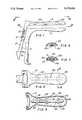
- FIG. 1is a side view of one embodiment of the present invention wherein the speculum is the endoscopic instrument
- FIG. 2is a top plan view of the upper dilator means of the present invention as taken through lines 2--2 in FIG. 1.
- FIG. 3is a bottom up view of the upper dilator means of the present invention as taken through lines 3--3 in FIG. 1.
- FIG. 4is an illustration of the preferred embodiment of the chemiluminescent light source of the present invention.
- FIG. 5is an illustration of an alternative embodiment of the chemiluminescent light source of the present invention.
- FIG. 6is a sectional view of the upper dilator blade of the embodiment of the present invention as shown in FIG. 2 and taken through lines 6--6 of FIG. 2.
- FIG. 7is a sectional view of an alternate embodiment of the upper dilator blade to the embodiment shown in FIG. 6.
- FIG. 8is a side view of a chemiluminescent light source disposed in a wand endoscope of the present invention.
- FIG. 9is an enlarged cross sectional view of the invention shown in FIG. 8 taken through lines 9--9 thereof.
- FIG. 10is a graph showing the profile of the wavelength of light from the prior art amber chemiluminescent light source.
- FIG. 11is a graph showing the profile of the wavelength of light from the present invention chemiluminescent light source.
- the present inventionis a method and apparatus for the examination of body cavities endoscopically which increases the likelihood of detection of many visually observable abnormalities, such as lesions, irregular vascularature, exophytic regions, ulcerations and other atypias of the cervix, vaginal cavity and other body cavities, as well as discharges.
- the apparatuscan take on any shape which may be usefully employed by insertion into the cavity to be examined, and generally a cylindrical shape corresponding to the shape and size of the cavity to be observed, although it is obviously smaller in diameter than the cavity in order to provide an area to enable the physician to view the tissue to be examined.
- Much of the detailed description which followsrelates specifically to a vaginal speculum.
- the inventionnot only is suitably employed in all speculae, but also is capable of serving as a light source for all endoscopic instruments for illumination and examination of the several body cavities including but not limited to the anal and oral cavities.
- FIG. 1one embodiment of the present invention is shown in which the chemiluminescent light source is attachable to a speculum.
- the instrument shown in FIGS. 1-3 and 6 and 7comprises a handle 2 connected to a lower dilator blade 4, and upper dilator blade 6 having a second handle 8.
- Handle 8is operatively connected to handle 2 in a slideably adjustable manner by means of slot 10 in handle 8 which engages around pivot pin 12 on handle 2.
- the upper dilator blade of the speculumis provided with extending locking tab 14, by means of which the physician can hold in position the remote ends of the two dilator blades separated within the vaginal cavity as the blades are pivoted with respect to one another at pivot pin 12.
- a plurality of rib-like members 22are located on the interior surface of upper dilator blade 6 intermediate the two dilator blades and have a channel 24 therein adapted to receive and retain therein, in a releasable, snap-fit, a medical examination light 26.
- Medical examination light 26produces light by chemiluminescent means and has an hermetically sealed, flexible light transmitting tube 28 with at least two compartments 30 and 32 separated by a breakable wall 34 with a chemiluminescent component in each compartment. In one compartment is the fluorescer component and in another is the activator component. Mixing of the components produces the chemiluminescent effect, as is well-known.
- Tube 28has a cross-sectional shape adapted to be received and retained in channel 24 in a releasable, snap-fitting engagement. Once in position and activated as described, medical examination light 26 directs its light intermediate the two dilator blades in their extended position to illuminate the cavity for the examining physician.
- FIGS. 6 and 7show two alternative retaining means for retaining the light in the speculum, which retaining means may be applied to any endoscopic instrument for use in accordance with the present invention.
- the upper dilator blade 6has a retaining means comprising a snap fit member 22 shaped to engage and retain the light 26 within the channel 24.
- the retaining means 22has a slot 23 which retains a flange 25 attached to the light 26.
- the lightis comprised of two compartments as described above.
- the inner compartment 42is breakable, such as a frangible ampoule, and contains one of the components, either the activator or the fluorescer.
- the outer compartment 44is partially or fully flexible, but is sturdy enough to resistant being cut or broken when the ampoule is broken. It contains the other component.
- the compartments 46 and 48are side by side with a breakable membrane 50 disposed between the two compartments to prevent mixture of the materials in the compartments until the membrane is broken.
- FIG. 8depicts a light source 52 of the present invention disposed in an endoscopic wand 54.
- FIG. 9shows a cross section of the light source in the wand with showing the inner compartment 55 within the outer compartment 57 and the entire light 52 being retained in the wand 54 by a plurality of detents 58.
- the light sourcemay alternatively be held in place by adhesive, tight fit, snap-fit or any other retaining means known in the art.
- FIG. 10shows the wavelength scan of the prior art amber light. As can be seen there is a single peak at about 600 nm. In the preferred embodiment of the present invention, the wavelength profile is as shown in FIG. 11. In FIG. 11, there are two major peaks at about 450 nm and 550 nm, and a minor peak at about 580 nm. This corresponds to green and blue light for the major peaks and red light for the minor peak.
- the combinationforms a chemiluminescent white light source with the advantages described herein.
- Such chemiluminescent light sourceis available from American Cyanimid (Wayne, N.J.)
- the method of the present inventioninvolves inspection of the surface to be examined using the invented endoscopic light comprising a chemiluminescent light source in the green and blue wavelength ranges described herein.
- the method and apparatus of the present inventionare designed to provide improved viewing of visual detectable anomalies in body cavities, such as vaginal exams, which together with necessary follow-up confirmatory diagnostic tests, is intended to provide significantly more accurate detection of problems in the region to be examined.
- body cavitiessuch as vaginal exams
- Acetowhite lesionwhich are thickened mucosal epithelium which reflect light upon examination following application of dilute acetic acid, and appear lighter or white when compared to the surrounding normal mucosa
- Leukoplakiawhich is thickened mucosal epitherlium which reflect light upon examination following application of dilute acetic acid, and appear lighter or white when compared to the surrounding normal mucosa; this appearance is not dependent upon the application of acetic acid.
- Condyloma/Human Papilloma Virus/Genital Wartsare wart-like verrucous, papillated mucosal lesions or growths which appear lighter or white after the application of dilute acetic acid (acetowhite).
- Dysplasia, Intra-Epithelial Neoplasia and Frank Carcinomais recognized by one or more lesions which may appear ulcerated or thickened, acetowhite, erythematous, may be raised or associated with an abnormal shape or pattern of microvascular such as punctated, mosaic like (mosaicism), erratic web-like shaped, or any combination thereof.
- Mucopurulenceis a yellow white exudate or discharge often found in cases of infectious microorganisms such as Chlamydia which elicit a leukocytic reaction.
- Ulcerationis recognized as a lesion of the mucosal, similar to Herpes Simplex Virus lesions, but not having the same diagnostic characteristics.
- Abnormal vascularizationwhich is indicative of possible cancerous or precancerous regions, and is recognized by the presence of excessive vascularization near the surface of the examined tissue.
- the physicianfirst activates the chemiluminescent light source by breaking the membrane or inner ampoule, and then attaches the ligth source to an endoscope, such as the speculum or wand described above.
- the physicianthen coats, sprays or paints the area to be examined with 1 to 10% acetic acid, and preferably 3 to 5% acetic acid.
- the cavityis then examined first under normal 1 ⁇ magnification, and then under higher magnification in the range of up to 10 ⁇ and preferably 3.5 to 5 ⁇ . Any necessary biopsies may be taken if the observation warrants follow-up testing.
- the purpose of the present inventionis to provide a tool to allow physicians to more accurately screen patients for various diseases, and to provide an indication of which patients require follow-up examinations.
- This inventive procedurecan be performed in the doctor's office with the patient present, without requiring the patient to wait for the results of the Pap smear which is usually performed over several days to a week at an outside laboratory.
- any follow upmay be accomplished or discussed immediately following detection of the problem, alleviating excessive patient concern. It also alleviates the problems which have been recognized relating to the tests being performed by outside laboratories without any control by the patient's personal physician.
- the results of the comparison of the two proceduresestablishes the accuracy of the present invention over the prior art method.
- the present inventionis not intended as a substitute for routine Pap smears; rather, it is intended to be used in conjunction with such tests to increase the accuracy of the screening procedure, adding a sensitive visual screening procedure to the existing cytological laboratory screening.
Landscapes
- Health & Medical Sciences (AREA)
- Life Sciences & Earth Sciences (AREA)
- Surgery (AREA)
- Nuclear Medicine, Radiotherapy & Molecular Imaging (AREA)
- Biomedical Technology (AREA)
- Optics & Photonics (AREA)
- Pathology (AREA)
- Radiology & Medical Imaging (AREA)
- Biophysics (AREA)
- Engineering & Computer Science (AREA)
- Physics & Mathematics (AREA)
- Heart & Thoracic Surgery (AREA)
- Medical Informatics (AREA)
- Molecular Biology (AREA)
- Animal Behavior & Ethology (AREA)
- General Health & Medical Sciences (AREA)
- Public Health (AREA)
- Veterinary Medicine (AREA)
- Endoscopes (AREA)
Abstract
Description
Claims (19)
Priority Applications (2)
| Application Number | Priority Date | Filing Date | Title |
|---|---|---|---|
| US07/660,674US5179938A (en) | 1983-02-17 | 1991-02-25 | Apparatus for endoscopic examination of body cavity using chemiluminescent light source |
| US07/991,444US5329938A (en) | 1983-02-17 | 1992-12-16 | Method for endoscopic examination of body cavity using chemilumine-scent light source |
Applications Claiming Priority (5)
| Application Number | Priority Date | Filing Date | Title |
|---|---|---|---|
| US58136383A | 1983-02-17 | 1983-02-17 | |
| US69409285A | 1985-01-23 | 1985-01-23 | |
| US2018887A | 1987-02-26 | 1987-02-26 | |
| US52677090A | 1990-05-18 | 1990-05-18 | |
| US07/660,674US5179938A (en) | 1983-02-17 | 1991-02-25 | Apparatus for endoscopic examination of body cavity using chemiluminescent light source |
Related Parent Applications (1)
| Application Number | Title | Priority Date | Filing Date |
|---|---|---|---|
| US52677090AContinuation-In-Part | 1983-02-17 | 1990-05-18 |
Related Child Applications (1)
| Application Number | Title | Priority Date | Filing Date |
|---|---|---|---|
| US07/991,444DivisionUS5329938A (en) | 1983-02-17 | 1992-12-16 | Method for endoscopic examination of body cavity using chemilumine-scent light source |
Publications (1)
| Publication Number | Publication Date |
|---|---|
| US5179938Atrue US5179938A (en) | 1993-01-19 |
Family
ID=27533862
Family Applications (2)
| Application Number | Title | Priority Date | Filing Date |
|---|---|---|---|
| US07/660,674Expired - LifetimeUS5179938A (en) | 1983-02-17 | 1991-02-25 | Apparatus for endoscopic examination of body cavity using chemiluminescent light source |
| US07/991,444Expired - LifetimeUS5329938A (en) | 1983-02-17 | 1992-12-16 | Method for endoscopic examination of body cavity using chemilumine-scent light source |
Family Applications After (1)
| Application Number | Title | Priority Date | Filing Date |
|---|---|---|---|
| US07/991,444Expired - LifetimeUS5329938A (en) | 1983-02-17 | 1992-12-16 | Method for endoscopic examination of body cavity using chemilumine-scent light source |
Country Status (1)
| Country | Link |
|---|---|
| US (2) | US5179938A (en) |
Cited By (81)
| Publication number | Priority date | Publication date | Assignee | Title |
|---|---|---|---|---|
| US5329937A (en)* | 1992-09-10 | 1994-07-19 | Nova Healthcare Industries, Inc. | Speculum protector with smoke tube |
| WO1995013009A1 (en)* | 1993-11-10 | 1995-05-18 | Advanced Medical Devices Incorporated | Illuminated vaginal speculum |
| US5601549A (en)* | 1994-11-17 | 1997-02-11 | Machida Endoscope Co., Ltd. | Medical observing instrument |
| US5702887A (en)* | 1993-03-19 | 1997-12-30 | Chiron Diagnostics Corporation | Long emission wavelength chemiluminescent compounds and their use in test assays |
| WO1998004183A3 (en)* | 1996-07-25 | 1998-03-26 | Light Medicine Inc | Photodynamic therapy with light emitting particles |
| US5800350A (en)* | 1993-11-01 | 1998-09-01 | Polartechnics, Limited | Apparatus for tissue type recognition |
| US5989184A (en)* | 1997-04-04 | 1999-11-23 | Medtech Research Corporation | Apparatus and method for digital photography useful in cervical cancer detection |
| US6088612A (en)* | 1997-04-04 | 2000-07-11 | Medtech Research Corporation | Method and apparatus for reflective glare removal in digital photography useful in cervical cancer detection |
| US6162172A (en)* | 1998-01-30 | 2000-12-19 | Edwards Lifesciences Corporation | Methods and apparatus for retracting tissue |
| US6277067B1 (en) | 1997-04-04 | 2001-08-21 | Kerry L. Blair | Method and portable colposcope useful in cervical cancer detection |
| US20020096643A1 (en)* | 1996-02-08 | 2002-07-25 | Bright Solutions, Inc., A Michigan Corporation | Portable light source and system for use in leak detection |
| US6447490B1 (en)* | 1997-08-07 | 2002-09-10 | James Zhou Liu | Vagina cleaning system for preventing pregnancy and sexually transmitted diseases |
| US6496718B1 (en) | 2000-05-12 | 2002-12-17 | The Trylon Corporation | Body cavity light using diffuse light source |
| US20030199737A1 (en)* | 2001-10-09 | 2003-10-23 | Richard Deslauriers | Inflatable speculums |
| WO2003075979A3 (en)* | 2002-03-06 | 2003-12-11 | James S Simon | Chemiluminescently illuminated medical appliances |
| US20040210114A1 (en)* | 2003-04-16 | 2004-10-21 | Simon James S. | Airway products having leds |
| US20050014145A1 (en)* | 2000-09-26 | 2005-01-20 | Burkett Douglas D. | Light-directed molecular analysis for cancer prognosis and diagnosis |
| WO2005032357A1 (en)* | 2003-10-09 | 2005-04-14 | Garry Felix Heynen | A surgical instrument |
| EP0967912A4 (en)* | 1997-01-24 | 2005-06-15 | Spectra Science Corp | Chemiluminescent sources for photodynamic therapy and photomedicine |
| WO2006041458A1 (en)* | 2004-09-30 | 2006-04-20 | Zila Biotechnology, Inc. | Light-directed method for detecting and aiding further evaluation of abnormal mucosal tissue |
| US20060177793A1 (en)* | 2005-02-07 | 2006-08-10 | Bobby Crohn | Intraoral illumination device |
| US20060224043A1 (en)* | 2005-04-04 | 2006-10-05 | Guinan William P | Vaginal speculum |
| US20060241501A1 (en)* | 2004-09-28 | 2006-10-26 | Zila Pharmaceuticals, Inc. | Method and apparatus for detecting abnormal epithelial tissue |
| US20060256575A1 (en)* | 2005-05-13 | 2006-11-16 | Spotlight Surgical, Inc. | Body cavity illumination system |
| US20070100211A1 (en)* | 2005-11-02 | 2007-05-03 | Depuy Spine, Inc. | Illuminated surgical access system including a surgical access device and coupled light emitter |
| US20070230164A1 (en)* | 2006-04-03 | 2007-10-04 | Welch Allyn, Inc. | Vaginal speculum assembly having portable illuminator |
| US20070230167A1 (en)* | 2006-04-03 | 2007-10-04 | Welch Allyn, Inc. | Power connections and interface for compact illuminator assembly |
| DE102007001925A1 (en)* | 2007-01-12 | 2008-07-24 | Siemens Ag | Endoscopic device e.g. endoscopic capsule, for insertion into e.g. gastrointestinal tract, of human body, has illumination device comprising light source e.g. LED, which is based on and/or operable by peroxyoxalate chemiluminescence |
| US20080186694A1 (en)* | 2007-02-01 | 2008-08-07 | Chemical Light, Inc. | Novelty glow spike |
| US20080228038A1 (en)* | 2005-04-01 | 2008-09-18 | Welch Allyn, Inc. | Illumination Assembly For Use With Vaginal Speculum Apparatus |
| US20080255462A1 (en)* | 2004-09-28 | 2008-10-16 | Zila Pharmaceuticals, Inc. | Light stick |
| EP1793727A4 (en)* | 2004-09-28 | 2009-01-07 | Zila Pharm Inc | Methods for detecting abnormal epithelial tissue |
| US20090076334A1 (en)* | 2007-09-19 | 2009-03-19 | Tien-Sheng Chen | Illuminated Vaginal Speculum and Illumination Device |
| WO2006047414A3 (en)* | 2004-10-25 | 2009-04-09 | Healthshine Inc | Chemiluminescent phototherapy device |
| US20090118624A1 (en)* | 2004-09-28 | 2009-05-07 | Zila Pharmaceuticals, Inc. | Device for oral cavity examination |
| US7551406B1 (en)* | 2005-07-01 | 2009-06-23 | Western Digital (Fremont), Llc | Dual electrical lapping guides with common bonding pad |
| US7554767B1 (en) | 2005-07-01 | 2009-06-30 | Western Digital (Fremont), Llc | Electrical lapping guide disposed laterally relative to a shield pedestal |
| US20090198108A1 (en)* | 2008-01-31 | 2009-08-06 | Tien-Sheng Chen | Cervix Examination Device and Cervix Examination Set |
| US20090287192A1 (en)* | 2005-04-01 | 2009-11-19 | Vivenzio Robert L | Medical diagnostic instrument having portable illuminator |
| US20090312610A1 (en)* | 2008-06-13 | 2009-12-17 | Obp Corporation | Disposable Speculums Having Single-Sided Support and Operating Mechanism |
| US20110004067A1 (en)* | 2006-01-04 | 2011-01-06 | Connie Marchek | Surgical Retractors and Methods of Minimally Invasive Surgery |
| US20110112408A1 (en)* | 2009-11-10 | 2011-05-12 | Lior Greenstein | Optical Speculum |
| US20110124965A1 (en)* | 2008-05-08 | 2011-05-26 | Park Jason Y | Chemiluminescence enhanced detection |
| US20110160540A1 (en)* | 2009-12-29 | 2011-06-30 | Michelle Davis Smith | Vaginal Speculum |
| US20110184272A1 (en)* | 2008-06-13 | 2011-07-28 | Kun Zeng | Portable Diagnostic Device for Precancerous Lesion of Cervical Cancer |
| US20110213207A1 (en)* | 2006-01-05 | 2011-09-01 | Depuy Spine, Inc. | Non-rigid surgical retractor |
| US20130006334A1 (en)* | 2010-02-04 | 2013-01-03 | Mauro Galli | Device for the treatment of the vaginal canal and relevant equipment |
| CN103027751A (en)* | 2011-10-09 | 2013-04-10 | 汤维波 | Fluorophor positioning device for jointly positioning and excising diseased region by gastrointestinal endoscope and peritoneoscope |
| US8550995B2 (en) | 2006-01-04 | 2013-10-08 | DePuy Synthes Products, LLC | Surgical access devices and methods of minimally invasive surgery |
| US8602984B2 (en) | 2003-12-18 | 2013-12-10 | DePuy Synthes Products, LLC | Surgical retractor systems and illuminated cannulae |
| US20140073858A1 (en)* | 2012-09-11 | 2014-03-13 | Danny A. Sherwinter | Gastric Bougie/Dilator with Integral Lighted Tip |
| US9271640B2 (en) | 2009-11-10 | 2016-03-01 | Illumigyn Ltd. | Optical speculum |
| US20160354072A1 (en)* | 2015-06-03 | 2016-12-08 | Obp Medical | Retractor |
| US9532706B2 (en) | 2014-08-07 | 2017-01-03 | Welch Allyn, Inc. | Vaginal speculum with illuminator |
| US9629741B2 (en) | 2014-07-18 | 2017-04-25 | Covidien Lp | Gastric tubes and methods of use |
| US9655758B2 (en) | 2013-11-11 | 2017-05-23 | Covidien Lp | Devices and methods facilitating sleeve gastrectomy procedures |
| US20170189004A1 (en)* | 2015-06-03 | 2017-07-06 | Obp Medical | Retractor |
| US9801748B2 (en) | 2013-11-08 | 2017-10-31 | Covidien Lp | Devices and methods for facilitating sleeve gastrectomy procedures |
| US9867602B2 (en) | 2015-02-05 | 2018-01-16 | Obp Medical Corporation | Illuminated surgical retractor |
| US9877644B2 (en) | 2009-11-10 | 2018-01-30 | Illumigyn Ltd. | Optical speculum |
| US9913577B2 (en) | 2010-09-28 | 2018-03-13 | Obp Medical Corporation | Speculum |
| US10149602B2 (en) | 2011-07-11 | 2018-12-11 | Ambu A/S | Endobronchial tube with integrated image sensor and a cleaning nozzle arrangement |
| US10159425B2 (en) | 2013-11-08 | 2018-12-25 | Covidien Lp | Devices and methods facilitating sleeve gastrectomy and other procedures |
| US10245402B2 (en) | 2011-07-11 | 2019-04-02 | Ambu A/S | Endobronchial tube with integrated image sensor |
| US10278572B1 (en) | 2017-10-19 | 2019-05-07 | Obp Medical Corporation | Speculum |
| US10420538B2 (en) | 2015-02-05 | 2019-09-24 | Obp Medical Corporation | Illuminated surgical retractor |
| US10512519B2 (en) | 2018-02-20 | 2019-12-24 | Obp Medical Corporation | Illuminated medical devices |
| WO2020051143A1 (en)* | 2018-09-04 | 2020-03-12 | Cyalume Technologies, Inc. | Vaginal speculum |
| US10687793B2 (en) | 2017-07-18 | 2020-06-23 | Obp Medical Corporation | Minimally invasive no touch (MINT) procedure for harvesting the great saphenous vein (GSV) and venous hydrodissector and retractor for use during the MINT procedure |
| US10687699B2 (en) | 2017-03-17 | 2020-06-23 | CEEK Enterprises | Lighting module for a medical device and methods for using the same |
| US10722621B2 (en) | 2016-07-11 | 2020-07-28 | Obp Medical Corporation | Illuminated suction device |
| US10799229B2 (en) | 2018-02-20 | 2020-10-13 | Obp Medical Corporation | Illuminated medical devices |
| USD904607S1 (en) | 2019-05-07 | 2020-12-08 | Obp Medical Corporation | Nasal retractor |
| USD911521S1 (en) | 2019-02-19 | 2021-02-23 | Obp Medical Corporation | Handle for medical devices including surgical retractors |
| US10939899B2 (en) | 2015-06-03 | 2021-03-09 | Obp Medical Corporation | End cap assembly for retractor and other medical devices |
| US10959609B1 (en) | 2020-01-31 | 2021-03-30 | Obp Medical Corporation | Illuminated suction device |
| US10966702B1 (en) | 2020-02-25 | 2021-04-06 | Obp Medical Corporation | Illuminated dual-blade retractor |
| US11399914B2 (en)* | 2017-08-09 | 2022-08-02 | Alcon Inc. | Self-illuminating microsurgical cannula device |
| USD963908S1 (en) | 2017-03-24 | 2022-09-13 | Ceek Women's Health, Inc. | Medical device lighting module |
| US11992193B2 (en) | 2017-08-07 | 2024-05-28 | Maxwell WEINMANN | Laryngoscope |
| US12318080B2 (en) | 2023-07-21 | 2025-06-03 | Coopersurgical, Inc. | Illuminated surgical retractor capable of hand-held operation and of being mounted to a fixed frame |
Families Citing this family (21)
| Publication number | Priority date | Publication date | Assignee | Title |
|---|---|---|---|---|
| DE69838560T2 (en)* | 1997-10-20 | 2008-08-14 | The Board of Regents, The University of Texas System, Austin | ACETIC ACID AS A SIGNAL IMPROVED CONTRASTANT IN FLUORESCENT SPECTROSCOPY |
| NL1013958C2 (en)* | 1999-12-24 | 2001-06-26 | Bernhard Wilhelm Geziena Nicol | Vagina speculum. |
| CA2479525A1 (en)* | 2002-04-02 | 2003-10-16 | Seedling Enterprises, Llc | Apparatus and methods using visible light for debilitating and/or killing microorganisms within the body |
| US7255691B2 (en)* | 2002-04-16 | 2007-08-14 | Lumerx Inc. | Chemiluminescent light source using visible light for biotherapy |
| US8376942B2 (en) | 2005-05-06 | 2013-02-19 | Welch Allyn, Inc. | Articulation mechanism for a vaginal speculum |
| JP2009512174A (en)* | 2005-10-17 | 2009-03-19 | デントゾーン カンパニー リミテッド | Oral lighting device |
| US20070191675A1 (en)* | 2006-02-13 | 2007-08-16 | Joel Gerardo Diaz Sanchez | Actinic light colposcope and method to detect lesions in the lower female genital tract produced by human papilloma virus using an actinic light colposcope |
| US8795197B2 (en) | 2007-07-17 | 2014-08-05 | Histologics, LLC | Frictional trans-epithelial tissue disruption collection apparatus and method of inducing an immune response |
| WO2009012392A1 (en) | 2007-07-17 | 2009-01-22 | Neal Marc Lonky | Frictional trans-epithelial tissue disruption and collection apparatus and method of inducing and/or augmenting an immune response |
| WO2010006513A1 (en)* | 2008-07-15 | 2010-01-21 | Zeng Kun | Portable diagnostic equipment for precancerous lesion of cervical cancer |
| US20100256125A1 (en)* | 2009-04-06 | 2010-10-07 | Zila Pharmaceuticals, Inc. | Use of improved toluidine blue in photodynamic therapy |
| US7991260B2 (en)* | 2009-05-05 | 2011-08-02 | Sensormed, Inc. | Light-diffusing safety cap |
| US20100284665A1 (en)* | 2009-05-05 | 2010-11-11 | Doody Michael C | Light Diffusing Safety Cap |
| US20110120459A1 (en)* | 2009-11-20 | 2011-05-26 | Omniglow Llc | Illuminated endotracheal stylet |
| US9044213B1 (en) | 2010-03-26 | 2015-06-02 | Histologics, LLC | Frictional tissue sampling and collection method and device |
| US9289114B2 (en)* | 2010-07-30 | 2016-03-22 | Nilesh R. Vasan | Disposable, self-contained laryngoscope and method of using same |
| US9307897B2 (en) | 2010-09-28 | 2016-04-12 | Obp Corporation | Disposable speculum having lateral stabilizing mechanism |
| US8696563B2 (en)* | 2011-11-17 | 2014-04-15 | Lexion Medical, Llc | Device and method for illumination of vaginal fornix with ureter location, isolation and protection during hysterectomy procedure |
| US10201332B1 (en) | 2012-12-03 | 2019-02-12 | Healoe Llc | Device and method of orienting a biopsy device on epithelial tissue |
| US11013466B2 (en) | 2016-01-28 | 2021-05-25 | Healoe, Llc | Device and method to control and manipulate a catheter |
| USD876625S1 (en) | 2018-08-07 | 2020-02-25 | Adroit Surgical, Llc | Laryngoscope |
Citations (5)
| Publication number | Priority date | Publication date | Assignee | Title |
|---|---|---|---|---|
| US3576987A (en)* | 1968-11-07 | 1971-05-04 | American Cyanamid Co | Chemical lighting device to store, initiate and display chemical light |
| US3769968A (en)* | 1971-11-22 | 1973-11-06 | L Blount | Speculum |
| US3789835A (en)* | 1972-03-16 | 1974-02-05 | R Whitman | Illuminating attachments for vaginal speculum |
| US4248214A (en)* | 1979-05-22 | 1981-02-03 | Robert S. Kish | Illuminated urethral catheter |
| US4905670A (en)* | 1988-12-28 | 1990-03-06 | Adair Edwin Lloyd | Apparatus for cervical videoscopy |
Family Cites Families (1)
| Publication number | Priority date | Publication date | Assignee | Title |
|---|---|---|---|---|
| DE1801135A1 (en)* | 1968-10-04 | 1970-04-16 | Maschf Augsburg Nuernberg Ag | Cylinder cover for liquid-cooled internal combustion engines |
- 1991
- 1991-02-25USUS07/660,674patent/US5179938A/ennot_activeExpired - Lifetime
- 1992
- 1992-12-16USUS07/991,444patent/US5329938A/ennot_activeExpired - Lifetime
Patent Citations (5)
| Publication number | Priority date | Publication date | Assignee | Title |
|---|---|---|---|---|
| US3576987A (en)* | 1968-11-07 | 1971-05-04 | American Cyanamid Co | Chemical lighting device to store, initiate and display chemical light |
| US3769968A (en)* | 1971-11-22 | 1973-11-06 | L Blount | Speculum |
| US3789835A (en)* | 1972-03-16 | 1974-02-05 | R Whitman | Illuminating attachments for vaginal speculum |
| US4248214A (en)* | 1979-05-22 | 1981-02-03 | Robert S. Kish | Illuminated urethral catheter |
| US4905670A (en)* | 1988-12-28 | 1990-03-06 | Adair Edwin Lloyd | Apparatus for cervical videoscopy |
Cited By (167)
| Publication number | Priority date | Publication date | Assignee | Title |
|---|---|---|---|---|
| US5329937A (en)* | 1992-09-10 | 1994-07-19 | Nova Healthcare Industries, Inc. | Speculum protector with smoke tube |
| US5702887A (en)* | 1993-03-19 | 1997-12-30 | Chiron Diagnostics Corporation | Long emission wavelength chemiluminescent compounds and their use in test assays |
| US5879894A (en)* | 1993-03-19 | 1999-03-09 | Chiron Diagnostics Corporation | Long emission wavelength chemiluminescent compounds and their use in test assays |
| US5800350A (en)* | 1993-11-01 | 1998-09-01 | Polartechnics, Limited | Apparatus for tissue type recognition |
| WO1995013009A1 (en)* | 1993-11-10 | 1995-05-18 | Advanced Medical Devices Incorporated | Illuminated vaginal speculum |
| US5465709A (en)* | 1993-11-10 | 1995-11-14 | Advanced Medical Products Inc. | Illuminated vaginal speculum |
| US5601549A (en)* | 1994-11-17 | 1997-02-11 | Machida Endoscope Co., Ltd. | Medical observing instrument |
| US20020096643A1 (en)* | 1996-02-08 | 2002-07-25 | Bright Solutions, Inc., A Michigan Corporation | Portable light source and system for use in leak detection |
| WO1998004183A3 (en)* | 1996-07-25 | 1998-03-26 | Light Medicine Inc | Photodynamic therapy with light emitting particles |
| EP0967912A4 (en)* | 1997-01-24 | 2005-06-15 | Spectra Science Corp | Chemiluminescent sources for photodynamic therapy and photomedicine |
| US5989184A (en)* | 1997-04-04 | 1999-11-23 | Medtech Research Corporation | Apparatus and method for digital photography useful in cervical cancer detection |
| US6277067B1 (en) | 1997-04-04 | 2001-08-21 | Kerry L. Blair | Method and portable colposcope useful in cervical cancer detection |
| US6088612A (en)* | 1997-04-04 | 2000-07-11 | Medtech Research Corporation | Method and apparatus for reflective glare removal in digital photography useful in cervical cancer detection |
| US6447490B1 (en)* | 1997-08-07 | 2002-09-10 | James Zhou Liu | Vagina cleaning system for preventing pregnancy and sexually transmitted diseases |
| US6162172A (en)* | 1998-01-30 | 2000-12-19 | Edwards Lifesciences Corporation | Methods and apparatus for retracting tissue |
| US6496718B1 (en) | 2000-05-12 | 2002-12-17 | The Trylon Corporation | Body cavity light using diffuse light source |
| US20050014145A1 (en)* | 2000-09-26 | 2005-01-20 | Burkett Douglas D. | Light-directed molecular analysis for cancer prognosis and diagnosis |
| US20030199737A1 (en)* | 2001-10-09 | 2003-10-23 | Richard Deslauriers | Inflatable speculums |
| US7041056B2 (en) | 2001-10-09 | 2006-05-09 | Deslauriers Richard J | Inflatable speculums |
| EP1463833A4 (en)* | 2001-12-14 | 2006-06-07 | Zila Inc | Light-directed molecular analysis for cancer prognosis and diagnosis |
| WO2003075979A3 (en)* | 2002-03-06 | 2003-12-11 | James S Simon | Chemiluminescently illuminated medical appliances |
| US20040210114A1 (en)* | 2003-04-16 | 2004-10-21 | Simon James S. | Airway products having leds |
| US20050039754A1 (en)* | 2003-04-16 | 2005-02-24 | Simon James S. | Airway products having LEDs |
| US7052456B2 (en) | 2003-04-16 | 2006-05-30 | Simon James S | Airway products having LEDs |
| WO2005032357A1 (en)* | 2003-10-09 | 2005-04-14 | Garry Felix Heynen | A surgical instrument |
| US8602984B2 (en) | 2003-12-18 | 2013-12-10 | DePuy Synthes Products, LLC | Surgical retractor systems and illuminated cannulae |
| US10869657B2 (en) | 2003-12-18 | 2020-12-22 | DePuy Synthes Products, Inc. | Surgical retractor systems and illuminated cannulae |
| US8622897B2 (en) | 2003-12-18 | 2014-01-07 | DePuy Synthes Products, LLC | Surgical methods and surgical kits |
| EP1793727A4 (en)* | 2004-09-28 | 2009-01-07 | Zila Pharm Inc | Methods for detecting abnormal epithelial tissue |
| US20080255462A1 (en)* | 2004-09-28 | 2008-10-16 | Zila Pharmaceuticals, Inc. | Light stick |
| US20060241501A1 (en)* | 2004-09-28 | 2006-10-26 | Zila Pharmaceuticals, Inc. | Method and apparatus for detecting abnormal epithelial tissue |
| US20090118624A1 (en)* | 2004-09-28 | 2009-05-07 | Zila Pharmaceuticals, Inc. | Device for oral cavity examination |
| WO2006041458A1 (en)* | 2004-09-30 | 2006-04-20 | Zila Biotechnology, Inc. | Light-directed method for detecting and aiding further evaluation of abnormal mucosal tissue |
| US20090124897A1 (en)* | 2004-09-30 | 2009-05-14 | Zila Biotechnology, Inc. | Light-Directed Method for Detecting and Aiding Further Evaluation of Abnormal Mucosal Tissue |
| WO2006047414A3 (en)* | 2004-10-25 | 2009-04-09 | Healthshine Inc | Chemiluminescent phototherapy device |
| US7153131B2 (en) | 2005-02-07 | 2006-12-26 | Crohn Enterprises Ltd. | Intraoral illumination device |
| US20060177793A1 (en)* | 2005-02-07 | 2006-08-10 | Bobby Crohn | Intraoral illumination device |
| US8821395B2 (en) | 2005-04-01 | 2014-09-02 | Welch Allyn, Inc. | Vaginal speculum apparatus |
| US9883792B2 (en) | 2005-04-01 | 2018-02-06 | Welch Allyn, Inc. | Vaginal speculum apparatus |
| US20140148653A1 (en)* | 2005-04-01 | 2014-05-29 | Welch Allyn, Inc. | Vaginal speculum apparatus |
| US11291359B2 (en)* | 2005-04-01 | 2022-04-05 | Welch Allyn, Inc. | Vaginal speculum apparatus |
| US9332898B2 (en)* | 2005-04-01 | 2016-05-10 | Welch Allyn, Inc. | Vaginal speculum apparatus |
| US10376138B2 (en) | 2005-04-01 | 2019-08-13 | Welch Allyn, Inc. | Vaginal speculum apparatus |
| US8157728B2 (en) | 2005-04-01 | 2012-04-17 | Welch Allyn, Inc. | Vaginal speculum |
| US12262878B2 (en) | 2005-04-01 | 2025-04-01 | Welch Allyn, Inc. | Vaginal speculum apparatus |
| US9949633B2 (en)* | 2005-04-01 | 2018-04-24 | Welch Allyn, Inc. | Vaginal speculum apparatus |
| US8388523B2 (en) | 2005-04-01 | 2013-03-05 | Welch Allyn, Inc. | Medical diagnostic instrument having portable illuminator |
| US8435175B2 (en) | 2005-04-01 | 2013-05-07 | Welch Allyn, Inc. | Vaginal speculum apparatus |
| US20080228038A1 (en)* | 2005-04-01 | 2008-09-18 | Welch Allyn, Inc. | Illumination Assembly For Use With Vaginal Speculum Apparatus |
| US20090287192A1 (en)* | 2005-04-01 | 2009-11-19 | Vivenzio Robert L | Medical diagnostic instrument having portable illuminator |
| US20170172404A1 (en)* | 2005-04-01 | 2017-06-22 | Welch Allyn, Inc. | Vaginal speculum apparatus |
| US20060224043A1 (en)* | 2005-04-04 | 2006-10-05 | Guinan William P | Vaginal speculum |
| US7384393B2 (en)* | 2005-04-04 | 2008-06-10 | Guinan William P | Vaginal speculum |
| US10912453B2 (en)* | 2005-05-13 | 2021-02-09 | Invuity, Inc. | Body cavity illumination system |
| US20060256575A1 (en)* | 2005-05-13 | 2006-11-16 | Spotlight Surgical, Inc. | Body cavity illumination system |
| US8684577B2 (en)* | 2005-05-13 | 2014-04-01 | Invuity, Inc. | Body cavity illumination system |
| US11903567B2 (en) | 2005-05-13 | 2024-02-20 | Invuity, Inc. | Body cavity illumination system |
| US7554767B1 (en) | 2005-07-01 | 2009-06-30 | Western Digital (Fremont), Llc | Electrical lapping guide disposed laterally relative to a shield pedestal |
| US7551406B1 (en)* | 2005-07-01 | 2009-06-23 | Western Digital (Fremont), Llc | Dual electrical lapping guides with common bonding pad |
| US7874982B2 (en)* | 2005-11-02 | 2011-01-25 | Depuy Spine, Inc. | Illuminated surgical access system including a surgical access device and coupled light emitter |
| US20110021882A1 (en)* | 2005-11-02 | 2011-01-27 | Sean Selover | Illuminated surgical access system including a surgical access device and coupled light emitter |
| US20070100211A1 (en)* | 2005-11-02 | 2007-05-03 | Depuy Spine, Inc. | Illuminated surgical access system including a surgical access device and coupled light emitter |
| US9005118B2 (en) | 2005-11-02 | 2015-04-14 | DePuy Synthes Products, LLC | Illuminated surgical access system including a surgical access device and coupled light emitter |
| US8550995B2 (en) | 2006-01-04 | 2013-10-08 | DePuy Synthes Products, LLC | Surgical access devices and methods of minimally invasive surgery |
| US20110004067A1 (en)* | 2006-01-04 | 2011-01-06 | Connie Marchek | Surgical Retractors and Methods of Minimally Invasive Surgery |
| US8517935B2 (en) | 2006-01-04 | 2013-08-27 | DePuy Synthes Products, LLC | Surgical retractors and methods of minimally invasive surgery |
| US20110213207A1 (en)* | 2006-01-05 | 2011-09-01 | Depuy Spine, Inc. | Non-rigid surgical retractor |
| US9254126B2 (en) | 2006-01-05 | 2016-02-09 | DePuy Synthes Products, Inc. | Non-rigid surgical retractor |
| US20070230167A1 (en)* | 2006-04-03 | 2007-10-04 | Welch Allyn, Inc. | Power connections and interface for compact illuminator assembly |
| US20070230164A1 (en)* | 2006-04-03 | 2007-10-04 | Welch Allyn, Inc. | Vaginal speculum assembly having portable illuminator |
| US7758203B2 (en) | 2006-04-03 | 2010-07-20 | Welch Allyn, Inc. | Power connections and interface for compact illuminator assembly |
| US8142352B2 (en) | 2006-04-03 | 2012-03-27 | Welch Allyn, Inc. | Vaginal speculum assembly having portable illuminator |
| WO2007117804A3 (en)* | 2006-04-07 | 2008-02-14 | Zila Pharm Inc | Method and apparatus for detecting abnormal epithelial tissue |
| DE102007001925B4 (en)* | 2007-01-12 | 2010-04-01 | Siemens Ag | endoscopy capsule |
| DE102007001925A1 (en)* | 2007-01-12 | 2008-07-24 | Siemens Ag | Endoscopic device e.g. endoscopic capsule, for insertion into e.g. gastrointestinal tract, of human body, has illumination device comprising light source e.g. LED, which is based on and/or operable by peroxyoxalate chemiluminescence |
| US7438428B2 (en)* | 2007-02-01 | 2008-10-21 | Filtrex Holdings Pte, Ltd. | Novelty glow spike |
| US20080186694A1 (en)* | 2007-02-01 | 2008-08-07 | Chemical Light, Inc. | Novelty glow spike |
| US20090076334A1 (en)* | 2007-09-19 | 2009-03-19 | Tien-Sheng Chen | Illuminated Vaginal Speculum and Illumination Device |
| US20090198108A1 (en)* | 2008-01-31 | 2009-08-06 | Tien-Sheng Chen | Cervix Examination Device and Cervix Examination Set |
| US20110124965A1 (en)* | 2008-05-08 | 2011-05-26 | Park Jason Y | Chemiluminescence enhanced detection |
| US20090312610A1 (en)* | 2008-06-13 | 2009-12-17 | Obp Corporation | Disposable Speculums Having Single-Sided Support and Operating Mechanism |
| US20110184272A1 (en)* | 2008-06-13 | 2011-07-28 | Kun Zeng | Portable Diagnostic Device for Precancerous Lesion of Cervical Cancer |
| US8096945B2 (en) | 2008-06-13 | 2012-01-17 | Obp Corporation | Disposable speculums having single-sided support and operating mechanism |
| US9314162B2 (en) | 2009-11-10 | 2016-04-19 | Illumigyn Ltd. | Optical speculum |
| US10694954B2 (en) | 2009-11-10 | 2020-06-30 | Illumigyn Ltd. | Optical speculum |
| US9259157B2 (en) | 2009-11-10 | 2016-02-16 | Illumigyn Ltd. | Optical speculum |
| US9877644B2 (en) | 2009-11-10 | 2018-01-30 | Illumigyn Ltd. | Optical speculum |
| US9968262B2 (en) | 2009-11-10 | 2018-05-15 | Illumigyn Ltd. | Optical speculum |
| US9603528B2 (en) | 2009-11-10 | 2017-03-28 | Illumigyn Ltd. | Optical speculum |
| US9603529B2 (en) | 2009-11-10 | 2017-03-28 | Illumigyn Ltd. | Optical speculum |
| US9629556B2 (en) | 2009-11-10 | 2017-04-25 | Illumigyn Ltd. | Optical speculum |
| US8638995B2 (en) | 2009-11-10 | 2014-01-28 | Illumigyn Ltd. | Optical speculum |
| US10188298B2 (en) | 2009-11-10 | 2019-01-29 | Illumigyn Ltd. | Optical speculum |
| US9271640B2 (en) | 2009-11-10 | 2016-03-01 | Illumigyn Ltd. | Optical speculum |
| US20110112408A1 (en)* | 2009-11-10 | 2011-05-12 | Lior Greenstein | Optical Speculum |
| US8858431B2 (en) | 2009-12-29 | 2014-10-14 | Michelle Davis Smith | Vaginal speculum |
| US20110160540A1 (en)* | 2009-12-29 | 2011-06-30 | Michelle Davis Smith | Vaginal Speculum |
| US20130006334A1 (en)* | 2010-02-04 | 2013-01-03 | Mauro Galli | Device for the treatment of the vaginal canal and relevant equipment |
| US10368733B2 (en) | 2010-09-28 | 2019-08-06 | Obp Medical Corporation | Speculum |
| US11744454B2 (en) | 2010-09-28 | 2023-09-05 | Obp Medical Corporation | Speculum |
| US9913577B2 (en) | 2010-09-28 | 2018-03-13 | Obp Medical Corporation | Speculum |
| US10888679B2 (en) | 2011-07-11 | 2021-01-12 | Ambu A/S | Endobronchial tube with integrated image sensor |
| US10149602B2 (en) | 2011-07-11 | 2018-12-11 | Ambu A/S | Endobronchial tube with integrated image sensor and a cleaning nozzle arrangement |
| US10406309B2 (en) | 2011-07-11 | 2019-09-10 | Ambu A/S | Endobronchial tube with integrated image sensor and a cleaning nozzle arrangement |
| US10245402B2 (en) | 2011-07-11 | 2019-04-02 | Ambu A/S | Endobronchial tube with integrated image sensor |
| CN103027751A (en)* | 2011-10-09 | 2013-04-10 | 汤维波 | Fluorophor positioning device for jointly positioning and excising diseased region by gastrointestinal endoscope and peritoneoscope |
| EP2895233A4 (en)* | 2012-09-11 | 2016-04-20 | Cook Medical Technologies Llc | GASTRIC CANDLE / DILATOR WITH AN INCORPORATED LUMINOUS TIP |
| US9375137B2 (en)* | 2012-09-11 | 2016-06-28 | Brainchild Surgical Devices Llc | Gastric bougie/dilator with integral lighted tip |
| US20140073858A1 (en)* | 2012-09-11 | 2014-03-13 | Danny A. Sherwinter | Gastric Bougie/Dilator with Integral Lighted Tip |
| US10524947B2 (en) | 2013-11-08 | 2020-01-07 | Covidien Lp | Devices and methods for facilitating sleeve gastrectomy procedures |
| US9801748B2 (en) | 2013-11-08 | 2017-10-31 | Covidien Lp | Devices and methods for facilitating sleeve gastrectomy procedures |
| US10159425B2 (en) | 2013-11-08 | 2018-12-25 | Covidien Lp | Devices and methods facilitating sleeve gastrectomy and other procedures |
| US11553855B2 (en) | 2013-11-08 | 2023-01-17 | Covidien Lp | Devices and methods facilitating sleeve gastrectomy and other procedures |
| US11432953B2 (en) | 2013-11-11 | 2022-09-06 | Covidien Lp | Devices and methods facilitating sleeve gastrectomy procedures |
| US10478326B2 (en) | 2013-11-11 | 2019-11-19 | Covidien Lp | Devices and methods facilitating sleeve gastrectomy procedures |
| US9655758B2 (en) | 2013-11-11 | 2017-05-23 | Covidien Lp | Devices and methods facilitating sleeve gastrectomy procedures |
| US11311401B2 (en) | 2014-07-18 | 2022-04-26 | Covidien Lp | Gastric tubes and methods of use |
| US10575977B2 (en) | 2014-07-18 | 2020-03-03 | Covidien Lp | Gastric tubes and methods of use |
| US10111772B2 (en) | 2014-07-18 | 2018-10-30 | Covidien Lp | Gastric tubes and methods of use |
| US9629741B2 (en) | 2014-07-18 | 2017-04-25 | Covidien Lp | Gastric tubes and methods of use |
| US10945594B2 (en) | 2014-08-07 | 2021-03-16 | Welch Allyn, Inc. | Vaginal speculum with illuminator |
| US9532706B2 (en) | 2014-08-07 | 2017-01-03 | Welch Allyn, Inc. | Vaginal speculum with illuminator |
| US12089829B2 (en) | 2015-02-05 | 2024-09-17 | Obp Surgical Corporation | Illuminated surgical retractor |
| US10420540B2 (en) | 2015-02-05 | 2019-09-24 | Obp Medical Corporation | Illuminated surgical retractor |
| US9867602B2 (en) | 2015-02-05 | 2018-01-16 | Obp Medical Corporation | Illuminated surgical retractor |
| US12329370B2 (en) | 2015-02-05 | 2025-06-17 | Coopersurgical, Inc. | Illuminated surgical retractor |
| US11197662B2 (en) | 2015-02-05 | 2021-12-14 | Obp Surgical Corporation | Illuminated surgical retractor |
| US10420538B2 (en) | 2015-02-05 | 2019-09-24 | Obp Medical Corporation | Illuminated surgical retractor |
| US11439379B2 (en) | 2015-02-05 | 2022-09-13 | Obp Surgical Corporation | Illuminated surgical retractor |
| US20160354072A1 (en)* | 2015-06-03 | 2016-12-08 | Obp Medical | Retractor |
| US10881387B2 (en)* | 2015-06-03 | 2021-01-05 | Obp Medical Corporation | Retractor |
| US20170245849A1 (en)* | 2015-06-03 | 2017-08-31 | Obp Medical | Retractor |
| US10939899B2 (en) | 2015-06-03 | 2021-03-09 | Obp Medical Corporation | End cap assembly for retractor and other medical devices |
| US12201287B2 (en)* | 2015-06-03 | 2025-01-21 | Coopersurgical, Inc. | Retractor |
| US10952712B2 (en)* | 2015-06-03 | 2021-03-23 | Obp Medical Corporation | Retractor |
| US20170189004A1 (en)* | 2015-06-03 | 2017-07-06 | Obp Medical | Retractor |
| US11622756B2 (en) | 2015-06-03 | 2023-04-11 | Obp Surgical Corporation | End cap assembly for retractor and other medical devices |
| US10966699B2 (en)* | 2015-06-03 | 2021-04-06 | Obp Medical Corporation | Retractor |
| US20210204926A1 (en)* | 2015-06-03 | 2021-07-08 | Obp Medical Corporation | Retractor |
| US11717374B2 (en) | 2016-07-11 | 2023-08-08 | Obp Surgical Corporation | Illuminated suction device |
| US10722621B2 (en) | 2016-07-11 | 2020-07-28 | Obp Medical Corporation | Illuminated suction device |
| US12167838B2 (en) | 2017-03-17 | 2024-12-17 | Ceek Women's Health, Inc. | Lighting module for a medical device and methods for using the same |
| US10687699B2 (en) | 2017-03-17 | 2020-06-23 | CEEK Enterprises | Lighting module for a medical device and methods for using the same |
| USD963908S1 (en) | 2017-03-24 | 2022-09-13 | Ceek Women's Health, Inc. | Medical device lighting module |
| US10687793B2 (en) | 2017-07-18 | 2020-06-23 | Obp Medical Corporation | Minimally invasive no touch (MINT) procedure for harvesting the great saphenous vein (GSV) and venous hydrodissector and retractor for use during the MINT procedure |
| US11540817B2 (en) | 2017-07-18 | 2023-01-03 | Obp Surgical Corporation | Minimally invasive no touch (MINT) procedure for harvesting the great saphenous vein (GSV) and venous hydrodissector and retractor for use during the mint procedure |
| US11992193B2 (en) | 2017-08-07 | 2024-05-28 | Maxwell WEINMANN | Laryngoscope |
| US11399914B2 (en)* | 2017-08-09 | 2022-08-02 | Alcon Inc. | Self-illuminating microsurgical cannula device |
| US11253145B2 (en) | 2017-10-19 | 2022-02-22 | Obp Medical Corporation | Speculum |
| US12383129B2 (en) | 2017-10-19 | 2025-08-12 | Coopersurgical, Inc. | Medical devices with battery removal |
| US10278572B1 (en) | 2017-10-19 | 2019-05-07 | Obp Medical Corporation | Speculum |
| US10441155B2 (en) | 2017-10-19 | 2019-10-15 | Obp Medical Corporation | Medical devices with battery removal |
| US10912455B2 (en) | 2017-10-19 | 2021-02-09 | Obp Medical Corporation | Medical devices with battery removal |
| US10799229B2 (en) | 2018-02-20 | 2020-10-13 | Obp Medical Corporation | Illuminated medical devices |
| US10512519B2 (en) | 2018-02-20 | 2019-12-24 | Obp Medical Corporation | Illuminated medical devices |
| US11744568B2 (en) | 2018-02-20 | 2023-09-05 | Obp Surgical Corporation | Illuminated medical devices |
| US11547292B2 (en)* | 2018-09-04 | 2023-01-10 | Cyalume Technologies, Inc. | Vaginal speculum |
| EP3846674A4 (en)* | 2018-09-04 | 2022-06-29 | Cyalume Technologies, Inc | Vaginal speculum |
| WO2020051143A1 (en)* | 2018-09-04 | 2020-03-12 | Cyalume Technologies, Inc. | Vaginal speculum |
| USD911521S1 (en) | 2019-02-19 | 2021-02-23 | Obp Medical Corporation | Handle for medical devices including surgical retractors |
| USD904607S1 (en) | 2019-05-07 | 2020-12-08 | Obp Medical Corporation | Nasal retractor |
| US10959609B1 (en) | 2020-01-31 | 2021-03-30 | Obp Medical Corporation | Illuminated suction device |
| US12246124B2 (en) | 2020-01-31 | 2025-03-11 | Coopersurgical, Inc. | Illuminated suction device |
| US11617822B2 (en) | 2020-01-31 | 2023-04-04 | Obp Surgical Corporation | Illuminated suction device |
| US11622758B2 (en) | 2020-02-25 | 2023-04-11 | Obp Surgical Corporation | Illuminated dual-blade retractor |
| US10966702B1 (en) | 2020-02-25 | 2021-04-06 | Obp Medical Corporation | Illuminated dual-blade retractor |
| US12318080B2 (en) | 2023-07-21 | 2025-06-03 | Coopersurgical, Inc. | Illuminated surgical retractor capable of hand-held operation and of being mounted to a fixed frame |
Also Published As
| Publication number | Publication date |
|---|---|
| US5329938A (en) | 1994-07-19 |
Similar Documents
| Publication | Publication Date | Title |
|---|---|---|
| US5179938A (en) | Apparatus for endoscopic examination of body cavity using chemiluminescent light source | |
| US5143054A (en) | Cervical videoscope with detachable camera unit | |
| US5026368A (en) | Method for cervical videoscopy | |
| US4905670A (en) | Apparatus for cervical videoscopy | |
| US6110106A (en) | Endoscopes and methods relating to direct viewing of a target tissue | |
| US6496718B1 (en) | Body cavity light using diffuse light source | |
| US20100210951A1 (en) | Optical System for Imaging of Tissue Lesions | |
| JP2008515573A (en) | System and method for a colposcopic tube for improving observation and examination | |
| CN109924938A (en) | The double light source gynecatoptron imaging systems of external | |
| WO1996021392A1 (en) | Vaginal fornix illuminator | |
| JP5681162B2 (en) | Disease detection system and method including an oral mirror and an ambient light processing system (ALMS) | |
| US9155458B2 (en) | Portable device for cervical inspection comprising groups of light-emitting diodes | |
| US20090118624A1 (en) | Device for oral cavity examination | |
| CN106037617A (en) | Endoscope probe and novel inherent fluorescence tumor diagnostic apparatus | |
| Marlow | History of laparoscopy, optics, fiberoptics, and instrumentation | |
| US20080255462A1 (en) | Light stick | |
| US20060241501A1 (en) | Method and apparatus for detecting abnormal epithelial tissue | |
| JPH03231627A (en) | Method and device for observing neck | |
| GB2383429A (en) | Optical probe and staining cells | |
| US20060241494A1 (en) | Methods for detecting abnormal epithelial tissue | |
| JP7538142B2 (en) | Medical equipment using narrowband light observation | |
| JPH059004B2 (en) | ||
| WO2005039403A1 (en) | Actinic light colposcope and method for specific detection of lesions caused by the human papilloma virus in the lower female genital tract | |
| SU891062A1 (en) | Luminescent endoscope | |
| Baggish et al. | Instrumentation for |
Legal Events
| Date | Code | Title | Description |
|---|---|---|---|
| AS | Assignment | Owner name:TRYLON CORPORATION, THE, 970 WEST 190TH STREET, TO Free format text:ASSIGNMENT OF ASSIGNORS INTEREST.;ASSIGNOR:LONKY, NEAL M.;REEL/FRAME:005642/0674 Effective date:19910318 | |
| STCF | Information on status: patent grant | Free format text:PATENTED CASE | |
| FEPP | Fee payment procedure | Free format text:PAYOR NUMBER ASSIGNED (ORIGINAL EVENT CODE: ASPN); ENTITY STATUS OF PATENT OWNER: SMALL ENTITY | |
| FPAY | Fee payment | Year of fee payment:4 | |
| AS | Assignment | Owner name:PACIFIC REPUBLIC CAPITAL CORP, CALIFORNIA Free format text:ASSIGNMENT OF ASSIGNORS INTEREST;ASSIGNOR:TRYLON CORPORATION, THE;REEL/FRAME:010226/0082 Effective date:19990825 | |
| FPAY | Fee payment | Year of fee payment:8 | |
| FPAY | Fee payment | Year of fee payment:12 | |
| AS | Assignment | Owner name:ZILA PHARMACEUTICALS, INC., ARIZONA Free format text:ASSIGNMENT OF ASSIGNORS INTEREST;ASSIGNOR:ZILA, INC.;REEL/FRAME:017198/0528 Effective date:20060221 Owner name:SHARED MEDICAL RESOURCES, LLC, CALIFORNIA Free format text:ASSIGNMENT OF ASSIGNORS INTEREST;ASSIGNOR:THE TRYLON CORPORATION;REEL/FRAME:017198/0532 Effective date:20060217 Owner name:ZILA, INC., ARIZONA Free format text:ASSIGNMENT OF ASSIGNORS INTEREST;ASSIGNOR:SHARED MEDICAL RESOURCES, LLC;REEL/FRAME:017198/0523 Effective date:20060217 | |
| AS | Assignment | Owner name:BLACK DIAMOND COMMERCIAL FINANCE, L.L.C.,CONNECTIC Free format text:SECURITY AGREEMENT;ASSIGNORS:ZILA, INC.;ZILA BIOTECHNOLOGY, INC.;ZILA NUTRACEUTICALS, INC.;AND OTHERS;REEL/FRAME:017411/0081 Effective date:20060324 Owner name:BLACK DIAMOND COMMERCIAL FINANCE, L.L.C., CONNECTI Free format text:SECURITY AGREEMENT;ASSIGNORS:ZILA, INC.;ZILA BIOTECHNOLOGY, INC.;ZILA NUTRACEUTICALS, INC.;AND OTHERS;REEL/FRAME:017411/0081 Effective date:20060324 | |
| AS | Assignment | Owner name:ZILA, INC., ARIZONA Free format text:RELEASE BY SECURED PARTY;ASSIGNOR:BLACK DIAMOND COMMERCIAL FINANCE, L.L.C.;REEL/FRAME:018323/0754 Effective date:20061002 Owner name:ZILA BIOTECHNOLOGY, INC., ARIZONA Free format text:RELEASE BY SECURED PARTY;ASSIGNOR:BLACK DIAMOND COMMERCIAL FINANCE, L.L.C.;REEL/FRAME:018323/0754 Effective date:20061002 Owner name:ZILA, INC.,ARIZONA Free format text:RELEASE BY SECURED PARTY;ASSIGNOR:BLACK DIAMOND COMMERCIAL FINANCE, L.L.C.;REEL/FRAME:018323/0754 Effective date:20061002 Owner name:ZILA BIOTECHNOLOGY, INC.,ARIZONA Free format text:RELEASE BY SECURED PARTY;ASSIGNOR:BLACK DIAMOND COMMERCIAL FINANCE, L.L.C.;REEL/FRAME:018323/0754 Effective date:20061002 Owner name:ZILA TECHNICAL, INC.,ARIZONA Free format text:RELEASE BY SECURED PARTY;ASSIGNOR:BLACK DIAMOND COMMERCIAL FINANCE, L.L.C.;REEL/FRAME:018323/0754 Effective date:20061002 Owner name:ZILA NUTRACEUTICALS, INC.,ARIZONA Free format text:RELEASE BY SECURED PARTY;ASSIGNOR:BLACK DIAMOND COMMERCIAL FINANCE, L.L.C.;REEL/FRAME:018323/0754 Effective date:20061002 Owner name:ZILA SWAB TECHNOLOGIES, INC.,ARIZONA Free format text:RELEASE BY SECURED PARTY;ASSIGNOR:BLACK DIAMOND COMMERCIAL FINANCE, L.L.C.;REEL/FRAME:018323/0754 Effective date:20061002 Owner name:ZILA TECHNICAL, INC., ARIZONA Free format text:RELEASE BY SECURED PARTY;ASSIGNOR:BLACK DIAMOND COMMERCIAL FINANCE, L.L.C.;REEL/FRAME:018323/0754 Effective date:20061002 Owner name:ZILA NUTRACEUTICALS, INC., ARIZONA Free format text:RELEASE BY SECURED PARTY;ASSIGNOR:BLACK DIAMOND COMMERCIAL FINANCE, L.L.C.;REEL/FRAME:018323/0754 Effective date:20061002 Owner name:ZILA SWAB TECHNOLOGIES, INC., ARIZONA Free format text:RELEASE BY SECURED PARTY;ASSIGNOR:BLACK DIAMOND COMMERCIAL FINANCE, L.L.C.;REEL/FRAME:018323/0754 Effective date:20061002 |