US5172418A - Image processing apparatus using disease-based image processing conditions - Google Patents
Image processing apparatus using disease-based image processing conditionsDownload PDFInfo
- Publication number
- US5172418A US5172418AUS07/564,238US56423890AUS5172418AUS 5172418 AUS5172418 AUS 5172418AUS 56423890 AUS56423890 AUS 56423890AUS 5172418 AUS5172418 AUS 5172418A
- Authority
- US
- United States
- Prior art keywords
- image processing
- image
- disease
- input
- section
- Prior art date
- Legal status (The legal status is an assumption and is not a legal conclusion. Google has not performed a legal analysis and makes no representation as to the accuracy of the status listed.)
- Expired - Lifetime
Links
- 238000012545processingMethods0.000titleclaimsabstractdescription122
- 201000010099diseaseDiseases0.000titleclaimsabstractdescription64
- 208000037265diseases, disorders, signs and symptomsDiseases0.000titleclaimsabstractdescription64
- 238000003745diagnosisMethods0.000claimsdescription19
- 230000005856abnormalityEffects0.000claimsdescription5
- 238000000034methodMethods0.000abstractdescription3
- OAICVXFJPJFONN-UHFFFAOYSA-NPhosphorusChemical compound[P]OAICVXFJPJFONN-UHFFFAOYSA-N0.000description19
- 230000005855radiationEffects0.000description15
- 210000004072lungAnatomy0.000description7
- 230000006870functionEffects0.000description6
- 206010035653pneumoconiosisDiseases0.000description5
- 206010028980NeoplasmDiseases0.000description4
- 201000011510cancerDiseases0.000description4
- 239000000463materialSubstances0.000description4
- 235000019504cigarettesNutrition0.000description3
- 239000003245coalSubstances0.000description3
- 238000002601radiographyMethods0.000description3
- 238000012360testing methodMethods0.000description3
- 206010058467Lung neoplasm malignantDiseases0.000description2
- 238000010586diagramMethods0.000description2
- 201000005202lung cancerDiseases0.000description2
- 208000020816lung neoplasmDiseases0.000description2
- 238000010339medical testMethods0.000description2
- 230000004936stimulating effectEffects0.000description2
- 210000001015abdomenAnatomy0.000description1
- NIXOWILDQLNWCW-UHFFFAOYSA-Nacrylic acid groupChemical groupC(C=C)(=O)ONIXOWILDQLNWCW-UHFFFAOYSA-N0.000description1
- 238000009534blood testMethods0.000description1
- 230000035622drinkingEffects0.000description1
- 230000008030eliminationEffects0.000description1
- 238000003379elimination reactionMethods0.000description1
- 230000002708enhancing effectEffects0.000description1
- 230000001747exhibiting effectEffects0.000description1
- 238000007689inspectionMethods0.000description1
- 230000003287optical effectEffects0.000description1
- 238000003672processing methodMethods0.000description1
- 229910052709silverInorganic materials0.000description1
- 239000004332silverSubstances0.000description1
- -1silver halideChemical class0.000description1
- 230000000391smoking effectEffects0.000description1
- 230000000638stimulationEffects0.000description1
- 238000003325tomographyMethods0.000description1
- 238000002562urinalysisMethods0.000description1
- 230000003313weakening effectEffects0.000description1
Images


Classifications
- G—PHYSICS
- G01—MEASURING; TESTING
- G01T—MEASUREMENT OF NUCLEAR OR X-RADIATION
- G01T1/00—Measuring X-radiation, gamma radiation, corpuscular radiation, or cosmic radiation
- G01T1/16—Measuring radiation intensity
- G01T1/20—Measuring radiation intensity with scintillation detectors
- G01T1/2012—Measuring radiation intensity with scintillation detectors using stimulable phosphors, e.g. stimulable phosphor sheets
- G01T1/2014—Reading out of stimulable sheets, e.g. latent image
- G—PHYSICS
- G16—INFORMATION AND COMMUNICATION TECHNOLOGY [ICT] SPECIALLY ADAPTED FOR SPECIFIC APPLICATION FIELDS
- G16H—HEALTHCARE INFORMATICS, i.e. INFORMATION AND COMMUNICATION TECHNOLOGY [ICT] SPECIALLY ADAPTED FOR THE HANDLING OR PROCESSING OF MEDICAL OR HEALTHCARE DATA
- G16H30/00—ICT specially adapted for the handling or processing of medical images
- G16H30/20—ICT specially adapted for the handling or processing of medical images for handling medical images, e.g. DICOM, HL7 or PACS
- G—PHYSICS
- G16—INFORMATION AND COMMUNICATION TECHNOLOGY [ICT] SPECIALLY ADAPTED FOR SPECIFIC APPLICATION FIELDS
- G16H—HEALTHCARE INFORMATICS, i.e. INFORMATION AND COMMUNICATION TECHNOLOGY [ICT] SPECIALLY ADAPTED FOR THE HANDLING OR PROCESSING OF MEDICAL OR HEALTHCARE DATA
- G16H30/00—ICT specially adapted for the handling or processing of medical images
- G16H30/40—ICT specially adapted for the handling or processing of medical images for processing medical images, e.g. editing
- G—PHYSICS
- G16—INFORMATION AND COMMUNICATION TECHNOLOGY [ICT] SPECIALLY ADAPTED FOR SPECIFIC APPLICATION FIELDS
- G16H—HEALTHCARE INFORMATICS, i.e. INFORMATION AND COMMUNICATION TECHNOLOGY [ICT] SPECIALLY ADAPTED FOR THE HANDLING OR PROCESSING OF MEDICAL OR HEALTHCARE DATA
- G16H50/00—ICT specially adapted for medical diagnosis, medical simulation or medical data mining; ICT specially adapted for detecting, monitoring or modelling epidemics or pandemics
- G16H50/20—ICT specially adapted for medical diagnosis, medical simulation or medical data mining; ICT specially adapted for detecting, monitoring or modelling epidemics or pandemics for computer-aided diagnosis, e.g. based on medical expert systems
- Y—GENERAL TAGGING OF NEW TECHNOLOGICAL DEVELOPMENTS; GENERAL TAGGING OF CROSS-SECTIONAL TECHNOLOGIES SPANNING OVER SEVERAL SECTIONS OF THE IPC; TECHNICAL SUBJECTS COVERED BY FORMER USPC CROSS-REFERENCE ART COLLECTIONS [XRACs] AND DIGESTS
- Y10—TECHNICAL SUBJECTS COVERED BY FORMER USPC
- Y10S—TECHNICAL SUBJECTS COVERED BY FORMER USPC CROSS-REFERENCE ART COLLECTIONS [XRACs] AND DIGESTS
- Y10S706/00—Data processing: artificial intelligence
- Y10S706/902—Application using ai with detail of the ai system
- Y10S706/924—Medical
Definitions
- This inventionrelates to an image processing apparatus for processing an image signal representing a diagnostic image in order to obtain a visible image suitable for diagnosis.
- an X-ray imageis recorded on an X-ray film having a small gamma value chosen according to the type of image processing to be carried out, the X-ray image is read out from the X-ray film and converted into an electric signal (image signal), and the image signal is processed and then used for reproducing the X-ray image as a visible image on a copy photograph or the like.
- image signalan electric signal
- phosphorswhen certain kinds of phosphors are exposed to radiation such as X-rays, ⁇ -rays, ⁇ -rays, ⁇ -rays, cathode rays or ultraviolet rays, they store part of the energy of the radiation. Then, when the phosphor which has been exposed to the radiation is exposed to stimulating rays such as visible light, light is emitted by the phosphor in proportion to the amount of energy stored thereon during its exposure to the radiation. A phosphor exhibiting such properties is referred to as a stimulable phosphor. As disclosed in U.S. Pat. Nos. 4,258,264, 4,276,473, 4,315,318, 4,387,428, and Japanese Unexamined Patent Publication No.
- a sheet provided with a layer of the stimulable phosphor(hereinafter referred to as a stimulable phosphor sheet) is first exposed to radiation which has passed through an object such as the human body in order to store a radiation image of the object thereon, and is then scanned with stimulating rays, such as a laser beam, which cause it to emit light in proportion to the amount of energy stored during exposure to the radiation.
- stimulating rayssuch as a laser beam
- the light emitted by the stimulable phosphor sheet, upon stimulation thereof,is photoelectrically detected and converted into an electric image signal-
- the image signalis then used to reproduce the radiation image of the object as a visible image on a recording material such as photographic film, on a display device such as a cathode ray tube (CRT), or the like.
- a recording materialsuch as photographic film
- a display devicesuch as a cathode ray tube (CRT), or the like.
- Radiation image recording and reproducing systems which use stimulable phosphor sheetsare advantageous over conventional radiography using silver halide photographic materials, in that images can be recorded even when the energy intensity of the radiation to which the stimulable phosphor sheet is exposed varies over a wide range. More specifically, since the amount of light which the stimulable phosphor sheet emits when being stimulated varies over a wide range and is proportional to the amount of energy stored thereon during its exposure to the radiation, it is possible to obtain an image having a desirable density regardless of the energy intensity of the radiation to which the stimulable phosphor sheet was exposed.
- an appropriate read-out gainis set when the emitted light is being detected and converted into an electric signal to be used in the reproduction of a visible image on a recording material, such as photographic film, or on a display device, such as a CRT.
- This systemis especially preferable to obtain a diagnostic image where reduction of radiation dose is required.
- the image processing methods which have been proposedare applied to an actual image, it must be determined what spatial frequency is to be enhanced (or weakened) to what extent, and the like.
- the imagesare grouped and the image processing condition is determined in advance for each group.
- the imagesare generally grouped by the recorded parts (e.g., head, neck, chest and abdomen) and/or the recording conditions (normal radiography, tomography and enlarged radiography).
- the doctorsometimes wants to inspect the heart and sometimes wants to inspect the lung, and accordingly, the image processing condition has been determined so that a visible image having acceptable image quality over the entire chest can be reproduced.
- the image processing conditionhas been determined so that a visible image having acceptable image quality over the entire chest can be reproduced.
- the primary object of the present inventionis to provide an image processing apparatus in which the image signal of areas which interest doctors can be processed optimally with a higher probability.
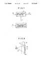
- the image processing apparatusin accordance with the present invention comprises an image processing condition storage section 1 and an image processing section 2. Names of various diseases and image processing conditions for the respective diseases are stored in the image processing condition storage section 1.
- a diagnostic image data Sl which is to be processedis input into the image processing section 2. Simultaneously with input of the image data S1 to the image processing section 2 or immediately before or after it, the name of a disease D which is to be diagnosed on the basis of the diagnostic image carried by the image data S1 is input into the image processing condition storage section 1, and the image processing condition P corresponding to the disease is input into the image processing section 2 from the image processing condition storage section 1.
- the image processing section 2carries out image processing on the image data S1 according to the image processing condition P input thereinto and generates a processed image data S2.
- the processed image data S2is sent to an image reproducing system (not shown) where a visible image is reproduced on the basis of the processed image data S2 or sent to an image storage system to be reproduced later.
- the name of the disease Dmay be manually input for each diagnostic image, but it is preferred that the name of the disease D be automatically determined according to the clinical history of the patient, the results of medical tests and the like, and the image data be processed according to the image processing condition corresponding to the name of the disease thus determined.
- a data analyzing section 3 and a diagnosis knowledge base storage section 4are added as shown in FIG. 2.
- the patient information Iincludes various data on the patient corresponding to the image data S1 which is input or has been input into the image processing section 2, e.g., age, physical features of the patient such as sex, results of various medical tests such as blood test and urinalysis, degree of drinking and/or smoking, business career, patient's own clinical history, clinical history of the patient's family, and the like.
- diagnosis knowledge base storage section 4is stored in advance a diagnosis knowledge base for relating one or more of the data included in the patient information I to the probability of various diseases to be diagnosed. For example, when the patient information says that the patient has been a coal miner for not less than fifteen years, it is determined that there is a high probability that he is suffering from pneumoconiosis, or when the patient information says that it is within a predetermined time since the patient was operated on for cancer, it is determined that there is a high probability that he is suffering from the cancer.
- one or more probable diseasesare specified with reference to the diagnosis knowledge base stored in the diagnosis knowledge base storage section 4, and the names of the diseases are delivered to the image processing condition storage section 1.
- the image processing condition P corresponding to the diseaseis input into the image processing section 2 from the image processing condition storage section 1.
- the image processing section 2carries out image processing on the image data S1 according to the image processing condition P input thereinto and generates a processed image data S2.
- the apparatusmay be so arranged that the image data S1 is input into the data analyzing section 3 together with the patient information I, the image data S1 is searched for an image of abnormality which is useful for specifying the disease such as an image corresponding to cancer, an image corresponding to pneumoconiosis, or the like (See, for instance, Japanese Unexamined Patent Publication No. 62(1987)-125481 and Japanese Patent Application Nos. 1(1989)-162901 to 162909), and the name of disease corresponding to the image of abnormality, if found, is delivered to the image processing condition storage section 1.
- Names of more than one diseasesmay be input into the image processing condition storage section 1 in either of the image processing apparatuses shown in FIGS. 1 and 2 and the image processing condition storage section 1 may output a plurality of image processing conditions P which are suitable for the respective names of diseases.
- the image processing section 2carries out image processing on the image data S1 a plurality of times according to the respective image processing conditions P and generates a plurality of processed image data S2.
- the image processing conditions and the names of diseasesare related to each other and are stored, and the image data is processed according to the image processing condition corresponding to the name of disease to be diagnosed. Accordingly, compared with the conventional image processing apparatus where the image processing condition is determined on the basis of the recorded parts, the areas which interest doctors can be processed optimally with-a higher probability.
- the name of diseasemay be manually input for each diagnostic image. However, in some cases, the disease cannot be diagnosed until the image is viewed. In such cases, the image is once reproduced and the disease is diagnosed roughly. Then the name of disease is input and the image data is processed according to the image processing condition corresponding to the name of disease. Then a visible image is reproduced again on the basis of the optimally processed image data.
- the name of diseaseis automatically determined on the basis of the patient information and accordingly, the time required to obtain an optimally processed image can be reduced and trouble to doctors or the like is reduced.
- FIG. 1is a block diagram for illustrating the general arrangement of the image processing apparatus in accordance with the present invention
- FIG. 2is a block diagram for illustrating a preferable arrangement of the image processing apparatus in accordance with the present invention
- FIG. 3is a schematic view showing an example of an X-ray image recording apparatus
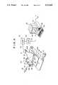
- FIG. 4is a perspective view showing an image processing apparatus in the form of a computer system in accordance with an embodiment of the present invention together with an example of an X-ray image read-out apparatus,
- FIG. 5is a view showing an example of the patient information
- FIGS. 6a and 6bare views showing different examples of the diagnosis knowledge base.
- an X-ray image recording apparatus 10includes an X-ray source 11. X-rays 12 are projected from the X-ray source 11 toward the chest 13a of a human body 13 and a stimulable phosphor sheet 14 is exposed to X-rays 12a which has passed through the human body 13 and stores an X-ray image of the chest 13a.
- the stimulable phosphor sheet 14 on which an X-ray image has been storedis sent to an X-ray image read-out apparatus 20 as shown in FIG. 4.
- the stimulable phosphor sheet 14is conveyed in a sub-scanning direction indicated by the arrow Y by a sheet conveyance means 22 constituted of an endless belt or the like operated by a motor 21.
- a light beam 24 produced by a laser beam source 23is reflected and deflected by a rotating polygon mirror 26, which is quickly rotated by a motor 25 in the direction indicated by the arrow, and the light beam 24 passes through a converging lens 27 constituted of an f ⁇ lens or the like.
- the direction of the optical path of the light beam 24is then changed by a mirror 28, and the light beam 24 impinges upon the stimulable phosphor sheet 14 and scans across it in a main scanning direction indicated by the arrow X, which main scanning direction is approximately normal to the sub-scanning direction indicated by the arrow Y.
- the stimulable phosphor sheet 14is exposed to the light beam 24, the exposed portion of the stimulable phosphor sheet 14 emits light 29 with an intensity proportional to the amount of energy stored during exposure to X-rays.
- the emitted light 29is guided by a light guide member 30, and photoelectrically detected by a photomultiplier 31 which acts as a photodetector.
- the light guide member 30is made of a light guiding material such as an acrylic plate, and has a linear light input face 30a positioned so that it extends along the main scanning line on the stimulable phosphor sheet 14, and a ring-shaped light output face 30b is positioned in close contact with a light receiving face of the photomultiplier 31.
- the emitted light 29 entering the light guide member 30 through its light input face 30ais guided through repeated total reflection inside of the light guide member 30, emanates from the light output face 30b, and is received by the photomultiplier 31. In this manner, the intensity of the emitted light 29, which carries the information about the X-ray image, is converted into an electrical signal by the photomultiplier 31.
- the analog output signal SO output from the photomultiplier 31is amplified by a logarithmic amplifier 32 and is digitized by an A/D converter 33, whereby an image data S1 in the form of a digital signal is obtained.
- the image data S1 thus obtainedis input into a computer system 40.
- the computer systemincludes therein an image processing apparatus in accordance with an embodiment of the present invention, and comprises a main portion 41 in which a CPU and a built-in memory are housed, a disk drive portion 42 which drives floppy disks as an auxiliary memory, a keyboard 43 for manually inputting instructions and the like, and a CRT display 44 for displaying a visible image and other necessary information.
- the computer system 40executes the processings described above in conjunction with FIGS. 1 and 2.
- Image processing conditions which depend upon the recording condition and the recorded part and image processing conditions which depend upon the kind of disease to be diagnosedhave been input into the computer system and stored therein.
- the computer system 40executes noise elimination processing for suppressing noise due to fluctuation of X-rays during recording, frequency processing for enhancing or weakening a particular spatial frequency component, processing for changing the gradation, brightness and the like of the visible image displayed by the CRT display 44, and other processings.
- the image processing conditionincludes a series of conditions relating to processing of images such as what spatial frequency is to be enhanced to what extent, and what degree the gradation is to be.
- the image data S1is processed according to the image processing condition determined based on the recording condition and the recorded part irrespective of the kind of the disease to be diagnosed.
- the processed image datais sent to the CRT display 44 and is used to reproduce a visible image on the CRT display 44, which is submitted to doctor's inspection. Since the visible image is based on the image data processed according to the image processing condition which has been determined only based on the recording condition and the recorded part, it cannot be always suitable for diagnosis of the disease to be diagnosed. Then the doctor visually inspects the visible image and refers to the patient information such as the result of various tests and doctor's questions, and then inputs the name of disease to be diagnosed by means of the keyboard 43.
- the name of disease to be diagnosedis delivered to the computer system 40, and the computer system 40 carries out image processing on the image data S1 according to the image processing condition determined based on the name of disease thus delivered to the computer system 40. Then the processed image data is used to reproduce a visible image on the CRT display 44.
- the name of disease to be diagnosedmay be determined solely based on the patient information such as the result of various tests and doctor's questions without processing the image data S1 according to the image processing condition determined based on the recording condition and the recorded part.
- the image data S1is input into the computer system 40 together with the patient information I corresponding to the image data S1.
- the patient information Iis stored in a floppy disk after completing various tests and doctor's questions, and is input into the computer system 40 by loading floppy disk into the disk drive portion 42.
- the patient information Imay be directly input into the computer system 40 without using a floppy disk.
- FIG. 5shows an example of the patient information I.
- the patient information Iincludes the recorded part, the result of doctor's questions, age of the patient, sex of the patient, patient's own clinical history, clinical history of patient's relatives and the like and is recorded in a floppy disk in the form of coded data.
- the patient informationis input into the computer system 40 by loading the floppy disk into the computer system 40.
- the image processing condition storage section 1 in the computer system 40is stored a table in which the names of diseases are related to image processing conditions.
- diagnosis knowledge base storage section 4are stored various diagnosis knowledge bases for determining highly probable disease or diseases on the basis of the patient information I.
- the data analyzing section 3determines highly probable disease or diseases on the basis of the patient information I according to the diagnosis knowledge bases.
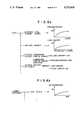
- FIGS. 6a and 6bshow examples of the diagnosis knowledge bases.
- the diagnosis knowledge basesare grouped by the recorded parts such as the lung, the lumber vertebra and the like.
- the diagnosis knowledge base on the lungsuch as shown in FIG. 6a is referred to.
- the diagnosis knowledge base on the lungincludes the probability of pneumoconiosis as a .function of the number of years for which the patient has been working as a coal miner. For example, when the patient information says that the patient has been working as a coal miner for ten years, the data analyzing section 3 determines according to the function that the probability of pneumoconiosis is 30%.
- the diagnosis knowledge base on the lungincludes the probability of lung cancer as a function of the product of the number of cigarettes which the patient smokes a day and the number of years for which the patient has smoked (the total number of cigarettes the patient smoked up to now). For example, when the patient information says that the patient has smoked twenty cigarettes a day for fifteen years, the data analyzing section 3 determines according to the function that the probability of lung cancer is 20%.
- the probability of the diseaseis determined to be 100%. Further the probability of disease is also derived from the clinical history of patient's relatives.
- the name of disease D thus obtainedis input into the image processing condition storage section 1 and the image processing condition storage section 1 inputs the image processing condition P corresponding to the disease into the image processing section 2.
- the computer system 40may be so arranged that the image data S1 is input into the data analyzing section 3 together with the patient information I, the image data S1 is searched for an image corresponding to cancer, an image corresponding to pneumoconiosis, or the like and the name of disease corresponding to the image of abnormality, if found, is delivered to the image processing condition storage section 1.
- the image data S1is processed according to the image processing condition corresponding to the disease, and the processed image data S2 is delivered to the CRT display 44 and is used to reproduce a visible image.
- the processed image data S2may be stored in a memory (not shown).
- More than one diseasemay be specified.
- the image data S1is processed for each disease and a plurality of visible images may be displayed at one time or in sequence.
- the visible imageis displayed on the CRT display 44.
- the visible imagemay be recorded on a photosensitive film by means of a laser printer or the like.
- the image processing apparatus of the present inventionmay be applied to diagnostic images obtained by the use of other various systems such as those obtained by the use of an X-ray film, CT, MRI and the like.
Landscapes
- Health & Medical Sciences (AREA)
- Engineering & Computer Science (AREA)
- Public Health (AREA)
- Medical Informatics (AREA)
- Primary Health Care (AREA)
- General Health & Medical Sciences (AREA)
- Epidemiology (AREA)
- Nuclear Medicine, Radiotherapy & Molecular Imaging (AREA)
- Radiology & Medical Imaging (AREA)
- Spectroscopy & Molecular Physics (AREA)
- Physics & Mathematics (AREA)
- Molecular Biology (AREA)
- High Energy & Nuclear Physics (AREA)
- General Physics & Mathematics (AREA)
- Life Sciences & Earth Sciences (AREA)
- Biomedical Technology (AREA)
- Data Mining & Analysis (AREA)
- Databases & Information Systems (AREA)
- Pathology (AREA)
- Medical Treatment And Welfare Office Work (AREA)
- Image Analysis (AREA)
- Radiography Using Non-Light Waves (AREA)
- Apparatus For Radiation Diagnosis (AREA)
- Image Processing (AREA)
Abstract
Description
Claims (4)
Applications Claiming Priority (4)
| Application Number | Priority Date | Filing Date | Title |
|---|---|---|---|
| JP1-207353 | 1989-08-10 | ||
| JP20735389 | 1989-08-10 | ||
| JP2-102022 | 1990-04-18 | ||
| JP2102022AJP2955873B2 (en) | 1989-08-10 | 1990-04-18 | Image processing device |
Publications (1)
| Publication Number | Publication Date |
|---|---|
| US5172418Atrue US5172418A (en) | 1992-12-15 |
Family
ID=26442772
Family Applications (1)
| Application Number | Title | Priority Date | Filing Date |
|---|---|---|---|
| US07/564,238Expired - LifetimeUS5172418A (en) | 1989-08-10 | 1990-08-08 | Image processing apparatus using disease-based image processing conditions |
Country Status (2)
| Country | Link |
|---|---|
| US (1) | US5172418A (en) |
| JP (1) | JP2955873B2 (en) |
Cited By (27)
| Publication number | Priority date | Publication date | Assignee | Title |
|---|---|---|---|---|
| US5272760A (en)* | 1992-05-29 | 1993-12-21 | Cimpiter Corporation | Radiographic image evaluation apparatus and method |
| US5384862A (en)* | 1992-05-29 | 1995-01-24 | Cimpiter Corporation | Radiographic image evaluation apparatus and method |
| US5425113A (en)* | 1991-12-26 | 1995-06-13 | Fuji Photo Film Co., Ltd. | Method and apparatus for performing various types of image processing in one operation |
| WO1995021419A1 (en)* | 1994-02-01 | 1995-08-10 | The Board Of Governors For Higher Education, State Of Rhode Island And Providence Plantations | An expert system intervention for smoking cessation |
| EP0679909A1 (en)* | 1994-04-29 | 1995-11-02 | Agfa-Gevaert N.V. | Customized and configurated radiation image read out system |
| WO1996010801A1 (en)* | 1994-09-30 | 1996-04-11 | Neopath, Inc. | Method and apparatus for highly efficient computer aided screening |
| US5619598A (en)* | 1990-08-20 | 1997-04-08 | Fuji Photo Film Co., Ltd. | Image filing apparatus using both reversible and irreversible compression |
| US5671359A (en)* | 1992-11-24 | 1997-09-23 | Eastman Kodak Company | Noise reduction in a storage phosphor data acquisition system |
| US5815591A (en)* | 1996-07-10 | 1998-09-29 | R2 Technology, Inc. | Method and apparatus for fast detection of spiculated lesions in digital mammograms |
| US5917929A (en)* | 1996-07-23 | 1999-06-29 | R2 Technology, Inc. | User interface for computer aided diagnosis system |
| US6016356A (en)* | 1994-03-31 | 2000-01-18 | Fuji Photo Film Co., Ltd. | Image superposition processing method |
| US6021404A (en)* | 1997-08-18 | 2000-02-01 | Moukheibir; Nabil W. | Universal computer assisted diagnosis |
| US6181811B1 (en) | 1998-01-13 | 2001-01-30 | Neopath, Inc. | Method and apparatus for optimizing biological and cytological specimen screening and diagnosis |
| US20030120514A1 (en)* | 2001-11-02 | 2003-06-26 | Rao R. Bharat | Patient data mining, presentation, exploration, and verification |
| US20040003001A1 (en)* | 2002-04-03 | 2004-01-01 | Fuji Photo Film Co., Ltd. | Similar image search system |
| US20040193650A1 (en)* | 2003-03-28 | 2004-09-30 | Fuji Photo Film Co., Ltd. | Image processing apparatus |
| US20060265253A1 (en)* | 2005-05-18 | 2006-11-23 | Rao R B | Patient data mining improvements |
| WO2007078658A1 (en)* | 2005-12-19 | 2007-07-12 | Carestream Health, Inc. | Medical image processing method and apparatus |
| US20070239491A1 (en)* | 2002-09-09 | 2007-10-11 | Rao R B | Patient Data Mining for Lung Cancer Screening |
| US20070286525A1 (en)* | 2006-06-08 | 2007-12-13 | General Electric Company | Generation of imaging filters based on image analysis |
| US20080015902A1 (en)* | 2003-05-27 | 2008-01-17 | Canon Kabushiki Kaisha | Image processing method and apparatus |
| US20090076851A1 (en)* | 2001-12-14 | 2009-03-19 | Siemens Medical Solutions Usa, Inc. | Early detection of disease outbreak using electronic patient data to reduce public health threat from bio-terrorism |
| US20100274776A1 (en)* | 2008-09-25 | 2010-10-28 | Canon Kabushiki Kaisha | Data search apparatus, control method therefor, and data search system |
| TWI486902B (en)* | 2013-01-07 | 2015-06-01 | ||
| JP2015150308A (en)* | 2014-02-18 | 2015-08-24 | 株式会社東芝 | Ultrasonic diagnostic apparatus and setting method of ultrasonic diagnostic apparatus |
| US10943676B2 (en) | 2010-06-08 | 2021-03-09 | Cerner Innovation, Inc. | Healthcare information technology system for predicting or preventing readmissions |
| EP4024115A4 (en)* | 2019-10-17 | 2022-11-02 | Sony Group Corporation | SURGICAL INFORMATION PROCESSING DEVICE, SURGICAL INFORMATION PROCESSING METHOD AND SURGICAL INFORMATION PROCESSING PROGRAM |
Families Citing this family (5)
| Publication number | Priority date | Publication date | Assignee | Title |
|---|---|---|---|---|
| JP3564531B2 (en)* | 1999-05-07 | 2004-09-15 | 株式会社モノリス | Image area tracking method and apparatus |
| JP2002008009A (en)* | 2000-06-27 | 2002-01-11 | Fuji Photo Film Co Ltd | Image processing condition determination method and device |
| JP4906404B2 (en)* | 2006-06-13 | 2012-03-28 | 富士フイルム株式会社 | Diagnosis support method, diagnosis support apparatus, diagnosis support system, and diagnosis support program |
| CN101861601A (en)* | 2007-11-14 | 2010-10-13 | 皇家飞利浦电子股份有限公司 | Computer Aided Detection (CAD) of Disease |
| JP7301510B2 (en)* | 2018-09-11 | 2023-07-03 | キヤノン株式会社 | Radiography apparatus, radiography method and program |
Citations (14)
| Publication number | Priority date | Publication date | Assignee | Title |
|---|---|---|---|---|
| US4199748A (en)* | 1976-11-01 | 1980-04-22 | Rush-Presbyterian-St. Luke's Medical Center | Automated method and apparatus for classification of cells with application to the diagnosis of anemia |
| US4232970A (en)* | 1977-04-30 | 1980-11-11 | Olympus Optical Co., Ltd. | Apparatus for automatic diagnosis of cells |
| US4258264A (en)* | 1978-07-12 | 1981-03-24 | Fuji Photo Film Co., Ltd. | Method of and apparatus for reading out a radiation image recorded in a stimulable phosphor |
| US4276473A (en)* | 1979-02-28 | 1981-06-30 | Fuji Photo Film Co., Ltd. | Gradation processing method and apparatus for radiation image recording system |
| US4315318A (en)* | 1978-12-26 | 1982-02-09 | Fuji Photo Film Co., Ltd. | Method and apparatus for processing a radiation image |
| US4387428A (en)* | 1979-12-25 | 1983-06-07 | Fuji Photo Film Co., Ltd. | Method of and apparatus for processing a radiation image |
| US4430749A (en)* | 1981-06-30 | 1984-02-07 | Siemens Gammasonics, Inc. | Medical imaging apparatus and method for furnishing difference images |
| US4453266A (en)* | 1980-04-21 | 1984-06-05 | Rush-Presbyterian-St. Luke's Medical Center | Method and apparatus for measuring mean cell volume of red blood cells |
| US4563701A (en)* | 1983-11-14 | 1986-01-07 | Elscint Ltd. | Digital diagnostic imaging system |
| US4764870A (en)* | 1987-04-09 | 1988-08-16 | R.A.P.I.D., Inc. | System and method for remote presentation of diagnostic image information |
| US4920491A (en)* | 1988-05-16 | 1990-04-24 | General Electric Company | Enhancement of image quality by utilization of a priori information |
| US4945478A (en)* | 1987-11-06 | 1990-07-31 | Center For Innovative Technology | Noninvasive medical imaging system and method for the identification and 3-D display of atherosclerosis and the like |
| US4995093A (en)* | 1987-03-31 | 1991-02-19 | Fuji Photo Film Co., Ltd. | Radiation field recognition method |
| US4998284A (en)* | 1987-11-17 | 1991-03-05 | Cell Analysis Systems, Inc. | Dual color camera microscope and methodology for cell staining and analysis |
Family Cites Families (2)
| Publication number | Priority date | Publication date | Assignee | Title |
|---|---|---|---|---|
| JPS63103225A (en)* | 1986-10-20 | 1988-05-07 | Fuji Photo Film Co Ltd | Reproduction and storing system for radiograph |
| JPH01169579A (en)* | 1987-12-24 | 1989-07-04 | Fuji Photo Film Co Ltd | System for reproducing and storing radiograph |
- 1990
- 1990-04-18JPJP2102022Apatent/JP2955873B2/ennot_activeExpired - Lifetime
- 1990-08-08USUS07/564,238patent/US5172418A/ennot_activeExpired - Lifetime
Patent Citations (14)
| Publication number | Priority date | Publication date | Assignee | Title |
|---|---|---|---|---|
| US4199748A (en)* | 1976-11-01 | 1980-04-22 | Rush-Presbyterian-St. Luke's Medical Center | Automated method and apparatus for classification of cells with application to the diagnosis of anemia |
| US4232970A (en)* | 1977-04-30 | 1980-11-11 | Olympus Optical Co., Ltd. | Apparatus for automatic diagnosis of cells |
| US4258264A (en)* | 1978-07-12 | 1981-03-24 | Fuji Photo Film Co., Ltd. | Method of and apparatus for reading out a radiation image recorded in a stimulable phosphor |
| US4315318A (en)* | 1978-12-26 | 1982-02-09 | Fuji Photo Film Co., Ltd. | Method and apparatus for processing a radiation image |
| US4276473A (en)* | 1979-02-28 | 1981-06-30 | Fuji Photo Film Co., Ltd. | Gradation processing method and apparatus for radiation image recording system |
| US4387428A (en)* | 1979-12-25 | 1983-06-07 | Fuji Photo Film Co., Ltd. | Method of and apparatus for processing a radiation image |
| US4453266A (en)* | 1980-04-21 | 1984-06-05 | Rush-Presbyterian-St. Luke's Medical Center | Method and apparatus for measuring mean cell volume of red blood cells |
| US4430749A (en)* | 1981-06-30 | 1984-02-07 | Siemens Gammasonics, Inc. | Medical imaging apparatus and method for furnishing difference images |
| US4563701A (en)* | 1983-11-14 | 1986-01-07 | Elscint Ltd. | Digital diagnostic imaging system |
| US4995093A (en)* | 1987-03-31 | 1991-02-19 | Fuji Photo Film Co., Ltd. | Radiation field recognition method |
| US4764870A (en)* | 1987-04-09 | 1988-08-16 | R.A.P.I.D., Inc. | System and method for remote presentation of diagnostic image information |
| US4945478A (en)* | 1987-11-06 | 1990-07-31 | Center For Innovative Technology | Noninvasive medical imaging system and method for the identification and 3-D display of atherosclerosis and the like |
| US4998284A (en)* | 1987-11-17 | 1991-03-05 | Cell Analysis Systems, Inc. | Dual color camera microscope and methodology for cell staining and analysis |
| US4920491A (en)* | 1988-05-16 | 1990-04-24 | General Electric Company | Enhancement of image quality by utilization of a priori information |
Cited By (47)
| Publication number | Priority date | Publication date | Assignee | Title |
|---|---|---|---|---|
| US5619598A (en)* | 1990-08-20 | 1997-04-08 | Fuji Photo Film Co., Ltd. | Image filing apparatus using both reversible and irreversible compression |
| US5425113A (en)* | 1991-12-26 | 1995-06-13 | Fuji Photo Film Co., Ltd. | Method and apparatus for performing various types of image processing in one operation |
| US5740267A (en)* | 1992-05-29 | 1998-04-14 | Echerer; Scott J. | Radiographic image enhancement comparison and storage requirement reduction system |
| US5384862A (en)* | 1992-05-29 | 1995-01-24 | Cimpiter Corporation | Radiographic image evaluation apparatus and method |
| US5272760A (en)* | 1992-05-29 | 1993-12-21 | Cimpiter Corporation | Radiographic image evaluation apparatus and method |
| US5671359A (en)* | 1992-11-24 | 1997-09-23 | Eastman Kodak Company | Noise reduction in a storage phosphor data acquisition system |
| WO1995021419A1 (en)* | 1994-02-01 | 1995-08-10 | The Board Of Governors For Higher Education, State Of Rhode Island And Providence Plantations | An expert system intervention for smoking cessation |
| US6016356A (en)* | 1994-03-31 | 2000-01-18 | Fuji Photo Film Co., Ltd. | Image superposition processing method |
| EP0679909A1 (en)* | 1994-04-29 | 1995-11-02 | Agfa-Gevaert N.V. | Customized and configurated radiation image read out system |
| US5646417A (en)* | 1994-04-29 | 1997-07-08 | Agfa-Gevaert | Customized and configurated radiation image read out system |
| WO1996010801A1 (en)* | 1994-09-30 | 1996-04-11 | Neopath, Inc. | Method and apparatus for highly efficient computer aided screening |
| US5799101A (en)* | 1994-09-30 | 1998-08-25 | Neopath, Inc. | Method and apparatus for highly efficient computer aided screening |
| US5815591A (en)* | 1996-07-10 | 1998-09-29 | R2 Technology, Inc. | Method and apparatus for fast detection of spiculated lesions in digital mammograms |
| US5917929A (en)* | 1996-07-23 | 1999-06-29 | R2 Technology, Inc. | User interface for computer aided diagnosis system |
| US6247004B1 (en) | 1997-08-18 | 2001-06-12 | Nabil W. Moukheibir | Universal computer assisted diagnosis |
| US6021404A (en)* | 1997-08-18 | 2000-02-01 | Moukheibir; Nabil W. | Universal computer assisted diagnosis |
| US6181811B1 (en) | 1998-01-13 | 2001-01-30 | Neopath, Inc. | Method and apparatus for optimizing biological and cytological specimen screening and diagnosis |
| US8280750B2 (en) | 2001-11-02 | 2012-10-02 | Siemens Medical Solutions Usa, Inc. | Patient data mining for cardiology screening |
| US20090259487A1 (en)* | 2001-11-02 | 2009-10-15 | Siemens Medical Solutions Usa, Inc. | Patient Data Mining |
| US20030130871A1 (en)* | 2001-11-02 | 2003-07-10 | Rao R. Bharat | Patient data mining for clinical trials |
| US8214225B2 (en) | 2001-11-02 | 2012-07-03 | Siemens Medical Solutions Usa, Inc. | Patient data mining, presentation, exploration, and verification |
| US8214224B2 (en) | 2001-11-02 | 2012-07-03 | Siemens Medical Solutions Usa, Inc. | Patient data mining for quality adherence |
| US8949079B2 (en) | 2001-11-02 | 2015-02-03 | Siemens Medical Solutions Usa, Inc. | Patient data mining |
| US20100222646A1 (en)* | 2001-11-02 | 2010-09-02 | Siemens Medical Solutions Usa, Inc. | Patient Data Mining for Cardiology Screening |
| US20030125988A1 (en)* | 2001-11-02 | 2003-07-03 | Rao R. Bharat | Patient data mining with population-based analysis |
| US8626533B2 (en) | 2001-11-02 | 2014-01-07 | Siemens Medical Soultions Usa, Inc. | Patient data mining with population-based analysis |
| US20030120514A1 (en)* | 2001-11-02 | 2003-06-26 | Rao R. Bharat | Patient data mining, presentation, exploration, and verification |
| US8392152B2 (en) | 2001-12-14 | 2013-03-05 | Siemens Medical Solutions Usa, Inc. | Early detection of disease outbreak using electronic patient data to reduce public health threat from bio-terrorism |
| US20090076851A1 (en)* | 2001-12-14 | 2009-03-19 | Siemens Medical Solutions Usa, Inc. | Early detection of disease outbreak using electronic patient data to reduce public health threat from bio-terrorism |
| US7374077B2 (en)* | 2002-04-03 | 2008-05-20 | Fujifilm Corporation | Similar image search system |
| US20040003001A1 (en)* | 2002-04-03 | 2004-01-01 | Fuji Photo Film Co., Ltd. | Similar image search system |
| US20070239491A1 (en)* | 2002-09-09 | 2007-10-11 | Rao R B | Patient Data Mining for Lung Cancer Screening |
| US8682693B2 (en)* | 2002-09-09 | 2014-03-25 | Siemens Medical Solutions Usa, Inc. | Patient data mining for lung cancer screening |
| US20040193650A1 (en)* | 2003-03-28 | 2004-09-30 | Fuji Photo Film Co., Ltd. | Image processing apparatus |
| US7957571B2 (en)* | 2003-05-27 | 2011-06-07 | Canon Kabushiki Kaisha | Image processing method and apparatus |
| US20080015902A1 (en)* | 2003-05-27 | 2008-01-17 | Canon Kabushiki Kaisha | Image processing method and apparatus |
| US20060265253A1 (en)* | 2005-05-18 | 2006-11-23 | Rao R B | Patient data mining improvements |
| WO2007078658A1 (en)* | 2005-12-19 | 2007-07-12 | Carestream Health, Inc. | Medical image processing method and apparatus |
| WO2007146495A3 (en)* | 2006-06-08 | 2008-03-20 | Gen Electric | Selection of image filters based on image information |
| US20070286525A1 (en)* | 2006-06-08 | 2007-12-13 | General Electric Company | Generation of imaging filters based on image analysis |
| US20100274776A1 (en)* | 2008-09-25 | 2010-10-28 | Canon Kabushiki Kaisha | Data search apparatus, control method therefor, and data search system |
| US10943676B2 (en) | 2010-06-08 | 2021-03-09 | Cerner Innovation, Inc. | Healthcare information technology system for predicting or preventing readmissions |
| US11664097B2 (en) | 2010-06-08 | 2023-05-30 | Cerner Innovation, Inc. | Healthcare information technology system for predicting or preventing readmissions |
| TWI486902B (en)* | 2013-01-07 | 2015-06-01 | ||
| JP2015150308A (en)* | 2014-02-18 | 2015-08-24 | 株式会社東芝 | Ultrasonic diagnostic apparatus and setting method of ultrasonic diagnostic apparatus |
| EP4024115A4 (en)* | 2019-10-17 | 2022-11-02 | Sony Group Corporation | SURGICAL INFORMATION PROCESSING DEVICE, SURGICAL INFORMATION PROCESSING METHOD AND SURGICAL INFORMATION PROCESSING PROGRAM |
| US12150617B2 (en) | 2019-10-17 | 2024-11-26 | Sony Group Corporation | Medical information processing apparatus, medical information processing method, and medical information processing program |
Also Published As
| Publication number | Publication date |
|---|---|
| JP2955873B2 (en) | 1999-10-04 |
| JPH03170129A (en) | 1991-07-23 |
Similar Documents
| Publication | Publication Date | Title |
|---|---|---|
| US5172418A (en) | Image processing apparatus using disease-based image processing conditions | |
| US4315318A (en) | Method and apparatus for processing a radiation image | |
| US5402338A (en) | Method for forming energy subtraction images | |
| EP0490532B1 (en) | Radiographic image processing apparatus and method | |
| EP0455986A2 (en) | Method and apparatus for forming energy subtraction images | |
| JPH0214378A (en) | Method and device for reading and reproducing radiographic image | |
| JP4307877B2 (en) | Image processing apparatus and image processing method | |
| US5272339A (en) | Method for adjusting conditions in radiation image recording, readout, and reproducing systems | |
| US5115476A (en) | Edge finding method and apparatus | |
| US5014045A (en) | Radiation image read-out apparatus using menu | |
| US5233519A (en) | Radiation image diagnostic apparatus | |
| US4816680A (en) | Radiation image recording and read-out apparatus | |
| US5267153A (en) | Method and apparatus for storing image signals | |
| US5015854A (en) | Radiation image displaying apparatus | |
| JPH0775635A (en) | Method and device to control imparted radiation dose effectively | |
| US5231574A (en) | Method for detecting artifact signal components | |
| US5526139A (en) | Image read-out apparatus | |
| US5229618A (en) | Radiation image read-out apparatus compensating for image distortion | |
| US5049747A (en) | Radiation image recording and reproducing system | |
| US4992664A (en) | Radiation image read-out, processing and reproducing methods | |
| JP2005051702A (en) | Medical image information processing method, medical image information processor, and medical image information processing system | |
| US5231575A (en) | Image read-out apparatus | |
| EP0254301B1 (en) | Method of adjusting radiation image processing conditions | |
| JP3181125B2 (en) | How to determine the signal range relevant to playback | |
| JPS6168031A (en) | Radiation image information reading apparatus |
Legal Events
| Date | Code | Title | Description |
|---|---|---|---|
| AS | Assignment | Owner name:FUJI PHOTO FILM CO., LTD., 210 NAKANUMA, MINAMIASH Free format text:ASSIGNMENT OF ASSIGNORS INTEREST.;ASSIGNORS:ITO, WATARU;NAKAJIMA, NOBUYOSHI;REEL/FRAME:005494/0977 Effective date:19901011 | |
| STCF | Information on status: patent grant | Free format text:PATENTED CASE | |
| FPAY | Fee payment | Year of fee payment:4 | |
| FPAY | Fee payment | Year of fee payment:8 | |
| FEPP | Fee payment procedure | Free format text:PAYOR NUMBER ASSIGNED (ORIGINAL EVENT CODE: ASPN); ENTITY STATUS OF PATENT OWNER: LARGE ENTITY | |
| FPAY | Fee payment | Year of fee payment:12 | |
| AS | Assignment | Owner name:FUJIFILM CORPORATION, JAPAN Free format text:ASSIGNMENT OF ASSIGNORS INTEREST;ASSIGNOR:FUJIFILM HOLDINGS CORPORATION (FORMERLY FUJI PHOTO FILM CO., LTD.);REEL/FRAME:018904/0001 Effective date:20070130 Owner name:FUJIFILM CORPORATION,JAPAN Free format text:ASSIGNMENT OF ASSIGNORS INTEREST;ASSIGNOR:FUJIFILM HOLDINGS CORPORATION (FORMERLY FUJI PHOTO FILM CO., LTD.);REEL/FRAME:018904/0001 Effective date:20070130 |