US5095887A - Microscope-endoscope assembly especially usable in surgery - Google Patents
Microscope-endoscope assembly especially usable in surgeryDownload PDFInfo
- Publication number
- US5095887A US5095887AUS07/579,812US57981290AUS5095887AUS 5095887 AUS5095887 AUS 5095887AUS 57981290 AUS57981290 AUS 57981290AUS 5095887 AUS5095887 AUS 5095887A
- Authority
- US
- United States
- Prior art keywords
- optical
- endoscope
- microscope
- assembly
- ocular
- Prior art date
- Legal status (The legal status is an assumption and is not a legal conclusion. Google has not performed a legal analysis and makes no representation as to the accuracy of the status listed.)
- Expired - Fee Related
Links
Images


Classifications
- A—HUMAN NECESSITIES
- A61—MEDICAL OR VETERINARY SCIENCE; HYGIENE
- A61B—DIAGNOSIS; SURGERY; IDENTIFICATION
- A61B1/00—Instruments for performing medical examinations of the interior of cavities or tubes of the body by visual or photographical inspection, e.g. endoscopes; Illuminating arrangements therefor
- A61B1/00163—Optical arrangements
- A61B1/00195—Optical arrangements with eyepieces
- A—HUMAN NECESSITIES
- A61—MEDICAL OR VETERINARY SCIENCE; HYGIENE
- A61B—DIAGNOSIS; SURGERY; IDENTIFICATION
- A61B3/00—Apparatus for testing the eyes; Instruments for examining the eyes
- A61B3/10—Objective types, i.e. instruments for examining the eyes independent of the patients' perceptions or reactions
- A61B3/13—Ophthalmic microscopes
- A61B3/132—Ophthalmic microscopes in binocular arrangement
- G—PHYSICS
- G02—OPTICS
- G02B—OPTICAL ELEMENTS, SYSTEMS OR APPARATUS
- G02B21/00—Microscopes
- G02B21/18—Arrangements with more than one light path, e.g. for comparing two specimens
Definitions
- the present inventionrelates to a microscope-endoscope assembly which is especially useful in surgery.
- those skilled in the artuse two kinds of devices, i.e. on the one hand a microscope and, on the other hand an endoscope, each having its own unique function.
- a typical example wherein these two devices are usedis ophthalmic surgery.
- one devicee.g. the microscope
- the otheri.e. the endoscope
- the microscope and endoscopeare used in sequence, the surgeon must alternately look through both oculars of each device. But, this is not easily done and does not enable certain operations to be carried out.
- the surgeon or the useruses the optical path of the microscope.
- the surgeonuses the optical path of the endoscope while looking through the microscope oculars. He does not have to move about and his free hands may be used as necessary. This is important in certain particular fields, such as in ophthalmic surgery where it is known that the endoscope is manipulated in a manner analogous to that of a pencil to orient it in any particular direction needed to obtain a desired good observation.
- the present inventionrelates to an assembly comprising, on the one hand, a microscope having a binocular, an optical body and an objective, and, on the other hand, an endoscope provided with an extension and an outlet ocular, the assembly including a commutating modulus placed between the binocular and the optical body of the microscope and the outlet ocular of the endoscope to enable an observer whose eyes are located at each ocular of the microscope to selectively observe either the optical path of the microscope or the optical or electronic path of the endoscope (if the endoscope is of the electronic type) or simultaneously both the optical path of the microscope and the optical or electronic path of the endoscope to scan the object to be investigated.
- the commutating modulusis removable and is comprised of at least one optical separator and a reflecting device having their respective normals aligned with the bisectrix between the incident beam and the direction of the respective optical axes;
- the distance between the separator and the reflecting deviceis equal to 1/ ⁇ 2a is the axis gap, i.e. the distance between the optical axes of both optical paths of the microscope;
- the separatoris a glass slab having one face subjected to a semi-reflecting treatment and the reflecting device is a mirror;
- the optical means of the endoscopeare adapted to that of the microscope by using a dioptric or catadioptric optical system which dimensionally and positionally brings into accord both images formed by the endoscope and the microscope;
- the endoscopecomprises an outlet ocular providing an image focused for infinity and the microscope provides an image focused for infinity, these images being projected in the space gap between the optical body and the binocular;
- the commutating devicecomprises an internal reflecting prism, a separating cube formed of two prisms and an opaque screen, all being retracted by means of a driving motor;
- the dioptric or catadioptric optical systemis formed of an air medium
- the dioptric or catadioptric optical systemis a mirror
- the dioptric or catadioptric optical systemis a combination of convergent and divergent lenses of a reflecting prism
- the opaque screenis a metal sheet painted to be light-opaque.
- FIGS. 1A and 1Bare a general diagrammatic view of the assembly of the invention.
- FIG. 1Cis a schematic representation of the position relation of the optical members of the commutating modulus
- FIG. 2shows an embodiment comprising a relatively small number of optical members
- FIG. 3is an alternative embodiment of the device shown in FIG. 1A;
- FIG. 4is another alternative embodiment of the assembly of the invention.
- FIG. 5is a vertical cross-section of an embodiment adapted to retract the commutating modulus
- FIG. 6is a partial plan, partial cross-sectional view of the device shown in FIG. 5.
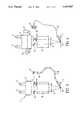
- the microscopecomprises a binocular 3, an optical body 8 and an objective 9.
- the subject to be investigatedis represented by reference numeral 15.
- the microscope 1has two oculars 4 and 5 and internal prisms 6 and 7.
- the endoscopecomprises an extension 10 which transmits an outlet image of the endoscope to the inlet of an adapting optical device, i.e. to an outlet ocular 16 of the extension.
- a supporting ring 11holds the outlet ocular 16 of extension 10 of endoscope 2 at a desired position.
- a commutating moduluscomprises a mirror 12, a separator 13 and an opaque screen 14.
- the separator 13is a glass slab having a surface which has been previously subjected to a semi-reflecting treatment as represented by dotted lines.
- the distance between the separator 13 and the mirror 12is related to the gap a which is the distance between the optical axes of the two optical paths of the microscope leading to oculars 4 and 5.
- the distance between the separator 13 and the mirror 12is preferably equal to 1/ ⁇ 2a.
- the mirroris a plan mirror and the separator is a dioptric slab with parallel sides.
- the orientation thereofcomprises a second parameter.
- the mirror 12 and the separator 13are so oriented that each of their respective normals 28 and 29 constitutes the bisectrix of the respective angles ⁇ and ⁇ between the incident optical beam i coming from the endoscope 2 and the direction of the respective optical axes AOG and AOD leading to oculars 4 and 5.
- the optics of endoscope 2 and of its extension 10are adapted to the optics of the microscope 1. To this end, use is made of a dioptric or catadioptric optical system which positionally and dimensionally brings into accord the images provided by endoscope 2 and microscope 1.
- this optical systemmerely consists of an air medium.
- the air mediumis an optical system having a focal magnitude of 1.
- the endoscope 2includes an outlet ocular 16 which provides an image focused at infinity whereas the microscope 1 also provides an image focused at infinity within the space gap between optical body 8 and binocular 3.
- FIG. 1Bdiffers from FIG. 1A in that extension 10 in FIG. 1B includes an optical fiber 10' and does not consist solely of lens and prism assemblies as in FIG. 1A.
- the supporting ring 11may be adjusted as desired by the user under the particular conditions of use.
- the opaque screen 14is deliberately located under the separator 13 to mask the right optical path, i.e. that of the separator.
- the surgeonmay observe subject 15 via the endoscope.
- the surgeoncan observe subject 15 via the optical path of the microscope. Movement of the modulus to provide this masking effect is preferably provided by any suitable means and may be actuated by means of a pedal to enable the surgeon to use his hands for any other useful operation.
- FIG. 2illustrates a practical embodiment of the present invention.
- the commutating moduluscomprises an internal reflecting prism 18, a separating cube 19 formed of two prisms 20 and 21 and the opaque screen 14.
- Prisms 18, 20 and 21are reflecting prisms. Both prisms 20 and 21 are, for example, secured to each other to form the cube, and have undergone a semi-reflecting treatment. This commutating modulus is driven by an electric motor 17 for retracting some or all of the members 18, 19, 14. When the entire assembly is retracted, the user who looks through the oculars 4 and 5 is able to observe the subject by way of the microscope. If desired, the opaque screen 14 may also be retracted separately.
- extension 10 of endoscope 2 with respect to microscope 1 shown in FIG. 2is facilitated by the use of a divergent lens 23, a convergent lens 24 and a reflecting prism 22 mounted between lenses 23, 24.
- the divergent lens 24is provided with a -50 focus
- the convergent lens 23is provided with a 100 focus
- the assemblyis afocal with a magnitude of 0.5.
- the commutating modulusis not retracted and the user looks through both oculars 4 and 5 of microscope 1 along the optical path of the endoscope.
- the commutating modulus assemblyIn the second position, the commutating modulus assembly is retracted, and the user looks through both oculars 4 and 5 along the optical path of the microscope.
- FIG. 3an alternative embodiment is illustrated, wherein use is made of a mirror 25 located in front of the outlet ocular 16 of the extension 10 of endoscope 2. This permits outlet ocular 16 to be disposed in the illustrated configuration.
- FIG. 4illustrates the provision of photographic fittings 26 and video apparatus 27 which may be mounted upwards of the optical body 8.
- FIGS. 5 and 6A displacement operation of the commutating device of the present invention is described with reference to FIGS. 5 and 6, wherein the commutating apparatus includes internal reflecting prism 18 and the separating cube comprising two prisms 20 and 21.
- the reflecting prism and separating cubeare mounted on a carriage 34 which is adapted to slide transversely with respect to a frame 35 on which are secured two parallel columns 36, 37. Guiding on column 37 is ensured by means of co-axial sleeves 38, 39 (FIG. 6) rigidly fixed to the base of carriage 34; and guiding on column 36 is ensured by a bracket 40 (FIG. 5) also rigidly fixed to the base of carriage 34.
- a rack mechanism 41is secured to a side of carriage 34 above sleeves 38, 39 and column 37 and in parallel to the axis of column 37.
- a reversible electric motor 17 affixed to frame 35 at one side of carriage 34rotatably drives an output shaft 42 on which is fixed a pinion 43 whose teeth mesh with those of the rack mechanism 41.
- pinion 43is rotatably driven in either direction by motor 17, the carriage 34 and the optical members supported thereon are moved in a corresponding direction along columns 36 and 37 and between two locations: one being in the optical path of the endoscope and the other being retracted from the optical path of the endoscope.
- the carriage 34has a window 44 at the side opposite to the motor 17.
- the opaque screen 14is slidably mounted on gliders provided in the base of the carriage 34 and may be retracted at will when it is desired to look through the optical paths of the microscope.
- Either sleeve 38 or sleeve 39actuates an end-of-travel switch comprising a lug 46 or 47 mounted at the end of a resilient arm 48 or 49 sidely projecting from the motor 17.
- the deformation or motion of the resilient armactuates a micro-switch 50 or 51 coupled to the motor to control current supplied thereto.
Landscapes
- Health & Medical Sciences (AREA)
- Life Sciences & Earth Sciences (AREA)
- Physics & Mathematics (AREA)
- Surgery (AREA)
- Biophysics (AREA)
- Optics & Photonics (AREA)
- Veterinary Medicine (AREA)
- Public Health (AREA)
- General Health & Medical Sciences (AREA)
- Engineering & Computer Science (AREA)
- Biomedical Technology (AREA)
- Heart & Thoracic Surgery (AREA)
- Medical Informatics (AREA)
- Molecular Biology (AREA)
- Animal Behavior & Ethology (AREA)
- Pathology (AREA)
- Radiology & Medical Imaging (AREA)
- Nuclear Medicine, Radiotherapy & Molecular Imaging (AREA)
- Ophthalmology & Optometry (AREA)
- Chemical & Material Sciences (AREA)
- Analytical Chemistry (AREA)
- General Physics & Mathematics (AREA)
- Endoscopes (AREA)
- Microscoopes, Condenser (AREA)
- Materials For Medical Uses (AREA)
- Prostheses (AREA)
Abstract
Description
The present invention relates to a microscope-endoscope assembly which is especially useful in surgery.
Considerable developments have been made during the last years in many fields of surgery. Surgeons have to use increasingly sophisticated devices for either diagnostic or therapeutics purposes.
In certain fields, those skilled in the art use two kinds of devices, i.e. on the one hand a microscope and, on the other hand an endoscope, each having its own unique function. For instance, a typical example wherein these two devices are used is ophthalmic surgery.
Typically, the microscope provides a user with a plan view determined by the intra-ocular members without any possible views of angular areas; i.e. a profile view cannot be provided.
The endoscope allows both angular areas to be viewed and therapeutic means to be introduced.
The use of one device, e.g. the microscope, followed by the use of the other, i.e. the endoscope, does not facilitate accurate determinations or orderly surgical procedures which, of course, are desired. When the microscope and endoscope are used in sequence, the surgeon must alternately look through both oculars of each device. But, this is not easily done and does not enable certain operations to be carried out.
There is a need for a device which enables a surgeon to observe and work while keeping his eyes on the oculars of a single device.
It is an object of the present invention to satisfy the aforesaid need.
Another object is to provide a compact assembly which enables a surgeon to perform those operations which may be required while keeping his eyes on the oculars of a microscope.
A further object is to facilitate three kinds of observations to be carried out by means of the inventive apparatus:
In a first position, the surgeon or the user uses the optical path of the microscope.
In a second position, the surgeon uses the optical path of the endoscope while looking through the microscope oculars. He does not have to move about and his free hands may be used as necessary. This is important in certain particular fields, such as in ophthalmic surgery where it is known that the endoscope is manipulated in a manner analogous to that of a pencil to orient it in any particular direction needed to obtain a desired good observation.
In a third position, the surgeon looks through the oculars of the microscope along both respective optical paths of the microscope and of the endoscope, and both paths are merged as to provide a single superposed image.
Accordingly, the present invention relates to an assembly comprising, on the one hand, a microscope having a binocular, an optical body and an objective, and, on the other hand, an endoscope provided with an extension and an outlet ocular, the assembly including a commutating modulus placed between the binocular and the optical body of the microscope and the outlet ocular of the endoscope to enable an observer whose eyes are located at each ocular of the microscope to selectively observe either the optical path of the microscope or the optical or electronic path of the endoscope (if the endoscope is of the electronic type) or simultaneously both the optical path of the microscope and the optical or electronic path of the endoscope to scan the object to be investigated.
As an aspect of the present invention, the commutating modulus is removable and is comprised of at least one optical separator and a reflecting device having their respective normals aligned with the bisectrix between the incident beam and the direction of the respective optical axes;
the distance between the separator and the reflecting device is equal to 1/√2a is the axis gap, i.e. the distance between the optical axes of both optical paths of the microscope;
the separator is a glass slab having one face subjected to a semi-reflecting treatment and the reflecting device is a mirror;
the optical means of the endoscope are adapted to that of the microscope by using a dioptric or catadioptric optical system which dimensionally and positionally brings into accord both images formed by the endoscope and the microscope;
the endoscope comprises an outlet ocular providing an image focused for infinity and the microscope provides an image focused for infinity, these images being projected in the space gap between the optical body and the binocular;
the commutating device comprises an internal reflecting prism, a separating cube formed of two prisms and an opaque screen, all being retracted by means of a driving motor;
in one embodiment the dioptric or catadioptric optical system is formed of an air medium;
in another embodiment the dioptric or catadioptric optical system is a mirror;
and in a further embodiment the dioptric or catadioptric optical system is a combination of convergent and divergent lenses of a reflecting prism;
the opaque screen is a metal sheet painted to be light-opaque.
Various advantages and features of the present invention will appear from the following detailed description with reference to the accompanying drawings in which:
FIGS. 1A and 1B are a general diagrammatic view of the assembly of the invention;
FIG. 1C is a schematic representation of the position relation of the optical members of the commutating modulus;
FIG. 2 shows an embodiment comprising a relatively small number of optical members;
FIG. 3 is an alternative embodiment of the device shown in FIG. 1A;
FIG. 4 is another alternative embodiment of the assembly of the invention;
FIG. 5 is a vertical cross-section of an embodiment adapted to retract the commutating modulus; and
FIG. 6 is a partial plan, partial cross-sectional view of the device shown in FIG. 5.
In the accompanying drawings, wherein identical parts are indicated by the same reference symbols and numerals, the microscope is designated by 1 and the endoscope by 2.
As is conventional, the microscope comprises a binocular 3, anoptical body 8 and an objective 9. The subject to be investigated is represented byreference numeral 15. The microscope 1 has two oculars 4 and 5 and internal prisms 6 and 7. The endoscope comprises anextension 10 which transmits an outlet image of the endoscope to the inlet of an adapting optical device, i.e. to an outlet ocular 16 of the extension. A supportingring 11 holds the outlet ocular 16 ofextension 10 of endoscope 2 at a desired position.
On FIGS. 1A, 1B, a commutating modulus comprises amirror 12, aseparator 13 and anopaque screen 14. Theseparator 13 is a glass slab having a surface which has been previously subjected to a semi-reflecting treatment as represented by dotted lines. The distance between theseparator 13 and themirror 12 is related to the gap a which is the distance between the optical axes of the two optical paths of the microscope leading tooculars 4 and 5. The distance between theseparator 13 and themirror 12 is preferably equal to 1/√2a. Generally, the mirror is a plan mirror and the separator is a dioptric slab with parallel sides.
In addition to the distance parameter between the mirror and separator, the orientation thereof comprises a second parameter.
As illustrated in FIG. 1C, themirror 12 and theseparator 13 are so oriented that each of theirrespective normals oculars 4 and 5. The optics of endoscope 2 and of itsextension 10 are adapted to the optics of the microscope 1. To this end, use is made of a dioptric or catadioptric optical system which positionally and dimensionally brings into accord the images provided by endoscope 2 and microscope 1.
In the example shown in FIGS. 1A and 1B, this optical system merely consists of an air medium. In this configuration, the air medium is an optical system having a focal magnitude of 1. The endoscope 2 includes anoutlet ocular 16 which provides an image focused at infinity whereas the microscope 1 also provides an image focused at infinity within the space gap betweenoptical body 8 and binocular 3. FIG. 1B differs from FIG. 1A in thatextension 10 in FIG. 1B includes an optical fiber 10' and does not consist solely of lens and prism assemblies as in FIG. 1A.
The supportingring 11 may be adjusted as desired by the user under the particular conditions of use.
On FIGS. 1A and 1B, theopaque screen 14 is deliberately located under theseparator 13 to mask the right optical path, i.e. that of the separator. With the modulus in the illustrated position, the surgeon may observe subject 15 via the endoscope. Alternatively, when the commutating modulus assembly is retracted, the surgeon can observe subject 15 via the optical path of the microscope. Movement of the modulus to provide this masking effect is preferably provided by any suitable means and may be actuated by means of a pedal to enable the surgeon to use his hands for any other useful operation.
FIG. 2 illustrates a practical embodiment of the present invention. Here, the commutating modulus comprises an internal reflectingprism 18, a separatingcube 19 formed of twoprisms opaque screen 14.
The position ofextension 10 of endoscope 2 with respect to microscope 1 shown in FIG. 2 is facilitated by the use of adivergent lens 23, aconvergent lens 24 and a reflectingprism 22 mounted betweenlenses divergent lens 24 is provided with a -50 focus, theconvergent lens 23 is provided with a 100 focus and the assembly is afocal with a magnitude of 0.5.
In the embodiments shown in the drawings, these positions are provided:
In the first position, the commutating modulus is not retracted and the user looks through bothoculars 4 and 5 of microscope 1 along the optical path of the endoscope.
In the second position, the commutating modulus assembly is retracted, and the user looks through bothoculars 4 and 5 along the optical path of the microscope.
In the third position, only theopaque screen 14 is retracted, and the user may observe through both the microscope path and the endoscope path which coincide to provide a single image, as in a mixed image.
On FIG. 3, an alternative embodiment is illustrated, wherein use is made of amirror 25 located in front of the outlet ocular 16 of theextension 10 of endoscope 2. This permits outlet ocular 16 to be disposed in the illustrated configuration.
FIG. 4 illustrates the provision ofphotographic fittings 26 and video apparatus 27 which may be mounted upwards of theoptical body 8.
A displacement operation of the commutating device of the present invention is described with reference to FIGS. 5 and 6, wherein the commutating apparatus includes internal reflectingprism 18 and the separating cube comprising twoprisms carriage 34 which is adapted to slide transversely with respect to aframe 35 on which are secured twoparallel columns column 37 is ensured by means ofco-axial sleeves 38, 39 (FIG. 6) rigidly fixed to the base ofcarriage 34; and guiding oncolumn 36 is ensured by a bracket 40 (FIG. 5) also rigidly fixed to the base ofcarriage 34.
Arack mechanism 41 is secured to a side ofcarriage 34 abovesleeves column 37 and in parallel to the axis ofcolumn 37. A reversibleelectric motor 17 affixed to frame 35 at one side ofcarriage 34 rotatably drives anoutput shaft 42 on which is fixed apinion 43 whose teeth mesh with those of therack mechanism 41. Thus, whenpinion 43 is rotatably driven in either direction bymotor 17, thecarriage 34 and the optical members supported thereon are moved in a corresponding direction alongcolumns carriage 34 has awindow 44 at the side opposite to themotor 17.
Theopaque screen 14 is slidably mounted on gliders provided in the base of thecarriage 34 and may be retracted at will when it is desired to look through the optical paths of the microscope.
The electric supply to the motor is switched off when thecarriage 34 reaches either of its extreme positions on theframe 35. Eithersleeve 38 orsleeve 39 actuates an end-of-travel switch comprising alug resilient arm motor 17. The deformation or motion of the resilient arm actuates a micro-switch 50 or 51 coupled to the motor to control current supplied thereto.
Claims (13)
1. An assembly comprising: a microscope including a binocular having a pair of oculars, an optical body, and an objective lens means comprising a first optical path; and endoscope having an extension, and an outlet ocular comprising a second optical; and commutating modulus means disposed between the binocular and the optical body of the microscope and in optical communication with the outlet ocular of the endoscope and selectively operable to enable an observer whose eyes are located at each ocular of the microscope, to observe an image projected along either the first optical path or the second optical path or both optical paths simultaneously.
2. The assembly of claim 1, wherein the commutating modulus means is retractable and is comprised of separator means, reflecting means optically coupled to said separator means along an optic axis aligned with the outlet ocular of said endoscope, and an opaque screen positionable to selectively block the optical path of the microscope, the reflecting means and the separator means having respective normals located on a bisectrix between the axis aligned with the endoscope outlet ocular and respective optical axes to said pair of oculars along the first optical path.
3. The assembly of claim 2 wherein the distance between the separator means and the reflecting means is equal to 1/√2a where a is a distance between said respective optical axes.
4. The assembly of claim 2 wherein said opaque screen comprises a metal sheet painted to be light-opaque.
5. The assembly of claim 2 wherein the separator means comprises a glass slab having a semi-reflecting face, and wherein the reflecting means comprises a mirror.
6. The assembly of claim 1 wherein the endoscope and the microscope include similar optical systems for bringing both images formed by the endoscope and the microscope into dimensional and positional alignment.
7. The assembly of claim 1, wherein at least the optical body of said microscope includes an air medium.
8. The assembly of claim 1 wherein the outlet ocular of the endoscope provides an image for infinity and the microscope provides an image for infinity in a gap between the optical body and the binocular.
9. The assembly of claim 1 wherein the commutating modulus means comprises an internal reflecting prism, a separating cube included in said first optical path, optically coupled to said reflecting prism and including two prisms, and an opaque screen selectively blocking said separating cube from said first optical path; and further including a driving motor for retracting said commutating modulus means.
10. The assembly of claim 1 wherein the endoscope includes dioptric optical imaging means.
11. The assembly of claim 1 wherein the endoscope includes catadioptric optical imaging means.
12. The assembly of claim 1, wherein said commutating modulus means comprises a dioptric optical system including convergent and divergent lenses coupled to the endoscope outlet ocular and a reflecting prism optically coupled to said lenses for imaging the second optical path to said commutating modulus means.
13. The assembly of claim 1 wherein said commutating modulus means comprises a catadioptric optical system including convergent and divergent lenses coupled to the endoscope outlet ocular and a reflecting prism optically coupled to said lenses for imaging the second optical path to said commutating modulus means.
Applications Claiming Priority (2)
| Application Number | Priority Date | Filing Date | Title |
|---|---|---|---|
| FR8911914 | 1989-09-12 | ||
| FR8911914AFR2651668B1 (en) | 1989-09-12 | 1989-09-12 | MICROSCOPE-ENDOSCOPE ASSEMBLY USEFUL IN PARTICULAR IN SURGERY. |
Publications (1)
| Publication Number | Publication Date |
|---|---|
| US5095887Atrue US5095887A (en) | 1992-03-17 |
Family
ID=9385369
Family Applications (1)
| Application Number | Title | Priority Date | Filing Date |
|---|---|---|---|
| US07/579,812Expired - Fee RelatedUS5095887A (en) | 1989-09-12 | 1990-09-10 | Microscope-endoscope assembly especially usable in surgery |
Country Status (13)
| Country | Link |
|---|---|
| US (1) | US5095887A (en) |
| EP (1) | EP0418109B1 (en) |
| JP (1) | JP2989237B2 (en) |
| AT (1) | ATE117187T1 (en) |
| AU (1) | AU632973B2 (en) |
| CA (1) | CA2023983A1 (en) |
| DE (1) | DE69016139T2 (en) |
| DK (1) | DK0418109T3 (en) |
| ES (1) | ES2069716T3 (en) |
| FR (1) | FR2651668B1 (en) |
| GR (1) | GR3015851T3 (en) |
| IL (1) | IL95491A (en) |
| ZA (1) | ZA906728B (en) |
Cited By (70)
| Publication number | Priority date | Publication date | Assignee | Title |
|---|---|---|---|---|
| US5742429A (en)* | 1994-07-13 | 1998-04-21 | Fujikura Ltd. | Device for stereoscopic visualization including a stereomicroscope and fiberscope |
| US5999840A (en)* | 1994-09-01 | 1999-12-07 | Massachusetts Institute Of Technology | System and method of registration of three-dimensional data sets |
| US6081371A (en)* | 1998-01-06 | 2000-06-27 | Olympus Optical Co., Ltd. | Surgical microscope including a first image and a changing projection position of a second image |
| US6088154A (en)* | 1997-04-03 | 2000-07-11 | Olympus Optical Co., Ltd. | Operating microscope |
| US6097538A (en)* | 1998-02-03 | 2000-08-01 | Olympus Optical Co., Ltd. | Lens barrel for use in a microscope |
| US6104426A (en)* | 1996-03-23 | 2000-08-15 | Street; Graham S. B. | Stereo-endoscope |
| US6106456A (en)* | 1995-02-20 | 2000-08-22 | Karl Storz Gmbh & Co. Kg | Device for examining cavities using an endoscope |
| US6333813B1 (en)* | 2000-06-06 | 2001-12-25 | Olympus Optical Co., Ltd. | Stereomicroscope |
| US6337765B1 (en)* | 1994-03-30 | 2002-01-08 | Leica Microsystems Ag | Stereomicroscope |
| US6398721B1 (en)* | 1999-02-19 | 2002-06-04 | Olympus Optical Co., Ltd. | Surgical microscope apparatus |
| US20020118451A1 (en)* | 2001-02-23 | 2002-08-29 | Deverin Jacques Alain | Optical instrument having a binocular viewing port |
| US20020143284A1 (en)* | 2001-04-03 | 2002-10-03 | Hosheng Tu | Drug-releasing trabecular implant for glaucoma treatment |
| US6473229B2 (en)* | 2000-01-27 | 2002-10-29 | Mitaka Kohki Co., Ltd. | Stereomicroscope |
| US6490083B1 (en) | 1998-09-09 | 2002-12-03 | Mcmanus Dennis Q. | Microscopy method and apparatus |
| US20030030899A1 (en)* | 2001-08-07 | 2003-02-13 | Olympus Optical Co., Ltd. | Microscope apparatus |
| US20030055372A1 (en)* | 1999-04-26 | 2003-03-20 | Lynch Mary G. | Shunt device and method for treating glaucoma |
| US20030060752A1 (en)* | 2000-04-14 | 2003-03-27 | Olav Bergheim | Glaucoma device and methods thereof |
| US6549333B1 (en)* | 1999-05-24 | 2003-04-15 | Fujikura Ltd. | Endoscope ocular with microscope gripping mechanism and endoscope holder and endoscope fixing method |
| US20030081310A1 (en)* | 1998-09-09 | 2003-05-01 | Mcmanus Dennis Q. | Microscopy method and apparatus |
| US20030097151A1 (en)* | 2001-10-25 | 2003-05-22 | Smedley Gregory T. | Apparatus and mitochondrial treatment for glaucoma |
| US6626858B2 (en) | 1999-04-26 | 2003-09-30 | Gmp Vision Solutions, Inc. | Shunt device and method for treating glaucoma |
| US20030187385A1 (en)* | 2000-04-14 | 2003-10-02 | Bergheim Olav B. | Implant with anchor |
| US6661571B1 (en) | 1999-09-21 | 2003-12-09 | Olympus Optical Co., Ltd. | Surgical microscopic system |
| US6666841B2 (en) | 2001-05-02 | 2003-12-23 | Glaukos Corporation | Bifurcatable trabecular shunt for glaucoma treatment |
| US20040024345A1 (en)* | 2002-04-19 | 2004-02-05 | Morteza Gharib | Glaucoma implant with valveless flow bias |
| US20040102729A1 (en)* | 2002-04-08 | 2004-05-27 | David Haffner | Devices and methods for glaucoma treatment |
| US6743168B2 (en) | 2001-02-02 | 2004-06-01 | Insight Instruments, Inc. | Endoscope system and method of use |
| US20040111050A1 (en)* | 2000-04-14 | 2004-06-10 | Gregory Smedley | Implantable ocular pump to reduce intraocular pressure |
| US20040127843A1 (en)* | 2000-04-14 | 2004-07-01 | Hosheng Tu | Glaucoma implant with therapeutic agents |
| US6765718B1 (en)* | 1999-10-13 | 2004-07-20 | Leica Microsystems (Schweiz) Ag | Stereo surgical microscope having an apparatus for reflecting in information |
| US20040147870A1 (en)* | 2002-04-08 | 2004-07-29 | Burns Thomas W. | Glaucoma treatment kit |
| US20040196548A1 (en)* | 2001-04-12 | 2004-10-07 | Jurgen Mannss | Optical viewing device for inwardly or outwardly reflecting image data provided with additional diaphragms |
| US20040223213A1 (en)* | 2003-04-25 | 2004-11-11 | Hiroya Fukuyama | Microscopic observing apparatus and probe microscope |
| US20050020876A1 (en)* | 2000-04-20 | 2005-01-27 | Olympus Corporation | Operation microscope |
| US20050049578A1 (en)* | 2000-04-14 | 2005-03-03 | Hosheng Tu | Implantable ocular pump to reduce intraocular pressure |
| US20050088732A1 (en)* | 2001-04-12 | 2005-04-28 | Roger Spink | Enhanced shutter control for images that are faded into a stereo microscope |
| US20050119636A1 (en)* | 2001-05-02 | 2005-06-02 | David Haffner | Implant with intraocular pressure sensor for glaucoma treatment |
| US20050192527A1 (en)* | 2001-05-02 | 2005-09-01 | Morteza Gharib | Glaucoma implant with extending members |
| US20050228257A1 (en)* | 2004-03-31 | 2005-10-13 | Tomonori Ishikawa | Imaging and displaying system with imaging unit and display unit which are supported by movable arm |
| US20050250788A1 (en)* | 2004-01-30 | 2005-11-10 | Hosheng Tu | Aqueous outflow enhancement with vasodilated aqueous cavity |
| US20050266047A1 (en)* | 2002-04-08 | 2005-12-01 | Hosheng Tu | Injectable glaucoma implants with multiple openings |
| US20050271704A1 (en)* | 2002-04-08 | 2005-12-08 | Hosheng Tu | Injectable glaucoma implants with multiple openings |
| US20050277864A1 (en)* | 2000-04-14 | 2005-12-15 | David Haffner | Injectable gel implant for glaucoma treatment |
| US20060241749A1 (en)* | 2001-08-28 | 2006-10-26 | Hosheng Tu | Glaucoma stent system |
| US7186232B1 (en) | 2002-03-07 | 2007-03-06 | Glaukoa Corporation | Fluid infusion methods for glaucoma treatment |
| US20070112292A1 (en)* | 2001-04-07 | 2007-05-17 | Hosheng Tu | Glaucoma stent and methods thereof for glaucoma treatment |
| US20070293807A1 (en)* | 2006-05-01 | 2007-12-20 | Lynch Mary G | Dual drainage pathway shunt device and method for treating glaucoma |
| US20080015488A1 (en)* | 2001-05-03 | 2008-01-17 | Glaukos Corporation | Glaucoma implant with double anchor mechanism |
| US20080172204A1 (en)* | 2007-01-15 | 2008-07-17 | Fujitsu Limited | Step counter and method of counting steps |
| US20080214940A1 (en)* | 2007-03-02 | 2008-09-04 | Benaron David A | Medical imaging lens system, and method with high-efficiency light collection and collinear illumination |
| US7488303B1 (en) | 2002-09-21 | 2009-02-10 | Glaukos Corporation | Ocular implant with anchor and multiple openings |
| US20090103174A1 (en)* | 2007-03-29 | 2009-04-23 | Junichi Nozawa | Surgical observation system |
| US20100245557A1 (en)* | 2009-03-31 | 2010-09-30 | Luley Iii Charles | Injection of secondary images into microscope viewing fields |
| US7951155B2 (en) | 2002-03-15 | 2011-05-31 | Glaukos Corporation | Combined treatment for cataract and glaucoma treatment |
| WO2011154970A1 (en)* | 2010-06-10 | 2011-12-15 | Ram Srikanth Mirlay | Integrated fiber optic ophthalmic intraocular surgical device with camera |
| CN101632570B (en)* | 2009-08-03 | 2012-06-13 | 深圳先进技术研究院 | Calibration method of medical endoscope |
| US8506515B2 (en) | 2006-11-10 | 2013-08-13 | Glaukos Corporation | Uveoscleral shunt and methods for implanting same |
| CN103892916A (en)* | 2012-12-29 | 2014-07-02 | 卿国平 | Microscopic endoscope |
| US9554940B2 (en) | 2012-03-26 | 2017-01-31 | Glaukos Corporation | System and method for delivering multiple ocular implants |
| US9592151B2 (en) | 2013-03-15 | 2017-03-14 | Glaukos Corporation | Systems and methods for delivering an ocular implant to the suprachoroidal space within an eye |
| US9603510B2 (en) | 2011-05-17 | 2017-03-28 | Mario Ammirati | Method and apparatus for delivering an endoscope via microsurgical instruments while performing microscopic surgery |
| US9730638B2 (en) | 2013-03-13 | 2017-08-15 | Glaukos Corporation | Intraocular physiological sensor |
| US9826900B2 (en)* | 2015-08-17 | 2017-11-28 | Novartis Ag | Surgical microscope with integrated optical coherence tomography and display systems |
| WO2018067082A1 (en)* | 2016-10-07 | 2018-04-12 | Kaya Ahmet Hilmi | An endomicroscopic device |
| USD846738S1 (en) | 2017-10-27 | 2019-04-23 | Glaukos Corporation | Implant delivery apparatus |
| US11116625B2 (en) | 2017-09-28 | 2021-09-14 | Glaukos Corporation | Apparatus and method for controlling placement of intraocular implants |
| US11224333B2 (en) | 2017-10-09 | 2022-01-18 | Olympus Winter & Ibe Gmbh | Stereo endoscope |
| US11363951B2 (en) | 2011-09-13 | 2022-06-21 | Glaukos Corporation | Intraocular physiological sensor |
| US11376040B2 (en) | 2017-10-06 | 2022-07-05 | Glaukos Corporation | Systems and methods for delivering multiple ocular implants |
| US12419783B2 (en) | 2010-11-24 | 2025-09-23 | Glaukos Corporation | Drug eluting ocular implant |
Families Citing this family (5)
| Publication number | Priority date | Publication date | Assignee | Title |
|---|---|---|---|---|
| FR2681150A1 (en)* | 1991-09-05 | 1993-03-12 | Oftalab | Improved eyepiece for an ophthalmological microscope |
| GB9213136D0 (en)* | 1992-06-20 | 1992-08-05 | Atomic Energy Authority Uk | Optical system |
| GB9325635D0 (en)* | 1993-12-15 | 1994-02-16 | Atomic Energy Authority Uk | Optical system |
| JP3642812B2 (en) | 1994-11-17 | 2005-04-27 | 株式会社町田製作所 | Medical observation device |
| DE10101184A1 (en) | 2000-02-11 | 2001-08-16 | Zeiss Carl | Operation microscope has image projection module containing image display unit, plane convex lens and plane concave lens with focal length ratio between 1.9 and 2.5 |
Citations (12)
| Publication number | Priority date | Publication date | Assignee | Title |
|---|---|---|---|---|
| US3796220A (en)* | 1972-03-24 | 1974-03-12 | H Bredemeier | Stereo laser endoscope |
| US4091814A (en)* | 1976-03-15 | 1978-05-30 | Mochida Pharmaceutical Co., Ltd. | Laser optical apparatus for operation under a microscope |
| US4364629A (en)* | 1979-05-16 | 1982-12-21 | Carl Zeiss-Stiftung | Operation microscope having a small working distance |
| FR2513772A1 (en)* | 1981-09-30 | 1983-04-01 | Zeiss Carl Fa | OPERATIVE MICROSCOPE WITH FIXING DEVICE |
| GB2140578A (en)* | 1983-05-26 | 1984-11-28 | Vickers Plc | Optical microscope having adjustable optics to view two specimens |
| US4544243A (en)* | 1984-05-24 | 1985-10-01 | Cooper Lasersonics, Inc. | Heads up display for microscope using remotely controlled instrument |
| US4580559A (en)* | 1984-07-24 | 1986-04-08 | Esperance Francis A L | Indirect ophthalmoscopic photocoagulation delivery system for retinal surgery |
| DE3610024A1 (en)* | 1985-03-25 | 1986-09-25 | Fa. Carl Zeiss, 7920 Heidenheim | LIGHTING STRENGTH DOSING DEVICE FOR OPERATING MICROSCOPE |
| US4710000A (en)* | 1985-03-02 | 1987-12-01 | Oculus Optikgeraete Gmbh | Surgical stereomicroscope |
| US4723842A (en)* | 1986-05-10 | 1988-02-09 | J. D. Moller Optische Werke Gmbh | Stereoscopic surgery microscope for eye surgery with selective image reversal |
| US4744649A (en)* | 1985-05-15 | 1988-05-17 | Kowa Kabushiki Kaisha | Ophthalmological measuring apparatus |
| WO1988004786A1 (en)* | 1986-12-16 | 1988-06-30 | Fantone Stephen D | Enhanced-image operating microscope |
Family Cites Families (1)
| Publication number | Priority date | Publication date | Assignee | Title |
|---|---|---|---|---|
| CA1292461C (en)* | 1987-02-12 | 1991-11-26 | Hiromu Terada | Endoscope |
- 1989
- 1989-09-12FRFR8911914Apatent/FR2651668B1/ennot_activeExpired - Lifetime
- 1990
- 1990-08-23ZAZA906728Apatent/ZA906728B/enunknown
- 1990-08-24DKDK90402364.5Tpatent/DK0418109T3/enactive
- 1990-08-24ATAT90402364Tpatent/ATE117187T1/enactive
- 1990-08-24CACA002023983Apatent/CA2023983A1/ennot_activeAbandoned
- 1990-08-24ESES90402364Tpatent/ES2069716T3/ennot_activeExpired - Lifetime
- 1990-08-24DEDE69016139Tpatent/DE69016139T2/ennot_activeExpired - Fee Related
- 1990-08-24EPEP90402364Apatent/EP0418109B1/ennot_activeExpired - Lifetime
- 1990-08-27ILIL9549190Apatent/IL95491A/ennot_activeIP Right Cessation
- 1990-09-10USUS07/579,812patent/US5095887A/ennot_activeExpired - Fee Related
- 1990-09-11JPJP2239173Apatent/JP2989237B2/ennot_activeExpired - Fee Related
- 1990-09-11AUAU62338/90Apatent/AU632973B2/ennot_activeCeased
- 1995
- 1995-04-17GRGR950400981Tpatent/GR3015851T3/enunknown
Patent Citations (14)
| Publication number | Priority date | Publication date | Assignee | Title |
|---|---|---|---|---|
| US3796220A (en)* | 1972-03-24 | 1974-03-12 | H Bredemeier | Stereo laser endoscope |
| US4091814A (en)* | 1976-03-15 | 1978-05-30 | Mochida Pharmaceutical Co., Ltd. | Laser optical apparatus for operation under a microscope |
| US4364629A (en)* | 1979-05-16 | 1982-12-21 | Carl Zeiss-Stiftung | Operation microscope having a small working distance |
| FR2513772A1 (en)* | 1981-09-30 | 1983-04-01 | Zeiss Carl Fa | OPERATIVE MICROSCOPE WITH FIXING DEVICE |
| US4478499A (en)* | 1981-09-30 | 1984-10-23 | Carl-Zeiss-Stiftung, Heidenheim/Brenz | Operation microscope with fixation device |
| GB2140578A (en)* | 1983-05-26 | 1984-11-28 | Vickers Plc | Optical microscope having adjustable optics to view two specimens |
| US4544243A (en)* | 1984-05-24 | 1985-10-01 | Cooper Lasersonics, Inc. | Heads up display for microscope using remotely controlled instrument |
| US4580559A (en)* | 1984-07-24 | 1986-04-08 | Esperance Francis A L | Indirect ophthalmoscopic photocoagulation delivery system for retinal surgery |
| US4710000A (en)* | 1985-03-02 | 1987-12-01 | Oculus Optikgeraete Gmbh | Surgical stereomicroscope |
| DE3610024A1 (en)* | 1985-03-25 | 1986-09-25 | Fa. Carl Zeiss, 7920 Heidenheim | LIGHTING STRENGTH DOSING DEVICE FOR OPERATING MICROSCOPE |
| US4657013A (en)* | 1985-03-25 | 1987-04-14 | Carl-Zeiss-Stiftung | Illuminance dosage device for an operation microscope |
| US4744649A (en)* | 1985-05-15 | 1988-05-17 | Kowa Kabushiki Kaisha | Ophthalmological measuring apparatus |
| US4723842A (en)* | 1986-05-10 | 1988-02-09 | J. D. Moller Optische Werke Gmbh | Stereoscopic surgery microscope for eye surgery with selective image reversal |
| WO1988004786A1 (en)* | 1986-12-16 | 1988-06-30 | Fantone Stephen D | Enhanced-image operating microscope |
Cited By (176)
| Publication number | Priority date | Publication date | Assignee | Title |
|---|---|---|---|---|
| US6337765B1 (en)* | 1994-03-30 | 2002-01-08 | Leica Microsystems Ag | Stereomicroscope |
| US5742429A (en)* | 1994-07-13 | 1998-04-21 | Fujikura Ltd. | Device for stereoscopic visualization including a stereomicroscope and fiberscope |
| US5999840A (en)* | 1994-09-01 | 1999-12-07 | Massachusetts Institute Of Technology | System and method of registration of three-dimensional data sets |
| US6106456A (en)* | 1995-02-20 | 2000-08-22 | Karl Storz Gmbh & Co. Kg | Device for examining cavities using an endoscope |
| US6104426A (en)* | 1996-03-23 | 2000-08-15 | Street; Graham S. B. | Stereo-endoscope |
| US6088154A (en)* | 1997-04-03 | 2000-07-11 | Olympus Optical Co., Ltd. | Operating microscope |
| US6266182B1 (en) | 1997-04-03 | 2001-07-24 | Olympus Optical Co., Ltd. | Operating microscope |
| US6081371A (en)* | 1998-01-06 | 2000-06-27 | Olympus Optical Co., Ltd. | Surgical microscope including a first image and a changing projection position of a second image |
| US6097538A (en)* | 1998-02-03 | 2000-08-01 | Olympus Optical Co., Ltd. | Lens barrel for use in a microscope |
| US6490083B1 (en) | 1998-09-09 | 2002-12-03 | Mcmanus Dennis Q. | Microscopy method and apparatus |
| US20030081310A1 (en)* | 1998-09-09 | 2003-05-01 | Mcmanus Dennis Q. | Microscopy method and apparatus |
| US6398721B1 (en)* | 1999-02-19 | 2002-06-04 | Olympus Optical Co., Ltd. | Surgical microscope apparatus |
| US8388568B2 (en) | 1999-04-26 | 2013-03-05 | Glaukos Corporation | Shunt device and method for treating ocular disorders |
| US8152752B2 (en) | 1999-04-26 | 2012-04-10 | Glaukos Corporation | Shunt device and method for treating glaucoma |
| US10492950B2 (en) | 1999-04-26 | 2019-12-03 | Glaukos Corporation | Shunt device and method for treating ocular disorders |
| US6626858B2 (en) | 1999-04-26 | 2003-09-30 | Gmp Vision Solutions, Inc. | Shunt device and method for treating glaucoma |
| US20030055372A1 (en)* | 1999-04-26 | 2003-03-20 | Lynch Mary G. | Shunt device and method for treating glaucoma |
| US6827700B2 (en) | 1999-04-26 | 2004-12-07 | Gmp Vision Solutions, Inc. | Shunt device and method for treating glaucoma |
| US8771217B2 (en) | 1999-04-26 | 2014-07-08 | Glaukos Corporation | Shunt device and method for treating ocular disorders |
| US20100004580A1 (en)* | 1999-04-26 | 2010-01-07 | Glaukos Corporation | Shunt device and method for treating ocular disorders |
| US6827699B2 (en) | 1999-04-26 | 2004-12-07 | Gmp Vision Solutions, Inc. | Shunt device and method for treating glaucoma |
| US20050119601A9 (en)* | 1999-04-26 | 2005-06-02 | Lynch Mary G. | Shunt device and method for treating glaucoma |
| US9492320B2 (en) | 1999-04-26 | 2016-11-15 | Glaukos Corporation | Shunt device and method for treating ocular disorders |
| US20110196281A1 (en)* | 1999-04-26 | 2011-08-11 | Glaukos Corporation | Shunt device and method for treating ocular disorders |
| US20050090806A1 (en)* | 1999-04-26 | 2005-04-28 | Gmp Vision Solutions Inc. | Shunt device and method for treating glaucoma |
| US7850637B2 (en) | 1999-04-26 | 2010-12-14 | Glaukos Corporation | Shunt device and method for treating glaucoma |
| US20030236484A1 (en)* | 1999-04-26 | 2003-12-25 | Gmp Vision Solutions, Inc. | Inflatable device and method for treating glaucoma |
| US6783544B2 (en) | 1999-04-26 | 2004-08-31 | Gmp Vision Solutions, Inc. | Stent device and method for treating glaucoma |
| US20050090807A1 (en)* | 1999-04-26 | 2005-04-28 | Gmp Vision Solutions, Inc. | Shunt device and method for treating glaucoma |
| US10568762B2 (en) | 1999-04-26 | 2020-02-25 | Glaukos Corporation | Stent for treating ocular disorders |
| US9827143B2 (en) | 1999-04-26 | 2017-11-28 | Glaukos Corporation | Shunt device and method for treating ocular disorders |
| DE10081782B4 (en)* | 1999-05-24 | 2005-09-29 | Fujikura Ltd. | An ocular part of an endoscope equipped with a microscope gripper mechanism and an endoscope holder, and method for attaching an endoscope |
| US6549333B1 (en)* | 1999-05-24 | 2003-04-15 | Fujikura Ltd. | Endoscope ocular with microscope gripping mechanism and endoscope holder and endoscope fixing method |
| US20040070822A1 (en)* | 1999-09-21 | 2004-04-15 | Olympus Optical Co., Ltd. | Surgical microscopic system |
| US6661571B1 (en) | 1999-09-21 | 2003-12-09 | Olympus Optical Co., Ltd. | Surgical microscopic system |
| US6765718B1 (en)* | 1999-10-13 | 2004-07-20 | Leica Microsystems (Schweiz) Ag | Stereo surgical microscope having an apparatus for reflecting in information |
| US6473229B2 (en)* | 2000-01-27 | 2002-10-29 | Mitaka Kohki Co., Ltd. | Stereomicroscope |
| US20100010414A1 (en)* | 2000-04-14 | 2010-01-14 | Glaukos Corporation | Method of delivering an implant for treating an ocular disorder |
| US8814820B2 (en) | 2000-04-14 | 2014-08-26 | Glaukos Corporation | Ocular implant with therapeutic agent and methods thereof |
| US20090137983A1 (en)* | 2000-04-14 | 2009-05-28 | Glaukos Corporation | Implant delivery device and methods thereof for treatment of ocular disorders |
| US20040210185A1 (en)* | 2000-04-14 | 2004-10-21 | Hosheng Tu | Glaucoma implant kit |
| US6736791B1 (en) | 2000-04-14 | 2004-05-18 | Glaukos Corporation | Glaucoma treatment device |
| US6780164B2 (en) | 2000-04-14 | 2004-08-24 | Glaukos Corporation | L-shaped implant with bi-directional flow |
| US20100056979A1 (en)* | 2000-04-14 | 2010-03-04 | Glaukos Corporation | Implantable ocular pump to reduce intraocular pressure |
| US20040249333A1 (en)* | 2000-04-14 | 2004-12-09 | Bergheim Olav B. | Glaucoma implant with bi-directional flow |
| US10485702B2 (en) | 2000-04-14 | 2019-11-26 | Glaukos Corporation | System and method for treating an ocular disorder |
| US20040254519A1 (en)* | 2000-04-14 | 2004-12-16 | Hosheng Tu | Glaucoma treatment device |
| US7708711B2 (en) | 2000-04-14 | 2010-05-04 | Glaukos Corporation | Ocular implant with therapeutic agents and methods thereof |
| US20050049578A1 (en)* | 2000-04-14 | 2005-03-03 | Hosheng Tu | Implantable ocular pump to reduce intraocular pressure |
| US9993368B2 (en) | 2000-04-14 | 2018-06-12 | Glaukos Corporation | System and method for treating an ocular disorder |
| US20040127843A1 (en)* | 2000-04-14 | 2004-07-01 | Hosheng Tu | Glaucoma implant with therapeutic agents |
| US20040111050A1 (en)* | 2000-04-14 | 2004-06-10 | Gregory Smedley | Implantable ocular pump to reduce intraocular pressure |
| US20070282244A1 (en)* | 2000-04-14 | 2007-12-06 | Glaukos Corporation | Glaucoma implant with anchor |
| US20070282245A1 (en)* | 2000-04-14 | 2007-12-06 | Glaukos Corporation | Glaucoma implant with valve |
| US9789001B2 (en) | 2000-04-14 | 2017-10-17 | Dose Medical Corporation | Ocular implant with therapeutic agents and methods thereof |
| US20050209549A1 (en)* | 2000-04-14 | 2005-09-22 | Bergheim Olav B | Glaucoma implant with multiple openings |
| US20050209550A1 (en)* | 2000-04-14 | 2005-09-22 | Bergheim Olav B | Method of treating glaucoma using an implant having a uniform diameter between the anterior chamber and Schlemm's canal |
| US9066782B2 (en) | 2000-04-14 | 2015-06-30 | Dose Medical Corporation | Ocular implant with therapeutic agents and methods thereof |
| US20080234624A2 (en)* | 2000-04-14 | 2008-09-25 | Glaukos Corporation | Ocular implant with anchor and therapeutic agent |
| US6955656B2 (en) | 2000-04-14 | 2005-10-18 | Glaukos Corporation | Apparatus and method for treating glaucoma |
| US8808219B2 (en) | 2000-04-14 | 2014-08-19 | Glaukos Corporation | Implant delivery device and methods thereof for treatment of ocular disorders |
| US8801648B2 (en) | 2000-04-14 | 2014-08-12 | Glaukos Corporation | Ocular implant with anchor and methods thereof |
| US20030060752A1 (en)* | 2000-04-14 | 2003-03-27 | Olav Bergheim | Glaucoma device and methods thereof |
| US20050277864A1 (en)* | 2000-04-14 | 2005-12-15 | David Haffner | Injectable gel implant for glaucoma treatment |
| US8348877B2 (en) | 2000-04-14 | 2013-01-08 | Dose Medical Corporation | Ocular implant with therapeutic agents and methods thereof |
| US8333742B2 (en) | 2000-04-14 | 2012-12-18 | Glaukos Corporation | Method of delivering an implant for treating an ocular disorder |
| US8273050B2 (en) | 2000-04-14 | 2012-09-25 | Glaukos Corporation | Ocular implant with anchor and therapeutic agent |
| US7297130B2 (en) | 2000-04-14 | 2007-11-20 | Glaukos Corporation | Implant with anchor |
| US20030187385A1 (en)* | 2000-04-14 | 2003-10-02 | Bergheim Olav B. | Implant with anchor |
| US6638239B1 (en) | 2000-04-14 | 2003-10-28 | Glaukos Corporation | Apparatus and method for treating glaucoma |
| US7867205B2 (en) | 2000-04-14 | 2011-01-11 | Glaukos Corporation | Method of delivering an implant for treating an ocular disorder |
| US20100234790A1 (en)* | 2000-04-14 | 2010-09-16 | Glaukos Corporation | Ocular implant with therapeutic agents and methods thereof |
| US8221304B2 (en) | 2000-04-20 | 2012-07-17 | Olympus Corporation | Operation microscope |
| US20050020876A1 (en)* | 2000-04-20 | 2005-01-27 | Olympus Corporation | Operation microscope |
| US6333813B1 (en)* | 2000-06-06 | 2001-12-25 | Olympus Optical Co., Ltd. | Stereomicroscope |
| US6743168B2 (en) | 2001-02-02 | 2004-06-01 | Insight Instruments, Inc. | Endoscope system and method of use |
| US20020118451A1 (en)* | 2001-02-23 | 2002-08-29 | Deverin Jacques Alain | Optical instrument having a binocular viewing port |
| US6804051B2 (en)* | 2001-02-23 | 2004-10-12 | Leica Microsystems (Schweiz) Ag | Optical instrument having a binocular viewing port |
| US20020143284A1 (en)* | 2001-04-03 | 2002-10-03 | Hosheng Tu | Drug-releasing trabecular implant for glaucoma treatment |
| US8075511B2 (en) | 2001-04-07 | 2011-12-13 | Glaukos Corporation | System for treating ocular disorders and methods thereof |
| US20040254520A1 (en)* | 2001-04-07 | 2004-12-16 | Eric Porteous | Coil implant for glaucoma treatment |
| US20090036819A1 (en)* | 2001-04-07 | 2009-02-05 | Glaukos Corporation | Drug eluting ocular implant with anchor and methods thereof |
| US9155654B2 (en) | 2001-04-07 | 2015-10-13 | Glaukos Corporation | Ocular system with anchoring implant and therapeutic agent |
| US9572963B2 (en) | 2001-04-07 | 2017-02-21 | Glaukos Corporation | Ocular disorder treatment methods and systems |
| US10828473B2 (en) | 2001-04-07 | 2020-11-10 | Glaukos Corporation | Ocular implant delivery system and methods thereof |
| US20090138022A1 (en)* | 2001-04-07 | 2009-05-28 | Glaukos Corporation | Ocular implant delivery system and method thereof |
| US8579846B2 (en) | 2001-04-07 | 2013-11-12 | Glaukos Corporation | Ocular implant systems |
| US7563241B2 (en) | 2001-04-07 | 2009-07-21 | Glaukos Corporation | Implant and methods thereof for treatment of ocular disorders |
| US9987472B2 (en) | 2001-04-07 | 2018-06-05 | Glaukos Corporation | Ocular implant delivery systems |
| US8118768B2 (en) | 2001-04-07 | 2012-02-21 | Dose Medical Corporation | Drug eluting ocular implant with anchor and methods thereof |
| US8062244B2 (en) | 2001-04-07 | 2011-11-22 | Glaukos Corporation | Self-trephining implant and methods thereof for treatment of ocular disorders |
| US7857782B2 (en) | 2001-04-07 | 2010-12-28 | Glaukos Corporation | Ocular implant delivery system and method thereof |
| US20070112292A1 (en)* | 2001-04-07 | 2007-05-17 | Hosheng Tu | Glaucoma stent and methods thereof for glaucoma treatment |
| US20040196548A1 (en)* | 2001-04-12 | 2004-10-07 | Jurgen Mannss | Optical viewing device for inwardly or outwardly reflecting image data provided with additional diaphragms |
| US20070002437A1 (en)* | 2001-04-12 | 2007-01-04 | Roger Spink | Enhanced Shutter Control For Image Insertion In A Stereo Microscope |
| US7106504B2 (en)* | 2001-04-12 | 2006-09-12 | Leica Microsystems (Schweiz) Ag | Enhanced shutter control for images that are faded into a stereo microscope |
| US20050088732A1 (en)* | 2001-04-12 | 2005-04-28 | Roger Spink | Enhanced shutter control for images that are faded into a stereo microscope |
| US20090076436A2 (en)* | 2001-05-02 | 2009-03-19 | Glaukos Corporation | Ocular implants with deployable structure |
| US8142364B2 (en) | 2001-05-02 | 2012-03-27 | Dose Medical Corporation | Method of monitoring intraocular pressure and treating an ocular disorder |
| US20100106073A1 (en)* | 2001-05-02 | 2010-04-29 | Glaukos Corporation | Method of monitoring intraocular pressure and treating an ocular disorder |
| US20050192527A1 (en)* | 2001-05-02 | 2005-09-01 | Morteza Gharib | Glaucoma implant with extending members |
| US6981958B1 (en) | 2001-05-02 | 2006-01-03 | Glaukos Corporation | Implant with pressure sensor for glaucoma treatment |
| US20050119636A1 (en)* | 2001-05-02 | 2005-06-02 | David Haffner | Implant with intraocular pressure sensor for glaucoma treatment |
| US6666841B2 (en) | 2001-05-02 | 2003-12-23 | Glaukos Corporation | Bifurcatable trabecular shunt for glaucoma treatment |
| US7678065B2 (en) | 2001-05-02 | 2010-03-16 | Glaukos Corporation | Implant with intraocular pressure sensor for glaucoma treatment |
| US8337445B2 (en) | 2001-05-03 | 2012-12-25 | Glaukos Corporation | Ocular implant with double anchor mechanism |
| US20080015488A1 (en)* | 2001-05-03 | 2008-01-17 | Glaukos Corporation | Glaucoma implant with double anchor mechanism |
| US7050225B2 (en)* | 2001-08-07 | 2006-05-23 | Olympus Corporation | Superimposing microscope having image pickup |
| US20030030899A1 (en)* | 2001-08-07 | 2003-02-13 | Olympus Optical Co., Ltd. | Microscope apparatus |
| US10285856B2 (en) | 2001-08-28 | 2019-05-14 | Glaukos Corporation | Implant delivery system and methods thereof for treating ocular disorders |
| US9561131B2 (en) | 2001-08-28 | 2017-02-07 | Glaukos Corporation | Implant delivery system and methods thereof for treating ocular disorders |
| US7879079B2 (en) | 2001-08-28 | 2011-02-01 | Glaukos Corporation | Implant delivery system and methods thereof for treating ocular disorders |
| US20060241749A1 (en)* | 2001-08-28 | 2006-10-26 | Hosheng Tu | Glaucoma stent system |
| US20070010827A1 (en)* | 2001-08-28 | 2007-01-11 | Hosheng Tu | Glaucoma stent system |
| US20030097151A1 (en)* | 2001-10-25 | 2003-05-22 | Smedley Gregory T. | Apparatus and mitochondrial treatment for glaucoma |
| US7186232B1 (en) | 2002-03-07 | 2007-03-06 | Glaukoa Corporation | Fluid infusion methods for glaucoma treatment |
| US9220632B2 (en) | 2002-03-07 | 2015-12-29 | Glaukos Corporation | Fluid infusion methods for ocular disorder treatment |
| US8617094B2 (en) | 2002-03-07 | 2013-12-31 | Glaukos Corporation | Fluid infusion methods for glaucoma treatment |
| US8882781B2 (en) | 2002-03-15 | 2014-11-11 | Glaukos Corporation | Combined treatment for cataract and glaucoma treatment |
| US7951155B2 (en) | 2002-03-15 | 2011-05-31 | Glaukos Corporation | Combined treatment for cataract and glaucoma treatment |
| US20040102729A1 (en)* | 2002-04-08 | 2004-05-27 | David Haffner | Devices and methods for glaucoma treatment |
| US10485701B2 (en) | 2002-04-08 | 2019-11-26 | Glaukos Corporation | Devices and methods for glaucoma treatment |
| US9597230B2 (en) | 2002-04-08 | 2017-03-21 | Glaukos Corporation | Devices and methods for glaucoma treatment |
| US9301875B2 (en) | 2002-04-08 | 2016-04-05 | Glaukos Corporation | Ocular disorder treatment implants with multiple opening |
| US7867186B2 (en) | 2002-04-08 | 2011-01-11 | Glaukos Corporation | Devices and methods for treatment of ocular disorders |
| US20050271704A1 (en)* | 2002-04-08 | 2005-12-08 | Hosheng Tu | Injectable glaucoma implants with multiple openings |
| US20050266047A1 (en)* | 2002-04-08 | 2005-12-01 | Hosheng Tu | Injectable glaucoma implants with multiple openings |
| US7431710B2 (en) | 2002-04-08 | 2008-10-07 | Glaukos Corporation | Ocular implants with anchors and methods thereof |
| US7879001B2 (en) | 2002-04-08 | 2011-02-01 | Glaukos Corporation | Devices and methods for treatment of ocular disorders |
| US20040147870A1 (en)* | 2002-04-08 | 2004-07-29 | Burns Thomas W. | Glaucoma treatment kit |
| US20040024345A1 (en)* | 2002-04-19 | 2004-02-05 | Morteza Gharib | Glaucoma implant with valveless flow bias |
| US7488303B1 (en) | 2002-09-21 | 2009-02-10 | Glaukos Corporation | Ocular implant with anchor and multiple openings |
| US8007459B2 (en) | 2002-09-21 | 2011-08-30 | Glaukos Corporation | Ocular implant with anchoring mechanism and multiple outlets |
| US20040223213A1 (en)* | 2003-04-25 | 2004-11-11 | Hiroya Fukuyama | Microscopic observing apparatus and probe microscope |
| US20050250788A1 (en)* | 2004-01-30 | 2005-11-10 | Hosheng Tu | Aqueous outflow enhancement with vasodilated aqueous cavity |
| US8265734B2 (en)* | 2004-03-31 | 2012-09-11 | Olympus Corporation | Imaging and displaying system with imaging unit and display unit which are supported by movable arm |
| US20050228257A1 (en)* | 2004-03-31 | 2005-10-13 | Tomonori Ishikawa | Imaging and displaying system with imaging unit and display unit which are supported by movable arm |
| US20070293807A1 (en)* | 2006-05-01 | 2007-12-20 | Lynch Mary G | Dual drainage pathway shunt device and method for treating glaucoma |
| US10828195B2 (en) | 2006-11-10 | 2020-11-10 | Glaukos Corporation | Uveoscleral shunt and methods for implanting same |
| US8506515B2 (en) | 2006-11-10 | 2013-08-13 | Glaukos Corporation | Uveoscleral shunt and methods for implanting same |
| US12186237B2 (en) | 2006-11-10 | 2025-01-07 | Glaukos Corporation | Uveoscleral shunt and methods for implanting same |
| US9962290B2 (en) | 2006-11-10 | 2018-05-08 | Glaukos Corporation | Uveoscleral shunt and methods for implanting same |
| US20080172204A1 (en)* | 2007-01-15 | 2008-07-17 | Fujitsu Limited | Step counter and method of counting steps |
| US20080214940A1 (en)* | 2007-03-02 | 2008-09-04 | Benaron David A | Medical imaging lens system, and method with high-efficiency light collection and collinear illumination |
| US20090103174A1 (en)* | 2007-03-29 | 2009-04-23 | Junichi Nozawa | Surgical observation system |
| US8264767B2 (en)* | 2007-03-29 | 2012-09-11 | Olympus Medical Systems Corp. | Surgical observation system |
| US20100245557A1 (en)* | 2009-03-31 | 2010-09-30 | Luley Iii Charles | Injection of secondary images into microscope viewing fields |
| CN101632570B (en)* | 2009-08-03 | 2012-06-13 | 深圳先进技术研究院 | Calibration method of medical endoscope |
| CN103220963A (en)* | 2010-06-10 | 2013-07-24 | R·S·米尔拉伊 | Integrated fiber optic ophthalmic intraocular surgical device with camera |
| WO2011154970A1 (en)* | 2010-06-10 | 2011-12-15 | Ram Srikanth Mirlay | Integrated fiber optic ophthalmic intraocular surgical device with camera |
| US12419783B2 (en) | 2010-11-24 | 2025-09-23 | Glaukos Corporation | Drug eluting ocular implant |
| US9603510B2 (en) | 2011-05-17 | 2017-03-28 | Mario Ammirati | Method and apparatus for delivering an endoscope via microsurgical instruments while performing microscopic surgery |
| US11363951B2 (en) | 2011-09-13 | 2022-06-21 | Glaukos Corporation | Intraocular physiological sensor |
| US12343288B2 (en) | 2012-03-26 | 2025-07-01 | Glaukos Corporation | System and method for delivering multiple ocular implants |
| US10271989B2 (en) | 2012-03-26 | 2019-04-30 | Glaukos Corporation | System and method for delivering multiple ocular implants |
| US11944573B2 (en) | 2012-03-26 | 2024-04-02 | Glaukos Corporation | System and method for delivering multiple ocular implants |
| US9554940B2 (en) | 2012-03-26 | 2017-01-31 | Glaukos Corporation | System and method for delivering multiple ocular implants |
| US11197780B2 (en) | 2012-03-26 | 2021-12-14 | Glaukos Corporation | System and method for delivering multiple ocular implants |
| CN103892916A (en)* | 2012-12-29 | 2014-07-02 | 卿国平 | Microscopic endoscope |
| US10849558B2 (en) | 2013-03-13 | 2020-12-01 | Glaukos Corporation | Intraocular physiological sensor |
| US9730638B2 (en) | 2013-03-13 | 2017-08-15 | Glaukos Corporation | Intraocular physiological sensor |
| US9592151B2 (en) | 2013-03-15 | 2017-03-14 | Glaukos Corporation | Systems and methods for delivering an ocular implant to the suprachoroidal space within an eye |
| US10188551B2 (en) | 2013-03-15 | 2019-01-29 | Glaukos Corporation | Systems and methods for delivering an ocular implant to the suprachoroidal space within an eye |
| US10285853B2 (en) | 2013-03-15 | 2019-05-14 | Glaukos Corporation | Systems and methods for delivering an ocular implant to the suprachoroidal space within an eye |
| US11523938B2 (en) | 2013-03-15 | 2022-12-13 | Glaukos Corporation | Systems and methods for delivering an ocular implant to the suprachoroidal space within an eye |
| US9826900B2 (en)* | 2015-08-17 | 2017-11-28 | Novartis Ag | Surgical microscope with integrated optical coherence tomography and display systems |
| WO2018067082A1 (en)* | 2016-10-07 | 2018-04-12 | Kaya Ahmet Hilmi | An endomicroscopic device |
| US11330974B2 (en) | 2016-10-07 | 2022-05-17 | Yeditepe Universitesi | Endomicroscopic device |
| US11116625B2 (en) | 2017-09-28 | 2021-09-14 | Glaukos Corporation | Apparatus and method for controlling placement of intraocular implants |
| US12226308B2 (en) | 2017-09-28 | 2025-02-18 | Glaukos Corporation | Method for controlling placement of intraocular implants |
| US11376040B2 (en) | 2017-10-06 | 2022-07-05 | Glaukos Corporation | Systems and methods for delivering multiple ocular implants |
| US12414798B2 (en) | 2017-10-06 | 2025-09-16 | Glaukos Corporation | Systems and methods for delivering multiple ocular implants |
| US11224333B2 (en) | 2017-10-09 | 2022-01-18 | Olympus Winter & Ibe Gmbh | Stereo endoscope |
| USD938585S1 (en) | 2017-10-27 | 2021-12-14 | Glaukos Corporation | Implant delivery apparatus |
| USD901683S1 (en) | 2017-10-27 | 2020-11-10 | Glaukos Corporation | Implant delivery apparatus |
| USD846738S1 (en) | 2017-10-27 | 2019-04-23 | Glaukos Corporation | Implant delivery apparatus |
Also Published As
| Publication number | Publication date |
|---|---|
| ZA906728B (en) | 1991-06-26 |
| JPH03105305A (en) | 1991-05-02 |
| AU6233890A (en) | 1991-03-21 |
| ATE117187T1 (en) | 1995-02-15 |
| DE69016139D1 (en) | 1995-03-02 |
| JP2989237B2 (en) | 1999-12-13 |
| AU632973B2 (en) | 1993-01-14 |
| IL95491A0 (en) | 1991-06-30 |
| DK0418109T3 (en) | 1995-06-19 |
| CA2023983A1 (en) | 1991-03-13 |
| FR2651668B1 (en) | 1991-12-27 |
| EP0418109B1 (en) | 1995-01-18 |
| FR2651668A1 (en) | 1991-03-15 |
| IL95491A (en) | 1994-07-31 |
| ES2069716T3 (en) | 1995-05-16 |
| DE69016139T2 (en) | 1995-08-03 |
| EP0418109A1 (en) | 1991-03-20 |
| GR3015851T3 (en) | 1995-07-31 |
Similar Documents
| Publication | Publication Date | Title |
|---|---|---|
| US5095887A (en) | Microscope-endoscope assembly especially usable in surgery | |
| US4448498A (en) | Operation microscope | |
| US4710000A (en) | Surgical stereomicroscope | |
| US6088154A (en) | Operating microscope | |
| US5282085A (en) | Stereoscopic microscope including a field-magnifying lens in front of the objective lens | |
| US5032720A (en) | Confocal imaging system | |
| JP4878114B2 (en) | Tube for microscope and microscope | |
| US5856883A (en) | Illuminating device for an operation microscope with optically-mechanically coupled observer tubes | |
| US5825534A (en) | Stereoendoscope having a folded sight line | |
| US5898518A (en) | Stereo microscope arrangement | |
| US3909106A (en) | Inclined prism ocular systems for stereomicroscope | |
| JPH0622502B2 (en) | Stereoscopic microscope for simultaneous observation for first and second observers | |
| JP2002174773A (en) | Stereomicroscope inspection system | |
| US10481376B2 (en) | Surgical microscope having optical interfaces | |
| US6072622A (en) | Operation microscope | |
| US4723842A (en) | Stereoscopic surgery microscope for eye surgery with selective image reversal | |
| JP3534733B2 (en) | Fixed high magnification switching microscope | |
| US5701197A (en) | Slit lamp microscope provided with a confocal scanning mechanism | |
| US6292214B1 (en) | Microscopic digital photography system | |
| JPH07168100A (en) | Optical equipment | |
| US3656829A (en) | Assembly for a stereoscopic microscope | |
| JPH08240776A (en) | Zoom system corresponding to at least two stereoscopic observation optical paths | |
| JP2004109488A (en) | Stereoscopic microscope | |
| JPS627853B2 (en) | ||
| JP3851880B2 (en) | Stereo microscope |
Legal Events
| Date | Code | Title | Description |
|---|---|---|---|
| CC | Certificate of correction | ||
| FEPP | Fee payment procedure | Free format text:PAYOR NUMBER ASSIGNED (ORIGINAL EVENT CODE: ASPN); ENTITY STATUS OF PATENT OWNER: SMALL ENTITY | |
| FPAY | Fee payment | Year of fee payment:4 | |
| FPAY | Fee payment | Year of fee payment:8 | |
| FEPP | Fee payment procedure | Free format text:PAYOR NUMBER ASSIGNED (ORIGINAL EVENT CODE: ASPN); ENTITY STATUS OF PATENT OWNER: SMALL ENTITY Free format text:PAYER NUMBER DE-ASSIGNED (ORIGINAL EVENT CODE: RMPN); ENTITY STATUS OF PATENT OWNER: SMALL ENTITY | |
| REMI | Maintenance fee reminder mailed | ||
| LAPS | Lapse for failure to pay maintenance fees | ||
| FP | Lapsed due to failure to pay maintenance fee | Effective date:20040317 | |
| STCH | Information on status: patent discontinuation | Free format text:PATENT EXPIRED DUE TO NONPAYMENT OF MAINTENANCE FEES UNDER 37 CFR 1.362 |