US5069214A - Flash reflectance oximeter - Google Patents
Flash reflectance oximeterDownload PDFInfo
- Publication number
- US5069214A US5069214AUS07/284,496US28449688AUS5069214AUS 5069214 AUS5069214 AUS 5069214AUS 28449688 AUS28449688 AUS 28449688AUS 5069214 AUS5069214 AUS 5069214A
- Authority
- US
- United States
- Prior art keywords
- light
- oximeter
- patient
- clothing
- tissue
- Prior art date
- Legal status (The legal status is an assumption and is not a legal conclusion. Google has not performed a legal analysis and makes no representation as to the accuracy of the status listed.)
- Expired - Lifetime
Links
Images

Classifications
- A—HUMAN NECESSITIES
- A61—MEDICAL OR VETERINARY SCIENCE; HYGIENE
- A61B—DIAGNOSIS; SURGERY; IDENTIFICATION
- A61B5/00—Measuring for diagnostic purposes; Identification of persons
- A61B5/145—Measuring characteristics of blood in vivo, e.g. gas concentration or pH-value ; Measuring characteristics of body fluids or tissues, e.g. interstitial fluid or cerebral tissue
- A61B5/1455—Measuring characteristics of blood in vivo, e.g. gas concentration or pH-value ; Measuring characteristics of body fluids or tissues, e.g. interstitial fluid or cerebral tissue using optical sensors, e.g. spectral photometrical oximeters
- A61B5/14551—Measuring characteristics of blood in vivo, e.g. gas concentration or pH-value ; Measuring characteristics of body fluids or tissues, e.g. interstitial fluid or cerebral tissue using optical sensors, e.g. spectral photometrical oximeters for measuring blood gases
Definitions
- Cardiovascular functionsinclude blood pressure, heart rate, ECG, and capillary refill time.
- Pulmonary functionsinclude respiration rate and respiration volume.
- the speed of assessing a patient's statusdetermines the immediacy and type of medical care for that patient, which, in turn, determines the ultimate survival of such a patient.
- the assessment of each patient's statusdetermines the priority of care to be administered to any one of the multiple patients, but also causes a delay in the administration of care while identifying patients who do not require immediate care or are beyond medical help.
- Tissue hypoxiais a major cause of morbidity and the ultimate cause of death in such patients.
- a primary cause of tissue hypoxiais a failure of peripheral oxygen delivery to the tissue of a patient.
- Oxygen deliverydepends on the patient's external oxygen supply, pulmonary function, cardiac output, and the oxygen transport capacity of the patient's blood.
- the oxygen transport capacity of blooddepends upon the concentration of functioning hemoglobin and the affinity of the hemoglobin for oxygen.
- the delivery of oxygen to the tissueis determined with noninvasive vital signs measurements or from invasive blood sampling and analysis of blood gases.
- vital signs measurementswhile appropriate for monitoring a patient at the scene of an emergency, is slow. Typically, such measurements are on the order of one to two minutes. Sensors must be applied, vital signs determined, and the sensors removed. With multiple patients and limited medical personnel, determining these measurements delays the onset of care to each patient.
- Oximetryis a photometric technique for determining the percentage of hemoglobin which is oxygenated in a tissue sample, either in vitro or in situ. Oximetry relies on unique spectral characteristics of oxygenated and deoxygenated hemoglobin moieties.
- Oximetrydoes not measure absolute hemoglobin concentration, but measures the relative amount of oxygenated hemoglobin. In the case of reduced hematocrit, oxygen extraction from the blood will be accelerated and thus arteriolar and capillary hemoglobin saturation will be reduced and the oximeter will indicate a low reading. Under these conditions the reading can underestimate the correct SaO 2 at values below 50% saturation, which is exacerbated by reduced hematocrit. However, the clinical meaning of such an underestimated reading is correct, because a percent saturation below 70% is outside of the normal clinical range and indicates life-threatening hypoxia. The oximeter will not overestimate SaO 2 .
- Oximetrycan be implemented either in a transmission mode or in a reflectance mode.
- transmission mode oximetrya tissue sample is transilluminated and the intensity of specific wavelengths of light transmitted through the tissue sample is measured to determine the percentage of oxygenated hemoglobin.
- reflectance mode oximetrythe tissue sample is illuminated and the intensity of specific wavelengths of backscattered light is measured to determine the percentage of oxygenated hemoglobin.
- Wood et al.[Photoelectric Determination of Arterial Oxygen Saturation in Man, J. Lab. Clin. Med., 34:387-401 (1949)] were the first to develop a device for absolute SaO 2 measurement in vivo. Wood et al. developed a transmission mode ear oximeter using red and infrared light. This oximeter yielded results within a few percentage points of direct gaseometric analysis. However, fundamental problems exist with this approach, and with all later non-pulsatile, transmission oximeters. The procedure is slow (on the order of minutes), bloodless ear readings are required for correction of a nonhemoglobin-related transmission offset signal, and the pinna must be warmed to arterialize the blood.
- the conventional, transmission mode, pulse oximeteris one means of rapid, noninvasive, and continuous monitoring of peripheral tissue oxygenation.
- this technologyis sorely inadequate for use at the scene of an emergency due to a minimum number of suitable measurement sites on the patient, the time required to affix sensors, the susceptibility to noise or vibration, patient motion, and the inability to operate through clothing.
- Transmission mode pulse oximetershave other important limitations. Raised bilirubin concentration will cause under-reading of the true SaO 2 saturation, as it absorbs light in the wavelengths used. A pulsating arteriolar vascular bed is required. The device will fail under conditions of cardiac arrest, placement of sensors distal to an inflated tourniquet or blood pressure cuff, intense vasoconstriction (due to chemical agents or hypothermia) or severe hypovolemia. A site for transillumination is required for sensor placement and motion artifact occurs when moving patients.
- U.S. Pat. No. 4,714,080 to Edgar, Jr. et al.concerns a non-invasive optical oximeter.
- a tissue sampleis illuminated with light at two wavelengths.
- a photodetectorsenses light reflected by the tissue sample and produces an output signal indicating oxygen saturation.
- reflectance oximetryIn contrast to transmission oximetry, reflectance oximetry does not require transillumination and, thus, there is no limitation regarding measurement sites. Reflectance oximeters work quite well except at low oxygen saturations and abnormal hematocrits (except for recent devices incorporating hematocrit correction). However, reflectance oximeters of the past have required the direct exposure of tissue of a patient to the oximeter. The presence of any material, such as clothing or a protective wrap, between the reflectance oximeter and the tissue of the patient prevented such an oximeter from indicating tissue oxygenation.
- the inventionconcerns an apparatus for indicating the oxygenation of a tissue sample.
- the apparatuscomprises a means for producing light of an intensity capable of penetrating material adjacent the tissue sample, such that the light is reflected by the tissue sample back through the material.
- the apparatusalso comprises a means for responding to the reflected light and for producing an output signal related to the oxygenation of the tissue sample.
- One embodiment of the inventionconcerns a small, lightweight, battery-operated device which yields virtually instantaneous, noninvasive measurement of tissue oxygenation.
- the embodimentemploys a high intensity, short duration light pulse that penetrates clothing of a patient. Reflected light is optically filtered, light intensity at specific wavelengths is measured, and the ratio of oxygenated hemoglobin to total hemoglobin is determined. This hemoglobin ratio indicates arterial blood gas oxygen saturation (SaO 2 ).
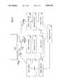
- FIG. 1shows a front view of a flash reflectance oximeter according to this invention.
- FIG. 2shows a side view of the oximeter of FIG. 1.
- FIGS. 3, 4, and 5show cutaway views of the oximeter of FIG. 1.
- FIG. 6shows an enlarged view of a detector and beam splitter assembly.
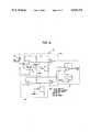
- FIG. 7is a block diagram illustrating the operation of the oximeter of this invention.
- FIG. 8is a schematic diagram illustrating an analog front end of this invention.
- Beer's Lawis not applicable to problems involving the multiple scattering processes found in biological heterogeneous media", as stated by Longini et al. [A Note on the Theory of Backscattering of Light by Living Tissue, IEEE Trans. BME, 15(1):156-160 (1968)].
- the inventorshave developed a reflection mode oximetry technique for noninvasive in vivo use in light of this linear relationship. Furthermore, the inventors have found that SaO 2 is approximately linearly related to the ratio of intensities of reflected infrared and red light. This linear relationship is quite good in the 55% to 95% saturation range, which is the only range of clinical interest for use at the scene of an emergency. To circumvent the problems of nonspecific absorption and arterialization, the inventors use the ratio of the difference of two temporally successive measurements (the change in intensity of infrared and red light) to determine SaO 2 . In this manner, an absolute determination of SaO 2 can be obtained at any number of reasonable sites on an individual and is not limited to fingers, toes, ears, or nose.
- a very high intensity light sourceis employed.
- the duration of each illuminationis kept extremely short.
- the duration of each illuminationis basically a "flash" of light.
- the very high intensity, short duration light pulsedissipates the same energy as a low intensity, continuous light source.
- FIG. 1shows a front view of the reflectance oximeter 10 of this invention, including a lamp face 11 and an annulus 12 for receiving backscattered light.
- FIG. 2shows a side view of the oximeter of FIG. 1.
- the cylindrical oximeter of FIGS. 1 and 2comprises an aluminum housing 13, a sliding collar switch 14, power switch 15, and a visual display 16.
- the aluminum housing 13is 2.18" by 7.0".
- the aluminum housing 13contains optics, electronics on printed circuit boards, batteries, operator controls, visual display, and an electrical connector (for recharging batteries).
- the visual display 16comprises either a liquid crystal display (LCD) or light emitting diode (LED) display.
- LCDliquid crystal display
- LEDlight emitting diode
- a LCDis preferable to a LED display due to the dramatically lower power consumption requirements of the LCD display.
- a LED displaycannot be used in bright sunlight without shading (i.e., by hand).
- a LCDcannot be used in the dark and requires backlighting and more power consumption.
- FIG. 3is a sectional view through a cylindrical axis of the oximeter 10 at line A--A of FIG. 2 and is perpendicular to the view of FIG. 1.
- FIG. 3shows the location of an optical assembly 23 and three printed circuit boards (lamp power circuit board 17, analog front ends circuit board 18, and digital circuit board 19). Integral mountings 20 are provided for the three printed circuit boards 17, 18, and 19. The analog front ends circuit boards 18 and digital circuit board 19 are mounted in a rear compartment 22. Digital electronics 21 on the digital circuit board 19 are shielded from the circuits of the analog front ends circuit board 18 to prevent electromagnetic interference with photodetectors and a microcomputer, described below.
- the optical assemblycomprises a lamp assembly 24 and a detector/beam splitter assembly 25, which are each mounted on three struts located at 120 degree intervals. One mounting strut 26 of three is shown supporting the lamp assembly 24 and the detector/beam splitter assembly 25.
- FIG. 4shows a sectional view at line B--B of FIG. 2, and is perpendicular to the cylindrical axis.
- FIG. 4shows the location of six batteries 27 and the digital circuitry 21.
- the batteriescomprise six 1.2 VDC, 1000 mA-Hr NiCd cells, which are mounted in a compartment 28.
- FIG. 5shows a sectional view at line C--C of FIG. 3, also perpendicular to the cylindrical axis.
- FIG. 5shows the location of the three mounting struts 26 for the lamp assembly 24.
- FIG. 6shows an enlarged view of the detector/beam splitter assembly 25.
- the detector/beam splitter assembly 25includes a diverging lens 29 that receives backscattered light.
- the diverging lens 29transmits the light to a beam splitter 30 that directs the light to two interference filters 31 and 32.
- One filterpasses red light to a first photodetector 33 and the other light passes infrared light to a second photodetector 34.
- the output of these photodetectors 33 and 34represents the intensity of red light and the intensity of infrared light reflected back from the tissue of a patient when a high intensity, short duration pulse of light is directed toward the patient. These intensities are used in determining tissue oxygenation according to this invention.
- FIG. 7is a block diagram illustrating the operation of the oximeter of this invention.
- a microcomputer 35executes a setup sequence for an SaO 2 measurement, enables lamp power circuity, enables a power indicator light 36 of FIG. 1, and retires to a low power consumption mode until either one minute has elapsed (in which case all power to the device is removed to conserve the battery) or until the sliding collar switch 14 is actuated by pushing the oximeter 10 onto a patient.
- the unitexecutes a series of four (2 ⁇ sec duration) lamp flashes over a 750 msec period, for instance. Between successive flashes, the microcomputer 35 retires to a low power consumption mode to conserve power.
- Each flash of light from a light source, encased in a collimating shell,is directed through a converging, infrared-grade lens 37 in contact with material comprising clothing or protective wraps on the patient.
- the flash of lightpasses through the material 38, reflects off the patient, and passes back through the material 38 to a photodetector of this invention.
- the light sourcerequires a high voltage, ultra-low current power, derived from the batteries 27 using a switch 39 and timing circuitry 40.
- a converging lens 37captures the backscattered light and directs the light to a metallized glass parabolic reflector 44.
- the parabolic reflector 44directs this light to a diverging lens 29 of the beam splitter assembly 25.
- Two beams of light emanate from the diverging lens 29,are filtered by narrow bandwidth optical filters 46 and 47 (670 and 905 nm with 10 nm half-bandwidths, for instance), and illuminate high-speed light detectors comprising red and infrared photodiodes 48 and 49.
- the output of each of the red and infrared light photodiodes 48 and 49is amplified and electronically filtered by amplifier/filter circuits 50 and 51.
- each amplifier/filter circuit 50 and 51is directed to an electronically resetable, analog peak-holds circuit 52 and 53.
- Each analog peak-holds circuit 52 or 53holds a maximum output signal from corresponding photodiode 48 or 49 until the peak-holds circuit 52 or 53 is reset.
- the ratio of these two held signals(corresponding to oxygenated hemoglobin and total hemoglobin) is proportional to the normalized oxygen saturation level of the tissue interrogated, and is available less than one millisecond after the flash lamp is triggered. Detector circuitry is properly synchronized, so the resulting reflected or backscattered light peak can be captured.
- the oximeterproduces four flashes. After the fourth flash and digitization, six differences are mathematically examined by a microcomputer 35 to derive the largest difference of intensity of the red light channel and the largest difference of intensity of the infrared channel. From the last three measured differences of the red channel (R 2 -R 1 ; R 3 -R 1 ; R 4 -R 1 ), all six possible differences (R 2 -R 1 ; R 3 -R 1 ; R 4 -R 1 ; R 3 -R 2 ; R 4 -R 2 ; R 4 -R 3 ) can be mathematically developed for the red channel. The same can be accomplished for the infrared channel.
- the first measurement on each channelpermits real-time compensation of imbalances in the analog front ends. It is not necessary to determine the pulse amplitude, but to determine a reasonable difference in the oscillating component to obtain an adequate signal to noise ratio. Over the range of 30 to 180 bpm pulse rate (half-periods of 167 to 1000 msec), a properly sequenced series of flashes yield the desired differences.
- SaO 2is determined by the following formula:
- the operator of this oximeter 10can command the microcomputer 35 to adjust the intensity and duration of each flash.
- An operator in a cold climatecan command the microcomputer 35 to increase the intensity of each flash to penetrate thick layers of clothing or material 38.
- An operator in a warm climatecan command the microcomputer 35 to decrease the intensity of each flash to penetrate thin layers of clothing or to reflect directly off exposed skin.
- the microcomputer 35adjusts the duration of a flash to less than 200 nsec or more than 10 ⁇ sec according to changes in the intensity of the flash.
- the circuitry of this inventioncomprises high amplitude, ultra-fast components to handle 4 or 5 pulses in one second; each pulse from 200 ns to 10 ⁇ s long.
- “high speed”refers to a component which is responsive to, or capable of producing, a pulse of having a width which is sufficiently small to avoid burning a patient's skin when the pulse intensity is sufficiently high to allow measurements through clothing.
- a low power CMOS single chip microcomputer 35comprises a single 68 pin PGA package, central processing unit, RAM, ROM, multiplex analog-to-digital converter, digital input and output, and timers.
- the microcomputer 35drives the visual display 16 (LED or LCD), controls the peak-holds circuits 52 and 53 and the lamp timing circuitry 40.
- the light source 11comprises a light emitting diode which can be driven with high power for a short time.
- a high speed timing circuit 39 and 40 to switch the lampcomprises a high speed buffer amplifier and a switching transistor.
- a high speed detector circuitcomprises photodiodes 48 and 49 (PIN Photodiodes) or a Photodiode with Operational Amplifier which are very sensitive to photons.
- the high speed detector circuitalso comprises a wide-bandwidth amplifier having a fast response time to see fast signals.
- FIG. 8is a schematic diagram illustrating a front end analog circuit for photodiode 48, it being understood from FIG. 7 that an identical circuit is used for photodiode 49.
- the front end analog circuitreceives timing signals at RESET 1 and RESET 2 from the timing circuitry 40.
- the front end analog circuitcomprises the circuit switch 39, one peak-holds circuit 52, and one photodetector 48 or 49 of FIG. 7.
- both peak-hold channels 54 and 55, of peak-holds circuit 52are connected to the photodiode 48.
- the resulting output of each peak-hold channel 54 and 55is the same.
- the output of a differential amplifier 56 connected to both peak-hold channels 54 and 55is zero, indicating that the front end analog circuit is balanced.
- the switch 39then opens the peak-hold channel 55 from the photodetector 48, such that, during the three subsequent flashes, only the peak-hold channel 54 is connected to the photodiode 48.
- the peak-hold channel 55continues to hold the signal corresponding to R 1 , whereas channel 54 holds R 2 , R 3 , and R 4 upon the second, third and fourth pulses, which allows differential amplifier 56 to produce R 2 -R 1 ; R 3 -R 1 ; and R 4 -R 1 .
- These three signalsmay be used to derive the signals or differences of R 3 -R 2 ; R 4 -R 2 ; and R 4 -R 3 in the microprocessor 35 such that the microprocessor has all six possible differences as discussed concerning FIG. 7 above.
- This approacheffectively increases the dynamic range of the measurement, by automatically eliminating the non-oscillatory component of the signal.
- This approachalso eliminates part of the oscillatory component, which can be mathematically recovered from the set of four sequential measurements.
- the gain of each of two such channelsis balanced to compensate for variations in the beam splitter assembly 45, the relative optical attenuation of the filters 46 and 47, the spectral distribution of the lamp 11 output, photodetector 48 and 49 imbalances, and gain differences in the electronics. This is accomplished by trimming a final stage difference amplifier and attenuator on each front end analog circuit.
- the flash reflectance oximeterdoes not necessarily require a microcomputer 35.
- Dedicated analog and digital circuitrycan be designed. However, it is significantly easier to reconfigure a microcomputer-based device by changing the software than to repeatedly redesign high speed analog and digital logic circuitry, with all the attendant noise and printed circuit board design problems. Therefore, to simplify development, a microcomputer has been employed.
- a temperature compensated pressure transducercan measure barometric pressure to compensate for changes in SaO 2 due to changes in altitude.
- a conventional pressure transducer(with a vent hole in the oximeter housing) or a stainless steel diaphragm pressure transducer, with the diaphragm exposed to the atmosphere, can be utilized.
- the inventorshave used relative measurements of the ratio of oxygenated hemoglobin to total hemoglobin under known physiological conditions.
- An occluding cuff(a blood pressure type cuff) was applied to an arm and periodic measurements distal to the cuff were made before, during and after occlusion.
- a baselinewas obtained of the measured ratio of SaO 2 .
- the measured ratio of SaO 2fell below the baseline.
- the measured ratio of SaO 2returned to baseline, transiently exceeded the baseline (the expected hyperemic response), and then returned to the original baseline.
- the oximeter of this inventionworks well through material such as standard clothing, and protective patient wraps. This is a joint function of the high intensity pulse of light, sensitive light detectors, and the porosity of these articles of clothing.
- the flash reflection oximeteris not measurement site limited and measurements can be made proximal to a tourniquet or an occlusion cuff. Furthermore, in the case of intense vasoconstriction of the limbs (e.g. secondary to hypothermia) more central anatomical areas (neck, thorax, abdomen) can be used as measurement sites and differential measurements can be made. A very low reading with the oximeter at various measurement sites will confirm the absence of a pulsating vascular bed, indicating cardiac arrest or death.
- the small size and weight of the oximeteris appropriate for use by medical personnel at the scene of an emergency.
- the ease of use and short measurement time of the oximeterreduces the workload of medical personnel.
- the short measurement timeis appropriate for use on board a helicopter, for instance, where noise and vibration impede vital signs measurements.
- the high intensity, short duration flashpenetrates patient clothing and protective wraps.
- the high intensity, short duration flash, with synchronization to the detection circuits,is insensitive to ambient lighting conditions. Direct application to the subject's clothing makes the oximeter insensitive to ambient humidity and smoke. Stable electronics and a rugged, lightweight housing makes the oximeter very reliable, requiring infrequent calibration.
- the oximeteris unaffected by sex, age, and pigmentation.
- the oximeteris also unaffected by clothing and patient movement.
- the oximeterpermits rapid measurement of SaO 2 in less than one second and without the need to affix sensors.
- the determination of percent SaO 2permits determination of peripheral tissue oxygenation and thus the direct assessment of the cardiopulmonary function of a patient.
- the oximeterpermits detection of tissue hypoxia, even in subjects completely encased in protective gear, and direct assessment of the degree of tissue hypoxia (from mild to life threatening).
- the oximeterhas other applications, such as a noninvasive input sensor for a servo-controlled low flow oxygen delivery system, which can reduce field oxygen consumption and loss while maintaining a patient normoxic.
- Another applicationis obstetrics, where (in combination with fiberoptics) the oximeter can be used in lieu of traditional, invasive fetal blood sampling for determination of fetal distress syndrome.
Landscapes
- Health & Medical Sciences (AREA)
- Physics & Mathematics (AREA)
- Life Sciences & Earth Sciences (AREA)
- Biomedical Technology (AREA)
- Medical Informatics (AREA)
- Biophysics (AREA)
- Pathology (AREA)
- Engineering & Computer Science (AREA)
- Spectroscopy & Molecular Physics (AREA)
- Heart & Thoracic Surgery (AREA)
- Optics & Photonics (AREA)
- Molecular Biology (AREA)
- Surgery (AREA)
- Animal Behavior & Ethology (AREA)
- General Health & Medical Sciences (AREA)
- Public Health (AREA)
- Veterinary Medicine (AREA)
- Measurement Of The Respiration, Hearing Ability, Form, And Blood Characteristics Of Living Organisms (AREA)
Abstract
Description
-D∇.sup.2 ψ(ρ)+Σ.sub.α (ρ)=S(ρ)
SaO.sub.2 =A-B {Δ[I.sub.ir ]/Δ[I.sub.r ]}Claims (3)
Priority Applications (1)
| Application Number | Priority Date | Filing Date | Title |
|---|---|---|---|
| US07/284,496US5069214A (en) | 1988-12-14 | 1988-12-14 | Flash reflectance oximeter |
Applications Claiming Priority (1)
| Application Number | Priority Date | Filing Date | Title |
|---|---|---|---|
| US07/284,496US5069214A (en) | 1988-12-14 | 1988-12-14 | Flash reflectance oximeter |
Publications (1)
| Publication Number | Publication Date |
|---|---|
| US5069214Atrue US5069214A (en) | 1991-12-03 |
Family
ID=23090424
Family Applications (1)
| Application Number | Title | Priority Date | Filing Date |
|---|---|---|---|
| US07/284,496Expired - LifetimeUS5069214A (en) | 1988-12-14 | 1988-12-14 | Flash reflectance oximeter |
Country Status (1)
| Country | Link |
|---|---|
| US (1) | US5069214A (en) |
Cited By (63)
| Publication number | Priority date | Publication date | Assignee | Title |
|---|---|---|---|---|
| US5259382A (en)* | 1991-03-04 | 1993-11-09 | Kronberg James W | Optical transcutaneous bilirubin detector |
| WO1994004070A1 (en)* | 1992-08-14 | 1994-03-03 | Angiomedics Ii, Incorporated | Non-invasive blood analysis by near infrared absorption measurements using two closely spaced wavelengths |
| USD351023S (en) | 1993-01-19 | 1994-09-27 | Biochem International Inc. | Hand held oximeter |
| US5376336A (en)* | 1991-05-10 | 1994-12-27 | Medical Instruments Ag | Apparatus for determining the flow of matter passing through a boundary surface |
| US5919132A (en)* | 1998-03-26 | 1999-07-06 | Universite De Montreal | On-line and real-time spectroreflectometry measurement of oxygenation in a patient's eye |
| EP0641542A3 (en)* | 1993-09-03 | 1999-10-06 | Ken Ishihara | Non-invasive blood analyzer and method using the same |
| US6144867A (en)* | 1998-09-18 | 2000-11-07 | The United States Of America As Represented By The Secretary Of The Army | Self-piercing pulse oximeter sensor assembly |
| US6149589A (en)* | 1998-03-26 | 2000-11-21 | Universite De Montreal | On-line and real-time spectroreflectometry measurement of oxygenation in a patient's eye |
| US6253098B1 (en) | 1998-09-09 | 2001-06-26 | The United States Of America As Represented By The Secretary Of The Army | Disposable pulse oximeter assembly and protective cover therefor |
| US6256524B1 (en) | 1998-09-09 | 2001-07-03 | The United States Of America As Represented By The Secretary Of The Army | Pulse oximeter sensor combined with a combination oropharyngeal airway and bite block |
| US6263223B1 (en) | 1998-09-09 | 2001-07-17 | The United States Of America As Represented By The Secretary Of The Army | Method for monitoring arterial oxygen saturation |
| US6266547B1 (en) | 1998-09-09 | 2001-07-24 | The United States Of America As Represented By The Secretary Of The Army | Nasopharyngeal airway with reflectance pulse oximeter sensor |
| US6345195B1 (en)* | 1999-09-29 | 2002-02-05 | Varda Herskowits | Methods and apparatus for 3D scanning of the human body form |
| US6400972B1 (en)* | 1998-06-17 | 2002-06-04 | Orsense Ltd. | Non-invasive method and system of optical measurements for determining the concentration of a substance in blood |
| WO2002026113A3 (en)* | 2000-09-29 | 2002-06-13 | Datex Ohmeda Inc | Method and apparatus for determining pulse oximetry differential values |
| US6470200B2 (en) | 2000-02-11 | 2002-10-22 | The United States Of America As Represented By The Secretary Of The Army | Pacifier pulse oximeter sensor |
| US6480311B1 (en)* | 1997-09-18 | 2002-11-12 | Sharp Kabushiki Kaisha | Peak-hold circuit and an infrared communication device provided with such a circuit |
| US6587704B1 (en)* | 1999-06-16 | 2003-07-01 | Orsense Ltd. | Method for non-invasive optical measurements of blood parameters |
| US20040002637A1 (en)* | 2002-03-13 | 2004-01-01 | Huang Johnnie W. | Power conserving adaptive control system for generating signal in portable medical devices |
| US6711424B1 (en) | 1999-12-22 | 2004-03-23 | Orsense Ltd. | Method of optical measurement for determing various parameters of the patient's blood |
| US20040249252A1 (en)* | 2003-06-03 | 2004-12-09 | Orsense Ltd. | Method and system for use in non-invasive optical measurements of blood parameters |
| US6898451B2 (en)* | 2001-03-21 | 2005-05-24 | Minformed, L.L.C. | Non-invasive blood analyte measuring system and method utilizing optical absorption |
| US20060009685A1 (en)* | 2004-07-08 | 2006-01-12 | Orsense Ltd. | Device and method for non-invasive optical measurements |
| DE102004032094A1 (en)* | 2004-07-01 | 2006-01-26 | Micro-Epsilon Messtechnik Gmbh & Co Kg | Blood oxygen saturation measuring method for use in field of anesthesia, involves comparing measured values, which are recorded from body of patient, with reference value for determining deviation of measured values from reference value |
| US20070038049A1 (en)* | 2005-07-08 | 2007-02-15 | Huang Johnnie W | System for adjusting power employed by a medical device |
| US7254432B2 (en) | 2005-08-17 | 2007-08-07 | Orsense Ltd. | Method and device for non-invasive measurements of blood parameters |
| US20080194906A1 (en)* | 2004-10-27 | 2008-08-14 | General Electric Company | Measurement and treatment system and method |
| US20080200784A1 (en)* | 2007-02-16 | 2008-08-21 | Xuefeng Cheng | Method and device for measuring parameters of cardiac function |
| US20090048495A1 (en)* | 2004-03-04 | 2009-02-19 | Masimo Corporation | Application identification sensor |
| US20090156918A1 (en)* | 2007-12-12 | 2009-06-18 | Davis Timothy J | Implantable optical sensor and method for use |
| US20100004518A1 (en)* | 2008-07-03 | 2010-01-07 | Masimo Laboratories, Inc. | Heat sink for noninvasive medical sensor |
| US20100240972A1 (en)* | 2009-03-20 | 2010-09-23 | Nellcor Puritan Bennett Llc | Slider Spot Check Pulse Oximeter |
| US20100249550A1 (en)* | 2009-03-25 | 2010-09-30 | Neilcor Puritan Bennett LLC | Method And Apparatus For Optical Filtering Of A Broadband Emitter In A Medical Sensor |
| US20100256470A1 (en)* | 2009-04-02 | 2010-10-07 | Seth Adrian Miller | Touch screen interfaces with pulse oximetry |
| US20100289772A1 (en)* | 2009-05-18 | 2010-11-18 | Seth Adrian Miller | Touch-sensitive device and method |
| US20120016245A1 (en)* | 2010-07-14 | 2012-01-19 | Rohm Co., Ltd. | Plethysmogram sensor |
| US20120229800A1 (en)* | 2011-03-08 | 2012-09-13 | Fluke Corporation | Pulse oximeter test instruments and methods |
| US20120259188A1 (en)* | 2011-04-08 | 2012-10-11 | Nxp B.V. | Flexible Eye Insert and Glucose Measuring System |
| US20130116512A1 (en)* | 2011-04-26 | 2013-05-09 | Mir Imran | Mouthpiece for measurement of biometric data of a diver and underwater communication |
| US8515509B2 (en) | 2008-08-04 | 2013-08-20 | Cercacor Laboratories, Inc. | Multi-stream emitter for noninvasive measurement of blood constituents |
| US8688183B2 (en) | 2009-09-03 | 2014-04-01 | Ceracor Laboratories, Inc. | Emitter driver for noninvasive patient monitor |
| WO2014085302A1 (en)* | 2012-11-27 | 2014-06-05 | Faurecia Automotive Seating, Llc | Vehicle seat with integrated sensors |
| WO2014085394A1 (en)* | 2012-11-27 | 2014-06-05 | Faurecia Automotive Seating, Llc | Oximetry sensor assembly and methodology for sensing blood oxygen concentration |
| US8918153B2 (en) | 2007-02-16 | 2014-12-23 | Mespere Lifesciences Inc. | Method and device for measuring parameters of cardiac function |
| USD740959S1 (en)* | 2013-10-01 | 2015-10-13 | Joy's International Cosmetics Pty Ltd | Human body energy analysis device set |
| US9839379B2 (en)* | 2013-10-07 | 2017-12-12 | Masimo Corporation | Regional oximetry pod |
| US9848814B2 (en) | 2014-02-20 | 2017-12-26 | Faurecia Automotive Seating, Llc | Vehicle seat with integrated sensors |
| US10046671B2 (en) | 2015-08-14 | 2018-08-14 | Faurecia Automotive Seating, Llc | Occupant-recognition system for vehicle seat |
| US20190069813A1 (en)* | 2001-07-02 | 2019-03-07 | Masimo Corporation | Low power pulse oximeter |
| US10398364B2 (en) | 2013-02-13 | 2019-09-03 | Mespere Lifesciences Inc. | Method and device for measuring venous blood oxygenation |
| US10709367B1 (en)* | 2009-01-26 | 2020-07-14 | Vioptix, Inc. | Multidepth tissue oximeter |
| US10730524B2 (en) | 2016-09-07 | 2020-08-04 | Faurecia Automotive Seating, Llc | Vehicle seat |
| WO2020210793A1 (en) | 2019-04-12 | 2020-10-15 | Verily Life Sciences Llc | Sensing physiological parameters through an article |
| US10918337B2 (en) | 2016-06-03 | 2021-02-16 | Faurecia Automotive Seating, Llc | Vehicle seat with integrated sensors |
| US10966643B1 (en)* | 2019-06-12 | 2021-04-06 | Fitbit, Inc. | Wearable non-invasive carbon monoxide inhalation tracking |
| US11083379B2 (en) | 2017-08-02 | 2021-08-10 | Faurecia Automotive Seating, Llc | Health-monitoring seat cover |
| US11096598B2 (en) | 2015-10-08 | 2021-08-24 | Mespere Lifesciences Inc. | System and method for non-invasive monitoring of central venous pressure |
| US11147518B1 (en) | 2013-10-07 | 2021-10-19 | Masimo Corporation | Regional oximetry signal processor |
| US11197637B2 (en) | 2016-06-20 | 2021-12-14 | Faurecia Automotive Seating, Llc | Control system for a vehicle seat |
| US11373102B2 (en) | 2018-05-04 | 2022-06-28 | The Procter & Gamble Company | Sensing and activity classification for infants |
| US11679036B2 (en) | 2019-04-12 | 2023-06-20 | Verily Life Sciences Llc | Determining diaper loading using color detection or activity state |
| US12230393B2 (en) | 2005-03-01 | 2025-02-18 | Willow Laboratories, Inc. | Multiple wavelength sensor emitters |
| US12336796B2 (en) | 2021-07-13 | 2025-06-24 | Masimo Corporation | Wearable device with physiological parameters monitoring |
Citations (15)
| Publication number | Priority date | Publication date | Assignee | Title |
|---|---|---|---|---|
| US3177757A (en)* | 1960-03-29 | 1965-04-13 | American Optical Corp | Method of performing reflective oximetry |
| US3461856A (en)* | 1965-10-23 | 1969-08-19 | American Optical Corp | Oximeters |
| US3647299A (en)* | 1970-04-20 | 1972-03-07 | American Optical Corp | Oximeter |
| US4167331A (en)* | 1976-12-20 | 1979-09-11 | Hewlett-Packard Company | Multi-wavelength incremental absorbence oximeter |
| US4305398A (en)* | 1977-12-30 | 1981-12-15 | Minolta Camera Kabushiki Kaisha | Eye fundus oximeter |
| US4447150A (en)* | 1981-02-27 | 1984-05-08 | Bentley Laboratories | Apparatus and method for measuring blood oxygen saturation |
| US4453218A (en)* | 1980-11-24 | 1984-06-05 | Oximetrix, Inc. | Signal filter method and apparatus |
| US4523279A (en)* | 1980-11-24 | 1985-06-11 | Oximetrix, Inc. | Apparatus for determining oxygen saturation levels in blood |
| US4586513A (en)* | 1982-02-19 | 1986-05-06 | Minolta Camera Kabushiki Kaisha | Noninvasive device for photoelectrically measuring the property of arterial blood |
| US4714080A (en)* | 1986-10-06 | 1987-12-22 | Nippon Colin Co., Ltd. | Method and apparatus for noninvasive monitoring of arterial blood oxygen saturation |
| US4759369A (en)* | 1986-07-07 | 1988-07-26 | Novametrix Medical Systems, Inc. | Pulse oximeter |
| US4796636A (en)* | 1987-09-10 | 1989-01-10 | Nippon Colin Co., Ltd. | Noninvasive reflectance oximeter |
| US4832484A (en)* | 1986-10-29 | 1989-05-23 | Nihon Kohden Corporation | Apparatus for determining the concentration of a light-absorbing material in blood |
| US4880304A (en)* | 1987-04-01 | 1989-11-14 | Nippon Colin Co., Ltd. | Optical sensor for pulse oximeter |
| US4883353A (en)* | 1988-02-11 | 1989-11-28 | Puritan-Bennett Corporation | Pulse oximeter |
- 1988
- 1988-12-14USUS07/284,496patent/US5069214A/ennot_activeExpired - Lifetime
Patent Citations (15)
| Publication number | Priority date | Publication date | Assignee | Title |
|---|---|---|---|---|
| US3177757A (en)* | 1960-03-29 | 1965-04-13 | American Optical Corp | Method of performing reflective oximetry |
| US3461856A (en)* | 1965-10-23 | 1969-08-19 | American Optical Corp | Oximeters |
| US3647299A (en)* | 1970-04-20 | 1972-03-07 | American Optical Corp | Oximeter |
| US4167331A (en)* | 1976-12-20 | 1979-09-11 | Hewlett-Packard Company | Multi-wavelength incremental absorbence oximeter |
| US4305398A (en)* | 1977-12-30 | 1981-12-15 | Minolta Camera Kabushiki Kaisha | Eye fundus oximeter |
| US4453218A (en)* | 1980-11-24 | 1984-06-05 | Oximetrix, Inc. | Signal filter method and apparatus |
| US4523279A (en)* | 1980-11-24 | 1985-06-11 | Oximetrix, Inc. | Apparatus for determining oxygen saturation levels in blood |
| US4447150A (en)* | 1981-02-27 | 1984-05-08 | Bentley Laboratories | Apparatus and method for measuring blood oxygen saturation |
| US4586513A (en)* | 1982-02-19 | 1986-05-06 | Minolta Camera Kabushiki Kaisha | Noninvasive device for photoelectrically measuring the property of arterial blood |
| US4759369A (en)* | 1986-07-07 | 1988-07-26 | Novametrix Medical Systems, Inc. | Pulse oximeter |
| US4714080A (en)* | 1986-10-06 | 1987-12-22 | Nippon Colin Co., Ltd. | Method and apparatus for noninvasive monitoring of arterial blood oxygen saturation |
| US4832484A (en)* | 1986-10-29 | 1989-05-23 | Nihon Kohden Corporation | Apparatus for determining the concentration of a light-absorbing material in blood |
| US4880304A (en)* | 1987-04-01 | 1989-11-14 | Nippon Colin Co., Ltd. | Optical sensor for pulse oximeter |
| US4796636A (en)* | 1987-09-10 | 1989-01-10 | Nippon Colin Co., Ltd. | Noninvasive reflectance oximeter |
| US4883353A (en)* | 1988-02-11 | 1989-11-28 | Puritan-Bennett Corporation | Pulse oximeter |
Non-Patent Citations (12)
| Title |
|---|
| "Design and Evaluation of a New Reflectance Pulse Oximeter Sensor", Y. Mendelson, J. C. Kent, B. L. Yocum, and M. J. Birle, Medical Instrumentation, vol. 22, No. 4, Aug. 1988. |
| "Noninvasive Pulse Oximetry Utilizing Skin Reflectance Photoplethysmography", Yitzhak Mendelson and Burt D. Ochs, IEEE Transactions on Biomedical Engineering, vol. 35, No. 10, Oct. 1988. |
| "Noninvasive Transcutaneous Monitoring of Arterial Blood Gases", Yitzhak Mendelson and Robert A. Peura, IEEE Transactions on Biomedical Engineering, vol. BME-31, No. 12, Dec. 1984. |
| Design and Evaluation of a New Reflectance Pulse Oximeter Sensor , Y. Mendelson, J. C. Kent, B. L. Yocum, and M. J. Birle, Medical Instrumentation, vol. 22, No. 4, Aug. 1988.* |
| Noninvasive Pulse Oximetry Utilizing Skin Reflectance Photoplethysmography , Yitzhak Mendelson and Burt D. Ochs, IEEE Transactions on Biomedical Engineering, vol. 35, No. 10, Oct. 1988.* |
| Noninvasive Transcutaneous Monitoring of Arterial Blood Gases , Yitzhak Mendelson and Robert A. Peura, IEEE Transactions on Biomedical Engineering, vol. BME 31, No. 12, Dec. 1984.* |
| Sutterer et al. [Calculation and Digital Display of Whole Blood Oxygen Saturation by Analog Techniques, IEEE Trans. BME, 16(2):116-122 (1969)]. |
| Sutterer et al. Calculation and Digital Display of Whole Blood Oxygen Saturation by Analog Techniques, IEEE Trans. BME, 16(2):116 122 (1969) .* |
| Wood et al. [Photoelectric Determination of Arterial Oxygen Saturation in Man, J. Lab. Clin. Med., 34:387-401 (1949)]. |
| Wood et al. Photoelectric Determination of Arterial Oxygen Saturation in Man, J. Lab. Clin. Med., 34:387 401 (1949) .* |
| Yoshiya et al. [Spectrophotometric Monitoring of Arterial Oxygen in the Fingertip, Med. & Biol. Eng. & Comput., 18:27-32 (1980)]. |
| Yoshiya et al. Spectrophotometric Monitoring of Arterial Oxygen in the Fingertip, Med. & Biol. Eng. & Comput., 18:27 32 (1980) .* |
Cited By (150)
| Publication number | Priority date | Publication date | Assignee | Title |
|---|---|---|---|---|
| US5259382A (en)* | 1991-03-04 | 1993-11-09 | Kronberg James W | Optical transcutaneous bilirubin detector |
| US5376336A (en)* | 1991-05-10 | 1994-12-27 | Medical Instruments Ag | Apparatus for determining the flow of matter passing through a boundary surface |
| WO1994004070A1 (en)* | 1992-08-14 | 1994-03-03 | Angiomedics Ii, Incorporated | Non-invasive blood analysis by near infrared absorption measurements using two closely spaced wavelengths |
| USD351023S (en) | 1993-01-19 | 1994-09-27 | Biochem International Inc. | Hand held oximeter |
| EP0641542A3 (en)* | 1993-09-03 | 1999-10-06 | Ken Ishihara | Non-invasive blood analyzer and method using the same |
| US6480311B1 (en)* | 1997-09-18 | 2002-11-12 | Sharp Kabushiki Kaisha | Peak-hold circuit and an infrared communication device provided with such a circuit |
| US5919132A (en)* | 1998-03-26 | 1999-07-06 | Universite De Montreal | On-line and real-time spectroreflectometry measurement of oxygenation in a patient's eye |
| US6149589A (en)* | 1998-03-26 | 2000-11-21 | Universite De Montreal | On-line and real-time spectroreflectometry measurement of oxygenation in a patient's eye |
| US6400972B1 (en)* | 1998-06-17 | 2002-06-04 | Orsense Ltd. | Non-invasive method and system of optical measurements for determining the concentration of a substance in blood |
| US6253098B1 (en) | 1998-09-09 | 2001-06-26 | The United States Of America As Represented By The Secretary Of The Army | Disposable pulse oximeter assembly and protective cover therefor |
| US6263223B1 (en) | 1998-09-09 | 2001-07-17 | The United States Of America As Represented By The Secretary Of The Army | Method for monitoring arterial oxygen saturation |
| US6266547B1 (en) | 1998-09-09 | 2001-07-24 | The United States Of America As Represented By The Secretary Of The Army | Nasopharyngeal airway with reflectance pulse oximeter sensor |
| US6256524B1 (en) | 1998-09-09 | 2001-07-03 | The United States Of America As Represented By The Secretary Of The Army | Pulse oximeter sensor combined with a combination oropharyngeal airway and bite block |
| US6144867A (en)* | 1998-09-18 | 2000-11-07 | The United States Of America As Represented By The Secretary Of The Army | Self-piercing pulse oximeter sensor assembly |
| US6587704B1 (en)* | 1999-06-16 | 2003-07-01 | Orsense Ltd. | Method for non-invasive optical measurements of blood parameters |
| US6345195B1 (en)* | 1999-09-29 | 2002-02-05 | Varda Herskowits | Methods and apparatus for 3D scanning of the human body form |
| US7317939B2 (en) | 1999-12-22 | 2008-01-08 | Orsense Ltd. | Method of optical measurements for determining various parameters of the patient's blood |
| US6711424B1 (en) | 1999-12-22 | 2004-03-23 | Orsense Ltd. | Method of optical measurement for determing various parameters of the patient's blood |
| US20060200014A1 (en)* | 1999-12-22 | 2006-09-07 | Orsense Ltd. | Method of optical measurements for determining various parameters of the patient's blood |
| US7043289B2 (en) | 1999-12-22 | 2006-05-09 | Orsense Ltd. | Method of optical measurements for determining various parameters of the patient's blood |
| US6470200B2 (en) | 2000-02-11 | 2002-10-22 | The United States Of America As Represented By The Secretary Of The Army | Pacifier pulse oximeter sensor |
| US6505060B1 (en)* | 2000-09-29 | 2003-01-07 | Datex-Ohmeda, Inc. | Method and apparatus for determining pulse oximetry differential values |
| WO2002026113A3 (en)* | 2000-09-29 | 2002-06-13 | Datex Ohmeda Inc | Method and apparatus for determining pulse oximetry differential values |
| US6898451B2 (en)* | 2001-03-21 | 2005-05-24 | Minformed, L.L.C. | Non-invasive blood analyte measuring system and method utilizing optical absorption |
| US10980455B2 (en)* | 2001-07-02 | 2021-04-20 | Masimo Corporation | Low power pulse oximeter |
| US11219391B2 (en)* | 2001-07-02 | 2022-01-11 | Masimo Corporation | Low power pulse oximeter |
| US20190069813A1 (en)* | 2001-07-02 | 2019-03-07 | Masimo Corporation | Low power pulse oximeter |
| US10433776B2 (en)* | 2001-07-02 | 2019-10-08 | Masimo Corporation | Low power pulse oximeter |
| US10959652B2 (en) | 2001-07-02 | 2021-03-30 | Masimo Corporation | Low power pulse oximeter |
| US6863652B2 (en) | 2002-03-13 | 2005-03-08 | Draeger Medical Systems, Inc. | Power conserving adaptive control system for generating signal in portable medical devices |
| US20040002637A1 (en)* | 2002-03-13 | 2004-01-01 | Huang Johnnie W. | Power conserving adaptive control system for generating signal in portable medical devices |
| US20060129040A1 (en)* | 2003-06-03 | 2006-06-15 | Orsense Ltd. | Method and system for use in non-invasive optical measurements of blood parameters |
| US7386336B2 (en) | 2003-06-03 | 2008-06-10 | Orsense Ltd. | Method and system for use in non-invasive optical measurements of blood parameters |
| US20040249252A1 (en)* | 2003-06-03 | 2004-12-09 | Orsense Ltd. | Method and system for use in non-invasive optical measurements of blood parameters |
| US6993372B2 (en)* | 2003-06-03 | 2006-01-31 | Orsense Ltd. | Method and system for use in non-invasive optical measurements of blood parameters |
| US9161713B2 (en) | 2004-03-04 | 2015-10-20 | Masimo Corporation | Multi-mode patient monitor configured to self-configure for a selected or determined mode of operation |
| US8337403B2 (en)* | 2004-03-04 | 2012-12-25 | Masimo Corporation | Patient monitor having context-based sensitivity adjustments |
| US20090048495A1 (en)* | 2004-03-04 | 2009-02-19 | Masimo Corporation | Application identification sensor |
| DE102004032094A1 (en)* | 2004-07-01 | 2006-01-26 | Micro-Epsilon Messtechnik Gmbh & Co Kg | Blood oxygen saturation measuring method for use in field of anesthesia, involves comparing measured values, which are recorded from body of patient, with reference value for determining deviation of measured values from reference value |
| US20060009685A1 (en)* | 2004-07-08 | 2006-01-12 | Orsense Ltd. | Device and method for non-invasive optical measurements |
| US7313425B2 (en) | 2004-07-08 | 2007-12-25 | Orsense Ltd. | Device and method for non-invasive optical measurements |
| US9872643B2 (en)* | 2004-10-27 | 2018-01-23 | General Electric Company | Measurement and treatment system and method |
| US20080194906A1 (en)* | 2004-10-27 | 2008-08-14 | General Electric Company | Measurement and treatment system and method |
| US12230393B2 (en) | 2005-03-01 | 2025-02-18 | Willow Laboratories, Inc. | Multiple wavelength sensor emitters |
| US20070038049A1 (en)* | 2005-07-08 | 2007-02-15 | Huang Johnnie W | System for adjusting power employed by a medical device |
| US8116837B2 (en) | 2005-07-08 | 2012-02-14 | Draeger Medical Systems, Inc. | System for adjusting power employed by a medical device |
| US7254432B2 (en) | 2005-08-17 | 2007-08-07 | Orsense Ltd. | Method and device for non-invasive measurements of blood parameters |
| US8417306B2 (en) | 2007-02-16 | 2013-04-09 | Mespere Lifesciences Inc. | Method and device for measuring parameters of cardiac function |
| US8918153B2 (en) | 2007-02-16 | 2014-12-23 | Mespere Lifesciences Inc. | Method and device for measuring parameters of cardiac function |
| US20080200784A1 (en)* | 2007-02-16 | 2008-08-21 | Xuefeng Cheng | Method and device for measuring parameters of cardiac function |
| US20090326352A1 (en)* | 2007-02-16 | 2009-12-31 | Xuefeng Cheng | Method and device for measuring parameters of cardiac function |
| US8290557B2 (en)* | 2007-12-12 | 2012-10-16 | Medtronic, Inc. | Implantable optical sensor and method for use |
| US20090156918A1 (en)* | 2007-12-12 | 2009-06-18 | Davis Timothy J | Implantable optical sensor and method for use |
| US9717425B2 (en) | 2008-07-03 | 2017-08-01 | Masimo Corporation | Noise shielding for a noninvaise device |
| US10610138B2 (en) | 2008-07-03 | 2020-04-07 | Masimo Corporation | Multi-stream data collection system for noninvasive measurement of blood constituents |
| US11647914B2 (en) | 2008-07-03 | 2023-05-16 | Masimo Corporation | User-worn device for noninvasively measuring a physiological parameter of a user |
| US10702195B1 (en) | 2008-07-03 | 2020-07-07 | Masimo Corporation | Multi-stream data collection system for noninvasive measurement of blood constituents |
| US10702194B1 (en) | 2008-07-03 | 2020-07-07 | Masimo Corporation | Multi-stream data collection system for noninvasive measurement of blood constituents |
| US8577431B2 (en) | 2008-07-03 | 2013-11-05 | Cercacor Laboratories, Inc. | Noise shielding for a noninvasive device |
| US10631765B1 (en) | 2008-07-03 | 2020-04-28 | Masimo Corporation | Multi-stream data collection system for noninvasive measurement of blood constituents |
| US11642037B2 (en) | 2008-07-03 | 2023-05-09 | Masimo Corporation | User-worn device for noninvasively measuring a physiological parameter of a user |
| US11642036B2 (en) | 2008-07-03 | 2023-05-09 | Masimo Corporation | User-worn device for noninvasively measuring a physiological parameter of a user |
| US11638532B2 (en) | 2008-07-03 | 2023-05-02 | Masimo Corporation | User-worn device for noninvasively measuring a physiological parameter of a user |
| US10624563B2 (en) | 2008-07-03 | 2020-04-21 | Masimo Corporation | Multi-stream data collection system for noninvasive measurement of blood constituents |
| US10624564B1 (en) | 2008-07-03 | 2020-04-21 | Masimo Corporation | Multi-stream data collection system for noninvasive measurement of blood constituents |
| US10617338B2 (en) | 2008-07-03 | 2020-04-14 | Masimo Corporation | Multi-stream data collection system for noninvasive measurement of blood constituents |
| US11484229B2 (en) | 2008-07-03 | 2022-11-01 | Masimo Corporation | User-worn device for noninvasively measuring a physiological parameter of a user |
| US11484230B2 (en) | 2008-07-03 | 2022-11-01 | Masimo Corporation | User-worn device for noninvasively measuring a physiological parameter of a user |
| US11426103B2 (en) | 2008-07-03 | 2022-08-30 | Masimo Corporation | Multi-stream data collection system for noninvasive measurement of blood constituents |
| US10912501B2 (en) | 2008-07-03 | 2021-02-09 | Masimo Corporation | User-worn device for noninvasively measuring a physiological parameter of a user |
| US10588554B2 (en) | 2008-07-03 | 2020-03-17 | Masimo Corporation | Multi-stream data collection system for noninvasive measurement of blood constituents |
| US9277880B2 (en) | 2008-07-03 | 2016-03-08 | Masimo Corporation | Multi-stream data collection system for noninvasive measurement of blood constituents |
| US10588553B2 (en) | 2008-07-03 | 2020-03-17 | Masimo Corporation | Multi-stream data collection system for noninvasive measurement of blood constituents |
| US10582886B2 (en) | 2008-07-03 | 2020-03-10 | Masimo Corporation | Multi-stream data collection system for noninvasive measurement of blood constituents |
| US9591975B2 (en) | 2008-07-03 | 2017-03-14 | Masimo Corporation | Contoured protrusion for improving spectroscopic measurement of blood constituents |
| US20100004518A1 (en)* | 2008-07-03 | 2010-01-07 | Masimo Laboratories, Inc. | Heat sink for noninvasive medical sensor |
| US12023139B1 (en) | 2008-07-03 | 2024-07-02 | Masimo Corporation | User-worn device for noninvasively measuring a physiological parameter of a user |
| US8437825B2 (en) | 2008-07-03 | 2013-05-07 | Cercacor Laboratories, Inc. | Contoured protrusion for improving spectroscopic measurement of blood constituents |
| US10945648B2 (en) | 2008-07-03 | 2021-03-16 | Masimo Corporation | User-worn device for noninvasively measuring a physiological parameter of a user |
| US10299708B1 (en) | 2008-07-03 | 2019-05-28 | Masimo Corporation | Multi-stream data collection system for noninvasive measurement of blood constituents |
| US10912502B2 (en) | 2008-07-03 | 2021-02-09 | Masimo Corporation | User-worn device for noninvasively measuring a physiological parameter of a user |
| US12036009B1 (en) | 2008-07-03 | 2024-07-16 | Masimo Corporation | User-worn device for noninvasively measuring a physiological parameter of a user |
| US10758166B2 (en) | 2008-07-03 | 2020-09-01 | Masimo Corporation | Multi-stream data collection system for noninvasive measurement of blood constituents |
| US10743803B2 (en) | 2008-07-03 | 2020-08-18 | Masimo Corporation | Multi-stream data collection system for noninvasive measurement of blood constituents |
| US11751773B2 (en) | 2008-07-03 | 2023-09-12 | Masimo Corporation | Emitter arrangement for physiological measurements |
| US10258265B1 (en) | 2008-07-03 | 2019-04-16 | Masimo Corporation | Multi-stream data collection system for noninvasive measurement of blood constituents |
| US10258266B1 (en) | 2008-07-03 | 2019-04-16 | Masimo Corporation | Multi-stream data collection system for noninvasive measurement of blood constituents |
| US10292628B1 (en) | 2008-07-03 | 2019-05-21 | Masimo Corporation | Multi-stream data collection system for noninvasive measurement of blood constituents |
| US10912500B2 (en) | 2008-07-03 | 2021-02-09 | Masimo Corporation | Multi-stream data collection system for noninvasive measurement of blood constituents |
| US10335068B2 (en) | 2008-07-03 | 2019-07-02 | Masimo Corporation | Multi-stream data collection system for noninvasive measurement of blood constituents |
| US10376191B1 (en) | 2008-07-03 | 2019-08-13 | Masimo Corporation | Multi-stream data collection system for noninvasive measurement of blood constituents |
| US10376190B1 (en) | 2008-07-03 | 2019-08-13 | Masimo Corporation | Multi-stream data collection system for noninvasive measurement of blood constituents |
| US10709366B1 (en) | 2008-07-03 | 2020-07-14 | Masimo Corporation | Multi-stream data collection system for noninvasive measurement of blood constituents |
| US8909310B2 (en) | 2008-08-04 | 2014-12-09 | Cercacor Laboratories, Inc. | Multi-stream sensor front ends for noninvasive measurement of blood constituents |
| US8630691B2 (en) | 2008-08-04 | 2014-01-14 | Cercacor Laboratories, Inc. | Multi-stream sensor front ends for noninvasive measurement of blood constituents |
| US8570503B2 (en) | 2008-08-04 | 2013-10-29 | Cercacor Laboratories, Inc. | Heat sink for noninvasive medical sensor |
| US8515509B2 (en) | 2008-08-04 | 2013-08-20 | Cercacor Laboratories, Inc. | Multi-stream emitter for noninvasive measurement of blood constituents |
| US12226207B1 (en) | 2009-01-26 | 2025-02-18 | Vioptix, Inc. | Multidepth tissue oximeter |
| US10709367B1 (en)* | 2009-01-26 | 2020-07-14 | Vioptix, Inc. | Multidepth tissue oximeter |
| US11564601B1 (en) | 2009-01-26 | 2023-01-31 | Vioptix, Inc. | Multidepth tissue oximeter |
| US20100240972A1 (en)* | 2009-03-20 | 2010-09-23 | Nellcor Puritan Bennett Llc | Slider Spot Check Pulse Oximeter |
| US20100249550A1 (en)* | 2009-03-25 | 2010-09-30 | Neilcor Puritan Bennett LLC | Method And Apparatus For Optical Filtering Of A Broadband Emitter In A Medical Sensor |
| US8320985B2 (en)* | 2009-04-02 | 2012-11-27 | Empire Technology Development Llc | Touch screen interfaces with pulse oximetry |
| US20100256470A1 (en)* | 2009-04-02 | 2010-10-07 | Seth Adrian Miller | Touch screen interfaces with pulse oximetry |
| US9427192B2 (en) | 2009-05-18 | 2016-08-30 | Empire Technology Development Llc | Touch-sensitive device and method |
| US20100289772A1 (en)* | 2009-05-18 | 2010-11-18 | Seth Adrian Miller | Touch-sensitive device and method |
| US8786575B2 (en) | 2009-05-18 | 2014-07-22 | Empire Technology Development LLP | Touch-sensitive device and method |
| US8688183B2 (en) | 2009-09-03 | 2014-04-01 | Ceracor Laboratories, Inc. | Emitter driver for noninvasive patient monitor |
| US9668680B2 (en) | 2009-09-03 | 2017-06-06 | Masimo Corporation | Emitter driver for noninvasive patient monitor |
| US9186102B2 (en) | 2009-09-03 | 2015-11-17 | Cercacor Laboratories, Inc. | Emitter driver for noninvasive patient monitor |
| US20120016245A1 (en)* | 2010-07-14 | 2012-01-19 | Rohm Co., Ltd. | Plethysmogram sensor |
| US20120229800A1 (en)* | 2011-03-08 | 2012-09-13 | Fluke Corporation | Pulse oximeter test instruments and methods |
| CN102670210A (en)* | 2011-03-08 | 2012-09-19 | 弗卢克公司 | Pulse oximeter test instruments and methods |
| US20120259188A1 (en)* | 2011-04-08 | 2012-10-11 | Nxp B.V. | Flexible Eye Insert and Glucose Measuring System |
| US9737245B2 (en)* | 2011-04-08 | 2017-08-22 | Nxp B.V. | Flexible eye insert and glucose measuring system |
| AU2016216717B2 (en)* | 2011-04-26 | 2018-03-15 | Incube Labs, Llc | Mouthpiece for measurement of biometric data of a diver and underwater communication |
| US9795296B2 (en)* | 2011-04-26 | 2017-10-24 | Incube Labs, Llc | Mouthpiece for measurement of biometric data of a diver and underwater communication |
| AU2012249600B2 (en)* | 2011-04-26 | 2016-05-19 | Incube Labs, Llc | Mouthpiece for measurement of biometric data of a diver and underwater communication |
| US20130116512A1 (en)* | 2011-04-26 | 2013-05-09 | Mir Imran | Mouthpiece for measurement of biometric data of a diver and underwater communication |
| US10786162B2 (en) | 2012-11-27 | 2020-09-29 | Faurecia Automotive Seating, Llc | Vehicle seat with integrated sensors |
| WO2014085302A1 (en)* | 2012-11-27 | 2014-06-05 | Faurecia Automotive Seating, Llc | Vehicle seat with integrated sensors |
| WO2014085394A1 (en)* | 2012-11-27 | 2014-06-05 | Faurecia Automotive Seating, Llc | Oximetry sensor assembly and methodology for sensing blood oxygen concentration |
| CN104837403A (en)* | 2012-11-27 | 2015-08-12 | 佛吉亚汽车座椅有限责任公司 | Vehicle seat with integrated sensors |
| CN104853675A (en)* | 2012-11-27 | 2015-08-19 | 佛吉亚汽车座椅有限责任公司 | Oximetry sensor assembly and methodology for sensing blood oxygen concentration |
| US10398364B2 (en) | 2013-02-13 | 2019-09-03 | Mespere Lifesciences Inc. | Method and device for measuring venous blood oxygenation |
| USD740959S1 (en)* | 2013-10-01 | 2015-10-13 | Joy's International Cosmetics Pty Ltd | Human body energy analysis device set |
| US10799160B2 (en) | 2013-10-07 | 2020-10-13 | Masimo Corporation | Regional oximetry pod |
| US11751780B2 (en) | 2013-10-07 | 2023-09-12 | Masimo Corporation | Regional oximetry sensor |
| US11147518B1 (en) | 2013-10-07 | 2021-10-19 | Masimo Corporation | Regional oximetry signal processor |
| US12357203B2 (en) | 2013-10-07 | 2025-07-15 | Masimo Corporation | Regional oximetry pod |
| US12357237B1 (en) | 2013-10-07 | 2025-07-15 | Masimo Corporation | Regional oximetry signal processor |
| US12318196B2 (en) | 2013-10-07 | 2025-06-03 | Masimo Corporation | Regional oximetry user interface |
| US11076782B2 (en) | 2013-10-07 | 2021-08-03 | Masimo Corporation | Regional oximetry user interface |
| US11717194B2 (en) | 2013-10-07 | 2023-08-08 | Masimo Corporation | Regional oximetry pod |
| US9839379B2 (en)* | 2013-10-07 | 2017-12-12 | Masimo Corporation | Regional oximetry pod |
| US9848814B2 (en) | 2014-02-20 | 2017-12-26 | Faurecia Automotive Seating, Llc | Vehicle seat with integrated sensors |
| US10046671B2 (en) | 2015-08-14 | 2018-08-14 | Faurecia Automotive Seating, Llc | Occupant-recognition system for vehicle seat |
| US11096598B2 (en) | 2015-10-08 | 2021-08-24 | Mespere Lifesciences Inc. | System and method for non-invasive monitoring of central venous pressure |
| US10918337B2 (en) | 2016-06-03 | 2021-02-16 | Faurecia Automotive Seating, Llc | Vehicle seat with integrated sensors |
| US11197637B2 (en) | 2016-06-20 | 2021-12-14 | Faurecia Automotive Seating, Llc | Control system for a vehicle seat |
| US10730524B2 (en) | 2016-09-07 | 2020-08-04 | Faurecia Automotive Seating, Llc | Vehicle seat |
| US11083379B2 (en) | 2017-08-02 | 2021-08-10 | Faurecia Automotive Seating, Llc | Health-monitoring seat cover |
| US11373102B2 (en) | 2018-05-04 | 2022-06-28 | The Procter & Gamble Company | Sensing and activity classification for infants |
| WO2020210793A1 (en) | 2019-04-12 | 2020-10-15 | Verily Life Sciences Llc | Sensing physiological parameters through an article |
| US11679036B2 (en) | 2019-04-12 | 2023-06-20 | Verily Life Sciences Llc | Determining diaper loading using color detection or activity state |
| US11607143B2 (en)* | 2019-04-12 | 2023-03-21 | Verily Life Sciences Llc | Sensing physiological parameters through an article |
| EP3952727A4 (en)* | 2019-04-12 | 2022-12-28 | Verily Life Sciences LLC | DETECTION OF PHYSIOLOGICAL PARAMETERS BY AN ARTICLE |
| US12369804B2 (en) | 2019-04-12 | 2025-07-29 | Verily Life Sciences Llc | Sensing physiological parameters through an article |
| US10966643B1 (en)* | 2019-06-12 | 2021-04-06 | Fitbit, Inc. | Wearable non-invasive carbon monoxide inhalation tracking |
| US12336796B2 (en) | 2021-07-13 | 2025-06-24 | Masimo Corporation | Wearable device with physiological parameters monitoring |
Similar Documents
| Publication | Publication Date | Title |
|---|---|---|
| US5069214A (en) | Flash reflectance oximeter | |
| US7239905B2 (en) | Active pulse blood constituent monitoring | |
| Mendelson et al. | Noninvasive pulse oximetry utilizing skin reflectance photoplethysmography | |
| US6931268B1 (en) | Active pulse blood constituent monitoring | |
| US5860919A (en) | Active pulse blood constituent monitoring method | |
| US5431159A (en) | Pulse oximetry | |
| US6801799B2 (en) | Pulse oximeter and method of operation | |
| JP3590409B2 (en) | Self-luminous non-invasive infrared spectrophotometer with temperature compensation | |
| US7650176B2 (en) | Physiological stress detector device and system | |
| US6553242B1 (en) | Physiological stress detector device and method | |
| US5111817A (en) | Noninvasive system and method for enhanced arterial oxygen saturation determination and arterial blood pressure monitoring | |
| US5499627A (en) | System for noninvasive hematocrit monitoring | |
| US7221970B2 (en) | Optical device | |
| US6097975A (en) | Apparatus and method for noninvasive glucose measurement | |
| GB2162939A (en) | A multiple wavelength light photometer for non-invasive monitoring | |
| EP0693900A1 (en) | System and method for noninvasive hematocrit monitoring | |
| EP0512987A1 (en) | Enhanced arterial oxygen saturation determination and arterial blood pressure monitoring | |
| Damianou | The wavelength dependence of the photoplethysmogram and its implication to pulse oximetry | |
| GB2244128A (en) | Non-invasive medical sensor | |
| JPH07265284A (en) | Oxygen saturation and blood flow measurement device | |
| Cysewska-Sobusiak | Noninvasive monitoring of arterial blood oxygenation with spectrophotometric technique | |
| Budidha | In vivo investigations of photoplethysmograms and arterial oxygen saturation from the auditory canal in conditions of compromised peripheral perfusion | |
| Kyriacou et al. | Arterial blood oxygen saturation during blood pressure cuff-induced hypoperfusion | |
| Geddes | Heritage of the tissue-bed oximeter | |
| CN116849650A (en) | Blood oxygen content sensor of all-fiber optical path |
Legal Events
| Date | Code | Title | Description |
|---|---|---|---|
| AS | Assignment | Owner name:GMS ENGINEERING, A CORP. OF MD, MARYLAND Free format text:ASSIGNMENT OF ASSIGNORS INTEREST.;ASSIGNORS:SAMARAS, GEORGE M.;FALK, STEVEN M.;BLAUMANIS, OTIS R.;REEL/FRAME:005005/0898 Effective date:19881212 | |
| STCF | Information on status: patent grant | Free format text:PATENTED CASE | |
| FEPP | Fee payment procedure | Free format text:PAYOR NUMBER ASSIGNED (ORIGINAL EVENT CODE: ASPN); ENTITY STATUS OF PATENT OWNER: SMALL ENTITY | |
| REMI | Maintenance fee reminder mailed | ||
| FEPP | Fee payment procedure | Free format text:PAYER NUMBER DE-ASSIGNED (ORIGINAL EVENT CODE: RMPN); ENTITY STATUS OF PATENT OWNER: SMALL ENTITY Free format text:PAYOR NUMBER ASSIGNED (ORIGINAL EVENT CODE: ASPN); ENTITY STATUS OF PATENT OWNER: SMALL ENTITY | |
| FPAY | Fee payment | Year of fee payment:4 | |
| SULP | Surcharge for late payment | ||
| FPAY | Fee payment | Year of fee payment:8 | |
| AS | Assignment | Owner name:SAMARAS, GEORGE M., COLORADO Free format text:ASSIGNMENT OF ASSIGNORS INTEREST;ASSIGNOR:GMS ENGINEERING CORPORATION;REEL/FRAME:011770/0781 Effective date:20010425 | |
| FPAY | Fee payment | Year of fee payment:12 |