US5020540A - Cardiac biopotential analysis system and method - Google Patents
Cardiac biopotential analysis system and methodDownload PDFInfo
- Publication number
- US5020540A US5020540AUS07/396,990US39699089AUS5020540AUS 5020540 AUS5020540 AUS 5020540AUS 39699089 AUS39699089 AUS 39699089AUS 5020540 AUS5020540 AUS 5020540A
- Authority
- US
- United States
- Prior art keywords
- values
- bispectral
- detecting cardiac
- cardiac phenomena
- noninvasively detecting
- Prior art date
- Legal status (The legal status is an assumption and is not a legal conclusion. Google has not performed a legal analysis and makes no representation as to the accuracy of the status listed.)
- Expired - Lifetime
Links
- 238000000034methodMethods0.000titleclaimsabstractdescription109
- 230000000747cardiac effectEffects0.000titleclaimsabstractdescription97
- 238000004458analytical methodMethods0.000titleabstractdescription25
- 238000003491arrayMethods0.000claimsabstractdescription57
- 208000029078coronary artery diseaseDiseases0.000claimsabstractdescription34
- 206010003119arrhythmiaDiseases0.000claimsabstractdescription25
- 230000006793arrhythmiaEffects0.000claimsabstractdescription22
- 230000003211malignant effectEffects0.000claimsabstractdescription10
- 208000031225myocardial ischemiaDiseases0.000claimsabstractdescription9
- 206010028980NeoplasmDiseases0.000claimsabstractdescription8
- 239000003416antiarrhythmic agentSubstances0.000claimsabstractdescription8
- 201000011510cancerDiseases0.000claimsabstractdescription8
- 230000036210malignancyEffects0.000claimsabstractdescription8
- 210000000056organAnatomy0.000claimsabstractdescription8
- 238000012545processingMethods0.000claimsdescription16
- 238000001228spectrumMethods0.000claimsdescription16
- 230000035945sensitivityEffects0.000claimsdescription12
- 206010047281Ventricular arrhythmiaDiseases0.000claimsdescription8
- 238000001914filtrationMethods0.000claimsdescription8
- 238000012360testing methodMethods0.000claimsdescription8
- 206010047302ventricular tachycardiaDiseases0.000claimsdescription6
- 208000003734Supraventricular TachycardiaDiseases0.000claimsdescription3
- 238000007689inspectionMethods0.000claimsdescription3
- 230000033764rhythmic processEffects0.000claimsdescription3
- 230000002159abnormal effectEffects0.000claims2
- 238000005311autocorrelation functionMethods0.000claims2
- 230000007935neutral effectEffects0.000claims1
- 238000013459approachMethods0.000abstractdescription13
- 230000001537neural effectEffects0.000abstractdescription10
- 238000011002quantificationMethods0.000abstractdescription9
- 238000005312nonlinear dynamicMethods0.000abstractdescription6
- 230000028161membrane depolarizationEffects0.000abstractdescription2
- 230000002336repolarizationEffects0.000abstractdescription2
- 230000002861ventricularEffects0.000abstract1
- 238000002565electrocardiographyMethods0.000description47
- 238000001514detection methodMethods0.000description20
- 238000002405diagnostic procedureMethods0.000description15
- 230000008569processEffects0.000description13
- 238000012544monitoring processMethods0.000description10
- 208000010125myocardial infarctionDiseases0.000description10
- 238000010586diagramMethods0.000description9
- 238000005070samplingMethods0.000description9
- 230000000875corresponding effectEffects0.000description8
- 230000006870functionEffects0.000description7
- 230000009021linear effectEffects0.000description7
- 238000000718qrs complexMethods0.000description7
- 239000003814drugSubstances0.000description6
- 229940079593drugDrugs0.000description6
- 230000003993interactionEffects0.000description5
- 230000003287optical effectEffects0.000description5
- 230000001360synchronised effectEffects0.000description5
- 206010008479Chest PainDiseases0.000description4
- 206010061216InfarctionDiseases0.000description4
- 230000004075alterationEffects0.000description4
- 201000010099diseaseDiseases0.000description4
- 208000037265diseases, disorders, signs and symptomsDiseases0.000description4
- 230000000694effectsEffects0.000description4
- 230000007574infarctionEffects0.000description4
- 238000003860storageMethods0.000description4
- 238000002560therapeutic procedureMethods0.000description4
- 238000012935AveragingMethods0.000description3
- 206010049418Sudden Cardiac DeathDiseases0.000description3
- 230000005856abnormalityEffects0.000description3
- 230000008878couplingEffects0.000description3
- 238000010168coupling processMethods0.000description3
- 238000005859coupling reactionMethods0.000description3
- 238000013523data managementMethods0.000description3
- 238000009795derivationMethods0.000description3
- 238000003745diagnosisMethods0.000description3
- 238000002001electrophysiologyMethods0.000description3
- 230000007831electrophysiologyEffects0.000description3
- 238000000605extractionMethods0.000description3
- 208000028867ischemiaDiseases0.000description3
- 230000004048modificationEffects0.000description3
- 238000012986modificationMethods0.000description3
- 230000009022nonlinear effectEffects0.000description3
- 230000000284resting effectEffects0.000description3
- 230000003068static effectEffects0.000description3
- KKJUPNGICOCCDW-UHFFFAOYSA-N7-N,N-Dimethylamino-1,2,3,4,5-pentathiocyclooctaneChemical compoundCN(C)C1CSSSSSC1KKJUPNGICOCCDW-UHFFFAOYSA-N0.000description2
- 206010056489Coronary artery restenosisDiseases0.000description2
- 208000009729Ventricular Premature ComplexesDiseases0.000description2
- 230000001154acute effectEffects0.000description2
- 239000003146anticoagulant agentSubstances0.000description2
- 238000004422calculation algorithmMethods0.000description2
- 238000004364calculation methodMethods0.000description2
- 238000002586coronary angiographyMethods0.000description2
- 238000007887coronary angioplastyMethods0.000description2
- 210000004351coronary vesselAnatomy0.000description2
- 230000002596correlated effectEffects0.000description2
- 238000005516engineering processMethods0.000description2
- 208000019622heart diseaseDiseases0.000description2
- 230000010354integrationEffects0.000description2
- 238000007639printingMethods0.000description2
- 230000010410reperfusionEffects0.000description2
- 208000037803restenosisDiseases0.000description2
- 239000004065semiconductorSubstances0.000description2
- 230000003595spectral effectEffects0.000description2
- 238000007619statistical methodMethods0.000description2
- 238000012353t testMethods0.000description2
- 230000001225therapeutic effectEffects0.000description2
- 230000002537thrombolytic effectEffects0.000description2
- 210000001519tissueAnatomy0.000description2
- 206010003211Arteriosclerosis coronary arteryDiseases0.000description1
- 206010016654FibrosisDiseases0.000description1
- 208000004957Out-of-Hospital Cardiac ArrestDiseases0.000description1
- 238000000692Student's t-testMethods0.000description1
- 206010042434Sudden deathDiseases0.000description1
- 208000001871TachycardiaDiseases0.000description1
- 206010047289Ventricular extrasystolesDiseases0.000description1
- 230000004913activationEffects0.000description1
- 210000000577adipose tissueAnatomy0.000description1
- 238000002583angiographyMethods0.000description1
- 210000001367arteryAnatomy0.000description1
- 239000003990capacitorSubstances0.000description1
- 230000008859changeEffects0.000description1
- 210000000038chestAnatomy0.000description1
- 239000002131composite materialSubstances0.000description1
- 230000001276controlling effectEffects0.000description1
- 208000026758coronary atherosclerosisDiseases0.000description1
- 230000006378damageEffects0.000description1
- 238000000354decomposition reactionMethods0.000description1
- 230000004069differentiationEffects0.000description1
- 238000001647drug administrationMethods0.000description1
- 230000000857drug effectEffects0.000description1
- 210000000620electrically active cellAnatomy0.000description1
- 238000011156evaluationMethods0.000description1
- 230000004761fibrosisEffects0.000description1
- 230000036541healthEffects0.000description1
- 230000004217heart functionEffects0.000description1
- 230000033083heart processEffects0.000description1
- 238000002955isolationMethods0.000description1
- 230000007774longtermEffects0.000description1
- 210000004072lungAnatomy0.000description1
- 238000007726management methodMethods0.000description1
- 238000004519manufacturing processMethods0.000description1
- 238000005259measurementMethods0.000description1
- 229910052754neonInorganic materials0.000description1
- GKAOGPIIYCISHV-UHFFFAOYSA-Nneon atomChemical compound[Ne]GKAOGPIIYCISHV-UHFFFAOYSA-N0.000description1
- 238000012633nuclear imagingMethods0.000description1
- 230000008520organizationEffects0.000description1
- 230000001734parasympathetic effectEffects0.000description1
- 238000002360preparation methodMethods0.000description1
- 238000000611regression analysisMethods0.000description1
- 238000009662stress testingMethods0.000description1
- 230000001629suppressionEffects0.000description1
- 238000001356surgical procedureMethods0.000description1
- 230000002459sustained effectEffects0.000description1
- 230000002889sympathetic effectEffects0.000description1
- 230000006794tachycardiaEffects0.000description1
- 230000002123temporal effectEffects0.000description1
- 238000012546transferMethods0.000description1
- 230000009466transformationEffects0.000description1
- 238000010200validation analysisMethods0.000description1
- 239000013598vectorSubstances0.000description1
- 238000012795verificationMethods0.000description1
Images


Classifications
- A—HUMAN NECESSITIES
- A61—MEDICAL OR VETERINARY SCIENCE; HYGIENE
- A61B—DIAGNOSIS; SURGERY; IDENTIFICATION
- A61B5/00—Measuring for diagnostic purposes; Identification of persons
- A61B5/24—Detecting, measuring or recording bioelectric or biomagnetic signals of the body or parts thereof
- A61B5/316—Modalities, i.e. specific diagnostic methods
- A61B5/318—Heart-related electrical modalities, e.g. electrocardiography [ECG]
- A61B5/339—Displays specially adapted therefor
- A—HUMAN NECESSITIES
- A61—MEDICAL OR VETERINARY SCIENCE; HYGIENE
- A61B—DIAGNOSIS; SURGERY; IDENTIFICATION
- A61B5/00—Measuring for diagnostic purposes; Identification of persons
- A61B5/24—Detecting, measuring or recording bioelectric or biomagnetic signals of the body or parts thereof
- A61B5/316—Modalities, i.e. specific diagnostic methods
- A61B5/318—Heart-related electrical modalities, e.g. electrocardiography [ECG]
- A61B5/346—Analysis of electrocardiograms
- A61B5/349—Detecting specific parameters of the electrocardiograph cycle
- A61B5/35—Detecting specific parameters of the electrocardiograph cycle by template matching
- A—HUMAN NECESSITIES
- A61—MEDICAL OR VETERINARY SCIENCE; HYGIENE
- A61B—DIAGNOSIS; SURGERY; IDENTIFICATION
- A61B5/00—Measuring for diagnostic purposes; Identification of persons
- A61B5/24—Detecting, measuring or recording bioelectric or biomagnetic signals of the body or parts thereof
- A61B5/316—Modalities, i.e. specific diagnostic methods
- A61B5/318—Heart-related electrical modalities, e.g. electrocardiography [ECG]
- A61B5/346—Analysis of electrocardiograms
- A61B5/349—Detecting specific parameters of the electrocardiograph cycle
- A61B5/363—Detecting tachycardia or bradycardia
- A—HUMAN NECESSITIES
- A61—MEDICAL OR VETERINARY SCIENCE; HYGIENE
- A61B—DIAGNOSIS; SURGERY; IDENTIFICATION
- A61B5/00—Measuring for diagnostic purposes; Identification of persons
- A61B5/41—Detecting, measuring or recording for evaluating the immune or lymphatic systems
- A61B5/413—Monitoring transplanted tissue or organ, e.g. for possible rejection reactions after a transplant
- A—HUMAN NECESSITIES
- A61—MEDICAL OR VETERINARY SCIENCE; HYGIENE
- A61B—DIAGNOSIS; SURGERY; IDENTIFICATION
- A61B5/00—Measuring for diagnostic purposes; Identification of persons
- A61B5/72—Signal processing specially adapted for physiological signals or for diagnostic purposes
- A61B5/7235—Details of waveform analysis
- A61B5/7253—Details of waveform analysis characterised by using transforms
- A61B5/7257—Details of waveform analysis characterised by using transforms using Fourier transforms
- A—HUMAN NECESSITIES
- A61—MEDICAL OR VETERINARY SCIENCE; HYGIENE
- A61B—DIAGNOSIS; SURGERY; IDENTIFICATION
- A61B5/00—Measuring for diagnostic purposes; Identification of persons
- A61B5/72—Signal processing specially adapted for physiological signals or for diagnostic purposes
- A61B5/7271—Specific aspects of physiological measurement analysis
- A61B5/7275—Determining trends in physiological measurement data; Predicting development of a medical condition based on physiological measurements, e.g. determining a risk factor
Definitions
- the present inventionrelates to a high-frequency, high-resolution cardiac biopotential analysis apparatus and method, and more particularly to a microcomputer-based cardiac biopotential analysis apparatus for qualitatively determining in a noninvasive manner, cardiac phenomena that can be ascertained by analyzing cardiac electrical activity.
- Cardiac biopotentialsarise from the discharge of hundreds of thousands of electrically active cells.
- the signal detected at the body surfaceis a composite determined by different types of tissue, differing locations of that tissue, and the type of organization (or disorganization) of the wavefront of activation.
- the signalWhen transmitted to the body surface the signal is altered in morphology and frequency content as a result of such factors as body fat, rib cage size, and position of the heart in relation to the lungs. All these variables lead to challenging signal processing problems.
- ECGcoronary artery disease
- MImyocardial infarct
- the scalar ECGis of much greater value for the detection of active, ongoing ischemia.
- Monitoring of the ST segment during chest painis a reliable indicator of cardiac ischemia and is used diagnostically when chest pain spontaneously presents itself or when chest pain is deliberately provoked for diagnostic purposes as in exercise stress testing.
- noninvasive testsnuclear imaging
- invasive assessmentthrough the use of cardiac catheterization and coronary angiography.
- a second area in which new computer based techniques have been applied to electrocardiographyis in the detection of patients at risk for malignant ventricular arrhythmias and sudden cardiac death.
- Simson in "Use of Signals in the Terminal QRS Complex to Identify Patients with Ventricular Tachycardia After Myocardian Infarction", Circulation. 64(2): pp. 235-242 (1981)showed that signal averaging in the time domain reveals the presence of low amplitude high frequency deflections in the terminal portion of the QRS complex, so-called 'late potentials".
- These late potentialshave been correlated with inducibility of serious arrhythmias in the electrophysiology (EP) lab and with an increased risk of sudden death during longterm follow up of survivors of heart attack.
- the detection of late potentialshas a poor predictive accuracy due to the problem of false positive tests.
- the cardiac electrical signalis a complex summary of spatial and temporal inputs and many nonlinear dynamic features should be expected.
- neural inputs to the heartwill have significant nonlinearities. What is true in health is at least equally true in disease.
- a disease processcan be expected to lead to characteristic alterations in nonlinear properties as well as linear ones.
- An ability to quantity abnormalities in nonlinear dynamicswould therefore be expected to enhance diagnostic power and improve the assessment of therapeutic efficacy.
- Another object of the present inventionis to provide a noninvasive system and method for quantifying linear and nonlinear properties of phase and energy components within the frequency structure of the electrocardiogram.
- a further object of the present inventionis to provide a noninvasive system and method for diagnosing and quantifying coronary artery disease.
- Another object of the present inventionis to provide a noninvasive system and method for the detection and quantification of myocardial ischemia in real time, for example as a part of intraoperative monitoring.
- Another object of the present inventionis to provide a noninvasive system and method for the detection of successful reperfusion of the infarct-related artery in patients given thrombolytic therapy for acute myocardian infarction.
- a further object of the present inventionis to provide a noninvasive system and method for the assessment of coronary artery restenosis after successful percutaneous transluminal coronary angioplasty.
- a further object of the present inventionis to provide a noninvasive system and method for the quantification of cardiac electrical stability fixed or real time and the propensity for arrhythmias whether due to drugs, heart disease, or neural factors.
- a further object of the present inventionis to provide a noninvasive system and method for the quantification of the extent of malignancy of cardiac arrhythmias.
- a further object of the present inventionis to provide a noninvasive system and method for the identification of wide-complex supraventricular tachycardia from sustained vertricular tachycardia.
- a further object of the present inventionis to provide a noninvasive system and method for assessing the efficacy of therapy for arrhythmias and sudden cardiac death whether that therapy is drugs or surgery.
- a further object of the present inventionis to provide a noninvasive system and method for quantifying the effects of neural and humoral inputs to the heart, including the sympathetic and parasympathetic systems.
- a further object of the present inventionis to provide a noninvasive system and method for evaluating pump function and quantifying ejection fraction.
- the cardiac biopotential analysis system and method of the present inventiondetects and quantifies the linear and nonlinear dynamic properties of cardiac depolarization and repolarization in a noninvasive manner.
- the inventionprovides a method for quantifying abnormalities in nonlinear dynamics and thereby enables without limitation the detection and quantification of coronary artery disease (CAD), myocardial ischemia, cardiac electrical stability, risk of malignant ventricular arrhythmia, site(s) of origin of malignant arrhythmias, extent of malignancy of arrhythmias, degree of antiarrhythmic drug efficacy, neural and humoral inputs to the heart, pump function/ejection fraction, and ongoing organ rejection in cardiac transplant patients.
- CADcoronary artery disease
- myocardial ischemiamyocardial ischemia
- cardiac electrical stabilityrisk of malignant ventricular arrhythmia
- risk of malignant ventricular arrhythmiasite(s) of origin of malignant arrhythmias
- extent of malignancy of arrhythmiasdegree of antiarrhythmic
- a suitable electrode and amplifier systemare used to acquire the cardiac electrical signal from the body surface of a region of interest. Very high frequency content is preserved by setting band pass filters at 0.05-512 Hz. Digital sampling is performed and digitized data is transmitted over a high speed serial line to a host microcomputer. A sinus QRST complex or a ventricular ectopic beat is chosen interactively as a template. Using standard crosscorrelation techniques a preselected number of complexes which match the template are extracted. Autobispectral or crossbispectral analysis is then performed using either an FFT approach or a parametric approach.
- a complex autotriple product array and a real autotriple product arrayis produced for a number of beats that match the preselected template. All of the autobispectral complex triple product arrays are then added point by point and divided by the total number of beats to create an average autobispectral complex triple product array.
- the autobispectral real triple product arraysare averaged in the same manner to create an average real triple product array. The magnitude of each averaged point in the complex autotriple product array is then divided by the square root of the real triple product array to form an autobiocoherence array.
- An autobiphase arraycan also be produced by deriving the arc tangent of the ratio of the imaginary to real part of the complex autotriple product array.
- a complex crosstriple product array and a real crosstriple product arrayis produced for a number of successive pairs of beats that match the preselected template. After averaging, the resultant average complex crosstriple product array and average real crosstriple product array are used to produce the crossbispectral density array, the crossbiocoherence array and the crossbiphase array.
- Each of the generated bispectral arrayscan contain up to (nfft/2+2)*nfft/8 data points if a nfft-point FFT is used. Although all, or nearly all of the values at these points can be expected to change from normal due to different interventions, drugs, or disease states, in the preferred embodiment only those points which show the greatest fidelity for tracking the diagnostic determination in question are utilized to create a diagnostic criterion. In the preferred embodiment the ensemble of points most sensitive to a particular intervention or physiologic process is used to create one or more clinically useful single value indices from the computed bispectral arrays.
- CADcoronary artery disease
- myocardial ischemiamyocardial ischemia
- cardiac electrical stabilityrisk of malignant ventricular arrhythmia
- site(s) of origin of malignant arrhythmiassite(s) of origin of malignant arrhythmias
- extent of malignancy of arrhythmiasdegree of antiarrhythmic drug efficacy
- neural and humoral inputs to the heartpump function/ejection fraction, and ongoing organ rejection in cardiac transplant patients.
- index values and other pertinent variablescan be sent to a hard copy output device or stored to magnetic storage device, such as a disk.
- FIG. 1is a schematic diagram of the components of the cardiac biopotential analysis system of the present invention
- FIG. 2is a schematic diagram of the 16 channel ECG data acquisition system, utilizing a serial interface, of the cardiac biopotential analysis system as shown in FIG. 1;
- FIG. 3is a schematic diagram of the microcomputer utilized by the cardiac biopotential analysis system of FIG. 1;
- FIG. 4is a block diagram of the interaction of the various tasks performed by the system and method of the present invention.
- FIG. 5is an overview flow chart of the operation of the system and method of the present invention.
- FIG. 6is a representation of the output provided by the system and method of the present invention.
- FIG. 7is a flow chart of the process utilized by the system and method of the present invention for the acquisition of ECG data
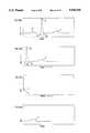
- FIG. 8(a)is a diagram of a sample PQRST complex utilized by the system and method of the present invention.
- FIGS. 8(b)-8(d)are diagrams of possible extraction templates utilized for bispectral analysis by the method and system of the present invention.
- FIG. 9is a flow chart of the frequency domain based steps for producing the autobispectrum or the crossbispectrum used by the system and method of the present;
- FIG. 10is a flow chart of the parametric based steps for producing the autobispectrum or the crossbispectrum used by the system and method of the present invention.
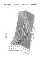
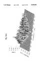
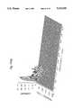
- FIG. 11are sample diagrams of autobispectral arrays of the QRS complex generated by the system and method of the present invention.
- FIG. 12is a flow chart of the steps used to generate diagnostic indices by the system and method of the present invention.
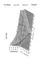
- FIG. 13are diagrams of several autobiocoherence arrays developed during the determination of the clinical reference arrays
- FIGS. 14(a)-14(b)are tables of the sample indices for normal subjects and for CAD subjects, respectively, generated by the system and method of the present invention in connection with the establishment of clinical reference arrays;
- FIG. 15are diagrams showing sample biocoherence values when the system and method of the present invention is used to identify patients at risk of malignant arrhythmias
- FIG. 16(a)-16(b)are tables of sample indices for MI subjects and VT/VF subjects respectively;
- FIGS. 17(a)-17(b)are graphs of coefficients generated by the system and method of the present invention.
- the apparatus of the present inventionis connected to a patient 100 through a set of surface electrodes using a standard limb, precordial and orthogonal placement protocol.
- the ECG signalsare picked up by the electrodes and transmitted over a patient cable 102 to a 16 channel ECG data acquisition system 104 with a serial interface.
- the data acquisition system 104filters, amplifies and digitizes the ECG waveforms and sends the digitized data to a microcomputer 110 via a high speed synchronous serial line 106.
- the serial line 106can be used to download filtering, gain and sampling rate instructions to the data acquisition unit 104.
- the microcomputer 110process the serial data stream in order to generate all computed data arrays. These arrays are then used in conjunction with predetermining reference arrays derived from clinical studies to produce diagnostic indices which indicate the status of the patient. These indices are displayed on the graphics display 108. Printed output of the diagnostic index is also available on the hard copy output device 116 which is connected to the microcomputer 110. Interaction between the operator and the system is provided by means of a keyboard 112 and pointing device 114 with feedback on the graphics display 108.
- the 16 channel data acquisition system 110is shown in greater detail in FIG. 2.
- the ECG surface potentialdetected by a surface electrode mounted on the patient 100, passes through an electrosurgery protection circuit 202, a defibrillator protection circuit 206 and an amplifier/filter circuit 208 before being passed on to the multi-channel analog to digital converter 210.
- the electrosurgery protection circuit 202includes a radio frequency (rf) filter, which limits the rf current through the patient leads 101 to less than 100 micro amperes and thus protects the patient 100 from rf burns and protects the amplifiers 36 from damage resulting from exceeding the absolute maximum input voltage specified by the manufacturer.
- This circuitcan be an LC section circuit consisting of a generic inductor connected in series to a generic capacitor which is then connected to ground.
- the defibrillator protection circuit 206limits the voltage to the amplifiers 208 to a safe level when a defibrillator is applied to the patient 100 and discharged.
- This circuitpreferably includes a neon light bulb and/or a parallel variable resistor connected in series to a grounded resistor.
- the amplifier/filter circuitry 208is controlled by a microprocessor 216 for default gain and filtering levels or alternate gain and filtering levels as requested by the operator. Preferred gain and filtering settings are discussed below.
- This circuitry 208includes three stages: the first is a pre-amplifier stage that can be assembled using a wide variety of high impedance pre-amplifiers such as those sold by National Semiconductor, Sunnyvale Calif.; the second is a programmable filters stage which can utilize filters sold by Frequency Devices, Haverhill Mass.; the third stage is a programmable amplifiers stage which can be assembled from operational amplifiers used in conjunction with a multiplying digital to analog (D/A) converter, both of which components are available from National Semiconductor. The multiplying D/A is used to set the gain to the appropriate levels requested by the microprocessor 216.
- D/Adigital to analog
- the high impedance pre-amplifier of each channelwill saturate to either the positive or negative supply voltage if the input of the pre-amplifier is not terminated. This will lead to large positive value or a large negative value at the output of amplifier section 208. Such values will be used to identify lead failure.
- A/Danalog to digital converter
- the multi-channel A/D converter 210is optically coupled to data bus 215 by optical isolator 214. All control lines to the amplifiers/filters 208 and the A/D convertor 210 are also optically isolated by optical isolator 212. Any known optical isolators can be used for this purpose.
- All DC power lines going to the amplifiers/filters 208 and A/D convertor 210are also isolated from the AC power line with a DC/DC convertor 204 in order to provide complete patient isolation from ground.
- DC/DC converters available from Burr Browncan be used for this purpose.
- the basic instructions for controlling operation of the microprocessor 216are stored in a read only memory (ROM) 218.
- the random access memory (RAM) 219is used as a buffer memory for data, and a portion of the RAM 219 can also be used as program memory when a control program is being downloaded from the microcomputer 110.
- Serial interface 220operates under the control of the microprocessor 216.
- the serial interface 220is optically coupled with optical isolators 222 to high speed synchronous serial drivers 224 to provide a synchronous serial link 106 between the 16 channel data acquisition system 104 and any compatible high speed synchronous serial interface card on any computer.
- the serial linesare isolated by optical isolators 222 and DC/DC convertor 204 to provide increased patient safety and to protect the host computer 110 from any transients.
- the host or microcomputer 110 of FIG. 1is shown in greater detail in FIG. 3.
- the entire microcomputer systemruns under control of a microprocessor 302 with the program memory for the microprocessor being stored in ROM 304.
- the RAM 306is used for storage of intermediate data.
- the mass storage device 308is used for storing clinical databases as well as archiving patient data.
- the microcomputer 110contains an array processor 310 (such as the Vortex sold by SKY of Lowell, Mass.) on which complex arithmetic calculations can be performed on entire arrays of data dimultaneously.
- the preferred embodimentalso includes a math coprocessor 312 which is connected directly to microprocessor 302. The math coprocessor 312 is used for scalar and graphic calculations while the array processor 310 is used to calculate bispectral and other data vectors.
- a graphics controller 314 operating under program control of the microprocessor 302drives a graphics display 316.
- a keyboard controller 318interfaces directly with the operator's keyboard 320.
- An interface port 322is provided for the pointing device 324.
- One high speed synchronous serial port 326is provided to interface with the 16 channel data acquisition system 104. Port 326 can be used to send control data to the system (e.g., filtering, gain, sampling rate, start/stop acquisition, perform self diagnostics) and to receive ECG data from the system, as well as to download program data to the system.
- Another serial or parallel interface port 328is provided to drive a hard copy output device 330 for printing desired diagnostic indices.
- the system and method of the present inventionquantify linear and nonlinear properties of phase and energy components within the frequency structure of the ECG from a preselected number of leads. Diagnostic indices are then generated from the bispectral data arrays by utilizing predetermined reference arrays.
- the indicesare used for the quantification of coronary artery disease (CAD), myocardial ischemia, cardiac electrical stability, risk of malignant ventricular arrhythmia, site(s) of origin of malignant arrhythmias, extent of malignancy of arrhythmias, degree of antiarrhythmic drug efficacy, neural and humoral inputs to the heart, pump function/ejection fraction, and ongoing organ rejection in cardiac transplant patients.
- CADcoronary artery disease
- the monitor module 402handles the overall operations of the system via integration of data and process information from the user interface module 404, acquisition and raw ECG data management module 406, bispectral processing module 408 and diagnostic index derivation module 410. A detailed description of the operation of module 402 will be provided below in connection with the description of FIG. 5.
- the user interface and display management module 404represents the means through which the operator controls and interacts with the system during the course of a procedure. This includes, but is not limited to, entry of information regarding the patient, type of diagnostic procedure being carried out, lead and acquisition settings; continuous display of acquisition status, lead integrity, display of diagnostic indices; and requests for printing and archiving results to disk. Module 404 directly interacts with the monitor module 402. The operations handled by module 404 can be achieved under one of many commercially available environments such as Microsoft's Windows.
- the acquisition and raw ECG data management module 406handles all of the raw ECG data checking and processing prior to bispectral analysis. This includes, but is not limited to, continuous acquisition of ECG data and the verification of the integrity of the data; performing QRS detection; performing crosscorrelation with the preselected template to identify suitable complexes; extracting suitable complexes from leads of interest in preparation for autobispectral and crossbispectral processing.
- Module 405directly interacts with the monitor module 402, and a more detailed description of module 406 will be provided below in connection with the description of FIG. 7.
- the bispectral processing module 408controls the generation of all data arrays measuring dynamic phase and energy properties within the ECG. This information can be organized in both autobispectral and crossbispectral arrays utilizing either an FFT based on parametric based approach.
- the tasksinclude, but are not limited to, nonlinear/exponential transform of the signal, Fourier transformation, the generation of power spectrum, autobispectral density, crossbispectral density, autobicoherence, crossbicoherence, autobiphase, and crossbiphase.
- Module 408directly interacts with the monitor module 402, and a more detailed description of module 408 is provided below in connection with FIGS. 9 and 10.
- the diagnostic index derivation module 410generates the data values utilized in the diagnostic process.
- the taskincludes, but is not limited to, identifying frequency pairs of interest through the use of predetermined clinical reference arrays and creating a diagnostic index from the values in the bispectral data arrays at the frequency locations defined by the reference array.
- Module 410directly interacts with the monitor module 402, and more detailed description of module 410 is provided below in connection with FIG. 12.
- step 502the data arrays used to store the digitized raw ECG and the bispectral data of each lead are initialized.
- the data files required for storage and files containing databases required for the computation of diagnostic indicesare also opened in the initializing step 502.
- step 504the system requests the information required to start the acquisition and diagnostic process from the user via the user interface module.
- This informationincludes the type of diagnostic procedure to be conducted and any operator requested modification to the system defaults such as leads used, clinical databases to access for diagnostic index computation, filtering, gain and sampling rate information for each lead.
- the type of diagnostic procedure as entered by the operatorwill be used by the system to inform the operator of the lead placement protocol required, and the type of templates to be selected (sinus rhythm or ectopic beat).
- the systemwill also use the type of diagnostic procedure it will perform to select the portion of the complexes to be used as time series for bispectral processing as well the type of bispectral arrays that need to be computed for use in conjunction with the predetermined clinical databases to yield a final diagnosis.
- the process of identifying the most effective leads for a particular diagnostic procedure and the generation of the clinical databaseswill be discussed later.
- step 506ECG signals are continuously acquired and displayed from the leads needed for the diagnostic operation being performed. All channels transmitting artifactual data are properly signaled to the operator to correct the problem. While using the pointing device, the operator is requested to select a suitable template against which incoming ECG complexes are matched.
- the systemin step 508, computes the necessary autobispectral and crossbispectral arrays required by the databases for the generation of the diagnostic indices requested by the operator.
- step 510the diagnostic indices from all generated autobispectral and crossbispectral arrays are computed. Autobispectral density and crossbispectral density clinical reference arrays are utilized in these diagnostic index computations.
- the systemdisplays, in step 512, the resultant diagnostic indices and a projected outcome based on information from the clinical databases.
- the indexis displayed continuously over time during the course of the procedure requiring it.
- step 514requested printouts are produced, results are stored to disk for archival purposes and all files are closed.
- step 516the process is terminated.
- FIG. 6A sample display representation generated by the system is shown in FIG. 6.
- the top section of the screen 602is divided into 16 sections 603 each representing the region probed by an electrode. Each section will be covered by a large "X" 604 if lead fail or artifact was detected from the lead corresponding to that section.
- a second portion of the screen 606can be assigned to the continuous display of the diagnostic index if the system is being used in monitoring mode.
- the background of that portionis color coded to reflect the possible values allowed for in the range of the selected diagnostic index.
- a third portion of the screen 608can be assigned to displaying one or several of the raw ECG data leads that is being acquired for processing. This will also provide for easy template selection using the pointing device.
- a fourth portion of the screen 610can be assigned to displaying the results of a static diagnostic test if the system is being used in that mode of operation.
- step 702the acquisition system 104 is programmed with requested filtering, gain, sampling rate, and lead selection information.
- step 704the acquisition system 104 acquires continuous ECG data for all requested leads and transfers this data to the host computer 110.
- the acquisition system 104detects lead failures during the acquisition cycle, and in step 706, the acquired data is examined for lead failure signals and for the presence of artifact.
- step 708leads generating fail signals and/or artifactual data are marked for the monitor module 402.
- step 710the system requests the operator to identify a template from the incoming data stream using the pointing device.
- QRS detectionis performed on the ECG data using any publicly available QRS detection program such as the algorithm disclosed by Engelese et al in "A Single Scan Algorithm for QRS-Detection and Feature Extraction , IEEE Computers in Cardiology (1979).
- step 714the system initiates continuous extraction of complexes that match the template from the incoming ECG data from each lead using standard cross-correlation techniques.
- the auto/cross bispectral time recordsare continuously generated by extracting a portion suitable for the diagnostic test from each matched complex from each of the leads required for the test. Each portion is assigned to an X i (t), where X i (t) are the individual time series records provided for autobispectral processing.
- Y i (t)is set to X i+1 (t) (the successive complex in the same lead) where Y i (t) are the time series records (in addition to X i (t)) required for crossbispectral processing within the same lead.
- Y i (t)can also assume the value of a corresponding X i (t) from another lead providing for crossbispectral analysis between two leads. It should be noted that for autobispectral analysis Y i (t) is set to equal X i (t) and in all cases the index i denotes the record number from 1 to k, and where k is the number of bispectral averages computed.
- the programwill continuously pass to the monitor module (via interrupt handlers) raw time series data until the diagnostic test is completed.
- the programreturns control to the monitor module 402 in step 718.
- FIG. 8(a)shows a sample PQRST template
- FIG. 8(b)shows the QRST portion extracted for processing
- FIG. 8(c)shows the terminal part of the QRS extracted for processing
- FIG. 8(d)shows the ST segment with the T wave extracted for processing.
- the axes in FIGS. 8(a)-8(d)are not calibrated and the figures are for illustration purposes only.
- step 902the system checks whether the computation to be performed is an autobispectral or crossbispectral computation.
- Autobispectral analysisis a special case of crossbispectral analysis and therefore different rules of symmetry apply.
- step 904the system sets the following symmetries in order to proceed with autobispectral computation:
- Nis the number of samples in the time series to be processed. N will depend on the length of the portion of the complex (in msec) and the sampling rate. For illustration purposes we will utilize the QRS portion of the selected complex with length of 160 msec. Since the sampling rate is 1024 samples/sec N will be equal to 160 samples or 1024 Hz.
- f 1 and f 2(also referred to as F 1 and F 2 or Frequency 1 and Frequency 2) denote the frequency pairs over which bispectral computation will be carried out
- X i (t) and Y i (t)denote the individual time series records used for bispectral computation
- X i (f) and Y i (f)denote the Fourier transform of the time series records
- idenotes the record number which in this embodiment ranges from 1 to k.
- step 906the following symmetries are adhered to for crossbispectral analysis:
- step 908the Discrete Fourier transform (DFT) X i (f) and Y i (f) of each record of the k selected records, is computed using a standard IEEE library routine or any other publicly available routine in step 908.
- DFTDiscrete Fourier transform
- step 910the power spectra P xi (f) and P yi (f) of each record of the k selected records is computed by squaring the magnitudes of each element of the Fourier transform X i (f) and Y i (f) respectively.
- the systemcomputes the average complex triple product in step 912 by utilizing the following equations where bc i (f 1 ,f 2 ) is an individual complex triple product from one record and BC(f 1 ,f 2 ) is the average complex triple product over all records:
- the average real triple productis computed in step 914 by using the following equations where br i (f 1 ,f 2 ) is an individual real triple product from the one record and BR(f 1 , f 2 ) is the average real triple product over all records: ##EQU2##
- step 916the auto/crossbispectral density array (BD(f 1 ,f 2 )) is computed using the following equation:
- step 918the system computes the auto/crossbiphase array ( ⁇ (f 1 , F 2 )) using the following equation:
- step 920the system computes the auto/crossbicoherence array (R(f 1 ,f 2 )) using the following equation:
- step 922the system returns the requested auto/cross bispectral density, average real triple product, biocoherence, and biphase arrays to the monitor module 402.
- steps 1002, 1004, and 1006the system sets the symmetries and time series records in the same manner as described above in steps 902, 904, and 906, respectively.
- the power spectra of X i (t) and Y i (t)are estimated in steps 1008, 1010, and 1012.
- This estimation methodincludes two major stages, the autoregressive (AR) model order selection and the power spectrum computation for X i (t) and Y i (t).
- step 1008the system computes two sequences of autocorrelations, ⁇ R 2X (m) ⁇ and ⁇ R 2Y (m) ⁇ using the following equation. ##EQU3## where M is the number of records (k in our case), and N is the number of samples per record (160 in our case). L is much greater than the possible AR filter order (we choose 50).
- FPE X (m) and FPE Y (m)respectively are chosen to be the orders of the AR filters of power spectra of X i (t) and Y i (t) respectively, i.e.,
- the autocorrelation sequences, ⁇ R 2X (m) ⁇ and ⁇ R 2Y (m) ⁇are entered into Levinson recursion with order Q X and Q Y , respectively, instead of L.
- the power spectrum, P z (f)is computed as the prediction error (O z 2 )divided by square of the magnitude of the Fourier transform of the coefficients, i.e., ##EQU4##
- the systemestimates the auto/cross bispectrum in steps 1014, 1016, and 1018.
- the estimation processincludes two major stages: the order selection and bispectrum computation.
- step 1016two super matrices T X and T Y are formed as follows. ##EQU6##
- the auto/cross bispectrum of X i (t) and Y i (t)are computed in step 1018 as the cubic root of the triple product of the skewnesses ( ⁇ X ⁇ Y ⁇ Y ) 1/3 divided by the triple product of the Fourier transforms of the AR filter coefficients (H z (f)), i.e., ##EQU8## and BR(f 1 , f 2 ) is the real triple produce for that same lead:
- the systemAfter obtaining power spectrum and auto/cross bispectrum, the system computes the bispectral density array, the biphase, and the bicoherence in step 1020 the same way as in steps 916, 918, 920. In step 1022, the system returns to the monitor module 402 the requested bispectral density, biphase, and bicoherence arrays.
- FIG. 11contains sample autobispectral arrays of the QRS complex showing frequency pairs 0 ⁇ f 1 ⁇ 512 Hz, and 0 ⁇ f 2 ⁇ 256 Hz.
- a bispectral density arrayis shown in FIG. 11(a) where the Z axis represents the magnitude in decibels (db) of the coupling interaction between all appropriate frequency pairs f 1 and f 2 .
- dbdecibels
- a biphase arrayis shown in FIG. 11(b) where the Z axis represents the phase in radians of the coupling interaction between all appropriate frequency pairs f 1 and f 2 .
- a bicoherence arrayis shown in FIG. 11(c) where the Z axis represents the normalized magnitude in percent (%) of the coupling interaction between all appropriate frequency pairs f 1 and f 2 .
- step 1202the software identifies the type of diagnostic test in progress.
- the possible optionsinclude but are not limited to:
- the systemretrieves the type of bispectral array to use in the diagnostic index computation as well the lead(s) of origin.
- the combination possibilitiescan lead to a very large number of computations associated with each diagnostic index as well as a substantial reference clinical database.
- Statistical methods to identify leads and bispectral arrays with the greatest diagnostic fidelity for each testare used to make this system practical. Such statistical methods will be discussed in greater detail below.
- step 1026the appropriate reference array is retrieved from resident memory or from disk. Each reference array will contain the locations of the frequency pairs which are most sensitive to the diagnostic test in progress.
- step 1208all data points in the bispectral array at the locations identified by the retrieved reference array are added together for a single value index. A counter (NP) of the total number of points added is kept.
- step 1210the single value index is divided by NP to obtain the diagnostic index.
- step 1212the program returns to the monitor module 402.
- the predetermined clinical reference arrays referred to aboveare critical to the device's ability to achieve clinically relevant diagnostic efficacy, and the process adopted for generating these clinical reference arrays will now be described. Since the total number of possible diagnostic applications will require many reference arrays, only two types of statistical approaches will be discussed in detail. All other reference arrays are acquired in a similar fashion utilizing a wide number of clinically appropriate statistical approaches. For illustration purposes the generation of the bicoherence reference array for detection of coronary artery disease with orthogonal lead X, assessment of risk to VT/VF with orthogonal lead Z, and assessment of ejection fraction with orthogonal lead X will be discussed in the following section.
- An autobicoherence arrayis generated from 100 QRS complexes from orthogonal lead X for all subjects.
- the arraysare grouped in 2 sets of arrays, the first representing the young normals and the second representing the coronary artery disease subjects.
- a paired Student's t testis performed on each of 1640 data points, comparing the first and second sets of arrays.
- the resulting 1640 t valuesare stored in a two dimensional array identical in structure to that of the bicoherence array.
- Each t value from t array(T(f1,f2)) is tested for significance based on the number of degrees of freedom. Where the degrees of freedom are equal to the total number of subjects--1. All t values not meeting the required significance level are set to 0. In the preferred embodiment all locations with a t value not corresponding to a p ⁇ 0.05 are set to 0.
- the application of the above conditionshas the effect of identifying all of the frequency pair locations that are significantly different in coronary artery patients when compared to young normal volunteers. Suppression of all other frequency pairs allows easier inspection of the most sensitive regions.
- FIG. 13(a)shows the mean autobicoherence array for orthogonal lead X for the normal subjects.
- FIG. 13(b)shows the mean autobicoherence array for orthogonal lead Y for the CAD subjects.
- FIG. 13(c)shows the t array with all t values not meeting P ⁇ 0.05 set to 0.
- the next stepinvolves sorting the t array for the most sensitive ensemble of frequency pair locations. In a preferred embodiment this would consist of the top 25% of all significant t values.
- the locations f1,f2 of the most significant t valueswill be used to generate a diagnostic index for each subject in the process described above.
- Table 14(a) of FIG. 14shows sample indices for normal subjects and Table 14(b) shows sample indices for CAD subjects. The coded filename of the subject tested precedes each index.
- the final stepis to identify a cutoff value for the diagnostic index above which subjects are normal and al values below will indicate the presence of CAD. This cutoff should be optimized to yield the best sensitivity and specificity. ##EQU9##
- FIG. 15(a)shows the mean autobicoherence array for orthogonal lead Z for the MI subjects.
- FIG. 15(b)shows the mean autobicoherence array for orthogonal lead Z for the VT/VF subjects.
- FIG. 15(c)shows the t array with all t values not meeting p ⁇ 0.05 set to 0.
- the t array generatedis then used to produce diagnostic indices for both groups as shown earlier for the CAD study.
- the whole statistical processis followed through including prospective studies to identify the best bispectral array and its corresponding lead for the identification of patients who are at risk of malignant arrhythmias.
- Table 16(a) of FIG. 16shows sample indices for MI subjects and Table 16(b) shows sample indices for the VT/VF subjects.
- the coded filename of the subject testedprecedes each index. In this particular case if we chose a cutoff of 26.0, the sensitivity and specificity will be 91.6%(11/12) and 86.4%(32/37) respectively.
- This inventionis not limited to the use of the t test and many other statistical ranking test might be used when appropriate.
- a continuous variablesuch as ejection fraction (EF) regression analysis is more suitably used.
- FIG. 17(a)shows the positive r values between EF and bicoherence at the corresponding frequency pairs
- FIG. 17(b)shows the negative r values.
- the system and method of the present inventionmay also be used to assess a myriad of cardiac phenomena based on the acquisition and processing of ECG signals into various bispectral arrays which are then compared to appropriate reference arrays.
- the system and method of the present inventionuses various bispectral values to measure dynamic frequency structure [higher order phase-locking] across all frequency pairs in a frequency range ignored by those knowledgeable in the art and uses various alterations in these bispectral parameters at a limited number of frequency locations as an index of physiological perturbation.
- the system an methodutilizes various bispectral arrays of transformed ECG signals of defined clinical populations to define the locations of the subset of frequencies used to calculate this index. Reference clinical arrays are further utilized to assess the meaning of this index and to measure the significance of deviations of this index from normality. This allows the quantitative gauging of the disturbances in cardiac function, whether due to coronary disease, electrical instability, restenosis after PTCA, drugs or ischemia for any particular ECG lead position.
- the invention disclosed herealso defines the graphic display of the diagnostic index, whether on video screen or on paper, whether in real-time or in digital archive.
- the ECGmay carry diagnostic information at frequencies much higher than the cutoff frequency of 512 Hz.
- the use of such high frequency low energy components of the ECG waveform by the system and method described aboveis intended to fall within the scope of the current invention. All such alterations and modifications are intended to fall within the scope of the appended claims.
Landscapes
- Health & Medical Sciences (AREA)
- Life Sciences & Earth Sciences (AREA)
- Cardiology (AREA)
- Animal Behavior & Ethology (AREA)
- Public Health (AREA)
- Pathology (AREA)
- Engineering & Computer Science (AREA)
- Biomedical Technology (AREA)
- Heart & Thoracic Surgery (AREA)
- Medical Informatics (AREA)
- Molecular Biology (AREA)
- Surgery (AREA)
- Physics & Mathematics (AREA)
- General Health & Medical Sciences (AREA)
- Biophysics (AREA)
- Veterinary Medicine (AREA)
- Transplantation (AREA)
- Immunology (AREA)
- Vascular Medicine (AREA)
- Measurement And Recording Of Electrical Phenomena And Electrical Characteristics Of The Living Body (AREA)
- Investigating Or Analysing Biological Materials (AREA)
- Measuring Pulse, Heart Rate, Blood Pressure Or Blood Flow (AREA)
- Electrotherapy Devices (AREA)
- Measurement Of The Respiration, Hearing Ability, Form, And Blood Characteristics Of Living Organisms (AREA)
Abstract
Description
f.sub.1 +f.sub.2 <N/2
0<f.sub.2 <f.sub.1
X.sub.i (t)=Y.sub.i (t)→X.sub.i (f)=Y.sub.i (f)
f.sub.1 +f.sub.2 <N/2
0<f.sub.1 <N/2
0<f.sub.2 <N/2
-2f.sub.2 <f.sub.1
X.sub.i (t)≠Y.sub.i (t)→X.sub.1 (f)≠Y.sub.1 (f)
bc.sub.i (f.sub.1, f.sub.2)=X.sub.i (f.sub.1)*Y.sub.i (f.sub.2)*Y.sub.i *(f.sub.1 +f.sub.2)
BD(f.sub.1,f.sub.2)=|BC(f.sub.1,f.sub.2)|
(φ(f.sub.1,f.sub.2)=tan.sup.-1 [Im(BC(f.sub.1,f.sub.2))/Re(BC(f.sub.1,f.sub.2))]
0<φ<2π (radians)
R(f.sub.1,f.sub.2)=BD(f.sub.1, f.sub.2)/[BR(f.sub.1,f.sub.2)].sup.1/2
0<R<1
FPE.sub.X (Q.sub.X)=min {FPE.sub.X (m)}and
FPE.sub.Y (Q.sub.Y)=min {FPE.sub.Y (m)}BR(f.sub.1, f.sub.2)=P.sub.x (f.sub.1)*P.sub.Y (f.sub.2)*P.sub.Y (f.sub.1 +f.sub.2)
f.sub.1 +f.sub.2 <N/2
Claims (56)
Priority Applications (8)
| Application Number | Priority Date | Filing Date | Title |
|---|---|---|---|
| US07/396,990US5020540A (en) | 1987-10-09 | 1989-08-22 | Cardiac biopotential analysis system and method |
| JP02504848AJP3138752B2 (en) | 1989-08-22 | 1990-03-13 | Cardiac biopotential analysis system and method |
| DE69032771TDE69032771T2 (en) | 1989-08-22 | 1990-03-13 | HEART BIOPOTENTIAL ANALYSIS SYSTEM AND METHOD |
| CA002064791ACA2064791C (en) | 1989-08-22 | 1990-03-13 | Cardiac biopotential analysis system and method |
| EP90905061AEP0489010B1 (en) | 1989-08-22 | 1990-03-13 | Cardiac biopotential analysis system and method |
| PCT/US1990/001385WO1991002484A1 (en) | 1989-08-22 | 1990-03-13 | Cardiac biopotential analysis system and method |
| AT90905061TATE173551T1 (en) | 1989-08-22 | 1990-03-13 | CARDIAC BIOPOTENTIAL ANALYSIS SYSTEM AND METHODS |
| AU52801/90AAU653318B2 (en) | 1989-08-22 | 1990-03-13 | Cardiac biopotential analysis system and method |
Applications Claiming Priority (2)
| Application Number | Priority Date | Filing Date | Title |
|---|---|---|---|
| US07/107,419US4924875A (en) | 1987-10-09 | 1987-10-09 | Cardiac biopotential analysis system and method |
| US07/396,990US5020540A (en) | 1987-10-09 | 1989-08-22 | Cardiac biopotential analysis system and method |
Related Parent Applications (1)
| Application Number | Title | Priority Date | Filing Date |
|---|---|---|---|
| US07/107,419Continuation-In-PartUS4924875A (en) | 1987-10-09 | 1987-10-09 | Cardiac biopotential analysis system and method |
Publications (1)
| Publication Number | Publication Date |
|---|---|
| US5020540Atrue US5020540A (en) | 1991-06-04 |
Family
ID=23569443
Family Applications (1)
| Application Number | Title | Priority Date | Filing Date |
|---|---|---|---|
| US07/396,990Expired - LifetimeUS5020540A (en) | 1987-10-09 | 1989-08-22 | Cardiac biopotential analysis system and method |
Country Status (8)
| Country | Link |
|---|---|
| US (1) | US5020540A (en) |
| EP (1) | EP0489010B1 (en) |
| JP (1) | JP3138752B2 (en) |
| AT (1) | ATE173551T1 (en) |
| AU (1) | AU653318B2 (en) |
| CA (1) | CA2064791C (en) |
| DE (1) | DE69032771T2 (en) |
| WO (1) | WO1991002484A1 (en) |
Cited By (118)
| Publication number | Priority date | Publication date | Assignee | Title |
|---|---|---|---|---|
| US5161539A (en)* | 1991-05-09 | 1992-11-10 | Physio-Control | Method and apparatus for performing mapping-type analysis including use of limited electrode sets |
| US5187657A (en)* | 1990-04-05 | 1993-02-16 | Hewlett-Packard Company | Cardiac analyzer with rem sleep detection |
| US5265617A (en)* | 1991-02-20 | 1993-11-30 | Georgetown University | Methods and means for non-invasive, dynamic tracking of cardiac vulnerability by simultaneous analysis of heart rate variability and T-wave alternans |
| WO1994010905A1 (en)* | 1992-11-18 | 1994-05-26 | Hutson William H | A method and system for near real-time analysis and display of electrocardiographic signals |
| EP0634136A1 (en)* | 1993-07-16 | 1995-01-18 | Siemens-Elema AB | Method and device for processing ECG-signals |
| US5421343A (en)* | 1992-04-03 | 1995-06-06 | Feng; Genquan | Computer network EEMPI system |
| US5425373A (en)* | 1991-01-15 | 1995-06-20 | Pacesetter, Inc. | Apparatus and method for analyzing and enhancing intercardiac signals |
| US5503159A (en)* | 1993-03-12 | 1996-04-02 | Hewlett-Packard Company | Method for enhancement of late potentials measurements |
| US5539832A (en)* | 1992-04-10 | 1996-07-23 | Ramot University Authority For Applied Research & Industrial Development Ltd. | Multi-channel signal separation using cross-polyspectra |
| US5560367A (en)* | 1993-08-13 | 1996-10-01 | Siemens Aktiengesellschaft | Method for high-resolution spectral analysis in multi channel observations using a singular valve decomposition (SVD) matrix technique |
| US5605159A (en)* | 1996-02-16 | 1997-02-25 | Smith; Joseph M. | System and method for determining spatial organization of atrial activation |
| US5655540A (en)* | 1995-04-06 | 1997-08-12 | Seegobin; Ronald D. | Noninvasive method for identifying coronary artery disease utilizing electrocardiography derived data |
| US5810014A (en)* | 1997-03-25 | 1998-09-22 | Davis; Dennis W. | Method and system for detection of physiological conditions |
| US5827195A (en)* | 1997-05-09 | 1998-10-27 | Cambridge Heart, Inc. | Electrocardiogram noise reduction using multi-dimensional filtering |
| US5842997A (en)* | 1991-02-20 | 1998-12-01 | Georgetown University | Non-invasive, dynamic tracking of cardiac vulnerability by simultaneous analysis of heart rate variability and T-wave alternans |
| EP0898234A1 (en)* | 1991-10-25 | 1999-02-24 | Aspect Medical Systems, Inc. | Method and system for generating a diagnostic index |
| US5891045A (en)* | 1996-07-17 | 1999-04-06 | Cambridge Heart, Inc. | Method and system for obtaining a localized cardiac measure |
| US5891047A (en)* | 1997-03-14 | 1999-04-06 | Cambridge Heart, Inc. | Detecting abnormal activation of heart |
| WO1999044500A1 (en)* | 1998-03-05 | 1999-09-10 | Dan Qun Fang | A system and method for detecting and locating heart disease |
| WO1999044498A1 (en) | 1996-09-10 | 1999-09-10 | Ralf Bousseljot | Evaluation system for obtaining diagnostic information from the signals and data of medical sensor systems |
| US5954664A (en)* | 1995-04-06 | 1999-09-21 | Seegobin; Ronald D. | Noninvasive system and method for identifying coronary disfunction utilizing electrocardiography derived data |
| US6169919B1 (en) | 1999-05-06 | 2001-01-02 | Beth Israel Deaconess Medical Center, Inc. | System and method for quantifying alternation in an electrocardiogram signal |
| US6171256B1 (en)* | 1998-04-30 | 2001-01-09 | Physio-Control Manufacturing Corporation | Method and apparatus for detecting a condition associated with acute cardiac ischemia |
| US6212428B1 (en) | 1998-06-17 | 2001-04-03 | Cardiac Pacemakers, Inc. | Multiple stage morphology-based system detecting ventricular tachycardia and supraventricular tachycardia |
| US6223078B1 (en)* | 1999-03-12 | 2001-04-24 | Cardiac Pacemakers, Inc. | Discrimination of supraventricular tachycardia and ventricular tachycardia events |
| US6266554B1 (en) | 1999-02-12 | 2001-07-24 | Cardiac Pacemakers, Inc. | System and method for classifying cardiac complexes |
| DE10001890A1 (en)* | 2000-01-19 | 2001-07-26 | Biotronik Mess & Therapieg | Method for predictive calculation of a cardiological signal curve and for controlling the stimulation pulse delivery of a cardiological device implant |
| WO2001054578A1 (en)* | 2000-01-31 | 2001-08-02 | Pearlman Justin D | Multivariate cardiac monitor |
| US6272377B1 (en) | 1999-10-01 | 2001-08-07 | Cardiac Pacemakers, Inc. | Cardiac rhythm management system with arrhythmia prediction and prevention |
| US6308095B1 (en) | 1999-02-12 | 2001-10-23 | Cardiac Pacemakers, Inc. | System and method for arrhythmia discrimination |
| US6312388B1 (en) | 1999-03-12 | 2001-11-06 | Cardiac Pacemakers, Inc. | Method and system for verifying the integrity of normal sinus rhythm templates |
| US6361503B1 (en) | 2000-06-26 | 2002-03-26 | Mediwave Star Technology, Inc. | Method and system for evaluating cardiac ischemia |
| US20020107552A1 (en)* | 1997-04-30 | 2002-08-08 | Cardiac Pacemakers, Inc. | Apparatus and method for treating ventricular tachyarrhythmias |
| US6491629B1 (en) | 1997-11-25 | 2002-12-10 | Bundesrepublik Deutschland | Method for determining at least one diagnostic piece of information from signal patterns of medical sensor systems |
| US20030013978A1 (en)* | 2001-07-12 | 2003-01-16 | Schlegel Todd T. | Real-time, high frequency QRS electrocardiograph |
| US20030044034A1 (en)* | 2001-08-27 | 2003-03-06 | The Regents Of The University Of California | Cochlear implants and apparatus/methods for improving audio signals by use of frequency-amplitude-modulation-encoding (FAME) strategies |
| US20030055461A1 (en)* | 1999-10-01 | 2003-03-20 | Girouard Steven D. | Cardiac rhythm management systems and methods predicting congestive heart failure status |
| US20030060849A1 (en)* | 1999-07-14 | 2003-03-27 | Cardiac Pacemakers, Inc. | Classification of supraventricular and ventricular cardiac rhythms using cross channel timing algorithm |
| EP1135057A4 (en)* | 1998-11-30 | 2003-06-11 | Sabine Vivian Kunig | Method and apparatus for measuring functionality of a periodically changing system |
| US20030109792A1 (en)* | 1999-04-01 | 2003-06-12 | Cardiac Pacemakers, Inc. | Cross chamber interval correlation |
| US20030120163A1 (en)* | 1997-07-31 | 2003-06-26 | Yoram Rudy | System and methods for noninvasive electrocardiographic imaging (ECGI) using generalized minimum residual (GMRes) |
| US20030130586A1 (en)* | 2001-12-26 | 2003-07-10 | Starobin Joseph M. | Method and system for evaluating arrhythmia risk with QT-RR interval data sets |
| WO2003057033A1 (en) | 2001-12-26 | 2003-07-17 | Mediwave Star Technology, Inc. | Method and system for evaluating cardiac ischemia with an exercise protocol |
| WO2003057032A1 (en) | 2001-12-26 | 2003-07-17 | Mediwave Star Technology, Inc. | Method and system for evaluating cardiac ischemia with an abrupt stop exercise protocol |
| US6604115B1 (en) | 1999-11-05 | 2003-08-05 | Ge Marquette Medical Systems, Inc. | Method and apparatus for storing data |
| US6648829B2 (en) | 2000-06-26 | 2003-11-18 | Mediwave Star Technology, Inc. | Method and system for evaluating and locating cardiac ischemia |
| US6656126B2 (en) | 2000-06-26 | 2003-12-02 | Mediwave Star Technology, Inc. | Method and system for evaluating cardiac ischemia with RR-interval data sets and pulse or blood pressure monitoring |
| US20040010200A1 (en)* | 2002-07-15 | 2004-01-15 | Sweeney Robert J. | Use of curvature based features for beat detection |
| US20040077967A1 (en)* | 2001-02-13 | 2004-04-22 | Jordan Kenneth George | Automated realtime interpretation of brain waves |
| US20040093035A1 (en)* | 2002-11-08 | 2004-05-13 | Mark Schwartz | Cardiac rhythm management systems and methods using multiple morphology templates for discriminating between rhythms |
| US20040127806A1 (en)* | 2000-10-31 | 2004-07-01 | Cardiac Pacemakers, Inc. | Curvature based method for selecting features from an electrophysiologic signals for purpose of complex identification and classification |
| US6768919B2 (en) | 2000-06-26 | 2004-07-27 | Mediwave Star Technology Inc | Method and system for evaluating cardiac ischemia with heart rate feedback |
| US6772004B2 (en) | 1997-07-31 | 2004-08-03 | Case Western Reserve University | System and method for non-invasive electrocardiographic imaging |
| US20040167381A1 (en)* | 1997-02-28 | 2004-08-26 | Qrs Diagnostic, Llc | Personal computer card for collection of real-time biological data |
| US20040176697A1 (en)* | 2002-11-01 | 2004-09-09 | Lukas Kappenberger | Methods of analyzing atrial fibrillations |
| US20040181160A1 (en)* | 1997-07-31 | 2004-09-16 | Case Western Reserve University | Electrophysiological cardiac mapping system based on a non-contact non-expandable miniature multi-electrode catheter and method therefor |
| US20040219600A1 (en)* | 2002-12-13 | 2004-11-04 | Williams Robert Wood | Method for determining sensitivity to environmental toxins and susceptibility to parkinson's disease |
| US20050027324A1 (en)* | 1999-06-26 | 2005-02-03 | Biotronik Mess-Und Therapiegeraete Gmbh & Co | Therapy system |
| US20050027202A1 (en)* | 2001-11-28 | 2005-02-03 | Aaron Ginzburg | Method and system for processing electrocardial signals |
| US20050038351A1 (en)* | 2003-07-23 | 2005-02-17 | Starobin Joseph M. | Method and system for evaluating cardiac ischemia based on heart rate fluctuations |
| DE4304269B4 (en)* | 1992-02-14 | 2005-04-21 | George John Dempsey | Apparatus for non-invasive detection, digital recording and processing of cardiac electrical signals |
| US20050107836A1 (en)* | 2002-02-28 | 2005-05-19 | Kjell Noren | Medical device |
| US20050119583A1 (en)* | 2001-12-22 | 2005-06-02 | Fuller Jonathan A. | Heart monitor |
| US20050182333A1 (en)* | 2002-03-05 | 2005-08-18 | Shinya Nagata | Electrocardiograohy chart apparatus and method thereof |
| US20050197586A1 (en)* | 2000-01-31 | 2005-09-08 | Pearlman Justin D. | Method of and system for signal separation during multivariate physiological monitoring |
| US20050234356A1 (en)* | 2004-04-15 | 2005-10-20 | Rowlandson G I | System and method for correlating implant and non-implant data |
| US20050234355A1 (en)* | 2004-04-15 | 2005-10-20 | Rowlandson G I | System and method for sudden cardiac death prediction |
| US20050234362A1 (en)* | 2004-04-15 | 2005-10-20 | Ge Medical Systems Information Technologies, Inc. | Method and apparatus for displaying alternans data |
| US20050234353A1 (en)* | 2004-04-15 | 2005-10-20 | Ge Medical Systems Information Technologies, Inc. | Method and apparatus for analysis of non-invasive cardiac parameters |
| US20050234363A1 (en)* | 2004-04-15 | 2005-10-20 | Ge Medical Systems Information Technologies, Inc. | Method and apparatus for determining alternans data of an ECG signal |
| US20050234357A1 (en)* | 2004-04-15 | 2005-10-20 | Ge Medical Systems Information Technologies, Inc. | Method and apparatus for detecting cardiac repolarization abnormality |
| US20050256544A1 (en)* | 2004-05-12 | 2005-11-17 | Cardiac Pacemakers, Inc. | Template based av/va interval comparison for the discrimination of cardiac arrhythmias |
| US6975900B2 (en) | 1997-07-31 | 2005-12-13 | Case Western Reserve University | Systems and methods for determining a surface geometry |
| US20060111643A1 (en)* | 2004-11-23 | 2006-05-25 | Shelley Cazares | Arrhythmia memory for tachyarrhythmia discrimination |
| US20060173368A1 (en)* | 2005-02-01 | 2006-08-03 | Yu Cecilia L | Low frequency analysis of cardiac electrical signals for detecting heart diseases, especially the coronary artery diseases |
| US20060211949A1 (en)* | 2001-06-05 | 2006-09-21 | Cardiac Pacemakers, Inc. | System and method for classifying cardiac depolarization complexes with multi-dimensional correlation |
| US20060281998A1 (en)* | 2005-06-13 | 2006-12-14 | Cardiac Pacemakers, Inc. | Method and apparatus for rate-dependent morphology-based cardiac arrhythmia classification |
| US7162294B2 (en) | 2004-04-15 | 2007-01-09 | Ge Medical Systems Information Technologies, Inc. | System and method for correlating sleep apnea and sudden cardiac death |
| US7171269B1 (en)* | 1999-05-01 | 2007-01-30 | Cardiodigital Limited | Method of analysis of medical signals |
| US20070054871A1 (en)* | 2005-09-06 | 2007-03-08 | Pastore Joseph M | Method and apparatus for device controlled gene expression for cardiac protection |
| US7203535B1 (en) | 1999-04-01 | 2007-04-10 | Cardiac Pacemakers, Inc. | System and method for classifying tachycardia arrhythmias having 1:1 atrial-to-ventricular rhythms |
| US20080081354A1 (en)* | 2006-10-02 | 2008-04-03 | Cardiac Pacemakers, Inc. | Devices, vectors and methods for inducible ischemia cardioprotection |
| US20080082135A1 (en)* | 2006-10-02 | 2008-04-03 | Cardiac Pacemakers, Inc. | Method and apparatus for identification of ischemic/infarcted regions and therapy optimization |
| US20080132769A1 (en)* | 2004-10-05 | 2008-06-05 | Henderson Leslie G | Non-invasively monitoring blood parameters |
| US20080177194A1 (en)* | 2007-01-19 | 2008-07-24 | Cardiac Pacemakers, Inc. | Heart attack detector |
| US7430446B2 (en) | 2005-01-20 | 2008-09-30 | Cardiac Pacemakers, Inc. | Methods and apparatuses for cardiac arrhythmia classification using morphology stability |
| EP1529487A4 (en)* | 2002-07-04 | 2009-05-27 | Dainippon Sumitomo Pharma Co | ELECTROCARDIOGRAM ANALYSIS DEVICE AND ASSOCIATED METHOD |
| US20100312130A1 (en)* | 2006-06-27 | 2010-12-09 | Yi Zhang | Graded response to myocardial ischemia |
| US8000780B2 (en) | 2006-06-27 | 2011-08-16 | Cardiac Pacemakers, Inc. | Detection of myocardial ischemia from the time sequence of implanted sensor measurements |
| US8216136B2 (en) | 2009-03-05 | 2012-07-10 | Nellcor Puritan Bennett Llc | Systems and methods for monitoring heart rate and blood pressure correlation |
| US20120184950A1 (en)* | 2005-08-11 | 2012-07-19 | Jose Carlos Pachon Mateos | Apparatus and Methods for Guiding Catheter-Based Ablation Therapy for Ventricular Arrhythmias Based on Spectral Mapping During Sinus Rhythm |
| USD671125S1 (en) | 2010-09-02 | 2012-11-20 | Nellcor Puritan Bennett Llc | Display screen with a graphical user interface |
| US20130053715A1 (en)* | 2010-04-13 | 2013-02-28 | Centre National De La Recherche Scientifique (C.N. R.S) | Method and system for analyzing the cardiac activity of a patient and uses thereof |
| US8409107B2 (en) | 2003-06-27 | 2013-04-02 | Cardiac Pacemakers, Inc. | Tachyarrhythmia detection and discrimination based on curvature parameters |
| US8608657B2 (en) | 2011-05-31 | 2013-12-17 | Covidien Lp | Clinical acceptance tool |
| US9131878B2 (en) | 2005-10-28 | 2015-09-15 | Covidien Lp | Adjusting parameters used in pulse oximetry analysis |
| US20150305687A1 (en)* | 2010-12-13 | 2015-10-29 | Acarix A/S | System and method for indicating coronary artery disease risk based on low and high frequency bands |
| WO2016033504A1 (en)* | 2014-08-29 | 2016-03-03 | Cardioinsight Technologies, Inc. | Signal characterization to facilitate therapy delivery |
| US20160073917A1 (en)* | 2013-06-26 | 2016-03-17 | Cameruddin Walimuhammad Vellani | Vector-cardio-graphic signal analyzer |
| US9649051B2 (en) | 2010-04-30 | 2017-05-16 | Centre National De La Recherche Scientifique (C.N.R.S) | Method and system for analyzing a patients respiratory activity and corresponding uses |
| CN107847745A (en)* | 2015-05-13 | 2018-03-27 | 阿库图森医疗有限公司 | Positioning system and method for collecting and analyzing cardiac information |
| EP3340872A4 (en)* | 2015-08-26 | 2019-06-19 | Analytics For Life Inc. | Method and apparatus for wide-band phase gradient signal acquisition |
| US10835174B2 (en) | 2016-01-12 | 2020-11-17 | Covidien Lp | System and method for monitoring cerebral activity |
| US11089988B2 (en) | 2016-06-24 | 2021-08-17 | Analytics For Life Inc. | Non-invasive method and system for estimating arterial flow characteristics |
| US11141114B2 (en) | 2017-03-02 | 2021-10-12 | Analytics For Life Inc. | Method and apparatus for wide-band phase gradient signal acquisition |
| US11273283B2 (en) | 2017-12-31 | 2022-03-15 | Neuroenhancement Lab, LLC | Method and apparatus for neuroenhancement to enhance emotional response |
| US11364361B2 (en) | 2018-04-20 | 2022-06-21 | Neuroenhancement Lab, LLC | System and method for inducing sleep by transplanting mental states |
| US11452839B2 (en) | 2018-09-14 | 2022-09-27 | Neuroenhancement Lab, LLC | System and method of improving sleep |
| US11717686B2 (en) | 2017-12-04 | 2023-08-08 | Neuroenhancement Lab, LLC | Method and apparatus for neuroenhancement to facilitate learning and performance |
| US11723579B2 (en) | 2017-09-19 | 2023-08-15 | Neuroenhancement Lab, LLC | Method and apparatus for neuroenhancement |
| US11786694B2 (en) | 2019-05-24 | 2023-10-17 | NeuroLight, Inc. | Device, method, and app for facilitating sleep |
| US11826126B2 (en) | 2016-09-21 | 2023-11-28 | Analytics For Life Inc. | Method and system for visualization of heart tissue at risk |
| US12102417B2 (en) | 2011-03-10 | 2024-10-01 | Acutus Medical, Inc. | Device and method for the geometric determination of electrical dipole densities on the cardiac wall |
| US12161398B2 (en) | 2015-05-12 | 2024-12-10 | Acutus Medical, Inc. | Ultrasound sequencing method |
| US12280219B2 (en) | 2017-12-31 | 2025-04-22 | NeuroLight, Inc. | Method and apparatus for neuroenhancement to enhance emotional response |
| CN120078428A (en)* | 2025-04-30 | 2025-06-03 | 中国人民解放军总医院 | Electrocardiogram signal analysis method and device |
| USD1090571S1 (en)* | 2023-04-05 | 2025-08-26 | GE Precision Healthcare LLC | Display screen or portion thereof with graphical user interface |
| US12402825B2 (en) | 2014-03-25 | 2025-09-02 | Enchannel Medical, Ltd. | Cardiac analysis user interface system and method |
Families Citing this family (16)
| Publication number | Priority date | Publication date | Assignee | Title |
|---|---|---|---|---|
| AUPO455297A0 (en)* | 1997-01-10 | 1997-01-30 | Micromedical Industries Limited | Universal ecg interface cable |
| CA2373123A1 (en)* | 1999-05-06 | 2000-11-16 | Vladimir Jankov | Physiological signal acquisition cable |
| US6463320B1 (en)* | 1999-12-22 | 2002-10-08 | Ge Medical Systems Information Technologies, Inc. | Clinical research workstation |
| US6497676B1 (en) | 2000-02-10 | 2002-12-24 | Baxter International | Method and apparatus for monitoring and controlling peritoneal dialysis therapy |
| WO2002058550A2 (en)* | 2000-11-10 | 2002-08-01 | C.R. Bard, Inc. | Tracking ectopic beats or deriving p-waves in electrocardiac signals having superimposed complexes |
| US6944495B2 (en) | 2000-11-10 | 2005-09-13 | C.R. Bard, Inc. | Methods for processing electrocardiac signals having superimposed complexes |
| EP1510172B1 (en)* | 2000-11-10 | 2009-07-01 | C.R. Bard, Inc. | Deriving non-synchronous subcomponent in electrocardiac signals having superimposed complexes |
| US7087036B2 (en) | 2002-05-24 | 2006-08-08 | Baxter International Inc. | Fail safe system for operating medical fluid valves |
| US7153286B2 (en) | 2002-05-24 | 2006-12-26 | Baxter International Inc. | Automated dialysis system |
| US7175606B2 (en) | 2002-05-24 | 2007-02-13 | Baxter International Inc. | Disposable medical fluid unit having rigid frame |
| US7238164B2 (en) | 2002-07-19 | 2007-07-03 | Baxter International Inc. | Systems, methods and apparatuses for pumping cassette-based therapies |
| JP2004215854A (en)* | 2003-01-14 | 2004-08-05 | Kokugen Cho | Heart monitoring device and heart monitoring method |
| JP4828538B2 (en)* | 2004-09-24 | 2011-11-30 | コーニンクレッカ フィリップス エレクトロニクス エヌ ヴィ | Medical monitoring method |
| US8870812B2 (en) | 2007-02-15 | 2014-10-28 | Baxter International Inc. | Dialysis system having video display with ambient light adjustment |
| US8062513B2 (en) | 2008-07-09 | 2011-11-22 | Baxter International Inc. | Dialysis system and machine having therapy prescription recall |
| US9514283B2 (en) | 2008-07-09 | 2016-12-06 | Baxter International Inc. | Dialysis system having inventory management including online dextrose mixing |
Citations (4)
| Publication number | Priority date | Publication date | Assignee | Title |
|---|---|---|---|---|
| US3995622A (en)* | 1975-04-25 | 1976-12-07 | Siemens Aktiengesellschaft | Device for determining changed pulses |
| US4680708A (en)* | 1984-03-20 | 1987-07-14 | Washington University | Method and apparatus for analyzing electrocardiographic signals |
| US4732158A (en)* | 1980-11-12 | 1988-03-22 | Ramot University Authority For Applied Research & Industrial Development Ltd. | Method and apparatus for monitoring electrocardiogram (ECG) signals |
| US4742458A (en)* | 1985-10-29 | 1988-05-03 | Software Plus, Inc. | Method and apparatus for performing pattern recognition analysis |
Family Cites Families (1)
| Publication number | Priority date | Publication date | Assignee | Title |
|---|---|---|---|---|
| US4924875A (en)* | 1987-10-09 | 1990-05-15 | Biometrak Corporation | Cardiac biopotential analysis system and method |
- 1989
- 1989-08-22USUS07/396,990patent/US5020540A/ennot_activeExpired - Lifetime
- 1990
- 1990-03-13WOPCT/US1990/001385patent/WO1991002484A1/enactiveIP Right Grant
- 1990-03-13EPEP90905061Apatent/EP0489010B1/ennot_activeExpired - Lifetime
- 1990-03-13JPJP02504848Apatent/JP3138752B2/ennot_activeExpired - Fee Related
- 1990-03-13AUAU52801/90Apatent/AU653318B2/ennot_activeCeased
- 1990-03-13CACA002064791Apatent/CA2064791C/ennot_activeExpired - Lifetime
- 1990-03-13ATAT90905061Tpatent/ATE173551T1/ennot_activeIP Right Cessation
- 1990-03-13DEDE69032771Tpatent/DE69032771T2/ennot_activeExpired - Fee Related
Patent Citations (4)
| Publication number | Priority date | Publication date | Assignee | Title |
|---|---|---|---|---|
| US3995622A (en)* | 1975-04-25 | 1976-12-07 | Siemens Aktiengesellschaft | Device for determining changed pulses |
| US4732158A (en)* | 1980-11-12 | 1988-03-22 | Ramot University Authority For Applied Research & Industrial Development Ltd. | Method and apparatus for monitoring electrocardiogram (ECG) signals |
| US4680708A (en)* | 1984-03-20 | 1987-07-14 | Washington University | Method and apparatus for analyzing electrocardiographic signals |
| US4742458A (en)* | 1985-10-29 | 1988-05-03 | Software Plus, Inc. | Method and apparatus for performing pattern recognition analysis |
Non-Patent Citations (38)
| Title |
|---|
| Automatic Real Time Arrhythmia Monitoring in the Intensive Coronary Care Unit, American Journal of Cardiology.* |
| Balm, G., Crosscorrelation Techniques Applied to the Electrocardiogram Interpretation Problem.* |
| Barnett, T. P., Bispectrum Analysis of Electroencephalogram Signals During Walking and Sleeping.* |
| Brillinger, D. R. An Introduction to Polyspectra.* |
| Cain, M. E. et al., Fast Fourier Transform Analysis of Signal Averaged Electrocardiograms for Identification of Patients Prone to Sustained Ventricular Tachycardia.* |
| Cain, M. E. et al., Fast-Fourier Transform Analysis of Signal-Averaged Electrocardiograms for Identification of Patients Prone to Sustained Ventricular Tachycardia. |
| Drawing No.: 261101 873, Name: Pre Amp Iso.* |
| Drawing No.: 261101-873, Name: Pre Amp Iso. |
| Dumermuth, G., Analysis of the Interrelations Between Frequency Bands of the EEG by Means of the Bispectrum.* |
| Holmer, N. G. Isolation Amplifier Energized by Ultrasound.* |
| Huber, P. J., Statistical Method for Investigating Phase Relations in Stationary Stochastic Processes.* |
| J. N. Hershleb, Signal Analysis of Ventricular Fibrillation.* |
| Kleiner, N., Analysis of the Interelations Between Frequency Bands of the EEG by Means of the Bispectrum.* |
| Klijn, J. A., The Isolation Amplifier, An Interface Between EEG Recorder and Data Processor.* |
| Meade, C. N. et al., Argus Algorithm Development.* |
| Moseley, H. et al., Removal of AC Interface from the Electrocardiogram.* |
| N. G. Chamoun, Autonomic Control of Phase Locking in the R R Interval Series as Assessed by the Bispectrum.* |
| N. G. Chamoun, Autonomic Control of Phase Locking in the R-R Interval Series as Assessed by the Bispectrum. |
| N. G. Chamoun, Bispectral Analysis of Phase Locking in the R R Interval Series.* |
| N. G. Chamoun, Bispectral Analysis of Phase Locking in the R-R Interval Series. |
| N. G. Chamoun, Bispectral Properties of the R R Interval Time Series.* |
| N. G. Chamoun, Bispectral Properties of the R-R Interval Time Series. |
| Nikias, C. L., Bispectrum Estimation: A Digital Signal Processing Framework.* |
| Nolle, F. M., A Clincial Computer System for Monitoring EKG Rythm.* |
| Nolle, F. M., et al., The ARGUS/H System for Rapid Analysis of Ventricular Arrhythmias.* |
| Oliver, G. C. et al., Detection of Premature Ventricular Contractions with a Clinical System for Monitoring Electrocardiographic Rhythms.* |
| Patient Safety, Hewlett Packard.* |
| Patient Safety, Hewlett-Packard. |
| Raghuveer, M. R., Bispectrum Estimation: A Parametric Approach.* |
| Simson, M., Use of Signals in the Terminal QRS Complex to Identify Patients with Ventricular Tachycardia After Myocardial Infarction.* |
| Solar Cells Make Monitoring Safe.* |
| Spitz, A. L. et al., Automated Family Classification in Ambulatory Arrhythmia Monitoring.* |
| Spitz, A.L. et al., Ambulatory Arrhythmia Quantification by a Correlation Technique.* |
| Susumum, T., Analysis of Wave Shapes of Alphas Wave on EEG by Means of the Bispectrum.* |
| Teichholz, L. E. et al., The Cardiointegram: Detection of Coronary Artery Disease in Males with Chest Pain and a Normal Resting Electrocardiogram.* |
| Tyron, P. V., The Bispectrum and Higher Order Spectra: A Bibliography.* |
| Tyron, P. V., The Bispectrum and Higher-Order Spectra: A Bibliography. |
| Whitton, J. L., Genetic Dependence of the Electroencephalogram Bispectrum.* |
Cited By (230)
| Publication number | Priority date | Publication date | Assignee | Title |
|---|---|---|---|---|
| US5187657A (en)* | 1990-04-05 | 1993-02-16 | Hewlett-Packard Company | Cardiac analyzer with rem sleep detection |
| US5425373A (en)* | 1991-01-15 | 1995-06-20 | Pacesetter, Inc. | Apparatus and method for analyzing and enhancing intercardiac signals |
| US5265617A (en)* | 1991-02-20 | 1993-11-30 | Georgetown University | Methods and means for non-invasive, dynamic tracking of cardiac vulnerability by simultaneous analysis of heart rate variability and T-wave alternans |
| US5842997A (en)* | 1991-02-20 | 1998-12-01 | Georgetown University | Non-invasive, dynamic tracking of cardiac vulnerability by simultaneous analysis of heart rate variability and T-wave alternans |
| US5921940A (en)* | 1991-02-20 | 1999-07-13 | Georgetown University | Method and apparatus for using physiologic stress in assessing myocardial electrical stability |
| US5161539A (en)* | 1991-05-09 | 1992-11-10 | Physio-Control | Method and apparatus for performing mapping-type analysis including use of limited electrode sets |
| EP0898234A1 (en)* | 1991-10-25 | 1999-02-24 | Aspect Medical Systems, Inc. | Method and system for generating a diagnostic index |
| DE4304269B4 (en)* | 1992-02-14 | 2005-04-21 | George John Dempsey | Apparatus for non-invasive detection, digital recording and processing of cardiac electrical signals |
| US5421343A (en)* | 1992-04-03 | 1995-06-06 | Feng; Genquan | Computer network EEMPI system |
| US5539832A (en)* | 1992-04-10 | 1996-07-23 | Ramot University Authority For Applied Research & Industrial Development Ltd. | Multi-channel signal separation using cross-polyspectra |
| WO1994006350A1 (en)* | 1992-09-22 | 1994-03-31 | Georgetown University | Cardiac vulnerability tracking method and apparatus |
| AU672232B2 (en)* | 1992-09-22 | 1996-09-26 | Georgetown University | Cardiac vulnerability tracking method and apparatus |
| WO1994010905A1 (en)* | 1992-11-18 | 1994-05-26 | Hutson William H | A method and system for near real-time analysis and display of electrocardiographic signals |
| AU685048B2 (en)* | 1992-11-18 | 1998-01-15 | William H. Hutson | A method and system for near real-time analysis and display of electrocardiographic signals |
| US5503159A (en)* | 1993-03-12 | 1996-04-02 | Hewlett-Packard Company | Method for enhancement of late potentials measurements |
| US5479933A (en)* | 1993-07-16 | 1996-01-02 | Siemens Elema Ab | Method and apparatus for processing ECG signals |
| EP0634136A1 (en)* | 1993-07-16 | 1995-01-18 | Siemens-Elema AB | Method and device for processing ECG-signals |
| US5560367A (en)* | 1993-08-13 | 1996-10-01 | Siemens Aktiengesellschaft | Method for high-resolution spectral analysis in multi channel observations using a singular valve decomposition (SVD) matrix technique |
| US5655540A (en)* | 1995-04-06 | 1997-08-12 | Seegobin; Ronald D. | Noninvasive method for identifying coronary artery disease utilizing electrocardiography derived data |
| US5954664A (en)* | 1995-04-06 | 1999-09-21 | Seegobin; Ronald D. | Noninvasive system and method for identifying coronary disfunction utilizing electrocardiography derived data |
| US5605159A (en)* | 1996-02-16 | 1997-02-25 | Smith; Joseph M. | System and method for determining spatial organization of atrial activation |
| US5891045A (en)* | 1996-07-17 | 1999-04-06 | Cambridge Heart, Inc. | Method and system for obtaining a localized cardiac measure |
| US6047206A (en)* | 1996-07-17 | 2000-04-04 | Cambridge Heart, Inc. | Generation of localized cardiac measures |
| DE19638738B4 (en)* | 1996-09-10 | 2006-10-05 | Bundesrepublik Deutschland, vertr. d. d. Bundesministerium für Wirtschaft und Technologie, dieses vertr. d. d. Präsidenten der Physikalisch-Technischen Bundesanstalt | Method for obtaining a diagnostic statement from signals and data of medical sensor systems |
| WO1999044498A1 (en) | 1996-09-10 | 1999-09-10 | Ralf Bousseljot | Evaluation system for obtaining diagnostic information from the signals and data of medical sensor systems |
| US6607480B1 (en) | 1996-09-10 | 2003-08-19 | Federal Republic Of Germany | Evaluation system for obtaining diagnostic information from the signals and data of medical sensor systems |
| US20040167381A1 (en)* | 1997-02-28 | 2004-08-26 | Qrs Diagnostic, Llc | Personal computer card for collection of real-time biological data |
| US5891047A (en)* | 1997-03-14 | 1999-04-06 | Cambridge Heart, Inc. | Detecting abnormal activation of heart |
| US5921937A (en)* | 1997-03-25 | 1999-07-13 | Davis; Dennis W. | Method and system for extraction and detection of physiological features |
| US5810014A (en)* | 1997-03-25 | 1998-09-22 | Davis; Dennis W. | Method and system for detection of physiological conditions |
| US7522956B2 (en) | 1997-04-30 | 2009-04-21 | Cardiac Pacemakers, Inc. | Apparatus and method for treating ventricular tachyarrhythmias |
| US8046068B2 (en) | 1997-04-30 | 2011-10-25 | Cardiac Pacemakers, Inc. | Apparatus and method for treating ventricular tachyarrhythmias |
| US20020107552A1 (en)* | 1997-04-30 | 2002-08-08 | Cardiac Pacemakers, Inc. | Apparatus and method for treating ventricular tachyarrhythmias |
| US20090234400A1 (en)* | 1997-04-30 | 2009-09-17 | Krig David B | Apparatus and method for treating ventricular tachyarrhythmias |
| US8306619B2 (en) | 1997-04-30 | 2012-11-06 | Cardiac Pacemakers, Inc. | Apparatus and method for treating ventricular tachyarrhythmias |
| US7113824B2 (en) | 1997-04-30 | 2006-09-26 | Cardiac Pacemakers, Inc. | Apparatus and method for treating ventricular tachyarrhythmias |
| US5827195A (en)* | 1997-05-09 | 1998-10-27 | Cambridge Heart, Inc. | Electrocardiogram noise reduction using multi-dimensional filtering |
| US20040181160A1 (en)* | 1997-07-31 | 2004-09-16 | Case Western Reserve University | Electrophysiological cardiac mapping system based on a non-contact non-expandable miniature multi-electrode catheter and method therefor |
| US20030120163A1 (en)* | 1997-07-31 | 2003-06-26 | Yoram Rudy | System and methods for noninvasive electrocardiographic imaging (ECGI) using generalized minimum residual (GMRes) |
| US6772004B2 (en) | 1997-07-31 | 2004-08-03 | Case Western Reserve University | System and method for non-invasive electrocardiographic imaging |
| US7016719B2 (en) | 1997-07-31 | 2006-03-21 | Case Western Reserve University | System and methods for noninvasive electrocardiographic imaging (ECGI) using generalized minimum residual (GMRes) |
| US6975900B2 (en) | 1997-07-31 | 2005-12-13 | Case Western Reserve University | Systems and methods for determining a surface geometry |
| US7471973B2 (en) | 1997-07-31 | 2008-12-30 | Case Western Reserve University | Determining a surface geometry of an object |
| US6839588B1 (en) | 1997-07-31 | 2005-01-04 | Case Western Reserve University | Electrophysiological cardiac mapping system based on a non-contact non-expandable miniature multi-electrode catheter and method therefor |
| US6491629B1 (en) | 1997-11-25 | 2002-12-10 | Bundesrepublik Deutschland | Method for determining at least one diagnostic piece of information from signal patterns of medical sensor systems |
| US6936010B2 (en)* | 1998-03-05 | 2005-08-30 | Dan Qun Fang | System and method for detecting and locating heart disease |
| WO1999044500A1 (en)* | 1998-03-05 | 1999-09-10 | Dan Qun Fang | A system and method for detecting and locating heart disease |
| US20040092834A1 (en)* | 1998-03-05 | 2004-05-13 | Fang Dan Qun | System and method for detecting and locating heart disease |
| US6148228A (en)* | 1998-03-05 | 2000-11-14 | Fang; Dan Oun | System and method for detecting and locating heart disease |
| US6171256B1 (en)* | 1998-04-30 | 2001-01-09 | Physio-Control Manufacturing Corporation | Method and apparatus for detecting a condition associated with acute cardiac ischemia |
| US6212428B1 (en) | 1998-06-17 | 2001-04-03 | Cardiac Pacemakers, Inc. | Multiple stage morphology-based system detecting ventricular tachycardia and supraventricular tachycardia |
| US6430435B1 (en) | 1998-06-17 | 2002-08-06 | Cardiac Pacemakers, Inc. | Multiple state morphology-based system detecting ventricular tachycardia and supraventricular tachycardia |
| US6275732B1 (en) | 1998-06-17 | 2001-08-14 | Cardiac Pacemakers, Inc. | Multiple stage morphology-based system detecting ventricular tachycardia and supraventricular tachycardia |
| EP1135057A4 (en)* | 1998-11-30 | 2003-06-11 | Sabine Vivian Kunig | Method and apparatus for measuring functionality of a periodically changing system |
| US6266554B1 (en) | 1999-02-12 | 2001-07-24 | Cardiac Pacemakers, Inc. | System and method for classifying cardiac complexes |
| US6959212B2 (en) | 1999-02-12 | 2005-10-25 | Cardiac Pacemakers, Inc. | System and method for arrhythmia discrimination |
| US6438410B2 (en) | 1999-02-12 | 2002-08-20 | Cardiac Pacemakers, Inc. | System and method for a classifying cardiac complexes |
| US6308095B1 (en) | 1999-02-12 | 2001-10-23 | Cardiac Pacemakers, Inc. | System and method for arrhythmia discrimination |
| US6728572B2 (en) | 1999-02-12 | 2004-04-27 | Cardiac Pacemakers, Inc. | System and method for classifying cardiac complexes |
| US20060079796A1 (en)* | 1999-03-12 | 2006-04-13 | Cardiac Pacemakers, Inc. | Method and system for verifying the integrity of normal sinus rhythm templates |
| US20110034817A1 (en)* | 1999-03-12 | 2011-02-10 | Marcovecchio Alan F | Discrimination of supraventricular tachycardia and ventricular tachycardia events |
| US6484055B1 (en) | 1999-03-12 | 2002-11-19 | Cardiac Pacemakers, Inc. | Discrimination of supraventricular tachycardia and ventricular tachycardia events |
| US6223078B1 (en)* | 1999-03-12 | 2001-04-24 | Cardiac Pacemakers, Inc. | Discrimination of supraventricular tachycardia and ventricular tachycardia events |
| US8548575B2 (en) | 1999-03-12 | 2013-10-01 | Cardiac Pacemakers, Inc. | System for verifying the integrity of cardiac complex templates |
| US20020049474A1 (en)* | 1999-03-12 | 2002-04-25 | Cardiac Pacemakers, Inc. | Method and system for verifying the integrity of normal sinus rhythm templates |
| US7653430B2 (en) | 1999-03-12 | 2010-01-26 | Cardiac Pacemakers, Inc. | Method and system for verifying the integrity of normal sinus rhythm templates |
| US6312388B1 (en) | 1999-03-12 | 2001-11-06 | Cardiac Pacemakers, Inc. | Method and system for verifying the integrity of normal sinus rhythm templates |
| US8229552B2 (en) | 1999-03-12 | 2012-07-24 | Cardiac Pacemakers, Inc. | Discrimination of supraventricular tachycardia and ventricular tachycardia events |
| US6996434B2 (en) | 1999-03-12 | 2006-02-07 | Cardiac Pacemakers, Inc. | Method and system for verifying the integrity of normal sinus rhythm templates |
| US7953476B2 (en) | 1999-03-12 | 2011-05-31 | Cardiac Pacemakers, Inc. | Discrimination of supraventricular tachycardia and ventricular tachycardia events |
| US7039463B2 (en) | 1999-03-12 | 2006-05-02 | Cardiac Pacemakers, Inc. | Discrimination of supraventricular tachycardia and ventricular tachycardia events |
| US6687540B2 (en) | 1999-03-12 | 2004-02-03 | Cardiac Pacemakers, Inc. | Discrimination of supraventricular tachycardia and ventricular tachycardia events |
| US7991457B2 (en) | 1999-03-12 | 2011-08-02 | Cardiac Pacemakers, Inc. | Discrimination of supraventricular tachycardia and ventricular tachycardia events |
| US8019408B2 (en) | 1999-03-12 | 2011-09-13 | Cardiac Pacemakers, Inc. | System for verifying the integrity of cardiac complex templates |
| US20030109792A1 (en)* | 1999-04-01 | 2003-06-12 | Cardiac Pacemakers, Inc. | Cross chamber interval correlation |
| US7062316B2 (en) | 1999-04-01 | 2006-06-13 | Cardiac Pacemakers, Inc. | Cross chamber interval correlation |
| US7203535B1 (en) | 1999-04-01 | 2007-04-10 | Cardiac Pacemakers, Inc. | System and method for classifying tachycardia arrhythmias having 1:1 atrial-to-ventricular rhythms |
| US7171269B1 (en)* | 1999-05-01 | 2007-01-30 | Cardiodigital Limited | Method of analysis of medical signals |
| US6169919B1 (en) | 1999-05-06 | 2001-01-02 | Beth Israel Deaconess Medical Center, Inc. | System and method for quantifying alternation in an electrocardiogram signal |
| US20050027324A1 (en)* | 1999-06-26 | 2005-02-03 | Biotronik Mess-Und Therapiegeraete Gmbh & Co | Therapy system |
| US7212858B2 (en) | 1999-06-26 | 2007-05-01 | Biotronik Mess- Und Therapiegeraete Gmbh & Co. Ingenieurbuero Berlin | Therapy system |
| US20030060849A1 (en)* | 1999-07-14 | 2003-03-27 | Cardiac Pacemakers, Inc. | Classification of supraventricular and ventricular cardiac rhythms using cross channel timing algorithm |
| US20090312811A1 (en)* | 1999-07-14 | 2009-12-17 | William Hsu | Classification of supraventricular and ventricular cardiac rhythms using cross channel timing algorithm |
| US8315697B2 (en) | 1999-07-14 | 2012-11-20 | Cardiac Pacemakers, Inc. | Classification of supraventricular and ventricular cardiac rhythms using cross channel timing algorithm |
| US6889081B2 (en) | 1999-07-14 | 2005-05-03 | Cardiac Pacemakers, Inc. | Classification of supraventricular and ventricular cardiac rhythms using cross channel timing algorithm |
| US7580744B2 (en) | 1999-07-14 | 2009-08-25 | Cardiac Pacemakers, Inc. | Classification of supraventricular and ventricular cardiac rhythms using cross channel timing algorithm |
| US8050757B2 (en) | 1999-07-14 | 2011-11-01 | Cardiac Pacemakers, Inc. | Classification of supraventricular and ventricular cardiac rhythms using cross channel timing algorithm |
| US20050159781A1 (en)* | 1999-07-14 | 2005-07-21 | Cardiac Pacemakers, Inc. | Classification of supraventricular and ventricular cardiac rhythms using cross channel timing algorithm |
| US7127290B2 (en) | 1999-10-01 | 2006-10-24 | Cardiac Pacemakers, Inc. | Cardiac rhythm management systems and methods predicting congestive heart failure status |
| US20020016550A1 (en)* | 1999-10-01 | 2002-02-07 | Cardiac Pacemakers, Inc. | Cardiac rhythm management system with arrhythmia prediction and prevention |
| US7050846B2 (en) | 1999-10-01 | 2006-05-23 | Cardiac Pacemakers, Inc. | Cardiac rhythm management system with arrhythmia prediction and prevention |
| US20030055461A1 (en)* | 1999-10-01 | 2003-03-20 | Girouard Steven D. | Cardiac rhythm management systems and methods predicting congestive heart failure status |
| US6272377B1 (en) | 1999-10-01 | 2001-08-07 | Cardiac Pacemakers, Inc. | Cardiac rhythm management system with arrhythmia prediction and prevention |
| US6400982B2 (en) | 1999-10-01 | 2002-06-04 | Cardiac Pacemakers, Inc. | Cardiac rhythm management system with arrhythmia prediction and prevention |
| US6604115B1 (en) | 1999-11-05 | 2003-08-05 | Ge Marquette Medical Systems, Inc. | Method and apparatus for storing data |
| US6647293B2 (en) | 2000-01-19 | 2003-11-11 | Biotronik Mess-und Therapiegeräte GmbH & Co. Ingenieurbüro Berlin | Method for predictively calculating a cardiac signal course and for controlling the delivery of a stimulation pulse by an implantable cardiac device |
| DE10001890A1 (en)* | 2000-01-19 | 2001-07-26 | Biotronik Mess & Therapieg | Method for predictive calculation of a cardiological signal curve and for controlling the stimulation pulse delivery of a cardiological device implant |
| US20010025139A1 (en)* | 2000-01-31 | 2001-09-27 | Pearlman Justin D. | Multivariate cardiac monitor |
| US7572231B2 (en) | 2000-01-31 | 2009-08-11 | Pearlman Justin D | Method of and system for signal separation during multivariate physiological monitoring |
| US20100056938A1 (en)* | 2000-01-31 | 2010-03-04 | Pearlman Justin D | Method and system for signal separation during multivariate physiological monitoring |
| US20050197586A1 (en)* | 2000-01-31 | 2005-09-08 | Pearlman Justin D. | Method of and system for signal separation during multivariate physiological monitoring |
| US20090264782A1 (en)* | 2000-01-31 | 2009-10-22 | Pearlman Justin D | Method of and system for signal separation during multivariate physiological monitoring |
| WO2001054578A1 (en)* | 2000-01-31 | 2001-08-02 | Pearlman Justin D | Multivariate cardiac monitor |
| US6648829B2 (en) | 2000-06-26 | 2003-11-18 | Mediwave Star Technology, Inc. | Method and system for evaluating and locating cardiac ischemia |
| US6361503B1 (en) | 2000-06-26 | 2002-03-26 | Mediwave Star Technology, Inc. | Method and system for evaluating cardiac ischemia |
| US6663572B2 (en) | 2000-06-26 | 2003-12-16 | Mediwave Star Technology Inc. | Method and system for evaluating cardiac ischemia |
| US6656126B2 (en) | 2000-06-26 | 2003-12-02 | Mediwave Star Technology, Inc. | Method and system for evaluating cardiac ischemia with RR-interval data sets and pulse or blood pressure monitoring |
| US6652467B2 (en) | 2000-06-26 | 2003-11-25 | Mediwave Star Technology Inc. | Method and system for evaluating cardiac ischemia with RR-interval data sets |
| US6648830B2 (en) | 2000-06-26 | 2003-11-18 | Mediwave Star Technology, Inc. | Method and system for evaluating cardiac ischemia with an abrupt stop exercise protocol |
| US6768919B2 (en) | 2000-06-26 | 2004-07-27 | Mediwave Star Technology Inc | Method and system for evaluating cardiac ischemia with heart rate feedback |
| US7104961B2 (en) | 2000-06-26 | 2006-09-12 | Mediwave Star Technology, Inc. | Method and system for evaluating cardiac ischemia with an exercise protocol |
| US7289845B2 (en) | 2000-10-31 | 2007-10-30 | Cardiac Pacemakers, Inc. | Curvature based method for selecting features from an electrophysiologic signal for purpose of complex identification and classification |
| US20040127806A1 (en)* | 2000-10-31 | 2004-07-01 | Cardiac Pacemakers, Inc. | Curvature based method for selecting features from an electrophysiologic signals for purpose of complex identification and classification |
| US6985769B2 (en) | 2001-02-13 | 2006-01-10 | Jordan Neuroscience, Inc. | Automated realtime interpretation of brain waves |
| US20040077967A1 (en)* | 2001-02-13 | 2004-04-22 | Jordan Kenneth George | Automated realtime interpretation of brain waves |
| US20060211949A1 (en)* | 2001-06-05 | 2006-09-21 | Cardiac Pacemakers, Inc. | System and method for classifying cardiac depolarization complexes with multi-dimensional correlation |
| US7610084B2 (en) | 2001-06-05 | 2009-10-27 | Cardiac Pacemakers, Inc. | System and method for classifying cardiac depolarization complexes with multi-dimensional correlation |
| US20030013978A1 (en)* | 2001-07-12 | 2003-01-16 | Schlegel Todd T. | Real-time, high frequency QRS electrocardiograph |
| WO2003005900A1 (en)* | 2001-07-12 | 2003-01-23 | Nash/Johnson Space Center | Real-time, high frequency qrs electrocardiograph |
| US20030044034A1 (en)* | 2001-08-27 | 2003-03-06 | The Regents Of The University Of California | Cochlear implants and apparatus/methods for improving audio signals by use of frequency-amplitude-modulation-encoding (FAME) strategies |
| US7412283B2 (en) | 2001-11-28 | 2008-08-12 | Aaron Ginzburg | Method and system for processing electrocardial signals |
| US20050027202A1 (en)* | 2001-11-28 | 2005-02-03 | Aaron Ginzburg | Method and system for processing electrocardial signals |
| US20050119583A1 (en)* | 2001-12-22 | 2005-06-02 | Fuller Jonathan A. | Heart monitor |
| WO2003057033A1 (en) | 2001-12-26 | 2003-07-17 | Mediwave Star Technology, Inc. | Method and system for evaluating cardiac ischemia with an exercise protocol |
| WO2003057032A1 (en) | 2001-12-26 | 2003-07-17 | Mediwave Star Technology, Inc. | Method and system for evaluating cardiac ischemia with an abrupt stop exercise protocol |
| US20030130586A1 (en)* | 2001-12-26 | 2003-07-10 | Starobin Joseph M. | Method and system for evaluating arrhythmia risk with QT-RR interval data sets |
| WO2003057034A1 (en) | 2001-12-26 | 2003-07-17 | Mediwave Star Technology, Inc. | Method and system for evaluating arrhythmia risk with qt-rr interval data sets |
| US7123953B2 (en) | 2001-12-26 | 2006-10-17 | Mediwave Star Technology Inc. | Method and system for evaluating arrhythmia risk with QT-RR interval data sets |
| US7283865B2 (en)* | 2002-02-28 | 2007-10-16 | St. Jude Medical Ab | Device and method for determining a cardiac condition using a complex signal formed from a detected cardiac signal |
| US20050107836A1 (en)* | 2002-02-28 | 2005-05-19 | Kjell Noren | Medical device |
| US20050182333A1 (en)* | 2002-03-05 | 2005-08-18 | Shinya Nagata | Electrocardiograohy chart apparatus and method thereof |
| US8508530B2 (en) | 2002-03-05 | 2013-08-13 | Nihon Kohden Corporation | Electrocardiogram chart device and method thereof |
| EP1488739A4 (en)* | 2002-03-05 | 2009-05-27 | Dainippon Sumitomo Pharma Co | ELECTROCARDIOGRAPHIC CARD APPARATUS AND ASSOCIATED METHOD |
| US20080021338A1 (en)* | 2002-03-05 | 2008-01-24 | Dainippon Sumitomo Pharma Co., Ltd. | Electrocardiogram chart device and method thereof |
| US8688205B2 (en) | 2002-03-05 | 2014-04-01 | Nihon Kohden Corporation | Electrocardiogram chart device and method thereof |
| US20110166469A1 (en)* | 2002-03-05 | 2011-07-07 | Dainippon Sumitomo Pharma Co., Ltd. | Electrocardiogram chart device and method thereof |
| US7907995B2 (en)* | 2002-03-05 | 2011-03-15 | Dainippon Sumitomo Pharma Co., Ltd. | Electrocardiography chart apparatus and method thereof |
| EP1529487A4 (en)* | 2002-07-04 | 2009-05-27 | Dainippon Sumitomo Pharma Co | ELECTROCARDIOGRAM ANALYSIS DEVICE AND ASSOCIATED METHOD |
| US6950702B2 (en) | 2002-07-15 | 2005-09-27 | Cardiac Pacemakers, Inc. | Use of curvature based features for beat detection |
| US20040010200A1 (en)* | 2002-07-15 | 2004-01-15 | Sweeney Robert J. | Use of curvature based features for beat detection |
| US20040176697A1 (en)* | 2002-11-01 | 2004-09-09 | Lukas Kappenberger | Methods of analyzing atrial fibrillations |
| US7539536B2 (en) | 2002-11-08 | 2009-05-26 | Cardiac Pacemakers, Inc. | Cardiac rhythm management systems and methods using multiple morphology templates for discriminating between rhythms |
| US7031764B2 (en) | 2002-11-08 | 2006-04-18 | Cardiac Pacemakers, Inc. | Cardiac rhythm management systems and methods using multiple morphology templates for discriminating between rhythms |
| US20040093035A1 (en)* | 2002-11-08 | 2004-05-13 | Mark Schwartz | Cardiac rhythm management systems and methods using multiple morphology templates for discriminating between rhythms |
| US20040219600A1 (en)* | 2002-12-13 | 2004-11-04 | Williams Robert Wood | Method for determining sensitivity to environmental toxins and susceptibility to parkinson's disease |
| US8409107B2 (en) | 2003-06-27 | 2013-04-02 | Cardiac Pacemakers, Inc. | Tachyarrhythmia detection and discrimination based on curvature parameters |
| US20050038351A1 (en)* | 2003-07-23 | 2005-02-17 | Starobin Joseph M. | Method and system for evaluating cardiac ischemia based on heart rate fluctuations |
| US7072709B2 (en) | 2004-04-15 | 2006-07-04 | Ge Medical Information Technologies, Inc. | Method and apparatus for determining alternans data of an ECG signal |
| US20050234363A1 (en)* | 2004-04-15 | 2005-10-20 | Ge Medical Systems Information Technologies, Inc. | Method and apparatus for determining alternans data of an ECG signal |
| US7509159B2 (en) | 2004-04-15 | 2009-03-24 | Ge Medical Systems Information Technologies, Inc. | Method and apparatus for detecting cardiac repolarization abnormality |
| US20050234356A1 (en)* | 2004-04-15 | 2005-10-20 | Rowlandson G I | System and method for correlating implant and non-implant data |
| US20050234355A1 (en)* | 2004-04-15 | 2005-10-20 | Rowlandson G I | System and method for sudden cardiac death prediction |
| US7415304B2 (en) | 2004-04-15 | 2008-08-19 | Ge Medical Systems Information Technologies, Inc. | System and method for correlating implant and non-implant data |
| US20050234362A1 (en)* | 2004-04-15 | 2005-10-20 | Ge Medical Systems Information Technologies, Inc. | Method and apparatus for displaying alternans data |
| US20050234353A1 (en)* | 2004-04-15 | 2005-10-20 | Ge Medical Systems Information Technologies, Inc. | Method and apparatus for analysis of non-invasive cardiac parameters |
| US20050234357A1 (en)* | 2004-04-15 | 2005-10-20 | Ge Medical Systems Information Technologies, Inc. | Method and apparatus for detecting cardiac repolarization abnormality |
| US8068900B2 (en) | 2004-04-15 | 2011-11-29 | Ge Medical Systems Information Technologies, Inc. | Method and apparatus for determining alternans data of an ECG signal |
| US20060173371A1 (en)* | 2004-04-15 | 2006-08-03 | Ge Medical Systems Information Technologies, Inc. | Method and apparatus for determining alternans data of an ECG signal |
| US7162294B2 (en) | 2004-04-15 | 2007-01-09 | Ge Medical Systems Information Technologies, Inc. | System and method for correlating sleep apnea and sudden cardiac death |
| US7187966B2 (en) | 2004-04-15 | 2007-03-06 | Ge Medical Systems Information Technologies, Inc. | Method and apparatus for displaying alternans data |
| US7272435B2 (en) | 2004-04-15 | 2007-09-18 | Ge Medical Information Technologies, Inc. | System and method for sudden cardiac death prediction |
| US7515956B2 (en) | 2004-05-12 | 2009-04-07 | Cardiac Pacemakers, Inc. | Template based AV/VA interval comparison for the discrimination of cardiac arrhythmias |
| US20050256544A1 (en)* | 2004-05-12 | 2005-11-17 | Cardiac Pacemakers, Inc. | Template based av/va interval comparison for the discrimination of cardiac arrhythmias |
| US9380951B2 (en)* | 2004-10-05 | 2016-07-05 | Covidien Lp | Non-invasively monitoring blood parameters |
| US20080132769A1 (en)* | 2004-10-05 | 2008-06-05 | Henderson Leslie G | Non-invasively monitoring blood parameters |
| US7277747B2 (en) | 2004-11-23 | 2007-10-02 | Cardiac Pacemakers, Inc. | Arrhythmia memory for tachyarrhythmia discrimination |
| US20060111643A1 (en)* | 2004-11-23 | 2006-05-25 | Shelley Cazares | Arrhythmia memory for tachyarrhythmia discrimination |
| US20090005826A1 (en)* | 2005-01-20 | 2009-01-01 | Cardiac Pacemakers, Inc. | Method and apparatus for cardiac arrhythmia classification using template band-based morphology analysis |
| US7430446B2 (en) | 2005-01-20 | 2008-09-30 | Cardiac Pacemakers, Inc. | Methods and apparatuses for cardiac arrhythmia classification using morphology stability |
| US8244348B2 (en) | 2005-01-20 | 2012-08-14 | Cardiac Pacemakers, Inc. | Method and apparatus for cardiac arrhythmia classification using template band-based morphology analysis |
| US20060173368A1 (en)* | 2005-02-01 | 2006-08-03 | Yu Cecilia L | Low frequency analysis of cardiac electrical signals for detecting heart diseases, especially the coronary artery diseases |
| US9314210B2 (en) | 2005-06-13 | 2016-04-19 | Cardiac Pacemakers, Inc. | Method and apparatus for rate-dependent morphology-based cardiac arrhythmia classification |
| US20060281998A1 (en)* | 2005-06-13 | 2006-12-14 | Cardiac Pacemakers, Inc. | Method and apparatus for rate-dependent morphology-based cardiac arrhythmia classification |
| US20120184950A1 (en)* | 2005-08-11 | 2012-07-19 | Jose Carlos Pachon Mateos | Apparatus and Methods for Guiding Catheter-Based Ablation Therapy for Ventricular Arrhythmias Based on Spectral Mapping During Sinus Rhythm |
| US9113910B2 (en)* | 2005-08-11 | 2015-08-25 | St. Jude Medical, Atrial Fibrillation Division, Inc. | Apparatus and methods for guiding catheter-based ablation therapy for ventricular arrhythmias based on spectral mapping during sinus rhythm |
| US20120191088A1 (en)* | 2005-08-11 | 2012-07-26 | Jose Carlos Pachon Mateos | Apparatus and Methods for Titrating Arrhythmia Therapy Based on Spectral Mapping During Sinus Rhythm |
| US20070054871A1 (en)* | 2005-09-06 | 2007-03-08 | Pastore Joseph M | Method and apparatus for device controlled gene expression for cardiac protection |
| US7774057B2 (en) | 2005-09-06 | 2010-08-10 | Cardiac Pacemakers, Inc. | Method and apparatus for device controlled gene expression for cardiac protection |
| US8538520B2 (en) | 2005-09-06 | 2013-09-17 | Cardiac Pacemakers, Inc. | Method and apparatus for device controlled gene expression for cardiac protection |
| US9131878B2 (en) | 2005-10-28 | 2015-09-15 | Covidien Lp | Adjusting parameters used in pulse oximetry analysis |
| US8000780B2 (en) | 2006-06-27 | 2011-08-16 | Cardiac Pacemakers, Inc. | Detection of myocardial ischemia from the time sequence of implanted sensor measurements |
| US20100312130A1 (en)* | 2006-06-27 | 2010-12-09 | Yi Zhang | Graded response to myocardial ischemia |
| US8219210B2 (en) | 2006-10-02 | 2012-07-10 | Cardiac Pacemakers, Inc. | Method and apparatus for identification of ischemic/infarcted regions and therapy optimization |
| US8489204B2 (en) | 2006-10-02 | 2013-07-16 | Caridac Pacemakers, Inc. | Method and apparatus for identification of ischemic/infarcted regions and therapy optimization |
| US20080082135A1 (en)* | 2006-10-02 | 2008-04-03 | Cardiac Pacemakers, Inc. | Method and apparatus for identification of ischemic/infarcted regions and therapy optimization |
| US20080081354A1 (en)* | 2006-10-02 | 2008-04-03 | Cardiac Pacemakers, Inc. | Devices, vectors and methods for inducible ischemia cardioprotection |
| US20080177194A1 (en)* | 2007-01-19 | 2008-07-24 | Cardiac Pacemakers, Inc. | Heart attack detector |
| US8014863B2 (en) | 2007-01-19 | 2011-09-06 | Cardiac Pacemakers, Inc. | Heart attack or ischemia detector |
| US8932219B2 (en) | 2009-03-05 | 2015-01-13 | Nellcor Puritan Bennett Ireland | Systems and methods for monitoring heart rate and blood pressure correlation |
| US8216136B2 (en) | 2009-03-05 | 2012-07-10 | Nellcor Puritan Bennett Llc | Systems and methods for monitoring heart rate and blood pressure correlation |
| US20130053715A1 (en)* | 2010-04-13 | 2013-02-28 | Centre National De La Recherche Scientifique (C.N. R.S) | Method and system for analyzing the cardiac activity of a patient and uses thereof |
| US8731647B2 (en)* | 2010-04-13 | 2014-05-20 | Centre National De La Recherche Scientifique (C.N.R.S) | Method and system for analyzing the cardiac activity of a patient and uses thereof |
| US9649051B2 (en) | 2010-04-30 | 2017-05-16 | Centre National De La Recherche Scientifique (C.N.R.S) | Method and system for analyzing a patients respiratory activity and corresponding uses |
| USD671125S1 (en) | 2010-09-02 | 2012-11-20 | Nellcor Puritan Bennett Llc | Display screen with a graphical user interface |
| US20150305687A1 (en)* | 2010-12-13 | 2015-10-29 | Acarix A/S | System and method for indicating coronary artery disease risk based on low and high frequency bands |
| US9451921B2 (en)* | 2010-12-13 | 2016-09-27 | Acarix A/S | System and method for indicating coronary artery disease risk based on low and high frequency bands |
| US12102417B2 (en) | 2011-03-10 | 2024-10-01 | Acutus Medical, Inc. | Device and method for the geometric determination of electrical dipole densities on the cardiac wall |
| US8608657B2 (en) | 2011-05-31 | 2013-12-17 | Covidien Lp | Clinical acceptance tool |
| US20160073917A1 (en)* | 2013-06-26 | 2016-03-17 | Cameruddin Walimuhammad Vellani | Vector-cardio-graphic signal analyzer |
| US9560981B2 (en)* | 2013-06-26 | 2017-02-07 | The Aga Khan University | Vector-cardio-graphic signal analyzer |
| US12402825B2 (en) | 2014-03-25 | 2025-09-02 | Enchannel Medical, Ltd. | Cardiac analysis user interface system and method |
| US10426401B2 (en) | 2014-08-29 | 2019-10-01 | Cardioinsight Technologies, Inc. | Signal characterization to facilitate therapy delivery |
| WO2016033504A1 (en)* | 2014-08-29 | 2016-03-03 | Cardioinsight Technologies, Inc. | Signal characterization to facilitate therapy delivery |
| US12161398B2 (en) | 2015-05-12 | 2024-12-10 | Acutus Medical, Inc. | Ultrasound sequencing method |
| CN107847745A (en)* | 2015-05-13 | 2018-03-27 | 阿库图森医疗有限公司 | Positioning system and method for collecting and analyzing cardiac information |
| CN107847745B (en)* | 2015-05-13 | 2022-06-24 | 阿库图森医疗有限公司 | Positioning system and method for acquiring and analyzing cardiac information |
| EP3340872A4 (en)* | 2015-08-26 | 2019-06-19 | Analytics For Life Inc. | Method and apparatus for wide-band phase gradient signal acquisition |
| US10542897B2 (en) | 2015-08-26 | 2020-01-28 | Analytics For Life Inc. | Method and apparatus for wide-band phase gradient signal acquisition |
| CN113693607A (en)* | 2015-08-26 | 2021-11-26 | 生命解析公司 | Method and apparatus for wideband phase gradient signal acquisition |
| US12011275B2 (en) | 2015-08-26 | 2024-06-18 | Analytics For Life, Inc. | Method and apparatus for wide-band phase gradient signal acquisition |
| US11395618B2 (en) | 2015-08-26 | 2022-07-26 | Analytics For Life Inc. | Method and apparatus for wide-band phase gradient signal acquisition |
| US11844624B2 (en) | 2016-01-12 | 2023-12-19 | Covidien Lp | System and method for monitoring cerebral activity |
| US10835174B2 (en) | 2016-01-12 | 2020-11-17 | Covidien Lp | System and method for monitoring cerebral activity |
| US12011276B2 (en) | 2016-06-24 | 2024-06-18 | Analytics For Life Inc. | Non-invasive method and system for measuring myocardial ischemia, stenosis identification, localization and fractional flow reserve estimation |
| US11089988B2 (en) | 2016-06-24 | 2021-08-17 | Analytics For Life Inc. | Non-invasive method and system for estimating arterial flow characteristics |
| US11826126B2 (en) | 2016-09-21 | 2023-11-28 | Analytics For Life Inc. | Method and system for visualization of heart tissue at risk |
| US11141114B2 (en) | 2017-03-02 | 2021-10-12 | Analytics For Life Inc. | Method and apparatus for wide-band phase gradient signal acquisition |
| US11723579B2 (en) | 2017-09-19 | 2023-08-15 | Neuroenhancement Lab, LLC | Method and apparatus for neuroenhancement |
| US11717686B2 (en) | 2017-12-04 | 2023-08-08 | Neuroenhancement Lab, LLC | Method and apparatus for neuroenhancement to facilitate learning and performance |
| US11273283B2 (en) | 2017-12-31 | 2022-03-15 | Neuroenhancement Lab, LLC | Method and apparatus for neuroenhancement to enhance emotional response |
| US11318277B2 (en) | 2017-12-31 | 2022-05-03 | Neuroenhancement Lab, LLC | Method and apparatus for neuroenhancement to enhance emotional response |
| US11478603B2 (en) | 2017-12-31 | 2022-10-25 | Neuroenhancement Lab, LLC | Method and apparatus for neuroenhancement to enhance emotional response |
| US12280219B2 (en) | 2017-12-31 | 2025-04-22 | NeuroLight, Inc. | Method and apparatus for neuroenhancement to enhance emotional response |
| US12383696B2 (en) | 2017-12-31 | 2025-08-12 | NeuroLight, Inc. | Method and apparatus for neuroenhancement to enhance emotional response |
| US12397128B2 (en) | 2017-12-31 | 2025-08-26 | NeuroLight, Inc. | Method and apparatus for neuroenhancement to enhance emotional response |
| US11364361B2 (en) | 2018-04-20 | 2022-06-21 | Neuroenhancement Lab, LLC | System and method for inducing sleep by transplanting mental states |
| US11452839B2 (en) | 2018-09-14 | 2022-09-27 | Neuroenhancement Lab, LLC | System and method of improving sleep |
| US11786694B2 (en) | 2019-05-24 | 2023-10-17 | NeuroLight, Inc. | Device, method, and app for facilitating sleep |
| USD1090571S1 (en)* | 2023-04-05 | 2025-08-26 | GE Precision Healthcare LLC | Display screen or portion thereof with graphical user interface |
| CN120078428A (en)* | 2025-04-30 | 2025-06-03 | 中国人民解放军总医院 | Electrocardiogram signal analysis method and device |
Also Published As
| Publication number | Publication date |
|---|---|
| EP0489010B1 (en) | 1998-11-18 |
| JP3138752B2 (en) | 2001-02-26 |
| DE69032771T2 (en) | 1999-07-29 |
| AU5280190A (en) | 1991-04-03 |
| CA2064791C (en) | 1999-09-14 |
| WO1991002484A1 (en) | 1991-03-07 |
| EP0489010A4 (en) | 1993-01-27 |
| ATE173551T1 (en) | 1998-12-15 |
| DE69032771D1 (en) | 1998-12-24 |
| AU653318B2 (en) | 1994-09-29 |
| JPH05500167A (en) | 1993-01-21 |
| EP0489010A1 (en) | 1992-06-10 |
| CA2064791A1 (en) | 1991-02-23 |
Similar Documents
| Publication | Publication Date | Title |
|---|---|---|
| US5020540A (en) | Cardiac biopotential analysis system and method | |
| EP0468999B1 (en) | Cerebral biopotential analysis system and method | |
| US5320109A (en) | Cerebral biopotential analysis system and method | |
| US5458117A (en) | Cerebral biopotential analysis system and method | |
| US4924875A (en) | Cardiac biopotential analysis system and method | |
| EP0512719B1 (en) | Method and apparatus for performing mapping-type analysis including use of limited electrode sets | |
| US5419338A (en) | Autonomic nervous system testing by bi-variate spectral analysis of heart period and QT interval variability | |
| EP0462690B1 (en) | An improved surface ECG frequency analysis system and method based upon spectral turbulence estimation | |
| JP4751971B2 (en) | Method for synthesizing ECG leads | |
| US6607480B1 (en) | Evaluation system for obtaining diagnostic information from the signals and data of medical sensor systems | |
| Pettersson et al. | Spatial, individual, and temporal variation of the high-frequency QRS amplitudes in the 12 standard electrocardiographic leads | |
| US6625483B2 (en) | Method of noninvasively determining a patient's susceptibility to arrhythmia | |
| Colombo et al. | A comprehensive PC solution to heart rate variability analysis in mental stress | |
| US6223073B1 (en) | Noninvasive method for identifying coronary disfunction utilizing electrocardiography derived data | |
| Pipberger | Potentialities, Problems and Perspectives of ECG Data Processing: (On-Line and Off-Line Analysis) |
Legal Events
| Date | Code | Title | Description |
|---|---|---|---|
| AS | Assignment | Owner name:BIOMETRAK CORPORATION, A CORP. OF DE, MASSACHUSETT Free format text:ASSIGNMENT OF ASSIGNORS INTEREST.;ASSIGNOR:CHAMOUN, NASSIB G.;REEL/FRAME:005475/0164 Effective date:19900918 | |
| STCF | Information on status: patent grant | Free format text:PATENTED CASE | |
| AS | Assignment | Owner name:ASPECT MEDICAL SYSTEMS, INC. Free format text:CHANGE OF NAME;ASSIGNOR:BIOMETRAK CORPORATION A CORP. OF DELAWARE;REEL/FRAME:005872/0721 Effective date:19910918 | |
| FPAY | Fee payment | Year of fee payment:4 | |
| AS | Assignment | Owner name:ONE LIBERTY FUND III, L.P., MASSACHUSETTS Free format text:SECURITY INTEREST;ASSIGNOR:ASPECT MEDICAL SYSTEMS, INC.;REEL/FRAME:007677/0152 Effective date:19951003 Owner name:CATALYST VENTURES, LIMITED PARTNERSHIP, MARYLAND Free format text:SECURITY INTEREST;ASSIGNOR:ASPECT MEDICAL SYSTEMS, INC.;REEL/FRAME:007677/0152 Effective date:19951003 Owner name:CHARLES RIVER PARTNERSHIP VII, LIMITED PARTNERSHIP Free format text:SECURITY INTEREST;ASSIGNOR:ASPECT MEDICAL SYSTEMS, INC.;REEL/FRAME:007677/0152 Effective date:19951003 Owner name:NEW ENTERPRISE ASSOCIATES IV, LIMITED PARTNERSHIP, Free format text:SECURITY INTEREST;ASSIGNOR:ASPECT MEDICAL SYSTEMS, INC.;REEL/FRAME:007677/0152 Effective date:19951003 | |
| AS | Assignment | Owner name:ASPECT MEDICAL SYSTEMS, INC., MASSACHUSETTS Free format text:TERMINATION OF SECURITY INTEREST;ASSIGNORS:ONE LIBERTY FUND III, L.P.;CHARLES RIVER PARTNERSHIP VII, LIMITED;CATALYST VENTURES, LIMITED PARTNERSHIP;AND OTHERS;REEL/FRAME:008067/0888 Effective date:19951102 | |
| AS | Assignment | Owner name:IMPERIAL BANK, CALIFORNIA Free format text:PATENT COLLATERAL AND PLEDGE AGREEMENT;ASSIGNOR:ASPECT MEDICAL SYSTEMS, INC.;REEL/FRAME:009360/0801 Effective date:19980622 | |
| FPAY | Fee payment | Year of fee payment:8 | |
| FEPP | Fee payment procedure | Free format text:PAYOR NUMBER ASSIGNED (ORIGINAL EVENT CODE: ASPN); ENTITY STATUS OF PATENT OWNER: SMALL ENTITY | |
| FPAY | Fee payment | Year of fee payment:12 | |
| AS | Assignment | Owner name:ASPECT MEDICAL SYSTEMS, INC,, MASSACHUSETTS Free format text:REASSIGNMENT AND RELEASE OF SECURITY INTEREST AT REEL 9360 AND FRAME 0801;ASSIGNOR:COMERICA BANK AS SUCCESSOR IN INTEREST TO IMPERIAL BANK;REEL/FRAME:023379/0663 Effective date:20091015 |