US5019975A - Method for constructing a data base in a medical image control system - Google Patents
Method for constructing a data base in a medical image control systemDownload PDFInfo
- Publication number
- US5019975A US5019975AUS07/342,828US34282889AUS5019975AUS 5019975 AUS5019975 AUS 5019975AUS 34282889 AUS34282889 AUS 34282889AUS 5019975 AUS5019975 AUS 5019975A
- Authority
- US
- United States
- Prior art keywords
- image
- medical
- importance
- rank
- data base
- Prior art date
- Legal status (The legal status is an assumption and is not a legal conclusion. Google has not performed a legal analysis and makes no representation as to the accuracy of the status listed.)
- Expired - Lifetime
Links
Images


Classifications
- G—PHYSICS
- G06—COMPUTING OR CALCULATING; COUNTING
- G06F—ELECTRIC DIGITAL DATA PROCESSING
- G06F16/00—Information retrieval; Database structures therefor; File system structures therefor
- G06F16/50—Information retrieval; Database structures therefor; File system structures therefor of still image data
- G06F16/53—Querying
- G06F16/532—Query formulation, e.g. graphical querying
- G—PHYSICS
- G06—COMPUTING OR CALCULATING; COUNTING
- G06F—ELECTRIC DIGITAL DATA PROCESSING
- G06F16/00—Information retrieval; Database structures therefor; File system structures therefor
- G06F16/50—Information retrieval; Database structures therefor; File system structures therefor of still image data
- G06F16/58—Retrieval characterised by using metadata, e.g. metadata not derived from the content or metadata generated manually
- G—PHYSICS
- G06—COMPUTING OR CALCULATING; COUNTING
- G06F—ELECTRIC DIGITAL DATA PROCESSING
- G06F16/00—Information retrieval; Database structures therefor; File system structures therefor
- G06F16/50—Information retrieval; Database structures therefor; File system structures therefor of still image data
- Y—GENERAL TAGGING OF NEW TECHNOLOGICAL DEVELOPMENTS; GENERAL TAGGING OF CROSS-SECTIONAL TECHNOLOGIES SPANNING OVER SEVERAL SECTIONS OF THE IPC; TECHNICAL SUBJECTS COVERED BY FORMER USPC CROSS-REFERENCE ART COLLECTIONS [XRACs] AND DIGESTS
- Y10—TECHNICAL SUBJECTS COVERED BY FORMER USPC
- Y10S—TECHNICAL SUBJECTS COVERED BY FORMER USPC CROSS-REFERENCE ART COLLECTIONS [XRACs] AND DIGESTS
- Y10S707/00—Data processing: database and file management or data structures
- Y10S707/99931—Database or file accessing
- Y10S707/99937—Sorting
Definitions
- This inventionrelates to a method for constructing a data base in a medical image control system so that signals representing a medical image such as a radiation image may be retrievably filed.
- Medical imagesshould be stored for investigating changes in the diseases of or injuries to patients, and it is legally stipulated that the medical images be stored for a predetermined period. Therefore, in hospitals or the like, the number of stored medical images increases from day to day. Medical images have heretofore been stored in the form of hard copies. However, storing the hard copies requires a large storage space, and burdensome operations for control and retrieval of the medical images in hospitals or the like must be conducted.
- the primary object of the present inventionis to provide a medical image control system which facilitates the selection and reproduction of medical images which are necessary to a diagnosis process.
- Another object of the present inventionis to provide a method for constructing a data base by which image retrieval data corresponding to medical images of relatively low utility can be deleted when the image retrieval data base is full.
- the present inventionprovides a method for constructing a data base in a medical image control system in which image signals representing medical images are filed on a recording medium and said image signals are retrieved by use of said data base,
- said methodcomprising the steps of recording image retrieval signal data in said data base, said image retrieval signal data including information indicating the time at which each medical image is recorded, a rank of importance for each medical image, and a number indicating how many times the image signals corresponding to each medical image filed on said recording medium have been retrieved; incrementing said number each time a corresponding image signal is retrieved; and, when said data base is full of image retrieval signal data, deleting the image retrieval signal data corresponding to the image signals of the medical image in which at least (i) said time at which said medical image is recorded is earlier than a predetermined time and (ii) said rank of importance of said medical image is lower than a predetermined value.
- a number identifying a medical image fulfilling specified retrieval conditions and information about the importance of the medical imagecan be displayed on a cathode ray tube (CRT) or the like in the course of image retrieval. Therefore, a medical image that is necessary to a diagnosis process can be obtained, and the importance of the medical image can be confirmed on the basis of the displayed information. The medical image which is to be reproduced can then be designated.
- CTRcathode ray tube
- the image retrieval signal data corresponding to a medical image which is considered to be of relatively low utilityin view of the time which has passed since the recording thereof, the rank of importance thereof and the number of retrievals thereof, can be deleted, and thereby more image retrieval signal data corresponding to important medical images can be recorded in the data base.
- FIG. 1is a schematic view showing an example of a medical image filing and reproducing system, which is an embodiment of the method for constructing a data base in accordance with the present invention
- FIG. 2is an explanatory view showing the recording format of an optical disk used in the embodiment shown in FIG. 1,
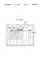
- FIG. 3is a schematic view showing an example of the display of the retrieval image list in the embodiment shown in FIG. 1, and
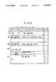
- FIG. 4is a flow chart showing a process for deleting image retrieval signal data.
- a medical image control system 50is basically constituted of a system control device 51, an optical disk device 52, and an operating console 61 consisting of a keyboard 61A and a display device 61B which may be a cathode ray tube (CRT) or the like.
- the medical image control system 50is connected to an image processing apparatus 30.
- the image processing apparatus 30receives image signals S1 from a radiation image recording and read-out apparatus 10 which is an example of an image signal source, carries out a predetermined type of image processing on the image signals S1, and sends image signals S1' obtained from the image processing to an image output apparatus 70.
- the radiation image recording and read-out apparatus 10may be of the type disclosed in, for example, Japanese Unexamined Patent Publication No. 61(1986)-29834 or 61(1986)-94035, wherein stimulable phosphor sheets 11, 11 are conveyed and circulated along a circulation path 12, and a stimulable phosphor sheet 11 is stopped at a position facing an image recording stand 13 and is exposed to radiation 15 emitted by a radiation source 14 in order to store an image of an object (patient) 16 on the sheet 11.
- the stimulable phosphor sheet 11 carrying the radiation image stored thereonis conveyed to an image read-out section and two-dimensionally scanned with a laser beam 18 output by a laser beam source 17 and deflected by a light deflector 19.
- the exposed portion of the sheet 11emits light in proportion to the amount of energy stored thereon during exposure to radiation.
- the emitted lightis photoelectrically detected by a photodetector 21 constituted of a photomultiplier or the like after it has been guided thereto by a light guide member 20.
- the analog output signal from the photodetector 21is amplified, converted by an A/D converter, and exits the radiation image recording and read-out apparatus 10 as a digital image signal S1 representing the radiation image of the object 16.
- the stimulable phosphor sheet 11is sent to an erasing section 22, where the sheet 11 is exposed to erasing light and any radiation energy remaining thereon is erased to such an extent that the sheet 11 is reusable for radiation image recording.
- the radiation image recording and read-out apparatus 10is connected to an ID terminal 25 at which information about a patient 16, which is written on an ID card 26 (hereinafter referred to as patient information), e.g. the name, sex, date of birth and similar information about the patient, is read out. Also, various conditions with regard to the recording of the radiation image (hereinafter referred to as image recording information), e.g. the information about the image number, the date the image was recorded, the portion of an object which was recorded, the size of the recorded image, the read-out sensitivity and the like are entered at the ID terminal 25.
- the patient information S2 and the image recording information S3are fed to the image processing apparatus 30 together with the image signal S1.
- the image processing apparatus 30is constituted so that, for example, not less than 20 types of gradation processing and not less than 10 types of frequency response processing can be effected for the digital image signals S1.
- the image processing conditionsare tabulated, and optimal image processing conditions are automatically selected from the table in accordance with the image recording information specified at the ID terminal 25.
- the image signals S1' obtained by carrying out image processing in the image processing apparatus in accordance with the optimal image processing conditionsare sent to image output apparatus 70.
- the image output apparatus 70is composed of a light beam scanning and recording apparatus for two-dimensionally scanning photographic film with a light beam modulated by an image signal S1', and an automatic developing machine for developing the exposed film.
- the image which the image signal S1' represents, i.e. a radiation image of the patient 16is recorded as a hard copy 71 on the photographic film.
- the hard copy 71 of the radiation image formed by use of the photographic film as mentioned aboveis utilized in the diagnosis of an illness of a patient 16.
- a CRT display device or the likemay be used as the image output apparatus 70.
- the system control device 51 of the medical image control system 50is constituted of a known computer system and composed of a central processing unit (CPU) 53, a memory 54, interfaces 55 and 56, a disk drive controller 57, a bus 58 which connects these sections, a magnetic disk drive unit 59, and a floppy disk drive unit 60.
- the aforesaid keyboard 61A and the display device 61Bare connected to the CPU 53, and the interface 55 is connected to an interface 31 in the image processing apparatus 30.
- the optical disk device 52is composed of an interface 62 connected to the interface 56 in the system control device 51, an optical disk drive controller 63, and an optical disk drive unit 64.
- the aforesaid patient information S2 and the image recording information S3are transferred from the image processing apparatus 30 to the system control device 51, and sequentially recorded on a magnetic disk 65 operated by the magnetic disk drive unit 59, thereby to construct a data base.
- a floppy disk 66 operated by the floppy disk drive unit 60is utilized for system control.
- the patient information S2, the image recording information S3 and the image signal S1are transferred from the image processing apparatus 30 to the optical disk device 52, and filed on an optical disk 67 operated by the optical disk drive unit 64.
- the image signal S1by-passes the image processing section in the image processing apparatus 30, and is recorded on the optical disk 67 as a raw signal which has not been subjected to image processing.
- each graduation along the vertical axisindicates a single track on the optical disk 67
- each graduation along the horizontal axisindicates a single sector.
- the image signal S1is recorded as a single image in an image signal recording region 80 which is sufficiently broadly formed on the optical disk 67.
- signal recordingis effected based on the formation of pits on the surface of the optical disk 67.
- a header 81A, where the patient information S2 and the image recording information S3 corresponding to the image signal S1 are to be recorded, and blocks 81B and 81C, where a signal S4 representing the image processing conditions for use in the image processing apparatus 30 is to be recordedare disposed before and after the image signal area 81 for the single image.
- These image processing conditions represented by the signal S4are identical with the image processing conditions which were used when the image signal S1 was recorded in the image processing apparatus 30 in and sent immediately to the image output apparatus 70 for reproduction.
- one of a plurality of image directories 83A, 83B, 83C, . . . which corresponds to the image signal S1 stored in the image signal area 81is recorded in an image directory region 82.
- the address of the header 81A corresponding to the image signal S1 recorded in the image signal area 81, the sector length of the image signal S1 in the image signal area 81, and characteristic information about the image signal S1 in the image signal area 81are recorded.
- the optical disk 67is also provided with a region 84 on which substitution directories 89A, 89B, 89C, . . . are formed.
- the substitution directoriesare used when a substitution for an image directory 83A, 83B, 83C, . . . is made, i.e. when a directory is changed, and a region 85 on which directories of newly recorded signals representing information from a diagnosis card or the like are formed.
- a block 86where the serial number of each optical disk 67 and a disk surface identification code are to be recorded, a block 87 for indication of the full status of the optical disk 67, and many directory entry blocks 88A, 88B, 88C, . . . .
- the first directory entry block 88Ais used for indicating that a group of the image directories 83A, 83B, 83C, . . . has been formed.
- the head address and the sector length of the image directory groupi.e. the group of directories formed in the image directory region 82) are recorded in the first directory entry block 88A.
- the head address and the sector length of the substitution directory group(89A, 89B, 89C, . . . ) are recorded. Also, in the third directory entry block 88C and subsequent directory entry blocks head addresses and sector lengths of future directory groups can be recorded.
- the image signal S1is sequentially recorded as a single image on the optical disk 67, and the patient information S2, the image recording information S3, and the image processing condition signal S4 are sequentially recorded thereon in conformity with the image signal S1 representing each image.
- the image signal S1should preferably be compressed by a known image signal compression technique before being recorded on the optical disk 67.
- an image signal S1its corresponding patient information S2 and image recording information S3 contain a very large amount of data, approximately 1,000 images can be filed on a single optical disk 67 if an image signal compression technique is used.
- the recording capacity of a magnetic disk 65is smaller than the recording capacity of the optical disk 67.
- only the patient information S2 and the image recording information S3are recorded on the magnetic disk 65, and therefore the patient information S2 and the image recording information S3 for approximately 1,000,000 images can be filed thereon for example.
- information on the importance of the radiation imagecan be entered into the system control device 51 by the operation of a keyboard 61A.
- a physician or the likeimmediately observes the hard copy 71 output derived from the image signal S1' received from the image processing apparatus 30, and enters the rank of importance of the image to a diagnosis process (for example, the physician chooses one of six ranks ranging from rank 0 to rank 5) from a keyboard 61A.
- the rank of importanceis entered, the number of the image recorded on the hard copy 71 is also entered.
- the CPU 53 of the system control device 51receives importance rank information S5 entered in this manner, and stores the importance rank information S5 on the magnetic disk 65 and the optical disk 67.
- the importance rank information S5is recorded as an image retrieval signal with respect to the image number entered in the manner as mentioned above. That is, this data base is structured so that the importance rank information S5 is recorded on the magnetic disk 65 together with the aforesaid patient information S2 and the image recording information S3. Also, on the optical disk 67, the importance rank information S5 is recorded in the headers 81A, 81A before and after the image signal area 81 corresponding to the aforesaid image number as shown in FIG. 2. The importance rank information S5 need not necessarily be recorded on the optical disk and may be recorded only on the magnetic disk 65.
- the importance rank information S5may be entered from the ID terminal 25 or other terminal devices.
- the patient information S2 and the image recording information S3are filed on the magnetic disk 65.
- An image retrieval operatorcorrectly operates the keyboard 61A and enters the desired retrieval signal data by observing the display device 61B of the operating console 61.
- the system control device 51retrieves a list of the images corresponding to the entered retrieval signal data from the data base on the magnetic disk 65, and displays the list on the display device 61B.
- the retrieval signal dataall the items of the patient information S2 and the image recording information S3 can be used.
- an image list indicating the image numbers of all the images of the designated patient, the patient information S2 other than the name of the patient, the image recording information S3, and the importance rank information S5is displayed on the display device 61B, for example, as shown in FIG. 3.
- the retrieval operatorselects a desired image from the displayed image list, and reserves the number of the image.
- the reserved image numberis stored in the memory 54.
- the image retrieval operationcan be completed in advance even though the image processing apparatus 30 and the image output apparatus 70 might be in the process of reproducing an image by use of an image signal S1 just received from the radiation image recording and read-out apparatus 10.
- the retrieval operation carried out as mentioned abovedoes not become invalid, and image reproduction can be started subsequently after the optical disk device 52 is loaded with the optical disk having the image corresponding to the reserved image number recorded thereon.
- the system control device 51activates the optical disk device 52 so that it reads out the image signal corresponding to the reserved image number from the optical disk 67.
- an instruction commanding that the image directory group in the region 82 be read outis given, with the first directory entry block 88A acting as a pointer, and thus the image directories 83A, 83B, 83C, . . . are read out.
- the image directories 83A, 83B, 83C, . . .are read out.
- the reserved image numberacts as a pointer, and the header 81A indicated by said image directory is designated.
- the information written in the header 81A, the image signals S1 written in the image signal area 81, and the information written in the blocks 81B and 81C corresponding to the header 81Aare read out.
- the number of times the read out image has been retrievedis recorded on the magnetic disk 65 as image retrieval signal data for that image This number is incremented each time image is read out. This number may be displayed in the list shown in FIG. 3.
- the image signal S1 in the image signal area 81, the patient information S2 and the image recording information S3 in the header 81A, and the signal S4 representing the image processing conditions in the blocks 81B and 81C, which have been read out in the manner described above,are transferred from the system control device 51 to the image processing apparatus 30.
- the image signal S1is subjected to image processing such as gradation processing or frequency response processing in accordance with the image processing conditions which the aforesaid signal S4 represents, and the processed image signal S1' is sent to the image output apparatus 70.
- the processed image signal S1'is used, and the image is reproduced in the same manner described above.
- a hard copy 71 of the radiation imageis formed.
- the patient information S2 and the image recording information S3are utilized when the patient information and the image recording information are written on the hard copy 71.
- importance rank information 90is indicated in the image list displayed on the display device 61B. Therefore, in cases where many images are retrieved for a single patient, the images of diagnostic importance can be selected by reference to the importance rank information 90 and can be reproduced.
- the image numberscan be displayed in the order of their rank of importance.
- the importance rank information S5is manually entered on the basis of the judgement of physicians or the like.
- the systemmay be constituted so that the CPU 53 or the like uses a predetermined program based the image recording method used, the image recording sequence or the like represented by the image recording information S3 to determine the rank of importance of the images automatically.
- the CPU 53transfers the image retrieval signal data to the magnetic disk 65 each time an image is recorded in the radiation image recording and read-out apparatus 10. However, since the memory capacity of the magnetic disk 65 is limited, the data base stored on the magnetic disk 65 will eventually be full.
- the image retrieval signal data corresponding to a medical image which is considered to be of relatively low utilityis deleted and new data is input into the data base.
- the disk drive controller 67sends a signal to the CPU 53 which then displays warning information on the display device 61B.
- the CPU 53selects the unnecessary image retrieval signal data and deletes it.
- the unnecessary datais deleted according to the process shown in FIG. 4. Namely, when it is ordered to retrieve and delete unnecessary data, the image retrieval signal data is examined for each image. In step 100, whether the image recording date, which is a part of the image recording information S3, is earlier than a predetermined date or not, i.e. whether the time which has passed since the image was recorded is longer than a predetermined time or not, is determined. Then, in step 101, whether the rank of importance indicated by the importance rank information S5 is lower than a predetermined value or not is determined. In step 102, whether the above-mentioned number of retrievals is smaller than a predetermined number or not is determined.
- the dataWhen at least one of the answers to these question is "NO”, the data is regarded as necessary and maintained in the data base.
- the answers to these questionsare "YES”, i.e. when the time which has passed since the image was recorded is long, the importance rank is low and the number of retrievals is small, the data is regarded as having relatively low utility and is deleted from the data base. After all the image retrieval signal data in the data base has been evaluated and the unnecessary data has been deleted accordingly, the data base has space in which new image retrieval signal data can be recorded.
- the standards for judging whether the time which has passed since the image was recorded is too long, the rank of importance of the image is too low and the number of retrievals is too few,may be predetermined or may be determined interactively by a user who uses the keyboard 61A and refers to the display device 61B each time the display device 61B indicates that the data base is full.
- the image retrieval signal datamay be evaluated and deleted on the basis of only the other two items, i.e. the time which has passed since the image was recorded and the rank of importance. Further, other items may be examined in addition to these three items.
- the medical image control system in accordance with the present inventionis also applicable to cases where other types of medical images such as CT images and MR images are to be filed.
Landscapes
- Engineering & Computer Science (AREA)
- Theoretical Computer Science (AREA)
- Physics & Mathematics (AREA)
- Data Mining & Analysis (AREA)
- Databases & Information Systems (AREA)
- General Engineering & Computer Science (AREA)
- General Physics & Mathematics (AREA)
- Library & Information Science (AREA)
- Mathematical Physics (AREA)
- Medical Treatment And Welfare Office Work (AREA)
- Processing Or Creating Images (AREA)
- Information Retrieval, Db Structures And Fs Structures Therefor (AREA)
Abstract
Description
Claims (9)
Applications Claiming Priority (3)
| Application Number | Priority Date | Filing Date | Title |
|---|---|---|---|
| JP61186188AJPS6343642A (en) | 1986-08-08 | 1986-08-08 | Medical image control system |
| JP1-27842 | 1989-02-07 | ||
| JP1027842AJP2551476B2 (en) | 1989-02-07 | 1989-02-07 | Database construction method for medical image management system |
Related Parent Applications (1)
| Application Number | Title | Priority Date | Filing Date |
|---|---|---|---|
| US07275623Continuation-In-Part | 1988-11-15 |
Publications (1)
| Publication Number | Publication Date |
|---|---|
| US5019975Atrue US5019975A (en) | 1991-05-28 |
Family
ID=26365826
Family Applications (1)
| Application Number | Title | Priority Date | Filing Date |
|---|---|---|---|
| US07/342,828Expired - LifetimeUS5019975A (en) | 1986-08-08 | 1989-04-25 | Method for constructing a data base in a medical image control system |
Country Status (1)
| Country | Link |
|---|---|
| US (1) | US5019975A (en) |
Cited By (44)
| Publication number | Priority date | Publication date | Assignee | Title |
|---|---|---|---|---|
| WO1992012490A1 (en)* | 1991-01-11 | 1992-07-23 | Health Innovations, Inc. | Method and apparatus to control diet and weight using human behavior modification techniques |
| EP0599466A1 (en)* | 1992-10-07 | 1994-06-01 | Sony Corporation | Apparatus and methods for managing picture data |
| US5329445A (en)* | 1989-12-25 | 1994-07-12 | Fuji Photo Film Co., Ltd. | Image filing apparatus preventing the storage of incorrect information |
| US5359512A (en)* | 1991-10-31 | 1994-10-25 | Kabushiki Kaisha Toshiba | Picture archiving communication system with migration processing file management for medical application |
| US5446709A (en)* | 1990-01-17 | 1995-08-29 | Fuji Photo Film Co., Ltd. | Image filing apparatus |
| EP0684568A1 (en) | 1994-05-27 | 1995-11-29 | Eastman Kodak Company | Medical image archiving with lossy images on two or more recordable cds |
| WO1996025719A3 (en)* | 1995-02-07 | 1996-10-17 | Merge Technologies Inc | Computer based multimedia medical database management system and user interface |
| US5572422A (en)* | 1991-10-16 | 1996-11-05 | Kabushiki Kaisha Toshiba | Method for managing clustered medical data and medical data filing system in clustered form |
| US5581460A (en)* | 1990-11-06 | 1996-12-03 | Kabushiki Kaisha Toshiba | Medical diagnostic report forming apparatus capable of attaching image data on report |
| US5605153A (en)* | 1992-06-19 | 1997-02-25 | Kabushiki Kaisha Toshiba | Medical image diagnostic system |
| US5636631A (en)* | 1992-05-12 | 1997-06-10 | Advanced Technology Laboratories, Inc. | Ultrasonic image data formats |
| US5644765A (en)* | 1993-12-09 | 1997-07-01 | Canon Kabushiki Kaisha | Image retrieving method and apparatus that calculates characteristic amounts of data correlated with and identifying an image |
| WO1998003933A1 (en)* | 1996-07-23 | 1998-01-29 | R2 Technology, Inc. | User interface for computer aided diagnosis system |
| US5872637A (en)* | 1989-03-01 | 1999-02-16 | Canon Kabushiki Kaisha | Image communication system |
| US5956409A (en)* | 1996-04-29 | 1999-09-21 | Quintet, Inc. | Secure application of seals |
| US20020048222A1 (en)* | 2000-02-11 | 2002-04-25 | Ken Wright | System and method for producing medical image data onto portable digital recording media |
| US6381557B1 (en) | 1998-11-25 | 2002-04-30 | Ge Medical Systems Global Technology Company, Llc | Medical imaging system service evaluation method and apparatus |
| US20020076106A1 (en)* | 2000-09-11 | 2002-06-20 | Tetsujiro Kondo | Image processing apparatus, image processing method, and recording medium |
| US20030084065A1 (en)* | 2001-10-31 | 2003-05-01 | Qian Lin | Method and system for accessing a collection of images in a database |
| US6578002B1 (en) | 1998-11-25 | 2003-06-10 | Gregory John Derzay | Medical diagnostic system service platform |
| EP0989500A3 (en)* | 1998-09-04 | 2004-01-07 | Canon Kabushiki Kaisha | File management system and its method and storage medium |
| US6678397B1 (en)* | 1999-01-26 | 2004-01-13 | Olympus Corporation | Medical image filing system |
| US6792153B1 (en)* | 1999-11-11 | 2004-09-14 | Canon Kabushiki Kaisha | Image processing method and apparatus, and storage medium |
| US20040179236A1 (en)* | 2003-03-12 | 2004-09-16 | Sharp Laboratories Of America, Inc. | Thumbnail audit trail in MFP and print processor/spooler-based print-job auditing |
| US6813365B1 (en)* | 1998-03-31 | 2004-11-02 | Fuji Photo Film Co., Ltd. | Image confirming method |
| US20040252861A1 (en)* | 2003-02-14 | 2004-12-16 | Sony Corporation | Image processing apparatus and method, program, and recording medium |
| US20050149360A1 (en)* | 1999-08-09 | 2005-07-07 | Michael Galperin | Object based image retrieval |
| US20050197860A1 (en)* | 2004-02-23 | 2005-09-08 | Rademr, Inc. | Data management system |
| US6954802B2 (en) | 1998-09-29 | 2005-10-11 | Tdk Electronics Corporation | Removable media recording station for the medical industry |
| US20050240445A1 (en)* | 1998-09-29 | 2005-10-27 | Michael Sutherland | Medical archive library and method |
| US20060045319A1 (en)* | 1999-05-10 | 2006-03-02 | Fuji Photo Film., Ltd. | Image processing method and apparatus |
| US20060193495A1 (en)* | 2003-09-02 | 2006-08-31 | Natsuki Kurokawa | Method for detecting object traveling direction |
| US20060256382A1 (en)* | 2001-12-26 | 2006-11-16 | Matraszek Tomasz A | Method for creating and using affective information in a digital imaging system |
| US20070050216A1 (en)* | 2000-02-11 | 2007-03-01 | Wright Kenneth L | Personal information system |
| US20070067295A1 (en)* | 2000-11-22 | 2007-03-22 | Parulski Kenneth A | Using metadata stored in image files and a separate database to facilitate image retrieval |
| EP1762949A3 (en)* | 2001-12-26 | 2007-08-08 | Eastman Kodak Company | Digital imaging method using importance rating |
| US20080243886A1 (en)* | 2007-03-30 | 2008-10-02 | Fujifilm Corporation | Case database management system and method |
| US20090082637A1 (en)* | 2007-09-21 | 2009-03-26 | Michael Galperin | Multi-modality fusion classifier with integrated non-imaging factors |
| US20100115288A1 (en)* | 2008-08-22 | 2010-05-06 | Datcard Systems, Inc. | System and method of encryption for dicom volumes |
| US8285083B2 (en) | 2006-04-26 | 2012-10-09 | Datcard Systems, Inc. | System for remotely generating and distributing DICOM-compliant media volumes |
| US8788519B2 (en) | 2008-10-24 | 2014-07-22 | John C. Canessa | System and methods for metadata management in content addressable storage |
| US8799650B2 (en) | 2010-12-10 | 2014-08-05 | Datcard Systems, Inc. | Secure portable medical information system and methods related thereto |
| US8799221B2 (en) | 2010-04-23 | 2014-08-05 | John Canessa | Shared archives in interconnected content-addressable storage systems |
| US8964850B2 (en) | 2008-07-08 | 2015-02-24 | Intellectual Ventures Fund 83 Llc | Method, apparatus and system for converging images encoded using different standards |
Citations (5)
| Publication number | Priority date | Publication date | Assignee | Title |
|---|---|---|---|---|
| US4458267A (en)* | 1981-10-06 | 1984-07-03 | Analogic Corporation | Digital x-ray system |
| US4603254A (en)* | 1982-11-04 | 1986-07-29 | Fuji Photo Film Co., Ltd. | Method of storing radiation image data |
| US4672683A (en)* | 1984-03-20 | 1987-06-09 | Olympus Optical Co., Ltd. | Image retrieval and registration system |
| US4817050A (en)* | 1985-11-22 | 1989-03-28 | Kabushiki Kaisha Toshiba | Database system |
| US4876643A (en)* | 1987-06-24 | 1989-10-24 | Kabushiki Kaisha Toshiba | Parallel searching system having a master processor for controlling plural slave processors for independently processing respective search requests |
- 1989
- 1989-04-25USUS07/342,828patent/US5019975A/ennot_activeExpired - Lifetime
Patent Citations (5)
| Publication number | Priority date | Publication date | Assignee | Title |
|---|---|---|---|---|
| US4458267A (en)* | 1981-10-06 | 1984-07-03 | Analogic Corporation | Digital x-ray system |
| US4603254A (en)* | 1982-11-04 | 1986-07-29 | Fuji Photo Film Co., Ltd. | Method of storing radiation image data |
| US4672683A (en)* | 1984-03-20 | 1987-06-09 | Olympus Optical Co., Ltd. | Image retrieval and registration system |
| US4817050A (en)* | 1985-11-22 | 1989-03-28 | Kabushiki Kaisha Toshiba | Database system |
| US4876643A (en)* | 1987-06-24 | 1989-10-24 | Kabushiki Kaisha Toshiba | Parallel searching system having a master processor for controlling plural slave processors for independently processing respective search requests |
Cited By (93)
| Publication number | Priority date | Publication date | Assignee | Title |
|---|---|---|---|---|
| US5872637A (en)* | 1989-03-01 | 1999-02-16 | Canon Kabushiki Kaisha | Image communication system |
| US5329445A (en)* | 1989-12-25 | 1994-07-12 | Fuji Photo Film Co., Ltd. | Image filing apparatus preventing the storage of incorrect information |
| US5446709A (en)* | 1990-01-17 | 1995-08-29 | Fuji Photo Film Co., Ltd. | Image filing apparatus |
| US5581460A (en)* | 1990-11-06 | 1996-12-03 | Kabushiki Kaisha Toshiba | Medical diagnostic report forming apparatus capable of attaching image data on report |
| WO1992012490A1 (en)* | 1991-01-11 | 1992-07-23 | Health Innovations, Inc. | Method and apparatus to control diet and weight using human behavior modification techniques |
| US5572422A (en)* | 1991-10-16 | 1996-11-05 | Kabushiki Kaisha Toshiba | Method for managing clustered medical data and medical data filing system in clustered form |
| US5359512A (en)* | 1991-10-31 | 1994-10-25 | Kabushiki Kaisha Toshiba | Picture archiving communication system with migration processing file management for medical application |
| US5636631A (en)* | 1992-05-12 | 1997-06-10 | Advanced Technology Laboratories, Inc. | Ultrasonic image data formats |
| US5605153A (en)* | 1992-06-19 | 1997-02-25 | Kabushiki Kaisha Toshiba | Medical image diagnostic system |
| EP0599466A1 (en)* | 1992-10-07 | 1994-06-01 | Sony Corporation | Apparatus and methods for managing picture data |
| US6081251A (en)* | 1992-10-07 | 2000-06-27 | Sony Corporation | Apparatus and method for managing picture data |
| US5644765A (en)* | 1993-12-09 | 1997-07-01 | Canon Kabushiki Kaisha | Image retrieving method and apparatus that calculates characteristic amounts of data correlated with and identifying an image |
| EP0684568A1 (en) | 1994-05-27 | 1995-11-29 | Eastman Kodak Company | Medical image archiving with lossy images on two or more recordable cds |
| US5724582A (en)* | 1994-05-27 | 1998-03-03 | Eastman Kodak Company | Medical image archiving with lossy images on two or more recordable CDs |
| US5740428A (en)* | 1995-02-07 | 1998-04-14 | Merge Technologies, Inc. | Computer based multimedia medical database management system and user interface |
| US5950207A (en)* | 1995-02-07 | 1999-09-07 | Merge Technologies Inc. | Computer based multimedia medical database management system and user interface |
| WO1996025719A3 (en)* | 1995-02-07 | 1996-10-17 | Merge Technologies Inc | Computer based multimedia medical database management system and user interface |
| US5956409A (en)* | 1996-04-29 | 1999-09-21 | Quintet, Inc. | Secure application of seals |
| US5917929A (en)* | 1996-07-23 | 1999-06-29 | R2 Technology, Inc. | User interface for computer aided diagnosis system |
| WO1998003933A1 (en)* | 1996-07-23 | 1998-01-29 | R2 Technology, Inc. | User interface for computer aided diagnosis system |
| US7280670B2 (en) | 1998-03-31 | 2007-10-09 | Fujifilm Corporation | Image confirming method |
| US6813365B1 (en)* | 1998-03-31 | 2004-11-02 | Fuji Photo Film Co., Ltd. | Image confirming method |
| US20040264738A1 (en)* | 1998-03-31 | 2004-12-30 | Fuji Photo Film Co., Ltd. | Image confirming method |
| EP0989500A3 (en)* | 1998-09-04 | 2004-01-07 | Canon Kabushiki Kaisha | File management system and its method and storage medium |
| US6954802B2 (en) | 1998-09-29 | 2005-10-11 | Tdk Electronics Corporation | Removable media recording station for the medical industry |
| US20050240445A1 (en)* | 1998-09-29 | 2005-10-27 | Michael Sutherland | Medical archive library and method |
| US6381557B1 (en) | 1998-11-25 | 2002-04-30 | Ge Medical Systems Global Technology Company, Llc | Medical imaging system service evaluation method and apparatus |
| US6578002B1 (en) | 1998-11-25 | 2003-06-10 | Gregory John Derzay | Medical diagnostic system service platform |
| US6678397B1 (en)* | 1999-01-26 | 2004-01-13 | Olympus Corporation | Medical image filing system |
| US20060045319A1 (en)* | 1999-05-10 | 2006-03-02 | Fuji Photo Film., Ltd. | Image processing method and apparatus |
| US7082215B1 (en)* | 1999-05-10 | 2006-07-25 | Fuji Photo Film Co., Ltd. | Image processing method and apparatus |
| US20060115136A1 (en)* | 1999-05-10 | 2006-06-01 | Fuji Photo Film Co., Ltd. | Image processing method and apparatus |
| US6941323B1 (en) | 1999-08-09 | 2005-09-06 | Almen Laboratories, Inc. | System and method for image comparison and retrieval by enhancing, defining, and parameterizing objects in images |
| US7483919B2 (en) | 1999-08-09 | 2009-01-27 | Almen Laboratories | Object based image retrieval |
| US20050149360A1 (en)* | 1999-08-09 | 2005-07-07 | Michael Galperin | Object based image retrieval |
| US8775451B2 (en) | 1999-08-09 | 2014-07-08 | Almen Laboratories, Inc. | Object based image retrieval |
| US6792153B1 (en)* | 1999-11-11 | 2004-09-14 | Canon Kabushiki Kaisha | Image processing method and apparatus, and storage medium |
| US7783163B2 (en) | 2000-02-11 | 2010-08-24 | Datcard Systems, Inc. | System and method for producing medical image data onto portable digital recording media |
| US20070050216A1 (en)* | 2000-02-11 | 2007-03-01 | Wright Kenneth L | Personal information system |
| US10248760B2 (en) | 2000-02-11 | 2019-04-02 | Datcard Systems, Inc. | System and method for producing medical image data onto portable digital recording media |
| US9111017B2 (en) | 2000-02-11 | 2015-08-18 | Datcard Systems, Inc. | Personal information system |
| US8515251B2 (en) | 2000-02-11 | 2013-08-20 | Datcard Systems, Inc. | System and method for producing medical image data onto portable digital recording media |
| US8483550B2 (en) | 2000-02-11 | 2013-07-09 | Datcard Systems, Inc. | System and method for producing medical image data onto portable digital recording media |
| US8509604B2 (en) | 2000-02-11 | 2013-08-13 | Datcard Systems, Inc. | System and method for producing medical image data onto portable digital recording media |
| US7783174B2 (en) | 2000-02-11 | 2010-08-24 | Datcard Systems, Inc. | System and method for producing medical image data onto portable digital recording media |
| US7734157B2 (en) | 2000-02-11 | 2010-06-08 | Datcard Systems, Inc. | System and method for producing medical image data onto portable digital recording media |
| US7979387B2 (en) | 2000-02-11 | 2011-07-12 | Datcard Systems, Inc. | Personal information system |
| US7729597B2 (en) | 2000-02-11 | 2010-06-01 | Datcard Systems, Inc. | System and method for producing medical image data onto portable digital recording media |
| US20090252480A1 (en)* | 2000-02-11 | 2009-10-08 | Datcard Systems, Inc. | System and method for producing medical image data onto portable digital recording media |
| US20020048222A1 (en)* | 2000-02-11 | 2002-04-25 | Ken Wright | System and method for producing medical image data onto portable digital recording media |
| US7302164B2 (en) | 2000-02-11 | 2007-11-27 | Datcard Systems, Inc. | System and method for producing medical image data onto portable digital recording media |
| US20090252479A1 (en)* | 2000-02-11 | 2009-10-08 | Datcard Systems, Inc. | System and method for producing medical image data onto portable digital recording media |
| US20080063368A1 (en)* | 2000-02-11 | 2008-03-13 | Datcard System, Inc. | System and Method for Producing Medical Image Data onto Portable Digital Recording Media |
| US20090245754A1 (en)* | 2000-02-11 | 2009-10-01 | Datcard Systems, Inc. | System and method for producing medical image data onto portable digital recording media |
| US20090248750A1 (en)* | 2000-02-11 | 2009-10-01 | Datcard Systems, Inc. | System and method for producing medical image data onto portable digital recording media |
| US20020076106A1 (en)* | 2000-09-11 | 2002-06-20 | Tetsujiro Kondo | Image processing apparatus, image processing method, and recording medium |
| US6934414B2 (en)* | 2000-09-11 | 2005-08-23 | Sony Corporation | Image processing apparatus, image processing method, and recording medium |
| US20050226504A1 (en)* | 2000-09-11 | 2005-10-13 | Tetsujiro Kondo | Image processiong apparatus, image processing method, and recording medium |
| US7340097B2 (en) | 2000-09-11 | 2008-03-04 | Sony Corporation | Image processing apparatus, image processing method, and recording medium |
| US20070067295A1 (en)* | 2000-11-22 | 2007-03-22 | Parulski Kenneth A | Using metadata stored in image files and a separate database to facilitate image retrieval |
| US20090238540A1 (en)* | 2001-01-17 | 2009-09-24 | Datcard Systems, Inc. | System and method for producing medical image data onto portable digital recording media |
| US7801422B2 (en) | 2001-01-17 | 2010-09-21 | Datcard Systems, Inc. | System and method for producing medical image data onto portable digital recording media |
| US20030084065A1 (en)* | 2001-10-31 | 2003-05-01 | Qian Lin | Method and system for accessing a collection of images in a database |
| WO2003038680A3 (en)* | 2001-10-31 | 2004-01-22 | Hewlett Packard Co | Method and system for accessing a collection of images in a database |
| US7130864B2 (en) | 2001-10-31 | 2006-10-31 | Hewlett-Packard Development Company, L.P. | Method and system for accessing a collection of images in a database |
| US20090297065A1 (en)* | 2001-12-26 | 2009-12-03 | Matraszek Tomasz A | Method for creating and using affective information in a digital imaging system |
| EP1762949A3 (en)* | 2001-12-26 | 2007-08-08 | Eastman Kodak Company | Digital imaging method using importance rating |
| US20090307222A1 (en)* | 2001-12-26 | 2009-12-10 | Matraszek Tomasz A | Method for creating and using affective information in a digital imaging system |
| US9082046B2 (en) | 2001-12-26 | 2015-07-14 | Intellectual Ventures Fund 83 Llc | Method for creating and using affective information in a digital imaging system |
| US20060256382A1 (en)* | 2001-12-26 | 2006-11-16 | Matraszek Tomasz A | Method for creating and using affective information in a digital imaging system |
| US7620270B2 (en) | 2001-12-26 | 2009-11-17 | Eastman Kodak Company | Method for creating and using affective information in a digital imaging system |
| US8184916B2 (en) | 2001-12-26 | 2012-05-22 | Eastman Kodak Company | Method for creating and using affective information in a digital imaging system |
| US8630496B2 (en) | 2001-12-26 | 2014-01-14 | Intellectual Ventures Fund 83 Llc | Method for creating and using affective information in a digital imaging system |
| US7933474B2 (en)* | 2001-12-26 | 2011-04-26 | Eastman Kodak Company | Method for creating and using affective information in a digital imaging system |
| US8036467B2 (en) | 2001-12-26 | 2011-10-11 | Eastman Kodak Company | Method for creating and using affective information in a digital imaging system |
| US7409075B2 (en)* | 2003-02-14 | 2008-08-05 | Sony Corporation | Image processing apparatus and method, program, and recording medium |
| US20040252861A1 (en)* | 2003-02-14 | 2004-12-16 | Sony Corporation | Image processing apparatus and method, program, and recording medium |
| US20040179236A1 (en)* | 2003-03-12 | 2004-09-16 | Sharp Laboratories Of America, Inc. | Thumbnail audit trail in MFP and print processor/spooler-based print-job auditing |
| US7397578B2 (en)* | 2003-03-12 | 2008-07-08 | Sharp Laboratories Of America, Inc. | Thumbnail audit trail in MFP and print processor/spooler-based print-job auditing |
| US7346193B2 (en)* | 2003-09-02 | 2008-03-18 | Matsushita Electric Industrial Co., Ltd. | Method for detecting object traveling direction |
| US20060193495A1 (en)* | 2003-09-02 | 2006-08-31 | Natsuki Kurokawa | Method for detecting object traveling direction |
| US20050197860A1 (en)* | 2004-02-23 | 2005-09-08 | Rademr, Inc. | Data management system |
| US8285083B2 (en) | 2006-04-26 | 2012-10-09 | Datcard Systems, Inc. | System for remotely generating and distributing DICOM-compliant media volumes |
| US8832114B2 (en)* | 2007-03-30 | 2014-09-09 | Fujifilm Corporation | Case database management system and method |
| US20080243886A1 (en)* | 2007-03-30 | 2008-10-02 | Fujifilm Corporation | Case database management system and method |
| US20090082637A1 (en)* | 2007-09-21 | 2009-03-26 | Michael Galperin | Multi-modality fusion classifier with integrated non-imaging factors |
| US8964850B2 (en) | 2008-07-08 | 2015-02-24 | Intellectual Ventures Fund 83 Llc | Method, apparatus and system for converging images encoded using different standards |
| US8756437B2 (en) | 2008-08-22 | 2014-06-17 | Datcard Systems, Inc. | System and method of encryption for DICOM volumes |
| US20100115288A1 (en)* | 2008-08-22 | 2010-05-06 | Datcard Systems, Inc. | System and method of encryption for dicom volumes |
| US8788519B2 (en) | 2008-10-24 | 2014-07-22 | John C. Canessa | System and methods for metadata management in content addressable storage |
| US8799221B2 (en) | 2010-04-23 | 2014-08-05 | John Canessa | Shared archives in interconnected content-addressable storage systems |
| US8930470B2 (en) | 2010-04-23 | 2015-01-06 | Datcard Systems, Inc. | Event notification in interconnected content-addressable storage systems |
| US8799650B2 (en) | 2010-12-10 | 2014-08-05 | Datcard Systems, Inc. | Secure portable medical information system and methods related thereto |
Similar Documents
| Publication | Publication Date | Title |
|---|---|---|
| US5019975A (en) | Method for constructing a data base in a medical image control system | |
| US4768099A (en) | Method of storing and reproducing a medical image in which image data is stored with corresponding information representing image processing conditions | |
| US5276805A (en) | Image filing system which has retrieval data containing link information between image data | |
| US4737912A (en) | Medical image filing apparatus | |
| US5241472A (en) | Method of identifying and archiving medical images | |
| US5359512A (en) | Picture archiving communication system with migration processing file management for medical application | |
| US20160055298A1 (en) | System and method for producing medical image data onto portable digital recording media | |
| US5001569A (en) | Image filing apparatus | |
| US5446709A (en) | Image filing apparatus | |
| US5329445A (en) | Image filing apparatus preventing the storage of incorrect information | |
| US5619598A (en) | Image filing apparatus using both reversible and irreversible compression | |
| US5173787A (en) | Method for storing image signals | |
| JPS6343642A (en) | Medical image control system | |
| JPH08317908A (en) | Reproduction or display method and system for medical picture with text column which can be optionally arranged | |
| JP2551476B2 (en) | Database construction method for medical image management system | |
| JP3272435B2 (en) | Radiation image storage and display device | |
| JPS63102551A (en) | Medical image accumulating method | |
| JPS6343641A (en) | Medical image accumulation method | |
| JPS6340527A (en) | Medical image control system | |
| JPS6371240A (en) | Medical image accumulation method | |
| JPS637518A (en) | Picture data recording method on optical disk | |
| JPS636666A (en) | Medical picture retrieving device | |
| JPH08256991A (en) | Medical image display device | |
| JP2579901B2 (en) | Diagnostic imaging device | |
| JP2640549B2 (en) | Image data recording method |
Legal Events
| Date | Code | Title | Description |
|---|---|---|---|
| AS | Assignment | Owner name:FUJI PHOTO FILM CO., LTD., JAPAN Free format text:ASSIGNMENT OF ASSIGNORS INTEREST.;ASSIGNOR:MUKAI, HACHIRO;REEL/FRAME:005123/0758 Effective date:19890531 | |
| STCF | Information on status: patent grant | Free format text:PATENTED CASE | |
| CC | Certificate of correction | ||
| FEPP | Fee payment procedure | Free format text:PAYOR NUMBER ASSIGNED (ORIGINAL EVENT CODE: ASPN); ENTITY STATUS OF PATENT OWNER: LARGE ENTITY | |
| FPAY | Fee payment | Year of fee payment:4 | |
| FEPP | Fee payment procedure | Free format text:PAYER NUMBER DE-ASSIGNED (ORIGINAL EVENT CODE: RMPN); ENTITY STATUS OF PATENT OWNER: LARGE ENTITY Free format text:PAYOR NUMBER ASSIGNED (ORIGINAL EVENT CODE: ASPN); ENTITY STATUS OF PATENT OWNER: LARGE ENTITY | |
| FPAY | Fee payment | Year of fee payment:8 | |
| FEPP | Fee payment procedure | Free format text:PAYOR NUMBER ASSIGNED (ORIGINAL EVENT CODE: ASPN); ENTITY STATUS OF PATENT OWNER: LARGE ENTITY Free format text:PAYER NUMBER DE-ASSIGNED (ORIGINAL EVENT CODE: RMPN); ENTITY STATUS OF PATENT OWNER: LARGE ENTITY | |
| FPAY | Fee payment | Year of fee payment:12 | |
| AS | Assignment | Owner name:FUJIFILM CORPORATION, JAPAN Free format text:ASSIGNMENT OF ASSIGNORS INTEREST;ASSIGNOR:FUJIFILM HOLDINGS CORPORATION (FORMERLY FUJI PHOTO FILM CO., LTD.);REEL/FRAME:018904/0001 Effective date:20070130 Owner name:FUJIFILM CORPORATION,JAPAN Free format text:ASSIGNMENT OF ASSIGNORS INTEREST;ASSIGNOR:FUJIFILM HOLDINGS CORPORATION (FORMERLY FUJI PHOTO FILM CO., LTD.);REEL/FRAME:018904/0001 Effective date:20070130 |