US5011486A - Composite nerve guidance channels - Google Patents
Composite nerve guidance channelsDownload PDFInfo
- Publication number
- US5011486A US5011486AUS07/273,236US27323688AUS5011486AUS 5011486 AUS5011486 AUS 5011486AUS 27323688 AUS27323688 AUS 27323688AUS 5011486 AUS5011486 AUS 5011486A
- Authority
- US
- United States
- Prior art keywords
- nerve
- membrane
- channels
- bsa
- active factor
- Prior art date
- Legal status (The legal status is an assumption and is not a legal conclusion. Google has not performed a legal analysis and makes no representation as to the accuracy of the status listed.)
- Expired - Fee Related
Links
Images


Classifications
- A—HUMAN NECESSITIES
- A61—MEDICAL OR VETERINARY SCIENCE; HYGIENE
- A61L—METHODS OR APPARATUS FOR STERILISING MATERIALS OR OBJECTS IN GENERAL; DISINFECTION, STERILISATION OR DEODORISATION OF AIR; CHEMICAL ASPECTS OF BANDAGES, DRESSINGS, ABSORBENT PADS OR SURGICAL ARTICLES; MATERIALS FOR BANDAGES, DRESSINGS, ABSORBENT PADS OR SURGICAL ARTICLES
- A61L31/00—Materials for other surgical articles, e.g. stents, stent-grafts, shunts, surgical drapes, guide wires, materials for adhesion prevention, occluding devices, surgical gloves, tissue fixation devices
- A61L31/14—Materials characterised by their function or physical properties, e.g. injectable or lubricating compositions, shape-memory materials, surface modified materials
- A61L31/16—Biologically active materials, e.g. therapeutic substances
- A—HUMAN NECESSITIES
- A61—MEDICAL OR VETERINARY SCIENCE; HYGIENE
- A61L—METHODS OR APPARATUS FOR STERILISING MATERIALS OR OBJECTS IN GENERAL; DISINFECTION, STERILISATION OR DEODORISATION OF AIR; CHEMICAL ASPECTS OF BANDAGES, DRESSINGS, ABSORBENT PADS OR SURGICAL ARTICLES; MATERIALS FOR BANDAGES, DRESSINGS, ABSORBENT PADS OR SURGICAL ARTICLES
- A61L31/00—Materials for other surgical articles, e.g. stents, stent-grafts, shunts, surgical drapes, guide wires, materials for adhesion prevention, occluding devices, surgical gloves, tissue fixation devices
- A61L31/04—Macromolecular materials
- A61L31/043—Proteins; Polypeptides; Degradation products thereof
- A61L31/047—Other specific proteins or polypeptides not covered by A61L31/044 - A61L31/046
- A—HUMAN NECESSITIES
- A61—MEDICAL OR VETERINARY SCIENCE; HYGIENE
- A61L—METHODS OR APPARATUS FOR STERILISING MATERIALS OR OBJECTS IN GENERAL; DISINFECTION, STERILISATION OR DEODORISATION OF AIR; CHEMICAL ASPECTS OF BANDAGES, DRESSINGS, ABSORBENT PADS OR SURGICAL ARTICLES; MATERIALS FOR BANDAGES, DRESSINGS, ABSORBENT PADS OR SURGICAL ARTICLES
- A61L31/00—Materials for other surgical articles, e.g. stents, stent-grafts, shunts, surgical drapes, guide wires, materials for adhesion prevention, occluding devices, surgical gloves, tissue fixation devices
- A61L31/04—Macromolecular materials
- A61L31/048—Macromolecular materials obtained by reactions only involving carbon-to-carbon unsaturated bonds
- A—HUMAN NECESSITIES
- A61—MEDICAL OR VETERINARY SCIENCE; HYGIENE
- A61L—METHODS OR APPARATUS FOR STERILISING MATERIALS OR OBJECTS IN GENERAL; DISINFECTION, STERILISATION OR DEODORISATION OF AIR; CHEMICAL ASPECTS OF BANDAGES, DRESSINGS, ABSORBENT PADS OR SURGICAL ARTICLES; MATERIALS FOR BANDAGES, DRESSINGS, ABSORBENT PADS OR SURGICAL ARTICLES
- A61L31/00—Materials for other surgical articles, e.g. stents, stent-grafts, shunts, surgical drapes, guide wires, materials for adhesion prevention, occluding devices, surgical gloves, tissue fixation devices
- A61L31/08—Materials for coatings
- A61L31/10—Macromolecular materials
- A—HUMAN NECESSITIES
- A61—MEDICAL OR VETERINARY SCIENCE; HYGIENE
- A61L—METHODS OR APPARATUS FOR STERILISING MATERIALS OR OBJECTS IN GENERAL; DISINFECTION, STERILISATION OR DEODORISATION OF AIR; CHEMICAL ASPECTS OF BANDAGES, DRESSINGS, ABSORBENT PADS OR SURGICAL ARTICLES; MATERIALS FOR BANDAGES, DRESSINGS, ABSORBENT PADS OR SURGICAL ARTICLES
- A61L31/00—Materials for other surgical articles, e.g. stents, stent-grafts, shunts, surgical drapes, guide wires, materials for adhesion prevention, occluding devices, surgical gloves, tissue fixation devices
- A61L31/14—Materials characterised by their function or physical properties, e.g. injectable or lubricating compositions, shape-memory materials, surface modified materials
- A61L31/146—Porous materials, e.g. foams or sponges
- A—HUMAN NECESSITIES
- A61—MEDICAL OR VETERINARY SCIENCE; HYGIENE
- A61L—METHODS OR APPARATUS FOR STERILISING MATERIALS OR OBJECTS IN GENERAL; DISINFECTION, STERILISATION OR DEODORISATION OF AIR; CHEMICAL ASPECTS OF BANDAGES, DRESSINGS, ABSORBENT PADS OR SURGICAL ARTICLES; MATERIALS FOR BANDAGES, DRESSINGS, ABSORBENT PADS OR SURGICAL ARTICLES
- A61L2300/00—Biologically active materials used in bandages, wound dressings, absorbent pads or medical devices
- A61L2300/20—Biologically active materials used in bandages, wound dressings, absorbent pads or medical devices containing or releasing organic materials
- A61L2300/252—Polypeptides, proteins, e.g. glycoproteins, lipoproteins, cytokines
- A—HUMAN NECESSITIES
- A61—MEDICAL OR VETERINARY SCIENCE; HYGIENE
- A61L—METHODS OR APPARATUS FOR STERILISING MATERIALS OR OBJECTS IN GENERAL; DISINFECTION, STERILISATION OR DEODORISATION OF AIR; CHEMICAL ASPECTS OF BANDAGES, DRESSINGS, ABSORBENT PADS OR SURGICAL ARTICLES; MATERIALS FOR BANDAGES, DRESSINGS, ABSORBENT PADS OR SURGICAL ARTICLES
- A61L2300/00—Biologically active materials used in bandages, wound dressings, absorbent pads or medical devices
- A61L2300/40—Biologically active materials used in bandages, wound dressings, absorbent pads or medical devices characterised by a specific therapeutic activity or mode of action
- A61L2300/412—Tissue-regenerating or healing or proliferative agents
- A61L2300/414—Growth factors
- A—HUMAN NECESSITIES
- A61—MEDICAL OR VETERINARY SCIENCE; HYGIENE
- A61L—METHODS OR APPARATUS FOR STERILISING MATERIALS OR OBJECTS IN GENERAL; DISINFECTION, STERILISATION OR DEODORISATION OF AIR; CHEMICAL ASPECTS OF BANDAGES, DRESSINGS, ABSORBENT PADS OR SURGICAL ARTICLES; MATERIALS FOR BANDAGES, DRESSINGS, ABSORBENT PADS OR SURGICAL ARTICLES
- A61L2430/00—Materials or treatment for tissue regeneration
- A61L2430/32—Materials or treatment for tissue regeneration for nerve reconstruction
Definitions
- the technical field of this inventionconcerns medical devices useful for the repair of severed nerves and methods for fabricating and using such devices for nerve repair.
- Axonal sproutswill appear from the tip of the regenerating axon. These sprouts grow distally and attempt to reenter the intact neurilemmal sheaths of the distal portion of the severed nerve. If entry is successfully made, axonal growth will continue down these sheaths and function will eventually be restored.
- the chief hazard to the successful repairis the trauma produced by the manipulation of the nerve ends and the subsequent suturing to maintain alignment.
- the traumaappears to stimulate the growth and/or migration of fibroblasts and other scar-forming connective tissue cells.
- the scar tissueprevents the regenerating axons in the proximal stump from reaching the distal stump to reestablish a nerve charge pathway. The result is a permanent loss of sensory or motor function.
- silastic cuffsfor peripheral nerve repair was reported by Ducker et al. in Vol. 28, Journal of Neurosurgery. pp. 582-587 (1968). Silicone rubber sheathing for nerve repair was reported by Midgley et al. in Vol. 19, Surgical Forum. pp. 519-528 (1968) and by Lundborg et al. in Vol. 41, Journal of Neuropathology in Experimental Neurology, pp 412-422 (1982). The use of bioresorbable, polyglactin mesh tubing was reported by Molander et al. in Vol. 5, Muscle & Nerve. pp. 54-58 (1982). The use of porous acrylic copolymer tubes in nerve regeneration was disclosed by Uzman et al.
- Channelshave been manipulated in various ways in an attempt to correct these shortcomings.
- channels prefilled with a laminin gelas disclosed in Madison et al., Vol. 44, Brain Res., pp. 325-334 (1985)
- a glycosaminoglycan templateas described in Yannas et al., Vol. 11, Trans. Soc. Biomat., pp. 146 (1985)
- fibrinWoilliams et al., Vol. 264, J. Como. Neurol. pp. 284-290 (1987)
- these substancesare normally substrate-bound materials, their conformation and, hence, their level of activity is decreased when they are solubilized.
- Channelshave also been preloaded with various growth factors (Politis et al., Vol. 253, Brain Res. pp. 1-12 (1982)).
- these factorstypically are not stable in an aqueous environment; their half lives are measured in hours rather than in weeks, which is the least amount of time usually required for completed regeneration.
- the delivery of these factorsis not continuous or controlled; it is dispensed as a one time bolus which is not conducive for long term nerve growth stimulation.
- nerve guidance channels containing diffusible nerve growth-inducing active factorscan greatly promote the regeneration of severed nerve ends over relatively large distances.
- These channelsconsist of biocompatible, porous, tubular membranes having active factor incorporated into the membrane. The factors are released at a controlled rate, thereby prolonging the stimulatory properties of the channel.
- factors incorporated into the channel wallshave a greater half life than those which are in soluble form within the lumen of the channels; factors sequestered within the hydrophobic environment of the channel wall are not exposed to proteases in the aqueous environment until they are released.
- nerveis used herein to mean both monofascicular and polyfascicular nerves.
- the same general principals of regeneration within the nerve guidance channels of the present inventionare applicable to both.
- the term "active factor”is used herein to describe any diffusible substance having bioactivity.
- the active factoris a "nerve growth enhancer,” such as, for example, a growth factor or active analog, fragment, or derivative thereof.
- the nerve guidance channels of the present inventionare designed to retain active factor within the membrane.
- the inner surface of the membraneis permeable to the active factor incorporated therein, while the outer membrane surface is impermeable to the factor.
- the inventionfurther encompasses methods of repairing a severed nerve using the guidance channels of the present invention.
- the severed nerve endsare placed in proximity to each other and secured within the lumen of the membrane.
- a mandrel having the dimensions of the desired nerve guidance channelcan be employed to fabricate the device.
- One or more coats of a first solution, containing a biocompatible polymer, an active factor, and a hydrophilic carrier,is applied to the mandrel.
- the hydrophilic carrierestablishes domains within the polymer, creating an interconnected pore structure in the finished device from which the active factor is released into the lumen.
- one or more finishing coats of a second solution containing the same or another biocompatible polymer without the carrieris applied to provide an impermeable or substantially less permeable outer surface. The device is then dried and removed from the mandrel.
- the nerve guidance channels described in the examples beloware generally tubular in shape, it should be clear that various alternative shapes can be employed.
- the lumens of the guidance channelscan be oval or even square in cross-section.
- the guidance channelscan also be constructed from two or more parts which are clamped together to secure the nerve stumps.
- polymeric sheet materials containing incorporated active factorcan be employed and formed into channels in situ. In such a procedure, the nerve stumps can be placed on the surface of the sheet and secured thereto by sutures, adhesives, or friction. The sheet is then wrapped around the nerve segments, and the resulting channel is closed by further sutures, adhesives, or friction.
- FIG. 1is a schematic representation of a nerve guidance channel of the present invention
- FIG. 2is a schematic cross-sectional view of the nerve guidance channel of FIG. 1;
- FIG. 3is an electron micrograph of a cross-section of a nerve guidance channel containing BSA and an active factor
- FIG. 4is a graphic representation of the rate at which a protein is released from the nerve guidance channel of the present invention as a function of time;
- FIGS. 5A and 5Bis a phase contrast micrograph of human (PC12) cells cultured in the presence of a control nerve guidance channel releasing BSA only (A), and a channel releasing BSA and b-FGF (B) respectively; and
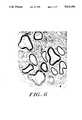
- FIG. 6is a photographic representation of an electron micrograph taken at the midpoint of a guidance channel releasing b-FGF four weeks post-implantation.
- FIG. 1shows a nerve guidance device 10, according to the present invention, as a tubular, porous membrane having openings 14 and 16 adapted to receive severed nerve ends 18 and 20.
- the inner membrane surface 22defines the boundary of a lumen 24 through which a nerve may regenerate.
- FIG. 2is a schematic cross-sectional view of the membrane 10 showing its porous wall structure 12.
- active factor 26is incorporated within the membrane wall 12.
- the outer membrane surface 28is nonporous, while porous inner membrane surface 22 allows for the diffusion therethrough of active factor 26.
- FIG. 3is an electron micrograph of a cross-section of an actual guidance channel having a nonporous membrane wall and pores containing BSA and active factor.
- Preferable nerve growth enhancersare growth factors, such as nerve growth factor (NGF) and fibroblast growth factor (FGF).
- NGFnerve growth factor
- FGFfibroblast growth factor
- Basic FGFb-FGF
- a-FGFacidic FGF
- a "second messenger” substanceis one that initiates a cellular response to a specific signal external to that cell.
- Useful second messenger substancesinclude, for example, cyclic adenosine monophosphate (cAMP). Active analogs of cAMP such as 8-bromo cAMP and chlorophenylthio cAMP, or active derivatives and fragments thereof, are also useful.
- cAMPcyclic adenosine monophosphate
- Active analogs of cAMPsuch as 8-bromo cAMP and chlorophenylthio cAMP, or active derivatives and fragments thereof, are also useful.
- “Second messenger inducers”are responsible for the synthesis or activation of a second messenger substance.
- Useful second messenger inducersinclude forskolin, and active derivatives and analogs thereof.
- the membrane of the channelmay be fabricated from any biocompatible polymers, such as, for example, polyethylene vinyl-acetate (EVA).
- EVApolyethylene vinyl-acetate
- the channelmay be composed of a biocompatible hydrogels, such as polyvinyl pyrolidone, polyethylene oxide (PEO), polyurethanes, acrylates, or mixtures thereof.
- Preferable acrylatesinclude methacrylates or hydroethylmethacrylates.
- the membraneinstead may be composed of a bioresorbable, biocompatible polymer, such as a polyanhydride, polyester, or mixtures thereof. If the channel is not biodegradable over time, it can be formed with longitudinal lines of weakness to facilitate removal from about the regenerated nerve after healing has progressed sufficiently.
- the membrane wall thickness of the nerve guidance channels of the present inventionrange from about 0.05 to about 1.0 millimeters (mm).
- the diameter of the channel lumencan vary from about 0.5 mm to about 3.0 centimeters (cm), depending upon the size of nerve to be repaired.
- the outer surface of the membraneis impermeable to solutes of any size, while the inner membrane surface contains pores having a diameter from about 0.1 to 10.0 microns ( ⁇ m) so as to be permeable to solutes having a molecular weight of about 100,000 daltons or less. These pores enable the active factors to diffuse out of the membrane and into the lumen of the channel.
- the particular pore sizecan be varied depending upon the active factors to be secreted, the size of the nerve to be repaired, and the preferred delivery rate of the active factors.
- the inventionfurther encompasses methods of repairing a severed nerve using the nerve guidance channels of the present invention.
- the nerve guidance channels of the present inventionas described above, are used by locating the severed nerve ends and selecting and providing an appropriately sized tubular device for the repair.
- the cut ends of the nerveare gently drawn into channel by manual manipulation or suction, placed in optimal proximity and then secured in position without undue trauma by sutures through the channel, or by a biocompatible adhesive (e.g., fibrin glue) or by frictional engagement with the channel.
- the channelis then situated in the general in vivo location of the nerve.
- Antibioticscan be administered to the site, and the wound is then closed.
- a mandrel/molding techniqueis employed.
- a cylindrical mandrel having an internal diameter of about 0.05 to 3.00 mmis particularly useful.
- At least one coat of a first solution containing a biocompatible polymer and an active factor of the type described aboveis applied to the mandrel.
- the solutionmay contain from about 1 to 30%, but preferably about 10% by weight polymer such as, for example, polyethylene vinyl acetate in a solvent.
- the active factor in the solutionmay comprise from about 0.001 to 40% by weight of this first solution, depending on the biological activity of the factor and on the desired diffusion rate of the factor from the membrane into the lumen.
- the remainder of the first solutioncan comprise a pore-forming, biocompatible agent, for example, a hydrophilic carrier, such as bovine serum albumin, which establishes domains within the polymer, creating an interconnected pore structure in the finished device from which the active factor is released into the lumen.
- a hydrophilic carriersuch as bovine serum albumin
- At least one coat of an active, factor-free, second solution containing a biocompatible polymeris layered on the first solution-coated mandrel prior to drying and removal from the mold.
- This second solutionmay again contain from about 1 to 30% but preferably about 10% by weight biocompatible polymer, such as, for example, polyethylene vinyl acetate without the pore-forming agent. From about 2 to 10 coats of the second solution are applied, with about 4 coats being preferable.
- the solvent for the polymer in both the first and second solutionscan be any one of a variety of organic solvents, such as, for example, methylene chloride
- the accumulated coats on the mandrelare then dried and removed from the mandrel to form the guidance channel of the present invention.
- This layering procedureallows deposition of an impermeable coat on the outer surface of the device, insuring that the active factors incorporated into the membrane walls will be inhibited from diffusing through the external surface, and will diffuse only through the inner membrane surface into the lumen of the channel.
- EVAPolyethylene vinyl-acetate pellets (Elvax-40) were obtained from Dupont (Wilmington, DE). Impurities were removed with multiple wash in pure ethanol and distilled water. A 10% (w/w) solution of EVA was prepared in methylene chloride.
- Tubular guidance devices50 mm long ⁇ 1.5 mm ID ⁇ 1.9 mm OD were prepared by dip molding over of a stainless steel wire mandrel 28 times.
- the channelswere then dried overnight in a fume hood, cut into 19 mm pieces, and placed under vacuum for 24 hours. Before implantation, the channels were cleaned and sterilized in a 70% ethanol solution.
- BSAbovine serum albumin
- Guidance channels containing b-FGF and BSAwere prepared as described in EXAMPLE 2, except that 100 ⁇ l of b-FGF (recombinant, Amgen, Thousand Oaks, CA) was added to a 10 ml solution 40% (w/w) BSA/EVA, resulting in a 0.004% (w/w) b-FGF/EVA solid matrix upon drying.
- the moldwas dipped 24 times in the b-FGF/BSA/EVA solution, followed by 4 times in 10% pure EVA solution
- Guidance channels containing ⁇ 1-GP and BSAwere prepared as described in EXAMPLE 2 except that ⁇ l -GP was added to a 10 ml solution of 40% (w/w) BSA/EVA which resulted in a 4% (w/w) ⁇ l -GP/ EVA solid matrix upon drying.
- the moldwas dipped 24 times in the ⁇ 1-GP/BSA/EVA solution, followed by 4 dips in a pure 10% EVA solution.
- Guidance channels containing b-FGF, ⁇ l -GP, and BSAwere prepared as described in EXAMPLE 2, except that 100 ⁇ l of b-FGF and ⁇ l -GP were added to a 10 ml solution of 40% (w/w) BSA/EVA which resulted in a 0.004% (w/w) b-FGF, 4% (w/w) ⁇ l -GP solid EVA matrix upon drying.
- the moldwas dipped 24 times in the b-FGF/ ⁇ l -GP/BSA/EVA solution, followed by 4 dips in a pure 10% EVA solution.
- the kinetics of BSA release in in vitro studieswere determined by incubating 6 mm long channels in sterile scintillation vials containing 10 ml saline at 37° C. The solutions were changed daily, and the amount of BSA released per day was monitored at 220 nm using a DU-65 spectrophotometer (Beckman, Fullerton, CA). Percentage cumulative release curves were prepared for each channel. It is assumed that the percentage cumulative release of b-FGF precisely followed that of BSA, as the molecules have similar molecular weight.
- FIG. 4shows the kinetics of active factor release from a nerve guidance channel as demonstrated by the release of BSA, a molecule having the same approximate molecular weight and diffusion characteristics as FGF.
- the kinetics of BSA release for 6 BSA/EVA channelswere found to consist of 2 phases. During the first three days, a burst phase was observed, during which approximately 50% of the total BSA was released from each channel. After three days, all six channels showed a linear pattern of release (zero order kinetics). During the linear phase, the rate of release ranged from 0.1 to 0.5% BSA per day.
- PC12is a cell line derived from a pheocromocytoma (Ryel et al., Vol. 1, J. Neurosci. pp. 3639-3653, (1987)). polystyrene cultures dishes coated with rat tail collagens were seeded with a 10 5 PC12 cells per ml. PC12 cells were cultivated for 3 days in RPMl 1640 medium supplemented with 10% heat-inactivated horse serum and 5% heat-inactivated fetal calf serum in general accordance with the procedures of Greene et al., Vol.
- the guidance channelswere implanted into rats as follows
- the left sciatic nerve of Nembutal anesthetized ratsis exposed through a skin incision along the anterior medial aspect of the thigh after retracting the gluteus maximus muscle.
- the sciatic nerveis mobilized from the ischial tuberosity to the tibial-peroneal bifurcation by gently dissecting the overlying connective tissue sheaths.
- An 8 mm segment of the nerve 1 mm proximal to the tibial-peroneal bifurcationis resected and discarded.
- the proximal and distal nerve stumpsare secured within the 19 mm long guidance channel lumen with a single 10-0 nylon suture.
- the nervesare positioned 2 mm from the channel ends, separating the proximal and distal stumps by a gap of 15 mm.
- the surgical siteis then irrigated with sterile saline Muscle approximation and skin closure is then achieved with 6.0 monofilament nylon (Ethilon®) and 6.0 braided silk sutures.
- Ethilon®monofilament nylon
- a septic surgical techniqueis maintained throughout the procedure, which is performed with the aid of a Zeiss operating microscope. Cohorts of 6 animals were implanted for 4 weeks with channels made of pure EVA, BSA/EVA, b-FGF/BSA/EVA, ⁇ l -GP/BSA/EVA, or b-FGF/ ⁇ l -GP/BSA/EVA.
- the ratswere deeply anesthetized with sodium thiopental and then perfused transcardially with 100 ml of phosphate-buffered saline (PBS), followed by 100 ml of fixative 3.0% paraformaldehyde, 2.5% glutaraldehyde in PBS at a pH of about 7.4.
- PBSphosphate-buffered saline
- fixative 3.0% paraformaldehyde, 2.5% glutaraldehyde in PBS at a pH of about 7.4The operative site was re-opened, and the guidance channel with 3 mm of native nerve at either end removed.
- the explantswere immediately immersed in fixative, and the guidance channels were cut transversely at their midpoint 24 hours later
- the specimenswere post-fixed in a 1% osmium tetroxide solution, dehydrated, and embedded in Spurr resin.
- Transverse sections taken at the midpoint of the guidance channelwere cut on a Sorvall MT-5000 microtome. Transverse sections are also taken at the level of the native proximal and distal nerve 2 mm away from the channel.
- Myelinated axon populations, blood vessel numbers, fascicle numbers, axonal diameter, and myelin thicknessare counted with the aid of a morphometric analysis system (CUE-2, Olympus Corp., Lake Success, NY) interfaced with an IM35 Zeiss microscope.
- CUE-2Olympus Corp., Lake Success, NY
- BSA/EVA channelscontained tissue cables without myelinated or unmyelinated axons, whereas four out of six b-FGF/BSA/EVA channels had nerve cables bridging both nerve stumps.
- the inclusion of ⁇ l -GP to b-FGF/BSA/EVA channelslead to enhanced regeneration. The regenerated cables were circular in shape and surrounded by a viscous gel. They were never seen growing along the inner wall of the channels.
Landscapes
- Health & Medical Sciences (AREA)
- Life Sciences & Earth Sciences (AREA)
- Public Health (AREA)
- Veterinary Medicine (AREA)
- Epidemiology (AREA)
- Surgery (AREA)
- Animal Behavior & Ethology (AREA)
- General Health & Medical Sciences (AREA)
- Heart & Thoracic Surgery (AREA)
- Vascular Medicine (AREA)
- Chemical & Material Sciences (AREA)
- Engineering & Computer Science (AREA)
- Biomedical Technology (AREA)
- Medicinal Chemistry (AREA)
- Molecular Biology (AREA)
- Dispersion Chemistry (AREA)
- Chemical Kinetics & Catalysis (AREA)
- Materials For Medical Uses (AREA)
Abstract
Description
Claims (9)
Priority Applications (4)
| Application Number | Priority Date | Filing Date | Title |
|---|---|---|---|
| US07/273,236US5011486A (en) | 1988-11-18 | 1988-11-18 | Composite nerve guidance channels |
| CA000613202ACA1328710C (en) | 1988-11-18 | 1989-09-26 | Composite nerve guidance channels |
| AU46205/89AAU4620589A (en) | 1988-11-18 | 1989-10-31 | Composite nerve guidance channels |
| PCT/US1989/004892WO1990005552A1 (en) | 1988-11-18 | 1989-10-31 | Composite nerve guidance channels |
Applications Claiming Priority (1)
| Application Number | Priority Date | Filing Date | Title |
|---|---|---|---|
| US07/273,236US5011486A (en) | 1988-11-18 | 1988-11-18 | Composite nerve guidance channels |
Publications (1)
| Publication Number | Publication Date |
|---|---|
| US5011486Atrue US5011486A (en) | 1991-04-30 |
Family
ID=23043098
Family Applications (1)
| Application Number | Title | Priority Date | Filing Date |
|---|---|---|---|
| US07/273,236Expired - Fee RelatedUS5011486A (en) | 1988-11-18 | 1988-11-18 | Composite nerve guidance channels |
Country Status (4)
| Country | Link |
|---|---|
| US (1) | US5011486A (en) |
| AU (1) | AU4620589A (en) |
| CA (1) | CA1328710C (en) |
| WO (1) | WO1990005552A1 (en) |
Cited By (66)
| Publication number | Priority date | Publication date | Assignee | Title |
|---|---|---|---|---|
| US5480436A (en)* | 1992-04-24 | 1996-01-02 | Osteotech, Inc. | Method for preventing tissue adhesion |
| US5834029A (en)* | 1994-07-20 | 1998-11-10 | Cytotherapeutics, Inc. | Nerve guidance channel containing bioartificial three-dimensional hydrogel extracellular matrix derivatized with cell adhesive peptide fragment |
| US5902745A (en)* | 1995-09-22 | 1999-05-11 | Gore Hybrid Technologies, Inc. | Cell encapsulation device |
| US5925053A (en)* | 1997-09-02 | 1999-07-20 | Children's Medical Center Corporation | Multi-lumen polymeric guidance channel, method for promoting nerve regeneration, and method of manufacturing a multi-lumen nerve guidance channel |
| US5980889A (en)* | 1993-08-10 | 1999-11-09 | Gore Hybrid Technologies, Inc. | Cell encapsulating device containing a cell displacing core for maintaining cell viability |
| US6031148A (en)* | 1990-12-06 | 2000-02-29 | W. L. Gore & Associates, Inc. | Implantable bioabsorbable article |
| US6080192A (en)* | 1994-12-02 | 2000-06-27 | Omeros Medical Systems, Inc. | Tendon and ligament repair system |
| US6106556A (en)* | 1994-12-02 | 2000-08-22 | Omeros Medical Systems, Inc. | Tendon and ligament repair system |
| US6106558A (en)* | 1997-09-15 | 2000-08-22 | Applied Medical Research, Inc. | Neuro decompression device |
| US6132360A (en)* | 1998-05-22 | 2000-10-17 | Halpern; Alan A. | Magnetic stretching of magnetized neurons for spinal cord or peripheral nerve repair and regeneration |
| US6328765B1 (en) | 1998-12-03 | 2001-12-11 | Gore Enterprise Holdings, Inc. | Methods and articles for regenerating living tissue |
| US6342051B1 (en)* | 1997-06-12 | 2002-01-29 | Gholam A. Peyman | Treatment of anoxic tissue with angiogenesis-inducing implants |
| WO2002096300A1 (en)* | 2001-05-25 | 2002-12-05 | Hb Medicals Corporation | Nerve anastomosis device |
| US20020187104A1 (en)* | 2001-06-08 | 2002-12-12 | Wyeth | Calcuim phosphate delivery vehicles for osteoinductive proteins |
| US20030049826A1 (en)* | 1986-07-01 | 2003-03-13 | Genetics Institute, Inc. | Novel BMP products |
| US20030170208A1 (en)* | 2001-06-01 | 2003-09-11 | Brian Clancy | Compositions and methods for systemic administration of sequences encoding bone morphogenetic proteins |
| JP3457339B2 (en) | 1992-02-27 | 2003-10-14 | 日本ハム株式会社 | Nerve regeneration aid and method for producing the same |
| US20030204197A1 (en)* | 2002-04-26 | 2003-10-30 | Medtronic, Inc. | Sintered titanium tube for the management of spinal cord injury |
| US20040142417A1 (en)* | 1993-09-17 | 2004-07-22 | Genetics Institute, Llc. | Receptor proteins |
| US20040192605A1 (en)* | 1999-02-01 | 2004-09-30 | Genetics Institute, Llc | Methods and compositions for healing and repair of articular cartilage |
| US20050089579A1 (en)* | 2003-09-12 | 2005-04-28 | Rebecca Li | Injectable calcium phosphate solid rods and pastes for delivery of osteogenic proteins |
| US20050096339A1 (en)* | 1991-03-11 | 2005-05-05 | Thangavel Kuberasampath | Morphogen-induced modulation of inflammatory response |
| US20050124862A1 (en)* | 2003-09-15 | 2005-06-09 | Mousa Shaker A. | Thyroid hormone analogs and methods of use |
| US20050152950A1 (en)* | 1995-11-13 | 2005-07-14 | Saffran Bruce N. | Method and apparatus for macromolecular delivery using a coated membrane |
| US6949505B1 (en) | 1991-03-11 | 2005-09-27 | Curis, Inc. | Morphogen-induced dendritic growth |
| US20050216051A1 (en)* | 1994-07-08 | 2005-09-29 | Ev3 Inc. | Method and device for filtering body fluid |
| US20050287135A1 (en)* | 2002-05-17 | 2005-12-29 | Wyeth | Injectable solid hyaluronic acid carriers for delivery of osteogenic proteins |
| US6984623B2 (en) | 1993-12-07 | 2006-01-10 | Genetics, Institute Institute, LLC. | Tendon-inducing compositions |
| US20060178554A1 (en)* | 2005-02-04 | 2006-08-10 | Mandel Shlomo S | Nerve protection barrier |
| US20060252724A1 (en)* | 2000-12-01 | 2006-11-09 | Wyeth | Method and composition for modulating bone growth |
| US20070010831A1 (en)* | 2002-08-01 | 2007-01-11 | Romero-Ortega Mario I | Biomimetic biosynthetic nerve implant |
| US7189392B1 (en) | 1999-10-15 | 2007-03-13 | Genetics Institute, Llc | Injectable carrier formulations of hyaluronic acid derivatives for delivery of osteogenic proteins |
| US20070156158A1 (en)* | 2005-12-29 | 2007-07-05 | Uri Herzberg | Device for treating carpal tunnel syndrome |
| US20070255393A1 (en)* | 2006-04-28 | 2007-11-01 | Boston Scientific Scimed, Inc. | Medical devices coated with porous carbon and methods of manufacturing the same |
| US20080124280A1 (en)* | 2003-09-15 | 2008-05-29 | Mousa Shaker A | Thyroid Hormone Analogs and Methods of Use |
| US20080125870A1 (en)* | 2006-11-06 | 2008-05-29 | Carmichael Ralph W | Nerve regeneration device |
| US20080139474A1 (en)* | 1991-11-04 | 2008-06-12 | David Israel | Recombinant bone morphogenetic protein heterodimers, compositions and methods of use |
| AT502795B1 (en)* | 2002-03-11 | 2008-06-15 | Ind Tech Res Inst | NERVE REGENERATION TUBE |
| WO2008103744A1 (en)* | 2007-02-20 | 2008-08-28 | Rutgers, The State University Of New Jersey | Nerve guidance tubes |
| US20080288084A1 (en)* | 2006-08-18 | 2008-11-20 | Nobutoshi Doi | Precursor of a tissue regenerating instrument provided with a swellable rod |
| US20080300691A1 (en)* | 2003-11-05 | 2008-12-04 | Texas Scottish Rite Hospital For Children | Biomimetic Synthetic Nerve Implant Casting Device |
| US20090022806A1 (en)* | 2006-12-22 | 2009-01-22 | Mousa Shaker A | Nanoparticle and polymer formulations for thyroid hormone analogs, antagonists and formulations and uses thereof |
| US20090105137A1 (en)* | 1991-11-04 | 2009-04-23 | David Israel | Recombinant bone morphogenetic protein heterodimers, compositions and methods of use |
| US20100159021A1 (en)* | 2008-12-23 | 2010-06-24 | Paul Davis | Small Molecule Ligands of the Integrin RGD Recognition Site and Methods of Use |
| US20100255108A1 (en)* | 2009-03-31 | 2010-10-07 | Hung-Yun Lin | Combination Treatment of Cancer With Cetuximab and Tetrac |
| US20110052715A1 (en)* | 2009-06-17 | 2011-03-03 | Davis Paul J | Nanoparticle and polymer formulations for thyroid hormone analogs, antagonists, and formulations and uses thereof |
| US20110129515A1 (en)* | 2009-05-29 | 2011-06-02 | Integra Lifesciences Corporation | Devices and Methods for Nerve Regeneration |
| US20110137328A1 (en)* | 2008-03-19 | 2011-06-09 | University Of Florida Research Foundation, Inc. | Nerve Repair with a Hydrogel and Optional Adhesive |
| US20110142941A1 (en)* | 2006-12-22 | 2011-06-16 | Davis Paul J | Nanoparticle and Polymer Formulations for Thyroid Hormone Analogs, Antagonists, and Formulations and Uses Thereof |
| US8668926B1 (en) | 2003-09-15 | 2014-03-11 | Shaker A. Mousa | Nanoparticle and polymer formulations for thyroid hormone analogs, antagonists, and formulations thereof |
| US20140163589A1 (en)* | 2009-03-10 | 2014-06-12 | The Johns Hopkins University | Biological tissue connection and repair devices and methods of using same |
| US8802240B2 (en) | 2011-01-06 | 2014-08-12 | Nanopharmaceuticals Llc | Uses of formulations of thyroid hormone analogs and nanoparticulate forms thereof to increase chemosensitivity and radiosensitivity in tumor or cancer cells |
| US9050393B2 (en) | 2005-02-08 | 2015-06-09 | Bruce N. Saffran | Medical devices and methods for modulation of physiology using device-based surface chemistry |
| US9198887B2 (en) | 2003-09-15 | 2015-12-01 | Nanopharmaceuticals Llc | Thyroid hormone analogs and methods of use |
| US9272049B2 (en) | 2005-09-16 | 2016-03-01 | Nanopharmaceuticals Llc | Methods of stimulating fat mobilization using a polymer conjugated polyphenol |
| US9498536B2 (en) | 2005-09-15 | 2016-11-22 | Nanopharmaceuticals Llc | Method and composition of thyroid hormone analogues and nanoformulations thereof for treating anti-inflammatory disorders |
| US10060934B2 (en) | 2013-11-18 | 2018-08-28 | Nanopharmaceuticals Llc | Methods for screening patients for resistance to angioinhibition, treatment and prophylaxis thereof |
| US10130686B2 (en) | 2005-09-15 | 2018-11-20 | Nanopharmaceuticals Llc | Method and composition of thyroid hormone analogues and nanoformulations thereof for treating inflammatory disorders |
| US10201616B2 (en) | 2016-06-07 | 2019-02-12 | Nanopharmaceuticals, Llc | Non-cleavable polymer conjugated with αVβ3 integrin thyroid antagonists |
| US10328043B1 (en) | 2018-04-11 | 2019-06-25 | Nanopharmaceuticals, Llc. | Composition and method for dual targeting in treatment of neuroendocrine tumors |
| US10961204B1 (en) | 2020-04-29 | 2021-03-30 | Nanopharmaceuticals Llc | Composition of scalable thyrointegrin antagonists with improved blood brain barrier penetration and retention into brain tumors |
| US11179157B2 (en) | 2016-12-02 | 2021-11-23 | Integra Lifesciences Corporation | Devices and methods for nerve regeneration |
| US20220125569A1 (en)* | 2019-02-22 | 2022-04-28 | Toray Industries, Inc. | Nerve regeneration-inducing tube |
| US11351137B2 (en) | 2018-04-11 | 2022-06-07 | Nanopharmaceuticals Llc | Composition and method for dual targeting in treatment of neuroendocrine tumors |
| US11723888B2 (en) | 2021-12-09 | 2023-08-15 | Nanopharmaceuticals Llc | Polymer conjugated thyrointegrin antagonists |
| US11844877B2 (en) | 2020-04-06 | 2023-12-19 | Integra Lifesciences Corporation | Devices and methods for nerve regeneration |
Families Citing this family (11)
| Publication number | Priority date | Publication date | Assignee | Title |
|---|---|---|---|---|
| WO1991010470A1 (en)* | 1990-01-08 | 1991-07-25 | Brown University Research Foundation | Devices and methods for enhanced delivery of active factors |
| IT1247157B (en)* | 1991-02-11 | 1994-12-12 | Fidia Spa | BIODEGRADABLE AND BIOABSORBABLE GUIDE CHANNELS TO BE USED FOR NERVE REGENERATION. |
| ES2107537T3 (en)* | 1991-04-25 | 1997-12-01 | Univ Brown Res Found | IMMUNO INSULATED BIOCOMPATIBLE VEHICLE IMPLANTABLE TO SUPPLY SELECTED THERAPEUTIC PRODUCTS. |
| US5800829A (en)* | 1991-04-25 | 1998-09-01 | Brown University Research Foundation | Methods for coextruding immunoisolatory implantable vehicles with a biocompatible jacket and a biocompatible matrix core |
| US5879359A (en)* | 1992-08-03 | 1999-03-09 | Fidia S.P.A. | Biodegradable guide channels comprised of esters of hyaluronic acid for use in tissue repair as surgical aids |
| IT1259141B (en)* | 1992-08-03 | 1996-03-11 | Fidia Spa | BIODEGRADABLE AND BIORIABSORBABLE GUIDE CHANNELS TO BE USED FOR TISSUE REPAIR AS ADJUVANT IN SURGICAL INTERVENTIONS |
| SE9601243D0 (en)* | 1996-03-29 | 1996-03-29 | Hans Arne Hansson | Promotion of regeneration of organized tissues |
| SE9602879D0 (en) | 1996-07-26 | 1996-07-26 | Henrich Cheng | Medical device |
| AU2007229982B2 (en)* | 2006-03-29 | 2012-11-01 | Swenora Biotech Ab | A method and a mould for manufacturing a nerve regeneration device |
| WO2013066619A1 (en) | 2011-10-17 | 2013-05-10 | University Of Utah Research Foundation | Methods and devices for connecting nerves |
| US10842494B2 (en) | 2011-10-17 | 2020-11-24 | University Of Utah Research Foundation | Methods and devices for connecting nerves |
Citations (14)
| Publication number | Priority date | Publication date | Assignee | Title |
|---|---|---|---|---|
| US3786817A (en)* | 1972-06-01 | 1974-01-22 | Palma J | Method and apparatus for aiding severed nerves to join |
| US3833002A (en)* | 1973-09-10 | 1974-09-03 | J Palma | Apparatus for aiding severed nerves to join |
| US3916905A (en)* | 1973-11-09 | 1975-11-04 | William E Kuhn | Method and means for the repair of severed peripheral nerves |
| US4182262A (en)* | 1978-07-05 | 1980-01-08 | Underground Surveys Corporation | Apparatus for impregnating a tube |
| US4185095A (en)* | 1977-11-23 | 1980-01-22 | The Massachusetts General Hospital | Nerve growth factor |
| US4287184A (en)* | 1977-11-23 | 1981-09-01 | The Massachusetts General Hospital | Process for healing wounds |
| US4407744A (en)* | 1977-11-23 | 1983-10-04 | Young David M | Process for obtaining nerve growth factor |
| US4609546A (en)* | 1982-06-24 | 1986-09-02 | Japan Chemical Research Co., Ltd. | Long-acting composition |
| US4669474A (en)* | 1984-01-12 | 1987-06-02 | Minnesota Mining And Manufacturing Company | Absorbable nerve repair device and method |
| US4778467A (en)* | 1984-04-25 | 1988-10-18 | The University Of Utah | Prostheses and methods for promoting nerve regeneration and for inhibiting the formation of neuromas |
| US4785079A (en)* | 1984-11-09 | 1988-11-15 | The Salk Institute For Biological Studies | Isolation of fibroblast growth factor |
| US4785059A (en)* | 1984-05-10 | 1988-11-15 | The Secretary Of State For Defence In Her Brittanic Majesty's Government Of The United Kingdon Of Great Britain And Northern Ireland | Process for the preparation of a hydrophilic water swellable graft copolymer |
| US4822352A (en)* | 1986-08-08 | 1989-04-18 | Ube Industries, Ltd. | Medical tubes with porous textured walls |
| US4878913A (en)* | 1987-09-04 | 1989-11-07 | Pfizer Hospital Products Group, Inc. | Devices for neural signal transmission |
Family Cites Families (1)
| Publication number | Priority date | Publication date | Assignee | Title |
|---|---|---|---|---|
| US4877029A (en)* | 1987-03-30 | 1989-10-31 | Brown University Research Foundation | Semipermeable nerve guidance channels |
- 1988
- 1988-11-18USUS07/273,236patent/US5011486A/ennot_activeExpired - Fee Related
- 1989
- 1989-09-26CACA000613202Apatent/CA1328710C/ennot_activeExpired - Fee Related
- 1989-10-31AUAU46205/89Apatent/AU4620589A/ennot_activeAbandoned
- 1989-10-31WOPCT/US1989/004892patent/WO1990005552A1/enunknown
Patent Citations (14)
| Publication number | Priority date | Publication date | Assignee | Title |
|---|---|---|---|---|
| US3786817A (en)* | 1972-06-01 | 1974-01-22 | Palma J | Method and apparatus for aiding severed nerves to join |
| US3833002A (en)* | 1973-09-10 | 1974-09-03 | J Palma | Apparatus for aiding severed nerves to join |
| US3916905A (en)* | 1973-11-09 | 1975-11-04 | William E Kuhn | Method and means for the repair of severed peripheral nerves |
| US4407744A (en)* | 1977-11-23 | 1983-10-04 | Young David M | Process for obtaining nerve growth factor |
| US4185095A (en)* | 1977-11-23 | 1980-01-22 | The Massachusetts General Hospital | Nerve growth factor |
| US4287184A (en)* | 1977-11-23 | 1981-09-01 | The Massachusetts General Hospital | Process for healing wounds |
| US4182262A (en)* | 1978-07-05 | 1980-01-08 | Underground Surveys Corporation | Apparatus for impregnating a tube |
| US4609546A (en)* | 1982-06-24 | 1986-09-02 | Japan Chemical Research Co., Ltd. | Long-acting composition |
| US4669474A (en)* | 1984-01-12 | 1987-06-02 | Minnesota Mining And Manufacturing Company | Absorbable nerve repair device and method |
| US4778467A (en)* | 1984-04-25 | 1988-10-18 | The University Of Utah | Prostheses and methods for promoting nerve regeneration and for inhibiting the formation of neuromas |
| US4785059A (en)* | 1984-05-10 | 1988-11-15 | The Secretary Of State For Defence In Her Brittanic Majesty's Government Of The United Kingdon Of Great Britain And Northern Ireland | Process for the preparation of a hydrophilic water swellable graft copolymer |
| US4785079A (en)* | 1984-11-09 | 1988-11-15 | The Salk Institute For Biological Studies | Isolation of fibroblast growth factor |
| US4822352A (en)* | 1986-08-08 | 1989-04-18 | Ube Industries, Ltd. | Medical tubes with porous textured walls |
| US4878913A (en)* | 1987-09-04 | 1989-11-07 | Pfizer Hospital Products Group, Inc. | Devices for neural signal transmission |
Non-Patent Citations (24)
| Title |
|---|
| da Silva et al., (1985) Brain Research, vol. 342, pp. 307 315.* |
| da Silva et al., (1985) Brain Research, vol. 342, pp. 307-315. |
| Ducker et al., (19) "Experimental Improvements in the Use of Silastic Cuff for Peripheral Nerve Repair", in Improvements in Silastic Cuffing, pp. 582-587. |
| Ducker et al., (19) Experimental Improvements in the Use of Silastic Cuff for Peripheral Nerve Repair , in Improvements in Silastic Cuffing, pp. 582 587.* |
| Gospodarowicz et al., (1986) Cell Differentiation, vol. 19, pp. 1 17.* |
| Gospodarowicz et al., (1986) Cell Differentiation, vol. 19, pp. 1-17. |
| Langer (1981) Methods in Enzymology, vol. 73, pp. 57 75.* |
| Langer (1981) Methods in Enzymology, vol. 73, pp. 57-75. |
| Longo et al., (1983) Experimental Neurology, vol. 81, 756 769.* |
| Longo et al., (1983) Experimental Neurology, vol. 81, 756-769. |
| Lumdborg et al., (1982) Journal of Neuropathology and Experimental Neurology, vol. 41, No. 4, pp. 412 422.* |
| Lumdborg et al., (1982) Journal of Neuropathology and Experimental Neurology, vol. 41, No. 4, pp. 412-422. |
| Midgley et al., (1968) Surgical Forum, vol. 19, pp. 519 520.* |
| Midgley et al., (1968) Surgical Forum, vol. 19, pp. 519-520. |
| Molander et al., (1982) Muscle & Nerve, vol. 5, pp. 54 57.* |
| Molander et al., (1982) Muscle & Nerve, vol. 5, pp. 54-57. |
| Nyilas et al., (1983) Trans. Am. Soc. Artif. Intern. Organs, vol. XXIX, pp. 307 313.* |
| Nyilas et al., (1983) Trans. Am. Soc. Artif. Intern. Organs, vol. XXIX, pp. 307-313. |
| Politis et al., (1982) Brain Research, vol. 253, pp. 1 12.* |
| Politis et al., (1982) Brain Research, vol. 253, pp. 1-12. |
| Seckel et al., (1983) Plastic and Reconstructive Surgery, vol. 74, No. 2, pp. 173 181.* |
| Seckel et al., (1983) Plastic and Reconstructive Surgery, vol. 74, No. 2, pp. 173-181. |
| Uzman et al., (1983) Journal of Neuroscience Research, vol. 9, No. 3, pp. 325 338.* |
| Uzman et al., (1983) Journal of Neuroscience Research, vol. 9, No. 3, pp. 325-338. |
Cited By (111)
| Publication number | Priority date | Publication date | Assignee | Title |
|---|---|---|---|---|
| US20030049826A1 (en)* | 1986-07-01 | 2003-03-13 | Genetics Institute, Inc. | Novel BMP products |
| US7217691B2 (en) | 1986-07-01 | 2007-05-15 | Genetics Institute, Llc | Methods of treatment of periodontal disease |
| US7300772B2 (en) | 1986-07-01 | 2007-11-27 | Genetics Institute, Llc | BMP products |
| US20040009916A1 (en)* | 1986-07-01 | 2004-01-15 | Elizabeth Wang | Methods of treatment of periodontal disease |
| US20050159348A9 (en)* | 1986-07-01 | 2005-07-21 | Elizabeth Wang | Methods of treatment of periodontal disease |
| US6031148A (en)* | 1990-12-06 | 2000-02-29 | W. L. Gore & Associates, Inc. | Implantable bioabsorbable article |
| US6949505B1 (en) | 1991-03-11 | 2005-09-27 | Curis, Inc. | Morphogen-induced dendritic growth |
| US20060025571A1 (en)* | 1991-03-11 | 2006-02-02 | Curis, Inc. | Morphogen-induced dendritic growth |
| US20050096339A1 (en)* | 1991-03-11 | 2005-05-05 | Thangavel Kuberasampath | Morphogen-induced modulation of inflammatory response |
| US20090105137A1 (en)* | 1991-11-04 | 2009-04-23 | David Israel | Recombinant bone morphogenetic protein heterodimers, compositions and methods of use |
| US20080139474A1 (en)* | 1991-11-04 | 2008-06-12 | David Israel | Recombinant bone morphogenetic protein heterodimers, compositions and methods of use |
| US7678885B2 (en) | 1991-11-04 | 2010-03-16 | Genetics Institute, Llc | Recombinant bone morphogenetic protein heterodimers, compositions and methods of use |
| JP3457339B2 (en) | 1992-02-27 | 2003-10-14 | 日本ハム株式会社 | Nerve regeneration aid and method for producing the same |
| US5480436A (en)* | 1992-04-24 | 1996-01-02 | Osteotech, Inc. | Method for preventing tissue adhesion |
| US5980889A (en)* | 1993-08-10 | 1999-11-09 | Gore Hybrid Technologies, Inc. | Cell encapsulating device containing a cell displacing core for maintaining cell viability |
| US6426214B1 (en) | 1993-08-10 | 2002-07-30 | Gore Enterprise Holdings, Inc. | Cell encapsulating device containing a cell displacing core for maintaining cell viability |
| US20040142417A1 (en)* | 1993-09-17 | 2004-07-22 | Genetics Institute, Llc. | Receptor proteins |
| US7091007B2 (en) | 1993-09-17 | 2006-08-15 | Genetics Institute, Llc | DNA molecules encoding BMP receptor proteins |
| US20070004005A1 (en)* | 1993-12-07 | 2007-01-04 | Genetics Institute, Llc | Tendon-inducing compositions |
| US7365052B2 (en) | 1993-12-07 | 2008-04-29 | Genetics Institute, Llc. | Tendon-inducing methods |
| US6984623B2 (en) | 1993-12-07 | 2006-01-10 | Genetics, Institute Institute, LLC. | Tendon-inducing compositions |
| US20050216051A1 (en)* | 1994-07-08 | 2005-09-29 | Ev3 Inc. | Method and device for filtering body fluid |
| US5834029A (en)* | 1994-07-20 | 1998-11-10 | Cytotherapeutics, Inc. | Nerve guidance channel containing bioartificial three-dimensional hydrogel extracellular matrix derivatized with cell adhesive peptide fragment |
| US6156572A (en)* | 1994-07-20 | 2000-12-05 | Neurotech S.A. | Bioartificial extracellular matrix containing hydrogel matrix derivatized with cell adhesive peptide fragment |
| US6080192A (en)* | 1994-12-02 | 2000-06-27 | Omeros Medical Systems, Inc. | Tendon and ligament repair system |
| US6106556A (en)* | 1994-12-02 | 2000-08-22 | Omeros Medical Systems, Inc. | Tendon and ligament repair system |
| US5902745A (en)* | 1995-09-22 | 1999-05-11 | Gore Hybrid Technologies, Inc. | Cell encapsulation device |
| US20050152950A1 (en)* | 1995-11-13 | 2005-07-14 | Saffran Bruce N. | Method and apparatus for macromolecular delivery using a coated membrane |
| US6342051B1 (en)* | 1997-06-12 | 2002-01-29 | Gholam A. Peyman | Treatment of anoxic tissue with angiogenesis-inducing implants |
| US5925053A (en)* | 1997-09-02 | 1999-07-20 | Children's Medical Center Corporation | Multi-lumen polymeric guidance channel, method for promoting nerve regeneration, and method of manufacturing a multi-lumen nerve guidance channel |
| US6214021B1 (en) | 1997-09-02 | 2001-04-10 | Children's Medical Center Corporation | Multi-lumen polymeric guidance channel and method of manufacturing a polymeric prosthesis |
| US6106558A (en)* | 1997-09-15 | 2000-08-22 | Applied Medical Research, Inc. | Neuro decompression device |
| US7041140B2 (en) | 1997-09-15 | 2006-05-09 | Applied Medical Research, Inc. | Neuro decompression device |
| US6132360A (en)* | 1998-05-22 | 2000-10-17 | Halpern; Alan A. | Magnetic stretching of magnetized neurons for spinal cord or peripheral nerve repair and regeneration |
| US6328765B1 (en) | 1998-12-03 | 2001-12-11 | Gore Enterprise Holdings, Inc. | Methods and articles for regenerating living tissue |
| US20040192605A1 (en)* | 1999-02-01 | 2004-09-30 | Genetics Institute, Llc | Methods and compositions for healing and repair of articular cartilage |
| US7323445B2 (en) | 1999-02-01 | 2008-01-29 | Genetics Institute, Llc | Methods and compositions for healing and repair of articular cartilage |
| US7189392B1 (en) | 1999-10-15 | 2007-03-13 | Genetics Institute, Llc | Injectable carrier formulations of hyaluronic acid derivatives for delivery of osteogenic proteins |
| US20060252724A1 (en)* | 2000-12-01 | 2006-11-09 | Wyeth | Method and composition for modulating bone growth |
| WO2002096300A1 (en)* | 2001-05-25 | 2002-12-05 | Hb Medicals Corporation | Nerve anastomosis device |
| US7226587B2 (en) | 2001-06-01 | 2007-06-05 | Wyeth | Compositions and methods for systemic administration of sequences encoding bone morphogenetic proteins |
| US20030170208A1 (en)* | 2001-06-01 | 2003-09-11 | Brian Clancy | Compositions and methods for systemic administration of sequences encoding bone morphogenetic proteins |
| US20020187104A1 (en)* | 2001-06-08 | 2002-12-12 | Wyeth | Calcuim phosphate delivery vehicles for osteoinductive proteins |
| US7413753B2 (en) | 2001-06-08 | 2008-08-19 | Wyeth | Calcium phosphate delivery vehicles for osteoinductive proteins |
| AT502795B1 (en)* | 2002-03-11 | 2008-06-15 | Ind Tech Res Inst | NERVE REGENERATION TUBE |
| US7147647B2 (en)* | 2002-04-26 | 2006-12-12 | Medtronic, Inc. | Sintered titanium tube for the management of spinal cord injury |
| US20030204197A1 (en)* | 2002-04-26 | 2003-10-30 | Medtronic, Inc. | Sintered titanium tube for the management of spinal cord injury |
| US20050287135A1 (en)* | 2002-05-17 | 2005-12-29 | Wyeth | Injectable solid hyaluronic acid carriers for delivery of osteogenic proteins |
| US20070100358A2 (en)* | 2002-08-01 | 2007-05-03 | Texas Scottish Rite Hospital For Children | A Biomimetic Synthetic Nerve Implant |
| US20070010831A1 (en)* | 2002-08-01 | 2007-01-11 | Romero-Ortega Mario I | Biomimetic biosynthetic nerve implant |
| US20050089579A1 (en)* | 2003-09-12 | 2005-04-28 | Rebecca Li | Injectable calcium phosphate solid rods and pastes for delivery of osteogenic proteins |
| US8507008B2 (en) | 2003-09-12 | 2013-08-13 | Etex Corporation | Injectable calcium phosphate solid rods and pastes for delivery of osteogenic proteins |
| US20100273706A1 (en)* | 2003-09-12 | 2010-10-28 | Wyeth and ETEX Corp. | Injectable Calcium Phosphate Solid Rods and Pastes for Delivery of Osteogenic Proteins |
| US7771755B2 (en) | 2003-09-12 | 2010-08-10 | Wyeth | Injectable calcium phosphate solid rods and pastes for delivery of osteogenic proteins |
| US8518451B2 (en) | 2003-09-15 | 2013-08-27 | Albany College of Pharmacy and Health Services | Thyroid hormone analogs and methods of use |
| US9579300B2 (en) | 2003-09-15 | 2017-02-28 | Nanopharmaceuticals Llc | Nanoparticle and polymer formulations for thyroid hormone analogs, antagonists, and formulations thereof |
| US8668926B1 (en) | 2003-09-15 | 2014-03-11 | Shaker A. Mousa | Nanoparticle and polymer formulations for thyroid hormone analogs, antagonists, and formulations thereof |
| US20080124280A1 (en)* | 2003-09-15 | 2008-05-29 | Mousa Shaker A | Thyroid Hormone Analogs and Methods of Use |
| US9980933B2 (en) | 2003-09-15 | 2018-05-29 | Nanopharmaceuticals Llc | Thyroid hormone analogs and methods of use |
| US20050124862A1 (en)* | 2003-09-15 | 2005-06-09 | Mousa Shaker A. | Thyroid hormone analogs and methods of use |
| US9198887B2 (en) | 2003-09-15 | 2015-12-01 | Nanopharmaceuticals Llc | Thyroid hormone analogs and methods of use |
| US9750709B2 (en) | 2003-09-15 | 2017-09-05 | Nanopharmaceuticals Llc | Nanoparticle and polymer formulations for thyroid hormone analogs, antagonists, and formulations thereof |
| US20100112079A1 (en)* | 2003-09-15 | 2010-05-06 | Ordway Research Institute, Inc. | Thyroid Hormone Analogs and Methods of Use |
| US8071134B2 (en) | 2003-09-15 | 2011-12-06 | Ordway Research Institute, Inc. | Thyroid hormone analogs and methods of use |
| US7785632B2 (en) | 2003-09-15 | 2010-08-31 | Ordway Research Institute, Inc. | Thyroid hormone analogs and methods of use |
| US20080300691A1 (en)* | 2003-11-05 | 2008-12-04 | Texas Scottish Rite Hospital For Children | Biomimetic Synthetic Nerve Implant Casting Device |
| US20060178554A1 (en)* | 2005-02-04 | 2006-08-10 | Mandel Shlomo S | Nerve protection barrier |
| US9050393B2 (en) | 2005-02-08 | 2015-06-09 | Bruce N. Saffran | Medical devices and methods for modulation of physiology using device-based surface chemistry |
| US9498536B2 (en) | 2005-09-15 | 2016-11-22 | Nanopharmaceuticals Llc | Method and composition of thyroid hormone analogues and nanoformulations thereof for treating anti-inflammatory disorders |
| US10130686B2 (en) | 2005-09-15 | 2018-11-20 | Nanopharmaceuticals Llc | Method and composition of thyroid hormone analogues and nanoformulations thereof for treating inflammatory disorders |
| US9272049B2 (en) | 2005-09-16 | 2016-03-01 | Nanopharmaceuticals Llc | Methods of stimulating fat mobilization using a polymer conjugated polyphenol |
| US7942918B2 (en) | 2005-12-29 | 2011-05-17 | Ethicon, Inc. | Device for treating carpal tunnel syndrome |
| US20070156158A1 (en)* | 2005-12-29 | 2007-07-05 | Uri Herzberg | Device for treating carpal tunnel syndrome |
| US20100161037A1 (en)* | 2006-04-28 | 2010-06-24 | Boston Scientific Scimed, Inc. | Medical devices coated with porous carbon and methods of manufacturing the same |
| US20070255393A1 (en)* | 2006-04-28 | 2007-11-01 | Boston Scientific Scimed, Inc. | Medical devices coated with porous carbon and methods of manufacturing the same |
| US7709045B2 (en)* | 2006-04-28 | 2010-05-04 | Boston Scientific Scimed, Inc. | Medical devices coated with porous carbon and methods of manufacturing the same |
| US20080288084A1 (en)* | 2006-08-18 | 2008-11-20 | Nobutoshi Doi | Precursor of a tissue regenerating instrument provided with a swellable rod |
| US8328826B2 (en)* | 2006-08-18 | 2012-12-11 | Nipro Corporation | Precursor of a tissue regenerating instrument provided with a swellable rod |
| US20080125870A1 (en)* | 2006-11-06 | 2008-05-29 | Carmichael Ralph W | Nerve regeneration device |
| US20110142941A1 (en)* | 2006-12-22 | 2011-06-16 | Davis Paul J | Nanoparticle and Polymer Formulations for Thyroid Hormone Analogs, Antagonists, and Formulations and Uses Thereof |
| US20090022806A1 (en)* | 2006-12-22 | 2009-01-22 | Mousa Shaker A | Nanoparticle and polymer formulations for thyroid hormone analogs, antagonists and formulations and uses thereof |
| US9289395B2 (en) | 2006-12-22 | 2016-03-22 | Nanopharmaceuticals Llc | Nanoparticle and polymer formulations for thyroid hormone analogs, antagonists, and formulations and uses thereof |
| WO2008103744A1 (en)* | 2007-02-20 | 2008-08-28 | Rutgers, The State University Of New Jersey | Nerve guidance tubes |
| US20110137328A1 (en)* | 2008-03-19 | 2011-06-09 | University Of Florida Research Foundation, Inc. | Nerve Repair with a Hydrogel and Optional Adhesive |
| US9386990B2 (en)* | 2008-03-19 | 2016-07-12 | University Of Florida Research Foundation, Inc. | Nerve repair with a hydrogel and adhesive |
| US20100159021A1 (en)* | 2008-12-23 | 2010-06-24 | Paul Davis | Small Molecule Ligands of the Integrin RGD Recognition Site and Methods of Use |
| US9539009B2 (en)* | 2009-03-10 | 2017-01-10 | The Johns Hopkins University | Biological tissue connection and repair devices and methods of using same |
| JP2015061604A (en)* | 2009-03-10 | 2015-04-02 | ザ ジョーンズ ホプキンズ ユニバーシティThe Johns Hopkins University | Biological tissue bonding and repair device and method of use thereof |
| US20140163589A1 (en)* | 2009-03-10 | 2014-06-12 | The Johns Hopkins University | Biological tissue connection and repair devices and methods of using same |
| US20100255108A1 (en)* | 2009-03-31 | 2010-10-07 | Hung-Yun Lin | Combination Treatment of Cancer With Cetuximab and Tetrac |
| US9180107B2 (en) | 2009-03-31 | 2015-11-10 | Nanopharmaceuticals Llc | Combination treatment of cancer with cetuximab and tetrac |
| US20110129515A1 (en)* | 2009-05-29 | 2011-06-02 | Integra Lifesciences Corporation | Devices and Methods for Nerve Regeneration |
| US12048777B2 (en) | 2009-05-29 | 2024-07-30 | Integra Lifesciences Corporation | Devices and methods for nerve regeneration |
| US9220788B2 (en) | 2009-06-17 | 2015-12-29 | Nanopharmaceuticals Llc | Nanoparticle and polymer formulations for thyroid hormone analogs, antagonists, and formulations and uses thereof |
| US20110052715A1 (en)* | 2009-06-17 | 2011-03-03 | Davis Paul J | Nanoparticle and polymer formulations for thyroid hormone analogs, antagonists, and formulations and uses thereof |
| US9839614B2 (en) | 2009-06-17 | 2017-12-12 | Nanopharmaceuticals, Llc | Nanoparticle and polymer formulations for thyroid hormone analogs, antagonists, and formulations and uses thereof |
| US8802240B2 (en) | 2011-01-06 | 2014-08-12 | Nanopharmaceuticals Llc | Uses of formulations of thyroid hormone analogs and nanoparticulate forms thereof to increase chemosensitivity and radiosensitivity in tumor or cancer cells |
| US10060934B2 (en) | 2013-11-18 | 2018-08-28 | Nanopharmaceuticals Llc | Methods for screening patients for resistance to angioinhibition, treatment and prophylaxis thereof |
| US10695436B2 (en) | 2016-06-07 | 2020-06-30 | Nanopharmaceuticals, Llc | Non-cleavable polymer conjugated with alpha V beta 3 integrin thyroid antagonists |
| US10201616B2 (en) | 2016-06-07 | 2019-02-12 | Nanopharmaceuticals, Llc | Non-cleavable polymer conjugated with αVβ3 integrin thyroid antagonists |
| US11179157B2 (en) | 2016-12-02 | 2021-11-23 | Integra Lifesciences Corporation | Devices and methods for nerve regeneration |
| US11730479B2 (en) | 2016-12-02 | 2023-08-22 | Integra Lifesciences Corporation | Devices and methods for nerve regeneration |
| US10328043B1 (en) | 2018-04-11 | 2019-06-25 | Nanopharmaceuticals, Llc. | Composition and method for dual targeting in treatment of neuroendocrine tumors |
| US11077082B2 (en) | 2018-04-11 | 2021-08-03 | Nanopharmaceuticals, Llc | Composition and method for dual targeting in treatment of neuroendocrine tumors |
| US11351137B2 (en) | 2018-04-11 | 2022-06-07 | Nanopharmaceuticals Llc | Composition and method for dual targeting in treatment of neuroendocrine tumors |
| US20220125569A1 (en)* | 2019-02-22 | 2022-04-28 | Toray Industries, Inc. | Nerve regeneration-inducing tube |
| US12115058B2 (en)* | 2019-02-22 | 2024-10-15 | Toray Industries, Inc. | Nerve regeneration-inducing tube |
| US11844877B2 (en) | 2020-04-06 | 2023-12-19 | Integra Lifesciences Corporation | Devices and methods for nerve regeneration |
| US10961204B1 (en) | 2020-04-29 | 2021-03-30 | Nanopharmaceuticals Llc | Composition of scalable thyrointegrin antagonists with improved blood brain barrier penetration and retention into brain tumors |
| US11186551B2 (en) | 2020-04-29 | 2021-11-30 | Nanopharmaceuticals Llc | Composition of scalable thyrointegrin antagonists with improved retention in tumors |
| US11723888B2 (en) | 2021-12-09 | 2023-08-15 | Nanopharmaceuticals Llc | Polymer conjugated thyrointegrin antagonists |
Also Published As
| Publication number | Publication date |
|---|---|
| AU4620589A (en) | 1990-06-12 |
| CA1328710C (en) | 1994-04-26 |
| WO1990005552A1 (en) | 1990-05-31 |
Similar Documents
| Publication | Publication Date | Title |
|---|---|---|
| US5011486A (en) | Composite nerve guidance channels | |
| EP0286284B1 (en) | Semipermeable nerve guidance channels | |
| AU596752B2 (en) | Devices and methods for neural signal transmission | |
| Oudega et al. | Axonal regeneration into Schwann cell grafts within resorbable poly (α-hydroxyacid) guidance channels in the adult rat spinal cord | |
| DE69817863T2 (en) | BLADDER RECONSTRUCTION | |
| Aebischer et al. | Basic fibroblast growth factor released from synthetic guidance channels facilitates peripheral nerve regeneration across long nerve gaps | |
| Bozkurt et al. | The role of microstructured and interconnected pore channels in a collagen-based nerve guide on axonal regeneration in peripheral nerves | |
| US5665391A (en) | Cultured, full-thickness integument substitutes based on three-dimensional matrix membranes | |
| US8216602B2 (en) | Nerve guide | |
| DE69731959T2 (en) | IMPLANTABLE ACRYLAMIDE COPOLYMER HYDROGEL FOR THERAPEUTIC APPLICATIONS | |
| JP4746046B2 (en) | Methods and devices for improved growth of peripheral nerves and neural tissue | |
| JP7741548B2 (en) | Biomimetic scaffolds for peripheral nerve injuries | |
| Donzelli et al. | Role of extracellular matrix components in facial nerve regeneration: an experimental study | |
| CN1580255A (en) | Tissue engineered peripheral nerve graft | |
| CN101072593B (en) | Method and device for promoting peripheral nerve and nerve tissue growth | |
| Fine et al. | Nerve regeneration | |
| AEBISCHER et al. | Regeneration of transected sciatic nerves through semi-permeable nerve guidance channels: effects of extracellular matrix protein additives | |
| Spector et al. | Facial nerve regeneration through semipermeable chambers in the rabbit | |
| JP2007037764A (en) | Artificial valve | |
| CN2830409Y (en) | tissue engineered peripheral nerve graft | |
| Chen et al. | Development of a multiple-lumen nerve cuff utilizing growth stimulant patterns for controlled regeneration | |
| Madison | Peripheral Neve Regeneration in Vivo with Tubular Prostheses | |
| Kljavin et al. | Peripheral Nerve Regeneration within Tubular Prosthesis: Effects of Laminin and Collagen Matrices on Cellular Ingrowth | |
| John Mahendra Kumar | Development of a nerve regeneration conduit | |
| Properties | Nerve Guidance Channels |
Legal Events
| Date | Code | Title | Description |
|---|---|---|---|
| AS | Assignment | Owner name:BROWN UNIVERSITY RESEARCH FOUNDATION, 42 CHARLESFI Free format text:ASSIGNMENT OF ASSIGNORS INTEREST.;ASSIGNOR:SALESSIOTIS, ANASTASSIOS N.;REEL/FRAME:004980/0887 Effective date:19881115 Owner name:BROWN UNIVERSITY RESEARCH FOUNDATION, 42 CHARLESFI Free format text:ASSIGNMENT OF ASSIGNORS INTEREST.;ASSIGNOR:AEBISCHER, PATRICK;REEL/FRAME:004999/0064 Effective date:19881111 Owner name:BROWN UNIVERSITY RESEARCH FOUNDATION, RHODE ISLAND Free format text:ASSIGNMENT OF ASSIGNORS INTEREST;ASSIGNOR:AEBISCHER, PATRICK;REEL/FRAME:004999/0064 Effective date:19881111 Owner name:BROWN UNIVERSITY RESEARCH FOUNDATION, A CORP. OF R Free format text:ASSIGNMENT OF ASSIGNORS INTEREST;ASSIGNOR:SALESSIOTIS, ANASTASSIOS N.;REEL/FRAME:004980/0887 Effective date:19881115 | |
| REMI | Maintenance fee reminder mailed | ||
| FP | Lapsed due to failure to pay maintenance fee | Effective date:19950503 | |
| FEPP | Fee payment procedure | Free format text:PETITION RELATED TO MAINTENANCE FEES FILED (ORIGINAL EVENT CODE: PMFP); ENTITY STATUS OF PATENT OWNER: SMALL ENTITY | |
| FEPP | Fee payment procedure | Free format text:PETITION RELATED TO MAINTENANCE FEES DENIED/DISMISSED (ORIGINAL EVENT CODE: PMFD); ENTITY STATUS OF PATENT OWNER: SMALL ENTITY | |
| FEPP | Fee payment procedure | Free format text:PETITION RELATED TO MAINTENANCE FEES FILED (ORIGINAL EVENT CODE: PMFP); ENTITY STATUS OF PATENT OWNER: SMALL ENTITY | |
| SULP | Surcharge for late payment | ||
| FPAY | Fee payment | Year of fee payment:4 | |
| SULP | Surcharge for late payment | ||
| FEPP | Fee payment procedure | Free format text:PETITION RELATED TO MAINTENANCE FEES GRANTED (ORIGINAL EVENT CODE: PMFG); ENTITY STATUS OF PATENT OWNER: SMALL ENTITY | |
| PRDP | Patent reinstated due to the acceptance of a late maintenance fee | Effective date:19990115 | |
| FPAY | Fee payment | Year of fee payment:8 | |
| SULP | Surcharge for late payment | ||
| LAPS | Lapse for failure to pay maintenance fees | ||
| LAPS | Lapse for failure to pay maintenance fees | Free format text:PATENT EXPIRED FOR FAILURE TO PAY MAINTENANCE FEES (ORIGINAL EVENT CODE: EXP.); ENTITY STATUS OF PATENT OWNER: SMALL ENTITY | |
| STCH | Information on status: patent discontinuation | Free format text:PATENT EXPIRED DUE TO NONPAYMENT OF MAINTENANCE FEES UNDER 37 CFR 1.362 | |
| FP | Lapsed due to failure to pay maintenance fee | Effective date:20030430 |