US5008914A - Quantitative imaging employing scanning equalization radiography - Google Patents
Quantitative imaging employing scanning equalization radiographyDownload PDFInfo
- Publication number
- US5008914A US5008914AUS07/358,239US35823989AUS5008914AUS 5008914 AUS5008914 AUS 5008914AUS 35823989 AUS35823989 AUS 35823989AUS 5008914 AUS5008914 AUS 5008914A
- Authority
- US
- United States
- Prior art keywords
- sensor
- function
- exposure
- scanning
- control function
- Prior art date
- Legal status (The legal status is an assumption and is not a legal conclusion. Google has not performed a legal analysis and makes no representation as to the accuracy of the status listed.)
- Expired - Lifetime
Links
- 238000002601radiographyMethods0.000titleclaimsabstractdescription23
- 238000003384imaging methodMethods0.000titledescription4
- 238000005316response functionMethods0.000claimsabstractdescription21
- 230000004044responseEffects0.000claimsabstract3
- 230000006870functionEffects0.000claimsdescription42
- 238000000034methodMethods0.000claimsdescription12
- OAICVXFJPJFONN-UHFFFAOYSA-NPhosphorusChemical compound[P]OAICVXFJPJFONN-UHFFFAOYSA-N0.000claimsdescription8
- 230000005855radiationEffects0.000claimsdescription8
- 238000010521absorption reactionMethods0.000claimsdescription6
- 230000003287optical effectEffects0.000claimsdescription3
- 210000003484anatomyAnatomy0.000claimsdescription2
- 238000012544monitoring processMethods0.000claims1
- 238000005259measurementMethods0.000abstractdescription6
- 238000002834transmittanceMethods0.000description8
- 210000005242cardiac chamberAnatomy0.000description2
- 238000002405diagnostic procedureMethods0.000description2
- 239000000463materialSubstances0.000description2
- 238000005481NMR spectroscopyMethods0.000description1
- XAGFODPZIPBFFR-UHFFFAOYSA-NaluminiumChemical compound[Al]XAGFODPZIPBFFR-UHFFFAOYSA-N0.000description1
- 229910052782aluminiumInorganic materials0.000description1
- 235000013405beerNutrition0.000description1
- 230000005540biological transmissionEffects0.000description1
- 230000000903blocking effectEffects0.000description1
- 210000004204blood vesselAnatomy0.000description1
- 210000000988bone and boneAnatomy0.000description1
- 238000002591computed tomographyMethods0.000description1
- 238000000326densiometryMethods0.000description1
- 238000002059diagnostic imagingMethods0.000description1
- 238000010586diagramMethods0.000description1
- RZILCCPWPBTYDO-UHFFFAOYSA-NfluometuronChemical compoundCN(C)C(=O)NC1=CC=CC(C(F)(F)F)=C1RZILCCPWPBTYDO-UHFFFAOYSA-N0.000description1
- 230000006335response to radiationEffects0.000description1
- 238000001228spectrumMethods0.000description1
Images


Classifications
- G—PHYSICS
- G21—NUCLEAR PHYSICS; NUCLEAR ENGINEERING
- G21K—TECHNIQUES FOR HANDLING PARTICLES OR IONISING RADIATION NOT OTHERWISE PROVIDED FOR; IRRADIATION DEVICES; GAMMA RAY OR X-RAY MICROSCOPES
- G21K1/00—Arrangements for handling particles or ionising radiation, e.g. focusing or moderating
- G21K1/02—Arrangements for handling particles or ionising radiation, e.g. focusing or moderating using diaphragms, collimators
- G21K1/04—Arrangements for handling particles or ionising radiation, e.g. focusing or moderating using diaphragms, collimators using variable diaphragms, shutters, choppers
- G—PHYSICS
- G21—NUCLEAR PHYSICS; NUCLEAR ENGINEERING
- G21K—TECHNIQUES FOR HANDLING PARTICLES OR IONISING RADIATION NOT OTHERWISE PROVIDED FOR; IRRADIATION DEVICES; GAMMA RAY OR X-RAY MICROSCOPES
- G21K1/00—Arrangements for handling particles or ionising radiation, e.g. focusing or moderating
- G21K1/10—Scattering devices; Absorbing devices; Ionising radiation filters
- H—ELECTRICITY
- H05—ELECTRIC TECHNIQUES NOT OTHERWISE PROVIDED FOR
- H05G—X-RAY TECHNIQUE
- H05G1/00—X-ray apparatus involving X-ray tubes; Circuits therefor
- H05G1/08—Electrical details
- H05G1/26—Measuring, controlling or protecting
- H—ELECTRICITY
- H05—ELECTRIC TECHNIQUES NOT OTHERWISE PROVIDED FOR
- H05G—X-RAY TECHNIQUE
- H05G1/00—X-ray apparatus involving X-ray tubes; Circuits therefor
- H05G1/08—Electrical details
- H05G1/26—Measuring, controlling or protecting
- H05G1/30—Controlling
- H05G1/46—Combined control of different quantities, e.g. exposure time as well as voltage or current
- H—ELECTRICITY
- H05—ELECTRIC TECHNIQUES NOT OTHERWISE PROVIDED FOR
- H05G—X-RAY TECHNIQUE
- H05G1/00—X-ray apparatus involving X-ray tubes; Circuits therefor
- H05G1/08—Electrical details
- H05G1/60—Circuit arrangements for obtaining a series of X-ray photographs or for X-ray cinematography
Definitions
- the present inventionrelates to radiography, and more particularly to improvements in scanning equalization radiography.
- SERscanning equalization radiography
- a beam of radiationis swept over an object to expose an image sensor such as a conventional x-ray film and intensifying screen contained in a cassette.
- a detectoris employed to detect the intensity of the beam after it has passed through the object, and a feedback signal from the detector is employed to modulate the exposure of the beam according to a control function, for example by controlling the output of an x-ray tube.
- a control functionfor example by controlling the output of an x-ray tube.
- control functionBy manipulating the control function, it is possible to produce radiographs having various properties of spatial frequency enhancement or attenuation, contrast adjustment, or inversion, and exposure latitude adjustment.
- Various control functionshave been proposed such as attempting to maintain a constant exposure regardless of the object's transmission. Such a control function acts to reject spatial frequencies below the inverse scanning beam width.
- Other control functionsproduce modulation at lower spatial frequencies, however the shape of an ideal control function has not been identified.
- diagnostic imaging proceduresare also presently employed to measure quantitative aspects of an object such as thickness and density.
- diagnostic proceduresinclude computed tomography and nuclear magnetic resonance spectroscopy. These diagnostic procedures are preformed with very expensive equipment at a limited number of facilities.
- the object of the inventionis achieved by providing a control function that is similar to the exposure response function of the x-ray image sensor.
- a scanning equalization radiography systemis operated with such a control function, it has been discovered that the density of the resulting radiograph will be linearly related to the x-ray attenuation of the object for objects larger than the scanning beam size.
- the thickness of the objectcan be directly measured from the density of the resulting radiographic image.
- the physical denisty of the objectcan be measured directly from the density of the radiographic image.
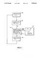
- FIG. 1is a schematic diagram showing a scan equalization radiography system according to the present invention
- FIG. 2is a graph showing the exposure response function of a typical film screen radiation image sensor; and the control function for a scanning equalization radiography system according to the present invention
- FIG. 3is a graph useful in describing the control of the x-ray dosage by pulse duration modulation.
- FIG. 4is a flow chart illustrating the steps of implementing a control function according to the present invention in a scanning equalization radiography system.
- the apparatusincludes a source of x-rays 10 for producing a beam of x-rays 12, and means 14 for modulating the exposure provided by the x-ray source 10.
- the exposure modulation meansmay comprised for example, electrical means for controlling the duration of pulses produced by the x-ray source, or a mechanically variable aperture means for modulating the intensity of beam 12 from the source as are known in the prior art.
- the apparatusincludes a scanner 16 for producing a scanning beam 18 of x-rays that scan an object 20.
- the scanning meansmay comprise for example, the combination of a moveable slit and a rotating wheel having a plurality of radial slits as is known in the prior art.
- the scanning beam of x-rays 18exposes an x-ray sensor 22, such as a conventional x-ray film/screen combination in a cassette.
- a detector 24detects the intensity of the beam 18 after passing through the object 20 and generates a feedback signal.
- the detectormay be positioned in front of or behind x-ray sensor 22.
- the detectormay be for example, a fluorescence detector comprised of a phosphor that emits light in response to radiation, and a photo detector such as a photo multiplier tube for detecting the emitted light, as is known in the prior art.
- the feedback signal generated by the detector 24is supplied to a feedback control unit 26 that controls the exposure modulator 14 as a function of the object dose rate.
- the feedback control unit 26comprises for example, a programmed microprocessor 28 and a memory 30 for storing a lookup table representing the control function provided by the feedback control unit 26.
- the control function stored in lookup table 30is similar to the exposure response function of the sensor 22.
- the term "similar” as used hereinmeans that the control function and exposure response function have the same general shape and slopes.
- the density of the image produced by the sensorwill be directly proportional to the x-ray attenuation of the objects in the image, thereby facilitating quantitative measurements of the object in the image.
- the thickness of an object having a known absorption coefficient, such as the human heart chamberis computed directly from the density of the resulting radiograph.
- the density of the object having a known thickness of a material and a known absorption coefficient, such as boneis likewise measured directly from the density of the resulting radiograph.
- the transmittance T(x) of an objectis given by the ratio of the transmitted exposure I(x) over the incident exposure Io. ##EQU1##
- the transmitted exposure I(x)is determined by Beer's law ##EQU2## where ⁇ is the x-ray attenuation coefficient of the object and x is the thickness.
- FIG. 2is a graph showing a typical D-logE curve 32 representing the exposure response function of a conventional x-ray film screen combination in the upper left quadrant of the graph.
- a control function 34 that is similar to the exposure response functionis shown in the lower left quadrant.
- the control function 34relates the log transmittance to the log exposure by controlling the dose rate as a function of the total dose of x-rays in the scanning equalization radiography system.
- Function 36 in the lower right quadrantis the mathematical relationship relating total x-ray attenuation to the log of transmittance T(x), which is simply a straight line with a slope of 0.434.
- function 38 in the upper right quadrantis the relationship between optical density in the radiograph and total x-ray attenuation (which is directly proportional to thickness) resulting from the use of a control function 34 that was similar to the detector response function 32.
- the function 38is simply a linear relationship, which gives the resulting radiograph the very useful property of having densities that are directly proportional to object thickness.
- Total dosecan be controlled by varying the intensity of the x-ray exposure, for example by a variable physical diaphragm or by varying the time of exposure for a constant intensity.
- the dose rateis measured by sensing the exposure for a predetermined time at the start of an exposure
- FIG. 3illustrates how the total dose is controlled in a pulse duration modulation SER system such as that described in the Plewes referenced above.
- the x-ray sourceis turned on for a predetermined time t 1 during which the dose rate is measured by the detector 24 (see FIG. 1).
- the total doseis then controlled by turning the beam off at some variable time t 2 later.
- FIG. 4illustrates the steps in the beam control process.
- the beamis turned on at t 0 (100) and the dose rate is measured at t 1 (102).
- the measured dose rate R Dis employed to address the lookup table 30 (104) containing the control function 34 to retrieve the total time T that the beam should be on.
- the beamis then turned off after the elapse of time T(106). This process is repeated many times for each scan line, and the scan lines are progressably stepped across the object to create the two-dimensional radiograph.
- an imaging detectori.e. an x-ray detector with a high spatial resolution and high signal-to-noise capabilities
- the length of the x-ray pulseis based on the x-ray transmittance of the part of the anatomy receiving the x-ray exposure at that instant in time.
- the x-ray generatoris capable of 650 mA and 150 kVp.
- the x-ray tubeis continuously powered at a filament current corresponsing to 400 mA, and a tube potential of 125 kV.
- a grid pulse tankis controlled via the computer.
- the grid pulse systemprovides a blocking potential to the x-ray tube's cathode, thereby controlling the flow of electrons from the cathode to the anode of the x-ray tube.
- the grid pulse tank and its electronic circuitrythus acts as a triode “valve” to switch the x-rays "on” or "off.”
- the x-ray filament currentis constant, so the grid pulse system controls the total x-ray exposure in any one pulse by controlling the length (in milliseconds) of the x-ray pulse.
- Fore and aft collimatorsdefine an x-ray beam of 0.25 square centimeters (0.5 centimeters across by 0.5 centimeters high), and sweep the beam across the patient in a raster fashion.
- the pulse tankis sent an electrical signal to turn “on” the x-ray beam.
- the monitor systemwhich is located behind the "patient” detects the x-radiation transmitted by the "patient.”
- the dose rate at this monitoris directly related to the transmittance of the patient at that instant.
- the computerretrieves a predetermined value from a lookup table, to determine how long to leave the x-ray beam "on” om order to obtain the desired total exposure value to the imaging detector, thereby "equalizing" the exposure to the imaging detector.
- Exposure timesrange from 50 microseconds to 700 microseconds. After a time increment of 700microseconds or less, the x-ray beam is turned “off” by the pulse tank system. After a time increment of 1000 microseconds (1 millisecond) from the time the x-ray beam was first turned on (independent of the length of the x-ray pulse) the pulse system is sent another signal to turn "on" the x-rays, and the process is repeated.
- the beamis swept across the patient at a rate of 0.25 centimeters per millisecond, or 0.25centimeters per pulse.
- a complete scanis accomplished in approximately 24 seconds.
- the systemwas operated using KODAK Lanex Regular screens and KODAK TMat-G film.
- the filmwas processed in a controlled KODAK M6-AW film processor.
- the sensitometry of the film as shown by curve 32 in FIG. 2was checked frequently with control strips.
- the lookup table in the computerwhich controls the generation of the "off" signal for the x-ray beam, was configured so that the log exposure versus the log exposure rate (i.e. the log transmittance) function (curve 34 in FIG. 2) was identical in shape to the Density-Log Exposure function (curve 32 in FIG. 2) of the Kodak Tmat-G film.
- the density of the filmwas directly proportional to the integral, or sum of differentials, of the x-ray attenuation.
- the imageis perfectly suitable for normal interpretation by a physician.
- the physiciancan make a simple measurement with a film densitometer, and determine relative (percentage) thickness variations.
- a simple measurementthe physician can tell, for example, that a blood vessel is reduced in caliber by 1/2 from its adjoining size.
- the physiciancan determine that a heart chamber is not of the right shape, again by simple densitometric measurement.
- FIG. 5is a graph showing the response function 110 of the stimulable phosphor in the upper left quadrant. Since the emitted signal from a storage phosphor plate is linearly proportional to the exposure reaching the plate, the log exposure versus emitted signal response function is an exponential curve 110. For this example, the lookup table relating the dose rate to the total dose, and hence the log transmittance to log exposure was configured to have the same exponential shape. This function 112 is shown in the lower left quadrant of FIG. 5.
- the function 114 relating total attenuation to log transmittanceis the same as shown in FIG. 2 above.
- the emitted signal from the storage phosphorwas linearly related to the total attenuation, and hence the thickness of the object, as shown by the function 116 shown in the upper right quadrant of FIG. 5 is linearly related to the intensity of the stimulated signal emitted by the phosphor.
- the scanning equalization radiography system of the present inventionis useful in diagnostic radiography, and is advantageous in that the method enables quantitative thickness measurements to be directly made from the radiography.
Landscapes
- Physics & Mathematics (AREA)
- Health & Medical Sciences (AREA)
- General Health & Medical Sciences (AREA)
- Toxicology (AREA)
- Spectroscopy & Molecular Physics (AREA)
- Engineering & Computer Science (AREA)
- General Engineering & Computer Science (AREA)
- High Energy & Nuclear Physics (AREA)
- Apparatus For Radiation Diagnosis (AREA)
- Radiography Using Non-Light Waves (AREA)
- X-Ray Techniques (AREA)
Abstract
Description
Claims (10)
Priority Applications (4)
| Application Number | Priority Date | Filing Date | Title |
|---|---|---|---|
| US07/358,239US5008914A (en) | 1989-05-30 | 1989-05-30 | Quantitative imaging employing scanning equalization radiography |
| PCT/US1990/002756WO1990015421A1 (en) | 1989-05-30 | 1990-05-24 | Quantitative imaging employing scanning equalization radiography |
| JP2509799AJPH04500327A (en) | 1989-05-30 | 1990-05-24 | Quantitative imaging using scanning equalization radiography |
| EP90909894AEP0426842A1 (en) | 1989-05-30 | 1990-05-24 | Quantitative imaging employing scanning equalization radiography |
Applications Claiming Priority (1)
| Application Number | Priority Date | Filing Date | Title |
|---|---|---|---|
| US07/358,239US5008914A (en) | 1989-05-30 | 1989-05-30 | Quantitative imaging employing scanning equalization radiography |
Publications (1)
| Publication Number | Publication Date |
|---|---|
| US5008914Atrue US5008914A (en) | 1991-04-16 |
Family
ID=23408856
Family Applications (1)
| Application Number | Title | Priority Date | Filing Date |
|---|---|---|---|
| US07/358,239Expired - LifetimeUS5008914A (en) | 1989-05-30 | 1989-05-30 | Quantitative imaging employing scanning equalization radiography |
Country Status (4)
| Country | Link |
|---|---|
| US (1) | US5008914A (en) |
| EP (1) | EP0426842A1 (en) |
| JP (1) | JPH04500327A (en) |
| WO (1) | WO1990015421A1 (en) |
Cited By (5)
| Publication number | Priority date | Publication date | Assignee | Title |
|---|---|---|---|---|
| US5333168A (en)* | 1993-01-29 | 1994-07-26 | Oec Medical Systems, Inc. | Time-based attenuation compensation |
| US5784040A (en)* | 1992-09-30 | 1998-07-21 | Sanyo Electric Co., Ltd. | Image information processor |
| US6249565B1 (en)* | 1998-06-18 | 2001-06-19 | Siemens Medical Systems, Inc. | Fractional monitor unit radiation delivery control using dose rate modulation |
| WO2003075764A1 (en)* | 2002-03-12 | 2003-09-18 | Xcounter Ab | Exposure control in scanning-based detection of ionizing radiation |
| US20120305790A1 (en)* | 2010-02-10 | 2012-12-06 | National Institute Of Radiological Sciences | Particle beam irradiation apparatus and control method of the particle beam irradiation apparatus |
Families Citing this family (1)
| Publication number | Priority date | Publication date | Assignee | Title |
|---|---|---|---|---|
| JP6026800B2 (en) | 2012-07-11 | 2016-11-16 | 株式会社東海理化電機製作所 | Shift device |
Citations (3)
| Publication number | Priority date | Publication date | Assignee | Title |
|---|---|---|---|---|
| US4454606A (en)* | 1983-05-23 | 1984-06-12 | General Electric Company | Reconfigurable x-ray AEC compensation |
| CA1244971A (en)* | 1985-11-14 | 1988-11-15 | Shih-Ping Wang | X-ray radiography method and system |
| US4857732A (en)* | 1984-10-19 | 1989-08-15 | Fuji Photo Film Co., Ltd. | Radiation image recording and read-out apparatus |
Family Cites Families (4)
| Publication number | Priority date | Publication date | Assignee | Title |
|---|---|---|---|---|
| US4681427A (en)* | 1985-05-06 | 1987-07-21 | Polaroid Corporation | Electronic printing method |
| US4773087A (en)* | 1986-04-14 | 1988-09-20 | University Of Rochester | Quality of shadowgraphic x-ray images |
| US4748649A (en)* | 1986-08-04 | 1988-05-31 | Picker International, Inc. | Phototiming control method and apparatus |
| NL8700781A (en)* | 1987-04-02 | 1988-11-01 | Optische Ind De Oude Delft Nv | METHOD AND APPARATUS FOR CONTRAST HARMONIZATION OF A ROENTGEN IMAGE. |
- 1989
- 1989-05-30USUS07/358,239patent/US5008914A/ennot_activeExpired - Lifetime
- 1990
- 1990-05-24EPEP90909894Apatent/EP0426842A1/ennot_activeWithdrawn
- 1990-05-24WOPCT/US1990/002756patent/WO1990015421A1/ennot_activeApplication Discontinuation
- 1990-05-24JPJP2509799Apatent/JPH04500327A/enactivePending
Patent Citations (3)
| Publication number | Priority date | Publication date | Assignee | Title |
|---|---|---|---|---|
| US4454606A (en)* | 1983-05-23 | 1984-06-12 | General Electric Company | Reconfigurable x-ray AEC compensation |
| US4857732A (en)* | 1984-10-19 | 1989-08-15 | Fuji Photo Film Co., Ltd. | Radiation image recording and read-out apparatus |
| CA1244971A (en)* | 1985-11-14 | 1988-11-15 | Shih-Ping Wang | X-ray radiography method and system |
Non-Patent Citations (6)
| Title |
|---|
| The article "A Scanning System for Chest Radiography with Regional Exposure Control: Theoretical Considerations" by D. B. Plewes, Med. Phys. 10(5) Sep./Oct. 1983, pp. 646-654 is cited on page 1 line 22 for showing seam equalization apparatus. |
| The article "Amber: A Scanning Multiple-Beam Equalization System for Chest Radiography" by Vlasbloem and Kool, Radiology, Oct. 1988, pp. 29-34 is cited for showing the use of different control curves in seam equalization radiography. |
| The article "Exposure Equalization Radiography of the Chest: Clinical Comparison of Slit and Raster Scanning Techniques by Wandtke and Plewes" AJR 144, Jun. 1985, pp. 171-181 is cited for showing slit and raster techniques. |
| The article A Scanning System for Chest Radiography with Regional Exposure Control: Theoretical Considerations by D. B. Plewes, Med. Phys. 10(5) Sep./Oct. 1983, pp. 646 654 is cited on page 1 line 22 for showing seam equalization apparatus.* |
| The article Amber: A Scanning Multiple Beam Equalization System for Chest Radiography by Vlasbloem and Kool, Radiology, Oct. 1988, pp. 29 34 is cited for showing the use of different control curves in seam equalization radiography.* |
| The article Exposure Equalization Radiography of the Chest: Clinical Comparison of Slit and Raster Scanning Techniques by Wandtke and Plewes AJR 144, Jun. 1985, pp. 171 181 is cited for showing slit and raster techniques.* |
Cited By (10)
| Publication number | Priority date | Publication date | Assignee | Title |
|---|---|---|---|---|
| US5784040A (en)* | 1992-09-30 | 1998-07-21 | Sanyo Electric Co., Ltd. | Image information processor |
| US5333168A (en)* | 1993-01-29 | 1994-07-26 | Oec Medical Systems, Inc. | Time-based attenuation compensation |
| US5400384A (en)* | 1993-01-29 | 1995-03-21 | Oec Medical Systems, Inc. | Time-based attenuation compensation |
| US6249565B1 (en)* | 1998-06-18 | 2001-06-19 | Siemens Medical Systems, Inc. | Fractional monitor unit radiation delivery control using dose rate modulation |
| WO2003075764A1 (en)* | 2002-03-12 | 2003-09-18 | Xcounter Ab | Exposure control in scanning-based detection of ionizing radiation |
| US20040141588A1 (en)* | 2002-03-12 | 2004-07-22 | Tom Francke | Exposure control in scanning-based detection of ionizing radiation |
| US6873682B2 (en) | 2002-03-12 | 2005-03-29 | Xcounter Ab | Exposure control in scanning-based detection of ionizing radiation |
| AU2003208691B2 (en)* | 2002-03-12 | 2008-04-10 | Xcounter Ab | Exposure control in scanning-based detection of ionizing radiation |
| US20120305790A1 (en)* | 2010-02-10 | 2012-12-06 | National Institute Of Radiological Sciences | Particle beam irradiation apparatus and control method of the particle beam irradiation apparatus |
| US8552408B2 (en)* | 2010-02-10 | 2013-10-08 | Kabushiki Kaisha Toshiba | Particle beam irradiation apparatus and control method of the particle beam irradiation apparatus |
Also Published As
| Publication number | Publication date |
|---|---|
| WO1990015421A1 (en) | 1990-12-13 |
| EP0426842A1 (en) | 1991-05-15 |
| JPH04500327A (en) | 1992-01-23 |
Similar Documents
| Publication | Publication Date | Title |
|---|---|---|
| US7431500B2 (en) | Dynamic exposure control in radiography | |
| EP0942682B1 (en) | Adjustable computer tomography device | |
| US6459765B1 (en) | Automatic exposure control and optimization in digital x-ray radiography | |
| US5485501A (en) | Method for the operation of an automatic x-ray exposure unit | |
| US5008915A (en) | Methods for forming a radiograph using slit radiography | |
| US7545915B2 (en) | Dose rate control in an X-ray system | |
| USRE33634E (en) | Method and structure for optimizing radiographic quality by controlling X-ray tube voltage, current focal spot size and exposure time | |
| US4763343A (en) | Method and structure for optimizing radiographic quality by controlling X-ray tube voltage, current, focal spot size and exposure time | |
| US6320931B1 (en) | Automated x-ray bone densitometer | |
| US4686695A (en) | Scanned x-ray selective imaging system | |
| US5396531A (en) | Method of achieving reduced dose X-ray fluoroscopy by employing statistical estimation of poisson noise | |
| EP0269302A2 (en) | Imaging apparatuses and methods | |
| EP0038666A1 (en) | Radiographic apparatus and method with automatic exposure control | |
| JP2000501552A (en) | Method and apparatus for controlling and optimizing the output of an x-ray source | |
| EP0200272A2 (en) | X-ray examination system and method of controlling an exposure | |
| Floyd Jr et al. | Quantitative radiographic imaging using a photostimulable phosphor system | |
| US4486896A (en) | X-Ray generator incorporating automatic correction of a dose-determining exposure parameter | |
| US5008914A (en) | Quantitative imaging employing scanning equalization radiography | |
| US4595949A (en) | Systems and methods for translating radiation intensity into pixel values | |
| EP0346530A1 (en) | Method and structure for optimizing radiographic quality by controlling X-ray tube voltage, current, focal spot size and exposure time | |
| US20020154802A1 (en) | Apparatus for and method of generating an enhanced contrast information digital image | |
| JPH05217689A (en) | X-ray imaging apparatus and X-ray imaging method | |
| US5436829A (en) | Method of achieving reduced dose X-ray fluoroscopy by employing transform-based estimation of Poisson noise | |
| Yoshiura et al. | The perceptibility curve test applied to direct digital dental radiography | |
| US6502985B1 (en) | Auto-collimating digital X-ray system |
Legal Events
| Date | Code | Title | Description |
|---|---|---|---|
| AS | Assignment | Owner name:EASTMAN KODAK COMPANY, ROCHESTER, NEW YORK, A NEW Free format text:ASSIGNMENT OF ASSIGNORS INTEREST.;ASSIGNOR:MOORE, WILLIAM E.;REEL/FRAME:005085/0928 Effective date:19890525 | |
| FEPP | Fee payment procedure | Free format text:PAYOR NUMBER ASSIGNED (ORIGINAL EVENT CODE: ASPN); ENTITY STATUS OF PATENT OWNER: LARGE ENTITY | |
| STCF | Information on status: patent grant | Free format text:PATENTED CASE | |
| FPAY | Fee payment | Year of fee payment:4 | |
| FEPP | Fee payment procedure | Free format text:PAYER NUMBER DE-ASSIGNED (ORIGINAL EVENT CODE: RMPN); ENTITY STATUS OF PATENT OWNER: LARGE ENTITY | |
| FPAY | Fee payment | Year of fee payment:8 | |
| FPAY | Fee payment | Year of fee payment:12 | |
| AS | Assignment | Owner name:CREDIT SUISSE, CAYMAN ISLANDS BRANCH, AS ADMINISTR Free format text:SECOND LIEN INTELLECTUAL PROPERTY SECURITY AGREEME;ASSIGNOR:CARESTREAM HEALTH, INC.;REEL/FRAME:019773/0319 Effective date:20070430 | |
| AS | Assignment | Owner name:CARESTREAM HEALTH, INC., NEW YORK Free format text:ASSIGNMENT OF ASSIGNORS INTEREST;ASSIGNOR:EASTMAN KODAK COMPANY;REEL/FRAME:020741/0126 Effective date:20070501 Owner name:CARESTREAM HEALTH, INC., NEW YORK Free format text:ASSIGNMENT OF ASSIGNORS INTEREST;ASSIGNOR:EASTMAN KODAK COMPANY;REEL/FRAME:020756/0500 Effective date:20070501 Owner name:CARESTREAM HEALTH, INC.,NEW YORK Free format text:ASSIGNMENT OF ASSIGNORS INTEREST;ASSIGNOR:EASTMAN KODAK COMPANY;REEL/FRAME:020741/0126 Effective date:20070501 Owner name:CARESTREAM HEALTH, INC.,NEW YORK Free format text:ASSIGNMENT OF ASSIGNORS INTEREST;ASSIGNOR:EASTMAN KODAK COMPANY;REEL/FRAME:020756/0500 Effective date:20070501 | |
| AS | Assignment | Owner name:CARESTREAM HEALTH, INC., NEW YORK Free format text:RELEASE OF SECURITY INTEREST IN INTELLECTUAL PROPERTY (FIRST LIEN);ASSIGNOR:CREDIT SUISSE AG, CAYMAN ISLANDS BRANCH;REEL/FRAME:026069/0012 Effective date:20110225 |