US5005559A - Video-graphic arthroscopy system - Google Patents
Video-graphic arthroscopy systemDownload PDFInfo
- Publication number
- US5005559A US5005559AUS07/386,343US38634389AUS5005559AUS 5005559 AUS5005559 AUS 5005559AUS 38634389 AUS38634389 AUS 38634389AUS 5005559 AUS5005559 AUS 5005559A
- Authority
- US
- United States
- Prior art keywords
- arthroscope
- display system
- monitoring
- display
- graphic
- Prior art date
- Legal status (The legal status is an assumption and is not a legal conclusion. Google has not performed a legal analysis and makes no representation as to the accuracy of the status listed.)
- Expired - Fee Related
Links
- 238000012544monitoring processMethods0.000claimsdescription7
- 230000035515penetrationEffects0.000claimsdescription5
- 239000000523sampleSubstances0.000description8
- 210000003127kneeAnatomy0.000description4
- 239000004677NylonSubstances0.000description3
- 210000000988bone and boneAnatomy0.000description3
- 229920001778nylonPolymers0.000description3
- 230000005355Hall effectEffects0.000description2
- 235000004522Pentaglottis sempervirensNutrition0.000description1
- FAPWRFPIFSIZLT-UHFFFAOYSA-MSodium chlorideChemical compound[Na+].[Cl-]FAPWRFPIFSIZLT-UHFFFAOYSA-M0.000description1
- 238000007792additionMethods0.000description1
- 238000006243chemical reactionMethods0.000description1
- 239000002131composite materialSubstances0.000description1
- 230000001143conditioned effectEffects0.000description1
- 238000010276constructionMethods0.000description1
- 238000010586diagramMethods0.000description1
- 239000003365glass fiberSubstances0.000description1
- 238000005286illuminationMethods0.000description1
- 238000002347injectionMethods0.000description1
- 239000007924injectionSubstances0.000description1
- 230000002452interceptive effectEffects0.000description1
- 210000000629knee jointAnatomy0.000description1
- 238000000034methodMethods0.000description1
- 230000003287optical effectEffects0.000description1
- 239000013307optical fiberSubstances0.000description1
- 230000000399orthopedic effectEffects0.000description1
- 238000012545processingMethods0.000description1
- 239000011780sodium chlorideSubstances0.000description1
- 238000001356surgical procedureMethods0.000description1
- 238000012549trainingMethods0.000description1
- 230000000007visual effectEffects0.000description1
- 238000012800visualizationMethods0.000description1
Images

Classifications
- A—HUMAN NECESSITIES
- A61—MEDICAL OR VETERINARY SCIENCE; HYGIENE
- A61B—DIAGNOSIS; SURGERY; IDENTIFICATION
- A61B1/00—Instruments for performing medical examinations of the interior of cavities or tubes of the body by visual or photographical inspection, e.g. endoscopes; Illuminating arrangements therefor
- A61B1/00147—Holding or positioning arrangements
- A—HUMAN NECESSITIES
- A61—MEDICAL OR VETERINARY SCIENCE; HYGIENE
- A61B—DIAGNOSIS; SURGERY; IDENTIFICATION
- A61B1/00—Instruments for performing medical examinations of the interior of cavities or tubes of the body by visual or photographical inspection, e.g. endoscopes; Illuminating arrangements therefor
- A61B1/313—Instruments for performing medical examinations of the interior of cavities or tubes of the body by visual or photographical inspection, e.g. endoscopes; Illuminating arrangements therefor for introducing through surgical openings, e.g. laparoscopes
- A61B1/317—Instruments for performing medical examinations of the interior of cavities or tubes of the body by visual or photographical inspection, e.g. endoscopes; Illuminating arrangements therefor for introducing through surgical openings, e.g. laparoscopes for bones or joints, e.g. osteoscopes, arthroscopes
Definitions
- the technical field of this inventionis arthroscopy and, in particular, methods and apparatus for displaying the location of an arthroscope during use.
- Arthroscopyallows for direct examination of biological structures (most commonly, the knee joint, but also the hip, shoulder, elbow and hand, as well) utilizing tiny incisions through which the arthroscope is inserted.
- the arthroscopecontains illuminating glass fibers and a series of magnifying optical lenses that project light into the joint and relay a magnified image back to the clinician.
- arthroscopespresent a number of problems to the inexperienced user. Since the view from the probe is circular, it is difficult to determine the scope's orientation from the scene without actually moving the scope. Moreover, the arthroscope typically has an offset of 30°; hence, the center of view is not in the direction of arthroscope motion into or out of the biological structure.
- the motion needed to manipulate the probeis not always obvious. For example, if the probe is rotated 180° such that the view is "upside down" relative to the user, the motion needed to manipulate the probe is completely reversed; to move up, the user must lift the arthroscope's distal end up.
- the fish-eye, two-dimensional view of the arthroscopecan often be uninformative; objects of interest within the joint are often not found in the field of view or hidden by other biological structures.
- An object of the present inventionis to provide better display systems for orthopedic surgeons in the practice of arthroscopy.
- Another object of the present inventionis to provide a teaching tool for the training of arthroscopists.
- Yet another object of the inventionis to provide visual systems which can enhance spatial visualization of arthroscopy, including the coordination of images seen by the surgeon through the arthroscope and varying reference points, such as natural bone structures and/or other instruments that are inserted into the observation region to perform surgical procedures.
- a position sensing systemwhich allows the arthroscopist to readily determine the location of an arthroscope's tip in relation to the point of entry has been developed.
- the position sensing apparatuscan be employed in conjunction with a graphics module to display the location of the arthroscope in real time and provide perspective views of the instrument's location.
- a sensing apparatusfor monitoring the four degrees of freedom (X, Y, Z and rotation) possessed by the arthroscope.
- a double-yoke gimbel structureis adapted for positioning on a joint, such as the knee, in order to measure the pivoting of the arthroscope (in the X and Y directions) about the point of entry.
- Rotation of the arthroscopeis measured by a sensor mounted on a sleeve which is fixed relative to the rotation of the arthroscope.
- penetration of the arthroscopeis measured by a fourth sensor which detects relative motion of the arthroscope into or out of the patient's joint relative to a fixed reference point.
- This sensing apparatusprovides an electrical output which can precisely define the location of the arthroscope's tip at all times.
- the electrical output of the position sensoris employed in conjunction with a graphics processor to generate perspective "bird's eye" views of the arthroscope in relation to an actual or a generic structure showing the major components of the joint. These graphic views can be employed for surgical or teaching purposes.
- processor-generated graphic imagescan be merged with the actual video display of the arthroscope to provide a composite picture.
- FIG. 1is an overall schematic block diagram of a video-graphic arthroscopy system according to the invention.
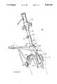
- FIG. 2is a detailed illustration of an arthroscopic position sensing apparatus according to the invention.
- a video-graphic arthroscopy system 10including an arthroscope 12 and a housing 16 for the position sensing apparatus which is attached to a patient's joint 14 (shown in phantom).
- the arthroscopeincludes conventional saline injection means (not shown) to distend the joint and also a conventional source of illumination (not shown) to illuminate the joint.
- Light from the observation regionis transmitted via optical fiber 24 to a video-image processor 18, which generates a video image of the biological structures under observation in the patient's joint.
- a position sensing apparatus in housing 16generates electrical signals via cable 26, which are transmitted to a position graphics processor 20 to generate a display of the arthroscope's position relative to a fixed reference point (typically the point of entry).
- a graphic imageis generated by the graphics processor 20 which is superimposed upon the video image using half silvered mirror 22 to provide a display for the arthroscopist.
- the position sensing apparatus 30 of the present inventionis shown in more detail (with the housing removed for illustration), including a rotation sensor 32 which is mounted upon the cannula 46 of arthroscope 12.
- a rotor 34Within the rotation sensor 32, a rotor 34 includes a keyway notch 48 which mates with a keyway or spline 36 carried upon sleeve 38.
- the sleeve 38is attached to sheath 40 of the arthroscope 12 by set screw 42.
- a depth sensor 44is also employed to measure the depth of penetration of the arthroscope 12 into the patient's joint.
- the depth sensor 44can be attached to the cannula 46 (Or alternatively to the sleeve 38).
- position sensing apparatus 30utilizes depth sensor 44 and nylon line 60, coupled to sleeve 38 and wound around a spool in depth sensor 44, to provide information representative of the depth of penetration of arthroscope 12 into the patient's joint.
- Depth sensor 44translates the degree to which nylon line 60 is wound or unwound from depth sensor 44 into an analog voltage which is supplied to a processor. As sleeve 38 is inserted or retracted, nylon line 60 is unwound or wound, respectively.
- a magnetwhich is coupled to the spool in depth sensor 44, rotates correspondingly.
- a Hall effect sensormeasures the motion of the magnet and generates a signal which is conditioned by conventional electronics.
- the depth sensorcan employ a rack and pinion-type drive mechanism, whereby a pinion gear drives the sensor and mates with a rack mounted upon sleeve 38.
- Semicircular sloted yokes 52, 56define an X-axis and a Y-axis for measuring the tilt of the arthroscope during usage.
- An X-axis sensor 58is joined to the center of rotation of the X-axis yoke 56, and a similar Y-axis sensor 54 is joined to the center of rotation of Y-axis yoke 52.
- the cannula 46is disposed between the yokes 52, 56 so that the sensors 54, 58, can provide a direct relationship between sensor rotation and the angular movement (tilting) or the arthroscope 12.
- the two-member systemeffectively decouples the motion in the X and Y directions.
- An angular range of motion in each direction of approximately ⁇ 40 degreesis typically sufficient for arthroscopic operations.
- the rotary sensor 32has been implemented employing a rotary variable differential transformer, such as the model RAl sensor manufactured by Transducers and Systems, Inc. (Northbranford, Conn.).
- the other sensorsfor measuring tilt and penetration
- commercially available Hall effect sensorssuch as those manufactured by Texas Instruments (Dallas, Tex.) can be employed.
- the analog voltages generated by the sensorsare transmitted, via cable 26, to a processor which includes an analog to digital converter to translate the analog votage to a digital representation thereof.
- the sensor informationis utilized to generate positional information of the probe of arthroscope 12 with respect to the patient's joint, and to generate computer graphics simulating objects not generally present in the patient's joint.
- the processorwhich can be a general purpose computer or other processing device, in conjunction with the information from the sensors can provide a computer-generated model of the joint in question.
- This computer graphic modelcan augment the video display of the arthroscope.
- a variety of commercially available computer graphics enginescan be used, such as the Rennaissance graphics engine manufactured bY Hewlett-Packard (Andover, Mass.) for use on the HP-9000 computer, a general purpose work station built around a Motorola 68020 processor.
- the Rennaissance engineis capable of generating high resolution images for both 2 and 3-dimensional displays.
- the graphics operations of this systemcan be implemented using a variety of commercially available graphics programs, such as the Hewlett-Packard Starbase graphics package.
- the main loop of such programscontinually poles the position of the arthroscope and then performs various tasks.
- a wire frame cross-section data base of the major bones of the kneewas entered into the processor memory.
- the programwas then configured to generate a line representing the probe and display it over the cross section.
- the probe orientation with respect to the wire frame illustration of the kneecould then be displayed from the sensor signals by using a set of transformational matrices.
- three orthogonal viewscan be presented to the viewer.
- More realistic knee illustrationscan, of course, be generated with appropriate graphics programs using more detailed data bases of the biological structures.
- such graphic displayscan be merged with the video images from the arthroscope. If the video signals generated by graphics engine are incompatable with the video signals generated by the viewing probe's camera, either a standards conversion box which reduces the high resolution graphics picture to a NTSC resolution interlaced video image can be employed. Alternatively, it is possible to convert the NTSC resolution arthroscope image up to the same resolution as the graphics image, and electronically merge them.
- the two imagescan be optically merged using a half-silvered mirror.
- a half-silvered mirrorUse of such a mirror enables one to merge the two images, both inexpensively, and with no loss of resolution.
- the imagesare merged together by placing a mirror, which is partially reflective and transmisive, at a 45° angle in between two monitors which are perpendicular to each other. The viewer sits in front of one monitor and sees the image from the other monitor superimposed upon the first monitor's image.
- the graphics processorcan include additional user input interfaces, such as a graphics tablet, mouse, knob box, button box or keyboard. These input devices can provide the user with a means of "pointing" to objects within the scene.
Landscapes
- Health & Medical Sciences (AREA)
- Life Sciences & Earth Sciences (AREA)
- Surgery (AREA)
- Biomedical Technology (AREA)
- Medical Informatics (AREA)
- Optics & Photonics (AREA)
- Pathology (AREA)
- Radiology & Medical Imaging (AREA)
- Biophysics (AREA)
- Engineering & Computer Science (AREA)
- Physics & Mathematics (AREA)
- Heart & Thoracic Surgery (AREA)
- Nuclear Medicine, Radiotherapy & Molecular Imaging (AREA)
- Molecular Biology (AREA)
- Animal Behavior & Ethology (AREA)
- General Health & Medical Sciences (AREA)
- Public Health (AREA)
- Veterinary Medicine (AREA)
- Orthopedic Medicine & Surgery (AREA)
- Physical Education & Sports Medicine (AREA)
- Endoscopes (AREA)
Abstract
Description
Claims (5)
Priority Applications (1)
| Application Number | Priority Date | Filing Date | Title |
|---|---|---|---|
| US07/386,343US5005559A (en) | 1989-07-27 | 1989-07-27 | Video-graphic arthroscopy system |
Applications Claiming Priority (1)
| Application Number | Priority Date | Filing Date | Title |
|---|---|---|---|
| US07/386,343US5005559A (en) | 1989-07-27 | 1989-07-27 | Video-graphic arthroscopy system |
Publications (1)
| Publication Number | Publication Date |
|---|---|
| US5005559Atrue US5005559A (en) | 1991-04-09 |
Family
ID=23525201
Family Applications (1)
| Application Number | Title | Priority Date | Filing Date |
|---|---|---|---|
| US07/386,343Expired - Fee RelatedUS5005559A (en) | 1989-07-27 | 1989-07-27 | Video-graphic arthroscopy system |
Country Status (1)
| Country | Link |
|---|---|
| US (1) | US5005559A (en) |
Cited By (53)
| Publication number | Priority date | Publication date | Assignee | Title |
|---|---|---|---|---|
| US5398685A (en)* | 1992-01-10 | 1995-03-21 | Wilk; Peter J. | Endoscopic diagnostic system and associated method |
| US5617858A (en)* | 1994-08-30 | 1997-04-08 | Vingmed Sound A/S | Apparatus for endoscopic or gastroscopic examination |
| US5733242A (en)* | 1996-02-07 | 1998-03-31 | Rayburn; Robert L. | Intubation system having an axially moveable memory cylinder |
| US5737506A (en)* | 1995-06-01 | 1998-04-07 | Medical Media Systems | Anatomical visualization system |
| US5765561A (en)* | 1994-10-07 | 1998-06-16 | Medical Media Systems | Video-based surgical targeting system |
| US5776050A (en)* | 1995-07-24 | 1998-07-07 | Medical Media Systems | Anatomical visualization system |
| US5825908A (en)* | 1995-12-29 | 1998-10-20 | Medical Media Systems | Anatomical visualization and measurement system |
| US5833605A (en)* | 1997-03-28 | 1998-11-10 | Shah; Ajit | Apparatus for vascular mapping and methods of use |
| US6120433A (en)* | 1994-09-01 | 2000-09-19 | Olympus Optical Co., Ltd. | Surgical manipulator system |
| US6151404A (en)* | 1995-06-01 | 2000-11-21 | Medical Media Systems | Anatomical visualization system |
| US6161080A (en)* | 1997-11-17 | 2000-12-12 | The Trustees Of Columbia University In The City Of New York | Three dimensional multibody modeling of anatomical joints |
| WO2001010289A3 (en)* | 1999-08-10 | 2001-09-20 | Scope Ltd G | Surgical camera apparatus and method |
| DE19955346A1 (en)* | 1999-11-17 | 2001-09-20 | Hans Rudolf Schwind | Endoscope imaging method and endoscope system |
| US20030208103A1 (en)* | 2002-05-02 | 2003-11-06 | Elazar Sonnenschein | Entry port for endoscopes and laparoscopes |
| US6690960B2 (en) | 2000-12-21 | 2004-02-10 | David T. Chen | Video-based surgical targeting system |
| US6702736B2 (en) | 1995-07-24 | 2004-03-09 | David T. Chen | Anatomical visualization system |
| US20040204630A1 (en)* | 2002-12-30 | 2004-10-14 | Zvika Gilad | Device, system and method for in vivo motion detection |
| US20040220450A1 (en)* | 2000-04-03 | 2004-11-04 | Neoguide Systems, Inc. | Endoscope having a guide tube |
| US20040220478A1 (en)* | 2003-02-26 | 2004-11-04 | Wallace Jeffrey M. | Method and devices for imaging and biopsy |
| US20050020901A1 (en)* | 2000-04-03 | 2005-01-27 | Neoguide Systems, Inc., A Delaware Corporation | Apparatus and methods for facilitating treatment of tissue via improved delivery of energy based and non-energy based modalities |
| US20050154261A1 (en)* | 2000-04-03 | 2005-07-14 | Ohline Robert M. | Tendon-driven endoscope and methods of insertion |
| US20050154258A1 (en)* | 2000-04-03 | 2005-07-14 | Tartaglia Joseph M. | Endoscope with adjacently positioned guiding apparatus |
| US20050222498A1 (en)* | 2000-04-03 | 2005-10-06 | Amir Belson | Steerable endoscope and improved method of insertion |
| US20050272971A1 (en)* | 2002-08-30 | 2005-12-08 | Olympus Corporation | Medical treatment system, endoscope system, endoscope insert operation program, and endoscope device |
| US20060098010A1 (en)* | 2004-03-09 | 2006-05-11 | Jeff Dwyer | Anatomical visualization and measurement system |
| US20060235458A1 (en)* | 2005-04-15 | 2006-10-19 | Amir Belson | Instruments having an external working channel |
| US7197170B2 (en) | 2003-11-10 | 2007-03-27 | M2S, Inc. | Anatomical visualization and measurement system |
| US20070135803A1 (en)* | 2005-09-14 | 2007-06-14 | Amir Belson | Methods and apparatus for performing transluminal and other procedures |
| US20070161291A1 (en)* | 2005-11-23 | 2007-07-12 | Neoguide Systems, Inc. | Non-metallic, multi-strand control cable for steerable instruments |
| US20070249901A1 (en)* | 2003-03-07 | 2007-10-25 | Ohline Robert M | Instrument having radio frequency identification systems and methods for use |
| US20070270650A1 (en)* | 2006-05-19 | 2007-11-22 | Robert Eno | Methods and apparatus for displaying three-dimensional orientation of a steerable distal tip of an endoscope |
| US20080154288A1 (en)* | 2002-01-09 | 2008-06-26 | Neoguide Systems, Inc. | Apparatus and method for endoscopic colectomy |
| US20090202117A1 (en)* | 2006-06-12 | 2009-08-13 | Fernando Vilarino | Device, system and method for measurement and analysis of contractile activity |
| US20090216083A1 (en)* | 2008-02-25 | 2009-08-27 | Neoguide Systems, Inc. | Systems and Methods for Articulating an Elongate Body |
| US20090284589A1 (en)* | 2006-03-13 | 2009-11-19 | Petia Radeva | Cascade analysis for intestinal contraction detection |
| US7702137B2 (en) | 2004-11-10 | 2010-04-20 | M2S, Inc. | Anatomical visualization and measurement system |
| US20100262000A1 (en)* | 2003-02-26 | 2010-10-14 | Wallace Jeffrey M | Methods and devices for endoscopic imaging |
| US20110044515A1 (en)* | 2006-03-13 | 2011-02-24 | Panagiota Spyridonos | Device, system and method for automatic detection of contractile activity in an image frame |
| US20110065993A1 (en)* | 2000-04-03 | 2011-03-17 | Amir Belson | Steerable segmented endoscope and method of insertion |
| WO2011092337A1 (en)* | 2010-02-01 | 2011-08-04 | Universiteit Antwerpen | Mounting system for medulloscopy |
| US8489192B1 (en) | 2008-02-15 | 2013-07-16 | Holaira, Inc. | System and method for bronchial dilation |
| US20140094649A1 (en)* | 2011-06-07 | 2014-04-03 | Olympus Corporation | Insertion opening attachment |
| EP2073685A4 (en)* | 2006-09-21 | 2014-04-30 | Mst Medical Surgery Technologies Ltd | ENDOSCOPIC POSITIONING SYSTEM |
| US8740895B2 (en) | 2009-10-27 | 2014-06-03 | Holaira, Inc. | Delivery devices with coolable energy emitting assemblies |
| US8808280B2 (en) | 2008-05-09 | 2014-08-19 | Holaira, Inc. | Systems, assemblies, and methods for treating a bronchial tree |
| US8888688B2 (en) | 2000-04-03 | 2014-11-18 | Intuitive Surgical Operations, Inc. | Connector device for a controllable instrument |
| US8911439B2 (en) | 2009-11-11 | 2014-12-16 | Holaira, Inc. | Non-invasive and minimally invasive denervation methods and systems for performing the same |
| US9149328B2 (en) | 2009-11-11 | 2015-10-06 | Holaira, Inc. | Systems, apparatuses, and methods for treating tissue and controlling stenosis |
| US9220398B2 (en) | 2007-10-11 | 2015-12-29 | Intuitive Surgical Operations, Inc. | System for managing Bowden cables in articulating instruments |
| US9339618B2 (en) | 2003-05-13 | 2016-05-17 | Holaira, Inc. | Method and apparatus for controlling narrowing of at least one airway |
| US9398933B2 (en) | 2012-12-27 | 2016-07-26 | Holaira, Inc. | Methods for improving drug efficacy including a combination of drug administration and nerve modulation |
| US11096563B2 (en) | 2005-11-22 | 2021-08-24 | Intuitive Surgical Operations, Inc. | Method of determining the shape of a bendable instrument |
| US20230023904A1 (en)* | 2021-07-23 | 2023-01-26 | Phaox LLC | Handheld wireless endoscope image streaming apparatus |
Citations (9)
| Publication number | Priority date | Publication date | Assignee | Title |
|---|---|---|---|---|
| US4078864A (en)* | 1976-07-08 | 1978-03-14 | United Technologies Corporation | Method and apparatus for viewing and measuring damage in an inaccessible area |
| US4277168A (en)* | 1978-04-20 | 1981-07-07 | Machida Endoscope Co., Ltd. | Endoscope with means for detecting rotation of a distal examining end |
| US4340302A (en)* | 1978-02-28 | 1982-07-20 | Machida Endoscope Co., Ltd. | Endoscope with sensor |
| US4407277A (en)* | 1980-10-27 | 1983-10-04 | Ellison Arthur E | Surgical apparatus |
| US4413278A (en)* | 1981-10-05 | 1983-11-01 | Designs For Vision, Inc. | Optical coupling apparatus for coupling an arthoscope to a camera |
| US4572594A (en)* | 1984-02-08 | 1986-02-25 | Schwartz C Bruce | Arthroscopy support stand |
| US4586079A (en)* | 1983-10-07 | 1986-04-29 | Westinghouse Electric Corp. | Fiberscope delivery system |
| US4590923A (en)* | 1983-04-18 | 1986-05-27 | Watanabe Robert S | Arthroscope-video camera assembly |
| US4689449A (en)* | 1986-10-03 | 1987-08-25 | Massachusetts Institute Of Technology | Tremor suppressing hand controls |
- 1989
- 1989-07-27USUS07/386,343patent/US5005559A/ennot_activeExpired - Fee Related
Patent Citations (9)
| Publication number | Priority date | Publication date | Assignee | Title |
|---|---|---|---|---|
| US4078864A (en)* | 1976-07-08 | 1978-03-14 | United Technologies Corporation | Method and apparatus for viewing and measuring damage in an inaccessible area |
| US4340302A (en)* | 1978-02-28 | 1982-07-20 | Machida Endoscope Co., Ltd. | Endoscope with sensor |
| US4277168A (en)* | 1978-04-20 | 1981-07-07 | Machida Endoscope Co., Ltd. | Endoscope with means for detecting rotation of a distal examining end |
| US4407277A (en)* | 1980-10-27 | 1983-10-04 | Ellison Arthur E | Surgical apparatus |
| US4413278A (en)* | 1981-10-05 | 1983-11-01 | Designs For Vision, Inc. | Optical coupling apparatus for coupling an arthoscope to a camera |
| US4590923A (en)* | 1983-04-18 | 1986-05-27 | Watanabe Robert S | Arthroscope-video camera assembly |
| US4586079A (en)* | 1983-10-07 | 1986-04-29 | Westinghouse Electric Corp. | Fiberscope delivery system |
| US4572594A (en)* | 1984-02-08 | 1986-02-25 | Schwartz C Bruce | Arthroscopy support stand |
| US4689449A (en)* | 1986-10-03 | 1987-08-25 | Massachusetts Institute Of Technology | Tremor suppressing hand controls |
Cited By (132)
| Publication number | Priority date | Publication date | Assignee | Title |
|---|---|---|---|---|
| US5398685A (en)* | 1992-01-10 | 1995-03-21 | Wilk; Peter J. | Endoscopic diagnostic system and associated method |
| US5617858A (en)* | 1994-08-30 | 1997-04-08 | Vingmed Sound A/S | Apparatus for endoscopic or gastroscopic examination |
| US6120433A (en)* | 1994-09-01 | 2000-09-19 | Olympus Optical Co., Ltd. | Surgical manipulator system |
| US5765561A (en)* | 1994-10-07 | 1998-06-16 | Medical Media Systems | Video-based surgical targeting system |
| US6675032B2 (en) | 1994-10-07 | 2004-01-06 | Medical Media Systems | Video-based surgical targeting system |
| US20050058327A1 (en)* | 1995-06-01 | 2005-03-17 | Pieper Steven D. | Anatomical visualization system |
| US5737506A (en)* | 1995-06-01 | 1998-04-07 | Medical Media Systems | Anatomical visualization system |
| US6801643B2 (en) | 1995-06-01 | 2004-10-05 | Medical Media Systems | Anatomical visualization system |
| US6151404A (en)* | 1995-06-01 | 2000-11-21 | Medical Media Systems | Anatomical visualization system |
| US7063660B2 (en) | 1995-07-24 | 2006-06-20 | Medical Media Systems | Anatomical visualization system |
| US7144367B2 (en) | 1995-07-24 | 2006-12-05 | Chen David T | Anatomical visualization system |
| US5776050A (en)* | 1995-07-24 | 1998-07-07 | Medical Media Systems | Anatomical visualization system |
| US6241657B1 (en) | 1995-07-24 | 2001-06-05 | Medical Media Systems | Anatomical visualization system |
| US20070112252A1 (en)* | 1995-07-24 | 2007-05-17 | Chen David T | Anatomical visualization system |
| US20040193006A1 (en)* | 1995-07-24 | 2004-09-30 | Chen David T. | Anatomical visualization system |
| US6612980B2 (en) | 1995-07-24 | 2003-09-02 | Medical Media Systems | Anatomical visualization system |
| US20040133074A1 (en)* | 1995-07-24 | 2004-07-08 | Chen David T. | Anatomical visualization system |
| US6702736B2 (en) | 1995-07-24 | 2004-03-09 | David T. Chen | Anatomical visualization system |
| US20050018892A1 (en)* | 1995-12-29 | 2005-01-27 | Pieper Steven D. | Anatomical visualization and measurement system |
| US7149333B2 (en) | 1995-12-29 | 2006-12-12 | Medical Media Systems | Anatomical visualization and measurement system |
| US5825908A (en)* | 1995-12-29 | 1998-10-20 | Medical Media Systems | Anatomical visualization and measurement system |
| US5733242A (en)* | 1996-02-07 | 1998-03-31 | Rayburn; Robert L. | Intubation system having an axially moveable memory cylinder |
| US5833605A (en)* | 1997-03-28 | 1998-11-10 | Shah; Ajit | Apparatus for vascular mapping and methods of use |
| US6081737A (en)* | 1997-03-28 | 2000-06-27 | Shah; Ajit | Apparatus for vascular mapping and methods of use |
| US6161080A (en)* | 1997-11-17 | 2000-12-12 | The Trustees Of Columbia University In The City Of New York | Three dimensional multibody modeling of anatomical joints |
| WO2001010289A3 (en)* | 1999-08-10 | 2001-09-20 | Scope Ltd G | Surgical camera apparatus and method |
| DE19955346A1 (en)* | 1999-11-17 | 2001-09-20 | Hans Rudolf Schwind | Endoscope imaging method and endoscope system |
| US10105036B2 (en) | 2000-04-03 | 2018-10-23 | Intuitive Surgical Operations, Inc. | Connector device for a controllable instrument |
| US9427282B2 (en) | 2000-04-03 | 2016-08-30 | Intuitive Surgical Operations, Inc. | Apparatus and methods for facilitating treatment of tissue via improved delivery of energy based and non-energy based modalities |
| US20040220450A1 (en)* | 2000-04-03 | 2004-11-04 | Neoguide Systems, Inc. | Endoscope having a guide tube |
| US20050154261A1 (en)* | 2000-04-03 | 2005-07-14 | Ohline Robert M. | Tendon-driven endoscope and methods of insertion |
| US20050154258A1 (en)* | 2000-04-03 | 2005-07-14 | Tartaglia Joseph M. | Endoscope with adjacently positioned guiding apparatus |
| US20050222498A1 (en)* | 2000-04-03 | 2005-10-06 | Amir Belson | Steerable endoscope and improved method of insertion |
| US12076102B2 (en) | 2000-04-03 | 2024-09-03 | Intuitive Surgical Operations, Inc. | Connector device for a controllable instrument |
| US10893794B2 (en) | 2000-04-03 | 2021-01-19 | Intuitive Surgical Operations, Inc. | Steerable endoscope and improved method of insertion |
| US9808140B2 (en) | 2000-04-03 | 2017-11-07 | Intuitive Surgical Operations, Inc. | Steerable segmented endoscope and method of insertion |
| US10736490B2 (en) | 2000-04-03 | 2020-08-11 | Intuitive Surgical Operations, Inc. | Connector device for a controllable instrument |
| US20110065993A1 (en)* | 2000-04-03 | 2011-03-17 | Amir Belson | Steerable segmented endoscope and method of insertion |
| US8517923B2 (en) | 2000-04-03 | 2013-08-27 | Intuitive Surgical Operations, Inc. | Apparatus and methods for facilitating treatment of tissue via improved delivery of energy based and non-energy based modalities |
| US11026564B2 (en) | 2000-04-03 | 2021-06-08 | Intuitive Surgical Operations, Inc. | Apparatus and methods for facilitating treatment of tissue via improved delivery of energy based and non-energy based modalities |
| US10327625B2 (en) | 2000-04-03 | 2019-06-25 | Intuitive Surgical Operations, Inc. | Apparatus and methods for facilitating treatment of tissue via improved delivery of energy based and non-energy based modalities |
| US8641602B2 (en) | 2000-04-03 | 2014-02-04 | Intuitive Surgical Operations, Inc. | Steerable endoscope and improved method of insertion |
| US8062212B2 (en) | 2000-04-03 | 2011-11-22 | Intuitive Surgical Operations, Inc. | Steerable endoscope and improved method of insertion |
| US8721530B2 (en) | 2000-04-03 | 2014-05-13 | Intuitive Surgical Operations, Inc. | Tendon-driven endoscope and methods of use |
| US20050020901A1 (en)* | 2000-04-03 | 2005-01-27 | Neoguide Systems, Inc., A Delaware Corporation | Apparatus and methods for facilitating treatment of tissue via improved delivery of energy based and non-energy based modalities |
| US8827894B2 (en) | 2000-04-03 | 2014-09-09 | Intuitive Surgical Operations, Inc. | Steerable endoscope and improved method of insertion |
| US9138132B2 (en) | 2000-04-03 | 2015-09-22 | Intuitive Surgical Operations, Inc. | Steerable endoscope and improved method of insertion |
| US8834354B2 (en) | 2000-04-03 | 2014-09-16 | Intuitive Surgical Operations, Inc. | Steerable endoscope and improved method of insertion |
| US8888688B2 (en) | 2000-04-03 | 2014-11-18 | Intuitive Surgical Operations, Inc. | Connector device for a controllable instrument |
| US8845524B2 (en) | 2000-04-03 | 2014-09-30 | Intuitive Surgical Operations, Inc. | Steerable segmented endoscope and method of insertion |
| US6690960B2 (en) | 2000-12-21 | 2004-02-10 | David T. Chen | Video-based surgical targeting system |
| US20050027186A1 (en)* | 2000-12-21 | 2005-02-03 | Chen David T. | Video-based surgical targeting system |
| US20080154288A1 (en)* | 2002-01-09 | 2008-06-26 | Neoguide Systems, Inc. | Apparatus and method for endoscopic colectomy |
| US9421016B2 (en) | 2002-01-09 | 2016-08-23 | Intuitive Surgical Operations, Inc. | Apparatus and method for endoscopic colectomy |
| US8696694B2 (en) | 2002-01-09 | 2014-04-15 | Intuitive Surgical Operations, Inc. | Apparatus and method for endoscopic colectomy |
| US10349816B2 (en) | 2002-01-09 | 2019-07-16 | Intuitive Surgical Operations, Inc. | Apparatus and method for endoscopic colectomy |
| US8361090B2 (en) | 2002-01-09 | 2013-01-29 | Intuitive Surgical Operations, Inc. | Apparatus and method for endoscopic colectomy |
| US20030208103A1 (en)* | 2002-05-02 | 2003-11-06 | Elazar Sonnenschein | Entry port for endoscopes and laparoscopes |
| US20100094088A1 (en)* | 2002-08-27 | 2010-04-15 | Ohline Robert M | Tendon-driven endoscope and methods of use |
| US20050272971A1 (en)* | 2002-08-30 | 2005-12-08 | Olympus Corporation | Medical treatment system, endoscope system, endoscope insert operation program, and endoscope device |
| US20040204630A1 (en)* | 2002-12-30 | 2004-10-14 | Zvika Gilad | Device, system and method for in vivo motion detection |
| US20040220478A1 (en)* | 2003-02-26 | 2004-11-04 | Wallace Jeffrey M. | Method and devices for imaging and biopsy |
| US7559890B2 (en)* | 2003-02-26 | 2009-07-14 | Ikona Medical Corporation | Endoscopic imaging of an organ system |
| US20100262000A1 (en)* | 2003-02-26 | 2010-10-14 | Wallace Jeffrey M | Methods and devices for endoscopic imaging |
| US9980778B2 (en) | 2003-03-07 | 2018-05-29 | Intuitive Surgical Operations, Inc. | Instrument having radio frequency identification systems and methods for use |
| US8882657B2 (en) | 2003-03-07 | 2014-11-11 | Intuitive Surgical Operations, Inc. | Instrument having radio frequency identification systems and methods for use |
| US10959807B2 (en) | 2003-03-07 | 2021-03-30 | Intuitive Surgical Operations, Inc. | Systems and methods for determining the state of motion of an instrument |
| US20070249901A1 (en)* | 2003-03-07 | 2007-10-25 | Ohline Robert M | Instrument having radio frequency identification systems and methods for use |
| US9339618B2 (en) | 2003-05-13 | 2016-05-17 | Holaira, Inc. | Method and apparatus for controlling narrowing of at least one airway |
| US10953170B2 (en) | 2003-05-13 | 2021-03-23 | Nuvaira, Inc. | Apparatus for treating asthma using neurotoxin |
| US7197170B2 (en) | 2003-11-10 | 2007-03-27 | M2S, Inc. | Anatomical visualization and measurement system |
| US20080018645A1 (en)* | 2003-11-10 | 2008-01-24 | Jeff Dwyer | Anatomical visualization and measurement system |
| US20060098010A1 (en)* | 2004-03-09 | 2006-05-11 | Jeff Dwyer | Anatomical visualization and measurement system |
| US7702137B2 (en) | 2004-11-10 | 2010-04-20 | M2S, Inc. | Anatomical visualization and measurement system |
| US20060235457A1 (en)* | 2005-04-15 | 2006-10-19 | Amir Belson | Instruments having a rigidizable external working channel |
| US20060235458A1 (en)* | 2005-04-15 | 2006-10-19 | Amir Belson | Instruments having an external working channel |
| US20070135803A1 (en)* | 2005-09-14 | 2007-06-14 | Amir Belson | Methods and apparatus for performing transluminal and other procedures |
| WO2007033379A3 (en)* | 2005-09-14 | 2009-05-28 | Neoguide Systems Inc | Methods and apparatus for performing transluminal and other procedures |
| US11096563B2 (en) | 2005-11-22 | 2021-08-24 | Intuitive Surgical Operations, Inc. | Method of determining the shape of a bendable instrument |
| US11617499B2 (en) | 2005-11-22 | 2023-04-04 | Intuitive Surgical Operations, Inc. | System for determining the shape of a bendable instrument |
| US20070161291A1 (en)* | 2005-11-23 | 2007-07-12 | Neoguide Systems, Inc. | Non-metallic, multi-strand control cable for steerable instruments |
| US8083879B2 (en) | 2005-11-23 | 2011-12-27 | Intuitive Surgical Operations, Inc. | Non-metallic, multi-strand control cable for steerable instruments |
| US8441530B2 (en) | 2006-03-13 | 2013-05-14 | Given Imaging Ltd. | Cascade analysis for intestinal contraction detection |
| US20090284589A1 (en)* | 2006-03-13 | 2009-11-19 | Petia Radeva | Cascade analysis for intestinal contraction detection |
| US20110044515A1 (en)* | 2006-03-13 | 2011-02-24 | Panagiota Spyridonos | Device, system and method for automatic detection of contractile activity in an image frame |
| US8396327B2 (en) | 2006-03-13 | 2013-03-12 | Given Imaging Ltd. | Device, system and method for automatic detection of contractile activity in an image frame |
| US9357901B2 (en) | 2006-05-19 | 2016-06-07 | Intuitive Surgical Operations, Inc. | Methods and apparatus for displaying three-dimensional orientation of a steerable distal tip of an endoscope |
| US10426412B2 (en) | 2006-05-19 | 2019-10-01 | Intuitive Surgical Operations, Inc. | Methods and apparatus for displaying three-dimensional orientation of a steerable distal tip of an endoscope |
| US12256891B2 (en) | 2006-05-19 | 2025-03-25 | Intuitive Surgical Operations, Inc. | Methods and apparatus for displaying three-dimensional orientation of a steerable distal tip of an endoscope |
| US20070270650A1 (en)* | 2006-05-19 | 2007-11-22 | Robert Eno | Methods and apparatus for displaying three-dimensional orientation of a steerable distal tip of an endoscope |
| US8568299B2 (en) | 2006-05-19 | 2013-10-29 | Intuitive Surgical Operations, Inc. | Methods and apparatus for displaying three-dimensional orientation of a steerable distal tip of an endoscope |
| US20090202117A1 (en)* | 2006-06-12 | 2009-08-13 | Fernando Vilarino | Device, system and method for measurement and analysis of contractile activity |
| US8335362B2 (en) | 2006-06-12 | 2012-12-18 | Given Imaging Ltd. | Device, system and method for measurement and analysis of contractile activity |
| EP2073685A4 (en)* | 2006-09-21 | 2014-04-30 | Mst Medical Surgery Technologies Ltd | ENDOSCOPIC POSITIONING SYSTEM |
| US9220398B2 (en) | 2007-10-11 | 2015-12-29 | Intuitive Surgical Operations, Inc. | System for managing Bowden cables in articulating instruments |
| US8489192B1 (en) | 2008-02-15 | 2013-07-16 | Holaira, Inc. | System and method for bronchial dilation |
| US8731672B2 (en) | 2008-02-15 | 2014-05-20 | Holaira, Inc. | System and method for bronchial dilation |
| US11058879B2 (en) | 2008-02-15 | 2021-07-13 | Nuvaira, Inc. | System and method for bronchial dilation |
| US9125643B2 (en) | 2008-02-15 | 2015-09-08 | Holaira, Inc. | System and method for bronchial dilation |
| US12357827B2 (en) | 2008-02-15 | 2025-07-15 | Nuvaira, Inc. | System and method for bronchial dilation |
| US20090216083A1 (en)* | 2008-02-25 | 2009-08-27 | Neoguide Systems, Inc. | Systems and Methods for Articulating an Elongate Body |
| US8608647B2 (en) | 2008-02-25 | 2013-12-17 | Intuitive Surgical Operations, Inc. | Systems and methods for articulating an elongate body |
| US8182418B2 (en) | 2008-02-25 | 2012-05-22 | Intuitive Surgical Operations, Inc. | Systems and methods for articulating an elongate body |
| US10149714B2 (en) | 2008-05-09 | 2018-12-11 | Nuvaira, Inc. | Systems, assemblies, and methods for treating a bronchial tree |
| US8821489B2 (en) | 2008-05-09 | 2014-09-02 | Holaira, Inc. | Systems, assemblies, and methods for treating a bronchial tree |
| US8961508B2 (en) | 2008-05-09 | 2015-02-24 | Holaira, Inc. | Systems, assemblies, and methods for treating a bronchial tree |
| US11937868B2 (en) | 2008-05-09 | 2024-03-26 | Nuvaira, Inc. | Systems, assemblies, and methods for treating a bronchial tree |
| US8808280B2 (en) | 2008-05-09 | 2014-08-19 | Holaira, Inc. | Systems, assemblies, and methods for treating a bronchial tree |
| US9668809B2 (en) | 2008-05-09 | 2017-06-06 | Holaira, Inc. | Systems, assemblies, and methods for treating a bronchial tree |
| US8961507B2 (en) | 2008-05-09 | 2015-02-24 | Holaira, Inc. | Systems, assemblies, and methods for treating a bronchial tree |
| US8740895B2 (en) | 2009-10-27 | 2014-06-03 | Holaira, Inc. | Delivery devices with coolable energy emitting assemblies |
| US9675412B2 (en) | 2009-10-27 | 2017-06-13 | Holaira, Inc. | Delivery devices with coolable energy emitting assemblies |
| US8777943B2 (en) | 2009-10-27 | 2014-07-15 | Holaira, Inc. | Delivery devices with coolable energy emitting assemblies |
| US9005195B2 (en) | 2009-10-27 | 2015-04-14 | Holaira, Inc. | Delivery devices with coolable energy emitting assemblies |
| US9649153B2 (en) | 2009-10-27 | 2017-05-16 | Holaira, Inc. | Delivery devices with coolable energy emitting assemblies |
| US9017324B2 (en) | 2009-10-27 | 2015-04-28 | Holaira, Inc. | Delivery devices with coolable energy emitting assemblies |
| US9931162B2 (en) | 2009-10-27 | 2018-04-03 | Nuvaira, Inc. | Delivery devices with coolable energy emitting assemblies |
| US8932289B2 (en) | 2009-10-27 | 2015-01-13 | Holaira, Inc. | Delivery devices with coolable energy emitting assemblies |
| US8911439B2 (en) | 2009-11-11 | 2014-12-16 | Holaira, Inc. | Non-invasive and minimally invasive denervation methods and systems for performing the same |
| US10610283B2 (en) | 2009-11-11 | 2020-04-07 | Nuvaira, Inc. | Non-invasive and minimally invasive denervation methods and systems for performing the same |
| US12343060B2 (en) | 2009-11-11 | 2025-07-01 | Nuvaira, Inc. | Non-invasive and minimally invasive denervation methods and systems for performing the same |
| US11389233B2 (en) | 2009-11-11 | 2022-07-19 | Nuvaira, Inc. | Systems, apparatuses, and methods for treating tissue and controlling stenosis |
| US12290309B2 (en) | 2009-11-11 | 2025-05-06 | Nuvaira, Inc. | Systems, apparatuses, and methods for treating tissue and controlling stenosis |
| US9149328B2 (en) | 2009-11-11 | 2015-10-06 | Holaira, Inc. | Systems, apparatuses, and methods for treating tissue and controlling stenosis |
| US9649154B2 (en) | 2009-11-11 | 2017-05-16 | Holaira, Inc. | Non-invasive and minimally invasive denervation methods and systems for performing the same |
| US11712283B2 (en) | 2009-11-11 | 2023-08-01 | Nuvaira, Inc. | Non-invasive and minimally invasive denervation methods and systems for performing the same |
| WO2011092337A1 (en)* | 2010-02-01 | 2011-08-04 | Universiteit Antwerpen | Mounting system for medulloscopy |
| US9872714B2 (en) | 2010-02-01 | 2018-01-23 | Kristoffel Govaers | Method and device for endoscopically assisted arthroplasty |
| US20140094649A1 (en)* | 2011-06-07 | 2014-04-03 | Olympus Corporation | Insertion opening attachment |
| US9398933B2 (en) | 2012-12-27 | 2016-07-26 | Holaira, Inc. | Methods for improving drug efficacy including a combination of drug administration and nerve modulation |
| US11627243B2 (en)* | 2021-07-23 | 2023-04-11 | Phaox LLC | Handheld wireless endoscope image streaming apparatus |
| US20230023904A1 (en)* | 2021-07-23 | 2023-01-26 | Phaox LLC | Handheld wireless endoscope image streaming apparatus |
Similar Documents
| Publication | Publication Date | Title |
|---|---|---|
| US5005559A (en) | Video-graphic arthroscopy system | |
| CN1056259C (en) | Visual information system | |
| Wentink | Eye-hand coordination in laparoscopy-an overview of experiments and supporting aids | |
| US9615772B2 (en) | Global endoscopic viewing indicator | |
| US5545120A (en) | Endoscopic viewing system for maintaining a surgeon's normal sense of kinesthesia during endoscopic surgery regardless of the orientation of the endoscope vis-a-vis the surgeon | |
| US6612980B2 (en) | Anatomical visualization system | |
| US8253779B2 (en) | System for remote guidance by expert for imaging device | |
| US6414708B1 (en) | Video system for three dimensional imaging and photogrammetry | |
| US5661519A (en) | Video camera fashioned as a handpiece for observing subjects in mouth of a patient | |
| US6097423A (en) | Image orientation for endoscopic video displays | |
| ES2292593T3 (en) | GUIDING SYSTEM | |
| Breedveld et al. | Theoretical background and conceptual solution for depth perception and eye-hand coordination problems in laparoscopic surgery | |
| US20120147359A9 (en) | Combining tomographic images in situ with direct vision in sterile environments | |
| US20020163499A1 (en) | Method and apparatus for augmented reality visualization | |
| EP0930046A3 (en) | Method of, and apparatus for, imaging | |
| Breedveld et al. | Observation in laparoscopic surgery: overview of impeding effects and supporting aids | |
| WO1997003601A9 (en) | Anatomical visualization system | |
| CN102843973A (en) | Ultrasound diagnostic apparatus and ultrasound diagnostic system | |
| JP3707830B2 (en) | Image display device for surgical support | |
| Saucer et al. | A head-mounted display system for augmented reality image guidance: towards clinical evaluation for imri-guided nuerosurgery | |
| JP2007527249A (en) | Real-time image or still image stereoscopic system | |
| CN109907834B (en) | Robot external vision mirror with 3D function | |
| JPH07136175A (en) | Real-time medical device and method | |
| CN110025383B (en) | A robotic medical mirror system with color Doppler ultrasound function | |
| JP3373782B2 (en) | Eyesight adjustment measurement system |
Legal Events
| Date | Code | Title | Description |
|---|---|---|---|
| AS | Assignment | Owner name:MASSACHUSETTS INSTITUTE OF TECHNOLOGY, MASSACHUSET Free format text:ASSIGNMENT OF ASSIGNORS INTEREST.;ASSIGNOR:LIPPMAN, ANDREW B.;REEL/FRAME:005185/0047 Effective date:19890726 Owner name:MASSACHUSETTS INSTITUTE OF TECHNOLOGY, MASSACHUSET Free format text:ASSIGNMENT OF ASSIGNORS INTEREST.;ASSIGNOR:CHESNAIS, PASCAL R.;REEL/FRAME:005185/0048 Effective date:19890726 Owner name:MASSACHUSETTS INSTITUTE OF TECHNOLOGY, MASSACHUSET Free format text:ASSIGNMENT OF ASSIGNORS INTEREST.;ASSIGNOR:BLANCO, ERNESTO E.;REEL/FRAME:005185/0050 Effective date:19890708 Owner name:MASSACHUSETTS INSTITUTE OF TECHNOLOGY, MASSACHUSET Free format text:ASSIGNMENT OF ASSIGNORS INTEREST.;ASSIGNOR:KRISTAL, PHYLISS K.;REEL/FRAME:005185/0049 Effective date:19890714 | |
| FEPP | Fee payment procedure | Free format text:PAYOR NUMBER ASSIGNED (ORIGINAL EVENT CODE: ASPN); ENTITY STATUS OF PATENT OWNER: SMALL ENTITY | |
| FPAY | Fee payment | Year of fee payment:4 | |
| FEPP | Fee payment procedure | Free format text:PAYOR NUMBER ASSIGNED (ORIGINAL EVENT CODE: ASPN); ENTITY STATUS OF PATENT OWNER: SMALL ENTITY | |
| REMI | Maintenance fee reminder mailed | ||
| LAPS | Lapse for failure to pay maintenance fees | ||
| FP | Lapsed due to failure to pay maintenance fee | Effective date:19990409 | |
| STCH | Information on status: patent discontinuation | Free format text:PATENT EXPIRED DUE TO NONPAYMENT OF MAINTENANCE FEES UNDER 37 CFR 1.362 |