US4991578A - Method and system for implanting self-anchoring epicardial defibrillation electrodes - Google Patents
Method and system for implanting self-anchoring epicardial defibrillation electrodesDownload PDFInfo
- Publication number
- US4991578A US4991578AUS07/333,391US33339189AUS4991578AUS 4991578 AUS4991578 AUS 4991578AUS 33339189 AUS33339189 AUS 33339189AUS 4991578 AUS4991578 AUS 4991578A
- Authority
- US
- United States
- Prior art keywords
- pericardium
- electrode
- distal electrode
- tip
- heart
- Prior art date
- Legal status (The legal status is an assumption and is not a legal conclusion. Google has not performed a legal analysis and makes no representation as to the accuracy of the status listed.)
- Expired - Fee Related
Links
- 238000000034methodMethods0.000titleclaimsabstractdescription90
- 238000004873anchoringMethods0.000titleclaimsdescription25
- 210000003516pericardiumAnatomy0.000claimsabstractdescription157
- 210000001519tissueAnatomy0.000claimsabstractdescription53
- 239000012530fluidSubstances0.000claimsabstractdescription28
- 241000124008MammaliaSpecies0.000claimsabstractdescription11
- 230000001746atrial effectEffects0.000claimsdescription30
- 208000007536ThrombosisDiseases0.000claimsdescription18
- 238000010438heat treatmentMethods0.000claimsdescription16
- 230000003902lesionEffects0.000claimsdescription11
- 239000008280bloodSubstances0.000claimsdescription8
- 210000004369bloodAnatomy0.000claimsdescription8
- 230000001112coagulating effectEffects0.000claimsdescription8
- 238000003780insertionMethods0.000claimsdescription8
- 230000037431insertionEffects0.000claimsdescription8
- 230000008878couplingEffects0.000claimsdescription6
- 238000010168coupling processMethods0.000claimsdescription6
- 238000005859coupling reactionMethods0.000claimsdescription6
- 206010016654FibrosisDiseases0.000claimsdescription4
- 208000005228Pericardial EffusionDiseases0.000claimsdescription4
- 230000004761fibrosisEffects0.000claimsdescription4
- 230000003601intercostal effectEffects0.000claimsdescription4
- 230000004936stimulating effectEffects0.000claimsdescription4
- 230000005641tunnelingEffects0.000claimsdescription4
- 230000015271coagulationEffects0.000claimsdescription2
- 238000005345coagulationMethods0.000claimsdescription2
- 238000013459approachMethods0.000description19
- 238000002513implantationMethods0.000description16
- 239000004020conductorSubstances0.000description13
- 239000007943implantSubstances0.000description9
- 230000007246mechanismEffects0.000description9
- 238000001356surgical procedureMethods0.000description8
- 238000010586diagramMethods0.000description6
- 230000002107myocardial effectEffects0.000description5
- 230000008901benefitEffects0.000description4
- 230000000747cardiac effectEffects0.000description4
- 238000007796conventional methodMethods0.000description4
- 239000000463materialSubstances0.000description4
- 210000002837heart atriumAnatomy0.000description3
- 210000005003heart tissueAnatomy0.000description3
- 238000012544monitoring processMethods0.000description3
- 210000004165myocardiumAnatomy0.000description3
- 229920002379silicone rubberPolymers0.000description3
- 239000004945silicone rubberSubstances0.000description3
- 229910001220stainless steelInorganic materials0.000description3
- 208000006017Cardiac TamponadeDiseases0.000description2
- 208000027418Wounds and injuryDiseases0.000description2
- 239000000853adhesiveSubstances0.000description2
- 230000001070adhesive effectEffects0.000description2
- 230000004075alterationEffects0.000description2
- 238000010009beatingMethods0.000description2
- 230000000740bleeding effectEffects0.000description2
- 210000000038chestAnatomy0.000description2
- 238000002224dissectionMethods0.000description2
- 230000023597hemostasisEffects0.000description2
- 238000001802infusionMethods0.000description2
- 208000014674injuryDiseases0.000description2
- 210000001370mediastinumAnatomy0.000description2
- 239000002184metalSubstances0.000description2
- 238000012986modificationMethods0.000description2
- 230000004048modificationEffects0.000description2
- 230000008569processEffects0.000description2
- 230000035939shockEffects0.000description2
- 239000010935stainless steelSubstances0.000description2
- 210000001562sternumAnatomy0.000description2
- 239000000126substanceSubstances0.000description2
- 230000008733traumaEffects0.000description2
- 230000000472traumatic effectEffects0.000description2
- 206010058039Cardiac perforationDiseases0.000description1
- 208000010228Erectile DysfunctionDiseases0.000description1
- 241000282412HomoSpecies0.000description1
- 235000013290Sagittaria latifoliaNutrition0.000description1
- FAPWRFPIFSIZLT-UHFFFAOYSA-MSodium chlorideChemical compound[Na+].[Cl-]FAPWRFPIFSIZLT-UHFFFAOYSA-M0.000description1
- 210000001015abdomenAnatomy0.000description1
- 238000002399angioplastyMethods0.000description1
- 239000000560biocompatible materialSubstances0.000description1
- 238000001574biopsyMethods0.000description1
- 210000001124body fluidAnatomy0.000description1
- 239000010839body fluidSubstances0.000description1
- 206010061592cardiac fibrillationDiseases0.000description1
- 239000003795chemical substances by applicationSubstances0.000description1
- 235000015246common arrowheadNutrition0.000description1
- 230000006835compressionEffects0.000description1
- 238000007906compressionMethods0.000description1
- 238000010276constructionMethods0.000description1
- 230000006378damageEffects0.000description1
- 230000001862defibrillatory effectEffects0.000description1
- 238000013461designMethods0.000description1
- 238000001514detection methodMethods0.000description1
- 230000000916dilatatory effectEffects0.000description1
- 230000004064dysfunctionEffects0.000description1
- -1e.g.Substances0.000description1
- 230000000694effectsEffects0.000description1
- 230000002600fibrillogenic effectEffects0.000description1
- 208000019622heart diseaseDiseases0.000description1
- 238000002347injectionMethods0.000description1
- 239000007924injectionSubstances0.000description1
- 239000011810insulating materialSubstances0.000description1
- 210000004731jugular veinAnatomy0.000description1
- 230000007774longtermEffects0.000description1
- 230000007257malfunctionEffects0.000description1
- MIKKOBKEXMRYFQ-WZTVWXICSA-Nmeglumine amidotrizoateChemical compoundC[NH2+]C[C@H](O)[C@@H](O)[C@H](O)[C@H](O)CO.CC(=O)NC1=C(I)C(NC(C)=O)=C(I)C(C([O-])=O)=C1IMIKKOBKEXMRYFQ-WZTVWXICSA-N0.000description1
- 210000004379membraneAnatomy0.000description1
- 239000012528membraneSubstances0.000description1
- 229910001120nichromeInorganic materials0.000description1
- 239000013307optical fiberSubstances0.000description1
- 230000029058respiratory gaseous exchangeEffects0.000description1
- 239000011780sodium chlorideSubstances0.000description1
- 230000000087stabilizing effectEffects0.000description1
- 210000001321subclavian veinAnatomy0.000description1
- 208000003663ventricular fibrillationDiseases0.000description1
- 238000004804windingMethods0.000description1
- 210000002417xiphoid boneAnatomy0.000description1
Images


Classifications
- A—HUMAN NECESSITIES
- A61—MEDICAL OR VETERINARY SCIENCE; HYGIENE
- A61M—DEVICES FOR INTRODUCING MEDIA INTO, OR ONTO, THE BODY; DEVICES FOR TRANSDUCING BODY MEDIA OR FOR TAKING MEDIA FROM THE BODY; DEVICES FOR PRODUCING OR ENDING SLEEP OR STUPOR
- A61M25/00—Catheters; Hollow probes
- A61M25/01—Introducing, guiding, advancing, emplacing or holding catheters
- A—HUMAN NECESSITIES
- A61—MEDICAL OR VETERINARY SCIENCE; HYGIENE
- A61N—ELECTROTHERAPY; MAGNETOTHERAPY; RADIATION THERAPY; ULTRASOUND THERAPY
- A61N1/00—Electrotherapy; Circuits therefor
- A61N1/02—Details
- A61N1/04—Electrodes
- A61N1/05—Electrodes for implantation or insertion into the body, e.g. heart electrode
- A61N1/0587—Epicardial electrode systems; Endocardial electrodes piercing the pericardium
- A—HUMAN NECESSITIES
- A61—MEDICAL OR VETERINARY SCIENCE; HYGIENE
- A61B—DIAGNOSIS; SURGERY; IDENTIFICATION
- A61B17/00—Surgical instruments, devices or methods
- A61B17/34—Trocars; Puncturing needles
- A61B17/3478—Endoscopic needles, e.g. for infusion
- A—HUMAN NECESSITIES
- A61—MEDICAL OR VETERINARY SCIENCE; HYGIENE
- A61B—DIAGNOSIS; SURGERY; IDENTIFICATION
- A61B17/00—Surgical instruments, devices or methods
- A61B17/30—Surgical pincettes, i.e. surgical tweezers without pivotal connections
- A61B2017/306—Surgical pincettes, i.e. surgical tweezers without pivotal connections holding by means of suction
- A—HUMAN NECESSITIES
- A61—MEDICAL OR VETERINARY SCIENCE; HYGIENE
- A61N—ELECTROTHERAPY; MAGNETOTHERAPY; RADIATION THERAPY; ULTRASOUND THERAPY
- A61N1/00—Electrotherapy; Circuits therefor
- A61N1/02—Details
- A61N1/04—Electrodes
- A61N1/05—Electrodes for implantation or insertion into the body, e.g. heart electrode
- A61N1/056—Transvascular endocardial electrode systems
- A61N1/057—Anchoring means; Means for fixing the head inside the heart
- A61N2001/0578—Anchoring means; Means for fixing the head inside the heart having means for removal or extraction
Definitions
- the present inventionrelates generally to implantable defibrillation leads and electrodes, and more particularly to methods for the sub-xiphoid implantation of deployable defibrillation electrodes, and means for anchoring the same to tissue within the pericardium.
- the pericardiumis a membranous sac that encloses the heart. It consists of an outer layer of dense fibrous tissue and an inner serous layer, termed the epicardium, which directly surrounds the heart.
- the phrase "within the pericardium” or “within the pericardial space”is used to mean any of the body tissue or fluid found inside of the dense outer layer of the pericardium, including the outer surface of the heart, but not including the interior of the heart.
- the defibrillation electrodesbe in direct contact with the heart tissue. Further, it is generally preferred that the electrodes cover large and strategic areas of the heart, thereby allowing the delivered electrical energy to be efficiently distributed throughout the fibrillating region. Attempts at placing the defibrillating electrodes on the inside of the heart, either in the atria or the ventricles, or both, similar to stimulating electrodes used with pacemakers, have proven less than satisfactory.
- implantable defibrillation electrodesare preferably placed around the exterior of the heart. Because of the large surface area covered by such electrodes, they are typically referred to as "patch electrodes", often resembling patches that are placed on the heart. Although there are some shortcomings associated with placement of defibrillation electrodes directly on the epicardial or endocardial surfaces, the advantages are overwhelming.
- a guide wire and a catheterare inserted into the heart transvenously, with the aid of an introducer, as required.
- the atrial lateral wallis punctured, making a hole therein, through which the non-deployed defibrillation electrode is inserted, thereby entering the pericardial space.
- the non-deployed electrodeis further positioned within the pericardial space to a desired position, and then the electrode is deployed so as to better contact a larger surface area of the outside of the heart.
- the transvenous implantation approachalso suffers from several drawbacks. For one, a fairly good size hole must be made in the atrial wall, and the trauma and long term effects of such a hole are uncertain. Further, the approach is generally limited to an introducer not much larger than a Fr 14. (A FR 14 instrument is approximately 4.7 millimeters in diameter.) Additionally, the introducer's path is somewhat tortuous, resulting in challenging lead placement. Moreover, once the lead is placed, the ensuing connection of the lead to the site of the implanted defibrillator is non-trivial.
- the lead connectorFrom the venous location of the lead, the lead connector must then be tunneled to the defibrillator site, generally in the abdomen.
- These limitationsplace severe restrictions on the geometry and flexibility of the electrode and the deployment system.
- the transvenous implantation approachoffers a very viable alternative to open chest surgery, particularly if a long tunneled lead is not objectionable.
- the transvenous approachis probably no more effective, and perhaps less effective, than a more direct surgical approach.
- the present inventionrecognizes that a small sub-xiphoid or other percutaneous access into the mediastinum (the space bounded by the two pleural membranes, the pericardium and the diaphragm) can be used to provide a direct access to the pericardium, through which an introducer can be placed.
- Such a sub-xiphoid or other introducercan easily be twice the diameter of a subclavian venous introducer, yet its placement can be less painful and cause less damage.
- this percutaneous direct access to the pericardial spaceis preferable over the transvenous implantation approach because it presumably (1) is easier to achieve, and (2) affords more latitude in the lead choice, placement and design, and (3) will entail less mortality and morbidity.
- the present inventionincludes means for distending the pericardium from the heart by injecting a small volume of fluid into the pericardium, thus creating a pericardial effusion. This injection extends the pericardium away from the heart. A conventional needle having a lumen therethrough is then inserted from the desired percutaneous location into the body tissue until a tip thereof punctures the distended pericardium at a selected location.
- a guide wireis next inserted into the pericardium through the lumen of the needle, whereupon the needle may be removed.
- a suitable sheath or introduceris then placed over the guide wire and inserted into the tissue until a distal end thereof is positioned within the pericardium.
- the defibrillation lead, with its electrode in a retracted position,is next inserted through the sheath or introducer until the electrode is likewise positioned within the pericardium, whereupon the electrode is deployed in order to make contact with a large area of tissue within the pericardium.
- the preferred percutaneous position from which access to the pericardium is attempted in accordance with the present inventionis a sub-xiphoid position.
- other access paths to the pericardium from a percutaneous locationcould also be used, such as intercostal access.
- the present inventionthus includes a method of implanting defibrillation leads within the pericardial space of a mammal that includes the following steps: (a) distending the pericardium; (b) inserting guide means into the distended pericardium from a desired percutaneous position, such as a sub-xiphoid position; (c) inserting the defibrillation lead(s) into the pericardium following these guide means, where following the guide means may include inserting the lead within the guide means or over the guide means, or where the guide means may include two elements and the lead is inserted over one and within the other; and (d) tunneling the body of the defibrillation lead to a desired tissue location, whereat it may be connected to a desired defibrillation device.
- the present inventionincludes a method of positioning defibrillation leads within the pericardium of a mammal.
- the defibrillation lead(s) used with such a methodpreferably has a deployable distal electrode means for selectively placing an electrode in contact with a large tissue area when the electrode is deployed, and for selectively maintaining the electrode in a retracted or non-deployed position when the electrode is being inserted through a narrow opening.
- This method of positioningincludes the steps of: (a) injecting a fluid between the heart and the pericardium, thereby extending the pericardium away from the heart; (b) percutaneously, e.g., sub-xiphoidally, inserting guide means into the extended pericardium to a desired tissue contact location; (c) inserting the electrode, in its retracted position, within the pericardium by following the guide means; and (d) deploying the electrode within the pericardium, thereby making contact with a large tissue area at the desired tissue contact location within the pericardial space.
- the present inventionincludes a method for anchoring a distal electrode of a defibrillation lead within the pericardial space.
- This anchoring methodcomprises the steps of: (a) capturing an autologous blood clot on the distal electrode; (b) inserting the distal electrode within the pericardium; and (c) coagulating the blood clot to tissue within the pericardium.
- the present inventionmay be characterized as a system for implanting one or more defibrillation leads in a mammal, such as a human, the mammal having a heart surrounded by a pericardium.
- the defibrillation lead(s) used in such a systempreferably has a deployable distal electrode that selectively assumes a retracted or extended position, the retracted position being adapted to promote the positioning of the distal electrode without having the distal electrode becoming entangled with body tissue, and the extended position being adapted to promote contact with body tissue over a large surface area.
- This implanting systemworks equally well with non-deployable electrodes.
- This implanting systemincludes: means for injecting a fluid between the heart and the pericardium, thereby extending the pericardium away from the heart; means for percutaneously inserting a guide means into the extended pericardium; sheath means for directing a sheath introducer into the pericardium over the guide means; insertion means for inserting the defibrillation lead, with its deployable distal electrode in its retracted position, into the pericardium through the sheath; and deployment means for extending the distal electrode to its extended position once it is positioned as desired within the pericardium.
- the present inventionis further characterized as a defibrillation lead system that includes: a sheath; means for percutaneously, e.g., sub-xiphoidally, inserting a distal end of the sheath into the pericardial space surrounding the heart; a defibrillation lead having at least one distal electrode, the defibrillation lead being of a size that allows it to be slidably inserted through the sheath until the distal electrode(s) resides within the pericardial space; and means for anchoring the distal electrode to a desired location within the pericardial space.
- this anchoring meansutilizes natural bonding mechanisms or agents, such as coagulated blood, to effectuate the desired adhesion between the lead and the pericardium.
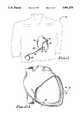
- FIG. 1is a simplified diagram of a mammalian heart surrounded by a pericardium, and further shows a defibrillation electrode positioned therein and connected to an implantable defibrillation device;
- FIG. 2Ais a simplified diagram of the heart of FIG. 1 prior to placement of the electrode in the pericardial space;
- FIG. 2Bis a diagram as in FIG. 2A illustrating a distended pericardium resulting from injecting a fluid into the pericardial space through a fixation catheter that has been attached transvenously to the interior of the atrial wall;
- FIG. 3Ais a schematic perspective view of the fixation catheter of FIG. 2B prior to its attachment to the atrial wall;
- FIG. 3Bis a schematic perspective view, shown partially in cross section, showing the fixation catheter of FIG. 2B after its attachment to the atrial wall, and further illustrating a J-tip guide wire puncturing the atrial wall to form a hole through which the fluid used to distend the pericardium may be injected;
- FIG. 4is a simplified schematic drawing illustrating the manner of making sub-xiphoid access with a needle to the pericardium
- FIG. 5Ais an expanded view of the sub-xiphoidally inserted needle tip of FIG. 4 as the needle tip just makes contact with the pericardial wall;
- FIG. 5Bis a view as in FIG. 5A after the needle tip has punctured the pericardial wall, and further illustrates the fluid in the pericardial space egressing via the lumen in the needle;
- FIG. 5Cis a view similar to FIG. 5A wherein a special perforation device is inserted in the lumen of the needle to aid in cutting through the pericardial wall;
- FIG. 5Dis a view as in FIG. 5C where the perforation device has punctured through the pericardial wall;
- FIG. 6Ais a view as in FIG. 5B further illustrating a guide wire or stylet inserted through the lumen of the needle into the pericardial space;
- FIG. 6Bis a view as in FIG. 6A further depicting a catheter inserted into the pericardial space over the needle and guide wire;
- FIG. 6Cis an elevated view of the distal tip of an expanding pleated sheath or catheter that may optionally be used to gain access into the pericardial space;
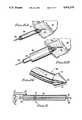
- FIG. 7shows the distal end of one type of defibrillation lead that could be used with the present invention, showing the electrode thereof in a partially retracted position;
- FIG. 8is an electrical schematic diagram of the lead of FIG. 7;
- FIG. 9is an exploded view of a section of the pericardial space and heart as in FIGS. 5A through 6B further illustrating the electrode of FIGS. 7 and 8 positioned within the pericardial space adjacent the heart;
- FIGS. 10A and 10Billustrate an alternate technique for piercing the pericardium without piercing the myocardium using an extendable piercing cylinder housed within the lumen of a special catheter;
- FIGS. 11A and 11Billustrate still another technique for using suction
- FIGS. 12A-12Dillustrate yet another technique for piercing the pericardium using a pre-formed suture.
- FIG. 1a simplified diagram of a mammalian heart 12 surrounded by a pericardium 14 is illustrated in a simplified format.
- the mammal illustrated in FIG. 1is a human 10.
- a portion of the pericardium 14is shown cutaway in order to better illustrate the heart 12 inside of the pericardium.
- a defibrillation electrode 16connected to the distal end of a defibrillation lead 18, is passed through an opening 17 within the pericardial wall 19 and positioned within the pericardial space (that space between the exterior heart surface and the interior of the pericardial wall).
- this defibrillation lead 18is directed to the heart from a sub-xiphoid location, i.e., from a location below or underneath the lower end of the xiphoid process below the sternum or breast bone 20.
- the proximal end of the defibrillation electrode 18is connected to a suitable defibrillation device 22.
- the defibrillation device 22 and the lead 18are preferably implanted within the human 10 using conventional techniques known in the art, i.e., forming a suitable "pocket" within the flesh of the human where the device may be located, and tunneling the lead 18 from the pocket to its sub-xiphoid channel into the pericardial space.
- the present inventionis directed to a method and system for effectuating and using a sub-xiphoid implantation of a defibrillation electrode, such as is shown in FIG. 1.
- a defibrillation leadis implanted sub-xiphoidally within the pericardial space as follows:
- the right atrial wall of the heartis perforated with a small opening using a suitable fixation catheter and guide wire, inserted into the heart transvenously.
- the perforationis limited to the atrial wall and does not traverse the pericardium. This perforation provides access into the pericardial space.
- this perforation in the atrial wallremains very small (compared to the large perforation required for the transvenous implantation approach described in the previously reference patent application), is used for only a very short period of time, and completely heals over after use.
- a small volume of fluid(e.g., 25-75 cc for humans), such as saline or renografin, is infused into the pericardial space through the fixation catheter and the opening in the atrial wall. This is done for the purpose of distending the pericardial wall away from the heart in order to facilitate a sub-xiphoid entry into the pericardial space.
- the volume of fluid infusedshould be less than the amount that would cause cardiac tamponade and dysfunction.
- a cardiac needleis inserted into the distended pericardium.
- the fluidsquirts out of the needle lumen to indicate that the needle advancement should be halted before perforating the heart.
- a guide wireis inserted through the needle into the pericardial space.
- the needleis removed and a sheath/dilator introducer is inserted into the pericardial space over the guide wire.
- the opening in the pericardial wall made by the needleis enlarged as required in order to allow the larger introducer to fit therewithin.
- the distal end of a defibrillation leadis inserted into the pericardium over the guide wire and within the sheath. (Alternatively, at the discretion of the physician, the guide wire may be removed after sheath placement, before lead placement.)
- This distal endpreferably includes a deployable defibrillation electrode having a large area electrode in a retracted position.
- the electrode of the defibrillation leadis positioned as desired and the electrode is deployed. Further, the deployed electrode is anchored, as required.
- the body of the defibrillation leadis tunneled through body tissue, using conventional methods, to a desired implant location.
- a suitable defibrillation deviceis connected to the lead and implanted, using conventional implant techniques and methods known in the art.
- FIG. 2Aan expanded simplified view of the heart 12 is shown inside of the pericardium 14, the pericardium being shown in cutaway fashion so as to clearly illustrate the pericardial wall 19 and the space between the pericardial wall and the heart, or the pericardial space 24. This space is shown greatly exaggerated in FIG. 2A for emphasis.
- pericardial space 24It is not uncommon for the pericardial space 24 to be very narrow in a normal heart, with the inside of the pericardial wall touching, or nearly touching, the exterior of the heart. Hence, it is very difficult to puncture or perforate the pericardial wall 19 without also damaging the myocardium, at the least, or causing tamponade and impending mortality at the worst.
- the present inventioncontemplates filling the pericardial space with a fluid 26, thereby extending the pericardial wall 19 away from the heart, as shown in FIG. 2B.
- the fluid 26is symbolically represented by small dots placed within the pericardial space 24.
- the fluid 26is injected into the pericardial space 24 through a fixation catheter 28 that is inserted transvenously, e.g., through the jugular or subclavian vein, into the heart and affixed to the interior of the right atrial wall 30.
- a small openingis made in the atrial wall 30 (described below in connection with FIGS. 3A and 3B). This need not be a large opening, and is preferably made with the tip of a suitable stylet or guide wire 36.
- suitable infusion meanssuch as a hand-held syringe 34, are used to selectively force the desired volume of fluid 26 through the catheter 28 and into the pericardial space 24.
- a hemostasis valveon the proximal end of the fixation catheter 28 may optionally be used during this process.
- fixation cathetersare known in the art, as is the method of transvenously inserting a catheter or lead into the heart, as guided by a stylet or guide wire placed inside of the catheter or lead.
- pacemaker artfor example, it is a very common practice to transvenously insert pacing leads into desired locations, either within the atrium, the ventricle, or both, of a heart using a stylet to guide placement of the distal tip of the lead.
- the distal end of the fixation catheter 28is shown in FIG. 3A prior to fixation thereof to the atrial wall 30.
- the catheterincludes a main body portion 38 made from a tube of silicone rubber (or other body compatible substance) having a helical tip 40 connected to the distal end.
- the helical tip 40is designed to be screwed into body tissue by rotation of the catheter.
- the helical tipis made from a wire, such as stainless steel wire (or other safe implantable material).
- the wire from which the helical tip is mademay extend, in a helical wound manner, along the entire length of the catheter 28.
- the helical tipmay be retracted within the tubular wall 38.
- the helical tip 40is extended as the entire catheter is rotated, as indicated by the arrow 42, so as to screw the helical tip 40 into the atrial wall 30.
- FIG. 3where a J-tip stylet wire 36' is shown inserted through the catheter 28 and through the atrial wall 30.
- the preferred puncturing techniqueis to retract the fixation catheter 28 in the direction shown by the arrow 46 while simultaneously advancing the guide wire 36', so that the atrial wall 30 is separated from the pericardial wall 19, thereby increasing the width of the pericardial space 24 as the guide wire 36' is punctured through the atrial wall 30. This minimizes the likelihood of having the guide wire 36' puncture through both the atrial wall 30 and the pericardial wall 19.
- the next stepis to insert a needle 50 into the pericardium from a sub-xiphoid location, as shown in FIG. 4.
- the goalis to puncture the inflated pericardium with the needle 50, but to avoid having the needle puncture the heart, as is commonly done in the art to relieve cardiac tamponade. This requires a controlled insertion depth of the needle, made easier by simply monitoring the lumen of the needle for egress of the fluid 26 with which the pericardium has been filled.
- pericardial drainageis a relatively common and safe procedure for the treatment of patients with pericardial effusions, and the incidence of cardiac perforation is very small.
- a perforation mechanismbe used that perforates the pericardium, and then becomes impotent (i.e., unable to further puncture the cardiac wall or any other tissue).
- the needle 50is advanced through the sub-xiphoid tissue into the mediastinum and abutted against the pericardial wall 19, as shown in FIG. 5A.
- This needleis preferably a blunt metal hypodermic needle tube of a type commonly known in the art.
- the needlehas a hole, or lumen, passing longitudinally therethrough.
- a rod 52is initially included within the lumen to prevent coring of tissue as the needle is inserted. Neither the needle 50 nor the rod 52 are extremely sharp. At the distal tip, both are slightly angled, but blunt.
- the pericardiumis stretched, but (due to the nature of the pericardial tissue) is not easily perforated.
- the rod 52is withdrawn from the needle tube 50 and the needle 50 is further inserted in order to punch through the pericardial wall 19.
- the fluid 26 within the pericardial space 26begins to egress through the lumen of the needle 50, as illustrated by the arrows 54.
- the physicianmay elect to use a sharper instrument.
- the rod 52is withdrawn from the needle lumen and is replaced by a sharp perforation device 56, as illustrated in the exploded view of FIG. 5C.
- This device 52fits within the clearance of the needle lumen, and includes a sharp cutting tip 58, a fairly rigid (although somewhat flexible) body portion 60, and a very flexible, compressible coupling portion 62.
- the coupling portion 62joins the tip 58 to the body 60, and is designed to allow force applied to the handle or body portion 60 to be efficiently transferred to the tip portion 58.
- This cutting device 56is inserted into the needle lumen and advanced, as required, applying a mild longitudinal force to the body portion 60 (to force the tip 58 against the pericardial wall 19), so as to have the tip 58 cleanly cut through the pericardial wall 19. Once the tip has cut through the wall 19, and without the lateral support of the needle lumen, the coupling portion 62 buckles, as shown in FIG. 5D.
- the body portion 60 of the perforation devicemay be made from a conventional hollow wire or tube, or equivalent semi-rigid material, that can efficiently couple forces along its length.
- the tip portion 58may be made from any suitable metal that can maintain a sharp edge, such as stainless steel.
- the coupling portion 62may be a stainless steel compression spring or, more preferably, a molded bellows made from, for example, silicone rubber.
- the lumen of the needle or tube 50can function as the introducer through which other devices can be passed into the pericardial space 24, including the defibrillation lead 18.
- a larger introduceris needed and/or a softer (polymeric) introducer is desired.
- a method of enlarging the hole through the pericardial wall and introducing a sheath thereinmust be employed.
- FIGS. 6A and 6BOne such method is illustrated in FIGS. 6A and 6B.
- a guide wirehaving a blunt tip 66 is inserted through the lumen of the needle 50 so that the blunt tip is within the pericardial space 24.
- the needlemay be withdrawn and a suitable sheath 68 may be inserted over a dilator which slides over the guide wire 64.
- the sheath 68may be inserted over both the needle 50 and guide wire 64, as shown in FIG. 6B, in which case the needle would not be withdrawn until after the distal end of the sheath has been inserted into the pericardial space 24.
- the guide wiremay then be used to assist in introducing the defibrillation lead 18 into the pericardial space, as well as to assist in the positioning of the defibrillation electrode to a desired location within the pericardial space.
- conventional techniquesmay be used for inserting the sheath 68 into the pericardium 14.
- the sheath 58is simply inserted over the needle 50 and, through the application of a gentle longitudinal force on the sheath, the sheath is pushed through the hole, thereby dilating the hole in the pericardial wall so as to enlarge it sufficiently to allow introduction of the sheath.
- pushing against the pericardiumin order to force the sheath thereinto may prove difficult without damaging the heart.
- enlarging the pericardial holemay be considered. These possibilities include: (1) using an angioplasty style ballon across the hole; (2) inserting a deflated balloon into the pericardial space, inflating the ballon, and retracting the inflated balloon through the hole; (3) using a mechanical device that enlarges the hole by cutting it larger; (4) using a mechanical device that enlarges the hole by stretching it using a biopsy forceps technique, similar to a blunt dissection; (5) using a mechanical device that enlarges the hole by stretching it using an expanding mini collet; or (6) using a pleated sheath that can be inserted with its pleat(s) in a folded position, thereby providing a small diameter sheath, and then enlarged to provide a large diameter sheath, stretching the hole as it enlarges.
- the blunt dissection approach mentioned abovecontemplates the use of a narrow, blunt tapered pair (or multiplicity) of jaws, pivotally mounted for closing or expanding. These jaws are placed in the hole in their closed position, then spread apart to separate (or tear) the layers of tissue apart.
- the pleated sheath approach mentioned abovecontemplates the use of a sheath 68' having an expanding diameter.
- An elevated view of the distal end of such a sheathis illustrated in FIG. 6C.
- the sheath 68'includes at least one pleat 69 along the length thereof. This pleat simply comprises an integral section of the wall of the sheath that is folded into the interior of the sheath.
- the sheath 68' including its pleat 69is made from a suitable biocompatible elastically expandable substance, such as silicone rubber.
- a tip 71 at the distal end of the sheath 68'is tapered from a narrow tip portion 73 to a larger end portion 75. As seen in FIG. 6C, the tip 71 resembles a conical arrow head. (Note that the sheath 68' shown in FIG. 6C is shown partially expanded, intermediate its most narrow diameter position, wherein the pleat 69 is completely folded inside of the sheat body, and its widest diameter position, where the pleat 69 is not folded in to the interior of the sheath body at all.)
- the sheath 68'is inserted over the guide wire and needle with the pleat(s) 69 completely folded into the sheath body, i.e., in its most narrow diameter position.
- the small portion of the tip 73is inserted into the pericardial hole.
- the sheathis then expanded, using conventional means, as a gentle longitudinal force is exerted on the sheath in order to force the tip 71 deeper into the pericardial hole.
- the sheathexpands, and as the tip moves deeper into the hole, the hole is expanded by the increasingly wider tip 71.
- the sheathmay then be fully expanded and inserted well into the pericardial space 24.
- the sheath 68With the guide wire in place and the pericardial hole enlarged (and assuming a pleated sheath 68' is not already in place as above described), the sheath 68 is inserted well into the pericardial space 24 (FIG. 6B). Over the guide wire 64 and within the sheath 68, a suitable defibrillation electrode is advanced. It is noted that the preferred defibrillation electrode is deployable, having its electrode(s) in a retracted position while being inserted through the sheath.
- this technique for sub-xiphoidally inserting a defibrillation leadwould also have application to smaller, non-deployable defibrillation leads, of the type known in the art.
- a plurality of such leads and/or electrodescan be positioned in a desired configuration, such as an orthogonal configuration, within the pericardial space using this technique.
- FIGS. 7 and 8illustrates the distal end of the electrode 18 with its electrode 16, a butterfly type of patch electrode, partially coiled in its retracted position.
- the electrode 16When fully retracted, the electrode 16 is wound sufficiently tight so that the overall retracted dimension D1 is not much larger than the diameter of the body portion of the lead D2. This tight winding facilitates insertion of the lead/electrode into the sheath 68, which sheath 68 must have a diameter slightly larger than D1.
- the distal tip of the electrodefurther includes a heating element 70, preferably protruding from the electrode somewhat so as to make good contact with the pericardium, the purpose and function of which is described below.
- the lead 18typically includes two conductors 72 and 74, insulated from each other by an intermediate layer of insulating material 76. Typically, one conductor is spirally wound concentric with the other. An outer insulating sheath 78 insulates both conductors from body fluids.
- a channel (or lumen) 80 through the center of the leadprovides a space through which a guide wire may pass, and through which platelets or blood may be infused (as described below).
- This channel 80exits at the distal tip near the heating element 70.
- At least one of the two conductorsmay be temporary, even removable. This is because this conductor only serves a purpose during the implant process. It is not needed after the implant has been completed. Hence, its inclusion in the lead 18 can be achieved inexpensively.
- Two conductors 72 and 74are employed in order to complete the circuit connection with the heating element 70, as shown in the electrical schematic diagram of the defibrillation lead in FIG. 8. If the heating element is not used, then a single lead conductor 72 could be used in unipolar fashion.
- the lead 18is inserted through the sheath 68 over the guide wire 66 (FIG. 6B).
- the guide wireis then used in conventional manner in order to position the electrode at a desired location within the cardiac space, and then the guide wire is withdrawn.
- the electrode 16is deployed, as shown in FIG. 9, so as to make contact with the largest possible tissue area within the pericardium, and anchored in this position, as described below.
- the sheath 68is then withdrawn, leaving just the electrode 18 entering the pericardial space.
- the lead 18is tunneled in the body tissue to a desired implant site where a suitable defibrillation device is implanted and connected to the lead 18.
- a suitable defibrillation deviceis implanted and connected to the lead 18.
- the defibrillation deviceneed not be implanted, but need only be carried by the patient, and the lead 18 exits from the body tissue at a suitable site so as to be electrically connected thereto. Further details associated with techniques and methods for positioning and deploying the electrode 16 within the pericardial space may be found in the above-referenced transvenous implantation approach patent application.
- One of the advantages of the present inventionresides in the manner in which the electrode 16 is anchored to a desired location within the pericardial space.
- One such anchoring techniqueis to use conventional passive sclerosing pads or pericardioscope guided stapling or suturing.
- a better approachis to use an adhesive that is carried into the pericardial space on the non-deployed electrode.
- no biocompatible adhesives that may be applied to a beating heartare known to the applicant.
- the desired adhesion effectcan advantageously be achieved by stimulating a fibrosis attachment site.
- Such a sitecan be acutely accomplished by cautery (burning or searing). That is, just as a bleeding vessel can be coagulated by heat (RF, DC, laser or microwave cautery, for example), the present invention contemplates capturing or creating a clot on the patch electrode and coagulating it to the pericardium.
- external sources of heatsuch as RF, DC, laser or microwave may be used to perform the cautery using conventional techniques.
- a channel 80may be provided down the spine of the electrode 16, or otherwise in contact therewith, through which wires for current (or optical fibers for laser) may pass. Further, infusion of platelets or whole blood may be passed through this channel 80.
- the heater element 70is a specific example of an electrical element that is used to coagulate (heat) platelets or whole blood that is passed through the channel 80 and over the heating element.
- the coagulated bloodforms a bond with the pericardium. If needed, a small lesion may be created on the pericardium itself near the heater element to further promote adhesion.
- the resistance of the heater element 70should be carefully selected. On the one hand, this resistance should be kept as low as possible, to keep the applied voltage requirements within manageable limits, the power dissipated in the heating element being proportional to the square of the applied voltage V and inversely proportional to the resistance R. Hence, by keeping R small, a higher power dissipation is achieved for a given applied voltage.
- the resistanceneeds to be kept as high as possible to avoid excessive Joule heating of the conductors.
- the resistance per unit length of the heating elementneeds to be large compared to the resistance per unit length of the conductors, yet not so large that enormous voltage levels must be applied thereto in order to achieve the desired heating of the element.
- an autologous blood clotmay be formed in the lead near the electrode 16 before the implant.
- the heater elementcould be activated, in order to coagulate the blood clot to the pericardium. If this alternative is used, a pocket formed in the electrode tip near the heater element can hold the blood clot prior to implantation.
- the present inventionalso encompasses placement and anchoring of the electrode(s) to the epicardium or pleural sac, as such is also considered as being within the pericardium for purposes of the present invention.
- the techniques of placing and anchoring a defibrillation electrode as taught hereinalso find application when other approaches to access the heart, besides a sub-xiphoid approach, are utilized. For example, an intercostal access to the heart could be used.
- defibrillation electrode(s)could be positioned and/or anchored extra-pericardially, in accordance with the teachings of the present invention, using sub-xiphoid, intercoastal, or other access techniques.
- This techniquewas designed primarily for puncturing the atrial wall from the inside of the heart without puncturing the pericardial wall, and was designed to include a perforation mechanism inside of the guiding catheter, as operated by a guide wire in the catheter.
- Guide wiresare commonly used in transvenous placement of leads and catheters.
- the techniquealso has applicability to sub-xiphoid, intercostal or other puncturing of the pericardial wall if the device is made small enough to fit within the lumen of the needle or catheter through which access to the pericardium is obtained. In such case, the guide wire is used only to extend the perforating portion from a recessed position within the tip of the catheter or needle.
- FIGS. 10A and 10BThe device used for this technique is shown in FIGS. 10A and 10B, with FIG. 10A showing a perforation mechanism 90 in its retracted position, and FIG. 10B showing the perforation mechanism 90 in its extended position.
- the perforation mechanism 90includes a catheter or other suitable housing 92 in which a perforation cylinder 94 is located.
- the perforation cylinder 94is spring loaded, with a spring 96 or equivalent mechanism, within the tip of the housing 92 so that it is normally recessed therewithin.
- the cylinder 94can be readily advanced by use of a simple guide wire 98.
- the cylinder 94is coupled to the spring 96 by way of an arm 100 that includes a slot or detent 102 that engages a pivot pin 104.
- the guide wire 98pushes the arms 100 so that they pivot and slide relative to the pivot pin 104, forcing the cylinder 94 out of its recessed position.
- the perforation cylinder 94travels only a preset distance, yet the guide wire 98 can be advanced through the device as far as desired, as shown in FIG. 10B. Because the guide wire, once extended, is unsupported by the catheter, it easily buckles, making it extremely unlikely that the myocardial tissue will be perforated.
- the perforation cylinder 94may be shaped to reduce the perforation force. As the guide wire is advanced several millimeters beyond that shown in FIG. 10A, the perforation cylinder 94 is forced out of the catheter 92, perforates the pericardial wall, whereupon the guide wire 98 can be advanced, if desired, into the pericardial space. The perforation cylinder 94 is shown in its extended position in FIG. 10B.
- the perforation catheter 90 shown in FIGS. 10A and 10Bcould be envisioned by those skilled in the art.
- the important features of such a deviceare: (1) the perforation tool 94 is recessed within the catheter 92 until it is ready to be used; (2) the perforation tool (perforation cylinder) can not advance past the tip more than a precisely controlled distance; (3) the guide wire 98 is compelled to enter the hole once created so that the entry site to the pericardium can not be "lost”; and (4) the guide wire tip is soft (not able to support appreciable compressive loads along its axis without buckling) so it will not go through the pericardial space and perforate the myocardial tissue.
- FIGS. 11A and 11BIn order to better control the entry into the pericardium, another technique is to contain the perforating instrument and pull the pericardial wall toward it.
- One technique for doing thisis through the use of suction.
- One embodiment, shown in FIGS. 11A and 11B,includes a catheter 110, having a large soft tip 112, the catheter being guided to the desired perforation site over the sub-xiphoid (or other percutaneously inserted) needle. Once the catheter is in place, the needle can be removed, if desired.
- the tip 112is fitted as tightly as possible up against the pericardial wall 19 in order to maintain and seal the suction force.
- Recessed within the tip 112is an extraordinarly sharp instrument 114.
- the pericardial wall 19is pulled into the catheter tip 112 and is impaled on the cutting instrument 114, and the perforation is made. Once made, a guide wire can be readily inserted into the pericardial space.
- the catheterWith no suction applied, the catheter is as shown in FIG. 11A. With suction applied, it is as shown in FIG. 11B.
- the heart tissueis not in danger of being pierced by the piercing tip 114.
- a suitable hemostasis valvecan be used, as required, so that suction can be applied with the guide wire and perhaps even the sub-xiphoid needle in position.
- suctionmay be lost.
- at least one form of mechanical active fixationcould be used with the suction.
- Such active fixationmay take the form of a helical screw.
- a remotely operated wire suturecould be used, as shown below in FIGS. 12A-12B. The use of more than one suture or ribbon wire would increase stability.
- a catheter 120 without piercing implementis placed against the pericardial wall 19. Suction is drawn, thus pulling the pericardial wall 19 into the lumen of the catheter 120. With the pericardial wall safely separated from the myocardial tissue, the suture(s) is placed. This is done by advancing a preformed tight radius wire 122 out of the catheter 120. The wire 122 pierces a "bite" of the pericardial wall, thus stabilizing the catheter tip. Once stabilized, the piercing can be safely performed within the recess of the catheter by inserting a piercing needle 124 through the lumen of the catheter 120 until the desired piercing has been completed. Once pierced, a suitable guide wire 126 can be inserted into the pericardial space. This complete sequence is illustrated in FIGS. 12A-12D.
Landscapes
- Health & Medical Sciences (AREA)
- Heart & Thoracic Surgery (AREA)
- Life Sciences & Earth Sciences (AREA)
- Veterinary Medicine (AREA)
- Animal Behavior & Ethology (AREA)
- Public Health (AREA)
- Biomedical Technology (AREA)
- Engineering & Computer Science (AREA)
- General Health & Medical Sciences (AREA)
- Pulmonology (AREA)
- Hematology (AREA)
- Anesthesiology (AREA)
- Biophysics (AREA)
- Cardiology (AREA)
- Nuclear Medicine, Radiotherapy & Molecular Imaging (AREA)
- Radiology & Medical Imaging (AREA)
- Electrotherapy Devices (AREA)
Abstract
Description
Claims (41)
Priority Applications (1)
| Application Number | Priority Date | Filing Date | Title |
|---|---|---|---|
| US07/333,391US4991578A (en) | 1989-04-04 | 1989-04-04 | Method and system for implanting self-anchoring epicardial defibrillation electrodes |
Applications Claiming Priority (1)
| Application Number | Priority Date | Filing Date | Title |
|---|---|---|---|
| US07/333,391US4991578A (en) | 1989-04-04 | 1989-04-04 | Method and system for implanting self-anchoring epicardial defibrillation electrodes |
Publications (1)
| Publication Number | Publication Date |
|---|---|
| US4991578Atrue US4991578A (en) | 1991-02-12 |
Family
ID=23302585
Family Applications (1)
| Application Number | Title | Priority Date | Filing Date |
|---|---|---|---|
| US07/333,391Expired - Fee RelatedUS4991578A (en) | 1989-04-04 | 1989-04-04 | Method and system for implanting self-anchoring epicardial defibrillation electrodes |
Country Status (1)
| Country | Link |
|---|---|
| US (1) | US4991578A (en) |
Cited By (287)
| Publication number | Priority date | Publication date | Assignee | Title |
|---|---|---|---|---|
| WO1993015791A1 (en)* | 1992-02-12 | 1993-08-19 | Dimed, Incorporated | System for access to pericardial space |
| US5269326A (en)* | 1991-10-24 | 1993-12-14 | Georgetown University | Method for transvenously accessing the pericardial space via the right auricle for medical procedures |
| US5330496A (en)* | 1991-05-06 | 1994-07-19 | Alferness Clifton A | Vascular catheter assembly for tissue penetration and for cardiac stimulation and methods thereof |
| US5336252A (en)* | 1992-06-22 | 1994-08-09 | Cohen Donald M | System and method for implanting cardiac electrical leads |
| US5571161A (en)* | 1995-04-12 | 1996-11-05 | Starksen; Niel F. | Apparatus and method for implanting electrical leads in the heart |
| WO1996040368A1 (en)* | 1995-06-07 | 1996-12-19 | Cormedics Corp. | Method and apparatus for accessing the pericardial space |
| US5618287A (en)* | 1994-01-28 | 1997-04-08 | Thomas J. Fogarty | Methods of surgically implanting a defibrillator electrode within a patient |
| US5797870A (en)* | 1995-06-07 | 1998-08-25 | Indiana University Foundation | Pericardial delivery of therapeutic and diagnostic agents |
| US5836311A (en)* | 1995-09-20 | 1998-11-17 | Medtronic, Inc. | Method and apparatus for temporarily immobilizing a local area of tissue |
| WO1999013936A1 (en) | 1997-09-19 | 1999-03-25 | Comedicus Incorporated | Direct pericardial access device and method |
| US5931810A (en)* | 1996-12-05 | 1999-08-03 | Comedicus Incorporated | Method for accessing the pericardial space |
| US5968010A (en)* | 1997-04-30 | 1999-10-19 | Beth Israel Deaconess Medical Center, Inc. | Method for transvenously accessing the pericardial space via the right atrium |
| US5972013A (en)* | 1997-09-19 | 1999-10-26 | Comedicus Incorporated | Direct pericardial access device with deflecting mechanism and method |
| WO1999060924A1 (en) | 1998-05-26 | 1999-12-02 | Comedicus Incorporated | Intrapericardial procedures and apparatuses |
| US6071295A (en)* | 1997-02-27 | 2000-06-06 | Medivas Opcab, Inc. | Device to hold an anastomotic site of coronary artery motionless and bloodless for the bypass operation |
| US6139492A (en)* | 1994-08-31 | 2000-10-31 | Heartport, Inc. | Device and method for isolating a surgical site |
| WO2000064531A1 (en) | 1999-04-22 | 2000-11-02 | Beth Israel Deaconess Medical Center, Inc. | Method and kit for transvenously accessing the pericardial space via the right atrium |
| US6206004B1 (en) | 1996-12-06 | 2001-03-27 | Comedicus Incorporated | Treatment method via the pericardial space |
| US6231518B1 (en) | 1998-05-26 | 2001-05-15 | Comedicus Incorporated | Intrapericardial electrophysiological procedures |
| US6231506B1 (en) | 1999-05-04 | 2001-05-15 | Cardiothoracic Systems, Inc. | Method and apparatus for creating a working opening through an incision |
| US6231585B1 (en) | 1997-11-20 | 2001-05-15 | Medivas, Llc | Device for stabilizing a treatment site and method of use |
| US6283912B1 (en) | 1999-05-04 | 2001-09-04 | Cardiothoracic Systems, Inc. | Surgical retractor platform blade apparatus |
| US6290644B1 (en) | 1996-02-20 | 2001-09-18 | Cardiothoracic Systems, Inc. | Surgical instruments and procedures for stabilizing a localized portion of a beating heart |
| US6296630B1 (en) | 1998-04-08 | 2001-10-02 | Biocardia, Inc. | Device and method to slow or stop the heart temporarily |
| US6315717B1 (en) | 1996-02-20 | 2001-11-13 | Cardiothoracic Systems, Inc. | Surgical instruments for stabilizing the beating heart during coronary artery bypass graft surgery |
| US20020049457A1 (en)* | 1999-05-20 | 2002-04-25 | Kaplan Aaron V. | Methods and apparatus for transpericardial left atrial appendage closure |
| US6381499B1 (en) | 1996-02-20 | 2002-04-30 | Cardiothoracic Systems, Inc. | Method and apparatus for using vagus nerve stimulation in surgery |
| US20020058925A1 (en)* | 1999-09-16 | 2002-05-16 | Kaplan Aaron V. | Methods and apparatus for pericardial access |
| US6394951B1 (en) | 1996-02-20 | 2002-05-28 | Cardiothoracic Systems, Inc. | Surgical instruments and procedures for stabilizing the beating heart during coronary artery bypass graft surgery |
| US20020065451A1 (en)* | 1997-09-17 | 2002-05-30 | Spence Paul A. | Device to permit offpump beating heart coronary bypass surgery |
| US6406424B1 (en) | 1999-09-16 | 2002-06-18 | Williamson, Iv Warren P. | Tissue stabilizer having an articulating lift element |
| US6423080B1 (en)* | 1997-02-13 | 2002-07-23 | Scimed Life Systems, Inc. | Percutaneous and hiatal devices and methods for use in minimally invasive pelvic surgery |
| US6464629B1 (en) | 1998-09-15 | 2002-10-15 | Medtronic, Inc. | Method and apparatus for temporarily immobilizing a local area of tissue |
| US20020161275A1 (en)* | 1997-01-02 | 2002-10-31 | Schweich Cyril J. | Heart wall tension reduction apparatus and method |
| US6478776B1 (en) | 2000-04-05 | 2002-11-12 | Biocardia, Inc. | Implant delivery catheter system and methods for its use |
| US6478029B1 (en) | 1993-02-22 | 2002-11-12 | Hearport, Inc. | Devices and methods for port-access multivessel coronary artery bypass surgery |
| US6494211B1 (en) | 1993-02-22 | 2002-12-17 | Hearport, Inc. | Device and methods for port-access multivessel coronary artery bypass surgery |
| US20020192858A1 (en)* | 2001-06-19 | 2002-12-19 | Yusuke Igarashi | Method for fabricating a circuit device |
| US6511416B1 (en) | 1999-08-03 | 2003-01-28 | Cardiothoracic Systems, Inc. | Tissue stabilizer and methods of use |
| US20030032979A1 (en)* | 1998-07-29 | 2003-02-13 | Myocor, Inc. | Transventricular implant tools and devices |
| US20030060415A1 (en)* | 1995-11-01 | 2003-03-27 | Chiron Corporation | Treatment of a coronary condition by delivery of therapeutics to the pericardial space |
| US20030093104A1 (en)* | 1999-10-29 | 2003-05-15 | Bonner Matthew D. | Methods and apparatus for providing intra-pericardial access |
| US6565582B2 (en) | 1995-02-24 | 2003-05-20 | Hearport, Inc. | Devices and methods for performing a vascular anastomosis |
| US20030094180A1 (en)* | 1995-04-10 | 2003-05-22 | Benetti Frederico J. | Method for coronary artery bypass |
| WO2003066147A1 (en)* | 2002-02-04 | 2003-08-14 | Kaplan Aaron V | Methods and apparatus for pericardial access |
| US20030158464A1 (en)* | 2001-12-04 | 2003-08-21 | Estech, Inc. (Endoscopic Technologies, Inc.) | Methods & devices for minimally invasive cardiac surgery for atrial fibrillation |
| US6613062B1 (en) | 1999-10-29 | 2003-09-02 | Medtronic, Inc. | Method and apparatus for providing intra-pericardial access |
| WO2003076007A1 (en)* | 2002-03-06 | 2003-09-18 | Medtronic, Inc. | Coronary sinus/cardiac vein pacing lead using a multi-purpose side lumen |
| US20030181928A1 (en)* | 2000-10-06 | 2003-09-25 | Myocor, Inc. | Endovascular splinting devices and methods |
| US6626830B1 (en) | 1999-05-04 | 2003-09-30 | Cardiothoracic Systems, Inc. | Methods and devices for improved tissue stabilization |
| US20030187460A1 (en)* | 1999-08-10 | 2003-10-02 | Chin Albert K. | Methods and apparatus for endoscopic cardiac surgery |
| US20030187461A1 (en)* | 1999-08-10 | 2003-10-02 | Chin Albert K. | Releasable guide and method for endoscopic cardiac lead placement |
| US6654636B1 (en)* | 1998-07-13 | 2003-11-25 | Genetronics, Inc. | Skin and muscle-targeted gene therapy by pulsed electrical field |
| US6676597B2 (en) | 2001-01-13 | 2004-01-13 | Medtronic, Inc. | Method and device for organ positioning |
| US6678556B1 (en)* | 1998-07-13 | 2004-01-13 | Genetronics, Inc. | Electrical field therapy with reduced histopathological change in muscle |
| EP1381406A1 (en)* | 2001-04-20 | 2004-01-21 | Medtronic, Inc. | Enhanced chronic lead removal |
| US6685632B1 (en) | 1999-05-04 | 2004-02-03 | Cardiothoracic Systems, Inc. | Surgical instruments for accessing and stabilizing a localized portion of a beating heart |
| WO2004012809A1 (en)* | 2002-08-06 | 2004-02-12 | Neopraxis Pty Ltd | A method and system for inserting an electrode |
| US20040087831A1 (en)* | 2002-10-31 | 2004-05-06 | Koen Michels | Anatomical space access tools and methods |
| US20040088035A1 (en)* | 2002-10-30 | 2004-05-06 | Medtronic, Inc. | Methods and apparatus for accessing and stabilizing an area of the heart |
| US20040092798A1 (en)* | 1997-09-17 | 2004-05-13 | Spence Paul A. | Device to permit offpump beating heart coronary bypass surgery |
| US20040102804A1 (en)* | 1999-08-10 | 2004-05-27 | Chin Albert K. | Apparatus and methods for endoscopic surgical procedures |
| WO2004043272A1 (en) | 2002-11-14 | 2004-05-27 | Sydney West Area Health Service | An intramural needle-tipped surgical device |
| US20040111101A1 (en)* | 1999-08-10 | 2004-06-10 | Chin Albert K. | Endoscopic subxiphoid surgical procedures |
| US6758808B2 (en) | 2001-01-24 | 2004-07-06 | Cardiothoracic System, Inc. | Surgical instruments for stabilizing a localized portion of a beating heart |
| US20040138621A1 (en)* | 2003-01-14 | 2004-07-15 | Jahns Scott E. | Devices and methods for interstitial injection of biologic agents into tissue |
| US20040138522A1 (en)* | 2002-08-21 | 2004-07-15 | Haarstad Philip J. | Methods and apparatus providing suction-assisted tissue engagement through a minimally invasive incision |
| US20040138531A1 (en)* | 2003-01-15 | 2004-07-15 | Bonner Matthew D. | Methods and apparatus for accessing and stabilizing an area of the heart |
| US20040153098A1 (en)* | 1999-08-10 | 2004-08-05 | Chin Albert K. | Apparatus and method for endoscopic cardiac mapping and lead placement |
| US20040167374A1 (en)* | 1997-01-02 | 2004-08-26 | Myocor, Inc. | Heart wall tension reduction apparatus and method |
| US6786898B2 (en) | 2003-01-15 | 2004-09-07 | Medtronic, Inc. | Methods and tools for accessing an anatomic space |
| US20040193138A1 (en)* | 2003-03-26 | 2004-09-30 | Levin Howard R. | Method and system to treat and prevent myocardial infarct expansion |
| US20040215307A1 (en)* | 2001-11-29 | 2004-10-28 | Koen Michels | Medical lead designs for lead placement through tissue |
| US20040215168A1 (en)* | 1997-04-30 | 2004-10-28 | Beth Israel Deaconess Medical Center | Kit for transvenously accessing the pericardial space via the right atrium |
| US20040216748A1 (en)* | 1999-08-10 | 2004-11-04 | Chin Albert K. | Apparatus and method for endoscopic encirclement of pulmonary veins for epicardial ablation |
| DE10316177A1 (en)* | 2003-04-10 | 2004-11-11 | Cardiac Pacemakers, Inc., St. Paul | Pacemaker electrode arrangement |
| US20040230283A1 (en)* | 2001-11-29 | 2004-11-18 | Medtronic, Inc. | Trans-septal pacing method and apparatus |
| DE10321337A1 (en)* | 2003-05-13 | 2004-12-30 | Osypka, Peter, Dr.-Ing. | Pacemaker electrode arrangement |
| US20050010197A1 (en)* | 2003-07-08 | 2005-01-13 | Liming Lau | Organ manipulator apparatus |
| US6852075B1 (en) | 1996-02-20 | 2005-02-08 | Cardiothoracic Systems, Inc. | Surgical devices for imposing a negative pressure to stabilize cardiac tissue during surgery |
| US20050033284A1 (en)* | 2000-04-27 | 2005-02-10 | Hooven Michael D. | Transmural ablation device with integral EKG sensor |
| US20050033283A1 (en)* | 2000-04-27 | 2005-02-10 | Hooven Michael D. | Sub-xyphoid method for ablating cardiac tissue |
| DE10337813A1 (en)* | 2003-08-14 | 2005-03-10 | Transmit Technologietransfer | Device for tissue and organ manipulation |
| US20050065396A1 (en)* | 1998-09-21 | 2005-03-24 | Myocor, Inc. | External stress reduction device and method |
| US20050075723A1 (en)* | 2000-10-06 | 2005-04-07 | Myocor, Inc. | Methods and devices for improving mitral valve function |
| US20050102003A1 (en)* | 2000-05-03 | 2005-05-12 | Grabek James R. | Perficardial pacing lead placement device and method |
| US20050113900A1 (en)* | 2003-10-24 | 2005-05-26 | Cardiac Pacemakers, Inc. | Myocardial lead with fixation mechanism |
| US20050119649A1 (en)* | 2003-12-02 | 2005-06-02 | Swanson David K. | Self-anchoring surgical methods and apparatus for stimulating tissue |
| US20050125044A1 (en)* | 2000-05-23 | 2005-06-09 | North Shore-Long Island Jewish Research Institute | Inhibition of inflammatory cytokine production by cholinergic agonists and vagus nerve stimulation |
| US20050137674A1 (en)* | 2003-10-24 | 2005-06-23 | Cardiac Pacemakers, Inc. | Distal or proximal fixation of over-the-tether myocardial leads |
| US20050148822A1 (en)* | 2003-12-30 | 2005-07-07 | Willis Geoffrey H. | Organ manipulator and positioner and methods of using the same |
| US20050148824A1 (en)* | 2003-12-30 | 2005-07-07 | Morejohn Dwight P. | Transabdominal surgery system |
| US20050148825A1 (en)* | 1997-09-17 | 2005-07-07 | Spence Paul A. | Device to permit offpump beating heart coronary bypass surgery |
| US20050154370A1 (en)* | 1999-10-29 | 2005-07-14 | Medtronic, Inc. | Methods and systems for providing therapies into the pericardial space |
| US20050154404A1 (en)* | 2003-10-09 | 2005-07-14 | Liddicoat John R. | Apparatus and method for the ligation of tissue |
| US20050165466A1 (en)* | 1999-10-29 | 2005-07-28 | Medtronic, Inc. | Methods and systems for accessing the pericardial space |
| US20050182465A1 (en)* | 2004-02-12 | 2005-08-18 | Ness Gregory O. | Instruments and methods for accessing an anatomic space |
| US20050209564A1 (en)* | 2001-01-13 | 2005-09-22 | Medtronic, Inc. | Devices and methods for interstitial injection of biologic agents into tissue |
| US6951555B1 (en) | 1998-03-16 | 2005-10-04 | Chase Medical, L.P. | Catheter having integral expandable/collapsible lumen |
| US20050234444A1 (en)* | 2004-04-14 | 2005-10-20 | Hooven Michael D | Electrode and bipolar ablation method using same |
| US20050234507A1 (en)* | 2004-04-16 | 2005-10-20 | Jeff Geske | Medical tool for access to internal tissue |
| US20050240175A1 (en)* | 2001-12-04 | 2005-10-27 | Estech, Inc. (Endoscopic Technologies, Inc.) | Cardiac treatment devices and methods |
| US20050282906A1 (en)* | 2004-03-25 | 2005-12-22 | North Shore-Long Island Jewish Research Institute | Neural tourniquet |
| US20060036317A1 (en)* | 2002-11-12 | 2006-02-16 | Myocor, Inc. | Decives and methods for heart valve treatment |
| US20060041243A1 (en)* | 2001-01-13 | 2006-02-23 | Medtronic, Inc. | Devices and methods for interstitial injection of biologic agents into tissue |
| US20060052868A1 (en)* | 1997-12-17 | 2006-03-09 | Myocor, Inc. | Valve to myocardium tension members device and method |
| US20060052660A1 (en)* | 1999-08-10 | 2006-03-09 | Chin Albert K | Apparatus and methods for cardiac restraint |
| US7020525B1 (en)* | 2002-06-14 | 2006-03-28 | Pacesetter, Inc. | Flexible electrical interconnect for an implantable medical device |
| US20060100699A1 (en)* | 2002-11-12 | 2006-05-11 | Myocor, Inc. | Devices and methods for heart valve treatment |
| US20060116746A1 (en)* | 2003-01-17 | 2006-06-01 | Chin Albert K | Cardiac electrode attachment procedure |
| US20060178703A1 (en)* | 2004-12-27 | 2006-08-10 | Huston Jared M | Treating inflammatory disorders by electrical vagus nerve stimulation |
| US20060247672A1 (en)* | 2005-04-27 | 2006-11-02 | Vidlund Robert M | Devices and methods for pericardial access |
| US20060247491A1 (en)* | 2005-04-27 | 2006-11-02 | Vidlund Robert M | Devices and methods for heart valve treatment |
| US20060253129A1 (en)* | 2005-04-07 | 2006-11-09 | Liddicoat John R | Apparatus and method for the ligation of tissue |
| US20060287574A1 (en)* | 1999-08-25 | 2006-12-21 | Chin Albert K | Longitudinal dilator |
| WO2007002554A2 (en) | 2005-06-23 | 2007-01-04 | Medtronic Vascular, Inc. | Methods and systems for treating injured cardiac tissue |
| US20070021730A1 (en)* | 1995-10-13 | 2007-01-25 | Medtronic Vascular, Inc. | Systems and Methods for Delivering Drugs to Selected Locations Within the Body |
| US7189201B2 (en) | 1995-09-20 | 2007-03-13 | Medtronic, Inc. | Method and apparatus for temporarily immobilizing a local area of tissue |
| US20070083217A1 (en)* | 2002-05-30 | 2007-04-12 | Eversull Christian S | Apparatus and Methods for Placing Leads Using Direct Visualization |
| US20070088203A1 (en)* | 2005-05-25 | 2007-04-19 | Liming Lau | Surgical assemblies and methods for visualizing and performing surgical procedures in reduced-access surgical sites |
| US20070100410A1 (en)* | 2002-04-11 | 2007-05-03 | Medtronic Vascular, Inc. | Devices and methods for transluminal or transthoracic interstitial electrode placement |
| US20070129598A1 (en)* | 2001-09-07 | 2007-06-07 | Raman Jaishanker | Method and apparatus for external stabilization of the heart |
| US20070129719A1 (en)* | 2005-05-26 | 2007-06-07 | Amar Kendale | Apparatus and methods for performing minimally-invasive surgical procedures |
| US20070156217A1 (en)* | 2002-05-10 | 2007-07-05 | Cardiac Pacemakers, Inc. | Apparatus for lead placement on a surface of the heart |
| US20070185502A1 (en)* | 2005-12-29 | 2007-08-09 | Karel Smits | Medical electrode delivery tool |
| EP1862128A1 (en)* | 2002-11-12 | 2007-12-05 | Myocor, Inc. | Devices for heart valve treatment |
| US20080009747A1 (en)* | 2005-02-02 | 2008-01-10 | Voyage Medical, Inc. | Transmural subsurface interrogation and ablation |
| US7319905B1 (en) | 2004-11-30 | 2008-01-15 | Pacesetter, Inc. | Passive fixation mechanism for epicardial sensing and stimulation lead placed through pericardial access |
| US20080015445A1 (en)* | 2005-02-02 | 2008-01-17 | Voyage Medical, Inc. | Tissue visualization device and method variations |
| US20080015569A1 (en)* | 2005-02-02 | 2008-01-17 | Voyage Medical, Inc. | Methods and apparatus for treatment of atrial fibrillation |
| US20080033241A1 (en)* | 2006-08-01 | 2008-02-07 | Ruey-Feng Peh | Left atrial appendage closure |
| US20080033290A1 (en)* | 2005-10-25 | 2008-02-07 | Voyage Medical, Inc. | Delivery of biological compounds to ischemic and/or infarcted tissue |
| US7338434B1 (en) | 2002-08-21 | 2008-03-04 | Medtronic, Inc. | Method and system for organ positioning and stabilization |
| US20080058591A1 (en)* | 2005-10-25 | 2008-03-06 | Voyage Medical, Inc. | Tissue visualization device and method variations |
| US7399272B2 (en) | 2004-03-24 | 2008-07-15 | Medtronic, Inc. | Methods and apparatus providing suction-assisted tissue engagement |
| US20080183036A1 (en)* | 2006-12-18 | 2008-07-31 | Voyage Medical, Inc. | Systems and methods for unobstructed visualization and ablation |
| US20080188759A1 (en)* | 2005-10-25 | 2008-08-07 | Voyage Medical, Inc. | Flow reduction hood systems |
| US20080208166A1 (en)* | 2007-02-28 | 2008-08-28 | Goode Johnson E | Pre-formed delivery catheters |
| US20080214889A1 (en)* | 2006-10-23 | 2008-09-04 | Voyage Medical, Inc. | Methods and apparatus for preventing tissue migration |
| US20080243183A1 (en)* | 2007-03-30 | 2008-10-02 | Miller Gary H | Devices, systems, and methods for closing the left atrial appendage |
| US20080249439A1 (en)* | 2004-03-25 | 2008-10-09 | The Feinstein Institute For Medical Research | Treatment of inflammation by non-invasive stimulation |
| EP1980212A1 (en) | 2002-11-06 | 2008-10-15 | Medtronic, Inc. | Suction-assisted tissue engagement through a minimally invasive incision |
| US20080275300A1 (en)* | 2007-04-27 | 2008-11-06 | Voyage Medical, Inc. | Complex shape steerable tissue visualization and manipulation catheter |
| US20080281293A1 (en)* | 2007-05-08 | 2008-11-13 | Voyage Medical, Inc. | Complex shape steerable tissue visualization and manipulation catheter |
| US20080294175A1 (en)* | 2007-05-21 | 2008-11-27 | Epitek, Inc. | Left atrial appendage closure |
| US20080294154A1 (en)* | 2007-05-21 | 2008-11-27 | Estech, Inc. | Cardiac ablation systems and methods |
| US20080312664A1 (en)* | 2007-05-21 | 2008-12-18 | Epitek, Inc. | Left atrial appendage closure |
| US20090012510A1 (en)* | 2001-12-04 | 2009-01-08 | Endoscopic Technologies, Inc. | Cardiac ablation devices and methods |
| WO2008139396A3 (en)* | 2007-05-11 | 2009-01-08 | Chamalow S A | Electrode catheter for univentricular and biventricular cardiac electrostimulation |
| US20090030412A1 (en)* | 2007-05-11 | 2009-01-29 | Willis N Parker | Visual electrode ablation systems |
| US20090062790A1 (en)* | 2007-08-31 | 2009-03-05 | Voyage Medical, Inc. | Direct visualization bipolar ablation systems |
| US20090062874A1 (en)* | 2007-08-27 | 2009-03-05 | Tracey Kevin J | Devices and methods for inhibiting granulocyte activation by neural stimulation |
| US20090076498A1 (en)* | 2007-08-31 | 2009-03-19 | Voyage Medical, Inc. | Visualization and ablation system variations |
| US20090082797A1 (en)* | 2007-09-20 | 2009-03-26 | Fung Gregory W | Devices and methods for remote suture management |
| US20090125022A1 (en)* | 2007-11-12 | 2009-05-14 | Voyage Medical, Inc. | Tissue visualization and ablation systems |
| US20090143831A1 (en)* | 2004-12-27 | 2009-06-04 | Huston Jared M | Treating inflammatory disorders by stimulation of the cholinergic anti-inflammatory pathway |
| US20090143640A1 (en)* | 2007-11-26 | 2009-06-04 | Voyage Medical, Inc. | Combination imaging and treatment assemblies |
| US20090163768A1 (en)* | 2007-12-20 | 2009-06-25 | Estech, Inc. | Magnetic introducer systems and methods |
| US20090163905A1 (en)* | 2007-12-21 | 2009-06-25 | Winkler Matthew J | Ablation device with internally cooled electrodes |
| US20090203962A1 (en)* | 2008-02-07 | 2009-08-13 | Voyage Medical, Inc. | Stent delivery under direct visualization |
| US20090221871A1 (en)* | 2006-09-01 | 2009-09-03 | Voyage Medical, Inc. | Precision control systems for tissue visualization and manipulation assemblies |
| US20090247934A1 (en)* | 2008-03-31 | 2009-10-01 | Tracey Kevin J | Methods and systems for reducing inflammation by neuromodulation of t-cell activity |
| US20090275842A1 (en)* | 2006-12-21 | 2009-11-05 | Vahid Saadat | Stabilization of visualization catheters |
| US20090275799A1 (en)* | 2006-12-21 | 2009-11-05 | Voyage Medical, Inc. | Axial visualization systems |
| US20090275997A1 (en)* | 2008-05-01 | 2009-11-05 | Michael Allen Faltys | Vagus nerve stimulation electrodes and methods of use |
| US20090281541A1 (en)* | 2008-05-09 | 2009-11-12 | Estech, Inc. | Conduction block systems and methods |
| JP2009542338A (en)* | 2006-06-30 | 2009-12-03 | シーヴィ デヴァイシズ,エルエルシー | Percutaneous endovascular access to heart tissue |
| US20090326572A1 (en)* | 2008-06-27 | 2009-12-31 | Ruey-Feng Peh | Apparatus and methods for rapid tissue crossing |
| US20100004633A1 (en)* | 2008-07-07 | 2010-01-07 | Voyage Medical, Inc. | Catheter control systems |
| US20100010311A1 (en)* | 2005-10-25 | 2010-01-14 | Voyage Medical, Inc. | Methods and apparatus for efficient purging |
| US20100016851A1 (en)* | 2001-12-04 | 2010-01-21 | Bertolero Arthur A | Cardiac ablation devices and methods |
| US20100036195A1 (en)* | 2002-12-06 | 2010-02-11 | Estech, Inc. (Endoscopic Technologies, Inc.) | Methods and devices for cardiac surgery |
| US7678145B2 (en) | 2002-01-09 | 2010-03-16 | Edwards Lifesciences Llc | Devices and methods for heart valve treatment |
| US20100094081A1 (en)* | 2008-10-10 | 2010-04-15 | Voyage Medical, Inc. | Electrode placement and connection systems |
| US20100125304A1 (en)* | 2008-11-18 | 2010-05-20 | Faltys Michael A | Devices and methods for optimizing electrode placement for anti-inflamatory stimulation |
| US20100130836A1 (en)* | 2008-11-14 | 2010-05-27 | Voyage Medical, Inc. | Image processing systems |
| US20100179570A1 (en)* | 2009-01-13 | 2010-07-15 | Salvatore Privitera | Apparatus and methods for deploying a clip to occlude an anatomical structure |
| US20100191234A1 (en)* | 2009-01-28 | 2010-07-29 | Spine Design, Inc. | Combination Tissue Removal and Cauterization Instrument |
| US20100204561A1 (en)* | 2009-02-11 | 2010-08-12 | Voyage Medical, Inc. | Imaging catheters having irrigation |
| US20100228221A1 (en)* | 2006-06-30 | 2010-09-09 | Kassab Ghassan S | Devices, systems, and methods for obtaining biopsy tissue samples |
| US7794387B2 (en) | 2006-04-26 | 2010-09-14 | Medtronic, Inc. | Methods and devices for stabilizing tissue |
| WO2010105081A1 (en) | 2009-03-13 | 2010-09-16 | Medtronic, Inc. | Method of treating heart failure |
| US20100256629A1 (en)* | 2009-04-06 | 2010-10-07 | Voyage Medical, Inc. | Methods and devices for treatment of the ostium |
| US20100262140A1 (en)* | 2008-10-10 | 2010-10-14 | Voyage Medical, Inc. | Integral electrode placement and connection systems |
| US20100274129A1 (en)* | 2009-04-24 | 2010-10-28 | Hooven Michael D | Apparatus And Methods for Separating Pericardial Tissue From The Epicardium of the Heart |
| US20100292558A1 (en)* | 2006-06-14 | 2010-11-18 | Voyage Medical, Inc. | In-vivo visualization systems |
| US20100312320A1 (en)* | 2009-06-09 | 2010-12-09 | Faltys Michael A | Nerve cuff with pocket for leadless stimulator |
| US20100312256A1 (en)* | 2006-06-30 | 2010-12-09 | Cvdevices, Llc | Devices, systems, and methods for lead delivery |
| US20100331854A1 (en)* | 2008-01-07 | 2010-12-30 | Ilan Greenberg | Device and method for performing treatment in a pericardial space |
| US20100331838A1 (en)* | 2009-06-25 | 2010-12-30 | Estech, Inc. (Endoscopic Technologies, Inc.) | Transmurality clamp systems and methods |
| US7881810B1 (en) | 2007-05-24 | 2011-02-01 | Pacesetter, Inc. | Cardiac access methods and apparatus |
| US20110034915A1 (en)* | 2009-08-05 | 2011-02-10 | Estech, Inc. (Endoscopic Technologies, Inc.) | Bipolar belt systems and methods |
| US20110054569A1 (en)* | 2009-09-01 | 2011-03-03 | Zitnik Ralph J | Prescription pad for treatment of inflammatory disorders |
| US20110060331A1 (en)* | 2009-05-19 | 2011-03-10 | ESTECH, Inc. (Endoscopic Technologies, Inc) | Magnetic navigation systems and methods |
| US20110060227A1 (en)* | 2005-02-02 | 2011-03-10 | Voyage Medical, Inc. | Tissue visualization and manipulation system |
| US20110060298A1 (en)* | 2005-02-02 | 2011-03-10 | Voyage Medical, Inc. | Tissue imaging and extraction systems |
| US20110060182A1 (en)* | 2007-04-27 | 2011-03-10 | Cvdevices, Llc | Systems for engaging a bodily tissue and methods of using the same |
| US7920928B1 (en) | 2007-01-31 | 2011-04-05 | Pacesetter, Inc. | Passive fixation for epicardial lead |
| US7922709B2 (en) | 1998-07-13 | 2011-04-12 | Genetronics, Inc. | Enhanced delivery of naked DNA to skin by non-invasive in vivo electroporation |
| US20110087247A1 (en)* | 2009-04-01 | 2011-04-14 | Fung Gregory W | Tissue ligation devices and controls therefor |
| US7931590B2 (en) | 2002-10-29 | 2011-04-26 | Maquet Cardiovascular Llc | Tissue stabilizer and methods of using the same |
| US20110106208A1 (en)* | 2009-10-30 | 2011-05-05 | Faltys Michael A | Modulation of the cholinergic anti-inflammatory pathway to treat pain or addiction |
| US7949411B1 (en) | 2007-01-23 | 2011-05-24 | Pacesetter, Inc. | Epicardial lead |
| US20110144576A1 (en)* | 2009-12-14 | 2011-06-16 | Voyage Medical, Inc. | Catheter orientation control system mechanisms |
| US20110144572A1 (en)* | 2007-04-27 | 2011-06-16 | Kassab Ghassan S | Steering engagement catheter devices, systems, and methods |
| US20110190849A1 (en)* | 2009-12-23 | 2011-08-04 | Faltys Michael A | Neural stimulation devices and systems for treatment of chronic inflammation |
| US8012143B1 (en) | 2006-12-12 | 2011-09-06 | Pacesetter, Inc. | Intrapericardial delivery tools and methods |
| US20110224720A1 (en)* | 2010-03-11 | 2011-09-15 | Cvdevices, Llc | Devices, systems, and methods for closing a hole in cardiac tissue |
| WO2011130456A1 (en)* | 2010-04-13 | 2011-10-20 | Sentreheart, Inc. | Methods and devices for pericardial access |
| US8083664B2 (en) | 2005-05-25 | 2011-12-27 | Maquet Cardiovascular Llc | Surgical stabilizers and methods for use in reduced-access surgical sites |
| US8244379B2 (en) | 2006-04-26 | 2012-08-14 | Medtronic, Inc. | Pericardium fixation concepts of epicardium pacing leads and tools |
| US8346373B2 (en) | 2010-08-03 | 2013-01-01 | Medtronic, Inc. | Method and apparatus for delivering a lead to a heart |
| US8382651B2 (en) | 2007-04-27 | 2013-02-26 | Cvdevices, Llc | Devices, systems, and methods to facilitate heart function |
| WO2013084968A1 (en)* | 2011-12-09 | 2013-06-13 | Olympus Corporation | Pericardial-liquid level control system |
| US8540674B2 (en) | 2007-04-27 | 2013-09-24 | Cvdevices, Llc | Devices, systems, and methods for transeptal atrial puncture using an engagement catheter platform |
| JP2014008079A (en)* | 2012-06-27 | 2014-01-20 | Olympus Corp | Medical sheath |
| US8694071B2 (en) | 2010-02-12 | 2014-04-08 | Intuitive Surgical Operations, Inc. | Image stabilization techniques and methods |
| US20140148786A1 (en)* | 2011-08-23 | 2014-05-29 | Simcha Milo | Device for creating temporary access and then closure |
| US8788034B2 (en) | 2011-05-09 | 2014-07-22 | Setpoint Medical Corporation | Single-pulse activation of the cholinergic anti-inflammatory pathway to treat chronic inflammation |
| US8834506B2 (en) | 2012-09-24 | 2014-09-16 | Fawaz ALHUMAID | Method for puncturing the pericardial membrane by synchronizing needle advancement with cardiac muscle motion |
| EP2716242A4 (en)* | 2011-05-27 | 2014-10-22 | Synaptic Medical Beijing Co Ltd | NEEDLE COMPONENT OF PERICARDIOCENTESIS |
| US8934962B2 (en) | 2005-02-02 | 2015-01-13 | Intuitive Surgical Operations, Inc. | Electrophysiology mapping and visualization system |
| US8998892B2 (en) | 2007-12-21 | 2015-04-07 | Atricure, Inc. | Ablation device with cooled electrodes and methods of use |
| US9022998B2 (en) | 2010-02-26 | 2015-05-05 | Maquet Cardiovascular Llc | Blower instrument, apparatus and methods of using |
| US20150342627A1 (en)* | 2014-06-02 | 2015-12-03 | Medtronic, Inc. | Over-the-wire delivery of a substernal lead |
| US9211410B2 (en) | 2009-05-01 | 2015-12-15 | Setpoint Medical Corporation | Extremely low duty-cycle activation of the cholinergic anti-inflammatory pathway to treat chronic inflammation |
| US20150359561A1 (en)* | 2012-12-28 | 2015-12-17 | Synaptic Medical (Beijing) Co, Ltd. | Pericardium puncture needle assembly |
| US9326793B2 (en) | 2013-07-16 | 2016-05-03 | Fawaz ALHUMAID | Needle for physically separating and penetrating the pericardium |
| US9339295B2 (en) | 2012-10-22 | 2016-05-17 | Sentreheart, Inc. | Pericardial access devices and methods |
| US9408608B2 (en) | 2013-03-12 | 2016-08-09 | Sentreheart, Inc. | Tissue ligation devices and methods therefor |
| US9486281B2 (en) | 2010-04-13 | 2016-11-08 | Sentreheart, Inc. | Methods and devices for accessing and delivering devices to a heart |
| US9498206B2 (en) | 2011-06-08 | 2016-11-22 | Sentreheart, Inc. | Tissue ligation devices and tensioning devices therefor |
| US9572983B2 (en) | 2012-03-26 | 2017-02-21 | Setpoint Medical Corporation | Devices and methods for modulation of bone erosion |
| US20170079541A1 (en)* | 2002-08-24 | 2017-03-23 | St. Jude Medical, Atrial Fibrillation Division, Inc. | Methods and apparatus for locating the fossa ovalis |
| US9655605B2 (en) | 2010-06-14 | 2017-05-23 | Maquet Cardiovascular Llc | Surgical instruments, systems and methods of use |
| US9662490B2 (en) | 2008-03-31 | 2017-05-30 | The Feinstein Institute For Medical Research | Methods and systems for reducing inflammation by neuromodulation and administration of an anti-inflammatory drug |
| US9764127B2 (en) | 2014-12-19 | 2017-09-19 | Cardiac Pacemakers, Inc. | Medical lead anchoring |
| US9814522B2 (en) | 2010-04-06 | 2017-11-14 | Intuitive Surgical Operations, Inc. | Apparatus and methods for ablation efficacy |
| US9833621B2 (en) | 2011-09-23 | 2017-12-05 | Setpoint Medical Corporation | Modulation of sirtuins by vagus nerve stimulation |
| US9883882B2 (en) | 2013-04-24 | 2018-02-06 | Medovex Corp. | Minimally invasive methods for spinal facet therapy to alleviate pain and associated surgical tools, kits and instructional media |
| USD810290S1 (en) | 2016-01-29 | 2018-02-13 | Medovex Corp. | Surgical portal driver |
| US9936956B2 (en) | 2015-03-24 | 2018-04-10 | Sentreheart, Inc. | Devices and methods for left atrial appendage closure |
| US9980771B2 (en) | 2014-07-30 | 2018-05-29 | Medovex Corp. | Surgical tools for spinal facet therapy to alleviate pain and related methods |
| US10004388B2 (en) | 2006-09-01 | 2018-06-26 | Intuitive Surgical Operations, Inc. | Coronary sinus cannulation |
| US10064540B2 (en) | 2005-02-02 | 2018-09-04 | Intuitive Surgical Operations, Inc. | Visualization apparatus for transseptal access |
| US10123821B2 (en) | 2009-09-10 | 2018-11-13 | Atricure, Inc. | Scope and magnetic introducer systems and methods |
| US10130369B2 (en) | 2015-03-24 | 2018-11-20 | Sentreheart, Inc. | Tissue ligation devices and methods therefor |
| US10258408B2 (en) | 2013-10-31 | 2019-04-16 | Sentreheart, Inc. | Devices and methods for left atrial appendage closure |
| US10292710B2 (en) | 2016-02-26 | 2019-05-21 | Sentreheart, Inc. | Devices and methods for left atrial appendage closure |
| US10314498B2 (en) | 2013-05-24 | 2019-06-11 | Bioventrix, Inc. | Cardiac tissue penetrating devices, methods, and systems for treatment of congestive heart failure and other conditions |
| US10314501B2 (en) | 2016-01-20 | 2019-06-11 | Setpoint Medical Corporation | Implantable microstimulators and inductive charging systems |
| US10398494B2 (en) | 2014-07-30 | 2019-09-03 | Medovex Corp. | Surgical tools for spinal facet therapy to alleviate pain and related methods |
| US10583304B2 (en) | 2016-01-25 | 2020-03-10 | Setpoint Medical Corporation | Implantable neurostimulator having power control and thermal regulation and methods of use |
| US10588613B2 (en) | 2013-08-30 | 2020-03-17 | Bioventrix, Inc. | Cardiac tissue anchoring devices, methods, and systems for treatment of congestive heart failure and other conditions |
| US10595919B2 (en) | 2014-12-12 | 2020-03-24 | Medovex Corp. | Surgical tools with positional components |
| US10596367B2 (en) | 2016-01-13 | 2020-03-24 | Setpoint Medical Corporation | Systems and methods for establishing a nerve block |
| US10631840B2 (en) | 2015-11-25 | 2020-04-28 | Talon Medical, LLC | Tissue engagement devices, systems, and methods |
| US10695569B2 (en) | 2016-01-20 | 2020-06-30 | Setpoint Medical Corporation | Control of vagal stimulation |
| US10912712B2 (en) | 2004-03-25 | 2021-02-09 | The Feinstein Institutes For Medical Research | Treatment of bleeding by non-invasive stimulation |
| US10925474B2 (en) | 2014-02-17 | 2021-02-23 | Children's National Medical Center | Delivery tool and method for devices in the pericardial space |
| US10953204B2 (en) | 2017-01-09 | 2021-03-23 | Boston Scientific Scimed, Inc. | Guidewire with tactile feel |
| US10959752B2 (en) | 2014-02-28 | 2021-03-30 | Sentreheart Llc | Pericardial access devices and methods |
| US10980481B2 (en) | 2018-07-31 | 2021-04-20 | Calyan Technologies, Inc. | Subcutaneous device for monitoring and/or providing therapies |
| US10987060B1 (en) | 2020-09-14 | 2021-04-27 | Calyan Technologies, Inc. | Clip design for a subcutaneous device |
| US11051744B2 (en) | 2009-11-17 | 2021-07-06 | Setpoint Medical Corporation | Closed-loop vagus nerve stimulation |
| US11173307B2 (en) | 2017-08-14 | 2021-11-16 | Setpoint Medical Corporation | Vagus nerve stimulation pre-screening test |
| US11179571B2 (en) | 2018-07-31 | 2021-11-23 | Manicka Institute Llc | Subcutaneous device for monitoring and/or providing therapies |
| US11253699B1 (en)* | 2019-03-21 | 2022-02-22 | Terrell M. Williams | Cardiac pacing lead |
| US11260229B2 (en) | 2018-09-25 | 2022-03-01 | The Feinstein Institutes For Medical Research | Methods and apparatuses for reducing bleeding via coordinated trigeminal and vagal nerve stimulation |
| US11311725B2 (en) | 2014-10-24 | 2022-04-26 | Setpoint Medical Corporation | Systems and methods for stimulating and/or monitoring loci in the brain to treat inflammation and to enhance vagus nerve stimulation |
| US11338130B2 (en)* | 2006-07-21 | 2022-05-24 | Boston Scientific Scimed, Inc. | Delivery of cardiac stimulation devices |
| US11406833B2 (en) | 2015-02-03 | 2022-08-09 | Setpoint Medical Corporation | Apparatus and method for reminding, prompting, or alerting a patient with an implanted stimulator |
| US11433233B2 (en) | 2020-11-25 | 2022-09-06 | Calyan Technologies, Inc. | Electrode contact for a subcutaneous device |
| US11446486B1 (en) | 2021-06-03 | 2022-09-20 | Gopi Dandamudi | Multielectrode medical lead |
| US11471681B2 (en) | 2016-01-20 | 2022-10-18 | Setpoint Medical Corporation | Batteryless implantable microstimulators |
| US11478650B2 (en) | 2018-07-31 | 2022-10-25 | Calyan Technologies, Inc. | Subcutaneous device |
| US11478152B2 (en) | 2005-02-02 | 2022-10-25 | Intuitive Surgical Operations, Inc. | Electrophysiology mapping and visualization system |
| US11660444B2 (en) | 2018-07-31 | 2023-05-30 | Manicka Institute Llc | Resilient body component contact for a subcutaneous device |
| US11717674B2 (en) | 2018-07-31 | 2023-08-08 | Manicka Institute Llc | Subcutaneous device for use with remote device |
| US11938324B2 (en) | 2020-05-21 | 2024-03-26 | The Feinstein Institutes For Medical Research | Systems and methods for vagus nerve stimulation |
| EP4216828A4 (en)* | 2020-09-24 | 2024-09-18 | Bioventrix, Inc. | PERICARDIAL INFLATION CATHETER AND SYSTEMS AND METHODS THEREOF |
| US12172017B2 (en) | 2011-05-09 | 2024-12-24 | Setpoint Medical Corporation | Vagus nerve stimulation to treat neurodegenerative disorders |
| US12274876B1 (en)* | 2023-11-02 | 2025-04-15 | The Future Of Pacing L.L.C. | Multielectrode medical lead with fixation |
| WO2025128425A1 (en)* | 2023-12-15 | 2025-06-19 | Edwards Lifesciences Corporation | Devices and methods for anchoring confirmation |
| US12343535B2 (en) | 2019-04-12 | 2025-07-01 | Setpoint Medical Corporation | Vagus nerve stimulation to treat neurodegenerative disorders |
| US12350503B2 (en)* | 2021-10-08 | 2025-07-08 | Fuwai Hospital, Cams & Pumc | Leadless left-bundle-branch pacemaker |
| US12444497B2 (en) | 2022-05-17 | 2025-10-14 | Setpoint Medical Corporation | Neurostimulation parameter authentication and expiration system for neurostimulation |
Citations (18)
| Publication number | Priority date | Publication date | Assignee | Title |
|---|---|---|---|---|
| US3719190A (en)* | 1971-03-09 | 1973-03-06 | Avery Lab Inc | Heart stimulation electrode with a conical positioning parachute |
| US3738370A (en)* | 1971-01-18 | 1973-06-12 | B Charms | Method of defibrillating a malfunctioning heart by means of electrodes located within the atrium |
| US3942536A (en)* | 1971-03-15 | 1976-03-09 | Mieczyslaw Mirowski | Cardioverting device having single intravascular catheter electrode system and method for its use |
| US4030509A (en)* | 1975-09-30 | 1977-06-21 | Mieczyslaw Mirowski | Implantable electrodes for accomplishing ventricular defibrillation and pacing and method of electrode implantation and utilization |
| US4270549A (en)* | 1979-04-30 | 1981-06-02 | Mieczyslaw Mirowski | Method for implanting cardiac electrodes |
| US4291707A (en)* | 1979-04-30 | 1981-09-29 | Mieczyslaw Mirowski | Implantable cardiac defibrillating electrode |
| US4355646A (en)* | 1980-11-26 | 1982-10-26 | Medtronic, Inc. | Transvenous defibrillating lead |
| EP0095727A1 (en)* | 1982-06-01 | 1983-12-07 | Purdue Research Foundation | Method and apparatus for inserting a defibrillator electrode and defibrillator electrode |
| US4499907A (en)* | 1982-11-15 | 1985-02-19 | Medtronic, Inc. | Energy limiting cardioversion lead |
| US4567900A (en)* | 1984-06-04 | 1986-02-04 | Moore J Paul | Internal deployable defibrillator electrode |
| US4603705A (en)* | 1984-05-04 | 1986-08-05 | Mieczyslaw Mirowski | Intravascular multiple electrode unitary catheter |
| US4640298A (en)* | 1980-06-03 | 1987-02-03 | Peter Pless | Esophageal electrode probe useful for electrical stimulation of the heart |
| US4641656A (en)* | 1985-06-20 | 1987-02-10 | Medtronic, Inc. | Cardioversion and defibrillation lead method |
| US4662377A (en)* | 1985-11-07 | 1987-05-05 | Mieczyslaw Mirowski | Cardioverting method and apparatus utilizing catheter and patch electrodes |
| US4774952A (en)* | 1985-06-20 | 1988-10-04 | Medtronic, Inc. | Cardioversion and defibrillation lead |
| US4827932A (en)* | 1987-02-27 | 1989-05-09 | Intermedics Inc. | Implantable defibrillation electrodes |
| US4865037A (en)* | 1987-11-13 | 1989-09-12 | Thomas J. Fogarty | Method for implanting automatic implantable defibrillator |
| US4884567A (en)* | 1987-12-03 | 1989-12-05 | Dimed Inc. | Method for transvenous implantation of objects into the pericardial space of patients |
- 1989
- 1989-04-04USUS07/333,391patent/US4991578A/ennot_activeExpired - Fee Related
Patent Citations (19)
| Publication number | Priority date | Publication date | Assignee | Title |
|---|---|---|---|---|
| US3738370A (en)* | 1971-01-18 | 1973-06-12 | B Charms | Method of defibrillating a malfunctioning heart by means of electrodes located within the atrium |
| US3719190A (en)* | 1971-03-09 | 1973-03-06 | Avery Lab Inc | Heart stimulation electrode with a conical positioning parachute |
| US3942536A (en)* | 1971-03-15 | 1976-03-09 | Mieczyslaw Mirowski | Cardioverting device having single intravascular catheter electrode system and method for its use |
| US3942536B1 (en)* | 1971-03-15 | 1987-03-24 | ||
| US4030509A (en)* | 1975-09-30 | 1977-06-21 | Mieczyslaw Mirowski | Implantable electrodes for accomplishing ventricular defibrillation and pacing and method of electrode implantation and utilization |
| US4270549A (en)* | 1979-04-30 | 1981-06-02 | Mieczyslaw Mirowski | Method for implanting cardiac electrodes |
| US4291707A (en)* | 1979-04-30 | 1981-09-29 | Mieczyslaw Mirowski | Implantable cardiac defibrillating electrode |
| US4640298A (en)* | 1980-06-03 | 1987-02-03 | Peter Pless | Esophageal electrode probe useful for electrical stimulation of the heart |
| US4355646A (en)* | 1980-11-26 | 1982-10-26 | Medtronic, Inc. | Transvenous defibrillating lead |
| EP0095727A1 (en)* | 1982-06-01 | 1983-12-07 | Purdue Research Foundation | Method and apparatus for inserting a defibrillator electrode and defibrillator electrode |
| US4499907A (en)* | 1982-11-15 | 1985-02-19 | Medtronic, Inc. | Energy limiting cardioversion lead |
| US4603705A (en)* | 1984-05-04 | 1986-08-05 | Mieczyslaw Mirowski | Intravascular multiple electrode unitary catheter |
| US4567900A (en)* | 1984-06-04 | 1986-02-04 | Moore J Paul | Internal deployable defibrillator electrode |
| US4641656A (en)* | 1985-06-20 | 1987-02-10 | Medtronic, Inc. | Cardioversion and defibrillation lead method |
| US4774952A (en)* | 1985-06-20 | 1988-10-04 | Medtronic, Inc. | Cardioversion and defibrillation lead |
| US4662377A (en)* | 1985-11-07 | 1987-05-05 | Mieczyslaw Mirowski | Cardioverting method and apparatus utilizing catheter and patch electrodes |
| US4827932A (en)* | 1987-02-27 | 1989-05-09 | Intermedics Inc. | Implantable defibrillation electrodes |
| US4865037A (en)* | 1987-11-13 | 1989-09-12 | Thomas J. Fogarty | Method for implanting automatic implantable defibrillator |
| US4884567A (en)* | 1987-12-03 | 1989-12-05 | Dimed Inc. | Method for transvenous implantation of objects into the pericardial space of patients |
Cited By (754)
| Publication number | Priority date | Publication date | Assignee | Title |
|---|---|---|---|---|
| US5330496A (en)* | 1991-05-06 | 1994-07-19 | Alferness Clifton A | Vascular catheter assembly for tissue penetration and for cardiac stimulation and methods thereof |
| US5269326A (en)* | 1991-10-24 | 1993-12-14 | Georgetown University | Method for transvenously accessing the pericardial space via the right auricle for medical procedures |
| WO1993015791A1 (en)* | 1992-02-12 | 1993-08-19 | Dimed, Incorporated | System for access to pericardial space |
| US5336252A (en)* | 1992-06-22 | 1994-08-09 | Cohen Donald M | System and method for implanting cardiac electrical leads |
| US6478029B1 (en) | 1993-02-22 | 2002-11-12 | Hearport, Inc. | Devices and methods for port-access multivessel coronary artery bypass surgery |
| US6494211B1 (en) | 1993-02-22 | 2002-12-17 | Hearport, Inc. | Device and methods for port-access multivessel coronary artery bypass surgery |
| US5618287A (en)* | 1994-01-28 | 1997-04-08 | Thomas J. Fogarty | Methods of surgically implanting a defibrillator electrode within a patient |
| US5690648A (en)* | 1994-01-28 | 1997-11-25 | Thomas J. Fogarty | Methods and apparatus for rolling a defibrillator electrode |
| EP0665030A3 (en)* | 1994-01-28 | 1998-01-14 | Thomas J. Fogarty | Improved implantable defibrillator electrodes and methods of implanting same |
| US6149583A (en)* | 1994-08-31 | 2000-11-21 | Heartport, Inc. | Device and method for isolating a surgical site |
| US6139492A (en)* | 1994-08-31 | 2000-10-31 | Heartport, Inc. | Device and method for isolating a surgical site |
| US7025722B2 (en) | 1994-08-31 | 2006-04-11 | Heartport, Inc. | Device and method for isolating a surgical site |
| US6482151B1 (en) | 1994-08-31 | 2002-11-19 | Heartport, Inc. | Method of performing a procedure on a coronary artery |
| US6821247B2 (en) | 1994-08-31 | 2004-11-23 | Heartport, Inc. | Device and method for isolating a surgical site |
| US20040254425A1 (en)* | 1994-08-31 | 2004-12-16 | Vierra Mark A. | Device and method for isolating a surgical site |
| US6699257B2 (en) | 1995-02-24 | 2004-03-02 | Heartport, Inc | Devices and methods for performing a vascular anastomosis |
| US6565582B2 (en) | 1995-02-24 | 2003-05-20 | Hearport, Inc. | Devices and methods for performing a vascular anastomosis |
| US7219671B2 (en) | 1995-04-10 | 2007-05-22 | Cardiothoracic Systems, Inc. | Method for coronary artery bypass |
| US20030094180A1 (en)* | 1995-04-10 | 2003-05-22 | Benetti Frederico J. | Method for coronary artery bypass |
| US5571161A (en)* | 1995-04-12 | 1996-11-05 | Starksen; Niel F. | Apparatus and method for implanting electrical leads in the heart |
| US6666844B1 (en) | 1995-06-07 | 2003-12-23 | Stephen R. Igo | Method and apparatus for accessing the pericardial space |
| US5797870A (en)* | 1995-06-07 | 1998-08-25 | Indiana University Foundation | Pericardial delivery of therapeutic and diagnostic agents |
| US5827216A (en)* | 1995-06-07 | 1998-10-27 | Cormedics Corp. | Method and apparatus for accessing the pericardial space |
| WO1996040368A1 (en)* | 1995-06-07 | 1996-12-19 | Cormedics Corp. | Method and apparatus for accessing the pericardial space |
| US6162195A (en)* | 1995-06-07 | 2000-12-19 | Cormedics Corp. | Method and apparatus for accessing the pericardial space |
| US7445594B1 (en) | 1995-09-20 | 2008-11-04 | Medtronic, Inc. | Method and apparatus for temporarily immobilizing a local area of tissue |
| US6334843B1 (en) | 1995-09-20 | 2002-01-01 | Medtronic, Inc. | Method and apparatus for temporarily immobilizing a local area of tissue |
| US6394948B1 (en) | 1995-09-20 | 2002-05-28 | Medtronic, Inc. | Method and apparatus for temporarily immobilizing a local area of tissue |
| US6015378A (en)* | 1995-09-20 | 2000-01-18 | Medtronic, Inc. | Method and apparatus for temporarily immobilizing a local area tissue |
| US6371906B1 (en) | 1995-09-20 | 2002-04-16 | Medtronic, Inc. | Method and apparatus for temporarily immobilizing a local area of tissue |
| US6364826B1 (en) | 1995-09-20 | 2002-04-02 | Medtronic, Inc. | Method and apparatus for temporarily immobilizing a local area of tissue |
| US7611455B2 (en) | 1995-09-20 | 2009-11-03 | Medtronic, Inc. | Method and apparatus for temporarily immobilizing a local area of tissue |
| US7189201B2 (en) | 1995-09-20 | 2007-03-13 | Medtronic, Inc. | Method and apparatus for temporarily immobilizing a local area of tissue |
| US6350229B1 (en) | 1995-09-20 | 2002-02-26 | Medtronic, Inc. | Method and apparatus for temporarily immobilizing a local area of tissue |
| US6328688B1 (en) | 1995-09-20 | 2001-12-11 | Medtronic, Inc. | Method and apparatus for temporarily immobilizing a local area of tissue |
| US5836311A (en)* | 1995-09-20 | 1998-11-17 | Medtronic, Inc. | Method and apparatus for temporarily immobilizing a local area of tissue |
| US20060036128A1 (en)* | 1995-09-20 | 2006-02-16 | Medtronic, Inc. | Method and apparatus for temporarily immobilizing a local area of tissue |
| US6336898B1 (en) | 1995-09-20 | 2002-01-08 | Medtronic, Inc. | Method and apparatus for temporarily immobilizing a local area of tissue |
| US7048683B2 (en) | 1995-09-20 | 2006-05-23 | Medtronic, Inc. | Method and apparatus for temporarily immobilizing a local area of tissue |
| US6464630B1 (en) | 1995-09-20 | 2002-10-15 | Medtronic, Inc. | Method and apparatus for temporarily immobilizing a local area of tissue |
| US6755780B2 (en) | 1995-09-20 | 2004-06-29 | Medtronic, Inc. | Method and apparatus for temporarily immobilizing a local area of tissue |
| US20070021730A1 (en)* | 1995-10-13 | 2007-01-25 | Medtronic Vascular, Inc. | Systems and Methods for Delivering Drugs to Selected Locations Within the Body |
| US7670329B2 (en)* | 1995-10-13 | 2010-03-02 | Medtronic Vascular, Inc. | Systems and methods for delivering drugs to selected locations within the body |
| US20100324471A1 (en)* | 1995-10-13 | 2010-12-23 | Medtronic Vascular, Inc. | Systems and Methods for Delivering Drugs to Selected Locations Within the Body |
| US8083708B2 (en) | 1995-10-13 | 2011-12-27 | Medtronic Vascular, Inc. | Systems and methods for delivering drugs to selected locations within the body |
| US20030060415A1 (en)* | 1995-11-01 | 2003-03-27 | Chiron Corporation | Treatment of a coronary condition by delivery of therapeutics to the pericardial space |
| US20020111537A1 (en)* | 1996-02-20 | 2002-08-15 | Taylor Charles S. | Surgical instruments and procedures for stabilizing the beating heart during coronary artery bypass graft surgery |
| US7497824B2 (en) | 1996-02-20 | 2009-03-03 | Maquet Cardiovasculer, Llc | Surgical devices for imposing a negative pressure to stabilize cardiac tissue during surgery |
| US6381499B1 (en) | 1996-02-20 | 2002-04-30 | Cardiothoracic Systems, Inc. | Method and apparatus for using vagus nerve stimulation in surgery |
| US7335158B2 (en) | 1996-02-20 | 2008-02-26 | Cardiothoracic Systems, Inc. | Surgical devices for imposing a negative pressure to stabilize the cardiac tissue during surgery |
| US7585277B2 (en) | 1996-02-20 | 2009-09-08 | Maquet Cardiovascular Llc | Surgical instruments and procedures for stabilizing the beating heart during coronary artery bypass graft surgery |
| US6743169B1 (en) | 1996-02-20 | 2004-06-01 | Cardiothoracic Systems, Inc. | Surgical instruments and procedures for stabilizing the beating heart during coronary artery bypass graft surgery |
| US6673013B2 (en) | 1996-02-20 | 2004-01-06 | Cardiothoracic Systems, Inc. | Surgical instruments and procedures for stabilizing the beating heart during coronary artery bypass graft surgery |
| US6893391B2 (en) | 1996-02-20 | 2005-05-17 | Cardiothoracic Systems, Inc. | Surgical devices for imposing a negative pressure to stabilize cardiac tissue during surgery |
| US6346077B1 (en) | 1996-02-20 | 2002-02-12 | Cardiothoracic Systems, Inc. | Surgical instrument for stabilizing the beating heart during coronary artery bypass graft surgery |
| US20050038316A1 (en)* | 1996-02-20 | 2005-02-17 | Taylor Charles S. | Surgical devices for imposing a negative pressure to stabilize cardiac tissue during surgery |
| US8277476B2 (en) | 1996-02-20 | 2012-10-02 | Maguet Cardiovascular LLC | Surgical instruments and procedures for stabilizing the beating heart during coronary artery bypass graft |
| US7056287B2 (en) | 1996-02-20 | 2006-06-06 | Cardiothoracic Systems, Inc. | Surgical instruments and procedures for stabilizing the beating heart during coronary artery bypass graft surgery |
| US6394951B1 (en) | 1996-02-20 | 2002-05-28 | Cardiothoracic Systems, Inc. | Surgical instruments and procedures for stabilizing the beating heart during coronary artery bypass graft surgery |
| US20110172568A1 (en)* | 1996-02-20 | 2011-07-14 | Taylor Charles S | Surgical Devices for Imposing a Negative Pressure to Stabilize the Cardiac Tissue During Surgery |
| US20070055108A1 (en)* | 1996-02-20 | 2007-03-08 | Taylor Charles S | Surgical instruments and procedures for stabilizing the beating heart during coronary artery bypass graft surgery |
| US6315717B1 (en) | 1996-02-20 | 2001-11-13 | Cardiothoracic Systems, Inc. | Surgical instruments for stabilizing the beating heart during coronary artery bypass graft surgery |
| US6290644B1 (en) | 1996-02-20 | 2001-09-18 | Cardiothoracic Systems, Inc. | Surgical instruments and procedures for stabilizing a localized portion of a beating heart |
| US20080114201A1 (en)* | 1996-02-20 | 2008-05-15 | Taylor Charles S | Surgical devices for imposing a negative pressure to stabilize the cardiac tissue during surgery |
| US7485090B2 (en) | 1996-02-20 | 2009-02-03 | Maquet Cardiovascular Llc | Surgical devices for imposing a negative pressure to stabilize cardiac tissue during surgery |
| US6656113B2 (en) | 1996-02-20 | 2003-12-02 | Cardiothoracic System, Inc. | Surgical instruments and procedures for stabilizing a localized portion of a beating heart |
| US8382654B2 (en) | 1996-02-20 | 2013-02-26 | Maquet Cardiovascular Llc | Surgical devices for imposing a negative pressure to stabilize the cardiac tissue during surgery |
| US6852075B1 (en) | 1996-02-20 | 2005-02-08 | Cardiothoracic Systems, Inc. | Surgical devices for imposing a negative pressure to stabilize cardiac tissue during surgery |
| US20070149844A1 (en)* | 1996-02-20 | 2007-06-28 | Benetti Federico J | Surgical devices for imposing a negative pressure to stabilize the cardiac tissue during surgery |
| US20040230099A1 (en)* | 1996-02-20 | 2004-11-18 | Taylor Charles S. | Surgical instruments and procedures for stabilizing the beating heart during coronary artery bypass graft surgery |
| US6701930B2 (en) | 1996-02-20 | 2004-03-09 | Cardiothoracic Systems, Inc. | Surgical instruments and procedures for stabilizing the beating heart during coronary artery bypass graft surgery |
| US20040087834A1 (en)* | 1996-02-20 | 2004-05-06 | Benetti Federico J. | Surgical instruments and procedures for stabilizing the beating heart during coronary artery bypass graft surgery |
| US6156009A (en)* | 1996-12-05 | 2000-12-05 | Comedicus Incorporated | Apparatus for accessing the pericardial space |
| US5931810A (en)* | 1996-12-05 | 1999-08-03 | Comedicus Incorporated | Method for accessing the pericardial space |
| US6206004B1 (en) | 1996-12-06 | 2001-03-27 | Comedicus Incorporated | Treatment method via the pericardial space |
| US8460173B2 (en) | 1997-01-02 | 2013-06-11 | Edwards Lifesciences, Llc | Heart wall tension reduction apparatus and method |
| US7695425B2 (en) | 1997-01-02 | 2010-04-13 | Edwards Lifesciences Llc | Heart wall tension reduction apparatus and method |
| US7883539B2 (en) | 1997-01-02 | 2011-02-08 | Edwards Lifesciences Llc | Heart wall tension reduction apparatus and method |
| US20020161275A1 (en)* | 1997-01-02 | 2002-10-31 | Schweich Cyril J. | Heart wall tension reduction apparatus and method |
| US20040167374A1 (en)* | 1997-01-02 | 2004-08-26 | Myocor, Inc. | Heart wall tension reduction apparatus and method |
| US8267852B2 (en) | 1997-01-02 | 2012-09-18 | Edwards Lifesciences, Llc | Heart wall tension reduction apparatus and method |
| US20090137863A1 (en)* | 1997-01-02 | 2009-05-28 | Myocor, Inc. | Heart wall tension reduction apparatus |
| US20100274076A1 (en)* | 1997-01-02 | 2010-10-28 | Edwards Lifesciences Llc | Heart Wall Tension Reduction Apparatus and Method |
| US8727962B2 (en) | 1997-02-13 | 2014-05-20 | Boston Scientific Scimed, Inc. | Devices for minimally invasive pelvic surgery |
| US7621865B2 (en) | 1997-02-13 | 2009-11-24 | Boston Scientific Scimed, Inc. | Systems, devices, and methods for minimally invasive pelvic surgery |
| US7691050B2 (en) | 1997-02-13 | 2010-04-06 | Boston Scientific Scimed, Inc. | Devices for minimally invasive pelvic surgery |
| US20050090706A1 (en)* | 1997-02-13 | 2005-04-28 | Scimed Life Systems, Inc. | Devices for minimally invasive pelvic surgery |
| US7691052B2 (en) | 1997-02-13 | 2010-04-06 | Boston Scientific Scimed, Inc. | Devices for minimally invasive pelvic surgery |
| US8814777B2 (en) | 1997-02-13 | 2014-08-26 | Boston Scientific Scimed, Inc. | Devices for minimally invasive pelvic surgery |
| US8790238B2 (en) | 1997-02-13 | 2014-07-29 | Boston Scientific Scimed, Inc. | Systems, devices, and methods for minimally invasive pelvic surgery |
| US7413540B2 (en) | 1997-02-13 | 2008-08-19 | Boston Scientific Scimed, Inc. | Devices for minimally invasive pelvic surgery |
| US8636641B2 (en) | 1997-02-13 | 2014-01-28 | Boston Scientific Scimed, Inc. | Devices for minimally invasive pelvic surgery |
| US20070021686A1 (en)* | 1997-02-13 | 2007-01-25 | Boston Scientific Scimed, Inc. | Systems, devices, and methods for minimally invasive pelvic surgery |
| US20080146867A1 (en)* | 1997-02-13 | 2008-06-19 | Scimed Life Systems, Inc. | Devices for minimally invasive pelvic surgery |
| US20080125791A1 (en)* | 1997-02-13 | 2008-05-29 | Scimed Life Systems, Inc. | Devices for minimally invasive pelvic surgery |
| US20080221386A1 (en)* | 1997-02-13 | 2008-09-11 | Gellman Barry N | Devices for minimally invasive pelvic surgery |
| US6423080B1 (en)* | 1997-02-13 | 2002-07-23 | Scimed Life Systems, Inc. | Percutaneous and hiatal devices and methods for use in minimally invasive pelvic surgery |
| US20100240947A1 (en)* | 1997-02-13 | 2010-09-23 | Scimed Life Systems, Inc. | Devices for minimally invasive pelvic surgery |
| US9314323B2 (en) | 1997-02-13 | 2016-04-19 | Boston Scientific Scimed, Inc. | Systems, devices and methods for minimally invasive pelvic surgery |
| US20040230207A1 (en)* | 1997-02-13 | 2004-11-18 | Scimed Life Systems, Inc. | Devices for minimally invasive pelvic surgery |
| US20100298817A1 (en)* | 1997-02-13 | 2010-11-25 | Boston Scientific Scimed, Inc. | Systems, devices, and methods for minimally invasive pelvic surgery |
| US7614999B2 (en) | 1997-02-13 | 2009-11-10 | Boston Scientific Scimed, Inc. | Systems, devices, and methods for minimally invasive pelvic surgery |
| US20040230206A1 (en)* | 1997-02-13 | 2004-11-18 | Scimed Life Systems, Inc. | Devices for minimally invasive pelvic surgery |
| US20080125621A1 (en)* | 1997-02-13 | 2008-05-29 | Scimed Life Systems, Inc. | Devices for minimally invasive pelvic surgery |
| US8172744B2 (en) | 1997-02-13 | 2012-05-08 | Boston Scientific Scimed, Inc. | Devices for minimally invasive pelvic surgery |
| US6338710B1 (en) | 1997-02-27 | 2002-01-15 | Medivas, Llc | Device for stabilizing a treatment site and method of use |
| US6071295A (en)* | 1997-02-27 | 2000-06-06 | Medivas Opcab, Inc. | Device to hold an anastomotic site of coronary artery motionless and bloodless for the bypass operation |
| US20040215168A1 (en)* | 1997-04-30 | 2004-10-28 | Beth Israel Deaconess Medical Center | Kit for transvenously accessing the pericardial space via the right atrium |
| US6200303B1 (en) | 1997-04-30 | 2001-03-13 | Beth Israel Deaconess Medical Center, Inc. | Method and kit for transvenously accessing the pericardial space via the right atrium |
| US5968010A (en)* | 1997-04-30 | 1999-10-19 | Beth Israel Deaconess Medical Center, Inc. | Method for transvenously accessing the pericardial space via the right atrium |
| EP0981384A4 (en)* | 1997-04-30 | 2000-05-24 | Beth Israel Hospital | METHOD FOR TRANSVENOUSLY REACHING THE HEART BAG OVER THE RIGHT Atrium |
| US20020161285A1 (en)* | 1997-09-17 | 2002-10-31 | Spence Paul A. | Device to permit offpump beating heart coronary bypass surgery |
| US20090099412A1 (en)* | 1997-09-17 | 2009-04-16 | Spence Paul A | Device to Permit Offpump Beating Heart Coronary Bypass Surgery |
| US20020065451A1 (en)* | 1997-09-17 | 2002-05-30 | Spence Paul A. | Device to permit offpump beating heart coronary bypass surgery |
| US20040138533A1 (en)* | 1997-09-17 | 2004-07-15 | Spence Paul A. | Device to permit offpump beating heart coronary bypass surgery |
| US7404792B2 (en) | 1997-09-17 | 2008-07-29 | Origin Medsystems, Inc. | Device to permit offpump beating heart coronary bypass surgery |
| US6743170B1 (en) | 1997-09-17 | 2004-06-01 | Cardiothoracic Systems, Inc. | Device to permit offpump beating heart coronary bypass surgery |
| US20050148825A1 (en)* | 1997-09-17 | 2005-07-07 | Spence Paul A. | Device to permit offpump beating heart coronary bypass surgery |
| US8753266B2 (en) | 1997-09-17 | 2014-06-17 | Maquet Cardiovascular Llc | Device to permit offpump beating heart coronary bypass surgery |
| US8162817B2 (en) | 1997-09-17 | 2012-04-24 | Maquet Cardiovascular Llc | Device to permit offpump beating heart coronary bypass surgery |
| US20090099411A1 (en)* | 1997-09-17 | 2009-04-16 | Spence Paul A | Device to permit offpump beating heart coronary bypass surgery |
| US6969349B1 (en) | 1997-09-17 | 2005-11-29 | Origin Medsystem, Inc. | Device to permit offpump beating heart coronary bypass surgery |
| US7476199B2 (en) | 1997-09-17 | 2009-01-13 | Maquet Cardiovascular, Llc. | Device to permit offpump beating heart coronary bypass surgery |
| US6705988B2 (en) | 1997-09-17 | 2004-03-16 | Origin Medsystems, Inc. | Device to permit offpump beating heart coronary bypass surgery |
| US7476196B2 (en) | 1997-09-17 | 2009-01-13 | Maquet Cardiovascular, Llc | Device to permit offpump beating heart coronary bypass surgery |
| US20070179344A1 (en)* | 1997-09-17 | 2007-08-02 | Spence Paul A | Device to permit offpump beating heart coronary bypass surgery |
| US7377895B2 (en) | 1997-09-17 | 2008-05-27 | Origin Medsystems, Inc. | Device to permit offpump beating heart coronary bypass surgery |
| US7195591B2 (en) | 1997-09-17 | 2007-03-27 | Origin Medsystems, Inc. | Device to permit offpump beating heart coronary bypass surgery |
| US20040225195A1 (en)* | 1997-09-17 | 2004-11-11 | Spence Paul A. | Device to permit offpump beating heart coronary bypass surgery |
| US20040092798A1 (en)* | 1997-09-17 | 2004-05-13 | Spence Paul A. | Device to permit offpump beating heart coronary bypass surgery |
| US8317695B2 (en) | 1997-09-17 | 2012-11-27 | Maquet Cardiovascular Llc | Device to permit offpump beating heart coronary bypass surgery |
| US6592552B1 (en) | 1997-09-19 | 2003-07-15 | Cecil C. Schmidt | Direct pericardial access device and method |
| US5972013A (en)* | 1997-09-19 | 1999-10-26 | Comedicus Incorporated | Direct pericardial access device with deflecting mechanism and method |
| WO1999013936A1 (en) | 1997-09-19 | 1999-03-25 | Comedicus Incorporated | Direct pericardial access device and method |
| EP1015066A4 (en)* | 1997-09-19 | 2004-05-12 | Comedicus Inc | Direct pericardial access device and method |
| US6231585B1 (en) | 1997-11-20 | 2001-05-15 | Medivas, Llc | Device for stabilizing a treatment site and method of use |
| US20060052868A1 (en)* | 1997-12-17 | 2006-03-09 | Myocor, Inc. | Valve to myocardium tension members device and method |
| US8226711B2 (en) | 1997-12-17 | 2012-07-24 | Edwards Lifesciences, Llc | Valve to myocardium tension members device and method |
| US6951555B1 (en) | 1998-03-16 | 2005-10-04 | Chase Medical, L.P. | Catheter having integral expandable/collapsible lumen |
| US8540694B2 (en) | 1998-03-25 | 2013-09-24 | Medtronic Vascular, Inc. | Systems and methods for delivering drugs to selected locations within the body |
| US6296630B1 (en) | 1998-04-08 | 2001-10-02 | Biocardia, Inc. | Device and method to slow or stop the heart temporarily |
| WO1999060924A1 (en) | 1998-05-26 | 1999-12-02 | Comedicus Incorporated | Intrapericardial procedures and apparatuses |
| US6231518B1 (en) | 1998-05-26 | 2001-05-15 | Comedicus Incorporated | Intrapericardial electrophysiological procedures |
| US7922709B2 (en) | 1998-07-13 | 2011-04-12 | Genetronics, Inc. | Enhanced delivery of naked DNA to skin by non-invasive in vivo electroporation |
| US7570992B2 (en) | 1998-07-13 | 2009-08-04 | Genetronics, Inc. | Electrical field therapy with reduced histopathological change in muscle |
| US6654636B1 (en)* | 1998-07-13 | 2003-11-25 | Genetronics, Inc. | Skin and muscle-targeted gene therapy by pulsed electrical field |
| US20040147964A1 (en)* | 1998-07-13 | 2004-07-29 | Edward Nolan | Electrical field therapy with reduced histopathological change in muscle |
| EP2428249B1 (en)* | 1998-07-13 | 2015-10-07 | Inovio Pharmaceuticals, Inc. | Skin and muscle-targeted gene therapy by pulsed electrical field |
| US6678556B1 (en)* | 1998-07-13 | 2004-01-13 | Genetronics, Inc. | Electrical field therapy with reduced histopathological change in muscle |
| US20030032979A1 (en)* | 1998-07-29 | 2003-02-13 | Myocor, Inc. | Transventricular implant tools and devices |
| US7722523B2 (en) | 1998-07-29 | 2010-05-25 | Edwards Lifesciences Llc | Transventricular implant tools and devices |
| US7981020B2 (en) | 1998-07-29 | 2011-07-19 | Edwards Lifesciences Llc | Transventricular implant tools and devices |
| US6740028B2 (en) | 1998-09-15 | 2004-05-25 | Medtronic, Inc. | Method and apparatus for temporarily immobilizing a local area of tissue |
| US7201716B2 (en) | 1998-09-15 | 2007-04-10 | Medtronic, Inc. | Method and apparatus for temporarily immobilizing a local area of tissue |
| US20040167549A1 (en)* | 1998-09-15 | 2004-08-26 | Eric Boone | Method and apparatus for temporarily immobilizing a local area of tissue |
| US20070123747A1 (en)* | 1998-09-15 | 2007-05-31 | Eric Boone | Method and apparatus for temporarily immobilizing a local area of tissue |
| US6464629B1 (en) | 1998-09-15 | 2002-10-15 | Medtronic, Inc. | Method and apparatus for temporarily immobilizing a local area of tissue |
| US8579798B2 (en) | 1998-09-21 | 2013-11-12 | Edwards Lifesciences, Llc | External cardiac stress reduction method |
| US20050065396A1 (en)* | 1998-09-21 | 2005-03-24 | Myocor, Inc. | External stress reduction device and method |
| WO2000064531A1 (en) | 1999-04-22 | 2000-11-02 | Beth Israel Deaconess Medical Center, Inc. | Method and kit for transvenously accessing the pericardial space via the right atrium |
| US6283912B1 (en) | 1999-05-04 | 2001-09-04 | Cardiothoracic Systems, Inc. | Surgical retractor platform blade apparatus |
| US6685632B1 (en) | 1999-05-04 | 2004-02-03 | Cardiothoracic Systems, Inc. | Surgical instruments for accessing and stabilizing a localized portion of a beating heart |
| US20020004628A1 (en)* | 1999-05-04 | 2002-01-10 | Hu Lawrence W. | Surgical retractor platform blade apparatus |
| US6231506B1 (en) | 1999-05-04 | 2001-05-15 | Cardiothoracic Systems, Inc. | Method and apparatus for creating a working opening through an incision |
| US6626830B1 (en) | 1999-05-04 | 2003-09-30 | Cardiothoracic Systems, Inc. | Methods and devices for improved tissue stabilization |
| US7220228B2 (en) | 1999-05-04 | 2007-05-22 | Cardiothoracic System, Inc. | Surgical retractor blade and system |
| US7736307B2 (en) | 1999-05-04 | 2010-06-15 | Maquet Cardiovascular Llc | Surgical instruments for accessing and stabilizing a localized portion of a beating heart |
| US9498198B2 (en) | 1999-05-04 | 2016-11-22 | Maquet Cardiovascular, Llc | Surgical instruments for accessing and stabilizing a localized portion of a beating heart |
| US6331158B1 (en) | 1999-05-04 | 2001-12-18 | Cardiothoracic Systems, Inc. | Surgical retractor apparatus for operating on the heart through an incision |
| US20040092799A1 (en)* | 1999-05-04 | 2004-05-13 | Hu Lawrence W. | Method and apparatus for creating a working opening through an incision |
| US20070156027A1 (en)* | 1999-05-04 | 2007-07-05 | Hu Lawrence W | Surgical retractor platform blade apparatus |
| US6652454B2 (en) | 1999-05-04 | 2003-11-25 | Lawrence W. Hu | Method and apparatus for creating a working opening through an incision |
| US20100210916A1 (en)* | 1999-05-04 | 2010-08-19 | Hu Lawrence W | Surgical Instruments for Accessing and Stabilizing a Localized Portion of a Beating Heart |
| US20040030223A1 (en)* | 1999-05-04 | 2004-02-12 | Calafiore Antonio M. | Method and devices for improved tissue stabilization |
| US7238155B2 (en) | 1999-05-04 | 2007-07-03 | Cardiothoracic Systems, Inc. | Method and apparatus for creating a working opening through an incision |
| US9724105B2 (en) | 1999-05-20 | 2017-08-08 | Sentreheart, Inc. | Methods and apparatus for transpericardial left atrial appendage closure |
| US7226458B2 (en) | 1999-05-20 | 2007-06-05 | Aaron V. Kaplan | Methods and apparatus for transpericardial left atrial appendage closure |
| US20020049457A1 (en)* | 1999-05-20 | 2002-04-25 | Kaplan Aaron V. | Methods and apparatus for transpericardial left atrial appendage closure |
| US20080125795A1 (en)* | 1999-05-20 | 2008-05-29 | Aaron V. Kaplan | Methods and apparatus for transpericardial left atrial appendage closure |
| US8974473B2 (en) | 1999-05-20 | 2015-03-10 | Sentreheart, Inc. | Methods and apparatus for transpericardial left atrial appendage closure |
| US8721663B2 (en) | 1999-05-20 | 2014-05-13 | Sentreheart, Inc. | Methods and apparatus for transpericardial left atrial appendage closure |
| US7318829B2 (en) | 1999-05-20 | 2008-01-15 | Aaron V. Kaplan | Methods and apparatus for transpericardial left atrial appendage closure |
| US6488689B1 (en) | 1999-05-20 | 2002-12-03 | Aaron V. Kaplan | Methods and apparatus for transpericardial left atrial appendage closure |
| US20020099390A1 (en)* | 1999-05-20 | 2002-07-25 | Kaplan Aaron V. | Methods and apparatus for transpericardial left atrial appendage closure |
| US20020103492A1 (en)* | 1999-05-20 | 2002-08-01 | Kaplan Aaron V. | Methods and apparatus for transpericardial left atrial appendage closure |
| US20090137865A1 (en)* | 1999-08-03 | 2009-05-28 | Green Ii Harry Leonard | Tissue Stabilizer and Methods of Use |
| US6511416B1 (en) | 1999-08-03 | 2003-01-28 | Cardiothoracic Systems, Inc. | Tissue stabilizer and methods of use |
| US7503891B2 (en) | 1999-08-03 | 2009-03-17 | Maquet Cardiovascular, Llc | Tissue stabilizer and methods of use |
| US20030187460A1 (en)* | 1999-08-10 | 2003-10-02 | Chin Albert K. | Methods and apparatus for endoscopic cardiac surgery |
| US20040153098A1 (en)* | 1999-08-10 | 2004-08-05 | Chin Albert K. | Apparatus and method for endoscopic cardiac mapping and lead placement |
| US7597698B2 (en) | 1999-08-10 | 2009-10-06 | Maquet Cardiovascular Llc | Apparatus and method for endoscopic encirclement of pulmonary veins for epicardial ablation |
| US20040216748A1 (en)* | 1999-08-10 | 2004-11-04 | Chin Albert K. | Apparatus and method for endoscopic encirclement of pulmonary veins for epicardial ablation |
| US20040102804A1 (en)* | 1999-08-10 | 2004-05-27 | Chin Albert K. | Apparatus and methods for endoscopic surgical procedures |
| US20030187461A1 (en)* | 1999-08-10 | 2003-10-02 | Chin Albert K. | Releasable guide and method for endoscopic cardiac lead placement |
| US20060229490A1 (en)* | 1999-08-10 | 2006-10-12 | Chin Albert K | Method for cardiac restaint |
| US7264587B2 (en) | 1999-08-10 | 2007-09-04 | Origin Medsystems, Inc. | Endoscopic subxiphoid surgical procedures |
| US20060052660A1 (en)* | 1999-08-10 | 2006-03-09 | Chin Albert K | Apparatus and methods for cardiac restraint |
| US7214180B2 (en) | 1999-08-10 | 2007-05-08 | Origin Medsystems, Inc. | Method for cardiac restraint |
| US7398781B1 (en) | 1999-08-10 | 2008-07-15 | Maquet Cardiovascular, Llc | Method for subxiphoid endoscopic access |
| US20080306333A1 (en)* | 1999-08-10 | 2008-12-11 | Chin Albert K | Apparatus and Method for Endoscopic Surgical Procedures |
| US7526342B2 (en) | 1999-08-10 | 2009-04-28 | Maquet Cardiovascular Llc | Apparatus for endoscopic cardiac mapping and lead placement |
| US20040111101A1 (en)* | 1999-08-10 | 2004-06-10 | Chin Albert K. | Endoscopic subxiphoid surgical procedures |
| US20060287574A1 (en)* | 1999-08-25 | 2006-12-21 | Chin Albert K | Longitudinal dilator |
| US7309328B2 (en) | 1999-09-16 | 2007-12-18 | Aaron V. Kaplan | Methods and apparatus for pericardial access |
| US20020165434A1 (en)* | 1999-09-16 | 2002-11-07 | Williamson Warren P. | Tissue stabilizer having an articulating lift element |
| US7326177B2 (en) | 1999-09-16 | 2008-02-05 | Cardiothoracic Systems, Inc. | Tissue stabilizer having an articulating lift element |
| US6406424B1 (en) | 1999-09-16 | 2002-06-18 | Williamson, Iv Warren P. | Tissue stabilizer having an articulating lift element |
| US20020058925A1 (en)* | 1999-09-16 | 2002-05-16 | Kaplan Aaron V. | Methods and apparatus for pericardial access |
| US7736347B2 (en) | 1999-09-16 | 2010-06-15 | Aaron V. Kaplan | Methods and apparatus for pericardial access |
| US20080108945A1 (en)* | 1999-09-16 | 2008-05-08 | Aaron Kaplan, M.D. | Methods and apparatus for pericardial access |
| US8475468B2 (en) | 1999-10-29 | 2013-07-02 | Medtronic, Inc. | Method and apparatus for providing intra-pericardial access |
| US7758521B2 (en) | 1999-10-29 | 2010-07-20 | Medtronic, Inc. | Methods and systems for accessing the pericardial space |
| US20030093104A1 (en)* | 1999-10-29 | 2003-05-15 | Bonner Matthew D. | Methods and apparatus for providing intra-pericardial access |
| US8000807B2 (en) | 1999-10-29 | 2011-08-16 | Medtronic, Inc. | Methods and systems for accessing the pericardial space |
| US20040024435A1 (en)* | 1999-10-29 | 2004-02-05 | Leckrone Michael E. | Method and apparatus for providing intra-pericardial access |
| US8852173B2 (en) | 1999-10-29 | 2014-10-07 | Medtronic, Inc. | Methods and systems for providing therapies into the pericardial space |
| US7207988B2 (en)* | 1999-10-29 | 2007-04-24 | Medtronic Inc. | Method and apparatus for providing intra-pericardial access |
| US20040087938A1 (en)* | 1999-10-29 | 2004-05-06 | Medtronic, Inc. | Method and apparatus for providing intra-pericardial access |
| US20100249729A1 (en)* | 1999-10-29 | 2010-09-30 | Medtronic, Inc. | Methods and Systems for Accessing the Pericardial Space |
| US6613062B1 (en) | 1999-10-29 | 2003-09-02 | Medtronic, Inc. | Method and apparatus for providing intra-pericardial access |
| US20050154370A1 (en)* | 1999-10-29 | 2005-07-14 | Medtronic, Inc. | Methods and systems for providing therapies into the pericardial space |
| US20050165466A1 (en)* | 1999-10-29 | 2005-07-28 | Medtronic, Inc. | Methods and systems for accessing the pericardial space |
| US6478776B1 (en) | 2000-04-05 | 2002-11-12 | Biocardia, Inc. | Implant delivery catheter system and methods for its use |
| US8529550B2 (en) | 2000-04-05 | 2013-09-10 | Biocardia, Inc. | Implant delivery catheter system and methods for its use |
| US6971998B2 (en) | 2000-04-05 | 2005-12-06 | Biocardia, Inc. | Implant delivery catheter system and methods for its use |
| US7393353B2 (en) | 2000-04-27 | 2008-07-01 | Atricure, Inc. | Transmural ablation device with temperature sensor |
| US20070135811A1 (en)* | 2000-04-27 | 2007-06-14 | Hooven Michael D | Method for ablating cardiac tissue |
| US20050033284A1 (en)* | 2000-04-27 | 2005-02-10 | Hooven Michael D. | Transmural ablation device with integral EKG sensor |
| US20050033283A1 (en)* | 2000-04-27 | 2005-02-10 | Hooven Michael D. | Sub-xyphoid method for ablating cardiac tissue |
| US7487780B2 (en) | 2000-04-27 | 2009-02-10 | Atricure, Inc. | Sub-xyphoid method for ablating cardiac tissue |
| US20050171530A1 (en)* | 2000-04-27 | 2005-08-04 | Hooven Michael D. | Transmural ablation device |
| US7468061B2 (en) | 2000-04-27 | 2008-12-23 | Atricure, Inc. | Transmural ablation device with integral EKG sensor |
| US7543589B2 (en) | 2000-04-27 | 2009-06-09 | Atricure, Inc. | Method for ablating cardiac tissue |
| US7604634B2 (en) | 2000-04-27 | 2009-10-20 | Atricure, Inc. | Transmural ablation device |
| US20050102003A1 (en)* | 2000-05-03 | 2005-05-12 | Grabek James R. | Perficardial pacing lead placement device and method |
| US20090248097A1 (en)* | 2000-05-23 | 2009-10-01 | Feinstein Institute For Medical Research, The | Inhibition of inflammatory cytokine production by cholinergic agonists and vagus nerve stimulation |
| US10561846B2 (en) | 2000-05-23 | 2020-02-18 | The Feinstein Institutes For Medical Research | Inhibition of inflammatory cytokine production by cholinergic agonists and vagus nerve stimulation |
| US20050125044A1 (en)* | 2000-05-23 | 2005-06-09 | North Shore-Long Island Jewish Research Institute | Inhibition of inflammatory cytokine production by cholinergic agonists and vagus nerve stimulation |
| US9987492B2 (en) | 2000-05-23 | 2018-06-05 | The Feinstein Institute For Medical Research | Inhibition of inflammatory cytokine production by cholinergic agonists and vagus nerve stimulation |
| US8914114B2 (en) | 2000-05-23 | 2014-12-16 | The Feinstein Institute For Medical Research | Inhibition of inflammatory cytokine production by cholinergic agonists and vagus nerve stimulation |
| US10166395B2 (en) | 2000-05-23 | 2019-01-01 | The Feinstein Institute For Medical Research | Inhibition of inflammatory cytokine production by cholinergic agonists and vagus nerve stimulation |
| US20030181928A1 (en)* | 2000-10-06 | 2003-09-25 | Myocor, Inc. | Endovascular splinting devices and methods |
| US20050075723A1 (en)* | 2000-10-06 | 2005-04-07 | Myocor, Inc. | Methods and devices for improving mitral valve function |
| US9198757B2 (en) | 2000-10-06 | 2015-12-01 | Edwards Lifesciences, Llc | Methods and devices for improving mitral valve function |
| US20040082830A1 (en)* | 2001-01-13 | 2004-04-29 | Guenst Gary W. | Device for organ positioning |
| US7740623B2 (en) | 2001-01-13 | 2010-06-22 | Medtronic, Inc. | Devices and methods for interstitial injection of biologic agents into tissue |
| US7326173B2 (en) | 2001-01-13 | 2008-02-05 | Medtronic, Inc. | Device for organ positioning |
| US6676597B2 (en) | 2001-01-13 | 2004-01-13 | Medtronic, Inc. | Method and device for organ positioning |
| US20050209564A1 (en)* | 2001-01-13 | 2005-09-22 | Medtronic, Inc. | Devices and methods for interstitial injection of biologic agents into tissue |
| US7438680B2 (en) | 2001-01-13 | 2008-10-21 | Medtronic, Inc. | Method and device for organ positioning |
| US7628780B2 (en) | 2001-01-13 | 2009-12-08 | Medtronic, Inc. | Devices and methods for interstitial injection of biologic agents into tissue |
| US20060041243A1 (en)* | 2001-01-13 | 2006-02-23 | Medtronic, Inc. | Devices and methods for interstitial injection of biologic agents into tissue |
| US6758808B2 (en) | 2001-01-24 | 2004-07-06 | Cardiothoracic System, Inc. | Surgical instruments for stabilizing a localized portion of a beating heart |
| EP1381406A1 (en)* | 2001-04-20 | 2004-01-21 | Medtronic, Inc. | Enhanced chronic lead removal |
| US20020192858A1 (en)* | 2001-06-19 | 2002-12-19 | Yusuke Igarashi | Method for fabricating a circuit device |
| US9289298B2 (en) | 2001-09-07 | 2016-03-22 | Mardil, Inc. | Method and apparatus for external stabilization of the heart |
| US8128553B2 (en) | 2001-09-07 | 2012-03-06 | Mardil, Inc. | Method and apparatus for external stabilization of the heart |
| US8092367B2 (en) | 2001-09-07 | 2012-01-10 | Mardil, Inc. | Method for external stabilization of the base of the heart |
| US20080215074A1 (en)* | 2001-09-07 | 2008-09-04 | Raman Jaishankar | Method and apparatus for external stabilization of the heart |
| US20070129598A1 (en)* | 2001-09-07 | 2007-06-07 | Raman Jaishanker | Method and apparatus for external stabilization of the heart |
| US8715160B2 (en) | 2001-09-07 | 2014-05-06 | Mardil, Inc. | Method and apparatus for external stabilization of the heart |
| US7546166B2 (en)* | 2001-11-29 | 2009-06-09 | Medtronic, Inc. | Medical lead designs for lead placement through tissue |
| US20040230283A1 (en)* | 2001-11-29 | 2004-11-18 | Medtronic, Inc. | Trans-septal pacing method and apparatus |
| US20040215307A1 (en)* | 2001-11-29 | 2004-10-28 | Koen Michels | Medical lead designs for lead placement through tissue |
| US20100298841A1 (en)* | 2001-11-29 | 2010-11-25 | Medtronic, Inc. | Trans-septal pacing method and apparatus |
| US20090076537A1 (en)* | 2001-12-04 | 2009-03-19 | Estech, Inc. (Endoscopic Technologies, Inc. ) | Methods and devices for minimally invasive cardiac surgery for atrial fibrillation |
| US20090076501A1 (en)* | 2001-12-04 | 2009-03-19 | Estech, Inc. (Endoscopic Technologies, Inc.) | Cardiac treatment devices and methods |
| US20110230903A1 (en)* | 2001-12-04 | 2011-09-22 | Estech, Inc. (Endoscopic Technologies, Inc.) | Methods and devices for minimally invasive cardiac surgery for atrial fibrillation |
| US7819867B2 (en) | 2001-12-04 | 2010-10-26 | Estech, Inc. (Endoscopic Technologies, Inc.) | Cardiac treatment devices and methods |
| US8454593B2 (en) | 2001-12-04 | 2013-06-04 | Endoscopic Technologies, Inc. | Method for ablating heart tissue to treat a cardiac arrhythmia |
| US20100016851A1 (en)* | 2001-12-04 | 2010-01-21 | Bertolero Arthur A | Cardiac ablation devices and methods |
| US8545498B2 (en) | 2001-12-04 | 2013-10-01 | Endoscopic Technologies, Inc. | Cardiac ablation devices and methods |
| US7951069B2 (en) | 2001-12-04 | 2011-05-31 | ESTECH, Inc. (Endoscopic Technologies, Inc. | Methods and devices for minimally invasive cardiac surgery for atrial fibrillation |
| US11026567B2 (en) | 2001-12-04 | 2021-06-08 | Atricure, Inc. | Methods and devices for minimally invasive cardiac surgery for atrial fibrillation |
| US7749157B2 (en)* | 2001-12-04 | 2010-07-06 | Estech, Inc. (Endoscopic Technologies, Inc.) | Methods and devices for minimally invasive cardiac surgery for atrial fibrillation |
| US20090012510A1 (en)* | 2001-12-04 | 2009-01-08 | Endoscopic Technologies, Inc. | Cardiac ablation devices and methods |
| US20030158464A1 (en)* | 2001-12-04 | 2003-08-21 | Estech, Inc. (Endoscopic Technologies, Inc.) | Methods & devices for minimally invasive cardiac surgery for atrial fibrillation |
| US8535307B2 (en) | 2001-12-04 | 2013-09-17 | Estech, Inc. (Endoscopic Technologies, Inc.) | Cardiac treatment devices and methods |
| US20050240175A1 (en)* | 2001-12-04 | 2005-10-27 | Estech, Inc. (Endoscopic Technologies, Inc.) | Cardiac treatment devices and methods |
| US20100185276A1 (en)* | 2002-01-09 | 2010-07-22 | Edwards Lifesciences Llc | Devices and Methods for Heart Valve Treatment |
| US8070805B2 (en) | 2002-01-09 | 2011-12-06 | Edwards Lifesciences Llc | Devices and methods for heart valve treatment |
| US8506624B2 (en) | 2002-01-09 | 2013-08-13 | Edwards Lifesciences, Llc | Devices and methods for heart valve treatment |
| US7678145B2 (en) | 2002-01-09 | 2010-03-16 | Edwards Lifesciences Llc | Devices and methods for heart valve treatment |
| WO2003066147A1 (en)* | 2002-02-04 | 2003-08-14 | Kaplan Aaron V | Methods and apparatus for pericardial access |
| WO2003076007A1 (en)* | 2002-03-06 | 2003-09-18 | Medtronic, Inc. | Coronary sinus/cardiac vein pacing lead using a multi-purpose side lumen |
| US6889091B2 (en) | 2002-03-06 | 2005-05-03 | Medtronic, Inc. | Method and apparatus for placing a coronary sinus/cardiac vein pacing lead using a multi-purpose side lumen |
| US8295947B2 (en)* | 2002-04-11 | 2012-10-23 | Medtronic Vascular, Inc. | Devices and methods for transluminal or transthoracic interstitial electrode placement |
| US20070100410A1 (en)* | 2002-04-11 | 2007-05-03 | Medtronic Vascular, Inc. | Devices and methods for transluminal or transthoracic interstitial electrode placement |
| US20070203554A1 (en)* | 2002-05-10 | 2007-08-30 | Cardiac Pacemakers, Inc. | Methods and apparatus for lead placement on a surface of the heart |
| US20070156217A1 (en)* | 2002-05-10 | 2007-07-05 | Cardiac Pacemakers, Inc. | Apparatus for lead placement on a surface of the heart |
| US7610104B2 (en) | 2002-05-10 | 2009-10-27 | Cerebral Vascular Applications, Inc. | Methods and apparatus for lead placement on a surface of the heart |
| US8996133B2 (en) | 2002-05-10 | 2015-03-31 | Sentreheart, Inc. | Methods and apparatus for lead placement on a surface of the heart |
| US20070156220A1 (en)* | 2002-05-10 | 2007-07-05 | Cardiac Pacemakers, Inc. | Methods for lead placement on a surface of the heart |
| US11058458B2 (en) | 2002-05-30 | 2021-07-13 | Intuitive Surgical Operations, Inc. | Catheter systems with imaging assemblies |
| US20070083217A1 (en)* | 2002-05-30 | 2007-04-12 | Eversull Christian S | Apparatus and Methods for Placing Leads Using Direct Visualization |
| US10368910B2 (en)* | 2002-05-30 | 2019-08-06 | Intuitive Surgical Operations, Inc. | Apparatus and methods for placing leads using direct visualization |
| US20160287287A1 (en)* | 2002-05-30 | 2016-10-06 | Intuitive Surgical Operations, Inc. | Apparatus And Methods For Placing Leads Using Direct Visualization |
| US11633213B2 (en) | 2002-05-30 | 2023-04-25 | Intuitive Surgical Operations, Inc. | Catheter systems with imaging assemblies |
| US8956280B2 (en)* | 2002-05-30 | 2015-02-17 | Intuitive Surgical Operations, Inc. | Apparatus and methods for placing leads using direct visualization |
| US7020525B1 (en)* | 2002-06-14 | 2006-03-28 | Pacesetter, Inc. | Flexible electrical interconnect for an implantable medical device |
| WO2004012809A1 (en)* | 2002-08-06 | 2004-02-12 | Neopraxis Pty Ltd | A method and system for inserting an electrode |
| US7494460B2 (en) | 2002-08-21 | 2009-02-24 | Medtronic, Inc. | Methods and apparatus providing suction-assisted tissue engagement through a minimally invasive incision |
| US7338434B1 (en) | 2002-08-21 | 2008-03-04 | Medtronic, Inc. | Method and system for organ positioning and stabilization |
| US8449449B2 (en) | 2002-08-21 | 2013-05-28 | Medtronic, Inc. | Methods and apparatus providing suction-assisted tissue engagement through a minimally invasive incision |
| US20040138522A1 (en)* | 2002-08-21 | 2004-07-15 | Haarstad Philip J. | Methods and apparatus providing suction-assisted tissue engagement through a minimally invasive incision |
| US20090082620A1 (en)* | 2002-08-21 | 2009-03-26 | Medtronic, Inc. | Methods and Apparatus Providing Suction-Assisted Tissue Engagement Through a Minimally Invasive Incision |
| US8734320B2 (en) | 2002-08-21 | 2014-05-27 | Medtronic, Inc. | Methods and apparatus providing suction-assisted tissue engagement through a minimally invasive incision |
| US20170079541A1 (en)* | 2002-08-24 | 2017-03-23 | St. Jude Medical, Atrial Fibrillation Division, Inc. | Methods and apparatus for locating the fossa ovalis |
| WO2004028613A2 (en) | 2002-09-25 | 2004-04-08 | Medtronic, Inc. | Method and apparatus for providing intra-pericardial access |
| US7931590B2 (en) | 2002-10-29 | 2011-04-26 | Maquet Cardiovascular Llc | Tissue stabilizer and methods of using the same |
| US7146225B2 (en) | 2002-10-30 | 2006-12-05 | Medtronic, Inc. | Methods and apparatus for accessing and stabilizing an area of the heart |
| US20040088035A1 (en)* | 2002-10-30 | 2004-05-06 | Medtronic, Inc. | Methods and apparatus for accessing and stabilizing an area of the heart |
| WO2004041349A1 (en)* | 2002-10-30 | 2004-05-21 | Medtronic, Inc. | Methods and apparatus for accessing and stabilizing an area of the heart |
| US20050273129A1 (en)* | 2002-10-31 | 2005-12-08 | Medtronic, Inc. | Anatomical space access tools and methods |
| US20040087831A1 (en)* | 2002-10-31 | 2004-05-06 | Koen Michels | Anatomical space access tools and methods |
| US6890295B2 (en) | 2002-10-31 | 2005-05-10 | Medtronic, Inc. | Anatomical space access tools and methods |
| EP1980212A1 (en) | 2002-11-06 | 2008-10-15 | Medtronic, Inc. | Suction-assisted tissue engagement through a minimally invasive incision |
| US7666224B2 (en) | 2002-11-12 | 2010-02-23 | Edwards Lifesciences Llc | Devices and methods for heart valve treatment |
| EP1862128A1 (en)* | 2002-11-12 | 2007-12-05 | Myocor, Inc. | Devices for heart valve treatment |
| US20060100699A1 (en)* | 2002-11-12 | 2006-05-11 | Myocor, Inc. | Devices and methods for heart valve treatment |
| US20060036317A1 (en)* | 2002-11-12 | 2006-02-16 | Myocor, Inc. | Decives and methods for heart valve treatment |
| WO2004043272A1 (en) | 2002-11-14 | 2004-05-27 | Sydney West Area Health Service | An intramural needle-tipped surgical device |
| EP1587426A4 (en)* | 2002-11-14 | 2007-08-01 | Sydney West Area Health Service | SURGICAL DEVICE WITH INCLINED INTERNAL NEEDLE |
| US20060253183A1 (en)* | 2002-11-14 | 2006-11-09 | Aravinda Thagalingam | Intramural needle-tipped surgical device |
| US8523759B2 (en) | 2002-12-06 | 2013-09-03 | Estech, Inc. | Methods and devices for cardiac surgery |
| US8092368B2 (en) | 2002-12-06 | 2012-01-10 | Estech, Inc. | Methods and devices for cardiac surgery |
| US20100036195A1 (en)* | 2002-12-06 | 2010-02-11 | Estech, Inc. (Endoscopic Technologies, Inc.) | Methods and devices for cardiac surgery |
| US20040138621A1 (en)* | 2003-01-14 | 2004-07-15 | Jahns Scott E. | Devices and methods for interstitial injection of biologic agents into tissue |
| US20070049863A1 (en)* | 2003-01-14 | 2007-03-01 | Jahns Scott E | Devices and methods for interstitial injection of biologic agents into tissue |
| US8273072B2 (en) | 2003-01-14 | 2012-09-25 | Medtronic, Inc. | Devices and methods for interstitial injection of biologic agents into tissue |
| WO2004064645A1 (en) | 2003-01-14 | 2004-08-05 | Medtronic, Inc. | Devices and methods for interstitial injection of biologic agents into tissue |
| US7744562B2 (en) | 2003-01-14 | 2010-06-29 | Medtronics, Inc. | Devices and methods for interstitial injection of biologic agents into tissue |
| US9220490B2 (en) | 2003-01-15 | 2015-12-29 | Medtronic, Inc. | Methods and tools for accessing an anatomic space |
| US20040267303A1 (en)* | 2003-01-15 | 2004-12-30 | Guenst Gary W. | Methods and tools for accessing an anatomic space |
| US20040138531A1 (en)* | 2003-01-15 | 2004-07-15 | Bonner Matthew D. | Methods and apparatus for accessing and stabilizing an area of the heart |
| US20060200002A1 (en)* | 2003-01-15 | 2006-09-07 | Guenst Gary W | Methods and tools for accessing an anatomic space |
| US7063693B2 (en) | 2003-01-15 | 2006-06-20 | Medtronic, Inc. | Methods and tools for accessing an anatomic space |
| US6786898B2 (en) | 2003-01-15 | 2004-09-07 | Medtronic, Inc. | Methods and tools for accessing an anatomic space |
| US6918908B2 (en) | 2003-01-15 | 2005-07-19 | Medtronic, Inc. | Methods and apparatus for accessing and stabilizing an area of the heart |
| US6837848B2 (en) | 2003-01-15 | 2005-01-04 | Medtronic, Inc. | Methods and apparatus for accessing and stabilizing an area of the heart |
| US7288096B2 (en) | 2003-01-17 | 2007-10-30 | Origin Medsystems, Inc. | Apparatus for placement of cardiac defibrillator and pacer |
| US20060116746A1 (en)* | 2003-01-17 | 2006-06-01 | Chin Albert K | Cardiac electrode attachment procedure |
| US7883500B2 (en)* | 2003-03-26 | 2011-02-08 | G&L Consulting, Llc | Method and system to treat and prevent myocardial infarct expansion |
| US20040193138A1 (en)* | 2003-03-26 | 2004-09-30 | Levin Howard R. | Method and system to treat and prevent myocardial infarct expansion |
| US20050033396A1 (en)* | 2003-04-10 | 2005-02-10 | Peter Ospyka | Cardiac electrode anchoring system |
| DE10316177A1 (en)* | 2003-04-10 | 2004-11-11 | Cardiac Pacemakers, Inc., St. Paul | Pacemaker electrode arrangement |
| US20060247752A1 (en)* | 2003-04-10 | 2006-11-02 | Cardiac Pacemakers, Inc. | Cardiac electrode anchoring system |
| DE10316177B4 (en)* | 2003-04-10 | 2007-05-31 | Cardiac Pacemakers, Inc., St. Paul | Pacemaker electrode arrangement |
| US8868214B2 (en) | 2003-04-10 | 2014-10-21 | Cardiac Pacemakers, Inc. | Cardiac electrode anchoring system |
| US9220891B2 (en) | 2003-04-10 | 2015-12-29 | Cardiac Pacemakers, Inc. | Cardiac electrode anchoring system |
| DE10321337A1 (en)* | 2003-05-13 | 2004-12-30 | Osypka, Peter, Dr.-Ing. | Pacemaker electrode arrangement |
| US20060052857A1 (en)* | 2003-05-13 | 2006-03-09 | Peter Osypka Stiftung | Pacemaker electrode array |
| US10383612B2 (en) | 2003-07-08 | 2019-08-20 | Maquet Cardiovascular Llc | Organ manipulator apparatus |
| US20050010197A1 (en)* | 2003-07-08 | 2005-01-13 | Liming Lau | Organ manipulator apparatus |
| US8641598B2 (en) | 2003-07-08 | 2014-02-04 | Maquet Cardiovascular Llc | Organ manipulator apparatus |
| US20090299131A1 (en)* | 2003-07-08 | 2009-12-03 | Green Ii Harry Leonard | Organ Manipulator Apparatus |
| US7479104B2 (en) | 2003-07-08 | 2009-01-20 | Maquet Cardiovascular, Llc | Organ manipulator apparatus |
| US9402608B2 (en) | 2003-07-08 | 2016-08-02 | Maquet Cardiovascular Llc | Organ manipulator apparatus |
| DE10337813A1 (en)* | 2003-08-14 | 2005-03-10 | Transmit Technologietransfer | Device for tissue and organ manipulation |
| US8795297B2 (en) | 2003-10-09 | 2014-08-05 | Sentreheart, Inc. | Apparatus and method for the ligation of tissue |
| US20070073313A1 (en)* | 2003-10-09 | 2007-03-29 | Sentreheart, Inc. | Apparatus and method for the ligation of tissue |
| US7846168B2 (en) | 2003-10-09 | 2010-12-07 | Sentreheart, Inc. | Apparatus and method for the ligation of tissue |
| US10327780B2 (en) | 2003-10-09 | 2019-06-25 | Sentreheart, Inc. | Apparatus and method for the ligation of tissue |
| US10806460B2 (en) | 2003-10-09 | 2020-10-20 | Sentreheart Llc | Apparatus and method for the ligation of tissue |
| US20080221593A1 (en)* | 2003-10-09 | 2008-09-11 | Sentreheart, Inc. | Apparatus and method for the ligation of tissue |
| US9271819B2 (en) | 2003-10-09 | 2016-03-01 | Sentreheart, Inc. | Apparatus and method for the ligation of tissue |
| US7828810B2 (en) | 2003-10-09 | 2010-11-09 | Sentreheart, Inc. | Apparatus and method for the ligation of tissue |
| US20080147097A1 (en)* | 2003-10-09 | 2008-06-19 | Sentreheart, Inc. | Apparatus and method for the ligation of tissue |
| US20050154404A1 (en)* | 2003-10-09 | 2005-07-14 | Liddicoat John R. | Apparatus and method for the ligation of tissue |
| US11350944B2 (en) | 2003-10-09 | 2022-06-07 | Sentreheart Llc | Apparatus and method for the ligation of tissue |
| US20080249596A1 (en)* | 2003-10-24 | 2008-10-09 | Jason Alan Shiroff | Myocardial lead with fixation mechanism |
| US7418298B2 (en) | 2003-10-24 | 2008-08-26 | Cardiac Pacemakers, Inc. | Myocardial lead with fixation mechanism |
| US20050113900A1 (en)* | 2003-10-24 | 2005-05-26 | Cardiac Pacemakers, Inc. | Myocardial lead with fixation mechanism |
| US7499759B2 (en) | 2003-10-24 | 2009-03-03 | Cardiac Pacemakers, Inc. | Distal or proximal fixation of over-the-tether myocardial leads |
| US20050119718A1 (en)* | 2003-10-24 | 2005-06-02 | Cardiac Pacemakers, Inc. | Absorbable myocardial lead fixation system |
| US20050113901A1 (en)* | 2003-10-24 | 2005-05-26 | Cardiac Pacemakers, Inc. | Myocardial lead attachment system |
| US20050137674A1 (en)* | 2003-10-24 | 2005-06-23 | Cardiac Pacemakers, Inc. | Distal or proximal fixation of over-the-tether myocardial leads |
| US7499757B2 (en) | 2003-10-24 | 2009-03-03 | Cardiac Pacemakers, Inc. | Absorbable myocardial lead fixation system |
| US20050137672A1 (en)* | 2003-10-24 | 2005-06-23 | Cardiac Pacemakers, Inc. | Myocardial lead |
| US8055357B2 (en) | 2003-12-02 | 2011-11-08 | Boston Scientific Scimed, Inc. | Self-anchoring surgical methods and apparatus for stimulating tissue |
| US20050119649A1 (en)* | 2003-12-02 | 2005-06-02 | Swanson David K. | Self-anchoring surgical methods and apparatus for stimulating tissue |
| US7179224B2 (en) | 2003-12-30 | 2007-02-20 | Cardiothoracic Systems, Inc. | Organ manipulator and positioner and methods of using the same |
| US20050148822A1 (en)* | 2003-12-30 | 2005-07-07 | Willis Geoffrey H. | Organ manipulator and positioner and methods of using the same |
| US20050148824A1 (en)* | 2003-12-30 | 2005-07-07 | Morejohn Dwight P. | Transabdominal surgery system |
| US7846088B2 (en) | 2004-02-12 | 2010-12-07 | Medtronic, Inc. | Instruments and methods for accessing an anatomic space |
| US7186214B2 (en) | 2004-02-12 | 2007-03-06 | Medtronic, Inc. | Instruments and methods for accessing an anatomic space |
| US20050182465A1 (en)* | 2004-02-12 | 2005-08-18 | Ness Gregory O. | Instruments and methods for accessing an anatomic space |
| US20070010708A1 (en)* | 2004-02-12 | 2007-01-11 | Ness Gregory O | Instruments and methods for accessing an anatomic space |
| US7399272B2 (en) | 2004-03-24 | 2008-07-15 | Medtronic, Inc. | Methods and apparatus providing suction-assisted tissue engagement |
| US10912712B2 (en) | 2004-03-25 | 2021-02-09 | The Feinstein Institutes For Medical Research | Treatment of bleeding by non-invasive stimulation |
| US20080249439A1 (en)* | 2004-03-25 | 2008-10-09 | The Feinstein Institute For Medical Research | Treatment of inflammation by non-invasive stimulation |
| US20050282906A1 (en)* | 2004-03-25 | 2005-12-22 | North Shore-Long Island Jewish Research Institute | Neural tourniquet |
| US8729129B2 (en) | 2004-03-25 | 2014-05-20 | The Feinstein Institute For Medical Research | Neural tourniquet |
| US20050234444A1 (en)* | 2004-04-14 | 2005-10-20 | Hooven Michael D | Electrode and bipolar ablation method using same |
| US7530980B2 (en) | 2004-04-14 | 2009-05-12 | Atricure, Inc | Bipolar transmural ablation method and apparatus |
| US20050234507A1 (en)* | 2004-04-16 | 2005-10-20 | Jeff Geske | Medical tool for access to internal tissue |
| US7319905B1 (en) | 2004-11-30 | 2008-01-15 | Pacesetter, Inc. | Passive fixation mechanism for epicardial sensing and stimulation lead placed through pericardial access |
| US7433739B1 (en) | 2004-11-30 | 2008-10-07 | Pacesetter, Inc. | Passive fixation mechanism for epicardial sensing and stimulation lead placed through pericardial access |
| WO2006060586A1 (en) | 2004-12-01 | 2006-06-08 | Medtronic, Inc. | Methods and systems for accessing the pericardial space |
| US11207518B2 (en) | 2004-12-27 | 2021-12-28 | The Feinstein Institutes For Medical Research | Treating inflammatory disorders by stimulation of the cholinergic anti-inflammatory pathway |
| US11344724B2 (en) | 2004-12-27 | 2022-05-31 | The Feinstein Institutes For Medical Research | Treating inflammatory disorders by electrical vagus nerve stimulation |
| US20060178703A1 (en)* | 2004-12-27 | 2006-08-10 | Huston Jared M | Treating inflammatory disorders by electrical vagus nerve stimulation |
| US20090143831A1 (en)* | 2004-12-27 | 2009-06-04 | Huston Jared M | Treating inflammatory disorders by stimulation of the cholinergic anti-inflammatory pathway |
| US12408824B2 (en) | 2005-02-02 | 2025-09-09 | Intuitive Surgical Operations, Inc. | Tissue visualization and manipulation system |
| US11478152B2 (en) | 2005-02-02 | 2022-10-25 | Intuitive Surgical Operations, Inc. | Electrophysiology mapping and visualization system |
| US20110060227A1 (en)* | 2005-02-02 | 2011-03-10 | Voyage Medical, Inc. | Tissue visualization and manipulation system |
| US10278588B2 (en) | 2005-02-02 | 2019-05-07 | Intuitive Surgical Operations, Inc. | Electrophysiology mapping and visualization system |
| US11406250B2 (en) | 2005-02-02 | 2022-08-09 | Intuitive Surgical Operations, Inc. | Methods and apparatus for treatment of atrial fibrillation |
| US11819190B2 (en) | 2005-02-02 | 2023-11-21 | Intuitive Surgical Operations, Inc. | Methods and apparatus for efficient purging |
| US10368729B2 (en) | 2005-02-02 | 2019-08-06 | Intuitive Surgical Operations, Inc. | Methods and apparatus for efficient purging |
| US20080015569A1 (en)* | 2005-02-02 | 2008-01-17 | Voyage Medical, Inc. | Methods and apparatus for treatment of atrial fibrillation |
| US20080009747A1 (en)* | 2005-02-02 | 2008-01-10 | Voyage Medical, Inc. | Transmural subsurface interrogation and ablation |
| US8814845B2 (en) | 2005-02-02 | 2014-08-26 | Intuitive Surgical Operations, Inc. | Delivery of biological compounds to ischemic and/or infarcted tissue |
| US8934962B2 (en) | 2005-02-02 | 2015-01-13 | Intuitive Surgical Operations, Inc. | Electrophysiology mapping and visualization system |
| US20110060298A1 (en)* | 2005-02-02 | 2011-03-10 | Voyage Medical, Inc. | Tissue imaging and extraction systems |
| US11889982B2 (en) | 2005-02-02 | 2024-02-06 | Intuitive Surgical Operations, Inc. | Electrophysiology mapping and visualization system |
| US9526401B2 (en) | 2005-02-02 | 2016-12-27 | Intuitive Surgical Operations, Inc. | Flow reduction hood systems |
| US8417321B2 (en) | 2005-02-02 | 2013-04-09 | Voyage Medical, Inc | Flow reduction hood systems |
| US10463237B2 (en) | 2005-02-02 | 2019-11-05 | Intuitive Surgical Operations, Inc. | Delivery of biological compounds to ischemic and/or infarcted tissue |
| US20080015445A1 (en)* | 2005-02-02 | 2008-01-17 | Voyage Medical, Inc. | Tissue visualization device and method variations |
| US12329360B2 (en) | 2005-02-02 | 2025-06-17 | Intuitive Surgical Operations, Inc. | Methods and apparatus for treatment of atrial fibrillation |
| US8050746B2 (en) | 2005-02-02 | 2011-11-01 | Voyage Medical, Inc. | Tissue visualization device and method variations |
| US10772492B2 (en) | 2005-02-02 | 2020-09-15 | Intuitive Surgical Operations, Inc. | Methods and apparatus for efficient purging |
| US10064540B2 (en) | 2005-02-02 | 2018-09-04 | Intuitive Surgical Operations, Inc. | Visualization apparatus for transseptal access |
| US9332893B2 (en) | 2005-02-02 | 2016-05-10 | Intuitive Surgical Operations, Inc. | Delivery of biological compounds to ischemic and/or infarcted tissue |
| US8419613B2 (en) | 2005-02-02 | 2013-04-16 | Voyage Medical, Inc. | Tissue visualization device |
| US9522006B2 (en) | 2005-04-07 | 2016-12-20 | Sentreheart, Inc. | Apparatus and method for the ligation of tissue |
| US7918865B2 (en) | 2005-04-07 | 2011-04-05 | Sentreheart, Inc. | Apparatus and method for the ligation of tissue |
| US20060253129A1 (en)* | 2005-04-07 | 2006-11-09 | Liddicoat John R | Apparatus and method for the ligation of tissue |
| US20110144660A1 (en)* | 2005-04-07 | 2011-06-16 | Liddicoat John R | Apparatus and method for the ligation of tissue |
| US20060247491A1 (en)* | 2005-04-27 | 2006-11-02 | Vidlund Robert M | Devices and methods for heart valve treatment |
| US20060247672A1 (en)* | 2005-04-27 | 2006-11-02 | Vidlund Robert M | Devices and methods for pericardial access |
| US20070088203A1 (en)* | 2005-05-25 | 2007-04-19 | Liming Lau | Surgical assemblies and methods for visualizing and performing surgical procedures in reduced-access surgical sites |
| US8083664B2 (en) | 2005-05-25 | 2011-12-27 | Maquet Cardiovascular Llc | Surgical stabilizers and methods for use in reduced-access surgical sites |
| US8932208B2 (en) | 2005-05-26 | 2015-01-13 | Maquet Cardiovascular Llc | Apparatus and methods for performing minimally-invasive surgical procedures |
| US20070129719A1 (en)* | 2005-05-26 | 2007-06-07 | Amar Kendale | Apparatus and methods for performing minimally-invasive surgical procedures |
| WO2007002554A2 (en) | 2005-06-23 | 2007-01-04 | Medtronic Vascular, Inc. | Methods and systems for treating injured cardiac tissue |
| US8137333B2 (en) | 2005-10-25 | 2012-03-20 | Voyage Medical, Inc. | Delivery of biological compounds to ischemic and/or infarcted tissue |
| US20100010311A1 (en)* | 2005-10-25 | 2010-01-14 | Voyage Medical, Inc. | Methods and apparatus for efficient purging |
| US20080188759A1 (en)* | 2005-10-25 | 2008-08-07 | Voyage Medical, Inc. | Flow reduction hood systems |
| US8078266B2 (en) | 2005-10-25 | 2011-12-13 | Voyage Medical, Inc. | Flow reduction hood systems |
| US9510732B2 (en) | 2005-10-25 | 2016-12-06 | Intuitive Surgical Operations, Inc. | Methods and apparatus for efficient purging |
| US20080058591A1 (en)* | 2005-10-25 | 2008-03-06 | Voyage Medical, Inc. | Tissue visualization device and method variations |
| US20080033290A1 (en)* | 2005-10-25 | 2008-02-07 | Voyage Medical, Inc. | Delivery of biological compounds to ischemic and/or infarcted tissue |
| US8221310B2 (en) | 2005-10-25 | 2012-07-17 | Voyage Medical, Inc. | Tissue visualization device and method variations |
| US9192287B2 (en) | 2005-10-25 | 2015-11-24 | Intuitive Surgical Operations, Inc. | Tissue visualization device and method variations |
| US20070185502A1 (en)* | 2005-12-29 | 2007-08-09 | Karel Smits | Medical electrode delivery tool |
| US7794387B2 (en) | 2006-04-26 | 2010-09-14 | Medtronic, Inc. | Methods and devices for stabilizing tissue |
| US8244379B2 (en) | 2006-04-26 | 2012-08-14 | Medtronic, Inc. | Pericardium fixation concepts of epicardium pacing leads and tools |
| US8025620B2 (en) | 2006-04-26 | 2011-09-27 | Medtronic, Inc. | Methods and devices for stabilizing tissue |
| US12376735B2 (en) | 2006-06-14 | 2025-08-05 | Intuitive Surgical Operations, Inc. | In-vivo visualization systems |
| US9055906B2 (en) | 2006-06-14 | 2015-06-16 | Intuitive Surgical Operations, Inc. | In-vivo visualization systems |
| US11882996B2 (en) | 2006-06-14 | 2024-01-30 | Intuitive Surgical Operations, Inc. | In-vivo visualization systems |
| US20100292558A1 (en)* | 2006-06-14 | 2010-11-18 | Voyage Medical, Inc. | In-vivo visualization systems |
| US10470643B2 (en) | 2006-06-14 | 2019-11-12 | Intuitive Surgical Operations, Inc. | In-vivo visualization systems |
| US20100069849A1 (en)* | 2006-06-30 | 2010-03-18 | Kassab Ghassan S | Percutaneous intravascular access to cardiac tissue |
| US8303481B2 (en) | 2006-06-30 | 2012-11-06 | Cvdevices, Llc | Devices and methods for assisting heart function |
| US11191955B2 (en)* | 2006-06-30 | 2021-12-07 | Cvdevices, Llc | Devices and systems for accessing cardiac tissue |
| US20100256599A1 (en)* | 2006-06-30 | 2010-10-07 | Cvdevices, Llc | Devices, systems, and methods for accessing cardiac tissue |
| US8147424B2 (en) | 2006-06-30 | 2012-04-03 | Cvdevices, Llc | Devices, systems, and methods for obtaining biopsy tissue samples |
| US9393383B2 (en)* | 2006-06-30 | 2016-07-19 | Cvdevices, Llc | Intravascular catheters, systems, and methods |
| US8894606B2 (en)* | 2006-06-30 | 2014-11-25 | Cvdevices, Llc | Devices, systems, and methods for accessing cardiac tissue |
| US20150231374A1 (en)* | 2006-06-30 | 2015-08-20 | Cvdevices, Llc | Intravascular catheters, systems, and methods |
| US9907954B2 (en)* | 2006-06-30 | 2018-03-06 | Cvdevices, Llc | Devices and systems for accessing cardiac tissue |
| US20100185044A1 (en)* | 2006-06-30 | 2010-07-22 | Cvdevices, Llc (A California Limited Liability Company) | Devices and methods for assisting heart function |
| US9295768B2 (en) | 2006-06-30 | 2016-03-29 | Cvdevices, Llc | Devices and methods for assisting cardiac function |
| US20100228221A1 (en)* | 2006-06-30 | 2010-09-09 | Kassab Ghassan S | Devices, systems, and methods for obtaining biopsy tissue samples |
| US8211084B2 (en) | 2006-06-30 | 2012-07-03 | Cvdevices, Llc | Devices, systems, and methods for accessing the epicardial surface of the heart |
| US20100312256A1 (en)* | 2006-06-30 | 2010-12-09 | Cvdevices, Llc | Devices, systems, and methods for lead delivery |
| JP2009542338A (en)* | 2006-06-30 | 2009-12-03 | シーヴィ デヴァイシズ,エルエルシー | Percutaneous endovascular access to heart tissue |
| US20110130744A1 (en)* | 2006-06-30 | 2011-06-02 | Cvdevices, Llc | Devices, systems, and methods for accessing the epicardial surface of the heart |
| US20180193635A1 (en)* | 2006-06-30 | 2018-07-12 | Cvdevices, Llc | Devices and systems for accessing cardiac tissue |
| US9023075B2 (en) | 2006-06-30 | 2015-05-05 | Cvdevices, Llc | Devices, systems, and methods for lead delivery |
| US12102822B2 (en) | 2006-07-21 | 2024-10-01 | Boston Scientific Scimed, Inc. | Delivery of cardiac stimulation devices |
| US11338130B2 (en)* | 2006-07-21 | 2022-05-24 | Boston Scientific Scimed, Inc. | Delivery of cardiac stimulation devices |
| US20080033241A1 (en)* | 2006-08-01 | 2008-02-07 | Ruey-Feng Peh | Left atrial appendage closure |
| US20090221871A1 (en)* | 2006-09-01 | 2009-09-03 | Voyage Medical, Inc. | Precision control systems for tissue visualization and manipulation assemblies |
| US10004388B2 (en) | 2006-09-01 | 2018-06-26 | Intuitive Surgical Operations, Inc. | Coronary sinus cannulation |
| US10070772B2 (en) | 2006-09-01 | 2018-09-11 | Intuitive Surgical Operations, Inc. | Precision control systems for tissue visualization and manipulation assemblies |
| US11779195B2 (en) | 2006-09-01 | 2023-10-10 | Intuitive Surgical Operations, Inc. | Precision control systems for tissue visualization and manipulation assemblies |
| US11337594B2 (en) | 2006-09-01 | 2022-05-24 | Intuitive Surgical Operations, Inc. | Coronary sinus cannulation |
| WO2008042034A3 (en)* | 2006-10-07 | 2008-10-30 | Cardiothoracic Sys Inc | Apparatus and methods for performing minimally-invasive surgical procedures |
| US10335131B2 (en)* | 2006-10-23 | 2019-07-02 | Intuitive Surgical Operations, Inc. | Methods for preventing tissue migration |
| US11369356B2 (en) | 2006-10-23 | 2022-06-28 | Intuitive Surgical Operations, Inc. | Methods and apparatus for preventing tissue migration |
| US20080214889A1 (en)* | 2006-10-23 | 2008-09-04 | Voyage Medical, Inc. | Methods and apparatus for preventing tissue migration |
| US8012143B1 (en) | 2006-12-12 | 2011-09-06 | Pacesetter, Inc. | Intrapericardial delivery tools and methods |
| US20080183036A1 (en)* | 2006-12-18 | 2008-07-31 | Voyage Medical, Inc. | Systems and methods for unobstructed visualization and ablation |
| US10441136B2 (en) | 2006-12-18 | 2019-10-15 | Intuitive Surgical Operations, Inc. | Systems and methods for unobstructed visualization and ablation |
| US11559188B2 (en) | 2006-12-21 | 2023-01-24 | Intuitive Surgical Operations, Inc. | Off-axis visualization systems |
| US9226648B2 (en) | 2006-12-21 | 2016-01-05 | Intuitive Surgical Operations, Inc. | Off-axis visualization systems |
| US20090299363A1 (en)* | 2006-12-21 | 2009-12-03 | Vahid Saadat | Off-axis visualization systems |
| US8131350B2 (en) | 2006-12-21 | 2012-03-06 | Voyage Medical, Inc. | Stabilization of visualization catheters |
| US10390685B2 (en) | 2006-12-21 | 2019-08-27 | Intuitive Surgical Operations, Inc. | Off-axis visualization systems |
| US12133631B2 (en) | 2006-12-21 | 2024-11-05 | Intuitive Surgical Operations, Inc. | Off-axis visualization systems |
| US20090275842A1 (en)* | 2006-12-21 | 2009-11-05 | Vahid Saadat | Stabilization of visualization catheters |
| US8758229B2 (en) | 2006-12-21 | 2014-06-24 | Intuitive Surgical Operations, Inc. | Axial visualization systems |
| US20090275799A1 (en)* | 2006-12-21 | 2009-11-05 | Voyage Medical, Inc. | Axial visualization systems |
| US7949411B1 (en) | 2007-01-23 | 2011-05-24 | Pacesetter, Inc. | Epicardial lead |
| US9002478B1 (en) | 2007-01-31 | 2015-04-07 | Pacesetter, Inc. | Passive fixation for epicardial lead |
| US7920928B1 (en) | 2007-01-31 | 2011-04-05 | Pacesetter, Inc. | Passive fixation for epicardial lead |
| US20080208166A1 (en)* | 2007-02-28 | 2008-08-28 | Goode Johnson E | Pre-formed delivery catheters |
| US9061123B2 (en) | 2007-02-28 | 2015-06-23 | Medtronic, Inc. | Pre-formed delivery catheters |
| US8827982B2 (en) | 2007-02-28 | 2014-09-09 | Medtronic, Inc. | Pre-formed delivery catheters |
| US20100010469A1 (en)* | 2007-02-28 | 2010-01-14 | Medtronic, Inc. | Pre-formed delivery catheters |
| US20090143791A1 (en)* | 2007-03-30 | 2009-06-04 | Sentreheart, Inc. | Devices, systems, and methods for closing the left atrial appendage |
| US11020122B2 (en) | 2007-03-30 | 2021-06-01 | Sentreheart Llc | Methods for closing the left atrial appendage |
| US8771297B2 (en) | 2007-03-30 | 2014-07-08 | Sentreheart, Inc. | Devices, systems, and methods for closing the left atrial appendage |
| US20090157118A1 (en)* | 2007-03-30 | 2009-06-18 | Sentreheart, Inc. | Devices, systems, and methods for closing the left atrial appendage |
| US9498223B2 (en) | 2007-03-30 | 2016-11-22 | Sentreheart, Inc. | Devices for closing the left atrial appendage |
| US11826050B2 (en) | 2007-03-30 | 2023-11-28 | Atricure, Inc. | Devices, systems, and methods for closing the left atrial appendage |
| US8986325B2 (en) | 2007-03-30 | 2015-03-24 | Sentreheart, Inc. | Devices, systems, and methods for closing the left atrial appendage |
| US10966725B2 (en) | 2007-03-30 | 2021-04-06 | Sentreheart Llc | Devices and systems for closing the left atrial appendage |
| US20080243183A1 (en)* | 2007-03-30 | 2008-10-02 | Miller Gary H | Devices, systems, and methods for closing the left atrial appendage |
| US8382651B2 (en) | 2007-04-27 | 2013-02-26 | Cvdevices, Llc | Devices, systems, and methods to facilitate heart function |
| US12193638B2 (en) | 2007-04-27 | 2025-01-14 | Intuitive Surgical Operations, Inc. | Complex shape steerable tissue visualization and manipulation catheter |
| US20110144572A1 (en)* | 2007-04-27 | 2011-06-16 | Kassab Ghassan S | Steering engagement catheter devices, systems, and methods |
| US10946127B2 (en) | 2007-04-27 | 2021-03-16 | 3Dt Holdings, Llc | Devices and methods for assisting cardiac function |
| US20080275300A1 (en)* | 2007-04-27 | 2008-11-06 | Voyage Medical, Inc. | Complex shape steerable tissue visualization and manipulation catheter |
| US9901710B2 (en)* | 2007-04-27 | 2018-02-27 | Cvdevices, Llc | Steering engagement catheter devices, systems, and methods |
| US11013892B2 (en) | 2007-04-27 | 2021-05-25 | Cvdevices, Llc | Steering engagement catheter devices, systems, and methods |
| US9955999B2 (en) | 2007-04-27 | 2018-05-01 | Cvdevices, Llc | Systems, devices, and methods for transeptal atrial puncture using an engagement catheter platform |
| US8540674B2 (en) | 2007-04-27 | 2013-09-24 | Cvdevices, Llc | Devices, systems, and methods for transeptal atrial puncture using an engagement catheter platform |
| US9050064B2 (en) | 2007-04-27 | 2015-06-09 | Cvdevices, Llc | Systems for engaging a bodily tissue and methods of using the same |
| US9155452B2 (en) | 2007-04-27 | 2015-10-13 | Intuitive Surgical Operations, Inc. | Complex shape steerable tissue visualization and manipulation catheter |
| US20110060182A1 (en)* | 2007-04-27 | 2011-03-10 | Cvdevices, Llc | Systems for engaging a bodily tissue and methods of using the same |
| US20080281293A1 (en)* | 2007-05-08 | 2008-11-13 | Voyage Medical, Inc. | Complex shape steerable tissue visualization and manipulation catheter |
| US10092172B2 (en) | 2007-05-08 | 2018-10-09 | Intuitive Surgical Operations, Inc. | Complex shape steerable tissue visualization and manipulation catheter |
| US8657805B2 (en) | 2007-05-08 | 2014-02-25 | Intuitive Surgical Operations, Inc. | Complex shape steerable tissue visualization and manipulation catheter |
| US8709008B2 (en) | 2007-05-11 | 2014-04-29 | Intuitive Surgical Operations, Inc. | Visual electrode ablation systems |
| US20090227999A1 (en)* | 2007-05-11 | 2009-09-10 | Voyage Medical, Inc. | Visual electrode ablation systems |
| US20090030412A1 (en)* | 2007-05-11 | 2009-01-29 | Willis N Parker | Visual electrode ablation systems |
| US10624695B2 (en) | 2007-05-11 | 2020-04-21 | Intuitive Surgical Operations, Inc. | Visual electrode ablation systems |
| US9155587B2 (en) | 2007-05-11 | 2015-10-13 | Intuitive Surgical Operations, Inc. | Visual electrode ablation systems |
| WO2008139396A3 (en)* | 2007-05-11 | 2009-01-08 | Chamalow S A | Electrode catheter for univentricular and biventricular cardiac electrostimulation |
| US20090048591A1 (en)* | 2007-05-21 | 2009-02-19 | Estech, Inc. | Cardiac ablation systems and methods |
| US8216221B2 (en) | 2007-05-21 | 2012-07-10 | Estech, Inc. | Cardiac ablation systems and methods |
| US8628522B2 (en) | 2007-05-21 | 2014-01-14 | Estech, Inc. (Endoscopic Technologies, Inc.) | Cardiac ablation systems and methods |
| US20080294175A1 (en)* | 2007-05-21 | 2008-11-27 | Epitek, Inc. | Left atrial appendage closure |
| US20080294154A1 (en)* | 2007-05-21 | 2008-11-27 | Estech, Inc. | Cardiac ablation systems and methods |
| US20080312664A1 (en)* | 2007-05-21 | 2008-12-18 | Epitek, Inc. | Left atrial appendage closure |
| US8311648B1 (en) | 2007-05-24 | 2012-11-13 | Pacesetter, Inc. | Cardiac access methods and apparatus |
| US8538555B1 (en) | 2007-05-24 | 2013-09-17 | Pacesetter, Inc. | Cardiac access methods and apparatus |
| US9561106B2 (en) | 2007-05-24 | 2017-02-07 | Pacesetter, Inc. | Cardiac access methods and apparatus |
| US7881810B1 (en) | 2007-05-24 | 2011-02-01 | Pacesetter, Inc. | Cardiac access methods and apparatus |
| US8391970B2 (en) | 2007-08-27 | 2013-03-05 | The Feinstein Institute For Medical Research | Devices and methods for inhibiting granulocyte activation by neural stimulation |
| US20090062874A1 (en)* | 2007-08-27 | 2009-03-05 | Tracey Kevin J | Devices and methods for inhibiting granulocyte activation by neural stimulation |
| US8235985B2 (en) | 2007-08-31 | 2012-08-07 | Voyage Medical, Inc. | Visualization and ablation system variations |
| US20090062790A1 (en)* | 2007-08-31 | 2009-03-05 | Voyage Medical, Inc. | Direct visualization bipolar ablation systems |
| US20090076498A1 (en)* | 2007-08-31 | 2009-03-19 | Voyage Medical, Inc. | Visualization and ablation system variations |
| US8469983B2 (en) | 2007-09-20 | 2013-06-25 | Sentreheart, Inc. | Devices and methods for remote suture management |
| US20090082797A1 (en)* | 2007-09-20 | 2009-03-26 | Fung Gregory W | Devices and methods for remote suture management |
| US20090125022A1 (en)* | 2007-11-12 | 2009-05-14 | Voyage Medical, Inc. | Tissue visualization and ablation systems |
| US20090143640A1 (en)* | 2007-11-26 | 2009-06-04 | Voyage Medical, Inc. | Combination imaging and treatment assemblies |
| US20090163768A1 (en)* | 2007-12-20 | 2009-06-25 | Estech, Inc. | Magnetic introducer systems and methods |
| US10136909B2 (en) | 2007-12-20 | 2018-11-27 | Atricure, Inc. | Magnetic introducer systems and methods |
| US8915878B2 (en) | 2007-12-21 | 2014-12-23 | Atricure, Inc. | Ablation device with internally cooled electrodes |
| US20090163905A1 (en)* | 2007-12-21 | 2009-06-25 | Winkler Matthew J | Ablation device with internally cooled electrodes |
| US8998892B2 (en) | 2007-12-21 | 2015-04-07 | Atricure, Inc. | Ablation device with cooled electrodes and methods of use |
| US8353907B2 (en) | 2007-12-21 | 2013-01-15 | Atricure, Inc. | Ablation device with internally cooled electrodes |
| US20100331854A1 (en)* | 2008-01-07 | 2010-12-30 | Ilan Greenberg | Device and method for performing treatment in a pericardial space |
| US8858609B2 (en) | 2008-02-07 | 2014-10-14 | Intuitive Surgical Operations, Inc. | Stent delivery under direct visualization |
| US11986409B2 (en) | 2008-02-07 | 2024-05-21 | Intuitive Surgical Operations, Inc. | Stent delivery under direct visualization |
| US20090203962A1 (en)* | 2008-02-07 | 2009-08-13 | Voyage Medical, Inc. | Stent delivery under direct visualization |
| US11241325B2 (en) | 2008-02-07 | 2022-02-08 | Intuitive Surgical Operations, Inc. | Stent delivery under direct visualization |
| US10278849B2 (en) | 2008-02-07 | 2019-05-07 | Intuitive Surgical Operations, Inc. | Stent delivery under direct visualization |
| US9211409B2 (en) | 2008-03-31 | 2015-12-15 | The Feinstein Institute For Medical Research | Methods and systems for reducing inflammation by neuromodulation of T-cell activity |
| US20090247934A1 (en)* | 2008-03-31 | 2009-10-01 | Tracey Kevin J | Methods and systems for reducing inflammation by neuromodulation of t-cell activity |
| US9662490B2 (en) | 2008-03-31 | 2017-05-30 | The Feinstein Institute For Medical Research | Methods and systems for reducing inflammation by neuromodulation and administration of an anti-inflammatory drug |
| US20090275997A1 (en)* | 2008-05-01 | 2009-11-05 | Michael Allen Faltys | Vagus nerve stimulation electrodes and methods of use |
| US12121289B2 (en) | 2008-05-09 | 2024-10-22 | Atricure, Inc. | Conduction block systems and methods |
| US20090281541A1 (en)* | 2008-05-09 | 2009-11-12 | Estech, Inc. | Conduction block systems and methods |
| US20090326572A1 (en)* | 2008-06-27 | 2009-12-31 | Ruey-Feng Peh | Apparatus and methods for rapid tissue crossing |
| US9101735B2 (en) | 2008-07-07 | 2015-08-11 | Intuitive Surgical Operations, Inc. | Catheter control systems |
| US11350815B2 (en) | 2008-07-07 | 2022-06-07 | Intuitive Surgical Operations, Inc. | Catheter control systems |
| US20100004633A1 (en)* | 2008-07-07 | 2010-01-07 | Voyage Medical, Inc. | Catheter control systems |
| US20100262140A1 (en)* | 2008-10-10 | 2010-10-14 | Voyage Medical, Inc. | Integral electrode placement and connection systems |
| US20100094081A1 (en)* | 2008-10-10 | 2010-04-15 | Voyage Medical, Inc. | Electrode placement and connection systems |
| US8333012B2 (en) | 2008-10-10 | 2012-12-18 | Voyage Medical, Inc. | Method of forming electrode placement and connection systems |
| US11950838B2 (en) | 2008-10-10 | 2024-04-09 | Intuitive Surgical Operations, Inc. | Integral electrode placement and connection systems |
| US8894643B2 (en) | 2008-10-10 | 2014-11-25 | Intuitive Surgical Operations, Inc. | Integral electrode placement and connection systems |
| US10111705B2 (en) | 2008-10-10 | 2018-10-30 | Intuitive Surgical Operations, Inc. | Integral electrode placement and connection systems |
| US20100130836A1 (en)* | 2008-11-14 | 2010-05-27 | Voyage Medical, Inc. | Image processing systems |
| US9468364B2 (en) | 2008-11-14 | 2016-10-18 | Intuitive Surgical Operations, Inc. | Intravascular catheter with hood and image processing systems |
| US11622689B2 (en) | 2008-11-14 | 2023-04-11 | Intuitive Surgical Operations, Inc. | Mapping and real-time imaging a plurality of ablation lesions with registered ablation parameters received from treatment device |
| US12433490B2 (en) | 2008-11-14 | 2025-10-07 | Intuitive Surgical Operations, Inc. | Ablation and imaging catheter with user interface to catalogue ablation parameters and identification labels for lesions |
| US8412338B2 (en) | 2008-11-18 | 2013-04-02 | Setpoint Medical Corporation | Devices and methods for optimizing electrode placement for anti-inflamatory stimulation |
| US20100125304A1 (en)* | 2008-11-18 | 2010-05-20 | Faltys Michael A | Devices and methods for optimizing electrode placement for anti-inflamatory stimulation |
| US9393023B2 (en) | 2009-01-13 | 2016-07-19 | Atricure, Inc. | Apparatus and methods for deploying a clip to occlude an anatomical structure |
| US20100179570A1 (en)* | 2009-01-13 | 2010-07-15 | Salvatore Privitera | Apparatus and methods for deploying a clip to occlude an anatomical structure |
| US8167879B2 (en) | 2009-01-28 | 2012-05-01 | Scott M. W. Haufe | Combination tissue removal and cauterization instrument |
| US20100191234A1 (en)* | 2009-01-28 | 2010-07-29 | Spine Design, Inc. | Combination Tissue Removal and Cauterization Instrument |
| US20100204561A1 (en)* | 2009-02-11 | 2010-08-12 | Voyage Medical, Inc. | Imaging catheters having irrigation |
| WO2010105081A1 (en) | 2009-03-13 | 2010-09-16 | Medtronic, Inc. | Method of treating heart failure |
| US20100234827A1 (en)* | 2009-03-13 | 2010-09-16 | Sigg Daniel C | Method of treating heart failure |
| US9198664B2 (en) | 2009-04-01 | 2015-12-01 | Sentreheart, Inc. | Tissue ligation devices and controls therefor |
| US10799241B2 (en) | 2009-04-01 | 2020-10-13 | Sentreheart Llc | Tissue ligation devices and controls therefor |
| US11950784B2 (en) | 2009-04-01 | 2024-04-09 | Atricure, Inc. | Tissue ligation devices and controls therefor |
| US20110087247A1 (en)* | 2009-04-01 | 2011-04-14 | Fung Gregory W | Tissue ligation devices and controls therefor |
| US20100256629A1 (en)* | 2009-04-06 | 2010-10-07 | Voyage Medical, Inc. | Methods and devices for treatment of the ostium |
| US20100274129A1 (en)* | 2009-04-24 | 2010-10-28 | Hooven Michael D | Apparatus And Methods for Separating Pericardial Tissue From The Epicardium of the Heart |
| US9211410B2 (en) | 2009-05-01 | 2015-12-15 | Setpoint Medical Corporation | Extremely low duty-cycle activation of the cholinergic anti-inflammatory pathway to treat chronic inflammation |
| US9849286B2 (en) | 2009-05-01 | 2017-12-26 | Setpoint Medical Corporation | Extremely low duty-cycle activation of the cholinergic anti-inflammatory pathway to treat chronic inflammation |
| US10932851B2 (en) | 2009-05-19 | 2021-03-02 | Atricure, Inc. | Magnetic navigation systems and methods |
| US20110060331A1 (en)* | 2009-05-19 | 2011-03-10 | ESTECH, Inc. (Endoscopic Technologies, Inc) | Magnetic navigation systems and methods |
| US9750566B2 (en) | 2009-05-19 | 2017-09-05 | Atricure, Inc. | Magnetic navigation systems and methods |
| US11759254B2 (en) | 2009-05-19 | 2023-09-19 | Atricure, Inc. | Magnetic navigation systems and methods |
| US12274492B2 (en) | 2009-05-19 | 2025-04-15 | Atricure, Inc. | Magnetic navigation systems and methods |
| US8430875B2 (en) | 2009-05-19 | 2013-04-30 | Estech, Inc. (Endoscopic Technologies, Inc.) | Magnetic navigation systems and methods |
| US9700716B2 (en) | 2009-06-09 | 2017-07-11 | Setpoint Medical Corporation | Nerve cuff with pocket for leadless stimulator |
| US10716936B2 (en) | 2009-06-09 | 2020-07-21 | Setpoint Medical Corporation | Nerve cuff with pocket for leadless stimulator |
| US8886339B2 (en) | 2009-06-09 | 2014-11-11 | Setpoint Medical Corporation | Nerve cuff with pocket for leadless stimulator |
| US10220203B2 (en) | 2009-06-09 | 2019-03-05 | Setpoint Medical Corporation | Nerve cuff with pocket for leadless stimulator |
| US12251558B2 (en) | 2009-06-09 | 2025-03-18 | Setpoint Medical Corporation | Nerve cuff with pocket for leadless stimulator |
| US9174041B2 (en) | 2009-06-09 | 2015-11-03 | Setpoint Medical Corporation | Nerve cuff with pocket for leadless stimulator |
| US20100312320A1 (en)* | 2009-06-09 | 2010-12-09 | Faltys Michael A | Nerve cuff with pocket for leadless stimulator |
| US20100331838A1 (en)* | 2009-06-25 | 2010-12-30 | Estech, Inc. (Endoscopic Technologies, Inc.) | Transmurality clamp systems and methods |
| US9572624B2 (en) | 2009-08-05 | 2017-02-21 | Atricure, Inc. | Bipolar belt systems and methods |
| US11793568B2 (en) | 2009-08-05 | 2023-10-24 | Atricure, Inc. | Bipolar belt systems and methods |
| US20110034915A1 (en)* | 2009-08-05 | 2011-02-10 | Estech, Inc. (Endoscopic Technologies, Inc.) | Bipolar belt systems and methods |
| US11229483B2 (en) | 2009-08-05 | 2022-01-25 | Atricure, Inc. | Bipolar belt systems and methods |
| US20110054569A1 (en)* | 2009-09-01 | 2011-03-03 | Zitnik Ralph J | Prescription pad for treatment of inflammatory disorders |
| US10123821B2 (en) | 2009-09-10 | 2018-11-13 | Atricure, Inc. | Scope and magnetic introducer systems and methods |
| US20110106208A1 (en)* | 2009-10-30 | 2011-05-05 | Faltys Michael A | Modulation of the cholinergic anti-inflammatory pathway to treat pain or addiction |
| US8996116B2 (en) | 2009-10-30 | 2015-03-31 | Setpoint Medical Corporation | Modulation of the cholinergic anti-inflammatory pathway to treat pain or addiction |
| US11051744B2 (en) | 2009-11-17 | 2021-07-06 | Setpoint Medical Corporation | Closed-loop vagus nerve stimulation |
| US20110144576A1 (en)* | 2009-12-14 | 2011-06-16 | Voyage Medical, Inc. | Catheter orientation control system mechanisms |
| US11110287B2 (en) | 2009-12-23 | 2021-09-07 | Setpoint Medical Corporation | Neural stimulation devices and systems for treatment of chronic inflammation |
| US9162064B2 (en) | 2009-12-23 | 2015-10-20 | Setpoint Medical Corporation | Neural stimulation devices and systems for treatment of chronic inflammation |
| US9993651B2 (en) | 2009-12-23 | 2018-06-12 | Setpoint Medical Corporation | Neural stimulation devices and systems for treatment of chronic inflammation |
| US10384068B2 (en) | 2009-12-23 | 2019-08-20 | Setpoint Medical Corporation | Neural stimulation devices and systems for treatment of chronic inflammation |
| US12290695B2 (en) | 2009-12-23 | 2025-05-06 | Setpoint Medical Corporation | Neural stimulation devices and systems for treatment of chronic inflammation |
| US8855767B2 (en) | 2009-12-23 | 2014-10-07 | Setpoint Medical Corporation | Neural stimulation devices and systems for treatment of chronic inflammation |
| US20110190849A1 (en)* | 2009-12-23 | 2011-08-04 | Faltys Michael A | Neural stimulation devices and systems for treatment of chronic inflammation |
| US8612002B2 (en) | 2009-12-23 | 2013-12-17 | Setpoint Medical Corporation | Neural stimulation devices and systems for treatment of chronic inflammation |
| US8694071B2 (en) | 2010-02-12 | 2014-04-08 | Intuitive Surgical Operations, Inc. | Image stabilization techniques and methods |
| US9662434B2 (en) | 2010-02-26 | 2017-05-30 | Maquet Cardiovascular Llc | Blower instrument, apparatus and methods of using |
| US9022998B2 (en) | 2010-02-26 | 2015-05-05 | Maquet Cardiovascular Llc | Blower instrument, apparatus and methods of using |
| US20110224720A1 (en)* | 2010-03-11 | 2011-09-15 | Cvdevices, Llc | Devices, systems, and methods for closing a hole in cardiac tissue |
| US9814522B2 (en) | 2010-04-06 | 2017-11-14 | Intuitive Surgical Operations, Inc. | Apparatus and methods for ablation efficacy |
| US8986278B2 (en) | 2010-04-13 | 2015-03-24 | Sentreheart, Inc. | Methods and devices for pericardial access |
| CN103096963B (en)* | 2010-04-13 | 2016-02-17 | 森特里心脏股份有限公司 | Methods and devices for pericardial access |
| EP2558151A4 (en)* | 2010-04-13 | 2018-01-10 | Sentreheart, Inc. | Methods and devices for pericardial access |
| US9486281B2 (en) | 2010-04-13 | 2016-11-08 | Sentreheart, Inc. | Methods and devices for accessing and delivering devices to a heart |
| WO2011130456A1 (en)* | 2010-04-13 | 2011-10-20 | Sentreheart, Inc. | Methods and devices for pericardial access |
| CN103096963A (en)* | 2010-04-13 | 2013-05-08 | 森特里心脏股份有限公司 | Methods and devices for pericardial access |
| US10405919B2 (en) | 2010-04-13 | 2019-09-10 | Sentreheart, Inc. | Methods and devices for treating atrial fibrillation |
| US10398422B2 (en) | 2010-06-14 | 2019-09-03 | Maquet Cardiovascular Llc | Surgical instruments, systems and methods of use |
| US9655605B2 (en) | 2010-06-14 | 2017-05-23 | Maquet Cardiovascular Llc | Surgical instruments, systems and methods of use |
| US12004732B2 (en) | 2010-06-14 | 2024-06-11 | Maquet Cardiovascular Llc | Surgical instruments, systems and methods of use |
| US11284872B2 (en) | 2010-06-14 | 2022-03-29 | Maquet Cardiovascular Llc | Surgical instruments, systems and methods of use |
| US8346373B2 (en) | 2010-08-03 | 2013-01-01 | Medtronic, Inc. | Method and apparatus for delivering a lead to a heart |
| US12357826B2 (en) | 2011-05-09 | 2025-07-15 | Setpoint Medical Corporation | Extremely low duty-cycle activation of the cholinergic anti-inflammatory pathway to treat chronic inflammation |
| US8788034B2 (en) | 2011-05-09 | 2014-07-22 | Setpoint Medical Corporation | Single-pulse activation of the cholinergic anti-inflammatory pathway to treat chronic inflammation |
| US12172017B2 (en) | 2011-05-09 | 2024-12-24 | Setpoint Medical Corporation | Vagus nerve stimulation to treat neurodegenerative disorders |
| US9717523B2 (en) | 2011-05-27 | 2017-08-01 | Synaptic Medical (Beijing) Co. Ltd. | Pericardiocentesis needle component |
| EP2716242A4 (en)* | 2011-05-27 | 2014-10-22 | Synaptic Medical Beijing Co Ltd | NEEDLE COMPONENT OF PERICARDIOCENTESIS |
| US11026690B2 (en) | 2011-06-08 | 2021-06-08 | Sentreheart Llc | Tissue ligation devices and tensioning devices therefor |
| US9498206B2 (en) | 2011-06-08 | 2016-11-22 | Sentreheart, Inc. | Tissue ligation devices and tensioning devices therefor |
| US20140148786A1 (en)* | 2011-08-23 | 2014-05-29 | Simcha Milo | Device for creating temporary access and then closure |
| US9610097B2 (en)* | 2011-08-23 | 2017-04-04 | Simcha Milo | Device for creating temporary access and then closure |
| US9833621B2 (en) | 2011-09-23 | 2017-12-05 | Setpoint Medical Corporation | Modulation of sirtuins by vagus nerve stimulation |
| CN103987306A (en)* | 2011-12-09 | 2014-08-13 | 奥林巴斯株式会社 | Pericardial-liquid level control system |
| US9662446B2 (en) | 2011-12-09 | 2017-05-30 | Olympus Corporation | Pericardial-liquid level control system |
| CN103987306B (en)* | 2011-12-09 | 2016-04-27 | 奥林巴斯株式会社 | Pericardial fluid amount control system |
| WO2013084968A1 (en)* | 2011-12-09 | 2013-06-13 | Olympus Corporation | Pericardial-liquid level control system |
| JP2013121391A (en)* | 2011-12-09 | 2013-06-20 | Olympus Corp | Pericardial fluid level control system |
| US9572983B2 (en) | 2012-03-26 | 2017-02-21 | Setpoint Medical Corporation | Devices and methods for modulation of bone erosion |
| US10449358B2 (en) | 2012-03-26 | 2019-10-22 | Setpoint Medical Corporation | Devices and methods for modulation of bone erosion |
| JP2014008079A (en)* | 2012-06-27 | 2014-01-20 | Olympus Corp | Medical sheath |
| US8834506B2 (en) | 2012-09-24 | 2014-09-16 | Fawaz ALHUMAID | Method for puncturing the pericardial membrane by synchronizing needle advancement with cardiac muscle motion |
| US9974560B2 (en) | 2012-09-24 | 2018-05-22 | Fawaz ALHUMAID | Method for puncturing the pericardial membrane by synchronizing needle advancement with cardiac muscle motion |
| US9339295B2 (en) | 2012-10-22 | 2016-05-17 | Sentreheart, Inc. | Pericardial access devices and methods |
| US10278727B2 (en)* | 2012-12-28 | 2019-05-07 | Synaptic Medical (Beijing) Co. Ltd. | Pericardium puncture needle assembly |
| US20150359561A1 (en)* | 2012-12-28 | 2015-12-17 | Synaptic Medical (Beijing) Co, Ltd. | Pericardium puncture needle assembly |
| US11207073B2 (en) | 2013-03-12 | 2021-12-28 | Sentreheart Llc | Tissue ligation devices and methods therefor |
| US9408608B2 (en) | 2013-03-12 | 2016-08-09 | Sentreheart, Inc. | Tissue ligation devices and methods therefor |
| US10251650B2 (en) | 2013-03-12 | 2019-04-09 | Sentreheart, Inc. | Tissue litigation devices and methods therefor |
| US11969253B2 (en) | 2013-04-10 | 2024-04-30 | Setpoint Medical Corporation | Closed-loop vagus nerve stimulation |
| US12402826B2 (en) | 2013-04-10 | 2025-09-02 | Setpoint Medical Corporation | Closed-loop vagus nerve stimulation |
| US10980563B2 (en) | 2013-04-24 | 2021-04-20 | Medovex Corp. | Minimally invasive methods for spinal facet therapy to alleviate pain and associated surgical tools, kits and instructional media |
| US10980562B2 (en) | 2013-04-24 | 2021-04-20 | Medovex Corp. | Minimally invasive methods for spinal facet therapy to alleviate pain and associated surgical tools, kits and instructional media |
| US9883882B2 (en) | 2013-04-24 | 2018-02-06 | Medovex Corp. | Minimally invasive methods for spinal facet therapy to alleviate pain and associated surgical tools, kits and instructional media |
| US10314498B2 (en) | 2013-05-24 | 2019-06-11 | Bioventrix, Inc. | Cardiac tissue penetrating devices, methods, and systems for treatment of congestive heart failure and other conditions |
| US9326793B2 (en) | 2013-07-16 | 2016-05-03 | Fawaz ALHUMAID | Needle for physically separating and penetrating the pericardium |
| US10588613B2 (en) | 2013-08-30 | 2020-03-17 | Bioventrix, Inc. | Cardiac tissue anchoring devices, methods, and systems for treatment of congestive heart failure and other conditions |
| US11540822B2 (en) | 2013-08-30 | 2023-01-03 | Bioventrix, Inc. | Cardiac tissue anchoring devices, methods, and systems for treatment of congestive heart failure and other conditions |
| US10799288B2 (en) | 2013-10-31 | 2020-10-13 | Sentreheart Llc | Devices and methods for left atrial appendage closure |
| US11844566B2 (en) | 2013-10-31 | 2023-12-19 | Atricure, Inc. | Devices and methods for left atrial appendage closure |
| US10258408B2 (en) | 2013-10-31 | 2019-04-16 | Sentreheart, Inc. | Devices and methods for left atrial appendage closure |
| US10925474B2 (en) | 2014-02-17 | 2021-02-23 | Children's National Medical Center | Delivery tool and method for devices in the pericardial space |
| US10959752B2 (en) | 2014-02-28 | 2021-03-30 | Sentreheart Llc | Pericardial access devices and methods |
| US20150342627A1 (en)* | 2014-06-02 | 2015-12-03 | Medtronic, Inc. | Over-the-wire delivery of a substernal lead |
| US12029895B2 (en) | 2014-06-02 | 2024-07-09 | Medtronic, Inc. | Over-the-wire delivery of a substernal lead |
| US10842988B2 (en)* | 2014-06-02 | 2020-11-24 | Medtronic, Inc. | Over-the-wire delivery of a substernal lead |
| US10398494B2 (en) | 2014-07-30 | 2019-09-03 | Medovex Corp. | Surgical tools for spinal facet therapy to alleviate pain and related methods |
| US9980771B2 (en) | 2014-07-30 | 2018-05-29 | Medovex Corp. | Surgical tools for spinal facet therapy to alleviate pain and related methods |
| US12150693B2 (en) | 2014-07-30 | 2024-11-26 | Medovex IP Pty Ltd | Surgical tools for spinal facet therapy to alleviate pain and related methods |
| US11253309B2 (en) | 2014-07-30 | 2022-02-22 | Medovex Corp. | Surgical tools for spinal facet therapy to alleviate pain and related methods |
| US10588688B2 (en) | 2014-07-30 | 2020-03-17 | Medovex Corp. | Surgical tools for spinal facet therapy to alleviate pain and related methods |
| US12383741B2 (en) | 2014-10-24 | 2025-08-12 | Setpoint Medical Corporation | Systems and methods for stimulating and/or monitoring loci in the brain to treat inflammation and to enhance vagus nerve stimulation |
| US11311725B2 (en) | 2014-10-24 | 2022-04-26 | Setpoint Medical Corporation | Systems and methods for stimulating and/or monitoring loci in the brain to treat inflammation and to enhance vagus nerve stimulation |
| US10595919B2 (en) | 2014-12-12 | 2020-03-24 | Medovex Corp. | Surgical tools with positional components |
| US9764127B2 (en) | 2014-12-19 | 2017-09-19 | Cardiac Pacemakers, Inc. | Medical lead anchoring |
| US11406833B2 (en) | 2015-02-03 | 2022-08-09 | Setpoint Medical Corporation | Apparatus and method for reminding, prompting, or alerting a patient with an implanted stimulator |
| US10130369B2 (en) | 2015-03-24 | 2018-11-20 | Sentreheart, Inc. | Tissue ligation devices and methods therefor |
| US9936956B2 (en) | 2015-03-24 | 2018-04-10 | Sentreheart, Inc. | Devices and methods for left atrial appendage closure |
| US10959734B2 (en) | 2015-03-24 | 2021-03-30 | Sentreheart Llc | Tissue ligation devices and methods therefor |
| US10716571B2 (en) | 2015-03-24 | 2020-07-21 | Sentreheart Llc | Devices and methods for left atrial appendage closure |
| US10631840B2 (en) | 2015-11-25 | 2020-04-28 | Talon Medical, LLC | Tissue engagement devices, systems, and methods |
| US11627951B2 (en) | 2015-11-25 | 2023-04-18 | Circa Scientific, Inc. | Tissue engagement devices, systems, and methods |
| US11278718B2 (en) | 2016-01-13 | 2022-03-22 | Setpoint Medical Corporation | Systems and methods for establishing a nerve block |
| US10596367B2 (en) | 2016-01-13 | 2020-03-24 | Setpoint Medical Corporation | Systems and methods for establishing a nerve block |
| US11471681B2 (en) | 2016-01-20 | 2022-10-18 | Setpoint Medical Corporation | Batteryless implantable microstimulators |
| US11547852B2 (en) | 2016-01-20 | 2023-01-10 | Setpoint Medical Corporation | Control of vagal stimulation |
| US10314501B2 (en) | 2016-01-20 | 2019-06-11 | Setpoint Medical Corporation | Implantable microstimulators and inductive charging systems |
| US12121726B2 (en) | 2016-01-20 | 2024-10-22 | Setpoint Medical Corporation | Control of vagal stimulation |
| US10695569B2 (en) | 2016-01-20 | 2020-06-30 | Setpoint Medical Corporation | Control of vagal stimulation |
| US11964150B2 (en) | 2016-01-20 | 2024-04-23 | Setpoint Medical Corporation | Batteryless implantable microstimulators |
| US12296169B2 (en) | 2016-01-20 | 2025-05-13 | Setpoint Medical Corporation | Batteryless implantable microstimulators |
| US10583304B2 (en) | 2016-01-25 | 2020-03-10 | Setpoint Medical Corporation | Implantable neurostimulator having power control and thermal regulation and methods of use |
| US11383091B2 (en) | 2016-01-25 | 2022-07-12 | Setpoint Medical Corporation | Implantable neurostimulator having power control and thermal regulation and methods of use |
| USD870887S1 (en) | 2016-01-29 | 2019-12-24 | Medovex Corp. | Surgical portal driver |
| USD854150S1 (en) | 2016-01-29 | 2019-07-16 | Medovex Corp. | Surgical portal driver |
| USD810290S1 (en) | 2016-01-29 | 2018-02-13 | Medovex Corp. | Surgical portal driver |
| US11389167B2 (en) | 2016-02-26 | 2022-07-19 | Atricure, Inc. | Devices and methods for left atrial appendage closure |
| US10292710B2 (en) | 2016-02-26 | 2019-05-21 | Sentreheart, Inc. | Devices and methods for left atrial appendage closure |
| US10953204B2 (en) | 2017-01-09 | 2021-03-23 | Boston Scientific Scimed, Inc. | Guidewire with tactile feel |
| US11173307B2 (en) | 2017-08-14 | 2021-11-16 | Setpoint Medical Corporation | Vagus nerve stimulation pre-screening test |
| US11890471B2 (en) | 2017-08-14 | 2024-02-06 | Setpoint Medical Corporation | Vagus nerve stimulation pre-screening test |
| US10980481B2 (en) | 2018-07-31 | 2021-04-20 | Calyan Technologies, Inc. | Subcutaneous device for monitoring and/or providing therapies |
| US11717674B2 (en) | 2018-07-31 | 2023-08-08 | Manicka Institute Llc | Subcutaneous device for use with remote device |
| US12011295B2 (en) | 2018-07-31 | 2024-06-18 | Calyan Technologies, Inc. | Subcutaneous device for monitoring and/or providing therapies |
| US12179029B2 (en) | 2018-07-31 | 2024-12-31 | Calyan Technologies, Inc. | Subcutaneous device |
| US11478650B2 (en) | 2018-07-31 | 2022-10-25 | Calyan Technologies, Inc. | Subcutaneous device |
| US12186550B2 (en) | 2018-07-31 | 2025-01-07 | Manicka Institute Llc | Resilient body component contact for a subcutaneous device |
| US11179571B2 (en) | 2018-07-31 | 2021-11-23 | Manicka Institute Llc | Subcutaneous device for monitoring and/or providing therapies |
| US11660444B2 (en) | 2018-07-31 | 2023-05-30 | Manicka Institute Llc | Resilient body component contact for a subcutaneous device |
| US11260229B2 (en) | 2018-09-25 | 2022-03-01 | The Feinstein Institutes For Medical Research | Methods and apparatuses for reducing bleeding via coordinated trigeminal and vagal nerve stimulation |
| US12220579B2 (en) | 2018-09-25 | 2025-02-11 | The Feinstein Institutes For Medical Research | Methods and apparatuses for reducing bleeding via coordinated trigeminal and vagal nerve stimulation |
| US11857788B2 (en) | 2018-09-25 | 2024-01-02 | The Feinstein Institutes For Medical Research | Methods and apparatuses for reducing bleeding via coordinated trigeminal and vagal nerve stimulation |
| US11654278B1 (en)* | 2019-03-21 | 2023-05-23 | Terrell M. Williams | Cardiac pacing lead |
| US11986651B1 (en) | 2019-03-21 | 2024-05-21 | The Future Of Pacing L.L.C. | Cardiac pacing lead |
| US11253699B1 (en)* | 2019-03-21 | 2022-02-22 | Terrell M. Williams | Cardiac pacing lead |
| US12343535B2 (en) | 2019-04-12 | 2025-07-01 | Setpoint Medical Corporation | Vagus nerve stimulation to treat neurodegenerative disorders |
| US11938324B2 (en) | 2020-05-21 | 2024-03-26 | The Feinstein Institutes For Medical Research | Systems and methods for vagus nerve stimulation |
| US12178614B2 (en) | 2020-09-14 | 2024-12-31 | Calyan Technologies, Inc. | Clip design for a subcutaneous device |
| US10987060B1 (en) | 2020-09-14 | 2021-04-27 | Calyan Technologies, Inc. | Clip design for a subcutaneous device |
| EP4216828A4 (en)* | 2020-09-24 | 2024-09-18 | Bioventrix, Inc. | PERICARDIAL INFLATION CATHETER AND SYSTEMS AND METHODS THEREOF |
| US11433233B2 (en) | 2020-11-25 | 2022-09-06 | Calyan Technologies, Inc. | Electrode contact for a subcutaneous device |
| US11446486B1 (en) | 2021-06-03 | 2022-09-20 | Gopi Dandamudi | Multielectrode medical lead |
| US12350503B2 (en)* | 2021-10-08 | 2025-07-08 | Fuwai Hospital, Cams & Pumc | Leadless left-bundle-branch pacemaker |
| US12444497B2 (en) | 2022-05-17 | 2025-10-14 | Setpoint Medical Corporation | Neurostimulation parameter authentication and expiration system for neurostimulation |
| US12274876B1 (en)* | 2023-11-02 | 2025-04-15 | The Future Of Pacing L.L.C. | Multielectrode medical lead with fixation |
| WO2025128425A1 (en)* | 2023-12-15 | 2025-06-19 | Edwards Lifesciences Corporation | Devices and methods for anchoring confirmation |
Similar Documents
| Publication | Publication Date | Title |
|---|---|---|
| US4991578A (en) | Method and system for implanting self-anchoring epicardial defibrillation electrodes | |
| US4991603A (en) | Transvenously placed defibrillation leads via an inferior vena cava access site and method of use | |
| EP1876963B1 (en) | Devices for pericardial access | |
| US5336252A (en) | System and method for implanting cardiac electrical leads | |
| WO1993015791A1 (en) | System for access to pericardial space | |
| US7881810B1 (en) | Cardiac access methods and apparatus | |
| US4865037A (en) | Method for implanting automatic implantable defibrillator | |
| US4946457A (en) | Defibrillator system with cardiac leads and method for transvenous implantation | |
| US4884567A (en) | Method for transvenous implantation of objects into the pericardial space of patients | |
| US5514174A (en) | Positive fixation device | |
| EP0041791B1 (en) | Percutaneous insertable electrode device for the stimulation of the heart or other organs | |
| US20030093104A1 (en) | Methods and apparatus for providing intra-pericardial access | |
| US8920432B2 (en) | Lead delivery device and method | |
| JP4722441B2 (en) | Kit for penetration into the septum and placement of the transseptal device, especially placement of the stimulation probe in the left heart chamber | |
| US20060009827A1 (en) | Method and apparatus for anchoring of pacing leads | |
| US9775989B2 (en) | Lead delivery device and method | |
| WO2005113061A1 (en) | Devices and methods for treating cardiac pathologies | |
| WO2008030328A2 (en) | Pacing lead and method for pacing in the pericardial space | |
| US11351386B2 (en) | Single incision subcutaneous implantable defibrillation system | |
| JP2025518800A (en) | Spiral pericardial anchor system | |
| JP2025518112A (en) | Pericardotomy device with suction device |
Legal Events
| Date | Code | Title | Description |
|---|---|---|---|
| AS | Assignment | Owner name:SIEMENS-PACESETTER, INC., CALIFORNIA Free format text:ASSIGNMENT OF ASSIGNORS INTEREST.;ASSIGNOR:COHEN, DONALD M.;REEL/FRAME:005074/0083 Effective date:19890331 | |
| AS | Assignment | Owner name:DIMED, INCORPORATED, A CORPORATION OF AL, ALABAMA Free format text:ASSIGNMENT OF ASSIGNORS INTEREST.;ASSIGNOR:ELLIOT, CLYDE DALE;REEL/FRAME:006096/0867 Effective date:19920225 Owner name:ELLIOTT, C. DALE Free format text:ASSIGNMENT OF ASSIGNORS INTEREST.;ASSIGNOR:SIEMENS-PACESETTER, INC.;REEL/FRAME:006096/0893 Effective date:19911207 | |
| FEPP | Fee payment procedure | Free format text:PAYOR NUMBER ASSIGNED (ORIGINAL EVENT CODE: ASPN); ENTITY STATUS OF PATENT OWNER: LARGE ENTITY | |
| CC | Certificate of correction | ||
| FPAY | Fee payment | Year of fee payment:4 | |
| AS | Assignment | Owner name:PACESETTER, INC., CALIFORNIA Free format text:ASSIGNMENT OF ASSIGNORS INTEREST;ASSIGNOR:SIEMENS PACESETTER, INC.;REEL/FRAME:007388/0042 Effective date:19940930 | |
| FEPP | Fee payment procedure | Free format text:PAYER NUMBER DE-ASSIGNED (ORIGINAL EVENT CODE: RMPN); ENTITY STATUS OF PATENT OWNER: LARGE ENTITY | |
| AS | Assignment | Owner name:PACESETTER, INC., CALIFORNIA Free format text:LICENSE;ASSIGNOR:ELLIOTT, CLYDE DALE;REEL/FRAME:008920/0666 Effective date:19971201 | |
| REMI | Maintenance fee reminder mailed | ||
| LAPS | Lapse for failure to pay maintenance fees | ||
| FP | Lapsed due to failure to pay maintenance fee | Effective date:19990212 | |
| STCH | Information on status: patent discontinuation | Free format text:PATENT EXPIRED DUE TO NONPAYMENT OF MAINTENANCE FEES UNDER 37 CFR 1.362 |