US4976736A - Coated biomaterials and methods for making same - Google Patents
Coated biomaterials and methods for making sameDownload PDFInfo
- Publication number
- US4976736A US4976736AUS07/345,194US34519489AUS4976736AUS 4976736 AUS4976736 AUS 4976736AUS 34519489 AUS34519489 AUS 34519489AUS 4976736 AUS4976736 AUS 4976736A
- Authority
- US
- United States
- Prior art keywords
- biomaterial
- accordance
- microns
- hydroxyapatite
- calcium carbonate
- Prior art date
- Legal status (The legal status is an assumption and is not a legal conclusion. Google has not performed a legal analysis and makes no representation as to the accuracy of the status listed.)
- Expired - Lifetime
Links
Images
Classifications
- A—HUMAN NECESSITIES
- A61—MEDICAL OR VETERINARY SCIENCE; HYGIENE
- A61L—METHODS OR APPARATUS FOR STERILISING MATERIALS OR OBJECTS IN GENERAL; DISINFECTION, STERILISATION OR DEODORISATION OF AIR; CHEMICAL ASPECTS OF BANDAGES, DRESSINGS, ABSORBENT PADS OR SURGICAL ARTICLES; MATERIALS FOR BANDAGES, DRESSINGS, ABSORBENT PADS OR SURGICAL ARTICLES
- A61L27/00—Materials for grafts or prostheses or for coating grafts or prostheses
- A61L27/40—Composite materials, i.e. containing one material dispersed in a matrix of the same or different material
- A—HUMAN NECESSITIES
- A61—MEDICAL OR VETERINARY SCIENCE; HYGIENE
- A61L—METHODS OR APPARATUS FOR STERILISING MATERIALS OR OBJECTS IN GENERAL; DISINFECTION, STERILISATION OR DEODORISATION OF AIR; CHEMICAL ASPECTS OF BANDAGES, DRESSINGS, ABSORBENT PADS OR SURGICAL ARTICLES; MATERIALS FOR BANDAGES, DRESSINGS, ABSORBENT PADS OR SURGICAL ARTICLES
- A61L27/00—Materials for grafts or prostheses or for coating grafts or prostheses
- A61L27/28—Materials for coating prostheses
- A61L27/30—Inorganic materials
- A61L27/32—Phosphorus-containing materials, e.g. apatite
- A—HUMAN NECESSITIES
- A61—MEDICAL OR VETERINARY SCIENCE; HYGIENE
- A61L—METHODS OR APPARATUS FOR STERILISING MATERIALS OR OBJECTS IN GENERAL; DISINFECTION, STERILISATION OR DEODORISATION OF AIR; CHEMICAL ASPECTS OF BANDAGES, DRESSINGS, ABSORBENT PADS OR SURGICAL ARTICLES; MATERIALS FOR BANDAGES, DRESSINGS, ABSORBENT PADS OR SURGICAL ARTICLES
- A61L27/00—Materials for grafts or prostheses or for coating grafts or prostheses
- A61L27/28—Materials for coating prostheses
- A61L27/34—Macromolecular materials
- A—HUMAN NECESSITIES
- A61—MEDICAL OR VETERINARY SCIENCE; HYGIENE
- A61L—METHODS OR APPARATUS FOR STERILISING MATERIALS OR OBJECTS IN GENERAL; DISINFECTION, STERILISATION OR DEODORISATION OF AIR; CHEMICAL ASPECTS OF BANDAGES, DRESSINGS, ABSORBENT PADS OR SURGICAL ARTICLES; MATERIALS FOR BANDAGES, DRESSINGS, ABSORBENT PADS OR SURGICAL ARTICLES
- A61L27/00—Materials for grafts or prostheses or for coating grafts or prostheses
- A61L27/50—Materials characterised by their function or physical properties, e.g. injectable or lubricating compositions, shape-memory materials, surface modified materials
- A61L27/54—Biologically active materials, e.g. therapeutic substances
- A—HUMAN NECESSITIES
- A61—MEDICAL OR VETERINARY SCIENCE; HYGIENE
- A61L—METHODS OR APPARATUS FOR STERILISING MATERIALS OR OBJECTS IN GENERAL; DISINFECTION, STERILISATION OR DEODORISATION OF AIR; CHEMICAL ASPECTS OF BANDAGES, DRESSINGS, ABSORBENT PADS OR SURGICAL ARTICLES; MATERIALS FOR BANDAGES, DRESSINGS, ABSORBENT PADS OR SURGICAL ARTICLES
- A61L27/00—Materials for grafts or prostheses or for coating grafts or prostheses
- A61L27/50—Materials characterised by their function or physical properties, e.g. injectable or lubricating compositions, shape-memory materials, surface modified materials
- A61L27/56—Porous materials, e.g. foams or sponges
- A—HUMAN NECESSITIES
- A61—MEDICAL OR VETERINARY SCIENCE; HYGIENE
- A61F—FILTERS IMPLANTABLE INTO BLOOD VESSELS; PROSTHESES; DEVICES PROVIDING PATENCY TO, OR PREVENTING COLLAPSING OF, TUBULAR STRUCTURES OF THE BODY, e.g. STENTS; ORTHOPAEDIC, NURSING OR CONTRACEPTIVE DEVICES; FOMENTATION; TREATMENT OR PROTECTION OF EYES OR EARS; BANDAGES, DRESSINGS OR ABSORBENT PADS; FIRST-AID KITS
- A61F2310/00—Prostheses classified in A61F2/28 or A61F2/30 - A61F2/44 being constructed from or coated with a particular material
- A61F2310/00389—The prosthesis being coated or covered with a particular material
- A61F2310/00592—Coating or prosthesis-covering structure made of ceramics or of ceramic-like compounds
- A61F2310/00796—Coating or prosthesis-covering structure made of a phosphorus-containing compound, e.g. hydroxy(l)apatite
- A—HUMAN NECESSITIES
- A61—MEDICAL OR VETERINARY SCIENCE; HYGIENE
- A61L—METHODS OR APPARATUS FOR STERILISING MATERIALS OR OBJECTS IN GENERAL; DISINFECTION, STERILISATION OR DEODORISATION OF AIR; CHEMICAL ASPECTS OF BANDAGES, DRESSINGS, ABSORBENT PADS OR SURGICAL ARTICLES; MATERIALS FOR BANDAGES, DRESSINGS, ABSORBENT PADS OR SURGICAL ARTICLES
- A61L2300/00—Biologically active materials used in bandages, wound dressings, absorbent pads or medical devices
- A61L2300/40—Biologically active materials used in bandages, wound dressings, absorbent pads or medical devices characterised by a specific therapeutic activity or mode of action
- A61L2300/404—Biocides, antimicrobial agents, antiseptic agents
- A61L2300/406—Antibiotics
- A—HUMAN NECESSITIES
- A61—MEDICAL OR VETERINARY SCIENCE; HYGIENE
- A61L—METHODS OR APPARATUS FOR STERILISING MATERIALS OR OBJECTS IN GENERAL; DISINFECTION, STERILISATION OR DEODORISATION OF AIR; CHEMICAL ASPECTS OF BANDAGES, DRESSINGS, ABSORBENT PADS OR SURGICAL ARTICLES; MATERIALS FOR BANDAGES, DRESSINGS, ABSORBENT PADS OR SURGICAL ARTICLES
- A61L2300/00—Biologically active materials used in bandages, wound dressings, absorbent pads or medical devices
- A61L2300/40—Biologically active materials used in bandages, wound dressings, absorbent pads or medical devices characterised by a specific therapeutic activity or mode of action
- A61L2300/412—Tissue-regenerating or healing or proliferative agents
- A61L2300/414—Growth factors
Definitions
- This inventionrelates to biomaterials useful in bone repair and replacement, especially as used for orthopedic, dental and oral surgery. More particularly, this invention relates to biomaterials having a special surface which resorbs more slowly than the underlying base.
- Porous carbonate echinoderm or scleractinian skeletal material of marine lifehas a unique structure.
- This materialhas a uniformly permeable interconnected three dimensional porosity characterized by a substantially uniform pore volume in the range from about 10 to about 90%.
- the microstructure of this materialresembles the cancellous structure characteristic of bony tissue or bone. Because of this unique microstructure of the porous carbonate echinoderm or scleractinian coral skeletal material of marine life, these materials are useful as bone substitutes.
- the carbonates of this materialsuch as provided in echinoid spine calcite and Porites skeletal aragonite, do not have the desired durability for use as bone substitutes.
- U.S. Pat. No. 3,929,971discloses a hydrothermal exchange reaction for converting the porous carbonate skeletal material of marine life into a phosphate or hydroxyapatite skeletal material possessing the same microstructure as the carbonate skeletal material.
- Interpore International Inc.Irvine, Calif.
- Interpore-200which is derived from certain coral of the genus Porites, which have an average pore diameter of about 200 microns
- Interpore-500derived from certain members of the family Goniopora, which have pore diameters of about 500 microns.
- Interpore-200 and Interpore-500have also been identified as replamineform hydroxyapatite and coralline hydroxyapatite, have been found to be useful as bone substitute materials in dental and surgical applications. These materials are essentially nondegradable. More information concerning these materials can be found in the article by Eugene White and Edwin C. Shors entitled "Biomaterial Aspects of Interpore-200 Porous Hydroxyapatite", which appeared in Dental Clinics of North America, Vol. 30, No. January 1986, pp. 49-67, incorporated herein by reference.
- surgeonsprefer that bone substitutes resorb within a few weeks or months following implantation, after new bone has grown through the implant site.
- One approach to increase the degradation rate of ceramic implantshas been to use tricalcium phosphate instead of hydroxyapatite. Tricalcium phosphate degrades, but its rate of degradation is inconsistent and unpredictable.
- Another approachutilizes polymers that are biodegradable and non-toxic to the host into whom the polymer is implanted. However, there is little evidence that these materials are osteoconductive or have adequate interconnected porosity.
- the present inventionis directed to an improved biomaterial which can support bone ingrowth but which will degrade at a controlled rate, allowing bone to fill the voids left by the degrading implant.
- a biomaterialwhich has a base portion of calcium carbonate and a surface layer of calcium phosphate or hydroxyapatite.
- the calcium carbonateis porous throughout and is derived from coral skeletal material.
- the calcium carbonate at the surface of a coral skeletal sampleis converted to calcium phosphate preferably by a hydrothermal chemical exchange reaction with a phosphate such as ammonium phosphate.
- the phosphate or hydroxyapatite surfaced calcium carbonate biomaterialmay be used to replace portions of the bony animal skeletal structure, such as bone implants and prostheses and dental implants and prostheses, or any application where a resorbable implant seems advantageous.
- the present inventioncan be practiced by providing granules of the phosphate or hydroxyapatite surfaced calcium carbonate biomaterial having diameters of about 400 microns to about 5 mm.
- the granulesmay be derived from porous coral or other marine life or may be essentially non-porous granules whose surface is converted to phosphate or hydroxyapatite by a hydrothermal conversion process.
- the pores of the phosphate or hydroxyapatite surfaced calcium carbonate biomaterial derived from skeletal marine life such as coralcan be filled with a biocompatible polymer.
- the polymermay itself be degradable by the host into which it is implanted or it may be nondegradable, depending on the proposed use.
- Degradable polymerspreferably include polyglycolic acid or polylactic acid, while nondegradable polymers may include polysulfones, silicone rubber, polyurethane, ultrahigh molecular weight polyethylene, or other polymers known to be nontoxic and implantable in humans.
- the biomaterials of the present inventionare made by converting the surface of a calcium carbonate sample to calcium phosphate, in the crystalline form hydroxyapatite.
- the conversionis accomplished by a hydrothermal chemical exchange with a phosphate, such as ammonium phosphate, and the thickness of the phosphate layer on the surface of the calcium carbonate may be controlled by varying the concentration of the phosphate employed in the process.
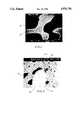
- FIG. 1is a rendering of an actual photomicrograph (magnified 150 ⁇ ) of a section of biomaterial of this invention showing the calcium carbonate base portion and calcium phosphate or hydroxyapatite surface layer;
- FIG. 2is a rendering of an actual photomicrograph (160 ⁇ ) showing a cross-section of an implant made from the biomaterial of this invention that had been implanted several months earlier in an animal.
- Hydroxyapatiteis widely used as a bone substitute material in oral, periodontal and craniofacial surgery, and is under investigation for various orthopedic applications, such as bone replacements due to trauma, spinal fusions, tumors, joint surgery and the like.
- the biocompatibility of hydroxyapatiteis well established and it is available commercially, mostly for oral surgery applications, in dense and porous forms. Hydroxyapatite promotes bone ingrowth in and around the implant, but even the porous form is resorbable only at a rate of 1-2 percent annually. Dense hydroxyapatite is essentially nonresorbable over a period of years.
- the resulting produced synthetic phosphate (hydroxyapatite or whitlockite) surfaced skeletal materialpossesses substantially the same microstructure of the original carbonate skeletal material from which it was derived.
- These synthetic materialsare useful for the manufacture of prosthetic devices, such as body and bone implants, tooth fixation, massive hard tissue replacements and the like since hydroxyapatite and whitlockite are biocompatible materials.
- the thin layer of hydroxyapatiteresorbs slowly after allowing bone and other tissue to grow into the pores during an initial repair period during which the surrounding bone can form a repair network. After the hydroxyapatite resorbs enough to expose the underlying base of calcium carbonate, the degradation process speeds up owing to the more rapid degradation of calcium carbonate as compared with hydroxyapatite. This allows even more bone ingrowth to occur, eventually permitting complete replacement of the artificial part with new bone and other tissue.
- the synthetic phosphate materials prepared in accordance with this inventionare particularly useful as biomaterials for use in the manufacture of prosthetic devices or for use as implants in human hard tissue and the like.
- the surface of the materials of this inventionparticularly those made from porous carbonate (aragonite) skeletal material of marine life, since they are comprised predominantly of hydroxyapatite Ca 10 (PO 4 )6(OH) 2 with some carbonate (CO 3 ) present, approximate the composition of the inorganic component of human hard tissue, i.e., human bone.
- This hydroxyapatite surfacehas osteophilic and osteoconductive properties, and helps promote the growth of bone tissue into the porosity of the biomaterial.
- Materials of this inventionwould preferably have a microstructure which is porous, completely interconnected, approximating the same pore size as cancellous human bone which would allow permeation of body fluids and blood cells thereinto.
- Materials in accordance with this inventioncould be prepared which would be suitable for root portions of tooth implants and mandibular restorations where it would permit rapid ingrowth of periodontal and hard tissue, as well as other bone repair functions such as segmental bone replacements for bone fractures, tumors, joint surgery and spinal fusion.
- porous carbonate skeletal materialsparticularly porous carbonate skeletal material of marine life
- Particularly usefulis the carbonate skeletal material of scleractinian coral Porites wherein the skeletal material is composed of the carbonate aragonite, and the average pore size is approximately 200 microns.
- Other corals of the genera Goniopora, Alveopora, Acropora and othersmay be suitable employed in the practice of this invention as the source of the carbonate skeletal material for conversion by hydrothermal chemical exchange with a phosphate into hydroxyapatite.
- Gonioporahas an average pore size of about 500 microns, and includes pores ranging in size from 5 microns to about 1000 microns.
- the carbonate skeletal materialis made up of a calcite carbonate marine skeletal material, such as echinoid spine calcite where the calcite contains a substantial amount of magnesium associated therewith
- whitlockiteis produced upon hydrothermal chemical exchange with a phosphate on the surface of the biomaterial.
- calcite carbonate marine skeletal materialsuch as echinoid spine calcite where the calcite contains a substantial amount of magnesium associated therewith
- whitlockiteis produced upon hydrothermal chemical exchange with a phosphate on the surface of the biomaterial.
- hydroxyapatite and whitlockiteare useful materials, with the hydroxyapatite being preferred for the manufacture of a prosthetic device and the like.
- granules having an average nominal diameter of about 425-600 microns and an average pore size of about 200 micronsshould be used; for reconstruction of the aveolar ridge, granules having an average nominal diameter of about 425 to 1000 microns and an average pore size of about 200 microns can be used. For orthopedic applications, larger granules having an average nominal diameter of 1-2 mm or 3- 5 mm can be used.
- porous carbonate skeletal materialIn the manufacture of the synthetic materials of this invention it would be desirable, before subjecting the naturally occurring porous carbonate skeletal material to hydrothermal chemical exchange with a phosphate, to first prepare the porous carbonate skeletal material by the removal of any organic material therefrom.
- a suitable technique for the removal of organic material from the porous skeletal materialwould be by immersion in a dilute (about 5%) aqueous solution of sodium hypochlorite. Usually an immersion time of about 30 hours is satisfactory for the removal of substantially all of the organic matter. Following this the material is rinsed, preferably in deionized water, and dried, such as at a temperature of about 90° C.
- the organic-free carbonate skeletal material after conversion by hydrothermal chemical exchange with a phosphate to hydroxyapatite or whitlockite, if not already shapedmay be shaped into a desired form or structure, for example, cylinders, screws, nuts, bolts, pins, flat or curved plates and the like.
- the conversion of porous carbonate skeletal materials into the phosphate surfaced carbonate biomaterials of the present inventionpreferably involves lower temperature and pressures than those disclosed in U.S. Pat. No. 3,929,971.
- the conversionmay be carried out by placing blocks or granules of calcium carbonate in phosphate solution or by freeze drying the phosphate onto the carbonate base and then carrying out the hydroconversion in a steam filled autoclave.
- Preferred temperaturesrange from about 200°-250° C., with about 200°-230° C. appearing optimum.
- the pressureshould be that developed in a sealed vessel or autoclave by the steam contained therein, which is estimated to be about 500 to about 4000 p.s.i.
- the temperatureshould preferably be about 230° C. and the pressure should be preferably about 1000 p.s.i., and the reaction should be carried out for about 10 to about 60 hours.
- substantially water-soluble phosphatesmay be employed as the phosphate contributing reactant in the hydrothermal chemical exchange reaction to produce the special materials of this invention.
- the preferred phosphatesinclude ammonium phosphates and orthophosphates. Also useful would be the calcium orthophosphates and the acid phosphates, as well as orthophosphoric acid including its hydrates and derivatives and mixtures of a weak acid, such as acetic acid, with a phosphate.
- orthophosphates and acid phosphatesuseful in the practices of this invention include Li 3 (PO 4 ), LiH 2 (PO 4 ), Na 3 (PO 4 ), Na 2 HPO 4 , Na 3 H 3 (PO 4 ) 2 , NaH 2 (PO 4 ), Na 4 H 5 (PO 4 ) 3 , NaH 5 (PO 4 ) 2 , K 3 PO 4 , K 2 HPO 4 , K 7 H 5 (PO 4 ) 4 , K 5 H 4 (PO 4 ) 3 , KH 2 (PO 4 ), KH 5 (PO 4 ) 2 , (HN 4 ) 3 PO 4 , (NH 4 ) 2 HPO 4 , NH 4 H 2 PO 4 , NH 4 H 5 (PO 4 ) 2 , NH 4 H 8 (PO 4 ) 3 , and their hydrates, and mixed salts especially of K, NH 4 and Na orthophosphates and acid phosphates, including also Rb and Cs orthophosphates and acid phosphates. Also useful in
- a cylinder 7/8 inch diameter by one inchwas machined from a head of Porites coral.
- the Porites coral cylinderwas cleaned ultrasonically to remove machining debris then rinsed and dried.
- the dried cylinderweighed 16.7 gm and fit into the Teflon liner of a test size reaction vessel
- To the dry Teflon liner (87.0 gm)was added 7.6 gm distilled H 2 O and 5.6 gm (NH 4 ) 2 HPO 4 ).
- the Teflon liner and contentswere preheated in 80° C. oven and contents stirred to dissolve phosphate.
- the coral cylinder prepared abovewas lowered into the 80° C.
- the Teflon liner with contentswas placed in preheated stainless steel vessel and sealed.
- the sealed vesselwas placed in a 220° C. oven and held at 220° C. for 12 hours. The vessel was allowed to cool down after which it was opened. After rinsing in distilled water and drying, the weight of hydroxyapatite-coated coral was 16.4 gm. Stereoptic microscope examination revealed excellent pore fidelity and no cracks.
- samples of coraleither Porites or Goniopora are cut with dimensions varying from 8 mm ⁇ 8 mm ⁇ 3 mm to 30 mm ⁇ 70 mm ⁇ x 15 mm rods or any other desired shape.
- the coralis cleaned by immersion in standard chlorine bleach (sodium hypochlorite) for 24 hours, then rinsed several times in water, and then completely dried.
- the blocks of coralare then weighed.
- Solutions of ammonium dibasic phosphate(NH 4 ) 2 HPO 4 ) approximately 5-40 percent by weight (Baker Chemicals, Catalog #0784-05) are made by dissolving the salt in deionized water.
- the dry blocks of coralare individually weighed and placed in separate polyethylene bags with sealable tops.
- An ammonium phosphate solutionis then piped into the bags to totally immerse the blocks.
- the bagsare transferred to a vacuum chamber and the blocks are degassed to fully infiltrate the solution into the pores.
- the tops to the bagsare then closed, making sure that the blocks remain fully submerged.
- the bagsare then transferred to a conventional freezer (approximately 15° C.) for approximately 24 hours to freeze the blocks.
- the frozen blocks and solutionare then removed from the bags and placed in a freeze-drying chamber. Freeze-drying is performed in a vacuum (less than 0.1 Torr) at a temperature of 35° C. for at least 24 hours. The excess dried ammonium phosphate crust around the blocks is then removed from the surface by scraping. The blocks are weighed and the percent weight gain is determined.
- Carbonate to phosphate substitution by hydrothermal conversionis then performed using a 750 ml high pressure autoclave (Berghof America, Catalog #7400) having a Teflon liner, filled with approximately 200 ml of deionized water.
- a Teflon platformis placed on the bottom of the liner such that the upper surface is above the waterline.
- the blocksare then stacked on the platform with Teflon webbing acting as a spacer between successive layers of the blocks.
- Teflon webbingacting as a spacer between successive layers of the blocks.
- Species of coral and concentrations of ammonium phosphatecan be mixed without cross-contamination.
- the top to the conversion vesselis closed and the vessel is placed in a conventional convection oven (Blue M, Catalog #POM7-136F-3).
- the temperatureis gradually raised to 230° and held there for about 60 hours.
- a pressure of about 1000 psiis generated by the vapor pressure of steam and the reactants at the stated temperature.
- the reaction vesselis opened and blocks removed.
- the thickness of the coatinghas been observed to be directly proportional to the concentration of ammonium phosphate solution used at the immersion step and to the weight gain for each of the two species of coral.
- the Goniopora coralresults in a thicker coating than the Porites coral for the same concentration of ammonium phosphate, because the Goniopora has a larger void fraction and a smaller specific surface area.
- the thickness of the hydroxyapatite-coatingis dependent on the concentration of ammonium phosphate used with the freeze dried treatment.
- the thicknesses of the coating achieved experimentally on Porites coralare:
- the thickness on the Goniopora coralis a follows:
- FIG. 1is an example of a photomicrograph from a scanning electron microscope with backscatter detector illustrating as sample of a porous biomaterial made from Porites coral.
- a distinct surface layer of phosphate 10was present on all surfaces of the calcium carbonate 12, and appeared uniform throughout the structure.
- the thickness of the hydroxyapatite layer 10was directly proportional to the concentration of the ammonium dibasic phosphate solution used to fabricate the biomaterial.
- the unique porous microstructure of the coralwas preserved.
- granules of either solid calcium carbonate (Mallinkrodt Chemicals, Catalog 6210) or porous calcium carbonate derived from corals (Porites, 425-1000 ⁇ m in diameter and Goniopora, 0.5 mm in diameter)are placed in plastic bags as described above.
- the ammonium phosphate ((NH 4 ) 2 HPO 4 )is added, frozen and freeze-dried.
- Hydrothermal conversionis accomplished by placing the freeze-dried granules in porous Teflon bags or in separate Teflon beakers, and then heating the sample in a closed container as discussed above.
- Another embodiment of the present inventioncombines the osteophyllic and osteoconductive properties of hydroxyapatites with biocompatible polymers used as implants.
- a hydroxyapatite coated porous calcium carbonate compositeis prepared as described above.
- the porosity of the compositeis filled with polymer either with positive injection pressure or by vacuum impregnation.
- polymers suitable for the practice of this inventioninclude polysulfone, polyethylene, such as ultrahigh molecular weight polyethylene, silicone rubber (Dow Corning) or polyurethane (Thermedics Inc., Tecoflex).
- the compositemay optionally be trimmed on all surfaces to expose the calcium carbonate structure.
- the compositeis then immersed in 10% acetic acid. This preferentially dissolves the calcium carbonate leaving behind the hydroxyapatite and polymer.
- An interconnected porous structureremains that is lined with hydroxyapatite and has an infrastructure of the polymer.
- the calcium carbonateis not dissolved away, or only partially dissolved away. After implantation in the body, however, the body preferentially degrades the calcium carbonate leaving the hydroxyapatite coating which degrades more slowly and the polymer.
- the porosity of the hydroxyapatite coated compositemay be filled with a polymer which may be degraded by the body after implantation.
- a polymerwhich may be degraded by the body after implantation.
- examples of such polymersinclude polylactic, polyglycolic acid or polycaprolactone (Union Carbide).
- the calcium carbonatemay be removed or left intact, depending upon the desired properties of the implant. The polymer in such an implant degrades after implantation, as does the calcium carbonate, when present. The dissolution of polymer and calcium carbonate provides additional space for bone or tissue ingrowth.
- the biomaterials of the present inventionprovide several important and unique advantages.
- the hydroxyapatite surface layerdegrades slowly as compared to calcium carbonate and helps modulate degradation.
- the implantwill degrade only slowly at first, allowing the bone or other tissue to fill the interconnected porous network. Thus ingrowth can occur prior to resorbtion.
- FIG. 2illustrates an implant 18 of the biomaterial of the present invention made by hydroconversion of Goniopora with 5% ammonium phosphate, which was implanted for approximately 12 weeks in a rabbit tibia.
- the implant 18includes the calcium carbonate base 20 and the hydroxyapatite or phosphate surface 22 surrounded by bone 24.
- FIG. 2once cracks or fissures appear in the hydroxyapatite surface 22 exposing the underlying calcium carbonate 20, degradation accelerates since calcium carbonate appears to degrade more rapidly than does hydroxyapatite. Bone 24 can be seen replacing the space 20a formerly filled with calcium carbonate 20.
- hydroxyapatite layer in the biomaterial of the present inventionis its inherent osteophilic nature. That is, hydroxyapatite on the surface of a porous implant seems to promote bone ingrowth into the pores of the implant, whereas calcium carbonate seems not to possess this property
- hydroxyapatiteis absorbency, which may explain its ability to bind other compositions which aid in the bone repair process.
- An antibioticsuch as tetracycline, oxytetracycline or other known synthetic or semisynthetic antibiotic may be introduced unto the pore cavities of the implant.
- one of several growth factorssuch as transforming growth factor or one of the Bone Morphogenic Proteins can be attached which help promote bone ingrowth.
- transforming growth factor ⁇TGF- ⁇
- TGF- ⁇can be added to the hydroxyapatite surface after hydroconversion to help enhance bone ingrowth.
- growth factor or an antibioticcan be intermixed with a preferably biodegradable polymer and injected or vacuum infiltrated into the porosity of the phosphate surfaced carbonate biomaterial.
- hydroxyapatite surface layercan be accomplished by a method other than hydroconversion. Therefore it is intended that the scope of the invention be governed by the following claims.
Landscapes
- Health & Medical Sciences (AREA)
- Chemical & Material Sciences (AREA)
- Life Sciences & Earth Sciences (AREA)
- Medicinal Chemistry (AREA)
- General Health & Medical Sciences (AREA)
- Public Health (AREA)
- Veterinary Medicine (AREA)
- Oral & Maxillofacial Surgery (AREA)
- Transplantation (AREA)
- Epidemiology (AREA)
- Dermatology (AREA)
- Animal Behavior & Ethology (AREA)
- Engineering & Computer Science (AREA)
- Composite Materials (AREA)
- Materials Engineering (AREA)
- Inorganic Chemistry (AREA)
- Biomedical Technology (AREA)
- Molecular Biology (AREA)
- Dispersion Chemistry (AREA)
- Materials For Medical Uses (AREA)
- Dental Prosthetics (AREA)
- Dental Preparations (AREA)
- Prostheses (AREA)
- Compounds Of Alkaline-Earth Elements, Aluminum Or Rare-Earth Metals (AREA)
Abstract
Description
10 CaCO.sub.3 +6(NH.sub.4).sub.2 HPO.sub.4 +2HO.sub.2
Ca.sub.10 (PO.sub.4).sub.6 (OH).sub.2 +6 (NH.sub.4).sub.2 CO.sub.3 +4H.sub.2 CO.sub.3
______________________________________ % HA Solution Thickness of Coating (μm) Range (μm) ______________________________________ 5% 0.8 0.6-1.2 10% 2.0 1.2-2.5 20% 3.4 3.1-3.8 30% 4.7 3.7-5.6 40% 6.19 6.2-7.5 ______________________________________
______________________________________ % Ammonium Phosphate Thickness of Coating Range (μm) ______________________________________ 5% 3.8 3.1-4.4 10% 5.6 5.0-6.3 20% 10.6 10.0-11.2 30% 13.7 12.5-15.0 40% 20.6 18.7-22.5 ______________________________________
TABLE 1 __________________________________________________________________________Center Core Analysis __________________________________________________________________________Accelerating voltage 20.0 KeV Beam - sample incidence angle 70.0 degrees Xray emergence angle 29.4 degrees Xray - window incidence angle 9.1 degrees Window thickness 12.0 microns __________________________________________________________________________STANDARDLESS EDS ANALYSIS (ZAF CORRECTIONS VIA MAGIC V) ELEMENT WEIGHT ATOMIC PRECISION & LINE PERCENT PERCENT* 3 SIGMA K-RATIO** ITER __________________________________________________________________________P KA 0.36 0.46 0.11 0.0028 Ca KA 99.84 99.54 0.51 0.9972 2 TOTAL 100.00 __________________________________________________________________________ *NOTE: ATOMIC PERCENT is normalized to 100 **NOTE: KRATIO = KRATIO × R where R reference(standard)/reference(sample) NORMALIZATION FACTOR: 0.998
TABLE 2 __________________________________________________________________________Surface Analysis __________________________________________________________________________Accelerating voltage 20.0 KeV Beam - sample incidence angle 70.0 degrees Xray emergence angle 29.4 degrees Xray - window incidence angle 9.1 degrees Window thickness 12.0 microns __________________________________________________________________________STANDARDLESS SKS ANALYSIS (ZAF CORRECTIONS VIA MAGIC V) ELEMENT WEIGHT ATOMIC PRECISION & LINE PERCENT PERCENT* 3 SIGMA K-RATIO ITER __________________________________________________________________________P KA 34.96 41.02 0.38 0.3364 Ca KA 65.04 58.98 0.49 0.6636 4 TOTAL 100.00 __________________________________________________________________________ *NOTE: ATOMIC PERCENT is normalized to 100 **NOTE: KRATIO = KRATIO × R where R = reference(standard)/reference(sample) NORMALIZATION FACTOR: 0.882
Claims (32)
Priority Applications (10)
| Application Number | Priority Date | Filing Date | Title |
|---|---|---|---|
| US07/345,194US4976736A (en) | 1989-04-28 | 1989-04-28 | Coated biomaterials and methods for making same |
| CA002013420ACA2013420C (en) | 1989-04-28 | 1990-03-29 | Coated biomaterials and methods for making same |
| JP2111624AJP3035316B2 (en) | 1989-04-28 | 1990-04-26 | Coated biological material and method for producing the biological material |
| ZA903187AZA903187B (en) | 1989-04-28 | 1990-04-26 | Coated biomaterials and methods for making same |
| EP90201079AEP0395187B1 (en) | 1989-04-28 | 1990-04-27 | Coated biomaterials and methods for making same |
| DE69024993TDE69024993T2 (en) | 1989-04-28 | 1990-04-27 | Coated biomaterials and processes for their production |
| DK90201079.2TDK0395187T3 (en) | 1989-04-28 | 1990-04-27 | Coated biomaterials and processes for their preparation |
| AT90201079TATE133340T1 (en) | 1989-04-28 | 1990-04-27 | COATED ORGANIC MATERIALS AND METHODS FOR THEIR PRODUCTION |
| ES90201079TES2081906T3 (en) | 1989-04-28 | 1990-04-27 | COATED BIOMATERIALS AND METHODS TO MANUFACTURE THEM. |
| GR950403370TGR3018784T3 (en) | 1989-04-28 | 1996-01-25 | Coated biomaterials and methods for making same |
Applications Claiming Priority (1)
| Application Number | Priority Date | Filing Date | Title |
|---|---|---|---|
| US07/345,194US4976736A (en) | 1989-04-28 | 1989-04-28 | Coated biomaterials and methods for making same |
Publications (1)
| Publication Number | Publication Date |
|---|---|
| US4976736Atrue US4976736A (en) | 1990-12-11 |
Family
ID=23353975
Family Applications (1)
| Application Number | Title | Priority Date | Filing Date |
|---|---|---|---|
| US07/345,194Expired - LifetimeUS4976736A (en) | 1989-04-28 | 1989-04-28 | Coated biomaterials and methods for making same |
Country Status (10)
| Country | Link |
|---|---|
| US (1) | US4976736A (en) |
| EP (1) | EP0395187B1 (en) |
| JP (1) | JP3035316B2 (en) |
| AT (1) | ATE133340T1 (en) |
| CA (1) | CA2013420C (en) |
| DE (1) | DE69024993T2 (en) |
| DK (1) | DK0395187T3 (en) |
| ES (1) | ES2081906T3 (en) |
| GR (1) | GR3018784T3 (en) |
| ZA (1) | ZA903187B (en) |
Cited By (127)
| Publication number | Priority date | Publication date | Assignee | Title |
|---|---|---|---|---|
| US5071434A (en)* | 1990-04-20 | 1991-12-10 | Ngk Spark Plug Co., Ltd. | Biologically active surface ceramic and process for producing the same |
| US5207710A (en)* | 1988-09-29 | 1993-05-04 | Collagen Corporation | Method for improving implant fixation |
| WO1994024956A1 (en)* | 1993-04-27 | 1994-11-10 | Dental Marketing Specialists, Inc. | Method and apparatus for installing dental implants |
| WO1995001760A3 (en)* | 1993-07-02 | 1995-03-02 | Dental Marketing Spec Inc | Method and apparatus for installation of implant |
| US5433751A (en)* | 1992-04-03 | 1995-07-18 | Inoteb | Bone prosthesis material containing calcium carbonate particles dispersed in a bioresorbable polymer matrix |
| US5455100A (en)* | 1991-01-30 | 1995-10-03 | Interpore International | Porous articles and methods for producing same |
| US5503558A (en)* | 1993-11-12 | 1996-04-02 | Mcgill University | Osseointegration promoting implant composition, implant assembly and method therefor |
| US5522896A (en)* | 1989-02-15 | 1996-06-04 | Xomed, Inc. | Biocompatible composite material |
| US5554188A (en)* | 1993-04-29 | 1996-09-10 | Xomed, Inc. | Universal middle ear prosthesis |
| US5591453A (en)* | 1994-07-27 | 1997-01-07 | The Trustees Of The University Of Pennsylvania | Incorporation of biologically active molecules into bioactive glasses |
| US5697981A (en)* | 1994-08-23 | 1997-12-16 | Norian Corporation | Method for repairing bone |
| US5697980A (en)* | 1991-04-19 | 1997-12-16 | Mitsubishi Chem Corp | Artificial filling and prosthetic material |
| US5716414A (en)* | 1994-04-28 | 1998-02-10 | Johnson & Johnson Professional, Inc. | Ceramic/metallic articulation component and prothesis |
| US5814104A (en)* | 1993-11-26 | 1998-09-29 | Beoni; Franco | Middle ear ossicular chain prosthesis, with a porous hydroxylapatite flange |
| US5817327A (en)* | 1994-07-27 | 1998-10-06 | The Trustees Of The University Of Pennsylvania | Incorporation of biologically active molecules into bioactive glasses |
| US5882631A (en)* | 1997-04-24 | 1999-03-16 | Sunstar Inc. | Oral composition |
| US5888067A (en)* | 1997-08-15 | 1999-03-30 | Gibbs; David | Dental implant |
| WO1999037246A1 (en)* | 1998-01-22 | 1999-07-29 | Perry Arthur C | Biocompatible structure comprising means for enhanced fibrovascular ingrowth |
| US5935172A (en)* | 1996-06-28 | 1999-08-10 | Johnson & Johnson Professional, Inc. | Prosthesis with variable fit and strain distribution |
| US6008430A (en)* | 1991-01-30 | 1999-12-28 | Interpore Orthopaedics, Inc. | Three-dimensional prosthetic articles and methods for producing same |
| US6136029A (en)* | 1997-10-01 | 2000-10-24 | Phillips-Origen Ceramic Technology, Llc | Bone substitute materials |
| US6248130B1 (en) | 1991-09-30 | 2001-06-19 | Arthur C. Perry | Pegs for orbital implants |
| US6296667B1 (en) | 1997-10-01 | 2001-10-02 | Phillips-Origen Ceramic Technology, Llc | Bone substitutes |
| US6342051B1 (en)* | 1997-06-12 | 2002-01-29 | Gholam A. Peyman | Treatment of anoxic tissue with angiogenesis-inducing implants |
| US6379740B1 (en)* | 1997-12-10 | 2002-04-30 | Sorin Biomedica Cardio S.P.A. | Method for treating a prosthesis having an apertured structure and associated devices |
| WO2002040398A1 (en)* | 2000-11-16 | 2002-05-23 | University Of Technology, Sydney | Processes for treating coral and coating an object |
| US6540784B2 (en) | 1994-08-08 | 2003-04-01 | Board Of Regents, The University Of Texas System | Artificial bone implants |
| US6644971B1 (en)* | 1998-10-23 | 2003-11-11 | Franck Rigoulet | Biological material for treating periodontal diseases |
| US20040078087A1 (en)* | 2002-08-30 | 2004-04-22 | Soo-Ryong Kim | Porous hydroxy apatite containing silicon and magnesium, and a preparation method thereof |
| US6743462B1 (en)* | 2001-05-31 | 2004-06-01 | Advanced Cardiovascular Systems, Inc. | Apparatus and method for coating implantable devices |
| US6881227B2 (en)* | 1999-11-30 | 2005-04-19 | Margarita Jordanova-Spassova | Hydroxylapatite material containing tricalcium phosphate with microporous structure |
| US20050177237A1 (en)* | 2001-04-12 | 2005-08-11 | Ben Shappley | Spinal cage insert, filler piece and method of manufacturing |
| US20050201987A1 (en)* | 2004-03-09 | 2005-09-15 | Inion Ltd. | Bone grafting material, method and implant |
| US20060121084A1 (en)* | 2004-12-08 | 2006-06-08 | Borden Mark D | Continuous phase composite for musculoskeletal repair |
| US20060135938A1 (en)* | 2004-12-17 | 2006-06-22 | Mckay William F | Device and method for the vacuum infusion of a porous medical implant |
| US7122057B2 (en) | 2001-04-12 | 2006-10-17 | Therics, Llc | Method and apparatus for engineered regenerative biostructures such as hydroxyapatite substrates for bone healing applications |
| US20080233203A1 (en)* | 2007-03-21 | 2008-09-25 | Jennifer Woodell-May | Porous orthapedic materials coated with demineralized bone matrix |
| US20080306554A1 (en)* | 2007-06-11 | 2008-12-11 | Mckinley Laurence M | Osseointegration and biointegration coatings for bone screw implants |
| US20090149569A1 (en)* | 2007-07-19 | 2009-06-11 | Shastri V Prasad | Surface engineering of tissue graft materials for enhanced porosity and cell adhesion |
| US7572336B2 (en) | 2002-12-12 | 2009-08-11 | Advanced Cardiovascular Systems, Inc. | Clamp mandrel fixture and a method of using the same to minimize coating defects |
| US20090227704A1 (en)* | 2008-03-05 | 2009-09-10 | Karen Troxel | Cohesive and compression resistant demineralized bone carrier matrix |
| US7601165B2 (en) | 2006-09-29 | 2009-10-13 | Biomet Sports Medicine, Llc | Method and apparatus for forming a self-locking adjustable suture loop |
| US7608092B1 (en) | 2004-02-20 | 2009-10-27 | Biomet Sports Medicince, LLC | Method and apparatus for performing meniscus repair |
| US7608098B1 (en) | 2004-11-09 | 2009-10-27 | Biomet Sports Medicine, Llc | Bone fixation device |
| US7622070B2 (en) | 2005-06-20 | 2009-11-24 | Advanced Cardiovascular Systems, Inc. | Method of manufacturing an implantable polymeric medical device |
| US20100047318A1 (en)* | 2008-08-21 | 2010-02-25 | Biomet Manufacturing Corp. | Ceramic implants affording controlled release of active materials |
| AU2007221771B2 (en)* | 2000-11-16 | 2010-04-01 | Biotomo Pty Ltd | Processes for treating coral and coating an object |
| US7735449B1 (en) | 2005-07-28 | 2010-06-15 | Advanced Cardiovascular Systems, Inc. | Stent fixture having rounded support structures and method for use thereof |
| US7740794B1 (en) | 2005-04-18 | 2010-06-22 | Biomet Sports Medicine, Llc | Methods of making a polymer and ceramic composite |
| US7740897B2 (en) | 1997-10-01 | 2010-06-22 | Wright Medical Technology, Inc. | Process for producing rigid reticulated articles |
| US7749250B2 (en) | 2006-02-03 | 2010-07-06 | Biomet Sports Medicine, Llc | Soft tissue repair assembly and associated method |
| US7823533B2 (en) | 2005-06-30 | 2010-11-02 | Advanced Cardiovascular Systems, Inc. | Stent fixture and method for reducing coating defects |
| US7857830B2 (en) | 2006-02-03 | 2010-12-28 | Biomet Sports Medicine, Llc | Soft tissue repair and conduit device |
| US7867547B2 (en) | 2005-12-19 | 2011-01-11 | Advanced Cardiovascular Systems, Inc. | Selectively coating luminal surfaces of stents |
| US20110022180A1 (en)* | 2009-07-24 | 2011-01-27 | Warsaw Orthopedic, Inc. | Implantable medical devices |
| US7905903B2 (en) | 2006-02-03 | 2011-03-15 | Biomet Sports Medicine, Llc | Method for tissue fixation |
| US7905904B2 (en) | 2006-02-03 | 2011-03-15 | Biomet Sports Medicine, Llc | Soft tissue repair device and associated methods |
| US7909851B2 (en) | 2006-02-03 | 2011-03-22 | Biomet Sports Medicine, Llc | Soft tissue repair device and associated methods |
| US7914539B2 (en) | 2004-11-09 | 2011-03-29 | Biomet Sports Medicine, Llc | Tissue fixation device |
| US7959650B2 (en) | 2006-09-29 | 2011-06-14 | Biomet Sports Medicine, Llc | Adjustable knotless loops |
| US7985440B2 (en) | 2001-06-27 | 2011-07-26 | Advanced Cardiovascular Systems, Inc. | Method of using a mandrel to coat a stent |
| US7985441B1 (en) | 2006-05-04 | 2011-07-26 | Yiwen Tang | Purification of polymers for coating applications |
| US8003156B2 (en) | 2006-05-04 | 2011-08-23 | Advanced Cardiovascular Systems, Inc. | Rotatable support elements for stents |
| US8034090B2 (en) | 2004-11-09 | 2011-10-11 | Biomet Sports Medicine, Llc | Tissue fixation device |
| US8088130B2 (en) | 2006-02-03 | 2012-01-03 | Biomet Sports Medicine, Llc | Method and apparatus for coupling soft tissue to a bone |
| US8118836B2 (en) | 2004-11-05 | 2012-02-21 | Biomet Sports Medicine, Llc | Method and apparatus for coupling soft tissue to a bone |
| US8128658B2 (en) | 2004-11-05 | 2012-03-06 | Biomet Sports Medicine, Llc | Method and apparatus for coupling soft tissue to bone |
| US8137382B2 (en) | 2004-11-05 | 2012-03-20 | Biomet Sports Medicine, Llc | Method and apparatus for coupling anatomical features |
| WO2012112499A1 (en) | 2011-02-14 | 2012-08-23 | Biomet Manufacturing Corp. | Non- resorbable polymer - ceramic composite implant materials |
| US8251998B2 (en) | 2006-08-16 | 2012-08-28 | Biomet Sports Medicine, Llc | Chondral defect repair |
| US8298262B2 (en) | 2006-02-03 | 2012-10-30 | Biomet Sports Medicine, Llc | Method for tissue fixation |
| US8303604B2 (en) | 2004-11-05 | 2012-11-06 | Biomet Sports Medicine, Llc | Soft tissue repair device and method |
| US8317825B2 (en) | 2004-11-09 | 2012-11-27 | Biomet Sports Medicine, Llc | Soft tissue conduit device and method |
| US8343227B2 (en) | 2009-05-28 | 2013-01-01 | Biomet Manufacturing Corp. | Knee prosthesis assembly with ligament link |
| US8361113B2 (en) | 2006-02-03 | 2013-01-29 | Biomet Sports Medicine, Llc | Method and apparatus for coupling soft tissue to a bone |
| US8444699B2 (en) | 2010-02-18 | 2013-05-21 | Biomet Manufacturing Corp. | Method and apparatus for augmenting bone defects |
| US8500818B2 (en) | 2006-09-29 | 2013-08-06 | Biomet Manufacturing, Llc | Knee prosthesis assembly with ligament link |
| US8506597B2 (en) | 2011-10-25 | 2013-08-13 | Biomet Sports Medicine, Llc | Method and apparatus for interosseous membrane reconstruction |
| US8535357B2 (en)* | 2004-12-09 | 2013-09-17 | Biomet Sports Medicine, Llc | Continuous phase compositions for ACL repair |
| US8562647B2 (en) | 2006-09-29 | 2013-10-22 | Biomet Sports Medicine, Llc | Method and apparatus for securing soft tissue to bone |
| US8562645B2 (en) | 2006-09-29 | 2013-10-22 | Biomet Sports Medicine, Llc | Method and apparatus for forming a self-locking adjustable loop |
| US8574235B2 (en) | 2006-02-03 | 2013-11-05 | Biomet Sports Medicine, Llc | Method for trochanteric reattachment |
| US8597327B2 (en) | 2006-02-03 | 2013-12-03 | Biomet Manufacturing, Llc | Method and apparatus for sternal closure |
| US8652171B2 (en) | 2006-02-03 | 2014-02-18 | Biomet Sports Medicine, Llc | Method and apparatus for soft tissue fixation |
| US8652172B2 (en) | 2006-02-03 | 2014-02-18 | Biomet Sports Medicine, Llc | Flexible anchors for tissue fixation |
| US8672969B2 (en) | 2006-09-29 | 2014-03-18 | Biomet Sports Medicine, Llc | Fracture fixation device |
| CN103764180A (en)* | 2011-06-03 | 2014-04-30 | 德鲁有限责任公司 | Process for the preparation of implant coatings and corresponding implants |
| US8771352B2 (en) | 2011-05-17 | 2014-07-08 | Biomet Sports Medicine, Llc | Method and apparatus for tibial fixation of an ACL graft |
| US8801783B2 (en) | 2006-09-29 | 2014-08-12 | Biomet Sports Medicine, Llc | Prosthetic ligament system for knee joint |
| US8840645B2 (en) | 2004-11-05 | 2014-09-23 | Biomet Sports Medicine, Llc | Method and apparatus for coupling soft tissue to a bone |
| US8936621B2 (en) | 2006-02-03 | 2015-01-20 | Biomet Sports Medicine, Llc | Method and apparatus for forming a self-locking adjustable loop |
| US8968364B2 (en) | 2006-02-03 | 2015-03-03 | Biomet Sports Medicine, Llc | Method and apparatus for fixation of an ACL graft |
| US8998949B2 (en) | 2004-11-09 | 2015-04-07 | Biomet Sports Medicine, Llc | Soft tissue conduit device |
| US9017381B2 (en) | 2007-04-10 | 2015-04-28 | Biomet Sports Medicine, Llc | Adjustable knotless loops |
| US9078955B2 (en) | 2010-10-26 | 2015-07-14 | Cap Biomaterials, Llc | Composites of hydroxyapatite and calcium carbonate and related methods of preparation and use |
| US9078644B2 (en) | 2006-09-29 | 2015-07-14 | Biomet Sports Medicine, Llc | Fracture fixation device |
| US9149267B2 (en) | 2006-02-03 | 2015-10-06 | Biomet Sports Medicine, Llc | Method and apparatus for coupling soft tissue to a bone |
| US9192459B2 (en) | 2000-01-14 | 2015-11-24 | Bonutti Skeletal Innovations Llc | Method of performing total knee arthroplasty |
| US9259217B2 (en) | 2012-01-03 | 2016-02-16 | Biomet Manufacturing, Llc | Suture Button |
| US9271713B2 (en) | 2006-02-03 | 2016-03-01 | Biomet Sports Medicine, Llc | Method and apparatus for tensioning a suture |
| US9314241B2 (en) | 2011-11-10 | 2016-04-19 | Biomet Sports Medicine, Llc | Apparatus for coupling soft tissue to a bone |
| US9357991B2 (en) | 2011-11-03 | 2016-06-07 | Biomet Sports Medicine, Llc | Method and apparatus for stitching tendons |
| US9370350B2 (en) | 2011-11-10 | 2016-06-21 | Biomet Sports Medicine, Llc | Apparatus for coupling soft tissue to a bone |
| US9381013B2 (en) | 2011-11-10 | 2016-07-05 | Biomet Sports Medicine, Llc | Method for coupling soft tissue to a bone |
| US9538998B2 (en) | 2006-02-03 | 2017-01-10 | Biomet Sports Medicine, Llc | Method and apparatus for fracture fixation |
| US9615822B2 (en) | 2014-05-30 | 2017-04-11 | Biomet Sports Medicine, Llc | Insertion tools and method for soft anchor |
| US9700291B2 (en) | 2014-06-03 | 2017-07-11 | Biomet Sports Medicine, Llc | Capsule retractor |
| US9757119B2 (en) | 2013-03-08 | 2017-09-12 | Biomet Sports Medicine, Llc | Visual aid for identifying suture limbs arthroscopically |
| US9801708B2 (en) | 2004-11-05 | 2017-10-31 | Biomet Sports Medicine, Llc | Method and apparatus for coupling soft tissue to a bone |
| WO2018047177A1 (en)* | 2016-09-08 | 2018-03-15 | B. G. Negev Technologies And Applications Ltd., At Ben-Gurion University | Porous mineral nucleus and a metal shell |
| US9918827B2 (en) | 2013-03-14 | 2018-03-20 | Biomet Sports Medicine, Llc | Scaffold for spring ligament repair |
| US9918826B2 (en) | 2006-09-29 | 2018-03-20 | Biomet Sports Medicine, Llc | Scaffold for spring ligament repair |
| US9955980B2 (en) | 2015-02-24 | 2018-05-01 | Biomet Sports Medicine, Llc | Anatomic soft tissue repair |
| US10039543B2 (en) | 2014-08-22 | 2018-08-07 | Biomet Sports Medicine, Llc | Non-sliding soft anchor |
| US10136886B2 (en) | 2013-12-20 | 2018-11-27 | Biomet Sports Medicine, Llc | Knotless soft tissue devices and techniques |
| US10517587B2 (en) | 2006-02-03 | 2019-12-31 | Biomet Sports Medicine, Llc | Method and apparatus for forming a self-locking adjustable loop |
| CN111886030A (en)* | 2018-03-22 | 2020-11-03 | 斯旺西大学 | Bone graft substitute and method of making the same |
| US20210008121A1 (en)* | 2019-07-10 | 2021-01-14 | Access2bone IP BV | Enhanced osteogenic composition |
| US10912551B2 (en) | 2015-03-31 | 2021-02-09 | Biomet Sports Medicine, Llc | Suture anchor with soft anchor of electrospun fibers |
| US11259794B2 (en) | 2006-09-29 | 2022-03-01 | Biomet Sports Medicine, Llc | Method for implanting soft tissue |
| US11259792B2 (en) | 2006-02-03 | 2022-03-01 | Biomet Sports Medicine, Llc | Method and apparatus for coupling anatomical features |
| US11311287B2 (en) | 2006-02-03 | 2022-04-26 | Biomet Sports Medicine, Llc | Method for tissue fixation |
| US12096928B2 (en) | 2009-05-29 | 2024-09-24 | Biomet Sports Medicine, Llc | Method and apparatus for coupling soft tissue to a bone |
| US12245759B2 (en) | 2008-08-22 | 2025-03-11 | Biomet Sports Medicine, Llc | Method and apparatus for coupling soft tissue to bone |
| CN119873770A (en)* | 2025-01-10 | 2025-04-25 | 慧康智园医疗器械(西安)有限公司 | Coral hydroxyapatite and preparation method and application thereof |
| US12329373B2 (en) | 2011-05-02 | 2025-06-17 | Biomet Sports Medicine, Llc | Method and apparatus for soft tissue fixation |
| US12419632B2 (en) | 2008-08-22 | 2025-09-23 | Biomet Sports Medicine, Llc | Method and apparatus for coupling anatomical features |
Families Citing this family (23)
| Publication number | Priority date | Publication date | Assignee | Title |
|---|---|---|---|---|
| GB1186481A (en)* | 1968-03-25 | 1970-04-02 | Parke Davis & Co | New Pyrrolidine Compounds and Methods for their Production |
| US5422340A (en)* | 1989-09-01 | 1995-06-06 | Ammann; Arthur J. | TGF-βformulation for inducing bone growth |
| CA2151486A1 (en)* | 1993-01-12 | 1994-07-21 | Arthur J. Ammann | Tgf-beta formulation for inducing bone growth |
| FR2706308B1 (en)* | 1993-02-05 | 1995-09-08 | Camprasse Georges | Orthopedic prostheses and pearl biological sealing cement. |
| NO940913L (en)* | 1993-03-26 | 1994-09-27 | Bristol Myers Squibb Co | Controlled Release Preparations of Biologically Active TGF |
| FR2706768B1 (en)* | 1993-05-13 | 1995-12-01 | Inoteb | |
| FR2705235B1 (en)* | 1993-05-13 | 1995-07-13 | Inoteb | Use of particles of a biocompatible and bioresorbable calcium salt as an active ingredient in the preparation of a medicament for the local treatment of demineralizing bone diseases. |
| US6376573B1 (en)* | 1994-12-21 | 2002-04-23 | Interpore International | Porous biomaterials and methods for their manufacture |
| FR2728797B1 (en)* | 1994-12-30 | 1997-03-28 | Diami | STENT WITH CALCITE COATING AND PROCESS FOR OBTAINING |
| FR2743496B1 (en)* | 1996-01-15 | 1998-04-10 | Univ Rennes | MACROPOROUS COMPOSITE SUPPORT FOR DRUG SUBSTANCE (S) FOR USE AS BONE RECONSTRUCTION MATERIAL AND PROCESS FOR PREPARING THE SAME |
| JP2001509422A (en)* | 1997-07-07 | 2001-07-24 | オーストラリアン、インスティチュート、オブ、マリーン、サイエンス | Articles or structures for medical or related purposes |
| WO2000015273A1 (en) | 1998-09-11 | 2000-03-23 | Gerhard Schmidmaier | Biologically active implants |
| FI110062B (en)* | 1998-12-11 | 2002-11-29 | Antti Yli-Urpo | New composition and its use |
| US20040002770A1 (en)* | 2002-06-28 | 2004-01-01 | King Richard S. | Polymer-bioceramic composite for orthopaedic applications and method of manufacture thereof |
| US7384430B2 (en) | 2004-06-30 | 2008-06-10 | Depuy Products, Inc. | Low crystalline polymeric material for orthopaedic implants and an associated method |
| US7879275B2 (en) | 2004-12-30 | 2011-02-01 | Depuy Products, Inc. | Orthopaedic bearing and method for making the same |
| US7896921B2 (en) | 2004-12-30 | 2011-03-01 | Depuy Products, Inc. | Orthopaedic bearing and method for making the same |
| US7883653B2 (en) | 2004-12-30 | 2011-02-08 | Depuy Products, Inc. | Method of making an implantable orthopaedic bearing |
| KR100786312B1 (en)* | 2006-05-03 | 2007-12-17 | 박진우 | Method for preparing calcium phosphate and calcium phosphate produced thereby |
| EP1961433A1 (en)* | 2007-02-20 | 2008-08-27 | National University of Ireland Galway | Porous substrates for implantation |
| US8133553B2 (en) | 2007-06-18 | 2012-03-13 | Zimmer, Inc. | Process for forming a ceramic layer |
| US20090112315A1 (en)* | 2007-10-29 | 2009-04-30 | Zimmer, Inc. | Medical implants and methods for delivering biologically active agents |
| CN106709394B (en)* | 2016-12-12 | 2019-07-05 | 北京慧眼智行科技有限公司 | A kind of image processing method and device |
Citations (3)
| Publication number | Priority date | Publication date | Assignee | Title |
|---|---|---|---|---|
| US3929971A (en)* | 1973-03-30 | 1975-12-30 | Research Corp | Porous biomaterials and method of making same |
| US4356572A (en)* | 1979-07-12 | 1982-11-02 | Etablissement Public Dit: Agence Nationale De Valorisation De La Recherche (Anvar) | Biodegradable implant useable as a bone prosthesis |
| US4917702A (en)* | 1984-09-10 | 1990-04-17 | Hans Scheicher | Bone replacement material on the basis of carbonate and alkali containing calciumphosphate apatites |
Family Cites Families (3)
| Publication number | Priority date | Publication date | Assignee | Title |
|---|---|---|---|---|
| NL8401062A (en)* | 1984-04-04 | 1985-11-01 | Stichting Biomaterials Science | METHOD FOR PREPARING AN IMPLANT MATERIAL DELIVERING MEDICINES IN THE BODY |
| DE3542744C1 (en)* | 1985-12-03 | 1987-05-27 | Ewers Rolf | Porous hydroxyapatite material |
| US4861733A (en)* | 1987-02-13 | 1989-08-29 | Interpore International | Calcium phosphate bone substitute materials |
- 1989
- 1989-04-28USUS07/345,194patent/US4976736A/ennot_activeExpired - Lifetime
- 1990
- 1990-03-29CACA002013420Apatent/CA2013420C/ennot_activeExpired - Lifetime
- 1990-04-26JPJP2111624Apatent/JP3035316B2/ennot_activeExpired - Lifetime
- 1990-04-26ZAZA903187Apatent/ZA903187B/enunknown
- 1990-04-27EPEP90201079Apatent/EP0395187B1/ennot_activeExpired - Lifetime
- 1990-04-27ESES90201079Tpatent/ES2081906T3/ennot_activeExpired - Lifetime
- 1990-04-27DKDK90201079.2Tpatent/DK0395187T3/enactive
- 1990-04-27DEDE69024993Tpatent/DE69024993T2/ennot_activeExpired - Lifetime
- 1990-04-27ATAT90201079Tpatent/ATE133340T1/ennot_activeIP Right Cessation
- 1996
- 1996-01-25GRGR950403370Tpatent/GR3018784T3/enunknown
Patent Citations (3)
| Publication number | Priority date | Publication date | Assignee | Title |
|---|---|---|---|---|
| US3929971A (en)* | 1973-03-30 | 1975-12-30 | Research Corp | Porous biomaterials and method of making same |
| US4356572A (en)* | 1979-07-12 | 1982-11-02 | Etablissement Public Dit: Agence Nationale De Valorisation De La Recherche (Anvar) | Biodegradable implant useable as a bone prosthesis |
| US4917702A (en)* | 1984-09-10 | 1990-04-17 | Hans Scheicher | Bone replacement material on the basis of carbonate and alkali containing calciumphosphate apatites |
Cited By (297)
| Publication number | Priority date | Publication date | Assignee | Title |
|---|---|---|---|---|
| US5207710A (en)* | 1988-09-29 | 1993-05-04 | Collagen Corporation | Method for improving implant fixation |
| US5578086A (en)* | 1989-02-15 | 1996-11-26 | Xomed, Inc. | Prosthesis using biocompatible composite material |
| US5728157A (en)* | 1989-02-15 | 1998-03-17 | Xomed Surgical Products, Inc. | Biocompatible composite prostheses |
| US5522896A (en)* | 1989-02-15 | 1996-06-04 | Xomed, Inc. | Biocompatible composite material |
| US5071434A (en)* | 1990-04-20 | 1991-12-10 | Ngk Spark Plug Co., Ltd. | Biologically active surface ceramic and process for producing the same |
| US5728510A (en)* | 1991-01-30 | 1998-03-17 | Interpore International | Prosthetic articles and methods for producing same |
| US5455100A (en)* | 1991-01-30 | 1995-10-03 | Interpore International | Porous articles and methods for producing same |
| US6008430A (en)* | 1991-01-30 | 1999-12-28 | Interpore Orthopaedics, Inc. | Three-dimensional prosthetic articles and methods for producing same |
| US5697980A (en)* | 1991-04-19 | 1997-12-16 | Mitsubishi Chem Corp | Artificial filling and prosthetic material |
| US6248130B1 (en) | 1991-09-30 | 2001-06-19 | Arthur C. Perry | Pegs for orbital implants |
| US5433751A (en)* | 1992-04-03 | 1995-07-18 | Inoteb | Bone prosthesis material containing calcium carbonate particles dispersed in a bioresorbable polymer matrix |
| US5759033A (en)* | 1992-05-01 | 1998-06-02 | Dental Marketing Spec. | Dental implant |
| US5372503A (en)* | 1993-04-27 | 1994-12-13 | Dental Marketing Specialists, Inc. | Method for installation of a dental implant |
| WO1994024956A1 (en)* | 1993-04-27 | 1994-11-10 | Dental Marketing Specialists, Inc. | Method and apparatus for installing dental implants |
| US5554188A (en)* | 1993-04-29 | 1996-09-10 | Xomed, Inc. | Universal middle ear prosthesis |
| WO1995001760A3 (en)* | 1993-07-02 | 1995-03-02 | Dental Marketing Spec Inc | Method and apparatus for installation of implant |
| US5503558A (en)* | 1993-11-12 | 1996-04-02 | Mcgill University | Osseointegration promoting implant composition, implant assembly and method therefor |
| US5814104A (en)* | 1993-11-26 | 1998-09-29 | Beoni; Franco | Middle ear ossicular chain prosthesis, with a porous hydroxylapatite flange |
| US5716414A (en)* | 1994-04-28 | 1998-02-10 | Johnson & Johnson Professional, Inc. | Ceramic/metallic articulation component and prothesis |
| US6105235A (en)* | 1994-04-28 | 2000-08-22 | Johnson & Johnson Professional, Inc. | Ceramic/metallic articulation component and prosthesis |
| US5817327A (en)* | 1994-07-27 | 1998-10-06 | The Trustees Of The University Of Pennsylvania | Incorporation of biologically active molecules into bioactive glasses |
| US5849331A (en)* | 1994-07-27 | 1998-12-15 | The Trustees Of The University Of Pennsylvania | Incorporation of biological molecules into bioactive glasses |
| US5861176A (en)* | 1994-07-27 | 1999-01-19 | The Trustees Of The University Of Pennsylvania | Incorporation of biological molecules into bioactive glasses |
| US5871777A (en)* | 1994-07-27 | 1999-02-16 | The Trustees Of The University Of Pennsylvania | Incorporation of biologically active molecules into bioactive glasses |
| US5874109A (en)* | 1994-07-27 | 1999-02-23 | The Trustees Of The University Of Pennsylvania | Incorporation of biological molecules into bioactive glasses |
| US5591453A (en)* | 1994-07-27 | 1997-01-07 | The Trustees Of The University Of Pennsylvania | Incorporation of biologically active molecules into bioactive glasses |
| US6540784B2 (en) | 1994-08-08 | 2003-04-01 | Board Of Regents, The University Of Texas System | Artificial bone implants |
| US5846312A (en)* | 1994-08-23 | 1998-12-08 | Norian Corporation | Storage stable calcium phosphate cements |
| US6053970A (en)* | 1994-08-23 | 2000-04-25 | Norian Corporation | Storage stable calcium phosphate cements |
| US5697981A (en)* | 1994-08-23 | 1997-12-16 | Norian Corporation | Method for repairing bone |
| US5964932A (en)* | 1994-08-23 | 1999-10-12 | Norian Corporation | Storage stable calcium phosphate cements |
| US5935172A (en)* | 1996-06-28 | 1999-08-10 | Johnson & Johnson Professional, Inc. | Prosthesis with variable fit and strain distribution |
| US5882631A (en)* | 1997-04-24 | 1999-03-16 | Sunstar Inc. | Oral composition |
| US6342051B1 (en)* | 1997-06-12 | 2002-01-29 | Gholam A. Peyman | Treatment of anoxic tissue with angiogenesis-inducing implants |
| US6250923B1 (en) | 1997-08-15 | 2001-06-26 | David Gibbs | Resorbable implant |
| US5888067A (en)* | 1997-08-15 | 1999-03-30 | Gibbs; David | Dental implant |
| US6527810B2 (en) | 1997-10-01 | 2003-03-04 | Wright Medical Technology, Inc. | Bone substitutes |
| US7740897B2 (en) | 1997-10-01 | 2010-06-22 | Wright Medical Technology, Inc. | Process for producing rigid reticulated articles |
| US6136029A (en)* | 1997-10-01 | 2000-10-24 | Phillips-Origen Ceramic Technology, Llc | Bone substitute materials |
| US6296667B1 (en) | 1997-10-01 | 2001-10-02 | Phillips-Origen Ceramic Technology, Llc | Bone substitutes |
| US6572642B2 (en) | 1997-12-10 | 2003-06-03 | Sorin Biomedica Cardio S.P.A. | Method for treating a prosthesis having an apertured structure and associated devices |
| US6379740B1 (en)* | 1997-12-10 | 2002-04-30 | Sorin Biomedica Cardio S.P.A. | Method for treating a prosthesis having an apertured structure and associated devices |
| AU739493B2 (en)* | 1998-01-22 | 2001-10-11 | Arthur C Perry | Biocompatible structure comprising means for enhanced fibrovascular ingrowth |
| EP1049423A4 (en)* | 1998-01-22 | 2002-01-30 | Arthur C Perry | Biocompatible structure comprising means for enhanced fibrovascular ingrowth |
| US6063117A (en)* | 1998-01-22 | 2000-05-16 | Perry; Arthur C. | Porous orbital implant structure |
| WO1999037246A1 (en)* | 1998-01-22 | 1999-07-29 | Perry Arthur C | Biocompatible structure comprising means for enhanced fibrovascular ingrowth |
| US6644971B1 (en)* | 1998-10-23 | 2003-11-11 | Franck Rigoulet | Biological material for treating periodontal diseases |
| US6881227B2 (en)* | 1999-11-30 | 2005-04-19 | Margarita Jordanova-Spassova | Hydroxylapatite material containing tricalcium phosphate with microporous structure |
| US9192459B2 (en) | 2000-01-14 | 2015-11-24 | Bonutti Skeletal Innovations Llc | Method of performing total knee arthroplasty |
| WO2002040398A1 (en)* | 2000-11-16 | 2002-05-23 | University Of Technology, Sydney | Processes for treating coral and coating an object |
| US20040091547A1 (en)* | 2000-11-16 | 2004-05-13 | Besim Ben-Nissan | Processes for treating coral and coating an object |
| AU2007221771B2 (en)* | 2000-11-16 | 2010-04-01 | Biotomo Pty Ltd | Processes for treating coral and coating an object |
| US20080063683A1 (en)* | 2000-11-16 | 2008-03-13 | Nanocoatings Pty. Ltd. | Processes for treating coral and coating an object |
| US7258898B2 (en) | 2000-11-16 | 2007-08-21 | Nanocoatings Pty, Ltd. | Processes for treating coral and coating an object |
| US20050177237A1 (en)* | 2001-04-12 | 2005-08-11 | Ben Shappley | Spinal cage insert, filler piece and method of manufacturing |
| US7122057B2 (en) | 2001-04-12 | 2006-10-17 | Therics, Llc | Method and apparatus for engineered regenerative biostructures such as hydroxyapatite substrates for bone healing applications |
| US6743462B1 (en)* | 2001-05-31 | 2004-06-01 | Advanced Cardiovascular Systems, Inc. | Apparatus and method for coating implantable devices |
| US7335391B1 (en) | 2001-05-31 | 2008-02-26 | Advanced Cardiovascular Systems, Inc. | Method for coating implantable devices |
| US7985440B2 (en) | 2001-06-27 | 2011-07-26 | Advanced Cardiovascular Systems, Inc. | Method of using a mandrel to coat a stent |
| US20040078087A1 (en)* | 2002-08-30 | 2004-04-22 | Soo-Ryong Kim | Porous hydroxy apatite containing silicon and magnesium, and a preparation method thereof |
| US7008450B2 (en) | 2002-08-30 | 2006-03-07 | Korea Institute Of Ceramic Engineering And Technology | Porous hydroxy apatite containing silicon and magnesium, and a preparation method thereof |
| US7572336B2 (en) | 2002-12-12 | 2009-08-11 | Advanced Cardiovascular Systems, Inc. | Clamp mandrel fixture and a method of using the same to minimize coating defects |
| US7648725B2 (en) | 2002-12-12 | 2010-01-19 | Advanced Cardiovascular Systems, Inc. | Clamp mandrel fixture and a method of using the same to minimize coating defects |
| US7608092B1 (en) | 2004-02-20 | 2009-10-27 | Biomet Sports Medicince, LLC | Method and apparatus for performing meniscus repair |
| US8221454B2 (en) | 2004-02-20 | 2012-07-17 | Biomet Sports Medicine, Llc | Apparatus for performing meniscus repair |
| US7189409B2 (en) | 2004-03-09 | 2007-03-13 | Inion Ltd. | Bone grafting material, method and implant |
| US20050201987A1 (en)* | 2004-03-09 | 2005-09-15 | Inion Ltd. | Bone grafting material, method and implant |
| US9504460B2 (en) | 2004-11-05 | 2016-11-29 | Biomet Sports Medicine, LLC. | Soft tissue repair device and method |
| US9801708B2 (en) | 2004-11-05 | 2017-10-31 | Biomet Sports Medicine, Llc | Method and apparatus for coupling soft tissue to a bone |
| US11109857B2 (en) | 2004-11-05 | 2021-09-07 | Biomet Sports Medicine, Llc | Soft tissue repair device and method |
| US8128658B2 (en) | 2004-11-05 | 2012-03-06 | Biomet Sports Medicine, Llc | Method and apparatus for coupling soft tissue to bone |
| US9572655B2 (en) | 2004-11-05 | 2017-02-21 | Biomet Sports Medicine, Llc | Method and apparatus for coupling soft tissue to a bone |
| US8118836B2 (en) | 2004-11-05 | 2012-02-21 | Biomet Sports Medicine, Llc | Method and apparatus for coupling soft tissue to a bone |
| US8303604B2 (en) | 2004-11-05 | 2012-11-06 | Biomet Sports Medicine, Llc | Soft tissue repair device and method |
| US8840645B2 (en) | 2004-11-05 | 2014-09-23 | Biomet Sports Medicine, Llc | Method and apparatus for coupling soft tissue to a bone |
| US8551140B2 (en) | 2004-11-05 | 2013-10-08 | Biomet Sports Medicine, Llc | Method and apparatus for coupling soft tissue to bone |
| US8137382B2 (en) | 2004-11-05 | 2012-03-20 | Biomet Sports Medicine, Llc | Method and apparatus for coupling anatomical features |
| US10265064B2 (en) | 2004-11-05 | 2019-04-23 | Biomet Sports Medicine, Llc | Soft tissue repair device and method |
| US8317825B2 (en) | 2004-11-09 | 2012-11-27 | Biomet Sports Medicine, Llc | Soft tissue conduit device and method |
| US8034090B2 (en) | 2004-11-09 | 2011-10-11 | Biomet Sports Medicine, Llc | Tissue fixation device |
| US8998949B2 (en) | 2004-11-09 | 2015-04-07 | Biomet Sports Medicine, Llc | Soft tissue conduit device |
| US7914539B2 (en) | 2004-11-09 | 2011-03-29 | Biomet Sports Medicine, Llc | Tissue fixation device |
| US7608098B1 (en) | 2004-11-09 | 2009-10-27 | Biomet Sports Medicine, Llc | Bone fixation device |
| US20060121084A1 (en)* | 2004-12-08 | 2006-06-08 | Borden Mark D | Continuous phase composite for musculoskeletal repair |
| WO2006062518A3 (en)* | 2004-12-08 | 2006-08-03 | Interpore Spine Ltd | Continuous phase composite for musculoskeletal repair |
| US9456905B2 (en) | 2004-12-08 | 2016-10-04 | Biomet Manufacturing, Llc | Continuous phase composite for musculoskeletal repair |
| US8679191B2 (en) | 2004-12-08 | 2014-03-25 | Biomet Manufacturing, Llc | Continuous phase composite for musculoskeletal repair |
| US7879109B2 (en) | 2004-12-08 | 2011-02-01 | Biomet Manufacturing Corp. | Continuous phase composite for musculoskeletal repair |
| US9211184B2 (en) | 2004-12-09 | 2015-12-15 | Biomet Sports Medicine, Llc | Continuous phase compositions for ACL repair |
| US8535357B2 (en)* | 2004-12-09 | 2013-09-17 | Biomet Sports Medicine, Llc | Continuous phase compositions for ACL repair |
| US20060135938A1 (en)* | 2004-12-17 | 2006-06-22 | Mckay William F | Device and method for the vacuum infusion of a porous medical implant |
| US8790677B2 (en) | 2004-12-17 | 2014-07-29 | Warsaw Orthopedic, Inc. | Device and method for the vacuum infusion of a porous medical implant |
| US7740794B1 (en) | 2005-04-18 | 2010-06-22 | Biomet Sports Medicine, Llc | Methods of making a polymer and ceramic composite |
| US7622070B2 (en) | 2005-06-20 | 2009-11-24 | Advanced Cardiovascular Systems, Inc. | Method of manufacturing an implantable polymeric medical device |
| US7823533B2 (en) | 2005-06-30 | 2010-11-02 | Advanced Cardiovascular Systems, Inc. | Stent fixture and method for reducing coating defects |
| US7735449B1 (en) | 2005-07-28 | 2010-06-15 | Advanced Cardiovascular Systems, Inc. | Stent fixture having rounded support structures and method for use thereof |
| US7867547B2 (en) | 2005-12-19 | 2011-01-11 | Advanced Cardiovascular Systems, Inc. | Selectively coating luminal surfaces of stents |
| US8968364B2 (en) | 2006-02-03 | 2015-03-03 | Biomet Sports Medicine, Llc | Method and apparatus for fixation of an ACL graft |
| US10987099B2 (en) | 2006-02-03 | 2021-04-27 | Biomet Sports Medicine, Llc | Method for tissue fixation |
| US12096931B2 (en) | 2006-02-03 | 2024-09-24 | Biomet Sports Medicine, Llc | Method and apparatus for coupling soft tissue to a bone |
| US12064101B2 (en) | 2006-02-03 | 2024-08-20 | Biomet Sports Medicine, Llc | Method and apparatus for forming a self-locking adjustable loop |
| US11998185B2 (en) | 2006-02-03 | 2024-06-04 | Biomet Sports Medicine, Llc | Method and apparatus for coupling soft tissue to a bone |
| US8273106B2 (en) | 2006-02-03 | 2012-09-25 | Biomet Sports Medicine, Llc | Soft tissue repair and conduit device |
| US11896210B2 (en) | 2006-02-03 | 2024-02-13 | Biomet Sports Medicine, Llc | Method and apparatus for coupling soft tissue to a bone |
| US8292921B2 (en) | 2006-02-03 | 2012-10-23 | Biomet Sports Medicine, Llc | Soft tissue repair device and associated methods |
| US8298262B2 (en) | 2006-02-03 | 2012-10-30 | Biomet Sports Medicine, Llc | Method for tissue fixation |
| US8337525B2 (en) | 2006-02-03 | 2012-12-25 | Biomet Sports Medicine, Llc | Soft tissue repair device and associated methods |
| US11819205B2 (en) | 2006-02-03 | 2023-11-21 | Biomet Sports Medicine, Llc | Soft tissue repair device and associated methods |
| US11786236B2 (en) | 2006-02-03 | 2023-10-17 | Biomet Sports Medicine, Llc | Method and apparatus for coupling anatomical features |
| US8361113B2 (en) | 2006-02-03 | 2013-01-29 | Biomet Sports Medicine, Llc | Method and apparatus for coupling soft tissue to a bone |
| US8409253B2 (en) | 2006-02-03 | 2013-04-02 | Biomet Sports Medicine, Llc | Soft tissue repair assembly and associated method |
| US11730464B2 (en) | 2006-02-03 | 2023-08-22 | Biomet Sports Medicine, Llc | Soft tissue repair assembly and associated method |
| US11723648B2 (en) | 2006-02-03 | 2023-08-15 | Biomet Sports Medicine, Llc | Method and apparatus for soft tissue fixation |
| US11617572B2 (en) | 2006-02-03 | 2023-04-04 | Biomet Sports Medicine, Llc | Soft tissue repair device and associated methods |
| US8088130B2 (en) | 2006-02-03 | 2012-01-03 | Biomet Sports Medicine, Llc | Method and apparatus for coupling soft tissue to a bone |
| US9801620B2 (en) | 2006-02-03 | 2017-10-31 | Biomet Sports Medicine, Llc | Method and apparatus for coupling soft tissue to bone |
| US9642661B2 (en) | 2006-02-03 | 2017-05-09 | Biomet Sports Medicine, Llc | Method and Apparatus for Sternal Closure |
| US9622736B2 (en) | 2006-02-03 | 2017-04-18 | Biomet Sports Medicine, Llc | Soft tissue repair device and associated methods |
| US11589859B2 (en) | 2006-02-03 | 2023-02-28 | Biomet Sports Medicine, Llc | Method and apparatus for coupling soft tissue to bone |
| US11471147B2 (en) | 2006-02-03 | 2022-10-18 | Biomet Sports Medicine, Llc | Method and apparatus for coupling soft tissue to a bone |
| US11446019B2 (en) | 2006-02-03 | 2022-09-20 | Biomet Sports Medicine, Llc | Method and apparatus for coupling soft tissue to a bone |
| US8574235B2 (en) | 2006-02-03 | 2013-11-05 | Biomet Sports Medicine, Llc | Method for trochanteric reattachment |
| US9993241B2 (en) | 2006-02-03 | 2018-06-12 | Biomet Sports Medicine, Llc | Method and apparatus for forming a self-locking adjustable loop |
| US8597327B2 (en) | 2006-02-03 | 2013-12-03 | Biomet Manufacturing, Llc | Method and apparatus for sternal closure |
| US11317907B2 (en) | 2006-02-03 | 2022-05-03 | Biomet Sports Medicine, Llc | Method and apparatus for forming a self-locking adjustable loop |
| US8608777B2 (en) | 2006-02-03 | 2013-12-17 | Biomet Sports Medicine | Method and apparatus for coupling soft tissue to a bone |
| US8632569B2 (en) | 2006-02-03 | 2014-01-21 | Biomet Sports Medicine, Llc | Soft tissue repair device and associated methods |
| US9603591B2 (en) | 2006-02-03 | 2017-03-28 | Biomet Sports Medicine, Llc | Flexible anchors for tissue fixation |
| US8652171B2 (en) | 2006-02-03 | 2014-02-18 | Biomet Sports Medicine, Llc | Method and apparatus for soft tissue fixation |
| US8652172B2 (en) | 2006-02-03 | 2014-02-18 | Biomet Sports Medicine, Llc | Flexible anchors for tissue fixation |
| US11311287B2 (en) | 2006-02-03 | 2022-04-26 | Biomet Sports Medicine, Llc | Method for tissue fixation |
| US11284884B2 (en) | 2006-02-03 | 2022-03-29 | Biomet Sports Medicine, Llc | Method and apparatus for coupling soft tissue to a bone |
| US7909851B2 (en) | 2006-02-03 | 2011-03-22 | Biomet Sports Medicine, Llc | Soft tissue repair device and associated methods |
| US11259792B2 (en) | 2006-02-03 | 2022-03-01 | Biomet Sports Medicine, Llc | Method and apparatus for coupling anatomical features |
| US8721684B2 (en) | 2006-02-03 | 2014-05-13 | Biomet Sports Medicine, Llc | Method and apparatus for coupling anatomical features |
| US10004489B2 (en) | 2006-02-03 | 2018-06-26 | Biomet Sports Medicine, Llc | Method and apparatus for coupling soft tissue to a bone |
| US11116495B2 (en) | 2006-02-03 | 2021-09-14 | Biomet Sports Medicine, Llc | Soft tissue repair assembly and associated method |
| US11065103B2 (en) | 2006-02-03 | 2021-07-20 | Biomet Sports Medicine, Llc | Method and apparatus for fixation of an ACL graft |
| US8771316B2 (en) | 2006-02-03 | 2014-07-08 | Biomet Sports Medicine, Llc | Method and apparatus for coupling anatomical features |
| US9561025B2 (en) | 2006-02-03 | 2017-02-07 | Biomet Sports Medicine, Llc | Soft tissue repair device and associated methods |
| US9538998B2 (en) | 2006-02-03 | 2017-01-10 | Biomet Sports Medicine, Llc | Method and apparatus for fracture fixation |
| US7905904B2 (en) | 2006-02-03 | 2011-03-15 | Biomet Sports Medicine, Llc | Soft tissue repair device and associated methods |
| US11039826B2 (en) | 2006-02-03 | 2021-06-22 | Biomet Sports Medicine, Llc | Method and apparatus for forming a self-locking adjustable loop |
| US7905903B2 (en) | 2006-02-03 | 2011-03-15 | Biomet Sports Medicine, Llc | Method for tissue fixation |
| US9763656B2 (en) | 2006-02-03 | 2017-09-19 | Biomet Sports Medicine, Llc | Method and apparatus for soft tissue fixation |
| US8932331B2 (en) | 2006-02-03 | 2015-01-13 | Biomet Sports Medicine, Llc | Method and apparatus for coupling soft tissue to bone |
| US8936621B2 (en) | 2006-02-03 | 2015-01-20 | Biomet Sports Medicine, Llc | Method and apparatus for forming a self-locking adjustable loop |
| US10973507B2 (en) | 2006-02-03 | 2021-04-13 | Biomet Sports Medicine, Llc | Method and apparatus for coupling soft tissue to a bone |
| US7857830B2 (en) | 2006-02-03 | 2010-12-28 | Biomet Sports Medicine, Llc | Soft tissue repair and conduit device |
| US9005287B2 (en) | 2006-02-03 | 2015-04-14 | Biomet Sports Medicine, Llc | Method for bone reattachment |
| US10932770B2 (en) | 2006-02-03 | 2021-03-02 | Biomet Sports Medicine, Llc | Soft tissue repair device and associated methods |
| US10729430B2 (en) | 2006-02-03 | 2020-08-04 | Biomet Sports Medicine, Llc | Method and apparatus for coupling soft tissue to a bone |
| US10729421B2 (en) | 2006-02-03 | 2020-08-04 | Biomet Sports Medicine, Llc | Method and apparatus for soft tissue fixation |
| US10716557B2 (en) | 2006-02-03 | 2020-07-21 | Biomet Sports Medicine, Llc | Method and apparatus for coupling anatomical features |
| US9149267B2 (en) | 2006-02-03 | 2015-10-06 | Biomet Sports Medicine, Llc | Method and apparatus for coupling soft tissue to a bone |
| US9173651B2 (en) | 2006-02-03 | 2015-11-03 | Biomet Sports Medicine, Llc | Soft tissue repair device and associated methods |
| US7749250B2 (en) | 2006-02-03 | 2010-07-06 | Biomet Sports Medicine, Llc | Soft tissue repair assembly and associated method |
| US10702259B2 (en) | 2006-02-03 | 2020-07-07 | Biomet Sports Medicine, Llc | Soft tissue repair assembly and associated method |
| US10695052B2 (en) | 2006-02-03 | 2020-06-30 | Biomet Sports Medicine, Llc | Method and apparatus for coupling soft tissue to a bone |
| US10687803B2 (en) | 2006-02-03 | 2020-06-23 | Biomet Sports Medicine, Llc | Method and apparatus for coupling soft tissue to a bone |
| US9271713B2 (en) | 2006-02-03 | 2016-03-01 | Biomet Sports Medicine, Llc | Method and apparatus for tensioning a suture |
| US10675073B2 (en) | 2006-02-03 | 2020-06-09 | Biomet Sports Medicine, Llc | Method and apparatus for sternal closure |
| US10603029B2 (en) | 2006-02-03 | 2020-03-31 | Biomet Sports Medicine, Llc | Method and apparatus for coupling soft tissue to bone |
| US10595851B2 (en) | 2006-02-03 | 2020-03-24 | Biomet Sports Medicine, Llc | Method and apparatus for coupling soft tissue to a bone |
| US10542967B2 (en) | 2006-02-03 | 2020-01-28 | Biomet Sports Medicine, Llc | Method and apparatus for coupling soft tissue to a bone |
| US10517587B2 (en) | 2006-02-03 | 2019-12-31 | Biomet Sports Medicine, Llc | Method and apparatus for forming a self-locking adjustable loop |
| US10441264B2 (en) | 2006-02-03 | 2019-10-15 | Biomet Sports Medicine, Llc | Soft tissue repair assembly and associated method |
| US10398428B2 (en) | 2006-02-03 | 2019-09-03 | Biomet Sports Medicine, Llc | Method and apparatus for coupling anatomical features |
| US9402621B2 (en) | 2006-02-03 | 2016-08-02 | Biomet Sports Medicine, LLC. | Method for tissue fixation |
| US9414833B2 (en) | 2006-02-03 | 2016-08-16 | Biomet Sports Medicine, Llc | Soft tissue repair assembly and associated method |
| US10321906B2 (en) | 2006-02-03 | 2019-06-18 | Biomet Sports Medicine, Llc | Method for tissue fixation |
| US10251637B2 (en) | 2006-02-03 | 2019-04-09 | Biomet Sports Medicine, Llc | Soft tissue repair device and associated methods |
| US10154837B2 (en) | 2006-02-03 | 2018-12-18 | Biomet Sports Medicine, Llc | Method and apparatus for coupling soft tissue to a bone |
| US10098629B2 (en) | 2006-02-03 | 2018-10-16 | Biomet Sports Medicine, Llc | Method and apparatus for coupling soft tissue to a bone |
| US9468433B2 (en) | 2006-02-03 | 2016-10-18 | Biomet Sports Medicine, Llc | Method and apparatus for forming a self-locking adjustable loop |
| US10092288B2 (en) | 2006-02-03 | 2018-10-09 | Biomet Sports Medicine, Llc | Method and apparatus for coupling soft tissue to a bone |
| US9492158B2 (en) | 2006-02-03 | 2016-11-15 | Biomet Sports Medicine, Llc | Method and apparatus for coupling soft tissue to a bone |
| US9498204B2 (en) | 2006-02-03 | 2016-11-22 | Biomet Sports Medicine, Llc | Method and apparatus for coupling anatomical features |
| US10022118B2 (en) | 2006-02-03 | 2018-07-17 | Biomet Sports Medicine, Llc | Method and apparatus for coupling soft tissue to a bone |
| US9510821B2 (en) | 2006-02-03 | 2016-12-06 | Biomet Sports Medicine, Llc | Method and apparatus for coupling anatomical features |
| US9510819B2 (en) | 2006-02-03 | 2016-12-06 | Biomet Sports Medicine, Llc | Soft tissue repair device and associated methods |
| US9532777B2 (en) | 2006-02-03 | 2017-01-03 | Biomet Sports Medicine, Llc | Method and apparatus for coupling soft tissue to a bone |
| US10004588B2 (en) | 2006-02-03 | 2018-06-26 | Biomet Sports Medicine, Llc | Method and apparatus for fixation of an ACL graft |
| US8741379B2 (en) | 2006-05-04 | 2014-06-03 | Advanced Cardiovascular Systems, Inc. | Rotatable support elements for stents |
| US8637110B2 (en) | 2006-05-04 | 2014-01-28 | Advanced Cardiovascular Systems, Inc. | Rotatable support elements for stents |
| US8596215B2 (en) | 2006-05-04 | 2013-12-03 | Advanced Cardiovascular Systems, Inc. | Rotatable support elements for stents |
| US7985441B1 (en) | 2006-05-04 | 2011-07-26 | Yiwen Tang | Purification of polymers for coating applications |
| US8465789B2 (en) | 2006-05-04 | 2013-06-18 | Advanced Cardiovascular Systems, Inc. | Rotatable support elements for stents |
| US8003156B2 (en) | 2006-05-04 | 2011-08-23 | Advanced Cardiovascular Systems, Inc. | Rotatable support elements for stents |
| US8069814B2 (en) | 2006-05-04 | 2011-12-06 | Advanced Cardiovascular Systems, Inc. | Stent support devices |
| US8777956B2 (en) | 2006-08-16 | 2014-07-15 | Biomet Sports Medicine, Llc | Chondral defect repair |
| US8251998B2 (en) | 2006-08-16 | 2012-08-28 | Biomet Sports Medicine, Llc | Chondral defect repair |
| US9788876B2 (en) | 2006-09-29 | 2017-10-17 | Biomet Sports Medicine, Llc | Fracture fixation device |
| US10398430B2 (en) | 2006-09-29 | 2019-09-03 | Biomet Sports Medicine, Llc | Method for implanting soft tissue |
| US9724090B2 (en) | 2006-09-29 | 2017-08-08 | Biomet Manufacturing, Llc | Method and apparatus for attaching soft tissue to bone |
| US11259794B2 (en) | 2006-09-29 | 2022-03-01 | Biomet Sports Medicine, Llc | Method for implanting soft tissue |
| US9681940B2 (en) | 2006-09-29 | 2017-06-20 | Biomet Sports Medicine, Llc | Ligament system for knee joint |
| US10695045B2 (en) | 2006-09-29 | 2020-06-30 | Biomet Sports Medicine, Llc | Method and apparatus for attaching soft tissue to bone |
| US9833230B2 (en) | 2006-09-29 | 2017-12-05 | Biomet Sports Medicine, Llc | Fracture fixation device |
| US9078644B2 (en) | 2006-09-29 | 2015-07-14 | Biomet Sports Medicine, Llc | Fracture fixation device |
| US10610217B2 (en) | 2006-09-29 | 2020-04-07 | Biomet Sports Medicine, Llc | Method and apparatus for forming a self-locking adjustable loop |
| US8672969B2 (en) | 2006-09-29 | 2014-03-18 | Biomet Sports Medicine, Llc | Fracture fixation device |
| US11672527B2 (en) | 2006-09-29 | 2023-06-13 | Biomet Sports Medicine, Llc | Method for implanting soft tissue |
| US9918826B2 (en) | 2006-09-29 | 2018-03-20 | Biomet Sports Medicine, Llc | Scaffold for spring ligament repair |
| US8500818B2 (en) | 2006-09-29 | 2013-08-06 | Biomet Manufacturing, Llc | Knee prosthesis assembly with ligament link |
| US10743925B2 (en) | 2006-09-29 | 2020-08-18 | Biomet Sports Medicine, Llc | Fracture fixation device |
| US10517714B2 (en) | 2006-09-29 | 2019-12-31 | Biomet Sports Medicine, Llc | Ligament system for knee joint |
| US9539003B2 (en) | 2006-09-29 | 2017-01-10 | Biomet Sports Medicine, LLC. | Method and apparatus for forming a self-locking adjustable loop |
| US10004493B2 (en) | 2006-09-29 | 2018-06-26 | Biomet Sports Medicine, Llc | Method for implanting soft tissue |
| US7959650B2 (en) | 2006-09-29 | 2011-06-14 | Biomet Sports Medicine, Llc | Adjustable knotless loops |
| US11096684B2 (en) | 2006-09-29 | 2021-08-24 | Biomet Sports Medicine, Llc | Method and apparatus for forming a self-locking adjustable loop |
| US7601165B2 (en) | 2006-09-29 | 2009-10-13 | Biomet Sports Medicine, Llc | Method and apparatus for forming a self-locking adjustable suture loop |
| US8672968B2 (en) | 2006-09-29 | 2014-03-18 | Biomet Sports Medicine, Llc | Method for implanting soft tissue |
| US9486211B2 (en) | 2006-09-29 | 2016-11-08 | Biomet Sports Medicine, Llc | Method for implanting soft tissue |
| US7658751B2 (en) | 2006-09-29 | 2010-02-09 | Biomet Sports Medicine, Llc | Method for implanting soft tissue |
| US8801783B2 (en) | 2006-09-29 | 2014-08-12 | Biomet Sports Medicine, Llc | Prosthetic ligament system for knee joint |
| US8562647B2 (en) | 2006-09-29 | 2013-10-22 | Biomet Sports Medicine, Llc | Method and apparatus for securing soft tissue to bone |
| US11376115B2 (en) | 2006-09-29 | 2022-07-05 | Biomet Sports Medicine, Llc | Prosthetic ligament system for knee joint |
| US8231654B2 (en) | 2006-09-29 | 2012-07-31 | Biomet Sports Medicine, Llc | Adjustable knotless loops |
| US10835232B2 (en) | 2006-09-29 | 2020-11-17 | Biomet Sports Medicine, Llc | Fracture fixation device |
| US8562645B2 (en) | 2006-09-29 | 2013-10-22 | Biomet Sports Medicine, Llc | Method and apparatus for forming a self-locking adjustable loop |
| US9414925B2 (en) | 2006-09-29 | 2016-08-16 | Biomet Manufacturing, Llc | Method of implanting a knee prosthesis assembly with a ligament link |
| US10349931B2 (en) | 2006-09-29 | 2019-07-16 | Biomet Sports Medicine, Llc | Fracture fixation device |
| US11612391B2 (en) | 2007-01-16 | 2023-03-28 | Biomet Sports Medicine, Llc | Soft tissue repair device and associated methods |
| US9849214B2 (en) | 2007-03-21 | 2017-12-26 | Biomet Manufacturing, Llc | Porous orthopedic materials coated with demineralized bone matrix |
| US20080233203A1 (en)* | 2007-03-21 | 2008-09-25 | Jennifer Woodell-May | Porous orthapedic materials coated with demineralized bone matrix |
| US11185320B2 (en) | 2007-04-10 | 2021-11-30 | Biomet Sports Medicine, Llc | Adjustable knotless loops |
| US9017381B2 (en) | 2007-04-10 | 2015-04-28 | Biomet Sports Medicine, Llc | Adjustable knotless loops |
| US12303122B2 (en) | 2007-04-10 | 2025-05-20 | Biomet Sports Medicine, Llc | Adjustable knotless loops |
| US9861351B2 (en) | 2007-04-10 | 2018-01-09 | Biomet Sports Medicine, Llc | Adjustable knotless loops |
| US10729423B2 (en) | 2007-04-10 | 2020-08-04 | Biomet Sports Medicine, Llc | Adjustable knotless loops |
| US20080306554A1 (en)* | 2007-06-11 | 2008-12-11 | Mckinley Laurence M | Osseointegration and biointegration coatings for bone screw implants |
| US9095391B2 (en)* | 2007-06-11 | 2015-08-04 | Aeolin Llc | Osseointegration and biointegration coatings for bone screw implants |
| US20090149569A1 (en)* | 2007-07-19 | 2009-06-11 | Shastri V Prasad | Surface engineering of tissue graft materials for enhanced porosity and cell adhesion |
| US20090227704A1 (en)* | 2008-03-05 | 2009-09-10 | Karen Troxel | Cohesive and compression resistant demineralized bone carrier matrix |
| US8293813B2 (en) | 2008-03-05 | 2012-10-23 | Biomet Manufacturing Corporation | Cohesive and compression resistant demineralized bone carrier matrix |
| US10010500B2 (en) | 2008-08-21 | 2018-07-03 | Biomet Manufacturing, Llc | Ceramic implants affording controlled release of active materials |
| US20100047318A1 (en)* | 2008-08-21 | 2010-02-25 | Biomet Manufacturing Corp. | Ceramic implants affording controlled release of active materials |
| US12245759B2 (en) | 2008-08-22 | 2025-03-11 | Biomet Sports Medicine, Llc | Method and apparatus for coupling soft tissue to bone |
| US11534159B2 (en) | 2008-08-22 | 2022-12-27 | Biomet Sports Medicine, Llc | Method and apparatus for coupling soft tissue to a bone |
| US12419632B2 (en) | 2008-08-22 | 2025-09-23 | Biomet Sports Medicine, Llc | Method and apparatus for coupling anatomical features |
| US8900314B2 (en) | 2009-05-28 | 2014-12-02 | Biomet Manufacturing, Llc | Method of implanting a prosthetic knee joint assembly |
| US10149767B2 (en) | 2009-05-28 | 2018-12-11 | Biomet Manufacturing, Llc | Method of implanting knee prosthesis assembly with ligament link |
| US8343227B2 (en) | 2009-05-28 | 2013-01-01 | Biomet Manufacturing Corp. | Knee prosthesis assembly with ligament link |
| US12251093B2 (en) | 2009-05-29 | 2025-03-18 | Biomet Sports Medicine, Llc | Method and apparatus for coupling soft tissue to a bone |
| US12096928B2 (en) | 2009-05-29 | 2024-09-24 | Biomet Sports Medicine, Llc | Method and apparatus for coupling soft tissue to a bone |
| US10933172B2 (en)* | 2009-07-24 | 2021-03-02 | Warsaw Orthopedic, Inc. | Implantable medical devices |
| AU2010275377B2 (en)* | 2009-07-24 | 2014-06-19 | Warsaw Orthopedic, Inc. | Porous composite implant based on ceramic and polymeric filler material |
| CN102648008A (en)* | 2009-07-24 | 2012-08-22 | 华沙整形外科股份有限公司 | Porous composite implant based on ceramic and polymeric filler material |
| US9399086B2 (en)* | 2009-07-24 | 2016-07-26 | Warsaw Orthopedic, Inc | Implantable medical devices |
| JP2013500085A (en)* | 2009-07-24 | 2013-01-07 | ウォーソー・オーソペディック・インコーポレーテッド | Porous composite implants based on ceramic and polymeric fillers |
| US20110022180A1 (en)* | 2009-07-24 | 2011-01-27 | Warsaw Orthopedic, Inc. | Implantable medical devices |
| US8444699B2 (en) | 2010-02-18 | 2013-05-21 | Biomet Manufacturing Corp. | Method and apparatus for augmenting bone defects |
| US9289299B2 (en) | 2010-02-18 | 2016-03-22 | Biomet Manufacturing, Llc | Method and apparatus for augumenting bone defects |
| US9078955B2 (en) | 2010-10-26 | 2015-07-14 | Cap Biomaterials, Llc | Composites of hydroxyapatite and calcium carbonate and related methods of preparation and use |
| US10016457B2 (en) | 2010-10-26 | 2018-07-10 | Cap Biomaterials, Llc | Composites of hydroxyapatite and calcium carbonate and related methods of preparation and use |
| WO2012112499A1 (en) | 2011-02-14 | 2012-08-23 | Biomet Manufacturing Corp. | Non- resorbable polymer - ceramic composite implant materials |
| US20130330394A1 (en)* | 2011-02-14 | 2013-12-12 | Biomet Manufacturing, Llc | Non-resorbable polymer composite implant materials |
| US12329373B2 (en) | 2011-05-02 | 2025-06-17 | Biomet Sports Medicine, Llc | Method and apparatus for soft tissue fixation |
| US12336701B2 (en) | 2011-05-02 | 2025-06-24 | Biomet Sports Medicine, Llc | Method and apparatus for soft tissue fixation |
| US9216078B2 (en) | 2011-05-17 | 2015-12-22 | Biomet Sports Medicine, Llc | Method and apparatus for tibial fixation of an ACL graft |
| US8771352B2 (en) | 2011-05-17 | 2014-07-08 | Biomet Sports Medicine, Llc | Method and apparatus for tibial fixation of an ACL graft |
| CN103764180A (en)* | 2011-06-03 | 2014-04-30 | 德鲁有限责任公司 | Process for the preparation of implant coatings and corresponding implants |
| US9445827B2 (en) | 2011-10-25 | 2016-09-20 | Biomet Sports Medicine, Llc | Method and apparatus for intraosseous membrane reconstruction |
| US8506597B2 (en) | 2011-10-25 | 2013-08-13 | Biomet Sports Medicine, Llc | Method and apparatus for interosseous membrane reconstruction |
| US10265159B2 (en) | 2011-11-03 | 2019-04-23 | Biomet Sports Medicine, Llc | Method and apparatus for stitching tendons |
| US9357991B2 (en) | 2011-11-03 | 2016-06-07 | Biomet Sports Medicine, Llc | Method and apparatus for stitching tendons |
| US11241305B2 (en) | 2011-11-03 | 2022-02-08 | Biomet Sports Medicine, Llc | Method and apparatus for stitching tendons |
| US10363028B2 (en) | 2011-11-10 | 2019-07-30 | Biomet Sports Medicine, Llc | Method for coupling soft tissue to a bone |
| US10368856B2 (en) | 2011-11-10 | 2019-08-06 | Biomet Sports Medicine, Llc | Apparatus for coupling soft tissue to a bone |
| US11534157B2 (en) | 2011-11-10 | 2022-12-27 | Biomet Sports Medicine, Llc | Method for coupling soft tissue to a bone |
| US9381013B2 (en) | 2011-11-10 | 2016-07-05 | Biomet Sports Medicine, Llc | Method for coupling soft tissue to a bone |
| US9370350B2 (en) | 2011-11-10 | 2016-06-21 | Biomet Sports Medicine, Llc | Apparatus for coupling soft tissue to a bone |
| US9357992B2 (en) | 2011-11-10 | 2016-06-07 | Biomet Sports Medicine, Llc | Method for coupling soft tissue to a bone |
| US9314241B2 (en) | 2011-11-10 | 2016-04-19 | Biomet Sports Medicine, Llc | Apparatus for coupling soft tissue to a bone |
| US9433407B2 (en) | 2012-01-03 | 2016-09-06 | Biomet Manufacturing, Llc | Method of implanting a bone fixation assembly |
| US9259217B2 (en) | 2012-01-03 | 2016-02-16 | Biomet Manufacturing, Llc | Suture Button |
| US9757119B2 (en) | 2013-03-08 | 2017-09-12 | Biomet Sports Medicine, Llc | Visual aid for identifying suture limbs arthroscopically |
| US10758221B2 (en) | 2013-03-14 | 2020-09-01 | Biomet Sports Medicine, Llc | Scaffold for spring ligament repair |
| US9918827B2 (en) | 2013-03-14 | 2018-03-20 | Biomet Sports Medicine, Llc | Scaffold for spring ligament repair |
| US10136886B2 (en) | 2013-12-20 | 2018-11-27 | Biomet Sports Medicine, Llc | Knotless soft tissue devices and techniques |
| US10806443B2 (en) | 2013-12-20 | 2020-10-20 | Biomet Sports Medicine, Llc | Knotless soft tissue devices and techniques |
| US11648004B2 (en) | 2013-12-20 | 2023-05-16 | Biomet Sports Medicine, Llc | Knotless soft tissue devices and techniques |
| US12251100B2 (en) | 2013-12-20 | 2025-03-18 | Biomet Sports Medicine, Llc | Knotless soft tissue devices and techniques |
| US9615822B2 (en) | 2014-05-30 | 2017-04-11 | Biomet Sports Medicine, Llc | Insertion tools and method for soft anchor |
| US9700291B2 (en) | 2014-06-03 | 2017-07-11 | Biomet Sports Medicine, Llc | Capsule retractor |
| US11219443B2 (en) | 2014-08-22 | 2022-01-11 | Biomet Sports Medicine, Llc | Non-sliding soft anchor |
| US12193656B2 (en) | 2014-08-22 | 2025-01-14 | Biomet Sports Medicine, Llc | Non-sliding soft anchor |
| US10039543B2 (en) | 2014-08-22 | 2018-08-07 | Biomet Sports Medicine, Llc | Non-sliding soft anchor |
| US10743856B2 (en) | 2014-08-22 | 2020-08-18 | Biomet Sports Medicine, Llc | Non-sliding soft anchor |
| US9955980B2 (en) | 2015-02-24 | 2018-05-01 | Biomet Sports Medicine, Llc | Anatomic soft tissue repair |
| US10912551B2 (en) | 2015-03-31 | 2021-02-09 | Biomet Sports Medicine, Llc | Suture anchor with soft anchor of electrospun fibers |
| WO2018047177A1 (en)* | 2016-09-08 | 2018-03-15 | B. G. Negev Technologies And Applications Ltd., At Ben-Gurion University | Porous mineral nucleus and a metal shell |
| CN111886030B (en)* | 2018-03-22 | 2024-07-05 | 3D-Bgs(英国)有限公司 | Bone graft substitute and method for producing same |
| CN111886030A (en)* | 2018-03-22 | 2020-11-03 | 斯旺西大学 | Bone graft substitute and method of making the same |
| US20210008121A1 (en)* | 2019-07-10 | 2021-01-14 | Access2bone IP BV | Enhanced osteogenic composition |
| CN119873770A (en)* | 2025-01-10 | 2025-04-25 | 慧康智园医疗器械(西安)有限公司 | Coral hydroxyapatite and preparation method and application thereof |
Also Published As
| Publication number | Publication date |
|---|---|
| EP0395187A2 (en) | 1990-10-31 |
| DE69024993D1 (en) | 1996-03-07 |
| DK0395187T3 (en) | 1996-05-06 |
| ZA903187B (en) | 1991-01-30 |
| JP3035316B2 (en) | 2000-04-24 |
| GR3018784T3 (en) | 1996-04-30 |
| ES2081906T3 (en) | 1996-03-16 |
| ATE133340T1 (en) | 1996-02-15 |
| CA2013420C (en) | 1998-07-21 |
| DE69024993T2 (en) | 1996-07-04 |
| CA2013420A1 (en) | 1990-10-28 |
| EP0395187A3 (en) | 1991-07-24 |
| EP0395187B1 (en) | 1996-01-24 |
| JPH0368370A (en) | 1991-03-25 |
Similar Documents
| Publication | Publication Date | Title |
|---|---|---|
| US4976736A (en) | Coated biomaterials and methods for making same | |
| CA2271884C (en) | Improved porous biomaterials and methods for their manufacture | |
| Hayashi et al. | Honeycomb blocks composed of carbonate apatite, β-tricalcium phosphate, and hydroxyapatite for bone regeneration: effects of composition on biological responses | |
| White et al. | Biomaterial aspects of Interpore-200 porous hydroxyapatite | |
| Pietrzak et al. | Calcium sulfate bone void filler: a review and a look ahead | |
| Schmitz et al. | Reconstruction of bone using calcium phosphate bone cements: a critical review | |
| Ferraro | Experimental evaluation of ceramic calcium phosphate as a substitute for bone grafts | |
| US4356572A (en) | Biodegradable implant useable as a bone prosthesis | |
| US6201039B1 (en) | Bone substitute composition comprising hydroxyapatite and a method of production therefor | |
| Zhang et al. | Conversion of natural marine skeletons as scaffolds for bone tissue engineering | |
| Walsh et al. | A resorbable porous ceramic composite bone graft substitute in a rabbit metaphyseal defect model | |
| WO2000027316A1 (en) | Implantation of surgical implants with calcium sulfate | |
| US6998135B1 (en) | Demineralized corticocancellous bone sheet | |
| JPH0233388B2 (en) | ||
| Ana | Bone substituting materials in dental implantology | |
| Kasperk et al. | Algae-derived (phycogene) hydroxylapatite: A comparative histological study | |
| Vikram et al. | 28 Role of Ceramics as Bone Graft Substitutes | |
| WO1999002200A1 (en) | Shaped products or structures for medical or related purposes | |
| Djaswandini et al. | Hydroxyapatite as bone graft materials to support dental implant treatment: systematic review | |
| Tsuru et al. | Fabrication of Fully Artificial Carbonate Apatite Bone Substitutes | |
| Lv et al. | Calcium‐Phosphate‐Based Ceramics for Biomedical Applications | |
| Maiti et al. | Ceramic biomaterials in reconstructive surgery: A review | |
| Katthagen | Bone-Replacement Materials | |
| George et al. | CORALLINE POROUS HYDROXYAPATITE IN PERIODONTAL REGENERATION. | |
| Li | Porous titanium for biomedical applications: development, characterization and biological evaluation |
Legal Events
| Date | Code | Title | Description |
|---|---|---|---|
| AS | Assignment | Owner name:INTERPORE INTERNATIONAL, 18008 SKYPARK CIRCLE IRVI Free format text:ASSIGNMENT OF ASSIGNORS INTEREST.;ASSIGNORS:WHITE, EUGENE W.;SHORS, EDWIN C.;REEL/FRAME:005112/0023;SIGNING DATES FROM 19890522 TO 19890606 | |
| STCF | Information on status: patent grant | Free format text:PATENTED CASE | |
| FEPP | Fee payment procedure | Free format text:PAYOR NUMBER ASSIGNED (ORIGINAL EVENT CODE: ASPN); ENTITY STATUS OF PATENT OWNER: SMALL ENTITY | |
| FPAY | Fee payment | Year of fee payment:4 | |
| FPAY | Fee payment | Year of fee payment:8 | |
| FPAY | Fee payment | Year of fee payment:12 | |
| SULP | Surcharge for late payment | Year of fee payment:11 | |
| REMI | Maintenance fee reminder mailed | ||
| AS | Assignment | Owner name:COMERICA BANK-CALIFORNIA, CALIFORNIA Free format text:SECURITY AGREEMENT;ASSIGNOR:INTERPORE INTERNATIONAL, INC.;REEL/FRAME:013699/0404 Effective date:20020614 | |
| AS | Assignment | Owner name:BIOMET MANUFACTURING CORP., INDIANA Free format text:ASSIGNMENT OF ASSIGNORS INTEREST;ASSIGNOR:INTERPORE INTERNATIONAL, INC.;REEL/FRAME:019181/0134 Effective date:20070418 | |
| AS | Assignment | Owner name:INTERPORE INTERNATIONAL, INC., CALIFORNIA Free format text:RELEASE BY SECURED PARTY;ASSIGNOR:COMERICA BANK-CALIFORNIA;REEL/FRAME:019331/0218 Effective date:20040725 | |
| AS | Assignment | Owner name:INTERPORE INTERNATIONAL INC., CALIFORNIA Free format text:RELEASE BY SECURED PARTY;ASSIGNOR:COMERICA BANK;REEL/FRAME:019714/0294 Effective date:20070816 | |
| AS | Assignment | Owner name:BANK OF AMERICA, N.A., AS ADMINISTRATIVE AGENT FOR Free format text:SECURITY AGREEMENT;ASSIGNORS:LVB ACQUISITION, INC.;BIOMET, INC.;REEL/FRAME:020362/0001 Effective date:20070925 | |
| AS | Assignment | Owner name:LVB ACQUISITION, INC., INDIANA Free format text:RELEASE OF SECURITY INTEREST IN PATENTS RECORDED AT REEL 020362/ FRAME 0001;ASSIGNOR:BANK OF AMERICA, N.A., AS ADMINISTRATIVE AGENT;REEL/FRAME:037155/0133 Effective date:20150624 Owner name:BIOMET, INC., INDIANA Free format text:RELEASE OF SECURITY INTEREST IN PATENTS RECORDED AT REEL 020362/ FRAME 0001;ASSIGNOR:BANK OF AMERICA, N.A., AS ADMINISTRATIVE AGENT;REEL/FRAME:037155/0133 Effective date:20150624 |