US4911782A - Method for forming a miniaturized biological assembly - Google Patents
Method for forming a miniaturized biological assemblyDownload PDFInfo
- Publication number
- US4911782A US4911782AUS07/174,163US17416388AUS4911782AUS 4911782 AUS4911782 AUS 4911782AUS 17416388 AUS17416388 AUS 17416388AUS 4911782 AUS4911782 AUS 4911782A
- Authority
- US
- United States
- Prior art keywords
- miniaturized
- surface energy
- altered
- component
- sample
- Prior art date
- Legal status (The legal status is an assumption and is not a legal conclusion. Google has not performed a legal analysis and makes no representation as to the accuracy of the status listed.)
- Expired - Lifetime
Links
- 238000000034methodMethods0.000titleclaimsdescription14
- 239000007788liquidSubstances0.000claimsabstractdescription21
- 230000006866deteriorationEffects0.000claimsabstractdescription8
- 239000011236particulate materialSubstances0.000claimsabstractdescription6
- 238000004519manufacturing processMethods0.000claimsabstractdescription5
- 239000000523sampleSubstances0.000claimsdescription28
- 239000011521glassSubstances0.000claimsdescription21
- 239000010408filmSubstances0.000claimsdescription17
- 239000000463materialSubstances0.000claimsdescription12
- 229920002120photoresistant polymerPolymers0.000claimsdescription6
- 239000010409thin filmSubstances0.000claimsdescription6
- 239000012472biological sampleSubstances0.000claimsdescription3
- YCKRFDGAMUMZLT-UHFFFAOYSA-NFluorine atomChemical compound[F]YCKRFDGAMUMZLT-UHFFFAOYSA-N0.000claimsdescription2
- 229910052731fluorineInorganic materials0.000claimsdescription2
- 239000011737fluorineSubstances0.000claimsdescription2
- 239000002609mediumSubstances0.000description20
- 210000004027cellAnatomy0.000description15
- 239000007789gasSubstances0.000description12
- XAGFODPZIPBFFR-UHFFFAOYSA-NaluminiumChemical compound[Al]XAGFODPZIPBFFR-UHFFFAOYSA-N0.000description8
- 229910052782aluminiumInorganic materials0.000description8
- 210000002381plasmaAnatomy0.000description8
- KWYUFKZDYYNOTN-UHFFFAOYSA-MPotassium hydroxideChemical compound[OH-].[K+]KWYUFKZDYYNOTN-UHFFFAOYSA-M0.000description6
- XLYOFNOQVPJJNP-UHFFFAOYSA-NwaterSubstancesOXLYOFNOQVPJJNP-UHFFFAOYSA-N0.000description6
- LYCAIKOWRPUZTN-UHFFFAOYSA-NEthylene glycolChemical compoundOCCOLYCAIKOWRPUZTN-UHFFFAOYSA-N0.000description4
- 239000000853adhesiveSubstances0.000description4
- 230000001070adhesive effectEffects0.000description4
- 239000012620biological materialSubstances0.000description4
- 239000012153distilled waterSubstances0.000description4
- 230000008020evaporationEffects0.000description4
- 238000001704evaporationMethods0.000description4
- 239000001307heliumSubstances0.000description4
- 229910052734heliumInorganic materials0.000description4
- SWQJXJOGLNCZEY-UHFFFAOYSA-Nhelium atomChemical compound[He]SWQJXJOGLNCZEY-UHFFFAOYSA-N0.000description4
- 210000000582semenAnatomy0.000description4
- OKKJLVBELUTLKV-UHFFFAOYSA-NMethanolChemical compoundOCOKKJLVBELUTLKV-UHFFFAOYSA-N0.000description3
- 230000000694effectsEffects0.000description3
- 230000002209hydrophobic effectEffects0.000description3
- 230000004899motilityEffects0.000description3
- 235000015097nutrientsNutrition0.000description3
- CSCPPACGZOOCGX-UHFFFAOYSA-NAcetoneChemical compoundCC(C)=OCSCPPACGZOOCGX-UHFFFAOYSA-N0.000description2
- 101100269850Caenorhabditis elegans mask-1 geneProteins0.000description2
- CURLTUGMZLYLDI-UHFFFAOYSA-NCarbon dioxideChemical compoundO=C=OCURLTUGMZLYLDI-UHFFFAOYSA-N0.000description2
- CTQNGGLPUBDAKN-UHFFFAOYSA-NO-XyleneChemical compoundCC1=CC=CC=C1CCTQNGGLPUBDAKN-UHFFFAOYSA-N0.000description2
- 239000012080ambient airSubstances0.000description2
- QVGXLLKOCUKJST-UHFFFAOYSA-Natomic oxygenChemical compound[O]QVGXLLKOCUKJST-UHFFFAOYSA-N0.000description2
- 230000036760body temperatureEffects0.000description2
- 238000004113cell cultureMethods0.000description2
- 238000011960computer-aided designMethods0.000description2
- 230000003247decreasing effectEffects0.000description2
- 238000011156evaluationMethods0.000description2
- WGCNASOHLSPBMP-UHFFFAOYSA-NhydroxyacetaldehydeNatural productsOCC=OWGCNASOHLSPBMP-UHFFFAOYSA-N0.000description2
- QSHDDOUJBYECFT-UHFFFAOYSA-NmercuryChemical compound[Hg]QSHDDOUJBYECFT-UHFFFAOYSA-N0.000description2
- 229910052753mercuryInorganic materials0.000description2
- 125000000956methoxy groupChemical group[H]C([H])([H])O*0.000description2
- 239000001301oxygenSubstances0.000description2
- 229910052760oxygenInorganic materials0.000description2
- 230000010412perfusionEffects0.000description2
- QQONPFPTGQHPMA-UHFFFAOYSA-NpropyleneNatural productsCC=CQQONPFPTGQHPMA-UHFFFAOYSA-N0.000description2
- 125000004805propylene groupChemical group[H]C([H])([H])C([H])([*:1])C([H])([H])[*:2]0.000description2
- 229920006395saturated elastomerPolymers0.000description2
- 239000002904solventSubstances0.000description2
- 239000007921spraySubstances0.000description2
- TXEYQDLBPFQVAA-UHFFFAOYSA-NtetrafluoromethaneChemical compoundFC(F)(F)FTXEYQDLBPFQVAA-UHFFFAOYSA-N0.000description2
- 239000008096xyleneSubstances0.000description2
- UFHFLCQGNIYNRP-UHFFFAOYSA-NHydrogenChemical compound[H][H]UFHFLCQGNIYNRP-UHFFFAOYSA-N0.000description1
- 239000003570airSubstances0.000description1
- 210000004102animal cellAnatomy0.000description1
- 239000011324beadSubstances0.000description1
- 238000012925biological evaluationMethods0.000description1
- 230000000903blocking effectEffects0.000description1
- 239000001569carbon dioxideSubstances0.000description1
- 229910002092carbon dioxideInorganic materials0.000description1
- 239000006143cell culture mediumSubstances0.000description1
- 239000003153chemical reaction reagentSubstances0.000description1
- 230000001010compromised effectEffects0.000description1
- 239000000356contaminantSubstances0.000description1
- 238000011109contaminationMethods0.000description1
- 238000007796conventional methodMethods0.000description1
- 230000001066destructive effectEffects0.000description1
- 239000001257hydrogenSubstances0.000description1
- 229910052739hydrogenInorganic materials0.000description1
- 239000007791liquid phaseSubstances0.000description1
- 238000007431microscopic evaluationMethods0.000description1
- 229910017604nitric acidInorganic materials0.000description1
- 239000002245particleSubstances0.000description1
- 238000009832plasma treatmentMethods0.000description1
- 238000002360preparation methodMethods0.000description1
- 238000009877renderingMethods0.000description1
- 230000019100sperm motilityEffects0.000description1
- 230000006641stabilisationEffects0.000description1
- 238000011105stabilizationMethods0.000description1
- 238000010561standard procedureMethods0.000description1
- 239000000758substrateSubstances0.000description1
- WFKWXMTUELFFGS-UHFFFAOYSA-NtungstenChemical compound[W]WFKWXMTUELFFGS-UHFFFAOYSA-N0.000description1
- 239000012808vapor phaseSubstances0.000description1
- 238000009736wettingMethods0.000description1
Images
Classifications
- B—PERFORMING OPERATIONS; TRANSPORTING
- B01—PHYSICAL OR CHEMICAL PROCESSES OR APPARATUS IN GENERAL
- B01L—CHEMICAL OR PHYSICAL LABORATORY APPARATUS FOR GENERAL USE
- B01L3/00—Containers or dishes for laboratory use, e.g. laboratory glassware; Droppers
- B01L3/50—Containers for the purpose of retaining a material to be analysed, e.g. test tubes
- B01L3/502—Containers for the purpose of retaining a material to be analysed, e.g. test tubes with fluid transport, e.g. in multi-compartment structures
- B01L3/5027—Containers for the purpose of retaining a material to be analysed, e.g. test tubes with fluid transport, e.g. in multi-compartment structures by integrated microfluidic structures, i.e. dimensions of channels and chambers are such that surface tension forces are important, e.g. lab-on-a-chip
- B—PERFORMING OPERATIONS; TRANSPORTING
- B01—PHYSICAL OR CHEMICAL PROCESSES OR APPARATUS IN GENERAL
- B01L—CHEMICAL OR PHYSICAL LABORATORY APPARATUS FOR GENERAL USE
- B01L2200/00—Solutions for specific problems relating to chemical or physical laboratory apparatus
- B01L2200/06—Fluid handling related problems
- B01L2200/0689—Sealing
- B—PERFORMING OPERATIONS; TRANSPORTING
- B01—PHYSICAL OR CHEMICAL PROCESSES OR APPARATUS IN GENERAL
- B01L—CHEMICAL OR PHYSICAL LABORATORY APPARATUS FOR GENERAL USE
- B01L2200/00—Solutions for specific problems relating to chemical or physical laboratory apparatus
- B01L2200/12—Specific details about manufacturing devices
- B—PERFORMING OPERATIONS; TRANSPORTING
- B01—PHYSICAL OR CHEMICAL PROCESSES OR APPARATUS IN GENERAL
- B01L—CHEMICAL OR PHYSICAL LABORATORY APPARATUS FOR GENERAL USE
- B01L2200/00—Solutions for specific problems relating to chemical or physical laboratory apparatus
- B01L2200/14—Process control and prevention of errors
- B01L2200/142—Preventing evaporation
- B—PERFORMING OPERATIONS; TRANSPORTING
- B01—PHYSICAL OR CHEMICAL PROCESSES OR APPARATUS IN GENERAL
- B01L—CHEMICAL OR PHYSICAL LABORATORY APPARATUS FOR GENERAL USE
- B01L2300/00—Additional constructional details
- B01L2300/08—Geometry, shape and general structure
- B01L2300/0809—Geometry, shape and general structure rectangular shaped
- B01L2300/0822—Slides
- B—PERFORMING OPERATIONS; TRANSPORTING
- B01—PHYSICAL OR CHEMICAL PROCESSES OR APPARATUS IN GENERAL
- B01L—CHEMICAL OR PHYSICAL LABORATORY APPARATUS FOR GENERAL USE
- B01L2300/00—Additional constructional details
- B01L2300/08—Geometry, shape and general structure
- B01L2300/0887—Laminated structure
- B—PERFORMING OPERATIONS; TRANSPORTING
- B01—PHYSICAL OR CHEMICAL PROCESSES OR APPARATUS IN GENERAL
- B01L—CHEMICAL OR PHYSICAL LABORATORY APPARATUS FOR GENERAL USE
- B01L2400/00—Moving or stopping fluids
- B01L2400/04—Moving fluids with specific forces or mechanical means
- B01L2400/0403—Moving fluids with specific forces or mechanical means specific forces
- B01L2400/0406—Moving fluids with specific forces or mechanical means specific forces capillary forces
Definitions
- This inventionrelates to the field of biological studies and the like, having particular reference to studies observed or recorded over a period of time under controlled conditions and while under magnification.
- samples of biological materialrequire study over a period of time and while under magnification.
- a semen samplemay require study to determine both the sperm count in the liquid medium of the sample and the motility of the sperm being observed. This may be done by providing a sample on a microscope slide and observing it under magnification of, say, 100x through a reference grid incorporated in the microscope objective.
- the gridmay be divided into 100 squares and the sperm count in each of a representative number of squares may be made by a human observer to approximate the total number of sperm within the grid.
- the number of sperm observed within one squaremay be in the order of 100-200.
- the observermay not count the number of sperm in each square of the grid and a judicious selection is made as to which and how many of the squares are selected for accurate counting. The approximation is, therefore, highly subjective in nature.
- the other important factor to determineis sperm motility. This is determined by the observer by noting and counting the number of sperm which swim or are otherwise moving in the liquid medium within the selected and observed squares. The total number of sperm having such motility is again approximated to determine the percentage of the total which may be regarded as having motility.
- the volume of the semen sample observed in the confines of the gridbe known and that the depth of such volumetric sample be such that the depth of the field of view permits all of the sperm within the confines of the grid to be observed.
- control over the factors which govern the volume of the sample confined to the grid area being observed and over deterioration of the sampleis not uniform. Since body temperature is maintained in the sample during the study, evaporation of the liquid medium of the sample rapidly causes deterioration and it is difficult at best to prevent evaporation affecting the sample. In regard to this particular example, control over the location of the interface between the liquid medium and ambient air is important for control of evaporation.
- this controlis effected by utilizing a miniaturized capillary environment which is wettable by the liquid medium of the sample.
- a miniaturized capillary environmentwhich is wettable by the liquid medium of the sample.
- materialssuch as glass, for example, are wettable by water, they may not be sufficiently wettable by the biological liquid medium to achieve the desired and necessary miniaturized capillary environment.
- Mere selection of materialsis inadequate because the desired wettability may not be present in any material unless it is specially prepared prior to use. That is, glass, for example, often and usually will possess surface film contamination which seriously affects its wettability characteristics and cannot be used as-received.
- Another example of biological study which may be desiredis the study of a cell or a group or colony of cells, again in some liquid medium.
- the volumetric considerationmay not be so important as in the above example, but it is still a consideration because miniaturized chambers to accept the biological material should be so sized that some degree of physical confinement of the cells is effected.
- control over surface energy or surface energiesis equally if not more important than in the above example, particularly as the study involved may well require the presence of a gas environment as well as liquid nutrients for the cell or cells, all within the miniaturized capillary environment.
- the invention disclosed hereinis also directed to a miniaturized assembly to facilitate study of microscopic size particulate material contained in a medium while under magnification in a field of view having a particular depth of field, the assembly comprising the combination of plate means for defining a chamber having a portion which is to be within the field of view and is wettable by the medium to cause introduction and stabilization of the medium and the particulate material therewithin, and means for controlling the depth dimension of said portion of the chamber accurate to within 100 nanometers and the width dimension accurate to within 2 micrometers so as to correspond to the microscopic size of the particles and assure their disposition in the field of view.
- the chamber containing the semen sample being observedmay have a width dimension of 1.0 mm +or -2 micrometers and a depth dimension of 10 micrometers +or -100 nanometers.
- the width and depth dimensionsassure an accurate determination of the volume being observed and the depth dimension is critical to assurance that all sperm being observed lie withinthe depth of field of the microscope under the magnification of interest.
- the inventionrelates to a system for microscopic evaluation of biological material contained in a field of view of a microscope, the biological material comprising discrete entities of the same kind dispersed in a medium, comprising the combination of first and second plates disposed in registry with each other, and means interposed between the plates for defining at least one biological evaluation chamber wettable by the medium and having a known set of dimensions which allows the determination of the concentration of entities in the field of view.
- the inventionalso involves the method of making a miniature chamber assembly to facilitate study of microscopic size particulate material contained in a medium while under magnification which comprises the steps of providing two glass plates and forming a thin film of photoresist material on a surface of at least one plate in which the film is of a thickness of 0.25-250 micrometers, exposing the thin film to a patterned image and removing film material from the glass plate to leave discrete portions of the film in accord with the pattern and to expose the glass, altering the patterned film to render it either unwettable by the medium by exposing it to a fluorine plasma, or wettable by the medium by exposing it to an oxygen plasma or by selectively applying a thin film of aluminum, and superimposing the second glass plate upon the patterned film to form a system of miniaturized chambers between the plates and bounded by the patterned film.
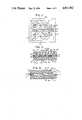
- FIG. 1is a plan view of a patterned component of an embodiment of the invention
- FIG. 2is a sectional view of the embodiment partially illustrated in FIG. 1;
- FIG. 3is a transverse section through the embodiment of FIG. 1 and 2;
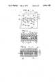
- FIG. 4is view similar to FIG. 1 but of another embodiment
- FIG. 5is a view similar to FIG. 2 but of the other embodiment.
- FIG. 6is a view similar to FIG. 3 but of the other embodiment.
- the glass substrate or bottom plate 10is provided with a layer 12 of photoresist and the top plate 16 is provided with a layer 14 of photoresist and the two components are adhered together to form the completed assembly. None of the Figures is to scale so that the details of the miniaturized structure is readily apparent.
- the bottom plate 10may be about 44 mm square and the thickness of each layer 12 and 14 may be 0.005 mm. In FIG. 1, only the first layer 12 as applied to the bottom plate 10 is illustrated, for clarity.
- the layer 12is patterned as indicated, to include the opposite side boundaries 17 and 18 and the intervening opposite side boundaries 20 and 22.
- the widths of the boundaries 17, 20 and 22may be about 4 mm whereas the width of the side boundary 18 may be about 12 mm except in the region of the notch 24 where it is about 4 mm.
- Extending from the opposite side boundary 17 and into the notch 24are parallel legs 26 and 28, each of about 1 mm in width and defining the bottom half of a channel 30 which is of about 2 mm in width.
- the assemblyis completed by registering the glass top plate 16 with its patterned resist layer 14 in position atop the bottom plate 10 with its patterned resist layer 12 so that the resist patterns are in registry, and effecting adhesion therebetween by means of spots of adhesive 48 which are received in the openings 40.
- the process as aboveresults in a unitary assembly which is the patterned resist disposed between the top and bottom glass plates as best seen in FIGS. 2 and 3.
- the fluorinating plasma treatmentas noted above conditions or alters the exposed glass surface of the bottom glass plate 10 and the exposed surfaces of the developed and cured resist respectively to make the glass surface more wettable (increasing its surface energy) while rendering the resist more hydrophobic (decreasing it surface energy).
- the volumes of the two chambers 50 and 52 on either side of the evaluation chamber 30are more than sufficient to accommodate the volume of a biological sample deposited at the region indicated at 54 in FIG.
- FIGS. 4-6The embodiment according to FIGS. 4-6 is for the study of individual cells or cell cultures and includes means for nourishing or growing them.
- substantially identically sized top and bottom glass plates 100 and 102are provided with a single resist layer 104 in the case of the top plate 100 and with three layers 106, 108 and 110 in the case of the bottom plate 102.
- FIG. 4is a plan view of the bottom plate with its layers 106, 108 and 110.
- the top plateWhen using the embodiment just described, the top plate is separated form the bottom plate in a sterile environment and an aliquot containing liquid medium and one or more cells is loaded to fill each of the wells or chambers within the layer 106, one such chamber being indicated at 158 in FIG. 5.
- the top plateis then placed in position on the bottom plate and clamped or otherwise secured in position thereon.
- a source of gassuch air mixed with 5% carbon dioxide is connected to the opening through the bottom plate corresponding to the circle 124 and is exhausted through the glass plate opening corresponding to the circle 122 to circulate the gas through the gas perfusion chamber 154.
- a source of cell culture mediais connected to the glass plate opening 150 and exhausted through the opening 152 to circulate the liquid media through the nutrient or reagent chamber 156.
- the cell culture chambers 158must be of a size to accommodate the original cells in the aliquot plus any cells which will grow up from the original cells during the study. Typically, these chambers may be 100 microns deep for egg cells or 20 microns deep for other types of animal cells. Therefore, the layer 106 may vary in thickness in accord with its intended use. The diameter of these chamber depends upon the number of cells to be studied in each chamber, for example typically ranging between about 250 microns and 1 centimeter.
- the aluminum layernormally is about 100 Angstrom units thick which will promote the wetting of the chamber 156 while allowing observations through the aluminum layer.
- miniaturized structuresare formed of contiguous or adjacent materials desired to have significantly different surface energy levels, these surface energy levels are often compromised or altered from those desired and the desired characteristics cannot be restored by well known methods.
- well known methodswhen attempted tend to compromise the surface energy levels of the materials involved, usually altering the surface energy level of one material in the desired direction while having the opposite effect on the other.
- the effect of attaining desired disparate surface energy levelscan be obtained and that, furthermore, it can even be obtained simultaneously by a single treatment.
- the desired effectcan be accomplished by subjecting the miniaturized structural assembly to fluorinating plasmas in the absence of contaminant gases such as oxygen or water.
- contaminant gasessuch as oxygen or water.
- hydrogen plasmasunder the same conditions, are effective as well.
- surface energy levels as high as or greater than 100 dynes per centimeter as well as surface energy levels less than 30 dynes per centimeterare advantageous and are considered necessary and surface energy levels as high as 300 dynes per centimeter and as low as 5 dynes per centimeter may be highly desirable.
- surface energy levels of this naturehave been simultaneously attained in structures smaller than 10 microns.
Landscapes
- Chemical & Material Sciences (AREA)
- Health & Medical Sciences (AREA)
- Dispersion Chemistry (AREA)
- Analytical Chemistry (AREA)
- General Health & Medical Sciences (AREA)
- Hematology (AREA)
- Clinical Laboratory Science (AREA)
- Chemical Kinetics & Catalysis (AREA)
- Apparatus Associated With Microorganisms And Enzymes (AREA)
Abstract
Description
Claims (11)
Priority Applications (3)
| Application Number | Priority Date | Filing Date | Title |
|---|---|---|---|
| US07/174,163US4911782A (en) | 1988-03-28 | 1988-03-28 | Method for forming a miniaturized biological assembly |
| US07/632,655US5200152A (en) | 1988-03-28 | 1990-12-27 | Miniaturized biological assembly |
| US08/287,608US5503803A (en) | 1988-03-28 | 1994-08-09 | Miniaturized biological assembly |
Applications Claiming Priority (1)
| Application Number | Priority Date | Filing Date | Title |
|---|---|---|---|
| US07/174,163US4911782A (en) | 1988-03-28 | 1988-03-28 | Method for forming a miniaturized biological assembly |
Related Child Applications (1)
| Application Number | Title | Priority Date | Filing Date |
|---|---|---|---|
| US37570089ADivision | 1988-03-28 | 1989-07-05 |
Publications (1)
| Publication Number | Publication Date |
|---|---|
| US4911782Atrue US4911782A (en) | 1990-03-27 |
Family
ID=22635095
Family Applications (1)
| Application Number | Title | Priority Date | Filing Date |
|---|---|---|---|
| US07/174,163Expired - LifetimeUS4911782A (en) | 1988-03-28 | 1988-03-28 | Method for forming a miniaturized biological assembly |
Country Status (1)
| Country | Link |
|---|---|
| US (1) | US4911782A (en) |
Cited By (104)
| Publication number | Priority date | Publication date | Assignee | Title |
|---|---|---|---|---|
| EP0437408A3 (en)* | 1990-01-12 | 1992-01-29 | United Medical Systems Israel Ltd. | A disposable device for determining the quality of sperm cells |
| WO1993022421A1 (en)* | 1992-05-01 | 1993-11-11 | Trustees Of The University Of Pennsylvania | Microfabricated sperm handling devices |
| US5296375A (en)* | 1992-05-01 | 1994-03-22 | Trustees Of The University Of Pennsylvania | Mesoscale sperm handling devices |
| US5304487A (en)* | 1992-05-01 | 1994-04-19 | Trustees Of The University Of Pennsylvania | Fluid handling in mesoscale analytical devices |
| US5306467A (en)* | 1993-02-17 | 1994-04-26 | Hamilton-Thorn Research | Apparatus for measurement of cell concentration in a biological sample employing a magnetic slide loading apparatus |
| US5317938A (en)* | 1992-01-16 | 1994-06-07 | Duke University | Method for making microstructural surgical instruments |
| US5349436A (en)* | 1992-12-02 | 1994-09-20 | Harry Fisch | Biological assembly |
| US5486335A (en)* | 1992-05-01 | 1996-01-23 | Trustees Of The University Of Pennsylvania | Analysis based on flow restriction |
| US5498392A (en)* | 1992-05-01 | 1996-03-12 | Trustees Of The University Of Pennsylvania | Mesoscale polynucleotide amplification device and method |
| DE4438785A1 (en)* | 1994-10-24 | 1996-05-02 | Wita Gmbh Wittmann Inst Of Tec | Micro-electronically produced analytical and dosing system |
| US5587128A (en)* | 1992-05-01 | 1996-12-24 | The Trustees Of The University Of Pennsylvania | Mesoscale polynucleotide amplification devices |
| DE19524795A1 (en)* | 1995-07-07 | 1997-01-09 | Danfoss As | Distribution device, in particular for a chemical analysis device |
| US5637469A (en)* | 1992-05-01 | 1997-06-10 | Trustees Of The University Of Pennsylvania | Methods and apparatus for the detection of an analyte utilizing mesoscale flow systems |
| US5726026A (en)* | 1992-05-01 | 1998-03-10 | Trustees Of The University Of Pennsylvania | Mesoscale sample preparation device and systems for determination and processing of analytes |
| US5744366A (en)* | 1992-05-01 | 1998-04-28 | Trustees Of The University Of Pennsylvania | Mesoscale devices and methods for analysis of motile cells |
| WO1998039645A1 (en)* | 1997-03-07 | 1998-09-11 | Beckman Coulter, Inc. | A novel capillary |
| US5837115A (en)* | 1993-06-08 | 1998-11-17 | British Technology Group Usa Inc. | Microlithographic array for macromolecule and cell fractionation |
| US5853894A (en)* | 1997-02-03 | 1998-12-29 | Cytonix Corporation | Laboratory vessel having hydrophobic coating and process for manufacturing same |
| WO1998053300A3 (en)* | 1997-05-23 | 1999-02-25 | Lynx Therapeutics Inc | System and apparaus for sequential processing of analytes |
| US6033544A (en)* | 1996-10-11 | 2000-03-07 | Sarnoff Corporation | Liquid distribution system |
| US6037168A (en)* | 1997-12-31 | 2000-03-14 | Cytonix Corporation | Microbiological assembly comprising resealable closure means |
| US6117396A (en)* | 1998-02-18 | 2000-09-12 | Orchid Biocomputer, Inc. | Device for delivering defined volumes |
| US6143496A (en)* | 1997-04-17 | 2000-11-07 | Cytonix Corporation | Method of sampling, amplifying and quantifying segment of nucleic acid, polymerase chain reaction assembly having nanoliter-sized sample chambers, and method of filling assembly |
| US6156389A (en)* | 1997-02-03 | 2000-12-05 | Cytonix Corporation | Hydrophobic coating compositions, articles coated with said compositions, and processes for manufacturing same |
| WO1998043739A3 (en)* | 1997-03-27 | 2001-06-07 | Biosite Diagnostics Inc | Diagnostic devices and apparatus for the controlled movement of reagents without membranes |
| US20020061529A1 (en)* | 1998-05-22 | 2002-05-23 | Lynx Therapeutics, Inc. | System and apparatus for sequential processing of analytes |
| US6495195B2 (en) | 1997-02-14 | 2002-12-17 | Arcturus Engineering, Inc. | Broadband absorbing film for laser capture microdissection |
| US6495624B1 (en) | 1997-02-03 | 2002-12-17 | Cytonix Corporation | Hydrophobic coating compositions, articles coated with said compositions, and processes for manufacturing same |
| US20020192701A1 (en)* | 2001-03-09 | 2002-12-19 | Adey Nils B. | Laminated microarray interface device |
| US20030040129A1 (en)* | 2001-08-20 | 2003-02-27 | Shah Haresh P. | Binding assays using magnetically immobilized arrays |
| US6551554B1 (en)* | 1995-02-15 | 2003-04-22 | Leja Products B.V. | Counting compartment for biological investigations and a method for manufacturing such a counting compartment |
| US6586253B1 (en) | 1998-02-27 | 2003-07-01 | The Governors Of The University Of Alberta | Microchip based enzymatic analysis |
| US6632652B1 (en) | 1996-08-26 | 2003-10-14 | Princeton University | Reversibly sealable microstructure sorting devices |
| US20030199081A1 (en)* | 1992-05-01 | 2003-10-23 | Peter Wilding | Mesoscale polynucleotide amplification analysis |
| US20030199165A1 (en)* | 2002-03-11 | 2003-10-23 | Becton, Dickinson And Company | System and method for the manufacture of surgical blades |
| US20040051957A1 (en)* | 2002-08-06 | 2004-03-18 | Dmetrix, Inc. | Miniature microscope objective lens |
| US6723290B1 (en)* | 1998-03-07 | 2004-04-20 | Levine Robert A | Container for holding biologic fluid for analysis |
| DE10247020A1 (en)* | 2002-10-09 | 2004-04-22 | Micro-Biolytics Gmbh | thin-film cell |
| US20040077103A1 (en)* | 1992-05-21 | 2004-04-22 | Biosite, Inc. | Diagnostic devices and apparatus for the controlled movement of reagents without membranes |
| US20040082699A1 (en)* | 1997-02-03 | 2004-04-29 | Brown James F. | Hydrophobic coating compositions, articles coated with said compositions, and processes for manufacturing same |
| US20040096118A1 (en)* | 2002-11-20 | 2004-05-20 | Dmetrix, Inc. | Multi-spectral miniature microscope array |
| US20040109793A1 (en)* | 2002-02-07 | 2004-06-10 | Mcneely Michael R | Three-dimensional microfluidics incorporating passive fluid control structures |
| US20040219531A1 (en)* | 2003-04-30 | 2004-11-04 | Dicesare Joseph L. | Method of scanning a sample plate surface mask in an area adjacent to a conductive area using matrix-assisted laser desorption and ionization mass spectrometry |
| US20050029446A1 (en)* | 2003-04-30 | 2005-02-10 | Dicesare Joseph L. | Sample plate for matrix-assisted laser desorption and ionization mass spectrometry |
| WO2005016530A1 (en)* | 2003-07-14 | 2005-02-24 | Qiagen Sciences, Inc. | Sample presentation device with differing wettability |
| US6905882B2 (en) | 1992-05-21 | 2005-06-14 | Biosite, Inc. | Diagnostic devices and apparatus for the controlled movement of reagents without membranes |
| US20050155955A1 (en)* | 2003-03-10 | 2005-07-21 | Daskal Vadim M. | Method for reducing glare and creating matte finish of controlled density on a silicon surface |
| US20050188548A1 (en)* | 2002-03-11 | 2005-09-01 | Daskal Vadim M. | Silicon blades for surgical and non-surgical use |
| US20050266680A1 (en)* | 2004-04-30 | 2005-12-01 | Daskal Vadim M | Methods of fabricating complex blade geometries from silicon wafers and strengthening blade geometries |
| US20060054503A1 (en)* | 2002-09-24 | 2006-03-16 | Duke University | Methods for manipulating droplets by electrowetting-based techniques |
| US20060148070A1 (en)* | 1997-10-01 | 2006-07-06 | Baer Thomas M | Consumable for laser capture microdissection |
| US20060275743A1 (en)* | 2005-06-02 | 2006-12-07 | Minitube Of America, Inc. | Counting, viability assessment, analysis and manipulation chamber |
| US20070026469A1 (en)* | 2005-07-29 | 2007-02-01 | Martin Fuchs | Devices and methods for enrichment and alteration of circulating tumor cells and other particles |
| US20070037294A1 (en)* | 2002-09-24 | 2007-02-15 | Duke University | Methods for performing microfluidic sampling |
| US20070187874A1 (en)* | 2003-09-17 | 2007-08-16 | Daskal Vadim M | System and method for creating linear and non-linear trenches in silicon and other crystalline materials with a router |
| US20070217956A1 (en)* | 2002-09-24 | 2007-09-20 | Pamula Vamsee K | Methods for nucleic acid amplification on a printed circuit board |
| US20070267294A1 (en)* | 1999-01-25 | 2007-11-22 | Nanolytics Inc. | Actuators for microfluidics without moving parts |
| US20080044914A1 (en)* | 2006-04-18 | 2008-02-21 | Pamula Vamsee K | Protein Crystallization Screening and Optimization Droplet Actuators, Systems and Methods |
| US20080274513A1 (en)* | 2005-05-11 | 2008-11-06 | Shenderov Alexander D | Method and Device for Conducting Biochemical or Chemical Reactions at Multiple Temperatures |
| US20090007436A1 (en)* | 2003-03-10 | 2009-01-08 | Daskal Vadim M | Silicon blades for surgical and non-surgical use |
| US7524456B1 (en) | 1992-05-21 | 2009-04-28 | Biosite Incorporated | Diagnostic devices for the controlled movement of reagents without membranes |
| US20100126860A1 (en)* | 2007-08-09 | 2010-05-27 | Advanced Liquid Logic, Inc. | PCB Droplet Actuator Fabrication |
| US20110206557A1 (en)* | 2009-12-18 | 2011-08-25 | Abbott Point Of Care, Inc. | Biologic fluid analysis cartridge |
| USRE43097E1 (en) | 1994-10-13 | 2012-01-10 | Illumina, Inc. | Massively parallel signature sequencing by ligation of encoded adaptors |
| EP2657869A2 (en) | 2007-08-29 | 2013-10-30 | Applied Biosystems, LLC | Alternative nucleic acid sequencing methods |
| US8653213B2 (en) | 1997-02-03 | 2014-02-18 | Cytonix, Llc | Hydrophobic coating compositions and articles coated with said compositions |
| US8797527B2 (en) | 2011-08-24 | 2014-08-05 | Abbott Point Of Care, Inc. | Biologic fluid sample analysis cartridge |
| US8911815B2 (en) | 2009-11-13 | 2014-12-16 | Ventana Medical Systems, Inc. | Thin film processing apparatuses for adjustable volume accommodation |
| USD728120S1 (en) | 2013-03-15 | 2015-04-28 | Ventana Medical Systems, Inc. | Arcuate member for moving liquids along a microscope slide |
| US9199233B2 (en) | 2010-03-31 | 2015-12-01 | Abbott Point Of Care, Inc. | Biologic fluid analysis cartridge with deflecting top panel |
| US9498791B2 (en) | 2009-11-13 | 2016-11-22 | Ventana Medical Systems, Inc. | Opposables and automated specimen processing systems with opposables |
| US9513253B2 (en) | 2011-07-11 | 2016-12-06 | Advanced Liquid Logic, Inc. | Droplet actuators and techniques for droplet-based enzymatic assays |
| EP3199937A1 (en) | 2016-01-28 | 2017-08-02 | Minitüb GmbH | Counting compartment and method for sample analysis |
| US9873118B2 (en) | 2010-12-30 | 2018-01-23 | Abbott Point Of Care, Inc. | Biologic fluid analysis cartridge with sample handling portion and analysis chamber portion |
| US10132794B2 (en) | 2015-09-14 | 2018-11-20 | Essenlix Corporation | Device and system for collecting and analyzing vapor condensate, particularly exhaled breath condensate, as well as method of using the same |
| US10324009B2 (en) | 2015-08-10 | 2019-06-18 | Essenlix Corporation | Bio/chemical assay devices and methods for simplified steps, small samples, accelerated speed, and ease-of-use |
| EP3502661A1 (en) | 2017-12-22 | 2019-06-26 | Minitüb GmbH | Method and devices for analyzing sperm samples |
| US10605805B2 (en) | 2015-09-14 | 2020-03-31 | Essenlix Corporation | Device and system for analyzing a sample, particularly blood, as well as methods of using the same |
| US10628693B2 (en) | 2016-12-21 | 2020-04-21 | Essenlix Corporation | Devices and methods for authenticating a sample and use of the same |
| US10746752B2 (en) | 2009-11-13 | 2020-08-18 | Ventana Medical Systems, Inc. | Opposables and automated specimen processing systems with opposables |
| US10807095B2 (en) | 2017-10-26 | 2020-10-20 | Essenlix Corporation | Making and tracking assay card |
| US11156606B2 (en) | 2018-01-11 | 2021-10-26 | Essenlix Corporation | Homogeneous assay (II) |
| US11237113B2 (en) | 2017-10-26 | 2022-02-01 | Essenlix Corporation | Rapid pH measurement |
| US11243201B2 (en) | 2017-08-01 | 2022-02-08 | Essenlix Corporation | Sample collection, holding and assaying |
| US11274996B2 (en) | 2017-02-07 | 2022-03-15 | Essenlix Corporation | Compressed open flow assay and use |
| US11280706B2 (en) | 2017-08-01 | 2022-03-22 | Essenlix Corporation | Dilution calibration |
| US11393561B2 (en) | 2017-10-13 | 2022-07-19 | Essenlix Corporation | Devices and methods for authenticating a medical test and use of the same |
| US11510608B2 (en) | 2017-12-14 | 2022-11-29 | Essenlix Corporation | Devices, systems, and methods for monitoring hair |
| US11523752B2 (en) | 2017-02-16 | 2022-12-13 | Essenlix Corporation | Assay for vapor condensates |
| US11604148B2 (en) | 2017-02-09 | 2023-03-14 | Essenlix Corporation | Colorimetric assays |
| US11609224B2 (en) | 2017-10-26 | 2023-03-21 | Essenlix Corporation | Devices and methods for white blood cell analyses |
| US11648551B2 (en) | 2017-12-12 | 2023-05-16 | Essenlix Corporation | Sample manipulation and assay with rapid temperature change |
| US11725227B2 (en) | 2017-08-01 | 2023-08-15 | Essenlix Corporation | Devices and methods for examining drug effects on microorganisms |
| US11883824B2 (en) | 2017-02-09 | 2024-01-30 | Essenlix Corporation | Assay using different spacing heights |
| US11885952B2 (en) | 2018-07-30 | 2024-01-30 | Essenlix Corporation | Optics, device, and system for assaying and imaging |
| US11927560B2 (en) | 2017-02-08 | 2024-03-12 | Essenlix Corporation | Bio/chemical material extraction and assay |
| US11940382B2 (en) | 2017-02-09 | 2024-03-26 | Essenlix Corporation | Assay with amplification |
| US12007315B2 (en) | 2017-02-08 | 2024-06-11 | Essenlix Corporation | Sample collection and handling for delayed analysis |
| US12038403B2 (en) | 2017-08-17 | 2024-07-16 | Abbott Point Of Care Inc. | Devices, systems, and methods for performing optical and electrochemical assays |
| US12066434B2 (en) | 2017-02-08 | 2024-08-20 | Essenlix Corporation | QMAX assays and applications |
| US12151246B2 (en) | 2017-02-08 | 2024-11-26 | Essenlix Corporation | Molecular manipulation and assay with controlled temperature |
| US12181472B2 (en) | 2017-06-12 | 2024-12-31 | Essenlix Corporation | Homogeneous assay |
| US12350680B2 (en) | 2017-02-15 | 2025-07-08 | Essenlix Corporation | Assay with rapid temperature change |
| US12403465B2 (en) | 2017-10-11 | 2025-09-02 | Essenlix Corporation | Containing a liquid sample |
Citations (1)
| Publication number | Priority date | Publication date | Assignee | Title |
|---|---|---|---|---|
| US3853467A (en)* | 1973-08-15 | 1974-12-10 | Gen Electric | Method and apparatus for immunological detection of biological particles |
- 1988
- 1988-03-28USUS07/174,163patent/US4911782A/ennot_activeExpired - Lifetime
Patent Citations (1)
| Publication number | Priority date | Publication date | Assignee | Title |
|---|---|---|---|---|
| US3853467A (en)* | 1973-08-15 | 1974-12-10 | Gen Electric | Method and apparatus for immunological detection of biological particles |
Cited By (253)
| Publication number | Priority date | Publication date | Assignee | Title |
|---|---|---|---|---|
| EP0437408A3 (en)* | 1990-01-12 | 1992-01-29 | United Medical Systems Israel Ltd. | A disposable device for determining the quality of sperm cells |
| US5317938A (en)* | 1992-01-16 | 1994-06-07 | Duke University | Method for making microstructural surgical instruments |
| US7005292B2 (en) | 1992-05-01 | 2006-02-28 | The Trustees Of The University Of Pennsylvania | Device and method for the detection of an analyte utilizing mesoscale flow systems |
| US5955029A (en)* | 1992-05-01 | 1999-09-21 | Trustees Of The University Of Pennsylvania | Mesoscale polynucleotide amplification device and method |
| US6953676B1 (en) | 1992-05-01 | 2005-10-11 | Trustees Of The University Of Pennsylvania | Mesoscale polynucleotide amplification device and method |
| US5296375A (en)* | 1992-05-01 | 1994-03-22 | Trustees Of The University Of Pennsylvania | Mesoscale sperm handling devices |
| WO1993022421A1 (en)* | 1992-05-01 | 1993-11-11 | Trustees Of The University Of Pennsylvania | Microfabricated sperm handling devices |
| US5427946A (en)* | 1992-05-01 | 1995-06-27 | Trustees Of The University Of Pennsylvania | Mesoscale sperm handling devices |
| US5486335A (en)* | 1992-05-01 | 1996-01-23 | Trustees Of The University Of Pennsylvania | Analysis based on flow restriction |
| US5498392A (en)* | 1992-05-01 | 1996-03-12 | Trustees Of The University Of Pennsylvania | Mesoscale polynucleotide amplification device and method |
| US20060040309A1 (en)* | 1992-05-01 | 2006-02-23 | Peter Wilding | Mesoscale polynucleotide amplification analysis |
| US5587128A (en)* | 1992-05-01 | 1996-12-24 | The Trustees Of The University Of Pennsylvania | Mesoscale polynucleotide amplification devices |
| US6184029B1 (en) | 1992-05-01 | 2001-02-06 | Trustees Of The University Of Pennsylvania | Mesoscale sample preparation device and systems for determination and processing of analytes |
| US5635358A (en)* | 1992-05-01 | 1997-06-03 | Trustees Of The University Of Pennsylvania | Fluid handling methods for use in mesoscale analytical devices |
| US5304487A (en)* | 1992-05-01 | 1994-04-19 | Trustees Of The University Of Pennsylvania | Fluid handling in mesoscale analytical devices |
| US5726026A (en)* | 1992-05-01 | 1998-03-10 | Trustees Of The University Of Pennsylvania | Mesoscale sample preparation device and systems for determination and processing of analytes |
| US5637469A (en)* | 1992-05-01 | 1997-06-10 | Trustees Of The University Of Pennsylvania | Methods and apparatus for the detection of an analyte utilizing mesoscale flow systems |
| US5744366A (en)* | 1992-05-01 | 1998-04-28 | Trustees Of The University Of Pennsylvania | Mesoscale devices and methods for analysis of motile cells |
| US20070190641A1 (en)* | 1992-05-01 | 2007-08-16 | Wilding Peter G | Mesoscale polynucleotide amplification device and method |
| US6660517B1 (en) | 1992-05-01 | 2003-12-09 | Trustees Of The University Of Pennsylvania | Mesoscale polynucleotide amplification devices |
| US20030199081A1 (en)* | 1992-05-01 | 2003-10-23 | Peter Wilding | Mesoscale polynucleotide amplification analysis |
| US6551841B1 (en) | 1992-05-01 | 2003-04-22 | The Trustees Of The University Of Pennsylvania | Device and method for the detection of an analyte utilizing mesoscale flow systems |
| US5928880A (en)* | 1992-05-01 | 1999-07-27 | Trustees Of The University Of Pennsylvania | Mesoscale sample preparation device and systems for determination and processing of analytes |
| US7018830B2 (en) | 1992-05-01 | 2006-03-28 | The Trustees Of The University Of Pennsylvania | Device and method for the detection of an analyte utilizing mesoscale flow systems |
| US7892819B2 (en) | 1992-05-01 | 2011-02-22 | Trustees Of The University Of Pennsylvania | Mesoscale polynucleotide amplification devices |
| US20110020876A1 (en)* | 1992-05-01 | 2011-01-27 | Peter Wilding | Mesoscale polynucleotide amplification devices |
| US7494770B2 (en) | 1992-05-01 | 2009-02-24 | Trustees Of The University Of Pennsylvania | Mesoscale polynucleotide amplification analysis |
| US6271040B1 (en) | 1992-05-21 | 2001-08-07 | Biosite Diagnostics Incorporated | Diagnostic devices method and apparatus for the controlled movement of reagents without membranes |
| US6767510B1 (en) | 1992-05-21 | 2004-07-27 | Biosite, Inc. | Diagnostic devices and apparatus for the controlled movement of reagents without membranes |
| US6905882B2 (en) | 1992-05-21 | 2005-06-14 | Biosite, Inc. | Diagnostic devices and apparatus for the controlled movement of reagents without membranes |
| US7824611B2 (en) | 1992-05-21 | 2010-11-02 | Biosite, Inc. | Diagnostic devices and apparatus for the controlled movement of reagents without membranes |
| US20050136552A1 (en)* | 1992-05-21 | 2005-06-23 | Biosite, Inc. | Diagnostic devices and apparatus for the controlled movement of reagents without membranes |
| US7524456B1 (en) | 1992-05-21 | 2009-04-28 | Biosite Incorporated | Diagnostic devices for the controlled movement of reagents without membranes |
| US20040077103A1 (en)* | 1992-05-21 | 2004-04-22 | Biosite, Inc. | Diagnostic devices and apparatus for the controlled movement of reagents without membranes |
| US7615191B2 (en) | 1992-05-21 | 2009-11-10 | Biosite, Inc. | Diagnostic devices and apparatus for the controlled movement of reagents without membranes |
| US5349436A (en)* | 1992-12-02 | 1994-09-20 | Harry Fisch | Biological assembly |
| USRE35589E (en)* | 1992-12-02 | 1997-08-19 | Fisch; Harry | Biological assembly |
| US5306467A (en)* | 1993-02-17 | 1994-04-26 | Hamilton-Thorn Research | Apparatus for measurement of cell concentration in a biological sample employing a magnetic slide loading apparatus |
| US5837115A (en)* | 1993-06-08 | 1998-11-17 | British Technology Group Usa Inc. | Microlithographic array for macromolecule and cell fractionation |
| USRE43097E1 (en) | 1994-10-13 | 2012-01-10 | Illumina, Inc. | Massively parallel signature sequencing by ligation of encoded adaptors |
| DE4438785A1 (en)* | 1994-10-24 | 1996-05-02 | Wita Gmbh Wittmann Inst Of Tec | Micro-electronically produced analytical and dosing system |
| US6551554B1 (en)* | 1995-02-15 | 2003-04-22 | Leja Products B.V. | Counting compartment for biological investigations and a method for manufacturing such a counting compartment |
| DE19524795A1 (en)* | 1995-07-07 | 1997-01-09 | Danfoss As | Distribution device, in particular for a chemical analysis device |
| US6117395A (en)* | 1995-07-07 | 2000-09-12 | Danfoss A/S | Distributor device, in particular for a chemical analysis arrangement |
| US6632652B1 (en) | 1996-08-26 | 2003-10-14 | Princeton University | Reversibly sealable microstructure sorting devices |
| US6033544A (en)* | 1996-10-11 | 2000-03-07 | Sarnoff Corporation | Liquid distribution system |
| US7268179B2 (en) | 1997-02-03 | 2007-09-11 | Cytonix Corporation | Hydrophobic coating compositions, articles coated with said compositions, and processes for manufacturing same |
| US6495624B1 (en) | 1997-02-03 | 2002-12-17 | Cytonix Corporation | Hydrophobic coating compositions, articles coated with said compositions, and processes for manufacturing same |
| US20070281110A1 (en)* | 1997-02-03 | 2007-12-06 | Cytonix Corporation | Hydrophobic coating compositions, articles coated with said compositions, and processes for manufacturing same |
| US20100021689A1 (en)* | 1997-02-03 | 2010-01-28 | Cytonix Llc | Articles comprising hydrophobic surfaces |
| US6663941B2 (en) | 1997-02-03 | 2003-12-16 | Cytonix Corporation | Hydrophobic coating compositions, articles coated with said compositions, and processes for manufacturing same |
| US7781027B2 (en) | 1997-02-03 | 2010-08-24 | Cytonix Llc | Hydrophobic coating compositions, articles coated with said compositions, and processes for manufacturing same |
| US7999013B2 (en) | 1997-02-03 | 2011-08-16 | Cytonix, Llc | Hydrophobic coating compositions and articles coated with said compositions |
| US8168264B2 (en) | 1997-02-03 | 2012-05-01 | Cytonix Llc | Hydrophobic coating compositions, articles coated with said compositions, and processes for manufacturing same |
| US8221870B2 (en) | 1997-02-03 | 2012-07-17 | Cytonix Llc | Articles comprising hydrophobic surfaces |
| US6156389A (en)* | 1997-02-03 | 2000-12-05 | Cytonix Corporation | Hydrophobic coating compositions, articles coated with said compositions, and processes for manufacturing same |
| US20040082699A1 (en)* | 1997-02-03 | 2004-04-29 | Brown James F. | Hydrophobic coating compositions, articles coated with said compositions, and processes for manufacturing same |
| US5853894A (en)* | 1997-02-03 | 1998-12-29 | Cytonix Corporation | Laboratory vessel having hydrophobic coating and process for manufacturing same |
| US20050003203A1 (en)* | 1997-02-03 | 2005-01-06 | Cytonix Corporation | Hydrophobic coating compositions and articles coated with said compositions |
| US20040131789A1 (en)* | 1997-02-03 | 2004-07-08 | Brown James F. | Hydrophobic coating compositions, articles coated with said compositions, and processes for manufacturing same |
| US6447919B1 (en) | 1997-02-03 | 2002-09-10 | Cytonix Corporation | Hydrophobic coating compositions, articles coated with said compositions, and processes for manufacturing same |
| US6767587B1 (en) | 1997-02-03 | 2004-07-27 | Cytonix Corporation | Hydrophobic coating compositions, articles coated with said compositions, and processes for manufacturing same |
| US20100316810A1 (en)* | 1997-02-03 | 2010-12-16 | Cytonix Llc | Hydrophobic coating compositions, articles coated with said compositions, and processes for manufacturing same |
| US8653213B2 (en) | 1997-02-03 | 2014-02-18 | Cytonix, Llc | Hydrophobic coating compositions and articles coated with said compositions |
| US7579056B2 (en) | 1997-02-03 | 2009-08-25 | Cytonix Corporation | Hydrophobic formulations and vessel surfaces comprising same |
| US8785556B2 (en) | 1997-02-03 | 2014-07-22 | Cytonix, Llc | Hydrophobic coating compositions and articles coated with said compositions |
| US6495195B2 (en) | 1997-02-14 | 2002-12-17 | Arcturus Engineering, Inc. | Broadband absorbing film for laser capture microdissection |
| US20030031781A1 (en)* | 1997-02-14 | 2003-02-13 | Baer Thomas M. | Broadband absorbing film for laser capture microdissection |
| WO1998039645A1 (en)* | 1997-03-07 | 1998-09-11 | Beckman Coulter, Inc. | A novel capillary |
| WO1998043739A3 (en)* | 1997-03-27 | 2001-06-07 | Biosite Diagnostics Inc | Diagnostic devices and apparatus for the controlled movement of reagents without membranes |
| US8822183B2 (en) | 1997-04-17 | 2014-09-02 | Applied Biosystems, Llc | Device for amplifying target nucleic acid |
| US8067159B2 (en) | 1997-04-17 | 2011-11-29 | Applied Biosystems, Llc | Methods of detecting amplified product |
| US6143496A (en)* | 1997-04-17 | 2000-11-07 | Cytonix Corporation | Method of sampling, amplifying and quantifying segment of nucleic acid, polymerase chain reaction assembly having nanoliter-sized sample chambers, and method of filling assembly |
| US6391559B1 (en) | 1997-04-17 | 2002-05-21 | Cytonix Corporation | Method of sampling, amplifying and quantifying segment of nucleic acid, polymerase chain reaction assembly having nanoliter-sized sample chambers, and method of filling assembly |
| US20090035759A1 (en)* | 1997-04-17 | 2009-02-05 | Cytonix | Method and device for detecting the presence of a single target nucleic acid in a sample |
| US20020164820A1 (en)* | 1997-04-17 | 2002-11-07 | Brown James F. | Method of sampling, amplifying and quantifying segment of nucleic acid, polymerase chain reaction assembly having nanoliter-sized sample chambers, and method of filling assembly |
| US7459315B2 (en) | 1997-04-17 | 2008-12-02 | Cytonix Corporation | Miniaturized assembly and method of filling assembly |
| US7972778B2 (en) | 1997-04-17 | 2011-07-05 | Applied Biosystems, Llc | Method for detecting the presence of a single target nucleic acid in a sample |
| US20040171055A1 (en)* | 1997-04-17 | 2004-09-02 | Cytonix Corporation | Method for detecting the presence of a single target nucleic acid in a sample |
| US8563275B2 (en) | 1997-04-17 | 2013-10-22 | Applied Biosystems, Llc | Method and device for detecting the presence of a single target nucleic acid in a sample |
| US20080213766A1 (en)* | 1997-04-17 | 2008-09-04 | Cytonix | Method and device for detecting the presence of a single target nucleic acid in samples |
| US20080171380A1 (en)* | 1997-04-17 | 2008-07-17 | Cytomix | Microfluidic assembly with reagent |
| US20080169184A1 (en)* | 1997-04-17 | 2008-07-17 | Cytonix | Device having regions of differing affinities to fluid, methods of making such devices, and methods of using such devices |
| US8551698B2 (en) | 1997-04-17 | 2013-10-08 | Applied Biosystems, Llc | Method of loading sample into a microfluidic device |
| US20080171326A1 (en)* | 1997-04-17 | 2008-07-17 | Cytonix | Method and device for detecting the presence of a single target nucleic acid in a sample |
| US20080171382A1 (en)* | 1997-04-17 | 2008-07-17 | Cytonix | Method and device for detecting the presence of a single target nucleic acid in a sample |
| US20080171325A1 (en)* | 1997-04-17 | 2008-07-17 | Cytonix | Method and device for detecting the presence of a single target nucleic acid in a sample |
| US20080171327A1 (en)* | 1997-04-17 | 2008-07-17 | Cytonix | Method and device for detecting the presence of a single target nucleic acid in a sample |
| US20080171324A1 (en)* | 1997-04-17 | 2008-07-17 | Cytonix | Method for quantifying number of molecules of target nucleic acid contained in a sample |
| US20080160525A1 (en)* | 1997-04-17 | 2008-07-03 | Cytonix | Method and device for detecting the presence of a single target nucleic acid in a sample |
| US20080153091A1 (en)* | 1997-04-17 | 2008-06-26 | Cytonix | Method and device for detecting the presence of target nucleic acids in a sample, and microfluidic device for use in such methods |
| US20080138815A1 (en)* | 1997-04-17 | 2008-06-12 | Cytonix | Method of loading sample into a microfluidic device |
| US8278071B2 (en) | 1997-04-17 | 2012-10-02 | Applied Biosystems, Llc | Method for detecting the presence of a single target nucleic acid in a sample |
| US8257925B2 (en) | 1997-04-17 | 2012-09-04 | Applied Biosystems, Llc | Method for detecting the presence of a single target nucleic acid in a sample |
| US9506105B2 (en) | 1997-04-17 | 2016-11-29 | Applied Biosystems, Llc | Device and method for amplifying target nucleic acid |
| US8859204B2 (en) | 1997-04-17 | 2014-10-14 | Applied Biosystems, Llc | Method for detecting the presence of a target nucleic acid sequence in a sample |
| US7282370B2 (en) | 1997-05-23 | 2007-10-16 | Solexa, Inc. | System and apparatus for sequential processing of analytes |
| US6831994B2 (en) | 1997-05-23 | 2004-12-14 | Lynx Therapeutics, Inc. | System and apparatus for sequential processing of analytes |
| WO1998053300A3 (en)* | 1997-05-23 | 1999-02-25 | Lynx Therapeutics Inc | System and apparaus for sequential processing of analytes |
| US20060051876A1 (en)* | 1997-05-23 | 2006-03-09 | Lynx Therapeutics, Inc. | System and apparatus for sequential processing of analytes |
| US9273354B2 (en) | 1997-05-23 | 2016-03-01 | Illumina, Inc. | System and apparatus for sequential processing of analytes |
| US20090143244A1 (en)* | 1997-05-23 | 2009-06-04 | Solexa, Inc. | System and apparatus for sequential processing of analytes |
| US8728729B2 (en) | 1997-05-23 | 2014-05-20 | Illumina, Inc. | Method for sequential sequencing nucleic acids |
| EP0985142A4 (en)* | 1997-05-23 | 2006-09-13 | Lynx Therapeutics Inc | System and apparaus for sequential processing of analytes |
| AU736321B2 (en)* | 1997-05-23 | 2001-07-26 | Lynx Therapeutics, Inc. | System and apparatus for sequential processing of analytes |
| US8361713B2 (en) | 1997-05-23 | 2013-01-29 | Illumina, Inc. | System and apparatus for sequential processing of analytes |
| US7075640B2 (en) | 1997-10-01 | 2006-07-11 | Arcturus Bioscience, Inc. | Consumable for laser capture microdissection |
| US7221447B2 (en) | 1997-10-01 | 2007-05-22 | Molecular Devices Corporation | Consumable for laser capture microdissection |
| US20060148070A1 (en)* | 1997-10-01 | 2006-07-06 | Baer Thomas M | Consumable for laser capture microdissection |
| US6037168A (en)* | 1997-12-31 | 2000-03-14 | Cytonix Corporation | Microbiological assembly comprising resealable closure means |
| US6117396A (en)* | 1998-02-18 | 2000-09-12 | Orchid Biocomputer, Inc. | Device for delivering defined volumes |
| US6586253B1 (en) | 1998-02-27 | 2003-07-01 | The Governors Of The University Of Alberta | Microchip based enzymatic analysis |
| US6723290B1 (en)* | 1998-03-07 | 2004-04-20 | Levine Robert A | Container for holding biologic fluid for analysis |
| US20040156755A1 (en)* | 1998-03-07 | 2004-08-12 | Robert Levine | Container for holding biologic fluid for analysis |
| US6969488B2 (en) | 1998-05-22 | 2005-11-29 | Solexa, Inc. | System and apparatus for sequential processing of analytes |
| US20020061529A1 (en)* | 1998-05-22 | 2002-05-23 | Lynx Therapeutics, Inc. | System and apparatus for sequential processing of analytes |
| US8734629B2 (en) | 1999-01-25 | 2014-05-27 | Advanced Liquid Logic, Inc. | Droplet actuator and methods |
| US20110209998A1 (en)* | 1999-01-25 | 2011-09-01 | Advanced Liquid Logic, Inc. | Droplet Actuator and Methods |
| US20070267294A1 (en)* | 1999-01-25 | 2007-11-22 | Nanolytics Inc. | Actuators for microfluidics without moving parts |
| US7943030B2 (en) | 1999-01-25 | 2011-05-17 | Advanced Liquid Logic, Inc. | Actuators for microfluidics without moving parts |
| US20020192701A1 (en)* | 2001-03-09 | 2002-12-19 | Adey Nils B. | Laminated microarray interface device |
| US20050019898A1 (en)* | 2001-03-09 | 2005-01-27 | Nils Adey | Fluid mixing in low aspect ratio chambers |
| US7235400B2 (en) | 2001-03-09 | 2007-06-26 | Biomicro Systems, Inc. | Laminated microarray interface device |
| US20040037739A1 (en)* | 2001-03-09 | 2004-02-26 | Mcneely Michael | Method and system for microfluidic interfacing to arrays |
| US7223363B2 (en) | 2001-03-09 | 2007-05-29 | Biomicro Systems, Inc. | Method and system for microfluidic interfacing to arrays |
| US20030040129A1 (en)* | 2001-08-20 | 2003-02-27 | Shah Haresh P. | Binding assays using magnetically immobilized arrays |
| US20040109793A1 (en)* | 2002-02-07 | 2004-06-10 | Mcneely Michael R | Three-dimensional microfluidics incorporating passive fluid control structures |
| US8409462B2 (en) | 2002-03-11 | 2013-04-02 | Beaver-Visitec International (Us), Inc. | System and method for the manufacture of surgical blades |
| US20110192819A1 (en)* | 2002-03-11 | 2011-08-11 | Beaver-Vistec International, Inc. | System and method for the manufacture of surgical blades |
| US20050188548A1 (en)* | 2002-03-11 | 2005-09-01 | Daskal Vadim M. | Silicon blades for surgical and non-surgical use |
| US7105103B2 (en) | 2002-03-11 | 2006-09-12 | Becton, Dickinson And Company | System and method for the manufacture of surgical blades |
| US7387742B2 (en) | 2002-03-11 | 2008-06-17 | Becton, Dickinson And Company | Silicon blades for surgical and non-surgical use |
| US20030199165A1 (en)* | 2002-03-11 | 2003-10-23 | Becton, Dickinson And Company | System and method for the manufacture of surgical blades |
| US7906437B2 (en) | 2002-03-11 | 2011-03-15 | Beaver-Visitec International (Us), Inc. | System and method for the manufacture of surgical blades |
| US7023622B2 (en)* | 2002-08-06 | 2006-04-04 | Dmetrix, Inc. | Miniature microscope objective lens |
| US20040051957A1 (en)* | 2002-08-06 | 2004-03-18 | Dmetrix, Inc. | Miniature microscope objective lens |
| US9180450B2 (en) | 2002-09-24 | 2015-11-10 | Advanced Liquid Logic, Inc. | Droplet manipulation system and method |
| US8221605B2 (en) | 2002-09-24 | 2012-07-17 | Duke University | Apparatus for manipulating droplets |
| US8394249B2 (en) | 2002-09-24 | 2013-03-12 | Duke University | Methods for manipulating droplets by electrowetting-based techniques |
| US7759132B2 (en) | 2002-09-24 | 2010-07-20 | Duke University | Methods for performing microfluidic sampling |
| US20100025242A1 (en)* | 2002-09-24 | 2010-02-04 | Duke University | Apparatuses and methods for manipulating droplets |
| US8388909B2 (en) | 2002-09-24 | 2013-03-05 | Duke University | Apparatuses and methods for manipulating droplets |
| US7569129B2 (en) | 2002-09-24 | 2009-08-04 | Advanced Liquid Logic, Inc. | Methods for manipulating droplets by electrowetting-based techniques |
| US8349276B2 (en) | 2002-09-24 | 2013-01-08 | Duke University | Apparatuses and methods for manipulating droplets on a printed circuit board |
| US8287711B2 (en) | 2002-09-24 | 2012-10-16 | Duke University | Apparatus for manipulating droplets |
| US20060054503A1 (en)* | 2002-09-24 | 2006-03-16 | Duke University | Methods for manipulating droplets by electrowetting-based techniques |
| US8524506B2 (en) | 2002-09-24 | 2013-09-03 | Duke University | Methods for sampling a liquid flow |
| US20070037294A1 (en)* | 2002-09-24 | 2007-02-15 | Duke University | Methods for performing microfluidic sampling |
| US9110017B2 (en) | 2002-09-24 | 2015-08-18 | Duke University | Apparatuses and methods for manipulating droplets |
| US20080264797A1 (en)* | 2002-09-24 | 2008-10-30 | Duke University | Apparatus for Manipulating Droplets |
| US20080247920A1 (en)* | 2002-09-24 | 2008-10-09 | Duke University | Apparatus for Manipulating Droplets |
| US8147668B2 (en) | 2002-09-24 | 2012-04-03 | Duke University | Apparatus for manipulating droplets |
| US20070217956A1 (en)* | 2002-09-24 | 2007-09-20 | Pamula Vamsee K | Methods for nucleic acid amplification on a printed circuit board |
| US9638662B2 (en) | 2002-09-24 | 2017-05-02 | Duke University | Apparatuses and methods for manipulating droplets |
| US8906627B2 (en) | 2002-09-24 | 2014-12-09 | Duke University | Apparatuses and methods for manipulating droplets |
| US8871071B2 (en) | 2002-09-24 | 2014-10-28 | Duke University | Droplet manipulation device |
| US8048628B2 (en) | 2002-09-24 | 2011-11-01 | Duke University | Methods for nucleic acid amplification on a printed circuit board |
| US20060175732A1 (en)* | 2002-10-09 | 2006-08-10 | Ralf Masuch | Thin-layer sensor |
| DE10247020A1 (en)* | 2002-10-09 | 2004-04-22 | Micro-Biolytics Gmbh | thin-film cell |
| US7479197B2 (en) | 2002-10-09 | 2009-01-20 | Micro-Biolytics Gmbh | Thin-layer cell |
| US7113651B2 (en) | 2002-11-20 | 2006-09-26 | Dmetrix, Inc. | Multi-spectral miniature microscope array |
| US20040096118A1 (en)* | 2002-11-20 | 2004-05-20 | Dmetrix, Inc. | Multi-spectral miniature microscope array |
| US20090007436A1 (en)* | 2003-03-10 | 2009-01-08 | Daskal Vadim M | Silicon blades for surgical and non-surgical use |
| US20050155955A1 (en)* | 2003-03-10 | 2005-07-21 | Daskal Vadim M. | Method for reducing glare and creating matte finish of controlled density on a silicon surface |
| US20050029446A1 (en)* | 2003-04-30 | 2005-02-10 | Dicesare Joseph L. | Sample plate for matrix-assisted laser desorption and ionization mass spectrometry |
| US6891156B2 (en) | 2003-04-30 | 2005-05-10 | Perkin Elmer Instruments Llc | Sample plate for matrix-assisted laser desorption and ionization mass spectrometry |
| US7858387B2 (en) | 2003-04-30 | 2010-12-28 | Perkinelmer Health Sciences, Inc. | Method of scanning a sample plate surface mask in an area adjacent to a conductive area using matrix-assisted laser desorption and ionization mass spectrometry |
| US7173241B2 (en) | 2003-04-30 | 2007-02-06 | Perkinelmer Las, Inc. | Sample plate for matrix-assisted laser desorption and ionization mass spectrometry |
| US6956209B2 (en) | 2003-04-30 | 2005-10-18 | Dicesare Joseph L | Sample plate for matrix-assisted laser desorption and ionization mass spectrometry |
| US20110056311A1 (en)* | 2003-04-30 | 2011-03-10 | Dicesare Joseph L | Method of Scanning a Sample Plate Surface Mask in an Area Adjacent to a Conductive Area Using Matrix-Assisted Laser Desorption and Ionization Mass Spectrometry |
| US20040219531A1 (en)* | 2003-04-30 | 2004-11-04 | Dicesare Joseph L. | Method of scanning a sample plate surface mask in an area adjacent to a conductive area using matrix-assisted laser desorption and ionization mass spectrometry |
| US20050274886A1 (en)* | 2003-04-30 | 2005-12-15 | Dicesare Joseph L | Sample plate for matrix-assisted laser desorption and ionization mass spectrometry |
| CN100431707C (en)* | 2003-07-14 | 2008-11-12 | 奇亚根科学公司 | Sample presentation device with differing wettability |
| WO2005016530A1 (en)* | 2003-07-14 | 2005-02-24 | Qiagen Sciences, Inc. | Sample presentation device with differing wettability |
| US20070187874A1 (en)* | 2003-09-17 | 2007-08-16 | Daskal Vadim M | System and method for creating linear and non-linear trenches in silicon and other crystalline materials with a router |
| US7785485B2 (en) | 2003-09-17 | 2010-08-31 | Becton, Dickinson And Company | System and method for creating linear and non-linear trenches in silicon and other crystalline materials with a router |
| US7396484B2 (en) | 2004-04-30 | 2008-07-08 | Becton, Dickinson And Company | Methods of fabricating complex blade geometries from silicon wafers and strengthening blade geometries |
| US20050266680A1 (en)* | 2004-04-30 | 2005-12-01 | Daskal Vadim M | Methods of fabricating complex blade geometries from silicon wafers and strengthening blade geometries |
| US9216415B2 (en) | 2005-05-11 | 2015-12-22 | Advanced Liquid Logic | Methods of dispensing and withdrawing liquid in an electrowetting device |
| US9517469B2 (en) | 2005-05-11 | 2016-12-13 | Advanced Liquid Logic, Inc. | Method and device for conducting biochemical or chemical reactions at multiple temperatures |
| US9452433B2 (en) | 2005-05-11 | 2016-09-27 | Advanced Liquid Logic, Inc. | Method and device for conducting biochemical or chemical reactions at multiple temperatures |
| US20080274513A1 (en)* | 2005-05-11 | 2008-11-06 | Shenderov Alexander D | Method and Device for Conducting Biochemical or Chemical Reactions at Multiple Temperatures |
| US20060275743A1 (en)* | 2005-06-02 | 2006-12-07 | Minitube Of America, Inc. | Counting, viability assessment, analysis and manipulation chamber |
| US7718124B2 (en) | 2005-06-02 | 2010-05-18 | Minitube Of America, Inc. | Counting, viability assessment, analysis and manipulation chamber |
| US8921102B2 (en) | 2005-07-29 | 2014-12-30 | Gpb Scientific, Llc | Devices and methods for enrichment and alteration of circulating tumor cells and other particles |
| US20070026469A1 (en)* | 2005-07-29 | 2007-02-01 | Martin Fuchs | Devices and methods for enrichment and alteration of circulating tumor cells and other particles |
| US8007739B2 (en) | 2006-04-18 | 2011-08-30 | Advanced Liquid Logic, Inc. | Protein crystallization screening and optimization droplet actuators, systems and methods |
| US20080230386A1 (en)* | 2006-04-18 | 2008-09-25 | Vijay Srinivasan | Sample Processing Droplet Actuator, System and Method |
| US20080044914A1 (en)* | 2006-04-18 | 2008-02-21 | Pamula Vamsee K | Protein Crystallization Screening and Optimization Droplet Actuators, Systems and Methods |
| US8845872B2 (en) | 2006-04-18 | 2014-09-30 | Advanced Liquid Logic, Inc. | Sample processing droplet actuator, system and method |
| US20100126860A1 (en)* | 2007-08-09 | 2010-05-27 | Advanced Liquid Logic, Inc. | PCB Droplet Actuator Fabrication |
| US8268246B2 (en) | 2007-08-09 | 2012-09-18 | Advanced Liquid Logic Inc | PCB droplet actuator fabrication |
| EP2657869A2 (en) | 2007-08-29 | 2013-10-30 | Applied Biosystems, LLC | Alternative nucleic acid sequencing methods |
| US9498791B2 (en) | 2009-11-13 | 2016-11-22 | Ventana Medical Systems, Inc. | Opposables and automated specimen processing systems with opposables |
| US9618430B2 (en) | 2009-11-13 | 2017-04-11 | Ventana Medical Systems, Inc. | Thin film processing apparatuses for adjustable volume accommodation |
| US10746752B2 (en) | 2009-11-13 | 2020-08-18 | Ventana Medical Systems, Inc. | Opposables and automated specimen processing systems with opposables |
| US8911815B2 (en) | 2009-11-13 | 2014-12-16 | Ventana Medical Systems, Inc. | Thin film processing apparatuses for adjustable volume accommodation |
| US9993817B2 (en) | 2009-12-18 | 2018-06-12 | Abbott Point Of Care, Inc. | Biologic fluid analysis cartridge |
| US20110206557A1 (en)* | 2009-12-18 | 2011-08-25 | Abbott Point Of Care, Inc. | Biologic fluid analysis cartridge |
| US9579651B2 (en) | 2009-12-18 | 2017-02-28 | Abbott Point Of Care, Inc. | Biologic fluid analysis cartridge |
| US9199233B2 (en) | 2010-03-31 | 2015-12-01 | Abbott Point Of Care, Inc. | Biologic fluid analysis cartridge with deflecting top panel |
| US10391487B2 (en) | 2010-12-30 | 2019-08-27 | Abbott Point Of Care, Inc. | Biologic fluid analysis cartridge with sample handling portion and analysis chamber portion |
| US11583851B2 (en) | 2010-12-30 | 2023-02-21 | Abbott Point Of Care Inc. | Biologic fluid analysis cartridge with sample handling portion and analysis chamber portion |
| US9873118B2 (en) | 2010-12-30 | 2018-01-23 | Abbott Point Of Care, Inc. | Biologic fluid analysis cartridge with sample handling portion and analysis chamber portion |
| US9513253B2 (en) | 2011-07-11 | 2016-12-06 | Advanced Liquid Logic, Inc. | Droplet actuators and techniques for droplet-based enzymatic assays |
| US8797527B2 (en) | 2011-08-24 | 2014-08-05 | Abbott Point Of Care, Inc. | Biologic fluid sample analysis cartridge |
| USD772424S1 (en) | 2013-03-15 | 2016-11-22 | Ventana Medical Systems, Inc. | Arcuate member for moving liquids along a microscope slide |
| USD728120S1 (en) | 2013-03-15 | 2015-04-28 | Ventana Medical Systems, Inc. | Arcuate member for moving liquids along a microscope slide |
| US11385143B2 (en) | 2015-08-10 | 2022-07-12 | Essenlix Corporation | Bio/chemical assay devices and methods for simplified steps, small samples, accelerated speed, and ease-of-use |
| US10324009B2 (en) | 2015-08-10 | 2019-06-18 | Essenlix Corporation | Bio/chemical assay devices and methods for simplified steps, small samples, accelerated speed, and ease-of-use |
| US10948389B2 (en) | 2015-08-10 | 2021-03-16 | Essenlix Corporation | Bio/chemical assay devices and methods for simplified steps, small samples, accelerated speed, and ease-of-use |
| US12276660B2 (en) | 2015-09-14 | 2025-04-15 | Essenlix Corporation | Device and system for analyzing a sample, particularly blood, as well as methods of using the same |
| US10605805B2 (en) | 2015-09-14 | 2020-03-31 | Essenlix Corporation | Device and system for analyzing a sample, particularly blood, as well as methods of using the same |
| US10416151B2 (en) | 2015-09-14 | 2019-09-17 | Essenlix Corporation | Device and system for collecting and analyzing vapor condensate, particularly exhaled breath condensate, as well as method of using the same |
| US10830761B2 (en) | 2015-09-14 | 2020-11-10 | Essenlix Corporation | Device and system for collecting and analyzing vapor condensate, particularly exhaled breath condensate, as well as method of using the same |
| US11543408B2 (en) | 2015-09-14 | 2023-01-03 | Essenlix Corporation | Device and system for analyzing a sample, particularly blood, as well as methods of using the same |
| US11415570B2 (en) | 2015-09-14 | 2022-08-16 | Essenlix Corporation | Rapid vapor condensate collection and analysis |
| US10132794B2 (en) | 2015-09-14 | 2018-11-20 | Essenlix Corporation | Device and system for collecting and analyzing vapor condensate, particularly exhaled breath condensate, as well as method of using the same |
| EP4462169A2 (en) | 2016-01-28 | 2024-11-13 | Minitüb GmbH | Counting compartment and method for sample analysis |
| EP3199937A1 (en) | 2016-01-28 | 2017-08-02 | Minitüb GmbH | Counting compartment and method for sample analysis |
| US10628693B2 (en) | 2016-12-21 | 2020-04-21 | Essenlix Corporation | Devices and methods for authenticating a sample and use of the same |
| US11274996B2 (en) | 2017-02-07 | 2022-03-15 | Essenlix Corporation | Compressed open flow assay and use |
| US11796428B2 (en) | 2017-02-07 | 2023-10-24 | Essenlix Corporation | Compressed open flow assay and use |
| US12151246B2 (en) | 2017-02-08 | 2024-11-26 | Essenlix Corporation | Molecular manipulation and assay with controlled temperature |
| US11927560B2 (en) | 2017-02-08 | 2024-03-12 | Essenlix Corporation | Bio/chemical material extraction and assay |
| US12007315B2 (en) | 2017-02-08 | 2024-06-11 | Essenlix Corporation | Sample collection and handling for delayed analysis |
| US12066434B2 (en) | 2017-02-08 | 2024-08-20 | Essenlix Corporation | QMAX assays and applications |
| US11883824B2 (en) | 2017-02-09 | 2024-01-30 | Essenlix Corporation | Assay using different spacing heights |
| US11604148B2 (en) | 2017-02-09 | 2023-03-14 | Essenlix Corporation | Colorimetric assays |
| US11940382B2 (en) | 2017-02-09 | 2024-03-26 | Essenlix Corporation | Assay with amplification |
| US12350680B2 (en) | 2017-02-15 | 2025-07-08 | Essenlix Corporation | Assay with rapid temperature change |
| US11523752B2 (en) | 2017-02-16 | 2022-12-13 | Essenlix Corporation | Assay for vapor condensates |
| US12181472B2 (en) | 2017-06-12 | 2024-12-31 | Essenlix Corporation | Homogeneous assay |
| US11725227B2 (en) | 2017-08-01 | 2023-08-15 | Essenlix Corporation | Devices and methods for examining drug effects on microorganisms |
| US11280706B2 (en) | 2017-08-01 | 2022-03-22 | Essenlix Corporation | Dilution calibration |
| US11243201B2 (en) | 2017-08-01 | 2022-02-08 | Essenlix Corporation | Sample collection, holding and assaying |
| US11796538B2 (en) | 2017-08-01 | 2023-10-24 | Essenlix Corporation | Sample collection, holding and assaying |
| US12038403B2 (en) | 2017-08-17 | 2024-07-16 | Abbott Point Of Care Inc. | Devices, systems, and methods for performing optical and electrochemical assays |
| US12292403B2 (en) | 2017-08-17 | 2025-05-06 | Abbott Point Of Care Inc. | Devices, systems, and methods for performing optical and electrochemical assays |
| US12403465B2 (en) | 2017-10-11 | 2025-09-02 | Essenlix Corporation | Containing a liquid sample |
| US11393561B2 (en) | 2017-10-13 | 2022-07-19 | Essenlix Corporation | Devices and methods for authenticating a medical test and use of the same |
| US11237113B2 (en) | 2017-10-26 | 2022-02-01 | Essenlix Corporation | Rapid pH measurement |
| US10807095B2 (en) | 2017-10-26 | 2020-10-20 | Essenlix Corporation | Making and tracking assay card |
| US11609224B2 (en) | 2017-10-26 | 2023-03-21 | Essenlix Corporation | Devices and methods for white blood cell analyses |
| US11648551B2 (en) | 2017-12-12 | 2023-05-16 | Essenlix Corporation | Sample manipulation and assay with rapid temperature change |
| US12226769B2 (en) | 2017-12-12 | 2025-02-18 | Essenlix Corporation | Sample manipulation and assay with rapid temperature change |
| US11510608B2 (en) | 2017-12-14 | 2022-11-29 | Essenlix Corporation | Devices, systems, and methods for monitoring hair |
| US11696723B2 (en) | 2017-12-14 | 2023-07-11 | Essenlix Corporation | Devices, systems, and methods for monitoring hair |
| US20190195770A1 (en)* | 2017-12-22 | 2019-06-27 | Minitüb GmbH | Method and devices for analyzing sperm samples |
| EP3502661A1 (en) | 2017-12-22 | 2019-06-26 | Minitüb GmbH | Method and devices for analyzing sperm samples |
| US11971341B2 (en)* | 2017-12-22 | 2024-04-30 | Minitüb GmbH | Method and devices for analyzing sperm samples |
| US11156606B2 (en) | 2018-01-11 | 2021-10-26 | Essenlix Corporation | Homogeneous assay (II) |
| US11885952B2 (en) | 2018-07-30 | 2024-01-30 | Essenlix Corporation | Optics, device, and system for assaying and imaging |
Similar Documents
| Publication | Publication Date | Title |
|---|---|---|
| US4911782A (en) | Method for forming a miniaturized biological assembly | |
| US5503803A (en) | Miniaturized biological assembly | |
| US5200152A (en) | Miniaturized biological assembly | |
| EP1077771B1 (en) | Microfluidic device | |
| CA1171302A (en) | Microscope slide with confirming wells | |
| US7619215B2 (en) | Sample plate for MALDI mass spectrometry and process for manufacture of the same | |
| US10471430B2 (en) | Substrate for supporting liquid sample, an assembly comprising such a substrate and use thereof | |
| DE60003642T2 (en) | USE OF A SURFACE HYDROPHILIZED IN GASPLASMA AS A LIQUID CONTACT SURFACE AND A DEVICE PRODUCED BY A MICROPROCESS WITH A SURFACE HYDROPHILIZED IN GASPLASMA | |
| US6632652B1 (en) | Reversibly sealable microstructure sorting devices | |
| US6600558B2 (en) | Micro-fluidic cell for optical detection of gases and method for producing same | |
| US20040202579A1 (en) | Microfluidic device | |
| CA2473390C (en) | Crystal forming apparatus and method for using same | |
| US5512157A (en) | Electrophoresis plate | |
| US4886565A (en) | Reactive ion etching apparatus | |
| JP4248610B2 (en) | Liquid circuit | |
| WO2006083151A1 (en) | Sample plate for maldi mass spectrometry and process for manufacture of the same | |
| WO2017157304A1 (en) | Microfluid ion source chip and preparation method therefor | |
| US20040228774A1 (en) | Microreactor, its production method, and sample screening device | |
| US7332029B2 (en) | Crystal forming apparatus and method for using same | |
| EP2802416B1 (en) | Improved patch area cell adhesion | |
| US20030029787A1 (en) | Membrane with multiple, differentially permeable regions | |
| JP2017181333A (en) | Sample mounting plate and manufacturing method therefor | |
| JPS60121652A (en) | Sample holder for mass spectrometry | |
| JPH0357960A (en) | Miniaturized biological assembly and method for the same | |
| CA1164648A (en) | Jig for inscribing a substrate |
Legal Events
| Date | Code | Title | Description |
|---|---|---|---|
| AS | Assignment | Owner name:CYTO-FLUIDICS, INC. A CORP. OF VA, MARYLAND Free format text:ASSIGNMENT OF ASSIGNORS INTEREST.;ASSIGNOR:BROWN, JAMES F.;REEL/FRAME:005196/0939 Effective date:19891201 | |
| STCF | Information on status: patent grant | Free format text:PATENTED CASE | |
| AS | Assignment | Owner name:CYTONICS CORPORATION, MD. CORP., MARYLAND Free format text:ASSIGNMENT OF ASSIGNORS INTEREST.;ASSIGNOR:CYTO-FLUIDICS, INC. A VA CORP.;REEL/FRAME:006269/0275 Effective date:19920813 | |
| AS | Assignment | Owner name:CONCEPTION TECHNOLOGIES, L.P., CALIFORNIA Free format text:ASSIGNMENT OF ASSIGNORS INTEREST.;ASSIGNOR:CYTONICS CORPORATION;REEL/FRAME:006238/0993 Effective date:19920826 Owner name:CONCEPTION TECHNOLOGIES, L.P., CALIFORNIA Free format text:SECURITY INTEREST;ASSIGNOR:CYTONIX CORPORATION, A CORP. OF MD;REEL/FRAME:006248/0841 Effective date:19920901 | |
| REMI | Maintenance fee reminder mailed | ||
| FEPP | Fee payment procedure | Free format text:PAT HLDR NO LONGER CLAIMS SMALL ENT STAT AS SMALL BUSINESS (ORIGINAL EVENT CODE: LSM2); ENTITY STATUS OF PATENT OWNER: SMALL ENTITY | |
| FEPP | Fee payment procedure | Free format text:PAT HOLDER CLAIMS SMALL ENTITY STATUS - SMALL BUSINESS (ORIGINAL EVENT CODE: SM02); ENTITY STATUS OF PATENT OWNER: SMALL ENTITY | |
| REFU | Refund | Free format text:REFUND OF EXCESS PAYMENTS PROCESSED (ORIGINAL EVENT CODE: R169); ENTITY STATUS OF PATENT OWNER: SMALL ENTITY | |
| REFU | Refund | Free format text:REFUND OF EXCESS PAYMENTS PROCESSED (ORIGINAL EVENT CODE: R169); ENTITY STATUS OF PATENT OWNER: SMALL ENTITY | |
| FPAY | Fee payment | Year of fee payment:4 | |
| SULP | Surcharge for late payment | ||
| FPAY | Fee payment | Year of fee payment:8 | |
| FPAY | Fee payment | Year of fee payment:12 |